Embed Size (px)
Citation preview
RESEARCH ARTICLE
β-Asarone Rescues Pb-Induced Impairments
of Spatial Memory and Synaptogenesis in Rats
Qian-Qian Yang1☯, Wei-Zhen Xue1☯, Rong-Xin Zou1, Yi Xu1, Yang Du1, Shuang Wang1,
Lai Xu2, Yuan-Zhi Chen2, Hui-Li Wang1, Xiang-Tao Chen2*
1 School of Food Science and Engineering, Hefei University of Technology, Hefei, Anhui, China, 2 School of
Pharmacy, Anhui Medical University, Hefei, Anhui, China
☯ These authors contributed equally to this work.
Abstract
Chronic lead (Pb) exposure causes cognitive deficits. This study aimed to explore the neuro-
protective effect and mechanism of β-asarone, an active component from Chinese Herbs
Acorus tatarinowii Schott, to alleviate impairments of spatial memory and synaptogenesis in
Pb-exposed rats. Both Sprague-Dawley developmental rat pups and adult rats were used in
the study. Developmental rat pups were exposed to Pb throughout the lactation period and
β-asarone (10, 40mg kg-1, respectively) was given intraperitoneally from postnatal day 14 to
21. Also, the adult rats were exposed to Pb from embryo stage to 11 weeks old and β-asar-
one (2.5, 10, 40mg kg-1, respectively) was given from 9 to 11 weeks old. The level of β-asar-
one in brain tissue was measured by High Performance Liquid Chromatography. The Morris
water maze test and Golgi-Cox staining method were used to assess spatial memory ability
and synaptogenesis. The protein expression of NR2B subunit of NMDA receptor, Activity-
regulated cytoskeleton-associated protein (Arc/Arg3.1) and Wnt family member 7A (Wnt7a)
in hippocampus, as well as mRNA expression of Arc/Arg3.1 and Wnt7a, was also explored.
We found that β-asarone could pass through the blood brain barrier quickly. And β-asarone
effectively attenuated Pb-induced reduction of spine density in hippocampal CA1 and den-
tate gyrus areas in a dose-dependent manner both in developmental and adult rats, mean-
while the Pb-induced impairments of learning and memory were partially rescued. In
addition, β-asarone effectively up-regulated the protein expression of NR2B, Arc and
Wnt7a, as well as the mRNA levels of Arc/Arg3.1 and Wnt7a, which had been suppressed
by Pb exposure. The results suggest the neuroprotective properties of β-asarone against
Pb-induced memory impairments, and the effect is possibly through the regulation of synap-
togenesis, which is mediated via Arc/Arg3.1 and Wnt pathway.
Introduction
Lead (Pb) is a well-established environmental poison. It interferes with the development of the
nervous system and the elevated blood lead levels in young children are associated with behav-
ioral and cognitive deficits [1, 2]. Mechanically, Pb is a potent non-competitive antagonist of
PLOS ONE | DOI:10.1371/journal.pone.0167401 December 9, 2016 1 / 16
a11111
OPENACCESS
Citation: Yang Q-Q, Xue W-Z, Zou R-X, Xu Y, Du Y,
Wang S, et al. (2016) β-Asarone Rescues Pb-
Induced Impairments of Spatial Memory and
Synaptogenesis in Rats. PLoS ONE 11(12):
e0167401. doi:10.1371/journal.pone.0167401
Editor: Giuseppe Biagini, University of Modena and
Reggio Emilia, ITALY
Received: May 5, 2016
Accepted: November 14, 2016
Published: December 9, 2016
Copyright: © 2016 Yang et al. This is an open
access article distributed under the terms of the
Creative Commons Attribution License, which
permits unrestricted use, distribution, and
reproduction in any medium, provided the original
author and source are credited.
Data Availability Statement: All relevant data are
within the paper and its Supporting Information
file.
Funding: This work was supported by the National
Key Basic Research Program of China (No.
2012CB525003, www.973.gov.cn), the National
Science Foundation of China (No.31200851,
31401671, 81673624, www.nsfc.gov.cn), the
Program for New Century Excellent Talents in
University (NCET-12-0835, www.dost.moe.edu.
cn), The Open Project of Key Laboratory of Xin’an
Medicine (No. 2015sbgj005), the Huangshan
Young Scholar Fund of Hefei University of
the N-methyl-D-aspartate (NMDA) receptor, which has been implicated as one of the princi-
pal target for Pb-induced deficits in long-term potentiation (LTP) and spatial learning process
[3]. Also, Pb exposure during synaptogenesis alters NMDA receptor targeting via NMDA
receptor inhibition [4].
β-asarone (cis-2,4,5-trimethoxy-1-allyl phenyl) is the major ingredient of the genus Acorus(e.g., Acorus tatarinowii Schott; ‘Sweet flag’) [5, 6]. Acorus tatarinowii has been used in oriental
medicines to ameliorate learning and memory deficits [7–9]. For example, it is used as a com-
ponent in some Chinese herbal formulas, such as Kai-Xin-San [10, 11] and Chong-Myung-Tang [12, 13], which have been applied to improve memory function. Acorus tatarinowii con-
tains volatile oils, consisting mainly of α-asarone (8.8–13.7%) and β-asarone (63.2–81.2%) [7,
9]. β-asarone can easily pass through the blood brain barrier (BBB) [14] and substantial experi-
mental evidence indicates that β-asarone is the active ingredient for attenuating learning and
memory deficits [15–17]. Moreover, β-asarone could alleviate cognitive impairments in Par-
kinson’s disease [13], Alzheimer’s disease [18, 19], and neuroinflammatory [20], etc. Tradi-
tional use and clinical reports showed that β-asarone is effective for the treatment of learning
and memory deficits, so we hypothesized that it may manage memory impairments following
chronic Pb exposure.
Evidence suggests that spatial memory performance of rats in the Morris water maze
(MWM) test is related to the level of granule cell neurogenesis [21]. Dendritic spines are major
sites of excitatory synaptic transmission, and changes in their numbers and morphology have
been associated with the deficits in synaptic plasticity and spatial learning [22]. Some proteins
are involved in regulating the formation and structure of dendritic spines [23], such as Activ-
ity-regulated cytoskeleton-associated protein (Arc/Arg3.1) [24] and Wnt family member 7A
(Wnt7a) [25].
In the present study, we aimed to assess β-asarone’s effects on spatial memory and synapto-
genesis in Pb-exposed rats. We found that β-asarone rescued the Pb-induced spatial memory
deficits both in development and adult rats, possibly through altering NR2B subunit of NMDA
receptor, protein and mRNA expression of Arc/Arg3.1 and Wnt7a.
Materials and Methods
β-asarone preparation
β-asarone was obtained from Sigma-Aldrich Co. LLC (CAS: 5273-86-9), which was isolated
from the extract of Acorus gramineus using various chromatographic procedures (for its struc-
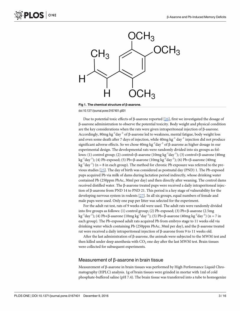
ture, see Fig 1). It is a fat-soluble substance with a small molecular weight, and was made by
dissolving in 2% Tween-80 (Sinopharm Chemical Reagent Co., Ltd).
Animals and experimental design
Sprague–Dawley rats were supplied by the Laboratory Animal Center, Anhui Medical Univer-
sity, P.R. China. Rats were individually housed in a temperature (20±3˚C) and humidity
(50±10%) controlled environment on a 12 hrs-12 hrs light-dark cycle with free access to food
and water. This study was carried out in strict accordance with the recommendations in the
Guide for the Care and Use of Laboratory Animals of the National Institutes of Health and
was approved by the Institutional Animal Care and Use Committee of Anhui Medical Univer-
sity, P.R. China. The movements, food intake, body weight and body temperature of animals
were monitored twice daily to determine their health and activity levels, including weekends
and holidays. There was no severe illness and unintended death of animals during this study.
All surgery was performed under carbon dioxide anesthesia, and all efforts were made to mini-
mize animal suffering.
β-Asarone and Pb-Induced Memory Deficits
PLOS ONE | DOI:10.1371/journal.pone.0167401 December 9, 2016 2 / 16
Technology (No. 407-037030) and the Training
Program of Innovation and Entrepreneurship for
Undergraduates of Anhui Medical University
(No.201510366121). The funders had no role in
study design, data collection and analysis, decision
to publish, or preparation of the manuscript.
Competing Interests: The authors have declared
that no competing interests exist.
Due to potential toxic effects of β-asarone reported [26], first we investigated the dosage of
β-asarone administration to observe the potential toxicity. Body weight and physical condition
are the key considerations when the rats were given intraperitoneal injection of β-asarone.
Accordingly, 80mg kg-1day-1 of β-asarone led to weakness, mental fatigue, body weight loss
and even some death after 7 days of injection, while 40mg kg-1 day-1 injection did not produce
significant adverse effects. So we chose 40mg kg-1day-1 of β-asarone as higher dosage in our
experimental design. The developmental rats were randomly divided into six groups as fol-
lows: (1) control group; (2) control+β-asarone (10mg kg-1day-1); (3) control+β-asarone (40mg
kg-1day-1); (4) Pb-exposed; (5) Pb+β-asarone (10mg kg-1day-1); (6) Pb+β-asarone (40mg
kg-1day-1) (n = 8 in each group). The method for chronic Pb exposure was referred to the pre-
vious studies [25]. The day of birth was considered as postnatal day (PND) 1. The Pb-exposed
pups acquired Pb via milk of dams during lactation period indirectly, whose drinking water
contained Pb (250ppm PbAc, 30ml per day) and then directly after weaning. The control dams
received distilled water. The β-asarone treated pups were received a daily intraperitoneal injec-
tion of β-asarone from PND 14 to PND 21. This period is a key stage of vulnerability for the
developing nervous system in rodents [27]. In all six groups, equal numbers of female and
male pups were used. Only one pup per litter was selected for the experiment.
For the adult rat test, rats of 9 weeks old were used. The adult rats were randomly divided
into five groups as follows: (1) control group; (2) Pb-exposed; (3) Pb+β-asarone (2.5mg
kg-1day-1); (4) Pb+β-asarone (10mg kg-1day-1); (5) Pb+β-asarone (40mg kg-1day-1) (n = 7 in
each group). The Pb-exposed adult rats acquired Pb from embryo stage to 11 weeks old via
drinking water which containing Pb (250ppm PbAc, 30ml per day), and the β-asarone treated
rat were received a daily intraperitoneal injection of β-asarone from 9 to 11 weeks old.
After the last administration of β-asarone, the animals were subjected to the MWM test and
then killed under deep anesthesia with CO2 one day after the last MWM test. Brain tissues
were collected for subsequent experiments.
Measurement of β-asarone in brain tissue
Measurement of β-asarone in brain tissues was performed by High Performance Liquid Chro-
matography (HPLC) analysis. 1g of brain tissues were grinded in mortar with 1ml of cold
phosphate-buffered saline (pH 7.4). The brain tissue was transferred into a tube to homogenize
Fig 1. The chemical structure of β-asarone.
doi:10.1371/journal.pone.0167401.g001
β-Asarone and Pb-Induced Memory Deficits
PLOS ONE | DOI:10.1371/journal.pone.0167401 December 9, 2016 3 / 16
by sonication (50W×15s) on ice. β-asarone was extracted by 1.5ml HPLC-grade hexane (Sino-
pharm Chemical Reagent Co., Ltd), and then the organic layer was filtered. β-asarone was sep-
arated under isocratic condition using 70% HPLC-grade methanol (Sinopharm Chemical
Reagent Co., Ltd) in ultrapure water and a column (Inertsil ODS-3, 5μm, 4×250mm). The
absorbance of the organic layer was measured at 257nm. Compound identification and analy-
sis calibration were based on use of β-asarone as external standards.
Golgi-Cox staining and spine density assay
Rats were anesthetized with CO2 and quickly decapitated. The brains were longitudinally cut
into two halves. One hemisphere was processed for morphological staining and the other
hemisphere was used to examine the expression of specific proteins. The Golgi-Cox staining
was applied with minor modification as described by Hu et al [25]. Briefly, brains stored in
dark place for 2 days (37˚C) in Golgi-Cox solution were sectioned at 200μm in the 6% sucrose
with a vibratome (VT1000S, Leica, Germany). All sections were collected on 2% gelatin-coated
slides. Then slices were stained with ammonia for 60 mins, washed with water for 3 times, fol-
lowed by Kodak Film Fix for 30 mins, and then washed with water, dehydrated, cleared, and
mounted using a resinous medium. The pyramidal neurons in hippocampal CA1 and Dentate
gyrus (DG) regions were imaged with a widefield microscope (Eclipse 80i, Nikon) using a 40x
objective.
Then, spine densities were calculated as mean numbers of spines per 10μm per dendrite per
neuron in individual rat per group. The spines counted in the present study were on 2, 3
stretches of the secondary dendrite about 20mm in length.
MWM test
The MWM experiments were performed in a circular pool with a diameter of 160cm and a
depth of 70cm. It filled to a depth of 40cm with opaque water by addition of caramel coloring,
keeping a temperature of 23±1˚C. The rats were gently put in the water facing the wall of pool.
Each rat was trained for four trials daily for 5 days to find the hidden platform. When found
the platform, it had 30 secs staying on it. If failed to reach the platform within 60 secs, it was
guided and allowed to remain on the platform for the same period of time. The platform was
removed on the sixth day, then each rat was afforded 60 secs for probe trial. Learning was
assessed by measuring the latency to find the platform. For characterization of memory, the
number of potential platform crossings and time spent in the target quadrant during the probe
trial were assessed.
Western blot analysis
Hippocampal tissues were homogenized and dissolved in the ice lysis-buffer containing a
cocktail of protein phosphatase and protease inhibitors (21μg/ml aprotinin, 0.5μg/ml leupetin,
4.9mM MgCl2, 1mM sodium-Meta-vanandante, 1% Triton X-100 and 1mM PMSF) to avoid
dephosphorylation and degradation of proteins. The samples were centrifuged at 14000rpm at
4˚C for 7 mins. The total protein of supernatant was quantified using the Bicinchoninic acid
protein assay (Beyotime Biotechnology). 30μg of proteins were resolved using 12% SDS-
PAGE, transferred onto PVDF membrane (Merck Millipore). The blot was probed with pri-
mary antibodies of NR2B, Arc/Arg3.1, Wnt7a and GAPDH (Abcam, Inc.). Proteins were incu-
bated with secondary antiboby and visualized using the electrochemiluminescence method.
All the results were normalized against GAPDH. Each target protein was performed 3 times.
β-Asarone and Pb-Induced Memory Deficits
PLOS ONE | DOI:10.1371/journal.pone.0167401 December 9, 2016 4 / 16
Semi-quantitative PCR
Total RNA of hippocampus extracted by AxyPrep Multisource Total RNA Miniprep Kit (Axy-
gen, USA) was used for cDNA transcription with cDNA Transcription Kit (TransGen, China).
Semi-quantitative PCR was performed on a S1000 Thermal Cycler (Bio-Rad, USA) using
primers pairs listed in Table 1. The results were normalized against β-actin as an internal con-
trol. Each target gene was performed 3 times.
Statistical analysis
All data were expressed as mean ± SEM. One-way ANOVA was applied to analyze the data of
HPLC, dendritic spine density, Western blot protein assay and Semi-quantitative PCR assay.
Two-way ANOVA was used to the data of training in MWM test. Difference between experi-
ment groups was tested by Fisher’s protected least significant difference (PLSD) with 95% con-
fidence. P<0.05 indicates a significance difference.
Results
β-asarone concentration in brain tissue
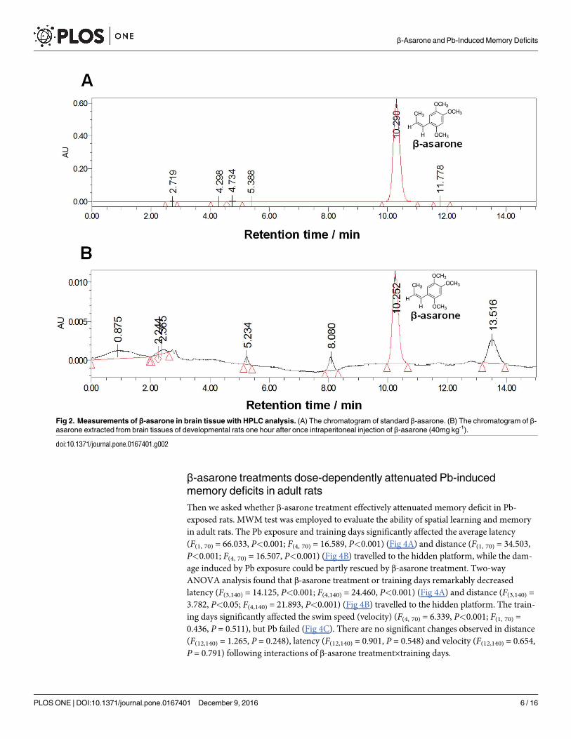
We first investigated whether β-asarone could pass through the BBB. The β-asarone in brain
tissues was measured with HPLC analysis using β-asarone as external standards for calibration
(Fig 2A). The result showed that rat pups exhibited 6.769±0.187μg g-1 of β-asarone in the brain
one hour after once intraperitoneal injection of β-asarone (40mg kg-1) (Fig 2B). The result was
consistent with previous study [14], which suggests that the β-asarone could penetrate the BBB
and be very rapidly absorbed in brain tissues.
β-asarone treatments reversed Pb-induced decrease of dendritic spine
density in hippocampal CA1 and DG areas of developmental rats
Our recent work showed that Pb exposure significantly decreased the spine density in both 14
and 21 days old pups [25], we wonder whether the addition of β-asarone could reverse this
impairment at very early developmental hippocampus. It is showed that, by Golgi-Cox staining
process, Pb exposure significantly decreased the spine density both in CA1 (Fig 3A) and DG
(Fig 3B) areas, while β-asarone administration significantly recovered the drop in spine density
in a dose-dependent manner. This result indicated that β-asarone treatments repaired Pb-
induced impairments in spine formation in developmental hippocampus.
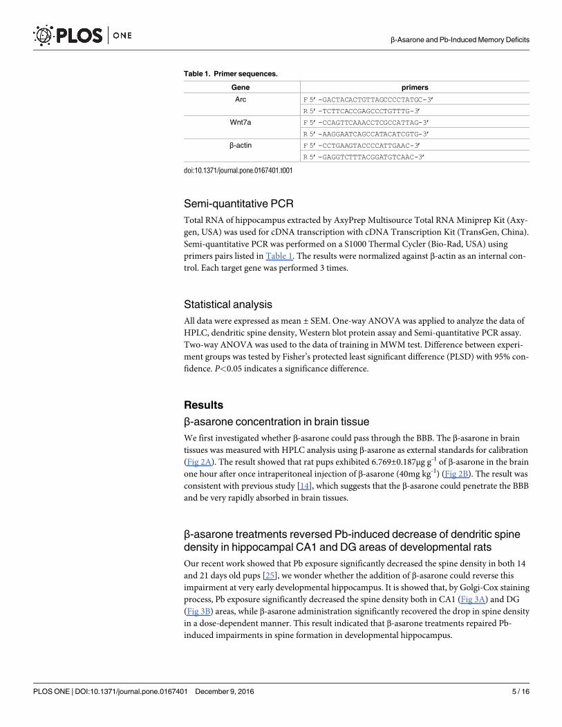
Table 1. Primer sequences.
Gene primers
Arc F 5’-GACTACACTGTTAGCCCCTATGC-3’
R 5’-TCTTCACCGAGCCCTGTTTG-3’
Wnt7a F 5’-CCAGTTCAAACCTCGCCATTAG-3’
R 5’-AAGGAATCAGCCATACATCGTG-3’
β-actin F 5’-CCTGAAGTACCCCATTGAAC-3’
R 5’-GAGGTCTTTACGGATGTCAAC-3’
doi:10.1371/journal.pone.0167401.t001
β-Asarone and Pb-Induced Memory Deficits
PLOS ONE | DOI:10.1371/journal.pone.0167401 December 9, 2016 5 / 16
β-asarone treatments dose-dependently attenuated Pb-induced
memory deficits in adult rats
Then we asked whether β-asarone treatment effectively attenuated memory deficit in Pb-
exposed rats. MWM test was employed to evaluate the ability of spatial learning and memory
in adult rats. The Pb exposure and training days significantly affected the average latency
(F(1, 70) = 66.033, P<0.001; F(4, 70) = 16.589, P<0.001) (Fig 4A) and distance (F(1, 70) = 34.503,
P<0.001; F(4, 70) = 16.507, P<0.001) (Fig 4B) travelled to the hidden platform, while the dam-
age induced by Pb exposure could be partly rescued by β-asarone treatment. Two-way
ANOVA analysis found that β-asarone treatment or training days remarkably decreased
latency (F(3,140) = 14.125, P<0.001; F(4,140) = 24.460, P<0.001) (Fig 4A) and distance (F(3,140) =
3.782, P<0.05; F(4,140) = 21.893, P<0.001) (Fig 4B) travelled to the hidden platform. The train-
ing days significantly affected the swim speed (velocity) (F(4, 70) = 6.339, P<0.001; F(1, 70) =
0.436, P = 0.511), but Pb failed (Fig 4C). There are no significant changes observed in distance
(F(12,140) = 1.265, P = 0.248), latency (F(12,140) = 0.901, P = 0.548) and velocity (F(12,140) = 0.654,
P = 0.791) following interactions of β-asarone treatment×training days.
Fig 2. Measurements of β-asarone in brain tissue with HPLC analysis. (A) The chromatogram of standard β-asarone. (B) The chromatogram of β-
asarone extracted from brain tissues of developmental rats one hour after once intraperitoneal injection of β-asarone (40mg kg-1).
doi:10.1371/journal.pone.0167401.g002
β-Asarone and Pb-Induced Memory Deficits
PLOS ONE | DOI:10.1371/journal.pone.0167401 December 9, 2016 6 / 16
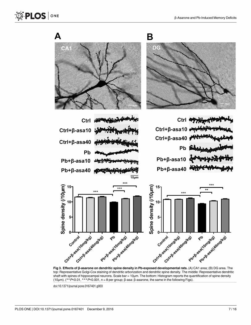
Fig 3. Effects of β-asarone on dendritic spine density in Pb-exposed developmental rats. (A) CA1 area; (B) DG area. The
top: Representative Golgi-Cox staining of dendritic arborization and dendritic spine density. The middle: Representative dendritic
shaft with spines of hippocampal neurons. Scale bar = 10μm. The bottom: Histogram reports the quantification of spine density
(10μm). (**P<0.01, ***P<0.001, n = 8 per group; β-asa: β-asarone, the same in the following Figs).
doi:10.1371/journal.pone.0167401.g003
β-Asarone and Pb-Induced Memory Deficits
PLOS ONE | DOI:10.1371/journal.pone.0167401 December 9, 2016 7 / 16
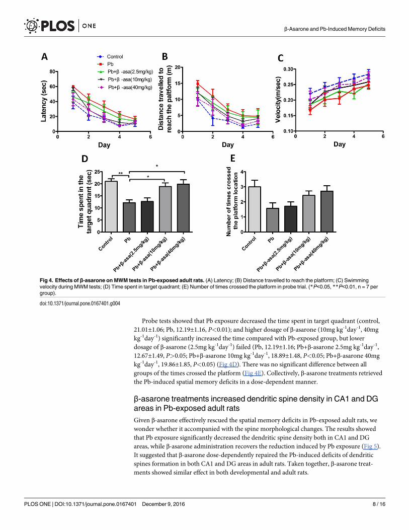
Probe tests showed that Pb exposure decreased the time spent in target quadrant (control,
21.01±1.06; Pb, 12.19±1.16, P<0.01); and higher dosage of β-asarone (10mg kg-1day-1, 40mg
kg-1day-1) significantly increased the time compared with Pb-exposed group, but lower
dosage of β-asarone (2.5mg kg-1day-1) failed (Pb, 12.19±1.16; Pb+β-asarone 2.5mg kg-1day-1,
12.67±1.49, P>0.05; Pb+β-asarone 10mg kg-1day-1, 18.89±1.48, P<0.05; Pb+β-asarone 40mg
kg-1day-1, 19.86±1.85, P<0.05) (Fig 4D). There was no significant difference between all
groups of the times crossed the platform (Fig 4E). Collectively, β-asarone treatments retrieved
the Pb-induced spatial memory deficits in a dose-dependent manner.
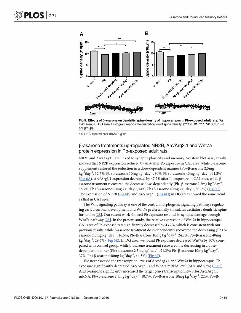
β-asarone treatments increased dendritic spine density in CA1 and DG
areas in Pb-exposed adult rats
Given β-asarone effectively rescued the spatial memory deficits in Pb-exposed adult rats, we
wonder whether it accompanied with the spine morphological changes. The results showed
that Pb exposure significantly decreased the dendritic spine density both in CA1 and DG
areas, while β-asarone administration recovers the reduction induced by Pb exposure (Fig 5).
It suggested that β-asarone dose-dependently repaired the Pb-induced deficits of dendritic
spines formation in both CA1 and DG areas in adult rats. Taken together, β-asarone treat-
ments showed similar effect in both developmental and adult rats.
Fig 4. Effects of β-asarone on MWM tests in Pb-exposed adult rats. (A) Latency; (B) Distance travelled to reach the platform; (C) Swimming
velocity during MWM tests; (D) Time spent in target quadrant; (E) Number of times crossed the platform in probe trial. (*P<0.05, **P<0.01, n = 7 per
group).
doi:10.1371/journal.pone.0167401.g004
β-Asarone and Pb-Induced Memory Deficits
PLOS ONE | DOI:10.1371/journal.pone.0167401 December 9, 2016 8 / 16
β-asarone treatments up-regulated NR2B, Arc/Arg3.1 and Wnt7a
protein expression in Pb-exposed adult rats
NR2B and Arc/Arg3.1 are linked to synaptic plasticity and memory. Western blot assay results
showed that NR2B expression reduced by 41% after Pb exposure in CA1 area, while β-asarone
supplement restored the reduction in a dose-dependent manner (Pb+β-asarone 2.5mg
kg-1day-1, 12.7%; Pb+β-asarone 10mg kg-1day-1, 30%; Pb+β-asarone 40mg kg-1day-1, 41.2%)
(Fig 6A). Arc/Arg3.1 expression decreased by 47.7% after Pb exposure in CA1 area, while β-
asarone treatment recovered the decrease dose-dependently (Pb+β-asarone 2.5mg kg-1day-1,
16.7%; Pb+β-asarone 10mg kg-1day-1, 44%; Pb+β-asarone 40mg kg-1day-1, 50.1%) (Fig 6C).
The expression of NR2B (Fig 6B) and Arc/Arg3.1 (Fig 6D) in DG area showed the same trend
as that in CA1 area.
The Wnt signaling pathway is one of the central morphogenic signaling pathways regulat-
ing early neuronal development and Wnt7a preferentially stimulates excitatory dendritic spine
formation [28]. Our recent work showed Pb exposure resulted in synapse damage through
Wnt7a pathway [25]. In the present study, the relative expression of Wnt7a in hippocampal
CA1 area of Pb-exposed rats significantly decreased by 43.2%, which is consistent with our
previous results, while β-asarone treatment dose-dependently recovered the decreasing (Pb+β-
asarone 2.5mg kg-1day-1, 16.5%; Pb+β-asarone 10mg kg-1day-1, 24.2%; Pb+β-asarone 40mg
kg-1day-1, 29.6%) (Fig 6E). In DG area, we found Pb exposure decreased Wnt7a by 50% com-
pared with control group, while β-asarone treatment recovered the decreasing in a dose-
dependent manner (Pb+β-asarone 2.5mg kg-1day-1, 21.3%; Pb+β-asarone 10mg kg-1day-1,
37%; Pb+β-asarone 40mg kg-1day-1, 44.3%) (Fig 6F).
We next assessed the transcription levels of Arc/Arg3.1 and Wnt7a in hippocampus. Pb
exposure significantly decreased Arc/Arg3.1 and Wnt7a mRNA level (61% and 57%) (Fig 7).
And β-asarone significantly increased the target genes transcription level (for Arc/Arg3.1
mRNA, Pb+β-asarone 2.5mg kg-1day-1, 10.7%, Pb+β-asarone 10mg kg-1day-1, 22%, Pb+β-
Fig 5. Effects of β-asarone on dendritic spine density of hippocampus in Pb-exposed adult rats. (A)
CA1 area; (B) DG area. Histogram reports the quantification of spine density. (**P<0.01, ***P<0.001, n = 8
per group).
doi:10.1371/journal.pone.0167401.g005
β-Asarone and Pb-Induced Memory Deficits
PLOS ONE | DOI:10.1371/journal.pone.0167401 December 9, 2016 9 / 16
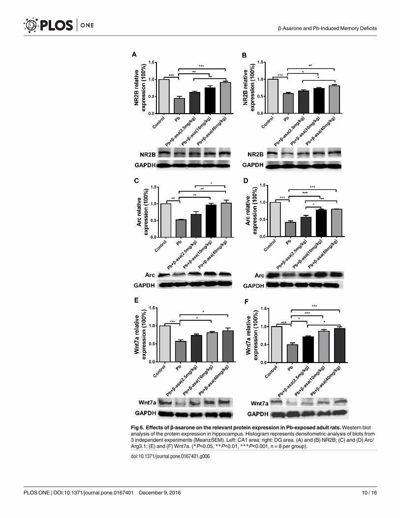
Fig 6. Effects of β-asarone on the relevant protein expression in Pb-exposed adult rats. Western blot
analysis of the protein expression in hippocampus. Histogram represents densitometric analysis of blots from
3 independent experiments (Mean±SEM). Left: CA1 area; right: DG area. (A) and (B) NR2B; (C) and (D) Arc/
Arg3.1; (E) and (F) Wnt7a. (*P<0.05, **P<0.01, ***P<0.001, n = 8 per group).
doi:10.1371/journal.pone.0167401.g006
β-Asarone and Pb-Induced Memory Deficits
PLOS ONE | DOI:10.1371/journal.pone.0167401 December 9, 2016 10 / 16
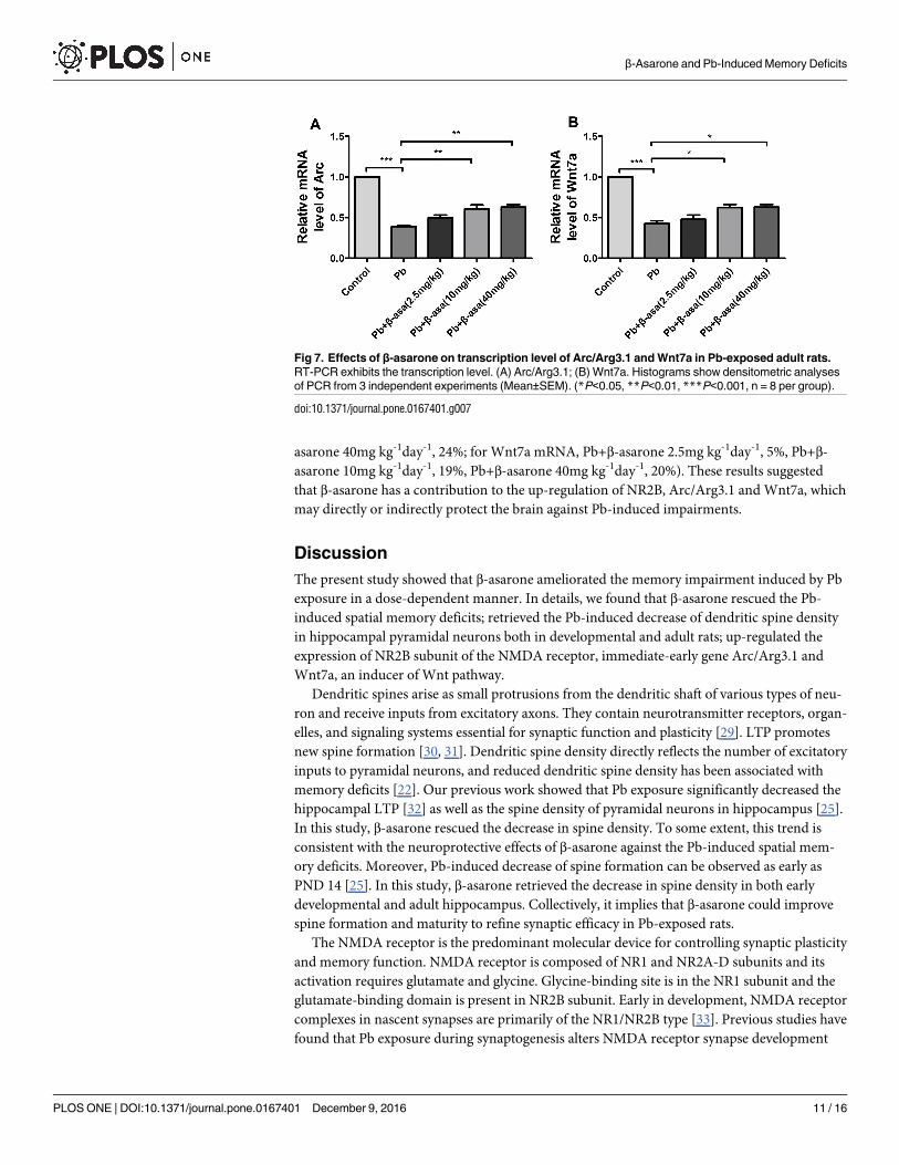
asarone 40mg kg-1day-1, 24%; for Wnt7a mRNA, Pb+β-asarone 2.5mg kg-1day-1, 5%, Pb+β-
asarone 10mg kg-1day-1, 19%, Pb+β-asarone 40mg kg-1day-1, 20%). These results suggested
that β-asarone has a contribution to the up-regulation of NR2B, Arc/Arg3.1 and Wnt7a, which
may directly or indirectly protect the brain against Pb-induced impairments.
Discussion
The present study showed that β-asarone ameliorated the memory impairment induced by Pb
exposure in a dose-dependent manner. In details, we found that β-asarone rescued the Pb-
induced spatial memory deficits; retrieved the Pb-induced decrease of dendritic spine density
in hippocampal pyramidal neurons both in developmental and adult rats; up-regulated the
expression of NR2B subunit of the NMDA receptor, immediate-early gene Arc/Arg3.1 and
Wnt7a, an inducer of Wnt pathway.
Dendritic spines arise as small protrusions from the dendritic shaft of various types of neu-
ron and receive inputs from excitatory axons. They contain neurotransmitter receptors, organ-
elles, and signaling systems essential for synaptic function and plasticity [29]. LTP promotes
new spine formation [30, 31]. Dendritic spine density directly reflects the number of excitatory
inputs to pyramidal neurons, and reduced dendritic spine density has been associated with
memory deficits [22]. Our previous work showed that Pb exposure significantly decreased the
hippocampal LTP [32] as well as the spine density of pyramidal neurons in hippocampus [25].
In this study, β-asarone rescued the decrease in spine density. To some extent, this trend is
consistent with the neuroprotective effects of β-asarone against the Pb-induced spatial mem-
ory deficits. Moreover, Pb-induced decrease of spine formation can be observed as early as
PND 14 [25]. In this study, β-asarone retrieved the decrease in spine density in both early
developmental and adult hippocampus. Collectively, it implies that β-asarone could improve
spine formation and maturity to refine synaptic efficacy in Pb-exposed rats.
The NMDA receptor is the predominant molecular device for controlling synaptic plasticity
and memory function. NMDA receptor is composed of NR1 and NR2A-D subunits and its
activation requires glutamate and glycine. Glycine-binding site is in the NR1 subunit and the
glutamate-binding domain is present in NR2B subunit. Early in development, NMDA receptor
complexes in nascent synapses are primarily of the NR1/NR2B type [33]. Previous studies have
found that Pb exposure during synaptogenesis alters NMDA receptor synapse development
Fig 7. Effects of β-asarone on transcription level of Arc/Arg3.1 and Wnt7a in Pb-exposed adult rats.
RT-PCR exhibits the transcription level. (A) Arc/Arg3.1; (B) Wnt7a. Histograms show densitometric analyses
of PCR from 3 independent experiments (Mean±SEM). (*P<0.05, **P<0.01, ***P<0.001, n = 8 per group).
doi:10.1371/journal.pone.0167401.g007
β-Asarone and Pb-Induced Memory Deficits
PLOS ONE | DOI:10.1371/journal.pone.0167401 December 9, 2016 11 / 16
via NMDA receptor inhibition [4]. And Pb impairs NMDA receptor-dependent LTP induc-
tion in hippocampus in rats [34] and down-regulates the expression of NR2A and NR2B sub-
units [35]. In this study, we found that the Pb-induced down-regulation of NR2B subunits in
hippocampus were partially reversed with β-asarone.
It should be noted that some studies reported Pb exposure during synaptogenesis in cul-
tured hippocampal neurons resulted in a decrease in NR2A-containing NMDA receptors,
while a increase in NR2B-containing NMDA receptors [4, 36]. It seems in conflict with our
present result. In fact, during brain development, there is a shift from NR2B- to NR2A-con-
taining NMDA receptors. Those studies focused on the altered composition of NMDA recep-
tors, and it is possibly that the Pb exposure changed the proportion of the NR2B subunit,
instead of the total number of it, suggesting Pb exposure probably impairs or delays the devel-
opmental switch during synaptogenesis. Our present study determined the NR2B protein level
before and after Pb exposure and β-asarone treatment. Actually, both up- and/or down-regula-
tion of NR2A/2B can plausibly disrupt the normal development of temporal processing, and
β-asarone may rescue the impairment in development and thereby improve memory.
Some proteins are involved in regulating the formation and structure of dendritic spines
[23], such as Arc/Arg3.1 [24] and Wnt7a [25]. Arc/Arg3.1, an immediate-early gene, plays an
important role in synaptic plasticity [37, 38], α-amino-3-hydroxy-5-methyl-4-isoxazole propi-
onate(AMPA) receptor trafficking [39] and neuro-behavior activity. NMDA receptor activa-
tion increases Arc/Arg3.1 levels [40] and sustained Arc/Arg3.1 synthesis has been implicated
in LTP consolidation and spine formation [41]. We observed a significant correlation between
the increases in NR2B and Arc/Arg3.1 expression levels following β-asarone treatment.
NMDA receptor activation also leads to Wnt release and β-catenin accumulation in hippo-
campus [42]. Wnt can promote the presynaptic assembly [43]. Wnt and Brain-derived neuro-
trophic factor (BDNF) cooperatively increase dendritic spine formation [44]. Inhibition of
Wnt signaling leads to decrease in arbor size of spine and is linked with neurodegenerative dis-
ease [45]. As an inducer of Wnt signaling pathway, Wnt7a can increase neurotransmitter
release in hippocampal synapses by increasing the frequency of miniature excitatory post-syn-
aptic currents (mEPSC) [46].
Our recent work showed that Pb exposure significantly decreased the spine density, accom-
panied with down-regulated Wnt7a expression [25]. In the present study, β-asarone restored
the Pb-induced decrease of Arc/Arg3.1 and Wnt7a transcription and expression, as well as the
reduction of spine density in hippocampus. In addition to our findings, β-asarone has been
reported to reverse decreased BDNF and promote hippocampal neurogenesis in chronic
unpredictable mild stress exposed rats [47]. Besides, β-asarone activated extracellular signal-
regulated kinase (ERK), a critical kinase cascades for neurogenesis, to promote proliferation
and self-renewal in neural progenitor cell [48]. BDNF can trigger the Wnt/β-catenin signaling
pathway [49]. It could mobilize synaptic vesicles and enhance synapse formation by disrupting
cadherin–β-catenin interactions [50]. It can regulate axon morphogenesis by influencing the
phosphorylation state of β-catenin [51]. Moreover, β-catenin signaling can also activate the
ERK signaling pathway [52]. For example, axin, a negative regulator of Wnt/β-catenin signal-
ing, can inhibit ERK Pathway [53]. And the Wnt antagonist Dkk1 and the β-catenin degrada-
tion stimulator Axin2 can abolish mechanical stimulation-induced ERK nuclear translocation
[54]. These reports together with our results suggest BDNF/Wnt signaling pathway might rep-
resent a key target for various neurogenesis agents, including β-asarone.
It has been previously established that the synaptic activation of NMDA receptors leads to
PKA-dependent increase of Arc/Arg3.1 expression [55, 56]. Since Pb exposure down-regulated
expressions of NR2B subunits of NMDA receptors, while β-asarone reversed the drop accom-
panied with the decrease of Arc/Arg3.1 and Wnt7a. It might be suggested that β-asarone may
β-Asarone and Pb-Induced Memory Deficits
PLOS ONE | DOI:10.1371/journal.pone.0167401 December 9, 2016 12 / 16
function by up-regulating of the NR2B, BDNF/Wnt, and ERK signaling leading to increase in
Arc/Arg3.1 expression. Although the exact molecular targets of β-asarone in promoting
neurogenesis are still unclear, our study provided preliminary hints for their underlying
mechanisms.
In conclusion, the present study demonstrated that β-asarone has neuroprotective proper-
ties against the Pb-induced impairments of spatial memory as well as dendritic spine morpho-
logical alteration. And the effect is possibly through the regulation of synaptogenesis, which is
mediated via Arc and Wnt pathway.
Supporting Information
S1 File. Experimental data of HPLC, MWM, qRT-PCR, Spine dinsity, Western-blot.
(ZIP)
Author Contributions
Conceptualization: HW XC.
Data curation: WX.
Formal analysis: QY WX.
Funding acquisition: HW XC.
Investigation: QY WX RZ YD SW LX YC.
Methodology: XC.
Project administration: XC.
Supervision: HW.
Validation: YX.
Writing – original draft: QY.
Writing – review & editing: XC.
References1. Grandjean P, Landrigan PJ. Neurobehavioural effects of developmental toxicity. Lancet Neurol. 2014;
13(3):330–8. doi: 10.1016/S1474-4422(13)70278-3 PMID: 24556010
2. Lidsky TI, Schneider JS. Adverse effects of childhood lead poisoning: the clinical neuropsychological
perspective. Environ Res. 2006; 100(2):284–93. doi: 10.1016/j.envres.2005.03.002 PMID: 16442997
3. Nihei MK, Guilarte TR. Molecular changes in glutamatergic synapses induced by Pb2+: association
with deficits of LTP and spatial learning. Neurotoxicology. 2001; 22(5):635–43. PMID: 11770885
4. Neal AP, Worley PF, Guilarte TR. Lead exposure during synaptogenesis alters NMDA receptor target-
ing via NMDA receptor inhibition. Neurotoxicology. 2011; 32(2):281–9. doi: 10.1016/j.neuro.2010.12.
013 PMID: 21192972
5. Rana TS, Mahar KS, Pandey MM, Srivastava SK, Rawat AK. Molecular and chemical profiling of ’sweet
flag’ (Acorus calamus L.) germplasm from India. Physiol Mol Biol Plants. 2013; 19(2):231–7. doi: 10.
1007/s12298-013-0164-8 PMID: 24431490
6. Satyal P, Paudel P, Poudel A, Dosoky NS, Moriarity DM, Vogler B, et al. Chemical compositions, phyto-
toxicity, and biological activities of Acorus calamus essential oils from Nepal. Nat Prod Commun. 2013;
8(8):1179–81. PMID: 24079199
7. Rajput SB, Tonge MB, Karuppayil SM. An overview on traditional uses and pharmacological profile of
Acorus calamus Linn. (Sweet flag) and other Acorus species. Phytomedicine. 2014; 21(3):268–76. doi:
10.1016/j.phymed.2013.09.020 PMID: 24200497
β-Asarone and Pb-Induced Memory Deficits
PLOS ONE | DOI:10.1371/journal.pone.0167401 December 9, 2016 13 / 16
8. Kim JH, Hahm DH, Lee HJ, Pyun KH, Shim I. Acori graminei rhizoma ameliorated ibotenic acid-induced
amnesia in rats. Evid Based Complement Alternat Med. 2009; 6(4):457–64. doi: 10.1093/ecam/
nem158 PMID: 18955253
9. Zhang H, Han T, Yu CH, Rahman K, Qin LP, Peng C. Ameliorating effects of essential oil from Acori gra-
minei rhizoma on learning and memory in aged rats and mice. J Pharm Pharmacol. 2007; 59(2):301–9.
doi: 10.1211/jpp.59.2.0016 PMID: 17270083
10. Yan L, Xu SL, Zhu KY, Lam KY, Xin G, Maiwulanjiang M, et al. Optimizing the compatibility of paired-
herb in an ancient Chinese herbal decoction Kai-Xin-San in activating neurofilament expression in cul-
tured PC12 cells. J Ethnopharmacol. 2015; 162:155–62. doi: 10.1016/j.jep.2014.12.049 PMID:
25560671
11. Zhu KY, Xu SL, Choi RC, Yan AL, Dong TT, Tsim KW. Kai-xin-san, a chinese herbal decoction contain-
ing ginseng radix et rhizoma, polygalae radix, acori tatarinowii rhizoma, and poria, stimulates the
expression and secretion of neurotrophic factors in cultured astrocytes. Evid Based Complement Alter-
nat Med. 2013; 2013:731385. doi: 10.1155/2013/731385 PMID: 24222781
12. Lee MR, Yun BS, Park SY, Ly SY, Kim SN, Han BH, et al. Anti-amnesic effect of Chong-Myung-Tang
on scopolamine-induced memory impairments in mice. J Ethnopharmacol. 2010; 132(1):70–4. doi: 10.
1016/j.jep.2010.07.041 PMID: 20673844
13. Liu L, Zhang M, Zhang R, Lee M, Wang Z, Hou J, et al. The multi-herbal formula Chong-Myung-Tang
improves spatial memory and increases cell genesis in the dentate gyrus of aged mice. Biosci Biotech-
nol Biochem. 2014; 78(10):1710–5. doi: 10.1080/09168451.2014.930319 PMID: 25273136
14. Fang YQ, Shi C, Liu L, Fang RM. Pharmacokinetics of beta-asarone in rabbit blood, hippocampus, cor-
tex, brain stem, thalamus and cerebellum. Pharmazie. 2012; 67(2):120–3. PMID: 22512081
15. Li Z, Zhao G, Qian S, Yang Z, Chen X, Chen J, et al. Cerebrovascular protection of beta-asarone in Alz-
heimer’s disease rats: a behavioral, cerebral blood flow, biochemical and genic study. J Ethnopharma-
col. 2012; 144(2):305–12. doi: 10.1016/j.jep.2012.09.013 PMID: 22985635
16. Lee B, Choi Y, Kim H, Kim SY, Hahm DH, Lee HJ, et al. Protective effects of methanol extract of Acori
graminei rhizoma and Uncariae Ramulus et Uncus on ischemia-induced neuronal death and cognitive
impairments in the rat. Life Sci. 2003; 74(4):435–50. PMID: 14609722
17. Cho J, Kim YH, Kong JY, Yang CH, Park CG. Protection of cultured rat cortical neurons from excitotoxi-
city by asarone, a major essential oil component in the rhizomes of Acorus gramineus. Life Sci. 2002;
71(5):591–9. PMID: 12052443
18. Geng Y, Li C, Liu J, Xing G, Zhou L, Dong M, et al. Beta-asarone improves cognitive function by sup-
pressing neuronal apoptosis in the beta-amyloid hippocampus injection rats. Biol Pharm Bull. 2010; 33
(5):836–43. PMID: 20460763
19. Wei G, Chen YB, Chen DF, Lai XP, Liu DH, Deng RD, et al. beta-Asarone inhibits neuronal apoptosis
via the CaMKII/CREB/Bcl-2 signaling pathway in an in vitro model and AbetaPP/PS1 mice. J Alzhei-
mers Dis. 2013; 33(3):863–80. doi: 10.3233/JAD-2012-120865 PMID: 23064259
20. Lim HW, Kumar H, Kim BW, More SV, Kim IW, Park JI, et al. beta-Asarone (cis-2,4,5-trimethoxy-1-allyl
phenyl), attenuates pro-inflammatory mediators by inhibiting NF-kappaB signaling and the JNK path-
way in LPS activated BV-2 microglia cells. Food Chem Toxicol. 2014; 72:265–72. doi: 10.1016/j.fct.
2014.07.018 PMID: 25066769
21. Drapeau E, Mayo W, Aurousseau C, Le Moal M, Piazza PV, Abrous DN. Spatial memory performances
of aged rats in the water maze predict levels of hippocampal neurogenesis. Proc Natl Acad Sci U S A.
2003; 100(24):14385–90. doi: 10.1073/pnas.2334169100 PMID: 14614143
22. Segal M. Dendritic spines and long-term plasticity. Nat Rev Neurosci. 2005; 6(4):277–84. doi: 10.1038/
nrn1649 PMID: 15803159
23. Matsuzaki M, Honkura N, Ellis-Davies GC, Kasai H. Structural basis of long-term potentiation in single
dendritic spines. Nature. 2004; 429(6993):761–6. doi: 10.1038/nature02617 PMID: 15190253
24. Tzingounis AV, Nicoll RA. Arc/Arg3.1: linking gene expression to synaptic plasticity and memory. Neu-
ron. 2006; 52(3):403–7. doi: 10.1016/j.neuron.2006.10.016 PMID: 17088207
25. Hu F, Xu L, Liu ZH, Ge MM, Ruan DY, Wang HL. Developmental lead exposure alters synaptogenesis
through inhibiting canonical Wnt pathway in vivo and in vitro. PLoS One. 2014; 9(7):e101894. doi: 10.
1371/journal.pone.0101894 PMID: 24999626
26. Unger P, Melzig MF. Comparative study of the cytotoxicity and genotoxicity of alpha- and Beta-asarone.
Sci Pharm. 2012; 80(3):663–8. doi: 10.3797/scipharm.1204-21 PMID: 23008813
27. Rice D, Barone S Jr., Critical periods of vulnerability for the developing nervous system: evidence from
humans and animal models. Environ Health Perspect. 2000; 108 Suppl 3:511–33.
28. Inestrosa NC, Varela-Nallar L. Wnt signaling in the nervous system and in Alzheimer’s disease. J Mol
Cell Biol. 2014; 6(1):64–74. doi: 10.1093/jmcb/mjt051 PMID: 24549157
β-Asarone and Pb-Induced Memory Deficits
PLOS ONE | DOI:10.1371/journal.pone.0167401 December 9, 2016 14 / 16

29. Rochefort NL, Konnerth A. Dendritic spines: from structure to in vivo function. EMBO Rep. 2012; 13
(8):699–708. doi: 10.1038/embor.2012.102 PMID: 22791026
30. Harvey CD, Svoboda K. Locally dynamic synaptic learning rules in pyramidal neuron dendrites. Nature.
2007; 450(7173):1195–200. doi: 10.1038/nature06416 PMID: 18097401
31. Colicos MA, Collins BE, Sailor MJ, Goda Y. Remodeling of synaptic actin induced by photoconductive
stimulation. Cell. 2001; 107(5):605–16. PMID: 11733060
32. Chen WH, Wang M, Yu SS, Su L, Zhu DM, She JQ, et al. Clioquinol and vitamin B12 (cobalamin) syner-
gistically rescue the lead-induced impairments of synaptic plasticity in hippocampal dentate gyrus area
of the anesthetized rats in vivo. Neuroscience. 2007; 147(3):853–64. doi: 10.1016/j.neuroscience.2007.
04.042 PMID: 17555879
33. Pina-Crespo JC, Gibb AJ. Subtypes of NMDA receptors in new-born rat hippocampal granule cells. J
Physiol. 2002; 541(Pt 1):41–64. doi: 10.1113/jphysiol.2001.014001 PMID: 12015419
34. Wang HL, Chen XT, Luo L, Lou ZY, Wang S, Chen JT, et al. Reparatory effects of nicotine on NMDA
receptor-mediated synaptic plasticity in the hippocampal CA1 region of chronically lead-exposed rats.
Eur J Neurosci. 2006; 23(5):1111–9. doi: 10.1111/j.1460-9568.2006.04645.x PMID: 16553775
35. Zhu X, Liu X, Wei F, Wang F, Merzenich MM, Schreiner CE, et al. Perceptual Training Restores
Impaired Cortical Temporal Processing Due to Lead Exposure. Cereb Cortex. 2016; 26(1):334–45. doi:
10.1093/cercor/bhu258 PMID: 25405943
36. Toscano CD, Hashemzadeh-Gargari H, McGlothan JL, Guilarte TR. Developmental Pb2+ exposure
alters NMDAR subtypes and reduces CREB phosphorylation in the rat brain. Brain Res Dev Brain Res.
2002; 139(2):217–26. PMID: 12480136
37. Lyford GL, Yamagata K, Kaufmann WE, Barnes CA, Sanders LK, Copeland NG, et al. Arc, a growth fac-
tor and activity-regulated gene, encodes a novel cytoskeleton-associated protein that is enriched in neu-
ronal dendrites. Neuron. 1995; 14(2):433–45. PMID: 7857651
38. Guzowski JF, Lyford GL, Stevenson GD, Houston FP, McGaugh JL, Worley PF, et al. Inhibition of activ-
ity-dependent arc protein expression in the rat hippocampus impairs the maintenance of long-term
potentiation and the consolidation of long-term memory. J Neurosci. 2000; 20(11):3993–4001. PMID:
10818134
39. Shepherd JD, Rumbaugh G, Wu J, Chowdhury S, Plath N, Kuhl D, et al. Arc/Arg3.1 mediates homeo-
static synaptic scaling of AMPA receptors. Neuron. 2006; 52(3):475–84. doi: 10.1016/j.neuron.2006.08.
034 PMID: 17088213
40. Steward O, Worley PF. Selective targeting of newly synthesized Arc mRNA to active synapses requires
NMDA receptor activation. Neuron. 2001; 30(1):227–40. PMID: 11343657
41. Messaoudi E, Kanhema T, Soule J, Tiron A, Dagyte G, da Silva B, et al. Sustained Arc/Arg3.1 synthesis
controls long-term potentiation consolidation through regulation of local actin polymerization in the den-
tate gyrus in vivo. J Neurosci. 2007; 27(39):10445–55. doi: 10.1523/JNEUROSCI.2883-07.2007 PMID:
17898216
42. Chen J, Park CS, Tang SJ. Activity-dependent synaptic Wnt release regulates hippocampal long term
potentiation. J Biol Chem. 2006; 281(17):11910–6. doi: 10.1074/jbc.M511920200 PMID: 16501258
43. Ahmad-Annuar A, Ciani L, Simeonidis I, Herreros J, Fredj NB, Rosso SB, et al. Signaling across the
synapse: a role for Wnt and Dishevelled in presynaptic assembly and neurotransmitter release. J Cell
Biol. 2006; 174(1):127–39. doi: 10.1083/jcb.200511054 PMID: 16818724
44. Hiester BG, Galati DF, Salinas PC, Jones KR. Neurotrophin and Wnt signaling cooperatively regulate
dendritic spine formation. Mol Cell Neurosci. 2013; 56:115–27. doi: 10.1016/j.mcn.2013.04.006 PMID:
23639831
45. Inestrosa NC, Montecinos-Oliva C, Fuenzalida M. Wnt signaling: role in Alzheimer disease and schizo-
phrenia. J Neuroimmune Pharmacol. 2012; 7(4):788–807. doi: 10.1007/s11481-012-9417-5 PMID:
23160851
46. Cerpa W, Godoy JA, Alfaro I, Farias GG, Metcalfe MJ, Fuentealba R, et al. Wnt-7a modulates the syn-
aptic vesicle cycle and synaptic transmission in hippocampal neurons. J Biol Chem. 2008; 283
(9):5918–27. doi: 10.1074/jbc.M705943200 PMID: 18096705
47. Dong H, Gao Z, Rong H, Jin M, Zhang X. beta-asarone reverses chronic unpredictable mild stress-
induced depression-like behavior and promotes hippocampal neurogenesis in rats. Molecules. 2014;
19(5):5634–49. doi: 10.3390/molecules19055634 PMID: 24786848
48. Mao J, Huang S, Liu S, Feng XL, Yu M, Liu J, et al. A herbal medicine for Alzheimer’s disease and its
active constituents promote neural progenitor proliferation. Aging Cell. 2015; 14(5):784–96. doi: 10.
1111/acel.12356 PMID: 26010330
β-Asarone and Pb-Induced Memory Deficits
PLOS ONE | DOI:10.1371/journal.pone.0167401 December 9, 2016 15 / 16
49. Chen BY, Wang X, Wang ZY, Wang YZ, Chen LW, Luo ZJ. Brain-derived neurotrophic factor stimulates
proliferation and differentiation of neural stem cells, possibly by triggering the Wnt/beta-catenin signal-
ing pathway. J Neurosci Res. 2013; 91(1):30–41. doi: 10.1002/jnr.23138 PMID: 23023811
50. Bamji SX, Rico B, Kimes N, Reichardt LF. BDNF mobilizes synaptic vesicles and enhances synapse
formation by disrupting cadherin-beta-catenin interactions. The Journal of cell biology. 2006; 174
(2):289–99. doi: 10.1083/jcb.200601087 PMID: 16831887
51. David MD, Yeramian A, Dunach M, Llovera M, Canti C, de Herreros AG, et al. Signalling by neurotro-
phins and hepatocyte growth factor regulates axon morphogenesis by differential beta-catenin phos-
phorylation. J Cell Sci. 2008; 121(Pt 16):2718–30. doi: 10.1242/jcs.029660 PMID: 18664491
52. Yun MS, Kim SE, Jeon SH, Lee JS, Choi KY. Both ERK and Wnt/beta-catenin pathways are involved in
Wnt3a-induced proliferation. J Cell Sci. 2005; 118(Pt 2):313–22. doi: 10.1242/jcs.01601 PMID:
15615777
53. Jeon SH, Yoon JY, Park YN, Jeong WJ, Kim S, Jho EH, et al. Axin inhibits extracellular signal-regulated
kinase pathway by Ras degradation via beta-catenin. The J Biol Chem. 2007; 282(19):14482–92. doi:
10.1074/jbc.M611129200 PMID: 17374607
54. Gortazar AR, Martin-Millan M, Bravo B, Plotkin LI, Bellido T. Crosstalk between caveolin-1/extracellular
signal-regulated kinase (ERK) and beta-catenin survival pathways in osteocyte mechanotransduction.
J Biol Chem. 2013; 288(12):8168–75. doi: 10.1074/jbc.M112.437921 PMID: 23362257
55. Bloomer WA, VanDongen HM, VanDongen AM. Arc/Arg3.1 translation is controlled by convergent N-
methyl-D-aspartate and Gs-coupled receptor signaling pathways. J Biol Chem. 2008; 283(1):582–92.
doi: 10.1074/jbc.M702451200 PMID: 17981809
56. Falsafi SK, Deli A, Hoger H, Pollak A, Lubec G. Scopolamine administration modulates muscarinic, nic-
otinic and NMDA receptor systems. PloS one. 2012; 7(2):e32082. doi: 10.1371/journal.pone.0032082
PMID: 22384146
β-Asarone and Pb-Induced Memory Deficits
PLOS ONE | DOI:10.1371/journal.pone.0167401 December 9, 2016 16 / 16