Embed Size (px)
Citation preview
ARTICLE
Broad and potent neutralizing human antibodies totick-borne flaviviruses protect mice from diseaseMarianna Agudelo1, Martin Palus2,3*, Jennifer R. Keeffe4*, Filippo Bianchini1,5, Pavel Svoboda3,6, Jirı Salat2,3, Avery Peace7,Anna Gazumyan1, Melissa Cipolla1, Tania Kapoor1, Francesca Guidetti1, Kai-Hui Yao1, Jana Elsterova2,3, Dana Teislerova8,Ales Chrdle8,9,10, Vaclav Honig2,3, Thiago Oliveira1, Anthony P. West Jr.4, Yu E. Lee4, Charles M. Rice7, Margaret R. MacDonald7,Pamela J. Bjorkman4, Daniel Ruzek2,3, Davide F. Robbiani1,5, and Michel C. Nussenzweig1,11
Tick-borne encephalitis virus (TBEV) is an emerging human pathogen that causes potentially fatal disease with nospecific treatment. Mouse monoclonal antibodies are protective against TBEV, but little is known about the humanantibody response to infection. Here, we report on the human neutralizing antibody response to TBEV in a cohort ofinfected and vaccinated individuals. Expanded clones of memory B cells expressed closely related anti-envelopedomain III (EDIII) antibodies in both groups of volunteers. However, the most potent neutralizing antibodies, with IC50sbelow 1 ng/ml, were found only in individuals who recovered from natural infection. These antibodies also neutralizedother tick-borne flaviviruses, including Langat, louping ill, Omsk hemorrhagic fever, Kyasanur forest disease, andPowassan viruses. Structural analysis revealed a conserved epitope near the lateral ridge of EDIII adjoining the EDI–EDIIIhinge region. Prophylactic or early therapeutic antibody administration was effective at low doses in mice that werelethally infected with TBEV.
IntroductionTick-borne flaviviruses are responsible for a series of emerginginfectious diseases including fatal encephalitis. Like other fla-viviruses, the tick-borne encephalitis virus (TBEV) envelopeprotein (E) is composed of three structural domains (envelopedomains I–III [EDI–EDIII]; Füzik et al., 2018; Pulkkinen et al.,2018). Mouse monoclonal antibodies against EDIII, an Ig-likedomain that mediates host cell attachment, are potent neutral-izers of TBEV (Baykov et al., 2014; Füzik et al., 2018; Levanovet al., 2010; Matveev et al., 2020; Phillpotts et al., 1985; Rey et al.,1995; Yang et al., 2019).
TBEV is one of the six flaviviruses transmitted by tickscausing human disease (Gould and Solomon, 2008; Kuno et al.,1998; LaSala and Holbrook, 2010). These include Omsk hemor-rhagic fever virus (OHFV) in Russia; Kyasanur forest diseasevirus (KFDV) in India; Alkhurma virus in Saudi Arabia; loupingill virus (LIV) in the United Kingdom, Ireland, Norway, Den-mark, and Russia; and Powassan virus in the United States andCanada. Upwards of 10,000 TBEV cases per year are reported,with a trend for increased incidence in recent years and
emergence of the disease in new geographic regions (Beauteet al., 2018; Girl et al., 2020; Kollaritsch et al., 2011; Morensand Fauci, 2020; Smura et al., 2019; Süss et al., 2006; Yoshii,2019; Zeman and Bene, 2004).
The bite of an infected tick, or the consumption of unpas-teurized milk from infected animals, causes a biphasic illness,which begins with a period of influenza-like symptoms followedby the development of neurological disease (tick-borne en-cephalitis [TBE]). There is no specific therapy for TBE, andtreatment is limited to supportive care. For those individualswho survive, long-term sequelae are common (Bogovic et al.,2018b; Caini et al., 2012; Cisak et al., 2010; Donoso-Mantkeet al., 2011; Holzmann, 2003; Holzmann et al., 2009; Kaiser,2008).
Although TBEV vaccines are available, immunity requiresregular boosting, and vaccination is less effective in the youngand elderly. Vaccination requires administration of three sepa-rate doses spaced over up to 2 yr, with booster doses recom-mended at intervals of 3–5 yr (World Health Organization,
.............................................................................................................................................................................1Laboratory of Molecular Immunology, The Rockefeller University, New York, NY; 2Institute of Parasitology, Biology Centre of the Czech Academy of Sciences, CeskeBudejovice, Czech Republic; 3Veterinary Research Institute, Brno, Czech Republic; 4Division of Biology and Biological Engineering, California Institute of Technology,Pasadena, CA; 5Institute for Research in Biomedicine, Universita della Svizzera italiana, Bellinzona, Switzerland; 6Department of Pharmacology and Pharmacy, Faculty ofVeterinary Medicine, University of Veterinary and Pharmaceutical Sciences Brno, Brno, Czech Republic; 7Laboratory of Virology and Infectious Disease, The RockefellerUniversity, New York, NY; 8Hospital Ceske Budejovice, Ceske Budejovice, Czech Republic; 9Faculty of Social and Health Sciences, University of South Bohemia, CeskeBudejovice, Czech Republic; 10Royal Liverpool University Hospital, Liverpool, UK; 11Howard Hughes Medical Institute, The Rockefeller University, New York, NY.
*M. Palus and J.R. Keeffe contributed equally to this paper; Correspondence to Michel C. Nussenzweig: [email protected]; Davide F. Robbiani: [email protected]; Daniel Ruzek: [email protected]; Pamela J. Bjorkman: [email protected].
© 2021 Agudelo et al. This article is available under a Creative Commons License (Attribution 4.0 International, as described at https://creativecommons.org/licenses/by/4.0/).
Rockefeller University Press https://doi.org/10.1084/jem.20210236 1 of 16
J. Exp. Med. 2021 Vol. 218 No. 5 e20210236
Dow
nloaded from http://rupress.org/jem
/article-pdf/218/5/e20210236/1412984/jem_20210236.pdf by guest on 24 January 2022
2019). Breakthrough TBEV infection occurs despite vaccination(Dobler et al., 2020; Lotric-Furlan et al., 2017).
Recovered individuals can produce potent serologic neutral-izing activity against TBEV, and the serum can be cross-reactiveagainst other tick-borne flaviviruses, but the nature of the an-tibodies that mediate these effects is not known (Calisher et al.,1989; Mansfield et al., 2011). Postexposure prophylaxis withhyperimmune plasma Ig from vaccinated donors provides pro-tection if administered within 3 d of an infected tick bite (Kreilet al., 1998; Pen’evskaia and Rudakov, 2010). However, thispractice was discontinued outside of Russia due to anecdotalevidence of adverse events in children and the concern of pos-sible antibody-dependent enhancement of disease (Halstead,2014; Phillpotts et al., 1985; Ruzek et al., 2019).
Here, we report on the molecular properties of humananti-TBEV antibodies with exceptional neutralizing potencyand breadth that are effective in protection and early ther-apy in mice.
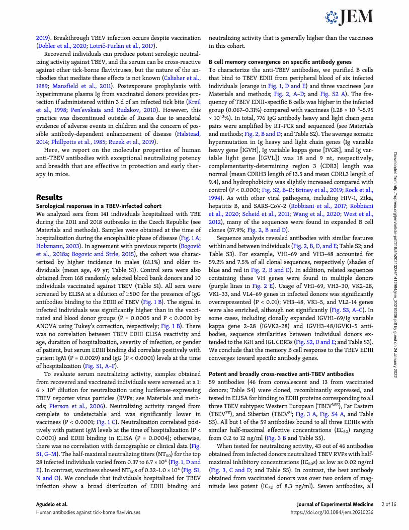
ResultsSerological responses in a TBEV-infected cohortWe analyzed sera from 141 individuals hospitalized with TBEduring the 2011 and 2018 outbreaks in the Czech Republic (seeMaterials and methods). Samples were obtained at the time ofhospitalization during the encephalitic phase of disease (Fig. 1 A;Holzmann, 2003). In agreement with previous reports (Bogovicet al., 2018a; Bogovic and Strle, 2015), the cohort was charac-terized by higher incidence in males (61.1%) and older in-dividuals (mean age, 49 yr; Table S1). Control sera were alsoobtained from 168 randomly selected blood bank donors and 10individuals vaccinated against TBEV (Table S1). All sera werescreened by ELISA at a dilution of 1:500 for the presence of IgGantibodies binding to the EDIII of TBEV (Fig. 1 B). The signal ininfected individuals was significantly higher than in the vacci-nated and blood donor groups (P = 0.0005 and P < 0.0001 byANOVA using Tukey’s correction, respectively; Fig. 1 B). Therewas no correlation between TBEV EDIII ELISA reactivity andage, duration of hospitalization, severity of infection, or genderof patient, but serum EDIII binding did correlate positively withpatient IgM (P = 0.0029) and IgG (P < 0.0001) levels at the timeof hospitalization (Fig. S1, A–F).
To evaluate serum neutralizing activity, samples obtainedfrom recovered and vaccinated individuals were screened at a 1:6 × 10⁵ dilution for neutralization using luciferase-expressingTBEV reporter virus particles (RVPs; see Materials and meth-ods; Pierson et al., 2006). Neutralizing activity ranged fromcomplete to undetectable and was significantly lower invaccinees (P < 0.0001; Fig. 1 C). Neutralization correlated posi-tively with patient IgM levels at the time of hospitalization (P <0.0001) and EDIII binding in ELISA (P = 0.0004); otherwise,there was no correlation with demographic or clinical data (Fig.S1, G–M). The half-maximal neutralizing titers (NT50) for the top28 infected individuals varied from 0.37 to 6.7 × 10⁶ (Fig. 1, D andE). In contrast, vaccinees showed NT50s of 0.32–1.0 × 10⁴ (Fig. S1,N and O). We conclude that individuals hospitalized for TBEVinfection show a broad distribution of EDIII binding and
neutralizing activity that is generally higher than the vaccineesin this cohort.
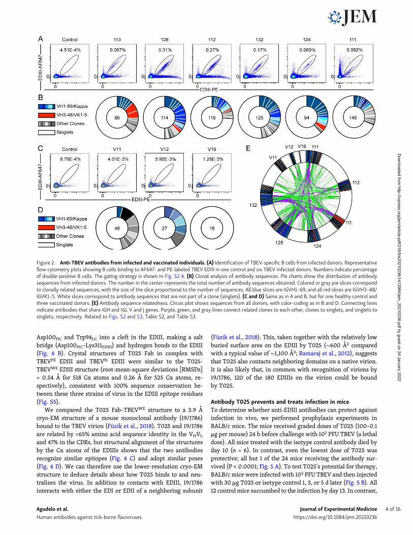
B cell memory convergence on specific antibody genesTo characterize the anti-TBEV antibodies, we purified B cellsthat bind to TBEV EDIII from peripheral blood of six infectedindividuals (orange in Fig. 1, D and E) and three vaccinees (seeMaterials and methods; Fig. 2, A–D; and Fig. S2 A). The fre-quency of TBEV EDIII–specific B cells was higher in the infectedgroup (0.067–0.31%) compared with vaccinees (1.28 × 10⁻³–5.95× 10⁻³%). In total, 776 IgG antibody heavy and light chain genepairs were amplified by RT-PCR and sequenced (see Materialsand methods; Fig. 2, B and D; and Table S2). The average somatichypermutation in Ig heavy and light chain genes (Ig variableheavy gene [IGVH], Ig variable kappa gene [IVGK], and Ig var-iable light gene [IGVL]) was 18 and 9 nt, respectively,complementarity-determining region 3 (CDR3) length wasnormal (mean CDRH3 length of 13.5 and mean CDRL3 length of9.4), and hydrophobicity was slightly increased compared withcontrol (P < 0.0001; Fig. S2, B–D; Briney et al., 2019; Rock et al.,1994). As with other viral pathogens, including HIV-1, Zika,hepatitis B, and SARS-CoV-2 (Robbiani et al., 2017; Robbianiet al., 2020; Scheid et al., 2011; Wang et al., 2020; West et al.,2012), many of the sequences were found in expanded B cellclones (37.9%; Fig. 2, B and D).
Sequence analysis revealed antibodies with similar featureswithin and between individuals (Fig. 2, B, D, and E; Table S2; andTable S3). For example, VH1–69 and VH3–48 accounted for59.2% and 7.5% of all clonal sequences, respectively (shades ofblue and red in Fig. 2, B and D). In addition, related sequencescontaining these VH genes were found in multiple donors(purple lines in Fig. 2 E). Usage of VH1–69, VH3–30, VK2–28,VK1–33, and VL4–69 genes in infected donors was significantlyoverrepresented (P < 0.01); VH3–48, VK1–5, and VL2–14 geneswere also enriched, although not significantly (Fig. S3, A–C). Insome cases, including clonally expanded IGVH1–69/Ig variablekappa gene 2–28 (IGVK2–28) and IGVH3–48/IGVK1–5 anti-bodies, sequence similarities between individual donors ex-tended to the IGH and IGL CDR3s (Fig. S2, D and E; and Table S3).We conclude that the memory B cell response to the TBEV EDIIIconverges toward specific antibody genes.
Potent and broadly cross-reactive anti-TBEV antibodies59 antibodies (46 from convalescent and 13 from vaccinateddonors; Table S4) were cloned, recombinantly expressed, andtested in ELISA for binding to EDIII proteins corresponding to allthree TBEV subtypes: Western European (TBEVWE), Far Eastern(TBEVFE), and Siberian (TBEVSi; Fig. 3 A, Fig. S4 A, and TableS5). All but 1 of the 59 antibodies bound to all three EDIIIs withsimilar half-maximal effective concentrations (EC50) rangingfrom 0.2 to 12 ng/ml (Fig. 3 B and Table S5).
When tested for neutralizing activity, 43 out of 46 antibodiesobtained from infected donors neutralized TBEV RVPswith half-maximal inhibitory concentrations (IC50s) as low as 0.02 ng/ml(Fig. 3, C and D; and Table S5). In contrast, the best antibodyobtained from vaccinated donors was over two orders of mag-nitude less potent (IC50 of 8.3 ng/ml). Seven antibodies, all
Agudelo et al. Journal of Experimental Medicine 2 of 16
Human antibodies against tick-borne flaviviruses https://doi.org/10.1084/jem.20210236
Dow
nloaded from http://rupress.org/jem
/article-pdf/218/5/e20210236/1412984/jem_20210236.pdf by guest on 24 January 2022
isolated from infected donors, were potent neutralizers of TBEVRVPs with IC50s below 1 ng/ml (Fig. 3 D). Four of these anti-bodies were also evaluated for neutralization of authentic TBEV(Fig. 3, E and F). All four antibodies showed potent activity, withIC50s ranging from 35.9 to 268.8 ng/ml (Table S5).
To determine whether the TBEV antibodies cross-react withrelated viruses, we screened them at a single concentration(1 µg/ml) for binding to the EDIIIs of Langat virus (LGTV), LIV,OHFV, KFDV, and Powassan lineage I and II viruses (Powassan-LB virus [POWV-LB] and Powassan deer tick virus [POWV-DTV]; see Materials and methods and Fig. S4, B and C). Broadcross-reactivity was observed for many of the antibodies tested(Fig. S4, B and C). To determine whether the antibodies are alsobroadly neutralizing, we screened them against RVPs corre-sponding to the same panel of tick-borne viruses. When tested ata concentration of 1 µg/ml, most of the IGHV1–69 antibodiesneutralized LGTV, LIV, POWV-LB, and POWV-DTV, and one ofthe IGVH3–48/IGVK1–5 antibodies neutralized all RVPs exceptPOWV-LB (Fig. S4, B and C). IC50s against the flavivirus RVPpanel were <10 ng/ml for several of the cross-reactive antibodies
(Fig. 3 G; Fig. S4, D–I; and Table S5). For example, an IGVH3–48/IGVK1–5 antibody, T056, is a potent neutralizer of LGTV, LIV,and OHFV, with IC50 values ≤1 ng/ml. We conclude that someTBEV neutralizing antibodies are broadly active against tick-borne flaviviruses.
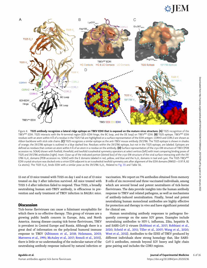
Antibody T025 binds to the EDIII lateral ridgeTo gain insights into the mechanism of neutralization by humananti-TBEV antibodies, we solved crystal structures of theantigen-binding fragment (Fab) from the broad and potent an-tibody T025 (IGVH3–30/IGVK3–15) in complex with the EDIIIdomains of all three subtypes of TBEV (Fig. 4, Fig. S5, and TableS6). The structure of the T025 Fab–TBEVWE EDIII complex re-vealed that the antibody binds the lateral ridge of EDIII in theproximity of the EDI–EDIII hinge region, making both heavychain (HC) and light chain (LC) contacts to the EDI–EDIII hingeand the BC loop and light chain contacts to the DE loop of theEDIII (Fig. 4 A). The antibody contacts EDIII using CDRH2,CDRH3, CDRL1, and CDRL3, and buries 598 A2 of surface area onthe EDIII (333 A2 by the VH and 265 A2 by the VL). T025 inserts
Figure 1. Screening individuals for TBEV antibodies. (A) Diagrammatic representation of the clinical course of TBE. The approximate time of serumcollection is shown in yellow. (B) TBEV EDIII IgG ELISA. Graph shows optical density measurement (y axis) relative to a negative control serum for samples from141 TBEV-infected individuals, 10 TBEV vaccinees, and 168 random blood donors (1:500 dilution) measured in singlicate. P = 0.0005 for infected versusvaccinees; P < 0.0001 for infected versus blood donors; P = 0.0003 for vaccinees versus blood donors; calculated by one-way ANOVA followed by Tukey’s test.Horizontal lines indicate the mean. (C) TBEV RVP neutralization screening. Graph shows ranked serum neutralizing activity (1:600,000 dilution) against TBEVRVPs (average of duplicate wells) relative to no serum control. The orange box (bottom right) indicates the 28 best neutralizers of 141 TBEV-infected individualsand 10 TBEV vaccinees tested. P < 0.0001; calculated using two-tailed Mann–Whitney test. (D) TBEV RVP neutralization curves. Plot shows representativeneutralization curves for each of the 28 most potent sera from C. Representative of two experiments, each performed in triplicate. Error bars indicate standarddeviation. (E) Ranked NT50s for the top 28 individuals. Average of two independent experiments. In D and E, orange indicates the donors of PBMCs for antibodycloning. Related to Fig. S1 and Table S1. RLU, relative light units.
Agudelo et al. Journal of Experimental Medicine 3 of 16
Human antibodies against tick-borne flaviviruses https://doi.org/10.1084/jem.20210236
Dow
nloaded from http://rupress.org/jem
/article-pdf/218/5/e20210236/1412984/jem_20210236.pdf by guest on 24 January 2022
Asp100HC and Trp94LC into a cleft in the EDIII, making a saltbridge (Asp100HC–Lys311EDIII) and hydrogen bonds to the EDIII(Fig. 4 B). Crystal structures of T025 Fab in complex withTBEVFE EDIII and TBEVSi EDIII were similar to the T025-TBEVWE EDIII structure (root-mean-square deviations [RMSDs]= 0.54 A for 518 Cα atoms and 0.26 A for 525 Cα atoms, re-spectively), consistent with 100% sequence conservation be-tween these three strains of virus in the EDIII epitope residues(Fig. S5).
We compared the T025 Fab–TBEVWE structure to a 3.9 Acryo-EM structure of a mouse monoclonal antibody (19/1786)bound to the TBEV virion (Füzik et al., 2018). T025 and 19/1786are related by <65% amino acid sequence identity in the VHVL
and 47% in the CDRs, but structural alignment of the structuresby the Cα atoms of the EDIIIs shows that the two antibodiesrecognize similar epitopes (Fig. 4 C) and adopt similar poses(Fig. 4 D). We can therefore use the lower-resolution cryo-EMstructure to deduce details about how T025 binds to and neu-tralizes the virus. In addition to contacts with EDIII, 19/1786interacts with either the EDI or EDII of a neighboring subunit
(Füzik et al., 2018). This, taken together with the relatively lowburied surface area on the EDIII by T025 (∼600 A2 comparedwith a typical value of ∼1,100 A2; Ramaraj et al., 2012), suggeststhat T025 also contacts neighboring domains on a native virion.It is also likely that, in common with recognition of virions by19/1786, 120 of the 180 EDIIIs on the virion could be boundby T025.
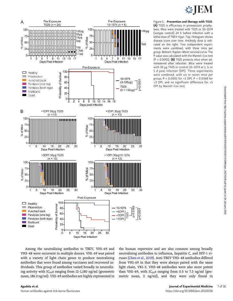
Antibody T025 prevents and treats infection in miceTo determine whether anti-EDIII antibodies can protect againstinfection in vivo, we performed prophylaxis experiments inBALB/c mice. The mice received graded doses of T025 (100–0.1µg per mouse) 24 h before challenge with 102 PFU TBEV (a lethaldose). All mice treated with the isotype control antibody died byday 10 (n = 6). In contrast, even the lowest dose of T025 wasprotective; all but 1 of the 24 mice receiving the antibody sur-vived (P < 0.0001; Fig. 5 A). To test T025’s potential for therapy,BALB/c mice were infected with 102 PFU TBEV and then injectedwith 30 µg T025 or isotype control 1, 3, or 5 d later (Fig. 5 B). All12 control mice succumbed to the infection by day 13. In contrast,
Figure 2. Anti-TBEV antibodies from infected and vaccinated individuals. (A) Identification of TBEV-specific B cells from infected donors. Representativeflow cytometry plots showing B cells binding to AF647- and PE-labeled TBEV EDIII in one control and six TBEV-infected donors. Numbers indicate percentageof double-positive B cells. The gating strategy is shown in Fig. S2 A. (B) Clonal analysis of antibody sequences. Pie charts show the distribution of antibodysequences from infected donors. The number in the center represents the total number of antibody sequences obtained. Colored or gray pie slices correspondto clonally related sequences, with the size of the slice proportional to the number of sequences. All blue slices are IGVH1–69, and all red slices are IGVH3–48/IGVK1–5. White slices correspond to antibody sequences that are not part of a clone (singlets). (C and D) Same as in A and B, but for one healthy control andthree vaccinated donors. (E) Antibody sequence relatedness. Circos plot shows sequences from all donors, with color-coding as in B and D. Connecting linesindicate antibodies that share IGH and IGL V and J genes. Purple, green, and gray lines connect related clones to each other, clones to singlets, and singlets tosinglets, respectively. Related to Figs. S2 and S3, Table S2, and Table S3.
Agudelo et al. Journal of Experimental Medicine 4 of 16
Human antibodies against tick-borne flaviviruses https://doi.org/10.1084/jem.20210236
Dow
nloaded from http://rupress.org/jem
/article-pdf/218/5/e20210236/1412984/jem_20210236.pdf by guest on 24 January 2022
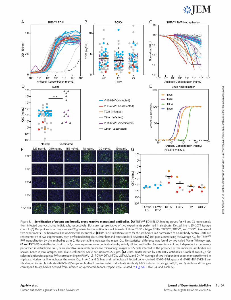
Figure 3. Identification of potent and broadly cross-reactive monoclonal antibodies. (A) TBEVWE EDIII ELISA binding curves for 46 and 13 monoclonalsfrom infected and vaccinated individuals, respectively. Data are representative of two experiments performed in singlicate. Dotted line is 10–1074 isotypecontrol. (B) Dot plot summarizing average EC50 values for the antibodies in A to each of three TBEV subtype EDIIIs: TBEVWE, TBEVFE, and TBEVSi. Average oftwo experiments. The horizontal lines indicate themean value. (C) RVP neutralization curves for the antibodies in A normalized to no antibody control. Data arerepresentative of two experiments, each performed in triplicate. Error bars indicate standard deviation. (D) Dot plot summarizing the average IC50 for TBEVWE
RVP neutralization by the antibodies as in C. Horizontal line indicates the mean IC50. No statistical difference was found by two-tailed Mann–Whitney test.(E and F) TBEV neutralization in vitro. In E, curves represent virus neutralization by serially diluted antibodies. Representative of two independent experimentsperformed in octuplicate. In F, representative immunofluorescence microscopy images of PS cells infected in the presence of the indicated antibodies areshown. Green is viral antigen, and blue is cell nuclei. Scale bar indicates 200 µm. (G) Cross-neutralization by anti-TBEV antibodies. Graph shows IC50s forselected antibodies against RVPs corresponding to POWV-LB, POWV-DTV, KFDV, LGTV, LIV, and OHFV. Average of two independent experiments performed intriplicate. Horizontal line indicates the mean IC50. In A–D and G, blue and red indicate infected donor-derived IGVH1–69/kappa and IGVH3-48/IGVK1–5 an-tibodies, while purple indicates IGHV1–69/kappa antibodies from vaccinated individuals. Antibody T025 is shown in orange. In B, D, and G, circles and trianglescorrespond to antibodies derived from infected or vaccinated donors, respectively. Related to Fig. S4, Table S4, and Table S5.
Agudelo et al. Journal of Experimental Medicine 5 of 16
Human antibodies against tick-borne flaviviruses https://doi.org/10.1084/jem.20210236
Dow
nloaded from http://rupress.org/jem
/article-pdf/218/5/e20210236/1412984/jem_20210236.pdf by guest on 24 January 2022
12 out of 13 mice treated with T025 on day 1 and 4 out of 13 micetreated on day 3 after infection survived. All mice treated withT025 5 d after infection failed to respond. Thus T025, a broadlyneutralizing human anti-TBEV antibody, is efficacious in pre-vention and early treatment of TBEV infection in BALB/c mice.
DiscussionTick-borne flaviviruses can cause a fulminant encephalitis forwhich there is no effective therapy. This group of viruses are agrowing public health concern in Europe, Asia, and NorthAmerica. Among disease-causing tick-borne flaviviruses, TBEVis prevalent in Central Europe and Russia. Although there is agreat deal of information on the polyclonal humoral immuneresponse to TBEV (Albinsson et al., 2018; Holzmann, 2003;Matveeva et al., 1995; McAuley et al., 2017; Remoli et al., 2015),there is little or no understanding of the molecular nature of theneutralizing antibody response induced by natural infection or
vaccination. We report on 776 antibodies obtained frommemoryB cells of six recovered and three vaccinated individuals, amongwhich are several broad and potent neutralizers of tick-borneflaviviruses. The data provide insights into the human antibodyresponse to TBEV and related pathogens, as well as mechanismsof antibody-induced neutralization. Finally, broad and potentneutralizing human monoclonal antibodies are highly effectivefor protection and therapy in vivo and have significant potentialfor clinical use.
Human neutralizing antibody responses to pathogens fre-quently converge on the same IGV genes. Examples includeneutralizing antibodies to HIV-1, influenza, Zika, hepatitis B,and SARS-CoV-2 viruses (Robbiani et al., 2017; Robbiani et al.,2020; Scheid et al., 2011; Tiller et al., 2007; Wang et al., 2020;West et al., 2012). Antibodies to the EDIII of TBEV produced bydifferent individuals show strong homology that, like SARS-CoV-2 antibodies, extends beyond IGV heavy and light chaingene pairing and includes the CDR3 regions.
Figure 4. T025 antibody recognizes a lateral ridge epitope on TBEV EDIII that is exposed on the mature virus structure. (A) T025 recognition of theTBEVWE EDIII. T025 interacts with the N-terminal region (EDI–EDIII hinge, the BC loop, and the DE loop) on TBEVWE EDIII. (B) T025 epitope. TBEVWE EDIIIresidues with an atom within 4 A of a residue in the T025 Fab are highlighted on a surface representation of the EDIII antigen. CDRH3 and CDRL3 are shown asribbon backbone with stick side chains. (C) T025 recognizes a similar epitope as the anti-TBEV mouse antibody 19/1786. The T025 epitope is shown in shadesof orange; the 19/1786 epitope is outlined in a blue dashed line. Residues within the 19/1786 epitope, but not in the T025 epitope, are labeled. Epitopes aredefined as residues that contain an atom within 4 A of an atom in a residue on the antibody. (D) Surface representation of the cryo-EM structure of TBEV (PDBaccession no. 5O6A) shown with fivefold, threefold, and twofold icosahedral symmetry operators at select vertices (left) with inset comparing binding poses ofT025 and 19/1786 antibodies (right). Inset: Close-up of the indicated portion (dotted box) of the cryo-EM structure of the viral surface interacting with the 19/1786 VHVL domains (PDB accession no. 5O6V) with the E domains labeled in red, yellow, and blue and the VHVL domains in teal and cyan. The T025-TBEVWE
EDIII crystal structure was docked onto a virion EDIII adjacent to an icosahedral twofold symmetry axis after alignment of the EDIII domains (RMSD = 0.97 A, 82Ca atoms). The T025 VHVL binds EDIII with a similar pose as the 19/1786 VHVL. Related to Fig. S5 and Table S6.
Agudelo et al. Journal of Experimental Medicine 6 of 16
Human antibodies against tick-borne flaviviruses https://doi.org/10.1084/jem.20210236
Dow
nloaded from http://rupress.org/jem
/article-pdf/218/5/e20210236/1412984/jem_20210236.pdf by guest on 24 January 2022
Among the neutralizing antibodies to TBEV, VH1–69 andVH3–48 were recurrent in multiple donors. VH1–69 was pairedwith a variety of light chain genes to produce neutralizingantibodies that were found among vaccinees and recovered in-dividuals. This group of antibodies varied broadly in neutraliz-ing activity with IC50s ranging from 12–1,180 ng/ml (geometricmean, 186.2 ng/ml). VH1–69 antibodies are highly represented in
the human repertoire and are also common among broadlyneutralizing antibodies to influenza, hepatitis C, and HIV-1 vi-ruses (Chen et al., 2019). Anti-TBEV VH3–48 antibodies differedfrom VH1–69 in that they were always paired with the samelight chain, VK1–5. VH3–48 antibodies were also more potentthan VH1–69, with IC50s ranging from 0.5 to 7.3 ng/ml (geo-metric mean, 2 ng/ml), and they were only found in
Figure 5. Prevention and therapy with T025.(A) T025 is efficacious in preexposure prophy-laxis. Mice were treated with T025 or 10–1074(isotype control) 24 h before infection with alethal dose of TBEV-Hypr. Top: Histogram showsdisease score over time. Antibody dose is indi-cated on the right. Two independent experi-ments were combined, with three mice pergroup. Bottom: Kaplan–Meier survival curve. TheP value was calculated with the Mantel–Cox test(P < 0.0001). (B) T025 protects mice when ad-ministered after infection. Mice were treatedwith 30 µg T025 or control 10–1074 at 1, 3, or5 d post infection (DPI). Three experimentswere combined, with six or seven mice pergroup; P < 0.0001 for +1 DPI, P = 0.0368 for+3 DPI, and no significant difference for +5DPI by Mantel–Cox test.
Agudelo et al. Journal of Experimental Medicine 7 of 16
Human antibodies against tick-borne flaviviruses https://doi.org/10.1084/jem.20210236
Dow
nloaded from http://rupress.org/jem
/article-pdf/218/5/e20210236/1412984/jem_20210236.pdf by guest on 24 January 2022
convalescent individuals. The absence of this class of potentantibodies in the vaccinees examined is consistent with thelower levels of serum neutralizing potency in this group. Finally,VH3–48 antibodies are also potent neutralizers of several relatedtick-borne flaviviruses including KFDV, LGTV, LIV, and OHFV,with IC50s of 1–36 ng/ml.
Antibodies to a number of different flaviviruses, includingdengue and Zika virus, can be protective if administered beforeand even after infection (Robbiani et al., 2017; Xu et al., 2017). InRussia and Kazakhstan, administration of TBEV hyperimmuneplasma Ig is recommended for postexposure prophylaxis forindividuals that present within 3 d of a tick bite (Pen’evskaia andRudakov, 2010; Russian Ministry of Health, 2008). The efficacyof this intervention may vary from batch to batch of donorplasma Ig (Rabel et al., 2012; Ruzek et al., 2019), and its use wasdiscontinued in some countries after a small number of adverseevents and concerns about the possibility of antibody-dependentenhancement of disease (Arras et al., 1996; Kluger et al., 1995;Waldvogel et al., 1996). Mouse monoclonal antibodies can alsoprotect against TBEV but have not been tested in the clinic(Baykov et al., 2014; Levanov et al., 2010; Matveev et al., 2020).Our experiments extend previous work by uncovering humanmonoclonal antibodies that prevent infection in mice even whenadministered at doses as low as ∼0.005 mg/kg. Notably, theseantibodies also suppress disease in mice, even when adminis-tered 3 d after infection at a dose of ∼1.5 mg/kg.
Antibodies against EDIII are often among the most potentraised during flavivirus infection (Beasley and Barrett, 2002;Beltramello et al., 2010; Crill and Roehrig, 2001; Screaton et al.,2015; Sun et al., 2017). The broad and potent anti-TBEV antibodyT025 recognizes a lateral ridge epitope on EDIII that is alsotargeted by antibodies raised against other flaviviruses (Edelinget al., 2014; Füzik et al., 2018; Nybakken et al., 2005; Renneret al., 2018; Robbiani et al., 2017; Zhao et al., 2020). Severalhuman antibodies against the lateral ridge of EDIII have beenpreviously characterized, including Z021 isolated from a con-valescent Zika donor, which recognizes a similar epitope and hasan angle of approach similar to T025 (Keeffe et al., 2018). Theseresults suggest a common mechanism for potent neutralizationof flaviviruses. Targeting this epitope likely interferes with thestructural rearrangement necessary for fusion, preventing in-fection (Füzik et al., 2018; Nybakken et al., 2005; Renner et al.,2018; Thompson et al., 2009). Although anti-EDIII antibodiescan be potently neutralizing, antibodies against the EDIII inhumans constitute only a small fraction of the immune response(Beltramello et al., 2010; Lai et al., 2008; Wahala et al., 2009),suggesting that a targeted vaccine strategy focusing on EDIIIcould be beneficial.
While most antibodies to flavivirus EDIII are thought to bevirus specific (Crill and Roehrig, 2001; Pierson et al., 2008;Roehrig, 2003; Stettler et al., 2016), T025 is a potent neutralizerof not only TBEV but also several other tick-borne flaviviruses.The pairwise sequence identity of the EDIII epitope of TBEV topredicted epitopes in LGTV, LIV, and OHMV EDIIIs is high(differences of 1–2 amino acids), and the residues that differhave similar biochemical properties (e.g., threonine to serine).The exception is KFDV, which is sensitive to T025 but differs in
sequence to TBEV in the T025 epitope on the EDIII. Contactsmade outside of the EDIII may also contribute to the sensitivityof KDFV to T025. Powassan virus contains an amino acid in-sertion in the BC loop of the EDIII compared with the TBEVEDIII, which likely prevents binding of T025 and therefore isinsensitive to T025 neutralization.
Available TBEV vaccines were developed over 30 yr ago andconsist of inactivated virus grown on chick embryo cells. Vac-cination is TBEV specific, requires priming and two boosts, andresults in 90–100% seroconversion depending on the vaccineused (Loew-Baselli et al., 2009; Maikova et al., 2019; Vorovitchet al., 2019). Additional boosts are recommended every 3–5 yrfor the lifetime of the individual. The existence of broad andpotent VH3–48 antibodies suggests that next-generation vac-cines specifically designed to target the epitope recognized bythese antibodies might be universally effective against TBEV,KFDV, LGTV, LIV, and OHFV. Finally, potent human antibodieswith broad activity against tick-borne flaviviruses have signif-icant potential for clinical use in individuals who are at high riskand do not respond to the vaccine, as well as for therapy in theearly stages of infection.
Materials and methodsHuman subjects and clinical informationSamples of peripheral blood were obtained upon consent fromindividuals previously hospitalized with confirmed TBEV in-fection or individuals previously vaccinated against TBEV inCeske Budejovice, Czech Republic, under protocols approved bythe ethical committees of the Hospital in Ceske Budejovice(approval no. 103/19), the Biology Center of the Czech Academyof Sciences (approval no. 1/2018), and The Rockefeller Univer-sity (IRB DRO-0984). Clinical data were obtained at the treatinghospital, and severity of disease was evaluated according to thefollowing scale: mild, flu-like symptoms with meningeal irrita-tion defined as meningitis, characterized by fever, fatigue,nausea, headache, back pain, arthralgia/myalgia and neck orback stiffness; moderate, previous symptoms together withtremor, vertigo, somnolence, and photophobia defined as me-ningoencephalitis; severe, prolonged neurological consequencesincluding ataxia, titubation, altered mental status, memory loss,quantitative disturbance of consciousness, and palsy revealed asencephalitis, encephalomyelitis, or encephalomyeloradiculitis(Bogovic and Strle, 2015; Ruzek et al., 2019).
Blood samples processing and storagePeripheral blood mononuclear cells (PBMCs) were obtained bygradient centrifugation using Ficoll and stored in liquid nitrogenin freezing media (90% FCS and 10% DMSO). Prior to experi-ments, aliquots of sera (from infected, vaccinated, and randomblood bank donors) were heat-inactivated at 56°C for 1 h andthen stored at 4°C.
Protein expression and purificationEDIII antigens were expressed in Escherichia coli and purifiedfrom inclusion bodies as previously reported (Robbiani et al.,2017; Sapparapu et al., 2016). Expression vectors containing
Agudelo et al. Journal of Experimental Medicine 8 of 16
Human antibodies against tick-borne flaviviruses https://doi.org/10.1084/jem.20210236
Dow
nloaded from http://rupress.org/jem
/article-pdf/218/5/e20210236/1412984/jem_20210236.pdf by guest on 24 January 2022
codon-optimized sequences encoding residues 299–397 for TBEVstrain Neudoerfl (TBEVWE; NC_001672.1) or 301–397 for strainsSofjin (TBEVFE; UniProtKB accession no. P07720) and Va-silchenko (TBEVSi; AF069066) were used to produce untaggedEDIII proteins or EDIII proteins containing a C-terminal 6XHis-Avitag. Constructs encoding untagged EDIIIs of other tick-borneflaviviruses were constructed similarly (POWV strain LB, Gen-Bank accession nos. L06436.1; POWV isolate DTV, HM440561.1;KFDV strain W-377, JF416960.1; LGTV strain TP21-636, NC_003690.1; LIV isolate LI3/1, KP144331.1; OHFV strain Bogolu-vovska, NC_005062). Expression plasmids were transformed intoBL21(DE3) E. coli and induced with 1 mM isopropyl β-D-1-thio-galactopyranoside at 37°C for 4 h. The cells were lysed and the in-soluble fraction containing inclusion bodies was solubilized andrefolded in 400 mM L-arginine, 100 mM Tris-base, pH 8.0, 2 mMEDTA, 0.2 mM phenyl-methylsulfonyl fluoride, 5 mM reduced and0.5 mM oxidized glutathione, and 10% glycerol at 4°C. Refoldedprotein was purified by size exclusion chromatography (Cytiva;Superdex 75) in 20 mM Tris, pH 8.0, 150 mM NaCl, and 0.02%NaN3. EDIIIs were concentrated to 10–20 mg/ml.
T025 Fabs for structural studies were produced and purifiedas described in previous studies (Keeffe et al., 2018; Robbianiet al., 2017; Robbiani et al., 2020;Wang et al., 2020). Briefly, Fabscontaining a 6XHis purification tag at the C terminus of the HCwere expressed by transiently transfecting Expi293 cells (LifeTechnologies) with appropriate heavy and light chain plasmids.His-tagged Fabs were purified from expression supernatantsusing Ni-nitrilotriacetic acid affinity chromatography (Cytiva)followed by size exclusion chromatography (Cytiva; Superdex200) in 20 mM Tris, pH 8.0, 150 mM NaCl, and 0.02% NaN3.Fabs were concentrated to ∼15 mg/ml.
Sequence analysisAntibody sequences were analyzed as described previously(Robbiani et al., 2020); briefly, sequences were trimmed andannotated using Igblastn v.1.14.0 (Ye et al., 2013) and Change-Otoolkit v.0.4.5 (Gupta et al., 2015). Sequences from the same cellwere paired and assigned clonotypes based on V and J genesusing in-house R and Perl scripts (available on GitHub; https://github.com/stratust/igpipeline). Nucleotide somatic hyper-mutation and CDR3 length were also analyzed using in-house Rand Perl scripts, as described previously (Robbiani et al., 2020);hypermutation analysis was based on the closest germlines inIgblastn. Hydrophobicity GRAVY (grand average of hydropathy)scores were calculated using Guy H.R. Hydrophobicity scale(Guy, 1985; Kyte and Doolittle, 1982) and R package Peptides(https://journal.r-project.org/archive/2015/RJ-2015-001/RJ-2015-001.pdf), based on 776 IGH CDR3 sequences from this studyand 22,654,256 IGH CDR3 sequences from public databases ofmemory B cell receptor sequences (DeWitt et al., 2016). Distri-bution was determined using the Shapiro–Wilk test with allCDR3 sequence GRAVY scores from this study and 5,000 ran-domly selected GRAVY scores from the public database. TheWilcoxon nonparametric test was used to test for significantdifference in hydrophobicity.
Frequency distributions of V genes in anti-TBEV antibodiesfrom six infected donors were compared with Sequence Read
Archive accession no. SRP010970 (https://trace.ncbi.nlm.nih.gov/Traces/sra/?study=SRP010970; Rubelt et al., 2012). V geneassignments were based on the above-described analysis, andfrequencies were calculated for six infected donors using se-quences with unique CDR3s. Statistical significance was deter-mined using two-tailed t tests with unequal variances. Sequencelogos were generated from left-aligned CDR3 sequences fromeach antibody set using WebLogo (Crooks et al., 2004).
Protein biotinylationAvi-tagged TBEVFE EDIII was biotinylated using the Biotin-Protein Ligase BIRA kit according to the manufacturer’sinstructions (Avidity) and conjugated to streptavidin-PE(BD Biosciences; 554061) and streptavidin-Alexa Fluor 647(BioLegend; 405237). Ovalbumin (Sigma-Aldrich; A5503-1G)was biotinylated using the EZ Sulfo-NHS-LC-Biotinylation kitaccording to the manufacturer’s instructions (Thermo FisherScientific; A39257) and conjugated to streptavidin BV711 (BDBiosciences; 563262). Biotinylation was confirmed by ELISAbefore use in flow cytometry.
Single-cell sortingPBMCs from sample 111 were enriched for B cells via positiveselection using CD19microbeads (Miltenyi Biotec; 130–050-301).PBMCs from all other donors were enriched for B cells by neg-ative selection (Miltenyi Biotec; 130–101-638). All selectionprotocols were performed according to the manufacturer’s in-structions. Enriched B cells were incubated for 30 min on ice inFACS buffer (1× PBS, 2% calf serum, 1 mM EDTA) withfluorophore-labeled EDIII and ovalbumin, and in the presence ofanti-human antibodies anti-CD3-APC-eFluro 780 (Invitrogen;47–0037-41), anti-CD8-APC-eFluro 780 (Invitrogen; 47–0086-42), anti-CD14-APC-eFluro 780 (Invitrogen; 47–0149-42), anti-CD16-APC-eFluro 780 (Invitrogen; 47–0168-41), anti-CD20-PECy7 (BDBiosciences; 335793), and Zombie NIR (BioLegend; 423105). SingleCD3−CD8−CD14−CD16−ZombieNIR−CD20+Ova−EDIII-PE+EDIII-AF647+
B cells were sorted using a FACS Aria III (Becton Dickinson) into in-dividual wells of 96-well plates. Each well contained 4 µl of a lysisbuffer comprising 0.5× PBS, 10 mM DTT, and 3,000 U/ml RNasinRibonuclease Inhibitors (Promega; N2615). Sorted cells were snap-frozen on dry ice and then stored at −80°C. Antibody sequences arederived frommemoryB cells because they originate fromsmall CD20+
cells, and the antibody genes were PCR amplified using IgG-specificprimers.
Antibody sequencing, cloning, and expressionRNA from single cells was reverse transcribed using SuperScriptIII Reverse transcription (Invitrogen; 18080–044). The resultingcDNA was stored at −20°C until amplification of the variableIg heavy (IGH), Ig light (IGL), and Ig kappa (IGK) genes bynested PCR followed by Sanger sequencing. Amplicons fromthe first PCR reaction were used as template for nested PCR-amplification followed by sequence- and ligation-independentcloning into antibody expression vectors as previously de-scribed (Robbiani et al., 2020). Recombinant monoclonal anti-bodies were produced and purified as previously detailed (Kleinet al., 2014).
Agudelo et al. Journal of Experimental Medicine 9 of 16
Human antibodies against tick-borne flaviviruses https://doi.org/10.1084/jem.20210236
Dow
nloaded from http://rupress.org/jem
/article-pdf/218/5/e20210236/1412984/jem_20210236.pdf by guest on 24 January 2022
Plasmids for the production of RVPA West Nile virus subgenomic replicon-expressing plasmid en-coding Renilla luciferase (pWNVII-Rep-REN-IB) and a ZIKVCprME expression plasmid had previously been obtained fromTed Pierson (National Institutes of Health, Bethesda, MD;Pierson et al., 2006; Robbiani et al., 2017). The ZIKV CprMEexpression plasmid was manipulated by restriction enzyme di-gestion and ligation to express the CprME of other flavivirusesas follows.
TBEVSynthetic DNA with CprME coding sequence (flanked at the 59by the polylinker and Kozak sequence 59-GGAATTCGCGGCCGCCTCAGG-39 and at the 39 by the stop codons and polylinker 59-TAATAGTTAATTAACTCGAGCCGCGG-39; “CprME flanked”)corresponding to TBEV, Western European subtype strainNeudoerfl (GenBank accession no. NC_001672), was amplifiedwith primers DFRp1532 (59-GGAATTCGCGGCCGCCTCAGG-39)and DFRp1533 (59-GCGGCTCGAGTTAATTAA-39) before cloningat the NotI and PacI sites of plasmid pPOWV-LB-CprME (seebelow), resulting in pTBEV-WE-CprME.
POWV-LBSynthetic DNA containing the CprME sequence (underlined) ofPOWV-LB strain (GenBank accession no. L06436.1 with sixsynonymous changes (in lowercase and bold) to reduce com-plexity; 59-CTACTTGGCAGTACATCTACGTATTAGTCATCGCTATTACCATGGTGATGCGGTTTTGGCAGTACATCAATGGGCGTGGATAGCGGTTTGACTCACGGGGATTTCCAAGTCTCCACCCCATTGACGTCAATGGGAGTTTGTTTTGGCACCAAAATCAACGGGACTTTCCAAAATGTCGTAACAACTCCGCCCCATTGACGCAAATGGGCGGTAGGCGTGTACGGTGGGAGGTCTATATAAGCAGAGCTCTCTGGCTAACTAGAGAACCCACTGCTTACTGGCTTATCGAAATTAATACGACTCACTATAGGGAGACCCAAGCTGGCTAGTTAAGCTATCAACAAGGAATTCGCGGCCGCCAGGCTATGATGACCACTTCTAAAGGAAAGGGGGGCGGTCCCCCTAGGCGCAAGCTTAAAGTGACCGCAAATAAGTCGCGACCAGCAACGAGCCCAATGCCAAAGGGCTTCGTGCTGTCGCGCATGCTGGGGATTCTTTGGCACGCCGTGACAGGCACGGCCAGACCCCCAGTGCTGAAAATGTTCTGGAAAACGGTACCACTGCGCCAGGCGGAGGCTGTTCTGAAGAAGATAAAGAGAGTTATCGGGAACTTGATGCAGAGCCTTCACATGAGAGGGCGTCGCAGGTCAGGTGTGGACTGGACTTGGATTTTTTTGACGATGGCGTTGATGACCATGGCCATGGCAACCACCATCCACCGGGACAGGGAAGGATACATGGTTATGCGGGCCAGTGGAAGGGACGCTGCAAGCCAGGTCAGGGTACAAAACGGAACGTGCGTCATCCTGGCAACAGACATGGGAGAGTGGTGTGAAGATTCAATCACCTACTCTTGCGTCACGATTGACCAGGAGGAAGAACCCGTTGACGTGGACTGCTTCTGCCGAGGTGTTGATAGGGTTAAGTTAGAGTATGGACGCTGTGGAAGGCAAGCTGGATCTAGGGGGAAAAGGTCTGTGGTCATTCCAACACATGCACAAAAAGACATGGTCGGGCGAGGTCATGCATGGCTTAAAGGTGACAATATTCGAGATCATGTCACCCGAGTCGAGGGCTGGATGTGGAAGAACAAGCTTCTAACTGCCGCCATTGTGGCCTTGGCTTGGCTCATGGTTGATAGTTGGATGGCCAGAGTGACTGTCATCCTCTTGGCGTTGAGTCTAGGGCCAGTGTACGCCACGAGGTGCACGCATCTTGAGAACAGAGATTTTGTGACAGGAACTCAAGGGACCACCAGAGTGTCCCTAGTTTTGGAACTTGGAGGCTGCGTGACCATC
ACAGCTGAGGGCAAGCCATCCATTGATGTATGGCTCGAAGACATTTTTCAGGAAAGCCCGGCTGAAACCAGAGAATACTGCCTGCACGCCAAATTGACCAACACAAAAGTGGAGGCTCGCTGTCCAACCACTGGACCGGCGACACTTCCGGAGGAGCATCAGGCTAATATGGTGTGCAAGAGAGACCAAAGCGACCGTGGATGGGGAAACCACTGtGGaTTcTTcGGGAAGGGCAGTATAGTGGCTTGTGCAAAGTTTGAATGCGAGGAAGCAAAAAAAGCTGTGGGCCACGTCTATGACTCCACAAAGATCACGTATGTTGTCAAGGTTGAGCCCCACACAGGGGATTACTTGGCTGCAAATGAGACCAATTCAAACAGGAAATCAGCACAGTTTACGGTGGCATCCGAGAAAGTGATCCTGCGGCTCGGCGACTATGGAGATGTGTCGCTGACGTGTAAAGTGGCAAGTGGGATTGATGTCGCCCAAACTGTGGTGATGTCACTCGACAGCAGCAAGGACCACCTGCCTTCTGCATGGCAAGTGCACCGTGACTGGTTTGAGGACTTGGCGCTGCCCTGGAAACACAAGGACAACCAAGATTGGAACAGTGTGGAGAAACTTGTGGAATTTGGACCACCACATGCTGTGAAAATGGATGTTTTCAATCTGGGGGACCAGACGGCTGTGCTGCTCAAATCACTGGCAGGAGTTCCGCTGGCCAGTGTGGAGGGCCAGAAATACCACCTGAAAAGCGGCCATGTTACTTGTGATGTGGGACTGGAAAAGCTGAAACTGAAAGGCACAACCTACTCCATGTGTGACAAAGCAAAGTTCAAATGGAAGAGAGTTCCTGTGGACAGCGGCCATGACACAGTAGTCATGGAGGTATCATACACAGGAAGCGACAAGCCATGTCGGATCCCGGTGCGGGCTGTGGCACATGGTGTCCCAGCGGTTAATGTAGCCATGCTCATAACCCCCAATCCAACCATTGAAACAAATGGTGGCGGATTCATAGAAATGCAGCTGCCACCAGGGGATAACATCATCTATGTGGGAGACCTTAGCCAGCAGTGGTTTCAGAAAGGCAGTACCATTGGTAGAATGTTTGAAAAAACCCGCAGGGGATTGGAAAGGCTCTCTGTGGTTGGAGAACATGCATGGGACTTTGGCTCAGTAGGCGGGGTACTGTCTTCTGTGGGGAAGGCAATCCACACGGTGCTGGGGGGAGCTTTCAACACCCTTTTTGGtGGtGTTGGATTCATCCCTAAGATGCTGCTGGGGGTTGCTCTGGTCTGGTTGGGACTAAATGCCAGGAATCCAACGATGTCCATGACGTTTCTTGCTGTGGGGGCTTTGACACTGATGATGACAATGGGAGTTGGGGCATAATAGTTAATTAACTCGAGCCGCGGTTCGAAGGTAAGCCT-39) was PCR amplified with primers DFRp1511 (59-ATCTACGTATTAGTCATCGCTATTA-39) and DFRp1514 (59-ACCGCGGCTCGAGTTAATTAA-39) and cloned at the Eco105I and SacIIsites of plasmid pZIKV-HPF-CprME (Pierson et al., 2006;Robbiani et al., 2017), resulting in pPOWV-LB-CprME.
POWV-DTVA three-piece assembly PCR strategy was used. DNA upstream ofthe CMV promoter in pZIKV-HFP-CprME to just downstream ofthe beginning of the C-encoding region was PCR amplified withprimers RU-O-24611 (59-CTTGACCGACAATTGCATGAAG-39)and RU-O-26690 (59-CTTTCCTTTAGAAGTAGTCACCATAGCCTGCTTTTTTGTACAAAC-39), resulting in a fragment fusing theCMV promoter with POWV-DTV C-encoding sequences (boldedin primer RU-O-26690). Using as template DTVp1 (Kenney et al.,2018), kindly provided by Aaron Brault (Centers for DiseaseControl and Prevention, Fort Collins, CO) and based on theSpooner strain of DTV, a fragment overlapping with the CMVpromoter–DTV C fusion to the region just downstream of a SacIIsite within DTV genome was generated by PCR using oligos RU-O-26689 (59-GTTTGTACAAAAAAGCAGGCTATGGTGACTACTTCTAAAGGAAAG-39) and RU-O-26711 (59-GTTTCCCCATCCTCTATCGCTCTG-39), with bolded nucleotides indicating
Agudelo et al. Journal of Experimental Medicine 10 of 16
Human antibodies against tick-borne flaviviruses https://doi.org/10.1084/jem.20210236
Dow
nloaded from http://rupress.org/jem
/article-pdf/218/5/e20210236/1412984/jem_20210236.pdf by guest on 24 January 2022
synonymous mutations introduced to ablate the SacII site. DNAwas amplified using DTVp1 as template and oligos RU-O-26710(59-CAGAGCGATAGAGGATGGGGAAAC-39; bolded nucleotidesindicate synonymous mutations) and RU-O-26688 (59-TTCGAACCGCGGCTGGGTCCTATTATGCTCCGACTCCCATTGTCATCATC-39) to generate a fragment overlapping the killed SacII site tothe end of the E protein coding region followed by a SacII site.The three DNA fragments were annealed, extended and thenPCR amplified using primers RU-O-24611 and RU-O-26688. Theresulting DNA fragment was digested with SnaBI and SacII andcloned into similarly digested pZIKV-HPF-CprME to generatepPOWV-DTV-CprME.
KFDVSynthetic DNA with the CprME-flanked sequence of KFDV,strain W-377 (GenBank accession no. JF416960.1), was amplifiedwith primers DFRp1532 and DFRp1533 before cloning at the NotIand PacI sites of plasmid pPOWV-LB-CprME (see above), re-sulting in pKFDV-W-377-CprME.
LGTVThe CprME of LGTV, isolate TP21-636, was amplified from aplasmid kindly provided by Dr. Sonja Best (Rocky MountainLaboratories of National Institutes of Health/National Instituteof Allergy and Infectious Diseases, Hamilton, MT) with primersDFRp1563 (59-GGAATTCGCGGCCGCCTCAGGATGGCCGGGAAGGCCGTTCTA-39) and DFRp1566 (59-CCGCGGCTCGAGTTAATTAACTATTAGGCTCCAACCCCCAGAGTCAT-39) before cloning atthe NotI and PacI sites of plasmid pPOWV-LB-CprME, resultingin pLGTV-TP21-636-CprME. There are two nucleotidemutationsfrom GenBank accession no. NC_003690 (A590G and A1893C).
LIVSynthetic DNA with the CprME-flanked sequence of LIV, isolateLI3/1 (GenBank accession no. KP144331), was amplified withprimers DFRp1532 and DFRp1533 before cloning at the NotI andPacI sites of plasmid pPOWV-LB-CprME, resulting in pLIV-LI3/1-CprME.
OHFVSynthetic DNA with the CprME-flanked sequence of OHFV,strain Bogoluvovska (GenBank accession no. NC_005062), wasamplified with primers DFRp1532 and DFRp1533 before cloningat the NotI and PacI sites of plasmid pPOWV-LB-CprME, re-sulting in pOHFV-CprME.
To confirm the absence of PCR-induced errors, all PCR-derived regions were sequenced in the final plasmids.
RVP productionRVPs were produced by cotransfecting 1 µg pWNVII-Rep-RE-N-IB plasmid with 3 µg of the flavivirus CprME plasmid ofchoice into the permissive cell line Lenti-X 293T using Lip-ofectamine 2000 (Invitrogen; 1166803) according to the manu-facturer’s instructions. Cells were seeded 24 h previously at 106
cells/well in collagen-coated 6-well plates. Following transfec-tion and 6 h incubation at 37°C, excess DNA–lipid complexeswere removed by aspiration and themedia replacedwith DMEM
(Gibco) containing 20mMHepes and 10% FBS. For the next 72 h,in 24-h intervals, RVP-containing supernatants were harvestedand filtered through a 0.45-μm filter and frozen at −80°C, andmedia were replaced with DMEM containing 20 mM Hepes and10% FBS. Frozen RVPswere later thawed and titrated onHuh-7.5cells to determine the dilution of RVPs at which cells express 106
relative light units in the absence of sera or antibody.
RVP neutralization assays96-well plates were seeded with 7,500 Huh-7.5 cells/well in50 µl DMEM (Gibco) supplemented with 10% FBS and 1% non-essential amino acids. After 24 h, 100 µl diluted RVPs werecombined with 100 µl diluted sera or antibody, incubated for 1 hat 37°C, and then 50 µl of the mix were added in triplicate to theplated cells. RVPs are diluted appropriately in BA-1 diluent(Medium 199 [Lonza] supplemented with 1% BSA and 100 units/ml penicillin/streptomycin) to achieve the desired relative lightunit expression. After an additional 24 h of incubation at 37°C,media was aspirated off the cells, replaced with 35 µl lysis buffer(Promega; E2810), and the plates were frozen at −80°C. 15 µl ofthe subsequently thawed lysis buffer was used for Renilla lu-ciferase expression measurement using the Renilla LuciferaseAssay System (Promega; E2810). Sera were either diluted to 1:600,000 final concentration for TBEV RVP neutralizationscreening or serially diluted to generate curves. Recombinantmonoclonal antibodies were used at 10 µg/ml final concentra-tion and serially diluted 1:3 for neutralization assays. NT50 andIC50 were determined by nonlinear regression analysis usingPrism software (GraphPad). In the cross-neutralization screen-ing against the panel of flavivirus RVPs, recombinant antibodieswere assayed at 1 µg/ml final concentration using the protocoldescribed above, and the results were compared with no anti-body control.
ELISA assaysBinding of serum IgG or recombinant IgG antibodies to EDIIIproteins was measured by standard ELISA. High-binding 96-well plates (Costar; 07–200-721) were coated overnight with250 ng of the EDIII protein in PBS per well at room temperature;plates were then blocked with 0.1 mM EDTA, 0.05% Tween, and2% BSA in PBS for 2 h at room temperature. Samples were di-luted in PBS-T, added to plates, and incubated for an additional1 h at room temperature. Secondary goat anti-human-IgG Fab92fragments conjugated to HRP (Jackson ImmunoResearch;109–036-088) were diluted 1:5,000 in PBS-T, added to the plates,and incubated again for 1 h at room temperature. Between eachstep, the plates were washed with PBS-T four times. Plates werefinally developed using TMB substrate (Thermo Fisher Scien-tific; 34021); the reactionwas stopped using 1M sulfuric acid andthe plates read at 450 nm. Sera were screened for binding at 1:500 dilution. Recombinant monoclonal antibodies were dilutedto 10 µg/ml and serially diluted 1:3; the half effective concen-tration (EC50) was determined by nonlinear regression analysisusing Prism 8 (GraphPad). For cross-binding assays, recombi-nant antibodies were assayed at 1 µg/ml according to the pro-tocol described above using the panel of flavivirus EDIIIproteins. The anti-HIV monoclonal antibody 10–1074 was used
Agudelo et al. Journal of Experimental Medicine 11 of 16
Human antibodies against tick-borne flaviviruses https://doi.org/10.1084/jem.20210236
Dow
nloaded from http://rupress.org/jem
/article-pdf/218/5/e20210236/1412984/jem_20210236.pdf by guest on 24 January 2022
as isotype control (Mouquet et al., 2012). Antibodies with opticaldensity >2.5 times isotype control signal were considered cross-reactive. The TBEV clinical tests (Table S1) were conducted usingthe EIA TBE Virus IgG (TBG096) and EIA TBE Virus IgM(TBM096) kits from TestLine Clinical Diagnostics.
Viruses and cellsThe low-passage TBEV strain Hypr was provided by the Collectionof Arboviruses, Institute of Parasitology, Biology Centre of theCzech Academy of Sciences, Ceske Budejovice, Czech Republic(http://www.arboviruscollection.cz/index.php?lang=en). Thevirus was originally isolated from the blood of a diseased10-yr-old child in Brno, Czech Republic (formerly Czechoslo-vakia), in 1953. Prior to the use in in vitro and in vivo ex-periments, the virus was propagated in suckling mouse brainsand/or BHK-21 cells.
PS (porcine kidney stable) cells (Kozuch and Mayer, 1975)were cultured at 37°C in Leibovitz (L-15) medium supplementedwith 3% FBS, 100 U/ml penicillin, 100 µg/ml streptomycin, and1% L-glutamine (Sigma-Aldrich).
Plaque assayTo determine virus titer, plaque assays were performed aspreviously described (De Madrid and Porterfield, 1969), withslight modifications (Pokorna Formanova et al., 2019). Briefly,10-fold dilutions of virus plus a suspension of PS cells (1.3 × 10⁵
cells per well) were added to 24-well tissue culture plates. After4 h of incubation at 37°C with 0.5% CO2, each well was overlaidwith carboxymethylcellulose (1.5% in L-15 medium). After a 5-dincubation at 37°C and 0.5% CO2, the cell monolayers were vi-sualized using naphthalene black. Viral titers were expressed asPFU per milliliter.
Virus neutralization testThe virus neutralization test was performed as described pre-viously (Sirmarova et al., 2014), with several modifications.Briefly, monoclonal antibodies (T025, T028, T034, and T038)were diluted to 2.5 µg/ml in L-15 medium and then serially di-luted 1:2 in 96-well plates. Diluted monoclonals were incubatedwith 50 PFU per well of TBEV-Hypr (sufficient to cause 90–95%cytolysis) for 90 min at 37°C. Thereafter, 5 × 10⁴ PS cells wereadded per well. After 4-d incubation (37°C), the cytopathic effectwas monitored microscopically, and cell viability was measuredusing the Cell Counting Kit-8 (Dojindo Molecular Technologies)according to the manufacturer’s instructions. IC50 was calcu-lated from two independent experiments done in octuplicatesusing GraphPad Prism (GraphPad Software; version 7.04).
Virus neutralization was also assayed using a fluorescence-based virus neutralization test. Monoclonal antibodies (T025,T028, T034, and T038; 10–1074 was used as an isotype control)were incubated with TBEV, and PS cells were infected as de-scribed above. The cells were incubated for 48 h at 37°C. Thecell monolayers were fixed with cold acetone/methanol (1:1),blocked with 10% FBS, and incubated with mouse anti-flavivirusantibody (Sigma-Aldrich; 1:250 dilution, clone D1-4G2-4-15;catalog no. MAB10216). After washing, the cells were labeledwith secondary goat anti-mouse antibody conjugated to FITC
(Sigma-Aldrich; diluted 1:500, catalog no. AP181F) and coun-terstained with DAPI (diluted to 1 µg/ml) to visualize cell nuclei.The fluorescence signal was recorded with an Olympus IX71epifluorescence microscope and processed by ImageJ software.
Statistical analysesData were analyzed using Mann–Whitney tests or ANOVA andTukey’s multiple comparison tests as specified and comparisonof survival curves was analyzed by log-rank (Mantel–Cox) test,calculated in GraphPad Prism (version 8.4.3). P values < 0.05were considered significant.
Crystallization, structure determination, and refinementComplexes for crystallization were produced by mixing Fab andantigen at a 1:1 molar ratio and incubating at room temperaturefor 1–2 h. Crystals of T025 Fab–TBEVWE EDIII-His-Avitag com-plex (space group P21; a = 55.5 A, b = 66.7 A, c = 91.2 A, α = 90°,β = 94.6°, γ = 90°; one complex per asymmetric unit) were ob-tained by combining 0.2 µl crystallization complex with 0.2 µl of0.1M sodium citrate tribasic dihydrate, pH 5.0, 10% PEG 6000 insitting drops at 22°C. Crystals of T025 Fab–TBEVFE EDIII-His-Avitag complex (space group P21212; a = 56.96 A, b = 69.72 A, c =180.20 A, α = 90°, β = 90°, γ = 90°; one complex per asymmetricunit) were obtained by combining 0.2 µl crystallization complexwith 0.2 µl of 0.1 M sodium citrate tribasic dihydrate, pH 5.0,10% PEG 6000 in sitting drops at 22°C. Crystals of T025 Fab–TBEVSi EDIII complex (space group P21; a = 55.4 A, b = 67.2 A, c =91.2 A, α = 90°, β = 94.8°, γ = 90°; one complex per asymmetricunit) were obtained by combining 0.2 µl crystallization complexwith 0.2 µl of 5% (±)-2-methyl-2,4-pentanediol, 0.1 M Hepes, pH7.5, 10% PEG 10,000 in sitting drops at 22°C. Crystals werecryoprotected with 25% glycerol before being cryopreserved inliquid nitrogen.
X-ray diffraction data were collected at Stanford SynchrotronRadiation Lightsource Beamline 12–2 using a Dectris Pilatus 6Mdetector. The data were integrated using Mosflm (Battye et al.,2011) and scaled using CCP4 (Winn et al., 2011). The T025-TBEVWE EDIII complex structure was solved by molecular re-placement using the VHVL domains from PDB accession no.2GHW, the CHCL domains from PDB accession no. 4OGX, andTBEV EDIII from PDB accession no. 6J5F as search models inPHASER (McCoy et al., 2007). The model was refined to 2.24 Aresolution using an iterative approach involving refinement inPhenix (Adams et al., 2010) and manual rebuilding into a sim-ulated annealed composite omit map using Coot (Emsley andCowtan, 2004). Residues that were disordered and not in-cluded in the model were HC residues 131–132, 214–219 and the6XHis tag; residue 214 of the LC; and residues 299–302, 397, andthe 6XHis tag and Avi tag of the TBEVWE EDIII domain. TheT025-TBEVFE EDIII and T025-TBEVSi EDIII complex structureswere solved similarly using the partially refined T025-TBEVWE
EDIII structure as the molecular replacement model. The T025-TBEVFE EDIII model was refined to 2.35 A resolution, and theT025-TBEVSi EDIII model was refined to 1.86 A resolution usingthe iterative approach described for T025-TBEVWE EDIII. TheKabat numbering scheme was used for Fab numbering. Struc-tures were superimposed, RMSDs were calculated, and figures
Agudelo et al. Journal of Experimental Medicine 12 of 16
Human antibodies against tick-borne flaviviruses https://doi.org/10.1084/jem.20210236
Dow
nloaded from http://rupress.org/jem
/article-pdf/218/5/e20210236/1412984/jem_20210236.pdf by guest on 24 January 2022
were generated using PyMOL. Buried surface areas and hydro-gen bonds were determined using PDBePISA (Krissinel andHenrick, 2007). Fab-antigen contact residues were identifiedas residues in which any atom is within 4 A of an atom on theother protein. The distance and geometry criteria used for as-signing hydrogen bonds were a distance of <4.0 A and a hy-drogen bond angle of 90–270°. The maximum distance allowedfor a van der Waals interaction was 4.0 A.
Animal ethics statementThe research complied with all relevant European Unionguidelines for work with animals and was in accordance withCzech national law guidelines on the use of experimental ani-mals and protection of animals against cruelty (Animal WelfareAct No. 246/1992 Coll.). The protocol was approved by theCommittee on the Ethics of Animal Experimentation of the In-stitute of Parasitology and the Departmental Expert Committeefor the Approval of Projects of Experiments on Animals of theCzech Academy of Sciences (permit no. 4253/2019).
Mice and virus inoculationSpecific pathogen–free BALB/c mice were obtained from EN-VIGO RMS. Sterilized pellet diet and water were supplied adlibitum. In all experiments, female mice aged 6–8 wk were used.Mice were housed in individually ventilated plastic cages(Techniplast) with wood-chip bedding, with a constant tem-perature of 22°C, a relative humidity of 65%, and under a 12-hlight/dark cycle. Three mice per group were used in experi-ments. Mice were inoculated i.p. 1 d before or 1, 3, or 5 d afterinfection with monoclonal antibodies T025 or 10–1074 in 200 μlPBS and infected subcutaneously with 100 PFU TBEV-Hypr(propagated eight times in suckling mouse brains). Mice weremonitored for symptoms and survival over time and euthanizedwhen reaching a humane endpoint.
Online supplemental materialFig. S1 shows correlations between clinical or demographic data,donor serum RVP neutralization or EDIII binding profiles, andRVP neutralization curves for vaccinee serum. Fig. S2 includesthe single-cell sorting strategy, somatic hypermutations, andCDR3 length analysis for individual donors and all donorsgrouped together, as well as hydrophobicity GRAVY scores. Fig.S3 shows VH, VK, and VL gene frequencies; CDR3 sequence lo-gos; and examples of antibody genes and CDR3 sequences thatare similar across donors. Fig. S4 includes antibody binding toTBEVFE and TBEVSi by ELISA, screening results for antibodybinding and neutralization of related tick-borne flaviviruses,and neutralization curves for selected antibodies against relatedtick-borne flaviviruses. Fig. S5 shows overlay of T025 crystalstructures with the three TBEV-lineage EDIIIs, the amino acidalignment of three TBEV-lineage EDIIIs, and contact residuesbetween T025 and TBEV EDIII. Table S1 contains clinical anddemographic data for infected and vaccinated donors. Table S2includes all antibody sequences obtained from sorted donorsamples. Table S3 includes the sequences of antibodies that aresimilar across donors. Table S4 contains the sequences of themonoclonal antibodies that were cloned and recombinantly
expressed. Table S5 shows EC50 and IC50 values for all antibodiestested. Table S6 contains data collection and refinement statis-tics for the crystal structures.
AcknowledgmentsWe especially thank the study participants of Ceske Budejoviceand Brno who agreed to take part in this study, as well as thestaff of the Hospital Ceske Budejovice for their assistance withthe clinical protocols, and all members of the Nussenzweiglaboratory for discussions. We are grateful to Mary EllenCastillo and Andrea Jurado for facilitating work with reporterviruses and Zoran Jankovic and Masa Jankovic for laboratorysupport. We thank Pauline Hoffman, Jost Vielmetter, and theCaltech Beckman Institute Protein Expression Center for ex-pression and purification of Fabs and Harry B. Gristick andChristopher O. Barnes for assistance with crystallographicmethods and helpful discussions. We also thank Jens Kaiserfrom the Molecular Observatory at the Beckman Institute atthe California Institute of Technology and the staff at Beam-line 12–2, Stanford Synchrotron Radiation Lightsource fortheir assistance with crystallographic data collection andprocessing.
This work was supported by National Institutes of Healthpilot award U19AI111825 and Swiss National Science Foundationgrant IRB startup funds (to D.F. Robbiani) and grantsR01AI037526, UM1AI100663, U19AI111825, and UL1TR001866(to M.C. Nussenzweig), P01AI138938 (to M.C. Nussenzweig,P.J. Bjorkman, and C.M. Rice), R01AI124690 (to C.M. Rice), andU19AI057229 (CCHI Opportunity Fund Project to C.M. Riceand M.R. MacDonald). This work was also supported by CzechScience Foundation grants 20-14325S and 20-30500S (to D.Ruzek and M. Palus), Czech Academy of Sciences grantMSM200962002 (to M. Palus), and Ministry of Health of theCzech Republic grant NV19-05-00457 (to D. Ruzek). Operationsat the Stanford Synchrotron Radiation Lightsource are sup-ported by the U.S. Department of Energy and the National In-stitutes of Health. M.C. Nussenzweig is an investigator of theHoward Hughes Medical Institute.
Author contributions: M. Agudelo conducted experiments,supervised and designed experiments, interpreted data, andwrote the paper. M. Palus, J. Salat, and P. Svoboda designed andconducted experiments with viruses, interpreted data, editedthe paper, and together with J. Elsterova and V. Honig coordi-nated and assisted in blood collection. A. Chrdle and D.Teislerova were responsible for the recruitment of the partic-ipants and blood collection. J.R. Keeffe solved and analyzedcrystal structures with Y.E. Lee, and wrote structural portions ofthe paper with P.J. Bjorkman. F. Bianchini, A. Gazumyan, M.Cipolla, T. Kapoor, A. Peace, F. Guidetti, and K-H. Yao conductedexperiments. T. Oliveira and A.P. West, Jr. performed statisticaland computational analysis. C.M. Rice and M.R. MacDonaldsupervised and interpreted experimental results. D. Ruzek co-ordinated clinical cohorts, supervised and designed experimentswith viruses, interpreted data, and edited the paper. D.F. Rob-biani and M.C. Nussenzweig supervised, designed, and in-terpreted experiments and wrote the paper.
Agudelo et al. Journal of Experimental Medicine 13 of 16
Human antibodies against tick-borne flaviviruses https://doi.org/10.1084/jem.20210236
Dow
nloaded from http://rupress.org/jem
/article-pdf/218/5/e20210236/1412984/jem_20210236.pdf by guest on 24 January 2022
Disclosures: M. Agudelo, D.F. Robbiani, and M.C. Nussenzweigreported a patent to Broadly Neutralizing Antibodies to Tick-Borne Encephalitis and Related Viruses (US 63/118,461) pending.M.C. Nussenzweig reported personal fees from Celldex outsidethe submitted work. Additionally, M.C. Nussenzweig is aFrontier Bioscience SAB member. No other disclosureswere reported.
Submitted: 28 January 2021Revised: 17 February 2021Accepted: 19 February 2021
ReferencesAdams, P.D., P.V. Afonine, G. Bunkóczi, V.B. Chen, I.W. Davis, N. Echols, J.J.
Headd, L.W. Hung, G.J. Kapral, R.W. Grosse-Kunstleve, et al. 2010.PHENIX: a comprehensive Python-based system for macromolecularstructure solution. Acta Crystallogr. D Biol. Crystallogr. 66:213–221.https://doi.org/10.1107/S0907444909052925
Albinsson, B., S. Vene, L. Rombo, J. Blomberg, A. Lundkvist, and B. Ronnberg.2018. Distinction between serological responses following tick-borneencephalitis virus (TBEV) infection vs vaccination, Sweden 2017. EuroSurveill. 23:17–00838. https://doi.org/10.2807/1560-7917.ES.2018.23.3.17-00838
Arras, C., R. Fescharek, and J.P. Gregersen. 1996. Do specific hyper-immunoglobulins aggravate clinical course of tick-borne encephalitis?Lancet. 347:1331. https://doi.org/10.1016/S0140-6736(96)90977-0
Battye, T.G., L. Kontogiannis, O. Johnson, H.R. Powell, and A.G. Leslie. 2011.iMOSFLM: a new graphical interface for diffraction-image processingwith MOSFLM. Acta Crystallogr. D Biol. Crystallogr. 67:271–281. https://doi.org/10.1107/S0907444910048675
Baykov, I.K., A.L. Matveev, O.V. Stronin, A.B. Ryzhikov, L.E. Matveev, M.F.Kasakin, V.A. Richter, and N.V. Tikunova. 2014. A protective chimericantibody to tick-borne encephalitis virus. Vaccine. 32:3589–3594.https://doi.org/10.1016/j.vaccine.2014.05.012
Beasley, D.W., and A.D. Barrett. 2002. Identification of neutralizing epitopeswithin structural domain III of the West Nile virus envelope protein.J. Virol. 76:13097–13100. https://doi.org/10.1128/JVI.76.24.13097-13100.2002
Beaute, J., G. Spiteri, E. Warns-Petit, and H. Zeller. 2018. Tick-borne en-cephalitis in Europe, 2012 to 2016. Euro Surveill. 23:1800201. https://doi.org/10.2807/1560-7917.ES.2018.23.45.1800201
Beltramello, M., K.L. Williams, C.P. Simmons, A. Macagno, L. Simonelli, N.T.Quyen, S. Sukupolvi-Petty, E. Navarro-Sanchez, P.R. Young, A.M. deSilva, et al. 2010. The human immune response to Dengue virus isdominated by highly cross-reactive antibodies endowed with neutral-izing and enhancing activity. Cell Host Microbe. 8:271–283. https://doi.org/10.1016/j.chom.2010.08.007
Bogovic, P., and F. Strle. 2015. Tick-borne encephalitis: A review of epide-miology, clinical characteristics, and management. World J. Clin. Cases.3:430–441. https://doi.org/10.12998/wjcc.v3.i5.430
Bogovic, P., S. Lotric-Furlan, T. Avsic-Zupanc, L. Lusa, and F. Strle. 2018a.Factors associated with severity of tick-borne encephalitis: A prospec-tive observational study. Travel Med. Infect. Dis. 26:25–31. https://doi.org/10.1016/j.tmaid.2018.10.003
Bogovic, P., D. Stupica, T. Rojko, S. Lotric-Furlan, T. Avsic-Zupanc, A. Kas-trin, L. Lusa, and F. Strle. 2018b. The long-term outcome of tick-borneencephalitis in Central Europe. Ticks Tick Borne Dis. 9:369–378. https://doi.org/10.1016/j.ttbdis.2017.12.001
Briney, B., A. Inderbitzin, C. Joyce, and D.R. Burton. 2019. Commonality de-spite exceptional diversity in the baseline human antibody repertoire.Nature. 566:393–397. https://doi.org/10.1038/s41586-019-0879-y
Caini, S., K. Szomor, E. Ferenczi, A. Szekelyne Gaspar, A. Csohan, K. Krisz-talovics, Z. Molnar, and J. Horvath. 2012. Tick-borne encephalitistransmitted by unpasteurised cow milk in western Hungary, Septem-ber to October 2011. Euro Surveill. 17:17.
Calisher, C.H., N. Karabatsos, J.M. Dalrymple, R.E. Shope, J.S. Porterfield, E.G.Westaway, and W.E. Brandt. 1989. Antigenic relationships betweenflaviviruses as determined by cross-neutralization tests with polyclonal
antisera. J. Gen. Virol. 70:37–43. https://doi.org/10.1099/0022-1317-70-1-37
Chen, F., N. Tzarum, I.A. Wilson, and M. Law. 2019. VH1-69 antiviral broadlyneutralizing antibodies: genetics, structures, and relevance to rationalvaccine design. Curr. Opin. Virol. 34:149–159. https://doi.org/10.1016/j.coviro.2019.02.004
Cisak, E., A. Wójcik-Fatla, V. Zajac, J. Sroka, A. Buczek, and J. Dutkiewicz.2010. Prevalence of tick-borne encephalitis virus (TBEV) in samples ofrawmilk taken randomly from cows, goats and sheep in eastern Poland.Ann. Agric. Environ. Med. 17:283–286.
Crill, W.D., and J.T. Roehrig. 2001.Monoclonal antibodies that bind to domainIII of dengue virus E glycoprotein are the most efficient blockers ofvirus adsorption to Vero cells. J. Virol. 75:7769–7773. https://doi.org/10.1128/JVI.75.16.7769-7773.2001
Crooks, G.E., G. Hon, J.M. Chandonia, and S.E. Brenner. 2004. WebLogo: asequence logo generator. Genome Res. 14:1188–1190. https://doi.org/10.1101/gr.849004
DeMadrid, A.T., and J.S. Porterfield. 1969. A simple micro-culturemethod forthe study of group B arboviruses. Bull. World Health Organ. 40:113–121.
DeWitt, W.S., P. Lindau, T.M. Snyder, A.M. Sherwood, M. Vignali, C.S.Carlson, P.D. Greenberg, N. Duerkopp, R.O. Emerson, and H.S. Robins.2016. A Public Database of Memory and Naive B-Cell Receptor Se-quences. PLoS One. 11:e0160853. https://doi.org/10.1371/journal.pone.0160853
Dobler, G., K. Kaier, P. Hehn, M.M. Bohmer, T.M. Kreusch, and J.P. Borde.2020. Tick-borne encephalitis virus vaccination breakthrough in-fections in Germany: a retrospective analysis from 2001 to 2018. Clin.Microbiol. Infect. 26:1090.e7–1090.e13. https://doi.org/10.1016/j.cmi.2019.12.001
Donoso-Mantke, O., L.S. Karan, and D. Ruzek. 2011. Tick-Borne EncephalitisVirus: A General Overview. In Flavivirus Encephalitis. D. Ruzek, editor.InTech, Rijeka, Croatia. pp. 133–156. https://doi.org/10.5772/21912
Edeling, M.A., S.K. Austin, B. Shrestha, K.A. Dowd, S. Mukherjee, C.A. Nel-son, S. Johnson, M.N. Mabila, E.A. Christian, J. Rucker, et al. 2014.Potent dengue virus neutralization by a therapeutic antibody with lowmonovalent affinity requires bivalent engagement. PLoS Pathog. 10:e1004072. https://doi.org/10.1371/journal.ppat.1004072
Emsley, P., and K. Cowtan. 2004. Coot: model-building tools for moleculargraphics. Acta Crystallogr. D Biol. Crystallogr. 60:2126–2132. https://doi.org/10.1107/S0907444904019158
Füzik, T., P. Formanova, D. Ruzek, K. Yoshii, M. Niedrig, and P. Plevka. 2018.Structure of tick-borne encephalitis virus and its neutralization by amonoclonal antibody. Nat. Commun. 9:436. https://doi.org/10.1038/s41467-018-02882-0
Girl, P., M. Bestehorn-Willmann, S. Zange, J.P. Borde, G. Dobler, and H. vonButtlar. 2020. Tick-Borne Encephalitis Virus Nonstructural Protein1 IgG Enzyme-Linked Immunosorbent Assay for Differentiating Infec-tion versus Vaccination Antibody Responses. J. Clin. Microbiol. 58:e01783-19. https://doi.org/10.1128/JCM.01783-19
Gould, E.A., and T. Solomon. 2008. Pathogenic flaviviruses. Lancet. 371:500–509. https://doi.org/10.1016/S0140-6736(08)60238-X
Gupta, N.T., J.A. Vander Heiden, M. Uduman, D. Gadala-Maria, G. Yaari, andS.H. Kleinstein. 2015. Change-O: a toolkit for analyzing large-scale B cellimmunoglobulin repertoire sequencing data. Bioinformatics. 31:3356–3358. https://doi.org/10.1093/bioinformatics/btv359
Guy, H.R. 1985. Amino acid side-chain partition energies and distribution ofresidues in soluble proteins. Biophys. J. 47:61–70. https://doi.org/10.1016/S0006-3495(85)83877-7
Halstead, S.B. 2014. Dengue Antibody-Dependent Enhancement: Knowns andUnknowns. Microbiol. Spectr. 2:2.
Holzmann, H. 2003. Diagnosis of tick-borne encephalitis. Vaccine. 21(Suppl 1):S36–S40. https://doi.org/10.1016/S0264-410X(02)00819-8
Holzmann, H., S.W. Aberle, K. Stiasny, P. Werner, A. Mischak, B. Zainer, M.Netzer, S. Koppi, E. Bechter, and F.X. Heinz. 2009. Tick-borne en-cephalitis from eating goat cheese in a mountain region of Austria.Emerg. Infect. Dis. 15:1671–1673. https://doi.org/10.3201/eid1510.090743
Kaiser, R. 2008. Tick-borne encephalitis. Infect. Dis. Clin. North Am. 22:561–575: x. https://doi.org/10.1016/j.idc.2008.03.013
Keeffe, J.R., K.K.A. Van Rompay, P.C. Olsen, Q. Wang, A. Gazumyan, S.A.Azzopardi, D. Schaefer-Babajew, Y.E. Lee, J.B. Stuart, A. Singapuri, et al.2018. A Combination of Two Human Monoclonal Antibodies PreventsZika Virus Escape Mutations in Non-human Primates. Cell Rep. 25:1385–1394.e7. https://doi.org/10.1016/j.celrep.2018.10.031
Kenney, J.L., M. Anishchenko, M. Hermance, H. Romo, C.I. Chen, S. Than-gamani, and A.C. Brault. 2018. Generation of a Lineage II Powassan
Agudelo et al. Journal of Experimental Medicine 14 of 16
Human antibodies against tick-borne flaviviruses https://doi.org/10.1084/jem.20210236
Dow
nloaded from http://rupress.org/jem
/article-pdf/218/5/e20210236/1412984/jem_20210236.pdf by guest on 24 January 2022
Virus (Deer Tick Virus) cDNA Clone: Assessment of Flaviviral GeneticDeterminants of Tick and Mosquito Vector Competence. Vector BorneZoonotic Dis. 18:371–381. https://doi.org/10.1089/vbz.2017.2224
Klein, F., L. Nogueira, Y. Nishimura, G. Phad, A.P. West Jr., A. Halper-Stromberg, J.A. Horwitz, A. Gazumyan, C. Liu, T.R. Eisenreich, et al.2014. Enhanced HIV-1 immunotherapy by commonly arising antibodiesthat target virus escape variants. J. Exp. Med. 211:2361–2372. https://doi.org/10.1084/jem.20141050
Kluger, G., A. Schottler, K. Waldvogel, D. Nadal, W. Hinrichs, G.F. Wündisch,and M.C. Laub. 1995. Tickborne encephalitis despite specific immuno-globulin prophylaxis. Lancet. 346:1502. https://doi.org/10.1016/S0140-6736(95)92527-9
Kollaritsch, H., V. Chmelık, I. Dontsenko, A. Grzeszczuk, M. Kondrusik, V.Usonis, and A. Lakos. 2011. The current perspective on tick-borne en-cephalitis awareness and prevention in six Central and Eastern Euro-pean countries: report from a meeting of experts convened to discussTBE in their region. Vaccine. 29:4556–4564. https://doi.org/10.1016/j.vaccine.2011.04.061
Kozuch, O., and V. Mayer. 1975. Pig kidney epithelial (PS) cells: a perfect tool forthe study of flaviviruses and some other arboviruses. Acta Virol. 19:498.
Kreil, T.R., E. Maier, S. Fraiss, and M.M. Eibl. 1998. Neutralizing antibodiesprotect against lethal flavivirus challenge but allow for the developmentof active humoral immunity to a nonstructural virus protein. J. Virol. 72:3076–3081. https://doi.org/10.1128/JVI.72.4.3076-3081.1998
Krissinel, E., and K. Henrick. 2007. Inference of macromolecular assembliesfrom crystalline state. J. Mol. Biol. 372:774–797. https://doi.org/10.1016/j.jmb.2007.05.022
Kuno, G., G.-J.J. Chang, K.R. Tsuchiya, N. Karabatsos, and C.B. Cropp. 1998.Phylogeny of the genus Flavivirus. J. Virol. 72:73–83. https://doi.org/10.1128/JVI.72.1.73-83.1998
Kyte, J., and R.F. Doolittle. 1982. A simple method for displaying the hydro-pathic character of a protein. J. Mol. Biol. 157:105–132. https://doi.org/10.1016/0022-2836(82)90515-0
Lai, C.Y., W.Y. Tsai, S.R. Lin, C.L. Kao, H.P. Hu, C.C. King, H.C. Wu, G.J.Chang, and W.K. Wang. 2008. Antibodies to envelope glycoprotein ofdengue virus during the natural course of infection are predominantlycross-reactive and recognize epitopes containing highly conservedresidues at the fusion loop of domain II. J. Virol. 82:6631–6643. https://doi.org/10.1128/JVI.00316-08
LaSala, P.R., and M. Holbrook. 2010. Tick-borne flaviviruses. Clin. Lab. Med.30:221–235. https://doi.org/10.1016/j.cll.2010.01.002
Levanov, L.N., L.E. Matveev, E.P. Goncharova, L.R. Lebedev, A.B. Ryzhikov,T.E. Yun, T.A. Batanova, A.N. Shvalov, I.K. Baykov, L.N. Shingarova,et al. 2010. Chimeric antibodies against tick-borne encephalitis virus.Vaccine. 28:5265–5271. https://doi.org/10.1016/j.vaccine.2010.05.060
Loew-Baselli, A., E.-M. Poellabauer, B.G. Pavlova, S. Fritsch, M. Koska, R.Bobrovsky, R. Konior, and H.J. Ehrlich. 2009. Seropersistence of tick-borne encephalitis antibodies, safety and booster response to FSME-IMMUN 0.5 ml in adults aged 18-67 years. Hum. Vaccin. 5:551–556.https://doi.org/10.4161/hv.5.8.8571
Lotric-Furlan, S., P. Bogovic, T. Avsic-Zupanc, M. Jelovsek, L. Lusa, and F.Strle. 2017. Tick-borne encephalitis in patients vaccinated against thisdisease. J. Intern. Med. 282:142–155. https://doi.org/10.1111/joim.12625
Maikova, G.B., L.L. Chernokhaeva, Y.V. Rogova, L.I. Kozlovskaya, I.S. Kho-lodilov, V.V. Romanenko, M.S. Esyunina, A.A. Ankudinova, A.S. Ki-lyachina, M.F. Vorovitch, and G.G. Karganova. 2019. Ability ofinactivated vaccines based on far-eastern tick-borne encephalitis virusstrains to induce humoral immune response in originally seropositiveand seronegative recipients. J. Med. Virol. 91:190–200. https://doi.org/10.1002/jmv.25316
Mansfield, K.L., D.L. Horton, N. Johnson, L. Li, A.D.T. Barrett, D.J. Smith, S.E.Galbraith, T. Solomon, and A.R. Fooks. 2011. Flavivirus-induced anti-body cross-reactivity. J. Gen. Virol. 92:2821–2829. https://doi.org/10.1099/vir.0.031641-0
Matveev, A., L. Matveev, O. Stronin, I. Baykov, L. Emeljanova, Y. Khlusevich,and N. Tikunova. 2020. Characterization of neutralizing monoclonalantibody against tick-borne encephalitis virus in vivo. Vaccine. 38:4309–4315. https://doi.org/10.1016/j.vaccine.2020.04.051
Matveeva, V.A., R.V. Popova, E.A. Kvetkova, L.O. Chernicina, V.I. Zlobin,N.M. Puchovskaya, and O.V. Morozova. 1995. Antibodies against tick-borne encephalitis virus (TBEV) non-structural and structural proteinsin human sera and spinal fluid. Immunol. Lett. 46:1–4. https://doi.org/10.1016/0165-2478(95)00021-V
McAuley, A.J., B. Sawatsky, T. Ksiazek, M. Torres, M. Korva, S. Lotric-Furlan,T. Avsic-Zupanc, V. von Messling, M.R. Holbrook, A.N. Freiberg, et al.
2017. Cross-neutralisation of viruses of the tick-borne encephalitiscomplex following tick-borne encephalitis vaccination and/or infection.NPJ Vaccines. 2:5. https://doi.org/10.1038/s41541-017-0009-5
McCoy, A.J., R.W. Grosse-Kunstleve, P.D. Adams, M.D. Winn, L.C. Storoni,and R.J. Read. 2007. Phaser crystallographic software. J. Appl. Cryst. 40:658–674. https://doi.org/10.1107/S0021889807021206
Morens, D.M., and A.S. Fauci. 2020. Emerging Pandemic Diseases: How WeGot to COVID-19. Cell. 182:1077–1092. https://doi.org/10.1016/j.cell.2020.08.021
Mouquet, H., L. Scharf, Z. Euler, Y. Liu, C. Eden, J.F. Scheid, A. Halper-Stromberg, P.N. Gnanapragasam, D.I. Spencer, M.S. Seaman, et al. 2012.Complex-type N-glycan recognition by potent broadly neutralizing HIVantibodies. Proc. Natl. Acad. Sci. USA. 109:E3268–E3277. https://doi.org/10.1073/pnas.1217207109
Nybakken, G.E., T. Oliphant, S. Johnson, S. Burke, M.S. Diamond, and D.H.Fremont. 2005. Structural basis of West Nile virus neutralization by atherapeutic antibody. Nature. 437:764–769. https://doi.org/10.1038/nature03956
Pen’evskaia, N.A., and N.V. Rudakov. 2010. [Efficiency of use of immuno-globulin preparations for the postexposure prevention of tick-borneencephalitis in Russia (a review of semi-centennial experience)]. Med.Parazitol. (Mosk.). 1:53–59.
Phillpotts, R.J., J.R. Stephenson, and J.S. Porterfield. 1985. Antibody-dependent enhancement of tick-borne encephalitis virus infectivity.J. Gen. Virol. 66:1831–1837. https://doi.org/10.1099/0022-1317-66-8-1831
Pierson, T.C., M.D. Sanchez, B.A. Puffer, A.A. Ahmed, B.J. Geiss, L.E. Valen-tine, L.A. Altamura, M.S. Diamond, and R.W. Doms. 2006. A rapid andquantitative assay for measuring antibody-mediated neutralization ofWest Nile virus infection. Virology. 346:53–65. https://doi.org/10.1016/j.virol.2005.10.030
Pierson, T.C., D.H. Fremont, R.J. Kuhn, and M.S. Diamond. 2008. Structuralinsights into the mechanisms of antibody-mediated neutralization offlavivirus infection: implications for vaccine development. Cell HostMicrobe. 4:229–238. https://doi.org/10.1016/j.chom.2008.08.004
Pokorna Formanova, P., M. Palus, J. Salat, V. Honig, M. Stefanik, P. Svoboda,and D. Ruzek. 2019. Changes in cytokine and chemokine profiles inmouse serum and brain, and in human neural cells, upon tick-borneencephalitis virus infection. J. Neuroinflammation. 16:205. https://doi.org/10.1186/s12974-019-1596-z
Pulkkinen, L.I.A., S.J. Butcher, and M. Anastasina. 2018. Tick-Borne En-cephalitis Virus: A Structural View. Viruses. 10:350. https://doi.org/10.3390/v10070350
Rabel, P.O., C.B. Planitzer, M.R. Farcet, and T.R. Kreil. 2012. Tick-borne en-cephalitis virus-neutralizing antibodies in different immunoglobulinpreparations. Clin. Vaccine Immunol. 19:623–625. https://doi.org/10.1128/CVI.05705-11
Ramaraj, T., T. Angel, E.A. Dratz, A.J. Jesaitis, and B. Mumey. 2012. Antigen-antibody interface properties: composition, residue interactions, andfeatures of 53 non-redundant structures. Biochim. Biophys. Acta. 1824:520–532. https://doi.org/10.1016/j.bbapap.2011.12.007
Remoli, M.E., A. Marchi, C. Fortuna, E. Benedetti, G. Minelli, C. Fiorentini, R.Mel, G. Venturi, and M.G. Ciufolini. 2015. Anti-tick-borne encephalitis(TBE) virus neutralizing antibodies dynamics in natural infectionsversus vaccination. Pathog. Dis. 73:1–3. https://doi.org/10.1093/femspd/ftu002
Renner, M., A. Flanagan, W. Dejnirattisai, C. Puttikhunt, W. Kasinrerk, P.Supasa,W.Wongwiwat, K. Chawansuntati, T. Duangchinda, A. Cowper,et al. 2018. Characterization of a potent and highly unusual minimallyenhancing antibody directed against dengue virus. Nat. Immunol. 19:1248–1256. https://doi.org/10.1038/s41590-018-0227-7
Rey, F.A., F.X. Heinz, C. Mandl, C. Kunz, and S.C. Harrison. 1995. The en-velope glycoprotein from tick-borne encephalitis virus at 2 A resolu-tion. Nature. 375:291–298. https://doi.org/10.1038/375291a0
Robbiani, D.F., L. Bozzacco, J.R. Keeffe, R. Khouri, P.C. Olsen, A. Gazumyan, D.Schaefer-Babajew, S. Avila-Rios, L. Nogueira, R. Patel, et al. 2017. Re-current Potent Human Neutralizing Antibodies to Zika Virus in Braziland Mexico. Cell. 169:597–609.e11. https://doi.org/10.1016/j.cell.2017.04.024
Robbiani, D.F., C. Gaebler, F. Muecksch, J.C.C. Lorenzi, Z. Wang, A. Cho, M.Agudelo, C.O. Barnes, A. Gazumyan, S. Finkin, et al. 2020. Convergentantibody responses to SARS-CoV-2 in convalescent individuals. Nature.584:437–442. https://doi.org/10.1038/s41586-020-2456-9
Rock, E.P., P.R. Sibbald, M.M. Davis, and Y.H. Chien. 1994. CDR3 length inantigen-specific immune receptors. J. Exp. Med. 179:323–328. https://doi.org/10.1084/jem.179.1.323
Agudelo et al. Journal of Experimental Medicine 15 of 16
Human antibodies against tick-borne flaviviruses https://doi.org/10.1084/jem.20210236
Dow
nloaded from http://rupress.org/jem
/article-pdf/218/5/e20210236/1412984/jem_20210236.pdf by guest on 24 January 2022
Roehrig, J.T. 2003. Antigenic structure of flavivirus proteins. Adv. Virus Res.59:141–175. https://doi.org/10.1016/S0065-3527(03)59005-4
Rubelt, F., V. Sievert, F. Knaust, C. Diener, T.S. Lim, K. Skriner, E. Klipp, R.Reinhardt, H. Lehrach, and Z. Konthur. 2012. Onset of immune senes-cence defined by unbiased pyrosequencing of human immunoglobulinmRNA repertoires. PLoS One. 7:e49774. https://doi.org/10.1371/journal.pone.0049774
Russian Ministry of Health. 2008. Sanitary and Epidemiological Rules SP3.1.3.2352-08. Russian Ministry of Health, Moscow.
Ruzek, D., T. Avsic Zupanc, J. Borde, A. Chrdle, L. Eyer, G. Karganova, I.Kholodilov, N. Knap, L. Kozlovskaya, A.Matveev, et al. 2019. Tick-borneencephalitis in Europe and Russia: Review of pathogenesis, clinicalfeatures, therapy, and vaccines. Antiviral Res. 164:23–51. https://doi.org/10.1016/j.antiviral.2019.01.014
Sapparapu, G., E. Fernandez, N. Kose, J.M. Bin Cao, J.M. Fox, R.G. Bombardi,H. Zhao, C.A. Nelson, A.L. Bryan, T. Barnes, et al. 2016. Neutralizinghuman antibodies prevent Zika virus replication and fetal disease inmice. Nature. 540:443–447. https://doi.org/10.1038/nature20564
Scheid, J.F., H. Mouquet, B. Ueberheide, R. Diskin, F. Klein, T.Y. Oliveira, J.Pietzsch, D. Fenyo, A. Abadir, K. Velinzon, et al. 2011. Sequence and struc-tural convergence of broad and potent HIV antibodies that mimic CD4binding. Science. 333:1633–1637. https://doi.org/10.1126/science.1207227
Screaton, G., J. Mongkolsapaya, S. Yacoub, and C. Roberts. 2015. New insightsinto the immunopathology and control of dengue virus infection. Nat.Rev. Immunol. 15:745–759. https://doi.org/10.1038/nri3916
Sirmarova, J., L. Ticha, M. Golovchenko, J. Salat, L. Grubhoffer, N. Rudenko,N. Nowotny, and D. Ruzek. 2014. Seroprevalence of Borrelia burgdor-feri sensu lato and tick-borne encephalitis virus in zoo animal species inthe Czech Republic. Ticks Tick Borne Dis. 5:523–527. https://doi.org/10.1016/j.ttbdis.2014.03.008
Smura, T., E. Tonteri, A. Jaaskelainen, G. von Troil, S. Kuivanen, O. Huitu, L.Kareinen, J. Uusitalo, R. Uusitalo, T. Hannila-Handelberg, et al. 2019.Recent establishment of tick-borne encephalitis foci with distinct virallineages in the Helsinki area, Finland. Emerg. Microbes Infect. 8:675–683.https://doi.org/10.1080/22221751.2019.1612279
Stettler, K., M. Beltramello, D.A. Espinosa, V. Graham, A. Cassotta, S. Bianchi,F. Vanzetta, A. Minola, S. Jaconi, F. Mele, et al. 2016. Specificity, cross-reactivity, and function of antibodies elicited by Zika virus infection.Science. 353:823–826. https://doi.org/10.1126/science.aaf8505
Sun, H., Q. Chen, and H. Lai. 2017. Development of Antibody Therapeuticsagainst Flaviviruses. Int. J. Mol. Sci. 19:19.
Süss, J., C. Klaus, R. Diller, C. Schrader, N. Wohanka, and U. Abel. 2006. TBEincidence versus virus prevalence and increased prevalence of the TBEvirus in Ixodes ricinus removed from humans. Int. J. Med. Microbiol.296(Suppl 40):63–68. https://doi.org/10.1016/j.ijmm.2005.12.005
Thompson, B.S., B. Moesker, J.M. Smit, J. Wilschut, M.S. Diamond, and D.H.Fremont. 2009. A therapeutic antibody against west nile virus neu-tralizes infection by blocking fusion within endosomes. PLoS Pathog. 5:e1000453. https://doi.org/10.1371/journal.ppat.1000453
Tiller, T., M. Tsuiji, S. Yurasov, K. Velinzon, M.C. Nussenzweig, and H.Wardemann. 2007. Autoreactivity in human IgG+ memory B cells.Immunity. 26:205–213. https://doi.org/10.1016/j.immuni.2007.01.009
Vorovitch, M.F., G.B. Maikova, L.L. Chernokhaeva, V.V. Romanenko, G.G.Karganova, and A.A. Ishmukhametov. 2019. Comparison of the
immunogenicity and safety of two pediatric TBE vaccines based on theFar Eastern and European virus subtypes. Adv. Virol. 2019:5323428.https://doi.org/10.1155/2019/5323428
Wahala, W.M., A.A. Kraus, L.B. Haymore, M.A. Accavitti-Loper, and A.M. deSilva. 2009. Dengue virus neutralization by human immune sera: roleof envelope protein domain III-reactive antibody. Virology. 392:103–113.https://doi.org/10.1016/j.virol.2009.06.037
Waldvogel, K., W. Bossart, T. Huisman, E. Boltshauser, and D. Nadal. 1996.Severe tick-borne encephalitis following passive immunization. Eur.J. Pediatr. 155:775–779. https://doi.org/10.1007/BF02002905
Wang, Q., E. Michailidis, Y. Yu, Z. Wang, A.M. Hurley, D.A. Oren, C.T. Mayer,A. Gazumyan, Z. Liu, Y. Zhou, et al. 2020. A Combination of HumanBroadly Neutralizing Antibodies against Hepatitis B Virus HBsAg withDistinct Epitopes Suppresses Escape Mutations. Cell Host Microbe. 28:335–349.e6. https://doi.org/10.1016/j.chom.2020.05.010
West, A.P. Jr., R. Diskin, M.C. Nussenzweig, and P.J. Bjorkman. 2012. Struc-tural basis for germ-line gene usage of a potent class of antibodiestargeting the CD4-binding site of HIV-1 gp120. Proc. Natl. Acad. Sci. USA.109:E2083–E2090. https://doi.org/10.1073/pnas.1208984109
West, A.P. Jr., L. Scharf, J. Horwitz, F. Klein, M.C. Nussenzweig, and P.J.Bjorkman. 2013. Computational analysis of anti-HIV-1 antibody neu-tralization panel data to identify potential functional epitope residues.Proc. Natl. Acad. Sci. USA. 110:10598–10603. https://doi.org/10.1073/pnas.1309215110
Winn, M.D., C.C. Ballard, K.D. Cowtan, E.J. Dodson, P. Emsley, P.R. Evans,R.M. Keegan, E.B. Krissinel, A.G. Leslie, A. McCoy, et al. 2011. Overviewof the CCP4 suite and current developments. Acta Crystallogr. D Biol.Crystallogr. 67:235–242. https://doi.org/10.1107/S0907444910045749
World Health Organization. 2019. International Travel and Health: Tick-borne Encephalitis. www.who.int/ith/diseases/tbe/en
Xu, M., R. Zuest, S. Velumani, F. Tukijan, Y.X. Toh, R. Appanna, E.Y. Tan, D.Cerny, P. MacAry, C.-I. Wang, and K. Fink. 2017. A potent neutralizingantibody with therapeutic potential against all four serotypes of denguevirus. NPJ Vaccines. 2:2. https://doi.org/10.1038/s41541-016-0003-3
Yang, X., J. Qi, R. Peng, L. Dai, E.A. Gould, G.F. Gao, and P. Tien. 2019. Mo-lecular Basis of a Protective/Neutralizing Monoclonal Antibody Tar-geting Envelope Proteins of both Tick-Borne Encephalitis Virus andLouping Ill Virus. J. Virol. 93:e02132-18. https://doi.org/10.1128/JVI.02132-18
Ye, J., N. Ma, T.L. Madden, and J.M. Ostell. 2013. IgBLAST: an immunoglob-ulin variable domain sequence analysis tool. Nucleic Acids Res. 41(WebServer issue, W1):W34-W40. https://doi.org/10.1093/nar/gkt382
Yoshii, K. 2019. Epidemiology and pathological mechanisms of tick-borneencephalitis. J. Vet. Med. Sci. 81:343–347. https://doi.org/10.1292/jvms.18-0373
Zeman, P., and C. Bene. 2004. A tick-borne encephalitis ceiling in CentralEurope has moved upwards during the last 30 years: possible impact ofglobal warming? Int. J. Med. Microbiol. 293(Suppl 37):48–54. https://doi.org/10.1016/S1433-1128(04)80008-1
Zhao, H., L. Xu, R. Bombardi, R. Nargi, Z. Deng, J.M. Errico, C.A. Nelson, K.A.Dowd, T.C. Pierson, J.E. Crowe Jr., et al. 2020.Mechanism of differentialZika and dengue virus neutralization by a public antibody lineagetargeting the DIII lateral ridge. J. Exp. Med. 217:e20191792. https://doi.org/10.1084/jem.20191792
Agudelo et al. Journal of Experimental Medicine 16 of 16
Human antibodies against tick-borne flaviviruses https://doi.org/10.1084/jem.20210236
Dow
nloaded from http://rupress.org/jem
/article-pdf/218/5/e20210236/1412984/jem_20210236.pdf by guest on 24 January 2022
Supplemental material
Agudelo et al. Journal of Experimental Medicine S1
Human antibodies against tick-borne flaviviruses https://doi.org/10.1084/jem.20210236
Dow
nloaded from http://rupress.org/jem
/article-pdf/218/5/e20210236/1412984/jem_20210236.pdf by guest on 24 January 2022
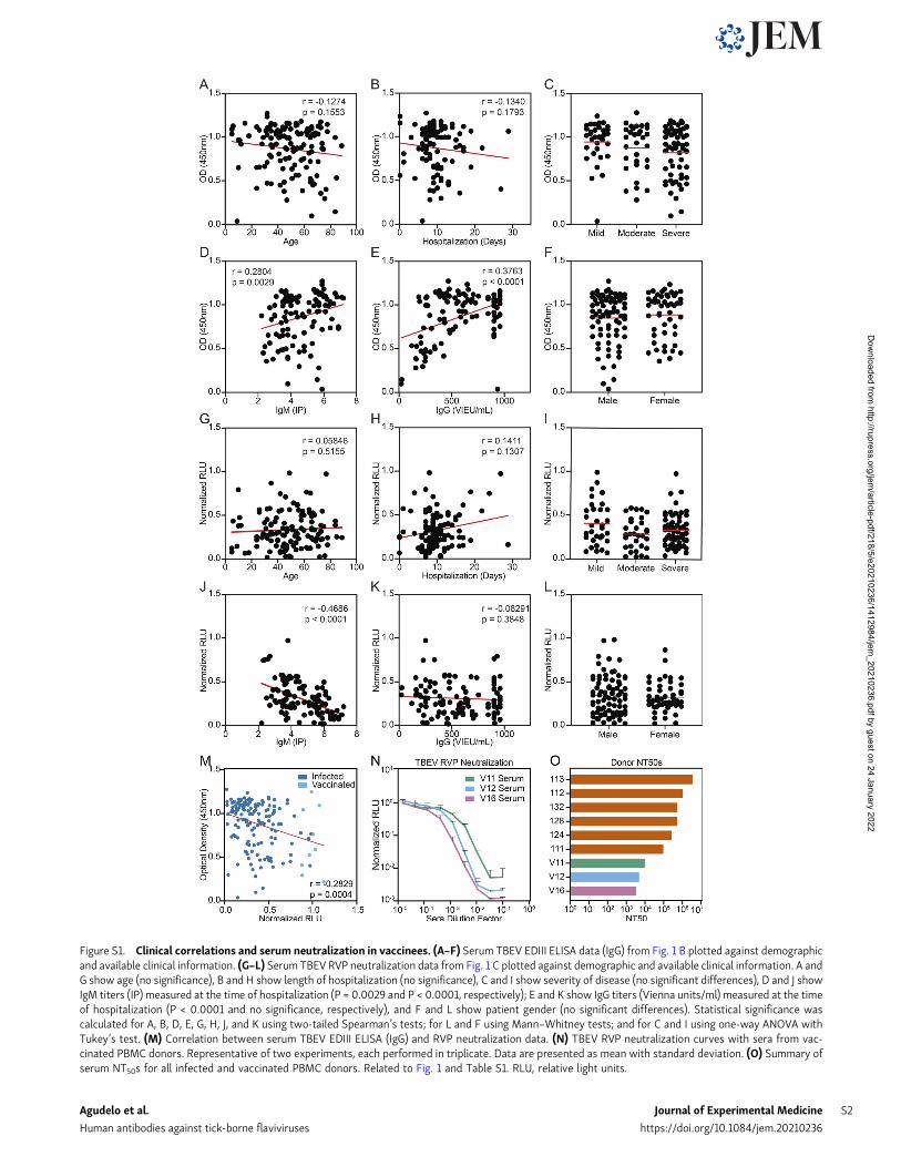
Figure S1. Clinical correlations and serum neutralization in vaccinees. (A–F) Serum TBEV EDIII ELISA data (IgG) from Fig. 1 B plotted against demographicand available clinical information. (G–L) Serum TBEV RVP neutralization data from Fig. 1 C plotted against demographic and available clinical information. A andG show age (no significance), B and H show length of hospitalization (no significance), C and I show severity of disease (no significant differences), D and J showIgM titers (IP) measured at the time of hospitalization (P = 0.0029 and P < 0.0001, respectively); E and K show IgG titers (Vienna units/ml) measured at the timeof hospitalization (P < 0.0001 and no significance, respectively), and F and L show patient gender (no significant differences). Statistical significance wascalculated for A, B, D, E, G, H, J, and K using two-tailed Spearman’s tests; for L and F using Mann–Whitney tests; and for C and I using one-way ANOVA withTukey’s test. (M) Correlation between serum TBEV EDIII ELISA (IgG) and RVP neutralization data. (N) TBEV RVP neutralization curves with sera from vac-cinated PBMC donors. Representative of two experiments, each performed in triplicate. Data are presented as mean with standard deviation. (O) Summary ofserum NT50s for all infected and vaccinated PBMC donors. Related to Fig. 1 and Table S1. RLU, relative light units.
Agudelo et al. Journal of Experimental Medicine S2
Human antibodies against tick-borne flaviviruses https://doi.org/10.1084/jem.20210236
Dow
nloaded from http://rupress.org/jem
/article-pdf/218/5/e20210236/1412984/jem_20210236.pdf by guest on 24 January 2022
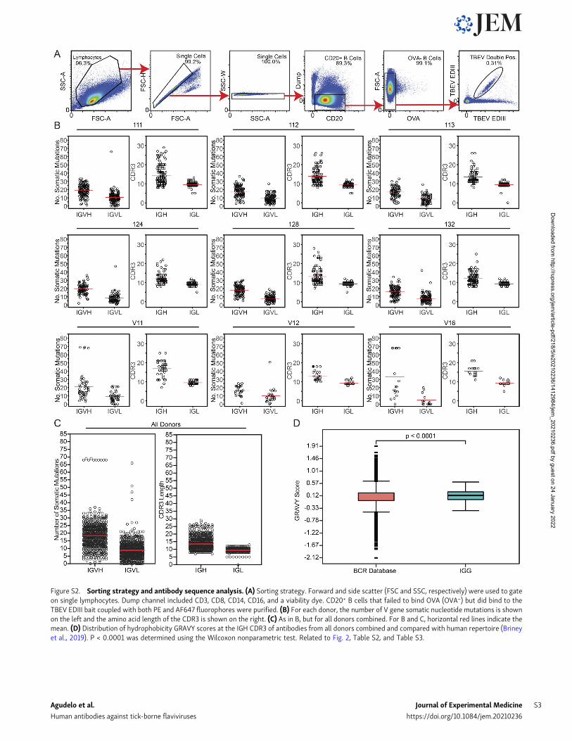
Figure S2. Sorting strategy and antibody sequence analysis. (A) Sorting strategy. Forward and side scatter (FSC and SSC, respectively) were used to gateon single lymphocytes. Dump channel included CD3, CD8, CD14, CD16, and a viability dye. CD20+ B cells that failed to bind OVA (OVA−) but did bind to theTBEV EDIII bait coupled with both PE and AF647 fluorophores were purified. (B) For each donor, the number of V gene somatic nucleotide mutations is shownon the left and the amino acid length of the CDR3 is shown on the right. (C) As in B, but for all donors combined. For B and C, horizontal red lines indicate themean. (D) Distribution of hydrophobicity GRAVY scores at the IGH CDR3 of antibodies from all donors combined and compared with human repertoire (Brineyet al., 2019). P < 0.0001 was determined using the Wilcoxon nonparametric test. Related to Fig. 2, Table S2, and Table S3.
Agudelo et al. Journal of Experimental Medicine S3
Human antibodies against tick-borne flaviviruses https://doi.org/10.1084/jem.20210236
Dow
nloaded from http://rupress.org/jem
/article-pdf/218/5/e20210236/1412984/jem_20210236.pdf by guest on 24 January 2022
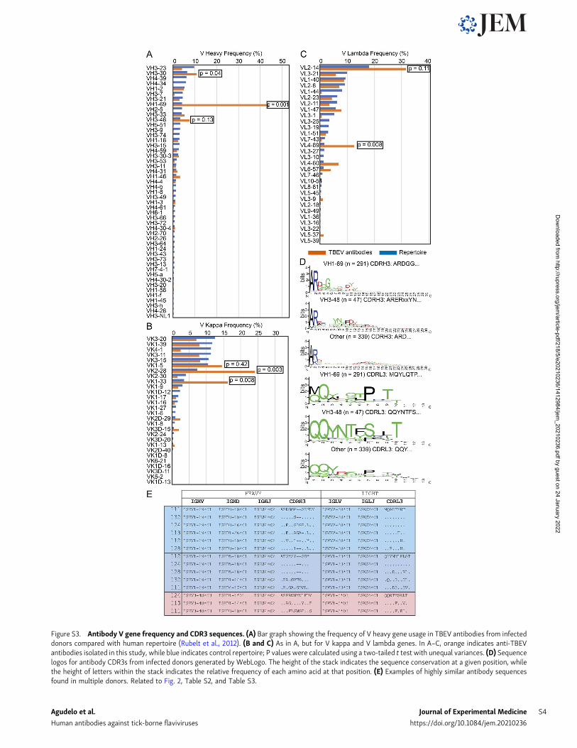
Figure S3. Antibody V gene frequency and CDR3 sequences. (A) Bar graph showing the frequency of V heavy gene usage in TBEV antibodies from infecteddonors compared with human repertoire (Rubelt et al., 2012). (B and C) As in A, but for V kappa and V lambda genes. In A–C, orange indicates anti-TBEVantibodies isolated in this study, while blue indicates control repertoire; P values were calculated using a two-tailed t test with unequal variances. (D) Sequencelogos for antibody CDR3s from infected donors generated by WebLogo. The height of the stack indicates the sequence conservation at a given position, whilethe height of letters within the stack indicates the relative frequency of each amino acid at that position. (E) Examples of highly similar antibody sequencesfound in multiple donors. Related to Fig. 2, Table S2, and Table S3.
Agudelo et al. Journal of Experimental Medicine S4
Human antibodies against tick-borne flaviviruses https://doi.org/10.1084/jem.20210236
Dow
nloaded from http://rupress.org/jem
/article-pdf/218/5/e20210236/1412984/jem_20210236.pdf by guest on 24 January 2022
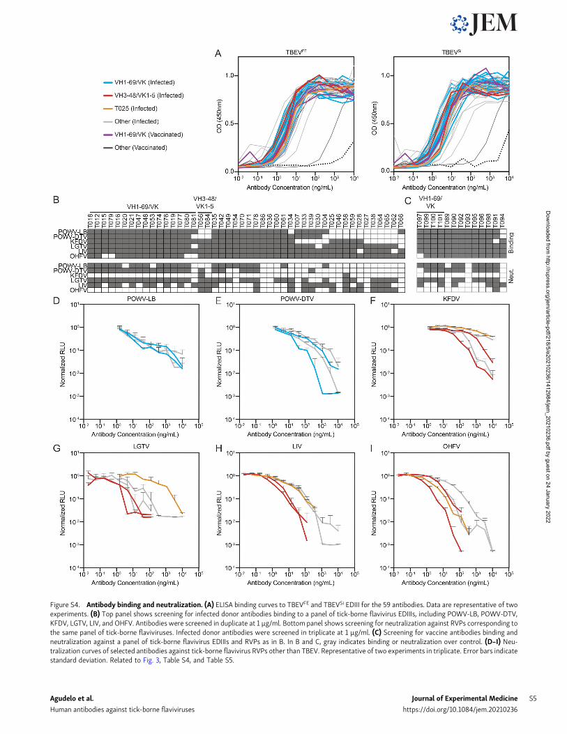
Figure S4. Antibody binding and neutralization. (A) ELISA binding curves to TBEVFE and TBEVSi EDIII for the 59 antibodies. Data are representative of twoexperiments. (B) Top panel shows screening for infected donor antibodies binding to a panel of tick-borne flavivirus EDIIIs, including POWV-LB, POWV-DTV,KFDV, LGTV, LIV, and OHFV. Antibodies were screened in duplicate at 1 µg/ml. Bottom panel shows screening for neutralization against RVPs corresponding tothe same panel of tick-borne flaviviruses. Infected donor antibodies were screened in triplicate at 1 µg/ml. (C) Screening for vaccine antibodies binding andneutralization against a panel of tick-borne flavivirus EDIIIs and RVPs as in B. In B and C, gray indicates binding or neutralization over control. (D–I) Neu-tralization curves of selected antibodies against tick-borne flavivirus RVPs other than TBEV. Representative of two experiments in triplicate. Error bars indicatestandard deviation. Related to Fig. 3, Table S4, and Table S5.
Agudelo et al. Journal of Experimental Medicine S5
Human antibodies against tick-borne flaviviruses https://doi.org/10.1084/jem.20210236
Dow
nloaded from http://rupress.org/jem
/article-pdf/218/5/e20210236/1412984/jem_20210236.pdf by guest on 24 January 2022
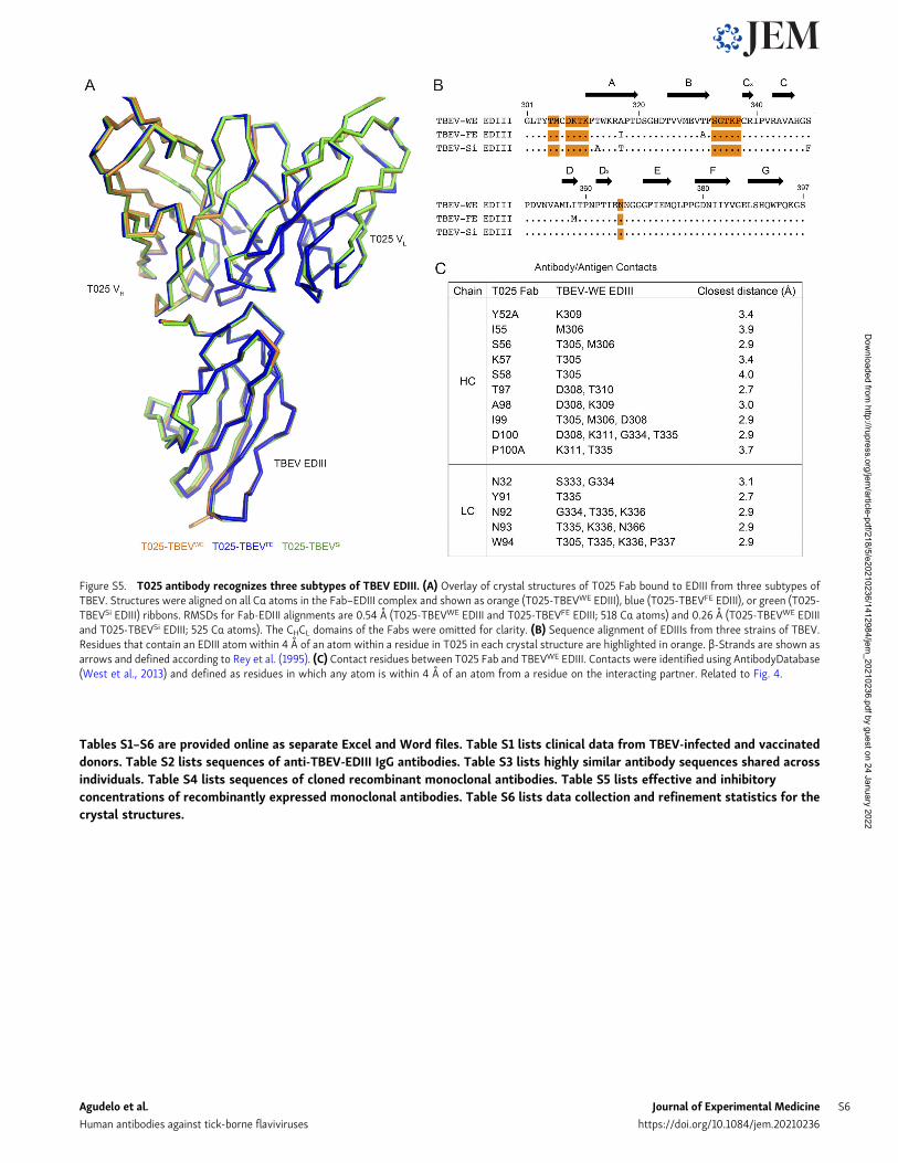
Tables S1–S6 are provided online as separate Excel and Word files. Table S1 lists clinical data from TBEV-infected and vaccinateddonors. Table S2 lists sequences of anti-TBEV-EDIII IgG antibodies. Table S3 lists highly similar antibody sequences shared acrossindividuals. Table S4 lists sequences of cloned recombinant monoclonal antibodies. Table S5 lists effective and inhibitoryconcentrations of recombinantly expressed monoclonal antibodies. Table S6 lists data collection and refinement statistics for thecrystal structures.
Figure S5. T025 antibody recognizes three subtypes of TBEV EDIII. (A) Overlay of crystal structures of T025 Fab bound to EDIII from three subtypes ofTBEV. Structures were aligned on all Cα atoms in the Fab–EDIII complex and shown as orange (T025-TBEVWE EDIII), blue (T025-TBEVFE EDIII), or green (T025-TBEVSi EDIII) ribbons. RMSDs for Fab-EDIII alignments are 0.54 A (T025-TBEVWE EDIII and T025-TBEVFE EDIII; 518 Cα atoms) and 0.26 A (T025-TBEVWE EDIIIand T025-TBEVSi EDIII; 525 Cα atoms). The CHCL domains of the Fabs were omitted for clarity. (B) Sequence alignment of EDIIIs from three strains of TBEV.Residues that contain an EDIII atom within 4 A of an atom within a residue in T025 in each crystal structure are highlighted in orange. β-Strands are shown asarrows and defined according to Rey et al. (1995). (C) Contact residues between T025 Fab and TBEVWE EDIII. Contacts were identified using AntibodyDatabase(West et al., 2013) and defined as residues in which any atom is within 4 A of an atom from a residue on the interacting partner. Related to Fig. 4.
Agudelo et al. Journal of Experimental Medicine S6
Human antibodies against tick-borne flaviviruses https://doi.org/10.1084/jem.20210236
Dow
nloaded from http://rupress.org/jem
/article-pdf/218/5/e20210236/1412984/jem_20210236.pdf by guest on 24 January 2022