Embed Size (px)
Citation preview
ASTHME ET DDB
Ali Ben Kheder - Fatma Tritar
Journées Francophones d’Allergologie - Nouméa - 9 Oct 2018
Pauvreté de la littérature sur ce sujet !
2 pathologies portant sur les bronches
Asthme et DDB
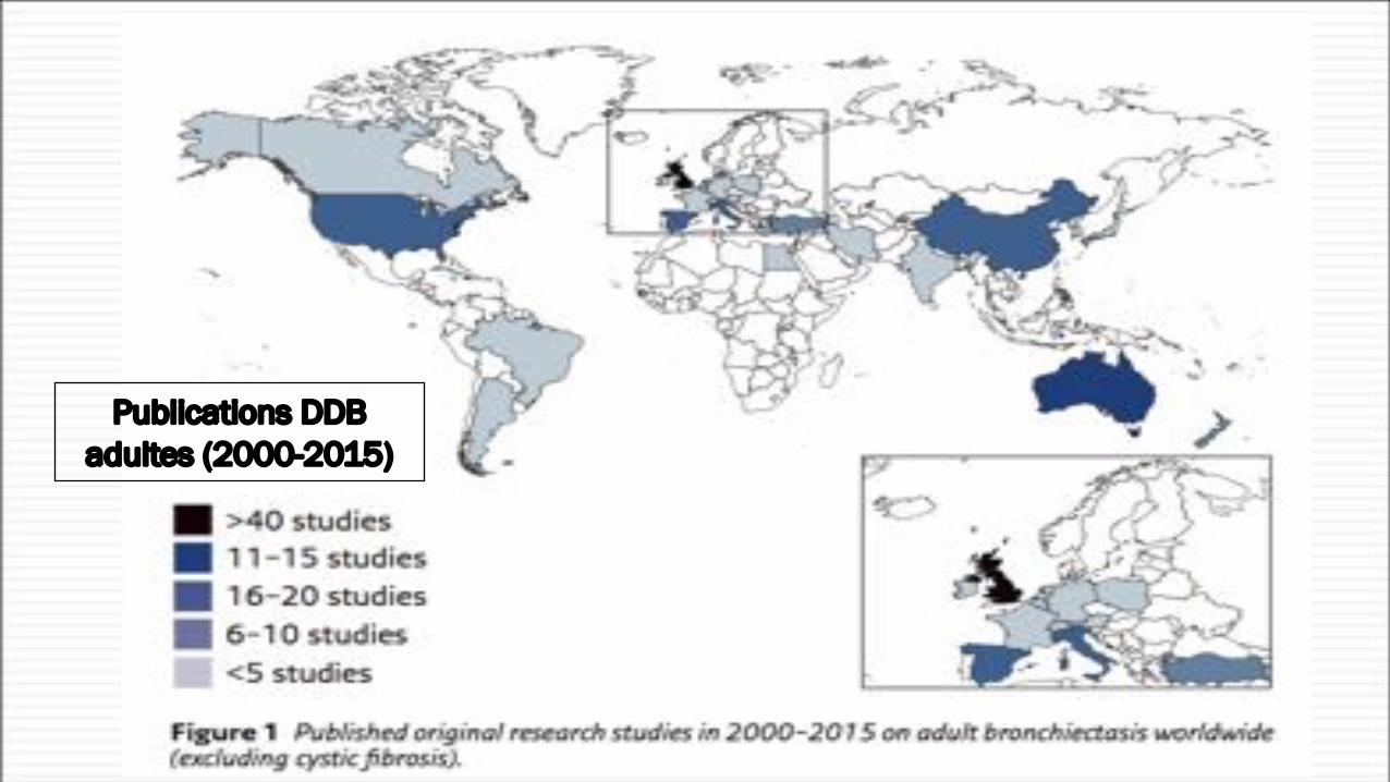
Publications DDB adultes (2000-2015)
Pauvreté de la littérature sur ce sujet !
Peu Intéressant Peu Impactant
Asthme et DDB
Entre Asthme et DDB : Existe-t-il un lien ?
Co-morbidité Multi-morbidité
Asthme et DDB
Les deux pathologies ont- elles une similitude clinique ?
Asthme et DDB
DDB sont-elles une évolution inéluctable de l’Asthme ?
Asthme et DDB
Cette association comporte -t-elle un facteur de risque aggravant l’une ou l’autre pathologie ?
Asthme et DDB
Ces 2 affections ont-elles une évolution chronologique parallèle ?
Asthme et DDB
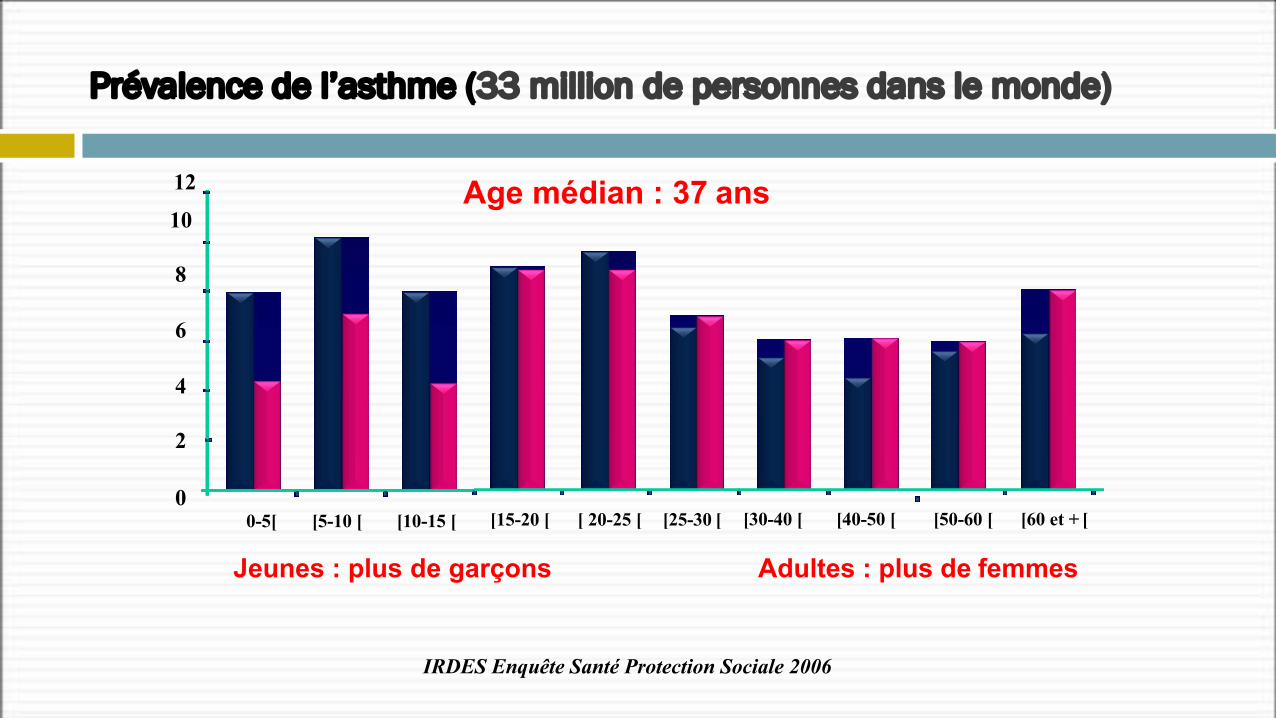
Jeunes : plus de garçons Adultes : plus de femmes
Prévalence de l’asthme (33 million de personnes dans le monde)
12
10
8
6
4
2
0]0-5[ [5-10 [ [10-15 [ [15-20 [ [ 20-25 [ [25-30 [ [30-40 [ [40-50 [ [50-60 [ [60 et + [
IRDES Enquête Santé Protection Sociale 2006
Age médian : 37 ans
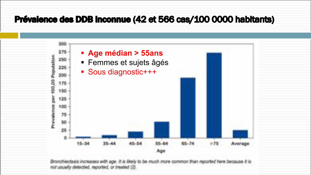
Prévalence des DDB inconnue (42 et 566 cas/100 0000 habitants)
§ Age médian > 55ans§ Femmes et sujets âgés§ Sous diagnostic+++
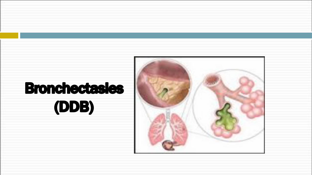
Bronchectasies(DDB)
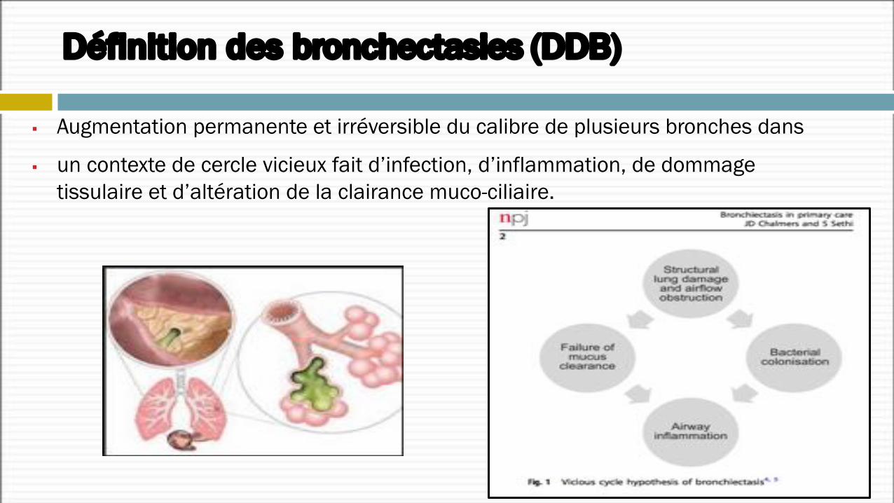
§ Augmentation permanente et irréversible du calibre de plusieurs bronches dans
§ un contexte de cercle vicieux fait d’infection, d’inflammation, de dommage tissulaire et d’altération de la clairance muco-ciliaire.
Définition des bronchectasies (DDB)
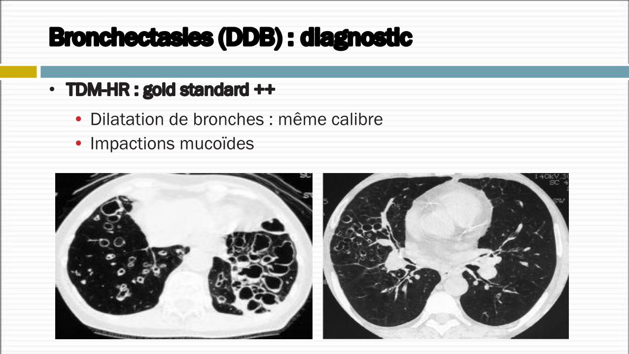
Bronchectasies (DDB) : diagnostic
• TDM-HR : gold standard ++
• Dilatation de bronches : même calibre• Impactions mucoïdes
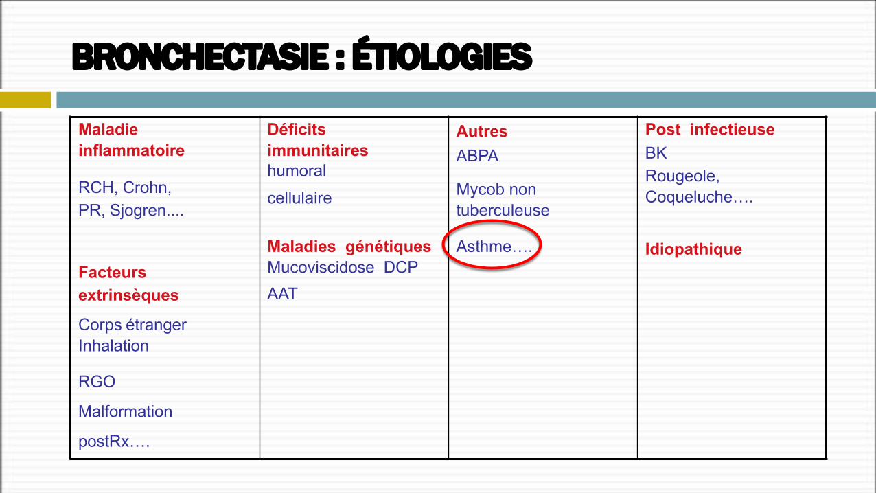
Maladie inflammatoire
Déficits immunitaires humoralcellulaire
Maladies génétiques Mucoviscidose DCPAAT
AutresABPA
Post infectieuseBKRougeole, Coqueluche….
Idiopathique
RCH, Crohn,PR, Sjogren....
Mycob non tuberculeuse
Asthme….Facteursextrinsèques
Corps étranger Inhalation
RGO
Malformation
postRx….
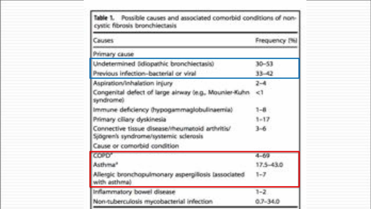
BRONCHECTASIE : ÉTIOLOGIES
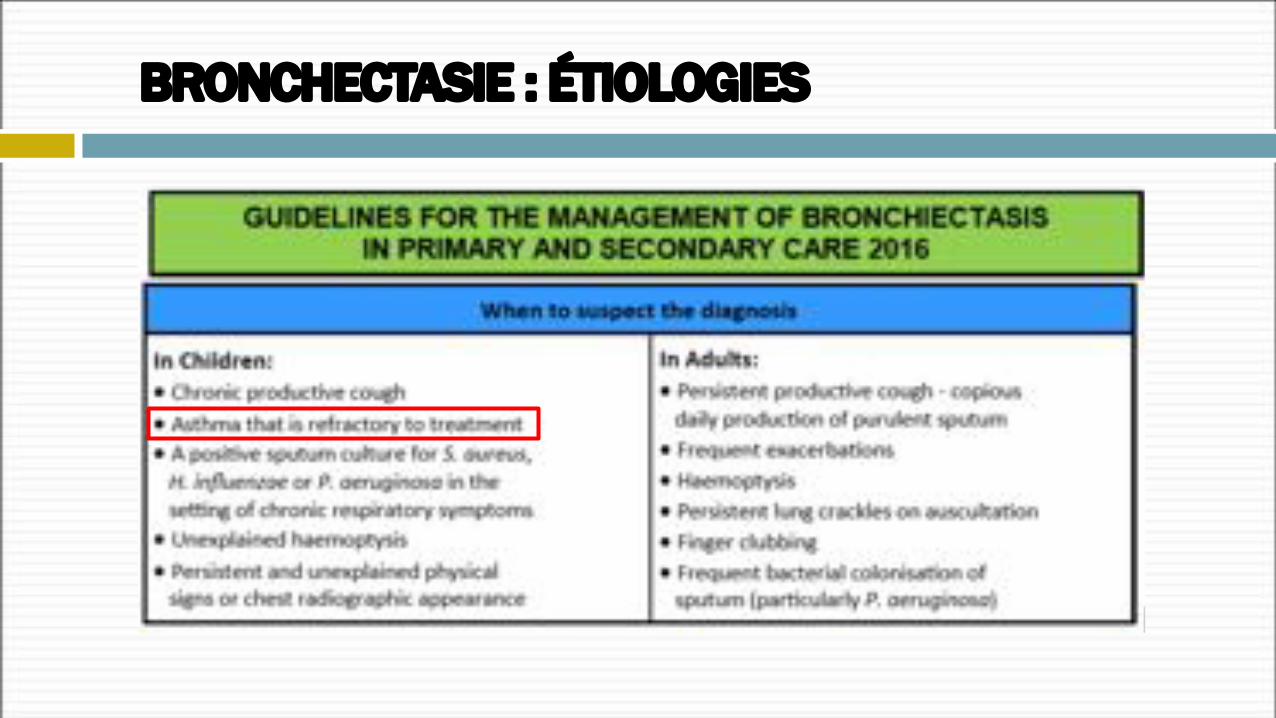
BRONCHECTASIE : ÉTIOLOGIES
The management of bronchiectasisin Europe
Data from the European Bronchiectasis Registry (ERS)
James Chalmers University of Dundee,UK
« European Multicentre Bronchiectasis Audit and Research Collaboration »
Recommandations basées sur la « vraie vie » sur la prise en charge des dilatations des bronches (DDB) de l’adulte
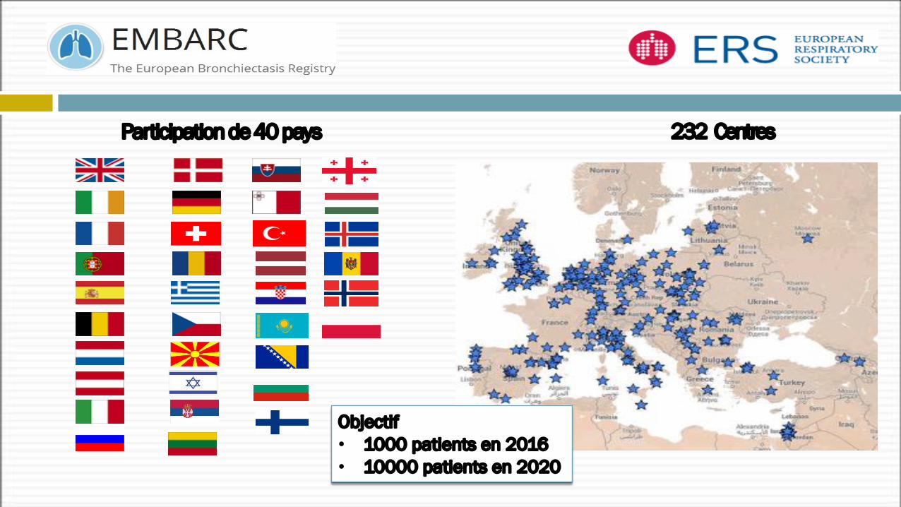
Participation de 40 pays 232 Centres
Objectif• 1000 patients en 2016• 10000 patients en 2020
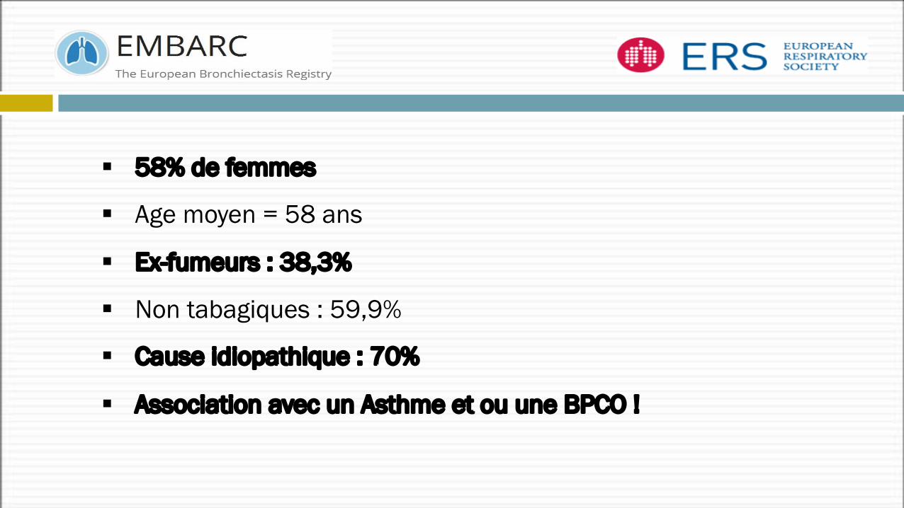
§ 58% de femmes
§ Age moyen = 58 ans
§ Ex-fumeurs : 38,3%
§ Non tabagiques : 59,9%
§ Cause idiopathique : 70%
§ Association avec un Asthme et ou une BPCO !
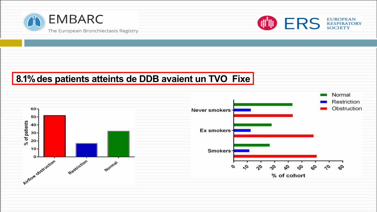
8.1% des patients atteints de DDB avaient un TVO Fixe
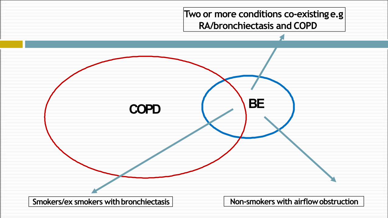
COPD BE
Non-smokers with airflowobstructionSmokers/ex smokers withbronchiectasis
Two or more conditions co-existinge.gRA/bronchiectasis and COPD
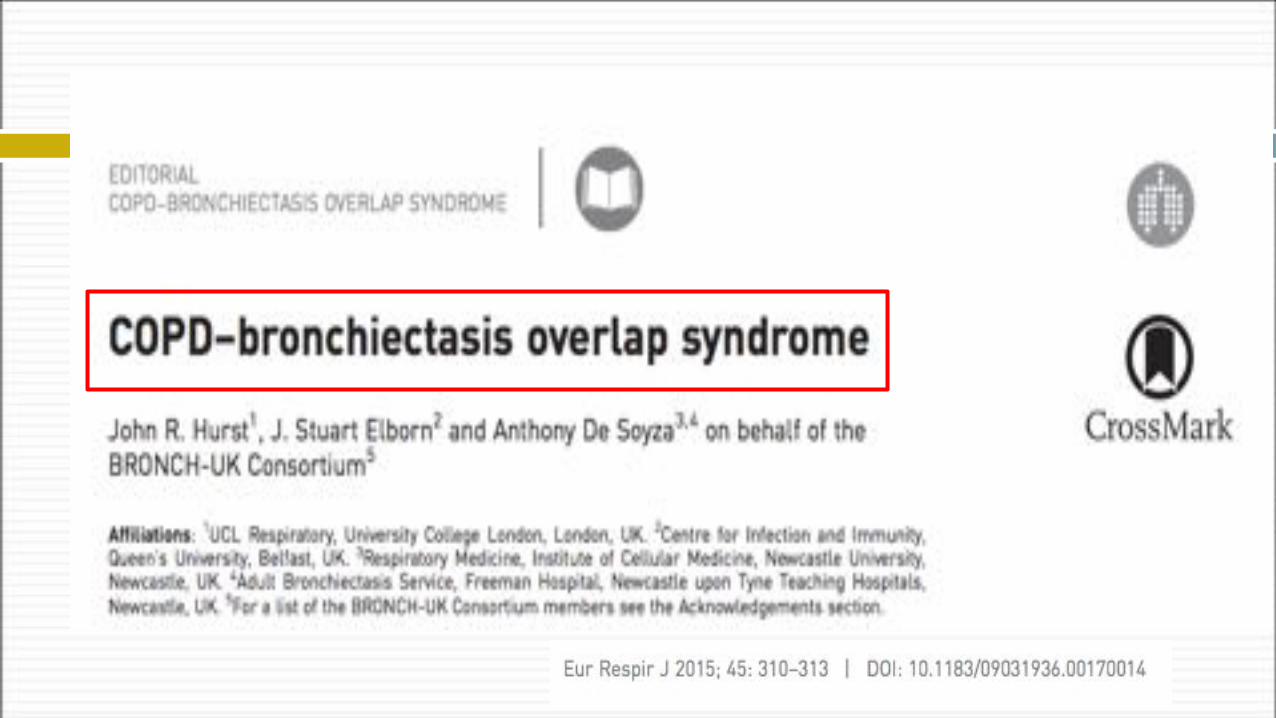
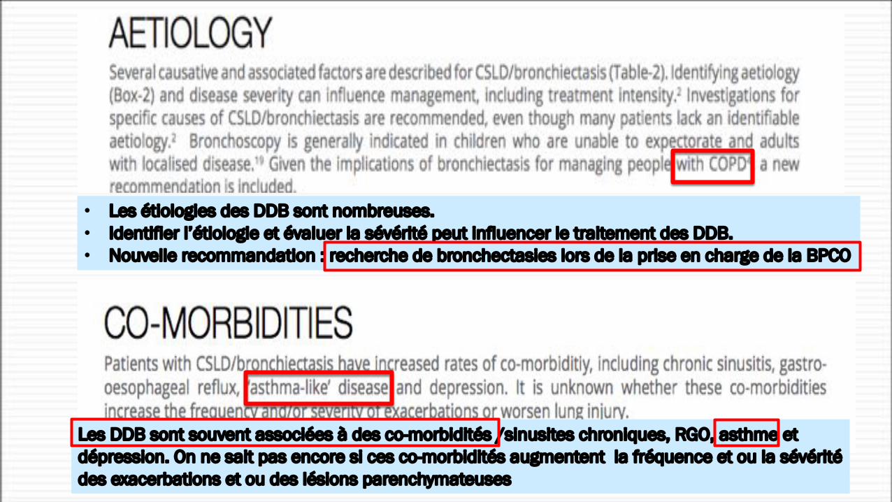
• Les étiologies des DDB sont nombreuses.• Identifier l’étiologie et évaluer la sévérité peut influencer le traitement des DDB.• Nouvelle recommandation : recherche de bronchectasies lors de la prise en charge de la BPCO
Les DDB sont souvent associées à des co-morbidités /sinusites chroniques, RGO, asthme et dépression. On ne sait pas encore si ces co-morbidités augmentent la fréquence et ou la sévérité des exacerbations et ou des lésions parenchymateuses

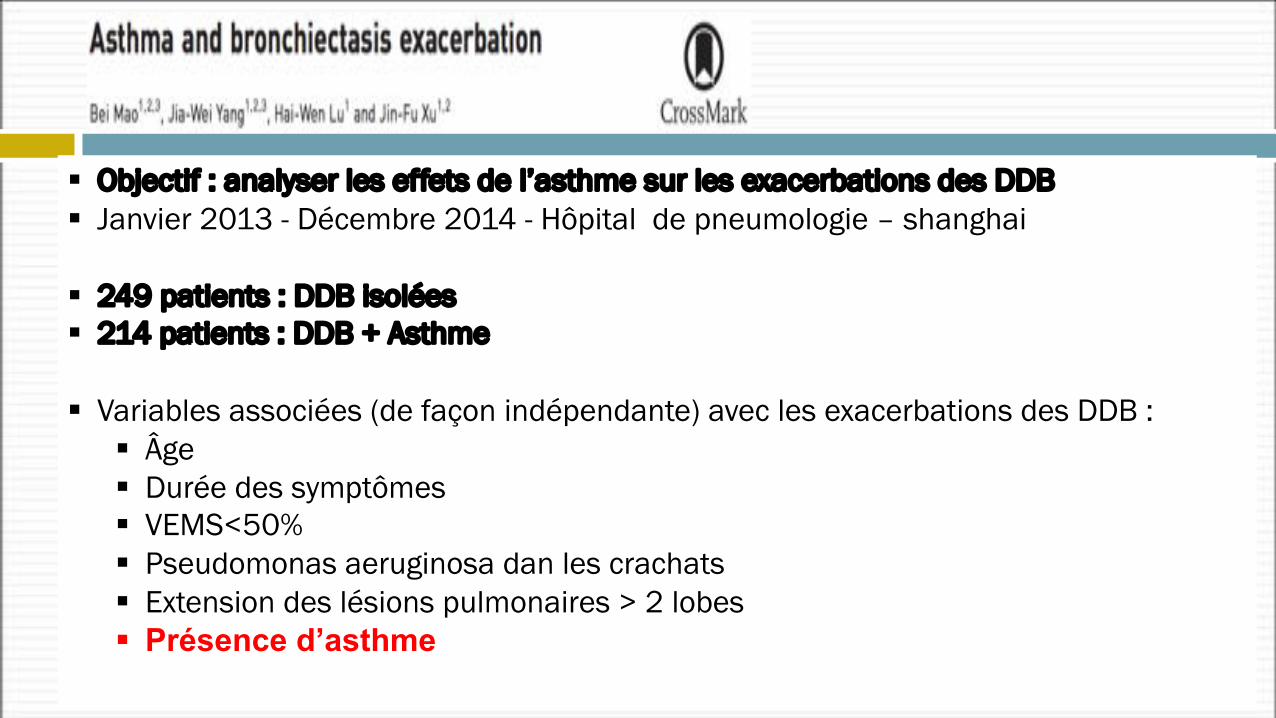
§ Objectif : analyser les effets de l’asthme sur les exacerbations des DDB§ Janvier 2013 - Décembre 2014 - Hôpital de pneumologie – shanghai
§ 249 patients : DDB isolées § 214 patients : DDB + Asthme
§ Variables associées (de façon indépendante) avec les exacerbations des DDB :§ Âge § Durée des symptômes§ VEMS<50% § Pseudomonas aeruginosa dan les crachats§ Extension des lésions pulmonaires > 2 lobes§ Présence d’asthme
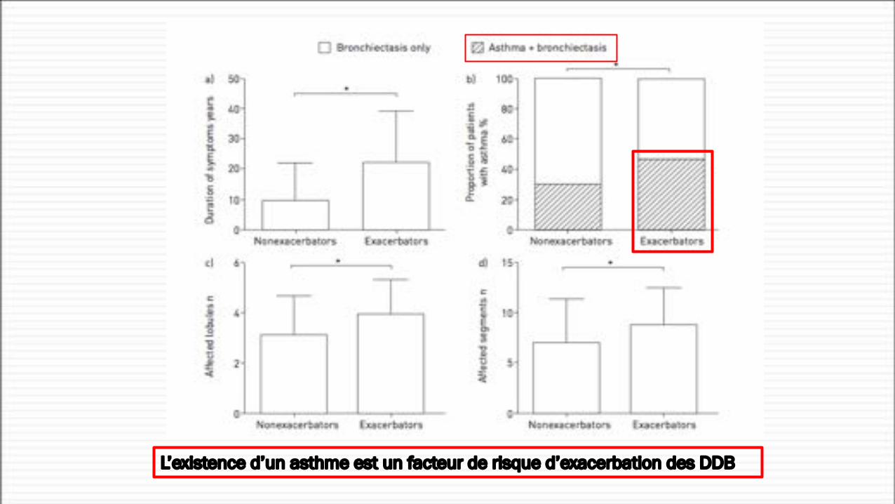
L’existence d’un asthme est un facteur de risque d’exacerbation des DDB
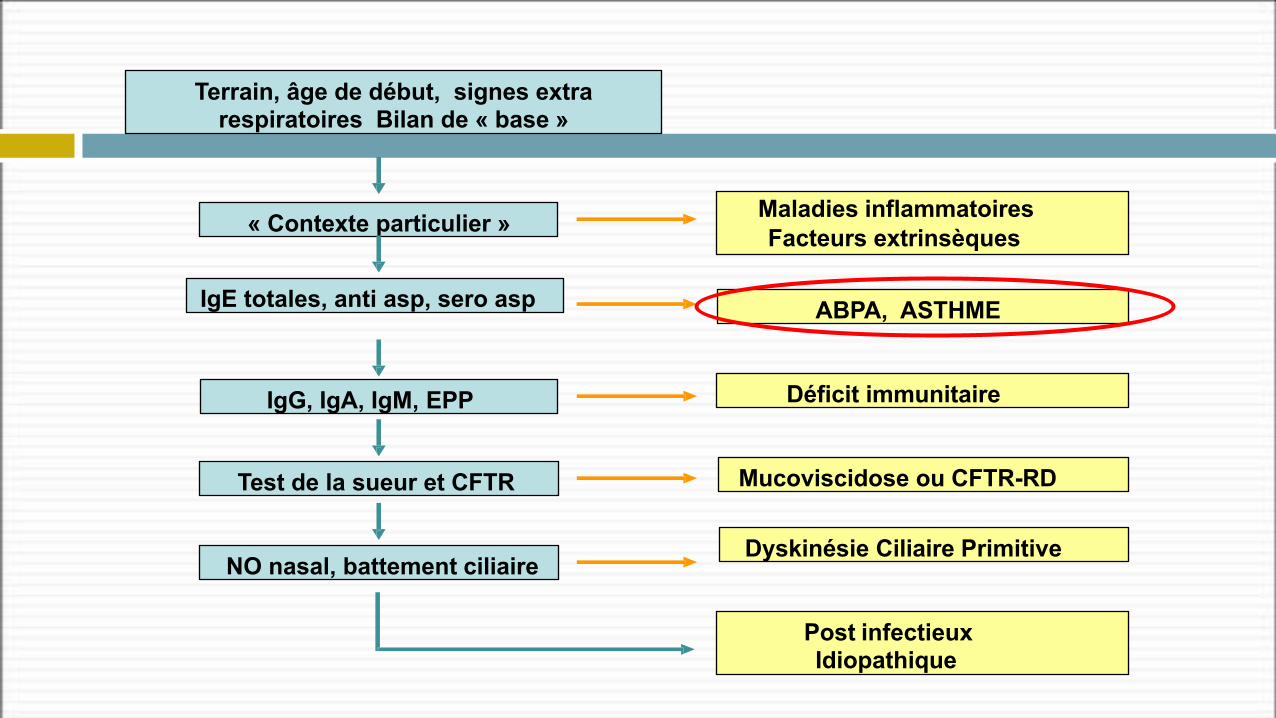
Terrain, âge de début, signes extra respiratoires Bilan de « base »
« Contexte particulier »
IgE totales, anti asp, sero asp
IgG, IgA, IgM, EPP
Test de la sueur et CFTR
NO nasal, battement ciliaire
Post infectieux Idiopathique
Maladies inflammatoires Facteurs extrinsèques
ABPA, ASTHME
Déficit immunitaire
Mucoviscidose ou CFTR-RD
Dyskinésie Ciliaire Primitive
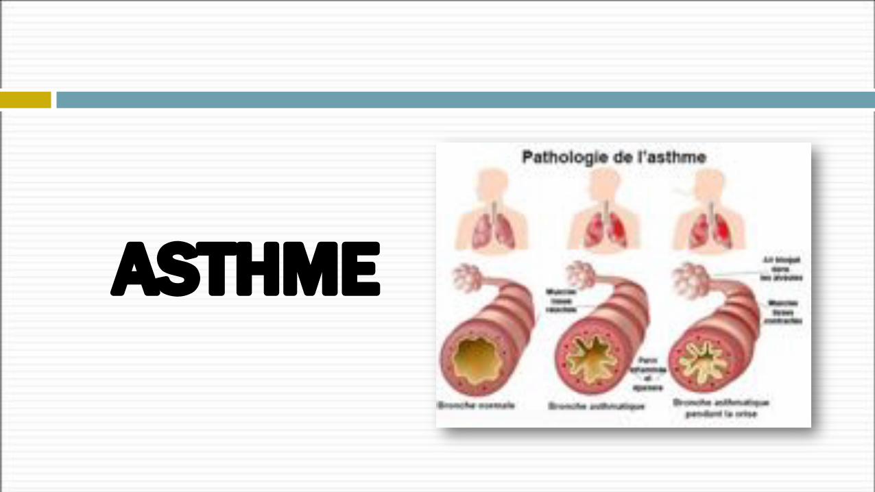
ASTHME
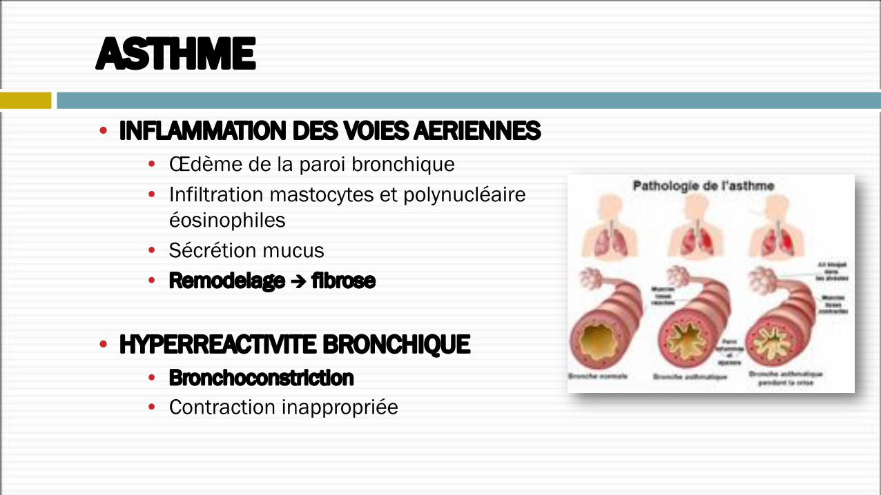
ASTHME
• INFLAMMATION DES VOIES AERIENNES• Œdème de la paroi bronchique• Infiltration mastocytes et polynucléaire
éosinophiles• Sécrétion mucus• Remodelage à fibrose
• HYPERREACTIVITE BRONCHIQUE• Bronchoconstriction• Contraction inappropriée
1995 2002 2004
2008
GINA CONSENSUS
2006
2018
GINA Global Strategy for Asthma Management and Prevention
This slide set is restricted for academic and educational purposes only. Use of the slide set, or of individual slides, for commercial or promotional purposes requires approval from GINA. Slides must not be changed without permission from GINA.
© Global Initiative for Asthma www.ginasthma.org
Global Initiative for Asthma (GINA)
What’s new in GINA 2018?
GINA GLOBAL STRATEGY FOR ASTHMA MANAGEMENT AND PREVENTIONGOLD GLOBAL STRATEGY FOR DIAGNOSIS, MANAGEMENT AND PREVENTION
OF COPD
SYNDROME DE CHEVAUCHEMENT ASTHME - BPCO (ACO)
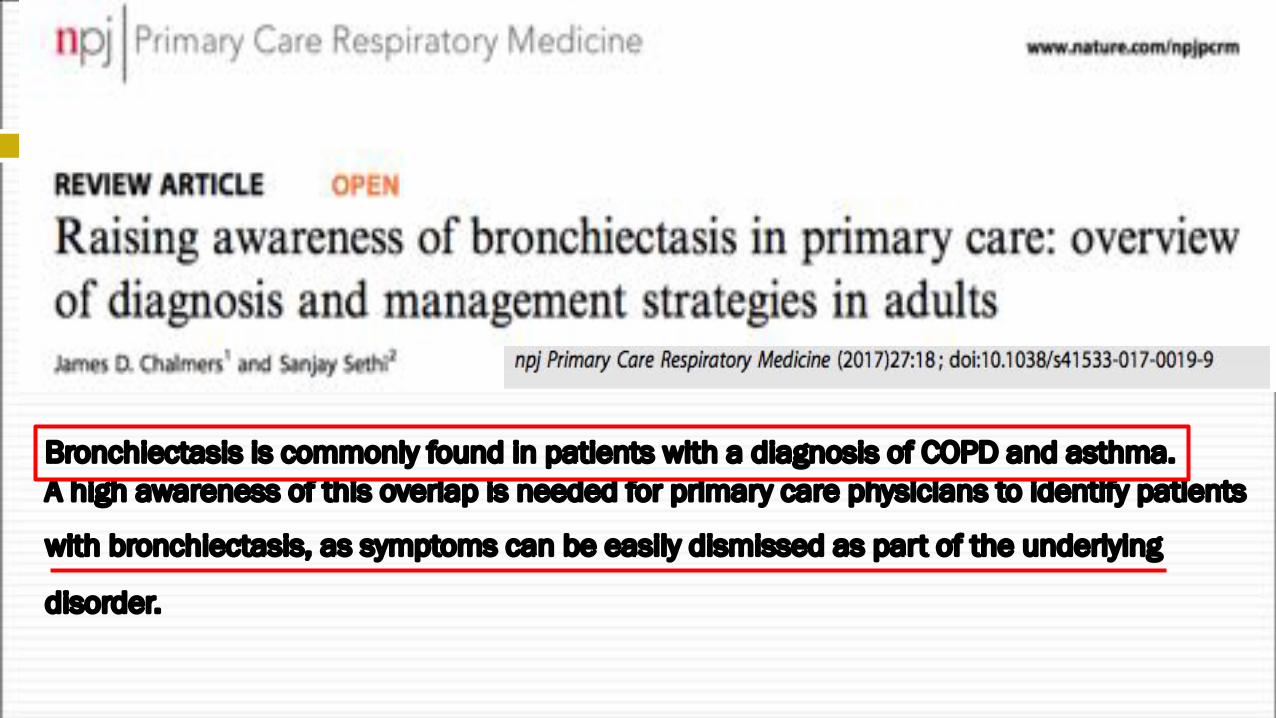
Bronchiectasis is commonly found in patients with a diagnosis of COPD and asthma.A high awareness of this overlap is needed for primary care physicians to identify patients
with bronchiectasis, as symptoms can be easily dismissed as part of the underlying
disorder.
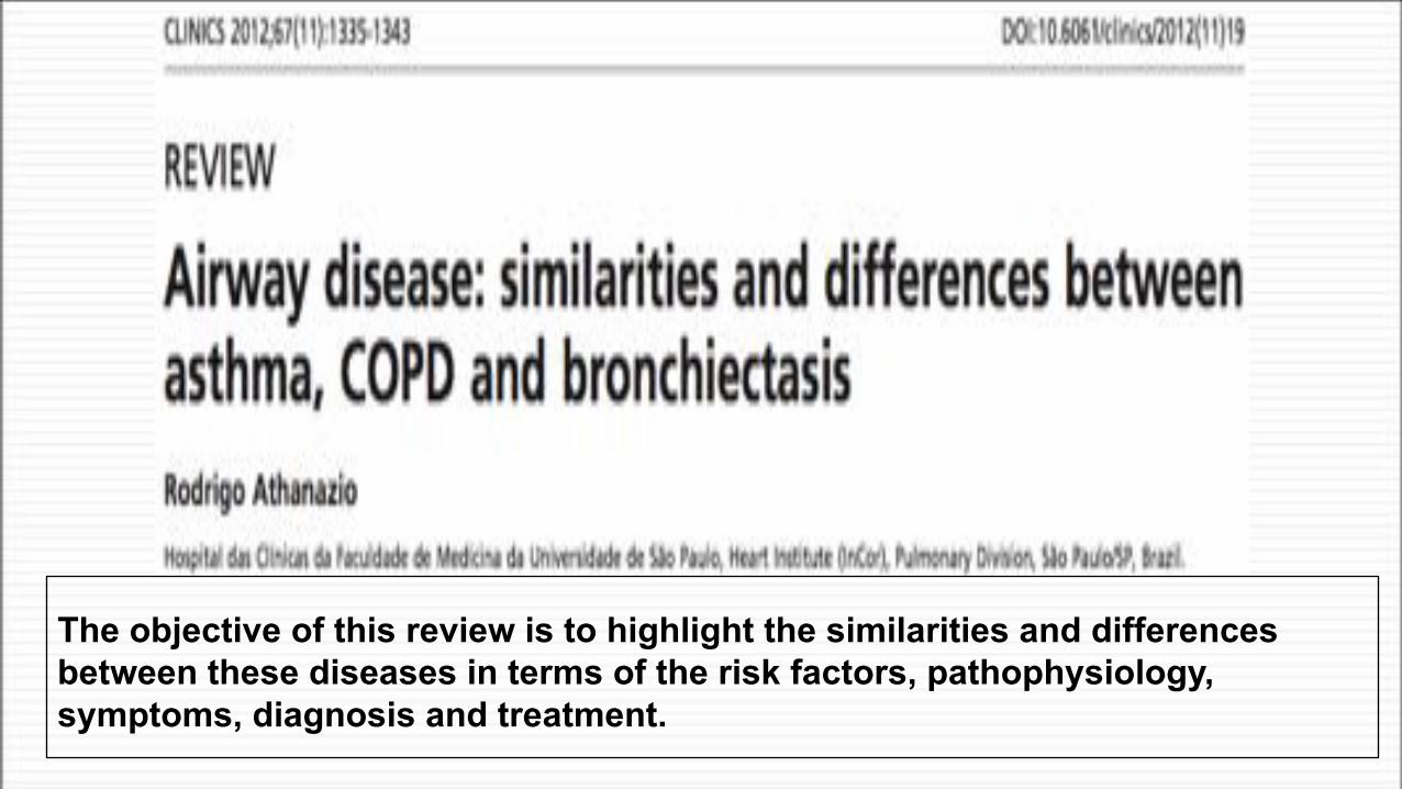
The objective of this review is to highlight the similarities and differences between these diseases in terms of the risk factors, pathophysiology, symptoms, diagnosis and treatment.
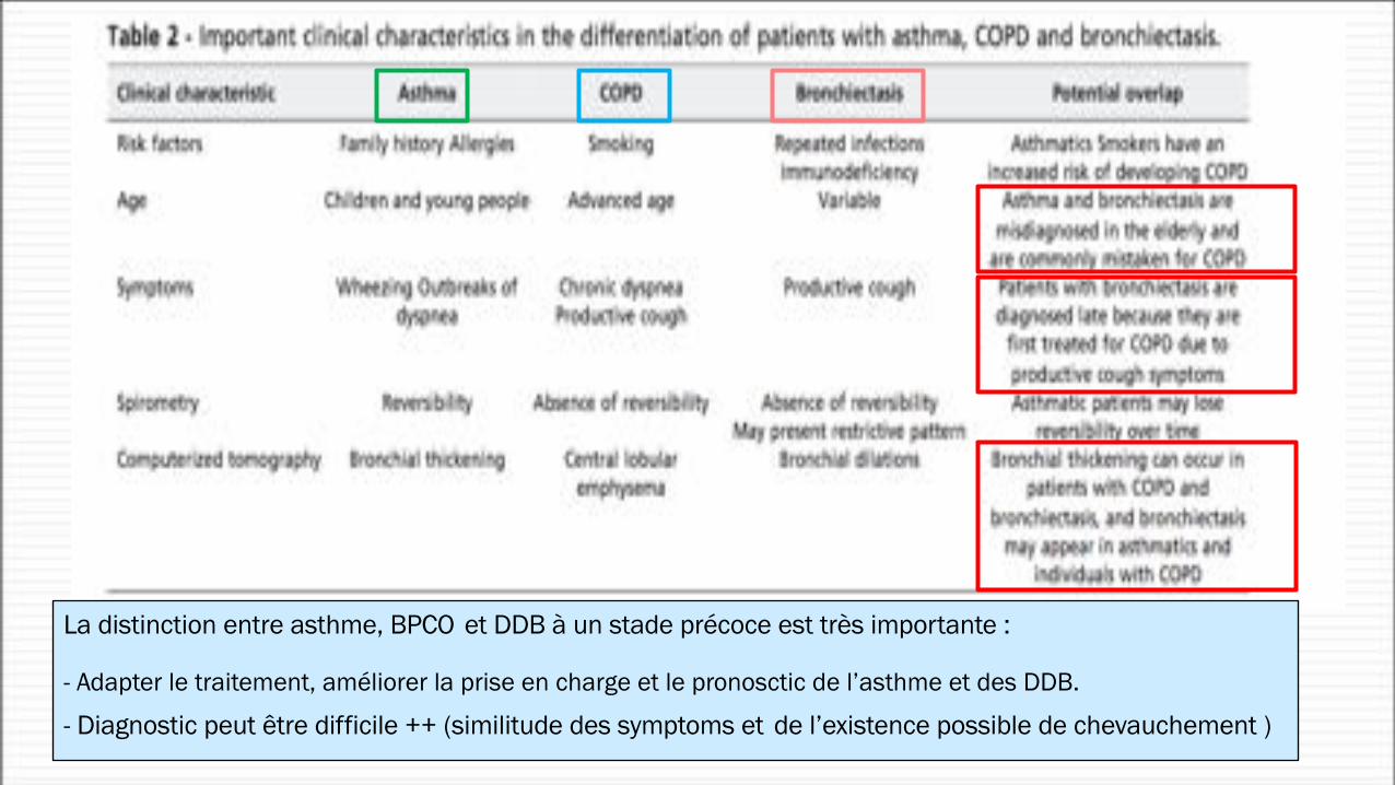
La distinction entre asthme, BPCO et DDB à un stade précoce est très importante :
- Adapter le traitement, améliorer la prise en charge et le pronosctic de l’asthme et des DDB.
- Diagnostic peut être difficile ++ (similitude des symptoms et de l’existence possible de chevauchement )
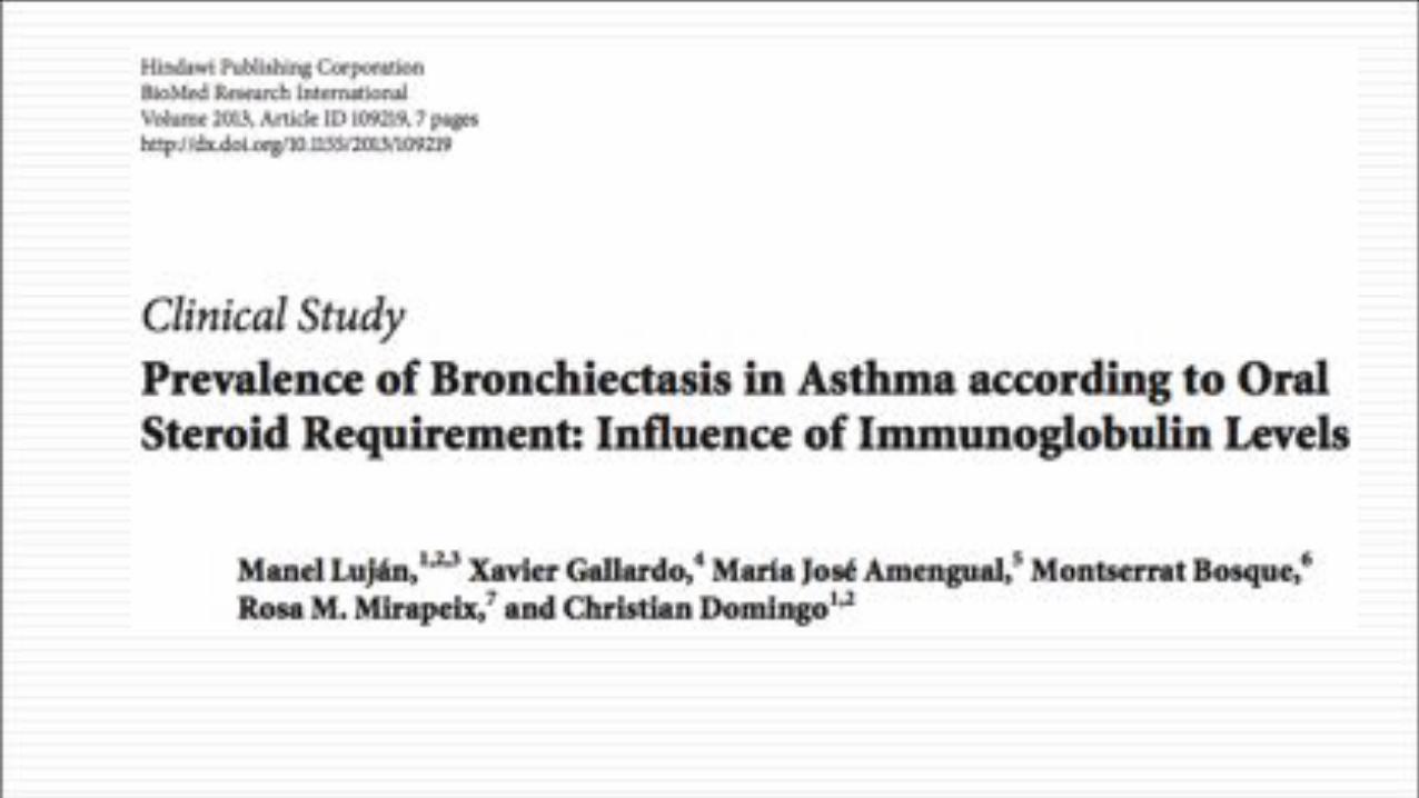
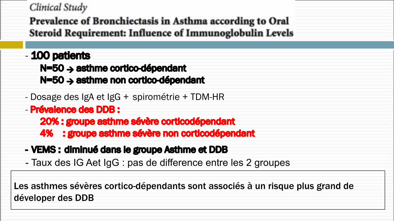
- 100 patientsN=50 à asthme cortico-dépendantN=50 à asthme non cortico-dépendant
- Dosage des IgA et IgG + spirométrie + TDM-HR- Prévalence des DDB :
20% : groupe asthme sévère corticodépendant4% : groupe asthme sévère non corticodépendant
- VEMS : diminué dans le groupe Asthme et DDB- Taux des IG Aet IgG : pas de difference entre les 2 groupes
Les asthmes sévères cortico-dépendants sont associés à un risque plus grand de déveloper des DDB
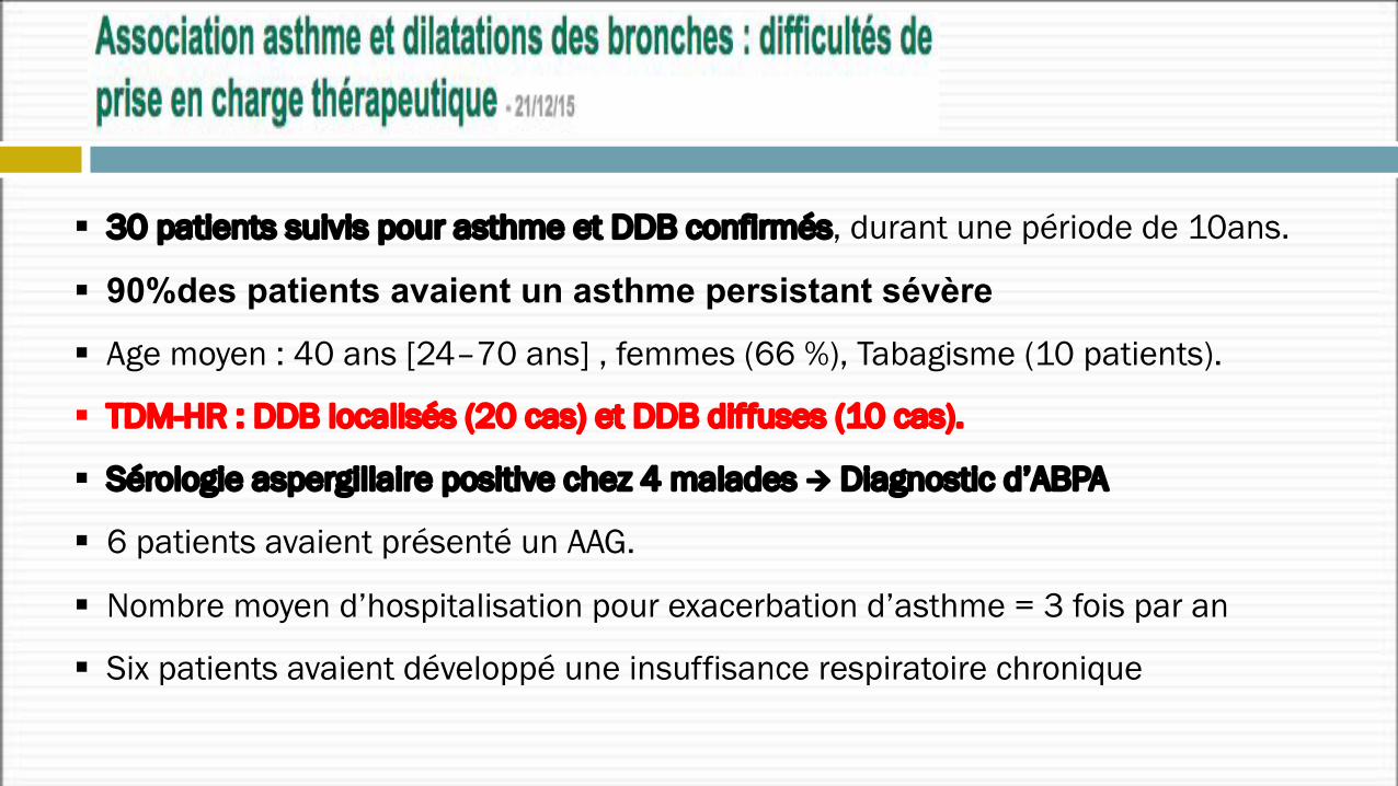
§ 30 patients suivis pour asthme et DDB confirmés, durant une période de 10ans.
§ 90%des patients avaient un asthme persistant sévère § Age moyen : 40 ans [24–70 ans] , femmes (66 %), Tabagisme (10 patients).
§ TDM-HR : DDB localisés (20 cas) et DDB diffuses (10 cas).
§ Sérologie aspergillaire positive chez 4 malades à Diagnostic d’ABPA
§ 6 patients avaient présenté un AAG.
§ Nombre moyen d’hospitalisation pour exacerbation d’asthme = 3 fois par an
§ Six patients avaient développé une insuffisance respiratoire chronique
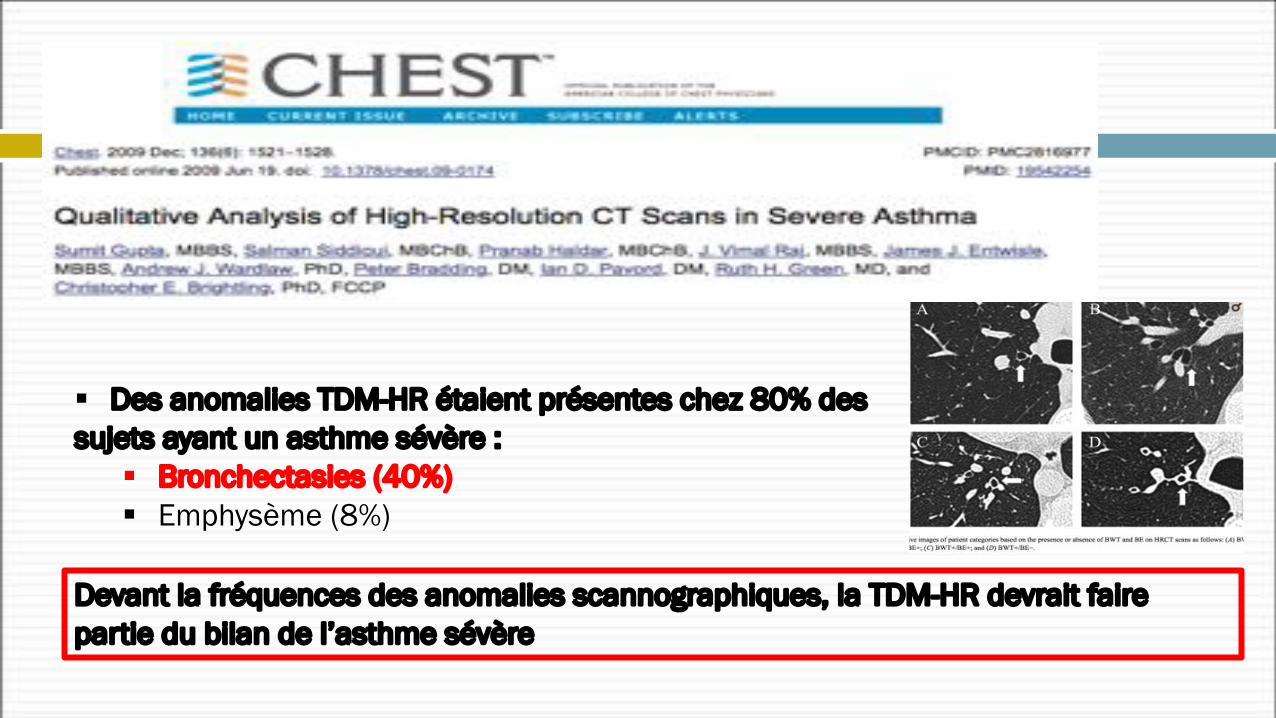
§ Des anomalies TDM-HR étaient présentes chez 80% des sujets ayant un asthme sévère :
§ Bronchectasies (40%)§ Emphysème (8%)
Devant la fréquences des anomalies scannographiques, la TDM-HR devrait faire partie du bilan de l’asthme sévère
NOPES score : varie de 0 à 4 points (0 = “pas de risqué” et 4 = “risque élevé” de DDB)1. FeNO [cut off point 20.5 ppb] 2. Pneumonie3. Expectorations4. Sévérité de l’asthme
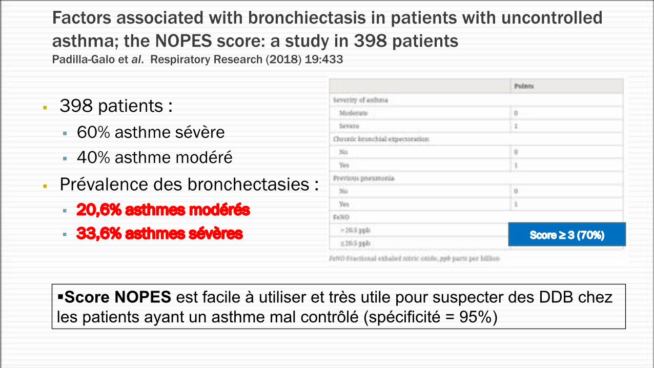
§ 398 patients :§ 60% asthme sévère§ 40% asthme modéré
§ Prévalence des bronchectasies :§ 20,6% asthmes modérés§ 33,6% asthmes sévères
Factors associated with bronchiectasis in patients with uncontrolled asthma; the NOPES score: a study in 398 patientsPadilla-Galo et al. Respiratory Research (2018) 19:433
Score ≥ 3 (70%)
§Score NOPES est facile à utiliser et très utile pour suspecter des DDB chez les patients ayant un asthme mal contrôlé (spécificité = 95%)
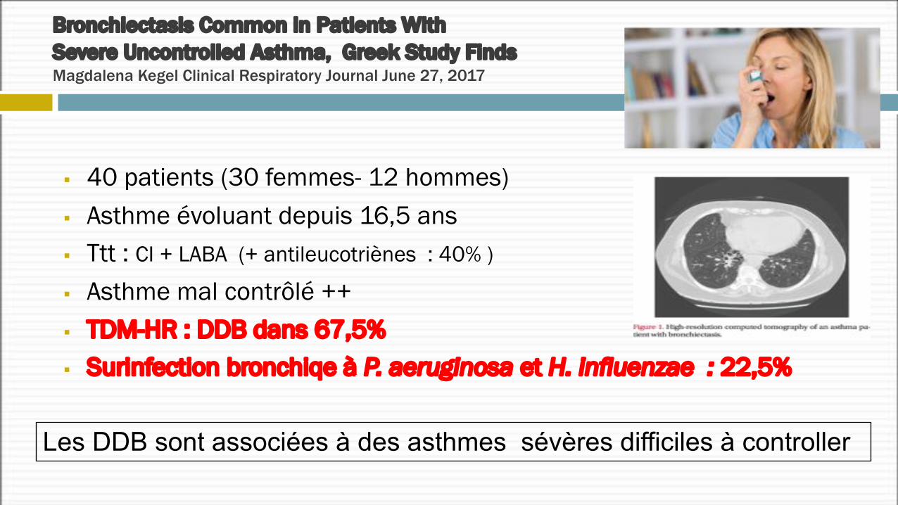
§ 40 patients (30 femmes- 12 hommes)§ Asthme évoluant depuis 16,5 ans§ Ttt : CI + LABA (+ antileucotriènes : 40% )
§ Asthme mal contrôlé ++§ TDM-HR : DDB dans 67,5%§ Surinfection bronchiqe à P. aeruginosa et H. influenzae : 22,5%
Bronchiectasis Common in Patients With Severe Uncontrolled Asthma, Greek Study FindsMagdalena Kegel Clinical Respiratory Journal June 27, 2017
Les DDB sont associées à des asthmes sévères difficiles à controller
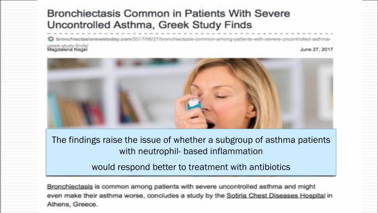
The findings raise the issue of whether a subgroup of asthma patients with neutrophil- based inflammation
would respond better to treatment with antibiotics
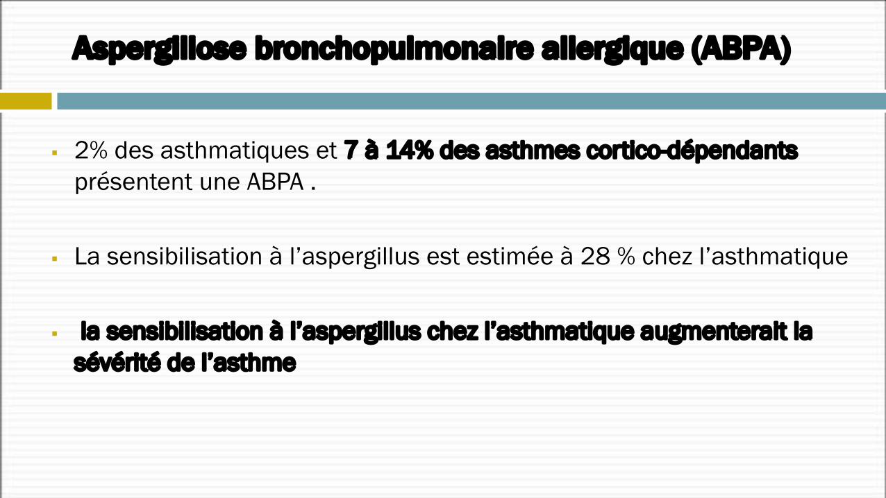
§ 2% des asthmatiques et 7 à 14% des asthmes cortico-dépendants présentent une ABPA .
§ La sensibilisation à l’aspergillus est estimée à 28 % chez l’asthmatique
§ la sensibilisation à l’aspergillus chez l’asthmatique augmenterait la sévérité de l’asthme
Aspergillose bronchopulmonaire allergique (ABPA)
opacities (Figure 3B-D) and atelectasis (Figure 3E).
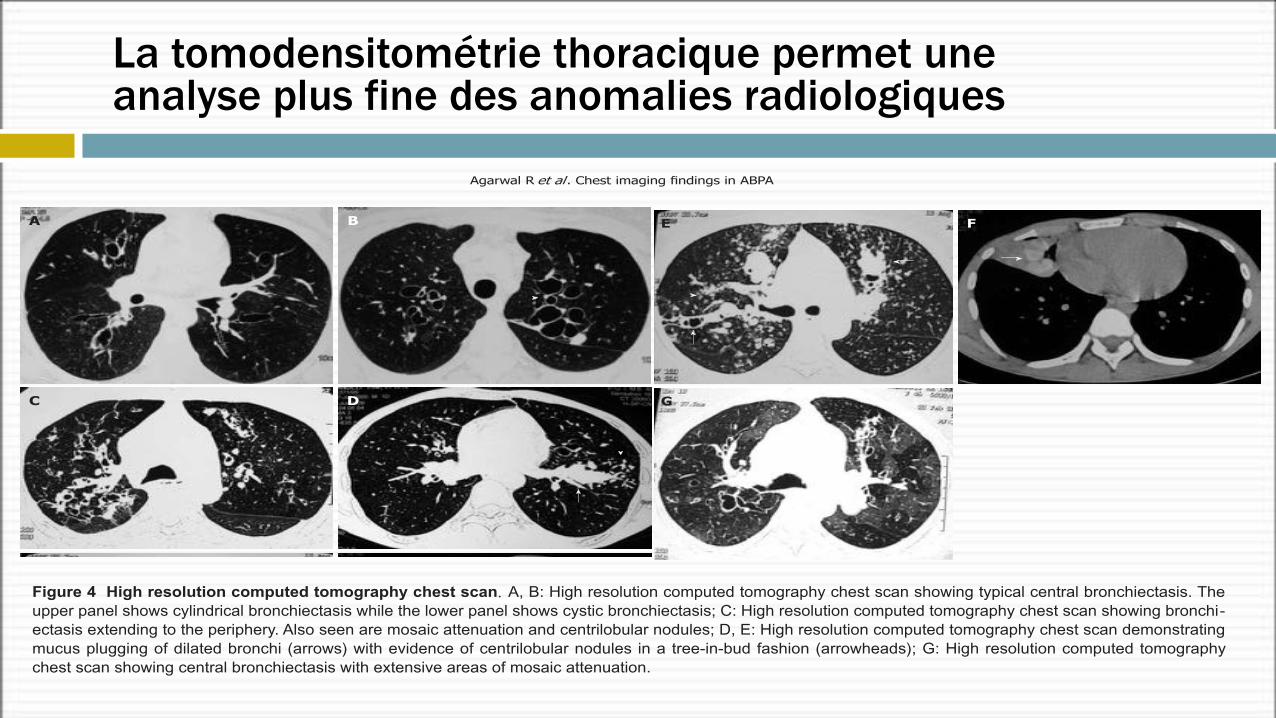
HRCT chest findings
HRCT of the chest was normal in 22 patients, while CB
was demonstrated in the remaining 38 patients (Figure 4A and B). Eight patients with normal chest radiographs were found to have CB on HRCT chest scan while all patients with abnormal chest radiographs had CB on chest CT.
145 April 28, 2012|Volume 4|Issue 4|WJR|www.wjgnet.com
Agarwal R et al . Chest imaging findings in ABPA
Figure 4 High resolution computed tomography chest scan. A, B: High resolution computed tomography chest scan showing typical central bronchiectasis. The upper panel shows cylindrical bronchiectasis while the lower panel shows cystic bronchiectasis;; C: High resolution computed tomography chest scan showing bronchi-ectasis extending to the periphery. Also seen are mosaic attenuation and centrilobular nodules;; D, E: High resolution computed tomography chest scan demonstrating
mucus plugging of dilated bronchi (arrows) with evidence of centrilobular nodules in a tree-in-bud fashion (arrowheads);; G: High resolution computed tomography
chest scan showing central bronchiectasis with extensive areas of mosaic attenuation.
A B
C D
E F
G
opacities (Figure 3B-D) and atelectasis (Figure 3E).
HRCT chest findings
HRCT of the chest was normal in 22 patients, while CB
was demonstrated in the remaining 38 patients (Figure 4A and B). Eight patients with normal chest radiographs were found to have CB on HRCT chest scan while all patients with abnormal chest radiographs had CB on chest CT.
145 April 28, 2012|Volume 4|Issue 4|WJR|www.wjgnet.com
Agarwal R et al . Chest imaging findings in ABPA
Figure 4 High resolution computed tomography chest scan. A, B: High resolution computed tomography chest scan showing typical central bronchiectasis. The upper panel shows cylindrical bronchiectasis while the lower panel shows cystic bronchiectasis;; C: High resolution computed tomography chest scan showing bronchi-ectasis extending to the periphery. Also seen are mosaic attenuation and centrilobular nodules;; D, E: High resolution computed tomography chest scan demonstrating
mucus plugging of dilated bronchi (arrows) with evidence of centrilobular nodules in a tree-in-bud fashion (arrowheads);; G: High resolution computed tomography
chest scan showing central bronchiectasis with extensive areas of mosaic attenuation.
A B
C D
E F
G
La tomodensitométrie thoracique permet une analyse plus fine des anomalies radiologiques
① Patients avec bronchectasies (ABPA-DDB)② Patients sans bronchectasies (ABPA-antigènes)
Le retard au traitement est responsable du développement de DDB ++
Les patients porteurs de ABPA peuvent être subdivisés en 2 groupes
Aspergillose bronchopulmonaire allergique (ABPA)
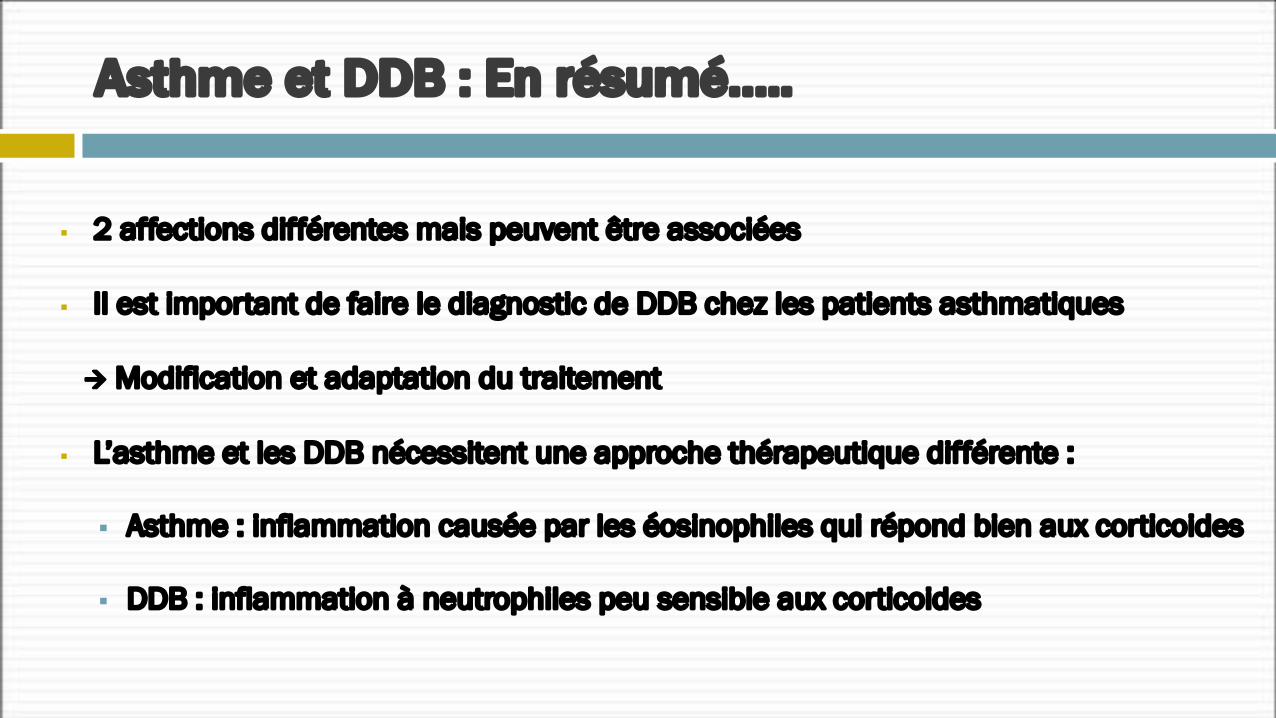
§ 2 affections différentes mais peuvent être associées
§ Il est important de faire le diagnostic de DDB chez les patients asthmatiques
à Modification et adaptation du traitement
§ L’asthme et les DDB nécessitent une approche thérapeutique différente :
§ Asthme : inflammation causée par les éosinophiles qui répond bien aux corticoides
§ DDB : inflammation à neutrophiles peu sensible aux corticoides
Asthme et DDB : En résumé…..
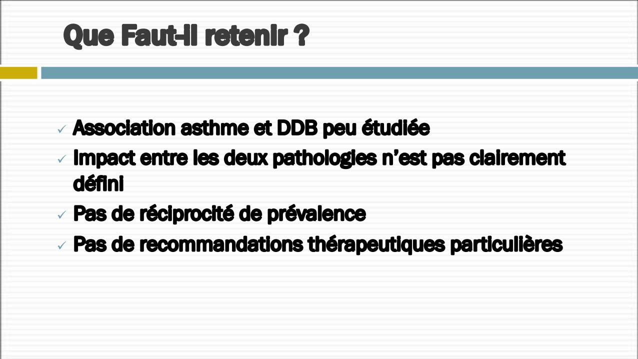
Que Faut-il retenir ?
ü Association asthme et DDB peu étudiéeü Impact entre les deux pathologies n’est pas clairement
définiü Pas de réciprocité de prévalenceü Pas de recommandations thérapeutiques particulières