Embed Size (px)
Citation preview
Brain oscillations in highly hypnotisable participants during neutral hypnosis,
hypnotic suggestions, and pre- and posthypnosis
Maria Lisa Birgitta Karevaara
Master’s Thesis
Psychology
Faculty of Medicine
April 2020
Supervisor: Petri Paavilainen
Tiedekunta – Fakultet – Faculty
Faculty of Medicine
Koulutusohjelma – Utbildingsprogram – Degree Programme
Psychology
Tekijä – Författare – Author
Maria Lisa Birgitta Karevaara Työn nimi – Arbetets titel – Title
Brain oscillations in highly hypnotisable participants during neutral hypnosis, hypnotic
suggestions, and pre- and posthypnosis
Oppiaine/Opintosuunta – Läroämne/Studieinriktning – Subject/Study track
Psychology Työn laji – Arbetets art – Level
Master’s Thesis
Aika – Datum – Month and year
April 2020
Sivumäärä – Sidoantal – Number of pages
67 Tiivistelmä – Referat – Abstract
Objective:
To contribute to the theory-building on hypnosis by studying the possible changes that
hypnosis causes in the electroencephalographic (EEG) spectral power in highly hypnotisable
individuals. In accordance with previous literature, hypnosis was hypothesised to cause an
increase in theta (4–8 Hz) power and a change in gamma (25–45 Hz) power.
Methods:
Nine highly hypnotisable individuals (8 females) participated. Continuous EEG was recorded
at ten electrodes during four conditions: prehypnosis, neutral hypnosis, hypnotic suggestion,
and posthypnosis. During all conditions, the participants watched a monotonous video while
sinusoidal tones following an oddball paradigm played silently in the background. The
participants were instructed not to pay any attention to the tones, and in the suggestion-
condition a suggestion to hear all tones as similar in pitch was given. Nine repeated-measures
analyses of variance, one for each frequency range, were performed. For research questions 2
and 3, the participants were divided into two groups depending on their responsiveness to a
hallucinatory suggestion in the screening phase, and the analyses were then run again.
Results:
No differences between conditions were found in the theta range, but a decrease was found in
the gamma range during hypnosis compared with wakefulness (posthypnosis). Spectral power
differences depending on responsiveness to the hallucinatory suggestion were also found.
Conclusions:
The findings support the hypothesis of changed gamma-frequency power during hypnosis, but
not the theory of increased theta frequencies as a marker of hypnosis. A tentative theoretical
connection between reduced peripheral awareness and reduced gamma power in hypnosis is
presented.
Avainsanat – Nyckelord – Keywords
Hypnosis, suggestion, EEG, oscillations, power spectral density
Ohjaaja tai ohjaajat – Handledare – Supervisor or supervisors
Petri Paavilainen
Säilytyspaikka – Förvaringställe – Where deposited
Helsinki University Library – Helda / E-thesis ethesis.helsinki.fi Muita tietoja – Övriga uppgifter – Additional information
Tiedekunta – Fakultet – Faculty
Lääketieteellinen tiedekunta
Koulutusohjelma – Utbildingsprogram – Degree Programme
Psykologia
Tekijä – Författare – Author
Maria Lisa Birgitta Karevaara
Työn nimi – Arbetets titel – Title
Aivo-oskillaatiot herkästi hypnotisoitavilla tutkittavilla ennen hypnoosia, neutraalissa
hypnoosissa, hypnoottisessa suggestiotilanteessa, ja hypnoosin jälkeen
Oppiaine/Opintosuunta – Läroämne/Studieinriktning – Subject/Study track
Psykologia
Työn laji – Arbetets art – Level
Pro gradu -tutkielma
Aika – Datum – Month and year
Huhtikuu 2020
Sivumäärä – Sidoantal – Number of pages
67 Tiivistelmä – Referat – Abstract
Tavoitteet:
Tämän tutkimuksen tavoitteena oli edistää yhtenäisen teorian muodostusta hypnoosista
tutkimalla muutoksia aivosähkökäyrän (EEG:n) tehotiheysspektrissä, joita hypnoosi
mahdollisesti aiheuttaa herkästi hypnotisoitaville tutkittaville. Aiemman kirjallisuuden nojalla
tehon oletettiin kasvavan EEG:n theta-taajuuksilla (4–8 Hz) ja muuttuvan gamma-taajuuksilla
(25–45 Hz) hypnoosin aikana.
Menetelmät:
Yhdeksän herkästi hypnotisoitavaa tutkittavaa (8 naista) osallistui kokeeseen. Jatkuvaa EEG:tä
mitattiin kymmenellä elektrodilla neljässä koetilanteessa: ennen hypnoosia, neutraalissa
hypnoosissa, hypnoottisessa suggestiotilanteessa, ja hypnoosin jälkeen. Tutkittavat katselivat
monotonista videota kaikissa koetilanteissa sinimuotoisten äänimerkkien soidessa hiljaa taustalla
oddball-paradigman mukaisesti. Tutkittavia ohjeistettiin jättämään äänimerkit huomiotta, ja
suggestiotilanteessa tutkittaville annettiin suggestio kuulla kaikki äänet saman korkuisina.
Analyysit toteutettiin jokaiselle määritellylle taajuuskaistalle erikseen yhteensä yhdeksällä
toistettujen mittausten varianssianalyysillä. Lisäksi tutkimuskysymyksiä 2 ja 3 varten tutkittavat
jaettiin kahteen ryhmään sen mukaan, toimiko hallusinatorinen suggestio heillä
seulontavaiheessa. Tämän jälkeen analyysit suoritettiin uudelleen.
Tulokset:
Koetilanteiden välillä ei löydetty eroa theta-taajuuskaistalla, mutta gamma-taajuuskaistalla tehon
havaittiin vähenevän hypnoosissa verrattuna hereillä oloon (hypnoosin jälkeen). Lisäksi todettiin
joitakin tehotiheysmuutoksia, jotka riippuivat hallusinatorisen suggestion toimivuudesta
tutkittavilla.
Johtopäätökset:
Tulokset tukevat hypoteesia muuttuneesta gamma-tehosta hypnoosin aikana, mutta eivät teoriaa
kasvaneesta theta-tehosta yhtenä hypnoosin indikaattorina. Aiempien tutkimusten ja tämän
tutkimuksen tulosten perusteella spekulatiivinen teoreettinen yhteys esitetään löytyvän
vähentyneen perifeerisen tietoisuuden ja vähentyneen gamman välillä hypnoosin aikana.
Avainsanat – Nyckelord – Keywords
Hypnoosi, suggestio, EEG, oskillaatio, tehotiheysspektri
Ohjaaja tai ohjaajat – Handledare – Supervisor or supervisors
Petri Paavilainen
Säilytyspaikka – Förvaringställe – Where deposited
Helsingin yliopiston kirjasto – Helda / E-thesis (opinnäytteet) ethesis.helsinki.fi Muita tietoja – Övriga uppgifter – Additional information
Foreword
This master’s thesis is about hypnosis and its effects on human brain oscillations.
Before starting the process of writing this thesis, I did not know very much about hypnosis or
its working principle. I had, like most people, seen and heard about people being hypnotised
as entertainment on stage and becoming entirely unaware of themselves and their
surroundings while blindly following any suggestions they were given by the hypnotist. I had
also heard about hypnosis being used in clinical settings as a treatment method, but while
being interested in the theory behind it, I also felt frustrated about the mysticism and
tabooness that seemed to reside around the matter. What was this phenomenon that was
barely discussed in my studies in psychology and seemed to divide opinions both among the
general public and among scientists? For my own, my fellow-students’ and for the sake of
anyone who was interested in the scientific base of hypnosis, I decided to try to get to the root
of it the best I could.
The data of this thesis were collected by Docent Maarit Virta and Mr. Seppo Hiltunen,
M.A., in the years of 2016–2017 in Helsinki as a part of Seppo Hiltunen’s doctoral
dissertation. The data have been used once in a recent article by Hiltunen, Virta, Kallio, and
Paavilainen (2019) for event-related potential analyses, and I am ever so thankful to the
authors and especially to Seppo for the opportunity to utilize the data in novel brain
oscillation analyses. Being able to write a thesis on a subject I am highly interested in and
that took a strong hold of me already at the very beginning of the process has truly been a
pleasure.
Given the opportunity here now, I would like to thank my devoted and inspiring
supervisor, Docent Petri Paavilainen (Department of Psychology and Logopedics, University
of Helsinki), for giving me useful feedback and supporting my work all along. Needless to
say, my work would not have attained its current edge without you. I would also like to thank
laboratory engineer Tommi Makkonen, M.Sc., for his guidance and especially for doing the
heavy lifting in extracting the EEG spectral power values from the raw data. Lastly, a big
thanks belongs to my encouraging husband and our beautiful daughter who was born during
the process. You inspired me through the work and believed in me every day of the project.
Maria Karevaara
Helsinki April 28th, 2020
Table of contents
1 Introduction ........................................................................................................................................ 1
1.1 Hypnosis: defining the subject ...................................................................................................... 3
1.1.1 Achieving hypnosis ................................................................................................................ 3
1.1.2 Hypnotisability and suggestibility ......................................................................................... 4
1.1.3 Hypnotic features and applications ........................................................................................ 7
1.2 Theories of hypnosis ..................................................................................................................... 9
1.2.1 Mesmer and Braid .................................................................................................................. 9
1.2.2 State versus non-state debate ................................................................................................. 9
1.2.3 Present-day neurophysiological hypotheses ........................................................................ 14
1.2.4 Integrative theories of hypnosis ........................................................................................... 18
1.3 Electroencephalography .............................................................................................................. 20
1.3.1 Basics ................................................................................................................................... 20
1.3.2 The frequency ranges of EEG outside the context of hypnosis ........................................... 22
1.3.3 EEG oscillations and hypnosis: previous studies ................................................................. 25
1.4 Aims of the present study ........................................................................................................... 29
1.4.1 The underlying study: Hiltunen, Virta, Kallio, and Paavilainen, 2019 ................................ 29
1.4.2 Research questions and hypotheses ..................................................................................... 30
2 Methods ............................................................................................................................................. 33
2.1 Participants .................................................................................................................................. 33
2.2 Procedure .................................................................................................................................... 33
2.3 EEG recording ............................................................................................................................ 36
2.4 Methods of analysis .................................................................................................................... 36
3 Results ............................................................................................................................................... 38
3.1 Research question 1 .................................................................................................................... 38
3.2 Research question 2 .................................................................................................................... 42
3.3 Research question 3 .................................................................................................................... 42
3.4 Experienced depth and background sound experiences .............................................................. 43
4 Discussion.......................................................................................................................................... 46
4.1 General discussion ...................................................................................................................... 46
4.2 Strengths and limitations ............................................................................................................. 52
4.3 Conclusions ................................................................................................................................. 54
5 References ......................................................................................................................................... 55
1
1 Introduction
Hypnosis is a phenomenon that is typically characterised by a person’s high suggestibility while
being in a focused and relaxed state. Hypnosis can be administered as self-hypnosis to oneself, but
traditionally it is achieved when a trained hypnotist administers a relaxing hypnotic induction on a
willing and motivated subject. When the induction has been finished and the subject is believed to have
achieved hypnosis, the hypnotist may give the subject various suggestions for changes in behaviour and
experience. Usually the suggestibility of the subject has risen, and so the subject follows the
suggestions without being aware of pursuing them. If no suggestions are given to the hypnotised person
after the hypnotic induction, he/she is believed to be in so-called neutral hypnosis.
Not all people are able to become hypnotised. Even though the willingness and motivation of
the individual being hypnotised are necessary components for hypnosis to work, they are not sufficient
factors for achieving hypnosis. According to research, it seems that hypnotisability is a trait-like
variable that follows roughly a normal distribution in the population (Bongartz, 1985; Piccione,
Hilgard, & Zimbardo, 1989; Peter, Geiger, Prade, Vogel, & Piesbergen, 2015; Piesbergen & Peter,
2006). The plupart of people are medium hypnotisable and respond to many, but not all types of
suggestions, while roughly 10–15% of the population are very easily hypnotisable individuals, being
able to exhibit profound alterations in behaviour and in their conscious experience during hypnosis.
Depending on the given suggestions, a hypnotised person may experience sensory alterations, a lack of
control of own actions, age regression, changes in memory, or an immersion in an illusory reality. Even
after exiting hypnosis, so called posthypnotic suggestions given during hypnosis may influence
behaviour. A socially anxious person may for example be given a posthypnotic suggestion to feel
confident and relaxed in upcoming social situations. The range of possible hypnotic suggestions is vast
and versatile, but simply put, the most essential feature of the realisation of hypnotic suggestions is
their perceived involuntariness or lack of associated conscious awareness. In a simple hand levitation
suggestion, for example, the participant believes that their hand rises by itself since they did not
consciously decide to raise the hand yet sees it rise before their eyes.
Due to its consciousness-altering nature, hypnosis is often regarded as something mystical, and
the general public as well as healthcare professionals tend to hold many misconceptions about it
(Daglish & Wright, 1991; Green, 2003; Yu, 2004). These prevailing opinions and beliefs, despite being
largely positive regarding the therapeutic benefits of hypnosis (Johnson & Hauck, 1999), are
undoubtedly a reason to the fact that hypnosis is still today a greatly underutilised practice. It should be
2
emphasised that hypnosis has been reliably established to have a great amount of possible applications,
and it is an effective and economical treatment method for a plethora of different medical conditions,
such as chronic pain and anxiety (Flammer & Bongartz, 2003; Häuser, Hagl, Schmierer, & Hansen,
2016; Thompson et al., 2019). However, what hypnosis is and how it actually works is not yet clear,
and many possible theories exist on the field.
At the beginning of hypnosis research, most of the theories became polarised into essentially
two opposing views that disagreed on whether hypnosis is a separate state of consciousness or merely a
social-cognitive phenomenon that lacks any special features that cannot be explained with social-
cognitive terms. These two competing sides became to compose the so-called state versus non-state
debate that although subdued from its prime time, still somewhat exists. In an effort to resolve the
matter, different brain imaging methods were and are used in hypnosis research. On the one hand, if
hypnosis is a separate state, it should involve some distinct features that would be distinguishable when
imaging the brain. On the other hand, if no hypnosis-specific changes are detected in the brain, it
supports the view of hypnosis as a social-cognitive phenomenon.
Following this principle, brain oscillations measured with electroencephalography (EEG), an
economical brain imaging method often used in distinguishing between brain states such as
wakefulness and sleep, have been studied during hypnosis and in highly hypnotisable individuals.
During early hypnosis EEG studies hypnosis was associated with both increased (e.g., Ulett, Akpinar,
& Itil, 1972) and decreased (e.g., De Pascalis, Ray, Tranquillo, & D'Amico, 1998; Graffin, Ray, &
Lundy, 1995) alpha frequency (~8–13 Hz) power, but more recently hypnosis has been associated with
heightened theta range (~4–8 Hz) power and an alteration in gamma range (>25 Hz) power (for a
review, see Jensen, Adachi, & Hakimian, 2015). Inconsistencies and methodological differences are,
however, common in the literature, and it has not for example been established what the role of the
hypnotic induction or the suggestions given during hypnosis are in the observed EEG changes.
The aim of the current study is by the use of EEG to study whether measured brain oscillations
in highly hypnotisable participants change during neutral hypnosis or when receiving hypnotic
suggestions in comparison to wakefulness before and after hypnosis. The present study can be seen as a
part of the continuum of studies trying to resolve the state versus non-state debate and define hypnosis.
Further, studying hypnosis and its foundations is a way of tackling the prevailing misconceptions
around hypnosis and ultimately promoting hypnosis in becoming a more widely used, evidence based
clinical practice that both individuals and the national economy can benefit from.
3
1.1 Hypnosis: defining the subject
1.1.1 Achieving hypnosis
Two different meanings of the term hypnosis can be derived, namely hypnosis as a procedure
and hypnosis as a product (see, e.g., Nash & Barnier, 2012). One may administer hypnosis on someone
else (procedure), while that person still fails to achieve hypnosis (product). What is usually meant with
a hypnotised person is an individual on which the procedure of hypnotisation has generated the product
of hypnosis. The former does not automatically induce the latter but is nevertheless a necessary part in
the achievement of the latter.
Two necessary components of hypnosis as a procedure were defined by American
Psychological Association (APA) Division 30 in 2003, namely introduction and induction (Green,
Barabasz, Barrett, & Montgomery, 2005). Introduction involves that “the subjects is told that
suggestions for imaginative experiences will be presented”, and the induction is essentially an
“extended initial suggestion for using one’s imagination” given after the introduction (Green et al.,
2005, p. 4). The hypnotic induction is generally meant to increase suggestibility for further suggestions
and is traditionally thought to initiate hypnosis. Typical inductions include eye fixation, relaxation (by
e.g. counting down or scanning the body), visualisation practices, and the request to enter deep into
hypnosis. From the various possible features associated with hypnotic inductions, using the word
hypnosis, fostering absorption, enhancing response expectancies, and reducing critical thinking have
been found to enhance suggestibility most effectively (Terhune & Cardeña, 2016). Contrary to what
one may assume, however, no specific features are obligatory for the suggestion or suggestions used
during induction. For example, relaxation, which is most commonly associated and used in the
hypnotic induction, is not necessary for achieving hypnosis (Banyai & Hilgard, 1976). What on the
other hand are deemed as necessary, are the hypnotisable person’s hypnotisability (susceptibility to
hypnosis) and willing attitude towards being hypnotised.
Motivation and response expectancies, meaning one’s own expectations of how one is
automatically going to respond to a given suggestion, can be regarded as features of a willing attitude,
and have even been seen by some researchers as the sole predictors of hypnotisability (Kirsch, 1999b;
Spanos, 1982). However, these factors have been found to be only weak-to-moderate, yet consistent
predictors of hypnotic responsiveness (Benham, Woody, Wilson, & Nash, 2006; Braffman & Kirsch,
1999; Lynn, Kirsch, & Hallquist, 2008; White, 1941), leaving a lot of variability unexplained. Also, as
Jensen and colleagues (Jensen, Adachi, Tomé-Pires, et al., 2015) concluded in their review,
mediational analyses, which are a more sophisticated form of a correlational study, are still needed to
4
be conducted for testing the role of these factors. Regarding the evidence of current studies, response
expectancies seem to have a partial mediational role that varies depending on the suggestions used and
the symptoms on which hypnosis is applied to (Jensen, Adachi, Tomé-Pires, et al., 2015). To conclude,
research suggests that attitudinal factors are necessary yet not sufficient for the induction of hypnosis.
1.1.2 Hypnotisability and suggestibility
Hypnotisability and suggestibility are two terms that are closely associated with each other and
often easily confounded. Suggestibility does however exist by itself as a separate process and does not
necessarily indicate anything about a person’s level of hypnotisability. What is typically meant by a
suggestion is a verbal utterance that resembles an instruction but is not similarly imperative. For
example, instead of commanding the individual to lower their arm, a suggestion that they start to feel
their arm becoming heavier and heavier by every breath, may be given. The wording of the suggestion
underlines thus the involuntariness of the suggested action. Broadly speaking, a suggestion can be any
cue in a person’s environment that results in an involuntary change in his or her behaviour, beliefs,
experience, and corresponding physiological state (Kirsch, 1999a). As Hilgard (1973a) concluded,
there are therefore many forms of suggestibility, such as conformity and placebo, that are separate from
hypnotic responding, and hypnotic responding is not in turn limited to specific suggestions.
One way to distinguish between hypnotic and nonhypnotic suggestions is defining a preceding
hypnotic induction as an obligatory part of hypnotic suggestions. Being hypnotised would therefore be
a prerequisite for a hypnotic suggestion. People may be highly suggestible already without a preceding
hypnotic induction, but hypnosis does generally amplify an individual’s suggestibility (Braffman &
Kirsch, 1999). However, in some cases it might do the opposite and lower an individual’s nonhypnotic
suggestibility, as Braffman and Kirsch’s (1999) study demonstrated. Their study found that 46% of the
participants showed an increase in suggestibility, but as many as a fourth (25%), including some highly
hypnotisable subjects, did in fact display a decrease in suggestibility after the hypnotic induction.
While the finding ought to be replicated (previous studies have not detected such a big amount of
suggestibility decreases) and while some of the decreases could be due to a natural fluctuation in the
scores, the outcome does nevertheless raise questions about the role of a preceding hypnotic induction
in suggestibility. In their review, Terhune and Cardeña (2016) reported that although more research is
needed, the effect of the induction seems to depend on the characteristics of the induction, subject,
method of assessment, and following suggestions.
The uncertainty of the role of the hypnotic induction calls for new terminology, so that
suggestibility would not automatically be interpreted as a person’s hypnotisability, and that researchers
5
would followingly be sure to study whichever they intended to. Kirsch and Braffman (2001) have as a
continuation to their study advocated researchers to recognise the difference between hypnotic
suggestibility and hypnotisability. They stipulate that hypnotisability should be understood as the
difference between imaginative suggestibility (i.e., suggestibility before a hypnotic induction) and
hypnotic suggestibility (i.e. suggestibility measures after a hypnotic induction). Since researchers have
unfortunately been slow in adopting these guidelines of specification, in the present thesis, the term
hypnotisability should be understood as a synonym for hypnotic suggestibility or susceptibility.
Hypnotisability has been shown to be a rather stable trait, much resembling IQ, over a 25-year
period (Piccione et al., 1989). It does however form a curvilinear relationship with age, so that children
under seven years are least hypnotisable and adolescents most, while hypnotisability slowly starts to
decrease after the age of 17 (Cooper & London, 1971; London & Cooper, 1969). With the aid of twin
studies, the heritability index of hypnotisability has been calculated to be .64, corresponding closely the
genetic contribution that is usually found in ability measures (Morgan, 1973).
Individual differences in hypnotisability are measured with standardized tests such as the
Stanford Hypnotic Susceptibility Scale, Form C (SHSS:C; Weitzenhoffer & Hilgard, 1962) and the
Harvard Group Scale of Hypnotic Susceptibility, Form A (HGSHS:A; Shor & Orne, 1962). These tests
have been widely used around the world, and translations to many different languages and norms for
various countries exist today. Both tests are built in a similar fashion, so that a hypnotic induction is
followed by further imaginative suggestions with accruing difficulty. In general, motor suggestions
(suggestions for a particular action, like arm rising, to happen without awareness of volitional
performance) are easier to follow, while cognitive or sensory suggestions, including hallucinations, are
deemed more difficult (Bongartz, 1985; Peter et al., 2015; Piesbergen & Peter, 2006).
In the hypnotisability tests the participant’s behavioural responses to the suggestions are scored
in accordance with predetermined criteria. The more points a participant scores, the more likely it is
that the participant has achieved hypnosis, and the more hypnotisable the participant is deemed to be.
The scores in a population are roughly normally distributed so that a small amount of people seem to be
refractory to hypnosis, a small amount are very susceptible to hypnosis (so called hypnotic virtuosos),
and the large majority of people are moderately hypnotisable (Bongartz, 1985; Peter et al., 2015;
Piesbergen & Peter, 2006). In research, participants are usually divided into low (“lows”), medium
(“mediums”) and high (“highs”) hypnotisable subjects according to their scoring in one or several
standardized hypnotisability tests. Most interest is usually directed towards the group of highly
hypnotisable subjects and their possible distinguishable characteristics. However, even though highs
are usually studied as a single uniform group, recent evidence do in fact suggest the exact opposite,
namely that highs comprise of discrete subtypes with different response profiles. Two different groups
6
of highly suggestible individuals have been found, and the groups differ in how responsive they are to
agnosia and cognitive distortions or to posthypnotic amnesia suggestions (Terhune, 2015). Hypnotic
virtuosos are in turn distinguishable from highs as homogeneously susceptible to all types of
suggestions (Terhune, 2015). Additionally, dissociative tendencies have been found to vary within high
hypnotisables and to modulate the influence that a hypnotic induction exerts on highs’ cognitive control
(Terhune, Cardeña, & Lindgren, 2011b).
As previously noted, some of the hypnotisability tests’ suggestions involve visual or auditory
hallucinations, and they are regarded as the most difficult ones to achieve, visual hallucinations being
even more difficult than auditory (Spanos, Churchill, & McPeake, 1976). While some studies have
detected gender differences regarding responsiveness to hallucinatory suggestions (women responding
more easily; Bowers, 1971), others have not been able to report a difference between males and
females (Spanos et al., 1976). Conflicting reports concerning the role of gender in overall
hypnotisability are also plentiful, since some studies have found a significant, albeit small tendency for
women to score higher than men in hypnotic susceptibility tests (Green, 2004; Rudski, Marra, &
Graham, 2004), whereas others have not found a difference (Bongartz, 1985; Geiger, Peter, Prade, &
Piesbergen, 2014; Peter et al., 2015; Piesbergen & Peter, 2006). The cause to these inconsistent reports
is not clear, but the possible difference is nevertheless so small that its value of application is largely
meaningless.
There have been many rather fruitless efforts to find some other variables, such as personality
traits, that could be used as predictors or explaining factors of a subject’s level of hypnotisability. A
consensus that traditional personality inventories, such as the Big Five Inventory, lack predictive value
to a subject’s hypnotisability, has somewhat been achieved (e.g., De Pascalis, Marucci, & Penna, 1989;
Nordenstrom, Council, & Meier, 2002). Only openness to experience has been found to correlate
weakly with hypnotic suggestibility in some studies, yet not all (Glisky, Tataryn, Tobias, Kihlstrom, &
McConkey, 1991; Green, 2004; Nordenstrom et al., 2002). The most reliable predictor that has been
discovered so far is absorption, which means getting deeply immersed in different experiences, and
which correlates both with openness and hypnotisability (Cardeña & Terhune, 2008; Piesbergen &
Peter, 2006; De Pascalis, Marucci, Penna, & Pessa, 1987; De Pascalis et al., 1989; Glisky et al., 1991).
Hypnotisability has also been associated with various other features ranging from insecure adult
attachment style and psychopathology to dopamine metabolism and grey matter volume (see, e.g.,
Geiger et al.d, 2014; Gruzelier et al., 2004; Horton, Crawford, Harrington, & Downs III, 2004; Huber,
Lui, Duzzi, Pagnoni, & Porro, 2014; Lichtenberg, Bachner-Melman, Ebstein, & Crawford, 2004; Peter,
Hagl, Bazijan, & Piesbergen, 2011; Spiegel & King, 1992).
7
1.1.3 Hypnotic features and applications
As already stated, hypnosis is a versatile phenomenon that affects many cognitive and sensory
processes, and which has many clinical and non-clinical applications. In addition to influencing
processes that can traditionally be reached by conscious control (e.g., memory and movement), even
sensations and other automated processes, such as reading or perceiving colour, may be altered with
suggestions and/or hypnosis in highly hypnotisable subjects. Raz and colleagues have with several
studies been able to demonstrate that with a specific suggestion given to highs, the interference effect
found within the Stroop paradigm (Stroop, 1935) is diminished or entirely removed (Raz & Campbell,
2011; Raz, Shapiro, Fan, & Posner, 2002; Raz, Fan, & Posner, 2005; Raz, Kirsch, Pollard, & Nitkin-
Kaner, 2006; Raz, Moreno-Íniguez, Martin, & Zhu, 2007). This was found to happen regardless if the
participants were in hypnosis, had been in hypnosis and were given a posthypnotic suggestion, or if no
hypnosis was involved.
Highly similar results have been obtained in relation to colour perception. With specific
suggestions given both in and out of hypnosis, highs have been found to be able to perceive coloured
Mondrian-like patterns as greyscale, and grayscale patterns in turn as coloured ones (Kosslyn,
Thompson, Costantini-Ferrando, Alpert, & Spiegel, 2000; Mazzoni et al., 2009; McGeown et al.,
2012). The effect was similar both in and out of hypnosis, but in McGeown et al.’s study (2012), the
experiential colour change as well as cortical activation changes were found to be enhanced during
hypnosis. Additionally, changes in colour-responsive areas in the visual cortex, such as the fusiform
gyri, were detected during the colour-change conditions (Kosslyn et al., 2000; McGeown et al., 2012).
These results suggest that even outside hypnosis, highly hypnotisable subjects are able with
hypothetically some kind of top-down regulation to alter largely automated processes.
Another interesting hypnotic phenomenon is (post)hypnotic amnesia, which is most commonly
referred to as a suggested inability to recall events and experiences that occurred during hypnosis.
Given that hypnotic amnesia is entirely reversible (the participant will remember the lost material again
if given a prearranged cue or suggestion for it), it clearly happens due to disruptions in memory
retrieval rather than in memory encoding or consolidation. As a noteworthy feature, hypnotic amnesia
does only affect explicit memory, while implicit memory, expressed with for example priming, remains
intact (for a review, see, e.g., Kihlstrom, 1997; Kihlstrom, 2007). Some older studies that have not been
replicated suggest that semantic memory, such as remembering some specific words, can also be
disrupted with hypnosis (Spanos, Radtke, & Dubreuil, 1982).
Hypnosis has many applications in the clinical setting, where it is traditionally called
hypnotherapy, and is a cost-effective way of treating different conditions either as sole treatment
8
method or as a conjunct treatment. Among others, hypnotherapy has been proven highly efficient in
reducing or eliminating pain, and one of the most established applications of hypnosis is hence its use
in pain management (effect sizes d= 0.70–.80; Adachi, Fujino, Nakae, Mashimo, & Sasaki, 2014;
Montgomery, DuHamel, & Redd, 2000; Stern, Brown, Ulett, & Sletten, 1977; Thompson et al., 2019).
Other medical conditions that have successfully been treated with hypnosis include anxiety,
posttraumatic stress disorder (PTSD) and different somatic complaints such as irritable bowel
syndrome and tinnitus (Flammer & Bongartz, 2003; Rotaru & Rusu, 2016; Schaefert, Klose, Moser, &
Häuser, 2014; Tefikow et al., 2013). In Kirsch, Montgomery, and Sapirstein’s (1995) meta-analysis of
18 studies, the addition of hypnotherapy was found to substantially improve the treatment outcome of
cognitive behavioural therapy (CBT) on several medical conditions, but particularly on obesity.
The possible use of hypnosis as a tool of learning and memory enhancement has also been
explored. In general, hypnosis cannot enhance memory recollection (hypermnesia), and for example
the previously common practice of using hypnosis in a forensic interview setting or in recovered
memory therapy is nowadays firmly discouraged (Kihlstrom, 1997). Nemeth, Janacsek, Polner, and
Kovacs (2013) found, however, in their study that hypnosis enhanced performance in a very specific,
striatum-dependant sequence learning task. The authors speculated that this could hypothetically be due
to inhibited frontal-lobe activity or disrupted interaction between the frontal lobes and the striatum.
Whether hypnosis could be useful in some other kind of particular learning tasks is yet to be explored.
In addition to applying hypnosis directly to different settings and conditions, hypnosis is also
valuable in instrumental research. Hypnosis can be used to produce specific cognitive states and
perceptions in a controlled setting and provides thus a unique opportunity for researchers to study
various phenomena in the laboratory. Hypnosis has for example been used in several studies
concerning different pathological symptoms, such as hallucinations or other perceptual distortions (for
a review, see Terhune, Cleeremans, Raz, & Lynn, 2017). Hypnosis can also be a useful tool when
studying consciousness, memory processes, or the sense of agency in healthy individuals. When doing
instrumental research, it is of course imperative to consider the role and possible effects that the
hypnotic induction and the suggestions bring about. Discerning the neurophysiological characteristics
of the phenomenon of interest from the changes caused by the induction of the phenomenon through
hypnotic suggestions evidently poses a real challenge for researchers.
9
1.2 Theories of hypnosis
1.2.1 Mesmer and Braid
Hypnosis, or meditative or trans-like practices resembling hypnosis, have existed in many
cultures for thousands of years. Present-day hypnosis is, however, attributed to have been found by the
German physician Franz Mesmer in the late 18th century (see, e.g., Pintar, J. & Lynn, S. J., 2009). First
with the aid of magnets, Mesmer discovered a new way of healing patients in his practice in Vienna.
Mesmer believed that healing was achieved when an invisible physiological “magnetic stream” was
transferred from the healer to the patient, whose own magnetic streams in the nervous system were
injured. Later, Mesmer called the healing force “animal magnetism”, and believed that diseases were
caused when this invisible fluid was unevenly distributed in the patient’s body. Despite the popularity
of Mesmer’s treatment among his patients, the scientific community condemned the practice harshly,
stating that no such thing as magnetic fluid existed. Mesmer’s ideas were therefore used only in small
local practices by his students and were in time further refined by several of them.
In the early 1840’s James Braid further developed the practice of mesmerism, and deliberately
distinguished his approach from Mesmer’s by calling it hypnotism (derived by the Greek word for
sleep, hypnos) and explaining its mechanism in a more realistic way, using known physiological and
psychological terms (Braid,1843). Braid played an important role in promoting hypnosis as a method,
or curable agent, to be used in clinical matters. According to Braid, hypnosis was induced with a
continued fixed stare, that strained the eyes and “paralyz[ed] nervous centres in the eyes and their
appendages” (Braid, 1843, p.16). Braid believed hypnosis, or nervous sleep as he first called it, to be a
separate state of consciousness that lies somewhere between normal sleep and wakefulness. This
viewpoint became to be the most supported theory of hypnosis at the time, and it gave rise to the belief
that hypnosis is a specific dreamlike state that one has to be woken up from. Even today, the so-called
state view remains highly influential.
1.2.2 State versus non-state debate
Despite having been used for a long time, the theories behind and mechanisms of hypnosis have
not been agreed upon. The question of what hypnosis actually is and how it should be defined has
evoked a lot of discussion and debate within the scientific community. Between the 1950’s and -70’s a
vast array of prevailing theories was essentially polarised into two opposing camps: the state and non-
state theories. The state theories have adopted Braid’s view of hypnosis, and regard hypnosis as a
separate state of consciousness which is often called trance. Non-state theories on the other hand
10
comprise mainly of social-cognitive views that claim that hypnosis can be explained by simple social
and cognitive factors, such as social triggers and personal beliefs, and that no hypnosis-specific state is
needed. Social cognitive theories do not altogether deny that hypnosis can include alterations in
consciousness, but rather than consider them necessary, they deem the alterations to be a subjective
feeling and a consequence of hypnosis instead of a prerequisite for it. Some theories representing these
two opposing sides will followingly be presented.
The paradigm of state-theories is rather simple (“hypnosis is a separate/altered state of
consciousness”), and consequently, not many theorists have further detailed the possible working
principle underlying it. One of the few elaborated state theories is Hilgard’s (1973b, 1974)
neodissociation theory. According to it, hypnosis disrupts the functioning of a multi-level and
hierarchical cognitive control system by forming a barrier between some of the subsystems. This
barrier then results for example in the subject being aware of the suggested actions happening, but
unaware of the processes behind the action. Essentially, in hypnosis the hypnotist is able to influence
the executive functions and alter the hierarchical structure of the cognitive control system, resulting in a
dissociation between perceived volition-induced action and concrete action. Hilgard’s main empirical
support for his theory was the so called “hidden observer”- phenomenon, where, as Hilgard interpreted
it, a part of the consciousness immune to suggestions during hypnosis remains aware of the actual state
of affairs. Hilgard demonstrated this with several subjects, for example with a subject who was given a
suggestion to be deaf and did accordingly not react to loud noises, but did, without being aware of it,
nevertheless manage to react to Hilgard’s request to lift his index-finger as a sign for hearing the
request. Later, the “hidden observer” has been used as evidence for both state and non-state theories,
and there has been considerable disagreement on how to study the phenomenon and how to interpret
the results.
Subsequent theorists have later developed the neodissociation theory, and various approaches
have been proposed. Woody and Sadler (2008) have introduced an overarching model in response to
different dissociation models with contradicting implications and have conceptualized the dissociation
to involve the weakening of different paths between executive control, executive monitoring, and the
subsystems of control. According to their theory, suggestions might be directly acted upon without
executive monitoring taking place and thus with the feeling of involuntariness and effortlessness
following.
Other state-theories include different psychoanalytically inclined theories. As Nash (2008)
concluded, psychoanalytic views do not disregard possible social or physiological aspects of hypnosis,
but they do in essence view hypnosis as involving a regressive shift in the experience of self, others,
and their relationship. Freud, for example, believed hypnosis to be a form of topographic regression,
11
where thoughts are regressed into images and transformed thus from a secondary process to a primary
process (Freud, 1957; Nash, 2008; for more psychoanalytic theories, see Fromm, 1977; Orne, 1959).
Support for the non-state theories lie originally in the finding that hypnotic responses are
influenced by personal, interpersonal, and cultural influences. Personal aim, motivation and
interpretation of appropriate behaviour have all been found to affect hypnosis. White was perhaps the
first one to try to systematically conceptualize the importance of motivation or goal-directed behaviour
in hypnosis and can thus be seen as a precursor for later social cognitive non-state theorists. White
(1941) argued that negative motivational factors could prohibit hypnosis, that positive motivational
factors were necessary for hypnosis, but yet that motivation was not alone enough for achieving
hypnosis. Unlike many later social cognitive theorists, White (1941) thought that also an innate
aptitude was needed for achieving deep hypnosis.
The first non-state theory to question the state-view altogether was Sarbin’s role theory,
according to which being hypnotised is a sort of role-taking, resembling much acting (Sarbin, 1950).
Sarbin argued that the role-taking of the stage-actor had many shared characteristics with the role
taking of the hypnotic subject, namely favourable motivation, role perception, and role-taking aptitude,
in which people may differ in. The differentiating factor of the two was according to Sarbin the amount
of self or consciousness involved in the role (Sarbin, 1950). Barber elaborated Sarbin’s theory by
distinguishing several more factors that affect a person’s suggestibility. These involved the expectancy
of how easy or difficult the suggestions were to achieve, and the definition of the situation as
“hypnosis” as opposed to “control” (Barber & Calverley, 1964).
Spanos, another non-state theorist, regarded hypnosis likewise to be a social-cognitive
construct, however refining the theories of his predecessors once again. Spanos concluded hypnosis to
be affected by contextual factors and a subject’s goal-directed fantasies (e.g., the imagining of a
balloon pulling the arm upwards in a hand-levitation suggestion) and deemed additionally
hypnotisability to be malleable with training (Spanos, 1982). The malleability of being able to become
hypnotised received with time a lot of attention, and there appeared a multitude of studies
demonstrating that programmes designed to increase rapport with the hypnotist and diminish resistance
to suggestions did significantly increase participants’ hypnotic responding (e.g., Gfeller, Lynn, &
Pribble, 1987).
Kirsch in turn believed that response expectancies, or predictions of one’s own experiences and
behaviour, were the bulk of hypnosis (Kirsch, 1999b). His theory was supported by findings that
response expectancies predict suggestibility to a considerable degree, that the manipulation of response
expectancies could significantly enhance suggestibility, and that a placebo told to produce a hypnotic
state could also enhance suggestibility in a hypnotic-like manner (Glass & Barber, 1961; Kirsch,
12
1999b; Kirsch, Silva, Comey, & Reed, 1995). Kirsch concluded that both hypnosis and placebos work
due to response expectancies, but in hypnosis the effect is simply achieved with the hypnotic induction
and without the need for a placebo (Kirsch, 1999b).
Kirsch also came to regard the reaction to suggestions as automatic, and together with Lynn, he
formulated the so-called response-set theory which is applicable not only to hypnotic actions but to all
actions (Kirsch & Lynn, 1999; Lynn, Kirsch, & Hallquist, 2008). According to the theory, actions are
prepared by response sets that are comprised of mental associations or representations. These response
sets are primed for automatic activation by intentions, or as in hypnosis, even without conscious
intention and with the simple expectation for an action to occur (Kirsch & Lynn, 1999; Lynn et al.,
2008). The point is that every action is initiated automatically in the moment it is happening rather than
due to a conscious intention. Kirsch and Lynn consequently argue that instead of the automaticity of
responses in hypnosis being an illusion, the everyday experience of volitionally initiating behaviour is
(Kirsch & Lynn, 1999; Lynn et al., 2008).
Lynn has also extensively studied factors affecting hypnotisability, and he has consequently
developed the integrative model of hypnosis. In his model, Lynn incorporates several situational,
personal, and interpersonal factors which he suggests that a hypnotisable individual integrates in a
problem-solving manner during hypnosis (Lynn, Rhue, & Weekes, 1990). Lynn, like many others,
distinguished the perceived involuntariness as an essential feature of hypnosis, and concluded that
several contextual features contribute to the experience. Lynn recognized that for example the wording
of the induction and the suggestions, or the level of rapport with the hypnotiser both shape response
expectancies and enhance the feeling of involuntariness (Gfeller et al., 1987; Lynn et al., 1990). By
time, the integrative model has received new neurophysiological support, and today it therefore
combines social, cultural, cognitive, and neurophysiological determinants in a dynamic and
multifaceted view of hypnosis (Lynn, Laurence, & Kirsch, 2015).
Other contemporary non-state theories include Dienes and Perner's (2007) cold control theory
of hypnosis and Barnier and Mitchell's discrepancy-attribution theory of hypnotic illusions (Barnier,
Dienes, & Mitchell, 2008). In cold control theory the distinction between first order states/thoughts that
include perceptions, and metacognitive higher order thoughts (HOTs) that make a person aware of the
first order state, is pivotal. According to the cold control theory, in hypnosis and sometimes outside it,
the participant forms the intention of performing an act without performing the HOT of being aware of
the intention to perform the act. The participant will still form some kind of HOT, and hypothetically
the expectations regarding hypnosis affect HOTs in a way that the HOT of intending is replaced by a
HOT that supports the expectation of involuntariness (e.g. “I am not making this movement happen”).
Thus, the participant does in fact initiate the execution of all suggestions, but instead of having a
13
second order thought of being aware of the initiation, they form another second order thought that is
aligned with their expectations (Dienes & Perner, 2007). A rather recent study involving the use of
low-frequency repeated transcranial magnetic stimulation (rTMS) on the left dorsolateral prefrontal
cortex (DLPFC) supports the cold control theory, since with the transient disruptions of the DLPFC, an
area associated with metacognition, medium hypnotisables became more hypnotically responsive
(Dienes & Hutton, 2013). In a similar fashion alcohol, which influences the functioning of the
prefrontal cortex (including the DLPFC and the anterior cingulate cortex, ACC) and disturbs therefore
the monitoring and control functions of the brain, has also been found to increase medium hypnotisable
subjects’ level of hypnotisability and supports thus any theory that supposes a reduction in executive
functions to take place during hypnosis (Semmens-Wheeler, Dienes, & Duka, 2013).
The discrepancy-attribution theory of hypnotic illusions again explains hypnotic illusions to be
caused by normal processes that just happen easier in the hypnotic context, and that are then wrongly
attributed (Barnier et al., 2008). A highly hypnotisable subject may for example be able to intentionally
imagine a positive visual hallucination such as a cat in front of him, but instead of attributing it to his
imagination, the subject attributes it to the external world and reality. A similar but less-specific theory
that Terhune has proposed is simply that instead of having any disruptions in executive control during
hypnosis, highs exhibit strategically impaired metacognition as a suggested top-down effect (Terhune
et al., 2017).
Along the development of brain imaging methods and as a way to resolve the state versus non-
state debate, researchers have focused their attention on the human brain. The central underlying
supposition is that for the state-theory to gain support, some consistent correlate that is always present
when a person is hypnotised, should be found. This supposed biological marker in the brain should
therefore be detectable regardless of wording or the content of particular suggestions. Inconsistent
changes detected in the brains of hypnotised individuals might be, and probably are, happening because
of what the individual is doing/sensing while in hypnosis, rather than due to a hypnosis-specific reason.
So far, several studies have found precisely these kind of suggestion-dependent changes in the
participants’ brain activation when regarding colour, pain, and auditory perception, for example
(Kosslyn et al., 2000; Rainville, Duncan, Price, Carrier, & Bushnell, 1997; Rainville et al., 1999;
Szechtman, Woody, Bowers, & Nahmias, 1998), whereas a hypnotic-state specific change is yet to be
discovered. In the next section, newer outcomes and theories that research has contributed with will be
presented.
14
1.2.3 Present-day neurophysiological hypotheses
Neuronal correlates of hypnosis have been of great interest since the development of brain
imaging methods, and various studies with diverse and to some part inconsistent outcomes have been
carried out. One reason to the frequent inconsistencies lies undoubtedly in the studies’ methodological
differences and terminological vaguenesses. Only recently have researchers begun to pay closer
attention to comparable methodologies and to distinguish between the meanings of hypnotic induction,
imaginative suggestions, and hypnotic suggestions. The aim of specifying the terminology is essential
so that the acquired results would reflect the matter of interest and not only different task-related
suggestions, as referred to previously. It has become increasingly common to study hypnosis in a so-
called neutral hypnosis condition, meaning a condition where no explicit suggestions are given to the
participant after the hypnotic induction. When hypnotic suggestions are used, special care should be
directed towards the wording of the suggestions, since the wording is essential for the consecutive
effects found in the brain. For example, regarding hypnotic analgesia, Rainville and colleagues (1997;
1999) found that reduced concern about the pain is associated with less activation of the anterior
cingulate gyrus, while reduced perception of pain produces reduced activity in the somatosensory
cortex. The same behavioural outcome can thus be achieved with different neurological changes
evoked by slightly differently worded suggestions.
There are two research settings that are primarily used when studying the correlates of hypnosis
in the brain. The first way is to simply compare the brain patterns of the same participant during normal
wake-state and hypnosis, making it thus possible to distinguish the changes that appear due to the
induction of hypnosis. The other common way is to divide participants according to their screened level
of hypnotisability, and then to compare the differences in brain patterns between the groups of high and
low hypnotisable individuals, usually leaving out but sometimes also including medium hypnotisable
individuals. Comparisons between highs and lows are made both within and outside hypnosis, and the
differences between the groups are attributed to highs’ capability to achieve hypnosis.
Within these different research settings, different brain research methods have been used in
different kinds of research questions depending on their strengths and weaknesses. Next, some of the
many neurophysiological outcomes and hypotheses derived from the outcomes will be presented in
brief. EEG and the findings acquired with it concerning distinct oscillations and oscillatory power will
not however be explored in this section, but rather separately in their section due to their higher
relevance to the current study.
The frontal lobes are involved in executive functions, and are essential in for example selective
attention, goal setting and action planning. According to a traditional view of hypnosis, hypnosis is
15
believed to involve focused attention, and differences in the ability to engage in such focused attention
are reflected in a person’s level of hypnotisability (Lichtenberg et al., 2004; Tellegen & Atkinson,
1974). Crawford (1994) was one of the early researchers to bring forth the role of attentional processes
in hypnosis, and she integrated her view of their importance in her neuropsychophysiological model of
hypnosis. Her review of the literature concluded that highs seem to possess greater sustained attentional
and disattentional abilities than lows, and that these differences are reflected as neurophysiological
differences in the fronto-limbic attentional system. This view of hypnosis involving focused attention
was also present in APA’s revised, 2014 definition of hypnosis: “[Hypnosis is] a state of consciousness
involving focused attention and reduced peripheral awareness characterized by an enhanced capacity
for response to suggestion.” (Elkins, Barabasz, Council, & Spiegel, 2015, p. 6). However, slightly
newer research has found frontal activity to decrease and performance in attention-craving tasks to
deteriorate when hypnotisable subjects are hypnotised, and question therefore the traditional view
suggesting instead the exact opposite, namely that hypnotisable subjects have impaired attentional
abilities during hypnosis. These two opposing views of focused attention versus impaired attention (or
increased frontal activity versus hypofrontality) have understandably been topics of interest and have
composed the base for many theories of hypnosis.
Gruzelier (1998) reviewed the available evidence and summarized hypnosis to involve the
inhibition of frontal lobe cognitive control functions, while Woody and Bowers (1994) suggested in
their dissociated control theory of hypnosis a dissociation of the frontal lobe to take place. Jamieson
and Sheehan (2004) further elaborated the dissociated control theory and suggested that in hypnosis the
control functions of the left DLPFC become functionally dissociated from the monitoring feedback of
the ACC and from the processes over which they normally exert control. Egner, Jamieson and
Gruzelier (2005) compared the brain responses of highs and lows in response conflict situations evoked
within the Stroop paradigm (Stroop, 1935). From their results, the authors concluded similarly as
Jamieson and Sheehan (2004) that during neutral hypnosis, highs have decreased attentional efficiency
and that a dissociative functional decoupling of response conflict monitoring in the ACC and cognitive
control processes in the frontal lobes takes place in highs, eventuating in lows’ better performance
accuracy during hypnosis (Egner et al., 2005).
Studies assessing the importance, or rather unimportance of the frontal lobe and attentional
inhibition in hypnosis by comparing the performance of highs, (mediums) and lows in different
attentional tasks during and outside hypnosis, have generally found no significant differences between
the groups (Cojan, Piguet, & Vuilleumier, 2015; Kallio, Revonsuo, Hämäläinen, Markela, &
Gruzelier’s, 2001; Varga, Németh, & Szekely, 2011). When some (largely inconsistent and variable)
differences between groups have been detected, highs have turned out to be slower yet make slightly
16
less errors than the rest (Cojan et al., 2015; Varga et al., 2011). The corresponding neural correlates of
highs’ performance outside hypnosis have, however, been found to be distinct from lows’, since highs
have been found to show higher activity in the right inferior frontal gyrus and less activity in the
intraparietal sulcus and ACC compared to lows, suggesting a better control of conflict at least outside
hypnosis (Cojan et al., 2015). Regarding the ACC, most studies have in general associated it with a
decrease in activation during hypnosis (e.g., Deeley et al., 2012; Jiang, White, Greicius, Waelde, &
Spiegel, 2017; McGeown, Mazzoni, Venneri, & Kirsch, 2009; Raz, Fan, & Posner, 2005), and
concerning specifically the dorsal ACC, its decreased activity has been found to be linearly correlated
with the intensity of feeling hypnotised (Jiang et al., 2017). Furthermore, since dorsal ACC activity has
been associated with perseverance, its decrease could also reinforce the feeling of diminished personal
agency and contribute thus to increased suggestibility during hypnosis (Jiang et al., 2017).
Although several studies have appeared later, in their review, Egner and Raz (2007) attempted
to reconcile the partly contradictory results concerning hypnosis and attentional abilities. The authors
concluded evidence to clearly support the view of impaired attention during hypnosis, but they
specified the theory by hypothesising that highs might exhibit impaired cognitive control after a
general, instruction-free hypnotic induction, but be contrariwise superior at implementing any
suggested strategy that improves task performance (for an example of highs’ enhanced performance in
an attentional task with the aid of a posthypnotic suggestion, see Iani, Ricci, Gherri, & Rubichi, 2006).
Terhune, Cardeña, and Lindgren’s (2011b) study later implied that dissociative tendencies of highly
hypnotisable subjects might moderate these distinctive effects on cognitive control, and Jensen and
colleagues (Jensen, Adachi, Tomé-Pires, et al., 2015) summarized most recently that whether an
increase or decrease takes place in the frontal cortices and to what extent, depends on the specific
hypnotic procedures used and suggestions made. Agreeably, Jensen and colleagues (Jensen, Adachi,
Tomé-Pires, et al., 2015) summarized that the midcingulate cortex (MCC) and ACC are also active in
hypnotic responding with their activation level increasing or decreasing suggestion-dependently.
Functional connectivity refers to the temporal correlation of different brain regions, or more
broadly to the statistical interdependence of neurophysiological data (Stephan & Friston, 2008). Highly
hypnotisable subjects have in several studies been found to display changes in the functional
connectivity, namely in the synchrony of oscillatory activity between different cortical regions of the
brain. During hypnosis, highs have been found to show a decrease in global connectivity and to
essentially lose active functional connections between the frontal areas and the rest of the cortex,
supporting thus the hypofrontality and/or impaired attention hypothesis (Cardeña, Jönsson, Terhune, &
Marcusson-Clavertz, 2013; Fingelkurts, Fingelkurts, Kallio, & Revonsuo, 2007a,b; Terhune, Cardeña,
& Lindgren, 2011a). On the other hand, during waking baseline, Kirenskaya, Novototsky-Vlasov, and
17
Zvonikov (2011) oppositely found highs to show a widespread increased functional connectivity
(measured as coherence) in the theta and alpha frequency ranges, and also in the long distance
coherence between frontal and posterior areas within beta-gamma frequency ranges compared to lows.
Highs have also been found to show a decrease in beta-range connectivity between especially
medial (e.g., ACC) and lateral prefrontal areas, and medial prefrontal and occipital areas during
hypnosis (Jamieson & Burgess, 2014; White, Ciorciari, Carbis, & Liley, 2008). Furthermore, highs but
not lows have exhibited an increased theta connectivity in central-parietal areas after a hypnotic
induction (Jamieson & Burgess, 2014). An older study by Sabourin, Cutcomb, Crawford, and Pribram
(1990) again reported no meaningful hypnosis-specific changes in coherence.
More recently, interest has been directed towards the functional connectivity between certain
regions that are part of some specific functional brain network. The networks of interest regarding
hypnosis are the salience network (SN), which is active for example during selective attention and
anxiousness, and comprises regions such as the anterior insula and dorsal ACC, the executive control
network (ECN), which is essential in working memory tasks and in focused attention and which
comprises most prominently the DLPFC, and the default mode network (DMN), which is active during
daydreaming and task-unrelated activities and involves the posterior cingulate cortex, precuneus, and
medial prefrontal cortex. In a functional magnetic resonance imaging (fMRI) study, highs were found
to show significantly higher connectivity between the left DLPFC and regions of the SN, such as the
ipsilateral insular cortex, during hypnosis (Jiang et al., 2017). Additionally, the ipsilateral DLPFC was
found to be negatively correlated with core DMN regions during hypnosis (Jiang et al., 2017). This
pattern of increased connectivity between the ECN and SN and decreased connectivity between the
ECN and DMN underlie according to the authors focused attention, enhanced somatic and emotional
control, and lack of self-consciousness that are characteristic for hypnosis (Jiang et al., 2017). In
another study, highs’ right inferior frontal gyrus, a part of the dorsal attention network, was more
closely connected to the default mode network than lows’, which the authors speculated to possibly
cause a greater flexibility in attention and thus underlie an enhanced ability to dissociate (Cojan et al.,
2015). Furthermore, in an fMRI study, highs were found to display heightened structural connectivity
between the dorsal ACC and DLPFC (parts of the SN and ECN; Hoeft et al., 2012). To conclude these
recent connectivity studies, it can be said that during hypnosis changes in the functional and structural
connectivity take place particularly in the prefrontal areas and between the ECN and SN.
Alongside the connectivity studies between different brain networks, the activity of the DMN
has also been of interest by itself. One hypothesis that has gained evidence states that hypnosis involves
a reduction in the activity of the DMN. In McGeown and colleagues’ (2009) study as well as in that of
Deeley and colleagues (2012), participants’ brain activity were studied with fMRI during a neutral
18
hypnosis condition that situated between performance tasks, and which participants did not know to be
an experimental condition. The brain activation of highs during neutral hypnosis in McGeown’s and
colleagues’ study, and increased subjective depth of hypnosis in Deeley and colleagues’ study, were
found to correlate with decreased activation in brain regions recognised as parts of the anterior DMN
(Deeley et al., 2012; McGeown et al., 2009). These areas include cortical midline structures of the
medial and superior frontal gyri, left inferior and middle frontal gyri, posterior cingulate gyri,
parahippocampal gyri, and the ACC (Deeley et al., 2012; McGeown et al., 2009). The result is in
accordance with logical reasoning, since the DMN is characteristically active during the absence of
goal-directed activity (i.e. during normal resting state), while its activity diminishes during attentional
demands. Deeley and colleagues (2012) did find increased activation in areas identified as important
for the maintenance of attention, namely ventrolateral prefrontal regions, but McGeown and colleagues
(2009) contradictingly reported opposite findings. The findings do of course therefore need further
studying, but as McGeown and colleagues (2009) conclude, it is possible that the deactivation of the
DMN might reflect the inhibition of irrelevant thought processes during hypnosis. Spiegel (2008) has
for example suggested that highly hypnotisable individuals may be better at DMN inhibition, and
therefore be able to more easily alter motor and sensory functions while as a trade-off lose self-
awareness and experience involuntariness.
As Landry, Lifshitz and Raz (2017) concluded in their review, from a neurophysiological
perspective it seems like hypnosis engages on the one hand frontal regions such as the ACC, anterior
insula and the DLPFC – areas associated with attention, executive control, and cognitive monitoring,
and which have been identified as the salience and executive control networks. On the other hand,
hypnosis seems to deactivate the anterior part of the DMN – a network associated with social
cognition, internal attention, and self-related thought. The authors suggest that the executive and
salience networks covary with the DMN and together reflect hypnosis, or the experience of hypnosis
(for a thorough review of the state of evidence, see original paper). In their meta-analysis, Landry and
colleagues (2017) did however not find support to their hypothesised theory, but their results did
instead associate hypnosis with activation in the medial lingual gyrus, an occipital region mainly
involved in higher-order visual processing. As the authors hypothesise, this is likely to reflect mental
imagery in hypnosis.
1.2.4 Integrative theories of hypnosis
As reported, many are the suggested and possible models of hypnosis, ranging from the state
versus non-state debate to today’s neurophysiologically driven theories, but few are those that account
19
for all, or even most of the findings supporting these different models. Being one of the very first to
attempt to form a unified picture of the accumulated evidence, Gruzelier (1996, 1998, 2000) strived
towards an integrative model of neurobiological and sociocognitive perspectives. Gruzelier presented a
thesis that hypnosis was an altered state of brain systems initiated by the specific interpersonal and
cultural context of hypnotic induction. He illustrated a view of the hypnotic induction as a temporal
process involving anterior left-sided attentional mechanisms and selective inhibition of frontal
functions, and which becomes conditioned to facilitate future inductions and self-hypnosis.
With new evidence rapidly accruing, Jensen and colleagues (Jensen, Adachi, Tomé-Pires, et al.,
2015) later introduced the development of a biopsychosocial model and Lynn and colleagues (2015)
their integrative model of hypnosis. Jensen and colleagues performed a scoping review which
summarized and clarified the current state of knowledge regarding associations between biological,
psychological, and social factors hypothesised to explain and contribute to hypnotic responding. The
authors distinguished twelve factors that current evidence points towards as reliable correlates of
hypnosis. The biological factors they distinguished were essentially the ones previously reviewed,
namely the altered activity in frontal and anterior cingulate cortices, changes in functional and
structural connectivity, increased hemispheric asymmetry, and the increase of theta oscillations
(reviewed later separately; Jensen, Adachi, & Hakimian, 2015; Jensen, Adachi, Tomé-Pires, et al.,
2015). The psychological and social factors that Jensen and colleagues (Jensen, Adachi, Tomé-Pires, et
al., 2015) regarded to have solid evidence were expectancies, motivation, absorption, hypnotisability,
rapport and hypnotic context. As the authors concluded, what still remains to be studied is which ones
of these factors play a causal role and which ones merely reflect hypnotic responding. Also, identifying
factors that moderate the influence of these 12 factors and exploring additional possible factors remain
topics of interest (Jensen, Adachi, Tomé-Pires, et al., 2015).
Lynn and colleagues (2015) draw a more critical picture in their review, calling for more
research about the differences between effects found during hypnosis and outside the context of
hypnosis. The authors extend in a way previous sociocognitive theories to fit new research evidence
and argue that social-psychological variables modulate cognitive and neurophysiological findings of
hypnosis, simultaneously pointing out that all neural correlations of hypnosis may well be produced by
social and cognitive variables. According to Lynn and colleagues (2015):
“The combination of a propensity to highly automatized cognition, which facilitates the
seamless operation of response sets and the recruitment of suggestion-related imaginings,
combined with cognitive flexibility and the use of cognitive strategies deployed in response to a
variety of suggestions, may be essential to optimal hypnotic responsiveness.” (p. 11)
20
As a way of explaining why some individuals fail to become hypnotised despite training, Lynn and
colleagues (2015) suggest that compared with highs, lows lack some abilities essential to automatic
information processing and attentional abilities that may be trait-like. Thus, leaving it open for further
elaboration, the authors advocate a model of hypnosis as a product of automatised, hard-wired
cognitive functions.
When zooming out of these integrative theories, what can be regarded as an upper-level,
overarching theory combining most of them and a large part of all the underlying findings, is regarding
hypnosis as a form of a top-down process. Top-down regulation can in general be understood as the
cognitive process in which mental representations affect or “override” lower-level neurobiological
phenomenon such as perception. Essentially, theories emphasising attention, the social context, or
personal attitudes and response expectancies all support the underlying assumption of top-down
influences. Consequently, when synthesising the current literature, Terhune and colleagues portray
hypnosis as a unique form of top-down regulation where verbal suggestions are able to cause
alterations in consciousness (Terhune et al., 2017). Of course, while gaining the role of a higher-level
theory, the top-down hypothesis loses specificity and lacks therefore a lot of meaningful predictive and
explanatory value that lower-level theories generally encompass.
1.3 Electroencephalography
1.3.1 Basics
Electroencephalography (EEG) is a noninvasive brain imaging method that measures the
electrical activity of the brain and which is widely used in research and diagnostics. The greatest
advantage of EEG compared to other brain research methods is its high temporal resolution in the
millisecond range, which allows its user to study very precisely temporal, and for example stimulus-
specific changes in the brain. It is also easy, safe, and economical to conduct, making it even more
popular. EEG is well-known for its use in sleep-studies since the different sleep phases have largely
been distinguished due to their discernible EEG patterns. Perhaps partly due to the historical view of
hypnosis as a sleep-like state and with the aim to recognise some distinguishable changes in brain
electrophysiology during hypnosis too, EEG has been a popular method in hypnosis research.
EEG was first measured on human subjects by Hans Berger in 1924 (Berger, 1929). Berger
measured rhythmic, 10 Hz frequency oscillations on a human subject by placing one electrode on the
occipital cortex and another one on the forehead. He named the oscillations alpha waves, since they
21
were the first ones he observed. Later, several more frequency waves have been detected, and the
underlying mechanisms have moreover become better-known.
The aim of EEG is to measure the electrical field on a participant’s scalp caused by brain
activity, or more specifically by masses, or different groups of neurons firing simultaneously at some
frequency. When an either inhibitory or excitatory postsynaptic potential is excited at a neuron’s
dendrites or close to its soma, it causes the surrounding extracellular fluid to become charged. The
evoked extracellular charge is either negative or positive, depending on the excitatory or respectively
inhibitory nature of the potential. This charge followingly generates an opposite, positive or negative
charge to form at the far-end of the neuron, constituting thereby a dipole. The positive and negative
charges from different nearby neurons summate or, if the neurons are oppositely charged and lay in a
similar orientation, cancel each other out. Then the sum of all charges propagates through several
layers (brain, dura, skull, and electrode gel) before eventually reaching the electrode on the scalp (for
further elaboration and a review see, e.g., Jackson & Bolger, 2014).
As implied earlier, the charge from a single neuron is not enough to cause a measurable electric
field on the scalp, but tens or hundreds of thousands of neurons that fire at the same time and in a
parallel arrangement, cause measurable voltage changes. The amount of neurons firing at the same time
then constitutes the measured power level. Pyramidal neurons are believed to be the primary cause of
the measured electric potential since they are found close to the scalp on the cortex and are oriented
perpendicular to the brain surface. However, the voltage on the surface is thought to be comprised of a
combination of thalamocortical and corticocortical connection’s synchronous activity (Olejniczak,
2006). More specifically, the thalamus has been found to be responsible of an internal source of
rhythmic fluctuation that further affects the cortical neurons on the scalp to fluctuate (Steriade, Gloor,
Llinas, Da Silva, & Mesulam, 1990). The measured voltage is voltage calculated between the electrode
in question and a reference electrode. Voltage is therefore never absolute, but rather a difference of two
sites that can be either positive or negative in nature. This difference is in the case of EEG so small (in
the microvolt range), that the voltage is amplified with an amplifier with high impedance. In this way, a
better signal to noise- ratio is strived to be acquired.
The measured oscillations constitute of different coalesced frequency components that can be
distinguished by spectral analysis methods. To put this in another way, the complex EEG data-wave
that is retrieved from the scalp is formed when many sinusoids with variable frequencies summate.
With the aid of a fast Fourier transform (FFT), the summation-wave can be divided into its underlying
sinusoidal components, and then transformed from the time-domain to the frequency domain. In this
way, the power of each frequency component found in the EEG-wave at a specific time-point (or as the
mean of several time points) can be calculated, and the information of the wave can be presented in
22
simple, quantifiable data. The FFT assumes, however, the signal to be stationary, so when calculating
the frequency powers from a selected time-window, information on any time-varying spectral content
existing in that window is ignored and lost. Visual inspection of the EEG signal in the time-domain
therefore generally gives valuable additional information to the FFT.
1.3.2 The frequency ranges of EEG outside the context of hypnosis
The underlying oscillatory components of an EEG signal are believed to reflect different
cognitive, affective, or attentional states, and are therefore regarded as the link between behaviour and
single-neuron activity. Even though the correlative nature between certain frequencies and behaviours
have been established, the measured oscillations vary slightly from one individual to another even in a
very specific behavioural context (e.g., deep sleep). Furthermore, research suggests that the temporal
synchrony of different regions and/or multiple frequency ranges represent the large-scale integration of
cerebral activity, resulting in thoughts, emotion, perceptions, memories and actions (for a review, see,
e.g., Cantero & Atienza, 2005; Varela, Lachaux, Rodriguez, & Martinerie, 2001).
The main frequency bases that can be distinguished in the EEG have been divided into different
frequency ranges or frequency bands. There are several different ways to define the ranges in the
literature, and the following way can be seen as one common way among others: delta range (1–4 Hz),
theta range (4–8 Hz), alpha range (8–12 Hz), beta range (13–25 Hz) and gamma range (>25 Hz). The
low frequency ranges delta, theta, and alpha span over larger areas, and proposedly integrate multiple
brain regions, thus determining the general mode of processing. The faster, high-frequency ranges beta
and gamma on the other hand distribute over smaller areas, and therefore generally represent more
specific cognitive processes. Recent evidence suggests that cross-frequency phase and/or amplitude
coupling especially between the slower and faster frequency ranges play an important role in various
brain functions (for reviews, see Canolty & Knight, 2010; Jensen & Colgin, 2007; Jensen & Mazaheri,
2010), but cautiousness is warranted due to possible artefactual couplings produced in common
analysis methods (for a discussion of overcoming some interpretational issues see Aru et al., 2015).
Next, we will look more closely into the correlates of the distinct frequency ranges.
The slowest frequency range, the delta range (1–4 Hz), has the highest amplitude and is present
during anaesthesia and deep, non-REM sleep. Delta frequencies typically originate in the ventral
tegmental area, nucleus accumbens, medial prefrontal cortex, and thalamic reticular nucleus (for a
review, see, e.g., Knyazev, 2007, 2012; McCormick & Bal, 1997). Delta frequencies are thus closely
linked to the brain reward system, which is involved in motivation and salience detection, and
consequently delta frequencies seem to increase when there is a possibility to satisfy key biological
23
needs such as hunger or sexual desire (Knyazev, 2007, 2012). Followingly, research supports the
evolutionary perspective that the delta range is the most ancient frequency range as it is associated with
old basic processes, and since it is prominent in reptiles (and possibly other lower vertebrates) during
wake-state and in mammals during slow-wave sleep (Knyazev, 2012).
Theta frequencies (4–8 Hz) can be measured all over the brain and are most prominent during
increasingly difficult tasks or increasingly complex visual stimuli, as well as during maintained
concentration (Gale, Coles, & Boyd, 1971; Gale, Spratt, Christie, & Smallbone, 1975; Mizuhara,
Wang, Kobayashi, & Yamaguchi, 2004). The theta range is therefore thought to represent working
memory and mental workload (Gevins, Smith, McEvoy, & Yu, 1997; Jensen & Lisman, 2005;
Klimesch, 1999). Theta activity has additionally been found to increase in various affective states
(Aftanas, Reva, Varlamov, Pavlov, & Makhnev, 2004; Cohen, Rosen, & Goldstein, 1976; Schwarz et
al., 1982; Stenberg, 1992) and as previously noted, is proposed rather than have a specific functional
role to serve as a global coordinator of brain activity and brain regions (Jones & Wilson, 2005).
Furthermore, the role of theta frequencies in mnemonic and sensory processes has recently started to
unravel as the interaction (i.e., cross-frequency coupling) between frontal theta and posterior gamma
frequencies has been studied. The coupling has been found to, for example, become enhanced during
new memory formation and correct memory retrieval (Friese et al., 2013; Köster, Friese, Schöne,
Trujillo-Barreto, & Gruber, 2014), supporting thus the hypothesis that theta and gamma frequencies
form together an encoding mechanism for different kinds of information (for a review, see Lisman &
Jensen, 2013). Hippocampal theta alone has also been found to be important for memory encoding and
retrieval, and it has been found to correlate with a vast amount of other overt and covert behaviours
(Buzsáki, 2005, 2006; Stella & Treves, 2011).
Alpha frequencies (8–12 Hz) are the most dominant frequencies in the human brain. They are
generated by the interaction of thalamocortical and corticocortical systems and are most prominent in
the occipital cortex with eyes closed and during relaxation (for a review, see Steriade et al., 1990).
Specific cognitive tasks like reading, arithmetic, classification, or memory use in turn decreases alpha
frequencies, as well as attentional demands or physical alertness (Dolce & Waldeier, 1974; Galin,
Johnstone, & Herron, 1978; Klimesch, Schimke, & Pfurtscheller, 1993; Klimesch, 1999; Klimesch,
Pfurtscheller, Mohl, & Schimke, 1990; Sterman, Mann, Kaiser, & Suyenobu, 1994). The decrease of
alpha is generally interpreted as an increase of cortical activation, and correspondingly the increase of
alpha is interpreted as an inhibitory process that focuses attention (see, e.g., Foxe, Simpson, & Ahlfors,
1998). It has been suggested that alpha frequencies are specifically related to the attentional processes
of suppression and selection (Klimesch, Wolfgang, 2012), but also challenging views and contradictory
results have been found (for a review, see Başar & Güntekin, 2012). Alpha frequencies seemingly have
24
a reciprocal relationship with low-frequency ranges so that when alpha increases, delta and theta
frequencies decrease and vice versa. It has been suggested that this covariance could be mediated by
the prefrontal cortex and in general be an indication of higher cognitive and attentional processes and
the inhibition of impulses (Knyazev, 2007). Alpha frequencies have also been found to covariate with
gamma frequencies, so that alpha range decreases and gamma increases in the areas engaged in active
processing (Jensen & Mazaheri, 2010).
The beta frequency range (13–25 Hz) is generated both in anterior and posterior regions and has
predominantly been studied in association to sensorimotor functions. Beta has been found to decrease
during planning, imagining, executing and suppressing movements (Kühn et al., 2004; Pfurtscheller,
Stancak, & Neuper, 1996; Pfurtscheller & Neuper, 1997; Zhang, Chen, Bressler, & Ding, 2008), and
has therefore been proposed to be signalling the maintenance of the sensorimotor status quo (Engel &
Fries, 2010). Beta frequencies have moreover been associated with emotional processing, and more
recently, occipital beta has also proven relevant in visual attention in humans (Gola, Magnuski,
Szumska, & Wróbel, 2013; Stenberg, 1992). Beta range’s role in other cognitive functions has also
been studied, but the results are still rather inconclusive.
The gamma range (>25 Hz) remains even up to this day a somewhat unknown frequency-range;
it is not yet clear where gamma frequencies are generated, what they represent, or what should be
considered as the range of gamma. In the late 20th century, it was proposed that gamma frequencies
were essential in the binding of a scene’s various visual features into a coherent percept (“binding
hypothesis”; Eckhorn et al., 1988; Gray, König, Engel, & Singer, 1989; Tallon, Bertrand, Bouchet, &
Pernier, 1995), but later studies did not support the theory (Palanca & DeAngelis, 2005; Roelfsema,
Lamme, & Spekreijse, 2004). More recently, the gamma range was interpreted as a neural correlate of
consciousness, as gamma frequencies were observed to increase “ignition-like” quickly and to sustain
relatively long after a consciously perceived stimuli (Dehaene & Changeux, 2011; Fisch et al., 2009;
Gaillard et al., 2009; Panagiotaropoulos, Deco, Kapoor, & Logothetis, 2012). Newer evidence
questions this interpretation, and it seems more likely that gamma frequencies increase due to
prerequisites for, or consequences of consciousness, rather than because of pure conscious awareness
(Aru, Bachmann, Singer, & Melloni, 2012). For example, the task-relevance of a percept has been
shown to explain the gamma increase (Pitts, Padwal, Fennelly, Martínez, & Hillyard, 2014), and
observed gamma increase is not always accompanied by the subjective experience of conscious
perception (Aru, Bachmann, et al., 2012). Additionally, gamma frequencies, especially in the 40 Hz
range, have been associated with many different operations, including sensory, memory, and attentional
tasks (Herrmann & Mecklinger, 2001; for a review see, e.g., Pulvermüller, Birbaumer, Lutzenberger, &
Mohr, 1997).
25
Accruing evidence suggests that two different gamma-ranges can be distinguished, namely
narrow band gamma (~20–60 Hz) and broadband gamma (>70 Hz) (Bartoli et al., 2019; Hermes,
Miller, Wandell, & Winawer, 2014). Narrow band gamma has been identified as especially important
for vision, and recent evidence suggests it to arise in the visual field exclusively from grated stimuli
(Bartoli et al., 2019). Broadband gamma has been found to be non-oscillatory (meaning that its spectra
lacks peaks) and a general correlate of activated local cortical circuits across many different
behavioural tasks. Broadband gamma increases, for example, when different types of visual stimuli,
including complex objects, are presented (Bartoli et al., 2019; Hermes et al., 2014; Miller, Honey,
Hermes, Rao, & Ojemann, 2014). Narrow band gamma seems thus to be elicited by more specific
stimuli, while broadband gamma reacts to a wider range of stimulation and tasks, and proposedly forms
primarily due to changes in asynchronous neural activity (Miller et al., 2014).
1.3.3 EEG oscillations and hypnosis: previous studies
As already noted, EEG is an economical brain imaging method that has been popular in studies
of different brain states, particularly sleep, and that has therefore also been widely used in hypnosis
research. Early EEG hypnosis research focused on studying the differences and similarities between
hypnosis and sleep, and although the study outcomes were somewhat inconsistent, hypnosis was
eventually deemed as resembling normal waking state (e.g., Loomis, Harvey, & Hobart, 1936). At the
beginning of correlational EEG and hypnotic susceptibility studies many researchers reported a higher
amount of alpha frequencies in highly susceptible participants compared to low susceptibles (e.g., Ulett
et al., 1972), and alpha frequencies were believed to reflect hypnosis and hypnotic susceptibility. The
idea was supported by the finding that meditation, a practice sharing some evident features with
hypnosis, was associated with increased alpha (for a review, see Cahn & Polich, 2006). With time
passing, many studies were however unable to replicate the results and reported in fact the exact
opposite result of highs showing lower alpha power than lows (e.g., De Pascalis et al., 1998; Graffin et
al., 1995), or then no group differences were attained. Interest in alpha as a marker of hypnosis
diminished, and focus was directed towards the theta range. Today, theta range frequencies have most
consistently been associated with hypnotisability and hypnosis.
Highly hypnotisable subjects have repeatedly been found to produce higher theta power during
baseline (normal wake-state) than low hypnotisable subjects all over the scalp, but particularly in
frontal regions (e.g., De Pascalis et al., 1998; Graffin et al., 1995; Kirenskaya et al., 2011; Sabourin et
al., 1990). Sabourin and colleagues (1990) found additionally theta power to increase during hypnosis
conditions compared to pre- and posthypnosis baselines in both highs and lows, with lows showing
26
proportionately a slightly larger increase in theta. Theta bandwidth (operationalized as 5–8 Hz)
measured on the occipital cortex during normal wake-state was found to be the strongest predictor of
the Harvard Group Scale hypnotisability scores in Galbraith, London, Leibovitz, Cooper, and Hart’s
(1970) study. In Tebecis, Provins, Farnbach, and Pentony’s (1975) study the experimental group, which
consisted of 19 highly hypnotisable subjects with experience in self-hypnosis, showed significantly
more theta range frequencies in both the awake and hypnosis conditions as compared to a random
sample of controls without any experience in hypnosis. Additionally, the experimental group showed a
clear, yet nonsignificant trend of higher theta frequencies in the hypnosis condition than in the wake-
state. Followingly, it is unclear whether highly hypnotisable subjects only have higher theta than lows
during baseline and hypnosis, or whether hypnosis actually significantly increases theta in highs.
Graffin and colleagues (1995) compared the baseline condition before hypnotic induction
administration to the baseline right after the induction and found highs to display a decrease and lows
an increase in theta frequencies after the induction. Alpha and beta activity decreased in both groups
from pre- to postinduction, whereas during the induction theta power increased in both groups in
posterior regions, and alpha frequencies increased across all sites.
Not all studies have evidently found a difference in theta power in baseline condition between
highs and lows, nor an association between theta power and hypnosis in highs after a relaxation
hypnotic induction (De Pascalis, 1993; Ulett et al., 1972; White et al., 2008). For example, Jamieson
and Burgess (2014) did not find any differences between the groups of highs and lows, between the
conditions of prehypnosis (eyes closed resting-state) and hypnosis, or within the interaction of group
and condition. Williams and Gruzelier (2001) did in turn not find hypnosis-specific differences in theta,
but the authors found instead theta power to increase during hypnosis in posterior regions, but also to
continue to increase posthypnotically for highs. Consequently, the authors argued that theta would
simply reflect relaxation in highly hypnotisable subjects. Instead, they supported rather the hypothesis
that alpha would reflect traitlike differences in hypnosis, since they found posterior alpha to increase
during hypnosis and then decrease after hypnosis in highs, while the exact opposite happened for lows.
Additionally, highs exhibited higher alpha power than lows both prehypnosis and in hypnosis.
After the emergence of studies associating 40 Hz gamma activity with different behavioural
factors, such as arousability and attention (for a review, see, e.g., Pulvermüller et al., 1997), interest for
possible connections between gamma frequencies (around 40 Hz in particular) and hypnotisability
arose. In De Pascalis and colleagues’ studies (1987, 1989), highs were found to produce significantly
less 40 Hz gamma activity than lows during normal wake-state and hypnosis, and emotional valence-
specific hemispheric differences in the 40 Hz range were more pronounced during hypnosis. These
valence-specific hemispheric differences were not replicated in Crawford’s (1996) study, but highs did
27
however display overall greater hemispheric asymmetry than lows, namely the highs’ right parietal
region showed more power in several different frequencies (“high theta”: 5.5–7.45 Hz; “high alpha”:
11.5–13.45 Hz; and beta activity between 16.5 and 25 Hz). In another study, De Pascalis (1993)
reported highs to show significantly higher beta (20–36 Hz) and gamma (36–44 Hz) power than lows
during a hypnotic induction, and concerning gamma, also in the conditions of neutral hypnosis, dream
suggestion, and age regression.
Kirenskaya et al. (2011) in turn found highly hypnotisable individuals to show lower gamma
(27–40 Hz) power in waking-rest condition than lows. High-frequency beta and gamma have
furthermore been associated with hypnotic depth (Cardeña et al., 2013), and even though gamma has
been found to correlate with pain perception in highs and lows outside hypnosis, for highs the
association has been found to disappear in hypnosis (Croft, Williams, Haenschel, & Gruzelier, 2002).
Hinterberger, Schöner, and Halsband (2011) in turn demonstrated in their time-series study
involving one highly hypnotised subject that EEG oscillations vary along the hypnotic induction
procedure and in relation to suggestions. During a trance-deepening induction a global, all-bandwidths
comprising increase took place, reaching values up to over 100 times the reference power spectral
density, and pointing thus towards a distinguishable deep hypnotic state (Hinterberger et al., 2011).
After the induction, the increase started to subside. However, alpha, beta, and gamma range activity
remained relatively heightened (Hinterberger et al., 2011).
Outside the traditional Fourier transformation and analysis of EEG, Montgomery, Dwyer and
Kelly (2000) studied the quantitative EEG during normal wake-state (eyes closed), and found self-
reported susceptibility scores to correlate positively with relative theta amplitude (r = 0.260) and
negatively with relative alpha amplitude (r = -.323) and alpha power (r = -0.322). Madeo, Castellani,
Santarcangelo, and Mocenni (2013) in turn addressed the EEG of highs and lows from a nonlinear
dynamic systems view, and used Recurrence Quantification Analysis (RQA) as analysing tool in their
study. In RQA, different variables that characterize the signals of a dynamical system can be extracted,
among them Determinism and Entropy, that relate to the amount of randomness and complexity of the
signals, respectively (for a more detailed description of RQA, see, e.g., Webber Jr & Zbilut, 2005). In
the study, highs were found to have more predictable and complex EEG signals than lows in a short
resting condition (Madeo et al., 2013).
Since a review concerning the role of theta, gamma and other EEG oscillations in hypnosis has
recently been made (Jensen, Adachi, & Hakimian, 2015), no further studies will be presented
separately here, but readers are instead encouraged to explore the review. In the review, Jensen, Adachi
and Hakimian (2015) conclude that even though evidence is not definite, highs do tend to exhibit more
baseline theta activity, and all subjects but particularly highs usually show an increase in theta after a
28
hypnotic induction. Regarding gamma, the authors present the findings to be more inconsistent, with
different studies showing higher or lower baseline gamma in highs compared to lows, and some
showing an increase and others a decrease in gamma after a hypnotic induction. From the reviewed
evidence, Jensen and colleagues suggest a theta/gamma model of hypnosis.
Jensen and colleagues (Jensen, Adachi, & Hakimian, 2015) propose that the role of theta
frequencies is twofold: theta frequencies facilitate access to limbic circuits, which could be necessary
in recalling memories or sensations needed for the responding to suggestions, for example, in
remembering the feeling of heaviness in limbs. Secondly, theta frequencies may reflect new
connections and learning, which is essential, for example, in responding to new ideas and perspectives
or to posthypnotic suggestions. Thus, processes increasing the amount of theta frequencies would
facilitate hypnotic responding, and individuals with higher baseline theta would be more suggestible.
Regarding gamma frequencies, Jensen, Adachi and Hakimian (2015) propose a model of
gamma frequencies changing, namely increasing and decreasing, depending on the given suggestion.
Since gamma frequencies reflect increases in neuronal firing, an alteration in gamma is expected in
brain regions involved in the implementation of particular suggestions. For example, responding to an
analgesia-suggestion could be observed as a reduction in gamma frequencies in the pain-associated
assemblies of the sensory cortex. However, the authors articulate that an absolute change in gamma
might not be necessary or easily measured due to possible theta-gamma coupling, which might
manifest itself as the amount of gamma varying within the phase of theta or between different brain
regions.
In Jensen and colleagues’ (Jensen, Adachi, & Hakimian, 2015) model, the amount or increase
of theta is regarded as most important for hypnosis. In their broadened definition, essentially any
process that involves an increase in slow oscillations and a suggestion (explicit or implicit) is viewed as
hypnosis, encompassing therefore some relaxation training and meditation practices. As the authors
state, more research is, however, needed to explore the accuracy of the model, and to investigate the
degree of sufficiency and necessity of theta oscillations in hypnosis. Also pinpointing the role of
gamma in hypnosis remains a question of interest in testing Jensen and colleagues’ theory and
ultimately in tackling the state versus non-state debate and forming a unified theory of hypnosis. The
present study aims to be one part in the pursuit of reaching these goals.

29
1.4 Aims of the present study
1.4.1 The underlying study: Hiltunen, Virta, Kallio, and Paavilainen, 2019
The present study was performed as a new analysis of the data Hiltunen, Virta, Kallio and
Paavilainen (2019) gathered for their research. In their original experiment, the aim was to study
whether neutral hypnosis and hypnotic suggestions could have top-down effects on largely involuntary
auditory processing mechanisms in highly hypnotisable participants. The highly hypnotisable
participants were distinguished with the HGSHS:A and operationalized as scoring 9 or more on the
scale. As a specific sidenote, six of the participants were suggestible to the hallucinatory fly-
suggestion, a difficult suggestion in which the participants are told to become aware of the annoying
buzzing of a fly, and then to shoo the fly away. EEG was used as research method, and the mismatch
negativity (MMN) component of the auditory event-related potentials (ERP) was studied. The MMN
reflects automatic attention-independent stimulus discrimination and was, in this case, evoked within a
passive oddball paradigm, where frequent standard and rare deviant tones (differing in pitch) were
presented.
The MMNs evoked by the tones were compared between four conditions: before hypnosis, in
neutral hypnosis, during a hypnotic suggestion, and after hypnosis. In the hypnotic suggestion-
condition suggestions to hear all tones as similar in pitch were given to the participants. It was
hypothesised that the MMN amplitude to a deviant stimulus would be increased in the neutral hypnosis
condition compared to the prehypnosis condition, diminished in the hypnotic suggestion condition
compared to the prehypnosis and neutral hypnosis conditions, and that there would be no statistically
significant MMN amplitude differences between the prehypnosis and posthypnosis conditions. Despite
previous literature predicting otherwise (Facco et al., 2014), no differences in the MMN component
between any of the conditions were found (Hiltunen et al., 2019). However, in the subgroup of six
participants who were responsive to the hallucinatory fly-suggestion, the MMN amplitude was lowest
in the hypnotic suggestion condition, yet no statistically significant differences between the MMN
amplitudes were found. The finding does therefore not support the hypothesis that hypnosis could be
used to alter in a top-down fashion automatized auditory processes, specifically those underlying the
MMN elicited by deviant tones.
Hiltunen and colleagues (2019) speculated in their paper that the contradicting outcome could
be due to methodological differences in comparison to earlier studies, or it might reflect some
limitations of the study. On one hand, the study was the first one to compare the MMN of highs in four
different conditions, rather than contrasting the MMN of highs and lows in wake-state and during
30
hypnosis. On the other hand, the participants might not have been deeply enough hypnotised during the
research conditions, and since the task was rather boring, their mind might have started to wander
which could have disturbed the MMN amplitudes (for an elaborate discussion on possible causes for
the outcome, see original paper).
Even though the basic experimental design is evidently the same in the current study as it was
in Hiltunen and colleagues’ (2019), the research question and methodology are markedly different. The
present study is more exploratory in nature, and its interest lies in continuous EEG oscillations rather
than in ERPs. Consequently, information from several electrodes on a wider cortical area is analysed.
Since the data were not gathered for the present research question specifically, it poses some limitations
on inference-making. One limitation worth mentioning already at this point are the sinusoidal tones that
were continuously playing in the background during all conditions. The tones might unnecessarily have
been disturbing the achievement and depth of hypnosis, and they might have caused some oscillatory
brain activity that is artefactual from the perspective of the present study. However, since the auditory
stimulation was constant and uniform between all conditions, it should not pose a problem when
comparing the oscillations in the different conditions. Instead, any effect that hearing sounds in the
background might produce should be found equally in all conditions and not affect the comparative
outcomes.
In order to link the current study to earlier research and to avoid unnecessary methodological
differences on the field, inspiration for this study was gained from Kirenskaya and colleagues’ (2011)
study. In particular, the frequency ranges and electrodes used for the analysis are the same. While
Kirenskaya and colleagues’ study (2011) focused on the differences in power spectra and coherence
between highs and lows, this study interests solely in highly suggestible individuals and their
oscillatory powers in four different conditions.
1.4.2 Research questions and hypotheses
The aim of the present explorative study was to compare the power of the various EEG brain
oscillations in highly hypnotisable individuals in four different conditions: wakefulness before
hypnosis, neutral hypnosis after a standard hypnotic induction, hypnosis while receiving hypnotic
suggestions, and wakefulness after the hypnosis conditions. This research setting is a rather unique one,
since to the knowledge of the present author, this is the first study to examine and compare brain
oscillations in these four different types of conditions, distinguishing not only hypnosis from pre- and
posthypnosis, but also separating between neutral hypnosis and hypnosis with hypnotic suggestions.
Earlier only Sabourin and colleagues (1990) have performed a similar kind of study, but they
31
contrasted neutral hypnosis with the hypnotic induction per se instead of with hypnotic suggestions
after an induction, and compared the power of the frequency ranges between groups of high and low
hypnotisable subjects instead of studying solely highs.
The main research question of this study is concerned with whether there are any hypnosis or
suggestion-specific effects to be found in the measured EEG oscillations of highly hypnotisable
individuals. More precisely, the research question is defined followingly:
1. Are there any significant differences in the power of the different oscillatory ranges between the
research conditions? Special interest lies in comparing theta and gamma power in wakefulness
before and after hypnosis with their power in the hypnosis conditions of a) neutral hypnosis and
b) hypnotic suggestion.
Even though the present study is conducted in an exploratory manner, in accordance with previous
literature (see Jensen, Adachi, & Hakimian, 2015) oscillatory power differences between conditions in
the theta and gamma frequency ranges are expected to be found. The power of theta is hypothesised to
be elevated in both of the hypnosis-conditions, and the power of gamma is hypothesised to change
either by increasing or decreasing in power during hypnosis. The differences are expected to be slightly
larger when comparing the suggestion condition to wakefulness, than when comparing neutral hypnosis
to wakefulness. No differences are expected to be found between the wake-states before and after
hypnosis.
From the highly hypnotisable individuals in this study the majority, yet not all, responded to the
hallucinatory fly-suggestion in the screening-phase. Since these participants might differ from the rest
by being possibly even more highly hypnotisable, and since a similar subdivision of participants was
done as a second research question also in the underlying study (Hiltunen et al., 2019), interest is
additionally directed towards analysing separately the subgroup of fly-responsive participants and
furthermore towards qualitatively assessing possible differences between the two subsets of highly
hypnotisable participants. The second and third research questions are therefore:
2. Within the subset of participants that responded to the hallucinatory fly-suggestion, are there
any significant differences in the power of the different oscillatory ranges between the research
conditions? Again, special interest lies in comparing theta and gamma power in wakefulness
before and after hypnosis with their power in the hypnosis conditions of a) neutral hypnosis and
b) hypnotic suggestion.
3. Is there a difference in the oscillatory powers of highly hypnotisable participants who
responded in the screening phase to the visual hallucination-suggestion compared to those that
did not? Special interest lies in possible group differences in the theta frequency range in the
wakefulness conditions before and after hypnosis.
32
Since no EEG oscillation studies have previously been conducted with similar subgroups as the ones
constructed for questions 2 and 3 in this study, no actual hypotheses are formulated concerning these
research questions. It is however speculated that since higher hypnotisability has been associated with
larger oscillatory changes during hypnosis, the effects found within fly-responsive participants might
be similar in direction but elevated in size compared to the effects found within all of the participants in
research question 1. Concerning research question 3 again, since higher hypnotisability has been
associated with higher baseline theta, participants that are responsive to the fly-suggestion might show
higher theta frequency power during pre- and posthypnosis compared to fly unresponsive participants.
The relative change in theta and/or gamma power when entering the hypnosis conditions is expected to
be equal in both groups. Research question 3 is primarily approached qualitatively due to the small
number of participants in the groups that are compared, and the generalisability of the results should be
acknowledged already at this point to be highly limited.
33
2 Methods
2.1 Participants
Participants were recruited by sending advertisements to the mailing lists for students studying
psychology or educational sciences at the University of Helsinki. The inclusion criteria for participation
in the hypnotisability measurement sessions were the following: 1) 18-45 years of age, 2) no diagnosis
of psychosis or bipolar disorder, 3) no current severe depression, and 4) no neurological disorders
(except migraine and ADHD were allowed). In total, 57 participants signed up for the hypnotisability
measurement group sessions. Nine persons did not come to the group session nor did they cancel their
reservation beforehand, so 48 participants participated. To ensure the potential participants’ suitability
for the study, they filled in a questionnaire concerning their education, work, health and medication,
and brought it to the hypnotisability measurement session. All individuals were found to meet the
inclusion criteria. Hypnotisability of the 48 participants was measured by using the Finnish version
(Kallio & Ihamuotila, 1999) of the Harvard Group Scale of Hypnotic Susceptibility, Form A
(HGSHS:A) (Shor & Orne, 1962). Thereafter, nine highly hypnotisable participants (scoring ≥ 9/12 in
HGSHS:A) were identified and all of them agreed to participate in the actual experiment with the EEG
recordings. All participants were inexperienced in hypnosis and received 20 euros worth of cultural and
sports vouchers for participating in the study.
The nine highly hypnotisable participants (8 females, age mean: 25.7 years, standard deviation,
sd: 5.1, range: 20-37 years; mean of education history: 16.1 years, sd: 2.8, range: 12.5-19 years) scored
an average of 10.1 on the HGSHS:A (sd: 0.9, range: 9-11). One had a diagnosis for migraine and one
for ADHD. Six participants were responsive to the hallucinatory fly-suggestion that was administered
as part of the HGSHS:A. All participants gave their written informed consent before participating in the
study. The time between the hypnotisability measurement session and the EEG measurement session
varied between 56-243 days (mean 139.2 days, sd: 61.2). The study was approved by the University of
Helsinki Ethical Review Board in the Humanities and Social and Behavioural Sciences and performed
in accordance with the ethical standards of the Declaration of Helsinki.
2.2 Procedure
During the four conditions, the participants were sitting on a comfortable reclining chair in an
electrically and acoustically shielded room. To avoid excessive EEG alpha range activity (which is
typical while being relaxed and keeping one’s eyes closed) contaminating the ERPs measured in the
34
underlying study by Hiltunen and colleagues (2019), participants were instructed to watch a silent
monotonous video of a calmly flowing narrow forest river. The used video was commercially sold for
relaxing purposes (OutpostFX AB, www.outpostfx.com) and chosen in order to prevent the participant
from becoming too alert and starting to wake from hypnosis during the experiment. The video was
shown from an 18–inch display located in front of the participant at about 140–150 cm distance from
the participant’s eyes. The experimenter, who administered hypnosis in all the experiments, sat on the
right side of the participant so that she was able to see the participant’s face sideways, and could thus
observe when the participant closed/opened his/her eyes, and adjust suggestions and instructions
accordingly.
For the aim of the original experiment, ERPs were measured in an oddball paradigm using pure
sinusoidal tones (duration 100 ms with linear 10 ms rise and fall times) in all experimental conditions.
Standard tones (500 Hz, p = .82) and deviant tones (520 Hz, p = .18) were presented in blocks of 637
stimuli with a 400 ms interstimulus interval (ISI) in a pseudorandom order so that there were 2–7
standard tones between two subsequent deviant tones. Ten standards were presented at the beginning of
each stimulus block, and each block lasted 6 min 8 s. The intensity of the stimuli was about 56 dB SPL
at the subject’s ear level. For further details on stimuli, see Hiltunen et al., 2019. Before the start of the
experiment, the participant was told that their task in all experimental conditions was just to sit relaxed
and watch the video, while there was no need to attend to the tones at all. Participants were instructed
to avoid excessive blinking of eyes, if possible.
Thereafter four different experimental conditions followed. Just before each condition, the
experimenter asked the participant on a 0–10 scale a subjective evaluation of the experienced depth of
hypnosis (0 = normal wake-state, no hypnosis at all; 10 = experience of maximum possible hypnosis
depth). During each condition, one block of the auditory stimuli was presented to the participant. The
experimental conditions succeeded each other in the following order:
1) Prehypnosis condition (PrH): The participant was instructed just to watch the video.
2) Neutral hypnosis condition (HY): Before presenting the auditory stimuli and starting the
EEG recording, a hypnotic induction was carried out in a structured way, whilst allowing for some
individual variability (e.g., the time of closing the eyes). The induction consisted of eye fixation and -
closure, relaxation, and deepening of hypnosis by counting numbers, and took roughly eight minutes to
carry out. Thereafter, the participant was asked to open their eyes and start watching the video, and the
auditory stimulus block was delivered. During the auditory stimulation (~3.5 min after the beginning),
the experimenter once gave a few more suggestions for strengthening the depth of hypnosis: You are
staying in deep hypnosis and you may even go into deeper hypnosis...deeper hypnosis. When the
stimulus block ended, the participant was asked to close his/her eyes.
35
3) Hypnotic suggestion condition (SU): The experimenter gave first the following suggestion
(translated from Finnish) to the participant who was still in hypnosis after the HY condition. The
suggestion was intended to alter the participant’s perception of the tone stimuli played in the
background: While watching the video, you may have heard beeping sounds in the background,
although you have not needed to pay attention to them at all. There is no need to attend to them in the
next condition either. When the condition begins the next time, all the beeping sounds in the
environment sound similar. They are just in the background as if they are meaningless and
muffled…all the beeping sounds sound exactly the same in their pitch…without meaning, silent in the
background, having a similar pitch,… beeping sounds are in the background silently, and meaningless,
all the beeping sounds have similar pitch…When I ask you to open your eyes, you can do it easily, your
hypnotic trance is not in any way disturbed and you will stay in deep hypnosis. You can easily focus on
watching the video, you can fully keep your attention on the video.
Thereafter the participant was asked to open their eyes and watch the video. Again, an auditory
stimulus block was delivered. Once during the stimulus presentation (~3.5 min from the beginning), the
experimenter gave a few more suggestions for strengthening the altered tone perception and the depth
of hypnosis: You are staying in deep hypnosis and you may even go into deeper hypnosis. Beeping
sounds are in the background silently, and meaninglessly…all the beeping sounds are similar and have
a similar pitch. You can easily keep your eyes open, and your hypnotic trance is not disturbed from it.
You may even go into deeper hypnosis and you can fully keep your attention on watching the video.
When the stimulus block ended, the experimenter asked the participant to close their eyes and
administered a hypnotic reversal procedure. It included counting numbers backwards from 10 to 1 and
suggestions about waking and returning to normal wake-state with an emphasis on normalisation of
tone perception. During the procedure, the participant opened their eyes.
4) Posthypnosis condition (PoH): The participant was instructed to watch the video and the last
auditory stimulus block was delivered.
After the experiment, the participants were interviewed about their experiences of the sounds
that had played in the background during the conditions. The experimental procedure (four conditions,
including induction, suggestions, termination etc.) lasted approximately 45 min. The whole experiment,
including the preparations for EEG recordings (cap and electrodes placing) and cleanup (cap/electrode
removing, hair washing) lasted from about 1h 45 min to 2 hours.
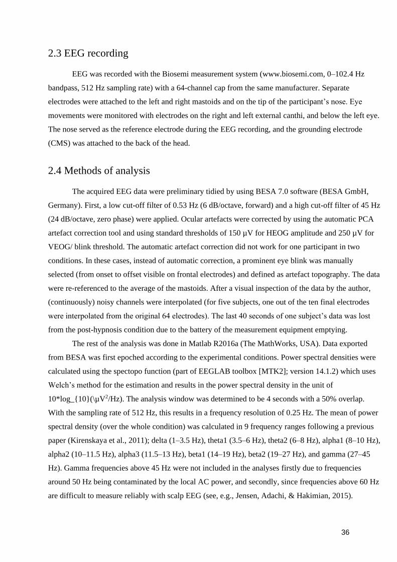
36
2.3 EEG recording
EEG was recorded with the Biosemi measurement system (www.biosemi.com, 0–102.4 Hz
bandpass, 512 Hz sampling rate) with a 64-channel cap from the same manufacturer. Separate
electrodes were attached to the left and right mastoids and on the tip of the participant’s nose. Eye
movements were monitored with electrodes on the right and left external canthi, and below the left eye.
The nose served as the reference electrode during the EEG recording, and the grounding electrode
(CMS) was attached to the back of the head.
2.4 Methods of analysis
The acquired EEG data were preliminary tidied by using BESA 7.0 software (BESA GmbH,
Germany). First, a low cut-off filter of 0.53 Hz (6 dB/octave, forward) and a high cut-off filter of 45 Hz
(24 dB/octave, zero phase) were applied. Ocular artefacts were corrected by using the automatic PCA
artefact correction tool and using standard thresholds of 150 µV for HEOG amplitude and 250 µV for
VEOG/ blink threshold. The automatic artefact correction did not work for one participant in two
conditions. In these cases, instead of automatic correction, a prominent eye blink was manually
selected (from onset to offset visible on frontal electrodes) and defined as artefact topography. The data
were re-referenced to the average of the mastoids. After a visual inspection of the data by the author,
(continuously) noisy channels were interpolated (for five subjects, one out of the ten final electrodes
were interpolated from the original 64 electrodes). The last 40 seconds of one subject’s data was lost
from the post-hypnosis condition due to the battery of the measurement equipment emptying.
The rest of the analysis was done in Matlab R2016a (The MathWorks, USA). Data exported
from BESA was first epoched according to the experimental conditions. Power spectral densities were
calculated using the spectopo function (part of EEGLAB toolbox [MTK2]; version 14.1.2) which uses
Welch’s method for the estimation and results in the power spectral density in the unit of
10*log_{10}(\µV2/Hz). The analysis window was determined to be 4 seconds with a 50% overlap.
With the sampling rate of 512 Hz, this results in a frequency resolution of 0.25 Hz. The mean of power
spectral density (over the whole condition) was calculated in 9 frequency ranges following a previous
paper (Kirenskaya et al., 2011); delta (1–3.5 Hz), theta1 (3.5–6 Hz), theta2 (6–8 Hz), alpha1 (8–10 Hz),
alpha2 (10–11.5 Hz), alpha3 (11.5–13 Hz), beta1 (14–19 Hz), beta2 (19–27 Hz), and gamma (27–45
Hz). Gamma frequencies above 45 Hz were not included in the analyses firstly due to frequencies
around 50 Hz being contaminated by the local AC power, and secondly, since frequencies above 60 Hz
are difficult to measure reliably with scalp EEG (see, e.g., Jensen, Adachi, & Hakimian, 2015).
37
Therefore, since the data of the present study was not gathered with the intention to analyse high
frequencies or even continuous EEG, frequencies above 50 Hz were also left out as likely unreliable.
Five electrodes from each hemisphere were used for the analysis in accordance with Kirenskaya and
colleagues (2011): Fp1, Fp2, F3, F4, C3, C4, P3, P4, O1, O2. The mean values were then transferred to
SPSS statistics 25 for statistical analysis.
Before running any analyses, the sampling errors were found to follow a normal distribution
sufficiently well. For research questions 1 and 2, nine 2 5 4 repeated-measures analyses of variance
(ANOVAs), one for each frequency range, were performed with lateralization (left and right
hemisphere), anteriority/posteriority (frontopolar, frontal, central, parietal, and occipital electrodes) and
condition (PrH, HY, SU, PoH) as within-subject factors. The measured power spectral density (in
lg(µV2/Hz)) at each participant’s electrodes served as the dependant variable. Greenhouse-Geisser
corrections for lack of sphericity were applied when appropriate and Bonferroni-corrected post-hoc
tests were conducted whenever necessary. All participants were included for research question 1,
whereas for research question 2 the analyses were run only for the subgroup of six participants that had
responded to the hallucinatory fly-suggestion administered in the screening session.
For research question number 3, the participants were again divided according to their
responsiveness to the fly-suggestion, resulting in two subgroups of six responsive and three
unresponsive participants. Since the groups were so small and also unbalanced, the research question
was primarily approached qualitatively with an emphasis on visual inspection of the power differences
in the different oscillatory ranges and conditions between the groups. Additionally, despite the results
of the analysis being undoubtedly rather unreliable and ungeneralisable, nine repeated-measures
ANOVAs were conducted in a similar fashion as for question one, with the only addition of subgroup
(responsive-unresponsive) as a between-subject factor. Again, Greenhouse-Geisser corrections and
Bonferroni-corrected post-hoc tests were applied when deemed necessary.
38
3 Results
3.1 Research question 1
The mean oscillatory powers of all frequency ranges in each condition are presented in Figure
1. Condition was found to have a significant effect in the gamma range (F(3,24) = 3.631, p = .027).
Bonferroni-corrected post-hoc tests revealed a significant difference between conditions SU and PoH
(p = .029) and a nearly significant difference between HY and PoH (p = .055) in the direction that the
hypnosis conditions had less gamma power than the posthypnosis condition (see Figure 1, bottom left
corner). In the beta1 and beta2 ranges no significant effects were found on a p<.05 level, but condition
approached statistical significance (F(3,24) = 2.957, p = .053, and F(3,24) = 2.665, p = .071
respectively). However, no significant differences between conditions were found in the theta1 (F(3,24)
= 0.203, p = .893) or theta2 (F(3,24) = 0.162, p = .921) ranges.
Oscillatory powers on the anteroposterior dimension are found in figure 2 for all frequency
ranges that were found to show significant effects on the dimension. An effect in the anteroposterior
dimension was found significant in the delta (F(4,32) = 4.713, p = .004), theta1 (F(4,32) = 11.048, p <
.001), theta2 (F(4,32) = 7.554, p = .007), alpha1 (F(4,32) = 8.781, p = .002), alpha2 (F(4,32) = 13.988,
p < .001) and alpha3 (F(4,32) = 9.241, p=.004) ranges, that is, systematically in oscillations under 14
Hz. As can be seen in figure 2, visual inspection revealed mean power to increase towards the central
electrodes in the delta, theta1 and theta2 ranges. In the alpha ranges, power was lower in the frontal
electrodes and increased towards the posterior sites, reaching highest values in the parietal (alpha1 and
alpha2) or occipital electrodes (alpha2 and alpha3). No significant differences were found in laterality
in any of the oscillatory ranges, nor were any significant interactions found between condition and the
anteroposterior dimension or between condition and laterality.
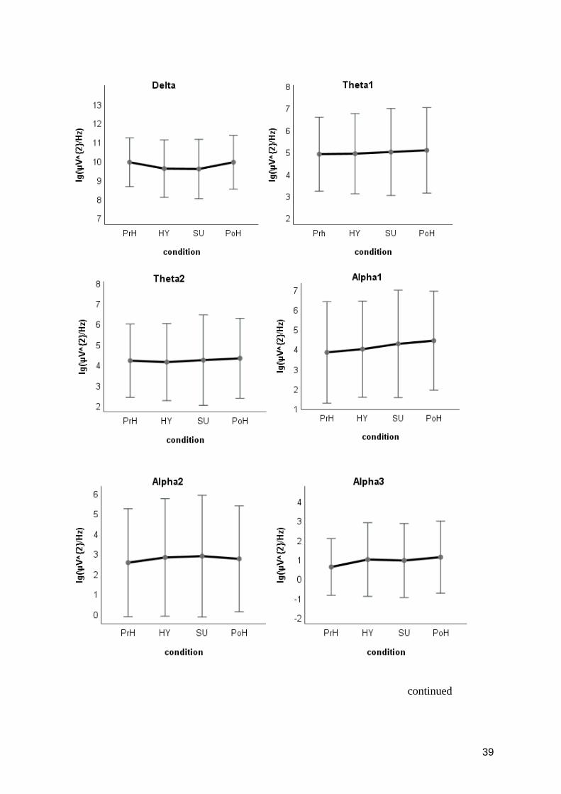
39
continued
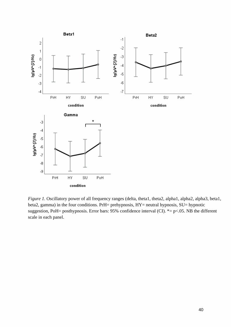
40
Figure 1. Oscillatory power of all frequency ranges (delta, theta1, theta2, alpha1, alpha2, alpha3, beta1,
beta2, gamma) in the four conditions. PrH= prehypnosis, HY= neutral hypnosis, SU= hypnotic
suggestion, PoH= posthypnosis. Error bars: 95% confidence interval (CI). *= p<.05. NB the different
scale in each panel.
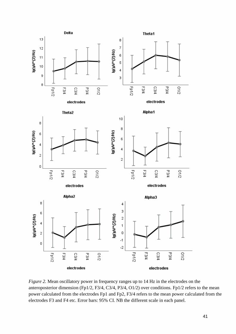
41
Figure 2. Mean oscillatory power in frequency ranges up to 14 Hz in the electrodes on the
anteroposterior dimension (Fp1/2, F3/4, C3/4, P3/4, O1/2) over conditions. Fp1/2 refers to the mean
power calculated from the electrodes Fp1 and Fp2, F3/4 refers to the mean power calculated from the
electrodes F3 and F4 etc. Error bars: 95% CI. NB the different scale in each panel.
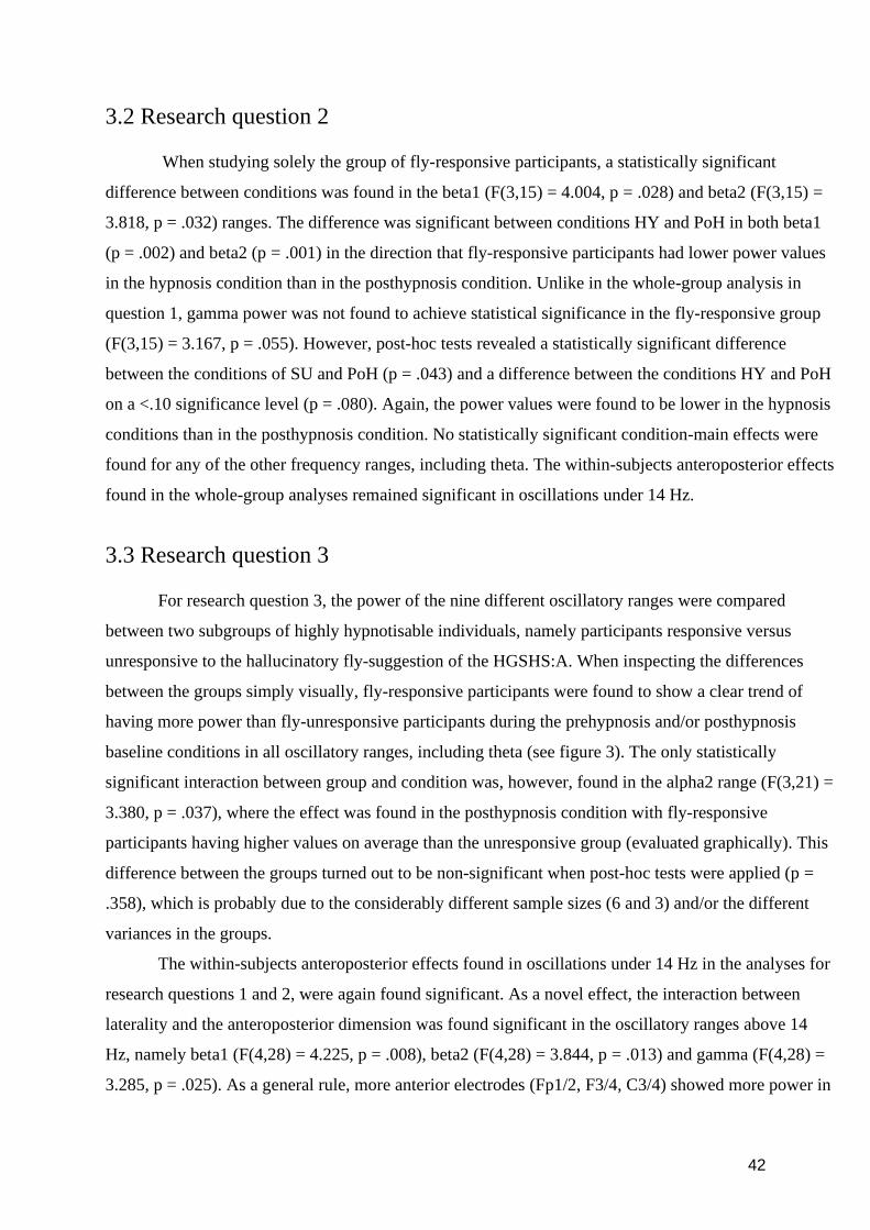
42
3.2 Research question 2
When studying solely the group of fly-responsive participants, a statistically significant
difference between conditions was found in the beta1 (F(3,15) = 4.004, p = .028) and beta2 (F(3,15) =
3.818, p = .032) ranges. The difference was significant between conditions HY and PoH in both beta1
(p = .002) and beta2 (p = .001) in the direction that fly-responsive participants had lower power values
in the hypnosis condition than in the posthypnosis condition. Unlike in the whole-group analysis in
question 1, gamma power was not found to achieve statistical significance in the fly-responsive group
(F(3,15) = 3.167, p = .055). However, post-hoc tests revealed a statistically significant difference
between the conditions of SU and PoH (p = .043) and a difference between the conditions HY and PoH
on a <.10 significance level (p = .080). Again, the power values were found to be lower in the hypnosis
conditions than in the posthypnosis condition. No statistically significant condition-main effects were
found for any of the other frequency ranges, including theta. The within-subjects anteroposterior effects
found in the whole-group analyses remained significant in oscillations under 14 Hz.
3.3 Research question 3
For research question 3, the power of the nine different oscillatory ranges were compared
between two subgroups of highly hypnotisable individuals, namely participants responsive versus
unresponsive to the hallucinatory fly-suggestion of the HGSHS:A. When inspecting the differences
between the groups simply visually, fly-responsive participants were found to show a clear trend of
having more power than fly-unresponsive participants during the prehypnosis and/or posthypnosis
baseline conditions in all oscillatory ranges, including theta (see figure 3). The only statistically
significant interaction between group and condition was, however, found in the alpha2 range (F(3,21) =
3.380, p = .037), where the effect was found in the posthypnosis condition with fly-responsive
participants having higher values on average than the unresponsive group (evaluated graphically). This
difference between the groups turned out to be non-significant when post-hoc tests were applied (p =
.358), which is probably due to the considerably different sample sizes (6 and 3) and/or the different
variances in the groups.
The within-subjects anteroposterior effects found in oscillations under 14 Hz in the analyses for
research questions 1 and 2, were again found significant. As a novel effect, the interaction between
laterality and the anteroposterior dimension was found significant in the oscillatory ranges above 14
Hz, namely beta1 (F(4,28) = 4.225, p = .008), beta2 (F(4,28) = 3.844, p = .013) and gamma (F(4,28) =
3.285, p = .025). As a general rule, more anterior electrodes (Fp1/2, F3/4, C3/4) showed more power in
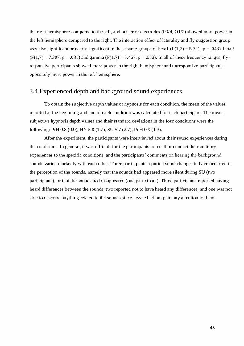
43
the right hemisphere compared to the left, and posterior electrodes (P3/4, O1/2) showed more power in
the left hemisphere compared to the right. The interaction effect of laterality and fly-suggestion group
was also significant or nearly significant in these same groups of beta1 (F(1,7) = 5.721, p = .048), beta2
(F(1,7) = 7.307, p = .031) and gamma (F(1,7) = 5.467, p = .052). In all of these frequency ranges, fly-
responsive participants showed more power in the right hemisphere and unresponsive participants
oppositely more power in the left hemisphere.
3.4 Experienced depth and background sound experiences
To obtain the subjective depth values of hypnosis for each condition, the mean of the values
reported at the beginning and end of each condition was calculated for each participant. The mean
subjective hypnosis depth values and their standard deviations in the four conditions were the
following: PrH 0.8 (0.9), HY 5.8 (1.7), SU 5.7 (2.7), PoH 0.9 (1.3).
After the experiment, the participants were interviewed about their sound experiences during
the conditions. In general, it was difficult for the participants to recall or connect their auditory
experiences to the specific conditions, and the participants’ comments on hearing the background
sounds varied markedly with each other. Three participants reported some changes to have occurred in
the perception of the sounds, namely that the sounds had appeared more silent during SU (two
participants), or that the sounds had disappeared (one participant). Three participants reported having
heard differences between the sounds, two reported not to have heard any differences, and one was not
able to describe anything related to the sounds since he/she had not paid any attention to them.
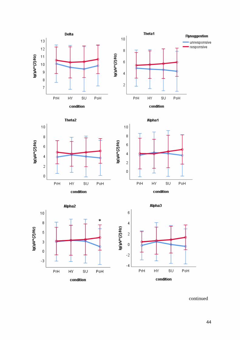
44
continued
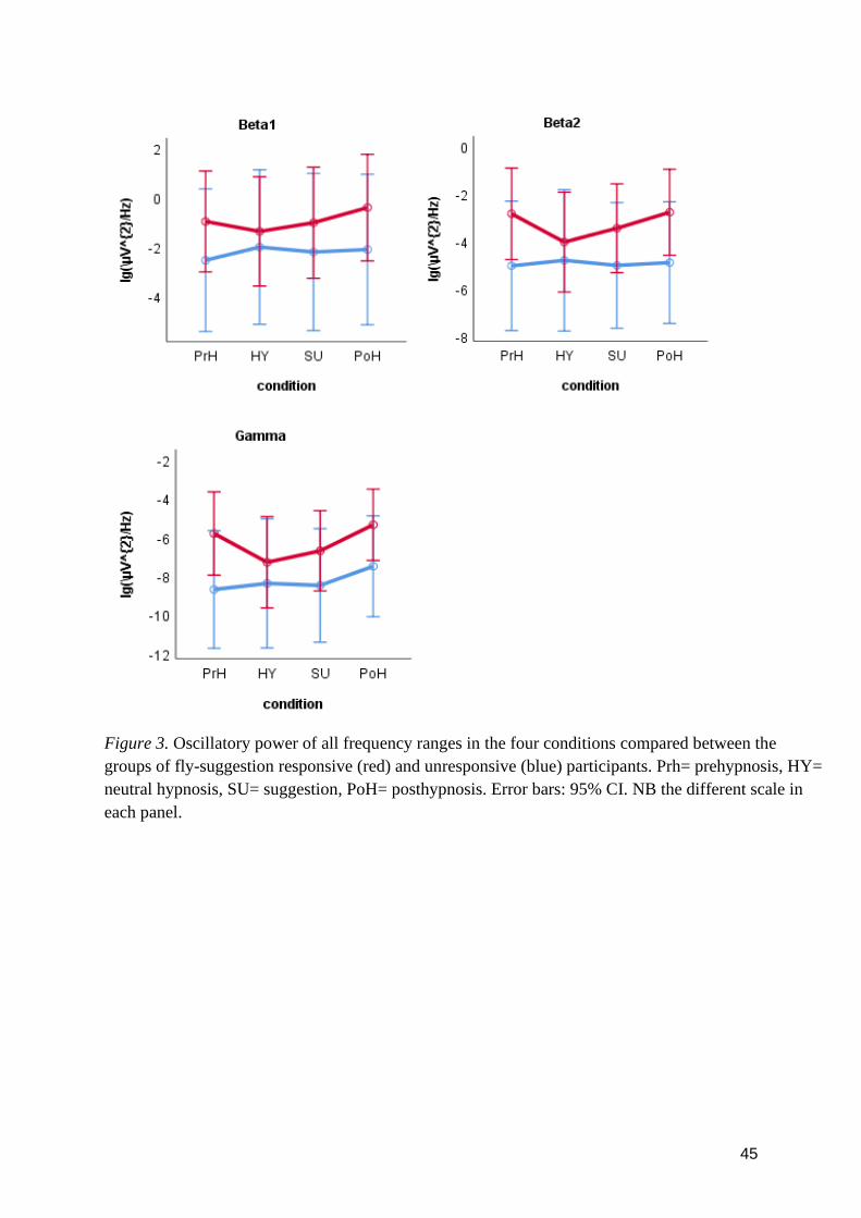
45
Figure 3. Oscillatory power of all frequency ranges in the four conditions compared between the
groups of fly-suggestion responsive (red) and unresponsive (blue) participants. Prh= prehypnosis, HY=
neutral hypnosis, SU= suggestion, PoH= posthypnosis. Error bars: 95% CI. NB the different scale in
each panel.
46
4 Discussion
4.1 General discussion
The primary aim of the present study was to explore, as a first study of its kind, the possible
EEG spectral power changes in highly hypnotisable individuals during four different research
conditions: prehypnosis, neutral hypnosis, hypnotic suggestion, and posthypnosis. Contrary to the main
hypothesis, no difference was found between any of the conditions in the theta range. The theory of
increased theta frequencies as a marker of hypnosis was therefore not supported by the present study,
and as outlined below, the prior studies that have been seen as support for the theory are not in fact
straightforwardly applicable to the theory. Instead, it seems that the changes in theta during hypnosis
only reflect differences in hypnotisability between participants. As for the other main hypothesis of the
present study, a difference, specifically a decrease was found in the gamma range between the
hypnosis conditions and baseline wakefulness (posthypnosis), supporting thus the theory of changed
gamma-frequency power in hypnosis. Following this finding, a connection between decreased
peripheral awareness and a decrease in gamma power during hypnosis is theorised as a novel
association. As secondary outcomes, differences in the cortical distribution of frequencies up to 14 Hz
were found, as well as spectral differences between two subgroups of highly hypnotisable participants
and between conditions in fly-suggestion responsive individuals.
As already concluded, in addition to the rather robust notion of highs showing higher baseline
theta than lows, hypnosis has been associated with increased theta frequencies and increased theta
power has even been deemed the most important marker of hypnosis (e.g., Jensen, Adachi, &
Hakimian, 2015). No increase in theta was, however, found in the current study, but both low (3.5–6
Hz) and high (6–8 Hz) theta power was instead found to remain unaffected throughout all the
experimental conditions. The results differ from prior studies likely because several, or even most of
the previous studies have studied the difference of theta power between high and low hypnotisable
subjects in the conditions of normal wake-state and hypnosis. Consequently, the results of the studies
reflect highs’ change in theta power in relation to lows’ change in power, and do not reveal whether
condition exerts a simple main effect on highs’ theta. Furthermore, although some have, several studies
have not separately reported the main effect of condition on highly hypnotisable participants (e.g., De
Pascalis, 1993; Graffin et al., 1995; Sabourin et al., 1990), and possible significant findings of the
absolute change in high’s theta during hypnosis has been left unknown.
47
From the studies that have reported the absolute change, none do in fact report any statistically
significant changes in theta during hypnosis. Williams and Gruzelier (2001) found a significant
difference in highs’ theta power only between pre- and posthypnosis conditions, and White et al.
(2008), Jamieson and Burgess (2014), and Tebecis et al. (1975) found no significant change between
conditions, although a trend towards heightened theta during hypnosis was visible in Tebecis et al.’s
study. In Sabourin et al.’s (1990) study, which corresponds most closely to the current study, both
highs’ and lows’ theta power increased significantly during various hypnosis conditions compared to
pre- and posthypnosis baselines. The results of the study are, however, reported rather ambiguously,
and it is left unclear whether the increase is also significant as a simple main effect for highs alone. Be
that as it may, lows who were not able to achieve hypnosis, did also show a substantial increase in theta
during hypnosis, and theta power may accordingly not be deduced to reflect hypnosis-specific state
differences in the brain even according to Sabourin et al.’s study (1990). It can be concluded that the
results of the present study, showing no significant hypnosis-specific increases in theta are in fact in
line with prior evidence, and consequently current EEG theta power research does not support the
hypothesis of theta as a marker of hypnosis. Rather, the amount of theta seems to reflect differences in
hypnotisability between highs and lows.
Concerning the effects of hypnosis on gamma power, it was hypothesised that compared with
the baselines before and after hypnosis, there would be either an increase or decrease of gamma during
hypnosis. A more specific hypothesis was not made due to the lack of prior studies using similar
methods and/or suggestions as the present study. The hypothesis was partially confirmed since gamma
power was found to be diminished during both of the hypnosis conditions (HY and SU) compared to
the posthypnosis condition. The hypnosis conditions did not statistically differ from the prehypnosis
condition, but there was a trend towards more gamma power in the prehypnosis condition than in the
hypnosis conditions.
That the change seen during hypnosis was in the case of the present study a decrease rather than
an increase can be speculated to be due to the simple nature of the experimental design and stimuli.
Gamma frequencies have according to recent studies been hypothesised to increase as a consequence of
(prerequisites of) conscious awareness, sensory stimuli and task relevant perceptions, and have been
found to have a central role in the primary sensory cortices (Aru, Axmacher, et al., 2012; Ojemann,
Ramsey, & Ojemann, 2013; Pitts et al., 2014; Pulvermüller et al.,1997). Narrow band gamma (~20–60
Hz), within which gamma was operationalized in the present study (27–45 Hz), has been found to be
elicited in particular from specific and simple sensory stimuli (e.g., Bartoli et al., 2019). It could be
hypothesised that hypnosis decreases the awareness of simple stimuli in the surrounding, such as the
short beeping sounds played in the background in the present study, and/or that during hypnosis, highs
48
would be better at ignoring task-irrelevant sensory stimuli. In the present study, the participants were
before the experiment given the instruction that there was no need to pay any attention to the
background tones in any of the conditions. In the suggestion condition the participants were again
given the instruction and additionally the suggestion to perceive all tones as similar in pitch. As no
significant differences were found in gamma between HY and SU, it seems more likely that hypnosis
decreases awareness of task-irrelevant sensory stimuli regardless of possible hypnotic suggestions
given to ignore and alter the perception of the stimuli in question. These results are therefore well in
line with definitions of hypnosis, such as the most recent APA definition, that characterize hypnosis as
involving reduced peripheral awareness (Elkins et al., 2015; Spiegel & Moore, 1997). This is, however,
to the knowledge of the present author, the first study where the reduction of peripheral awareness in
hypnosis is theoretically associated with a decrease in gamma power.
Prior studies on the connection between hypnosis and gamma are scarce and have only
considered the differences between high and low hypnotisable subjects during normal wake-state
and/or hypnosis. In a couple of studies, highs have been found to exhibit lower gamma power than
lows during baseline wake-state and during hypnosis while at rest or when recalling emotional events
(gamma operationalized as 27–40 Hz and 35–45 Hz) (De Pascalis et a., 1987; De Pascalis et al., 1989;
Kirenskaya et al., 2011). One study has reported higher beta (20–36 Hz) and gamma (36–44 Hz) power
in highs than lows during a hypnotic induction, and concerning gamma, also in the conditions of
neutral hypnosis, dream suggestion, and age regression (De Pascalis, 1993). The study did not
explicitly report any significant main effects separately for highs, but by visually inspecting the
figures, highs’ gamma power seems to have increased significantly in the posterior areas and
particularly in the right hemisphere during an age regression suggestion, compared to hypnosis baseline
and a hypnotic dream suggestion. As an age regression suggestion is very far from the suggestion used
in the present study, it can be concluded that none of the previous studies’ results are directly
applicable to the current study, but rather the present study and its outcomes are so far unique on the
field. To sum it up, in a neutral hypnosis and simple suggestion condition where participants were at
rest, watching a monotonous video and hearing some tones in the background that they were told not to
need to pay any attention to, gamma power decreased and was significantly lower than during
wakefulness after hypnosis. Referring to definitions that hypnosis involves reduced peripheral
awareness and findings that gamma power is inversely connected to consciousness and specific sensory
stimuli, it is theorised that reduced peripheral awareness in hypnosis might be reflected as reduced
gamma power during hypnosis.
Possible differences in the various bandwidths’ spatial power distribution on the scalp were also
exploratively studied in the current study. No differences between hemispheres were observed, but
49
statistically significant differences within the power distribution in the anteroposterior dimension over
the scalp was found in all frequency ranges up to 14 Hz (delta, theta1, theta2, alpha1, alpha2, alpha3).
There was in general a trend for power under 14 Hz to be lower in the frontal scalp and higher in the
central, parietal, or occipital scalp areas. Since the spatial differences were significant only as main
effects and not as interactions of any sorts, it should be emphasised that the reported spatial differences
are mean differences over all the conditions, and do not therefore convey any hypnosis-specific
information per se.
Since few or perhaps no studies have previously reported mean spatial powers over similar
kinds of conditions, the spatial distributions found in this study can be thought to resemble, although
not flawlessly, most closely that of a normal resting wake-state with eyes open. Even so, not many
studies have reported differences in the frequency ranges along the anteroposterior dimension. Alpha
frequencies have been found to be most prominent in the occipital scalp areas (Barry, Clarke,
Johnstone, Magee, & Rushby, 2007; De Gennaro, Ferrara, Curcio, & Cristiani, 2001; Steriade et al.,
1990) and to be generated most posteriorly of the frequency ranges (Michel, Lehmann, Henggeler, &
Brandeis, 1992). Since alpha was divided into three subcategories in the current study, comparing the
results is not straightforward. When visually inspecting the graphs associated with the three alpha
ranges, power does, however, affirmatively seem to rise beginning from the central electrodes towards
the occipital ones, with the strongest posterior increase being found in the alpha3 range.
Theta has been found all over the brain with pronounced activity in the central areas (Barry et
al., 2007), varying in accordance with subject characteristics (Stenberg, 1992) and originating in more
central areas than alpha (Michel et al., 1992). Delta frequencies have typically been measured slightly
more anteriorly than theta (Michel et al., 1992). With visual inspection, the current study located delta
and theta frequencies as strongest in the central electrodes/regions, agreeing therefore roughly with
previous studies. As slower frequencies have been identified as global frequency ranges and are
expected to be found across a wider area, it is rather surprising that the differences in the
anteroposterior dimension were in the present study found systematically precisely in slower
frequencies, while frequencies above 14 Hz were not found to differ significantly on the dimension.
Faster fluctuations in power and therefore more variability both within and between conditions might
be one reason to no significant differences being found on average in the anteroposterior dimension in
the beta and gamma ranges.
For research questions 2 and 3 the participants were divided into two subgroups according to
their responsiveness or unresponsiveness to the fly-suggestion, a hallucinatory suggestion used as part
of the HGSHS:A in the screening phase. As hallucinatory suggestions are more difficult to respond to
than other types of suggestions and consequently participants that are responsive to hallucinatory
50
suggestions are generally deemed more susceptible to hypnosis than unresponsive individuals, the
formed groups were assumed to reflect two levels of highly hypnotisable individuals. Possible spectral
power changes in the different conditions were studied first solely for the fly-responsive participants,
and the results were expected to be slightly enhanced compared to the results obtained from the whole-
group analysis. Indeed, in addition to finding a statistically significant change in gamma (as in the
whole-group analysis), fly-responsive participants did also exhibit significant changes in the beta1 and
beta2 ranges. The differences were significant between the conditions of neutral hypnosis and
posthypnosis in the beta ranges, and between the hypnotic suggestion and posthypnosis conditions in
the gamma range. In all of these three frequency ranges, the fly-responsive participants were found to
show less power in the hypnosis conditions than during wakefulness posthypnosis.
What could be made out of these results? The significance of the difference in the gamma range
was already discussed above, and it was theorised that the decrease of gamma might reflect reduced
peripheral awareness during hypnosis. Regarding beta, few or no prior studies have reported any
differences in beta power during hypnosis, making the finding in this study unexpected. Again, an
explanation might be found in the characteristics of the participants or the study design, or the observed
effects might be false positives. In any case, caution is needed when speculating about possible causes
to the outcome. It is, however, worth noting that both beta and gamma frequencies are considered high-
frequency ranges, and since the borders of the different frequency ranges are factually artificial, the
behavioural correlates of the ranges may easily overlap. The decrease of beta could speculatively also
have to do with decreased (visual) peripheral awareness, especially since occipital beta has recently
proven relevant in visual attention in humans (Gola et al., 2013).
As a third research question the fly-responsive and unresponsive subgroups were compared for
possible between-group differences, and an expectation that fly-responsive participants might show
higher baseline theta power than unresponsive participants was formulated. Before any analyses were
run, it was acknowledged and should be emphasised here that since the amount of participants in the
two groups was so small (six and three), the differences between the groups were studied primarily
qualitatively and the obtained results should be seen as approximate at best.
What the visual analysis instantly pointed towards when figures of the differences between the
groups were drawn, was that in all frequency ranges there was a clear trend towards more power in fly-
responsive participants compared with unresponsive participants during baseline wakefulness (pre-
and/or posthypnosis). This was surprising as it differed quite markedly from the expected results.
However, when running the statistical analyses, no statistically significant differences in baseline
values were found between the groups in any of the frequency ranges, including theta. It is therefore
possible that with a bigger sample of especially fly-unresponsive highs, statistical significance might
51
have been achieved between the groups. In the alpha2 range a significant effect was in fact found but
only in the posthypnosis condition, where fly-responsive participants had a higher power value on
average than the unresponsive group. According to visual inspection, fly-unresponsive participants’
alpha2 power seem to have decreased from hypnosis to posthypnosis, while it remained rather stable
for fly-responsive participants. Even though this could implicate differences between the groups in, for
example, attentional processes or relaxation after hypnosis, the groups were, again, small and the
standard deviations correspondingly large, making the results very preliminary and ungeneralisable.
Another finding that emerged when comparing the two subgroups with each other, was that the
interaction between laterality and the anteroposterior dimension was found significant in the oscillatory
ranges above 14 Hz, namely beta1, beta2, and gamma. As a general rule, more anterior electrodes
showed more power in the right hemisphere compared to the left, and posterior electrodes showed more
power in the left hemisphere compared to the right. Beta and gamma frequencies have not typically
been found to exhibit hemispheric asymmetries, so the results might reflect sample or experimental
procedure-specific features. What these might be is due to lack of prior similar studies speculative, and
thus left without further exploration in the present thesis.
The interaction effect of laterality and fly-suggestion group was also significant in these same
groups of beta1, beta2 and gamma. In all of these frequency ranges, fly-responsive participants showed
more power in the right hemisphere and unresponsive participants oppositely more power in the left
hemisphere. Some, predominantly early hypnosis studies, have reported differences in laterality
between high and low hypnotisable participants, and the right hemisphere has been suggested to be an
important mediator of hypnosis (see, e.g., Kallio & Revonsuo, 2003). However, with unsystematic
evidence accruing, the differences in laterality have generally been deemed to be unreflective of
hypnosis and hypnotisability (e.g., Graffin et al., 1995). The result of the present study of no
hemispheric differences in the whole group of highs but yet an interaction of hemisphere and the
subgroups of fly-responsive -and unresponsive participants is interesting and might shed light on the
reasons behind the unsystematic findings. Laterality differences in hypnosis and/or between highs and
lows might be to some extent explained by the participant’s responsiveness to hallucinatory
suggestions. However, since the differences found in the present study are again mean differences over
all of the conditions, comparing the results to previous or later studies is not straightforwardly reliable.
Therefore, no detailed revision will be made concerning the matter here.
When comparing the findings obtained by the subdivision of participants with the findings
obtained in the whole-group analysis, it can be deduced that changes seen in power in the beta and
gamma ranges vary significantly in the fly-responsive group between conditions, while the power
remains more stable throughout the experiment in the fly-unresponsive group. This indicates that the
52
fly-unresponsive participants do in this study attenuate differences between conditions, and that fly-
responsive and unresponsive participants do exhibit some, albeit not many significant differences in
their brain oscillations. Recent literature has indeed found highs to comprise of several subgroups
rather than being a uniform group. Subtypes of highs have been established according to their
responsiveness to agnosia and cognitive distortions or to posthypnotic amnesia suggestions, and
according to their dissociative tendencies (Terhune et al., 2011b, 2015). Even though the hallucinatory
suggestion has not been distinguished as a dividing factor, further studies from different research
groups will have to replicate the effects before any finite conclusions can be made.
4.2 Strengths and limitations
The most significant strength of the current study is that it is unique in studying highly
hypnotisable subjects in four different conditions, comparing EEG oscillatory powers not only between
baseline and hypnosis, but also distinguishing between neutral hypnosis and hypnotic suggestion
conditions and baselines before and after hypnosis. Even though the lack of controls of low and
medium hypnotisable subjects could be seen as a limitation, comparing possible differences between
participants of different hypnotisability levels was not the aim of the present study. Rather, since the
changes between conditions were expected to be largest in the group of highly hypnotisable
individuals, highs were deemed most suitable to be studied in a preliminary research such as this. Of
course, future studies may extend the study to lows and mediums in order to add value and information
to the current outcomes.
From the conditions used, the hypnotic suggestion condition could have been more optimal.
Now, the present study was restricted by the design of the underlying study (Hiltunen et al., 2019), and
the suggestion used in the hypnotic suggestion condition was therefore a rather unorthodox and
difficult one, namely hearing tones as similar in pitch. For the aims of the current study, it would have
been more optimal to use instead one or several standardised suggestions that the participants would
definitely have been able to respond to and to which the responding could have been evaluated
objectively by an observer. Now, as the participants were interviewed after finishing all of the
conditions and were therefore not reliably able to recall any experiential changes to have happened
specifically after the suggestion, it is not clear whether the suggestion really worked or altered anything
experientially or neurally. Consequently, the suggestion condition did not differ as much as it optimally
could have from the neutral hypnosis condition, but, at the very least, listening to the suggestion and
trying to follow the suggestion differentiated the two hypnosis conditions from each other.
53
The four conditions not being balanced is also a limitation of the present study. The study
design did, however, enforce the unbalancing, since pre- and posthypnosis can evidently not be
balanced, and even though the hypnosis conditions could be balanced, it would not be optimal due to
the nature of the suggestion. Suggestions and their effects can in general be ended abruptly, but there
might still have been a possibility that the suggestion used would have affected the neutral hypnosis
condition had it been given before it. Another limitation that follows from the underlying study were
the sinusoidal tones that were playing in the background. However, given that the auditory stimuli were
the same throughout the experiment, the effect that the tones possibly evoked in the recorded EEG
ought not have affected the comparative outcomes. The time it took for the participants to become
accustomed to the tones was compensated by a period of playing the tones in the background for the
participants before commencing the analysis.
In order to minimize excessive alpha contaminating the data, the participants were instructed to
hold their eyes open throughout the experiment. As a downside, four out of the nine participants felt
that holding their eyes open disturbed their depth of hypnosis, and five participants reported that they
had not felt to have been as deeply hypnotised as in the hypnosis measurement session. With a
maximum score of 10, the average subjective depth of hypnosis was 5.8 in the neutral hypnosis and 5.7
in the suggestion conditions, reaching thus sufficient but not ideal levels. Previous research varies
largely on whether the participants have kept their eyes shut or open during the experiment, making
comparisons between studies somewhat problematic. However, although some differences exist in the
outcomes between the eyes-open and eyes-closed conditions in hypnosis research, these differences
seem to be few and mostly nonsignificant (Sabourin et al., 1990). The way the eyes were kept should
nonetheless be reported clearly in each study, and hopefully some clear guidelines on the matter, as
well as on many other aspects concerning hypnosis research, will be formed in the future.
Further limitations of the current study involve the relatively small sample size, the possibility
of minimal saccades contaminating the data, and the induction being read/spoken out rather than played
from a recording to the participants. A small sample size is, however, customary in hypnosis research
and especially in pioneering studies, and speaking the induction is also normative and gives some
flexibility and freedom to the hypnotist to help the participant reach as deep a hypnosis as possible. The
possibility of minimal saccades not being accounted for in the EEG refining-phase is, in turn, a
problem all EEG studies face. Moreover, in addition to screening the participants with the group
assessment test HGSHS:A, using an individual assessment test would have given a better, more reliable
estimate of the participants’ level of hypnotisability and more information regarding the subgroups of
fly-responsive and -unresponsive participants.

54
In the future, it would be enlightening to replicate this study with a more optimal suggestion
condition and with low and medium hypnotisable subjects as well. Studying the theorised connection
between decreased peripheral awareness in hypnosis and the gamma frequency range is encouraged, as
well as continuing to study the alleged yet undecided notion of the connection between hypnosis (or
hypnotisability) and the theta range. Additionally, exploring the variability within highly hypnotisable
individuals is important, since contrary to the usual way of treating highs as a uniform group, recent
studies and also this study have indicated that it is not the case. Identifying different subtypes within
highs could be one path in finding explanations for the variable results in hypnosis research.
Furthermore, uniform terminology and general guidelines of good hypnosis research practices are
called for, so that systematic research and comparable outcomes can with time uncover the essence of
hypnosis, resulting in a unified theory and evidence-based utilisation of hypnosis in its various fields of
application.
4.3 Conclusions
The present study aimed to contribute to the theory-building on hypnosis by studying human brain
oscillations in hypnosis. The obtained results were not found to support the theory of increased theta-
frequency power as a marker of hypnosis, but theta power was instead deemed to most likely only
reflect differences in hypnotisability. Gamma range power was found to decrease during hypnosis,
supporting therefore the hypothesis of changed gamma power in hypnosis. In a subgroup of highly
hypnotisable participants responsive to a hallucinatory suggestion, beta range power was also found to
decrease during hypnosis. The decrease of high-frequency power, particularly in the gamma range, was
tentatively theorised to reflect the decrease in peripheral awareness that is held characteristic for
hypnosis. The brain oscillation results and literature review provided within the present study may
serve as guidance for future studies trying to resolve the state versus non-state debate and define the
working principle of hypnosis.
55
5 References
Adachi, T., Fujino, H., Nakae, A., Mashimo, T., & Sasaki1, J. (2014). A meta-analysis of hypnosis for
chronic pain problems: A comparison between hypnosis, standard care, and other psychological
interventions. International Journal of Clinical and Experimental Hypnosis, 62, 1–28.
Aftanas, L. I., Reva, N. V., Varlamov, A. A., Pavlov, S. V., & Makhnev, V. P. (2004). Analysis of
evoked EEG synchronization and desynchronization in conditions of emotional activation in
humans: Temporal and topographic characteristics. Neuroscience and Behavioral Physiology, 34,
859–867.
Aru, J., Axmacher, N., Do Lam, A. T., Fell, J., Elger, C. E., Singer, W., & Melloni, L. (2012). Local
category-specific gamma band responses in the visual cortex do not reflect conscious perception.
Journal of Neuroscience, 32, 14909–14914.
Aru, J., Bachmann, T., Singer, W., & Melloni, L. (2012). Distilling the neural correlates of
consciousness. Neuroscience & Biobehavioral Reviews, 36, 737–746.
Aru, J., Priesemann, V., Wibral, M., Lana, L., Pipa, G., . . . Vicente, R. (2015). Untangling cross-
frequency coupling in neuroscience. Current Opinion in Neurobiology, 31, 51–61.
Banyai, E. I., & Hilgard, E. R. (1976). A comparison of active-alert hypnotic induction with traditional
relaxation induction. Journal of Abnormal Psychology, 85, 218.
Barber, T. X., & Calverley, D. S. (1964). Toward a theory of hypnotic behavior: Effects on
suggestibility of defining the situation as hypnosis and defining response to suggestions as easy.
The Journal of Abnormal and Social Psychology, 68, 585.
Barnier, A. J., Dienes, Z., & Mitchell, C. J. (2008). How hypnosis happens: New cognitive theories of
hypnotic responding. In M. R. Nash, & A. Barnier (Eds.), The Oxford Handbook of Hypnosis:
Theory, Research, and Practice (pp. 141–177). Oxford, UK: Oxford University Press.
Barry, R. J., Clarke, A. R., Johnstone, S. J., Magee, C. A., & Rushby, J. A. (2007). EEG differences
between eyes-closed and eyes-open resting conditions. Clinical Neurophysiology, 118, 2765–2773.
Bartoli, E., Bosking, W., Li, Y., Beauchamp, M. S., Yoshor, D., & Foster, B. (2019). Distinct narrow
and broadband gamma responses in human visual cortex. bioRxiv, 572313.
Başar, E., & Güntekin, B. (2012). A short review of alpha activity in cognitive processes and in
cognitive impairment. International Journal of Psychophysiology, 86, 25–38.
Benham, G., Woody, E. Z., Wilson, K. S., & Nash, M. R. (2006). Expect the unexpected: Ability,
attitude, and responsiveness to hypnosis. Journal of Personality and Social Psychology, 91, 342.
Berger, H. (1929). Über das Elektrenkephalogramm des Menschen [On the Electroencephalogram of
Man]. European Archives of Psychiatry and Clinical Neuroscience, 87, 527–570.
Bongartz, W. (1985). German norms for the Harvard roup scale of hypnotic susceptibility, form A. The
International Journal of Clinical and Experimental Hypnosis, 33, 131–139.
Bowers, K. S. (1971). Sex and susceptibility as moderator variables in the relationship of creativity and
hypnotic susceptibility. Journal of Abnormal Psychology, 78, 93.
56
Braffman, W., & Kirsch, I. (1999). Imaginative suggestibility and hypnotizability: An empirical
analysis. Journal of Personality and Social Psychology, 77, 578.
Braid, J. (1843). Neurypnology; or, the rationale of nervous sleep, considered in relation with animal
magnetism. London: J. Churchill. Retrieved November 15, 2019, from https://books.google.fi
Buzsáki, G. (2005). Theta rhythm of navigation: Link between path integration and landmark
navigation, episodic and semantic memory. Hippocampus, 15, 827–840.
Buzsáki, G. (2006). Rhythms of the brain. Oxford, UK: Oxford University Press. Retrieved September
2, 2019 from https://books.google.fi
Cahn, B. R., & Polich, J. (2006). Meditation states and traits: EEG, ERP, and neuroimaging studies.
Psychological Bulletin, 132, 180.
Canolty, R. T., & Knight, R. T. (2010). The functional role of cross-frequency coupling. Trends in
Cognitive Sciences, 14, 506–515.
Cantero, J. L., & Atienza, M. (2005). The role of neural synchronization in the emergence of cognition
across the wake-sleep cycle. Reviews in the Neurosciences, 16, 69–84.
Cardeña, E., Jönsson, P., Terhune, D. B., & Marcusson-Clavertz, D. (2013). The neurophenomenology
of neutral hypnosis. Cortex, 49, 375–385.
Cardeña, E., & Terhune, D. (2008). A distinct personality trait? The relationship between
hypnotizability, absorption, self-transcendence, and mental boundaries. Proceedings of the 51st
Annual Convention of the Parapsychological Association, 61–73.
Cohen, H. D., Rosen, R. C., & Goldstein, L. (1976). Electroencephalographic laterality changes during
human sexual orgasm. Archives of Sexual Behavior, 5, 189–199.
Cojan, Y., Piguet, C., & Vuilleumier, P. (2015). What makes your brain suggestible? hypnotizability is
associated with differential brain activity during attention outside hypnosis. NeuroImage, 117,
367–374.
Cooper, L. M., & London, P. (1971). The development of hypnotic susceptibility: A longitudinal
(convergence) study. Child Development, 42, 487–503.
Crawford, H. J. (1994). Brain dynamics and hypnosis: Attentional and disattentional processes.
International Journal of Clinical and Experimental Hypnosis, 42, 204–232.
Crawford, H. J., Clarke, S. W., & Kitner-Triolo, M. (1996). Self-generated happy and sad emotions in
low and highly hypnotizable persons during waking and hypnosis: Laterality and regional EEG
activity differences. International Journal of Psychophysiology, 24, 239–266.
Croft, R. J., Williams, J. D., Haenschel, C., & Gruzelier, J. H. (2002). Pain perception, hypnosis and 40
hz oscillations. International Journal of Psychophysiology, 46, 101–108.
Daglish, M. R., & Wright, P. (1991). Opinions about hypnosis among medical and psychology
students. Contemporary Hypnosis, 8, 51–55.
De Gennaro, L., Ferrara, M., Curcio, G., & Cristiani, R. (2001). Antero-posterior EEG changes during
the wakefulness–sleep transition. Clinical Neurophysiology, 112, 1901–1911.
De Pascalis, V., Ray, W. J., Tranquillo, I., & D'Amico, D. (1998). EEG activity and heart rate during
recall of emotional events in hypnosis: Relationships with hypnotizability and suggestibility.
57
International Journal of Psychophysiology, 29, 255–275.
De Pascalis, V. (1993). EEG spectral analysis during hypnotic induction, hypnotic dream and age
regression. International Journal of Psychophysiology, 15, 153–166.
De Pascalis, V., Marucci, F. S., & Penna, P. M. (1989). 40-hz EEG asymmetry during recall of
emotional events in waking and hypnosis: Differences between low and high hypnotizables.
International Journal of Psychophysiology, 7, 85–96.
De Pascalis, V., Marucci, F. S., Penna, P. M., & Pessa, E. (1987). Hemispheric activity of 40 hz EEG
during recall of emotional events: Differences between low and high hypnotizables. International
Journal of Psychophysiology, 5, 167–180.
Deeley, Q., Oakley, D. A., Toone, B., Giampietro, V., Brammer, M. J., Williams, S. C., & Halligan, P.
W. (2012). Modulating the default mode network using hypnosis. International Journal of Clinical
and Experimental Hypnosis, 60, 206–228.
Dehaene, S., & Changeux, J. (2011). Experimental and theoretical approaches to conscious processing.
Neuron, 70, 200–227.
Dienes, Z., & Hutton, S. (2013). Understanding hypnosis metacognitively: rTMS applied to left
DLPFC increases hypnotic suggestibility. Cortex, 49, 386–392.
Dienes, Z., & Perner, J. (2007). Executive control without conscious awareness: The cold control
theory of hypnosis. Hypnosis and Conscious States: The Cognitive Neuroscience Perspective,
293–314.
Dolce, G., & Waldeier, H. (1974). Spectral and multivariate analysis of EEG changes during mental
activity in man. Electroencephalography and Clinical Neurophysiology, 36, 577–584.
Eckhorn, R., Bauer, R., Jordan, W., Brosch, M., Kruse, W., Munk, M., & Reitboeck, H. J. (1988).
Coherent oscillations: A mechanism of feature linking in the visual cortex? Biological
Cybernetics, 60, 121–130.
Egner, T., Jamieson, G., & Gruzelier, J. (2005). Hypnosis decouples cognitive control from conflict
monitoring processes of the frontal lobe. NeuroImage, 27, 969–978.
Egner, T., & Raz, A. (2007). Cognitive control processes and hypnosis. In G. Jamieson (Ed.), Hypnosis
and Conscious States: The Cognitive Neuroscience Perspective (pp. 29–50). Oxford, UK: Oxford
University Press
Elkins, G. R., Barabasz, A. F., Council, J. R., & Spiegel, D. (2015). Advancing research and practice:
The revised APA division 30 definition of hypnosis. International Journal of Clinical and
Experimental Hypnosis, 63, 1–9.
Engel, A. K., & Fries, P. (2010). Beta-band oscillations—signalling the status quo? Current Opinion in
Neurobiology, 20, 156–165.
Facco, E., Ermani, M., Rampazzo, P., Tikhonoff, V., Saladini, M., Zanette, G., . . . Spiegel, D. (2014).
Top-down regulation of left temporal cortex by hypnotic amusia for rhythm: A pilot study on
mismatch negativity. International Journal of Clinical and Experimental Hypnosis, 62, 129–144.
Fingelkurts, A. A., Fingelkurts, A. A., Kallio, S., & Revonsuo, A. (2007a). Hypnosis induces a changed
composition of brain oscillations in EEG: A case study. Contemporary Hypnosis, 24, 3–18.
Fingelkurts, A. A., Fingelkurts, A. A., Kallio, S., & Revonsuo, A. (2007b). Cortex functional
58
connectivity as a neurophysiological correlate of hypnosis: An EEG case study. Neuropsychologia,
45, 1452–1462.
Fisch, L., Privman, E., Ramot, M., Harel, M., Nir, Y., Kipervasser, S., . . . Fried, I. (2009). Neural
“ignition”: Enhanced activation linked to perceptual awareness in human ventral stream visual
cortex. Neuron, 64, 562–574.
Flammer, E., & Bongartz, W. (2003). On the efficacy of hypnosis: A meta‐analytic study.
Contemporary Hypnosis, 20, 179–197.
Foxe, J. J., Simpson, G. V., & Ahlfors, S. P. (1998). Parieto-occipital ∼10 Hz activity reflects
anticipatory state of visual attention mechanisms. Neuroreport, 9, 3929–3933.
Freud, S. (1957). A metapsychological supplement to the theory of dreams. In J. Strachey & A. Freud
(Eds.), The Standard Edition of the Complete Psychological Works of Sigmund Freud, volume XIV
(1914–1916): On the history of the psycho-analytic movement, papers on metapsychology and
other works (pp. 217–235). London, UK: Hogarth Press and Institute of Psychoanalysis.
Friese, U., Köster, M., Hassler, U., Martens, U., Trujillo-Barreto, N., & Gruber, T. (2013). Successful
memory encoding is associated with increased cross-frequency coupling between frontal theta and
posterior gamma oscillations in human scalp-recorded EEG. NeuroImage, 66, 642–647.
Fromm, E. (1977). An ego-psychological theory of altered states of consciousness. International
Journal of Clinical and Experimental Hypnosis, 25, 372–387.
Gaillard, R., Dehaene, S., Adam, C., Clémenceau, S., Hasboun, D., Baulac, M., . . . Naccache, L.
(2009). Converging intracranial markers of conscious access. PLoS Biology, 7.
Galbraith, G. C., London, P., Leibovitz, M. P., Cooper, L. M., & Hart, J. T. (1970). EEG and hypnotic
susceptibility. Journal of Comparative and Physiological Psychology, 72, 125.
Gale, A., Coles, M., & Boyd, E. (1971). Variation in visual input and the occipital EEG: II.
Psychonomic Science, 23, 99–100.
Gale, A., Spratt, G., Christie, B., & Smallbone, A. (1975). Stimulus complexity, EEG abundance
gradients, and detection efficiency in a visual recognition task. British Journal of Psychology, 66,
289–298.
Galin, D., Johnstone, J., & Herron, J. (1978). Effects of task difficulty on EEG measures of cerebral
engagement. Neuropsychologia, 16, 461–472.
Geiger, E., Peter, B., Prade, T., & Piesbergen, C. (2014). Intelligence and hypnotizability: Is there a
connection? International Journal of Clinical and Experimental Hypnosis, 62, 310–329.
Gevins, A., Smith, M. E., McEvoy, L., & Yu, D. (1997). High-resolution EEG mapping of cortical
activation related to working memory: Effects of task difficulty, type of processing, and practice.
Cerebral Cortex, 7, 374–385.
Gfeller, J. D., Lynn, S. J., & Pribble, W. E. (1987). Enhancing hypnotic susceptibility: Interpersonal
and rapport factors. Journal of Personality and Social Psychology, 52, 586.
Glass, L. B., & Barber, T. X. (1961). A note on hypnotic behavoir, the definition of the situation and
the placebo effect. Journal of Nervous and Mental Disease, 132, 539–541.
Glisky, M. L., Tataryn, D. J., Tobias, B. A., Kihlstrom, J. F., & McConkey, K. M. (1991). Absorption,
openness to experience, and hypnotizability. Journal of Personality and Social Psychology, 60,
59
263.
Gola, M., Magnuski, M., Szumska, I., & Wróbel, A. (2013). EEG beta band activity is related to
attention and attentional deficits in the visual performance of elderly subjects. International
Journal of Psychophysiology, 89, 334–341.
Graffin, N. F., Ray, W. J., & Lundy, R. (1995). EEG concomitants of hypnosis and hypnotic
susceptibility. Journal of Abnormal Psychology, 104, 123.
Gray, C. M., König, P., Engel, A. K., & Singer, W. (1989). Oscillatory responses in cat visual cortex
exhibit inter-columnar synchronization which reflects global stimulus properties. Nature, 338, 334.
Green, J. P. (2003). Beliefs about hypnosis: Popular beliefs, misconceptions, and the importance of
experience. International Journal of Clinical and Experimental Hypnosis, 51, 369–381.
Green, J. P. (2004). The five factor model of personality and hypnotizability: Little variance in
common. Contemporary Hypnosis, 21, 161–168.
Green, J. P., Barabasz, A. F., Barrett, D., & Montgomery, G. H. (2005). Forging ahead: The 2003 APA
division 30 definition of hypnosis. International Journal of Clinical and Experimental Hypnosis,
53, 259–264.
Gruzelier, J. (1996). The state of hypnosis: Evidence and applications. QJM: An International Journal
of Medicine, 89, 313–317.
Gruzelier, J. (1998). A working model of the neurophysiology of hypnosis: A review of evidence.
Contemporary Hypnosis, 15, 3–21.
Gruzelier, J. H. (2000). Redefining hypnosis: Theory, methods and integration. Contemporary
Hypnosis, 17, 51–70.
Gruzelier, J., Pascalis, V. D., Jamieson, G., Laidlaw, T., Naito, A., Bennett, B., & Dwivedi, P. (2004).
Relations between hypnotizability and psychopathology revisited. Contemporary Hypnosis, 21,
169–176.
Häuser, W., Hagl, M., Schmierer, A., & Hansen, E. (2016). The efficacy, safety and applications of
medical hypnosis: A systematic review of meta-analyses. Deutsches Ärzteblatt International, 113,
289.
Hermes, D., Miller, K. J., Wandell, B. A., & Winawer, J. (2014). Stimulus dependence of gamma
oscillations in human visual cortex. Cerebral Cortex, 25, 2951–2959.
Herrmann, C. S., & Mecklinger, A. (2001). Gamma activity in human EEG is related to highspeed
memory comparisons during object selective attention. Visual Cognition, 8, 593–608.
Hilgard, E. R. (1973a). The domain of hypnosis: With some comments on alternative paradigms.
American Psychologist, 28, 972.
Hilgard, E. R. (1973b). A neodissociation interpretation of pain reduction in hypnosis. Psychological
Review, 80, 396.
Hilgard, E. R. (1974). Toward a neo-dissociation theory: Multiple cognitive controls in human
functioning. Perspectives in Biology and Medicine, 17, 301–316.
Hiltunen, S., Virta, M., Kallio, S., & Paavilainen, P. (2019). The effects of hypnosis and hypnotic
suggestions on the mismatch negativity in highly hypnotizable subjects. International Journal of
60
Clinical and Experimental Hypnosis, 67, 192–216.
Hinterberger, T., Schöner, J., & Halsband, U. (2011). Analysis of electrophysiological state patterns
and changes during hypnosis induction. International Journal of Clinical and Experimental
Hypnosis, 59, 165–179.
Hoeft, F., Gabrieli, J. D., Whitfield-Gabrieli, S., Haas, B. W., Bammer, R., Menon, V., & Spiegel, D.
(2012). Functional brain basis of hypnotizability. Archives of General Psychiatry, 69, 1064–1072.
Horton, J. E., Crawford, H. J., Harrington, G., & Downs III, J. H. (2004). Increased anterior corpus
callosum size associated positively with hypnotizability and the ability to control pain. Brain, 127,
1741–1747.
Huber, A., Lui, F., Duzzi, D., Pagnoni, G., & Porro, C. A. (2014). Structural and functional cerebral
correlates of hypnotic suggestibility. PloS One, 9.
Iani, C., Ricci, F., Gherri, E., & Rubichi, S. (2006). Hypnotic suggestion modulates cognitive conflict:
The case of the flanker compatibility effect. Psychological Science, 17, 721–727.
Jackson, A. F., & Bolger, D. J. (2014). The neurophysiological bases of EEG and EEG measurement:
A review for the rest of us. Psychophysiology, 51, 1061–1071.
Jamieson, G. A., & Burgess, A. P. (2014). Hypnotic induction is followed by state-like changes in the
organization of EEG functional connectivity in the theta and beta frequency bands in high-
hypnotically susceptible individuals. Frontiers in Human Neuroscience, 8, 528.
Jamieson, G. A., & Sheehan, P. W. (2004). An empirical test of woody and bowers’s dissociated-
control theory of hypnosis. Int J of Clinical & Exp Hypnosis, 52, 232–249.
Jensen, M. P., Adachi, T., & Hakimian, S. (2015). Brain oscillations, hypnosis, and hypnotizability.
American Journal of Clinical Hypnosis, 57, 230–253.
Jensen, M. P., Adachi, T., Tomé-Pires, C., Lee, J., Osman, Z. J., & Miró, J. (2015). Mechanisms of
hypnosis: Toward the development of a biopsychosocial model. International Journal of Clinical
and Experimental Hypnosis, 63, 34–75.
Jensen, O., & Colgin, L. L. (2007). Cross-frequency coupling between neuronal oscillations. Trends in
Cognitive Sciences, 11, 267–269.
Jensen, O., & Lisman, J. E. (2005). Hippocampal sequence-encoding driven by a cortical multi-item
working memory buffer. Trends in Neurosciences, 28, 67–72.
Jensen, O., & Mazaheri, A. (2010). Shaping functional architecture by oscillatory alpha activity: Gating
by inhibition. Frontiers in Human Neuroscience, 4, 186.
Jiang, H., White, M. P., Greicius, M. D., Waelde, L. C., & Spiegel, D. (2017). Brain activity and
functional connectivity associated with hypnosis. Cerebral Cortex, 27, 4083-4093.
Johnson, M. E., & Hauck, C. (1999). Beliefs and opinions about hypnosis held by the general public: A
systematic evaluation. American Journal of Clinical Hypnosis, 42, 10–20.
Jones, M. W., & Wilson, M. A. (2005). Theta rhythms coordinate hippocampal–prefrontal interactions
in a spatial memory task. PLoS Biology, 3.
Kallio, S. P., & Ihamuotila, M. J. (1999). Finnish norms for the harvard group scale of hypnotic
susceptibility, form A. International Journal of Clinical and Experimental Hypnosis, 47, 227–235.
61
Kallio, S., & Revonsuo, A. (2003). Hypnotic phenomena and altered states of consciousness: A
multilevel framework of description and explanation. Contemporary Hypnosis, 20, 111–164.
Kallio, S., Revonsuo, A., Hämäläinen, H., Markela, J., & Gruzelier, J. (2001). Anterior brain functions
and hypnosis: A test of the frontal hypothesis. International Journal of Clinical and Experimental
Hypnosis, 49, 95–108.
Kihlstrom, J. F. (1997). Hypnosis, memory and amnesia. Philosophical Transactions of the Royal
Society of London.Series B: Biological Sciences, 352, 1727–1732.
Kihlstrom, J. F. (2007). Consciousness in hypnosis. In P.D. Zelazo, M. Moscovitch, & E. Thompson
(Eds.), Cambridge Handbook of Consciousness (pp. 445–479). Cambridge University press.
Kirenskaya, A. V., Novototsky-Vlasov, V. Y., & Zvonikov, V. M. (2011). Waking EEG spectral power
and coherence differences between high and low hypnotizable subjects. International Journal of
Clinical and Experimental Hypnosis, 59, 441–453.
Kirsch, I. (1999a). Clinical hypnosis as a nondeceptive placebo. In I. Kirsch, E. Cardeña, A. Capafons,
& S. Amigó (Eds.), Clinical hypnosis and self-regulation therapy: A cognitive-behavioral
perspective (pp. 211–225). American Psychological Association, Washington DC.
Kirsch, I. (1999b). Hypnosis and placebos: Response expectancy as a mediator of suggestion effects.
Anales De Psicología, 15, 99–110.
Kirsch, I., & Braffman, W. (2001). Imaginative suggestibility and hypnotizability. Current Directions
in Psychological Science, 10, 57–61.
Kirsch, I., & Lynn, S. J. (1999). Automaticity in clinical psychology. American Psychologist, 54, 504.
Kirsch, I., Montgomery, G., & Sapirstein, G. (1995). Hypnosis as an adjunct to cognitive-behavioral
psychotherapy: A meta-analysis. Journal of Consulting and Clinical Psychology, 63, 214.
Kirsch, I., Silva, C. E., Comey, G., & Reed, S. (1995). A spectral analysis of cognitive and personality
variables in hypnosis: Empirical disconfirmation of the two-factor model of hypnotic responding.
Journal of Personality and Social Psychology, 69, 167.
Klimesch, W. (1999). EEG alpha and theta oscillations reflect cognitive and memory performance: A
review and analysis. Brain Research Reviews, 29, 169–195.
Klimesch, W. (2012). Alpha-band oscillations, attention, and controlled access to stored information.
Trends in Cognitive Sciences, 16, 606–617.
Klimesch, W., Schimke, H., & Pfurtscheller, G. (1993). Alpha frequency, cognitive load and memory
performance. Brain Topography, 5, 241–251.
Klimesch, W., Pfurtscheller, G., Mohl, W., & Schimke, H. (1990). Event-related desynchronization,
ERD-mapping and hemispheric differences for words and numbers. International Journal of
Psychophysiology, 8, 297–308.
Knyazev, G. G. (2007). Motivation, emotion, and their inhibitory control mirrored in brain oscillations.
Neuroscience & Biobehavioral Reviews, 31, 377–395.
Knyazev, G. G. (2012). EEG delta oscillations as a correlate of basic homeostatic and motivational
processes. Neuroscience & Biobehavioral Reviews, 36, 677–695.
Kosslyn, S. M., Thompson, W. L., Costantini-Ferrando, M. F., Alpert, N. M., & Spiegel, D. (2000).
62
Hypnotic visual illusion alters color processing in the brain. American Journal of Psychiatry, 157,
1279–1284.
Köster, M., Friese, U., Schöne, B., Trujillo-Barreto, N., & Gruber, T. (2014). Theta–gamma coupling
during episodic retrieval in the human EEG. Brain Research, 1577, 57–68.
Kühn, A. A., Williams, D., Kupsch, A., Limousin, P., Hariz, M., Schneider, G., . . . Brown, P. (2004).
Event‐related beta desynchronization in human subthalamic nucleus correlates with motor
performance. Brain, 127, 735–746.
Landry, M., Lifshitz, M., & Raz, A. (2017). Brain correlates of hypnosis: A systematic review and
meta-analytic exploration. Neuroscience & Biobehavioral Reviews, 81, 75–98.
Lichtenberg, P., Bachner-Melman, R., Ebstein, R. P., & Crawford, H. J. (2004). Hypnotic
susceptibility: Multidimensional relationships with cloninger’s tridimensional personality
questionnaire, COMT polymorphisms, absorption, and attentional characteristics. International
Journal of Clinical and Experimental Hypnosis, 52, 47–72.
Lisman, J. E., & Jensen, O. (2013). The theta-gamma neural code. Neuron, 77, 1002–1016.
London, P., & Cooper, L. M. (1969). Norms of hypnotic susceptibility in children. Developmental
Psychology, 1, 113.
Loomis, A. L., Harvey, E. N., & Hobart, G. (1936). Brain potentials during hypnosis. Science, 83, 239–
241.
Lynn, S. J., Rhue, J. W., & Weekes, J. R. (1990). Hypnotic involuntariness: A social cognitive analysis.
Psychological Review, 97, 169.
Lynn, S. J., Kirsch, I., & Hallquist, M. N. (2008). Social cognitive theories of hypnosis. In M. R. Nash,
& A. Barnier (Eds.), The Oxford Handbook of Hypnosis: Theory, Research, and Practice (pp.
111–139). Oxford, UK: Oxford University Press.
Lynn, S. J., Laurence, J., & Kirsch, I. (2015). Hypnosis, suggestion, and suggestibility: An integrative
model. American Journal of Clinical Hypnosis, 57, 314–329.
Madeo, D., Castellani, E., Santarcangelo, E. L., & Mocenni, C. (2013). Hypnotic assessment based on
the recurrence quantification analysis of EEG recorded in the ordinary state of consciousness.
Brain and Cognition, 83, 227–233.
Mazzoni, G., Rotriquenz, E., Carvalho, C., Vannucci, M., Roberts, K., & Kirsch, I. (2009). Suggested
visual hallucinations in and out of hypnosis. Consciousness and Cognition, 18, 494–499.
McCormick, D. A., & Bal, T. (1997). Sleep and arousal: Thalamocortical mechanisms. Annual Review
of Neuroscience, 20, 185–215.
McGeown, W. J., Mazzoni, G., Venneri, A., & Kirsch, I. (2009). Hypnotic induction decreases anterior
default mode activity. Consciousness and Cognition, 18, 848–855.
McGeown, W. J., Venneri, A., Kirsch, I., Nocetti, L., Roberts, K., Foan, L., & Mazzoni, G. (2012).
Suggested visual hallucination without hypnosis enhances activity in visual areas of the brain.
Consciousness and Cognition, 21, 100–116.
Michel, C. M., Lehmann, D., Henggeler, B., & Brandeis, D. (1992). Localization of the sources of EEG
delta, theta, alpha and beta frequency bands using the FFT dipole approximation.
Electroencephalography and Clinical Neurophysiology, 82, 38–44.
63
Miller, K. J., Honey, C. J., Hermes, D., Rao, R. P., & Ojemann, J. G. (2014). Broadband changes in the
cortical surface potential track activation of functionally diverse neuronal populations.
NeuroImage, 85, 711–720.
Mizuhara, H., Wang, L., Kobayashi, K., & Yamaguchi, Y. (2004). A long-range cortical network
emerging with theta oscillation in a mental task. Neuroreport, 15, 1233–1238.
Montgomery, D. D., Dwyer, K. V., & Kelly, S. M. (2000). Relationship between QEEG relative power
and hypnotic susceptibility. American Journal of Clinical Hypnosis, 43, 71–75.
Montgomery, G. H., DuHamel, K. N., & Redd, W. H. (2000). A meta-analysis of hypnotically induced
analgesia: How effective is hypnosis? International Journal of Clinical and Experimental
Hypnosis, 48, 138–153.
Morgan, A. H. (1973). The heritability of hypnotic susceptibility in twins. Journal of Abnormal
Psychology, 82, 55.
Nash, M. R. (2008). A psychoanalytic theory of hypnosis: A clinically informed approach. In M. R.
Nash, & A. Barnier (Eds.), The oxford handbook of hypnosis: Theory, research, and practice (pp.
201–224). Oxford, UK: Oxford University Press.
Nash, M. R., & Barnier, A. J. (2012). The Oxford Handbook of Hypnosis: Theory, Research, and
Practice. Oxford, UK: Oxford University Press.
Nemeth, D., Janacsek, K., Polner, B., & Kovacs, Z. A. (2013). Boosting human learning by hypnosis.
Cerebral Cortex, 23, 801–805.
Nordenstrom, B. K., Council, J. R., & Meier, B. P. (2002). The “big five” and hypnotic suggestibility.
International Journal of Clinical and Experimental Hypnosis, 50, 276–281.
Ojemann, G. A., Ramsey, N. F., & Ojemann, J. (2013). Relation between functional magnetic
resonance imaging (fMRI) and single neuron, local field potential (LFP) and electrocorticography
(ECoG) activity in human cortex. Frontiers in Human Neuroscience, 7, 34.
Olejniczak, P. (2006). Neurophysiologic basis of EEG. Journal of Clinical Neurophysiology, 23, 186–
189.
Orne, M. T. (1959). The nature of hypnosis: Artifact and essence. The Journal of Abnormal and Social
Psychology, 58, 277.
Palanca, B. J., & DeAngelis, G. C. (2005). Does neuronal synchrony underlie visual feature grouping?
Neuron, 46, 333–346.
Panagiotaropoulos, T. I., Deco, G., Kapoor, V., & Logothetis, N. K. (2012). Neuronal discharges and
gamma oscillations explicitly reflect visual consciousness in the lateral prefrontal cortex. Neuron,
74, 924–935.
Peter, B., Geiger, E., Prade, T., Vogel, S., & Piesbergen, C. (2015). Norms of german adolescents for
the harvard group scale of hypnotic susceptibility, form A. International Journal of Clinical and
Experimental Hypnosis, 63, 92–109.
Peter, B., Hagl, M., Bazijan, A., & Piesbergen, C. (2011). Hypnotic suggestibility and adult attachment.
Contemporary Hypnosis and Integrative Therapy, 28, 171–186.
Pfurtscheller, G., & Neuper, C. (1997). Motor imagery activates primary sensorimotor area in humans.
Neuroscience Letters, 239, 65–68.
64
Pfurtscheller, G., Stancak Jr, A., & Neuper, C. (1996). Post-movement beta synchronization. A
correlate of an idling motor area? Electroencephalography and Clinical Neurophysiology, 98,
281–293.
Piccione, C., Hilgard, E. R., & Zimbardo, P. G. (1989). On the degree of stability of measured
hypnotizability over a 25-year period. Journal of Personality and Social Psychology, 56, 289.
Piesbergen, C., & Peter, B. (2006). An investigation of the factor structure of the harvard group scale of
hypnotic susceptibility, form A (HGSHS:A). Contemporary Hypnosis, 23, 59–71.
Pintar, J., & Lynn, S. J. (2009). Hypnosis: A brief history. Hoboken, NJ, USA: John Wiley & Sons, Inc.
Pitts, M. A., Padwal, J., Fennelly, D., Martínez, A., & Hillyard, S. A. (2014). Gamma band activity and
the P3 reflect post-perceptual processes, not visual awareness. NeuroImage, 101, 337–350.
Pulvermüller, F., Birbaumer, N., Lutzenberger, W., & Mohr, B. (1997). High-frequency brain activity:
Its possible role in attention, perception and language processing. Progress in Neurobiology, 52,
427–445.
Rainville, P., Duncan, G. H., Price, D. D., Carrier, B., & Bushnell, M. C. (1997). Pain affect encoded in
human anterior cingulate but not somatosensory cortex. Science, 277, 968–971.
Rainville, P., Hofbauer, R. K., Paus, T., Duncan, G. H., Bushnell, M. C., & Price, D. D. (1999).
Cerebral mechanisms of hypnotic induction and suggestion. Journal of Cognitive Neuroscience,
11, 110–125.
Raz, A., & Campbell, N. K. (2011). Can suggestion obviate reading? supplementing primary stroop
evidence with exploratory negative priming analyses. Consciousness and Cognition, 20, 312–320.
Raz, A., Fan, J., & Posner, M. I. (2005). Hypnotic suggestion reduces conflict in the human brain.
Proceedings of the National Academy of Sciences, 102, 9978–9983.
Raz, A., Kirsch, I., Pollard, J., & Nitkin-Kaner, Y. (2006). Suggestion reduces the stroop effect.
Psychological Science, 17, 91–95.
Raz, A., Moreno-Íniguez, M., Martin, L., & Zhu, H. (2007). Suggestion overrides the stroop effect in
highly hypnotizable individuals. Consciousness and Cognition, 16, 331–338.
Raz, A., Shapiro, T., Fan, J., & Posner, M. I. (2002). Hypnotic suggestion and the modulation of stroop
interference. Archives of General Psychiatry, 59, 1155–1161.
Roelfsema, P. R., Lamme, V. A., & Spekreijse, H. (2004). Synchrony and covariation of firing rates in
the primary visual cortex during contour grouping. Nature Neuroscience, 7, 982.
Rotaru, T., & Rusu, A. (2016). A meta-analysis for the efficacy of hypnotherapy in alleviating PTSD
symptoms. International Journal of Clinical and Experimental Hypnosis, 64, 116–136.
Rudski, J. M., Marra, L. C., & Graham, K. R. (2004). Sex differences on the HGSHS: A. International
Journal of Clinical and Experimental Hypnosis, 52, 39–46.
Sabourin, M. E., Cutcomb, S. D., Crawford, H. J., & Pribram, K. (1990). EEG correlates of hypnotic
susceptibility and hypnotic trance: Spectral analysis and coherence. International Journal of
Psychophysiology, 10, 125–142.
Sarbin, T. R. (1950). Contributions to role-taking theory: I. Hypnotic behavior. Psychological Review,
57, 255.
65
Schaefert, R., Klose, P., Moser, G., & Häuser, W. (2014). Efficacy, tolerability, and safety of hypnosis
in adult irritable bowel syndrome: Systematic review and meta-analysis. Psychosomatic Medicine,
76, 389–398.
Schwarz, E., Kielholz, P., Hobi, V., Goldberg, L., Hofstetter, M., & Ladewig, D. (1982). Changes in
EEG, blood levels, mood scales and performance scores during long term treatment with
diazepam, phenobarbital or placebo in patients. Progress in Neuro-Psychopharmacology &
Biological Psychiatry, 6, 249–263.
Semmens-Wheeler, R., Dienes, Z., & Duka, T. (2013). Alcohol increases hypnotic susceptibility.
Consciousness and Cognition, 22, 1082–1091.
Shor, R. E., & Orne, E. C. (1962). Harvard group scale of hypnotic susceptibility, form A. Palo Alto,
CA: Consulting Psychologists Press.
Spanos, N. P. (1982). A social psychological approach to hypnotic behavior. In G. Weary, H. Mirels, &
H. L. Mirels, (Eds.), Integrations of Clinical and Social Psychology (pp. 231–271). Oxford, UK:
Oxford University Press on Demand.
Spanos, N. P., Churchill, N., & McPeake, J. D. (1976). Experiential response to auditory and visual
hallucination suggestions in hypnotic subjects. Journal of Consulting and Clinical Psychology, 44,
729.
Spanos, N. P., Radtke, H. L., & Dubreuil, D. L. (1982). Episodic and semantic memory in posthypnotic
amnesia: A reevaluation. Journal of Personality and Social Psychology, 43, 565.
Spiegel, D. (2008). Intelligent design or designed intelligence? Hypnotizability as neurobiological
adaptation. In M. R. Nash, & A. Barnier (Eds.), The oxford handbook of hypnosis: Theory,
research, and practice (pp. 179–199). Oxford, UK: Oxford University Press.
Spiegel, D., & King, R. (1992). Hypnotizability and CSF HVA levels among psychiatric patients.
Biological Psychiatry, 31, 95–98.
Spiegel, D., & Moore, R. (1997). Imagery and hypnosis in the treatment of cancer patients. Oncology-
Huntington, 11, 1179–1188.
Stella, F., & Treves, A. (2011). Associative memory storage and retrieval: Involvement of theta
oscillations in hippocampal information processing. Neural Plasticity, 2011.
Stenberg, G. (1992). Personality and the EEG: Arousal and emotional arousability. Personality and
Individual Differences, 13, 1097–1113.
Stephan, K. E., & Friston, K. J. (2008). Functional connectivity. In L. R. Squire (Ed.), Encyclopedia of
neuroscience (pp. 391–397). Academic Press. Retrieved January 5, 2020 from https://helka.finna.fi
Steriade, M., Gloor, P., Llinas, R. R., Da Silva, F. L., & Mesulam, M. (1990). Basic mechanisms of
cerebral rhythmic activities. Electroencephalography and Clinical Neurophysiology, 76, 481–508.
Sterman, M. B., Mann, C. A., Kaiser, D. A., & Suyenobu, B. Y. (1994). Multiband topographic EEG
analysis of a simulated visuomotor aviation task. International journal of psychophysiology, 16(1),
49-56.
Stern, J. A., Brown, M., Ulett, G. A., & Sletten, I. (1977). A comparison of hypnosis, acupuncture,
morphine, valium, aspirin, and placebo in the management of experimentally induced pain. Annals
of the New York Academy of Sciences, 296, 175–193.
66
Stroop, J. R. (1935). Studies of interference in serial verbal reactions. Journal of Experimental
Psychology, 18, 643.
Szechtman, H., Woody, E., Bowers, K. S., & Nahmias, C. (1998). Where the imaginal appears real: A
positron emission tomography study of auditory hallucinations. Proceedings of the National
Academy of Sciences of the United States of America, 95, 1956–1960.
Tallon, C., Bertrand, O., Bouchet, P., & Pernier, J. (1995). Gamma‐band activity evoked by coherent
visual stimuli in humans. European Journal of Neuroscience, 7, 1285–1291.
Tebecis, A. K., Provins, K. A., Farnbach, R. W., & Pentony, P. (1975). Hypnosis and the EEG: A
quantitative investigation. Journal of Nervous and Mental Disease, 161, 1–17.
Tefikow, S., Barth, J., Maichrowitz, S., Beelmann, A., Strauss, B., & Rosendahl, J. (2013). Efficacy of
hypnosis in adults undergoing surgery or medical procedures: A meta-analysis of randomized
controlled trials. Clinical Psychology Review, 33, 623–636.
Tellegen, A., & Atkinson, G. (1974). Openness to absorbing and self-altering experiences ("
absorption"), a trait related to hypnotic susceptibility. Journal of Abnormal Psychology, 83, 268.
Terhune, D. B., & Cardeña, E. (2016). Nuances and uncertainties regarding hypnotic inductions:
Toward a theoretically informed praxis. American Journal of Clinical Hypnosis, 59, 155–174.
Terhune, D. B., Cleeremans, A., Raz, A., & Lynn, S. J. (2017). Hypnosis and top-down regulation of
consciousness. Neuroscience & Biobehavioral Reviews, 81, 59–74.
Terhune, D. B. (2015). Discrete response patterns in the upper range of hypnotic suggestibility: A
latent profile analysis. Consciousness and Cognition, 33, 334–341.
Terhune, D. B., Cardeña, E., & Lindgren, M. (2011a). Differential frontal‐parietal phase synchrony
during hypnosis as a function of hypnotic suggestibility. Psychophysiology, 48, 1444–1447.
Terhune, D. B., Cardeña, E., & Lindgren, M. (2011b). Dissociated control as a signature of typological
variability in high hypnotic suggestibility. Consciousness and Cognition, 20, 727–736.
Thompson, T., Terhune, D. B., Oram, C., Sharangparni, J., Rouf, R., Solmi, M., . . . Stubbs, B. (2019).
The effectiveness of hypnosis for pain relief: A systematic review and meta-analysis of 85
controlled experimental trials. Neuroscience & Biobehavioral Reviews, 99, 298–310.
Ulett, G. A., Akpinar, S., & Itil, T. M. (1972). Quantitative EEG analysis during hypnosis.
Electroencephalography and Clinical Neurophysiology, 33, 361–368.
Varela, F., Lachaux, J., Rodriguez, E., & Martinerie, J. (2001). The brainweb: Phase synchronization
and large-scale integration. Nature Reviews Neuroscience, 2, 229.
Varga, K., Németh, Z., & Szekely, A. (2011). Lack of correlation between hypnotic susceptibility and
various components of attention. Consciousness and Cognition, 20, 1872–1881.
Webber Jr, C. L., & Zbilut, J. P. (2005). Recurrence quantification analysis of nonlinear dynamical
systems. Tutorials in Contemporary Nonlinear Methods for the Behavioral Sciences, 94, 26–94.
Weitzenhoffer, A. M., & Hilgard, E. R. (1962). Stanford hypnotic susceptibility scale, form C. Palo
Alto, CA: Consulting Psychologists Press.
White, D., Ciorciari, J., Carbis, C., & Liley, D. (2008). EEG correlates of virtual reality hypnosis.
International Journal of Clinical and Experimental Hypnosis, 57, 94–116.
67
White, R. W. (1941). An analysis of motivation in hypnosis. The Journal of General Psychology, 24,
145–162.
Williams, J. D., & Gruzelier, J. H. (2001). Differentiation of hypnosis and relaxation by analysis of
narrow band theta and alpha frequencies. International Journal of Clinical and Experimental
Hypnosis, 49, 185–206.
Woody, E. Z., & Bowers, K. S. (1994). A frontal assault on dissociated control. In S. J. Lynn, & J. W.
Rhue (Eds.), Dissociation: Clinical and Theoretical Perspectives (pp. 52–79). New York, NY:
Guilford Press.
Woody, E. Z., & Sadler, P. (2008). Dissociation theories of hypnosis. In M. R. Nash, & A. Barnier
(Eds.), The oxford handbook of hypnosis: Theory, research, and practice (pp. 81–110). Oxford,
UK: Oxford University Press.
Yu, C. K. (2004). Beliefs and opinions regarding hypnosis and its applications among Chinese
professionals in medical settings. Contemporary Hypnosis, 21, 177–186.
Zhang, Y., Chen, Y., Bressler, S. L., & Ding, M. (2008). Response preparation and inhibition: The role
of the cortical sensorimotor beta rhythm. Neuroscience, 156, 238–246.


![Oscillations mécaniques libres non amorties Oscillations ...ww2.cnam.fr/physique/PHR004/04_L08_PHR004.pdf · Leçon n°8 : Oscillations [1] PHR 004 1 Oscillations mécaniques libres](https://img.pdfslide.fr/doc/110x75/5b968ab509d3f206218b9064/oscillations-mecaniques-libres-non-amorties-oscillations-ww2cnamfrphysiquephr00404l08.jpg)