Embed Size (px)
Citation preview
Crystal Structures of Physcomitrella patens AOC1 andAOC2: Insights into the Enzyme Mechanism andDifferences in Substrate Specificity1[W][OA]
Piotr Neumann2, Florian Brodhun2, Kristin Sauer, Cornelia Herrfurth, Mats Hamberg, Jens Brinkmann,Julia Scholz, Achim Dickmanns, Ivo Feussner*, and Ralf Ficner
Department of Molecular Structural Biology, Institute of Microbiology and Genetics (P.N., J.B., A.D., R.F.), andDepartment of Plant Biochemistry, Albrecht von Haller Institute for Plant Sciences (F.B., K.S., C.H., J.S., I.F.),University of Goettingen, 37077 Goettingen, Germany; and Department for Medical Chemistry II, Institute forMedical Biochemistry and Biophysics, Karolinska Institute, Stockholm S–171 77, Sweden (M.H.)
In plants, oxylipins regulate developmental processes and defense responses. The first specific step in the biosynthesis of thecyclopentanone class of oxylipins is catalyzed by allene oxide cyclase (AOC) that forms cis(+)-12-oxo-phytodienoic acid. Themoss Physcomitrella patens has two AOCs (PpAOC1 and PpAOC2) with different substrate specificities for C18- and C20-derivedsubstrates, respectively. To better understand AOC’s catalytic mechanism and to elucidate the structural properties that explainthe differences in substrate specificity, we solved and analyzed the crystal structures of 36 monomers of both apo and ligandcomplexes of PpAOC1 and PpAOC2. From these data, we propose the following intermediates in AOC catalysis: (1) a restingstate of the apo enzyme with a closed conformation, (2) a first shallow binding mode, followed by (3) a tight binding of thesubstrate accompanied by conformational changes in the binding pocket, and (4) initiation of the catalytic cycle by opening of theepoxide ring. As expected, the substrate dihydro analog cis-12,13S-epoxy-9Z,15Z-octadecadienoic acid did not cyclize in thepresence of PpAOC1; however, when bound to the enzyme, it underwent isomerization into the corresponding trans-epoxide.By comparing complex structures of the C18 substrate analog with in silico modeling of the C20 substrate analog bound to theenzyme allowed us to identify three major molecular determinants responsible for the different substrate specificities (i.e. largeractive site diameter, an elongated cavity of PpAOC2, and two nonidentical residues at the entrance of the active site).
Oxylipins comprise a large family of oxidized fattyacids and metabolites thereof (Acosta and Farmer,2010). They are abundant in mammals (Funk, 2001)and flowering plants (Creelman and Mulpuri, 2002). Inaddition, they have been found in fungi (Brodhun andFeussner, 2011) as well as in nonflowering plants likemosses and algae (Andreou et al., 2009). In plants,these lipids serve as signaling molecules regulatingdevelopmental processes and mediating defense reac-tions (Howe and Jander, 2008; Browse, 2009; Acostaand Farmer, 2010). The first committed step in oxylipinbiosynthesis is the peroxidation of a polyunsaturated
fatty acid containing a 1Z,4Z-pentadiene system bylipoxygenase (LOX) or the peroxidation at the C2 po-sition of a fatty acid by a-dioxygenase. These reactionsstart the so-called LOX or oxylipin pathway (Feussnerand Wasternack, 2002) and are followed by furtherenzymatic reactions in which the hydroperoxy fattyacid is converted to a set of different secondary products.In the case of LOX-derived hydroperoxy fatty acids,such conversions are mainly catalyzed by membersof the cytochrome P450 subfamily Cyp74 (i.e. fatty acidhydroperoxide lyase, divinyl ether synthase, epoxyalcohol synthase, and allene oxide synthase [AOS];Stumpe and Feussner, 2006; Lee et al., 2008). Additionalconversions of the fatty acid hydroperoxide are cata-lyzed by other proteins, such as LOX or peroxygenase(Mosblech et al., 2009).
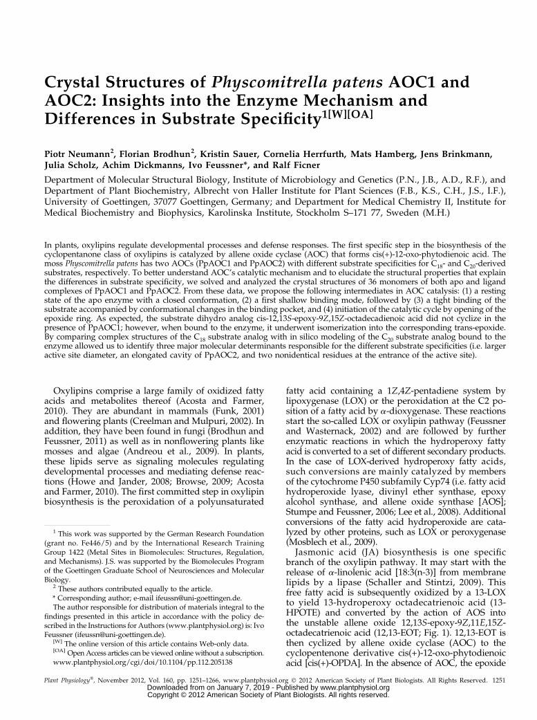
Jasmonic acid (JA) biosynthesis is one specificbranch of the oxylipin pathway. It may start with therelease of a-linolenic acid [18:3(n-3)] from membranelipids by a lipase (Schaller and Stintzi, 2009). Thisfree fatty acid is subsequently oxidized by a 13-LOXto yield 13-hydroperoxy octadecatrienoic acid (13-HPOTE) and converted by the action of AOS intothe unstable allene oxide 12,13S-epoxy-9Z,11E,15Z-octadecatrienoic acid (12,13-EOT; Fig. 1). 12,13-EOT isthen cyclized by allene oxide cyclase (AOC) to thecyclopentenone derivative cis(+)-12-oxo-phytodienoicacid [cis(+)-OPDA]. In the absence of AOC, the epoxide
1 This work was supported by the German Research Foundation(grant no. Fe446/5) and by the International Research TrainingGroup 1422 (Metal Sites in Biomolecules: Structures, Regulation,and Mechanisms). J.S. was supported by the Biomolecules Programof the Goettingen Graduate School of Neurosciences and MolecularBiology.
2 These authors contributed equally to the article.* Corresponding author; e-mail [email protected] author responsible for distribution of materials integral to the
findings presented in this article in accordance with the policy de-scribed in the Instructions for Authors (www.plantphysiol.org) is: IvoFeussner ([email protected]).
[W] The online version of this article contains Web-only data.[OA] Open Access articles can be viewed online without a subscription.www.plantphysiol.org/cgi/doi/10.1104/pp.112.205138
Plant Physiology�, November 2012, Vol. 160, pp. 1251–1266, www.plantphysiol.org � 2012 American Society of Plant Biologists. All Rights Reserved. 1251 www.plantphysiol.orgon January 7, 2019 - Published by Downloaded from
Copyright © 2012 American Society of Plant Biologists. All rights reserved.
is hydrolyzed into ketols and racemic 12-oxo-phytodienoicacid (OPDA). cis(+)-OPDA is the first cyclic and bio-logically active compound in that pathway (Dave andGraham, 2012). While the reactions leading from 18:3(n-3) to cis(+)-OPDA occur in the plastid, all furtherenzymatic steps resulting in the formation of JA arelocalized in the peroxisomes (Wasternack, 2007). Here,cis(+)-OPDA is reduced in a NADPH-dependent re-action by cis(+)-OPDA reductase isoform 3 to 3-oxo-2(29Z-pentenyl)-cyclopentane-1-octanoic acid. This stepis followed by activation of the carboxyl group andthree steps of b-oxidation and finally leads to the for-mation of (+)-7-iso-JA (Dave and Graham, 2012).
The conversion of 13-HPOTE into cis(+)-OPDA wasfirst observed using a flaxseed (Linum usitatissimum)
acetone powder preparation and was suggested totake place via a hypothetical epoxide intermediate(Vick et al., 1980). Later studies unequivocally dem-onstrated that 12,13-EOT (Hamberg, 1987; Brash et al.,1988), an allene oxide formed from 13-HPOTE by AOS(Song and Brash, 1991; Song et al., 1993), serves assubstrate for the cyclization reaction catalyzed by AOC(Hamberg and Fahlstadius, 1990). The enzyme waspurified (Ziegler et al., 1997), characterized with re-gard to the substrate specificity (Ziegler et al., 1999),and cloned and recombinantly expressed (Ziegleret al., 2000; Stenzel et al., 2003). In 2006, the crystalstructure of an AOC from Arabidopsis (Arabidopsisthaliana; AtAOC2) was solved (Hofmann et al., 2006),and the reaction mechanism as well as the subcellular
Figure 1. Overview of the enzymaticsteps in JA biosynthesis with molecularfocus (box) on the reaction catalyzedby AOC. JA biosynthesis may start withthe release of 18:3(n-3) or roughanicacid from a lipid. Next, the fatty acid isoxidized by a 13-LOX, yielding the 13-hydroperoxy derivative. This servesas a substrate for a subsequent con-version catalyzed by AOS and AOC,yielding the cyclopentenone derivativescis(+)-OPDA and cis(+)-dinorOPDA, re-spectively, via an unstable allene oxide.Cyclization of the allene oxide seemsto be initiated by one particular Gluresidue in the active site of AOC thatleads to an opening of the epoxy ring,conformational changes, and a con-certed pericyclic ring closure (detailsare explained in the text). After reductionof the cyclopentenone by cis(+)-OPDAreductase isoform 3 (OPR3), the octanoicor hexanoic side chain is shortened byb-oxidation cycles.
1252 Plant Physiol. Vol. 160, 2012
Neumann et al.
www.plantphysiol.orgon January 7, 2019 - Published by Downloaded from Copyright © 2012 American Society of Plant Biologists. All rights reserved.
localization were studied (Schaller et al., 2008). Theenzyme crystallized as a homotrimer, with each sub-unit forming an eight-stranded antiparallel b-barrelharboring a hydrophobic cavity in which the active siteof the enzyme is located. While the exterior loopsshowed a high degree of flexibility, the central part ofthe enzyme was very rigid, and no induced-fit mech-anism could be observed upon binding of a substrateanalog (Hofmann et al., 2006). Based on the structureof AtAOC2 in complex with vernolic acid [cis(+/2)-12,13-epoxy-9Z-octadecenoic acid (12,13-EOM)] as aninert substrate analog, the following reaction mecha-nism has been proposed (Fig. 1, box): the allene oxidesubstrate binds with its fatty acid backbone deep in thebarrel, where it interacts with hydrophobic amino acidresidues, while the polar carboxy head group is lo-cated on the exterior of the cavity. One particular Gluresidue (Glu-23 in AtAOC2) pointing to the D15Z-double bond of the substrate may induce a partialcharge separation that leads to a delocalization of thep-electron system, thereby facilitating opening of theepoxide ring. The oxyanion thus formed is stabilizedvia polar interactions with a catalytic, structurallyconserved water molecule that is positioned in thepolar cavity of the enzyme formed by two Asn resi-dues (Asn-25 and Asn-53 in AtAOC2, respectively),one Ser (Ser-31 in AtAOC2), and one Pro (Pro-32 inAtAOC2). The ring closure that leads to the formationof the cyclopentenone derivative is achieved by a con-formational reorganization of the C10-C11 substratebond from the trans- to the cis-geometry. Due to steric
limitations in the active site, this rotation may be ac-companied by a cis/trans-isomerization of the C8-C9substrate bond. Since the enzyme dictates the stereo-chemistry of the final ring closure, the released product isexclusively the (+)-enantiomer, cis(+)-OPDA (Schalleret al., 2008). Notably, this reaction competes with thespontaneous decomposition of the allene oxide substratethat leads to the formation of racemic OPDA as well asa-ketols and g-ketols. This hints toward a low-energybarrier of the cyclization reaction and suggests thatAOC does not need much of a catalytic functionality interms of lowering this barrier (Schaller and Stintzi, 2009).It has been proposed that the enzymatic cyclization re-action is achieved according to the rules of Hoffmannand Woodward (1970) via a concerted pericyclic ringclosure while spontaneous cyclization proceeds througha dipolar ring closure (Grechkin et al., 2002). The factsthat the allene oxide formed by AOS has a very shorthalf-life in aqueous solution and that natural OPDA isfound in its enantiopure cis(+)-configuration suggest thatAOS and AOC are coupled. However, no physical in-teraction of both enzymes may be necessary to form cis(+)-OPDA in vitro (Zerbe et al., 2007).
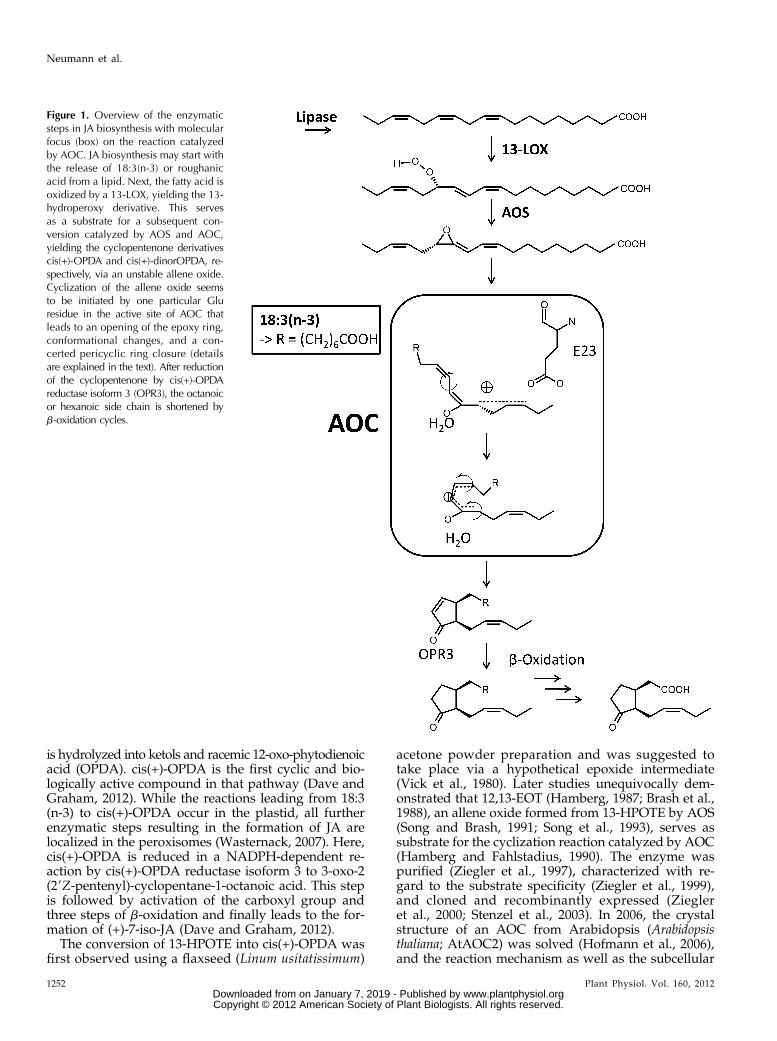
Recently, it was shown that the moss Physcomitrellapatens harbors and metabolizes not only C18 but alsoC20 polyunsaturated fatty acids to form oxylipins (Fig.2; Stumpe et al., 2010). In particular, it was shown that(12S)-hydroperoxy eicosatetraenoic acid (12-HPETE) isendogenously formed by a bifunctional LOX as themajor hydroperoxy fatty acid of arachidonic acid [20:4(n-6)] (Wichard et al., 2004). 12-HPETE serves as a
Figure 2. AOS/AOC pathways in P. patens. 13-HPOTE is converted by PpAOS to 12,13-EOT, which may either hydrolyze in theabsence of PpAOC1 or PpAOC1 to ketols and racemic OPDA or, in the presence of PpAOC1 and PpAOC2, cyclize to cis(+)-OPDA. 12-HPETE is converted by PpAOS to 11,12-EET, which again may either hydrolyze in the absence of PpAOC2 toketols and racemic OPDA or, in the presence of PpAOC2, cyclize to 11-OPTA.
Plant Physiol. Vol. 160, 2012 1253
Structural Basis for Allene Oxide Cyclization in Moss
www.plantphysiol.orgon January 7, 2019 - Published by Downloaded from Copyright © 2012 American Society of Plant Biologists. All rights reserved.
substrate for further conversions either leading to theformation of C8- and C9-volatiles (e.g. octenals, octenols,and nonenals) or the cyclopentenone derivative 11-oxoprostatrienoic acid (11-OPTA; Stumpe et al., 2010).Whereas the volatiles are formed by at least two bi-functional LOXs with an additional hydroperoxide ly-ase activity (Wichard et al., 2004; Senger et al., 2005;Anterola et al., 2009) or by a Cyp74-derived hydroper-oxide lyase (Stumpe et al., 2006), 11-OPTA is formed inanalogy to the octadecanoids by one particular AOC,PpAOC2, via the allene oxide intermediate formed byPpAOS (Bandara et al., 2009). In contrast, PpAOC1 doesnot accept the 12-HPETE-derived C20-allene oxide andthus converts only the 13-HPOTE-derived allene oxide.
In this study, the crystal structures of PpAOC1 andPpAOC2 were solved. Data were also obtained for mu-tated forms of PpAOC1 and for PpAOC1 and PpAOC2in complex with the allene oxide stable analog 12,13-EOD. In this way, detailed information about the alleneoxide-to-cyclopentenone conversions promoted by thetwo AOCs was obtained.
RESULTS
Expression and Purification
In order to optimize the protein yield in Escherichiacoli of previously reported PpAOC1 and PpAOC2 con-structs (Stumpe et al., 2010), both open reading frameswere expressed as glutathione S-transferase (GST) fu-sion proteins. They could be obtained in high yieldby affinity chromatography using reduced glutathione(GSH)-Sepharose. N-terminally fused GST was subse-quently cleaved and separated from AOC by anion-exchange chromatography. The last purification stepusing gel-filtration chromatography was used to re-move large and small protein aggregates. Purificationwas followed by SDS-PAGE (Supplemental Fig. S1)and yielded 20 to 30 mg AOC L21 culture and resultedin nearly homogenous protein.
Overall Structure
The crystal structures of both PpAOC isoforms havebeen solved by molecular replacement, resulting in thelocation of four trimers of PpAOC1 and of two trimersof PpAOC2 per asymmetric unit (for PpAOC2, seeSupplemental Fig. S2A). The structure of apo PpAOC1has been refined at 1.35 Å resolution to crystallo-graphic R factors of 13.9% and 17.5% for Rwork andRfree, respectively. The PpAOC2 diffracted to thehighest resolution limit of 1.98 Å and has been refinedto Rwork and Rfree factors of 18.9% and 23.3%, respec-tively. The 12,13-EOD complex structures of both iso-forms have been determined at similar resolution asthe apo structures and refined to good crystallographicR factors (Table I). The monomers of PpAOC1 andPpAOC2 fold into an eight-stranded antiparallel b-barrel,forming an elongated cavity containing the ligand-
binding site with the overall structure very similar tothat of AOC2 from Arabidopsis (AtAOC2) describedpreviously (Hofmann and Pollmann, 2008; Fig. 3, Aand B, the entrance of the substrate-binding site ismarked by the gray substrate molecule). The calcu-lated root mean square deviation (RMSD) against themonomer of AtAOC2 (Protein Data Bank [PDB] ac-cession code 2BRJ) amounts to 0.51 Å (163 matched Caatoms, 105 conserved in sequence) and 0.80 Å (163matched Ca atoms, 100 conserved in sequence) for themonomers of PpAOC1 and PpAOC2, respectively.Similar RMSD values have been obtained when com-paring the trimer of AtAOC2 with the trimers of bothPpAOCs isoforms (PpAOC1, 0.74 Å for 515 matched Capositions; PpAOC2, 0.85 Å for 504 equivalent Ca posi-tions), revealing exactly the same trimeric quaternarystructure. The largest differences between AtAOC2 andPpAOC monomers can be seen in the conformationsof two out of three flexible loops (AtOC2, 149–151and 38–46; PpAOC1, 161–170 and 33–45; Fig. 3A). Thelater loop in both PpAOCs isoforms contains a seven-amino-acid-long insertion in comparison with AtAOC2(Supplemental Fig. S3) and is not traceable in somemonomers due to its high flexibility.
Comparison of PpAOC1 and PpAOC2
The two PpAOC isoforms share 76% sequence iden-tity and are structurally very similar. The calculatedRMSD between the monomers of the two structuresamounts to 0.83 Å for 175 matched Ca positions, ofwhich 139 are identical in sequence (Fig. 3, A and B;Supplemental Fig. S3), but the sequence of PpAOC2 isone amino acid shorter in comparison with PpAOC1due to a deletion at position 39 localized in one of thethree flexible loops (Fig. 3A; Supplemental Fig. S3). As aconsequence, there is an offset in numbering betweenthe equivalent residues of both isoforms starting fromthat position. Sequence comparison of the two PpAOCsreveals 46 not conserved residues, with half of themrepresenting conservative changes (Supplemental Fig.S3). The differing amino acid positions are locatedmostly on the protein surface, predominantly in theloop regions. In PpAOC1, these are the followingamino acids: 33 to 45, 79 to 83, and 161 to 170. Inter-estingly, two amino acid positions (PpAOC1 Leu-72[PpAOC2 Ile] and PpAOC1 Leu-90 [PpAOC2 Gln])are found on the b-barrel surface, just between theregions responsible for trimerization (Fig. 3B, red as-terisks). The side chain of PpAOC2 Ile-71 forms a weak(distances vary between 3.3 and 3.5 Å for differentmonomers) interaction with the side chain of Thr-73from the neighboring monomer. However, this weakinteraction does not affect formation of the trimer,which appears to be a relevant oligomerization statealso in vivo (Ziegler et al., 1997). This hypothesis issupported by the fact that all AOCs determined struc-turally so far crystallized as trimers (Hofmann andPollmann, 2008). Another differing amino acid position,
1254 Plant Physiol. Vol. 160, 2012
Neumann et al.
www.plantphysiol.orgon January 7, 2019 - Published by Downloaded from Copyright © 2012 American Society of Plant Biologists. All rights reserved.
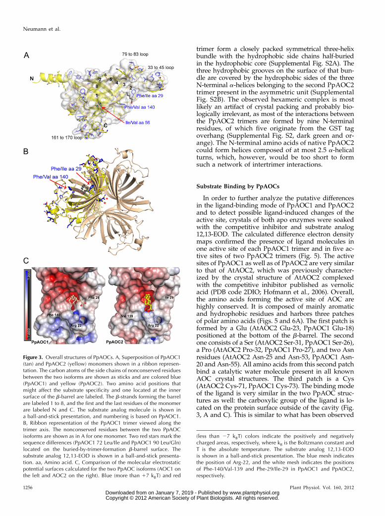
PpAOC1 Ile-56 (PpAOC2 Val) in the b-strand 2 (Fig. 3A),occurs on the inner surface of the b-barrel, toward theN terminus. Interestingly, two differences in amino acidpositions are located near the entrance of the active site,namely PpAOC1 Phe-29 (PpAOC2 Ile) and PpAOC1Phe-140 (PpAOC2 Val; Fig. 3). Because of their localiza-tion, the latter may be candidates for determinants thatcause the observed difference in substrate specificity.In order to analyze this hypothesis, we generated
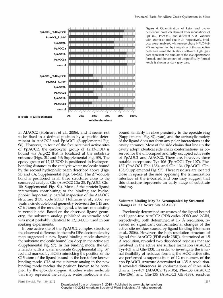
PpAOC1 single and double variants with (1) a Phe-140-Val substitution (PpAOC1_F140V) and (2) an additionalPhe-29-Ile exchange (PpAOC1_F29I/F140V). Both va-riants were purified as described for the wild-typeenzymes. The catalytic activity of both variants wasevaluated by incubating a mixture of PpAOS andPpAOC (molar ratio of 0.001) with either 12-HPETEor 13-HPOTE as the control experiment. The formedproducts were analyzed and quantified (Fig. 4). Incu-bations of 18:3(n-3)-derived 13-HPOTE with the different
enzyme variants yielded significant amounts of OPDA(47%–65%), indicating that all enzyme variants were ac-tive with the 13-HPOTE-derived allene oxide (Fig. 4,bottom). However, when incubated with 20:4(n-6)-derived 12-HPETE, we observed the formation of 11-OPTA only with PpAOC2 (Fig. 4, top). Neither PpAOC1,the single PpAOC1_F140V variant, nor the doublePpAOC1_F29I/F140V variant was able to convert the12-HPETE-derived allene oxide into 11-OPTA.
The superposition of PpAOC1 and PpAOC2 mono-mers revealed three regions that exhibit significantdifferences: two flexible loops (PpAOC1 from aminoacids 33–45 and 79–83) and the N terminus (includingthe loop from amino acids 161–170; Fig. 3A). TheN-terminal 10 residues of PpAOC1 are not defined inthe electron density map. In contrast, these residuesare traceable in PpAOC2 and fold into an amphipathica-helix protruding out of the monomer (Fig. 3A;Supplemental Fig. S2). Three helices of one PpAOC2
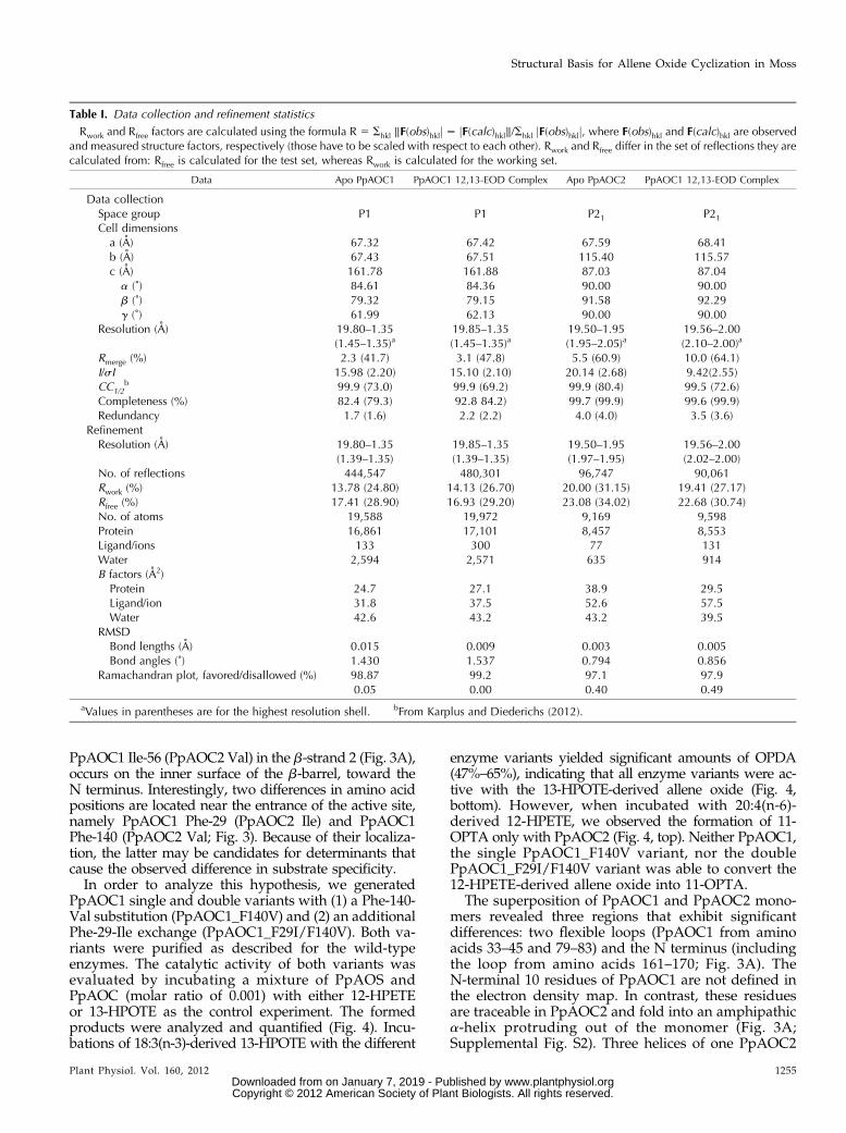
Table I. Data collection and refinement statistics
Rwork and Rfree factors are calculated using the formula R 5 Shkl ‖F(obs)hklj 2 jF(calc)hkl‖/Shkl jF(obs)hklj, where F(obs)hkl and F(calc)hkl are observedand measured structure factors, respectively (those have to be scaled with respect to each other). Rwork and Rfree differ in the set of reflections they arecalculated from: Rfree is calculated for the test set, whereas Rwork is calculated for the working set.
Data Apo PpAOC1 PpAOC1 12,13-EOD Complex Apo PpAOC2 PpAOC1 12,13-EOD Complex
Data collectionSpace group P1 P1 P21 P21
Cell dimensionsa (A) 67.32 67.42 67.59 68.41b (A) 67.43 67.51 115.40 115.57c (A) 161.78 161.88 87.03 87.04a (˚) 84.61 84.36 90.00 90.00b (˚) 79.32 79.15 91.58 92.29g (˚) 61.99 62.13 90.00 90.00
Resolution (A) 19.80–1.35 19.85–1.35 19.50–1.95 19.56–2.00(1.45–1.35)a (1.45–1.35)a (1.95–2.05)a (2.10–2.00)a
Rmerge (%) 2.3 (41.7) 3.1 (47.8) 5.5 (60.9) 10.0 (64.1)I/sI 15.98 (2.20) 15.10 (2.10) 20.14 (2.68) 9.42(2.55)CC1/2
b 99.9 (73.0) 99.9 (69.2) 99.9 (80.4) 99.5 (72.6)Completeness (%) 82.4 (79.3) 92.8 84.2) 99.7 (99.9) 99.6 (99.9)Redundancy 1.7 (1.6) 2.2 (2.2) 4.0 (4.0) 3.5 (3.6)
RefinementResolution (A) 19.80–1.35 19.85–1.35 19.50–1.95 19.56–2.00
(1.39–1.35) (1.39–1.35) (1.97–1.95) (2.02–2.00)No. of reflections 444,547 480,301 96,747 90,061Rwork (%) 13.78 (24.80) 14.13 (26.70) 20.00 (31.15) 19.41 (27.17)Rfree (%) 17.41 (28.90) 16.93 (29.20) 23.08 (34.02) 22.68 (30.74)No. of atoms 19,588 19,972 9,169 9,598Protein 16,861 17,101 8,457 8,553Ligand/ions 133 300 77 131Water 2,594 2,571 635 914B factors (A2)Protein 24.7 27.1 38.9 29.5Ligand/ion 31.8 37.5 52.6 57.5Water 42.6 43.2 43.2 39.5
RMSDBond lengths (A) 0.015 0.009 0.003 0.005Bond angles (˚) 1.430 1.537 0.794 0.856
Ramachandran plot, favored/disallowed (%) 98.87 99.2 97.1 97.90.05 0.00 0.40 0.49
aValues in parentheses are for the highest resolution shell. bFrom Karplus and Diederichs (2012).
Plant Physiol. Vol. 160, 2012 1255
Structural Basis for Allene Oxide Cyclization in Moss
www.plantphysiol.orgon January 7, 2019 - Published by Downloaded from Copyright © 2012 American Society of Plant Biologists. All rights reserved.
trimer form a closely packed symmetrical three-helixbundle with the hydrophobic side chains half-buriedin the hydrophobic core (Supplemental Fig. S2A). Thethree hydrophobic grooves on the surface of that bun-dle are covered by the hydrophobic sides of the threeN-terminal a-helices belonging to the second PpAOC2trimer present in the asymmetric unit (SupplementalFig. S2B). The observed hexameric complex is mostlikely an artifact of crystal packing and probably bio-logically irrelevant, as most of the interactions betweenthe PpAOC2 trimers are formed by nine N-terminalresidues, of which five originate from the GST tagoverhang (Supplemental Fig. S2, dark green and or-ange). The N-terminal amino acids of native PpAOC2could form helices composed of at most 2.5 a-helicalturns, which, however, would be too short to formsuch a network of intertrimer interactions.
Substrate Binding by PpAOCs
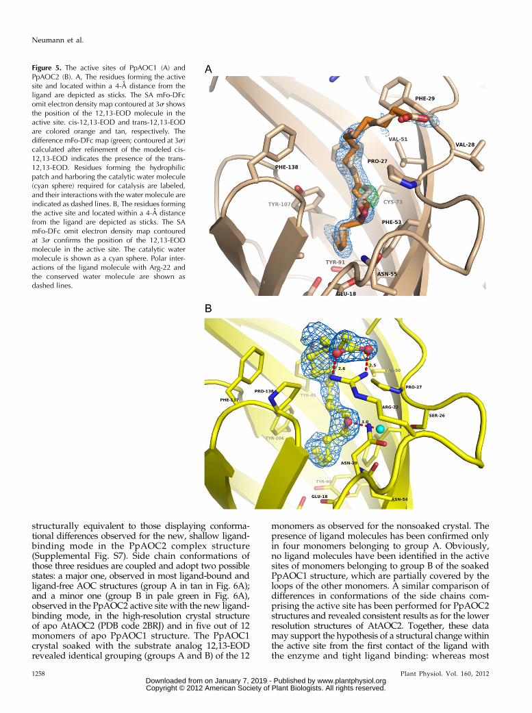
In order to further analyze the putative differencesin the ligand-binding mode of PpAOC1 and PpAOC2and to detect possible ligand-induced changes of theactive site, crystals of both apo enzymes were soakedwith the competitive inhibitor and substrate analog12,13-EOD. The calculated difference electron densitymaps confirmed the presence of ligand molecules inone active site of each PpAOC1 trimer and in five ac-tive sites of two PpAOC2 trimers (Fig. 5). The activesites of PpAOC1 as well as of PpAOC2 are very similarto that of AtAOC2, which was previously character-ized by the crystal structure of AtAOC2 complexedwith the competitive inhibitor published as vernolicacid (PDB code 2DIO; Hofmann et al., 2006). Overall,the amino acids forming the active site of AOC arehighly conserved. It is composed of mainly aromaticand hydrophobic residues and harbors three patchesof polar amino acids (Figs. 5 and 6A). The first patch isformed by a Glu (AtAOC2 Glu-23, PpAOC1 Glu-18)positioned at the bottom of the b-barrel. The secondone consists of a Ser (AtAOC2 Ser-31, PpAOC1 Ser-26),a Pro (AtAOC2 Pro-32, PpAOC1 Pro-27), and two Asnresidues (AtAOC2 Asn-25 and Asn-53, PpAOC1 Asn-20 and Asn-55). All amino acids from this second patchbind a catalytic water molecule present in all knownAOC crystal structures. The third patch is a Cys(AtAOC2 Cys-71, PpAOC1 Cys-73). The binding modeof the ligand is very similar in the two PpAOC struc-tures as well: the carboxylic group of the ligand is lo-cated on the protein surface outside of the cavity (Fig.3, A and C). This is similar to what has been observed
Figure 3. Overall structures of PpAOCs. A, Superposition of PpAOC1(tan) and PpAOC2 (yellow) monomers shown in a ribbon represen-tation. The carbon atoms of the side chains of nonconserved residuesbetween the two isoforms are shown as sticks and are colored blue(PpAOC1) and yellow (PpAOC2). Two amino acid positions thatmight affect the substrate specificity and one located at the innersurface of the b-barrel are labeled. The b-strands forming the barrelare labeled 1 to 8, and the first and the last residues of the monomerare labeled N and C. The substrate analog molecule is shown ina ball-and-stick presentation, and numbering is based on PpAOC1.B, Ribbon representation of the PpAOC1 trimer viewed along thetrimer axis. The nonconserved residues between the two PpAOCisoforms are shown as in A for one monomer. Two red stars mark thesequence differences (PpAOC1 72 Leu/Ile and PpAOC1 90 Leu/Gln)located on the buried-by-trimer-formation b-barrel surface. Thesubstrate analog 12,13-EOD is shown in a ball-and-stick presenta-tion. aa, Amino acid. C, Comparison of the molecular electrostaticpotential surfaces calculated for the two PpAOC isoforms (AOC1 onthe left and AOC2 on the right). Blue (more than 17 kBT) and red
(less than 27 kBT) colors indicate the positively and negativelycharged areas, respectively, where kB is the Boltzmann constant andT is the absolute temperature. The substrate analog 12,13-EODis shown in a ball-and-stick presentation. The blue mesh indicatesthe position of Arg-22, and the white mesh indicates the positionsof Phe-140/Val-139 and Phe-29/Ile-29 in PpAOC1 and PpAOC2,respectively.
1256 Plant Physiol. Vol. 160, 2012
Neumann et al.
www.plantphysiol.orgon January 7, 2019 - Published by Downloaded from Copyright © 2012 American Society of Plant Biologists. All rights reserved.
in AtAOC2 (Hofmann et al., 2006), and it seems notto be fixed in a defined position by a specific deter-minant in AtAOC2 and PpAOC1 (Supplemental Fig.S6). However, in four of the five occupied active sitesof PpAOC2, the carboxylic group of 12,13-EOD isbound via Arg-22 that is localized at the substrateentrance (Figs. 3C and 5B; Supplemental Fig. S5). Theepoxy group of 12,13-EOD is positioned in hydrogen-bonding distance to the catalytic water molecule boundby the second hydrophilic patch described above (Figs.5B and 6A; Supplemental Figs. S4–S6). The D15-doublebond is positioned in all three structures close to theconserved catalytic Glu (AtAOC2 Glu-23, PpAOCs Glu-18; Supplemental Fig. S4). Most of the protein-ligandinteractions contributing to the binding are hydro-phobic. Importantly, careful inspection of the AtAOC2structure (PDB code 2DIO; Hofmann et al., 2006) re-veals a cis-double bond geometry between the C15 andC16 atoms of the modeled ligand, a feature not existingin vernolic acid. Based on the observed ligand geom-etry, the substrate analog published as vernolic acidwas most probably identical to 12,13-EOD used in oursoaking experiments.In one active site of the PpAOC2 complex structure,
the observed difference in the mFo-DFc electron densitymap suggested a new ligand-binding mode showingthe substrate molecule bound less deep in the active site(Supplemental Fig. S7). In this binding mode, the Gluinteracts with a water molecule (Supplemental Fig. S7,red and marked by HOH) mimicking the position of theC15 atom of the ligand bound in the heretofore knownbinding mode. C18 of the substrate analog in the newbinding mode reaches the position known to be occu-pied by the epoxide oxygen. Another water moleculethat may represent the catalytic water molecule is still
bound similarly in close proximity to the epoxide ring(Supplemental Fig. S7, cyan), and the carboxylic moietyof the ligand does not form any polar interactions at thecavity entrance. Most of the side chains that line up thecavity adopt identical side chain conformations, as ob-served for the unoccupied and fully occupied active siteof PpAOC1 and AtAOC2. There are, however, threenotable exceptions: Tyr-106 (PpAOC1 Tyr-107), Phe-137 (PpAOC1 Phe-138), and Gln-134 (PpAOC1 Gln-135; Supplemental Fig. S7). These residues are locatedclose in space at the side opposing the trimerizationinterface of the b-barrel, and one may suggest thatthis structure represents an early stage of substratebinding.
Substrate Binding May Be Accompanied by StructuralChanges in the Active Site of AOCs
Previously determined structures of the ligand-boundand ligand-free AtAOC2 (PDB codes 2DIO and 2GIN,respectively), both determined at 1.7 Å resolution, re-vealed no significant conformational changes of theactive site residues caused by ligand binding (Hofmannet al., 2006). However, the high-resolution structure ofligand-free AtAOC2 (PDB code 2BRJ), determined at 1.5Å resolution, revealed two disordered residues that areinvolved in the active site surface formation (AtAOC2Tyr-105 and Gln-133). In order to investigate the inter-nal flexibility of residues forming the AOC active site,we performed a superposition of 12 monomers of theapo PpAOC1 structure determined at 1.35 Å resolution.It revealed differences in conformations of three sidechains: Tyr-107 (AtAOC2 Tyr-105), Phe-138 (AtAOC2Phe-136), and Gln-135 (AtAOC2 Gln-133), residues
Figure 4. Quantification of ketol and cyclo-pentenone products derived from incubations ofPpLOX2, PpAOS1, and different AOC variantswith 20:4(n-6) and 18:3(n-3), respectively. Prod-ucts were analyzed via reverse-phase HPLC-MS/MS and quantified by integration of the respectivepeak area using the Xcalibur software. Light graybars represent the amount of the cyclopentenoneformed, and the amount of unspecifically formedketols is shown as dark gray bars.
Plant Physiol. Vol. 160, 2012 1257
Structural Basis for Allene Oxide Cyclization in Moss
www.plantphysiol.orgon January 7, 2019 - Published by Downloaded from Copyright © 2012 American Society of Plant Biologists. All rights reserved.
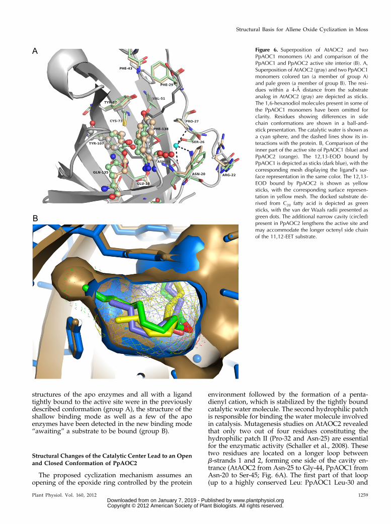
structurally equivalent to those displaying conforma-tional differences observed for the new, shallow ligand-binding mode in the PpAOC2 complex structure(Supplemental Fig. S7). Side chain conformations ofthose three residues are coupled and adopt two possiblestates: a major one, observed in most ligand-bound andligand-free AOC structures (group A in tan in Fig. 6A);and a minor one (group B in pale green in Fig. 6A),observed in the PpAOC2 active site with the new ligand-binding mode, in the high-resolution crystal structureof apo AtAOC2 (PDB code 2BRJ) and in five out of 12monomers of apo PpAOC1 structure. The PpAOC1crystal soaked with the substrate analog 12,13-EODrevealed identical grouping (groups A and B) of the 12
monomers as observed for the nonsoaked crystal. Thepresence of ligand molecules has been confirmed onlyin four monomers belonging to group A. Obviously,no ligand molecules have been identified in the activesites of monomers belonging to group B of the soakedPpAOC1 structure, which are partially covered by theloops of the other monomers. A similar comparison ofdifferences in conformations of the side chains com-prising the active site has been performed for PpAOC2structures and revealed consistent results as for the lowerresolution structures of AtAOC2. Together, these datamay support the hypothesis of a structural change withinthe active site from the first contact of the ligand withthe enzyme and tight ligand binding: whereas most
Figure 5. The active sites of PpAOC1 (A) andPpAOC2 (B). A, The residues forming the activesite and located within a 4-A distance from theligand are depicted as sticks. The SA mFo-DFcomit electron density map contoured at 3s showsthe position of the 12,13-EOD molecule in theactive site. cis-12,13-EOD and trans-12,13-EODare colored orange and tan, respectively. Thedifference mFo-DFc map (green; contoured at 3s)calculated after refinement of the modeled cis-12,13-EOD indicates the presence of the trans-12,13-EOD. Residues forming the hydrophilicpatch and harboring the catalytic water molecule(cyan sphere) required for catalysis are labeled,and their interactions with the water molecule areindicated as dashed lines. B, The residues formingthe active site and located within a 4-A distancefrom the ligand are depicted as sticks. The SAmFo-DFc omit electron density map contouredat 3s confirms the position of the 12,13-EODmolecule in the active site. The catalytic watermolecule is shown as a cyan sphere. Polar inter-actions of the ligand molecule with Arg-22 andthe conserved water molecule are shown asdashed lines.
1258 Plant Physiol. Vol. 160, 2012
Neumann et al.
www.plantphysiol.orgon January 7, 2019 - Published by Downloaded from Copyright © 2012 American Society of Plant Biologists. All rights reserved.
structures of the apo enzymes and all with a ligandtightly bound to the active site were in the previouslydescribed conformation (group A), the structure of theshallow binding mode as well as a few of the apoenzymes have been detected in the new binding mode“awaiting” a substrate to be bound (group B).
Structural Changes of the Catalytic Center Lead to an Openand Closed Conformation of PpAOC2
The proposed cyclization mechanism assumes anopening of the epoxide ring controlled by the protein
environment followed by the formation of a penta-dienyl cation, which is stabilized by the tightly boundcatalytic water molecule. The second hydrophilic patchis responsible for binding the water molecule involvedin catalysis. Mutagenesis studies on AtAOC2 revealedthat only two out of four residues constituting thehydrophilic patch II (Pro-32 and Asn-25) are essentialfor the enzymatic activity (Schaller et al., 2008). Thesetwo residues are located on a longer loop betweenb-strands 1 and 2, forming one side of the cavity en-trance (AtAOC2 from Asn-25 to Gly-44, PpAOC1 fromAsn-20 to Ser-45; Fig. 6A). The first part of that loop(up to a highly conserved Leu: PpAOC1 Leu-30 and
Figure 6. Superposition of AtAOC2 and twoPpAOC1 monomers (A) and comparison of thePpAOC1 and PpAOC2 active site interior (B). A,Superposition of AtAOC2 (gray) and two PpAOC1monomers colored tan (a member of group A)and pale green (a member of group B). The resi-dues within a 4-A distance from the substrateanalog in AtAOC2 (gray) are depicted as sticks.The 1,6-hexanodiol molecules present in some ofthe PpAOC1 monomers have been omitted forclarity. Residues showing differences in sidechain conformations are shown in a ball-and-stick presentation. The catalytic water is shown asa cyan sphere, and the dashed lines show its in-teractions with the protein. B, Comparison of theinner part of the active site of PpAOC1 (blue) andPpAOC2 (orange). The 12,13-EOD bound byPpAOC1 is depicted as sticks (dark blue), with thecorresponding mesh displaying the ligand’s sur-face representation in the same color. The 12,13-EOD bound by PpAOC2 is shown as yellowsticks, with the corresponding surface represen-tation in yellow mesh. The docked substrate de-rived from C20 fatty acid is depicted as greensticks, with the van der Waals radii presented asgreen dots. The additional narrow cavity (circled)present in PpAOC2 lengthens the active site andmay accommodate the longer octenyl side chainof the 11,12-EET substrate.
Plant Physiol. Vol. 160, 2012 1259
Structural Basis for Allene Oxide Cyclization in Moss
www.plantphysiol.orgon January 7, 2019 - Published by Downloaded from Copyright © 2012 American Society of Plant Biologists. All rights reserved.
AtAOC2 Leu-35) adopts an extended conformationand forms a number of hydrogen bond interactionswith b-strand 2, while the remaining part (PpAOC1amino acids 31–45, AtAOC2 amino acids 36–44) isneither conserved in sequence nor in length betweenthe AOCs from flowering plants and those two fromthe moss (Supplemental Fig. S3). The hydrophilicpatch II is partially formed by the first part of thementioned loop connecting b-strands 1 and 2 (Fig. 6A;Supplemental Fig. S8). It is one of the structural ele-ments forming the AOC active site and is thought to beits integral part. The structure of apo PpAOC2, how-ever, revealed an unexpected conformational flexibilityof the essential Pro residue (PpAOC2 Pro-27, AtAOC2Pro-32), resulting in covering the active site entranceby a flexible loop, and only Val-28 and Ile-29 followingthe conserved Pro are traceable (Supplemental Fig. S8,yellow structure). The Val-28 side chain points towardArg-22, and the first eight carbon atoms of 12,13-EODin the PpAOC2 complex (as well as in all other complexstructures) occupy exactly the same position as the twohydrophobic residues (Val-28 and Ile-29) covering theactive site entrance in the apo PpAOC2 structure. Theobserved structural rearrangements have been identifiedin three out of six apo PpAOC2monomers occupying theasymmetric unit, and in one of them no electron densityhas been observed for the catalytic water molecule. Uponligand binding, however, the disordered loop coveringthe active site adopts the conformation known from theother AOC structures, and the conserved water moleculeis bound by all monomers of the PpAOC2 complexstructure (Supplemental Fig. S8, orange structure).
cis/trans-Isomerization of 12,13-EOD Bound to PpAOC1
The quality and shape of the difference electrondensity, calculated at 1.35 Å resolution for the soakedPpAOC1 crystal, allowed modeling of the 12,13-EODmolecule in one active site of each PpAOC1 trimer.Subsequent refinement resulted in the appearance ofpositive peaks of the difference mFo-DFc electron den-sity map at the 3s level, close to the C11 atoms. Inter-estingly, the density peaks, which appeared in threeactive sites, could only be explained by modeling 12,13-EOD in trans-configuration of the epoxide ring (Fig. 5A,green area), although the 12,13-EOD that has been usedfor soaking experiments was in the cis-configuration atthe epoxide ring. No evidence for the presence of trans-epoxide as an impurity has been obtained based onHPLC/mass spectrometry (MS) and gas chromatogra-phy-MS analysis (Supplemental Fig. S9).
Careful examination of electron density maps allowedmodeling of single ligand conformers in three active sitesof the PpAOC1 complex structure: one with cis-12,13-EOD and two with trans-12,13-EOD. The fourth’s activesite has been modeled with the ligand in two almostequally populated, alternate configurations of the epox-ide ring: cis and trans. Despite differences in the epoxidering configuration, the overall binding mode of the cis/
trans-12,13-EOD in PpAOC1 is very similar also whencompared with the published AtAOC2 complexed withmost likely the identical ligand (PDB code 2DIO). Thus,the observed isomerization of the epoxide ring affectsneither the conformation nor the positioning of thepentenyl moiety comprising the D15-double bond. Theepoxide ring of the trans-12,13-EOD is only slightlyreoriented relative to its position observed for the cis-12,13-EOD, preserving the hydrogen bonding distanceto the catalytic water molecule (Supplemental Fig. S6).The largest difference was observed for the position ofatom C11 and consequently the D9-double bond, albeitthe changes were very limited. The carboxyl moiety isknown to be flexible and does not form any polar in-teractions with PpAOC1 atoms comprising the activesite entrance (vide supra; Supplemental Fig. S6). Inter-estingly, the electron density maps calculated for thestructure of 12,13-EOD complexed by PpAOC2 did notconfirm the presence of trans-12,13-EOD in any activesite. This could be due to a limited number of details thatcould be seen at 2 Å resolution or the lack of geometricalisomerization caused by the different time and pH ofsoaking conditions (pH 7.5 for PpAOC2 versus pH 4.5for PpAOC1).
Comparison of PpAOC1 and PpAOC2 Active Sites
Notwithstanding structural similarities between ac-tive sites, the two PpAOC isoenzymes differ in theirsubstrate specificity. The ability of PpAOC2 to acceptboth C18 and C20 fatty acid derivatives indicates at leastslight structural differences within the binding cavitiesof the two PpAOC isoforms. Those differences mustallow PpAOC2 to accommodate, deep in the activesite, an octenyl side chain of the substrate derived from12-HPETE and still not impair the enzymatic activityfor the substrate originating from 13-HPOTE having apentenyl side chain. Subtle changes could easily escapedetection when comparing very similar macromole-cules. Our attention, however, was riveted by differentRMSD values calculated on Ca positions betweenmonomers of PpAOCs and individually against themonomer of AtAOC2. Surprisingly, based on RMSDvalues, one could expect higher structural similaritybetween PpAOC1 and AtAOC2 (having the same sub-strate specificity) than between both PpAOCs (RMSDvalues of 0.51 Å and 0.83 Å, respectively). In comparison,the RMSD calculated between monomers of PpAOC2and AtAOC2 amounts to 0.80 Å, suggesting that theb-barrel of the PpAOC2 monomer is indeed slightlydifferent. In order to address this aspect, we comparedthe active site diameters of both PpAOC isoforms asmeasured across the b-barrel between amino acidsforming the inner part of the binding cavity, mostlyresponsible for binding of the pentenyl side chain ofthe C18-derived substrate molecule. We have chosenamino acids lying on opposite sides of the active site,namely PpAOCs Glu-18, PpAOC1 Leu-142 (PpAOC2Leu-141), and PpAOC1 Leu-133 (PpAOC2 Leu-132) on
1260 Plant Physiol. Vol. 160, 2012
Neumann et al.
www.plantphysiol.orgon January 7, 2019 - Published by Downloaded from Copyright © 2012 American Society of Plant Biologists. All rights reserved.
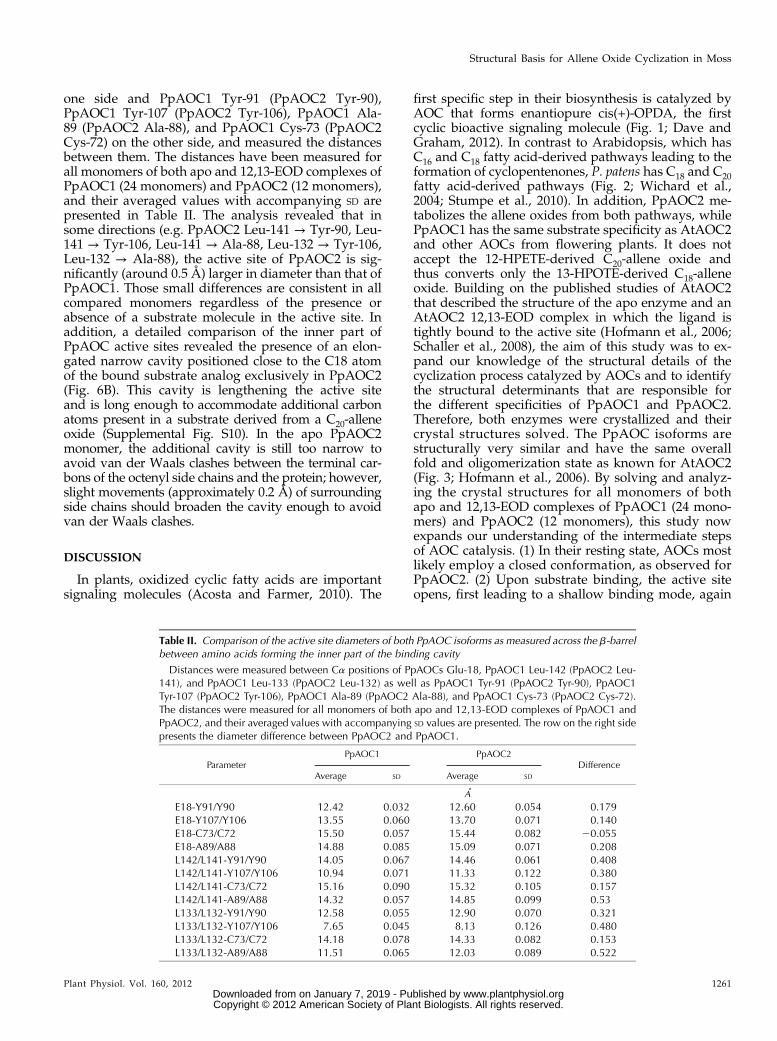
one side and PpAOC1 Tyr-91 (PpAOC2 Tyr-90),PpAOC1 Tyr-107 (PpAOC2 Tyr-106), PpAOC1 Ala-89 (PpAOC2 Ala-88), and PpAOC1 Cys-73 (PpAOC2Cys-72) on the other side, and measured the distancesbetween them. The distances have been measured forall monomers of both apo and 12,13-EOD complexes ofPpAOC1 (24 monomers) and PpAOC2 (12 monomers),and their averaged values with accompanying SD arepresented in Table II. The analysis revealed that insome directions (e.g. PpAOC2 Leu-141 → Tyr-90, Leu-141 → Tyr-106, Leu-141 → Ala-88, Leu-132 → Tyr-106,Leu-132 → Ala-88), the active site of PpAOC2 is sig-nificantly (around 0.5 Å) larger in diameter than that ofPpAOC1. Those small differences are consistent in allcompared monomers regardless of the presence orabsence of a substrate molecule in the active site. Inaddition, a detailed comparison of the inner part ofPpAOC active sites revealed the presence of an elon-gated narrow cavity positioned close to the C18 atomof the bound substrate analog exclusively in PpAOC2(Fig. 6B). This cavity is lengthening the active siteand is long enough to accommodate additional carbonatoms present in a substrate derived from a C20-alleneoxide (Supplemental Fig. S10). In the apo PpAOC2monomer, the additional cavity is still too narrow toavoid van der Waals clashes between the terminal car-bons of the octenyl side chains and the protein; however,slight movements (approximately 0.2 Å) of surroundingside chains should broaden the cavity enough to avoidvan der Waals clashes.
DISCUSSION
In plants, oxidized cyclic fatty acids are importantsignaling molecules (Acosta and Farmer, 2010). The
first specific step in their biosynthesis is catalyzed byAOC that forms enantiopure cis(+)-OPDA, the firstcyclic bioactive signaling molecule (Fig. 1; Dave andGraham, 2012). In contrast to Arabidopsis, which hasC16 and C18 fatty acid-derived pathways leading to theformation of cyclopentenones, P. patens has C18 and C20fatty acid-derived pathways (Fig. 2; Wichard et al.,2004; Stumpe et al., 2010). In addition, PpAOC2 me-tabolizes the allene oxides from both pathways, whilePpAOC1 has the same substrate specificity as AtAOC2and other AOCs from flowering plants. It does notaccept the 12-HPETE-derived C20-allene oxide andthus converts only the 13-HPOTE-derived C18-alleneoxide. Building on the published studies of AtAOC2that described the structure of the apo enzyme and anAtAOC2 12,13-EOD complex in which the ligand istightly bound to the active site (Hofmann et al., 2006;Schaller et al., 2008), the aim of this study was to ex-pand our knowledge of the structural details of thecyclization process catalyzed by AOCs and to identifythe structural determinants that are responsible forthe different specificities of PpAOC1 and PpAOC2.Therefore, both enzymes were crystallized and theircrystal structures solved. The PpAOC isoforms arestructurally very similar and have the same overallfold and oligomerization state as known for AtAOC2(Fig. 3; Hofmann et al., 2006). By solving and analyz-ing the crystal structures for all monomers of bothapo and 12,13-EOD complexes of PpAOC1 (24 mono-mers) and PpAOC2 (12 monomers), this study nowexpands our understanding of the intermediate stepsof AOC catalysis. (1) In their resting state, AOCs mostlikely employ a closed conformation, as observed forPpAOC2. (2) Upon substrate binding, the active siteopens, first leading to a shallow binding mode, again
Table II. Comparison of the active site diameters of both PpAOC isoforms as measured across the b-barrelbetween amino acids forming the inner part of the binding cavity
Distances were measured between Ca positions of PpAOCs Glu-18, PpAOC1 Leu-142 (PpAOC2 Leu-141), and PpAOC1 Leu-133 (PpAOC2 Leu-132) as well as PpAOC1 Tyr-91 (PpAOC2 Tyr-90), PpAOC1Tyr-107 (PpAOC2 Tyr-106), PpAOC1 Ala-89 (PpAOC2 Ala-88), and PpAOC1 Cys-73 (PpAOC2 Cys-72).The distances were measured for all monomers of both apo and 12,13-EOD complexes of PpAOC1 andPpAOC2, and their averaged values with accompanying SD values are presented. The row on the right sidepresents the diameter difference between PpAOC2 and PpAOC1.
ParameterPpAOC1 PpAOC2
DifferenceAverage SD Average SD
A
E18-Y91/Y90 12.42 0.032 12.60 0.054 0.179E18-Y107/Y106 13.55 0.060 13.70 0.071 0.140E18-C73/C72 15.50 0.057 15.44 0.082 20.055E18-A89/A88 14.88 0.085 15.09 0.071 0.208L142/L141-Y91/Y90 14.05 0.067 14.46 0.061 0.408L142/L141-Y107/Y106 10.94 0.071 11.33 0.122 0.380L142/L141-C73/C72 15.16 0.090 15.32 0.105 0.157L142/L141-A89/A88 14.32 0.057 14.85 0.099 0.53L133/L132-Y91/Y90 12.58 0.055 12.90 0.070 0.321L133/L132-Y107/Y106 7.65 0.045 8.13 0.126 0.480L133/L132-C73/C72 14.18 0.078 14.33 0.082 0.153L133/L132-A89/A88 11.51 0.065 12.03 0.089 0.522
Plant Physiol. Vol. 160, 2012 1261
Structural Basis for Allene Oxide Cyclization in Moss
www.plantphysiol.orgon January 7, 2019 - Published by Downloaded from Copyright © 2012 American Society of Plant Biologists. All rights reserved.
as described for the PpAOC2 12,13-EOD complex. (3)Next, tight binding of the substrate occurs by confor-mational changes of at least three amino acid sidechains in the binding pocket, as demonstrated bycomparing different structures of PpAOC2 12,13-EODcomplexes. (4) The catalytic cycle is initiated by theopening of the epoxide ring. This is supported by theobservation of an epoxide cis/trans-isomerization ofthe PpAOC1 12,13-EOD complex.
The central part of the enzyme harboring the activesite is very rigid, thus limiting possible conformationalchanges induced upon ligand binding. However, a partof the active site is not entirely formed by residuesconstituting the b-barrel, namely, the hydrophilic patchII binding the catalytic water molecule (Fig. 5B). It ispartially formed by part of a loop between b-strands1 and 2 comprising the essential Pro-27 (SupplementalFig. S8). The structure of the apo PpAOC2 revealed aconformational flexibility of Pro-27 leading to closure ofthe active site entrance by this flexible loop and conse-quently destroying the hydrophilic patch. The observedstructural rearrangements indicate that the enzyme canalso exist in a nonfunctional or resting state, which maybe required for protecting the hydrophobic cavity frombeing exposed to the solvent if there is no substratearound or in a very hydrophilic environment. In addi-tion, the loop connecting b-strands 1 and 2, due to itsflexibility, amino acid composition, length, and locali-zation at the active site entrance, may play a role inthe cooperation with AOS, especially for substrate re-cruitment (two adjacent Lys residues, of which one,Lys-35, in both PpAOCs is conserved in sequence;Supplemental Fig. S3). The positively charged residueson the flexible loop flanking the active site entrancecould be used to anchor the negatively charged sub-strate molecules without the necessity of forming theAOS/AOCmolecular complex,whose existence has notbeen experimentally proven yet (Schaller et al., 2008).
For a deeper insight into possible differences in thebinding modes of AOC substrates, crystal structuresmay be needed with either the substrate or the productbound within the active site. Due to the short life timeof the allene oxides in aqueous solution of about 30 s(Hamberg, 1987), details about ligand binding haveonly been obtained from a crystal structure of AtAOC2complexedwith 12,13-EOD, a stable substrate analog (acompetitive inhibitor) lacking the vinylic double bondpresent in allene oxides. The flexible carboxylic moietyof the ligand is located in all structures on the proteinsurface, but only in PpAOC2 is it fixed by forming in-teractions with Arg-22 located at the active site entrance(Figs. 3C and 5B; Supplemental Figs. S4 and S5). Thisinteraction is probably responsible for a positional shiftof the ligand’s epoxide ring and D15-double bond ofapproximately 1.2 Å toward the active site entrance, incomparison with PpAOC1 and AtAOC2 complexes.Most interestingly, the structure of the PpAOC2 12,13-EOD complex revealed, in one active site, a new ligand-binding mode with the substrate analog bound lessdeep in the active site (Supplemental Fig. S7). In this
orientation, the methyl group of the pentenyl side chainreaches the position occupied by the epoxide ring inhitherto known ligand-binding mode and the carbox-ylicmoiety is not involved in forming anypolar contactsat the active site entrance. The observed accompanyingconformational changes of the three side chains consti-tuting the active site, Tyr-106 (PpAOC1 Tyr-107), Phe-137 (PpAOC1 Phe-138), andGln-134 (PpAOC1Gln-135;Fig. 6A), reveal the intrinsic dynamics inside the rigidand narrow cavity, which may be important for thesubstrate binding and/or release of the product. Thestructurally equivalent residues of PpAOC1 revealedthe same conformational flexibility as observed forsome monomers of the apo structure. This stronglysuggests that the side chain conformations of those threeresidues are coupled and the enzyme samples betweentwo distinct conformations, of which only one is able tolock the flexible acyl moiety in the position essential forcatalysis.
To expand our knowledge of different substrate-binding modes, crystals of both apo enzymes weresoaked in this study with three different substrate ana-logs, 12,13-EOD, vernolic acid [cis(+/2)-12,13-epoxy-9Z-octadecenoic acid (12,13-EOM)], and 11,12-epoxy-5Z,8Z,14Z-eicosatrienoic acid (11,12-EET), as well asthe product, cis(+)-OPDA. However, soaking was onlysuccessful for the 12,13-EOD ligand. Interestingly, thecrystal structure of the PpAOC1 12,13-EOD complexrevealed a cis/trans-isomerization of the epoxide ring,which, however, did not affect the binding mode of thesubstrate analog (Fig. 5A; Supplemental Figs. S4 andS6). The D11-double bond present in the natural sub-strate precludes such isomerization and causes the ep-oxide ring to be in plane with the C11 atom; therefore,its position would slightly differ from that observedin both cis- and trans-12,13-EOD (Supplemental Fig.S11). The in silico docking experiments of the C18-derived allene oxide substrate derived from 13-HPOTEto both PpAOC isoforms confirmed this hypothesis(Supplemental Fig. S11). The docking results revealedthat the limited changes in position of the C11 atomdue to cis/trans-isomerization do not cause signifi-cant movements of the epoxide ring and the D15Z-doublebond. Irrespective of the epoxide ring being cis- or trans-configured, all structurally characterized complex struc-tures of AOCs share a very similar binding mode of12,13-EOD. Oxiranes are stable when stored in alcohols;however, in aqueous solution, under both acidic andbasic conditions, they can undergo ring-opening reac-tions leading to geometrical isomerization of the epoxidering (Yamaguchi et al., 1993). Oxirane ring opening mayoccur via C-O or C-C bond cleavage, and the preferredroute is often determined by the nature of the substituent(MacDonald and Crawford, 1972). The formed interme-diates can either spontaneously close back to the originalring form or partake another fragment/compound,yielding a nonepoxide product. The mechanistic fea-tures of electrocyclic epoxide ring-opening reactionshave been described by Woodward and Hoffmann(1965), who formulated, based on orbital symmetry,
1262 Plant Physiol. Vol. 160, 2012
Neumann et al.
www.plantphysiol.orgon January 7, 2019 - Published by Downloaded from Copyright © 2012 American Society of Plant Biologists. All rights reserved.
rules predicting the stereochemistry of pericyclic reac-tions. Considering these rules, the preferred way of ep-oxide ring reclosure is a conrotatory mode. Thus, thespontaneous reclosing reaction can result in the geomet-rical isomerization of epoxides, which passes through theopen intermediate form, and is likely to yield a morestable epoxide ring structure in the trans-configuration(MacDonald and Crawford, 1972; Davey et al., 2008). Inthe case of a ligand that cannot form a cyclopentadienylcation, cyclization is not possible; therefore, the epoxidering is being formed again, this time in its more stabletrans-conformation (Fig. 1). Therefore, thedata on the cis/trans-isomerization of 12,13-EODmay support themodelpublished recently in which an opening of the epoxidering of the allene oxide substrate via the catalytic Gluresidue (Glu-23 in AtAOC2) initiates the cyclization re-action (Hofmann et al., 2006). However, analysis of thisinitial reaction step is hampered by the fact that a muta-tion in the catalytic Glu-23 (in AtAOC2) resulted in aninactive enzyme (Schaller et al., 2008).Previously, we have reported the preferential for-
mation of cis(+)-OPDA in a coupled AOS/PpAOC as-say using 13-HPOTE as the substrate in the presence ofeither PpAOC1 or PpAOC2 (Stumpe et al., 2010). Incontrast, incubation of both PpAOC isoforms with theallene oxide derived from 12-HPETE resulted in cyclo-pentenone formation (11-OPTA) only when PpAOC2was used. Due to the different lengths of side chains ofsubstrate molecules derived from both C18- and C20-allene oxide derivatives, slight differences within thebinding cavities of the two PpAOCs can be expected(Fig. 2). A detailed analysis of active sites of bothPpAOC isoforms revealed differences in active site di-ameter and an additional cavity in PpAOC2 that mightaccommodate a longer octenyl side chain of the sub-strate derived from C20 fatty acid (Fig. 6B). In order tocheck the possibility of an alleged binding of the longeroctenyl side chain inside the narrow active site, weperformed in silico docking of 11,12-EET to monomersof both PpAOC isoforms. The docking results clearlydemonstrated that slight conformational changes of afew hydrophobic side chains forming the narrow cavity(PpAOC2 Tyr-90, Phe-92, Val-102, Leu-114, Leu-132,and Phe-145) allow PpAOC2 to accommodate the lon-ger octenyl side chain and simultaneously maintaincorrect positioning of the epoxide oxygen and thehomoallylic double bond required for catalysis (Fig. 6B;Supplemental Fig. S10). In order to find additional fac-tors that may have an influence on the observed differ-ences in substrate specificity between the two PpAOCisoenzymes,weanalyzedknownplantAOCsbysequencealignment on the basis of the sequence differences ob-served between the two PpAOCs (Supplemental Fig.S3). Among the analyzed plant AOC sequences, onlyPpAOC2 has been reported to convert the 12-HPETE-derived substrate. The residues comprising the activesite are strictly conserved between the AOCs. However,two amino acid positions located near the cavity en-trance differ in size between the PpAOCs: PpAOC1Phe-29 and Phe-140 correspond to the less space-filling
residues Ile-29 and Val-139 in PpAOC2, respectively(Fig. 3C). The substrate C20-allene oxide has a one-carbon-shorter carboxylic side chain that is less flexible(one additional double bond in the cis-configuration) incomparison with the saturated octanoyl side chain ofC18-allene oxide (Fig. 2). Consequently, its carboxylicmoiety will be positioned closer to the negativelycharged active site. Therefore, it would be energeticallyfavorable to neutralize its charge by forming interac-tions with positively charged protein residues (Fig. 3C).The structure of PpAOC2 with bound 12,13-EOD re-vealed that such interactions are possible. In all activesites, with the substrate molecule bound in hithertoknown binding mode, the ligand’s carboxylic groupinteracts with the side chain of Arg-22 located near theactive site entrance (Supplemental Fig. S5). In contrast,the PpAOC1 complex structure revealed disorderedcarboxylmoieties of the same substrate analog thatwerenot forming any polar interactions with the proteinresidues, most probably due to a steric hindrance of thePhe-140 side chain (Supplemental Fig. S6). Replacementof PpAOC1Phe-140with the less bulky side chain of Valin PpAOC2 should make the side chain of Arg-22 ac-cessible to the substrate carboxylic moiety, which isa unique feature of PpAOC2. In order to check theinfluence of those two amino acid positions (PpAOC1Phe-29/PpAOC2 Ile-29 and PpAOC1 Phe-140/PpAOC2Val-139), we generated the respective PpAOC1 single(PpAOC1_F140V) and double (PpAOC1_F29I/F140V)variants by means of site-directed mutagenesis and ana-lyzed their catalytic activities. Similar to the wild-typeenzymes of PpAOC1 and PpAOC2, both variants cata-lyzed the specific conversion of 18:3(n-3)-derivedallene oxide to 47% to 65% ofOPDA, as shown in Figure4. When incubated with 20:4(n-6)-derived allene oxide,specific formation of the respective cyclopentenonewas observed only in incubations with PpAOC2 butnot with PpAOC1 (as has been reported before) orwith PpAOC1_F104V or PpAOC1_F29I/F140V mu-tants. Although our experimental data thus could notsupport the influence of these amino acids on differentsubstrate specificities, it should be emphasized herethat we also could not exclude the function of thoseresidues in combination with other steric factors (di-ameter and depth of the cavity) to determine substratespecificity. Cocrystallization of the respective enzymevariant with different substrates might help to tacklethis question.
CONCLUSION
The structure-based elucidation of the enzyme mech-anism of two AOCs from P. patens obtained detailedinformation about the allene oxide-to-cyclopentenoneconversions. By solving 36 monomeric AOC structures,we suggest now the following different conformations:(1) a closed conformation for the resting state, (2) a firstshallow binding mode, (3) followed by the recentlypublished tight binding of the substrate, and finally (4)
Plant Physiol. Vol. 160, 2012 1263
Structural Basis for Allene Oxide Cyclization in Moss
www.plantphysiol.orgon January 7, 2019 - Published by Downloaded from Copyright © 2012 American Society of Plant Biologists. All rights reserved.
the initiation of cyclization by the ring opening of theepoxide group. In addition, different substrate specific-ities may be accompanied by different active site diam-eters and an elongated cavity.
MATERIALS AND METHODS
Cloning and Recombinant Expression of PpAOC1and PpAOC2
PpAOC1 and PpAOC2 from Physcomitrella patens were cloned from therespective pGEM-T plasmid reported before using the BamHI and HindIIIrecognition sites as described (Stumpe et al., 2010). The resulting fragment wascloned in the pGEX-6-P-1 vector (GE Healthcare), resulting in the plasmidspGEX-6P-1/PpAOC1 and pGEX-6P-1/PpAOC2 that encode the respectiveAOC with an N-terminal fusion to GST via the PreScission protease recogni-tion site. These clones were used as templates for site-directed mutagenesisPCR (AOC1_F140V and AOC1_F29I/F140V) using the Pfu polymerase.
For expression, the respective plasmid was transformed in BL21 Star cells(Invitrogen). Cells were cultivated in Luria-Bertani broth or 2YT broth con-taining 100 mg mL21 ampicillin and grown at 37°C to an optical density at600 nm of 0.6 to 0.8. Protein expression was induced with a final concentrationof 1 mM isopropyl-b-D-thiogalactopyranoside. Bacterial cultures were culti-vated by shaking at 28°C for approximately 18 h and harvested by centrifu-gation (3,220g) for 20 min. The resulting cell precipitate was shock frozen inliquid nitrogen and stored at 220°C.
Protein Purification
Thecell pelletwas resuspended in20mMTris-HCl (pH7.5) containing150mM
NaCl, 2 mM dithiothreitol (DTT), and 0.2 mM phenylmethylsulfonyl fluoride.Lysozyme was added (final concentration of approximately 0.1 mg mL21), andcells were incubated on ice for 30 min. Cell lysis was increased by sonication onice using a Sonifier B15 cell disruptor (Branson). Cell debris was removed bycentrifugation at 45,000g for 20 min at 4°C. The resulting cell-free crude extractwas loaded on a GSH-Sepharose matrix (30 mL of GSH-Sepharose 4 Fast Flowin an XK16/20 column; GE Healthcare) using an ÄKTAprime system (GEHealthcare). The column was washed with 20 mM Tris-HCl (pH 7.5) containing200mMNaCl, 0.1% Triton X-100, and 2mMDTT. Protein was elutedwith 20mM
Tris-HCl (pH 7.5), 150 mM NaCl, 2 mM DTT, and 10 mM GSH. The GST-AOCfusion protein was cleaved in the elution buffer by adding 1 mol % PreScissionprotease (GE Healthcare) and incubation overnight at 4°C, yielding AOC withan N-terminal Gly-Pro-Leu-Gly-Ser extension. After buffer exchange against20 mM Tris-HCl (pH 8.0) containing 2 mM DTT, the sample was loaded on aSource 30Q anion-exchange resin (30 mL; XK16/20; GE Healthcare) in order toseparate AOC fromGST. Elution of the protein(s) was performed using a lineargradient of 0 to 0.5MNaCl (in 20mMTris-HCl [pH8.0] containing 2mMDTT) anda length of 50 to 100 mL. Fractions containing AOC were combined and con-centrated to a final volume of 1 to 4 mL by using Vivaspin 20 (mass of 10,000 D;Sartorius Stedim Biotech). The enzyme concentrate was loaded on a Superdex200 26/60pg or on a Superdex 75 16/60 gel filtration column (GE Healthcare)equilibrated with 20 mM Tris-HCl (pH 7.5) containing 150 mM NaCl and 2 mM
DTT for final purification (Supplemental Fig. S1).
Purity Control of the Substrate Analog 12,13-EOD
Theligand investigated inbothstructureshasbeensynthesizedfrom18:3(n-3)containing three double bonds in the cis-configuration. The synthesis of theepoxide ring at position 12Z is stereo specific, meaning that stereoisomerism inthe alkene will be reflected in the epoxide product. The synthesized substrateanalog was stored in ethanol, where it is stable. Its enantiomeric purity wasassessed by using reverse-phase HPLC-MS. The analysis was carried out on aSurveyor HPLC system (Thermo Finnigan) equipped with an EC 50/2 Nucle-odur C18 gravity column (503 2 mm, 1.8-mm particle size; Macherey & Nagel)using a solvent system consisting of acetonitrile:water:acetic acid (40:60:0.1,v/v/v) as solvent system A and acetonitrile:water:acetic acid (100:0.1, v/v) assolvent systemB. The gradient elution profile startedwith a linear gradient from100% solvent A to 100% solvent Bwithin 25min and continuedwith an isocraticrun for 5 min. The column was reequilibrated by a linear gradient back to 100%
solvent systemAwithin 2min, whichwas followed by an isocratic run of 3min.Theflow ratewas set to 0.3mLmin21. TheMS analysiswas performed on a LCQAdvantage mass spectrometer (Thermo Finnigan) in the negative electrosprayionizationmode scanning for ions in amass range ofmass-to-charge ratio 250 to350 in the full-scan mode. The ion spray voltage was set to 4 kV, while thecapillary voltage was used at 13 V. The capillary temperature was 300°C, and asheath gasflow rate of 22 arbitrary units and an auxiliary/sweep gasflow rate of8 arbitrary units were applied. As the trans-12,13-EOD enantiomer was com-mercially unavailable, we tested the separation efficiency by using cis-12,13-EOM and trans-12,13-EOM (close homologs of 12,13-EOD) as standards. Asshown in Supplemental Figure S9, both enantiomers were chromatographicallyseparated by using this method.
Activity Assay
The catalytic activity of different PpAOC variants (AOC1_F140V andAOC1_F29I/F140V) was analyzed in a coupled assay similar to the methoddescribed by Stumpe et al. (2010). The PpAOS cDNA (Bandara et al., 2009) wasPCR amplified from a cDNA library of P. patens protonema and cloned intopQE30, yielding the construct pQE30/PpAOS. The enzyme was expressed inEscherichia coli SG13009 [pRep4] (3 d at 16°C under constant shaking) in framewith an N-terminal hexa-His peptide. Protein extraction was essentially per-formed in accordance with a method described earlier (Richardson et al., 1993).It involves the generation of spheroplasts prior to final cell lysis. PpAOS waspurifiedwith immobilized Ni2+-affinity chromatography according to standardprocedures. Next, 1 mL of 50 mM Tris-HCl (pH 8.0) containing 10% (v/v)glycerol and 100 mM 18:3(n-3) and 20:4(n-6), respectively, was preincubatedwith PpAOS/PpAOC (in a molar ratio of approximately 0.001) at 4°C. The re-action was started by the addition of a purified truncated version of PpLOX2(F. Brodhun and I. Feussner, unpublished data) and allowed to proceed for30 min. Reaction products were extracted with 2 mL of diethylether. The polarphase was reextracted with 2 mL of diethylether containing 100 mL of glacialacid. Product analysis was performed on the same instrument as for the puritycontrol experiment (vide supra) using anEC250/2Nucleodur 120-5C18 column(250 3 2.1 mm, 5-mm particle size; Macherey & Nagel), and a similar solvent/gradient systemwas employed with a flow rate of 200 mLmin21. Products wereidentified bymeans of their characteristicMS/MS spectra using an identical MSdevice described above with similar MS parameters, scanning for ions in themass range of mass-to-charge ratio 80 to 350 in the negative electrospray ioni-zation mode. For MS/MS analysis, collision energy of 1 eV was employed.Quantificationwasperformedby integrating the respective signal area using theXcalibur software (Thermo Finnigan).
Crystallization, Soaking Experiments, and Data Collection
Crystals of PpAOC1 and PpAOC2 were obtained using the sitting-dropvapor-diffusion method on 24-well Cryschem plates (Hampton Research) at20°C and 4°C. The crystallization droplets (2mL)were prepared bymixing equalamounts of protein (at a concentration of 20 to 30 mg mL21 in 20 mM Tris-HCl[pH 7.5] containing 150 mM NaCl and 2 mM DTT) with reservoir solution andwere equilibrated against a 500-mL reservoir. The initial needle-shaped crystalsof PpAOC1 (diffracting to about 6 Å) were obtained from Hampton MagicScreen (condition No. 49 containing 20% [w/v] polyethylene glycol [PEG] 8000,0.1 M Na3PO4, pH 4.2; Hampton Research). The crystallization condition wasfurther optimized by using additives (Hampton Research). It was found that theaddition of 1mL of 30% (w/v) 1,6-hexandiol improves the crystal formation andtheir diffraction properties. The final optimization of crystallization conditionshas been performed by slightly changing the concentrations of PEG 8000 and1,6-hexandiol. The crystals appeared within 7 to 10 d and grew to the size of0.13 0.13 0.25mmwithin 4weeks. Prior to diffraction experiments, the crystalswere cryoprotected using the solution with a higher concentration of PEG8000 and 1,6-hexandiol (35% [w/v] for both chemicals). The initial crystals ofPpAOC2 were obtained from condition No. 9 of JBScreen 3 (Jena Bioscience)containing 16% (w/v) PEG 4000, 0.2 M (NH4)2SO4, 10% (w/v) isopropanol, and0.1 M HEPES, pH 7.5. The final optimized crystallization condition was com-posed of 12% to 14% (w/v)PEG4000, 0.25M (NH4)2SO4, 10% (w/v) isopropanol,and 0.1 M HEPES-KOH, pH 7.5. Before mounting on a loop, crystals were firstsoaked in suitable cryoprotectant solution containing 25% (w/v) 2-methyl-2,4-pentanediol (MPD) in addition to the reservoir solution.
Soaking experiments (6–72 h) of PpAOC1 and PpAOC2 crystals wereperformed using the crystallization solutions containing in addition 10- to20-fold molar excess of the substrate analogs: 12,13-EOD, vernolic acid or
1264 Plant Physiol. Vol. 160, 2012
Neumann et al.
www.plantphysiol.orgon January 7, 2019 - Published by Downloaded from Copyright © 2012 American Society of Plant Biologists. All rights reserved.
12,13-EOM, and 11,12-EET. The data sets from several crystals (two or threerepetitions for each ligand differing in soaking time) were collected. Thestructures were refined using the available apo structures as starting models.The estimation of the success in soaking experiments was made based on thepresence and quality of the difference electron density maps (mFo-DFc and2mFo-DFc) inside the elongated cavity known to be the ligand-binding site ofAOCs. Inspection of those difference electron density maps and later a sim-ulated annealing (SA) omit map confirmed successful soaking only for the12,13-EOD ligand. The electron density maps calculated for crystals soakedwith the remaining substrate analogs did not allow us to unambiguously tracethe ligand molecules, as the active sites were also partially occupied by low-Mralcohols (1,6-hexanodiol for PpAOC1, isopropanol and MPD for PpAOC2),which were present in soaking or cryo solutions. Desoaking of 1,6-hexanodiolfrom the PpAOC1 crystals or soaking experiments with reduced amounts orlack of that alcohol resulted in complete crystal damage. Other cryoprotectantsthan MPD for PpAOC2 crystals resulted in significant loss of diffractionproperties.
The data were collected at beamlines BL 14.1 and 14.2 at the BESSY IIsynchrotron in Berlin (Mueller et al., 2012). The beamlines were equippedwith a MARMosaic-CCD detector (225 and 165 mm; MARRESEARCH) anda MARdtbgoniostat (MARRESEARCH). The data sets were integrated andscaled using the XDS package (Kabsch, 2010). The details are presented inTable I.
Structure Solution and Refinement
PpAOC1 crystallized in the P1 space group with 12 monomers occupyingthe asymmetric unit with a corresponding solvent content of 54.9%. Thestructure of PpAOC1 was solved by molecular replacement with the PHASER(McCoy et al., 2007) program using the trimer of AtAOC2 (PDB accession code2GIN) as search model. PpAOC2 crystallized in the P21 space group with sixmonomers in the asymmetric unit (corresponding solvent content of 57.6%).The phase problem was solved by molecular replacement using the trimer ofPpAOC1 as the search model in PHASER. Prior refinement the R-free setwas chosen in thin shells in order to avoid noncrystallographic symmetry(NCS)-based bias of the R-free factor. The structures of PpAOC1 andPpAOC2 (apo and complexes) were refined using PHENIX (Adams et al.,2010) and Refmac5 (Winn et al., 2003). Medium NCS restraints were used atthe early stages of refinement of both structures and were released forvariable parts of the AOC monomers where appropriate. The final refine-ment steps were applied without any NCS restraints. Manual model re-building and verification were performed against difference electron densitymaps as well as SA omit electron density maps in Coot (Emsley et al., 2010).Alternative steps of model adjustment and refinement were performed untilthe crystallographic R factors converged (Table I). The atomic coordinatesand the necessary geometry definitions for the ligand molecules were cre-ated using the PRODRG server (Schüttelkopf and van Aalten, 2004). Thequality of the final models was assessed using MolProbity (Chen et al., 2010;Table I). Figures were prepared using Pymol (http://www.pymol.org).The atomic coordinates as well as diffraction data have been deposited inthe PDB under accession codes 4H6C (apo PpAOC1), 4H6B (PpAOC112,13-EOD complex), 4H6A (apo PpAOC2), and 4H69 (PpAOC2 12,13-EODcomplex).
In Silico Docking Experiments
The docking experiments were performed using AutoDock Vina (Trott andOlson, 2010). Briefly, the initial ligand’s coordinates (12,13-EOT and 11,12-EET)were obtained from the PRODRG server. Autodock Tools (Adams et al., 2010)was used to set up the docking experiments, which were performed with thecatalytically importantwatermolecule bound in the hydrophilic cavity. BothC18(12,13-EOT) and C20 (11,12-EET) substrates were docked to both PpAOC1 andPpAOC2 monomers. The predicted binding orientations were compared withthe crystallographically determined position of the 12,13-EOD and were con-sidered to be reasonable if the following criteria were fulfilled: the differencesbetween positions of epoxide oxygen atoms as well as C15 atom and its equiv-alent should not exceed 1.2 Å. For docking experiments with C18-derived sub-strate, the receptor atomswerefixed; thus, the only rotatable degrees of freedomwere possessed by the ligand molecule (double bonds were fixed in the cis-configuration). Docking experiments using the standard settings revealedmostly nonsense binding predictions when considering 10 predictions withthe lowest energy. Therefore, several docking experiments were performed
changing the exhaustiveness parameter from the default value, determinedautomatically, up to 1,000 in steps of 50. The exhaustiveness parameter describesthe time spent on the search and is varied heuristically depending on the numberof atoms, flexibility, etc. Setting it to a larger value increases the time of thecalculations linearly and decreases the probability of not finding the minimumexponentially. The program reports at most 20 binding predictions sortedaccording to the affinity, expressed in kcal mol21. We observed that increasingthe value of the exhaustiveness parameter to 100 or higher results in obtainingthe highest affinity decoys, which are consistent with the orientation of thesubstrate analog known from the crystal structure. Finally, for the 12,13-EODligand, we have chosen the highest affinity binding predictions calculatedwith the exhaustiveness parameter of 1,000,whichfits verywell the position of12,13-EOD determined experimentally (Supplemental Fig. S11). The calcu-lated affinity predicted for the PpAOC1 receptor (27.4 kcal mol21) was0.3 kcal mol21 lower than that calculated for the PpAOC2 monomer. Theobserved difference in predicted substrate affinity between the two PpAOCisoforms has not been experimentally investigated so far. The 11,12-EET lig-and was docked first to the rigid monomers of PpAOC isoforms running20 individual docking jobs with different values of the exhaustiveness pa-rameter (see above). As expected, no reasonable binding predictions could befound, as 11,12-EET comprises a three-carbon-atom-longer octenyl side chainin comparison with 12,13-EOT (Fig. 2). Binding of three additional carbonatoms inside the narrow active site requires conformational changes of someside chains building its inner surface. Therefore, subsequent experimentswereperformed with setting selected side chains of both receptor molecules to beflexible during the in silico docking experiments (PpAOC2 Tyr-90, Phe-92,Val-102, Leu-114, Leu-132, Phe-145, Arg-22, Ile-29, and Val-139, and equiva-lent residues of PpAOC1). Considering one decoy with the highest reportedbinding affinity (approximately 7.7 kcal mol21) from each individual dockingjob and applying the distance-based criteria, rational substrate-binding pre-dictions were exclusively obtained for six docking jobs with the PpAOC2monomer used as the receptor (Supplemental Fig. S10).
Sequence data from this article can be found in the GenBank/EMBLdata libraries under the following accession numbers: P. patens, AOC1(Q8GS38) and AOC2 (Q8H0N6); Solanum tuberosum, AOC (Q8H1X5); Lyco-persicon esculentum, AOC (Q9LEG5); Humulus lupulus, AOC4 (Q68IP6) andAOC1 (Q68IP7); Zea mays, AOC (Q6RW09); Hordeum vulgare, AOC (Q711R0);(Q711R1); Oryza sativa, AOC (Q8L6H4); Medicago truncatula, AOCb (Q599T8)and AOCa (Q711Q9); Pisum sativum, AOC (Q3LI84); Arabidopsis (Arabidopsisthaliana), AOC4 (Q93ZC5), AOC3 (Q9LS01), AOC1 (Q9LS03), and AOC2(Q9LS02).
Supplemental Data
The following materials are available in the online version of this article.
Supplemental Figure S1. Analysis of the purification of PpAOC1 andPpAOC2 by SDS-PAGE.
Supplemental Figure S2. Oligomerization state of PpAOC2.
Supplemental Figure S3. Sequence alignment of different AOCs.
Supplemental Figure S4. Superposition of individual PpAOC2 andPpAOC1 monomers.
Supplemental Figure S5. Representation of the PpAOC2 active site.
Supplemental Figure S6. The PpAOC1 active site.
Supplemental Figure S7. Superposition of the active sites of two PpAOC2monomers.
Supplemental Figure S8. Superposition of two ligand-free PpAOC2 mon-omers differing in conformation of the region connecting b-strands1 and 2.
Supplemental Figure S9. Reverse-phase HPLC-MS separation of cis/trans-isomers of 12,13-EOM.
Supplemental Figure S10. Results of docking experiments of 11,12-EET toa monomer of PpAOC2.
Supplemental Figure S11. The active site of PpAOC1 is shown in cartoonrepresentation.
Plant Physiol. Vol. 160, 2012 1265
Structural Basis for Allene Oxide Cyclization in Moss
www.plantphysiol.orgon January 7, 2019 - Published by Downloaded from Copyright © 2012 American Society of Plant Biologists. All rights reserved.
ACKNOWLEDGMENTS
We gratefully thank Sabine Freitag for help with the HPLC-MS and gaschromatography-MS experiments and for providing the substrate analogs andcis(+)-OPDA for the soaking experiments. We acknowledge the expert techni-cal assistance and cooperation of the staff of BESSY and their help in perform-ing the diffraction experiments.
Received August 7, 2012; accepted September 14, 2012; published September17, 2012.
LITERATURE CITED
Acosta IF, Farmer EE (2010) Jasmonates. The Arabidopsis Book 8: e0129,doi/10.1199/tab.0129/
Adams PD, Afonine PV, Bunkóczi G, Chen VB, Davis IW, Echols N,Headd JJ, Hung LW, Kapral GJ, Grosse-Kunstleve RW, et al (2010)PHENIX: a comprehensive Python-based system for macromolecularstructure solution. Acta Crystallogr D Biol Crystallogr 66: 213–221
Andreou A, Brodhun F, Feussner I (2009) Biosynthesis of oxylipins in non-mammals. Prog Lipid Res 48: 148–170
Anterola A, Göbel C, Hornung E, Sellhorn G, Feussner I, Grimes H (2009)Physcomitrella patens has lipoxygenases for both eicosanoid and octa-decanoid pathways. Phytochemistry 70: 40–52
Bandara PK, Takahashi K, Sato M, Matsuura H, Nabeta K (2009) Cloningand functional analysis of an allene oxide synthase in Physcomitrellapatens. Biosci Biotechnol Biochem 73: 2356–2359
Brash AR, Baertschi SW, Ingram CD, Harris TM (1988) Isolation and charac-terization of natural allene oxides: unstable intermediates in the metabolismof lipid hydroperoxides. Proc Natl Acad Sci USA 85: 3382–3386
Brodhun F, Feussner I (2011) Oxylipins in fungi. FEBS J 278: 1047–1063Browse J (2009) Jasmonate passes muster: a receptor and targets for the
defense hormone. Annu Rev Plant Biol 60: 183–205Chen VB, Arendall WB III, Headd JJ, Keedy DA, Immormino RM,
Kapral GJ, Murray LW, Richardson JS, Richardson DC (2010) Mol-Probity: all-atom structure validation for macromolecular crystallogra-phy. Acta Crystallogr D Biol Crystallogr 66: 12–21
Creelman RA, Mulpuri R (2002) The oxylipin pathway in Arabidopsis. TheArabidopsis Book 1: e0012, doi/10.1199/tab.0012
Dave A, Graham IA (2012) Oxylipin signaling: a distinct role for the jasmonic acidprecursor cis-(+)-12-oxo-phytodienoic acid (cis-OPDA). Front Plant Sci 3: 42
Davey MP, Burrell MM, Woodward FI, Quick WP (2008) Population-specific metabolic phenotypes of Arabidopsis lyrata ssp. petraea. NewPhytol 177: 380–388
Emsley P, Lohkamp B, Scott WG, Cowtan K (2010) Features and devel-opment of Coot. Acta Crystallogr D Biol Crystallogr 66: 486–501
Feussner I, Wasternack C (2002) The lipoxygenase pathway. Annu RevPlant Biol 53: 275–297
Funk CD (2001) Prostaglandins and leukotrienes: advances in eicosanoidbiology. Science 294: 1871–1875
Grechkin AN, Chechetkin IR, Mukhtarova LS, Hamberg M (2002) Role ofstructure and pH in cyclization of allene oxide fatty acids: implicationsfor the reaction mechanism. Chem Phys Lipids 120: 87–99
Hamberg M (1987) Mechanism of corn hydroperoxide isomerase: detection of12,13(S)-oxido-9(Z),11-octadecadienoic acid. Biochim Biophys Acta 920: 76–84
Hamberg M, Fahlstadius P (1990) Allene oxide cyclase: a new enzyme inplant lipid metabolism. Arch Biochem Biophys 276: 518–526
Hoffmann R, Woodward RB (1970) Orbital symmetry control of chemicalreactions. Science 167: 825–831
Hofmann E, Pollmann S (2008) Molecular mechanism of enzymatic alleneoxide cyclization in plants. Plant Physiol Biochem 46: 302–308
Hofmann E, Zerbe P, Schaller F (2006) The crystal structure of Arabidopsisthaliana allene oxide cyclase: insights into the oxylipin cyclization reac-tion. Plant Cell 18: 3201–3217
Howe GA, Jander G (2008) Plant immunity to insect herbivores. Annu RevPlant Biol 59: 41–66
Kabsch W (2010) XDS. Acta Crystallogr D Biol Crystallogr 66: 125–132Karplus PA, Diederichs K (2012) Linking crystallographic model and data
quality. Science 336: 1030–1033Lee D-S, Nioche P, Hamberg M, Raman CS (2008) Structural insights into the
evolutionary paths of oxylipin biosynthetic enzymes. Nature 455: 363–368MacDonald HH, Crawford RJ (1972) Racemization and geometrical
isomerization of oxiranes. Can J Chem 50: 428–433
McCoy AJ, Grosse-Kunstleve RW, Adams PD, Winn MD, Storoni LC, ReadRJ (2007) PHASER crystallographic software. J Appl Cryst 40: 658–674
Mosblech A, Feussner I, Heilmann I (2009) Oxylipins: structurally diversemetabolites from fatty acid oxidation. Plant Physiol Biochem 47: 511–517
Mueller U, Darowski N, Fuchs MR, Förster R, Hellmig M, Paithankar KS,Pühringer S, Steffien M, Zocher G, Weiss MS (2012) Facilities for macro-molecular crystallography at the Helmholtz-Zentrum Berlin. J SynchrotronRadiat 19: 442–449
Richardson TH, Hsu MH, Kronbach T, Barnes HJ, Chan G, WatermanMR, Kemper B, Johnson EF (1993) Purification and characterization ofrecombinant-expressed cytochrome P450 2C3 from Escherichia coli: 2C3encodes the 6 beta-hydroxylase deficient form of P450 3b. Arch BiochemBiophys 300: 510–516
Schaller A, Stintzi A (2009) Enzymes in jasmonate biosynthesis: structure,function, regulation. Phytochemistry 70: 1532–1538
Schaller F, Zerbe P, Reinbothe S, Reinbothe C, Hofmann E, Pollmann S(2008) The allene oxide cyclase family of Arabidopsis thaliana: localizationand cyclization. FEBS J 275: 2428–2441
Schüttelkopf AW, van Aalten DM (2004) PRODRG: a tool for high-throughput crystallography of protein-ligand complexes. Acta Crys-tallogr D Biol Crystallogr 60: 1355–1363
Senger T, Wichard T, Kunze S, Göbel C, Lerchl J, Pohnert G, Feussner I (2005)A multifunctional lipoxygenase with fatty acid hydroperoxide cleaving ac-tivity from the moss Physcomitrella patens. J Biol Chem 280: 7588–7596
Song W-C, Brash AR (1991) Purification of an allene oxide synthase andidentification of the enzyme as a cytochrome P-450. Science 253: 781–784
Song WC, Funk CD, Brash AR (1993) Molecular cloning of an allene oxidesynthase: a cytochrome P450 specialized for the metabolism of fatty acidhydroperoxides. Proc Natl Acad Sci USA 90: 8519–8523
Stenzel I, Hause B, Miersch O, Kurz T, Maucher H, Weichert H, Ziegler J,Feussner I, Wasternack C (2003) Jasmonate biosynthesis and the alleneoxide cyclase family of Arabidopsis thaliana. Plant Mol Biol 51: 895–911
Stumpe M, Bode J, Göbel C, Wichard T, Schaaf A, Frank W, Frank M,Reski R, Pohnert G, Feussner I (2006) Biosynthesis of C9-aldehydes inthe moss Physcomitrella patens. Biochim Biophys Acta 1761: 301–312
Stumpe M, Feussner I (2006) Formation of oxylipins by CYP74 enzymes.Phytochem Rev 5: 347–357
Stumpe M, Göbel C, Faltin B, Beike AK, Hause B, Himmelsbach K, BodeJ, Kramell R, Wasternack C, Frank W, et al (2010) The moss Phys-comitrella patens contains cyclopentenones but no jasmonates: mutationsin allene oxide cyclase lead to reduced fertility and altered sporophytemorphology. New Phytol 188: 740–749
Trott O, Olson AJ (2010) AutoDock Vina: improving the speed and ac-curacy of docking with a new scoring function, efficient optimization,and multithreading. J Comput Chem 31: 455–461
Vick BA, Feng P, Zimmerman DC (1980) Formation of 12-[18-O]oxo-cis-10,cis-15-phytodienoic acid from 13[18-O]hydroperoxylinolenic acid byhydroperoxide cyclase. Lipids 15: 468–471
Wasternack C (2007) Jasmonates: an update on biosynthesis, signal trans-duction and action in plant stress response, growth and development.Ann Bot (Lond) 100: 681–697
Wichard T, Göbel C, Feussner I, Pohnert G (2004) Unprecedentedlipoxygenase/hydroperoxide lyase pathways in the moss Physcomitrellapatens. Angew Chem Int Ed Engl 44: 158–161
Winn MD, Murshudov GN, Papiz MZ (2003) Macromolecular TLS refinementin REFMAC at moderate resolutions. Methods Enzymol 374: 300–321
Woodward RB, Hoffmann R (1965) Stereochemistry of electrocyclic reac-tions. J Am Chem Soc 87: 395–399
Yamaguchi Y, Schaefer HF, Alberts IL (1993) A mechanistic study of thering-opening reaction of singlet oxirane. J Am Chem Soc 115: 5790–5798
Zerbe P, Weiler EW, Schaller F (2007) Preparative enzymatic solid phasesynthesis of cis(+)-12-oxo-phytodienoic acid: physical interaction ofAOS and AOC is not necessary. Phytochemistry 68: 229–236
Ziegler J, Hamberg M, Miersch O, Parthier B (1997) Purification andcharacterization of allene oxide cyclase from dry corn seeds. PlantPhysiol 114: 565–573
Ziegler J, Stenzel I, Hause B, Maucher H, Hamberg M, Grimm R, GanalM, Wasternack C (2000) Molecular cloning of allene oxide cyclase: theenzyme establishing the stereochemistry of octadecanoids and jasmo-nates. J Biol Chem 275: 19132–19138
Ziegler J, Wasternack C, Hamberg M (1999) On the specificity of alleneoxide cyclase. Lipids 34: 1005–1015
1266 Plant Physiol. Vol. 160, 2012
Neumann et al.
www.plantphysiol.orgon January 7, 2019 - Published by Downloaded from Copyright © 2012 American Society of Plant Biologists. All rights reserved.


![CRYSTAL GARANTIES 2011 BAT10ï Mise en page 1 - Net … · 2011-05-05 · [CRYSTAL STUDIES] Crystal Studies, l’assurance complète de vos études à l’étranger ! Crystal Studies](https://img.pdfslide.fr/doc/110x75/5ebc95e11463d476e401c447/crystal-garanties-2011-bat10-mise-en-page-1-net-2011-05-05-crystal-studies.jpg)