Embed Size (px)
Citation preview
Cirugía y Cirujanos. 2015;83(4):329---333
www.amc.org.mx www.elsevier.es/circir
CIRUGÍA y CIRUJANOSÓrgano de difusión científica de la Academia Mexicana de Cirugía
Fundada en 1933
CLINICAL CASE
Drainage of amoebic liver abscess by single incision
laparoscopic surgery. Report of a case�
José Eduardo Telich-Tarriba, Iris Jocelyn Parrao-Alcántara,Jesús Manuel Montes-Hernández, Jesús Vega-Pérez ∗
Departamento de Cirugía General, Hospital General Regional 1 Querétaro, Instituto Mexicano del Seguro Social,
Querétaro, México
Received 19 June 2014; accepted 19 August 2014
KEYWORDSLiver abscess;Entamoeba
histolytica;Laparoscopy;Minimally invasivesurgical procedures;Single-incisionlaparoscopic surgery
Abstract
Background: Single incision laparoscopic surgery has increased recently due to successfulresults, achieved in several procedures. The aim of the present work is to present the firstcase in which single incision laparoscopy is used for the drainage of an amoebic liver abscess.Clinical case: A 44-year-old man presented with intense right upper quadrant pain, generalisedjaundice, tachycardia, fever, hepatomegaly and a positive Murphy’s sign. Laboratory resultsrevealed an increased plasma bilirubin, elevated alkaline phosphatase and transaminases,leucocytosis, negative viral panel for hepatitis, and positive antibodies against Entamoeba
histolytica. On an abdominal computed tomography a 15 cm × 12.1 cm hypodense lesion wasobserved in the patient’s liver, identified as an amoebic liver abscess. Analgesics and antibi-otics were started and subsequently the patient was submitted to laparoscopic drainage of theabscess using a single port approach. Drainage and irrigation of the abscess was performed.Four days later the patient was discharged without complications.Conclusion: Management of amoebic liver abscess is focused on the elimination of the infectiousagent and obliteration of the abscess cavity in order to prevent its complications, especiallyrupture. Laparoscopic surgery has proved to be a safe and effective way to manage this entity.© 2015 Academia Mexicana de Cirugía A.C. Published by Masson Doyma México S.A. Thisis an open access article under the CC BY-NC-ND license (http://creativecommons.org/licenses/by-nc-nd/4.0/).
� Please cite this article as: Telich-Tarriba JE, Parrao-Alcántara IJ, Montes-Hernández JM, Vega-Pérez J. Drenaje de absceso hepáticoamebiano por laparoscopia de puerto único. Reporte de un caso. Cir Cir. 2015;83:329---333.
∗ Corresponding author at: Departamento de Cirugía General, Hospital General Regional 1 Querétaro, Instituto Mexicano del Seguro Social,Avenida 5 de Febrero, esq. Calzada Zaragoza S/N, Col.: Centro, 76000 Querétaro, Querétaro, México. Tel.: +52 442 315 2207/442 216 2662.
E-mail address: [email protected] (J. Vega-Pérez).
2444-0507/© 2015 Academia Mexicana de Cirugía A.C. Published by Masson Doyma México S.A. This is an open access article under the CCBY-NC-ND license (http://creativecommons.org/licenses/by-nc-nd/4.0/).
330 J.E. Telich-Tarriba et al.
PALABRAS CLAVEAbsceso hepático;Entamoeba
histolytica;Laparoscopia;Cirugía de mínimainvasión;Cirugía por puertoúnico
Drenaje de absceso hepático amebiano por laparoscopia de puerto único. Reporte de
un caso
Resumen
Antecedentes: El uso del puerto único ha adquirido impulso, debido a los resultados exitososlogrados recientemente en diversas disciplinas. El objetivo del presente trabajo es reportarel primer caso de la laparoscopia por puerto único, para el drenaje de un absceso hepáticoamebiano.Caso clínico: Hombre de 44 anos con dolor abdominal intenso en hipocondrio derecho, ictericiageneralizada, taquicardia y fiebre; se palpa borde hepático a 5 cm por debajo del rebordecostal, signo de Murphy positivo. Los estudios de laboratorio revelaron: hiperbilirrubinemiadirecta, elevación de fosfatasa alcalina y transaminasas, leucocitosis a expensas de neutrófilos,panel viral negativo para hepatitis, y anticuerpos positivos para Entamoeba histolytica. Latomografía computada abdominal mostró una lesión hipodensa de 15 por 12.1 cm en lóbulo he-pático derecho. Se inició tratamiento con analgésicos, y doble esquema antibiótico. El pacientefue sometido a drenaje del absceso hepático por la vía laparoscópica a través de puerto único,aspirando 1200 cc de contenido de aspecto achocolatado, se realizó lavado de cavidad. Al cuartodía de internamiento el paciente fue dado de alta sin complicaciones.Conclusión: El tratamiento del absceso hepático amebiano va dirigido hacia la erradicacióndel agente infeccioso y de la cavidad abscedada, ya que se trata de una entidad clínica quepuede tener complicaciones severas, especialmente la ruptura. La laparoscopia por puertoúnico ha demostrado ser una alternativa segura y efectiva, en el tratamiento de los pacientesque requieren drenaje de abscesos amebianos.© 2015 Academia Mexicana de Cirugía A.C. Publicado por Masson Doyma México S.A. Estees un artículo Open Access bajo la licencia CC BY-NC-ND (http://creativecommons.org/licenses/by-nc-nd/4.0/).
Background
Liver amoebiasis is the commonest extraintestinal man-ifestation of the infection by Entamoeba histolytic (E.
histolytic), complicating the course of the disease in 3---10%of the subjects infected by this micro-organism.1 It normallypresents between the fifth and sixth decade of life,2,3 withan equivalent distribution in men and women; 75% of thecases locates in the right hepatic lobe, they are charac-terised as unique and being surrounded by a thin cover ofgranulation tissue.4
This entity is found mainly in developing countrieswith tropical and subtropical weather. In Mexico, intesti-nal amoebiasis and its complications present an incidenceof 384.15/100,000 inhabitants, being considered an areawhere the disease is endemic.5,6
Amoebicidal drugs are the first line of treatment in themanagement ofamoebic hepatic abscesses, metronidazolebeing the first line medication at national and internationallevel due to its effectiveness and accessibility. Up to 90% ofthe cases are solved solely with pharmacological treatment.It is necessary to resort to invasive therapeutic measuresin the remaining 10% which do not respond to treatment.This includes interventional radiology procedures and con-ventional or minimally invasive surgical procedures.7,8
Percutaneous drainage is considered the first invasivechoice; however, there are conditions in which this proce-dure fails, in particular due to the density of the abscesscontent or technical or logistical difficulties in its perfor-mance; surgical drainage is indicated in these patients.Generally, the surgical treatment of these patients is
performed through open procedures, and it has recentlybeen demonstrated that the laparoscopic treatment of liverabscesses is safe and effective, with long-term favourableresults.7,9
Advances in laparoscopic surgery have led to the develop-ment of new approaches that seek to be even less invasive,with similar postoperative results and higher aestheticresults compared to those of conventional laparoscopy. Sin-gle port laparoscopy or single incision laparoscopic surgeryare among these techniques and have been growing in favourdue to the results obtained in procedures such as cholecys-tectomy, adrenalectomy and colon resection.10---12
The aim of the present work is to present the first casein which single incision laparoscopy is used for the drainageof an amoebic liver abscess.
Clinical case
A 44-year-old man presented in the emergency departmentwith a 9-day evolution history of abdominal pain located inthe right hypochondrium, which increased in intensity untilbecoming intolerable, of oppressive and continuous nature,irradiated in girdling pain towards the back and subsidedintermittently with the use of non-steroidal analgesics. Thepain was accompanied by the appearance of one-week evo-lution generalised jaundice and unquantified fever the daybefore his admission.
He does not report relevant family history; the patient’smedical history only reveals occasional consumption of alco-holic beverages, especially beer and pulque during youth.
Drainage of amoebic liver abscess 331
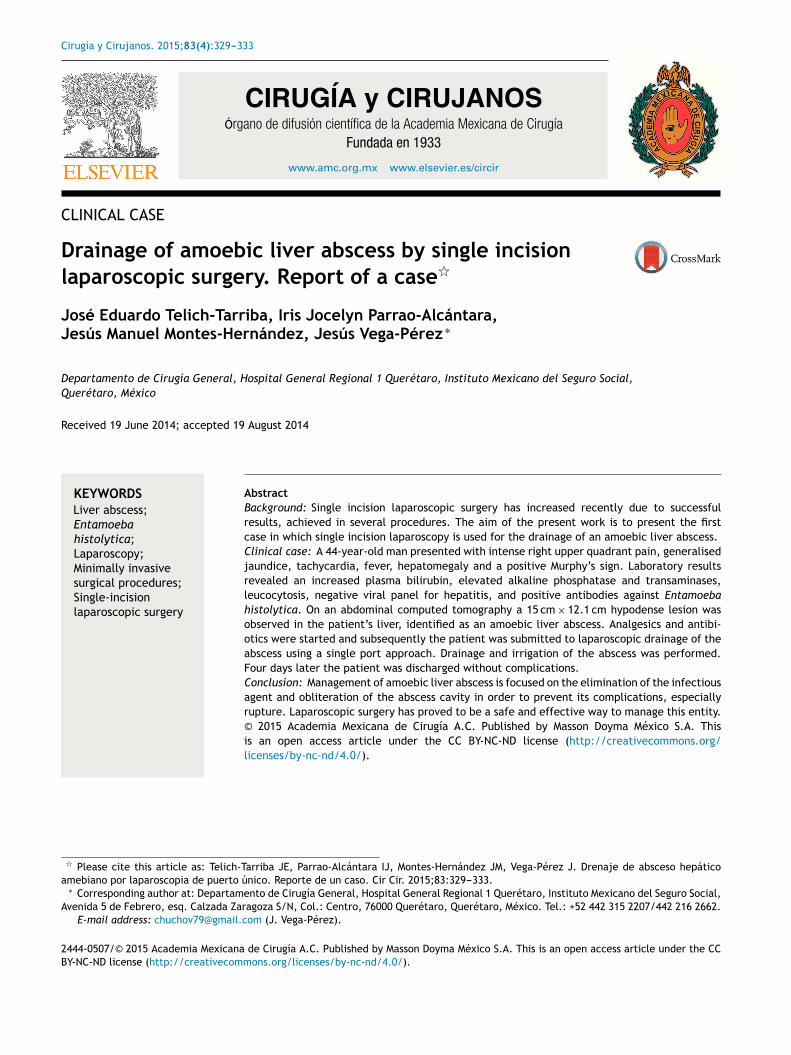
Figure 1 Abdomen computed tomography in which unilocularliver abscess is seen.
During physical examination, he had tachycardia and a39 ◦C fever, generalised jaundice in skin and scleras, soft anddepressible abdomen with intense pain on palpation over theright hypochondrium, palpable liver edge at 5 cm under thecostal margin and positive Murphy’s sign.
The initial laboratory studies showed hyperbilirubi-naemia at the expense of direct bilirubin, with increase ofalkaline phosphatase and transaminases, leukocytosis withneutrophilia, negative viral panel for hepatitis B and C andpositive IgG antibodies for E. histolytic. A hepatic ultrasoundwas performed that revealed a well-defined hypoechoiclesion in segments V and VIII. The abdominal computedtomography showed a well-defined homogeneous hypodenselesion in the right hepatic lobe of 15.5 cm × 12.1 cm (Fig. 1).
The patient was hospitalised and treated with analgesicsand double antibiotic regime with metronidazole and cef-triaxone.
Because of the lack of an interventional radiology pro-gramme in our institution and the characteristics of theabscess regarding its volume, being unilocular and locatingin the anterior region, it was considered that the patientwas an ideal candidate for the laparoscopic procedure usinga single port approach. This procedure was decided withthe patient, who accepted and signed the informed consentform.
Surgical technique
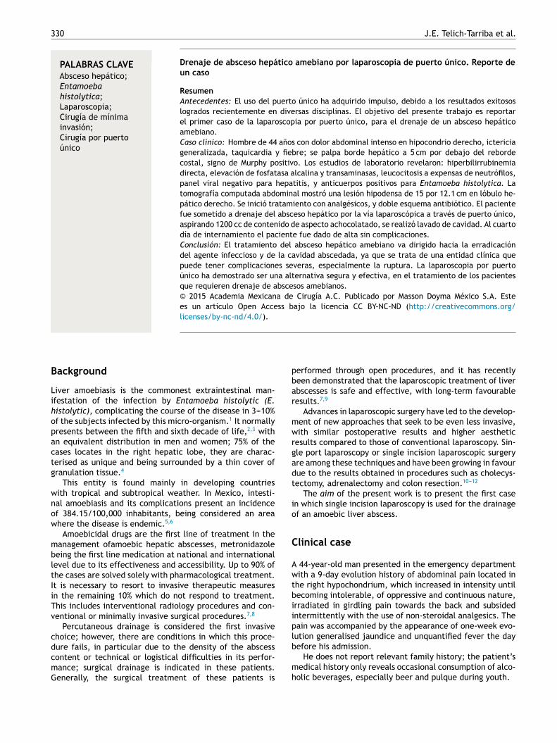
A 3-cm vertical transumbilical incision was performedwith plane dissection until entering the peritoneal cavity,when the multiple access port (Single Incision LaparoscopicSurgery, SILS Port, Covidien) was inserted under directvision and a pneumoperitoneum was created at 15 mmHg(Fig. 2). A 5 mm laparoscope and a zero-degree lens, aspi-rator/irrigator and a SILS clinch were introduced. Whenassessing the cavity, a mass over the lower hepatic face was
Figure 2 Placement of the working port.
found which, when dissected, let brown chocolate-aspectmaterial coming from the liver flow, revealing the presenceof a restrained broken liver abscess. A total of 1.200 cc of theabscess content was aspirated and samples were taken forculture. Later, irrigation with 4.000 cc warm sterile salinesolution was carried out until obtaining a clear aspiration.It was found that this was a single abscess with walls cov-ered by fibrinopurulent material when assessing the abscesswalls. A Saratoga drain was placed in the abscess bed, whichwas externalised at subcostal level and anterior axillary line.
The patient started a diet and walking the next day; hewas given an abdominal computed tomography for controlthe third postoperative day, in which obliteration of theabscess space was found. The patient was asymptomatic andafebrile the fourth day of hospitalisation; therefore, he wasdischarged. The drainage was removed 2 weeks later andthe patient was continuing asymptomatic at 6 months offollow-up.
Discussion
Amoebic liver abscesses are a potentially serious clinicalcondition due to the high morbidity and mortality of itscomplications. Patients tend to present with a history offever, generalised discomfort, abdominal distension, hepa-tomegaly and abdominal pain in the right hypochondrium.They are usually located in the right hepatic lobe andare generally single.6 The diagnosis of this disease is pre-dominantly clinical. It can be based on image procedures,particularly hepatic ultrasound and computed tomography.13
Once the amoebic liver abscess has been identified, it isnecessary to eliminate the infectious agent and obliteratethe abscess cavity. In this group of patients, treatment muststart immediately because this disease may have seriouscomplications, especially rupture towards the peritoneum,pleurae, pericardium or through the skin.
Metronidazol is within the group of medications forthe amibiasis treatment, which is the first choice drug toremove the Entamoeba; due to 10---20% of the amoebicabscesses showing bacterial infection, especially gram-negative bacilli, it is recommended that an antibiotic beadministered.4
332 J.E. Telich-Tarriba et al.
Despite the proven usefulness of pharmacological man-agement, there are cases in which this will not be enoughand will require invasive manoeuvres for the removal of theabscess. Today percutaneous drainage preceded by radiol-ogy is the basis of invasive management, with success ratesnear 85%.9
The surgical management of patients with liver abscessesis usually reserved for patients whom medical and percuta-neous treatment failed, when there are large, multilocularor dense content abscesses, imminence of rupture or devel-opment of serious complications such as peritonitis.7 Thecontraindication for the percutaneous management or thelack of infrastructure to perform it can also be consideredindications for surgical management.14
Traditionally, drainage of liver abscesses was performedthrough open surgeries; however, laparoscopic surgery hasbeen demonstrated to be a safe and effective choice forthe management of this group of patients.9 Among themany advantages it offers are surgical trauma minimisation,reduction of hospital stay and postoperative pain, as well asobtaining more aesthetic scars.15
Among the technologies for minimally invasive surgery,single port laparoscopy is the most promising in thelast decade. Its current indications are limited to thecommonest surgeries, such as cholecystectomies16,17 andappendectomies.18,19 However, recent medical literaturehas gradually demonstrated its usefulness in interven-tions on other organs such as adrenal glands, colon andkidney.12,20,21
Various studies have demonstrated that single-portlaparoscopy is technically as feasible and safe as con-ventional laparoscopy. Its main advantage is the aestheticaspect by using the umbilical scar for the instrumental inser-tion and avoiding more scars. Some authors also report adecrease in postoperative pain and less time needed offwork.22---24 However, further studies are required in this field.
Although it has been argued that technically single-incision laparoscopic surgery is more demanding thanconventional laparoscopy, Mutter et al. have demonstratedthat programmes focused on this surgical approach cansafely be started in teaching hospitals that have experiencedsurgeons in laparoscopy, since the learning curve starts nearthe plateau.25
Conclusion
The present case shows that the drainage of the liverabscesses through single-port laparoscopy is technically fea-sible and safe. In the future, it may turn into an optionwithin the set of surgical approaches available for this dis-ease despite the technical and economic limits that stillexist today on its large-scale implementation.
Conflict of interest
The authors declare that there are no conflicts of interest.
References
1. Hughes MA, Petri WA Jr. Amebic liver abscess. Infect Dis ClinNorth Am. 2000;14:565---82.
2. Álvarez-Pérez JA, González-González JJ, Baldonedo-CernudaRF, Sanz-Álvarez L. Abscesos hepáticos piógenos. Cir Esp.2001;70:164---72.
3. Kurland JE, Brann OS. Pyogenic and amebic liver abscesses. CurrGastroenterol Rep. 2004;6:273---9.
4. Sayek I, Onat D. Pyogenic and amebic liver abscess. En:Holzheimer RG, Mannick JA, eds. Surgical treatment: Evidence-based and problem-oriented. Munich: Zuckschwerdt; 2001.Available at http://www.ncbi.nlm.nih.gov/books/NBK6955/[accessed 05.06.15].
5. Secretaría de Salud. Epidemiología. Información epidemiológicade morbilidad, anuario, 2011. Versión ejecutiva. México, D.F.:Secretaría de Salud; 2011. p. 28---9.
6. Valenzuela O, Morán P, Gómez A, Cordova K, Corrales N,Cardoza J, et al. Epidemiology of amoebic liver abscess in Mex-ico: the case of Sonora. Ann Trop Med Parasitol. 2007;101:533---8.
7. Cruz-Mendoza I, Munoz-Prado JA, González-Pena J, Utrilla-Parrilla CG. Drenaje laparoscópico de abscesos hepáticos. RevHosp Jua Mex. 2001;68:15---8.
8. Avendano-Arredondo AA, Gil-Galindo G, García-Solís MJ, Pulido-Rodríguez J. Experiencia clínica con la punción temprana delabsceso hepático amibiano. Cir Cir. 2007;75:157---62.
9. Domínguez-Guzmán DJ, Moreno-Portillo M, García-Flores C,Blas-Blanco M. Drenaje laparoscópico de absceso hepático.Experiencia inicial. Cir Cir. 2006;74:189---94.
10. Arroyo JP, Martín-del-Campo LA, Torres-Villalobos G.Single-incision laparoscopic cholecystectomy: is it aplausible alternative to the traditional four-port laparo-scopic approach? Minim Invasive Surg. 2012. Available athttp://www.ncbi.nlm.nih.gov/pmc/articles/PMC3357560/[accessed 05.06.15].
11. Goo TT, Agarwal A, Goel R, Tan CTK, Lomanto D, Cheah WK.Single-port access adrenalectomy: our initial experience. JLaparoendosc Adv Surg Tech A. 2011;21:815---9.
12. Morales-Conde S, García-Moreno J, Canete-Gómez J, Barranco-Moreno A, Socas-Macías M. Hemicolectomía derecha por cáncerde colon por vía laparoscópica con puerto único. Cir Esp.2010;88:129---31.
13. Rodriguez-Herrera R, Carbajal-Rodriguez L, Zarco-Roman J,Perea-Martínez A, Pérez-Fernández L, Lizárraga-López S, et al.Absceso hepático amibiano complicado con rotura intraabdom-inal y torácica. Rev Enfer Infec Pediatr. 2010;23:64---8.
14. Krige JEJ, Beckingham IJ. ABC of diseases of liver, pancreasand biliary system. Liver abscesses and hydatid disease. BMJ.2001;322:537---40.
15. Chikobava GI. Diagnosis and treatment of acute amebianliver abscess with laparoscopic method. Khirurgiia (Mosk).2006;12:30---2.
16. Bucher P, Pugin F, Buchs NC, Ostermann S, Morel P. Randomizedclinical trial of laparoendoscopic single-site versus conventionallaparoscopic cholecystectomy. Br J Surg. 2011;98:1695---702.
17. Saad S, Strassel V, Sauerland S. Randomized clinical trial ofsingle-port, minilaparoscopic and conventional laparoscopiccholecystectomy. Br J Surg. 2013;100:339---49.
18. Lee WS, Choi ST, Lee JN, Kim KK, Park YH, Lee WK, et al.Single- port laparoscopic appendectomy versus conventionallaparoscopic appendectomy: a prospective randomized con-trolled study. Ann Surg. 2013;257:214---8.
19. Frutos MD, Abrisqueta J, Lujan J, Abellan I, Parrilla P.Randomized prospective study to compare laparoscopic appen-dectomy versus umbilical single-incision appendectomy. AnnSurg. 2013;257:413---8.
20. Cabrera PM, Cáceres F, García-Tello A, García-Mediero JM,Arconada J, Angulo JC. Umbilical single-port pyelolithec-tomy on horseshoe kidney: a new indication. Actas Urol Esp.2012;36:121---5.
Drainage of amoebic liver abscess 333
21. Cáceres F, Cabrera PM, García-Tello A, García-Mediero JM,Angulo JC. Safety study of umbilical single-port laparoscopicradical prostatectomy with a new DuoRotate system. Eur Urol.2012;62:1143---9.
22. Pan MX, Jiang ZS, Cheng Y, Xu XP, Zhang Z, Qin JS,et al. Single-incision vs three-port laparoscopic cholecystec-tomy: prospective randomized study. World J Gastroenterol.2013;19:394---8.
23. Gao J, Li P, Li Q, Tang D, Wang DR. Comparison betweensingle-incision and conventional three-port laparoscopic appen-dectomy: a meta-analysis from eight RCTs. Int J Colorectal
Dis. 2013;28:1319---27. Available at http://link.springer.com/article/10.1007%2Fs00384-013-1726-5
24. Tran H. Safety and efficacy of single incision laparoscopicsurgery for total extraperitoneal inguinal hernia repair. JSLS.2011;15:47---52.
25. Mutter D, Callari C, Diana M, Dallemagne B, Leroy J, MarescauxJ. Single port laparoscopic cholecystectomy: which technique,which surgeon, for which patient? A study of the implemen-tation in a teaching hospital. J Hepatobiliary Pancreat Sci.2011;18:453---7.


![Le foie et les chirurgiens. [The liver and surgeons.]](https://img.pdfslide.fr/doc/110x75/58a18d061a28ab244d8c1ca5/le-foie-et-les-chirurgiens-the-liver-and-surgeons.jpg)