Embed Size (px)
Citation preview
RESEARCH ARTICLE
Evaluation of the sensitivity and specificity of a
novel line immunoassay for the detection of
criteria and non-criteria antiphospholipid
antibodies in comparison to established
ELISAs
Markus A. ThalerID1*, Andreas Bietenbeck1, Udo Steigerwald2, Thomas Buttner3,
Peter Schierack4, Edelgard Lindhoff-Last5, Dirk Roggenbuck3,4, Peter B. Luppa1
1 Institut fur Klinische Chemie und Pathobiochemie, Klinikum rechts der Isar der Technischen Universitat
Munchen, Munchen, Germany, 2 Zentrallabor, Zentrum Innere Medizin—A4, Universitatsklinikum Wurzburg,
Wurzburg, Germany, 3 Medipan / GA Generic Assays GmbH, Dahlewitz, Germany, 4 Institut fur
Biotechnologie, Fakultat Umwelt und Naturwissenschaften, Brandenburgische Technische Universitat
Cottbus-Senftenberg, Senftenberg, Germany, 5 Coagulation Research Center CCB (Cardioangiologisches
Centrum Bethanien), Frankfurt am Main, Germany
Abstract
Background
Persistent antiphospholipid antibodies (aPL) constitute the serological hallmark of the anti-
phospholipid syndrome (APS). Recently, various new assay technologies for the detection
of aPL better suited to multiplex reaction environments than ELISAs emerged. We evalu-
ated the diagnostic performance of such a novel line immunoassay (LIA) for the simulta-
neous detection of 10 different aPL.
Methods
Fifty-three APS patients and 34 healthy controls were investigated for criteria (antibodies
against cardiolipin [aCL], β2-glycoprotein I [aβ2-GPI]) and non-criteria aPL (antibodies
against phosphatidic acid [aPA], phosphatidyl-choline [aPC], -ethanolamine [aPE], -glycerol
[aPG], -inositol [aPI], -serine [aPS], annexin V [aAnnV], prothrombin [aPT]) IgG and IgM by
LIA. Criteria aPL were additionally determined with the established Alegria (ALE), AcuStar
(ACU), UniCap (UNI), and AESKULISA (AES) systems and non-criteria aPL with the AES
system. Diagnostic performance was evaluated with a gold standard for criteria aPL derived
from the results of the four established assays via latent class analysis and with the clinical
diagnosis as gold standard for non-criteria aPL.
Results
Assay performance of the LIA for criteria aPL was comparable to that of ALE, ACU, UNI,
and AES. For non-criteria aPL, sensitivities of the LIA for aPA-, aPI-, aPS-IgG and aPA-IgM
PLOS ONE | https://doi.org/10.1371/journal.pone.0220033 July 24, 2019 1 / 17
a1111111111
a1111111111
a1111111111
a1111111111
a1111111111
OPEN ACCESS
Citation: Thaler MA, Bietenbeck A, Steigerwald U,
Buttner T, Schierack P, Lindhoff-Last E, et al.
(2019) Evaluation of the sensitivity and specificity
of a novel line immunoassay for the detection of
criteria and non-criteria antiphospholipid antibodies
in comparison to established ELISAs. PLoS ONE
14(7): e0220033. https://doi.org/10.1371/journal.
pone.0220033
Editor: Daniela Flavia Hozbor, Universidad Nacional
de la Plata, ARGENTINA
Received: September 28, 2018
Accepted: July 7, 2019
Published: July 24, 2019
Copyright: © 2019 Thaler et al. This is an open
access article distributed under the terms of the
Creative Commons Attribution License, which
permits unrestricted use, distribution, and
reproduction in any medium, provided the original
author and source are credited.
Data Availability Statement: All relevant data are
within the manuscript and its Supporting
Information files.
Funding: The authors received no specific funding
for this work. Dirk Roggenbuck has a management
role and is a shareholder of GA Generic Assays
GmbH. Thomas Buttner is an employee of GA
Generic Assays GmbH. Medipan/GA Generic
Assays GmbH provided support in the form of
were significantly higher and for aPC-, aPE-, aAnnV-IgG and aPC- and aPE-IgM signifi-
cantly lower than AES. Specificities did not differ significantly.
Conclusions
The LIA constitutes a valuable diagnostic tool for aPL profiling. It offers increased sensitivity
for the detection of aPL against anionic phospholipids. In contrast, ELISAs exhibit strengths
for the sensitive detection of aPL against neutral phospholipids.
1. Introduction
Antiphospholipid syndrome (APS) is clinically characterized by recurrent venous or arterial
thromboses or pregnancy complications [1]. To ensure comparability of clinical studies,
within a research setting presence of APS is usually assumed when the criteria of the so-called
Sydney-classification are fulfilled, i.e. persistent positivity for lupus anticoagulant (LA), anti-
cardiolipin (aCL) or anti-β2-glycoprotein I (aβ2-GPI) IgG or IgM in addition to the typical
clinical manifestations [2]. LA is determined in functional coagulation assays, whereas aCL
and aβ2-GPI are commonly measured in ELISA systems [3].
Beyond the criteria anti-phospholipid antibodies (aPL) aCL and aβ2-GPI, aPL against sev-
eral other anionic phospholipids (PLs) (e.g., phosphatidic acid, phosphatidyl-serine, -glycerol,
-inositol), neutral PL (e.g., phosphatidyl-choline,–ethanolamine), and PL-binding proteins
(e.g., annexin V, prothrombin) have been observed in APS [4, 5]. Their pathogenic role and
clinical significance is not well understood and still a matter of debate [6–8]. These non-crite-
ria aPL are also commonly detected with ELISA-based methods [5], which are generally less
well evaluated than those for the criteria aPL [7, 9]. In order to obtain comprehensive aPL pro-
files for research and clinical purposes, new assay formats better suited to multiplexing than
ELISA are badly needed. Consequently, various new assay technologies for detection of aPL
have emerged. These include thin-layer chromatography immunostaining and line immuno-
assay (LIA) [10–12]. Employing a unique hydrophobic solid phase, the latter technique has
already been successfully applied to analyze lipopolysaccharide- and glycolipid-(auto)antibod-
ies [13]. In consideration of the similar physicochemical properties of the aforementioned
antigens and PLs, LIA was identified as ideally suited for aPL detection and adapted to
multiplexing.
Evaluation of diagnostic performances of novel technologies for aPL detection is challeng-
ing. Generally, methods for measurement of aPL still lack standardization [14–16]. Reference
materials for aCL and aβ2-GPI have been suggested [17–21], but are controversially discussed
and far from being fully accepted in daily practice. Moreover, candidate reference materials do
not exist for non-criteria aPL. In addition, defining a proper gold standard for presence of APS
in order to calculate sensitivities and specificities of aPL assays is complex as diseased or non-
diseased status cannot be accurately determined without the results of the laboratory tests
themselves [2].
The aim of the study was to investigate the diagnostic performance of a novel commercially
available LIA enabling simultaneous detection of 10 different aPL. Results for the non-criteria
aPL were evaluated with respect to the clinical diagnosis. Sensitivities and specificities for crite-
ria-aPL were assessed using latent class analysis (LCA). We recently demonstrated [22], that
LCA offers a statistically sound and innovative approach to explore the diagnostic
Line immunoassay for antiphospholipid antibodies
PLOS ONE | https://doi.org/10.1371/journal.pone.0220033 July 24, 2019 2 / 17
salaries for authors DR and TB, and GA Generic
Assays provided the LIA kits for the study, but
Medipan/GA Generic Assays GmbH did not have
any role in the study design, data collection and
analysis, decision to publish, or preparation of the
original draft of the manuscript. DR reviewed the
manuscript and performed minor edits. TB did not
have any role in reviewing and editing of the
manuscript. The specific roles of these authors are
also articulated in the ‘author contributions’
section.
Competing interests: Dirk Roggenbuck has a
management role and is a shareholder of GA
Generic Assays GmbH. Thomas Buttner is an
employee of GA Generic Assays GmbH. GA
Generic Assays manufactures and distributes the
LIA described in the manuscript. GA Generic
Assays provided the LIA kits for the study and as
such constitutes a commercial funder of the study.
This does not alter our adherence to PLOS ONE
policies on sharing data and materials. All other
authors declare no conflicts of interest.
Abbreviations: aAnnV, anti-annexin V; aβ2-GPI,
anti-β2-glycoprotein I; aCL, anti-cardiolipin; ACU,
ACL AcuStar; AES, AESKULISA; ALE, Alegria; aPA,
anti-phosphatidic acid; aPC, anti-phosphatidyl-
choline; aPE, anti-phosphatidyl-ethanolamine; aPG,
anti-phosphatidyl-glycerol; aPI, anti-phosphatidyl-
inositol; aPL, anti-phospholipid antibodies; aPS,
anti-phosphatidyl-serine; APS, antiphospholipid
syndrome; aPT, anti-prothrombin; CI, confidence
interval; CLSI, Clinical and Laboratory Standards
Institute; con, concordant; GA, Generic Assays; LA,
lupus anticoagulant; LCA, latent class analysis; LIA,
line immunoassay; PL, phospholipid; UNI, UniCAP
250.
performance of research biosensor assays for aPL detection [23, 24] despite the absence of
proper reference material and method.
2. Materials and methods
2.1. Patients
Fifty-three sera of APS patients seen at the University Hospitals of Wurzburg, Frankfurt, and
the TU Munchen were included in the study. All patients fulfilled the Sydney-criteria [2] as
concluded from the local diagnostic work-up. Following immunoassays were used for initial
evaluation of criteria aPL at the participating institutions: Varelisa ELISAs from Thermo
Fisher Scientific (Freiburg, Germany) for 29, the ACL AcuStar (ACU) from Instrumentation
Laboratory (Kirchheim, Germany) for 12, and the Alegria (ALE) system from Orgentec
(Mainz, Germany) for 12 patients. The patient group consisted of 14 men and 39 women with
a median age of 44 years. Primary APS had been diagnosed in 35 and secondary APS in 17 sub-
jects. One patient could not definitely be assigned to either the primary or secondary group. 45
patients exhibited thromboembolic events (32 venous, 10 arterial, 3 venous and arterial), 3 suf-
fered from pregnancy complications, and 5 subjects from a combination of both. Forty-six
patients were positive for LA and according to the results of LCA (see below) 37, 12, 32, and 12
for aCL IgG and IgM, and aβ2-GPI IgG and IgM, respectively. Three patients exhibited double
and 32 triple positivity as defined by Pengo et al. [25]. Two patients with missing data on LA
status would either be double or triple positive. A healthy control group was recruited from
the laboratory staff of the University Hospital of the TU Munchen consisting of 9 men and 25
women (median age 41 years). Age and sex where not significantly different between patients
and controls.
The investigated samples constitute a subgroup of the samples employed in a recent study
[22], i.e. only samples with a sufficient amount of sample volume were included in the present
work. As such, 53 of the 63 APS patients and all 34 healthy controls of [22] were investigated
in the present study.
The study was approved by the local Ethics Committee (Ethikkommission der Fakultat fur
Medizin der Technischen Universitat Munchen, Prof. Dr. G. Schmidt, approval number 1558/
06). All subjects gave written informed consent prior to inclusion. No financial compensation
was provided.
2.2. aPL assays
Sera were analysed for aCL, anti-phosphatidic acid (aPA), -phosphatidyl-choline (aPC), -phos-
phatidyl-ethanolamine (aPE), -phosphatidyl-glycerol (aPG), -phosphatidyl-inositol (aPI),
-phosphatidyl-serine (aPS), -annexin V (aAnnV), aβ2-GPI, and anti-prothrombin (aPT) IgG
and IgM with the novel aPL LIA (GA Generic Assays (GA), Dahlewitz, Germany). The immo-
bilized cofactors β2-GPI, prothrombin, and Annexin V are of human origin. PLs are immobi-
lized on the polyvinylidene fluoride membrane of the LIA without any cofactor. Thus, the
patient sample was the only possible source of β2-GPI for immobilized, negatively charged
PLs. Processed strips were analyzed qualitatively, i.e. samples were classified as positive or neg-
ative by visually comparing (“eyeballing”) the obtained intensities of the respective bands to
those of an interpretation template. Intensities of the reference bands on the interpretation
template were established employing a scanner with the evaluation software Dr. DotLine Ana-
lyzer (GA). Optical density values equal to or above 50 score positive. This cut-off was deter-
mined by calculating the 99% percentile of 150 apparently healthy individuals as
recommended by the international classification criteria for aPL testing [2, 26] and the Clinical
and Laboratory Standards Institute (CLSI) guideline C28-A3. Using this cut-off, good
Line immunoassay for antiphospholipid antibodies
PLOS ONE | https://doi.org/10.1371/journal.pone.0220033 July 24, 2019 3 / 17
agreement with qualitative data obtained by ELISA for the detection of aCL and aβ2-GPI was
shown [27]. Linearity of dilution with optical density values was demonstrated in the range
from 10 to 80 optical density units for most of the aPL-positive samples.
In addition, criteria aPL (aCL and aβ2-GPI IgG and IgM) were determined with four well-
established immunoassay systems in all included sera: ALE, ACU, UniCAP 250 (UNI) from
Thermo Fisher Scientific (Freiburg, Germany), and AESKULISA (AES) from Aesku.diagnos-
tics (Wendelsheim, Germany). All commercial aβ2-GPI assays employed the human protein
as antigen. Detection of aCL was performed with human β2-GPI in the ALE, ACU, and AES
systems and bovine β2-GPI in the AES ELISA. IgG and IgM isotypes of the non-criteria aPL
(aPA, aPC, aPE, aPG, aPI, aPS, aAnnV, aPT) were additionally measured with the AES system
in all sera with sufficient sample material left after performing the analyses mentioned above.
The wells of the AES assays for aPA, aPC, aPE, aPG, aPI, and aPS contained human β2-GPI as
cofactor in addition to the respective PL. AES assays for aAnnV and aPT employed the human
protein as antigen. Detailed information on the interpretation of the results obtained by the
comparative immunoassay systems is given in Supporting Information S1 Table.
Samples were stored at -80˚C in separate aliquots. For each measurement, a fresh aliquot
was thawed. An explicit quality control for sample stability, however, was not performed. The
presented results from the ALE, ACU, UNI, and AES for the criteria aPL are the same results
also employed in [20] and hence constitute “old” measurements. Only the LIA results and the
AES results for the non-criteria aPL are “new” measurements.
2.3. Statistics
Diagnostic performance of the LIA for criteria aPL was assessed by employing a LCA-derived
“gold standard” calculated as described previously [22]. Briefly, in the context of this work, LCA
assigns the observed test results to an unknown (“latent”) binary variable representing presence or
absence of the respective antibody. For calculation of the LCA-derived gold standard, the results
of the ALE, ACU, UNI, and AES where used, i.e. antibody positive or negative according to the
cut-off recommended by the manufacturers. The underlying statistical model is expressed by a
likelihood function. Provided at least three different tests have been performed on the respective
antibody, maximizing this likelihood function enables estimation of its parameters [22]. As a
result, sera were assigned to either the antibody-positive or -negative group in a way to yield the
observed test outcomes with the highest probability. This assignment was then considered as
“gold standard” to evaluate the diagnostic performance of the LIA for the criteria aPL.
LIA results for non-criteria aPL were compared to the AES ELISAs. Inter-rater agreement
was assessed via Cohen’s κ. Agreements with Cohens’s κ of� 0.81, 0.61–0.80, 0.41–0.60, 0.21–
0.40, and� 0.20 were considered as very good, good, moderate, fair, and poor, respectively
[28]. For calculation of sensitivities and specificities, affiliation of the serum to either the
patient or control group at inclusion was considered as the gold standard.
Sensitivities and specificities were compared in the McNemar test (significance level 0.05).
“R” version 3.2.0 (R Foundation for Statistical Computing, Vienna, Austria) was employed for
statistical evaluations. The statistical approach is illustrated in Supporting Information S1 Fig.
3. Results
3.1. Results of patients and controls in the LIA
A comprehensive overview on the positivity of the APS patients for criteria and non-criteria
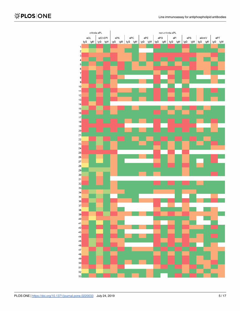
aPL in the various assays is given in Fig 1. By LIA, 42 (79.2%) and 10 (18.9%) patients tested
positive for aCL IgG and IgM, respectively, and 43 (81.1%) and 16 (30.2%) positive for
aβ2-GPI IgG and IgM, respectively. With respect to non-criteria aPL, high positivity rates were
Line immunoassay for antiphospholipid antibodies
PLOS ONE | https://doi.org/10.1371/journal.pone.0220033 July 24, 2019 4 / 17
Line immunoassay for antiphospholipid antibodies
PLOS ONE | https://doi.org/10.1371/journal.pone.0220033 July 24, 2019 5 / 17
found in the patient group for aPA IgG, aPS IgG, aPG IgG, and aPI IgG (n = 47 (88.7%), 43
(81.1%), 38 (71.2%), and 35 (66.0%), respectively), intermediate positivity rates for aPA IgM,
aPT IgG, and aPS IgM (n = 25 (47.2%), 20 (37.7%), and 17 (32.1%), respectively), and low posi-
tivity rates for aPG and aPI IgM, aAnnV IgG and IgM, and aPT IgM (n = 11 (20.8%), 11
(20.8%), 4 (7.5%), 4 (7.5%), and 4 (7.5%), respectively). No patient exhibited a positive reactiv-
ity for aPC and aPE IgG or IgM.
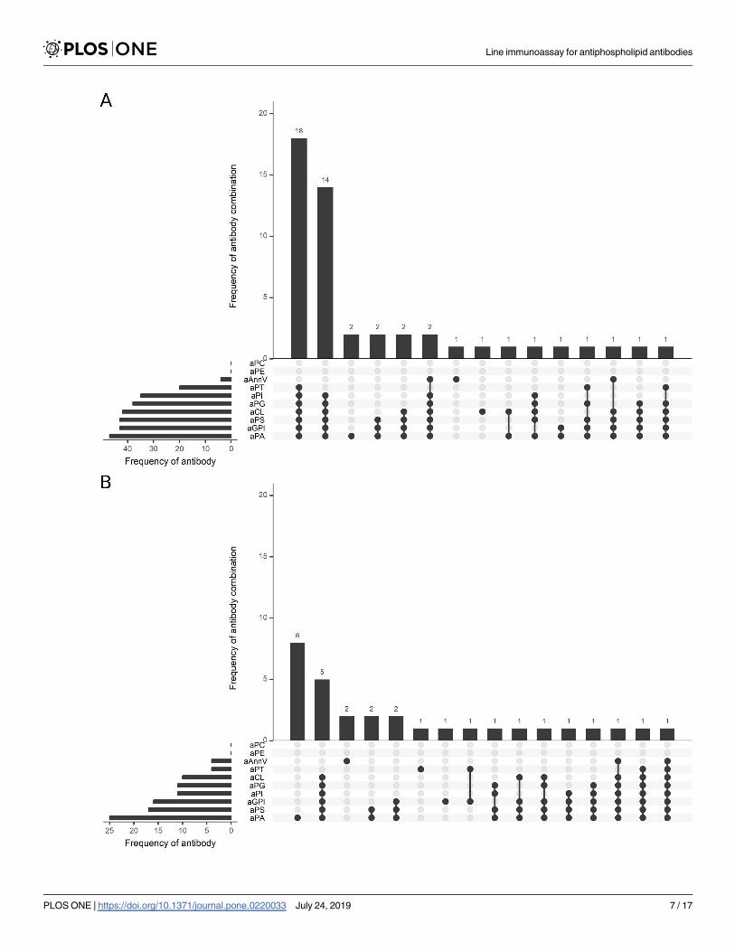
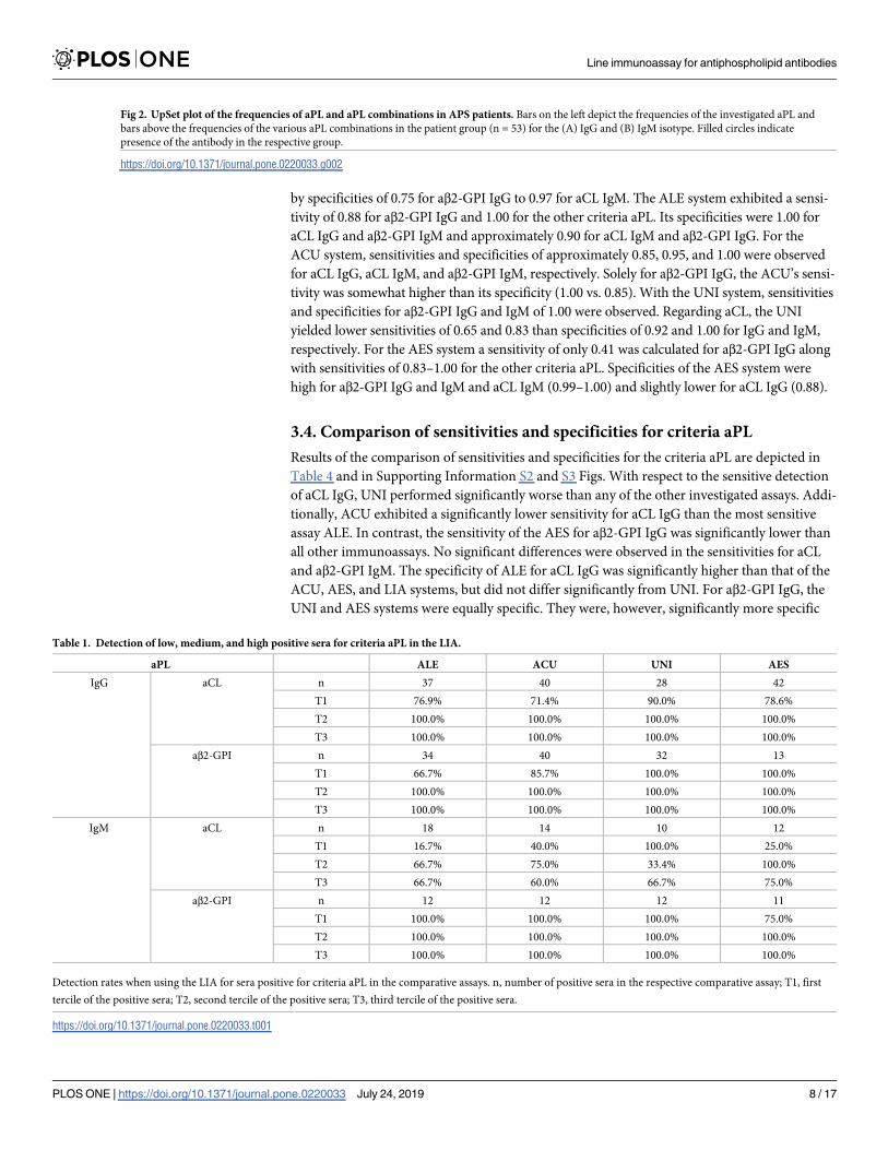
The aPL IgG combination found most frequently in the patient group was a concurrent
positivity for criteria aPL (aCL and aβ2-GPI) and non-criteria aPL against anionic PLs (aPA,
aPS, aPG, and aPI) with or without additional aPT (n = 18 (34.0%) and 14 (26.4%), respec-
tively). Regarding IgM, the combination of positive criteria aPL and positive non-criteria aPL
against anionic PL was observed frequently, too (n = 5 (9.4%)). The most frequent pattern for
this isotype, however, was single positivity for aPA (n = 8 (15.1%)). All other observed aPL
combinations were detected only in a minority of samples. An overview on the frequencies of
the investigated aPL and their respective combinations is given in Fig 2.
Controls did not measure positive for aCL IgG and IgM and aβ2-GPI IgM. Three (8.8%)
control sera were positive for aβ2-GPI IgG. When investigating the controls for non-criteria
aPL, 4 (11.8%) positive reactions were found for aPT IgG, 3 (8.8%) for aAnnV IgM, 2 (5.9%)
for aPA IgG, aPI IgG, and aPT IgM and 1 (2.9%) for aAnnV IgG and aPA IgM. Controls did
not show positive staining for any of the other tested aPL.
3.2. Detection of low, medium, and high positive aPL sera in the LIA
Data on the detection rates for criteria aPL employing the LIA for sera with low, medium, and
high positive titers in the comparative assays are given in Table 1. For aCL IgG and aβ2-GPI
IgG and IgM 66.7–100.0% of the sera in the lowest terciles of the comparative assays were also
found positive for the respective antibody in the LIA. Moreover, all sera located in the middle
and highest terciles of the comparative assays for the aforementioned autoantibodies displayed
positive reactions for the respective aPL in the LIA. With respect to aCL IgM, however, positiv-
ity rates in the LIA exhibited higher heterogeneity ranging from 16.7 to 100.0%, from 33.4 to
100.0%, and from 60.0 to 75.0% for sera in the first, second, and third terciles of the compara-
tive assays, respectively.
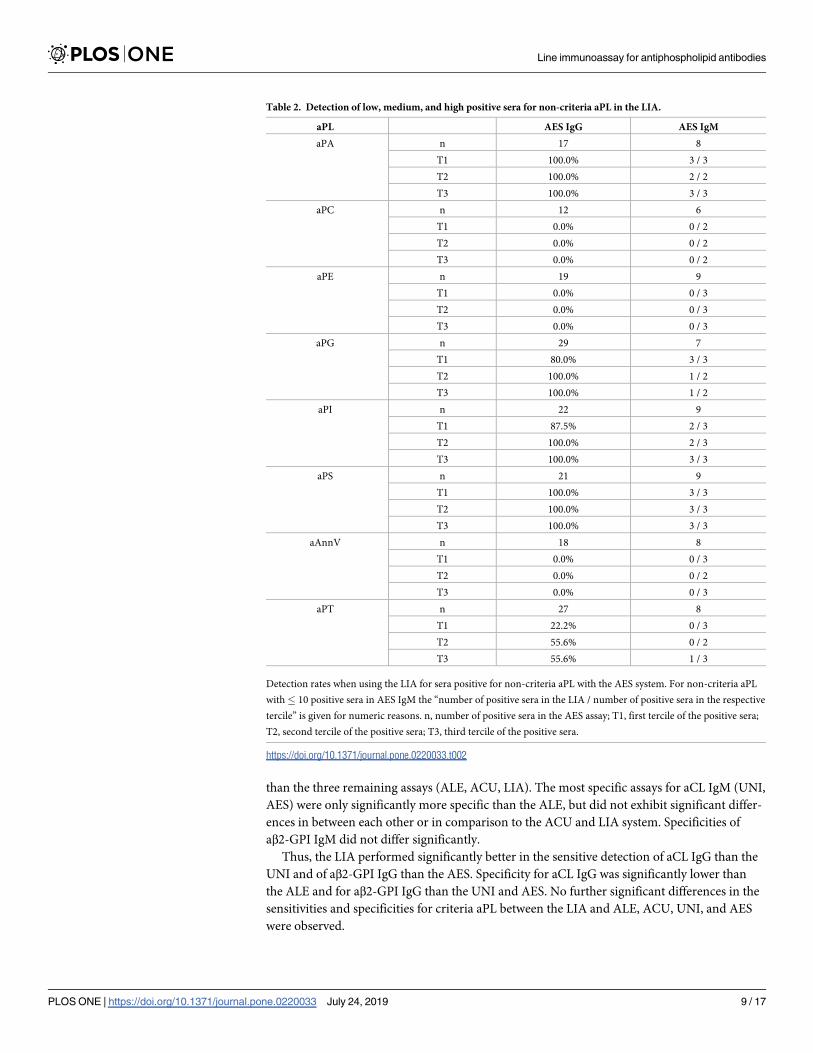
Considering non-criteria aPL (Table 2), the LIA correctly identified all sera positive for aPA
and aPS IgG and IgM in the AES system regardless of their respective terciles. Positivity rates
of the LIA for aPG, aPI, and aPT IgG as compared to AES increased from first to third tercile
and were generally lower for aPT IgG (range: 22.2–55,6%) than for aPG and aPI (range: 80.0%
- 100.0%). The same effect seems also to hold true for aPG, aPI, and aPT IgM. However, sound
conclusions are impaired by the low number of positive sera here. Finally, none of the sera pos-
itive for aPC, aPE, and aAnnV IgG or IgM in the AES system exhibited a positive reaction in
the respective field of the LIA.
3.3. Sensitivities and specificities for criteria aPL
Data on the diagnostic performances for criteria aPL are given in Table 3. Sensitivities of the
LIA ranged from 0.67 for aCL IgM to 1.00 for aβ2-GPI IgG and IgM and were accompanied
Fig 1. Heat map: Positivity of APS patients for criteria and non-criteria aPL in the various assays. Individual APS
sera (n = 53) on the left. Investigated aPL on the top. Criteria aPL: dark green, no assay positive; bright green, one assay
positive; yellow, two assays positive; bright orange, three assays positive; dark orange, four assays positive; red: all five
assays positive (assays: ALE, ACU, UNI, AES, LIA). Non-criteria aPL: green, no assay positive; orange, one assay
positive; red, both assays positive; white, not determined due to lack of sample material (assays: AES, LIA).
https://doi.org/10.1371/journal.pone.0220033.g001
Line immunoassay for antiphospholipid antibodies
PLOS ONE | https://doi.org/10.1371/journal.pone.0220033 July 24, 2019 6 / 17
Line immunoassay for antiphospholipid antibodies
PLOS ONE | https://doi.org/10.1371/journal.pone.0220033 July 24, 2019 7 / 17
by specificities of 0.75 for aβ2-GPI IgG to 0.97 for aCL IgM. The ALE system exhibited a sensi-
tivity of 0.88 for aβ2-GPI IgG and 1.00 for the other criteria aPL. Its specificities were 1.00 for
aCL IgG and aβ2-GPI IgM and approximately 0.90 for aCL IgM and aβ2-GPI IgG. For the
ACU system, sensitivities and specificities of approximately 0.85, 0.95, and 1.00 were observed
for aCL IgG, aCL IgM, and aβ2-GPI IgM, respectively. Solely for aβ2-GPI IgG, the ACU’s sensi-
tivity was somewhat higher than its specificity (1.00 vs. 0.85). With the UNI system, sensitivities
and specificities for aβ2-GPI IgG and IgM of 1.00 were observed. Regarding aCL, the UNI
yielded lower sensitivities of 0.65 and 0.83 than specificities of 0.92 and 1.00 for IgG and IgM,
respectively. For the AES system a sensitivity of only 0.41 was calculated for aβ2-GPI IgG along
with sensitivities of 0.83–1.00 for the other criteria aPL. Specificities of the AES system were
high for aβ2-GPI IgG and IgM and aCL IgM (0.99–1.00) and slightly lower for aCL IgG (0.88).
3.4. Comparison of sensitivities and specificities for criteria aPL
Results of the comparison of sensitivities and specificities for the criteria aPL are depicted in
Table 4 and in Supporting Information S2 and S3 Figs. With respect to the sensitive detection
of aCL IgG, UNI performed significantly worse than any of the other investigated assays. Addi-
tionally, ACU exhibited a significantly lower sensitivity for aCL IgG than the most sensitive
assay ALE. In contrast, the sensitivity of the AES for aβ2-GPI IgG was significantly lower than
all other immunoassays. No significant differences were observed in the sensitivities for aCL
and aβ2-GPI IgM. The specificity of ALE for aCL IgG was significantly higher than that of the
ACU, AES, and LIA systems, but did not differ significantly from UNI. For aβ2-GPI IgG, the
UNI and AES systems were equally specific. They were, however, significantly more specific
Fig 2. UpSet plot of the frequencies of aPL and aPL combinations in APS patients. Bars on the left depict the frequencies of the investigated aPL and
bars above the frequencies of the various aPL combinations in the patient group (n = 53) for the (A) IgG and (B) IgM isotype. Filled circles indicate
presence of the antibody in the respective group.
https://doi.org/10.1371/journal.pone.0220033.g002
Table 1. Detection of low, medium, and high positive sera for criteria aPL in the LIA.
aPL ALE ACU UNI AES
IgG aCL n 37 40 28 42
T1 76.9% 71.4% 90.0% 78.6%
T2 100.0% 100.0% 100.0% 100.0%
T3 100.0% 100.0% 100.0% 100.0%
aβ2-GPI n 34 40 32 13
T1 66.7% 85.7% 100.0% 100.0%
T2 100.0% 100.0% 100.0% 100.0%
T3 100.0% 100.0% 100.0% 100.0%
IgM aCL n 18 14 10 12
T1 16.7% 40.0% 100.0% 25.0%
T2 66.7% 75.0% 33.4% 100.0%
T3 66.7% 60.0% 66.7% 75.0%
aβ2-GPI n 12 12 12 11
T1 100.0% 100.0% 100.0% 75.0%
T2 100.0% 100.0% 100.0% 100.0%
T3 100.0% 100.0% 100.0% 100.0%
Detection rates when using the LIA for sera positive for criteria aPL in the comparative assays. n, number of positive sera in the respective comparative assay; T1, first
tercile of the positive sera; T2, second tercile of the positive sera; T3, third tercile of the positive sera.
https://doi.org/10.1371/journal.pone.0220033.t001
Line immunoassay for antiphospholipid antibodies
PLOS ONE | https://doi.org/10.1371/journal.pone.0220033 July 24, 2019 8 / 17
than the three remaining assays (ALE, ACU, LIA). The most specific assays for aCL IgM (UNI,
AES) were only significantly more specific than the ALE, but did not exhibit significant differ-
ences in between each other or in comparison to the ACU and LIA system. Specificities of
aβ2-GPI IgM did not differ significantly.
Thus, the LIA performed significantly better in the sensitive detection of aCL IgG than the
UNI and of aβ2-GPI IgG than the AES. Specificity for aCL IgG was significantly lower than
the ALE and for aβ2-GPI IgG than the UNI and AES. No further significant differences in the
sensitivities and specificities for criteria aPL between the LIA and ALE, ACU, UNI, and AES
were observed.
Table 2. Detection of low, medium, and high positive sera for non-criteria aPL in the LIA.
aPL AES IgG AES IgM
aPA n 17 8
T1 100.0% 3 / 3
T2 100.0% 2 / 2
T3 100.0% 3 / 3
aPC n 12 6
T1 0.0% 0 / 2
T2 0.0% 0 / 2
T3 0.0% 0 / 2
aPE n 19 9
T1 0.0% 0 / 3
T2 0.0% 0 / 3
T3 0.0% 0 / 3
aPG n 29 7
T1 80.0% 3 / 3
T2 100.0% 1 / 2
T3 100.0% 1 / 2
aPI n 22 9
T1 87.5% 2 / 3
T2 100.0% 2 / 3
T3 100.0% 3 / 3
aPS n 21 9
T1 100.0% 3 / 3
T2 100.0% 3 / 3
T3 100.0% 3 / 3
aAnnV n 18 8
T1 0.0% 0 / 3
T2 0.0% 0 / 2
T3 0.0% 0 / 3
aPT n 27 8
T1 22.2% 0 / 3
T2 55.6% 0 / 2
T3 55.6% 1 / 3
Detection rates when using the LIA for sera positive for non-criteria aPL with the AES system. For non-criteria aPL
with� 10 positive sera in AES IgM the “number of positive sera in the LIA / number of positive sera in the respective
tercile” is given for numeric reasons. n, number of positive sera in the AES assay; T1, first tercile of the positive sera;
T2, second tercile of the positive sera; T3, third tercile of the positive sera.
https://doi.org/10.1371/journal.pone.0220033.t002
Line immunoassay for antiphospholipid antibodies
PLOS ONE | https://doi.org/10.1371/journal.pone.0220033 July 24, 2019 9 / 17
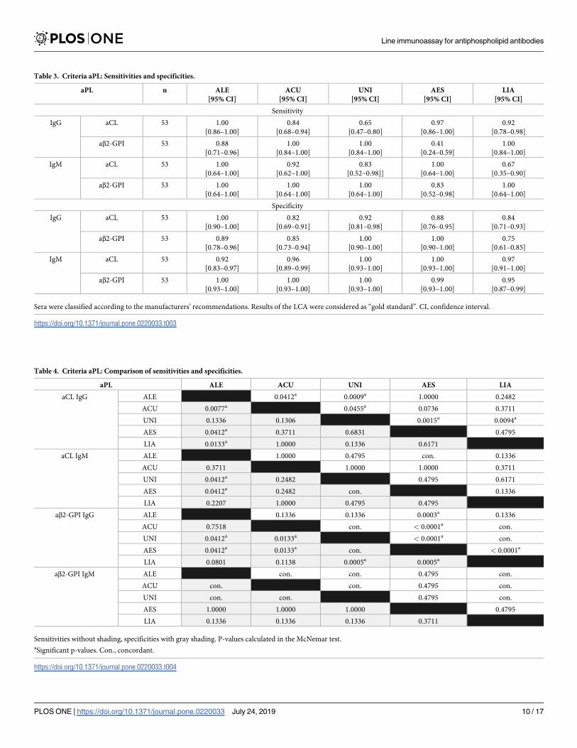
Table 3. Criteria aPL: Sensitivities and specificities.
aPL n ALE
[95% CI]
ACU
[95% CI]
UNI
[95% CI]
AES
[95% CI]
LIA
[95% CI]
Sensitivity
IgG aCL 53 1.00
[0.86–1.00]
0.84
[0.68–0.94]
0.65
[0.47–0.80]
0.97
[0.86–1.00]
0.92
[0.78–0.98]
aβ2-GPI 53 0.88
[0.71–0.96]
1.00
[0.84–1.00]
1.00
[0.84–1.00]
0.41
[0.24–0.59]
1.00
[0.84–1.00]
IgM aCL 53 1.00
[0.64–1.00]
0.92
[0.62–1.00]
0.83
[0.52–0.98]]
1.00
[0.64–1.00]
0.67
[0.35–0.90]
aβ2-GPI 53 1.00
[0.64–1.00]
1.00
[0.64–1.00]
1.00
[0.64–1.00]
0.83
[0.52–0.98]
1.00
[0.64–1.00]
Specificity
IgG aCL 53 1.00
[0.90–1.00]
0.82
[0.69–0.91]
0.92
[0.81–0.98]
0.88
[0.76–0.95]
0.84
[0.71–0.93]
aβ2-GPI 53 0.89
[0.78–0.96]
0.85
[0.73–0.94]
1.00
[0.90–1.00]
1.00
[0.90–1.00]
0.75
[0.61–0.85]
IgM aCL 53 0.92
[0.83–0.97]
0.96
[0.89–0.99]
1.00
[0.93–1.00]
1.00
[0.93–1.00]
0.97
[0.91–1.00]
aβ2-GPI 53 1.00
[0.93–1.00]
1.00
[0.93–1.00]
1.00
[0.93–1.00]
0.99
[0.93–1.00]
0.95
[0.87–0.99]
Sera were classified according to the manufacturers’ recommendations. Results of the LCA were considered as “gold standard”. CI, confidence interval.
https://doi.org/10.1371/journal.pone.0220033.t003
Table 4. Criteria aPL: Comparison of sensitivities and specificities.
aPL ALE ACU UNI AES LIA
aCL IgG ALE 0.0412a 0.0009a 1.0000 0.2482
ACU 0.0077a 0.0455a 0.0736 0.3711
UNI 0.1336 0.1306 0.0015a 0.0094a
AES 0.0412a 0.3711 0.6831 0.4795
LIA 0.0133a 1.0000 0.1336 0.6171
aCL IgM ALE 1.0000 0.4795 con. 0.1336
ACU 0.3711 1.0000 1.0000 0.3711
UNI 0.0412a 0.2482 0.4795 0.6171
AES 0.0412a 0.2482 con. 0.1336
LIA 0.2207 1.0000 0.4795 0.4795
aβ2-GPI IgG ALE 0.1336 0.1336 0.0003a 0.1336
ACU 0.7518 con. < 0.0001a con.
UNI 0.0412a 0.0133a < 0.0001a con.
AES 0.0412a 0.0133a con. < 0.0001a
LIA 0.0801 0.1138 0.0005a 0.0005a
aβ2-GPI IgM ALE con. con. 0.4795 con.
ACU con. con. 0.4795 con.
UNI con. con. 0.4795 con.
AES 1.0000 1.0000 1.0000 0.4795
LIA 0.1336 0.1336 0.1336 0.3711
Sensitivities without shading, specificities with gray shading. P-values calculated in the McNemar test.aSignificant p-values. Con., concordant.
https://doi.org/10.1371/journal.pone.0220033.t004
Line immunoassay for antiphospholipid antibodies
PLOS ONE | https://doi.org/10.1371/journal.pone.0220033 July 24, 2019 10 / 17
3.5. Inter-rater agreement of LIA and AES for non-criteria aPL
With Cohen’s κ of 0.61–0.75, inter-rater agreement of LIA and AES for aPG and aPI IgG and
IgM can be considered as good. For aPS, agreement was good for the IgM and moderate for
the IgG isotype (Cohen’s κ 0.75 vs. 0.53, respectively). Similarly, a slightly lower Cohen’s κ of
0.34 indicating fair agreement was found for aPA IgG as compared to a Cohen’s κ of 0.43 for
the IgM isotype suggesting moderate agreement. The reverse pattern was observed for aPT
with Cohen’s κ 0.29 for IgG (fair agreement) vs. 0.10 for IgM (poor agreement). Cohen’s κfrom -0.10 to 0.00 for aPC, aPE, and aAnnV IgG and IgM pointed to poor agreements. A very
good agreement could not be demonstrated for any non-criterion aPL (Supporting Informa-
tion S2 Table).
In consideration of the only modest agreement of both assay systems, application of β2-GPI
as cofactor in the AES aPA, aPC, aPE, aPG, aPI, and aPS assays was further investigated. In the
AES system, measured levels of these non-criteria aPL and the respective aβ2-GPI isotype
exhibit a noteworthy positive correlation. With respect to the antibody isotype, the correlation
seems to be more distinct for IgM than for IgG (Supporting Information S4 Fig).
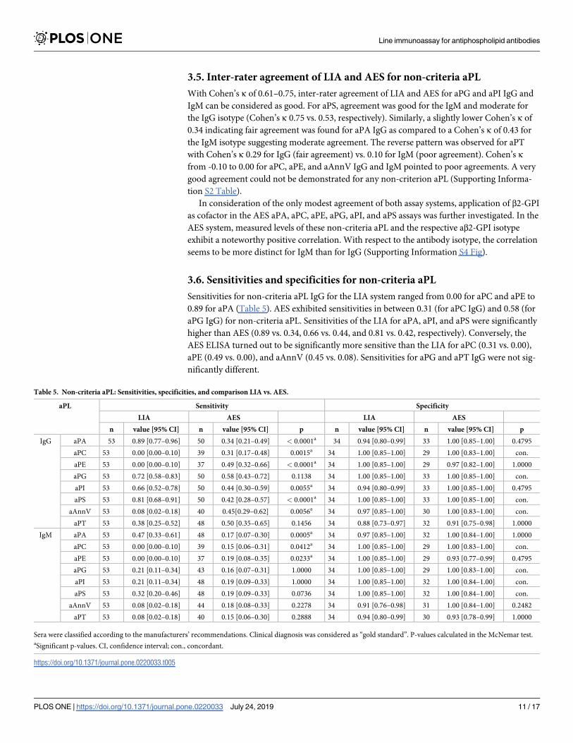
3.6. Sensitivities and specificities for non-criteria aPL
Sensitivities for non-criteria aPL IgG for the LIA system ranged from 0.00 for aPC and aPE to
0.89 for aPA (Table 5). AES exhibited sensitivities in between 0.31 (for aPC IgG) and 0.58 (for
aPG IgG) for non-criteria aPL. Sensitivities of the LIA for aPA, aPI, and aPS were significantly
higher than AES (0.89 vs. 0.34, 0.66 vs. 0.44, and 0.81 vs. 0.42, respectively). Conversely, the
AES ELISA turned out to be significantly more sensitive than the LIA for aPC (0.31 vs. 0.00),
aPE (0.49 vs. 0.00), and aAnnV (0.45 vs. 0.08). Sensitivities for aPG and aPT IgG were not sig-
nificantly different.
Table 5. Non-criteria aPL: Sensitivities, specificities, and comparison LIA vs. AES.
aPL Sensitivity Specificity
LIA AES LIA AES
n value [95% CI] n value [95% CI] p n value [95% CI] n value [95% CI] p
IgG aPA 53 0.89 [0.77–0.96] 50 0.34 [0.21–0.49] < 0.0001a 34 0.94 [0.80–0.99] 33 1.00 [0.85–1.00] 0.4795
aPC 53 0.00 [0.00–0.10] 39 0.31 [0.17–0.48] 0.0015a 34 1.00 [0.85–1.00] 29 1.00 [0.83–1.00] con.
aPE 53 0.00 [0.00–0.10] 37 0.49 [0.32–0.66] < 0.0001a 34 1.00 [0.85–1.00] 29 0.97 [0.82–1.00] 1.0000
aPG 53 0.72 [0.58–0.83] 50 0.58 [0.43–0.72] 0.1138 34 1.00 [0.85–1.00] 33 1.00 [0.85–1.00] con.
aPI 53 0.66 [0.52–0.78] 50 0.44 [0.30–0.59] 0.0055a 34 0.94 [0.80–0.99] 33 1.00 [0.85–1.00] 0.4795
aPS 53 0.81 [0.68–0.91] 50 0.42 [0.28–0.57] < 0.0001a 34 1.00 [0.85–1.00] 33 1.00 [0.85–1.00] con.
aAnnV 53 0.08 [0.02–0.18] 40 0.45[0.29–0.62] 0.0056a 34 0.97 [0.85–1.00] 30 1.00 [0.83–1.00] con.
aPT 53 0.38 [0.25–0.52] 48 0.50 [0.35–0.65] 0.1456 34 0.88 [0.73–0.97] 32 0.91 [0.75–0.98] 1.0000
IgM aPA 53 0.47 [0.33–0.61] 48 0.17 [0.07–0.30] 0.0005a 34 0.97 [0.85–1.00] 32 1.00 [0.84–1.00] 1.0000
aPC 53 0.00 [0.00–0.10] 39 0.15 [0.06–0.31] 0.0412a 34 1.00 [0.85–1.00] 29 1.00 [0.83–1.00] con.
aPE 53 0.00 [0.00–0.10] 37 0.19 [0.08–0.35] 0.0233a 34 1.00 [0.85–1.00] 29 0.93 [0.77–0.99] 0.4795
aPG 53 0.21 [0.11–0.34] 43 0.16 [0.07–0.31] 1.0000 34 1.00 [0.85–1.00] 29 1.00 [0.83–1.00] con.
aPI 53 0.21 [0.11–0.34] 48 0.19 [0.09–0.33] 1.0000 34 1.00 [0.85–1.00] 32 1.00 [0.84–1.00] con.
aPS 53 0.32 [0.20–0.46] 48 0.19 [0.09–0.33] 0.0736 34 1.00 [0.85–1.00] 32 1.00 [0.84–1.00] con.
aAnnV 53 0.08 [0.02–0.18] 44 0.18 [0.08–0.33] 0.2278 34 0.91 [0.76–0.98] 31 1.00 [0.84–1.00] 0.2482
aPT 53 0.08 [0.02–0.18] 40 0.15 [0.06–0.30] 0.2888 34 0.94 [0.80–0.99] 30 0.93 [0.78–0.99] 1.0000
Sera were classified according to the manufacturers’ recommendations. Clinical diagnosis was considered as “gold standard”. P-values calculated in the McNemar test.aSignificant p-values. CI, confidence interval; con., concordant.
https://doi.org/10.1371/journal.pone.0220033.t005
Line immunoassay for antiphospholipid antibodies
PLOS ONE | https://doi.org/10.1371/journal.pone.0220033 July 24, 2019 11 / 17
Amongst the non-criteria aPL IgM, sensitivities were again lowest for aPC and aPE (0.00)
and highest for aPA (0.47) with the LIA. Sensitivities for the AES varied less and ranged from
0.15 for aPC and aPT IgM to 0.19 for aPE, aPI, and aPS IgM. The LIA was significantly more
sensitive than AES for aPA IgM (0.47 vs. 0.17) and significantly less sensitive for aPC and aPE
IgM (0.00 vs. 0.15 and 0.00 vs. 0.19, respectively). No further significant differences between
the two investigated systems in the sensitivities for non-criteria aPL IgM were observed.
Specificities for the non-criteria aPL were� 0.88 and� 0.91 with the LIA and� 0.91
and� 0.93 with AES for the IgG and IgM isotype, respectively. Differences of specificities
were not significant for any non-criteria aPL.
3.7. Clinical value of non-criteria aPL
Eleven (20.8%) out of the 53 APS-patients were positive for LA, but negative for aCL or
aβ2-GPI IgG or IgM as concluded from LCA. When using the LIA, 9 (81.8%) of these patients
exhibited positivity for any of the non-criteria aPL. All 9 patients were positive for aPA IgG. In
addition, aPS IgG was found in 6, aPG IgG in 5, aPI IgG in 4, and aPT IgM in 2 samples and
aAnnV IgG, aPT IgG, and aPA IgM in one sample each. With the AES assays, 6 (54.5%) of the
11 patients were positive for non-criteria aPL: in 4 samples solely aPT IgG or IgM was
detected, in one sample aPT IgG and aPE IgM, and in one sample a combination of aPG IgG,
aPI IgG, aPS IgG, and aAnnV IgG. All 6 samples positive for non-criteria aPL with the AES
system were also found positive for non-criteria aPL in the LIA.
4. Discussion
The diagnostic performance of a novel LIA for the detection of aPL was investigated. Sensitivi-
ties and specificities of the LIA for criteria aPL are well comparable to established immunoas-
says. With respect to non-criteria aPL, the LIA offers advantages in the sensitive detection of
aPL against anionic PLs whereas the classical ELISA exhibits strengths in the sensitive detec-
tion of aPL against neutral PLs.
Sensitivity of the LIA for aCL and aβ2-GPI IgG was significantly better than the worst and
comparable to the three other established assays. Considering specificity for aCL and aβ2-GPI
IgG, the LIA performed worse than the best and two best, but equal to three and two of the
comparative assays, respectively. Moreover, with respect to aCL and aβ2-GPI IgM, no signifi-
cant differences in sensitivity and specificity between the LIA and the established immunoas-
says were observed. As such, the diagnostic performance of the LIA for criteria aPL can be
considered equal to the investigated established immunoassays.
In contrast, considerable differences in the results for non-criteria aPL obtained by LIA and
an established ELISA system were demonstrated. This is illustrated by Cohen’s κ indicating
very good agreement for none and good agreement for only 5, but poor agreement for 7 out of
the 16 analytes. Differences are based on varying sensitivities for non-criteria aPL. Sensitivity
of the LIA was significantly better for aPA, aPI and aPS IgG, and aPA IgM and approached sig-
nificance for aPS IgM. The ELISA, however, demonstrated a significantly higher sensitivity for
aPC and aPE IgG and IgM. Phosphatidic acid, phosphatidyl-serine, -glycerol, and -inositol
carry a negative net charge at physiological pH. Phosphatidyl-choline and–ethanolamine in
contrast constitute neutral molecules at pH 7.4. Thus, the LIA exhibits higher sensitivities for
antibodies against anionic and the ELISA for antibodies against neutral PL. Since the ELISAs
contained β2-GPI as cofactor apart from the respective PL as autoantigenic target, positive
serum reactivity in these assays could be due to aβ2-GPI, too. With the AES system, aPA, aPC,
aPE, aPG, aPI, and aPS indeed exhibited a positive correlation with aβ2-GPI in our cohort.
However, it remains to be shown, whether the aPL reactivity in the respective ELISAs is based
Line immunoassay for antiphospholipid antibodies
PLOS ONE | https://doi.org/10.1371/journal.pone.0220033 July 24, 2019 12 / 17
on β2-GPI binding alone. Thus, further studies are warranted to investigate whether this phe-
nomenon can be explained by actual analytical interference or if the respective aPL arise con-
currently for biological reasons.
The diagnostic performance of LIAs for aPL detection has been evaluated in two major
studies: Egerer et al. [29] applied a shorter predecessor version and Roggenbuck et al. [27] the
full-length LIA also investigated here. Our data not only confirm the comparable diagnostic
performance of LIA and ELISA with respect to the criteria aPL, but also substantiate the find-
ings of Egerer and Roggenbuck in several aspects. First, processed strips were read out manu-
ally (“eyeballing”) in the present study and not densitometrically with scanner and evaluation
software as in the previous studies. The software, however, is not used in each routine labora-
tory. As such, our results are closer to the actual scenario in a diagnostic laboratory. Second,
the aforementioned studies compared the determination of the criteria aPL via LIA only to a
single ELISA system, which had been produced by the same manufacturer as the LIA. In our
study, four comparative assays from a variety of manufacturers were included. And third, sen-
sitivities and specificities for criteria aPL were calculated with respect to a LCA-derived gold
standard. We introduced this approach recently [22] and consider it beneficial when evaluat-
ing agreement of aPL assays. A definitive diagnosis of APS requires positive laboratory criteria.
Establishing the clinical diagnosis as gold standard is therefore inherently biased by the assays
applied during the diagnostic work-up. The LCA-driven approach, however, does not require
knowledge about the diagnosis: it merely evaluates whether the assay investigated agrees with
the “consensus” opinion of already established assays regarding antibody-positivity or–
negativity.
To the best of our knowledge, our work is the first study to compare non-criteria aPL detec-
tion of the LIA with established ELISAs thus revealing different strengths of the various assay
surfaces. A LCA-based approached would clearly have been desirable here as well. A compre-
hensive investigation of all non-criteria aPL detected in the LIA, however, was only possible
with one of the four assay systems that had been applied for the evaluation of the criteria aPL.
We, therefore, choose to use the clinical diagnosis as the gold standard. One may argue that
this approach is less critical for non-criteria aPL as they are not required for the diagnosis of
APS.
Characteristics of patient and control group entail some limitations of our study. First of
all, the limited sample size may impair generalizability of our data. Furthermore, a high per-
centage of double and triple positive subjects in our patient cohort may have increased the sen-
sitivities for criteria aPL. And finally, the lack of disease controls may have yielded rather high
specificities. As such, selection of patient and control groups may explain the mostly higher
sensitivities and specificities in our study as compared to previous ones. From an analytical
point of view, the time interval between the “old” ELISA measurements of the criteria aPL and
the “new” measurements with the LIA and of non-criteria aPL via ELISA may introduce some
bias into our work. If at all, this bias would be to the disadvantage of the LIA and should not
result in a false-good evaluation of this assay.
Nevertheless, the observed high sensitivity of the LIA for aPL against anionic PLs sheds
light on the optimal solid phases for testing of non-criteria aPL. The hydrophobic membrane
of the LIA seems to be more suited for the detection of aPL against anionic PLs, whereas the
polystyrene plastics of the ELISA plate detects neutral PLs with higher sensitivity. Of note,
β2-GPI reactivity in these ELISAs cannot be excluded. Keeping in mind, that the most relevant
autoantigenic epitopes for aPL are located on β2-GPI, our data support the hypothesis previ-
ously proposed [27, 30]: the porous hydrophobic polyvinylidene fluoride membrane of the
LIA incorporates the hydrophobic tails of the PLs; only their hydrophilic and in part anionic
moieties are exposed allowing optimal antigen presentation to aPL. The negative net charge of
Line immunoassay for antiphospholipid antibodies
PLOS ONE | https://doi.org/10.1371/journal.pone.0220033 July 24, 2019 13 / 17
anionic PL favors binding of β2-GPI domain V resulting in optimal presentation of the immu-
nodominant epitopes in domain I. In contrast, neutral PL do not bind domain V of β2-GPI in
the LIA. Thus, the planar ELISA solid phase with immobilization of PLs along with β2-GPI as
cofactor in random orientation may offer a more advantageous surface for aPL binding in this
reaction environment. As such, tailoring the solid-phase to the respective PL rather than a
“one size fits all” approach may be crucial to improve assay performance.
The LIA proved to be a suitable tool for detection of criteria and presumably also of non-
criteria aPL. As compared to classical ELISAs, the LIA offers several interesting features: in
one single run a comprehensive aPL profile can be generated considerably quicker and with
less reagent costs and effort as compared to ELISAs as one LIA test strip replaces 10 single ELI-
SAs; it is suited for low as well as for high sample throughput and requires only a basic labora-
tory equipment. Our data demonstrate that the majority of APS patients with LA but without
criteria aPL exhibits positivity for non-criteria aPL–thereby pointing to possible diagnostic
benefits of the determination of non-criteria aPL. Of note, strength of positivity for non-crite-
ria aPL as well as the time course of titers were not considered in this study. As such, better
availability and convenient analysis of aPL profiles may foster research in this area and help to
better elucidate the clinical significance of non-criteria aPL.
In summary, our work demonstrates that the LIA exhibits a diagnostic performance com-
parable to established immunoassays for criteria aPL and offers advantages with respect to the
detection of non-criteria aPL against anionic PL. It constitutes a valuable tool for aPL profiling
in a clinical as well as in a research setting. Beyond that, our data will serve as a guide for assay
developers when designing the solid-phase for detection of non-criteria aPL.
Supporting information
S1 Fig. Illustration of the statistical approach. For criteria aPL, the diagnostic performance
was calculated against a “gold standard” derived from the results of the ALE, ACU, UNI, and
AES systems via LCA. In contrast, the clinical diagnosis served as the gold standard to deter-
mine sensitivities and specificities for non-criteria aPL.
(PPTX)
S2 Fig. Comparison of the sensitivities for criteria aPL. Sensitivities of the investigated
assays for aCL IgG (A), aCL IgM (B), aβ2-GPI IgG (C), and aβ2-GPI IgM (D). Error bars
denote a 95% confidence interval. Significant differences are marked with horizontal square
brackets (�: 0.05 > p� 0.01; ��: 0.01> p� 0.001; ���: p< 0.001).
(TIF)
S3 Fig. Comparison of the specificities for criteria aPL. Specificities of the investigated assays
for aCL IgG (A), aCL IgM (B), aβ2-GPI IgG (C), and aβ2-GPI IgM (D). Error bars denote a
95% confidence interval. Significant differences are marked with horizontal square brackets (�:
0.05> p� 0.01; ��: 0.01> p� 0.001; ���: p< 0.001).
(TIF)
S4 Fig. Association of AES non-criteria aPL and AES aβ2-GPI. Levels of aPA, aPC, aPE,
aPG, aPI, and aPS as a function of aβ2-GPI as measured with the AES system for IgG (A) and
IgM (B) for all samples included in the study. AES aPE IgG: samples > 300 U/ml were not fur-
ther diluted; AES aPG IgG and AES aPI IgG: samples > 315 U/ml were not further diluted.
(TIFF)
S1 Table. Reference ranges of the comparative immunoassay systems.
(DOCX)
Line immunoassay for antiphospholipid antibodies
PLOS ONE | https://doi.org/10.1371/journal.pone.0220033 July 24, 2019 14 / 17
S2 Table. Non-criteria aPL: Inter-rater agreement LIA vs. AES.
(DOCX)
S1 File. Raw Data. Data underlying the findings described in the manuscript.
(CSV)
Acknowledgments
We thank Anita Schreiegg for excellent technical assistance and Dr. Evangeline Thaler for
carefully reviewing the manuscript.
Author Contributions
Conceptualization: Markus A. Thaler, Andreas Bietenbeck, Udo Steigerwald, Peter B. Luppa.
Data curation: Markus A. Thaler, Peter B. Luppa.
Formal analysis: Markus A. Thaler, Andreas Bietenbeck, Peter B. Luppa.
Investigation: Markus A. Thaler, Andreas Bietenbeck.
Methodology: Markus A. Thaler, Andreas Bietenbeck, Peter B. Luppa.
Resources: Udo Steigerwald, Thomas Buttner, Edelgard Lindhoff-Last, Dirk Roggenbuck,
Peter B. Luppa.
Software: Andreas Bietenbeck.
Supervision: Udo Steigerwald, Peter B. Luppa.
Validation: Markus A. Thaler, Andreas Bietenbeck, Udo Steigerwald, Peter Schierack, Edel-
gard Lindhoff-Last, Peter B. Luppa.
Writing – original draft: Markus A. Thaler.
Writing – review & editing: Andreas Bietenbeck, Udo Steigerwald, Peter Schierack, Edelgard
Lindhoff-Last, Dirk Roggenbuck, Peter B. Luppa.
References1. Hughes GR. Thrombosis, abortion, cerebral disease, and the lupus anticoagulant. Br Med J (Clin Res
Ed). 1983; 287: 1088–1089.
2. Miyakis S, Lockshin MD, Atsumi T, Branch DW, Brey RL, Cervera R, et al. International consensus state-
ment on an update of the classification criteria for definite antiphospholipid syndrome (APS). J Thromb
Haemost. 2006; 4: 295–306. https://doi.org/10.1111/j.1538-7836.2006.01753.x PMID: 16420554
3. Devreese KM, Pierangeli SS, de Laat B, Tripodi A, Atsumi T, Ortel TL, et al. Testing for antiphospholipid
antibodies with solid phase assays: guidance from the SSC of the ISTH. J Thromb Haemost. 2014; 12:
792–795. https://doi.org/10.1111/jth.12537 PMID: 24589091
4. Nayfe R, Uthman I, Aoun J, Saad Aldin E, Merashli M, Khamashta MA. Seronegative antiphospholipid
syndrome. Rheumatology (Oxford). 2013; 52: 1358–1367.
5. Rodriguez-Garcia V, Ioannou Y, Fernandez-Nebro A, Isenberg DA, Giles IP. Examining the prevalence
of non-criteria anti-phospholipid antibodies in patients with anti-phospholipid syndrome: a systematic
review. Rheumatology (Oxford). 2015; 54: 2042–2050.
6. Zohoury N, Bertolaccini ML, Rodriguez-Garcia JL, Shums Z, Ateka-Barrutia O, Sorice M, et al. Closing
the Serological Gap in the Antiphospholipid Syndrome: The Value of "Non-criteria" Antiphospholipid
Antibodies. J Rheumatol. 2017; 44: 1597–1602. https://doi.org/10.3899/jrheum.170044 PMID:
28864642
7. Tebo AE. Antiphospholipid syndrome and the relevance of antibodies to negatively charged phospholip-
ids in diagnostic evaluation. Lupus. 2014; 23: 1313–1316. https://doi.org/10.1177/0961203314544534
PMID: 25228736
Line immunoassay for antiphospholipid antibodies
PLOS ONE | https://doi.org/10.1371/journal.pone.0220033 July 24, 2019 15 / 17

8. Castanon A, Pierre G, Willis R, Harris EN, Papalardo E, Romay-Penabad Z, et al. Performance evalua-
tion and clinical associations of immunoassays that detect antibodies to negatively charged phospholip-
ids other than cardiolipin. Am J Clin Pathol. 2018; 149: 401–411. https://doi.org/10.1093/ajcp/aqy003
PMID: 29547897
9. Bertolaccini ML, Amengual O, Atsumi T, Binder WL, de Laat B, Forastiero R, et al. ’Non-criteria’ aPL
tests: report of a task force and preconference workshop at the 13th International Congress on Antipho-
spholipid Antibodies, Galveston, TX, USA, April 2010. Lupus. 2011; 20: 191–205. https://doi.org/10.
1177/0961203310397082 PMID: 21303836
10. Misasi R, Capozzi A, Longo A, Recalchi S, Lococo E, Alessandri C, et al. "New" antigenic targets and
methodological approaches for refining laboratory diagnosis of antiphospholipid syndrome. J Immunol
Res. 2015; 2015: 858542. https://doi.org/10.1155/2015/858542 PMID: 25874238
11. Roggenbuck D, Egerer K, von Landenberg P, Hiemann R, Feist E, Burmester GR, et al. Antiphospholi-
pid antibody profiling: time for a new technical approach? Autoimmun Rev. 2012; 11: 821–826. https://
doi.org/10.1016/j.autrev.2012.02.016 PMID: 23006529
12. Sciascia S, Amigo MC, Roccatello D, Khamashta M. Diagnosing antiphospholipid syndrome: ’extra-cri-
teria’ manifestations and technical advances. Nat Rev Rheumatol. 2017; 13: 548–560. https://doi.org/
10.1038/nrrheum.2017.124 PMID: 28769114
13. Conrad K, Schneider H, Ziemssen T, Talaska T, Reinhold D, Humbel RL, et al. A new line immunoassay
for the multiparametric detection of antiganglioside autoantibodies in patients with autoimmune periph-
eral neuropathies. Ann N Y Acad Sci. 2007; 1109: 256–264. https://doi.org/10.1196/annals.1398.031
PMID: 17785314
14. Favaloro EJ, Wong RC. Antiphospholipid antibody testing for the antiphospholipid syndrome: a compre-
hensive practical review including a synopsis of challenges and recent guidelines. Pathology. 2014; 46:
481–495. https://doi.org/10.1097/PAT.0000000000000142 PMID: 25158812
15. Devreese KM. Antiphospholipid antibody testing and standardization. Int J Lab Hematol. 2014; 36:
352–363. https://doi.org/10.1111/ijlh.12234 PMID: 24750682
16. Devreese KM. Standardization of antiphospholipid antibody assays. Where do we stand? Lupus. 2012;
21: 718–721. https://doi.org/10.1177/0961203312439335 PMID: 22635211
17. Harris EN, Gharavi AE, Patel SP, Hughes GR. Evaluation of the anti-cardiolipin antibody test: report of
an international workshop held 4 April 1986. Clin Exp Immunol. 1987; 68: 215–222. PMID: 3652514
18. Ichikawa K, Tsutsumi A, Atsumi T, Matsuura E, Kobayashi S, Hughes GR, et al. A chimeric antibody
with the human gamma1 constant region as a putative standard for assays to detect IgG beta2-glyco-
protein I-dependent anticardiolipin and anti-beta2-glycoprotein I antibodies. Arthritis Rheum. 1999; 42:
2461–2470. https://doi.org/10.1002/1529-0131(199911)42:11<2461::AID-ANR25>3.0.CO;2-O PMID:
10555042
19. Ichikawa K, Khamashta MA, Koike T, Matsuura E, Hughes GRV. β2-Glycoprotein i reactivity of mono-
clonal anticardiolipin antibodies from patients with the antiphospholipid syndrome. Arthritis Rheum.
1994; 37: 1453–1461. PMID: 7945470
20. Willis R, Grossi C, Orietta Borghi M, Martos-Sevilla G, Zegers I, Sheldon J, et al. International standards
for IgG and IgM anti-beta2glycoprotein antibody measurement. Lupus. 2014; 23: 1317–1319. https://
doi.org/10.1177/0961203314544535 PMID: 25228737
21. Meroni PL, Biggioggero M, Pierangeli SS, Sheldon J, Zegers I, Borghi MO. Standardization of autoanti-
body testing: a paradigm for serology in rheumatic diseases. Nat Rev Rheumatol. 2014; 10: 35–43.
https://doi.org/10.1038/nrrheum.2013.180 PMID: 24275965
22. Thaler MA, Bietenbeck A, Yin MX, Steigerwald U, Holmes AB, Lindhoff-Last E, et al. Evaluation of anti-
phospholipid antibody assays using latent class analysis to address the lack of a reference standard.
Clin Chem Lab Med. 2016; 54: 1929–1937. https://doi.org/10.1515/cclm-2016-0116 PMID: 27227709
23. Metzger J, von Landenberg P, Kehrel M, Buhl A, Lackner KJ, Luppa PB. Biosensor analysis of beta2-
glycoprotein I-reactive autoantibodies: evidence for isotype-specific binding and differentiation of patho-
genic from infection-induced antibodies. Clin Chem. 2007; 53: 1137–1143. https://doi.org/10.1373/
clinchem.2006.079632 PMID: 17434906
24. Schlichtiger A, Baier C, Yin MX, Holmes AB, Maruyama M, Strasser R, et al. Covalent attachment of
functionalized cardiolipin on a biosensor gold surface allows repetitive measurements of anticardiolipin
antibodies in serum. Anal Bioanal Chem. 2013; 405: 275–285. https://doi.org/10.1007/s00216-012-
6467-8 PMID: 23090649
25. Pengo V, Banzato A, Bison E, Denas G, Padayattil Jose S, Ruffatti A. Antiphospholipid syndrome: criti-
cal analysis of the diagnostic path. Lupus. 2010; 19: 428–431. https://doi.org/10.1177/
0961203309360543 PMID: 20353982
26. Lakos G, Favaloro EJ, Harris EN, Meroni PL, Tincani A, Wong RC, et al. International consensus guide-
lines on anticardiolipin and anti-beta2-glycoprotein I testing: report from the 13th International Congress
Line immunoassay for antiphospholipid antibodies
PLOS ONE | https://doi.org/10.1371/journal.pone.0220033 July 24, 2019 16 / 17
on Antiphospholipid Antibodies. Arthritis Rheum. 2012; 64: 1–10. https://doi.org/10.1002/art.33349
PMID: 21953634
27. Roggenbuck D, Borghi MO, Somma V, Buttner T, Schierack P, Hanack K, et al. Antiphospholipid anti-
bodies detected by line immunoassay differentiate among patients with antiphospholipid syndrome,
with infections and asymptomatic carriers. Arthritis Res Ther. 2016; 18: 111. https://doi.org/10.1186/
s13075-016-1018-x PMID: 27209064
28. Altman DG. Practical statistics for medical research. London: Chapman & Hall; 1991.
29. Egerer K, Roggenbuck D, Buttner T, Lehmann B, Kohn A, von Landenberg P, et al. Single-step autoan-
tibody profiling in antiphospholipid syndrome using a multi-line dot assay. Arthritis Res Ther. 2011; 13:
R118. https://doi.org/10.1186/ar3421 PMID: 21777436
30. Roggenbuck D, Somma V, Schierack P, Borghi MO, Meroni PL. Autoantibody profiling in APS. Lupus.
2014; 23: 1262–1264. https://doi.org/10.1177/0961203314534305 PMID: 25228721
Line immunoassay for antiphospholipid antibodies
PLOS ONE | https://doi.org/10.1371/journal.pone.0220033 July 24, 2019 17 / 17