Embed Size (px)
Citation preview

695GEODIVERSITAS • 2003 • 25 (4) © Publications Scientifiques du Muséum national d’Histoire naturelle, Paris. www.geodiversitas.com
Early Eocene snakes from Kutch, Western India, with a review of the Palaeophiidae
Jean-Claude RAGEDépartement Histoire de la Terre, USM 0203, UMR Muséum-CNRS 5143,
Muséum national d’Histoire naturelle, 8 rue Buffon, F-75231 Paris cedex 05 (France)[email protected]
Sunil BAJPAIDepartment of Earth Sciences, Indian Institute of Technology,
Roorkee 247 667 (India)[email protected]
Johannes G. M. THEWISSENDepartment of Anatomy, Northeastern Ohio Universities College of Medicine,
Rootstown, Ohio 44272 (USA)[email protected]
Brahma N. TIWARIWadia Institute of Himalayan Geology, Mahadeo Singh Road, Dehradun 248 001 (India)
Rage J.-C., Bajpai S., Thewissen J. G. M. & Tiwari B. N. 2003. — Early Eocene snakes fromKutch, Western India, with a review of the Palaeophiidae. Geodiversitas 25 (4) : 695-716.
ABSTRACTThe early Eocene of Panandhro Mine (northwestern India) has produced arich snake fauna largely dominated by palaeophiids. Three families are pres-ent: Palaeophiidae, ?Madtsoiidae or Boidae, and an indeterminate family ofColubroidea. The Palaeophiidae include two species: Pterosphenus kutchensisn. sp., that shows peculiar features, and Pt. biswasi n. sp. They are the earliestrepresentatives of the genus. Madtsoiidae or Boidae are represented by onlytwo specimens that do not permit distinction between these two families. Ifthese fossils belong to the Boidae, then they might be the earliest representa-tives of that family in Asia. The colubroid from this site ranks among the ear-liest Cenozoic representatives of the group. The possibility that it belongs tothe Colubridae cannot be excluded; if this is the case, it would be the earliestknown colubrid. Nearly all specimens belong to Pterosphenus Lucas, 1899that was a highly aquatic genus. It lived in shallow water, probably in marineenvironment close to the coasts and/or in freshwater.
KEY WORDSReptilia,
Serpentes, Colubroidea,
Palaeophiidae, Madtsoiidae,
Boidae, Ypresian,
Eocene, India,
palaeoenvironment, new species.
RÉSUMÉSerpents de l’Éocène inférieur de Kutch, ouest de l’Inde ; revue des Palaeophiidae.L’Éocène inférieur de Panandhro Mine (nord-ouest de l’Inde) a fourni unefaune de serpents riche et nettement dominée par les Palaeophiidae. Troisfamilles sont présentes : Palaeophiidae, ? Madtsoiidae ou Boidae, et une familleindéterminée de Colubroidea. Les Palaeophiidae comprennent deux espèces :Pterosphenus kutchensis n. sp., qui montre des caractères particuliers, et Pt. bis-wasi n. sp. Il s’agit des plus anciens représentants du genre. Madtsoiidae ouBoidae ne sont représentés que par deux spécimens qui ne permettent pas dedistinguer les deux familles. Si ce sont des Boidae, ils pourraient être les plusanciens représentants de la famille en Asie. Le colubroïde est l’un des plusanciens représentants du groupe dans le Tertiaire. Son appartenance auxColubridae ne peut pas être exclue ; dans ce cas il s’agirait du plus ancienColubridae. Presque tous les spécimens appartiennent à Pterosphenus Lucas,1899, genre fortement aquatique. Il vivait en eau peu profonde, dans des envi-ronnements probablement marins près des côtes et/ou en eau douce.
and occasional ochreous muds. The top of theNaredi Formation consists of lateritic clays thatare believed to represent a late early Eoceneunconformity of regional extent (Biswas 1992).Overlying the Naredi sediments is the HarudiFormation (dated as middle Eocene) which haslong been known to yield fossils of archaeocetewhales (Bajpai & Thewissen 1998).This paper describes the snake fauna from twoclosely situated localities (HD Pit and ChannelPit) within the Panandhro Lignite Mine. Thefossils have been recovered largely by surfacecollecting; large scale screenwashing is yet to becarried out. Snake remains described in this paperconsist of vertebrae that taxonomically representthree different families. The associated fauna willbe described separately.The present record is of considerable significancebecause fossil snakes are extremely poorly knownfrom the Indian Cenozoic. To the best of ourknowledge, there is just one published record offossil snakes from the entire Cenozoic of India atpresent; it includes Acrochordidae Bonaparte,1838 (Acrochordus dehmi Hoffstetter, 1964),indeterminate Colubridae Oppel, 1811 and per-haps Elapidae Boie, 1827 from the upperSiwaliks of the Jammu region (Rage et al. 2001).The few other occurrences known from
Rage J.-C. et al.
696 GEODIVERSITAS • 2003 • 25 (4)
INTRODUCTION
A rich lower vertebrate fauna was recovered forthe first time from the early Eocene (Ypresian)sediments associated with lignites nearPanandhro, District Kutch (= Kachchh), GujaratState, on the western margin of India (Fig. 1).This recent discovery (Bajpai & Thewissen 2002)includes snakes, in addition to fishes, turtles,crocodiles and whales. This fauna, possibly oneof the oldest records of Cenozoic vertebrates inIndia, was recovered from an approximately 2 mthick horizon of grey silty shales occurring in anumber of pits in the Panandhro Lignite Mine(under Gujarat Mineral DevelopmentCorporation Ltd.). These pits are: HD Pit (alsocalled North Pit), Akri Pit, and Channel Pit. Thesnake bearing silty shales form part of thePanandhro Formation of Saraswati & Banerjee(1984) or its broadly correlatable unit designatedas Naredi Formation by Biswas (1992). Both ofthese formations have been considered to beYpresian (early Eocene) in age, the latter on thebasis of benthic foraminifera including Assilinagranulosa (d’Archiac, 1847) and A. spinosa Davies& Pinfold, 1937 (Biswas 1992). The NarediFormation in the lignite mines consists mainly oflignite seams, lignitic and grey carbonaceous clays
MOTS CLÉSReptilia,
Serpentes, Colubroidea,
Palaeophiidae, Madtsoiidae,
Boidae, Yprésien,
Eocène, Inde,
paléoenvironnement, nouvelles espèces.

elsewhere in the Indian subcontinent includethose of Boidae Gray, 1825 from the early-mid-dle Eocene Kuldana Formation of Kohat,Pakistan (Rage 1987a) and Neogene from Nepal(Conroy et al. 1985; West et al. 1991), and ofAcrochordidae and Boidae from the NeogeneSiwalik beds of Pakistan (Hoffstetter 1964). The material described here is catalogued asRUSB numbers at Department of Earth Sciences,Indian Institute of Technology, Roorkee 247667, India.
SYSTEMATICS
Family PALAEOPHIIDAE Lydekker, 1888
Palaeophiidae Lydekker, 1888.
Vialovophiidae Nessov, 1984.
The Kutch localities have yielded palaeophiidsnakes that pose a peculiar problem within this
family. Before considering the fossil palaeophiidsfrom Kutch, it appears necessary to discuss thesystematics of the Palaeophiidae.The Palaeophiidae is an extinct family of snakesthat includes two subfamilies: the PalaeophiinaeLydekker, 1888, and the Archaeophiinae Janensch,1906 (Rage 1983a, 1984). The oldest palaeophiidappears to be an incomplete vertebra from theCenomanian of Sudan (Rage & Werner 1999).Confirmed palaeophiids are known only from theMaastrichtian to the late Eocene (Rage 1984).The Archaeophiinae are represented by only twoarticulated specimens that are rather complete andthat represent two taxa: Archaeophis proavusMassalongo, 1859 (uppermost lower Eocene ofItaly) and Archaeophis turkmenicus Tatarinov,1963 (lower Eocene of Turkmenistan). Tatarinov(1963, 1988) described the latter species andreferred it to Archaeophis Massalongo, 1859, but itmight belong to a distinct, undescribed genus(Rage 1984).
Early Eocene snakes from India
697GEODIVERSITAS • 2003 • 25 (4)
FIG. 1. — Location of the fossiliferous locality.

The Palaeophiinae are known only from disartic-ulated vertebrae and ribs and a few portions ofvertebral columns. They range from theMaastrichtian to the late Eocene.The Palaeophiinae and Archaeophiinae areplaced in the same family on the basis of vertebralmorphology: vertebrae more or less compressedlaterally, tendency toward the reduction of theprezygapophyses, presence of pterapophyses on atleast a part of the vertebrae, axis of the cotyle andcondyle horizontal or nearly horizontal.Moreover, the cross section of the centrum is tri-angular in the Archaeophiinae and in juvenilePalaeophiinae, a condition very unusual insnakes.The vertebrae of Archaeophiinae are more elong-ate than those of Palaeophiinae. In posterior, and perhaps middle, trunk vertebrae ofarchaeophiines, the hypapophysis is replaced by ahaemal keel, whereas it is present throughout thewhole trunk region in palaeophiines (with rareexceptions in posterior trunk vertebrae; seebelow). In archaeophiines, the paradiapophysesare markedly less developed than in palaeophi-ines, they do not extend dorsoventrally and alsodo not project below the level of the ventral faceof the centrum. In addition, the plane formed bythe prezygapophyses in the archaeophiines islocated clearly higher than in palaeophiines.Only the Palaeophiinae is present in Kutch.
Sub-family PALAEOPHIINAE Lydekker, 1888
Palaeophiinae Lydekker, 1888
Vialovophiidae Nessov, 1984
The vertebrae of the Palaeophiinae have hyp-apophyses throughout the entire trunk region(only on anterior, ? and middle, trunk vertebraein the Archaeophiinae), except in Palaeophis toli-apicus Owen, 1841 (and likely P. casei Holman,1982) in which the hypapophyses of posteriortrunk vertebrae are very reduced. The anteriortrunk vertebrae have a second, short hypapo-physis below the cotyle. Pterapophyses are devel-oped above the postzygapophyses of trunkvertebrae, except on posteriormost ones of
Palaeophis toliapicus and P. casei in which they arereduced to a low keel or are absent. The roof ofthe zygantrum is reduced. The prezygapophysesof the Palaeophiinae are somewhat peculiar: theylack a prezygapophyseal process but the buttressis compressed and it forms an anterolateral ridgethat extends from the dorsal border of thediapophysis to the anterolateral tip of the prezy-gapophysis, just beneath the articular facet (themorphology of the prezygapophyses of theArchaeophiinae is unknown). In theRussellophiidae and Anomalophiidae (Eocene,probably aquatic snakes) and the aquaticNigerophiidae (Cenomanian-?middle Eocene)the morphology of the prezygapophyses is simi-lar. McDowell (1987) regarded this morphologyof the prezygapophyses as a character of systemat-ic significance, but it might represent an adapta-tion to aquatic life and therefore a feature liableto convergence.The Palaeophiinae includes snakes of all sizes,from Palaeophis colossaeus Rage, 1983 that wasperhaps over 9 m (Rage 1983b) to Palaeophiscasei that was apparently about 0.5 m (Holman1982).The palaeophiine species form a morphologicalseries from forms slightly adapted to aquatic lifeto snakes strongly modified by this mode of life.This series is subdived into two phenotypic gen-era: Palaeophis Owen, 1841, known from theMaastrichtian to the Bartonian, and PterosphenusLucas, 1899, up to now known from the middleand late Eocene (but see below).In fact, irrespective of the generic subdivision,three assemblages of species may be distinguishedwithin this series (Rage 1984). It should be notedthat the variation within the vertebral column ispoorly known in the Palaeophiinae. A thoroughrevision of the subfamily would probably showthat features on which several species are basedare only intracolumnar variation. In other words,these species are certainly not all valid. But such arevision is beyond the scope of the present study.The three species assemblages are as follows: 1) Primitive Palaeophis grade: species with verte-brae only slightly modified by aquatic life (lateralcompression weak, pterapophyses low, prezyg-
Rage J.-C. et al.
698 GEODIVERSITAS • 2003 • 25 (4)

apophyses not markedly reduced, paradiapophy-ses not located very low and not distant from thecentrum). This assemblage is composed of severalspecies that are referred to Palaeophis: P. zhylan(Nessov, 1984) (Thanetian or Ypresian ofKazakhstan), P. maghrebianus Arambourg, 1952(Ypresian of Morocco), P. virginianus Lynn,1934 (Ypresian or Lutetian of the USA), andP. colossaeus Rage, 1983 (Lutetian of Mali).P. africanus Andrews, 1924 (Lutetian of Nigeria)may be allocated to this assemblage although itshows tendencies toward the morphology of thenext assemblage. Remark: Nessov (1984) described the genus andspecies Vialovophis zhylan and he referred it to anew subfamily, Vialovophiinae, of theNigerophiidae Rage, 1975. Rage (1987b) sug-gested that Vialovophis is a palaeophiid, and evena synonym of Palaeophis. But, Averianov (1997)maintained Vialovophis as a distinct genus and he raised the subfamily to family rank(Vialovophiidae Nessov, 1984). The characterson which Vialovophis is based are unquestionablefeatures of Palaeophis. Only one character, theoblique axis of condyle in posterior trunk verte-brae, might represent a distinctive feature; but itcannot be considered significant because the ver-tebrae are badly distorted. Averianov (1997) alsostressed the fact that the condyle and cotyle aremore depressed than in Palaeophis, and he usedthis to characterize the distinction at the familylevel. It should be noted that this character variesduring ontogeny in snakes and it appears to showintracolumnar variation in, at least, Palaeophismaghrebianus. In a phylogenetic analysis byAverianov (1997: fig. 8), Vialovophis appears as aclade distinct from the Palaeophiidae. But, inaddition to the fact that the value of a charactermatrix based only on isolated vertebrae may bequestioned, only three characters in the matrixdistinguish Vialovophis from Palaeophis andPterosphenus: shape of prezygapophyseal facets,anteroposterior length of neural spine, and pres-ence of anterior hypapophyses on anterior trunkvertebrae. As used by Averianov (1997), the firsttwo characters are erroneous, they are similar inVialovophis and Palaeophis. The third character
cannot be checked because the vertebra that isconsidered an anterior trunk (Nessov &Udovitschenko 1984: fig. 19) is certainly not ananterior one: the anteroposterior length of itsneural spine shows that it comes from a moreposterior region than the holotype, which is amid-trunk vertebra; in other words, no anteriortrunk vertebra is known for V. zhylan. In fact,Vialovophis cannot be distinguished fromPalaeophis. Therefore, the species V. zhylan isassigned to the latter genus and the familyVialovophiidae is referred to the synonymy of thePalaeophiidae.2) Advanced Palaeophis grade: species that areclearly modified by adaptation to aquatic life(vertebrae laterally compressed, pterapophysesdeveloped, prezygapophyses reduced, paradi-apophyses low and distant from the centrum).Several species show this morphology, they are allassigned to Palaeophis: P. casei Holman, 1982(Ypresian of the USA), P. ferganicus Averianov,1997 (Ypresian of Kirghizia), P. littoralis Cope,1868 (Ypresian and ?Lutetian of the USA),P. toliapicus Owen, 1841 (Ypresian of westernEurope), P. typhaeus Owen, 1850 (Ypresian andLutetian of western Europe), P. grandis Marsh,1869 (Lutetian of the USA), P. tamdy(Averianov, 1997) (Bartonian of Uzbekistan),P. nessovi Averianov, 1997 (Priabonian ofKazakhstan), and P. udovichenkoi Averianov,1997 (Bartonian or Priabonian of Ukraine).Remark: Averianov (1997) erected the genusNessovophis to accomodate two species from theEocene of Central Asia: N. tamdy and N. zhylga.He assigned this genus to the Nigerophiidae buthe did not justify this allocation. The reducedprezygapophyses of N. tamdy are not consistentwith the Nigerophiidae. Moreover, Averianovstated that the axis of the condyle in Nessovophisis oblique, but the illustrations show, at least inN. tamdy, that it is horizontal. The very reducedpterapophyses of the mid- and posterior trunkvertebrae of N. tamdy do not differ from those ofPalaeophis toliapicus and P. casei as is shown bythe neural arch that is shouldered in posteriorview. Finally, the hypapophysis of N. tamdyappears reminiscent of that of P. toliapicus and
Early Eocene snakes from India
699GEODIVERSITAS • 2003 • 25 (4)

P. casei; it is present in mid-trunk vertebrae ofN. tamdy (regarded as anterior ones by Averianov1997) and it is replaced by a marked haemal keelin posteriormost trunk vertebrae. Therefore,Nessovophis tamdy, the type species of the genus,is referred to Palaeophis; it belongs to the samephenotypic assemblage as P. toliapicus.“Nessovophis” zhylga may in fact be a nigerophiidsnake.Palaeophis nessovi Averianov, 1997 is known froma single vertebra. The degree of reduction of itsprezygapophyses appears to be similar to thatoccurring in the second assemblage of Palaeophisand in various vertebrae of Pterosphenus. Thezygosphene is slightly arched dorsally, a featurereminiscent of Pterosphenus, but the anterior edgeof the neural spine is posterior to the anteriorborder of the zygosphene. The vertebra is notclearly compressed laterally. This vertebra pres-ents characters of Palaeophis and others thatmight suggest Pterosphenus. The status of thisspecies, based on a single specimen, is unclear. Itis here provisionally considered valid andretained in Palaeophis. If this “species” reallybelongs to this genus, then it represents theyoungest Palaeophis.Averianov (1997) described the species Palaeophisudovichenskoi. The vertebrae of this species aremarkedly compressed laterally and thezygosphene is slightly convex dorsally. These fea-tures are reminiscent of Pterosphenus. However,the anterior border of the zygosphene is not con-cave in dorsal view and the vertebrae are elongate,which is not consistent with Pterosphenus. Thegeneric status of P. udovichenskoi appears to besomewhat doubtful; it is provisionally retained inPalaeophis.3) Pterosphenus grade: species that are stronglymodified by aquatic life (vertebrae markedlycompressed laterally, pterapophyses high, prezyg-apophyses reduced at least as in the precedingassemblage, paradiapophyses at least as low anddistant from the centrum as in the precedingassemblage). Up to now, four species have beendescribed; they are referred to the genusPterosphenus (see discussion below): Pt. schuchertiLucas, 1899 (middle and late Eocene of the
USA), Pt. schweinfurthi (Andrews, 1901)(Priabonian of Egypt and Libya), Pt. sheppardiHoffstetter, 1958 (Priabonian of Ecuador), andPt. muruntau Averianov, 1997 (Bartonian ofUzbekistan). Finally, it is worth noting that the distinctionbetween the third assemblage (i.e. Pterophenus)and the second assemblage (i.e. part ofPalaeophis) is not more pronounced than thatseparating the two assemblages of Palaeophis. Thedistinction between Palaeophis and Pterosphenusis likely artificial, but since it is not possible toestablish the interrelationships within palaeo-phiids, it remains a convenient solution.Most of the specimens from Kutch do not fullyfit this morphological series. They represent aspecies that shows features more advanced thanin the most advanced species referred toPterosphenus, but they also display a Palaeophis-like feature. They are, however, referred toPterosphenus. Rare vertebrae represent a secondspecies the vertebrae of which typically corre-spond to Pterosphenus.
Genus Pterosphenus Lucas, 1899
Pterosphenus Lucas, 1899: 637, 638, pls 45, 46.
Moeriophis Andrews, 1901: 438-440, fig. 2.
TYPE SPECIES. — Pterosphenus schucherti Lucas, 1899,by monotypy.
The inclusion of one of the species from Kutch inPterosphenus leads to a slightly altered diagnosis of thegenus proposed by Rage (1984) (see also Rage 1983a;Holman 2000).
EMENDED DIAGNOSIS. — Vertebrae strongly com-pressed laterally; pterapophyses high; prezygapophysesmarkedly reduced; paradiapophyses situated low;zygosphene convex dorsally; anterior border of theneural spine close to the anterior border of thezygosphene or originating from the top of the latterborder.
THE SPECIES OF PTEROSPHENUS
Four species were referred to Pterosphenus: Pt.schucherti, the type species of the genus, is knownfrom the Priabonian (late Eocene) and the lateLutetian or early Bartonian (middle Eocene) of
Rage J.-C. et al.
700 GEODIVERSITAS • 2003 • 25 (4)
the USA. Parmley & Case (1988) reportedPt. schucherti from the Yazoo Clay (Louisiana)and they stated that this Formation is earlyEocene in age. This is likely a lapsus; theYazoo Clay is Jacksonian, i.e. Priabonian (lateEocene). Pt. schweinfurthi (Andrews, 1901) has beenfound in the Priabonian of Egypt and Libya.Rage (1984) erroneously indicated that theBirket Qarun Formation (Egypt), that producedthis species, is late Lutetian (= Biarritzian) in age.Gingerich (1992) has shown that the age of thisFormation is Priabonian. The vertebral morphol-ogy of Pt. schweinfurthi is similar to that of Pt.schucherti. According to Rage (1984), Pt. schwein-furthi differs from Pt. schucherti in having athicker roof of the zygantrum and shorter pter-apophyses. But, in specimens belonging to Pt.schucherti, described by Westgate & Ward (1981)and Westgate (1989), the zygantral roof is asthick as that of Pt. schweinfurthi. The height ofthe pterapophyses remains the only feature thatdistinguishes the two species, but it might repre-sent only intracolumnar variation. Pt. schwein-furthi might be a junior synonym of Pt.schucherti, but this cannot be demonstrated onthe basis of the available material. Pt. sheppardi Hoffstetter, 1958 comes from thelate Eocene (Jacksonian, i.e. Priabonian) ofEcuador. It is represented by a single specimen,i.e. some articulated vertebrae exposed in lateralview; therefore, thorough comparison with otherspecies is not possible. It is distinguished fromthe above two species by its shorter pterapophy-ses. This may result from intracolumnar variationand this casts doubts on the validity of thespecies. Pterosphenus muruntau Averianov, 1997, fromthe Bartonian of Uzbekistan, is represented byonly two poorly preserved vertebrae. Apart fromone feature (“pterapophyses incipient” accordingto Averianov), the characters cited in the diagno-sis of this species are either characters diagnosingthe genus Pterosphenus or characters connected toontogeny (neural canal small, external walls ofvertebrae relatively thick) or taxonomically non-significant (lateral and pterapophyseal marrow
cavities present). The small size of the pter-apophyses would be a feature of interest becausein other species they are high. But they are bro-ken away in the two known specimens and theirremaining bases do not permit inference thatthey were small or incipient. In summary, thesetwo specimens are too poorly preserved, and thisspecies should be considered a nomen dubium.
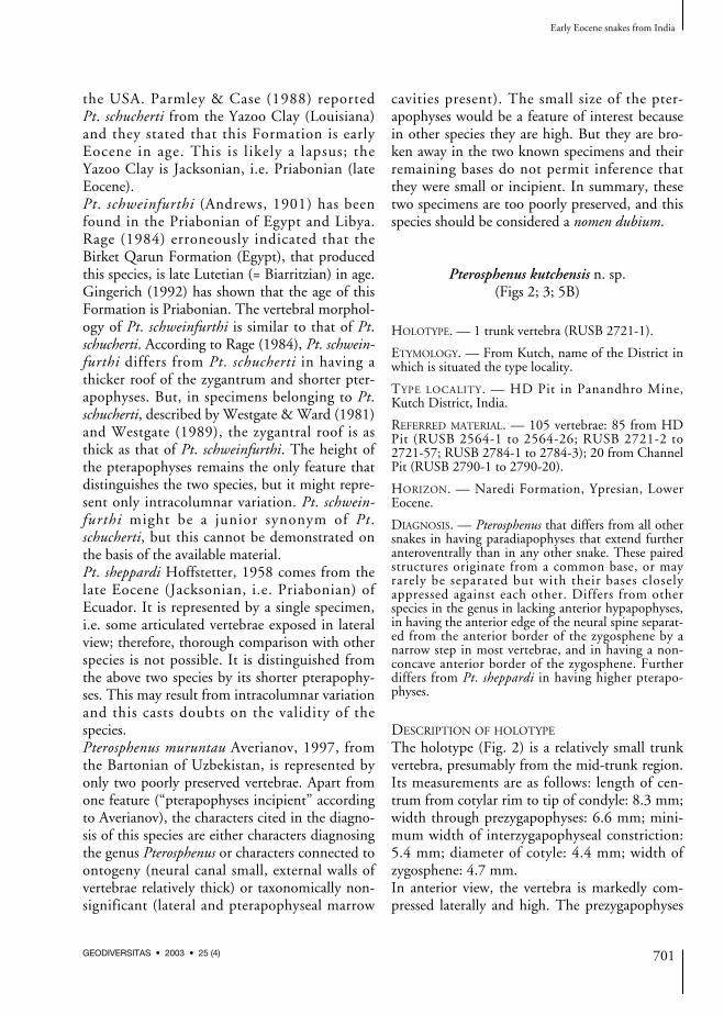
Pterosphenus kutchensis n. sp.(Figs 2; 3; 5B)
HOLOTYPE. — 1 trunk vertebra (RUSB 2721-1).
ETYMOLOGY. — From Kutch, name of the District inwhich is situated the type locality.
TYPE LOCALITY. — HD Pit in Panandhro Mine,Kutch District, India.
REFERRED MATERIAL. — 105 vertebrae: 85 from HDPit (RUSB 2564-1 to 2564-26; RUSB 2721-2 to2721-57; RUSB 2784-1 to 2784-3); 20 from ChannelPit (RUSB 2790-1 to 2790-20).
HORIZON. — Naredi Formation, Ypresian, LowerEocene.
DIAGNOSIS. — Pterosphenus that differs from all othersnakes in having paradiapophyses that extend furtheranteroventrally than in any other snake. These pairedstructures originate from a common base, or mayrarely be separated but with their bases closelyappressed against each other. Differs from otherspecies in the genus in lacking anterior hypapophyses,in having the anterior edge of the neural spine separat-ed from the anterior border of the zygosphene by anarrow step in most vertebrae, and in having a non-concave anterior border of the zygosphene. Furtherdiffers from Pt. sheppardi in having higher pterapo-physes.
DESCRIPTION OF HOLOTYPE
The holotype (Fig. 2) is a relatively small trunkvertebra, presumably from the mid-trunk region.Its measurements are as follows: length of cen-trum from cotylar rim to tip of condyle: 8.3 mm;width through prezygapophyses: 6.6 mm; mini-mum width of interzygapophyseal constriction:5.4 mm; diameter of cotyle: 4.4 mm; width ofzygosphene: 4.7 mm.In anterior view, the vertebra is markedly com-pressed laterally and high. The prezygapophyses
Early Eocene snakes from India
701GEODIVERSITAS • 2003 • 25 (4)
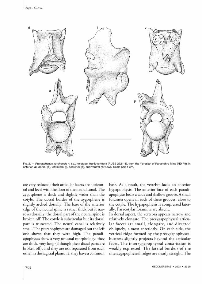
are very reduced; their articular facets are horizon-tal and level with the floor of the neural canal. Thezygosphene is thick and slightly wider than thecotyle. The dorsal border of the zygosphene isslightly arched dorsally. The base of the anterioredge of the neural spine is rather thick but it nar-rows dorsally; the dorsal part of the neural spine isbroken off. The cotyle is subcircular but its dorsalpart is truncated. The neural canal is relativelysmall. The pterapophyses are damaged but the leftone shows that they were high. The paradi-apophyses show a very unusual morphology: theyare thick, very long (although their distal parts arebroken off), and they are not separated from eachother in the sagittal plane, i.e. they have a common
base. As a result, the vertebra lacks an anteriorhypapophysis. The anterior face of each paradi-apophysis bears a wide and shallow groove. A smallforamen opens in each of these grooves, close tothe cotyle. The hypapophysis is compressed later-ally. Paracotylar foramina are absent.In dorsal aspect, the vertebra appears narrow andrelatively elongate. The prezygapophyseal articu-lar facets are small, elongate, and directedobliquely, almost anteriorly. On each side, thevertical ridge formed by the prezygapophysealbuttress slightly projects beyond the articularfacet. The interzygapophyseal constriction isweakly expressed. The lateral borders of theinterzygapophyseal ridges are nearly straight. The
Rage J.-C. et al.
702 GEODIVERSITAS • 2003 • 25 (4)
d
a
p
l v
FIG. 2. — Pterosphenus kutchensis n. sp., holotype, trunk vertebra (RUSB 2721-1), from the Ypresian of Panandhro Mine (HD Pit), inanterior (a), dorsal (d), left lateral (l), posterior (p), and ventral (v) views. Scale bar: 1 cm.

zygosphene comprises two lateral lobes that donot strongly project anteriorly; between them,the anterior border is feebly convex. The neuralspine approaches the anterior border of thezygosphene but it does not reach it. The remain-ing part of the left pterapophysis appears as a low,but well defined keel. The median notch in theposterior border of the neural arch is wide andobtuse, it appears as a broad embayment. As in allpalaeophiids, the zygantral roof is reduced.In lateral view, the vertebra is markedly higherthan long, despite the fact that the dorsal part ofthe neural spine and the ventral parts of the para-diapophyses and hypapophysis are broken off.The height of the neural spine cannot be estimat-ed. The zygosphenal facets are small, ovaloid andoblique. There is no marked interzygapophysealridge. The prezygapophysis lacks a prezyg-apophyseal process, but it forms a vertical ridgethat extends from the tip of the articular facet tothe anterolateral border of the paradiapophysis.The paradiapophysis is directed ventrally andslightly anteriorly. The articular facet for the ribis lacking, but an eroded area on the distal part ofthe remaining portion might correspond to thedorsal part of the diapophyseal surface. Anyway,at least most of the articular facet was on themissing part, i.e. it occupied a very ventral posi-tion, far from the centrum. The incompletehypapophysis is vertical and not located very pos-teriorly. The axis of the condyle is horizontal.There is no perceivable lateral foramen.In posterior view, as in anterior aspect, the later-ally compressed morphology is striking. Beneaththe pterapophyses the lateral flanks of the neuralarch are subvertical. Only the left zygantral fora-men appears to be present. The centrum is some-what triangular in cross-section.The ventral view displays the unusual position ofthe paradiapophyses the bases of which are notseparated in the sagittal plane. As a consequenceof the subtriangular cross-section of the centrum,subcentral ridges are lacking. Anterior to thecondyle, the centrum forms a neck that is clearlynarrower than the condyle. Two subcentralforamina open between the bases of the hyp-apophysis and paradiapophyses.
OTHER VERTEBRAE AND VARIATION
No caudal vertebrae are known. Two vertebraeeach preserve a complete pterapophysis. In lateralaspect, this process appears as a triangular laminathe anterior border of which is sharp. In RUSB2790-1, the pterapophysis is directed dorsolater-ally (Fig. 3A) whereas in RUSB 2784-1 it is morevertical.A few vertebrae of juvenile individuals are known(Fig. 3C). They are of interest because they provethat the “large” vertebrae of Pt. kutchensis n. sp.,that are small for the genus Pterosphenus, belongto adults. The vertebrae of juveniles show the fea-tures that are usual in all snake families: neuralcanal relatively wider than in adults, zygospheneand lateral walls of vertebrae thinner, cotyle moredepressed dorsoventrally, and zygosphene entire-ly overhanging (i.e. anterior parts of lateral wallsof the neural canal not completed).Variation in the trunk vertebrae is minimal. Inmost vertebrae, as in the holotype, the anterioredge of the neural spine is separated from theanterior border of the zygosphene by a narrowsurface; however, in a few vertebrae the top of theanterior border of the zygosphene is prolongedwithout a break into the anterior edge of the neu-ral spine. The latter condition is seen in otherspecies of Pterosphenus. In Pt. kutchensis n. sp.,the variation of this feature does not appear to berelated to the position of vertebrae in the verte-bral column. In some vertebrae, that are more lat-erally compressed than the holotype, thecommon base of the paradiapophyses is deeper; itappears as a thick process beneath the cotyle (Fig.3B). It is not possible to determine whether suchvertebrae are more anterior or more posteriorthan those exemplified by the holotype. In a few,damaged vertebrae, it is possible that the com-mon base of the paradiapophyses is very shallowor the paradiapophyses are separated but closelyappressed against each other. Zygantral foraminaare often lacking whereas their presence is con-stant in non-palaeophiid snakes. But, irrespectiveof the presence or absence of the usual zygantralforamina, a sagittal foramen sometimes piercesthe posterior wall of the neural arch between thetwo zygantral fossae, below the neural spine. This
Early Eocene snakes from India
703GEODIVERSITAS • 2003 • 25 (4)

condition of the zygantral foramina seems com-mon in Palaeophiidae. Paracotylar, lateral, andsubcentral foramina are rarely and irregularlypresent. The foramen that opens in the anteriorgroove of each paradiapophysis, close to thecotyle, is nearly always present.The size ranges from juveniles (centrum length:about 4.3 mm) to largest adults (centrum length:10.5 mm).
COMMENTS
This snake poses a peculiar problem. The longparadiapophyses are more or less reminiscent ofpleurapophyses, i.e. processes present only in cau-dal vertebrae. Since, on the available vertebrae,paradiapophyseal articular facets are not obser-
vable we are led to conclude that either thesefacets were on the distal parts of the paradi-apophyses that are always broken off (which isquite possible because the facets are borne byspongy bone) or that the processes are pleurapo-physes. But, if these processes are pleurapophyses,then all vertebrae come from the caudal region,which is not possible. Caudal vertebrae are, byfar, more rarely found than vertebrae from thetrunk region. Moreover, these vertebrae do notcome from a single individual; they have beenfound in two sites (HD Pit and Channel Pit) andthe vertebrae are of different sizes. Besides, caudalvertebrae of Palaeophis are known, and as in near-ly all snakes they have typical pleurapophyses andpaired haemapophyses (Rage 1983a). The verte-
Rage J.-C. et al.
704 GEODIVERSITAS • 2003 • 25 (4)
A (a)
C (a) C (d) C (l)
A (l) B (a) B (l)
FIG. 3. — Pterosphenus kutchensis n. sp. from the Ypresian of Panandhro Mine; A, trunk vertebra (RUSB 2790-1) in which a pter-apophysis is complete and the paradiapophyses are separated (or their common base is very shallow?), Channel Pit; B, trunk verte-bra (RUSB 2721-2) showing a very deep common base of the paradiapophyses, HD Pit; C, trunk vertebra (RUSB 2790-2) of ajuvenile individual, Channel pit. Anterior (a), dorsal (d), and lateral (l) views. Scale bars: 1 cm.
brae of Pt. kutchensis n. sp. lack the latter process-es but they have all a hypapophysis. The caudalvertebrae of nearly all snakes have pairedhaemapophyses; they are replaced by a haemalkeel in a very few snakes (Szyndlar & Böhme1996). In the caudal region, hypapophyses occuronly in the anterior caudal vertebrae of two livinggenera; moreover, they appear as deep keelsrather than true hypapophyses (Szyndlar & Rage2003). Consequently, the presence of true hyp-apophyses on all vertebrae demonstrates that theycome from the trunk region. Caudal vertebrae ofPt. kutchensis n. sp. have not been found.These vertebrae show characteristic features ofthe Palaeophiinae, more especially of the genusPterosphenus (see above).They differ from all other species of Pterosphenusin having a non-concave anterior border of thezygosphene in dorsal aspect and longer, deeperparadiapophyses. Moreover, the two paradi-apophyses originate from a common base, or atleast (in a few vertebrae) the bases of the twoparadiapophyses are perhaps very narrowly sepa-rated, which is unique in snakes. This conditionplus the marked ventral orientation of the paradi-apophyses and the narrowness of the vertebrae leadto a reduction of the width but it increases thedepth of the animal. This certainly corresponds toa very strong adaptation to aquatic life. This lateralcompression is stronger in Pt. kutchensis n. sp. thanin other species of Pterosphenus; therefore, as far asthis feature is concerned, Pt. kutchensis n. sp.appears to be the most advanced palaeophiid. As aconsequence of the position of the paradiapophy-ses, the anterior hypapophysis that is characteris-tic of other species of Pterosphenus is absent in Pt.kutchensis n. sp. In addition, in most vertebrae ofPt. kutchensis n. sp. there is a step between theanterior border of the zygosphene and the base ofthe anterior edge of the neural spine. This charac-ter recalls Palaeophis although the step is clearlynarrower than in the latter genus. This step doesnot occur in the other species of Pterosphenus. Thisfeature probably represents a plesiomorphic statewithin palaeophiids.It may be added that the pterapophyses of Pt.kutchensis n. sp. are higher than those of Pt. shep-
pardi, but this difference might be a result ofintracolumnar variation. Finally, it should benoted that Pt. kutchensis n. sp. is the smallest andone of the two earliest species of Pterosphenus.
Pterosphenus biswasi n. sp.(Figs 4; 5A)
HOLOTYPE. — 1 trunk vertebra (RUSB 2784-4).
ETYMOLOGY. — Named for Dr. S. K. Biswas, inrecognition of his work on the geology of Kutch.
TYPE LOCALITY. — HD Pit in Panandhro Mine,Kutch District, India.
REFERRED MATERIAL. — 2 vertebrae: 1 from HD Pit(RUSB 2565-1) and 1 from Channel Pit (RUSB2790-21).
HORIZON. — Naredi Formation, Ypresian, LowerEocene.
DIAGNOSIS. — Species of Pterosphenus distinguishedfrom Pt. schucherti, Pt. schweinfurthi, and Pt. murun-tau by its markedly less deeply concave anterior borderof the zygosphene. Differs from Pt. schucherti and Pt.schweinfurti in having the zygapophyseal plane locatedslightly higher. Differs from Pt. sheppardi by its longerand more oblique paradiapophyses, and the anteropos-teriorly longer basis of its hypapophysis. Distinguishedfrom Pt. kutchensis n. sp. by its less laterally com-pressed vertebrae, the concave anterior border of itszygosphene, its markedly shorter paradiapophyses,separated bases of the paradiapophyses, and the pres-ence of an anterior hypapophysis.
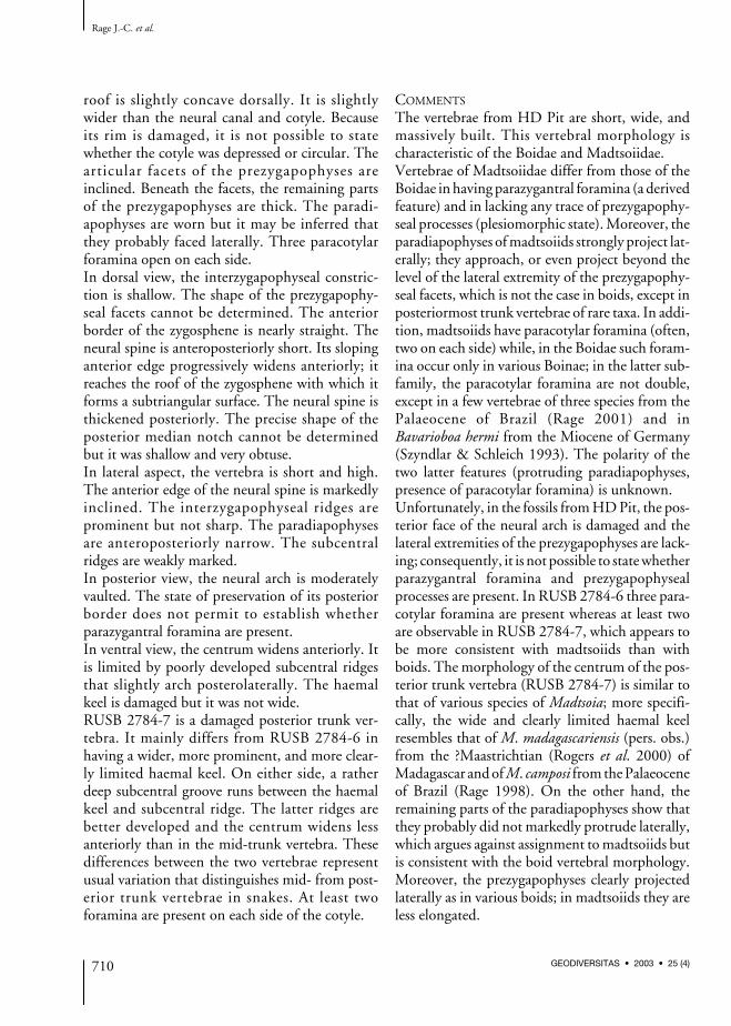
DESCRIPTION OF HOLOTYPE
The holotype is a large, massive trunk (presum-ably mid-trunk) vertebra (Fig. 4). The measure-ments are as follows: length of centrum fromcotylar rim to tip of condyle: 18.9 mm; widththrough prezygapophyses: 19.7 mm; width ofinterzygapophyseal constriction: 18.2 mm; diam-eter of cotyle: 12.8 mm; width of zygosphene:13.2 mm.In anterior view, the vertebra is clearly com-pressed laterally and high. The prezygapophysesare small; their articular facets are slightlyinclined above the horizontal and they lie slight-ly above the level of the floor of the neuralcanal. The zygosphene is thick and hardly widerthan the cotyle; its dorsal border forms the base
Early Eocene snakes from India
705GEODIVERSITAS • 2003 • 25 (4)

of the anterior edge of the neural spine, whichgives a subtriangular shape to the frontal aspectof the zygosphene. The cotyle appears to beslightly depressed dorsoventrally and its dorsalpart is truncated. The section of the neural canalis small , markedly narrower than thezygosphene and cotyle. The pterapophyses areincomplete; the base of the right one shows thatthey were high. The paradiapophyses are situat-ed low and distant from the centrum. Below thecotyle, a space that represents about one thirdthe diameter of the cotyle, separates the bases ofthe paradiapophyses. A small anterior hyp-apophysis is present beneath the cotyle, betweenthe paradiapophyses. The vertebra lacks para-
cotylar foramina but irregular small foraminaopen in the anterior face of the prezygapophy-seal buttresses.In dorsal aspect, the vertebra appears to be moreor less squarish, not clearly longer than wide. Theprezygapophyseal facets are small, not elongate,and directed more anteriorly than laterally. Theinterzygapophyseal constriction is hardlyexpressed. The lateral borders of the interzyg-apophyseal ridges are slightly convex laterally.The zygosphene does not form clearly definedlateral lobes; its anterior border is weakly con-cave. Anteriorly, the neural spine reaches theanterior face of the zygosphene; it grows thickerposteriorly. The basal parts of the pterapophyses
Rage J.-C. et al.
706 GEODIVERSITAS • 2003 • 25 (4)
dv
l
ap
FIG. 4. — Pterosphenus biswasi n. sp., holotype, trunk vertebra (RUSB 2784-4), in anterior (a), dorsal (d), left lateral (l), posterior (p),and ventral (v) views, Ypresian of Panandhro Mine (HD Pit). Scale bar: 1 cm.
that are preserved form blunt, poorly definedkeels. The median notch in the posterior borderof the neural arch is shallow and obtuse, but itsbottom is clearly triangular. The roof of thezygantrum is not extended.In lateral view, the vertebra is short and high.The neural spine and the hypapophysis are bro-ken off. The zygosphenal facet is small, subcircu-lar, and directed more dorsally than anteriorly.The interzygapophyseal ridge is strong andprominent. The anterolateral ridge of the prezyg-apophyseal buttress originates on the anterodor-sal margin of the paradiapophysis. The articularfacet of the paradiapophysis is elongate andmarkedly oblique (about 45° from the vertical);there is no distinction between the dia- and par-apophyseal areas. The axis of the condyle is hori-zontal. A small lateral foramen opens below theinterzygapophyseal ridge.In posterior view, the lateral flanks of the neuralarch are vertical. The cotyle is slightly depressed.The area of zygantral foramina is obscured bymatrix.In ventral view, the centrum appears cylindrical.It lacks subcentral ridges. The base of the hyp-apophysis is elongate. Posteriorly, it reaches thecondyle; anteriorly, it is prolonged by a thin keelthe anterior part of which forms the anteriorhypapophysis. Elongate subcentral foramina arepresent.
OTHER SPECIMENS AND VARIATION
Only two other specimens are available. Onelarge vertebra (RUSB 2565-1) is very worn; asmaller vertebra (RUSB 2790-21) is damaged.They are referred to Pt. biswasi n. sp. on the basisof markedly separated bases of paradiapophyses(i.e. they clearly differ from Pt. kutchensis n. sp.from the same locality), slightly concave anteriorborder of zygosphene, and an anterior hypapophy-sis below the cotyle (the latter feature cannot bechecked in RUSB 2565-1). In both vertebrae, asin the holotype, the anterior edge of the neuralspine is continuous with the anterior face of thezygosphene; as a result, the latter face is subtrian-gular. The presence or absence of foramina is notverifiable in these two specimens.
COMMENTS
A problem arises from the fact that there are twospecies at Panandhro Mine, a small and a large one.Therefore, it may be argued that the palaeophiiddescribed above as Pt. kutchensis n. sp. is only rep-resented by juvenile individuals of Pt. biswasi n. sp.However, as shown above, the vertebrae referredto Pt. kutchensis n. sp. include some juveniles butmainly adult specimens. Moreover, at least one ofthe characters that distinguish the two species can-not be interpreted as an ontogenetic change: theparadiapophyses originate from a common base inPt. kutchensis n. sp. whereas the bases are markedlyseparated in Pt. biswasi n. sp. as in all other snakes.Such an ontogenetic change has never beenreported. In addition, vertebrae of similar sizebelonging to these two species (i.e. a large vertebraof Pt. kutchensis n. sp. and a small one of Pt. biswasin. sp.) display the conditions of the paradi-apophyses typical for these two species: the para-diapophyses arise from a single base in the largevertebra of Pt. kutchensis n. sp. while the bases ofthe two paradiapophyses are clearly separated onthe vertebra of similar size belonging to Pt. biswasin. sp. (Fig. 5). This clearly demonstrates that thisdifference is not of ontogenetic nature and thatthere are two distinct species.
Early Eocene snakes from India
707GEODIVERSITAS • 2003 • 25 (4)
A B
FIG. 5. — Comparison between Pterosphenus biswasi n. sp. andPt. kutchensis n. sp., trunk vertebrae of similar sizes in antero-ventral views; A, Pt. biswasi n. sp., the bases (hatched areas) ofthe paradiapophyses (broken off) are markedly separated(RUSB 2790-21); B, Pt. kutchensis n. sp., the paradiapophyses(broken off; hatched areas) originate from a common base(RUSB 2790-3). Both vertebrae from Channel Pit. Scale bars:5 mm.

Pt. biswasi n. sp. is easily distinguished from Pt.kutchensis n. sp. Apart from its larger size and sep-arate bases of paradiapophyses, it differs from Pt.kutchensis n. sp. in having an anterior hypapophy-sis, less laterally compressed vertebrae, shorterparadiapophyses, and a concave anterior borderof zygosphene.The distinction between Pt. biswasi n. sp. and theother species of Pterosphenus is less marked. It dif-fers from all other species in having a shallowconcave anterior border of zygosphene, while it isdeeply concave, even notched, in Pt. schucherti,Pt. schweinfurthi, and Pt. muruntau (not observ-able in Pt. sheppardi). Pt. biswasi n. sp. furtherdiffers from Pt. schucherti and Pt. schweinfurthiby its zygapophyseal plane that is located slightlyhigher (mainly shown by the postzygapophysealfacets) and from Pt. sheppardi by its more elong-ate and more oblique paradiapophyses, and theanteroposteriorly longer base of its hypapophysis.
Pterosphenus sp.
REFERRED MATERIAL. — 15 vertebrae: 8 from HD Pit(RUSB 2721-58, 2721-59; RUSB 2564-27 to 2564-31; RUSB 2784-5) and 7 from Channel Pit (RUSB2790-22 to 2790-28).
These vertebrae are too damaged to be allocatedat species level. But their referral to Pterosphenusis unquestionable.
COMMENTS ON THE PALAEOPHIIDAEFROM KUTCH
Thus far, the earliest Pterosphenus (Pt. schucherti)has been reported from the middle Eocene(Westgate 1989), more precisely the late Lutetianor early Bartonian (Westgate pers. comm.), of theUSA. Therefore, the two species of Pterosphenusfrom Panandhro Mine antedate the NorthAmerican species. Although Pt. kutchensis n. sp., one of the twospecies from Panandhro Mine, is one of the twoearliest species of Pterosphenus, it is the mostadvanced palaeophiid species as far as adaptationto aquatic life is concerned.
It appears to be somewhat peculiar and ratherdifferent from the other known species ofPterosphenus. It appears to be more stronglyadapted to aquatic life, i.e. it is more advancedthan other species of Pterosphenus in being deeperand more laterally flattened. But it is lessadvanced than the other species of Pterosphenusin having a space between the anterior face of thezygosphene and the anterior edge of the neuralspine in most vertebrae, which is the conditionretained in Palaeophis. In addition, the anteriorborder of the zygosphene is not concave in Pt.kutchensis n. sp. In other species of Pterosphenusthe anterior border of the zygosphene is concaveand, except in Pt. biswasi n. sp., it is evennotched. The zygosphene is notched in lizardsand in most early snakes; consequently, thenotched zygosphene would represent the ple-siomorphic state. Therefore, Pt. kutchensis n. sp.likely represents a distinct lineage of Pterosphenus.Finally, the recovery of the genus Pterosphenusfrom the early Eocene necessitates a change inour views on the evolution of the Palaeophiinae.It was suggested that the palaeophiines evolvedfrom “primitive” Palaeophis to Pterosphenus,through “advanced” Palaeophis (Janensch 1906).Hoffstetter (1958) showed that this over-simpli-fied view was wrong and he implicitly inferredthat Palaeophis is a paraphyletic assemblage, stemgroup of Pterosphenus. The discovery of a veryderived Pterosphenus from the early Eocenestrongly supports Hoffstetter’s opinion.
Family ?MADTSOIIDAE Hoffstetter, 1961 or BOIDAE Gray, 1825
The Madtsoiidae and Boidae are two clearly dis-tinct families. Madtsoiids are basal snakes (Scanlon& Lee 2000) whereas boids are living snakes thatmay be considered “relatively advanced”. Althoughthe two families are phylogenetically clearly dis-tinct, their vertebrae show a similar overall mor-phology. The referral of well preserved vertebrae atfamily level is easy, but the assignment may bedoubtful when the vertebrae are damaged, which isthe case of the fossils from Panandhro Mine.
Rage J.-C. et al.
708 GEODIVERSITAS • 2003 • 25 (4)

Indeterminate genus(Fig. 6A)
REFERRED MATERIAL. — 2 vertebrae (RUSB 2784-6and 2784-7) from HD Pit.
DESCRIPTION
RUSB 2784-6 is a large mid-trunk vertebra(length of centrum from cotylar lip to tip of
condyle: 13.6 mm; minimum width of interzyg-apophyseal constriction: 18.2 mm; width ofzygosphene: 9.1 mm). The neural spine and lat-eral parts of prezygapophyses are broken off whilethe paradiapophyses and the posterior border ofthe neural arch are eroded.In anterior view, the vertebra is wide anddepressed. The zygosphene is rather thick and its
Early Eocene snakes from India
709GEODIVERSITAS • 2003 • 25 (4)
A (d)
A (a)
B (d)
B (l) B (a)
B (v)
A (p)
A (l) A (v)
FIG. 6. — A, Madtsoiidae or Boidae, trunk vertebra (RUSB 2784-6), Ypresian of Panandhro Mine (HD Pit); B, Colubroidea, familyindeterminate, trunk vertebra (RUSB 2790-29), Ypresian of Panandhro Mine (Channel Pit). Anterior (a), dorsal (d), lateral (l), posterior(p), and ventral (v) views. Scale bars: 1 cm.
roof is slightly concave dorsally. It is slightlywider than the neural canal and cotyle. Becauseits rim is damaged, it is not possible to statewhether the cotyle was depressed or circular. Thearticular facets of the prezygapophyses areinclined. Beneath the facets, the remaining partsof the prezygapophyses are thick. The paradi-apophyses are worn but it may be inferred thatthey probably faced laterally. Three paracotylarforamina open on each side.In dorsal view, the interzygapophyseal constric-tion is shallow. The shape of the prezygapophy-seal facets cannot be determined. The anteriorborder of the zygosphene is nearly straight. Theneural spine is anteroposteriorly short. Its slopinganterior edge progressively widens anteriorly; itreaches the roof of the zygosphene with which itforms a subtriangular surface. The neural spine isthickened posteriorly. The precise shape of theposterior median notch cannot be determinedbut it was shallow and very obtuse.In lateral aspect, the vertebra is short and high.The anterior edge of the neural spine is markedlyinclined. The interzygapophyseal ridges areprominent but not sharp. The paradiapophysesare anteroposteriorly narrow. The subcentralridges are weakly marked.In posterior view, the neural arch is moderatelyvaulted. The state of preservation of its posteriorborder does not permit to establish whetherparazygantral foramina are present.In ventral view, the centrum widens anteriorly. Itis limited by poorly developed subcentral ridgesthat slightly arch posterolaterally. The haemalkeel is damaged but it was not wide.RUSB 2784-7 is a damaged posterior trunk ver-tebra. It mainly differs from RUSB 2784-6 inhaving a wider, more prominent, and more clear-ly limited haemal keel. On either side, a ratherdeep subcentral groove runs between the haemalkeel and subcentral ridge. The latter ridges arebetter developed and the centrum widens lessanteriorly than in the mid-trunk vertebra. Thesedifferences between the two vertebrae representusual variation that distinguishes mid- from post-erior trunk vertebrae in snakes. At least twoforamina are present on each side of the cotyle.
COMMENTS
The vertebrae from HD Pit are short, wide, andmassively built. This vertebral morphology ischaracteristic of the Boidae and Madtsoiidae.Vertebrae of Madtsoiidae differ from those of theBoidae in having parazygantral foramina (a derivedfeature) and in lacking any trace of prezygapophy-seal processes (plesiomorphic state). Moreover, theparadiapophyses of madtsoiids strongly project lat-erally; they approach, or even project beyond thelevel of the lateral extremity of the prezygapophy-seal facets, which is not the case in boids, except inposteriormost trunk vertebrae of rare taxa. In addi-tion, madtsoiids have paracotylar foramina (often,two on each side) while, in the Boidae such foram-ina occur only in various Boinae; in the latter sub-family, the paracotylar foramina are not double,except in a few vertebrae of three species from thePalaeocene of Brazil (Rage 2001) and inBavarioboa hermi from the Miocene of Germany(Szyndlar & Schleich 1993). The polarity of thetwo latter features (protruding paradiapophyses,presence of paracotylar foramina) is unknown.Unfortunately, in the fossils from HD Pit, the pos-terior face of the neural arch is damaged and thelateral extremities of the prezygapophyses are lack-ing; consequently, it is not possible to state whetherparazygantral foramina and prezygapophysealprocesses are present. In RUSB 2784-6 three para-cotylar foramina are present whereas at least twoare observable in RUSB 2784-7, which appears tobe more consistent with madtsoiids than withboids. The morphology of the centrum of the pos-terior trunk vertebra (RUSB 2784-7) is similar tothat of various species of Madtsoia; more specifi-cally, the wide and clearly limited haemal keelresembles that of M. madagascariensis (pers. obs.)from the ?Maastrichtian (Rogers et al. 2000) ofMadagascar and of M. camposi from the Palaeoceneof Brazil (Rage 1998). On the other hand, theremaining parts of the paradiapophyses show thatthey probably did not markedly protrude laterally,which argues against assignment to madtsoiids butis consistent with the boid vertebral morphology.Moreover, the prezygapophyses clearly projectedlaterally as in various boids; in madtsoiids they areless elongated.
Rage J.-C. et al.
710 GEODIVERSITAS • 2003 • 25 (4)
Finally, it does not seem possible to confidentlyrefer these vertebrae to one of these two families.The Madtsoiidae range from the mid-Cretaceous tothe Pleistocene (Rage & Werner 1999). However,post-Eocene madtsoiids are known only in Australia(Scanlon 1995, 1997). They primarily inhabitedGondwanan regions. The earliest Boidae come fromthe latest Cretaceous (Campanian-Maastrichtian)of Europe, and South and North America (Rage1984; Albino 2000). In Asia, aside from the possibleboid from the Ypresian of Panandhro Mine, theoldest representative of the family was recoveredfrom the early-middle Eocene of Pakistan (Rage1987a). The Boidae probably originated in aGondwanan region, but as early as the Eocene theywere widely distributed on Laurasian continents.
Super-family COLUBROIDEA Oppel, 1811
The Colubroidea are regarded as the most advancedsnakes. They comprise four living (Colubridae,Atractaspididae Günther, 1858, Elapidae, ViperidaeGray, 1825) and two extinct (Anomalophiidae,Russellophiidae) families. The Russellophiidae areknown from the mid-Cretaceous (Cenomanian) tothe late Eocene (Rage & Werner 1999) whereas theAnomalophiidae are restricted to the early Eocene.Besides, colubroids without family reference werereported from the Cenomanian of Sudan(Colubroidea incertae sedis; Rage & Werner 1999),the late early Eocene of France (Colubroidea incertaesedis; Augé et al. 1997), and the late Eocene ofBritain (Vectophis wardi Rage & Ford, 1980;Headonophis harrisoni Holman, 1993). The earliestmember of a living family is a Colubridae from thelate Eocene of Thailand (Rage et al. 1992).In the Ypresian of Panandhro Mine, the Colu-broidea are represented by a single vertebra whoseassignment is not possible at family level.
Indeterminate family(Fig. 6B)
REFERRED MATERIAL. — 1 trunk vertebra (RUSB2790-29) from Channel Pit.
DESCRIPTION
The vertebra probably comes from the mid-trunkregion. The posterior part of the neural arch, theneural spine, tips of prezygapophyses, and paradi-apophyses are damaged. The vertebra is not heavilybuilt and it is comparatively elongate (lengthof centrum from cotylar rim to tip of condyle:5.1 mm; width of zygosphene: 3.6 mm; minimumwidth of interzygapophyseal constriction: 4.6 mm).In anterior aspect, the vertebra appears wide andrelatively lightly built. The zygosphene is wide,moderately thick, and its roof is slightly archeddorsally. The neural canal is comparatively broad.The cotyle is rather small and depressed dorsoven-trally. The zygapophyseal facets are nearly hori-zontal; they lie above the floor of the neural canal.The tip of each prezygapophysis is damaged, butthe thickness of the remaining lateral part suggeststhat prezygapophyseal processes were present. Theparadiapophyses are eroded, but it may beinferred that they faced lateroventrally. On theright side, a foramen opens in the position of aparacotylar foramen, but on the left side fourforamina are present in the “paracotylar area”.The fact that four foramina are present on oneside does not permit to definitely regard theseforamina as homologous to paracotylar foramina,but this cannot be rejected. On either side, a largeparazygosphenial foramen opens in a deep fossalocated between the zygosphenial and prezyg-apophyseal facets.In dorsal view, the prezygapophyseal facets areelongate and oblique. The interzygapophysealconstriction is shallow. The zygosphene is wide;its anterior border is trilobate but the lobes proj-ect only weakly anteriorly. On each side, the largeparazygosphenial foramen is visible. Anteriorly,the neural spine reaches the roof of thezygosphene but it does not approach the anteriorborder.In lateral view, the vertebra is approximately ashigh as long. The zygosphenial facets are broad.The damaged lateral tips of the prezygapophysessuggest that prezygapophyseal processes werepresent but this cannot be definitely confirmed.The interzygapophyseal ridges are sharply defined.On each side, below the interzygapophyseal ridge
Early Eocene snakes from India
711GEODIVERSITAS • 2003 • 25 (4)

is a large and deep fossa; on the right side, matrixobscures the bottom of the fossa, but on the leftside the fossa contains the lateral foramen. Theparadiapophyses are small, not more elongatedorsoventrally than anteroposteriorly. The sub-central ridges are well developed. Like the ventralborder of the haemal keel, they are slightly archeddorsally. The axis of the condyle appears to beslightly oblique.In ventral view, the centrum is narrow and limit-ed by parallel subcentral ridges; its ventral surfaceis flat. The haemal keel is not strongly defined; itis narrow and moderately prominent. Two sub-central foramina are present. The damaged posterior face of the vertebra showsthat the neural arch was vaulted. Parazygantralforamina are absent.
COMMENTS
The relatively light build and elongation of thevertebra, as well as the narrowness of the centrumshow that this specimen belongs to theColubroidea. The presence of parazygosphenial foramina inRUSB 2790-29 makes it possible to distinguish itfrom all other colubroids, but it should be notedthat the significance of these foramina is unknown.Such foramina are present in Pouitella Rage, 1988,a basal snake of unknown family reference fromthe Cenomanian (Rage 1988), Palaeophis colos-saeus, a palaeophiid from the Lutetian (Rage1983b), and in the Acrochordidae, a living family(Hoffstetter & Gayrard 1964). These foramina arealso known in a mosasauroid lizard from theCenomanian (Rage & Néraudeau in press). Thepresence of parazygosphenial foramina in anearly colubroid appears to be consistent with theirpresence in acrochordids that are the sister groupto colubroids; these foramina probably represent aplesiomorphic state within Colubroidea.The presence of foramina in the paracotylarregion permits us to distinguish RUSB 2790-29from other colubroids that do not belong torecent families, except Headonophis Holman,1993 that has paracotylar foramina.The absence of compressed buttresses of theprezygapophyses forming a vertical ridge on
either side of the vertebra, demonstrates thatRUSB 2790-29 cannot be referred to theRussellophiidae or Anomalophiidae. Moreover,the weak inclination of the zygapophyseal facetsis like that of nearly all other snakes (i.e. dorso-medial), whereas in russellophiids they face dor-solaterally. The vertebra from Panandhro Minefurther differs from those of the Anomalophiidaein being more lightly built.Apart from the presence of the foramina dis-cussed above (parazygosphenial and ?paracotylarforamina), RUSB 2790-29 clearly differs fromthe colubroids from the Cenomanian of Sudanand from the early Eocene of France in having amuch more vaulted neural arch, and from thevertebrae of Vectophis Rage & Ford, 1980 inbeing markedly more elongate.On the whole, RUSB 2790-29 clearly resemblesmodern colubroids belonging to the Colubridaeand Elapidae, although in the latter family hypa-pophyses are present on trunk vertebrae. Theoverall morphology of the vertebra is clearly rem-iniscent of that of Colubridae. The vertebral dif-ferences between colubroids belonging tomodern families and Cretaceous-Eocene fossilsare the presence of prezygapophyseal processesand of subdivided paradiapophyseal areas inmodern forms. Moreover, recent colubroids haveparacotylar foramina, whereas the foramina areabsent in Eocene and pre-Eocene forms, exceptHeadonophis. Unfortunately, it does not appearpossible to state whether the foramina that openon either side of the cotyle of RUSB 2790-29 aretrue paracotylar foramina. The paradiapophysesare eroded and one cannot determine whetherthe articular surfaces were subdivided into para-and diapophyseal areas. Finally, from the form oflateral tip of the prezygapophyses, it is stronglysuspected that prezygapophyseal processes werepresent, but this cannot be definitely confirmed.The presence of prezygapophyseal processeswould represent a derived character that, alongwith the presence of possible paracotylar forami-na, might suggest that this vertebra belong to thecolubrid lineage; if this is right, RUSB 2790-29would represent the earliest member of thisgroup. Unfortunately, the state of preservation of
Rage J.-C. et al.
712 GEODIVERSITAS • 2003 • 25 (4)

the specimen does not permit us to refer it to theColubridae.Whatever the precise taxonomic position ofRUSB 2790-29 within the colubroids, it repre-sents a “modern” snake within the present fauna.In fact, its close resemblance to the modernColubridae might lead to the suspicion that thevertebra belongs to a recent snake that becamemixed with specimens from the fossiliferous bed.However, the specimen is mineralized and itshows the same color as most of the palaeophiidvertebrae from the site; in addition, parts areworn and polished in such a manner that thisspecimen cannot be a bone of a recent individual.Therefore, RUSB 2790-29 really represents a col-ubroid from the early Eocene.This specimen represents a new genus andspecies, but this single and incomplete vertebracannot be a name-bearer of a new taxon.Consequently, this new colubroid snake remainsunnamed.
CONCLUSIONS
The Eocene of Panandhro Mine has produced arich fauna of snakes that is largely dominated bypalaeophiids. The presence of the snakePterosphenus would argue for a middle or lateEocene age, but the early Eocene age suggestedby foraminifera (for the correlative units in theNaredi formation) appears more likely at present. The fauna includes Palaeophiidae (Pterosphenuskutchensis n. sp. and Pt. biswasi n. sp.), a snakethat is either a Madtsoiidae or a Boidae, repre-sented by an indeterminate genus and species,and an indeterminate family of Colubroidea. ThePalaeophiidae are represented by 124 vertebrae,while two vertebrae are referred to the madtsoiidor boid snake, and only one belongs to theColubroidea.Within palaeophiids, Pterophenus kutchensisn. sp., a small species, markedly outnumbers thelarge Pt. biswasi n. sp. (106 vertebrae to 3). Pt.biswasi n. sp. is a typical Pterosphenus that doesnot call for particular comments. But Pt. kutchen-sis n. sp. is a peculiar species that shows a unique
feature, i.e. the two paradiapophyses originatefrom a common base, or, in a few specimens,there is perhaps not a common base but the baseof each paradiapophysis is closely appressedagainst the base of the opposite paradiapophysis.These two species are the first palaeophiidsreported from India. The presence of Madtsoiidae in India remainsdoubtful. The specimens from the Ypresian ofKutch, as the specimen from the Maastrichtian ofTakli (= Gitti Khadan) (Gayet et al. 1985), arethe only fossils from India that might be referredto madtsoiids. Unfortunately, their state ofpreservation does not permit a secure referral. Ifthey do not belong to the Madtsoiidae, then theyrepresent Boidae. In the latter case, the vertebraefrom Panandhro Mine might represent the earli-est Boidae from Asia.The colubroid from Channel Pit is the first pre-Neogene representative of the group reportedfrom India.It should be noted that, assuming that theYpresian age is well established, Pt. kutchensisn. sp. and Pt. biswasi n. sp. represent the earliestmembers of Pterosphenus (see above). This issomewhat astonishing since Pt. kutchensis n. sp. ismore strongly adapted to aquatic life than thespecies from the late Eocene. Previously, it wassupposed that this adaptation more or less pro-gressively developed in the palaeophiines toculminate in the late Eocene Pterosphenus. Pt.kutchensis n. sp. probably corresponds to a diver-gent lineage of Pterosphenus, unfortunately theavailable material does not permit a phylogeneticanalysis within the group.Pterosphenus was a snake highly adapted to aquat-ic life. Its vertebrae are tall and narrow, and theribs are weakly curved; as a result, the body waslaterally compressed. Such a body form is knownonly in highly aquatic snakes: living laticaudineand hydrophiine Elapidae, and extinct bipedalsnakes from the mid-Cretaceous. In addition, theparadiapophyses (i.e. the articulations for ribs)are displaced ventrally; consequently, the centreof gravity is also shifted ventrally, which certainlyimproved trim and maneuverability in water.Unfortunately, these anatomical characteristics
Early Eocene snakes from India
713GEODIVERSITAS • 2003 • 25 (4)

do not suggest whether these snakes lived inmarine or freshwater (or both).According to Westgate & Gee (1990), Pt.schucherti from the middle Eocene of Texas livedin brackish and freshwater, close to or in an estu-arine mangrove, under tropical conditions. Thesame species has also been found in open marinedeposits from the late Eocene (Westgate 2001).The Birket Qarun and Qasr el Sagha Formationsof Egypt that yielded Pt. schweinfurthi also corre-spond to brackish and/or shallow marine coastalenvironments (lagoon, delta front, mangrove)(Gingerich 1992). From this, it appears thatPterosphenus lived in marine, brackish, and fresh-water, close to the coasts. Mangrove areas wereperhaps especially favourable to this snakes. Themode of life of the species of Pterosphenus mighthave been similar to that of the living Acrochordusgranulatus (Acrochordidae) that is highly adaptedto salt water (Dunson & Dunson 1973) and livesin marine water, along the coasts, but may enterrivers and lakes (McDowell 1979).The vertebrae of the colubroid and madtsoiid orboid found at Panandhro Mine do not displayany adaptation to aquatic life (which does notmean that they were unable to temporarily enterwater). They are probably allochthonous terres-trial snakes within the fauna of Panandhro Mine.From a palaeobiogeographic point of view, onlythe Madtsoiidae are significant (if madtsoiids arepresent at Panandhro Mine). This family is essen-tially Gondwanan. Out of Gondwanan areas theyare known only from Spain (Rage 1996, 1999)and southern France (Sigé et al. 1997).Unfortunately, the presence of this family inIndia is still doubtful. During the Eocene, boidswere likely nearly cosmopolitan and they provideno palaeobiogeographical information if the sub-family is not identified. Pterosphenus was proba-bly widely distributed as a consequence of itsaquatic mode of life. It is known in the earlyEocene of India, while it is present in the middleEocene of North America, and in the late Eoceneof Africa, North and South America. The ques-tion arises whether this distribution is significantas far as the geographic origin and dispersal of thegenus are concerned. In our present state of
knowledge, no conclusion can be drawn becausetoo few localities bearing Pterosphenus are known.
AcknowledgementsThis study was carried out during a visit byS. Bajpai as Professeur invité at Muséum nationald’Histoire naturelle, Paris. Financial supportfrom the Department of Science andTechnology, Government of India (New Delhi,Sanc. No. 100/IFD/2413/1998-1999 and100/IFD/572/2002-2003) and the NationalGeographic Society (Washington DC) is thank-fully acknowledged. S. Bajpai would also like tothank Prof. Ashok Sahni (Chandigarh, India) forhelpful discussions. Dr. D. Parmley (GeorgiaCollege, USA) and Dr. J.W. Westgate (LamarUniversity, USA) provided helpful information.P. Meylan (Eckerd College , USA) andZ. Szyndlar (Polska Akademia Nauk, Poland)made helpful suggestions.
REFERENCES
ALBINO A. M. 2000. — New record of snakes fromthe Cretaceous of Patagonia (Argentina).Geodiversitas 22 (2): 247-253.
ANDREWS C. W. 1901. — Preliminary notes on somerecently discovered extinct vertebrates from Egypt.Part II. Geological Magazine 8: 436-444.
AUGÉ M., DUFFAUD S., LAPPARENT DE BROIN F. DE,RAGE J.-C. & VASSE D. 1997. — Les amphibiens etles reptiles de Prémontré (Cuisien, Bassin Parisien) :une herpétofaune de référence pour l’Éocèneinférieur. Géologie de la France 1: 23-33.
AVERIANOV A. O. 1997. — Paleogene sea snakes fromthe eastern part of the Tethys. Russian Journal ofHerpetology 4: 128-142.
BAJPAI S. & THEWISSEN J. G. M. 1998. — MiddleEocene cetaceans from the Harudi and Subathu for-mations of India, in THEWISSEN J. G. M. (ed.), TheEmergence of Whales. Plenum Press, New York:213-234.
BAJPAI S. & THEWISSEN J. G. M. 2002. — Vertebratefauna from Panandhro lignite field (Lower Eocene),District Kachchh, western India. Current Science82: 507-509.
BISWAS S. K. 1992. — Tertiary stratigraphy of Kutch.Journal of the Palaeontological Society of India 37: 1-29.
CONROY G., WEST R. M. & MUNTHE J. 1985. — TheSiwaliks of Nepal: recent contributions to vertebratepaleontology and biostratigraphy, in GUPTA V. J.
Rage J.-C. et al.
714 GEODIVERSITAS • 2003 • 25 (4)

(ed.), Geology of Western Himalayas. HindustanPublishing Corporation, New Delhi: 52-61.
DUNSON W. A. & DUNSON M. K. 1973. —Convergent evolution of sublingual salt glands inthe marine file snake and the true sea snakes.Journal of Comparative Physiology 86: 193-208.
GAYET M., RAGE J.-C. & RANA R. S. 1985. —Nouvelles ichthyofaune et herpétofaune de GittiKhadan, le plus ancien gisement connu du Deccan(Crétacé/Paléocène) à microvertébrés. Implicationspaléogéographiques, in BUFFETAUT É., JAEGER J.-J.& RAGE J.-C. (eds), Paléogéographie de l’Inde, duTibet et du Sud-Est asiatique. Mémoires de la Sociétégéologique de France 147: 55-65.
GINGERICH P. D. 1992. — Marine mammals (Cetaceaand Sirenia) from the Eocene of Gebel Mokattanand Fayum, Egypt: stratigraphy, age, and paleoenvi-ronments. University of Michigan, Papers inPaleontology 30: 1-84.
HOFFSTETTER R. 1958. — Un serpent marin dugenre Pterosphenus (Pt. sheppardi nov. sp.) dansl’Éocène supérieur de l’Équateur (Amérique duSud). Bulletin de la Société géologique de France 8:45-50.
HOFFSTETTER R. 1964. — Les serpents du Néogènedu Pakistan (couches des Siwaliks). Bulletin de laSociété géologique de France 6: 467-474.
HOFFSTETTER R. & GAYRARD Y. 1964. —Observations sur l’ostéologie et la classification desAcrochordidae (Serpentes). Bulletin du Muséumnational d’Histoire naturelle 2e sér., 36 (5): 677-696.
HOLMAN J. A. 1982. — Palaeophis casei, new species, atiny palaeophid snake from the early Eocene ofMississipi. Journal of Vertebrate Paleontology 2:163-166.
HOLMAN J. A. 2000. — Fossil Snakes of North America.Indiana University Press, Bloomington andIndianapolis, xi + 357 p.
JANENSCH W. 1906. — Pterosphenus SchweinfurthiAndrews und die Entwicklung der Palaeophiden.Archiv für Biontologie 1: 307-350.
LUCAS F. A. 1899. — A new snake from the Eocene ofAlabama. Proceedings of the US National Museum21: 637-638.
MCDOWELL S. B. 1979. — A catalogue of the snakesof new Guinea and the Solomons... Part III. Boinaeand Acrochordoidea (Reptilia, Serpentes). Journal ofHerpetology 13: 1-92.
MCDOWELL S. B. 1987. — Systematics, in SEIGEL R.A., COLLINS J. T. & NOVAK S. S. (eds), Snakes.Ecology, and Evolutionary Biology. McMillan, NewYork: 3-50.
NESSOV L. A. 1984. — [Subfamily Vialovophiinaesubfam. nov.], in NESSOV L. A. & UDOVITS-CHENKO N. I. (eds), Paleogene sea snakes and elas-mobranch fishes of south Kazakhstan.Paleontologitchesky Sbornik 21: 71-73 (in Russian).
NESSOV L. A. & UDOVITSCHENKO N. I. 1984. —[Paleogene sea snakes and elasmobranch fishes of
south Kazakhstan]. Paleontologitchesky Sbornik 21:69-74 (in Russian).
PARMLEY D. & CASE G. R. 1988. — Palaeopheid snakesfrom the Gulf Coastal region of North America.Journal of Vertebrate Paleontology 8: 334-339.
RAGE J.-C. 1983a. — Les serpents aquatiques del’Éocène européen. Définition des espèces et aspectsstratigraphiques. Bulletin du Muséum nationald’Histoire naturelle 4e sér., section C, 5 (2):213-241.
RAGE J.-C. 1983b. — Palaeophis colossaeus nov. sp. (leplus grand serpent connu ?) de l’Éocène du Mali etle problème du genre chez les Palaeopheidae.Comptes Rendus des Séances de l’Académie desSciences, Paris II, 296: 1741-1744.
RAGE J.-C. 1984. — Serpentes, in WELLNHOFER P.(ed.), Handbuch der Paläoherpetologie. Part 11.Gustav Fischer, Stuttgart, xii + 80 p.
RAGE J.-C. 1987a. — Lower Vertebrates from theearly-middle Eocene Kuldana Formation of Kohat(Pakistan): Squamata. Contributions from theMuseum of Paleontology, The University of Michigan27: 187-193.
RAGE J.-C. 1987b. — Fossil History, in SEIGEL R. A.,COLLINS J. T. & NOVAK S. S. (eds), Snakes. Ecology,and Evolutionary Biology. McMillan, New York: 51-76.
RAGE J.-C. 1988. — Un serpent primitif (Reptilia,Serpentes) dans le Cénomanien (base du Crétacésupérieur). Comptes Rendus des Séances de l’Académiedes Sciences, Paris II, 307: 1027-1032.
RAGE J.-C. 1996. — Les Madtsoiidae (Reptilia,Serpentes) du Crétacé supérieur d’Europe : témoinsgondwaniens d’une dispersion transtéthysienne.Comptes Rendus de l’Académie des Sciences, Paris II a,322: 603-608.
RAGE J.-C. 1998. — Fossil snakes from the Palaeoceneof São José de Itaboraí, Brazil. Part I. Madtsoiidae,Aniliidae. Palaeovertebrata 27: 109-144.
RAGE J.-C. 1999. — Squamates (Reptilia) from theUpper Cretaceous of Laño (Basque Country,Spain). Estudios del Museo de Ciencias Naturales deAlava 14: 121-133.
RAGE J.-C. 2001. — Fossil snakes from the Palaeoceneof São José de Itaboraí, Brazil. Part II. Boidae.Palaeovertebrata 30: 111-150.
RAGE J.-C., BUFFETAUT É., BUFFETAUT-TONG H.,CHAIMANEE Y., DUCROCQ S., JAEGER J.-J. &SUTEETHORN V. 1992. — A colubrid snake in thelate Eocene of Thailand: the oldest knownColubridae (Reptilia, Serpentes). Comptes Rendusdes Séances de l’Académie des Sciences, Paris II, 314:1085-1089.
RAGE J.-C., GUPTA S. S. & PRASAD G. V. R. 2001. —Amphibians and squamates from the NeogeneSiwalik beds of Jammu and Kashmir, India.Paläontologische Zeitschrift 75: 197-205.
RAGE J.-C. & NÉRAUDEAU D. in press. — A newpachyostotic squamate reptile from the Ceno-manian of France. Palaeontology.
Early Eocene snakes from India
715GEODIVERSITAS • 2003 • 25 (4)
RAGE J.-C. & WERNER C. 1999. — Mid-Cretaceous(Cenomanian) snakes from Wadi Abu Hashim,Sudan: the earliest snake assemblage. Palaeontologiaafricana 35: 85-110.
ROGERS R. R., HARTMAN J. H. & KRAUSE D. W.2000. — Stratigraphic analysis of Upper Cretaceousrocks in the Mahajanga Basin, NorthwesternMadagascar: implications for ancient and modernfaunas. Journal of Geology 108: 275-301.
SARASWATI P. K. & BANERJEE R. K. 1984. —Lithostratigraphic classification of the Tertiarysequence of northwestern Kutch, in BADVE R. M.,BORKAR V. D., GHARE M. A. & RAJSHEKHAR C. S.(eds), Proceedings of the Xth Colloquium onMicropaleontology and Stratigraphy, 1982, Pune:369-376.
SCANLON J. D. 1995. — First record from WellingtonCaves, New South Wales, of the extinct madtsoiidsnake Wonambi naracoortensis Smith, 1976.Proceedings of the Linnean Society of New SouthWales 115: 233-238.
SCANLON J. D. 1997. — Nanowana gen. nov., smallmadtsoiid snakes from the Miocene Riversleigh: sym-patric species with divergently specialised dentition.Memoirs of the Queensland Museum 41: 393-412.
SCANLON J. D. & LEE M. S. Y. 2000. — ThePleistocene serpent Wonambi and the early evolu-tion of snakes. Nature 403: 416-420.
SIGÉ B., BUSCALIONI A. D., DUFFAUD S., GAYET M.,ORTH B., RAGE J.-C. & SANZ J. L. 1997. — Étatdes données sur le gisement crétacé supérieur conti-nental de Champ-Garimond (Gard, Sud de laFrance). Münchner Geowissenschaftliche AbhandlungenA, 34: 111-130.
SZYNDLAR Z. & BÖHME W. 1996. — Redescription ofTropidonotus atavus von Meyer, 1855 from theupper Oligocene of Rott (Germany) and its alloca-tion to Rottophis gen. nov. (Serpentes, Boidae).Palaeontographica A, 240: 145-161.
SZYNDLAR Z. & RAGE J.-C. 2003. — Non-erycineBooidea from the Oligocene and Miocene of Europe.Institute of Systematics and Evolution of Animals,Polish Academy of Sciences, Cracow, 109 p.
SZYNDLAR Z. & SCHLEICH H. H. 1993. —Description of miocene snakes from Petersbuch 2with comments on the lower and middle Mioceneophidian faunas of southern Germany. StuttgarterBeiträge zur Naturkunde B, 192: 1-47.
TATARINOV L. P. 1963. — [First occurrence of ancientsea snakes in the USSR]. Paleontologitchesky Zhurnal2: 109-115 (in Russian).
TATARINOV L. P. 1988. — The cranial anatomy of thelower Eocene sea snake “Archaeophis” turkmenicusfrom Turkmenia. Palaeontological Journal 22: 73-79.
WEST R. M., HUTCHISON J. H. & MUNTHE J. 1991.— Miocene vertebrates from the Siwalik Group,Western Nepal. Journal of Vertebrate Paleontology11: 108-129.
WESTGATE J. W. 1989. — Lower vertebrates from anestuarine facies of the middle Eocene LaredoFormation (Clairborne Group), Webb County,Texas. Journal of Vertebrate Paleontology 9:282-294.
WESTGATE J. W. 2001. — Paleoecology and bio-stratigraphy of marginal marine Gulf Coast Eocenevertebrate localities, in GUNNEL G. F. (ed.), EoceneBiodiversity: Unusual Occurrences and RarelySampled Habitats. Kluwer Academic/Plenum, NewYork: 263-297.
WESTGATE J. W. & GEE C. T. 1990. — Paleoecologyof a middle Eocene mangrove biota (vertebrates,plants, and invertebrates) from southwest Texas.Palaeogeography Palaeoclimatology Palaeoecology 78:163-177.
WESTGATE J. W. & WARD J. F. 1981. — The giantaquatic snake Pterosphenus schucherti (Palaeophidae)in Arkansas and Mississipi. Journal of VertebratePaleontology 1: 161-164.
Submitted on 2 October 2002;accepted on 4 July 2003.
Rage J.-C. et al.
716 GEODIVERSITAS • 2003 • 25 (4)