Embed Size (px)
Citation preview

1
THÈSE
En vue de l'obtention du
DOCTORAT DE L’UNIVERSITÉ DE TOULOUSE
Délivré par l'Université Toulouse III - Paul Sabatier
Discipline ou spécialité : Chimie Biologie Santé
JURY
MOREAU Pascale Professeur à l'Université Blaise Pascal, Clermont-Ferrand
GREINER Jacques Chargé de Recherche au CNRS, Nice
MARTIN VACA Blanca Professeur à l'Université Toulouse III
GUIDETTI Brigitte Maître de Conférence à l'Université Toulouse III
Ecole doctorale : Sciences de la Matière
Unité de recherche : Laboratoire de Synthèse et Physico-Chimie de Molécules d'Intérêt
Biologique (LSPCMIB) UMR 5068
118, route de Narbonne 31062 TOULOUSE Cedex 9
Directeurs de Thèse : Brigitte Guidetti et Joëlle Azéma
Rapporteurs : MOREAU Pascale et GREINER Jacques
Présentée et soutenue par Korolyov Alexander
Le 11 Avril 2011
Dérivés lipophiles de la Ciprofloxacine et de la Lévofloxacine:
Synthèse et évaluation de leurs activités antibactérienne,
antimycobactérienne et antiproliférative.

2
3
REMERCIEMENTS
• Laboratoire de Synthèse et Physicochimie de Molécules d‘Intérêt Biologique, Michel Baltas
• Groupe de RMN Biomédicale, Myriam Malet Martino
• Les directrices Brigitte Guidetti et Joëlle Azéma
• Les équipes de service de RMN, spectroscopie de mass, HPLC
• Les collaborateurs: Christine Roques, Laboratoire de Microbiologie Industrielle, Toulouse;
Patricia Constant et Mamadou Daffé, Laboratoire de Mécanismes Moléculaires des Infections
Mycobactériennes, Toulouse; Robert Kiss, Laboratoire de Toxicologie, Bruxelles

4

5
LIST OF ABBREVIATIONS
2-TT, 2-thiazoline-2-thiol.
B. fragilis, Bacteroides fragilis.
Boc, t-butyloxycarbonyl.
CAP, community-acquired pneumonia.
CC50,
concentration that reduces cell
viability by 50%.
CIP, ciprofloxacin.
CLI, clinafloxacin.
DCC, N,N‘-dicyclohexylcarbodiimide.
DIPEA, diisopropylethylamine.
DMSO, dimethylsulfoxide.
E. coli, Escherichia coli.
EARSS, european antimicrobial resistance
surveillance system.
EC50, half maximal effective concentration.
FLE, fleroxacin.
FQ, fluoroquinolone.
GAT, gatifloxacin.
GEM, gemifloxacin.
GRE, grepafloxacin.
HOBt, 1-hydroxybenzotriazole.
HPLC, high-performance liquid
chromatography.
IC50, concentration of compound required
for 50% inhibition effect.
ITQ, isothiazoloquinolones.
K. pneumoniae, Klebsiella pneumoniae.
LEV, levofloxacin.
M. smegmatis, Mycobacterium smegmatis.
M. tuberculosis, Mycobacterium
tuberculosis.
MBC, minimum bactericidal concentration.
MDR, multi-drug resistant.
MDRSA, multidrug resistant
Staphylococcus aureus.
MIC, minimum inhibitory concentration.
MOX, moxifloxacin.
MPC, mutant prevention concentration.
MRSA, methicillin-resistant Staphylococcus
aureus.
MTD, maximum tolerated dose.
MTT, 3-[4,5-dimethylthiazol-2yl]-diphenyl
tetrazolium bromide.
NAD, nadifloxacin.
NAL, nalidixic acid.
NHS, N-hydroxysuccinimide.
NMR, nuclear magnetic resonance.
NOR, norfloxacin.
OFL, ofloxacin.
P. aeruginosa, Pseudomonas aeruginosa.
PAZ, pazufloxacin.
PBS, phosphate buffer saline.
PEF, pefloxacin.
QRDR, quinolone-resistance determining
region.
S. aureus, Staphylococcus aureus.
S. pneumoniae, Streptococcus pneumoniae.
S. pyogenes, Streptococcus pyogenes.
SAR, structure-activity relationship.
SPA, sparfloxacin.
TB, tuberculosis.
TBAI, tetrabutylammonium iodide.
TCA, trichloroacetonitrile.
TMP, 2,4,6-trimethylpyridine.
TPP, triphenylphosphine.
TRO, trovafloxacin.
ULI, ulifloxacin.
UTI, urinary tract infections.
UV, ultraviolet.
VRSA, vancomycin-resistant
Staphylococcus aureus.
WHO, world health organization.
XDR, extensively drug-resistant.

6

7
Contents
INTRODUCTION GÉNÉRALE ....................................................................................... 11
CHAPITRE I BIBLIOGRAPHIE ................................................................................... 13
1. Les quinolones antibactériennes .......................................................................... 15
1.1 Généralités ....................................................................................................... 15
1.2 Histoire ............................................................................................................ 15
1.3 Mechanism of action and resistance ................................................................ 20
1.4 Structure-activity relationships ........................................................................ 27
2. Quinolones as antimycobacterial agents .............................................................. 44
2.1 Introduction ...................................................................................................... 44
2.2 Mechanism of action and resistance ................................................................ 46
2.3 Structure-activity relationships ........................................................................ 50
3. Quinolones as antiproliferative agents ................................................................. 60
3.1 Introduction ...................................................................................................... 60
3.2 Eukaryotic quinolone targets ........................................................................... 61
3.3 Structure-activity relationships ........................................................................ 67
4. Conclusion ........................................................................................................... 84
CHAPITRE II SYNTHÈSE DES DÉRIVÉS DE LA CIPROFLOXACINE ET DE LA
LÉVOFLOXACINE ................................................................................................................ 85
1. Introduction .......................................................................................................... 87
2. Dérivés ―monomères‖ de la ciprofloxacine ......................................................... 88
2.1 Introduction ...................................................................................................... 88
2.2 Synthèse des dérivés 7-(4-(alcanoyle)pipérazin-1-yl) de la CIP ..................... 89
2.3 Synthèse des dérivés 7-(4-(2-oxoéthylalcanoate)pipérazin-1-yl) de la CIP .... 90
2.4 Synthèse des dérivés 7-(4-(alkyloxycarbonyl)pipérazin-1-yl) de la CIP ........ 92
3. ―Dimeric‖ ciprofloxacin derivatives .................................................................... 94
3.1 Choice of compounds to develop .................................................................... 94
3.2 Synthesis of 7-(4-(alkanoyl)piperazin-1-yl))-linked CIP ―dimers‖ ................. 97

8
3.3 Synthesis of 7-(4-(oxoethylalkanoate)piperazin-1-yl)-linked CIP ―dimers‖ 102
4. Levofloxacin derivatives ................................................................................... 107
4.1 Choice of compounds to develop .................................................................. 107
4.2 Synthesis of C-6-acyloxymethyl esters of LEV: Previous attempts ............. 109
4.3 Synthesis of C-6-acyloxymethyl esters of LEV: Optimisation..................... 113
4.4 Synthesis of C-6-(alkylcarboxamide)-linked LEV ―monomers‖ and ―dimers‖
125
5. Stability tests ..................................................................................................... 129
5.1 Conditions for the stability testing ................................................................ 130
5.2 Results ........................................................................................................... 130
6. Conclusion ........................................................................................................ 138
CHAPITRE III ÉVALUATION BIOLOGIQUE DES DÉRIVÉS SYNTHÉTISÉS ... 139
1. Introduction ....................................................................................................... 141
2. L'activité antibactérienne .................................................................................. 142
2.1 Activité antimicrobienne in vitro des dérivés de la CIP ............................... 142
2.2 Activité antimicrobienne in vitro des dérivés de la LEV .............................. 146
2.3 In vitro antimicrobial activities of CIP derivatives against standard S. aureus,
MRSA and MDRSA strains .............................................................................................. 148
2.4 SAR attempts ................................................................................................ 153
3. Antimycobacterial activity ................................................................................ 156
3.1 In vitro antimycobacterial inhibitory activities of CIP derivatives ............... 156
3.2 In vitro antimycobacterial inhibitory activities of LEV derivatives ............. 159
3.3 In vitro determination of MIC and IC50 against M. tuberculosis .................. 160
3.4 SAR on CIP derivatives ................................................................................ 161
SAR on LEV derivatives .............................................................................. 161 1.1
4. Antiproliferative activity ................................................................................... 163
4.1 In vitro antiproliferative activities of CIP derivatives .................................. 163
4.2 In vitro antiproliferative activities of LEV derivatives ................................. 169

9
4.3 Conclusion on antiproliferative activity of LEV derivatives ......................... 174
4.4 General conclusion on antiproliferative activity ............................................ 174
5. General overview of biological activity ............................................................. 177
CONCLUSION GÉNÉRALE ......................................................................................... 185
EXPERIMENTAL PART ............................................................................................... 187
1. Chemistry ........................................................................................................... 189
2. Synthesis of octadecanoyloxymethyl ester of LEV 14h .................................... 209
2.1 Reaction monitoring during the synthesis of octadecanoyloxymethyl ester of
LEV 14h ....................................................................................................................... 209
2.2 Reaction optimization experiments for the synthesis of
octadecanoyloxymethyl ester of LEV 14h ......................................................................... 212
3. Stability tests ...................................................................................................... 214
3.1 Detailed procedure for stability investigation of CIP ―di-amides‖ ................ 214
3.2 Detailed procedure for stability investigation of LEV ―mono-amides‖ 17 and
―mono-acyloxymethyl esters‖ 14 ....................................................................................... 217
4. Biological evaluation of synthesized derivatives ............................................... 219
4.1 Microbiology ................................................................................................. 219
4.2 Evaluation of in vitro cell proliferation by means of the MTT colorimetric
assay ....................................................................................................................... 220
4.3 In vivo testing maximum tolerated dose procedure ....................................... 221
BIBLIOGRAPHY ............................................................................................................ 223

10
11
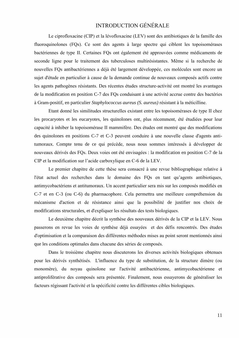
INTRODUCTION GÉNÉRALE
Le ciprofloxacine (CIP) et la lévofloxacine (LEV) sont des antibiotiques de la famille des
fluoroquinolones (FQs). Ce sont des agents à large spectre qui ciblent les topoisomérases
bactériennes de type II. Certaines FQs ont également été approuvées comme médicaments de
seconde ligne pour le traitement des tuberculoses multirésistantes. Même si la recherche de
nouvelles FQs antibactériennes a déjà été largement développée, ces molécules sont encore un
sujet d'étude en particulier à cause de la demande continue de nouveaux composés actifs contre
les agents pathogènes résistants. Des récentes études structure-activité ont montré les avantages
de la modification en position C-7 des FQs conduisant à une activité accrue contre des bactéries
à Gram-positif, en particulier Staphylococcus aureus (S. aureus) résistant à la méticilline.
Etant donné les similitudes structurelles existant entre les topoisomérases de type II chez
les procaryotes et les eucaryotes, les quinolones ont, plus récemment, été étudiées pour leur
capacité à inhiber la topoisomérase II mammifère. Des études ont montré que des modifications
des quinolones en positions C-7 et C-3 peuvent conduire à une nouvelle classe d'agents anti-
tumoraux. Compte tenu de ce qui précède, nous nous sommes intéressés à développer de
nouveaux dérivés des FQs. Deux voies ont été envisagées : la modification en position C-7 de la
CIP et la modification sur l‘acide carboxylique en C-6 de la LEV.
Le premier chapitre de cette thèse sera consacré à une revue bibliographique relative à
l'état actuel des recherches dans le domaine des FQs en tant qu‘agents antibiotiques,
antimycobactériens et antitumoraux. Un accent particulier sera mis sur les composés modifiés en
C-7 et en C-3 (ou C-6) du pharmacophore. Cela permettra une meilleure compréhension du
mécanisme d'action et de résistance ainsi que la possibilité de justifier nos choix de
modifications structurales, et d'expliquer les résultats des tests biologiques.
Le deuxième chapitre décrit la synthèse des nouveaux dérivés de la CIP et la LEV. Nous
passerons en revue les voies de synthèse déjà essayées et des défis rencontrés. Des études
d'optimisation et la comparaison des différentes méthodes mises au point seront mentionnés ainsi
que les conditions optimales dans chacune des séries de composés.
Dans le troisième chapitre nous discuterons les diverses activités biologiques obtenues
pour les dérivés synthétisés. L'influence du type de substitution, de la structure dimère (ou
monomère), du noyau quinolone sur l'activité antibactérienne, antimycobactérienne et
antiproliférative des composés sera présentée. Finalement, nous essayerons de généraliser les
facteurs régissant l'activité et la spécificité contre les différentes cibles biologiques.

12

13
CHAPITRE I BIBLIOGRAPHIE

14

15
1. Les quinolones antibactériennes
1.1 Généralités
Pour des raisons de compréhension, avant de détailler les propriétés antibactériennes des
quinolones, certains concepts fondamentaux de microbiologie seront présentés. La plupart des
bactéries peuvent être classées dans deux groupes selon la coloration de Gram. Cette technique
permet de mettre en évidence les propriétés de la paroi bactérienne, et d'utiliser ces propriétés
pour les distinguer et les classifier (à Gram-positif et à gram-négatif). Des exemples
représentatifs des bactéries à Gram-négatif sont: Escherichia coli (E. coli) et Pseudomonas
aeruginosa (P. aeruginosa). Des bactéries à Gram-positif d'importance médicale incluent: S.
aureus et Streptococcus pneumoniae (S. pneumoniae). Une autre classification est basée sur la
nécessité (ou pas) de la présence de dioxygène dans le milieu de culture des microorganismes: on
parle alors de bactéries aérobies ou anaérobies.
1.2 Histoire
Le motif quinolone est apparu il y a 50 ans1 et c‘est toujours l'une des structures les plus
courantes dans le domaine des molécules biologiquement actives. Les quinolones sont des agents
anti-infectieux totalement synthétiques dont la structure moléculaire se compose de plusieurs
systèmes cycliques (hétérocycliques). Les plus importants, ainsi que leurs systèmes de
numérotation sont illustrés sur la Figure 1.
Figure 1 Structures et systèmes de numérotation des quinolones les plus
importantes
Les quinolones de première génération ont été reconnues comme des antibiotiques dont le
spectre d‘activité était limité aux bactéries à Gram négatif aérobies. Au fil des années, de
nouvelles quinolones ont vu le jour avec l'introduction en 1980 de l'atome de fluor en position C-
6. Les fluoroquinolones (FQs) ne sont pas seulement plus actives que les agents précédents
contre les bactéries à Gram-négatif, mais leur spectre a été étendu successivement aux bactéries à

16
Gram positif et aux anaérobies cliniquement importantes. Les FQs sont devenues l'un des
groupes les plus utilisés des antibiotiques à large spectre2.
La famille des quinolones antibactériennes est généralement divisée en générations, dans
le paragraphe suivant nous allons utiliser la classification basée sur le spectre antimicrobien3.
1.2.1 Première génération
Les quinolones sont issues des premiers travaux de George Lesher et ses collaborateurs
en 19621. L'activité antimicrobienne de l'acide nalidixique (NAL, Tableau 1) a été étudiée sur un
panel de micro-organismes, elle a révélé son activité contre les agents pathogènes à Gram négatif
comme E. coli et Klebsiella pneumoniae (K. pneumoniae). Cependant, plusieurs inconvénients
tels qu‘une pharmacocinétique défavorable, des effets indésirables, et une faible activité contre
les bactéries à Gram-positif et anaérobies, ont limité les indications de NAL aux infections des
voies urinaires non compliquées (IVU)4. Néanmoins, ce composé a stimulé un intérêt croissant
pour cette classe et a donné lieu à des développements ultérieurs.
1.2.2 Deuxième génération
Les quinolones de deuxième génération sont marqués par l'introduction d'un atome de
fluor en position C-6, ce qui a grandement amélioré leur spectre d‘action et augmenté l
'absorption des médicaments par les cellules bactériennes5. Ce groupe est généralement
représenté par deux sous-classes.
La classe I est représentée par la norfloxacine (NOR, Tableau 1) et la péfloxacine (PEF,
Tableau 1) qui possèdent une activité remarquablement augmentée contre les bactéries à Gram-
négatif et une activité importante contre certains agents pathogènes à Gram-positifs, comme S.
aureus sensible à la méticilline. Pourtant, cette classe présente encore des inconvénients majeurs
par exemple, une distribution tissulaire faible6.
La classe II est marqué par l'émergence de la ciprofloxacine7 (CIP, Tableau 1), dérivant
de NOR par le remplacement du substituant N-1 éthyl par cyclopropyl. CIP possède l'une des
activités les plus puissantes contre une large gamme d'organismes à Gram-négatif, une
augmentation d'activité contre les bactéries à Gram-positif et les bactéries atypiques8. Un autre
représentant de cette classe est l'ofloxacine (OFL, Tableau 1) qui introduit un pont entre les
positions N-1 et C-8 du noyau quinolone, conduisant au squelette 1,4-benzoxazine. En raison de
la pharmacocinétique améliorée9 (biodisponibilité et distribution tissulaire) et du large spectre
d'activité, la LEV et la CIP sont devenues des antibiotiques par voie orale largement utilisés et
ont élargi les indications des FQs pour inclure : les infections gastro-intestinales, génitales,

17
osseuses et ostéo-articulaires. Cependant, les molécules de cette classe II n'ont pas d'activité anti-
streptocoques à Gram-positif telles que S. pneumoniae. Par ailleurs, l'utilisation importante (et
parfois abusive) du CIP a provoqué l'émergence de souches résistantes dans les hôpitaux et les
communautés10
.
1.2.3 Troisième génération
La séparation des énantiomères de OFL a montré que l'isomère R est presque inactif alors
que l'isomère S est responsable de l'activité du racémate11
. L'isomère S a été nommé
lévofloxacine (LEV, Tableau 1) et a présenté un niveau élevé d'activité contre les
staphylocoques et les anaérobies, y compris les souches résistantes à la méticilline. Toutefois, la
LEV a présenté une activité légèrement inférieure à CIP contre les agents pathogènes à Gram-
négatif, y compris P. aeruginosa12
. Les autres représentants de cette classe, la gatifloxacine
(GAT, Tableau 1) et la moxifloxacine (MOX, Tableau 1) ont également une bonne activité
contre les souches à Gram-positif 13
, y compris les souches résistantes à la CIP14
. Les 8-OMe
FQs ont été introduites avec l‘intention de diminuer la fréquence de sélection des mutants15
. Les
concentrations sériques élevées et les demi-vie plus longues ont permis le dosage une seule fois
par jour pour les représentants de la troisième génération16
. Ces caractéristiques ont élargi la
gamme de leurs indications à la pneumonie acquise en communauté (PAC) en comparaison avec
les générations précédentes.
1.2.4 Quatrième génération
Les quinolones de quatrième génération sont les composés les plus puissants avec un
spectre large et un haut degré d'activité. Avec l'augmentation de l'activité anti-Gram-positif ces
dérivés possèdent une activité considérablement augmentée contre les anaérobies17
et les
bactéries à Gram-négatif, y compris P. aeruginosa18
. L‘élargissement du spectre et
l‘augmentation du degré d'activité antibactérienne a permis de nouvelles indications aux FQs de
4ème génération dans le traitement des infections intra-abdominales. Du point de vue de la
structure chimique, en plus des changements de substituants en position C-7, ces agents comme
la trovafloxacine (TRO) et la gémifloxacine (GEM,) sont des dérivés de type 1,8-naphtyridine,
similaires à NAL, tandis que ulifloxacin19
(ULI, Tableau 1) qui est utilisé sous la forme de
prodrogue (prulifloxacin20
, PRU, Tableau 1) comporte une annélation originale entre le N-1 et le
C-2 de la quinolone.
Pour conclure, au cours des différentes modifications structurales les quinolones ont
considérablement augmenté leur spectre antibactérien et leur degré d‘activité. Ces

18
caractéristiques, combinées à l'amélioration de leur pharmacocinétique ont permis un
élargissement de leur indication pour le traitement des infections urinaires, des voies
respiratoires et intra-abdominales. Toutefois, les problèmes de toxicités ont entraîné le retrait de
plusieurs quinolones commercialisées. Par ailleurs, l‘apparition de phénomènes de résistance aux
anciennes quinolones a également perturbé le développement de cette classe d'antibactériens.
Dans le prochain paragraphe, nous donnerons un aperçu du mécanisme d'action et de résistance
des FQs, ce qui nous aidera à rationaliser nos synthèses et à expliquer nos résultats biologiques.
19
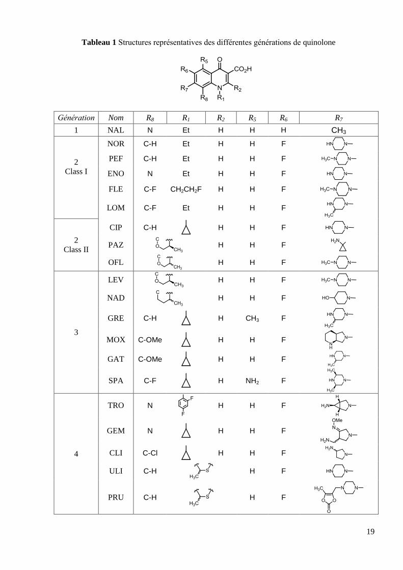
Tableau 1 Structures représentatives des différentes générations de quinolone
Génération Nom R8 R1 R2 R5 R6 R7
1 NAL N Et H H H CH3
2
Class I
NOR C-H Et H H F
PEF C-H Et H H F
ENO N Et H H F
FLE C-F CH2CH2F H H F
LOM C-F Et H H F
2
Class II
CIP C-H
H H F
PAZ
H H F
OFL
H H F
3
LEV
H H F
NAD
H H F
GRE C-H
H CH3 F
MOX C-OMe
H H F
GAT C-OMe
H H F
SPA C-F
H NH2 F
4
TRO N
H H F
GEM N
H H F
CLI C-Cl
H H F
ULI C-H
H F
PRU C-H
H F
20
1.3 Mechanism of action and resistance
1.3.1 Targets for antibacterial quinolones
DNA gyrase was first found in Gram-negative organisms. For many years it was thought
to be the only quinolone target21
. However, in 1990 DNA topoisomerase IV, a homolog of
gyrase, was discovered22
and it was demonstrated that this enzyme is a primary target of FQ in
Gram-positive S. aureus23
. Both gyrase and topoisomerase IV are composed of two subunits
(GyrA and GyrB in gyrase, ParC and ParE* in topo IV) assembled into a functional
heterotetramer i.e. A2B2 and C2E2 respectively. Both enzymes are type II topoisomerases as they
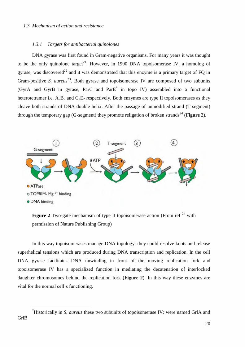
cleave both strands of DNA double-helix. After the passage of unmodified strand (T-segment)
through the temporary gap (G-segment) they promote religation of broken strands24
(Figure 2).
Figure 2 Two-gate mechanism of type II topoisomerase action (From ref 24
with
permission of Nature Publishing Group)
In this way topoisomerases manage DNA topology: they could resolve knots and release
superhelical tensions which are produced during DNA transcription and replication. In the cell
DNA gyrase facilitates DNA unwinding in front of the moving replication fork and
topoisomerase IV has a specialized function in mediating the decatenation of interlocked
daughter chromosomes behind the replication fork (Figure 2). In this way these enzymes are
vital for the normal cell‘s functioning.
*Historically in S. aureus these two subunits of topoisomerase IV: were named GrlA and
GrlB

21
Quinolones bind the DNA-enzyme complex where DNA is kept in the intermediate stage
of double helix passing in which both DNA strands are cut. Ternary complex forms a barrier to
the moving replication fork and arrests it 10 base-pairs upstream the DNA cleavage sites25
.
However due to reversibility of the quinolone-enzyme-DNA complex formation, DNA inhibition
cannot be the cause of immediate cell death and fails to correlate with rapid cell death in terms of
kinetics, quinolone concentration. The most straightforward model is that drug-enzyme-DNA
complexes block bacterial growth (determined by Minimum inhibitory concentration, MIC - is
the lowest concentration of drug that inhibits more than 99% of the bacterial population) while
the release of double-stranded DNA breaks was proposed to be the cause of lethal
consequences26
.
1.3.2 Structure of ternary complex
For a long time the structure of the ternary complex remained unresolved and only
tentative models based on structure-activity relationship (SAR) and mutations studies were
proposed for the quinolone-DNA-topoisomerase interaction. Recently Lapogonov et al. finally
resolved the crystal structure of the quinolone-DNA cleavage complex of the topoisomerase IV
from S. pneumoniae27
. Two molecules of MOX are intercalated in the gap between -1 and +1
nucleotides at the two ends of DNA double-strand cut (Figure 3).
Figure 3 Two molecules of MOX bound to the S. pneumoniae topoisomerase IV-
DNA cleavage complex. (From ref 27
with permission of Nature Publishing
Group)
The bulky substituent at C-7 position is positioned next to the DNA bases at +4 and +5
positions on the opposite DNA strand and projects into large solvent-accessible volume above
the DNA cut (Figure 3). This result is consistent with SAR studies showing that large variations

22
at C-7 position are accepted (see section 2.3.2). The N-1 cyclopropane ring is located close to
ParC -helix residue Ser79 and Asp83, whose mutation is responsible for quinolone resistance
28(Figure 4).
Figure 4 MOX bound to S. pneumoniae topoisomerase IV-DNA cleavage
complex. Quinolone-enzyme interactions (From ref 27
with permission of Nature
Publishing Group)
The fluorine atom at C-6 position is situated far from the protein chain and DNA strand
so its influence on the quinolone potency might be explained by altering the electron density of
the heterocyclic core. Studies have shown that by fine tuning of other substituents one could
achieve high level of potency without fluorine atom at C-629
. The 3-carboxy and 4-keto groups
of the quinolone are located closest to the ParC -helix Gly77, Asp78 residues which are
possible partners for hydrogen bonding; but far away from the magnesium binding side of the
ParC TOPRIM (topoisomerase-primase) domain, though direct metal chelation is questioned.
1.3.3 Resistance to quinolones
Over the 30 years that have elapsed since the introduction of FQs, resistance to these
agents has become common and widespread. A number of reports of the European Antimicrobial
Resistance Surveillance System (EARSS) showed gradual increase of the part of FQ-resistant
pathogens in Europe: for example from 9% (2001) to 20% (2009) for E. coli30
. These facts show
the growing threat of emerging non-susceptibility to older quinolones and the constant need for
the development of new agents.

23
1.3.3.1 Modifications at the target site and target preference
The most important mechanism of FQ resistance is mediated by amino acid substitutions
in the target enzymes. This type of resistance arises stepwise. In several species, the first-step
mutations occur in the gyrA and occasionally in gyrB (genes encoding GyrA and GyrB subunits
of DNA gyrase) while in others they occur in parC and less often in parE (genes encoding ParC
and ParE subunits of topoisomerase IV). Genetic evidences of this type are often used to prove
the target preference of the FQ.
In Gram-negative species, GyrA is the primary target of 4-quinolones, as the first
mutations conferring resistance occur in GyrA and single ParC mutations do not change the MIC
of quinolones for E. coli31
. The whole set of the most frequently occurred mutation was named
―quinolone-resistance determining region‖ (QRDR) which is located between amino acids
positions 67 and 106 in GyrA subunit of E. coli gyrase32
. These residues are situated in the
proximity to the putative quinolone binding pocket in the enzyme-DNA cleavage complex and
thus reduce the drug affinity to the complex of modified enzyme and DNA. Amino acid
positions 447 and 426 in GyrB subunit were found to be responsible for the low-level
resistance33
.
In Gram-positive species, the situation with target preference is not so evident; both
topoisomerase IV and gyrase may be the targets with different degree of preference.
Research with a large number of S. aureus isolates showed that the Ser80Phe alteration in
GrlA and Ser84Leu change in GyrA are the principal ones and both mutations in DNA gyrase
and topoisomerase IV are needed for high-level resistance34
.
The target preference in S. aureus varies between quinolones and according to studies of
Takei et al.35
these agents can be divided into three categories. The first type includes NOR,
ENO, fleroxacin (FLE), CIP, OFL and LEV (Tableau 1); which have greater MICs for grlA
mutant strains (grlA is the gene encoding GrlA subunit of topo IV) than for gyrA mutant strains
(gyrA is the gene encoding GyrA subunit of gyrase). These quinolones displayed lower half
maximal inhibitory concentration (IC50) values (the concentration of drug required for 50%
inhibition of enzyme activity in vitro) for purified topoisomerase IV from wild-type strain than
IC50 values for DNA gyrase. These results suggest the preferential topoisomerase IV inhibition
by this group of quinolones.
The second type includes sparfloxacin (SPA) and nadifloxacin (NAD) (Tableau 1).
These quinolones showed reversed order of activity: greater MICs for gyrA than for grlA mutants
and lower IC50 values for DNA gyrase. These results suggest the preferential DNA gyrase
inhibition by these analogs.

24
The third type comprises GAT, pazufloxacin (PAZ), MOX and clinafloxacin (CLI)
(Tableau 1). In this group, similar values of MICs and IC50 for both targets were observed. It has
been suggested that antibacterial activity of these compounds are mediated by dual-inhibition of
both DNA gyrase and topoisomerase IV.
The most frequent resistance mutations in S. pneumoniae comprise alterations in ParC
(Ser79Tyr/Phe, Ala84Thr) and in GyrA (Ser81Tyr)36
, which are analogous to the previously
mentioned in other species. Also single mutations in ParC conferred only low-level resistance
while for the high-level resistance both DNA gyrase and topoisomerase IV have to be altered37
.
The target preference at S. pneumoniae is also dependent on type of quinolone. Comparing IC50
of purified enzymes and single-step mutations it was proposed that for CIP, LEV, NOR, TRO38
topo IV is a primary target, while for SPA, GAT38
, grepafloxacin39
(GRE, Tableau 1) it is the
DNA gyrase; CLI seems to be equipotent against both enzymes40
.
1.3.3.2 Efflux mediated resistance
The second mechanism of resistance in bacteria is an active drug efflux by
overexpression of certain efflux pumps. These are transmembrane proteins which are responsible
for the active transport of different molecules. Efflux pump mechanisms probably have a
preexisting physiological role, protecting the bacillus against high intracellular levels of toxic
molecules. In addition, efflux pumps maintain cellular homeostasis and physiological balance
through transport of the toxins or metabolites to the extracellular environment41
. Specific gene
norA was found to confer quinolones resistance in S. aureus42
. The study of relative contribution
of efflux mediated resistance by selective inhibition of multidrug efflux pumps showed that
hydrophilic quinolones are more susceptible to the active transport while more hydrophobic
MOX, SPA and TRO are less affected by this type of resistance43
. Bulky substituents and large
molecular weight of compounds should also reduce their transport by efflux pumps.
1.3.3.3 Plasmid mediated resistance
The third and recently discovered mechanism of resistance is associated with intracellular
plasmids. A multiresistance plasmid was recently discovered that encodes transferable resistance
to quinolone44
. The plasmid-quinolone resistance gene was termed qnr and the corresponding
protein Qnr was shown to protect E. coli DNA gyrase from the inhibition by CIP. It was
hypothesised that protective action of Qnr results from the formation of Qnr–gyrase complex
which occurs before the formation of the cleavage complex with DNA. It is noteworthy that

25
plasmids are extrachromosomal genetic elements, thereby granting plasmid mediated resistance a
high degree of mobility which poses risks of the resistance spreading among different organisms.
In this paragraph, we have shown that bacterial type II topoisomerases are intracellular
targets of FQs that turn these enzymes into cellular poisons. Between different resistance
mechanisms, mutations at the target site are the most important. However, the relative activity of
FQs is influenced by their intracellular concentration in the cell. The latter is determined by the
ability of drug molecules to penetrate the cellular wall and will be discussed in detail in the next
paragraph.
1.3.4 Penetration into bacterial cell
Penetration of quinolones into bacterial cell is tightly related with cellular efflux/influx
balance. In the case of extracellular flow, there are evidences for the presence of active multidrug
efflux in resistant strains, as we have described in the section 1.3.3.2. By contrast, quinolone
influx seems to be energy-independent, governed by the passive diffusion45
.
Figure 5 Protonation equilibria of FQ
To date, there are numerous studies that reveal correlations between lipophilicity,
molecular weight, acidity/basicity and FQ uptake in different organisms. Because quinolones
present both acidic and basic functionality, these molecules could exist in four different
microspecies in aqueous solution: cationic H2Q+, neutral HQ
0, zwitterionic HQ
+– and anionic Q
–
(Figure 5). Several experimental evidences testify that in Gram-positive organisms with a single
cell membrane FQ transferred in the neutral HQ0 form by passive diffusion through phospholipid
bilayer membrane; more lipophilic analogs should have higher transport rates46
. In Gram-

26
negative species penetration is complicated by the presence of the additional lipopolysacharide
outer membrane, which is 2 orders of magnitude less permeable than ordinary phospholipid
bilayer towards lipophilic compounds (Figure 6).
Figure 6 Simplified cell wall structures in Gram-positive and Gram-negative
bacteria and their penetration by FQ.
In this case the main part of the FQ influx assisted through the porin channels. Porins are
beta-barrel proteins that cross an outer membrane and act as a pore with size-limited
permeability (with slight cation selectivity). So only relatively small and hydrophilic molecules
can freely penetrate them47
. Chelation of FQ with magnesium ions would provide hydrophilic
positively-charged species which are thought to be responsible for the FQ influx in Gram-
negative species45
.
In this paragraph, we have described the modern understanding of quinolone action and
resistance which will help us to understand some of structure-activity dependencies in the next
paragraph.

27
1.4 Structure-activity relationships
The history, development and SARs of the quinolones have been extensively reviewed48
.
Quinolone SARs are usually examined position by position so in this section we will introduce
general findings; then we will focus on the C-3 and C-7 positions which are relevant to our work.
1.4.1 General trends
As synthetic antibacterial agents, FQ consist of the main 3-carboxy-4-pyridone
pharmacophore and, as auxopharmacophore, a fused aromatic ring with attached substituents
which serves to modulate the potency and antibacterial spectrum (Figure 7).
Figure 7 General trends in FQ SAR
SAR studies in the early era suggested that optimal groups to be attached to the N-1
position should be relatively small and hydrophobic5. Apart from the N-1 ethyl substituent in
NOR or cyclopropyl group in CIP, some aryl groups at N-1 could improve Gram-negative
spectrum of activity in comparison to NOR49
(Table 2).

28
Table 2 Antibacterial activity of some N-1 substituted quinolones: alkyl and aryl
substituents
R1
MIC, M
Gram (–) Gram (+)
E. colia S. aureus
b
CH3 1.26 20.2
CH2CH3 (NOR) 0.16 1.22
CH2CH2CH3 0.60 4.68
CH2CH2F 0.30 4.64
CH2CH2OH 1.16 4.65
E. colic S. aureus
d
CH2CH3 (NOR) 0.31 2.44
C6H5 0.56 1.06
4'-FC6H4 0.13 0.54
Data from ref 5, 49
aE. coli NIHJ JC-2 strain
bS. aureus 209P strain
cE. coli Juhl strain
dS.
aureus ATCC 6538P strain
Further research for possible N-1 substituents has led to preparation of tricyclic quinolone
ring systems in which a new saturated ring is formed to give 1,8-bridged quinolones – LEV and
OFL. Studies have shown that there is a stereochemical preference for the methyl at the
bridging 3-position50
. Stereochemistry of LEV gives approximately 12 fold increase in DNA
gyrase inhibitory activity in comparison to its enantiomer, while ratio for the in vitro MICs
varies in 31-125 fold increase and is much greater than expected from gyrase inhibition50b
(Table 3)

29
Table 3 Antibacterial activity of 1,8-bridged quinolones: Stereoselectivity
Name R1 R2
MIC( M) IC50 ( M),
DNA
gyrase a
Gram (+) Gram (–)
E. colib P. aeruginosa
c S. aureus
d
OFL
(racemate) H/CH3 H/CH3 0.14 0.55 1.1 2.10
LEV H CH3 0.07 0.28 0.55 1.05
- CH3 H 2.16 17.3 69 13.0
Data from ref 50
aCorresponds to 50% inhibition of DNA gyrase supercoiling activity
isolated from E. coli KL 16 b
E. coli KL 16 strain cP. aeruginosa 32122 strain
dS. aureus 209P
strain
The C-2 position is also close to the fixation site, so for a long period of time hydrogen
atom was the only successful substituent. However, an example of C-2 modified potent
compound is ULI (Table 4) with a broad-spectrum activity comparable to that of CIP19
.
Table 4 Ulifloxacin antimicrobial activity
Compound
MIC50, Ma
Gram (–) Gram (+)
E. coli(43b) P. aeruginosa(42
b) S. aureus(37
b)
ULI 0.29 1.13 2.26
CIP 0.15 0.60 1.18
Data from ref 19
aCompounds were tested against collection of clinical isolates; MIC50 is
MIC for 50% of isolates tested. bTotal number of isolates in a collection

30
Initial findings showed that modifications in the 4-pyridone ring render significantly less
active compounds. There are no analogs with alterations at C-3 and C-4 positions that reached
noticeable degree of activity. From the structural data of the quinolone-DNA-enzyme complex, it
was proposed that these positions take part in the quinolone fixation at the active site of enzyme.
The C-5 position is left unsubstituted in the large part of commercial quinolones. It is
thought to influence potency and provide additional Gram-positive activity. This position
demonstrates important phenomena of the different substituent interaction within quinolone
heterocyclic core. While C-5 substitution itself induces chromosomal injury the use of finely
tuned combination of 5-NH2 and C-8 substituents results in a significant decrease of toxicity51
.
Thus far, there are few examples of successful 5-position modification and 5-NH2 (SPA), 5-CH3
(GRE) are the most beneficial for in vitro activity52
(Table 5).
Table 5 Comparative in vitro antimicrobial activity of GRE and SPA
Compound
MIC90, Ma
Gram (–) Gram (+)
E. coli P. aeruginosa S. aureus
GRE 0.17 22.3 0.33
SPA 0.15 10.2 0.31
Data from ref 52
aCompounds were tested against collection of clinical isolates; MIC90 is
MIC for 90% of strains tested.
Original quinolones were often unsubstituted at C-6 position. The main finding of the
pioneering work of Koga et al.5 illustrated a very clear superiority of NOR, a derivative bearing
a fluorine atom at the 6 position over its non-fluorinated analog (Table 6).

31
Table 6 In vitro antimicrobial activity of C-6 fluorinated vs non-fluorinated NOR
R6
MIC ( M)
Gram (–) Gram (+)
E. colia P. aeruginosa
b S. aureus
c
F (NOR) 0.16 1.22 1.22
H 2.59 10.4 41.5
Data from ref 5 aE. coli NIHJ JC-2 strain
bP. aeruginosa V-1 strain
cS. aureus 209P strain
Strong increase of activity due to fluorine atom at C-9 has also been demonstrated among
LEV derivatives53
(Table 7).
Table 7 In vitro antimicrobial activity of C-9 fluorinated vs non-fluorinated LEV
R9
MIC ( M)
Gram (–) Gram (+)
E. colia P. aeruginosa
b S. aureus
c
F (LEV) 0.044 1. 0.33
H 23 >93 12
Data from ref 53
aE. coli ES142 strain
b P. aeruginosa PS 96 strain
cS. aureus MI246 strain
It has become almost a dogma that a fluorine atom at C-6 position (or C-9 position on
benzoxazine skeleton) is essential for antibacterial activity. However, several studies have shown
that positive effect of a C-6 fluorine atom is diminished when the molecule contains other
helpful substituents. The development of the first marketed drug without fluorine atom at C-6
position – Garenoxacin54
(GAR, R6 = H, Table 8) resulted from a number of analogs by fine

32
tuning of other substituents in order to minimize toxicity while maintaining high levels of
potency.
Table 8 Antimicrobial activity and IC50 of GAR and its fluorinated analog
R6
IC50( M) MIC ( M)
E. coli
Gyrasea
S. aureus
topo IVb
Gram (–)
E. colic
Gram (+)
S. aureusd
H (GAR) 0.40 5.14 0.14 0.016
F 0.36 10.4 0.28 0.016
Data from ref 54
aCorresponds to 50% inhibition of gyrase supercoiling activity
bCorresponds to 50% inhibition of topoisomerase IV decatenation activity
cE. coli A29179
strain: genotype GyrA Ser83Leu dS. aureus
MR A27223 strain: genotype homogeneous MR
It has long been known that the substituent at C-8 position controls in vivo efficacy and
affects the antibacterial spectrum. The 8-position substituent can improve anaerobic activity,
modulate physicochemical properties and adverse effects. The most commonly studied groups at
C-8 position were X= C-H, C-F, C-Cl and N, with the following order of in vitro antibacterial
potency: C-Cl ≈ C-F > C-H > N55
(Table 9). While 8-F and 8-Cl substituents showed improved
activity, these groups were associated with a serious adverse effect of phototoxicity56

33
Table 9 In vitro activities of C-8 modified quinolones
X
Gyrase-drug induced
cleavagea
E. coli H560
IC50( M)
MIC ( M)
Gram (–)
E. colib
Gram(+)
S. aureusc
N 3.0 0.15 4.8
C-H (CIP) 1.5 0.15 9.4
C-F 1.44 0.29 1.1
C-Cl 1.4 0.068 0.27
Data from ref 55
aMinimum concentration of drug needed to produce linear DNA at an
intensity relative to oxolinic acid at 10 g/mL bE. coli Vogel strain
cS. aureus H228 strain
The 8-OMe group was found to improve the potency against S. pneumoniae resistant
strains with alterations in target enzymes57
and to slower the development of quinolone
resistance58
. In contrast to 8-F derivatives, 8-methoxy quinolones are photostable and do not
cause phototoxicity58
. The 8-OMe quinolone MOX (Tableau 1) is characterized by a safety
profile comparable to that of older monofluorinated quinolones59
.
1.4.2 Position C-7
The C-7 position is one of the most widely explored among all other positions. From
numerous studies it was deduced that this position determines the antibacterial spectrum, may
influence target preference and is tightly related to pharmacokinetics. In the foregoing section we
will discuss separately ―dimeric‖ and ―monomeric‖ C-7 modified compounds.
1.4.2.1 Monomers
Many thousands of analogs have been prepared employing various substituents at this
promising position, leading to conclusion that a cyclic system containing a secondary or tertiary
amine moiety is preferred2.
34
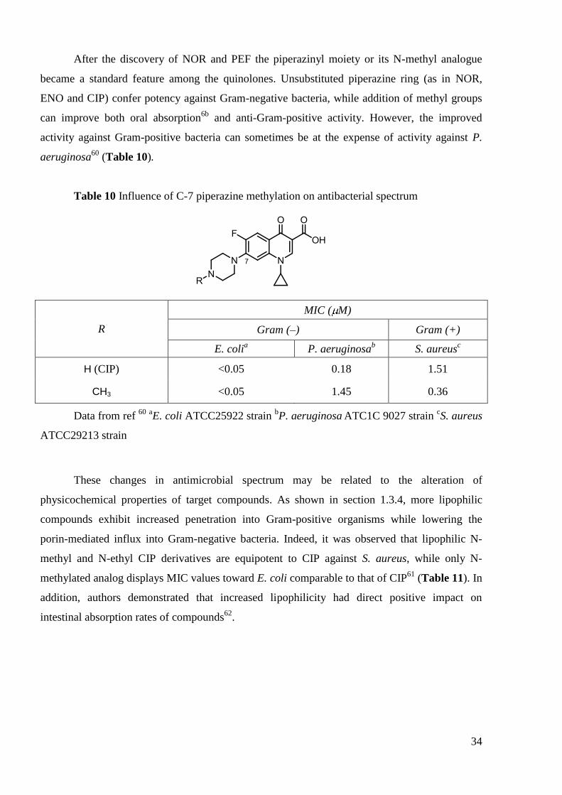
After the discovery of NOR and PEF the piperazinyl moiety or its N-methyl analogue
became a standard feature among the quinolones. Unsubstituted piperazine ring (as in NOR,
ENO and CIP) confer potency against Gram-negative bacteria, while addition of methyl groups
can improve both oral absorption6b
and anti-Gram-positive activity. However, the improved
activity against Gram-positive bacteria can sometimes be at the expense of activity against P.
aeruginosa60
(Table 10).
Table 10 Influence of C-7 piperazine methylation on antibacterial spectrum
R
MIC ( M)
Gram (–) Gram (+)
E. colia P. aeruginosa
b S. aureus
c
H (CIP) <0.05 0.18 1.51
CH3 <0.05 1.45 0.36
Data from ref 60
aE. coli ATCC25922 strain
bP. aeruginosa
ATC1C 9027 strain
cS. aureus
ATCC29213 strain
These changes in antimicrobial spectrum may be related to the alteration of
physicochemical properties of target compounds. As shown in section 1.3.4, more lipophilic
compounds exhibit increased penetration into Gram-positive organisms while lowering the
porin-mediated influx into Gram-negative bacteria. Indeed, it was observed that lipophilic N-
methyl and N-ethyl CIP derivatives are equipotent to CIP against S. aureus, while only N-
methylated analog displays MIC values toward E. coli comparable to that of CIP61
(Table 11). In
addition, authors demonstrated that increased lipophilicity had direct positive impact on
intestinal absorption rates of compounds62
.

35
Table 11 Comparative antimicrobial activity of N-alkyl CIP analogs
R LogPa
MIC ( M)
Gram (–)
E. colib
Gram (+)
S. aureusc
H (CIP) -1.12 0.076 0.76
Me 0.27 0.073 0.73
Et 0.37 0.14 0.70
n-Pr 1.05 0.27 1.34
n-Bu 1.48 0.51 1.29
Data from ref 62a
aP is the bulk phase partition coefficient determined between n-octanol
and 0.066 M Sörensen phosphate buffer, pH 7.00 bE.coli
ATCC25922 strain
cS. aureus
ATCC29213 strain
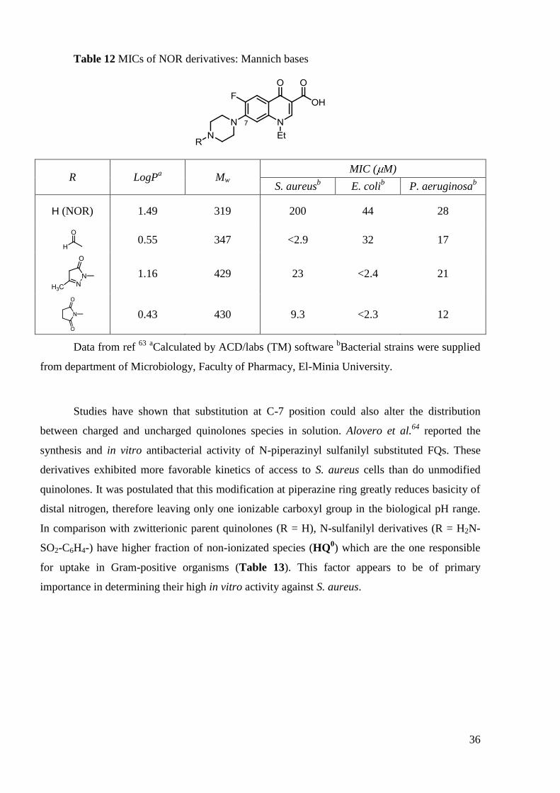
Recently, Abuo-Rahma et al.63
reported the synthesis, physicochemical parameters and
antibacterial activity for a series of N-4-piperazinyl modified NOR (Table 12). The authors
showed that the succinimide Mannich base and the N-formyl analogue revealed a better activity
than NOR in all the tested strains. Interestingly, strong correlation was established between
logMIC and LogP values for P. aeruginosa (r2 = 0.97), while less evident dependence was found
for microorganisms such as S. aureus and E. coli (r2
= 0.67, 0.62 respectively). These results
were explained by the fact that the penetration into bacterial cell is not the only determining
factor which can affect activity. Target affinity is another factor which can influence in vitro
MIC. The difference in the steric bulk at the C-7 substitution may play an important role in the
accomodation of these molecules in the active site of enzyme. Another set of correlations
between molecular weight and activity were obtained, which are consistent with assumption of
porin-mediated quinolone penetration into Gram-negative organisms and passive diffusion
through membrane at Gram-positives.
36
Table 12 MICs of NOR derivatives: Mannich bases
R LogPa
Mw MIC ( M)
S. aureusb
E. colib
P. aeruginosab
H (NOR) 1.49 319 200 44 28
0.55 347 <2.9 32 17
1.16 429 23 <2.4 21
0.43 430 9.3 <2.3 12
Data from ref 63
aCalculated by ACD/labs (TM) software
bBacterial strains were supplied
from department of Microbiology, Faculty of Pharmacy, El-Minia University.
Studies have shown that substitution at C-7 position could also alter the distribution
between charged and uncharged quinolones species in solution. Alovero et al.64
reported the
synthesis and in vitro antibacterial activity of N-piperazinyl sulfanilyl substituted FQs. These
derivatives exhibited more favorable kinetics of access to S. aureus cells than do unmodified
quinolones. It was postulated that this modification at piperazine ring greatly reduces basicity of
distal nitrogen, therefore leaving only one ionizable carboxyl group in the biological pH range.
In comparison with zwitterionic parent quinolones (R = H), N-sulfanilyl derivatives (R = H2N-
SO2-C6H4-) have higher fraction of non-ionizated species (HQ0) which are the one responsible
for uptake in Gram-positive organisms (Table 13). This factor appears to be of primary
importance in determining their high in vitro activity against S. aureus.

37
Table 13 In vitro antimicrobial activity of N-sulfanilyl CIP derivatives
R
MIC ( M)
Gram (–)
E. colia
Gram (+)
S. aureusb
H (CIP) 0.75 0.09
0.26 4.1
Data from ref 64b
aE. coli
ATCC 25922 strain
bS. aureus
ATCC29213 strain
Another important result is that modifications at C-7 position determine not only the
potency but also the target enzyme preference. The authors established that unlike unsubstituted
CIP which targets topoisomerase IV in S. pneumoniae, N-sufanilyl CIP derivatives demonstrated
a target preference for DNA gyrase. Moreover, N-sulfanilyl CIP selected first-step mutant with
alterations at QRDR of DNA gyrase. These genetic studies revealed that 4-
aminobenzenesulfonamide group attached to the distal nitrogen at piperazine moiety provokes
target shift in S. pneumoniae from topoisomerase IV to DNA gyrase and increases activity in
vitro65
. However, it is not clear whether the target shift is related with improved potency or not.
It is worth to mention N-piperazinyl modified CIP derivatives synthesized by Foroumadi
et al. (Table 14). The development started from derivatives containing a N-[2-(furan-3-
yl)ethyl]66
and N-[2-(thiophen-3-yl)ethyl67
residues. These compounds exhibited improved
activity against Gram-positive S. aureus, including methicillin-resistant strains (MRSA) with
respect to CIP. Generally, these agents were significantly less active than parent quinolone
towards Gram-negative P. aeruginosa and E. coli (entry 2, 3 Table 14). Further exploration
yielded a series of compounds with attached phenyl moiety68
. Tuning the substitution pattern at
phenyl ring allowed to greatly increase their potency against Gram-negatives, while maintaining
activity generally comparable to that of parent quinolone against Gram-positives S. aureus and
MRSA (entry 4, Table 14). More recently, the authors created conformationally-constrained

38
analogs69
(entry 5, Table 14). This approach lead to coumarine based compound70
that exhibited
improved potency against Gram-positive S. aureus and MRSA. It is noteworthy to mention that
this derivative was more active against E. coli and P. aeruginosa in comparison with CIP which
exhibits one of the lowest MIC towards Gram-negatives (entry 6, Table 14).
Table 14 Antibacterial activity of various N-substituted piperazinylquinolones
synthetized by Foroumadi et al.
N R
MIC ( M)
Ref Gram (–) Gram (+)
E.
colia
P.
aeruginosab
S.
aureusc
MRSA Id MRSA II
d
1 H (CIP) 0.04 1.2 0.57 1.2 1.2 66
2
0.42 28 0.22 0.42 0.42 66
3
0.83 >212 0.21 0.40 0.40 67
4
0.046 2.9 0.71 0.71 0.71 68
5
1.6 102 0.1 0.2 - 69
6
0.025 0.75 0.37 0.75 0.75 70
aE. coli ATCC 8739 strain
bP. aeruginosa ATCC 9027 strain
cS .aureus
ATCC 6538p
strain dMRSA I and II: methicillin-resistant S. aureus (clinical isolates I and II)
All these reports showed that C-7 position possesses unique features, like great tolerance
for modifications coupled with high influence on antibacterial potency, spectrum, target
preference and physiochemical properties. Cyclic amines are preferred substituents at this
position, with piperazine and aminopyrrolidine being the most popular. Attachment of different
39
groups to the distant 4-nitrogen atom at piperazine moiety, generally increases the potency
against Gram-positives bacteria, including MRSA and lowers activity towards Gram-negatives.
These results could be related to the fact that increased steric bulk and lipophilicity are better
tolerated in Gram-positive organisms with a single cellular membrane. In contrast, substitutions
at piperazine moiety that increase lipophilicity and steric bulk, generally decrease the potency
towards Gram-negatives. However, no single property of C-7 modification determines activity.
The combination of different factors, like structural features, LogP, molecular weight governs
the degree and the spectrum of activity.
1.4.2.2 “Dimers”
Since the first insights into mechanism of inhibition by quinolones it was proposed that
there is more than one molecule present in the quinolone-enzyme complex. In 1989, Shen et al.71
reported for the first time, the coupling of two quinolone pharmacophores in a single molecule.
He showed that among the synthesized N-1 linked NOR ―dimers‖, the derivative containing 4
methylene units, was the most potent against E. coli gyrase (with IC50 value roughly equal to that
of NOR). Further study of Kerns et al.72
identified piperazinyl-linked CIP ―dimers‖ that
displayed equivalent or lowered MIC values compared with those of parent FQ against S.
pneumoniae and E. coli. However, raised MIC values were observed for ―dimers‖ in comparison
with CIP against P. aeruginosa (Table 15). The most potent linkers were trans-butenyl and
para-xylenyl and corresponding ―dimers‖ were tested against wild-type isolate and drug-
resistant strains of S. aureus.

40
Table 15 Piperazinyl-linked CIP ―dimers‖: spectrum of antibacterial activity
Linker
MIC ( M)
Gram (–) Gram (+)
E. colia P. aeruginosa
b S. pneumoniae
c
0.17 >22 0.17
0.33 >21 1.3
—
(CIP) <0.9 1.5 0.75
Data from ref 72-73
aE. coli R973 strain
bP. aeruginosa
27853 strain
cS. pneumoniae
49616
strain
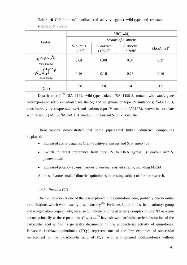
These CIP-based ―dimers‖ displayed up to 10-fold increased activity against wild-type S.
aureus and methicillin-resistant strains of S. aureus73
(Table 16). More interestingly, a 50-fold
increased potency was observed against resistant mutant possessing both NorA efflux-mediated
resistance and a topoisomerase IV mutation. The authors also demonstrated that dimerization
affected the target preference in S. aureus. In fact, the target of ―dimer‖ in S. aureus is primarily
DNA gyrase, whereas topoisomerase IV is the primary target of CIP74
. This could explain
increased potency against topoisomerase IV mutant strain. The authors also reported similar
switch in target preference in S. pneumoniae75
(Table 16).
41
Table 16 CIP “dimers‖: antibacterial activity against wild-type and resistant
strains of S. aureus
Linker
MIC ( M)
Strains of S. aureus
S. aureus
1199a
S. aureus
1199-3b
S. aureus
1199Bc
MRSA 494d
0.04 0.08 0.04 0.17
0.16 0.16 0.16 0.16
—
(CIP) 0.38 3.0 24 1.5
Data from ref 73
aSA 1199, wild-type isolate;
bSA 1199-3, mutant with norA gene
overexpression (efflux-mediated resistance) and no gyrase or topo IV mutations; cSA-1199B,
constitutively overexpresses norA and harbors topo IV mutation (A116E), known to correlate
with raised FQ MICs; dMRSA 494, methicillin resistant S. aureus isolate.
These reports demonstrated that some piperazinyl linked ―dimeric‖ compounds
displayed:
Increased activity against Gram-positive S. aureus and S. pneumoniae
Switch in target preference from topo IV to DNA gyrase (S.aureus and S.
pneumoniae)
Increased potency against various S. aureus resistant strains, including MRSA
All these features make ―dimeric‖ quinolones interesting subject of further research.
1.4.3 Position C-3
The C-3 position is one of the less explored in the quinolone core, probably due to initial
modifications which were usually unsatisfactory48b
. Positions 3 and 4 must be a carboxyl group
and oxygen atom respectively, because quinolone binding at ternary complex drug-DNA-enzyme
occurs primarily at these positions. Chu et al.76
have shown that bioisosteric substitution of the
carboxylic acid at C-3 is generally detrimental to the antibacterial activity of quinolones.
However, isothiazoloquinolones (ITQs) represent one of the few examples of successful
replacement of the 3-carboxylic acid of FQs (with a ring-fused isothiazolone) without
42
compromising antibacterial activity. The subsequent work of Chu et al77
initially investigated
compound A-62824 (Table 17), in which the 3-carboxylic acid group of CIP is replaced by a
fused isothiazolo ring and demonstrated that A-62824 was more potent (4 to 10 times) in vitro
than CIP and possessed enhanced activity against DNA gyrase. The isothiazolo ring system in A-
62824 possesses an aromatic character and the nitrogen proton which is very acidic, can be
considered to mimic carboxylic acid. The authors also suggested that the planarity between the
4-oxo group and the enolized isothiazolo group must be important for the DNA gyrase binding.
Unfortunately, these molecules also inhibited mammalian topoisomerase II78
and caused
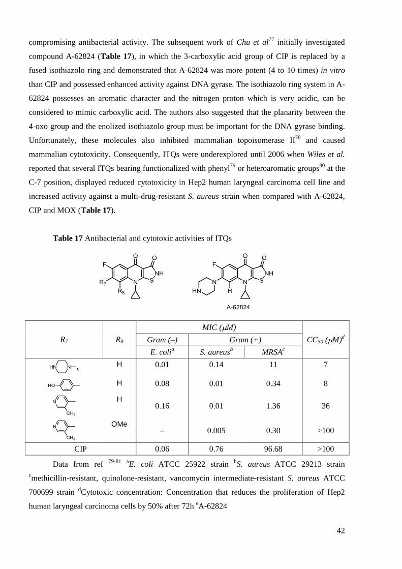
mammalian cytotoxicity. Consequently, ITQs were underexplored until 2006 when Wiles et al.
reported that several ITQs bearing functionalized with phenyl79
or heteroaromatic groups80
at the
C-7 position, displayed reduced cytotoxicity in Hep2 human laryngeal carcinoma cell line and
increased activity against a multi-drug-resistant S. aureus strain when compared with A-62824,
CIP and MOX (Table 17).
Table 17 Antibacterial and cytotoxic activities of ITQs
R7 R8
MIC ( M)
CC50 ( M)d Gram (–) Gram (+)
E. colia S. aureus
b MRSA
c
e
H 0.01 0.14 11 7
H 0.08 0.01 0.34 8
H 0.16 0.01 1.36 36
OMe – 0.005 0.30 >100
CIP 0.06 0.76 96.68 >100
Data from ref 79-81
aE. coli ATCC 25922 strain
bS. aureus
ATCC 29213 strain
cmethicillin-resistant, quinolone-resistant, vancomycin intermediate-resistant S. aureus ATCC
700699 strain dCytotoxic concentration: Concentration that reduces the proliferation of Hep2
human laryngeal carcinoma cells by 50% after 72h eA-62824

43
Further modification of the ITQ nucleus at C-8 position81
provided the 8-methoxy
derivative with the most attractive in vitro biological profile (Table 17). ITQs displayed broad-
spectrum activities especially against Gram-positive pathogens, including several antibiotic-
resistant organisms, such as MRSA, vancomycin-resistant S. aureus (VRSA) and quinolone-
resistant strains82
. Moreover, ITQs were proved to interact with both DNA gyrase and
topoisomerase IV – so called dual-targeting mode of inhibition, with the preference for DNA
gyrase83
. Genetic studies suggested the potential utility of heteroaryl isothiazolones in combating
infections caused by S. aureus, including multidrug-resistant MRSA.
Another interesting approach is represented by the introduction of amide moiety as a
mimic of carboxyl group. Patel et al.84
described the synthesis and antimicrobial activity of CIP
carboxamides, whereas LEV carboxamides were evaluated by Sultana et al.85
. Significant
enhancements of potency towards Gram-positive organisms were achieved in comparison with
unmodified precursor (Table 18)
Table 18 Zone of inhibition of LEV and its C-6 carboxamide derivative
R
Zone of inhibition, mma
Gram (–) Gram (+)
E. coli P. aeruginosa S. aureus S. pneumoniae
OH (LEV) 16 14 12 16
NH-C6H5 15 14 16 19
Data from ref 85
aDiameter of inhibited zone proportional to the bacterial susceptibility to
the antimicrobial present in the disk; measured at 10 ppm concentration. The bacterial strains are
not indicated.
In this section, we have shown studies demonstrated that in spite of the low tolerance for
a structural modification an efficient mimic of carboxylate group with acidic moiety could be
introduced at C-3 position. This would increase in vitro potency and broader the spectrum of
activity. Varying substituents at this position could also control the target preference.

44
2. Quinolones as antimycobacterial agents
2.1 Introduction
Tuberculosis (TB) is one of the most common infectious diseases known to humans.
About 32% of the world‘s population (1.9 billion people) is infected with TB. Every year,
approximately 9.4 million of the infected people develop active TB and almost 1.8 million of
these infected people die from the disease, a life lost due to TB every 15 s. The first-line drugs
currently used for the treatment of TB, an infection caused by M. tuberculosis, are isoniazid,
ethambutol, pyrazinamide and rifampicin (Figure 8)86
. However, TB is still a challenging
worldwide health problem, especially due to the emergence of multi-drug resistant (MDR) strain
of M. tuberculosis that is resistant to, at least, the two first-line medications, isoniazid and
rifampin. It is estimated that, 5% of the more than 9 million persons who develop TB every year
are infected with a MDR strain of TB87
.
Figure 8 WHO recommended first-line drugs in TB treatment
The anti-TB activity of FQs has been under investigation since the 1980s. Many are
active in vitro but only a few, including OFL, CIP, SPA, LEV, MOX have been clinically
tested88
. On the basis of these studies CIP, OFL and LEV have been suggested as alternative
treatments in TB (second-line therapy). FQs and injectable agents (aminoglycosides such as
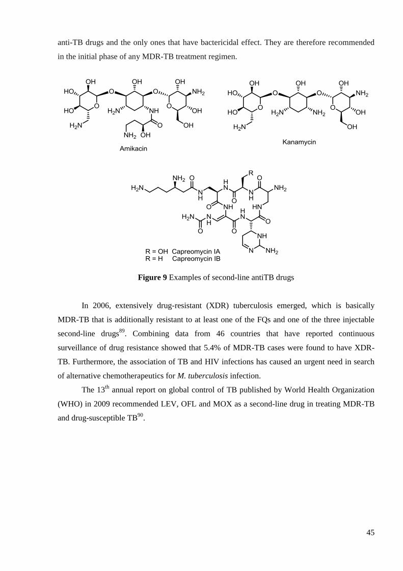
amikacin, kanamycin and peptide capreomycin, Figure 9) are the most effective second-line
45
anti-TB drugs and the only ones that have bactericidal effect. They are therefore recommended
in the initial phase of any MDR-TB treatment regimen.
Figure 9 Examples of second-line antiTB drugs
In 2006, extensively drug-resistant (XDR) tuberculosis emerged, which is basically
MDR-TB that is additionally resistant to at least one of the FQs and one of the three injectable
second-line drugs89
. Combining data from 46 countries that have reported continuous
surveillance of drug resistance showed that 5.4% of MDR-TB cases were found to have XDR-
TB. Furthermore, the association of TB and HIV infections has caused an urgent need in search
of alternative chemotherapeutics for M. tuberculosis infection.
The 13th
annual report on global control of TB published by World Health Organization
(WHO) in 2009 recommended LEV, OFL and MOX as a second-line drug in treating MDR-TB
and drug-susceptible TB90
.

46
2.2 Mechanism of action and resistance
2.2.1 Quinolone target in M. tuberculosis
As previously described in section 1, the bacterial targets of quinolones are type II
topoisomerases (DNA gyrase and topoisomerase IV) which grant quinolone antibiotics with
dual-inhibition mode, lower rates of resistance emerging and high potency. In 1998 the full
genome sequence of M. tuberculosis was revealed and two genes gyrA and gyrB were identified
to encode A and B subunits of DNA gyrase91
. However, contrary to ―common‖ bacteria there is
no evidence of the parC and parE genes for topoisomerase IV in the genome of M. tuberculosis.
DNA gyrase is the only type II topoisomerase present in this organism and hence is the only
target for FQ action. Because topoisomerase IV is necessary for the decatenation of daughter
chromosomes during DNA replication; functions of the single M. tuberculosis type II
topoisomerase were explored by Aubry et al.92
The intermolecular passage activities of purified
DNA gyrase from M. tuberculosis was demonstrated: decatenation activity of this enzyme was
30-fold higher than that of E. coli DNA gyrase, but was lower than that of topoisomerase IV
from S. pneumoniae. Overall, the type II topoisomerase from M. tuberculosis exhibits classical
supercoiling activity of DNA gyrase while having additional decatenation topo IV like activity.
The absence of dual inhibition together with unique topoisomerase protein sequence marks out
M. tuberculosis among ―common‖ Gram-positive/negative bacteria.
2.2.2 Penetration into Mycobacterium cell
Bacterial topoisomerases are intracellular targets and quinolones have to pass through the
cell wall to get into the intracellular space. Mycobacterium species share a characteristic cell
wall, thicker than in many other Gram-positive and Gram-negative bacteria, which is
hydrophobic, waxy and rich in mycolic acids/mycolates93
. Mycobacteria possess, in addition to
phospholipid bilayer and peptidoglycan, the hydrophobic layer of long chain (C70-C80) fatty acids
known as mycolic acids. They are held together with peptidoglycan layer by the polysaccharide,
arabinogalactan (Figure 10).

47
Figure 10 Structure of Mycobacterium cell wall in comparison with Gram-
positive and Gram-negative bacteria.
The low permeability of the mycobacterial cell wall, with its unusual structure, is now
known to be a major factor in intrinsic antibiotic resistance of M. tuberculosis94
. In addition to
the cell wall, there are evidences for the outer capsule existence that creates a barrier for
diffusion of foreign molecules into the inner parts of the envelope95
. It is supposed that more
hydrophilic agents cross the cell wall slowly because the mycobacterial porin is expressed in low
quantities and is inefficient in allowing the permeation of solutes. More lipophilic agents are
presumably slowed down by the lipid bilayer which is of unusually low fluidity and abnormal
thickness.
2.2.3 Resistance to quinolones
In 1970s a dramatic decline in the prevalence of TB infections was observed and TB
ceased to be a problem in industrialised countries. Unfortunately, TB yet remains one of the most
deadly infections primarily due to the emergence of resistance development. As the rate of
mutations conferring resistance to multiple drugs is very low, the WHO-recommended standard
chemotherapy consists of a combination of four first-line drugs (isoniazid, ethambutol,
pyrazinamide and rifampicin), while second-line drugs (FQs, aminoglycosides) are mostly used

48
in the treatment of MDR-TB. Another approach has been proposed to prevent resistance; the FQ
must be administred at doses that produce serum concentrations that exceed the ―mutant
prevention concentration‖ (MPC). This drug concentration is capable of inhibiting all
spontaneous first-step mutants96
. However, to date there is no quinolone which possesses such a
good pharmacodynamic safety profile and high degree of activity.
Emerging resistance poses a serious problem on the way of the world-wide eradication of
TB and there is a constant need for new antimycobacterial agents.
2.2.3.1 Mutations at the target site
The primary source of quinolone resistance in M. tuberculosis is mutations at the DNA
gyrase. Various studies demonstrated that laboratory-selected FQ resistant mutants of M.
tuberculosis showed exactly the same changes described in clinical isolates. Significant
resistance to FQs can be achieved with a single gyrase mutation, whereas at least two mutations
(two mutations in gyrA, or mutations in gyrA plus gyrB) found to be necessary for the highest
level of resistance. Mutations encoutered in FQ-resistant M. tuberculosis were located in codons
equivalent to those in GyrA genes of other FQ-resistant bacteria. The most frequently mutated
codons were: codon 90 (with a Ala/Val change), codon 91 (Ser/Pro) and codon 94 (with five
different amino acid change: Asp/Ala-Gly-His-Tyr or Asn)97
.
2.2.3.2 Efflux mediated resistance
Previously mentioned cell wall barrier alone cannot produce significant level of drug
resistance, which requires synergistic contribution from a second factor, such as the efflux
pumps. Recent evidence suggests that Mycobacteria extrude many drugs via active efflux
systems98
and efflux-mediated FQ resistance has been described in the Mycobacterium
smegmatis (M. smegmatis)99
. The LfrA protein gene has been identified in M. smegmatis; it was
shown to have greater affinity for hydrophilic FQ‘s (CIP, LEV) than for lipophilic ones (SPA,
NAL)100
.
2.2.3.3 DNA mimicking
In 2001 genetic selection for FQ resistance in M. smegmatis identified a new type of
resistance mechanism mediated by the MfpA protein101
. The sequence of MfpA revealed it to be
a member of the ―pentapeptide repeat‖ family of bacterial proteins, in which every fifth amino

49
acid is either a leucine or phenylalanine. This protein forms an adduct with gyrase from M.
tuberculosis and inhibits its activity102
.

50
2.3 Structure-activity relationships
2.3.1 General trends
In comparison with thousands of quinolones tested against ―common‖ bacteria, there are
less data on structure activity studies for Mycobacteria. Because M. tuberculosis divides very
slowly (about 15-20 hours for division), 20-30 days are necessary for testing. In addition,
analysis requires special protective conditions. For all these reasons, several authors utilized less
hazardous and rapidly growing M. fortuitum to estimate the quinolone potency against M.
tuberculosis. Testing results against M. smegmatis were often included for comparative
purposes. Generally, M. fortuitum was more sensitive to the compounds than M. smegmatis.
Renau et al.103
carried out systematic studies focused on the influence of lipohilicity at N-1 and
C-7 positions on quinolone antimycobacterial activity. The evident rationale for these studies
was the fact that, unlike ―common‖ bacteria, M. tuberculosis possesses a thick and lipophilic cell
wall that forms a transport barrier for antibiotics. However, the authors showed that ―intrinsic‖
activity against Gram-positive and/or Gram-negative bacteria is the important factor for
antimycobacterial activity, more determining than lipophilicity of N-1 substituent. The results
summarized (Table 19), demonstrated that compounds with close CLogP values exhibit
dramatic differences in activities toward Mycobacteria.

51
Table 19 Comparative effect of N-1 substituents on antibacterial /
antimycobacterial activity
R1 CLogPe
MIC ( M)
Bacteria G(–)/G(+) Mycobacteria
E. colia S. aureus
b M. fortuitum
c M. smegmatis
d
Et (NOR) 2.21 0.31 9.7 1.6 6.3
(CIP) 2.04 0.15 2.4 0.18 0.75
i-Pr 2.52 2.4 >9.3 3.0 12
2.60 1.2 9.0 1.5 5.8
t-Bu 2.92 0.6 2.3 <0.09 0.37
4.85 0.25 1.0 0.62 1.2
5.20 0.5 2.0 2.5 10
5.53 2.0 >7.8 41 81
Data from ref 103
aE. coli Vogel strain
bS. aureus H-228 strain
cM. fortuitum ATCC6841
strain dM. smegmatis ATCC19420
eA computational model (MedChem Version 3.54, Pomona
College, Pomona, CA) was used to determine the theoretical distribution coefficients (clog P) for
each compound
In this study, the most active agents against mycobacteria were compounds substituted at
N-1 with a tert-butyl and cyclopropyl group. CIP was further modified at C-7 piperazine moiety
with side chains bearing various alkyl substituents in order to modify the lipophilic character of
the heterocycle103
. Detailed results on the influence of the C-7 substituent on the
antimycobacterial activity will be discussed in the corresponding section.
C-8 substituent has influence on the potency against Mycobacteria. In the case of M.
smegmatis the following order was demonstrated for activity against both wild-type strains and
resistant mutants: OMe ≈ Cl > Br > F > H104
(Table 20). Moreover, the C-8 methoxy group,
when compared to unsubstituted C-8 analog greatly reduced the selection of resistant mutants of
M. tuberculosis105
.

52
Table 20 Influence of C-8 position on the activity against M. smegmatis
R8 M. smegmatis, MIC99 ( M)
a
gyrA+b
gyrAc
H 0.290 8.10
F 0.099 3.30
Br 0.075 2.26
Cl 0.076 1.73
OMe 0.080 1.30
Data from ref 104
aThe MIC at which 99% of the isolates tested were inhibited
bM.
smegmatis wild-type mc2155 strain
cM. smegmatis gyrA (Asp95Gly) quinolone-resistant mutant
KD2003
2.3.2 SAR at C-7 position
C-7 position tolerates large structural changes, but an important difference between
mycobacteria and other Gram-positive or Gram-negative bacteria was demonstrated. Haemers et
al.106
studied the influence of N-substitution of C-7 piperazine on antimycobacterial activity of
CIP. Derivatives with longer alkyl chain (and higher lipophilicity) showed higher
antimycobacterial activity. However, introduction of bulky phenyl or benzyl substituents
lowered the potency in spite of high lipophilicity (Table 21).

53
Table 21 Influence of the C-7 piperazine alkylation on the antimycobacterial
activity of CIP
R M. tuberculosis, MIC ( M)
MIC50a MIC90
a
H (CIP) 1.5 3.0
Me 0.36 1.5
Et 0.35 0.70
n-Pr 0.33 0.67
Ph 4.9 9.8
CH2Ph 2.4 4.8
Data from ref 106
aCompounds were tested against collection of M. tuberculosis strains;
MIC50 and MIC90 are MIC for 50% and 90% of isolates tested, respectively.
As it was demonstrated that C-8 methoxy FQs are more active against M. tuberculosis,
Aldeida et al.107
described the anti-TB evaluation of various 8-methoxy-FQ derivatives bearing
some mono N-alkylated 1,3-propane diamine substituents at C-7 position. It was observed that,
similarly with earlier discussed N-alkyl CIP analogs, in the series of GAT analogs 10 carbon
atoms chain length confer maximum potency. In the same time, compounds with shorter or
longer chains are less active (Table 22).

54
Table 22 Activity of C-7 modified GAT analogs against M. tuberculosis H37Rv
(virulent strain)
n m MIC ( M)
3 5 115
3 7 2.71
3 9 0.63
3 11 1.20
GAT 0.27
Data from ref 107
The group of Renau et al. conducted systematic studies on the influence of C-7
substituent on the activity against Mycobacteria. The authors have demonstrated that N-1
cyclopropyl (and t-butyl) quinolones were more active than N-1 2,4-difluorophenyl analogs
(Table 19)103
. This report also included over 15 different residues based on piperazine and
pyrrolidine side chains at C-7 position. The biological evaluation of this series of compounds
revealed that an increase in lipophilicity (via alkyl substitution of piperazine moiety) led to a
decrease in antimycobacterial activity. Results from Table 23 show that in each series the most
lipophilic compounds were the less active towards M. fortuitum. Comparison between the two
series of compounds demonstrated that piperazine-substituted analogs were almost equipotent to
pyrrolidine-substituted derivatives.

55
Table 23 Piperazine vs pyrrolidine side chain at C-7 position: influence on the
antimycobacterial activity
R7 CLogPa
MIC ( M)
M. fortuitum M. smegmatis
(CIP)
2.04 0.18 0.75
2.89 0.17 0.72
3.42 0.17 0.36
3.73 0.67 0.67
4.48 2.6 1.3
1.94 <0.09 0.75
2.13 0.69 1.4
2.62 0.35 1.3
2.66 1.3 1.3
2.97 0.65 1.3
Data from ref 103
aA computational model (MedChem Version 3.54, Pomona College,
Pomona, CA) was used to determine the theoretical distribution coefficients (clog P) for each
compound
The group of Sriram et al.108
synthesized and evaluated some OFL and CIP derivatives
modified at C-7 position. The authors first explored the in vitro antimycobacterial activity of
some Mannich bases of CIP (Table 24) and demonstrated that simply increasing the lipophilic
character of the C-7 substituent (as seen for clogP values) increased the activity.

56
Table 24 Mannich bases of CIP and their antimycobacterial activity
R R' CLogPa
% Inhibition at
6.25 g/mLb
MIC ( M)b
NHCONH2 Br 2.04 99 1.98
NHCSNH2 Br 1.66 100 1.88
F 5.29 100 1.00
CIP 0.01 95 6.04
Ethambutol – 98 9.22
Data from ref 108
aCalculated using online service www.logp.com
bM. tuberculosis H37Rv
strain
To investigate this observation in more depth, the authors prepared and evaluated a series
of C-7 substituted CIP analogs (Table 25)109
. Biological evaluation showed that compounds with
high bulky substituent at C-7 position could display high potency towards M. tuberculosis. In
addition, the most active derivatives were evaluated against MDR strains of M. tuberculosis
showing low values of MICs. A comparison of substitution pattern at C-7 position demonstrated
the following order of activity: piperidine > piperazine > morpholine.

57
Table 25 Influence of bulk and liphophilicity at C-7 position on in vitro
antimycobacterial activity
R7 MIC ( M)
M. tuberculosisa MDR-TB
b
(CIP) 6.04 –
0.17 0.17
0.94 0.94
1.08 2.16
Data from ref 109
aM. tuberculosis H37Rv strain
bMDR-TB clinical isolate from
Tuberculosis Research Center, Chennai, India
The authors recently described a series of OFL analogs bearing various substituents at C-
8 and C-10 positions on the benzoxazine moiety (Table 26)110
. The results demonstrated the
positive impact of C-8 nitro group on antimycobacterial activity with the following order of
activity NO2 > H > NH2. Concerning the substitution pattern at C-10 position the authors have
found the following trend in antimycobacterial activity: piperidine > morpholine > piperazine;
this is quite similar to that obtained for CIP derivatives.

58
Table 26 Influence of C-8 and C-10 substitution on antimycobacterial activity of
OFL derivatives
R8 R10 MIC99 ( M)
M. tuberculosisa MDR-TB
NO2
2.63 2.63
H
0.91 0.44
NO2
0.19 0.09
H (OFL) 2.16 34.6
Data from ref 110
aM. tuberculosis H37Rv strain
2.3.3 SAR at C-3 position
Similar to antibacterial potency C-3 position have low tolerance for modification and
carboxyl group is generally kept to render high levels of antimycobacterial activity. However,
some ITQs that have been already mentioned as potent antibacterials showed noticeable activity
against Mycobacteria. The most potent compound among them is ACH-702111
(Table 27),
which exhibited MICs towards the clinical isolates of M. tuberculosis superior or equal to that of
marketed quinolones.

59
Table 27 Chemical structure of ACH-702 and its antimycobacterial activity
Compouund M. tuberculosis (31)
a ( M)
MIC50 MIC90
ACH-702 0.14 0.28
MOX 0.30 0.62
Data from ref 111
aM. tuberculosis H37Rv strain (number of clinical isolates)
In summary, we can conclude that Mycobacteria species represent a specific target for
FQs with different SAR trends established for other bacteria. Lipophilicity of the antibacterial
agent was shown to play an important role but it is not the only governing factor in determining
the potency towards Mycobacteria. Intrinsic antibacterial activity was also necessary to achieve
high levels of antimycobacterial activity. Recent studies have shown that FQs have a full
potential of becoming a safe and reliable tool for the treatment of classic and resistant TB.

60
3. Quinolones as antiproliferative agents
3.1 Introduction
We have described (section 1.3) the quinolone‘s mechanism of antibacterial action, which
comprises inhibition of bacterial type II DNA topoisomerase enzymes (gyrase and topoisomerase
IV)112
. In order to manage DNA, eukaryotic cells have also evolved a topoisomerase II.
Eukaryotic type II topoisomerase cannot introduce supercoils; it can only relax them. As a result
of its catalytic function, topoisomerase II is necessary for the viability of all organisms from
unicellular bacteria to humans and is the target for some of the most active anticancer agents
used for the treatment of human malignancies113
.
One of the first reports of possible anticancer effect of quinolones came in the middle of
80‘s. It was found that antibacterial quinolones such as CIP, NOR and OFL display only slight
inhibitory activity against calf thymus topoisomerase II114
. Meanwhile, to improve the quinolone
activity toward Gram-positive bacteria, a series of C-7 aryl derivatives were explored and some
of them, bearing 4-pyridinyl substituent, enhanced topoisomerase II-mediated DNA damage and
turned out to be too toxic for the use as antibiotic115
. These early findings provided lead
compounds for further exploration of potential antitumor quinolones with different biological
targets78, 116
. As a result of numerous efforts SNS-595 (Voreloxin®)117
and CX-3543
(Quarfloxin®)118
were discovered and entered phase II clinical trials on patients. However,
currently there is no quinolone-based anticancer drug on the market.

61
3.2 Eukaryotic quinolone targets
3.2.1 Topoisomerase II
First exploited target in eukaryotic species was topoisomerase II, an enzyme responsible
for relaxing superhelical tension in DNA strands during transcription, replication and
recombination. Eukaryotic topoisomerase II have been first isolated from lower eukaryotes such
as yeast and Drosophila; these species have only a single isoform of the enzyme. On the other
hand, studies have confirmed the existence of two closely related topoisomerase II isoforms in
higher eucaryotes (vertebrate species), termed topoisomerase II and 119
. Multiple sequence
alignments between species revealed a striking degree of similarity between the type II DNA
topoisomerases of different species, including rat, mouse, hamster, drosophila, yeast, bacteria
and viruses120
. In addition, Wasserman et al. have demonstrated that yeast can be used in the
study of anticancer drugs targeting human DNA topoisomerase II .
The two mammalian topoisomerase II isoforms, α and β possess a high degree of amino
sequence identity (~70%) while being encoded by different genes. However, they are expressed
differently during growth cycle. Whereas topoisomerase II appears to be present in most cell
types, topoisomerase II concentrates in rapidly proliferating cells, while being almost
nonexistent in resting tissues. Prokaryotic type II topoisomerases are heterodimers (A2B2),
whereas eukaryotic analogs are homodimeric (A2) in nature. Over the course of evolution the
ParC and ParE parts of bacterial enzyme fused into single protein, encoded by single gene in
mammalians122
.

62
Figure 11 Representative examples of anticancer drugs acting through
topoisomerase II inhibition
Mammalian topoisomerase II is a target for a number of anticancer drugs, such as
etoposide, doxorubicin, mitoxantrone and amsacrine (Figure 11)123
. These drugs can be divided
into inhibitors and poisons. While topo II poisons stabilize specifically covalent DNA-
topoisomerase complex (cleavage complex), topo II inhibitors act at any other step of catalytic
cycle. Topo II inhibitors bind the enzyme, thus excluding it from catalytic cycle. This blocks
DNA related processes, like replication, translation and transcription and causes some cytostatic
effects124
. Topo II poisons can act in two possible ways: they could block enzyme in the ternary
complex with torn DNA and inhibit religation of DNA breaks or increase the forward rate of
topoisomerase II-DNA cleavage complex formation, in each case producing double-stranded
breaks in DNA. The presence of numerous double-stranded breaks starts up a repair mechanism,
which tends to provoke single-nucleotide exchanges or single-base deletions. In such way topo II
poisons turn topoisomerase into endogenous toxin which causes damage to genetic material. This
triggers a sequence of biochemical events leading to a programmed cell death, apoptosis125
.
Quinolone derivatives with postulated action as topo II poisons are CP-115,953116a, 126
,
WIN-58,161127
and SNS-595128
, while A-62,176 is likely to be a catalytic topo II inhibitor129
(Figure 12).

63
Figure 12 Representative examples of quinolone topo II inhibitors and poisons
3.2.2 Topoisomerase I
A number of currently used antitumor drugs such as camptothecin derivatives, topotecan
and irinotecan target topoisomerase I130
. This enzyme has functions similar to those of
topoisomerase II; it relieves tension in duplex DNA by introducing a temporary, enzyme-bond,
single-strand break in the DNA double-helix. The resulting nick permits rotation of the
downstream helix about one or more bonds in the intact strand that is driven by the free energy
associated with DNA supercoiling. Topo I targeted drugs act by trapping enzyme-DNA covalent
complex and by reducing the rate of religation they prolong the lifetime of nicked intermediated.
It is generally believed that lethal action of these drugs is mediated through stabilization of
topoisomerase-DNA complex that could produce lethal double-stranded breaks when
encountered by DNA replication enzymes131
. You et al.132
designed, synthesized and evaluated a
series of 32 quinolone derivatives as potential topoisomerase I inhibitors. The 2 most potent
compounds (Figure 13) presented a significant inhibitory effect on topo I, was proved to induce
cell death via apoptosis and accelerated DNA strand breaks without significant alteration in cell
cycle populations.

64
Figure 13 Quinolone-based topo I inhibitors synthesized by You et al.132
3.2.3 G-quadruplex structures
During the course of extensive search for potent anticancer drugs new targets recently
started to emerge. G-quadruplex structures are known to inhibit telomerase activity by
sequestration of the substrate required for enzyme activity133
. Therefore small organic molecules
with planar structure that stabilize or induce G-quadruplex structures have been suggested as
potential anticancer therapeutic agents. Indeed, research in this direction led to discovery of the
fluoroquinonaphtoxazine CX-3543 which is the first G-quadruplex interactive agent to enter
human clinical trials. This molecule is currently under evaluation against
carcinoid/neuroendocrine tumors in a phase II clinical trial118
. Various FQs with attached
naphtoxazine or anthroxazine rings and planar structure are able to interact with duplex DNA
and to intercalate with the more expansive planarity of G-quadruplex structures116b, 134
(Figure
14). CX-3543 binds to G-quadruplex DNA and was shown to selectively disrupt interaction of
rDNA G-quadruplexes with the nucleolin protein thereby inhibiting RNA Polymerase I complex
transcription and inducing apoptotic death in cancer cells135
.

65
Figure 14 Representative quinolone-based G-quadruplex inhibitors
3.2.4 Microtubules
Microtubules are intracellular targets for anticancer chemotherapy. These fibrillar
structures play important role in a variety of cellular processes, in particular the cell mitosis.
Microtubules are formed as a result of polymerization of - and -tubulin with microtubule
associated proteins. Wide range of drugs could interfere with normal formation of microtubule
and mitotic spindle thereby provoking the cell cycle arrest in the metaphase136
. Compounds that
target microtubules are usually divided into stabilizers and destabilizers. Microtubule stabilizers
are represented by drugs from taxane family, paclitaxel and docetaxel, along with the natural
epothilones and their analogs (Figure 15).
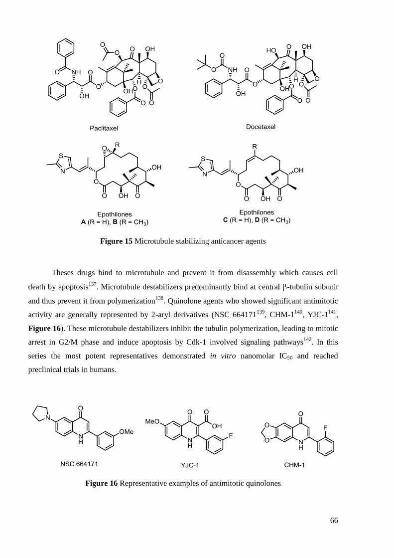
66
Figure 15 Microtubule stabilizing anticancer agents
Theses drugs bind to microtubule and prevent it from disassembly which causes cell
death by apoptosis137
. Microtubule destabilizers predominantly bind at central -tubulin subunit
and thus prevent it from polymerization138
. Quinolone agents who showed significant antimitotic
activity are generally represented by 2-aryl derivatives (NSC 664171139
, CHM-1140
, YJC-1141
,
Figure 16). These microtubule destabilizers inhibit the tubulin polymerization, leading to mitotic
arrest in G2/M phase and induce apoptosis by Cdk-1 involved signaling pathways142
. In this
series the most potent representatives demonstrated in vitro nanomolar IC50 and reached
preclinical trials in humans.
Figure 16 Representative examples of antimitotic quinolones

67
3.3 Structure-activity relationships
In contrast to the quinolone antibacterials with thousands of tested compounds and
detailed SAR studies, quinolone-based anticancer agents only started to emerge with a few most
potent representatives. There are numerous obstacles to define SAR for cytotoxic quinolones,
like the variety of intracellular targets and a large diversity of tested cancer cell line types.
Moreover, similar to the previously described antibacterial SARs, no single position has a
controlling effect on the observed antitumor activity but, more likely, a combination of various
substituents contributes to the cytotoxic potency. So, in order to maintain clarity and reliability,
one should account for the type of cancer cell line used, biological target and the nature of other
substituents presented in the molecule. In the following sections quinolone structures will be
discussed separately for each target (if known) and a last paragraph will be devoted to ―cytotoxic
quinolones‖ with unidentified target.
3.3.1 Topoisomerase II poisons and inhibitors
In vitro binding studies have revealed a common domain of interaction between
eukaryotic topoisomerase II with both antineoplastic inhibitors such as etoposide and FQs143
.
However, the FQ concentrations required in vitro to form a cleavage complex with native
eukaryotic type II topoisomerase were 100- to 1000-fold higher than those required for gyrase114-
115, 144. Initial studies were directed by attempts to shift inhibitory activity from bacterial to
eukaryotic topoisomerase II. As a result of these reports several compounds with high potency
were identified and SARs were established. A schematic historical development of anti-
topoisomerase II quinolones is reported in Figure 17.
New structures with C-7 aryl substituents were first identified and the antibacterial lead
compound Rosoxacin was transformed into cytotoxic CP-67,015 by addition of two fluorine
atoms at C-6 and C-8 positions115
. Further optimization led to CP-67,804 and CP-115,953 with
4-hydroxyphenyl substituent at C-7 position. These compounds were both cytotoxic in vitro and
number of experimental evidences indicated that they target topoisomerase II116a
.
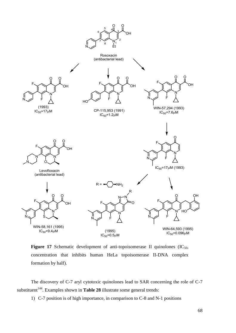
68
Figure 17 Schematic development of anti-topoisomerase II quinolones (IC50,
concentration that inhibits human HeLa topoisomerase II-DNA complex
formation by half).
The discovery of C-7 aryl cytotoxic quinolones lead to SAR concerning the role of C-7
substituent144
. Examples shown in Table 28 illustrate some general trends:
1) C-7 position is of high importance, in comparison to C-8 and N-1 positions

69
2) 4-Hydroxyphenyl substituent at C-7 position increase anti-topoisomerase II activity
but also increase the cytotoxicity of compounds
It is worth to note that a clear correlation exists between in vitro topoisomerase II
inhibitory activity, DNA cleavage and cytotoxicity. This supposes that the common biological
target of these derivatives is topoisomerase II, in spite of structural differences at C-7 position.
Table 28 Anti-topoisomerase II activity and cytotoxicity of various C-7 modified
CP-115,953 analogs
Compound R7 DNA cleavage,
EC2 ( M)a
DNA relaxation,
IC50 ( M)b
Cytotoxicity,
CC50( M)c
CP-115,953 0.3 3.2 9
CP-162,600
105 198 >500
CP-171,472
53 140 104
Difluorociprofloxacin
63 132 235
Data from ref 144
aEC2, Effective concentration of agent required to enhance calf thymus
topoisomerase II-mediated DNA cleavage twofold bThe agent concentration required to inhibit
DNA relaxation by calf thymus topoisomerase II by 50% cConcentration of compound required
to kill 50% of Chinese hamster ovary (CHO) cells.
At the same time Wentland et al. studied a series of pyridine based C-7 substituents and
their influence on topoisomerase II inhibition145
. The authors demonstrated that 4-pyridinyl
moiety at C-7 position could also give rise to cytotoxic compounds; however, their levels of
potency were lower than that of 4-hydroxyphenyl analog (CP-115,953) and tuning with
additional methyl groups is needed to increase the topoisomerase II inhibitory activity (Table
29).

70
Table 29 Pyridinyl vs phenyl substituent at C-7 position with regard to anti-
topoisomerase II activity
R7 IC50 ( M)
a
( CP-115,953)
1.2
17
(WIN-57,294)
7.6
Data from ref 145
aConcentration that inhibits human HeLa topoisomerase II-DNA
complex formation by half
During the initial studies SNS-595 didn‘t induce topoisomerase II-mediated DNA
cleavage, thereby it wasn‘t supposed to act as a topoisomerase poison146
. However, detailed
studies established that voreloxin intercalates DNA and poisons topoisomerase II, inducing site-
selective double-strand DNA breaks and G2 arrest128
. During the development of SNS-595 large
number of derivatives bearing different groups at C-7 position were prepared and evaluated.
Some SARs were established and, particularly, aminopyrrolidine moieties at C-7 provided
promising cytotoxic activity against murine P388 leukemia cells. Analysis of different
substitution patterns revealed that additional substitution at 4‘-position of pyrrolidine cycle
further increases the cytotoxicity. Among all, 3'-methoxy-4'-methylaminopyrrolidinyl analog
showed the highest cytotoxic activity in both in vitro and in vivo assays146b
.

71
Table 30 In vitro antiproliferative activity of SNS-595 and its C-7 analogs against
murine P388 leukemia cells
R7 IC50 ( M)a
18
19
0.16
0.028
SNS-595 (®Voreloxin)
0.019
Data from ref 146b
aConcentration of agent that reduces murine P388 leukemia cell
viability by 50%
In the previously discussed work, Wentland et al.145
also studied the influence of N-1 and
C-3 positions on overall potency. Although all compounds were less active than CP-115,953 the
authors demonstrated that (Table 31):
1) Ethyl and unsubstituted N-1 position are not tolerated, with cyclopropyl being
the optimal for topoisomerase II inhibition;
2) The presence of 3-CO2H group is not necessary for high levels of anti-
topoisomerase II activity and 3-H analog was also significantly active;
3) In the absence of C-3 carboxylic group, C-2 position could be substituted with
small groups (CH3) without loss of activity, while larger phenyl derivative is
inactive

72
Table 31 Influence of R-1, R-2, R-3 substituents on human topoisomerase II
inhibition
R1 R2 R3 IC50,( M)a
c-Pr
(WIN-57,294) H CO2H 7.6
Et H CO2H 110
H H CO2H >300
c-Pr H H 17
c-Pr CH3 H 16
c-Pr Ph H >240
Data from ref 145
aConcentration that inhibits human HeLa topoisomerase II-DNA
complex formation by half
This unexpected result contrasts the SAR determined in quinolone antibacterials, where
the 3-CO2H (or isosteric replacement) is required for prokaryotic topoisomerase inhibition.
Interestingly, these results induced further structural exploration at C-3 position, leading
to the discovery of WIN-64,593. This 2,6-dihydroxybenzyl derivative is a topoisomerase II
inhibitor147
(Figure 17, Figure 18) with IC50 of 0.096 M (80-fold more active than parent
WIN-57,294) and in vivo antitumor properties in mice.
In addition, a series of compounds with annulated cycles at C-3 position showed
enhanced cytotoxicity against mammalian cells. Studies have shown that the requirement for
inhibition of eucaryotic enzyme seems to be the coplanarity of the C-3 substituent with the
quinolone. Isothiazoloquinolones A-65,281 and A-65,282, (Figure 18) inhibited DNA
unknotting with 50% inhibitory concentrations of 21 M and were found to induce significant
DNA breakage mediated by calf thymus topoisomerase II78
. Another interesting example of
successful C-3 modification is represented by derivatives bearing a pyrazolo-ring fusion at the 3-
and 4-positions of the quinoline moiety (Figure 18). The most potent pyrazoloquinoline
displayed human HeLa topoisomerase II IC50 = 0.5 M, Figure 18), and demonstrated antitumor
activity both in vitro (P388 mouse leukemia cells) and in vivo (mice)148
.
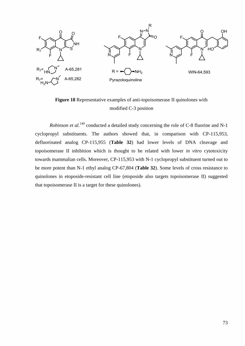
73
Figure 18 Representative examples of anti-topoisomerase II quinolones with
modified C-3 position
Robinson et al.149
conducted a detailed study concerning the role of C-8 fluorine and N-1
cyclopropyl substituents. The authors showed that, in comparison with CP-115,953,
defluorinated analog CP-115,955 (Table 32) had lower levels of DNA cleavage and
topoisomerase II inhibition which is thought to be related with lower in vitro cytotoxicity
towards mammalian cells. Moreover, CP-115,953 with N-1 cyclopropyl substituent turned out to
be more potent than N-1 ethyl analog CP-67,804 (Table 32). Some levels of cross resistance to
quinolones in etoposide-resistant cell line (etoposide also targets topoisomerase II) suggested
that topoisomerase II is a target for these quinolones).

74
Table 32 Influence of C-8 fluorine and N-1 substituents on topoisomerase II
activity and cytotoxicity
Compound R1 R8 Topoisomerase II activity
a Cytotoxicity CC50 ( M)
d
DNA cleavageb IC50 ( M)
c Wild type Vpm
R-5
e
CP-115,953 c-Pr F 1,00 60 9 12
CP-115,955 c-Pr H 0.38 130 20 40
CP-67,804 Et F 0.39 325 70 265
Data from ref 116a, 149
aDrosophila melanogaster topoisomerase II
bRelative scale of
potency with 1,00 value set for CP-115,953 cConcentration that inhibits topoisomerase II DNA
relaxation ability by half dConcentration that reduces the proliferation of Chinese hamster ovary
(CHO) cells by 50% eSelected cell line with resistance to etoposide
At the same time Suto et al.150
investigated a large number of FQs for their cytotoxicity
towards mammalian cells, which is thought to be related with topoisomerase II inhibition. In this
report, halogens at C-8 and cyclopropyl at N-1 gave the most cytotoxic derivatives, which is
consistent with previous results of Robinson et al.149
(Table 32)
Numerous SARs were established during the development of SNS-595146
from
antibacterial lead compound AT-3639151
(Figure 19).
Figure 19 Development of SNS-595, promising antitumor quinolone

75
Interesting results were obtained by Tsuzuki et al.151
when different C-3 substituents
where introduced into SNS-595 molecule. The authors provided another prove for tolerating
modifications at C-3 position with regard to cytotoxic activity. The analog bearing formyl group
was almost equipotent to parent SNS-595 compound both in vitro and in vivo (mice implanted
with P388 leukemia cells) (Table 33). However, low solubility of the formyl analog hampers its
practical usage as a therapeutic agent. Notwithstanding these studies gave basis to the further
exploration of more usefull antitumor drug candidates.
Table 33 In vitro cytotoxicity and in vivo antitumor activity of C-3 modified
SNS-595 analogs
R3 In vitro In vivo
IC50a ( M) Dose, mg/kg T/C
b, %
CO2H
(SNS-595) 0.01 3.13 200
CHO 0.03 3.13 188
CH2OH 0.06 – –
Data from ref 151
aConcentration of agent that reduces murine P388 leukemia cell
viability by 50% b(Median survival time of treated mice)/(median survival time of controls)×100.
While exploring C-6 substituent Tomita et al146a
reported that non-fluorinated analog was
twofold more cytotoxic against murine P388 leukemia cells than fluorinated AT-3639 (Table
34). In comparison with the 6-F counterpart, 6-NO2, 6-NH2 and 6-OH derivatives were
significantly less active (IC50 values were 5.1; 2.4; 10 M respectively). In addition, the authors
demonstrated that further modifications at C-5 position of the 6-unsubstituted 1,8 naphthyridone
lead to less cytotoxic activity, with exception of 5-NH2 derivative which was almost equipotent
to the unsubstituted compound with IC50 = 0.019 M.
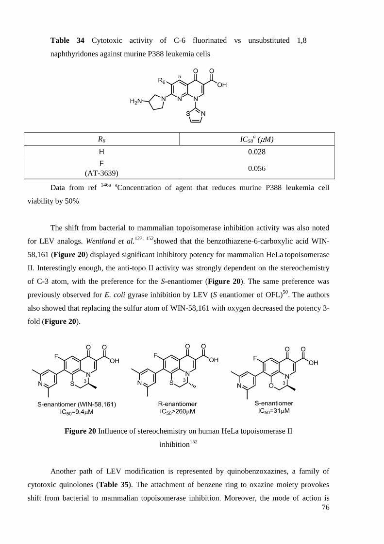
76
Table 34 Cytotoxic activity of C-6 fluorinated vs unsubstituted 1,8
naphthyridones against murine P388 leukemia cells
R6 IC50a ( M)
H 0.028
F
(AT-3639) 0.056
Data from ref 146a
aConcentration of agent that reduces murine P388 leukemia cell
viability by 50%
The shift from bacterial to mammalian topoisomerase inhibition activity was also noted
for LEV analogs. Wentland et al.127, 152
showed that the benzothiazene-6-carboxylic acid WIN-
58,161 (Figure 20) displayed significant inhibitory potency for mammalian HeLa topoisomerase
II. Interestingly enough, the anti-topo II activity was strongly dependent on the stereochemistry
of C-3 atom, with the preference for the S-enantiomer (Figure 20). The same preference was
previously observed for E. coli gyrase inhibition by LEV (S enantiomer of OFL)50
. The authors
also showed that replacing the sulfur atom of WIN-58,161 with oxygen decreased the potency 3-
fold (Figure 20).
Figure 20 Influence of stereochemistry on human HeLa topoisomerase II
inhibition152
Another path of LEV modification is represented by quinobenzoxazines, a family of
cytotoxic quinolones (Table 35). The attachment of benzene ring to oxazine moiety provokes
shift from bacterial to mammalian topoisomerase inhibition. Moreover, the mode of action is

77
different. Quinobenzoxazines don‘t mediate topoisomerase II dependent DNA double-strand
breaks. Instead, the in vitro cytotoxicity of compounds correlates well with both topoisomerase II
inhibition and DNA binding153
(Table 35). Thus, the authors supposed that quinobenzoxazines
are catalytic topoisomerase II inhibitors129
which act before DNA cleavage stage in contrast to
previously mentioned topo II poisons154
.
Table 35 Quinobenzoxazines: relationship between in vitro cytotoxicity, DNA
binding and topoisomerase II inhibition
Compound Cytotoxicity
CC50, Ma
DNA bindingb
Topoisomerase II
inhibition
IC50, Mc
(R)-A-62,176 0.89 ++ 3.1
(S)-A-62,176 0.53 +++ 0.9
Data from ref 153b a
Concentration of compound that reduces A-549 cell viability by 50% b
(+++) – compound fully protects DNA sequence from DNase I at 1 M; (++) – compound fully
protects DNA sequence from DNase I at 10 M cConcentration of compound that inhibits
kinetoplast DNA decatenation by human topoisomerase II by 50%
3.3.2 G-quadruplex inhibitors
G-quadruplex inhibitors, in order to effectively interact with nucleotide bases, should
possess extensive degree of planarity. Historically, these molecules evolved from antibacterial
lead LEV by step-wise annelation of flat aromatic cycles (Figure 21).

78
Figure 21 Development of G-quadruplex inhibitors
The quinobenzoxazine (S)-A-62,176 termed AT-62,176, a mammalian cytotoxic agent,
was shown to be both a topoisomerase II catalytic inhibitor129
and poison154
. The
pyridobenzophenoxazine analogs, with an expanded ring system, also showed strong cytotoxic
effects on several tumor cell lines and potent inhibitory effects against human topoisomerase
II153a
.
CX-3543135
and the fluoroquinophenoxazine QQ58116b
both demonstrated enhanced
stacking interactions with G-quadruplex, but had lost the topoisomerase II poisoning activity. On
the other hand, the fluoroquinoanthroxazines, containing a naphthyl ring in the cis orientation,

79
were able to recognize and bind quite specifically to G-quadruplex structures and topoisomerase
II-DNA complexes. Within this group, FQA-CR showed a higher G-quadruplex interaction,
while FQA-CS displayed more potent topoisomerase II poisoning activity (Table 36)134
.
Table 36 Biological activities of fluoroquinoanthroxazines
FQA-CS FQA-CR
G-quadruplex interaction:
polymerase stop assay (IC50, M) 0.67 0.06
Topoisomerase II inhibition:
cytotoxic ratio topoisomerase II-R/-S
cellsa
4.02 1.55
Cytotoxicity:
MCF7 breast cells (IC50, M) 1.1 0.46
Major mechanism of action topoisomerase II poison G-quadruplex interaction
Data from ref 134
a
The ratio of cytotoxicity of FQA-CS and FQA-CR against
8226/DOX1V (topoisomerase II-resistant) and 8226/S (topoisomerase II-sensitive) cells.
3.3.3 Antimitotic agents
The key structural features which shifted quinolone‘s biological action from
topoisomerase II poisoning to inhibition of tubulin polymerization are C-2-aryl substituent and
unsubstituted N-1 atom155
. A large tolerance for aromatic groups at C-2 position was
demonstrated and carboxylic group at C-3 position serves to improve activity and
pharmacokinetics141
. The C-6 substituent plays very important role for antimitotic activity and
generally, the groups attached at C-6 should be lipophilic and electrondonating, such as methyl,
hydroxymethyl, fluor and some cyclic amines. Other positions like C-5, C-7 and C-8 are

80
preferred to be unsubstituted (Figure 22). The most potent representatives of antimitotic
quinolones were previously shown on the Figure 16 (page 66)
Figure 22 Summary of SAR for antimitotic quinolones
3.3.4 Cytotoxic quinolones
In this section we decided to present cytotoxic agents with ―unidentified mechanism of
action‖. This paragraph discusses the influence of modifications at C-3 and C-7 positions on
cytotoxicity.
Detailed investigation of C-7 piperazinyl and pyrrolidinyl substituents was held by
Yamashita et al.156
and Suto et al.150
. A large number of derivatives were evaluated for antitumor
activity both in vitro and in vivo and the authors showed that:
1) pyrrolidinyl based derivatives are generally more cytotoxic in vitro (an
example is presented Table 37)
2) piperazinyl and pyrrolidinyl based derivatives have comparable activities in
vivo

81
Table 37 Evaluation of C-7-piperazinyl vs pyrrolidinyl cytotoxic quinolones
R7 In vitro
cytotoxicity IC50 ( M)a
In vivo antitumor activity
ILS %b
134 30
30 34
Data from ref 150, 156
aConcentration of compound that reduces Chinese hamster V79 cells
viability by 50% bIncreased life span (ILS) in murine P388 leukemia model
Recently novel substitutions were introduced at C-7 position, which can provide good
models for further design of potent antitumor quinolones. Substitution of distal nitrogen atom at
piperazine moiety provides significant improvement in cytotoxicity. Initial findings of Fang et
al. 157
demonstrated that 7-[4-(2-hydroxyiminoethyl)piperazin-1-yl] quinolones displayed
significant activities against renal cancer cell lines, among them, 7-4-[2-(4-chlorophenyl)-2-
hydroxyiminoethyl]-1-piperazinyl-derivative was the most potent. Foroumadi et al. identified
new aromatic moieties (furan-2-yl and thiophen-2-yl) that provide highly cytotoxic
compounds158
. In addition, the cytotoxicity was further enhanced by converting the oxime group
to the respective O-benzyl or O-4-chlorobenzyl oxime159
(Table 38).
To analyse the data authors have utilized the similar approach as in the screening
program of National Cancer Institute with the panel of 60 human tumor cell lines used to screen
for the antiproliferative activity. During these tests a new method of getting an idea on the
substance‘s mechanism of action was discovered. In fact, the pattern of growth inhibition in
these cell lines was similar for compounds of similar mechanism of action160
. In the current
article the Foroumadi et al. have compared log IC50 values of synthesized compounds with those
of etoposide against less numerous panel of cancer cell lines. Finally they suggested that these
quinolone derivatives share a similar mechanism of action with etoposide, already mentioned
topoisomerase II inhibitor (Table 38).

82
Table 38 Cytotoxic oxyiminoethyl CIP derivatives
Ar R Cytotoxicity IC50, M
a
MCF-7b A-549
c SKMEL-3
d KB
e
H 117 108 125 108
Me 116 111 127 102
4-Cl-Bn 3.6 2.1 3.1 2.3
Bn 1.8 1.6 3.3 1.9
Me 2.9 – 7.7 17
CIP 199 – 181 160
Etoposide 11.6 1.5 0.34 4.8
Data from ref 158-159
aConcentration of compound that reduces cell viability by 50%
bBreast carcinoma cell line
cLung cancer cell line
dMelanoma cell line
eCervical carcinoma cell
line
Concerning C-3 position 3-oxadiazole-aryl derivatives showed moderate cytotoxic
activity against human tumor lung cancer cells (A-549)161
. It is worth to note, that CIP
derivatives were more potent than corresponding NOR analogs. The most active compounds bear
para-nitrophenyl and -naphtyloxymethyl substituents attached to the oxadiazole moiety (Table
39).

83
Table 39 Cytotoxic 3-oxadiazoyl quinolone derivatives
Ar IC50 ( M)a
1.7
3.1
Data from ref 161 a
Concentration of compound that reduces human lung tumor cell (A
549) viability by 50%
Analogously to antibacterials, cytotoxic quinolones were obtained by combining
two pharmacophores through appropriate linker. By using fused heterocyclic triazolo[2,1-
b][1,3,4]thiadiazole linker a series of ―dimeric‖ FQ derivatives were prepared and showed in
vitro cytotoxicity against CHO Chinese hamster ovary and L-1210 murine leukemia cell lines.
The most potent compounds are CIP ―heterodimers‖ presented in Table 40162
.
Table 40 C-3 linked cytotoxic FQ ―dimers‖
FQ1 FQ2 IC50 ( M)a
CIP CIP 0.2
CIP N-Me-CIP 1.2
CIP N-Et-CIP 2.5
Data from ref 162
aConcentration of compound that reduces L210 (murine leukemia) cell
viability by 50%

84
In summary, initial cytotoxic FQs developed from antibiotic agents exhibitied target shift
from bacterial to mammalian topoisomerase inhibition. Studies revealed new potent stuctures
which contrasted with previously established antibacterial SARs. The most active compounds
possessed C-7 aryl substituents and, what is more interesting, some analogs exhibited structural
modifications at C-3 position which was not tolerated in regard to antibacterial activity. Further
research prompted the emergence of new structures for different targets, like G-quadruplex and
tubulin polymerization inhibitors, therefore broadening the FQs spectrum of biological activity.
4. Conclusion
In this chapter we have described the different aspects concerning FQs, their emergence,
development, mechanism of action and discussed their SARs for different biological targets. FQs
represent an important group of molecules with crucial applications in medicine. However, old
FQs displayed several drawbacks, such as emerging resistance, so the development of new
agents is needed. For all these reasons, the synthesis of new FQ derivatives will be described in
the next chapter along with the detailed rationalization for chosen compounds.

85
CHAPITRE II SYNTHÈSE DES DÉRIVÉS DE LA CIPROFLOXACINE ET
DE LA LÉVOFLOXACINE

86
87
1. Introduction
Dans ce chapitre, nous décrivons le développement et la synthèse de nouveaux dérivés de
la LEV et de la CIP.
Tout d'abord, le choix des composés synthétisés sera examiné. Nous discuterons les
modifications structurales envisagées et la rationalisation pour l'introduction de différents
substituants au niveau de l'acide carboxylique en position C-6 de la LEV et de la fonction amine
N-4-pipérazine en position C-7 de la CIP.
Dans la deuxième partie, nous décrirons la synthèse des dérivés sélectionnés. Nous
discuterons des travaux antérieurs effectués au laboratoire et nous détaillerons les différentes
voies de synthèse choisies après optimisation.
La troisième partie de ce chapitre sera consacrée à l'étude de la stabilité des composés
synthétisés dans un tampon phosphate aqueux, pH 7.4 à 37 °C, effectuée par HPLC.

88
2. Dérivés “monomères” de la ciprofloxacine
2.1 Introduction
Dans le premier chapitre, nous avons résumé quelques-uns des résultats de la littérature
qui indiquent que la modification de la position C-7 des FQs ouvre des perspectives intéressantes
pour le développement de nouveaux composés possédant des activités antibactériennes,
antimycobactériennes ou antitumorales. En effet, l'inhibition de l'ADN topoisomérase par les
quinolones est fortement influencée par la nature du substituant en position C-7. De plus, des
modifications structurales en cette position auraient des effets sur la sélectivité enzymatique
procaryotes versus eucaryotes. Certains auteurs ont montré que l'introduction de substituants
volumineux au niveau du motif pipérazine des quinolones entraîne un déplacement de l'activité
biologique antibactérienne de ces composés vers des agents cytotoxiques contre les cellules
cancéreuses humaines157-158
. Toutes ces caractéristiques ont suscité l'intérêt de notre laboratoire.
Trois séries homologues de dérivés lipophiles de la CIP ont été conçues et synthétisées lors de
travaux antérieurs dans notre groupe. Ces dérivés constituent une famille de dérivés 7-((4-
substitué)pipérazin-1-yl)-CIP considérés comme des dérivés ―monomères‖. La CIP a été choisie
pour sa disponibilité commerciale et la possibilité de fonctionnalisation de l‘amine secondaire N-
4-pipérazine. En outre, l‘introduction de groupements de longueurs de chaîne variables pourrait
permettre de moduler les propriétés physico-chimiques des composés et ainsi modifier le profil
de l'activité biologique. Les trois séries de composés synthétisés diffèrent les unes des autres par
la nature de la fonction reliant la chaîne alkyle à l'atome d'azote N-4 du groupement pipérazine
(Figure 23).
Figure 23 Représentation schématique des dérivés ―monomères‖ 7-((4-
substitué)pipérazin-1-yl)-CIP
Il est important de mentionner que la fonction acide carboxylique en C-3 de la CIP a une
valeur de pKa élevé (6.08)61
comparée aux autres aryl-acides. Cette fonction est en outre
désactivée en raison de la formation d‘une liaison hydrogène intramoléculaire avec la fonction

89
carbonyle adjacente en position 4 (Figure 24). Ainsi, aucune protection de l‘acide carboxylique
n‘est nécessaire lors des synthèses pour la fonctionnalisation de la pipérazine de la CIP décrites
dans la section suivante.
Figure 24 Liaison hydrogène intramoléculaire dans la molécule de CIP
2.2 Synthèse des dérivés 7-(4-(alcanoyle)pipérazin-1-yl) de la CIP
La première série de dérivés 7-(4-(alcanoyle)pipérazin-1-yl) de la CIP (dite ―mono-
amides‖) a été aisément préparée en une seule étape d‘acylation de la CIP par les chlorures
d‘alcanoyle disponibles commercialement pour conduire aux composés correspondants 1a-l avec
de bons rendements variant de 45% à 90% (Schéma 1, Tableau 41). Certains dérivés de cette
même série ont été difficiles à purifier, ce qui explique les différences entre les rendements.
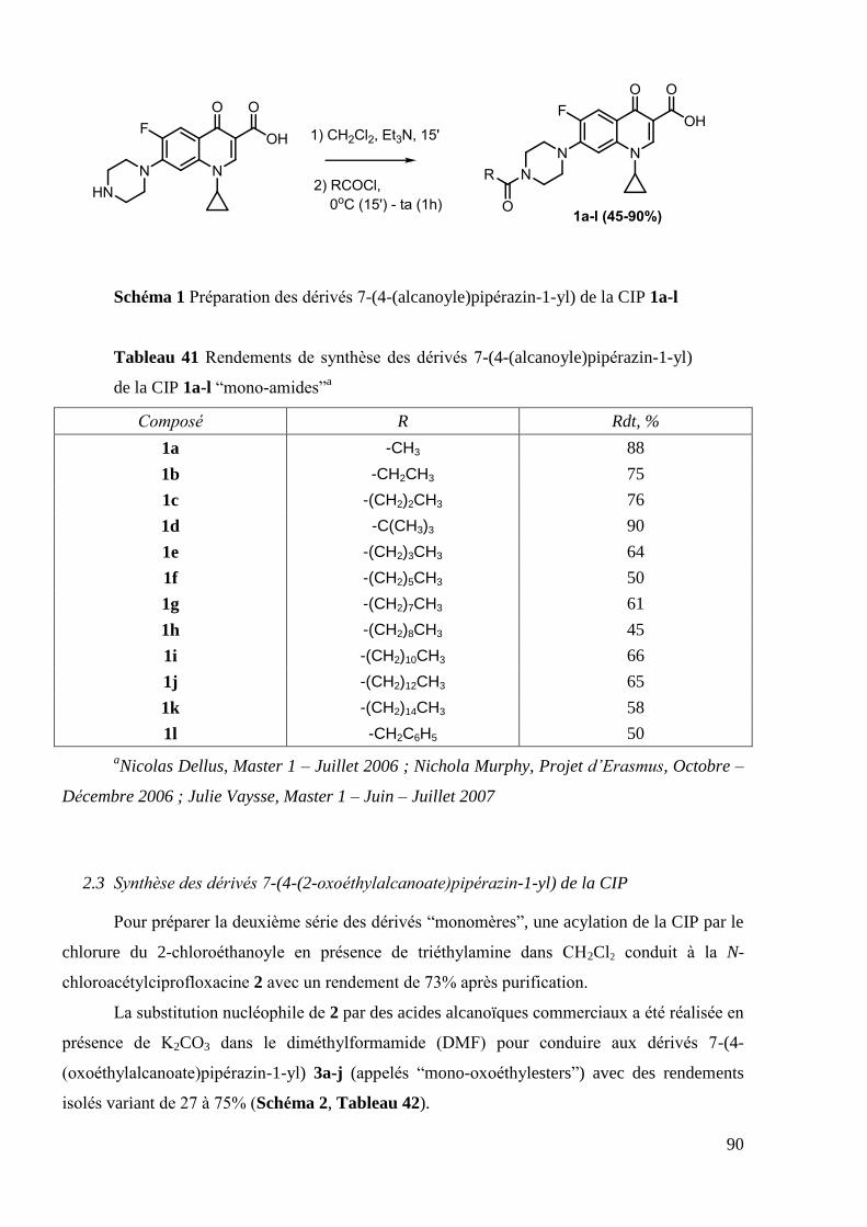
90
Schéma 1 Préparation des dérivés 7-(4-(alcanoyle)pipérazin-1-yl) de la CIP 1a-l
Tableau 41 Rendements de synthèse des dérivés 7-(4-(alcanoyle)pipérazin-1-yl)
de la CIP 1a-l ―mono-amides‖a
Composé R Rdt, %
1a -CH3 88
1b -CH2CH3 75
1c -(CH2)2CH3 76
1d -C(CH3)3 90
1e -(CH2)3CH3 64
1f -(CH2)5CH3 50
1g -(CH2)7CH3 61
1h -(CH2)8CH3 45
1i -(CH2)10CH3 66
1j -(CH2)12CH3 65
1k -(CH2)14CH3 58
1l -CH2C6H5 50
aNicolas Dellus, Master 1 – Juillet 2006 ; Nichola Murphy, Projet d’Erasmus, Octobre –
Décembre 2006 ; Julie Vaysse, Master 1 – Juin – Juillet 2007
2.3 Synthèse des dérivés 7-(4-(2-oxoéthylalcanoate)pipérazin-1-yl) de la CIP
Pour préparer la deuxième série des dérivés ―monomères‖, une acylation de la CIP par le
chlorure du 2-chloroéthanoyle en présence de triéthylamine dans CH2Cl2 conduit à la N-
chloroacétylciprofloxacine 2 avec un rendement de 73% après purification.
La substitution nucléophile de 2 par des acides alcanoïques commerciaux a été réalisée en
présence de K2CO3 dans le diméthylformamide (DMF) pour conduire aux dérivés 7-(4-
(oxoéthylalcanoate)pipérazin-1-yl) 3a-j (appelés ―mono-oxoéthylesters‖) avec des rendements
isolés variant de 27 à 75% (Schéma 2, Tableau 42).

91
Schéma 2 Préparation des dérivés 7-(4-(oxoéthylalcanoate)pipérazin-1-yl) de la
CIP 3a-j
Tableau 42 Rendements de synthèse des dérivés 7-(4-
(oxoéthylalcanoate)pipérazin-1-yl) de la CIP 3a-j ―mono-oxoéthylesters‖a
Comoposé n Rdt, %
3a 0 60
3b 2 63
3c 4 75
3d 6 45
3e 7 27
3f 8 47
3g 9 43
3h 10 46
3i 12 41
3j 14 46
a Florence Argeliès, Master 2, Février – Juin 2006.
L‘hydrolyse enzymatique ou chimique de ces dérivés 3a-j conduirait à la formation de la
CIP native ou du dérivé 7-(4-(2-hydroxyéthanoyle)pipérazin-1-yl) 4 (Figure 25).

92
Figure 25 Hydrolyse possible des dérivés ―mono-oxoéthylesters‖ de la CIP
Afin d‘évaluer la stabilité des composés synthétisés 3a-j et de pouvoir comparer leurs
activités biologiques à celles du produit d‘hydrolyse, le composé 4 a été synthétisé. L‘acylation
de la CIP par le chlorure de benzyloxyéthanoyle dans CH2Cl2 conduit au dérivé 7-(4-(2-
benzyloxyéthanoyle)pipérazin-1-yl) 5 (rdt: 43%) qui, soumis à une hydrogénation catalytique
(Pd/C) dans le DMF, permet d‘obtenir le composé 4 avec un rendement de 60% (Schéma 3).
Schéma 3 Synthèse du dérivé 7-(4-(2-hydroxyéthanoyl)pipérazin-1-yl) de la CIP
4 (Florence Argeliès, Master 2, Février – Juin 2006)
2.4 Synthèse des dérivés 7-(4-(alkyloxycarbonyl)pipérazin-1-yl) de la CIP
La troisième série des dérivés 7-(4-(alkyloxycarbonyl)pipérazin-1-yl) de la CIP (nommés
"mono-carbamates") a été préparée par condensation de la CIP avec divers alkylchlorofomates
commerciaux. La réaction a été réalisée dans le CH2Cl2 et les composés 6a-f ont été isolés avec
des rendements allant de 33% à 66% (Schéma 4, Tableau 43) Seul le composé 6g portant un
groupement t-butyloxycarbonyle (Boc) a été préparée en utilisant Boc2O dans un mélange
hydroxyde de sodium aqueux 2M / dioxane (Schéma 4, Tableau 43).

93
Schéma 4 Synthèse des dérivés 7-(4-(alkyloxycarbonyl)pipérazin-1-yl) de la CIP
6a-g
Tableau 43 Rendements de synthèse des dérivés 7-(4-
(alkyloxycarbonyl)pipérazin-1-yl) de la CIP 6a-g: ―mono-carbamates‖a
Composé n Rdt, %
6a 1 66
6b 3 42
6c 7 37
6d 8 33
6e 9 41
6f 11 38
6g - 74
a Nichola Murphy, Projet d’Erasmus, Octobre – Décembre 2006 ; Julie Vaysse, Master 1
– Juin – Juillet 2007.
Les dérivés ―monomères‖ de la CIP appartenant à ces trois séries homologues ont été
évalués pour leurs activités antibactériennes, antiprolifératives et antimycobactériennes. Les
résultats biologiques obtenus, qui seront discutés dans le chapitre suivant, nous ont conduit à
explorer la synthèse de dérivés ―dimères‖ bis-C-7-((4-substitué)pipérazin-1-yl) de la CIP. Les
travaux préliminaires de synthèse avaient déjà été réalisés au début de cette thèse, qui a été
concentrée sur l'optimisation de la synthèse de ces dérivés dimères de CIP.

94
3. “Dimeric” ciprofloxacin derivatives
3.1 Choice of compounds to develop
We were motivated to develop a series of C-7-((4-substituted)piperazin-1-yl)-linked CIP
―dimers‖ because of several practical and theoretical considerations.
The influence of lipophilicity on antibacterial activity was extensively studied and it was
proven to have a positive effect on activity against Gram-positive microorganisms. This feature
is thought to be related with increased penetration of more lipophilic compounds through the
single cellular wall. Inspired by biological results obtained for previously synthesized
―monomeric‖ CIP derivatives, that had an enhanced activity against standard and resistant S.
aureus strains (which will be discussed in the corresponding 0), we extended our field to
―dimeric‖ compounds as more lipophilic agents.
Increased steric bulk was proven to enhance the activity towards strains of S. aureus with
efflux-mediated resistance. In fact, multidrug-efflux proteins are less susceptible to bulkier and
lipophilic agents. This could be responsible for the enhanced activity of ―monomeric‖ CIP
derivatives. Thus bulkier and more lipophilic ―dimers‖ should be interesting candidates for their
evaluation against resistant strains of S. aureus.
It has been long proposed that more than one molecule of quinolone binds to the target
enzyme; however until recently there was no clear evidence about the structure of binding
complex. Nowadays, it is thought that the target bacterial enzyme forms a ternary complex with
DNA and two molecules of quinolone which are oriented in a ―tail-to-tail‖ manner (Figure 26).
Figure 26 Schematic view of quinolone-enzyme complex and representation of
multibinding concept (7 – piperazinyl ―tails‖ of quinolone)
This finding prompted the detailed study of quinolone ―dimers‖ with most representative
works of Kerns et al (Figure 27). The authors demonstrated increased activity of ―dimeric‖
compounds towards S. aureus strains in comparison to parent FQs. However linkers used by
Kerns were relatively rigid and short. Taking into account recent structural data on the ternary

95
quinolone-enzyme-DNA complex, these ―dimers‖ were unlikely to have a favorable structure to
allow multiple binding.
Figure 27 C-7-((4-substituted)piperazin-1-yl)-linked CIP ―dimers‖ developed by
Kerns et al
The situation with anticancer activity is more complicated because of multiple
mechanisms of action and multiple targets for quinolones. There are less data in the literature on
SAR studies for antiproliferative quinolones, in comparison with antibacterial ones. Expanding
this relatively unexplored area could reveal important dependencies and trends.
For all these reasons, we decided to synthesize two series of CIP ―dimers‖. We chose a
flexible linker based on alkyl chain of varying length. The linking groups were chosen as the
ones from the most active ―monomeric‖ derivatives, that were ―ester‖ and ―amide‖ functions
(Figure 28).
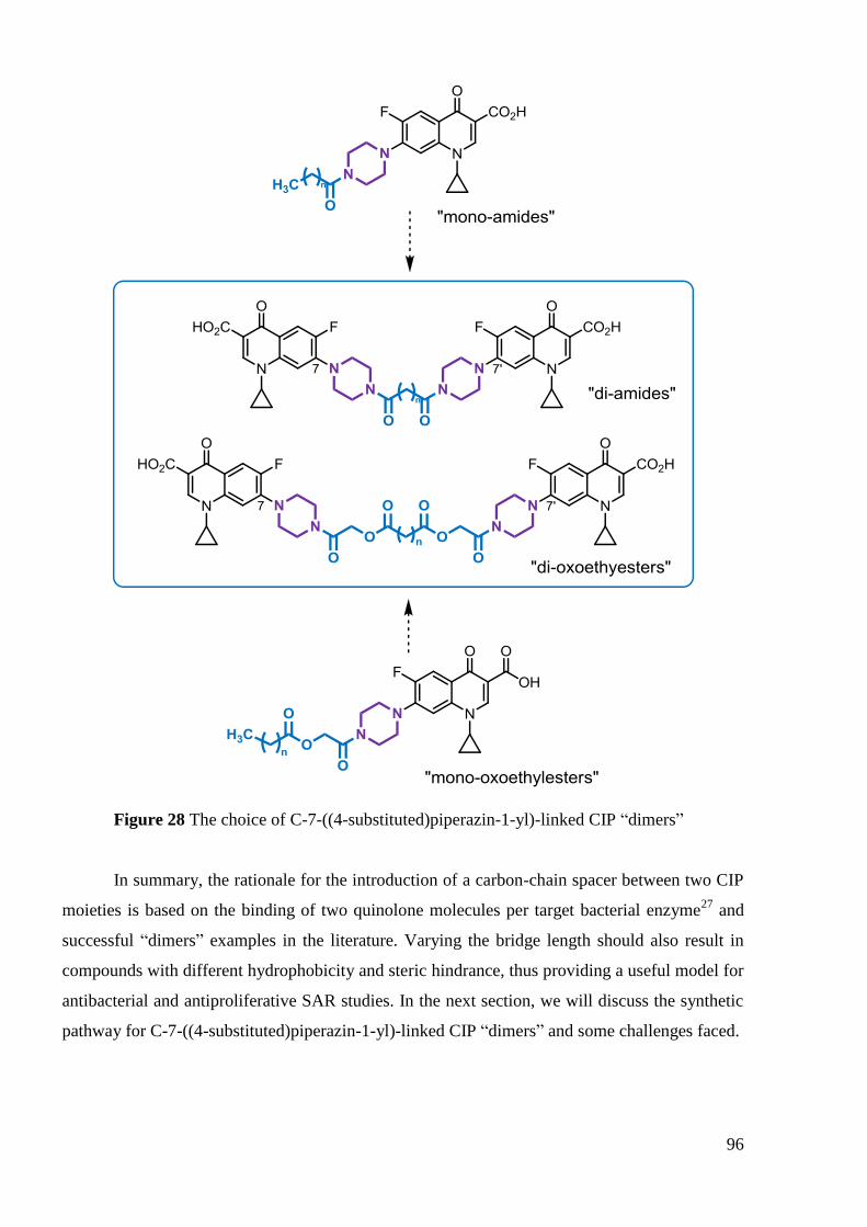
96
Figure 28 The choice of C-7-((4-substituted)piperazin-1-yl)-linked CIP ―dimers‖
In summary, the rationale for the introduction of a carbon-chain spacer between two CIP
moieties is based on the binding of two quinolone molecules per target bacterial enzyme27
and
successful ―dimers‖ examples in the literature. Varying the bridge length should also result in
compounds with different hydrophobicity and steric hindrance, thus providing a useful model for
antibacterial and antiproliferative SAR studies. In the next section, we will discuss the synthetic
pathway for C-7-((4-substituted)piperazin-1-yl)-linked CIP ―dimers‖ and some challenges faced.
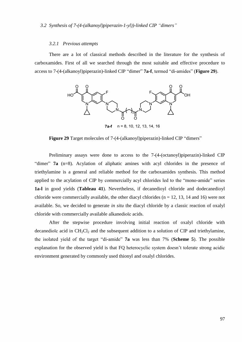
97
3.2 Synthesis of 7-(4-(alkanoyl)piperazin-1-yl))-linked CIP “dimers”
3.2.1 Previous attempts
There are a lot of classical methods described in the literature for the synthesis of
carboxamides. First of all we searched through the most suitable and effective procedure to
access to 7-(4-(alkanoyl)piperazin)-linked CIP ―dimer‖ 7a-f, termed ―di-amides‖ (Figure 29).
Figure 29 Target molecules of 7-(4-(alkanoyl)piperazin)-linked CIP ―dimers‖
Preliminary assays were done to access to the 7-(4-(octanoyl)piperazin)-linked CIP
―dimer‖ 7a (n=8). Acylation of aliphatic amines with acyl chlorides in the presence of
triethylamine is a general and reliable method for the carboxamides synthesis. This method
applied to the acylation of CIP by commercially acyl chlorides led to the ―mono-amide‖ series
1a-l in good yields (Tableau 41). Nevertheless, if decanedioyl chloride and dodecanedioyl
chloride were commercially available, the other diacyl chlorides (n = 12, 13, 14 and 16) were not
available. So, we decided to generate in situ the diacyl chloride by a classic reaction of oxalyl
chloride with commercially available alkanedioïc acids.
After the stepwise procedure involving initial reaction of oxalyl chloride with
decanedioïc acid in CH2Cl2 and the subsequent addition to a solution of CIP and triethylamine,
the isolated yield of the target ―di-amide‖ 7a was less than 7% (Scheme 5). The possible
explanation for the observed yield is that FQ heterocyclic system doesn‘t tolerate strong acidic
environment generated by commonly used thionyl and oxalyl chlorides.

98
Scheme 5 Synthesis of 7-(4-(octanoyl)piperazin)-linked CIP ―dimer‖ 7a through
an acyl chloride pathway
During further attempts the main idea was to utilize a ―powerful‖ activating agent for
alkanoïc diacid which is mild enough to preserve the heterocyclic system of CIP. It was decided
to avoid acidic agents by using carbodiimide reagents which are a widely used class of agents for
peptide synthesis. Dicyclohexyl carbodiimide163
(DCC) and N-(3-dimethylaminopropyl)-N′-
ethylcarbodiimide hydrochloride salt164
(EDCI) are frequently used as a carboxyl activating
agent for amide bond formation. Additives, such as N-hydroxybenzotriazole (HOBt), N-
hydroxysuccinimide (NHS) or 2-thiazoline-2-thiol (2-TT) are often added to increase yields
and/or decrease side reactions (Figure 30).
Figure 30 Examples of carbodiimide reagents and additives
No additional amine is theoretically required during this one-pot procedure. Concerning
the mode of reaction (Figure 31), the carbodiimide reacts with the carboxylic acid to form the O-
acylisourea I. The nucleophilic substitution of amine on I yields the desired amide and the urea
by-product II. The isolation of the symmetric carboxylic anhydride from the reaction mixture
suggested, however, that a more complex mechanism might co-exist165
. The driving force of this
reaction is the formation of the urea II. However, the racemisation and acetyl transfer which
form the unreactive N-acylurea III are often observed This side reaction can be considerably

99
diminished by carrying out the procedure in a two-step manner : the initial reaction between the
acid and the coupling agent is carried out at 0°C, followed by amine addition. Furthermore, a
selected nucleophile can be used as an additive. The nucleophile also prevents the undesired
product formation, because it reacts faster than the competing acyl transfer and generates the
intermediate IV on which the nucleophilic substitution by the amine yields the desired amide.
Figure 31 Carbodiimide-assisted amide synthesis, with and without additive
(HOBt is showed as example of nucleophile)
An attempt was conducted by using the DCC / 2-TT system in dioxane. However, as the
desired ―di-amide‖ 7a and the resulting dicyclohexylurea byproduct showed similar low
solubility in common solvents, it was too difficult to separate them.
This practical problem was solved by the use of EDCI / 2-TT system. The resulting urea
was water-soluble and thereby could be easily removed by extractive work-up.
In this third attempt, 1,10-bis(2-thioxothiazolidin-3-yl)decane-1,10-dione 8 was obtained
by coupling 1,10-decanedioïc acid with 2-TT in the presence of EDCI in dioxane. Unfortunately,
due to tedious purifications, the yield was very low (8%). The ―di-amide‖ 7a was synthesized by
the subsequent addition of 8 to a solution of CIP and K2CO3 in acetonitrile (CH3CN) in 39%
yield. However, the overall yield after these two steps was very low 3% (Scheme 6). This result
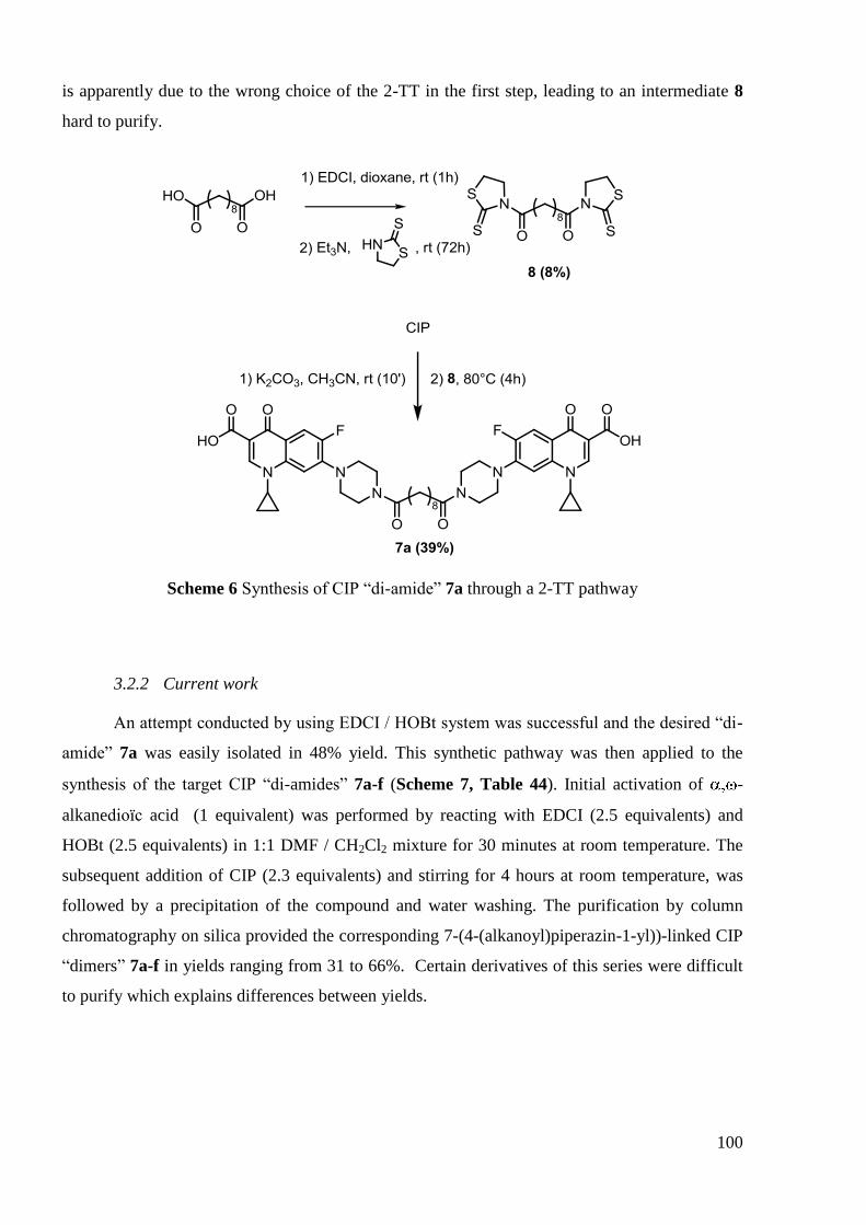
100
is apparently due to the wrong choice of the 2-TT in the first step, leading to an intermediate 8
hard to purify.
Scheme 6 Synthesis of CIP ―di-amide‖ 7a through a 2-TT pathway
3.2.2 Current work
An attempt conducted by using EDCI / HOBt system was successful and the desired ―di-
amide‖ 7a was easily isolated in 48% yield. This synthetic pathway was then applied to the
synthesis of the target CIP ―di-amides‖ 7a-f (Scheme 7, Table 44). Initial activation of -
alkanedioïc acid (1 equivalent) was performed by reacting with EDCI (2.5 equivalents) and
HOBt (2.5 equivalents) in 1:1 DMF / CH2Cl2 mixture for 30 minutes at room temperature. The
subsequent addition of CIP (2.3 equivalents) and stirring for 4 hours at room temperature, was
followed by a precipitation of the compound and water washing. The purification by column
chromatography on silica provided the corresponding 7-(4-(alkanoyl)piperazin-1-yl))-linked CIP
―dimers‖ 7a-f in yields ranging from 31 to 66%. Certain derivatives of this series were difficult
to purify which explains differences between yields.

101
Scheme 7 Preparation of 4-(alkanoyl)piperazin-1-yl-linked CIP ―dimers‖ 7a-f
Table 44 Yields of CIP ―di-amides‖ derivatives 7a-f
Compound n Yield, %
7a 8 48
7b 10 31
7c 12 66
7d 13 61
7e 14 55
7f 16 53
In summary, several synthetic methods have been used during our attempts to synthesize
CIP ―di-amides‖ derivatives. We have faced degradation-related problems using oxalyl chloride
pathway whereas challenging purification was a major issue when DCC or EDCI / 2-TT systems
were used. As a result, EDCI / HOBt system was found to be the most efficient in terms of yields
and practical convenience. Having successfully synthesized the ―di-amide‖ series of CIP
derivatives we have shifted to the ―di-oxoethylester‖ series which will be discussed in greater
details in the next section.

102
3.3 Synthesis of 7-(4-(oxoethylalkanoate)piperazin-1-yl)-linked CIP “dimers”
Keeping in mind that we wanted to establish a SAR of synthetized FQs derivatives, the
length of the alkyl chain was preserved as before that for ―di-amides‖ series (Figure 32)
Figure 32 Target molecules of 7-(4-(oxoethylalkanoate)piperazin-1-yl)-linked
CIP ―dimers‖
The 7-(4-(oxoethylalkanoate)piperazin-1-yl)-linked CIP ―dimers‖ 9a-f, termed ―di-
oxoethylester‖ derivatives were prepared by using the method previously described for ―mono-
oxoethylester‖ analogues 3a-j (Schéma 2, Tableau 42). From the synthetic point of view it
consists of two classical nucleophilic substitution reactions.
First step was the substitution of chloroacetyl chloride by the 4-(piperazinyl)nitrogen of
CIP to afford 7-(4-(2-chloroacetyl)piperazin-1-yl) CIP derivative 2 as described previously in
Schéma 2.
The next stage was the condensation of carboxylic diacids with derivative 2. The
mechanism involves classical nucleophilic attack of the in situ generated dicarboxylate anion on
the derivative 2 (Figure 33). This step was a subject for condition optimization during
preliminary studies which were carried out in our group.
Figure 33 Schematic representation for the preparation of CIP ―di-oxoethylesters‖

103
3.3.1 Optimisation of reaction conditions for 1,12-dodecanoïc diacid
In order to find the best conditions for the condensation of carboxylic diacids, it was
decided to perform a series of optimization experiments conducted by using 1,12-dodecanedioïc
acid (n = 10) to produce 9b derivative (Figure 32).
All optimization runs involved the following typical procedure : A solution of carboxylic
diacids (1 equivalent) and M2CO3 (2 equivalents) in DMF or CH3CN under argon was stirred
during 2 hours. Compound 2 (2 equivalents) and TBAI (none, 0.2 or 2 equivalents) were added
and was stirred at the mentionned temperature for 3, 5 or 7 days (Table 45).
Firstly, the conditions previously described for the synthesis of ―mono-oxoethylesters‖
3a-j were used, that were K2CO3 as a base and DMF as a solvent. However, in comparison with
the ―monomer‖ analog, these conditions gave ―di-oxoethylester‖ derivative in lower yield (10-
15% for 9b versus 46% for 3h (n=10)) and required longer reaction time, from 4 hours for 3h to
5-7 days for 9b (Table 45, entries 1-2).
Changing the solvent to CH3CN allowed us to slightly decrease the reaction time from 7
days to 5 days. However, yield (15%) was still poor and additional optimization was needed.
(Table 45, entry 3).
We decided to use tetrabutylammonium iodide (TBAI) in order to increase the yield of
this reaction. TBAI is supposed to in situ convert N-chloroacetylciprofloxacin into the more
reactive N-iodo-analog. Because iodides are generally more reactive in nucleophilic substitution,
we expected a faster completion of the reaction. However, when a catalytic amount of TBAI (0.2
equivalent) was used, no improved yield was observed (Table 45, entry 4). Whereas a
stoechiometric amount of TBAI (2 equivalents) led to an enhanced yield with a 3-fold increase
from 15 to 46% and allowed us to reduce the reaction time to a 3 days period (Table 45, entry
5). However, it was noticed that the crude mixture was more complex when reaction was carried
out in CH3CN instead of DMF
It is known that cesium counterion and dipolar aprotic solvents (such as DMF,
dimethylsulfoxide (DMSO) and hexamethylphosphorotriamide) facilitate nucleophilic
substitution reactions. The more voluminous cesium cation tends to form weaker ionic bond with
hard anion, thereby freeing nucleophilic anion from the ionic pair and making it more active.
DMF, as a polar aprotic solvent, stabilizes free anion thereby promoting the dissociation of the
carboxylic salt. While having similar yields, the use of cesium carbonate (Cs2CO3) / DMF
combination (Table 45, entry 6) allowed us to decrease the reaction temperature in comparison
to K2CO3 / CH3CN conditions. The formation of byproducts were less important in DMF at 50°C
leading to easier purifications. This effect of the temperature seemed to be important to avoid the

104
formation of byproducts as if the reaction was conducted in DMF at temperature above 60°C
more complex mixtures were obtained.
Table 45 Optimisation runs for the synthesis of 7-(4-(2-
oxoethyldodecanoate)piperazin-1-yl))-linked CIP ―dimer‖ 9b
Entry Base Solvent Relative
amounts of
TBAI
Temperature Reaction time Yield (%)
1 K2CO3 DMF - 80°C 5 days 10
2 K2CO3 DMF - 80°C 7 days 15
3 K2CO3 CH3CN - 80°C 5 days 15
4 K2CO3 CH3CN TBAI (0.2eq) 80°C 5 days 15
5 K2CO3 CH3CN TBAI (2eq) 80°C 3 days 46
6 Cs2CO3 DMF TBAI (2eq) 50°C 3 days 50
In summary, after a series of experiments two set of optimized reaction conditions were
found (Table 45, entries 5-6). They were further applied to the other chain lengths in order to
compare their effectiveness for the synthesis of both short and long-chain ―dimers‖. These
studies will be described in the next section.
3.3.2 Application of optimized runs to the synthesis of 7-(4-
(oxoethylalkanoate)piperazin)-linked CIP “dimers”
In order to compare the two selected methods according to the chain length, we have
tested them from 1,12-decanedioïc, 1,14-tetradecanedioïc and 1,16-hexadecanedioïc acids. The
results, summarized in Table 46 (entries 1-6), showed that both methods provided the desired
―di-oxoethylester‖ derivatives 9a,c,e in better yields with Cs2CO3/DMF (42, 56 and 41%
respectively) than K2CO3/CH3CN (37, 29 and 15% respectively) combinations, specially for the
synthesis of 9e from 1,16-hexadecanedioïc acid. The best conditions were further applied to the
synthesis of ―di-oxoethylesters‖ 9d,f from 1,15-pentadecanedioïc and 1,18-octadecanedioïc acids
leading to the desired compounds in good yields of 56 and 70% respectively (Table 46, entries
7-8)

105
Table 46 Yields of ―di-oxoethylester‖ CIP derivatives 9a,c-f according to
optimized experimental conditions
Entry Compound
(n)
Base Solvent Catalyst Temp. Time Yield (%)
1 9a (8) K2CO3 CH3CN TBAI (2eq) 80°C 3 days 37
2 9a (8) Cs2CO3 DMF TBAI (2eq) 50°C 3 days 42
3 9c (12) K2CO3 CH3CN TBAI (2eq) 80°C 3 days 29
4 9c (12) Cs2CO3 DMF TBAI (2eq) 50°C 3 days 56
5 9e (14) K2CO3 CH3CN TBAI (2eq) 80°C 3 days 15
6 9e (14) Cs2CO3 DMF TBAI (2eq) 50°C 3 days 41
7 9d (13) Cs2CO3 DMF TBAI (2eq) 50°C 3 days 56
8 9f (16) Cs2CO3 DMF TBAI (2eq) 50°C 3 days 70
Finally, the desired series of ―di-oxoethylester‖ CIP derivatives 9a-f were synthesized by
the following procedure: A solution of -alkanedioïc acid (1 equivalent) and Cs2CO3 (2
equivalents) was stirred in DMF at 50˚C for 2 hours to obtain the corresponding carboxylate salt.
Then, TBAI (2 equivalents) and N-chloroacetylciprofloxacin 2 (2 equivalents) were added and
the reaction mixture was stirred for 3 days at 50°C. After work-up and purification by silica gel
chromatography desired ―di-oxoethylester‖ CIP derivatives 9a-f were isolated in 41-70 % yields
(Scheme 8, Table 46, entries 2, 4, 6-8). Yields varied because certain products were difficult to
purify.
Scheme 8 Preparation of 7-(4-(oxoethylalkanoate)piperazin-1-yl))-linked CIP
―dimers‖ 9a-f
In summary, we have synthesized the second series of CIP ―dimer‖ derivatives. During
the optimization process we have found that, among tested combinations, the Cs2CO3 in DMF
106
provides the highest yields for different chain lengths. Having successfully synthesized C-7-
linked CIP ―dimers‖, we have shifted our attention to C-6 modified LEV derivatives. The
detailed data concerning the synthesis of different LEV derivatives will be discussed in the next
section.
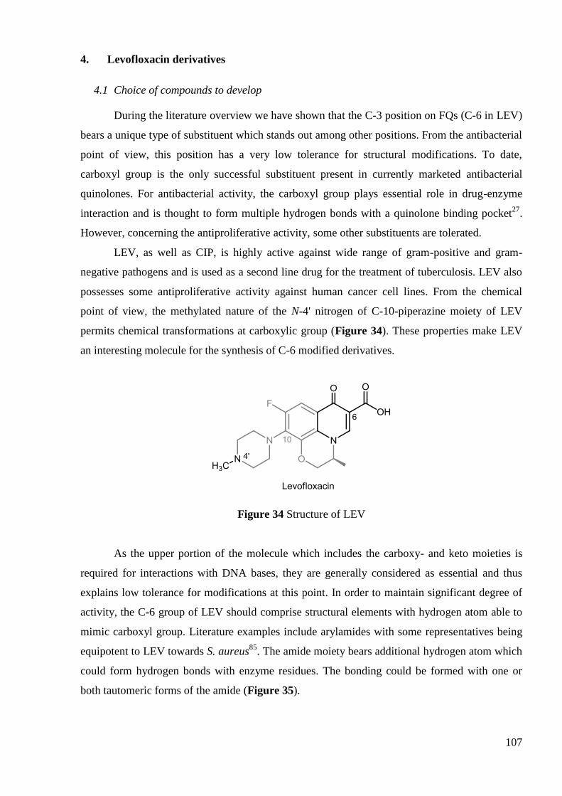
107
4. Levofloxacin derivatives
4.1 Choice of compounds to develop
During the literature overview we have shown that the C-3 position on FQs (C-6 in LEV)
bears a unique type of substituent which stands out among other positions. From the antibacterial
point of view, this position has a very low tolerance for structural modifications. To date,
carboxyl group is the only successful substituent present in currently marketed antibacterial
quinolones. For antibacterial activity, the carboxyl group plays essential role in drug-enzyme
interaction and is thought to form multiple hydrogen bonds with a quinolone binding pocket27
.
However, concerning the antiproliferative activity, some other substituents are tolerated.
LEV, as well as CIP, is highly active against wide range of gram-positive and gram-
negative pathogens and is used as a second line drug for the treatment of tuberculosis. LEV also
possesses some antiproliferative activity against human cancer cell lines. From the chemical
point of view, the methylated nature of the N-4' nitrogen of C-10-piperazine moiety of LEV
permits chemical transformations at carboxylic group (Figure 34). These properties make LEV
an interesting molecule for the synthesis of C-6 modified derivatives.
Figure 34 Structure of LEV
As the upper portion of the molecule which includes the carboxy- and keto moieties is
required for interactions with DNA bases, they are generally considered as essential and thus
explains low tolerance for modifications at this point. In order to maintain significant degree of
activity, the C-6 group of LEV should comprise structural elements with hydrogen atom able to
mimic carboxyl group. Literature examples include arylamides with some representatives being
equipotent to LEV towards S. aureus85
. The amide moiety bears additional hydrogen atom which
could form hydrogen bonds with enzyme residues. The bonding could be formed with one or
both tautomeric forms of the amide (Figure 35).

108
In contrast to antibacterial activity, antiproliferative potency tolerates structural changes
at C-3 carboxylic acid of quinolones with a number of successful examples.
Figure 35 Carboxyl group mimicking by amide group
Taking into account these considerations we synthesized series of alkyl carboxamides
(termed ―mono-amides‖) and ―dimeric‖ analogs (termed ―di-amides‖) of LEV. We have also
decided to synthesize a series of acyloxymethyl esters of LEV that would represent prodrugs of
LEV. In the literature various acyloxyalkyl ester prodrugs of butyric acid with anticancer activity
were described.166
.
In a similar way to the derivatives synthesized from CIP, varying the chain length should
result in compounds with different hydrophobicity and steric hindrance, thus providing a useful
model for SAR studies.
Figure 36 Target molecules of LEV derivatives.
In summary, after reviewing the literature and taking into account previous results of our
group we have decided to synthesize two types of LEV derivatives: carboxamides and
acyloxymethyl esters (Figure 36). The detailed synthetic pathway and challenges faced will be
described in the next section.

109
4.2 Synthesis of C-6-acyloxymethyl esters of LEV: Previous attempts
4.2.1 Synthesis of C-3-acyloxymethyl esters of nalidixic acid
The FQ modification at C-3 position was first carried out in our group by using NAL, a
parent compound for the quinolone family167
. Two possible pathways were evaluated for the
synthesis of acyloxymethyl esters of NAL (Scheme 9). The pathway A consists in a nucleophilic
substitution of chloromethyl alkanoates 10, synthesized from alkanoic acids, by NAL
carboxylate. The first step of the second pathway B consists in synthesis of the chloromethyl
ester of NAL 11 and in the subsequent nucleophilic substitution by various fatty carboxylates
(Scheme 9).
Two methods of chloromethylation of alkanoic acids were employed, either by a phase-
transfert system or by the use of a Lewis acid in presence of 1,3,5-trioxane (pathway A). It was
shown that that higher yields of alkylcarbonyloxymethyl chlorides 10 could be obtained by the
phase-transfert system. Unfortunately, when attempting the alkylation of NAL potassium salt
with one equivalent of these reagents in DMF at 50°C for 24 h, complex mixtures were obtained
resulting from incomplete reaction (even in the presence of sodium iodide) or decomposition.
The corresponding acyloxymethyl esters 12a-b were then isolated in poor yields ranging from
22% to 35% (Table 47).
On the contrary, the pathway B provided the clean chloromethylation of NAL by using
chloromethyl chlorosulfate (ClCH2OSO2Cl) in a phase-transfer H2O/CH2Cl2 system. The
chloromethyl NAL derivative 11 was obtained in a 55% yield. Further condensation with the salt
of fatty acid in DMF at room temperature for 4 days rendered the corresponding acyloxymethyl
esters 12a-d in 50-70% yields (Table 47).

110
Reagents and conditions : Pathway A : a) i – CH3(CH2)nCOOH, Na2CO3, H2O (rt or 100°C
according to n) ii – CH2Cl2, (nBu)4NHSO4, ClO2SOCH2Cl, 0°C (1h) – rt (18h) or a‘) i –
ClCOCOCl, CH2Cl2, 0°C (1h) – rt (2h), ii- ZnCl2, CaCl2, rt (15‘) iii – 1,3,5-trioxane, 0°C (2h) –
rt (20h). b) 10, K2CO3, DMF, 50°C (24h). Pathway B : c) i – Na2CO3, H2O, rt (90‘) ii – CH2Cl2,
(nBu)4NHSO4, ClO2SOCH2Cl, 0°C (1h) – rt (18h). d) i – CH3(CH2)nCOOH, K2CO3, DMF, 50°C
(5h) ii – 11, rt (4d)
Scheme 9 Preparation of acyloxymethyl esters of NAL
Table 47 Yields (%) of acyloxymethyl esters of NAL according to pathways A or
B
Compound n Pathway A
(from 10)
Pathway B
(from 11)
12a 6 35 70
12b 10 22 50
12c 12 - 60
12d 14 - 56
The pathway B turned out to be more convenient in terms of purification and yields and
above all it does not require the synthesis of each chloromethyl alkanoates 10. So, this pathway
B was chosen for the preparation of a series of acyloxymethyl esters of LEV.

111
4.2.2 Synthesis of acyloxymethyl esters of LEV
However when this method B was applied to LEV the first stage couldn‘t be completed.
The two alternative protocols for the chloromethylation were tried, but none of them provided
the desired chloromethyl ester of LEV 13 (Figure 37).
Figure 37 Unsuccessful attempts for the chloromethylation of LEV
So, it was decided to use the pathway A for the synthesis of acyloxymethyl esters of LEV
14, that is the direct alkylation of LEV with various chloromethyl alkanoates (Figure 38). In
order to apply this method to various alkyl chain lengths it was necessary to synthesize a series
of alkylcarbonyloxymethyl chlorides 10, except for the commercially chloromethyl butyrate (n =
2) 10a.
Figure 38 Schematic representation of the preparation of acyloxymethyl esters of
LEV 14
4.2.3 Synthesis of chloromethyl alkanoates
Examination of the literature on the preparation of chloromethyl esters revealed two main
protocols : alkylation of carboxylates with chloromethyl chlorosulfate in phase-transfer
conditions168
(Scheme 9, pathway A a)) and condensation of an aldehyde with acid chlorides in
the presence of a Lewis acid169
(Scheme 9, pathway A a')). Preliminary studies involving the
synthesis of chloromethyl octanoate 10c (n=6) revealed that the A a) method provides higher

112
yields (60% yield for 10c) but uses hazardous, potentially carcinogenic alkylating agent, that is
chloromethyl chlorosulfate. In order to improve the A a') procedure, several Lewis acids were
tried for the preparation of chloromethyl octanoate 10c (Scheme 10, Table 48). It was observed
that the use of zinc chloride in the presence of calcium chloride improved the yield of reaction to
52%.
Scheme 10 Procedures for the synthesis of chloromethyloctanoate 10c
Table 48 Optimization of the synthesis of chloromethyl octanoate (n = 6) 167
Pathway A Lewis Acid Yield of 10c, %
a) – 60a
a') SnCl4 33b
ZrCl4 9b
AlCl3 16b
ZnCl2 25b
ZnCl2 + CaCl2 57b (52
a)
aYields after isolation.
bYields calculated from
1H NMR (Nuclear Magnetic Resonance)
spectra of mixtures obtained after usual workup.
After these optimization runs with chloromethyl octanoate, both methods were applied to
the synthesis of a series of lipophilic alkylating agents. It was observed that the pathway A a) is
often more effective for the synthesis of chloromethyl alkanoate in comparison with the pathway
A a') (Table 49).
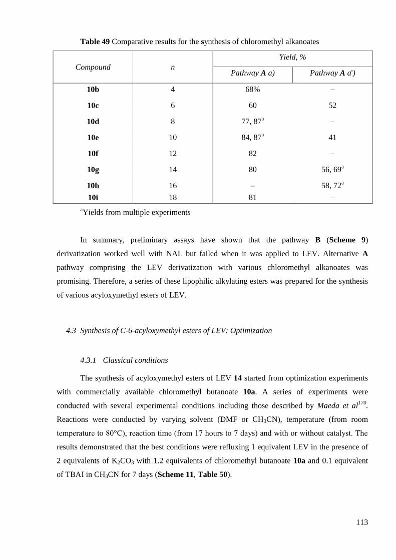
113
Table 49 Comparative results for the synthesis of chloromethyl alkanoates
Compound n
Yield, %
Pathway A a) Pathway A a')
10b 4 68% –
10c 6 60 52
10d 8 77, 87a –
10e 10 84, 87a 41
10f 12 82 –
10g 14 80 56, 69a
10h 16 – 58, 72a
10i 18 81 –
aYields from multiple experiments
In summary, preliminary assays have shown that the pathway B (Scheme 9)
derivatization worked well with NAL but failed when it was applied to LEV. Alternative A
pathway comprising the LEV derivatization with various chloromethyl alkanoates was
promising. Therefore, a series of these lipophilic alkylating esters was prepared for the synthesis
of various acyloxymethyl esters of LEV.
4.3 Synthesis of C-6-acyloxymethyl esters of LEV: Optimization
4.3.1 Classical conditions
The synthesis of acyloxymethyl esters of LEV 14 started from optimization experiments
with commercially available chloromethyl butanoate 10a. A series of experiments were
conducted with several experimental conditions including those described by Maeda et al170
.
Reactions were conducted by varying solvent (DMF or CH3CN), temperature (from room
temperature to 80°C), reaction time (from 17 hours to 7 days) and with or without catalyst. The
results demonstrated that the best conditions were refluxing 1 equivalent LEV in the presence of
2 equivalents of K2CO3 with 1.2 equivalents of chloromethyl butanoate 10a and 0.1 equivalent
of TBAI in CH3CN for 7 days (Scheme 11, Table 50).

114
Scheme 11 Synthetic route to the target butanoyloxymethyl ester of LEV 14a
Table 50 Optimization assays for the synthesis of butanoyloxymethyl ester of
LEV (n = 2)
Base Solvent Catalyst (relative
amounts/LEV) Temperature
Reaction
time
Yield of 14a
(%)
K2CO3a DMF – 50ºC 17h traces
K2CO3b
DMF – rt 4d 23
K2CO3b DMF – 50ºC 7d 28
K2CO3b CH3CN – rt 4d 15
K2CO3b CH3CN – 50ºC 7d 36
K2CO3b CH3CN – Reflux 7d 25
K2CO3b CH3CN TBAI (0.1 eq) Reflux 7d 48
aExperimental conditions described by Maeda et al
170
bSandra Dorbes, Master 2,
February-June 2005
Later on this protocol was applied to the synthesis of long-chain acyloxymethyl esters of
LEV 14e-i. However, in comparison with short-chain compound 14a, the long-chain analogs
were obtained with a significant decrease of the yield (0-10% yields) and noticeable quantities of
methylene dialkanoate by-product 15 (14-35% yields) were isolated (Scheme 12, Table 51). The
formation of these symmetric double esters may be explained by the hydrolysis of the
chloromethyl alkanoates 10 leading to the formation of corresponding carboxylates which
probably react more rapidly with the remaining chloromethyl alkanoate than LEV carboxylate.

115
Scheme 12 Synthesis of acyloxymethyl esters of LEV 14
Table 51 Yields of long-chain acyloxymethyl esters of LEV 14e-i and methylene
dialkanoate 15e-i.a
n Compound Yield of 14 ( %)b Coumpound Yield of 15 (%)
b
10 14e 5 15e 17
12 14f 10 15f 35
14 14g 3 15g 14
16 14h 5 15h 28
18 14i 0 15i 0
aSandra Dorbes, Master 2, February-June 2005
bIsolated yields after purification
In summary, classical conditions for the alkylation of LEV with chloromethyl butanoate
were optimized and gave a moderate yield of 48%. However, applying these conditions for the
synthesis of long-chain acyloxymethyl esters of LEV resulted in the significant drop of the yield.
This was probably due to the hydrolysis of the chloromethyl alkanoates during the prolonged 7
days heating of the reaction mixture. In order to overcome these difficulties we decided to
perform microwave-assisted attempts.

116
4.3.2 Microwave-assisted synthesis
4.3.2.1 Optimization for the commercial chloromethyl butyrate
The microwave-assisted (MW) synthesis was attractive because of its major advantages
such as rapid heat transfer and selective heating. This should lower the reaction time and
therefore decrease the possible hydrolysis of the alkylating agent171
.
Initial attempts were carried out with chloromethyl butyrate 10a and showed, in
comparison with classical oil bath conditions, a significant decrease of the reaction time
(Scheme 13, Table 52). In an effort to ascertain the reaction parameters governing the
nucleophilic substitution, for all optimization runs (Table 52, entries 2-13), the reactive
potassium or cesium salt was always prepared under MW in a discrete step using 20 min
irradiation time from 1 equivalent of LEV and 2 equivalents of base. In the nucleophilic
substitution step, 1.5 equivalents of chloromethyl butyrate was used with regard to LEV.
Reactions were first conducted in DMF at 120°C, by varying reaction time and catalyst
amounts (Table 52, entries 2-6). Three experiments were conducted to assess the effect of the
relative amounts of TBAI on the isolated yields while keeping constant the irradiation time (60
min) (Table 52, entries 2-4). The yield was significantly dependent on the amount of catalyst
used, the use of 0.2 equivalent of TBAI led to an optimized yield of 46%. Next, the effect of
irradiation time on the substitution (Table 52, entries 4–6) was assessed and showed that
increasing the reaction time from 60 to 120 min led to a loss of yield of the expected 14a.
Esterifications with chloroalkylesters were described as more effective when the reaction was
conducted in N,N’-dimethylpropyleneurea DMPU172
and we have found out that it gave similar
yield as in DMF (Table 52, entry 7).
CH3CN as solvent at 90°C for 60 minutes with 0.2 equivalent of TBAI, led to an enhance
yield of 56% (Table 52, entry 8). As observed with DMF, an increase of time of reaction from
60 to 90 minutes led to a loss of yield (Table 52, entry 9). We also assessed the effect of the
relative amounts of TBAI. When the reaction was carried out with 0.1 equivalent of TBAI, we
observed a loss of yield and with 0.3 equivalent of TBAI, the yield remained unchanged (Table
52, entries 10-11). The change of the base by using Cs2CO3 instead of K2CO3, either in DMF or
CH3CN, did not lead to an improved yield (Table 52, entries 12-13).
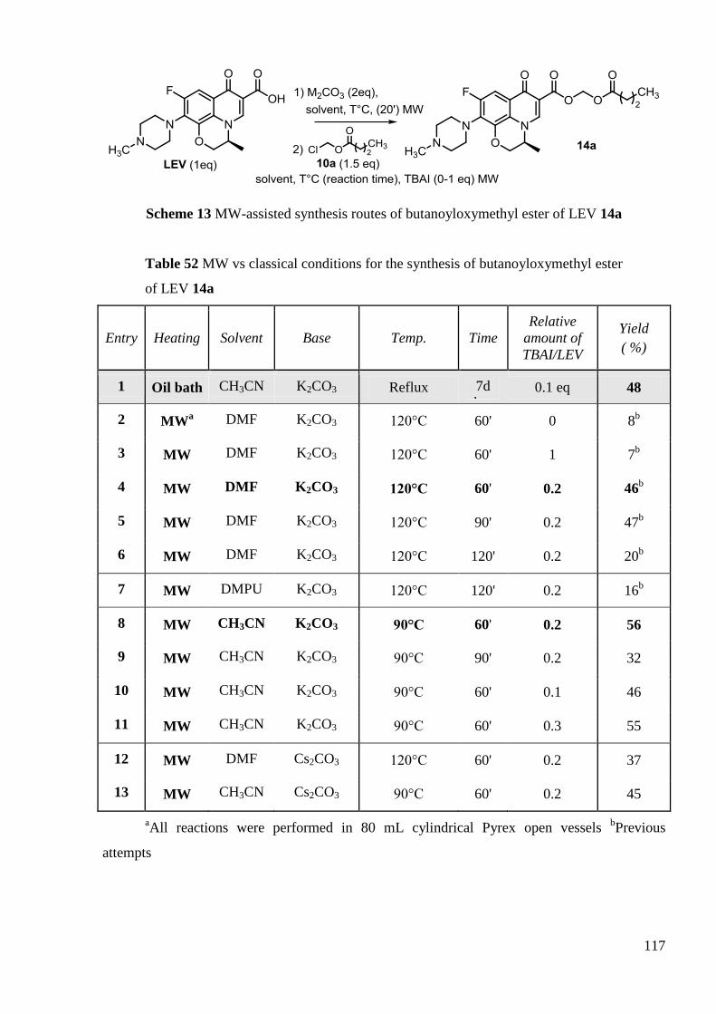
117
Scheme 13 MW-assisted synthesis routes of butanoyloxymethyl ester of LEV 14a
Table 52 MW vs classical conditions for the synthesis of butanoyloxymethyl ester
of LEV 14a
Entry Heating Solvent Base Temp. Time
Relative
amount of
TBAI/LEV
Yield
( %)
1 Oil bath CH3CN K2CO3 Reflux 7d
days 0.1 eq 48
2 MWa DMF K2CO3 120°C 60' 0 8
b
3 MW DMF K2CO3 120°C 60' 1 7b
4 MW DMF K2CO3 120°C 60' 0.2 46b
5 MW DMF K2CO3 120°C 90' 0.2 47b
6 MW DMF K2CO3 120°C 120' 0.2 20b
7 MW DMPU K2CO3 120°C 120' 0.2 16b
8 MW CH3CN K2CO3 90°C 60' 0.2 56
9 MW CH3CN K2CO3 90°C 90' 0.2 32
10 MW CH3CN K2CO3 90°C 60' 0.1 46
11 MW CH3CN K2CO3 90°C 60' 0.3 55
12 MW DMF Cs2CO3 120°C 60' 0.2 37
13 MW CH3CN Cs2CO3 90°C 60' 0.2 45
aAll reactions were performed in 80 mL cylindrical Pyrex open vessels
bPrevious
attempts

118
Then, the optimal MW conditions for the synthesis of butanoyloxymethyl ester 14a were
found to be the use of 0.2 equivalent of catalyst (TBAI) in CH3CN at 90°C for 60 minutes in the
presence of K2CO3.
4.3.2.2 Synthesis of a series of acyloxymethyl esters of LEV
We then applied this experimental protocol to the synthesis of the series of
acyloxymethyl esters LEV 14b-i with longer chain lengths. Potassium salt of LEV was prepared
by reaction of K2CO3 (2 equivalent) on LEV (1 equivalent) in CH3CN at 90°C under MW using
20 min irradiation time. After the addition of TBAI catalyst (0.2 eq) and chloromethyl alkanoates
10b-i (1.5 eq), the mixture was irradiated one hour more at 90°C. Though yields were somewhat
higher (from 10 to 23% yields) for the synthesis of 14e-i than those obtained under oil bath
heating (from 3 to 10% yields), they were significantly lower than the yields obtained for the
short chain analog 14a (n=2). (Scheme 14, Table 53)
Scheme 14 MW-assisted synthesis of acyloxymethyl esters of LEV 14b-i
Table 53 Yields of acyloxymethyl esters of LEV 14b-i under MW conditions
Compound n Yield, %
14b 4 16
14c 6 18
14d 8 16
14e 10 17
14f 12 9
14g 14 11
14h 16 23
14i 18 –a
aFailed to isolate the pure product
In order to investigate the reason for these poor yields, we decided to examine the
byproduct formation. A representative example of long-chain derivatives was chosen for this

119
study, that was the MW-assisted synthesis of octadecanoyloxymethyl ester of LEV 14h. Indeed,
during the synthesis of acyloxymethyl esters of LEV conducted under classical oil-bath
conditions, high quantities of methylene dialkanoates by-product 15 were observed and were
thought to be the possible cause of the low yield (Table 51).
In order to clarify the reaction behavior we have evaluated the rate of by-products, that
were methylene dioctadecanoate 15h, octadecanoic acid (arising from the hydrolysis of
chloromethyl octadecanoate) along with unreacted reagent 10h and the desired
octadecanoyloxymethyl ester of LEV 14h (Table 54). After a tedious purification, we have got
many batches containing pure product or a mixture of two products. In this last case, the yields
have been estimated by 1H NMR spectroscopy. We observed that under MW conditions, much
less of the symmetric methylene dioctadecanoate 15h and more of the desired
octadecanoyloxymethyl ester of LEV 14h were produced in a shorter time of reaction comparing
to classical oil-bath conditions. However, we have seen that the main part of the alkylating agent
10h remains unreacted or hydrolyzed to octadecanoic acid. In addition, unreacted LEV was also
detected.
Table 54 Conversion of chloromethyloctadecanoate 10h during the synthesis of
long-chain compound (n = 16)
Compound Conversion
a, %
Microwave, 1 hour Oil-bath, 7 days
14h 18b 3
b
n-C17H35CO2CH2Cl, 10h 32c –
d
(n-C17H35COO)2CH2, 15h 5c 28
b
n-C17H35CO2H 45e –
d
aBased on the n-C17H35CO2CH2Cl (Its initial quantity is set to 100%)
bIsolated product
cEstimated by
1H NMR spectroscopy for the mixture of 10h and 15h
dWas not evaluated during
experiment eCalculated as a difference between the initial quantity of n-C17H35CO2CH2Cl and
other C17 containing components
In summary, the MW-assisted synthesis of acyloxymethyl esters of LEV had some
advantages over classical conditions (significantly lower reaction time) but the yields were low
for the long-chain derivatives. This could be explained by the relatively high hydrolysis rate of
chloromethylalkanoate. The presence of unreacted LEV and alkylating agent may be due to their

120
low reactivity in these conditions, which is supposed to be the second reason for the low yield.
So we decided to search for alternative conditions in order to optimize this synthetic protocol.
4.3.3 Modified classical oil-bath conditions: use of cesium salts of LEV
4.3.3.1 Preliminary attempt for butyloxymethyl ester of LEV (n = 2)
Cesium salts generally display higher solubility in organic solvents. We first isolated
LEV cesium salt after refluxing LEV with cesium carbonate in dichloromethane. DMF was first
evaluated as solvent. The other parameters were kept the same as for the microwave and
classical conditions: 90 ºC, 0.2 equivalent of TBAI and 1.5 equivalents of chloromethyl butyrate
10a (Scheme 15). Initial experiment rendered the desired butyloxymethyl ester of LEV 14a in
moderate yield (50%) after only 3 hours while classical conditions discussed above required 7
days under reflux to achieve 48% yield.
Scheme 15 Synthesis of butyloxymethyl ester of LEV in DMF by using cesium
salt
This reaction conducted with cesium salt of LEV seemed to be faster than the one from
LEV. However, it should be pointed out that this procedure, when applied to a series of long
chain derivatives (bearing 11-19 carbon atoms), was always plagued with difficulties during
purification. Generally, several (2–3) silica gel columns (with different solvent systems) were
necessary to isolate the lipophilic esters in pure form without any trace of TBAI. Low isolated
yields were thus obtained. To circumvent this problem, the reaction with chloromethyl
octadecanoate was further investigated using oil-bath heating and without TBAI.
4.3.3.2 Preliminary attempts for the synthesis of octadecanoyloxymethyl ester of
levofloxacin 14h (n = 16)
We decided to optimize the reaction on a long-chain derivative, as the increase of chain
length of alkylating agents posed greater problems. The reaction was conducted with
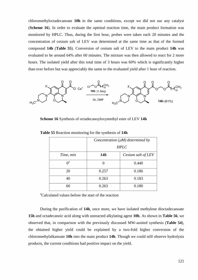
121
chloromethyloctadecanoate 10h in the same conditions, except we did not use any catalyst
(Scheme 16). In order to evaluate the optimal reaction time, the main product formation was
monitored by HPLC. Thus, during the first hour, probes were taken each 20 minutes and the
concentration of cesium salt of LEV was determined at the same time as that of the formed
compound 14h (Table 55). Conversion of cesium salt of LEV to the main product 14h was
evaluated to be around 64% after 60 minutes. The mixture was then allowed to react for 2 more
hours. The isolated yield after this total time of 3 hours was 60% which is significantly higher
than ever before but was appreciably the same to the evaluated yield after 1 hour of reaction.
Scheme 16 Synthesis of octadecanoyloxymethyl ester of LEV 14h
Table 55 Reaction monitoring for the synthesis of 14h
Concentration ( M) determined by
HPLC
Time, min 14h Cesium salt of LEV
0a 0 0.440
20 0.257 0.186
40 0.263 0.183
60 0.263 0.180
aCalculated values before the start of the reaction
During the purification of 14h, once more, we have isolated methylene dioctadecanoate
15h and octadecanoic acid along with unreacted alkylating agent 10h. As shown in Table 56, we
observed that, in comparison with the previously discussed MW-assited synthesis (Table 54),
the obtained higher yield could be explained by a two-fold higher conversion of the
chloromethylalkanoate 10h into the main product 14h. Though we could still observe hydrolysis
products, the current conditions had positive impact on the yield.

122
Table 56 Comparison of conversion of chloromethyloctadecanoate 10h for the
current conditions and MW-assisted synthesis of 14h (n = 16)
Compound
Conversiona, %
Current conditions, 3 hours MW, 1 hour
14h 34b 18
b
n-C17H35CO2CH2Cl, 10h 17c 32
c
(n-C17H35COO)2CH2, 15h 4c 5
c
n-C17H35CO2H 45d 45
d
aBased on the n-C17H35CO2CH2Cl (Its initial quantity is set to 100%)
bIsolated product
cEstimated by
1H NMR spectroscopy for the mixture of 10h and 15h
dCalculated as a difference
between the initial quantity of n-C17H35CO2CH2Cl (1.5 eq) and other C17 containing
components
A series of small-scale experiments were conducted in order to find the best conditions of
reaction. The yields of compound 14h were estimated by HPLC analysis of the crude reaction
mixture. Thus, we assessed the effect of CH3CN instead of DMF (Table 57, entries 1, 2), the
time of reaction (Table 57, entries 1, 3) and the amount of chloromethyloctadecanoate (Table
57, entries 1, 4).
Table 57 Optimization of reaction conditions for the synthesis of 14h
Entry Time (hour) Solvent RCO2CH2Cl ( eq) Yield (%)
1 3 DMF 1.5 64
2 3 CH3CN 1.5 58
3 1 DMF 1.5 70
4 1 DMF 1 63
First of all, we observed that CH3CN gave lower yield than DMF. As already observed
the reaction time in DMF can be decreased to 1 hour. Using 1 equivalent of the alkylating agent
instead of 1.5 equivalents slightly lowered the yield, but facilitated the purification step. Though
an increased yield was achieved, this protocol still presented some drawbacks: the step of the
cesium salt preparation and the moisture sensitivity of the cesium salt making it uneasy to
handle. The resolution of these issues will be discussed in the next section.

123
4.3.4 Modified classical conditions: in situ formation of LEV salts
In order to overcome these drawbacks, we decided to prepare in situ the cesium (or
potassium) salt of LEV. During an optimization study, isolated yields of compound 14h were
determined. A mixture of LEV and M2CO3 (1 equivalent) was heated in CH3CN or DMF for 20
minutes. After adding octadecanoyloxymethyl chloride, the reaction was performed during 1
hour (Scheme 17). The results obtained for various parameters: the solvent (DMF or CH3CN),
the base (Cs2CO3 or K2CO3) and the quantity of alkylating agent (1 or 1.5 equivalents) are
presented in Table 58.
Scheme 17 Synthesis of octadecanoyloxymethyl ester of LEV 14h
Table 58 Optimization runs for the synthesis of 14h
Base, eq Solvent RCO2CH2Cl, eq Yield, %
Cs2CO3 (1eq) DMF 1 83
Cs2CO3 (1 eq) CH3CN 1 23
K2CO3 (1 eq) DMF 1 81
K2CO3 (1 eq) CH3CN 1 9
K2CO3 (1 eq) DMF 1.5 86
From obtained results, we observed that the use of in situ salts formation increased the
yield and favored the conversion of LEV into the desired compound 14h. The nature of the base
used was not really important in this case as both Cs2CO3 and K2CO3 gave similar yields, if the
reaction was conducted in DMF. The nature of the used solvent turned out to be the determining
factor for this reaction: when DMF was used, the isolated yields were four- to eight-folds higher
than those in CH3CN. This phenomenon could be explained by the preferred cation-solvation by
DMF, thereby activating the LEV carboxylate-anion. A poor difference was obtained between
equimolar and 1.5 equivalent quantities of the chloromethyloctadecanoate. We selected as
optimal conditions for the synthesis of the rest of acyloxymethyl esters of LEV derivatives: 1

124
equivalent of K2CO3 (cheaper and less hygroscopic), DMF as solvent, 1 equivalent of alkylating
agent RCO2CH2Cl, 1 hour reaction time at 90°C.
4.3.5 Synthesis of acyloxymethyl esters of LEV derivatives using optimized conditions
We have synthesized the series of LEV acyloxymethyl esters 14a-i in yields varying from
59 to 81% depending on the length of the chain (Table 59). 1H NMR spectra of the crude
mixture showed no significant difference between long and short chain derivatives. As the lower
yields were obtained for compounds with shorter chains, we proposed that as they are more polar
they are probably held tighter by the silica column during the purification step.
Table 59 Yields of acyloxymethyl esters of LEV
Compound n Yield, %
14a 2 59
14b 4 69
14c 6 75
14d 8 79
14e 10 79
14f 12 77
14g 14 79
14h 16 81
14i 18 79
In summary, we have designed and synthesized the series of acyloxymethyl esters of
LEV. We have tried three general synthetic protocols while optimizing the experimental
conditions. During these experiments we have deduced the most important factors influencing
the reaction: the reaction time (1h) and the nature of the solvent (DMF). The other factors such
as the nature of the base (K2CO3 vs Cs2CO3) and the quantity of the alkylating agent (1
equivalent versus 1.5 equivalents) showed minor influence on the yield. As it was previously
mentioned this compounds could represent prodrugs of LEV. Though acyloxymethyl esters of

125
LEV are more stable than simple esters, they are sensible to hydrolysis. So we have shifted our
attention to the synthesis of ―mono-‖ and ―di-amide‖ derivatives of LEV with higher stability
towards enzymatic and chemical hydrolysis.
4.4 Synthesis of C-6-(alkylcarboxamide)-linked LEV “monomers” and “dimers”
4.4.1 Synthesis of “monomer” dodecylcarboxamide derivative of LEV
As previously outlined for the synthesis of CIP derivatives, FQs did not tolerate the use
of thionyl chloride or oxalyl chloride due to the high degree of degradation of FQs with these
chlorinating agents.
Carbodiimides were previously used for the synthesis of CIP derivatives. In the previous
section 3.2 we discussed some methods for formation of the amide bond in peptide chemistry.
Carbodiimides are often used for the coupling of aminoacids and amide formation from aliphatic
acids.
We compared two methods of carboxylic acid activation : i) the use of the salt of uronium
with a non-nucleophilic anion which exists in the O- and N-forms: 1-
[bis(dimethylamino)methylene]-1H-1,2,3-triazolo-[4,5-b]pyridinium hexafluorophosphate 3-
oxide (N-HATU) and N-[(1H-1,2,3-triazolo[4,5-b]pyridin-1-yloxy)(dimethylamino)methylene]-
N-methylmethanaminium hexafluorophosphate (O-HATU)173
and ii) the use of the
triphenylphosphine (TPP) / trichloroacetonitrile174
(TCA) system.
It was previously reported that HATU is very efficient in solution synthesis175
and tertiary
amines such as diisopropylethylamine (DIPEA) have been considered as practically useful bases
due to their non-nucleophilic property, while the highly hindered 2,4,6-trimethylpyridine (TMP)
was recommended by Carpino176
. Both O- and N-forms serve as excellent coupling agents.
(Figure 39).
Figure 39 Two isomers of HATU coupling agent
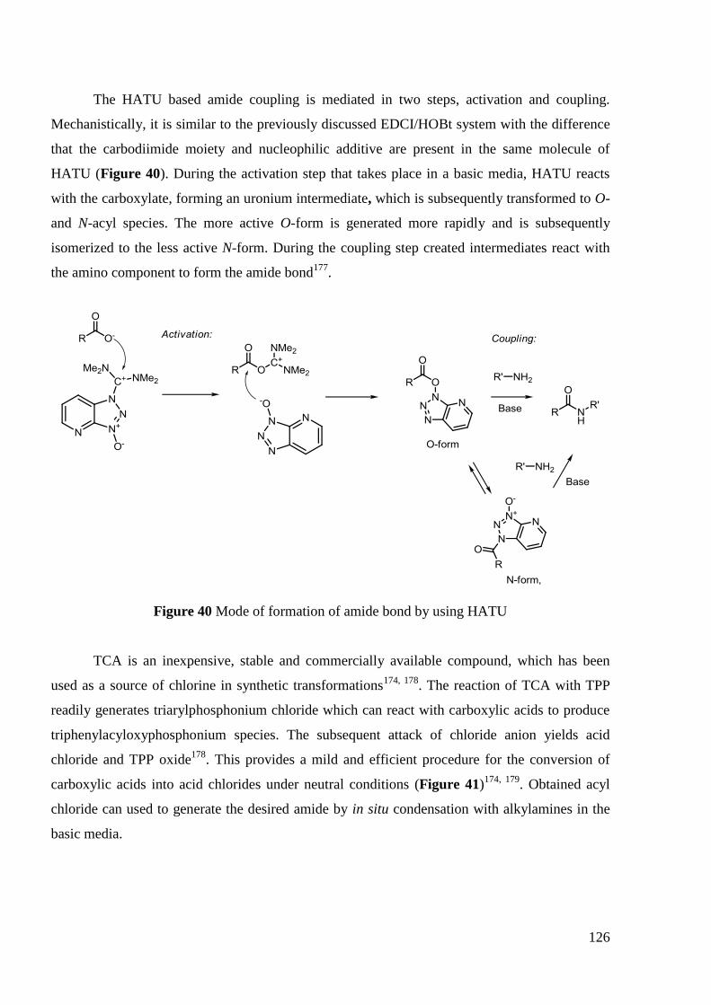
126
The HATU based amide coupling is mediated in two steps, activation and coupling.
Mechanistically, it is similar to the previously discussed EDCI/HOBt system with the difference
that the carbodiimide moiety and nucleophilic additive are present in the same molecule of
HATU (Figure 40). During the activation step that takes place in a basic media, HATU reacts
with the carboxylate, forming an uronium intermediate, which is subsequently transformed to O-
and N-acyl species. The more active O-form is generated more rapidly and is subsequently
isomerized to the less active N-form. During the coupling step created intermediates react with
the amino component to form the amide bond177
.
Figure 40 Mode of formation of amide bond by using HATU
TCA is an inexpensive, stable and commercially available compound, which has been
used as a source of chlorine in synthetic transformations174, 178
. The reaction of TCA with TPP
readily generates triarylphosphonium chloride which can react with carboxylic acids to produce
triphenylacyloxyphosphonium species. The subsequent attack of chloride anion yields acid
chloride and TPP oxide178
. This provides a mild and efficient procedure for the conversion of
carboxylic acids into acid chlorides under neutral conditions (Figure 41)174, 179
. Obtained acyl
chloride can used to generate the desired amide by in situ condensation with alkylamines in the
basic media.

127
Figure 41 Mechanism of formation of amide bond TPP/TCA system
16
We applied these two methods to the synthesis of dodecylcarboxamide derivative of LEV
17f (Scheme 18).
Scheme 18 Synthesis of dodecylcarboxamide derivative of LEV 17f
The initial activation of the LEV by 1 equivalent of HATU and 3 equivalents of DIPEA
in DMF for 1 hour at room temperature was followed by addition of 1 equivalent of
dodecylamine. After stirring reaction mixture at room temperature for 8 hours, the target
compound 17f was isolated with 52% yield. Reaction went smoothly and the purification didn‘t
pose any problems.
Acyl chloride of LEV 16 was prepared after the treatment of LEV with TPP/TCA (2
equivalents / 2 equivalents) in CH2Cl2 for 4 hours at room temperature. This derivative was
filtered off and used in the next step without purification. Reaction with 1 equivalent of
dodecylamine in the presence of 3 equivalents of triethylamine for 18 hours in CH2Cl2 at room

128
temperature provided the desired dodecylcarboxamide derivative of LEV 17f with moderate
overall yield (38%) after an easy purification (Scheme 18).
TPP/TCA system was chosen for the synthesis of C-6-(alkylcarboxamide)-linked LEV
―monomers‖ and ―dimers‖ which will be described in the next section.
4.4.1.1 Synthesis of “monomer” alkylcarboxamide derivatives of LEV
The series of ―monomer‖ alkylcarboxamide derivatives of LEV 17a-j was obtained, by
using TPP/TCA system, with moderate yields. The summary of these results is presented in
Table 60.
Table 60 Synthesis of ―monomer‖ alkylcarboxamide derivatives of LEV 17a-j
Compound n Yield, %
17a 3 34
17b 5 37
17c 6 36
17d 7 36
17e 9 35
17f 11 37
17g 12 39
17h 13 39
17i 14 40
17j 16 41
4.4.1.2 Synthesis of “dimer” alkylcarboxamide derivatives of LEV
For the synthesis of ―di-amide‖ derivatives of LEV 18, the same TPP/TCA system was
used. The ―dimeric‖ derivatives were obtained with yields somewhat lower (20-41%) than that
for their ―monomeric‖ analogs 17 (Scheme 19, Table 61).
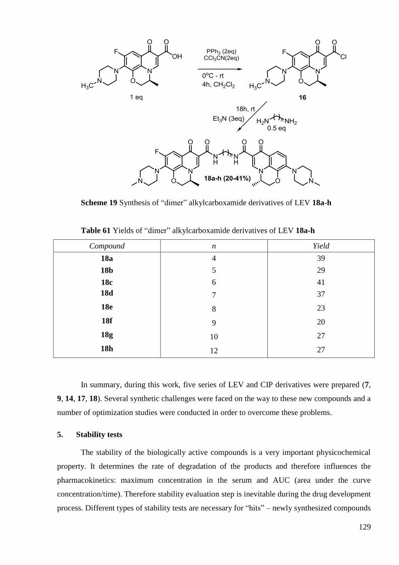
129
Scheme 19 Synthesis of ―dimer‖ alkylcarboxamide derivatives of LEV 18a-h
Table 61 Yields of ―dimer‖ alkylcarboxamide derivatives of LEV 18a-h
Compound n Yield
18a 4 39
18b 5 29
18c 6 41
18d 7 37
18e 8 23
18f 9 20
18g 10 27
18h 12 27
In summary, during this work, five series of LEV and CIP derivatives were prepared (7,
9, 14, 17, 18). Several synthetic challenges were faced on the way to these new compounds and a
number of optimization studies were conducted in order to overcome these problems.
5. Stability tests
The stability of the biologically active compounds is a very important physicochemical
property. It determines the rate of degradation of the products and therefore influences the
pharmacokinetics: maximum concentration in the serum and AUC (area under the curve
concentration/time). Therefore stability evaluation step is inevitable during the drug development
process. Different types of stability tests are necessary for ―hits‖ – newly synthesized compounds

130
with biological activity. The simplest initial tests measure the stability in vitro - in the water
buffer at physiological pH = 7.4 and 37ºC. These tests could provide some insight into the
stability of the compounds and select the successful candidates for in vivo testing.
Though there is no standard universal procedure for stability evaluation of every
compound, analytical HPLC is one the most frequently used methods180
. It is relatively simple
and quick with a high potential of separating complex mixtures of compounds. The optical
ultraviolet (UV) detection provides quantification and a high sensitivity of this method.
So we decided to use the HPLC procedure and UV detection to measure the stability of
our compounds.
5.1 Conditions for the stability testing
Though there are a number of examples of experimental conditions for the stability
testing180
there is no generalized procedure recognized as a standard. We have chosen phosphate
buffer saline (PBS) as a buffer solution for the testing. Our compounds weren‘t soluble directly
in the buffer so mixture of PBS/acetonitrile (50:50) was used. Initially compounds were
dissolved in the DMSO and then they were diluted by the PBS/acetonitrile mixture.
Experimental evidences from the literature showed that up to 10% of DMSO doesn‘t result in
significant changes of stability180a
. However, even in these conditions the most liphophilic
compounds weren‘t soluble. We decided to test synthesized compounds for 6-7 days with
intermediate sampling.
5.2 Results
5.2.1 CIP derivatives
We have selected representatives from each series with wide range of chain lengths. In a
typical experiment 1mM DMSO solution of compound was diluted by PBS to render the final
concentration 10-4
M of tested compound and 10% of DMSO. An aliquot was taken and kept at
4ºC (0 day). Then the vial was sealed and incubated for 6 days at 37ºC (6 days). In order to
evaluate the degradation degree of tested compounds these two probes were injected.
Chromatograms were extracted at the wavelength = 280 nm as it represents an absorbance
maximum for these series of CIP derivatives. By the comparison of the initial chromatogram and
the one after the incubation we could estimate the percentage of the remaining parent compound
relative to the initial concentration. Typical chromatograms for 0 day and 6 days probes are
demonstrated on the Figure 42. The peak with tr = 1.0 min is associated with the dead volume of
the column. The series of ―monomeric‖ CIP derivatives can provide two possible degradation
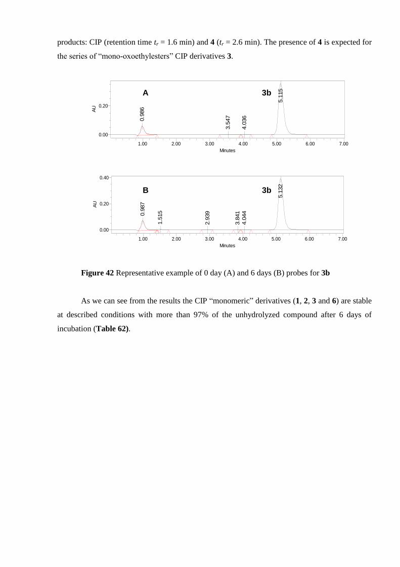
131
products: CIP (retention time tr = 1.6 min) and 4 (tr = 2.6 min). The presence of 4 is expected for
the series of ―mono-oxoethylesters‖ CIP derivatives 3.
Figure 42 Representative example of 0 day (A) and 6 days (B) probes for 3b
As we can see from the results the CIP ―monomeric‖ derivatives (1, 2, 3 and 6) are stable
at described conditions with more than 97% of the unhydrolyzed compound after 6 days of
incubation (Table 62).
AU
0.00
0.20
Minutes
1.00 2.00 3.00 4.00 5.00 6.00 7.000.9
86
3.5
47
4.0
36
5.1
15
AU
0.00
0.20
0.40
Minutes
1.00 2.00 3.00 4.00 5.00 6.00 7.00
0.9
87
1.5
15
2.9
39
3.8
41
4.0
44
5.1
32
A 3b
B 3b

132
Table 62 Stability of CIP ―monomeric‖ derivatives
Compound R Stability
a, %
6 days
1f -CO(CH2)5CH3 100
1h -CO(CH2)8CH3 100
1k -CO(CH2)14CH3 100
2 -COCH2Cl >99
3b -COCH2OCO(CH2)2CH3 100
3c -COCH2OCO(CH2)4CH3 >99
3d -COCH2OCO(CH2)6CH3 >97
6a -C(O)OCH2CH3 100
6b -C(O)O(CH2)3CH3 100
6c -C(O)O(CH2)7CH3 100
6g -C(O)OC(CH3)3 >99 aStability(%) at x day = [mean measured concentration (n = 2) at x day/mean measured
concentration (n = 2) at 0 day] × 100
During stability studies of ―dimeric‖ CIP derivatives we have monitored the intermediate
concentration at 3 and 5 days point in order to estimate the rate of hydrolysis. We have also
evaluated the presence of CIP in the solution during stability testing. However, for all
compounds the peak corresponding to CIP was not observed (Figure 43) and the data shown on
the Table 63 proves that CIP ―di-amide‖ derivatives 7 exhibit no hydrolysis after 7 days of
incubation.
A
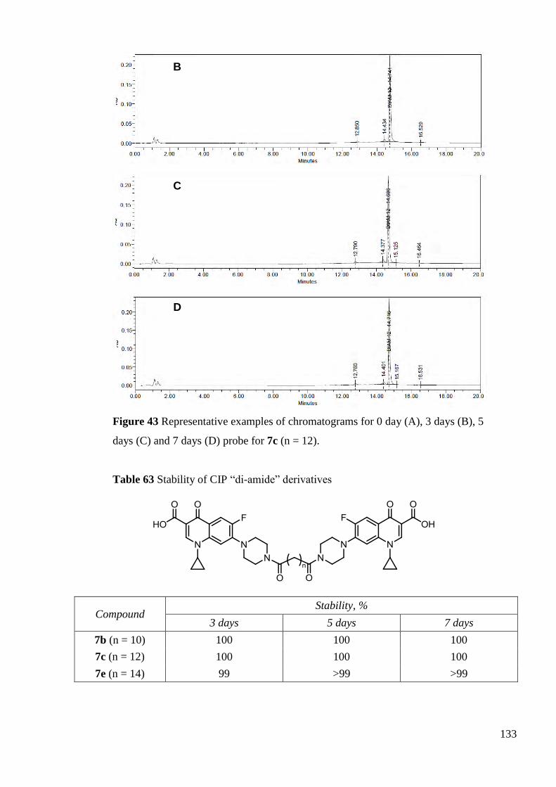
133
Figure 43 Representative examples of chromatograms for 0 day (A), 3 days (B), 5
days (C) and 7 days (D) probe for 7c (n = 12).
Table 63 Stability of CIP ―di-amide‖ derivatives
Compound Stability, %
3 days 5 days 7 days
7b (n = 10) 100 100 100
7c (n = 12) 100 100 100
7e (n = 14) 99 >99 >99
B
C
D
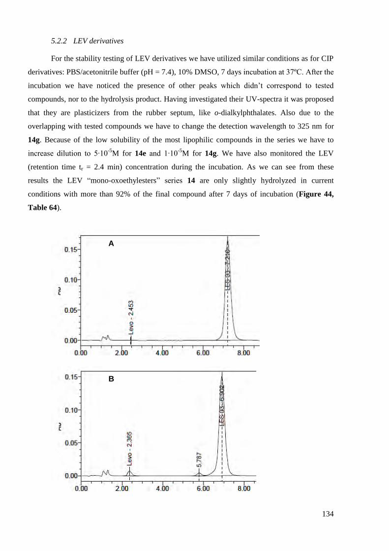
134
5.2.2 LEV derivatives
For the stability testing of LEV derivatives we have utilized similar conditions as for CIP
derivatives: PBS/acetonitrile buffer (pH = 7.4), 10% DMSO, 7 days incubation at 37ºC. After the
incubation we have noticed the presence of other peaks which didn‘t correspond to tested
compounds, nor to the hydrolysis product. Having investigated their UV-spectra it was proposed
that they are plasticizers from the rubber septum, like o-dialkylphthalates. Also due to the
overlapping with tested compounds we have to change the detection wavelength to 325 nm for
14g. Because of the low solubility of the most lipophilic compounds in the series we have to
increase dilution to 5·10-5
M for 14e and 1·10-5
M for 14g. We have also monitored the LEV
(retention time tr = 2.4 min) concentration during the incubation. As we can see from these
results the LEV ―mono-oxoethylesters‖ series 14 are only slightly hydrolyzed in current
conditions with more than 92% of the final compound after 7 days of incubation (Figure 44,
Table 64).
A
B

135
Figure 44 Representative examples of chromatograms for 0 day (A), 3 days (B), 5
days (C) and 7 days (D) probe for 14a (n = 2)
Table 64 Stability of LEV acyloxymethyl esters
Compound Stability, %
0 days 3 days 5 days 7 days
14a (n = 2) 100 95 93 92
14c (n = 6) 100 98 94 92
14e (n = 10) 100 96 93 93
14g (n = 14) - 95a 93
a 93.0
a
C
D
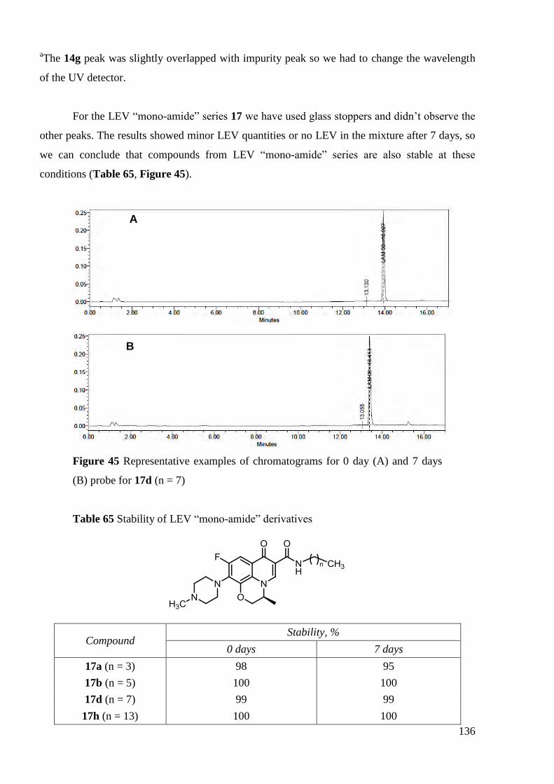
136
aThe 14g peak was slightly overlapped with impurity peak so we had to change the wavelength
of the UV detector.
For the LEV ―mono-amide‖ series 17 we have used glass stoppers and didn‘t observe the
other peaks. The results showed minor LEV quantities or no LEV in the mixture after 7 days, so
we can conclude that compounds from LEV ―mono-amide‖ series are also stable at these
conditions (Table 65, Figure 45).
Figure 45 Representative examples of chromatograms for 0 day (A) and 7 days
(B) probe for 17d (n = 7)
Table 65 Stability of LEV ―mono-amide‖ derivatives
Compound Stability, %
0 days 7 days
17a (n = 3) 98 95
17b (n = 5) 100 100
17d (n = 7) 99 99
17h (n = 13) 100 100
A
B

137
In summary, all tested compounds were found to be quite stable at pH = 7.4 and 37ºC
with higher than 92% of unhydrolyzed compound remained after incubation. The least stable
compounds were LEV acyloxymethyl derivatives which showed noticeable amounts of the
hydrolysis product. This could be explained by the fact that they possess acyloxymethyl ester
bound which is more easily hydrolyzed than the amide bond. Indeed, the other series of
compounds containing amide bond didn‘t exhibit significant hydrolysis. After stability
evaluation of synthesized compound we have switched on the biological testing which will be
described in the next chapter.

138
6. Conclusion
In summary, during the thesis we have prepared five series of LEV and CIP derivatives (7,
9, 14, 17, 18). During the synthesis of 7-(4-(oxoethylalkanoate)piperazin-1-yl)-linked CIP
―dimers‖ 9 we have faced the problem of the low reactivity of N-chloroacetylciprofloxacin so we
conducted optimization studies with varying solvent, temperature, catalyst quantity and base. We
have found that Cs2CO3 in the DMF with equimolar quantities of TBAI provide the best
combination in the terms of reaction time and yield. The synthesis of 7-(4-(alkanoyl)piperazin-1-
yl))-linked CIP ―dimers‖ 7 required the implication of EDCI coupling agent with HOBt additive
to achieve high yields and facilitate the purification.
The C-6 LEV modification was also complicated by the low reactivity of its carboxyl
moiety. For the synthesis of C-6-acyloxymethyl esters of LEV 14 we have tried microwave
conditions to decrease the reaction time in comparison with original thermal pathway. However,
we have found that after several optimizations (Cs2CO3, DMF) thermal conditions were capable
to render the desired compounds within 1hour and with good yields (>59%). We have also faced
several synthetic challenges on the way to C-6-alkylcarboxamides of LEV 17 because of
inapplicability of classical activation agents, like thionyl and oxalyl chloride. Finally, we found
an alternative TPP/TCA system which was used for the synthesis of the both C-6-
(alkylcarboxamide)-linked LEV ―monomers‖ and ―dimers‖ 17, 18.
A range of CIP derivatives and LEV with different chain lengths were also evaluated for
their stability at PBS buffer at physiological pH = 7.4 and 37ºC for 6-7 days. All tested
compounds showed significant stability towards hydrolysis (>93% of unhydrolyzed compound
remained) under present conditions.

139
CHAPITRE III ÉVALUATION BIOLOGIQUE DES DÉRIVÉS
SYNTHÉTISÉS

140
141
1. Introduction
Ce chapitre sera consacré à l'évaluation in vitro de l'activité biologique des dérivés de la
CIP et de la LEV.
Tout d'abord, un criblage initial de l'activité antimicrobienne des dérivés synthétisés
contre des souches à Gram-positif et à Gram-négatif sera décrit. Nous présenterons également les
études concernant l‘activité de certains dérivés contre les souches de S. aureus Méticilline
Résistantes (SAMR) et MultiDrogues Résistantes (SAMDR) . Aux vues des résultats d‘activités
antimicrobiennes obtenues, nous avons essayé de réaliser une relation structure-activité pour les
différents dérivés de la CIP et de la LEV testés.
Dans la deuxième partie de ce chapitre, l'activité antimycobactérienne des composés
synthétisés sera présentée. Nous discuterons l'influence des modifications structurales sur
l'activité contre M. tuberculosis.
La dernière partie de ce chapitre sera dédiée à l‘évaluation de l'activité antiproliférative
des dérivés de la CIP et de la LEV. Nous discuterons les résultats concernant leur cytotoxicité
sur cinq lignées cancéreuses humaines. Nous mettrons en évidence l'influence des modifications
structurales de la CIP et de la LEV sur leur potentialité et leur spectre d‘activité cytotoxique.
En conclusion, nous donnerons un aperçu de l'ensemble des propriétés biologiques et
nous essayerons de décrire les relations structure-activité générales concernant l'influence du
type de substitution, de la nature du noyau quinolone et de la structure ―monomère‖ ou ―dimère‖
sur la sélectivité et la potentialité des dérivés contre ces cibles biologiques.

142
2. L'activité antibactérienne
De part la propriété antibiotique majeure des composés parents, la CIP et LEV, les
activités antimicrobiennes des composés synthétisés ont été déterminées. Cette étude a été
effectuée par le Professeur Christine Roques au Laboratoire de Microbiologie Industrielle, UFR
des Sciences Pharmaceutiques, Université Paul Sabatier, Toulouse.
Un premier criblage contre quatre souches standards de bactéries sélectionnées a été
effectué. Cette étude a été réalisée sur deux souches Gram-négatifs : E. coli (bactérie impliquée
dans la diarrhée et autres infections gastro-intestinales) et P. aéruginosa (bactérie impliquée dans
les infections de blessures par brûlures et de l'oreille externe), ainsi que deux souches Gram-
positifs : S. aureus (bactérie impliquée dans les infections de la peau et des plaies, les abcès, la
pneumonie, la méningite ou l'ostéomyélite) et Enterococcus hirae (E. hirae, bactérie impliquée
dans les infections de plaies). Les composés les plus actifs contre S. aureus ont été également
testés sur un ensemble des souches SAMR et SAMDR qui représentent une cause majeure des
maladies nosocomiales.
2.1 Activité antimicrobienne in vitro des dérivés de la CIP
Les concentrations minimales inhibitrices (CMI) et les concentrations minimales
bactéricides (CMB) de certains composés synthétisés parmi les dérivés de la CIP ont été
évaluées, comparativement à celles de la CIP (Tableau 66). La CMI est déterminée comme la
concentration minimale de l‘agent antibactérien qui inhibe la croissance visible de la souche. La
CMB est déterminée comme la concentration minimale de l‘agent antibactérien qui tue >99.9%
de la souche. Ces concentrations sont déterminées par des techniques des dilution consécutives.
On définit les antibiotiques comme bactéricides lorsque le rapport CMB/CMI est égal à 4, et les
antibiotiques bactériostatiques, lorsque ce rapport est compris entre 8 et 16.

143
Tableau 66 Activité antimicrobienne in vitro des dérivés de la CIP
CMI (CMB), M
Gram-négatif Gram-positif
Comp R CLogPa E. coli
b
P.
aéruginosac
S. aureusd E. hirae
e
CIP H -0,72 0,97(0,97) 0,97 (1,95) 0,97 (0,97) 0,97 (1,95)
2 COCH2Cl 1,75 <0,97(<0,97) 27,8 (250) <0,97 (<0,97) 3,9 (3,9)
1a COCH3 1,14 1,95 (1,95) 31,2 (31,2) <0,97 (1,95) 7,8 (7,8)
1b COCH2CH3 1,67 3,9 (>500) 31,2 (250) <0,97 (<0,97) 3,9 (3,9)
1c CO(CH2)2CH3 2,19 7,8 (7,8) 62,5 (250) <0,97 (<0,97) 7,8 (7,8)
1d COC(CH3)3 2,37 15,6 (31,2) 62,5 (250) <0,97 (<0,97) 7,8 (7,8)
1e CO(CH2)3CH3 2,72 7,8 (15,6) 62,5 (250) <0,97 (<0,97) 7,8 (7,8)
1f CO(CH2)5CH3 3,78 31,2 (31,2) 62,5 (250) <0,97 (<0,97) 31,2 (31,2)
1g CO(CH2)7CH3 4,84 31,2 (>500) 62,5 (250) <0,97 (<0,97) 31,2 (31,2)
1h CO(CH2)8CH3 5,37 125 (>500) 125 (250) 7,8 (62,5) 15,6 (15,6)
1i CO(CH2)10CH3 6,43 7,8 (7,8) 62,5 (250) 3,9 (7,8) 7,8 (7,8)
1j CO(CH2)12CH3 7,48 125 (500) 125 (250) 125 (125) 62,5 (250)
1k CO(CH2)14CH3 8,54 125 (250) 125 (250) 125 (250) 62,5 (250)
1l COCH2C6H5 3,19 9,3 (7,8) 62,5 (250) <0,97 (<0,97) 3,9 (7,8)
4 COCH2OH 1,35 <0,97 (1,95) 15,6 (250) <0,97 (1,95) 7,8 (7,8)
5 COCH2OCH2Ph 3,31 7.8 (15.6) 62,5 (250) <0.97 (>500) 15,6 (62,5)
3a COCH2OCOCH3 1,71 7,8 (15,6) 62,5 (250) <0,97 (<0,97) 15,6 (15,6)
3b COCH2OCO(CH2)2CH3 2,77 31,2 (31,2) 62,5 (250) <0,97 (<0,97) <0,97 (<0,97)
3c COCH2OCO(CH2)4CH3 3,83 31,2 (31,2) 62,5 (250) <0,97 (<0,97) <0,97 (<0,97)
3d COCH2OCO(CH2)6CH3 4,89 31,2 (62,5) 31,2 (250) <0,97 (<0,97) <0,97 (<0,97)
3e COCH2OCO(CH2)7CH3 5,41 62,5 (500) 31,2 (250) <0,97 (<0,97) <0,97 (<0,97)
3f COCH2OCO(CH2)8CH3 5,95 15,6 (15,6) 31,2 (250) <0,97 (<0,97) 7,8 (15,6)
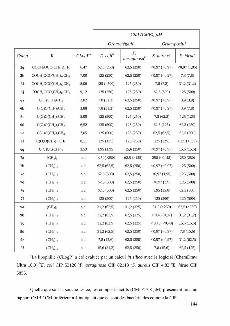
144
CMI (CMB), M
Gram-négatif Gram-positif
Comp R CLogPa E. coli
b
P.
aéruginosac
S. aureusd E. hirae
e
3g COCH2OCO(CH2)9CH3 6,47 62,5 (250) 62,5 (250) <0,97 (<0,97) <0,97 (1,95)
3h COCH2OCO(CH2)10CH3 7,00 125 (250) 62,5 (250) <0,97 (<0,97) 7,8 (7,8)
3i COCH2OCO(CH2)12CH3 8,06 125 (>500) 125 (250) 7,8 (7,8) 31,2 (31,2)
3j COCH2OCO(CH2)14CH3 9,12 125 (250) 125 (250) 62,5 (500) 125 (500)
6a C(O)OCH2CH3 2,82 7,8 (31,2) 62,5 (250) <0,97 (<0,97) 3,9 (3,9)
6b C(O)O(CH2)3CH3 3,88 7,8 (31,2) 62,5 (250) <0,97 (<0,97) 3,9 (7,8)
6c C(O)O(CH2)7CH3 5,99 125 (500) 125 (250) 7,8 (62,5) 125 (125)
6d C(O)O(CH2)8CH3 6,52 125 (500) 125 (250) 62,5 (125) 62,5 (250)
6e C(O)O(CH2)9CH3 7,05 125 (500) 125 (250) 62,5 (62,5) 62,5 (500)
6f C(O)O(CH2)11CH3 8,11 125 (125) 125 (250) 125 (125) 62,5 (>500)
6g C(O)OC(CH3)3 3,53 1,95 (1,95) 15,6 (250) <0,97 (<0,97) 15,6 (15,6)
7a (CH2)8 n.d. >250(>250) 62,5 (>125) 250 (<0, 48) 250 (250)
7b (CH2)10 n.d. 62,5 (62,5) 62,5 (250) <0,97 (<0,97) 125 (500)
7c (CH2)12 n.d. 62,5 (500) 62,5 (250) <0,97 (1,95) 125 (500)
7d (CH2)13 n.d. 62,5 (500) 62,5 (250) <0,97 (3,9) 125 (500)
7e (CH2)14 n.d. 62,5 (500) 62,5 (250) 1,95 (15,6) 62,5 (500)
7f (CH2)16 n.d. 125 (500) 125 (250) 125 (500) 125 (500)
9a (CH2)8 n.d. 31,2 (62,5) 31,2 (125) 31,2 (>250) 62,5 (>250)
9b (CH2)10 n.d. 31,2 (62,5) 62,5 (125) < 0,48 (0,97) 31,2 (31,2)
9c (CH2)12 n.d. 31,2 (62,5) 62,5 (125) < 0,48 (<0,48) 15,6 (15,6)
9d (CH2)13 n.d. 31,2 (62,5) 62,5 (250) <0,97 (<0,97) 7,8 (15,6)
9e (CH2)14 n.d. 7,8 (15,6) 62,5 (250) <0,97 (<0,97) 31,2 (62,5)
9f (CH2)16 n.d. 15,6 (31,2) 62,5 (250) 7,8 (15,6) 62,5 (125)
aLa lipophilie (CLogP) a été évaluée par un calcul in silico avec le logiciel (ChemDraw
Ultra 10,0) bE. coli CIP 53126
cP. aeruginosa CIP 82118
dS. aureus CIP 4.83
eE. hirae CIP
5855.
Quelle que soit la souche testée, les composés actifs (CMI ≤ 7,8 M) présentent tous un
rapport CMB / CMI inférieur à 4 indiquant que ce sont des bactéricides comme la CIP.
145
D'une manière générale, les activités inhibitrices et bactéricides des composés évalués
contre la souche Gram-positif S. aureus ont été conservées. Parmi les 45 composés testés,
l'activité antibactérienne in vitro pour 29 d'entre eux est équivalente à celle de la CIP (CMI: 0,97
M) contre S. aureus. L'activité contre la souche Gram-positif E. hirae est plus aléatoire ; seuls 5
composés, appartenant à la série 7-(4-(2-oxoéthylalkanoate)pipérazin-1-yl) 3, présentent la
même activité antibactérienne in vitro comparé à la CIP.
En revanche, l'activité antibactérienne in vitro de tous les composés synthétisés sont 20 à
128 fois moins actifs contre P. aeruginosa et de 2 à 250 fois moins actifs contre E. coli que la
CIP (CMI: 0,97 M). Seuls les composés 2 (ClogP: 1,75) et 4 (1,35), qui sont les dérivés les plus
polaires, montrent une activité antibactérienne in vitro contre E. coli équivalente à celle de la
CIP.
Rappelons que le composé 4 est le produit issu d‘une possible hydrolyse enzymatique ou
chimique des dérivés de la série 7-(4-(2-oxoéthylalkanoate)pipérazin-1-yl) 3. Le composé 4 est
actif contre les deux souches Gram-positifs (S. aureus et E. hirae) avec des valeurs de CMI et
CMB équivalentes à celles des dérivés 3a-l. En revanche, alors que les dérivés 3a-j ne sont pas
actifs contre les deux souches Gram-négatifs (E. coli et P. aeruginosa), le composé 4 présente
une activité conte E. coli équivalente à celle de la CIP. Il semblerait donc que les dérivés de la
série 3 ne subissent pas d‘hydrolyse dans ces milieux cellulaires.
Une activité contre S. aureus comparable à celle de la CIP a été conservée :
pour les dérivés de la CIP ―monomères‖ 7-(4-(oxoéthylalcanoate) pipérazin-1-
yl) 3a-h possédant des valeurs de ClogPs comprises entre 1,71 et 7,00;
pour les dérivés 7-(4-(alkyloxyoxycarbonyle)pipérazin-1-yl) 6a (ClogP: 2,82);
6b (3,88) et 6g (3,53) ;
pour les dérivés 7-(4-(alcanoyle)pipérazin-1-yl) 1a-g,k (1,14<ClogP < 4,84).
Pour ces trois séries homogènes, les composés présentant les plus hautes valeurs de
ClogP ne sont pas actifs contre S. aureus.
Parmi les dérivés ―dimères‖ de la CIP 7a-f et 9a-f, les composés 7b, 9b ; 7c, 9c ; 7d, 9d
et 7e, 9e sont aussi actifs ou deux fois moins actifs pour le dérivé 7e (CMI: 1,95 M) contre S.
aureus par rapport à la CIP.
Les FQs sont connues pour traverser la membrane cytoplasmique des bactéries Gram-
positifs par diffusion passive. Seules les formes non chargées de ces composés peuvent diffuser à
travers la bicouche lipidique des membranes biologiques45a, 181
. Les FQs pénètrent dans les
bactéries Gram-négatifs par une voie essentiellement hydrophile grâce aux porines ou par une

146
voie hydrophobe à travers la bicouche lipidique182
. Il en découle que plus un médicament est
hydrophile, plus la pénétration cellulaire se fera par la voie des porines alors que plus un
médicament est hydrophobe, plus la diffusion au travers de la bicouche lipidique aura lieu.
La modification structurale de la CIP en position C-7 entraîne une rupture des propriétés
zwittérioniques des dérivés synthétisés. Ainsi, pour ces dérivés de la CIP, seul l‘équilibre non-
chargé / déprotoné peut se produire, facilitant la diffusion passive à travers la bicouche lipidique,
ce qui est cohérent avec les activités observées contre la souche Gram-positif S. aureus au
détriment des souches Gram négatifs. Ces résultats sont en accord avec les travaux reportés dans
la littérature par Foroumadi et coll.67, 158, 183
.
2.2 Activité antimicrobienne in vitro des dérivés de la LEV
Les résultats de l'évaluation des CMIs et des CMBs de certains dérivés de la LEV contre
S. aureus, E. hirae, P. aeruginosa et E. coli ainsi que ceux de la LEV sont résumés dans le
Tableau 67.
Tableau 67 Activité antimicrobienne in vitro des dérivés de la LEV
CMI (CMB), M
Gram-negatif Gram-positif
Comp R CLogP E. coli P. aeruginosa S. aureus E. hirae
LEV OH -0,51 0,97(0,97) 0,97 (0,97) 0,97 (0,97) 0,97 (0,97)
14d OCH2OCO(CH2)8CH3 4,84 <0,97(<0,97) < 0,97 (3,9) < 0,97 (< 0,97) 0,97 (1,95)
14e OCH2OCO(CH2)10CH3 5,90 3,9 (3.9) < 0,97 (< 3,9) < 0,97 (< 0,97) 1,95 (1,95)
14f OCH2OCO(CH2)13CH3 6,96 15,6 (15,6) 62,5 (250) 7,8 (3,9) 15,6 (15,6)
17a NH(CH2)3CH3 2,13 62,5 (62,5) 62,5 (250) 125 (125) 125 (250)
17b NH(CH2)5CH3 3,19 125 (500) 62,5 (250) 250 (250) 125 (250)
17d NH(CH2)7CH3 4,25 125 (500) 62.5 (250) 31,2 (31,2) 31,2 (31,2)
17e NH(CH2)9CH3 5,31 125 (500) 62,5 (250) 7,8 (7,8) 7,8 (15,6)
17f NH(CH2)11CH3 6,36 125 (500) 62,5 (250) 3,9 (15,6) 3,9 (3,9)
17h NH(CH2)13CH3 7,42 125 (500) 62,5 (250) 7,8 (31,2) 3,9 (3,9)
17i NH(CH2)14CH3 7,95 125 (500) 62,5 (250) 15,6 (62,5) 7,8 (7,8)
18b (CH2)5 n.d. 62,5 (62,5) 62,5 (250) 125 (500) 125 (250)
18c (CH2)6 n.d. 62,5 (62,5) 62,5 (250) 125 (500) 125 (125)
18e (CH2)8 n.d. 3,9 (3,9) 62,5 (250) 15,6 (15,6) 62,5 (125)
18f (CH2)9 n.d. 3,9 (3,9) 62,5 (250) 31,2 (31,2) 31,2 (31,2)
18g (CH2)10 n.d. <0,97(<0,97) 31,2 (62,5) 7,8 (7,8) 31,2 (62,5)
18h (CH2)12 n.d. <0,97(<0,97) 31,2 (31,2) 1,95 (1,95) 3,9 (3,9)
147
As outlined in the introduction, the upper portion of the molecule which includes the C-3
carboxy and C-4 keto moieties of FQs is considered as essential for the antibacterial activity.
Pyvaloyloxymethyl ester of ofloxacin, the racemate LEV, synthesized as prodrug by Maeda et
al.170
did not exhibit antibacterial activity against a panel of Gram-positive and Gram-negative
strains (MIC > 25 g/mL). Our previous report concerning the in vitro antimicrobial evaluation
of various alkanoyloxymethyl esters of NAL ranging from 3 to 15 carbon units167
showed that
this chemical modification of the C-3 carboxylic group of NAL led to the loss of the antibacterial
activity, as expected (CMI > 128 g/mL). We however evaluated the in vitro antibacterial
activity of 3 ―monomer‖ alkanoyloxymethyl esters of LEV, 14d, 14e and 14f, selected
depending on their ClogPs which were included in the same range (from 4.84 to 6.96) than the
most potent ―monomer‖ CIP derivatives.
Surprisingly, these latter derivatives and in particular 14d and 14e, retained an
antibacterial activity against the Gram-positive and Gram-negative standard strains. These
activities could be due to the hydrolysis of the compounds into the parent LEV and therefore
compounds could act as prodrugs. If hydrolyse occurs, this one should be more important as the
alkyl chain length decreases and can explain that 14d and 14e are more potent against bacteria
strains than 14f. However, as we will discuss in next sections, acyloxymethyl ester derivatives
14a (n=2), 14c (n=6), 14d (n=8) and 14f (n=12) showed IC50 and MIC value against M.
tuberculosis, roughly similar to that of LEV whatever the chain length. Moreover, 14a (n=2)
exhibited an antiproliferative activity with IC50 values against cancer cell lines lower than those
of LEV, whilst 14b (n=4), 14c (n=6) and 14d (n=8) didn‘t inhibit the cancer cell lines growth.
Compounds 14e-h (10<n<16) displayed antiproliferative activities against U373-MG, LoVo and
MCF-7 cancer cell lines with IC50 values for 14e 200- to 2000-fold lower than that of LEV. So,
these observations don‘t seem to be in favour of a hydrolyse for compounds 14e and 14f.
Some authors reported the synthesis and antimicrobial activities of carboxamide
derivatives of LEV and CIP and showed that they displayed antibacterial activities, contradicting
the dogma that C-3 carboxylic acid is required85, 184
. In our case, the alkylcarboxamide
―monomer‖ derivatives of LEV 17 were inactive against Gram-positive and Gram-negative
strains. Compounds 17e-h (5.31<ClogP<7.42) showed moderate activities against Gram-positive
strains with MICs higher than LEV. But interestingly, when increasing the linker length of the C-
6-(N-alkylcarboxamide)-linked LEV ―dimers‖ 18, an antibacterial activity in particularly against
the Gram-negative E. coli strain was observed for compounds 18g-h.
One time more, the active compounds (CMI ≤ 7.8 M) are bactericides as their native
parent, LEV, as they displayed a MBC / MIC ratio less than 4.

148
By considering ―monomer‖ and ―dimer‖ derivatives of LEV, for which the acidic
function is modified, only the uncharged/protonated equilibria can occur. Even if the
hydrophobicity of carboxamide ―monomer‖ derivatives 17 allows them to diffuse through the
membrane, they seem not to act effectively on the enzymatic target. The mechanism of
antibacterial activity of C-6-(N-alkylcarboxamide)-linked LEV ―dimers‖ 18 seem to differ as by
increasing the alkyl chain length, not only the activity against Gram-positive strains increases but
also the activity against Gram-negative strains. These results also show that amide function can
act as a bioisostere of carboxylic acid and therefore new derivatives of FQ can be envisaged for
the future as antibacterials.
2.3 In vitro antimicrobial activities of CIP derivatives against standard S. aureus, MRSA and
MDRSA strains
Due to the observed in vitro activities against S. aureus of CIP derivatives, we selected
the most potent derivatives to be tested against a standard strain of S. aureus (CIP4.83), two
MRSA strains (ATCC33591 and ATCC33592) and two clinical isolates MDRSA strains
(MDRSA1 and MDRSA2). The MICs and the MBCs of all the 29 selected compounds against
these different strains of S. aureus are shown in Table 68 along with those of CIP.

149
Table 68 In vitro antimicrobial activity (MIC(MBC) in M) of selected
coumpounds against the S. aureus standard strain (Sa CIP 4.83), two MRSA
strains (Sa ATCC33591 and Sa ATCC33592) and two MDRSA clinical isolates
(Sa MDRSA1 and Sa MDRSA2)
MIC (MBC), M
S. aureus strains Clinical isolates
Comp CLogPa CIP 4.83 MRSA1 MRSA2 MDRSA1 MDRSA2
CIP -0.72 0.97 (1.95) 0.97 (1.95) 0.48 (0.97) 31.2 (62.5) 31.2 (31.2)
2 1.75 0.97 (1.95) 0.97 (0.97) 0.48 (0.97) 125 (500) 62.5 (62.5)
1a 1.14 0.48 (0.48) 0.97 (0.97) 0.48 (0.97) 62.5 (500) 31.2 (62.5)
1b 1.67 0.48 (0.97) 0.48 (0.97) 0.24 (0.48) 62.5 (500) 31.2 (31.2)
1c 2.19 0.48 (0.48) 0.48 (0.48) 0.24 (0.48) 31.2 (62.5) 31.2 (62.5)
1d 2.37 0.48 (0.97) 0.48 (0.48) 0.24 (0.48) 31.2 (500) 31.2 (31.2)
1e 2.72 0.24 (0.48) 0.48 (0.48) 0.24 (0.48) 62.5 (500) 31.2 (31.2)
1l 3.19 0.12 (0.24) 0.12 (0.24) 0.12 (0.24) 31.2 (62.5) 15.6 (15.6)
1f 3.78 0.03 (0.12) 0.061 (0.48) 0.061 (0.24) 62.5 (500) 250 (>500)
1g 4.84 7.6·10
-3
(0.03) 0.015 (0.24) 0.12 (0.24) 31.2 (500) 31.2 (500)
3a 1.71 1.95 (3.9) 3.9 (3.9) 0.97 (1.95) 125 (500) 125 (125)
3b 2.77 0.24 (0.48) 0.48 (0.97) 0.061 (0.12) 15.6 (62.5) 15.6 (15.6)
3c 3.83 0.03 (0.24) 0.03 (0.061) 3.8·10-3
(7.6·10-3
)
15.6 (500) 7.8 (15.6)
3d 4.89 <9.5·10
-4
(<9.5·10-4
)
<9.5·10-4
(1.9·10-3
)
<9.5·10-4
(<9.5·10-4
) 7.8 (500) 15.6 (15.6)
3e 5.41 3.8·10
-3
(0.12) 0.03 (0.24) 0.015 (0.015) 7.8 (15.6) 7.8 (15.6)
3f 5.95 0.12 (0.97) 0.24 (0.48) 3.8·10-3
(7.6·10-3
)
7.8 (15.6) 7.8 (15.6)
3g 6.47 0.97 (0.97) 0.97 (1.95) 3.8·10-3
(0.03)
15.6 (500) 7.8 (15.6)
3h 7.00 0.97 (1.95) 1.95 (3.9) 0.061 (0.97) 125 (500) 62.5 (250)
4 1.35 0.97 (1.95) 0.97 (1.95) 0.24 (1.95) 125 (500) 62.5 (62.5)
5 3.31 0.12 (0.24) 0.48 (0.97) 0.24 (0.97) 125 (500) 62.5 (125)
6a 2.82 0.97 (0.97) 0.97 (1.95) 0.48 (1.95) 125 (500) 62.5 (125)
6b 3.88 0.061 (0.12) 0.03 (0.12) 7.6·10-3
(0.12) 62.5 (500) 31.2 (31.2)
6g 3.53 0.12 (0.24) 0.061 (0.48) 0.12 (0.48) 31.2 (500) 15.6 (31.2)
7b n.d. 0.12 (7.8) 0.48 (3.9) 0.12 (1.95) 125 (500) 125 (500)
7c n.d. 0.12 (1.95) 0.061 (3.9) 0.12 (0.97) 7.8 (500) 7.8 (15.6)
7d n.d. 0.061 (1.95) 0.061 (0.97) 0.061 (0.48) 1.95 (500) 15.6 (31.2)
9b n.d. 1.95 (3.9) 3.9 (7.8) 0.48 (0.97) 250 (500) 125 (500)
9c n.d. 1.95 (1.95) 1.95 (7.8) 0.015 (0.97) 125 (500) 62.5 (125)
9d n.d. 0.12 (0.48) 0.48 (1.95) 0.12 (0.97) 62.5 (500) 31.2 (62.5)
9e n.d. 1.95 (7.8) 7.8 (15.6) 0.97 (1.95) 125 (500) 125 (500)

150
The first group of ―mono-amide‖ derivatives displayed increased activity against both
standard and methicillin-resistant strains. Compounds 1a-e,l displayed MIC values somewhat
equivalent with an improvement of MIC and MBC values towards standard, MRSA1 and
MRSA2 strains than those of CIP. The most lipophilic (CLogPs = 3.78 and 4.84) compounds
1f,g displayed interesting activities against standard and MRSA strains with a 60-fold decrease
of MICs compared to CIP. However, none of compounds from the ―mono-amide‖ group
exhibited significant activity against MDR clinical isolates.
Considering the 7-(4-(alkanoyl)piperazin-1-yl) derivatives 1a-g and 1l, we observed a
significant increase of activity only for two of them, that were compounds 1f (ClogP = 3.78; Mw
= 443.5 g·mol-1
; MICs from 0.03 to 0.061 M) and 1g (4.84 ; 471.6 g·mol-1
; 0.12 M to 7.6 nM).
As shown in Table 69, representing the MICs values of 1a-g, 1l homologous derivatives
along with those of CIP against the standard CIP4.83 and the two MRSA strains, we observed a
constant improvement of MIC and MBC values, comparing to CIP, for compounds 1b-e and 1l
with ClogP values varying from 1.67 to 3.19. The two last and most lipophilic compounds 1f-g
(CLogPs = 3.78 and 4.84) turned out to be the most potent derivatives of this series.
Table 69 MICs values of CIP and homologous derivatives 1 against standard
strains CIP4.83 and the two MRSA strains
Cmpd 1a 1b 1c 1d 1e 1l 1f 1g
n 0 1 2 -* 3 -* 5 7
CLogP 1.14 1.67 2.19 2.37 2.72 3.19 3.78 4.84
Mw, g·mol-1
373.4 387.4 401.4 415.5 415.5 449.5 443.5 471.6
* R = t-Bu for 1d and R = CH2Ph for 1l

151
Compounds 1f (n = 5; cLogP = 3.78; Mw = 443.5 g.mol-1) and 1g (7; 4.84; 471.6 g.mol-
1) exhibited respectively MICs values 8- to 32- fold and 4- to 128- fold lower than those of CIP
against standard CIP4.83 and the two MRSA strains.
The ―mono-oxoethylester‖ CIP derivatives exhibited the highest degree of activity among
all ―monomeric‖ series. Compound 4, resulting from a possible hydrolyse of derivatives 3,
displayed IC50 values equivalent to those of CIP. Once more, it seems that no hydrolyse of
derivatives 3 occurs in bacterial cellular media.
The eight 7-(4-(oxoethylalkanoate)piperazin-1-yl) derivatives 3a-h (2.77 < ClogPs <
5.95) displayed MICs and MBCs equivalent or lower than CIP. Among them, compounds 3d
(ClogP = 4.89; Mw = 515.6 g·mol-1
; MIC<0.95 nM) and 3e (5.41 ; 529.6 g·mol-1
; from 0.03 M
to 3.8 nM) exhibited very good in vitro activities.
Table 70 MICs values of CIP and homologous derivatives 3 against standard
strains CIP4.83 and the two MRSA strains
Cmpd 3a 3b 3c 3d 3e 3f 3g 3h
n 0 2 4 6 7 8 9 10
CLogP 1.71 2.77 3.83 4.89 5.41 5.95 6.47 7.00
Mw, g·mol-1
431.4 459.5 487.5 515.6 529.6 543.6 557.7 571.7
We observed a clear minimum on the curve representing MICs values of CIP and 3a-h
homologous derivatives against standard strains CIP4.83 and the two MRSA strains (Table 70)
Compound 3d (n = 6; cLogP = 4.89; Mw = 516.6 g.mol-1
), with subnanomolar MIC and MBC (<
0.95 nM) values against standard and methicillin-resistant strains, turned out to be the most
potent derivatives with MICs values 500- to 1000- fold lower than those of CIP.

152
Among the 7-(4-(alkoxycarbonyl)piperazin-1-yl) derivatives 6, only compound 6b (n = 3;
ClogP = 3.88; Mw = 431.5 g.mol-1
) exhibited lowest MICs than CIP and was found to be 16- to
63- fold more potent than CIP with a MIC value of 7.6 nM against MRSA strain ATCC33592.
In a general way, 7-(4-(alkanoyl)piperazin-1-yl))-linked CIP ―dimers‖ 7, displayed more
potent activities against the standard and MRSA strains. Compound 7d (n = 13) was found to be
8- to 16- fold more potent than CIP against these strains.
7-(4-(2-Oxoethylalkanoate)piperazin-1-yl))-linked CIP ―dimers‖ 9 didn‘t show any
significant enhancement of the activity against the standard and MRSA strains compared to CIP.
Only compound 9d (n = 13) was found to be 2- to 8- fold more potent than CIP.
For these two series of C7-C7-linked CIP ―dimers‖, it would seem that the required
conformation for an interaction with the enzymatic target is reached for a chain length of 13
carbon units.
On the other hand, even if some compounds exhibited better inhibitory activities against
the two clinical isolates MDRSA (MDRSA1 and MDRSA2) than CIP, the benefit was less
important.
No compounds from 7-(4-alkanoyl)piperazin-1-yl) 1, 7-(4-alkoxycarbonyl)piperazin-1-
yl) 6 and 7-(4-(2-oxoethylalkanoate)piperazin-1-yl))-linked 9 CIP derivatives were active against
MDRSA strains.
7-(4-(Oxoethylalkanoate)piperazin-1-yl) derivatives 3b-g (2 < n < 9; 2.77 < ClogPs <
6.47; 459.5 < Mw < 557.7 g.mol-1
) showed MICs from 7.8 to 15.6 M against these two clinical
isolates that is 2- to 4-fold more potent than CIP. From all series of tested ―monomer‖ CIP
derivatives, this series of derivatives 3 exhibited the more potent activities against the tested S.
aureus strains.
7-(4-(Alkanoyl)piperazin-1-yl))-linked CIP ―dimer‖ 7c was found to be 4- fold more
potent than CIP against the two clinical isolates with MICs of 7.8 M. The most potent
compound against MDRSA1 was found to be the ―di-amide‖ derivative 7d (MIC: 1.95 M), that
is 16- fold lower than that of CIP.
We can also notice that if active compounds against standard and MRSA strains retained
a bactericide activity, this is not always true for MDRSA strains.
To conclude, for the most potent compounds against S. aureus, the activity is also
maintained for MRSA strains. For each series of ―monomer‖ derivatives the activity against
standard and MRSA strains increases with ClogP until an optimal value of ClogP (Tableau 66,
Table 68). We observed an optimal value for ClogP = 4.84 for the alkanoyl 1 CIP derivatives,
4.89 for the oxoethylalkanoate 3 and 3.88 for the alkoxycarbonyl 6. The more potent antibacterial
compounds were found in the oxoethylalkanoate 3 series, but whatever the considered

153
―monomer‖ series, all of them showed better activities against S. aureus strains than ―dimer‖ CIP
derivatives.
2.4 SAR attempts
Several general trends in antibacterial activity were deduced from these studies.
Both active CIP or LEV derivatives (―monomeric‖ and ―dimeric‖ structures) against
bacterial strains (with CMIs values ≤ 7.8 M) were bactericides (CMB / CMI < 4). So,
they displayed the same antimicrobial profile than their native parents, CIP and LEV. It is
nevertheless necessary to specify that this observation is not general for the antibacterial
activities observed against MDRSA strains.
Tested compounds were significantly less active towards Gram-negative species in
comparison with the native parents CIP and LEV.
Nevertheless, CIP and LEV derivatives didn‘t display the same antimicrobial profile of
activity.
CIP derivatives showed a selectivity of the antimicrobial activity against S.
aureus.
LEV derivatives showed various antimicrobial profile according to the structure.
2.4.1 SAR on CIP derivatives
The substitution of the distal nitrogen-4 atom at C-7 position of CIP resulted in an
increased activity specifically towards S. aureus along with a significant decrease against other
pathogens in comparison with the broad-spectrum activity of CIP.
The most active CIP derivatives against the various studied strains of S. aureus are
represented in Table 71
The degree of activity is strongly dependent on the nature of attached functional group
and decreases in the following order:
for ―monomers‖: oxoethylesters > amides ≈ carbamates,
and the reversed order for ―dimers‖: amides > oxoethylesters.
There is also a strong influence of the chain length on MIC values. The highest activity
could be achieved with values of methylene groups of respectively n = 7 and 6 for ―mono-
amide‖ and ―mono-oxoethylesters‖ derivatives. For these two series, the activities against S.

154
aureus evolve in the same way according to the chain length and the ClogP. As outlined before,
too short or too long attached moieties was not beneficial. The optimal CLogP values were
situated within a close range for the most active ―monomers‖ 1g and 3d (CLogP = 4.84 and 4.89
respectively). These observations can‘t be done for the series of ―mono-carbamates‖ derivatives
6 as the highest activity is achieved with a chain length of n = 3 and a ClogP = 3.88. In fact, no
derivatives with a chain length included between n = 3 (ClogP = 3.88) and n = 7 (5.99) were
synthesized.
Concerning the ―dimer‖ CIP derivatives, it seems that the optimal chain length ranges
around 12-13 methylene groups and that the presence of two quinolone cores in the molecule
leads to an enhanced activity against MDRSA clinical isolates.
Table 71 Most active CIP derivatives against the various strains of S. aureus
MIC, M
S. aureus strains
Structure Comp (n) CLogP CIP 4.83 MRSA1 MRSA2 MDRSA
1f (5) 3.78 0.03 0.061 0.061 Non active
1g (7) 4.84 7.6.10-3
0.015 0.12 Non active
3c (4) 3.83 0.03 0.03 3.8.103
7.8-15.6
3d (6) 4.89 < 9.5.10-4
< 9.5.10-4
< 9.5.10-4
7.8-15.6
3e (7) 5.41 3.8.10-3
0.03 0.015 7.8-7.8
3f (8) 5.95 0.12 0.24 3.8.10-3
7.8-7.8
3g (9) 6.47 0.97 0.97 3.8.10-3
7.8-15.6
6b (3) 3.88 0.061 0.03 7.6.10-3
Non active
7c (12) Nd 0.12 0.061 0.12 7.8-7.8
7d (13) Nd 0.061 0.061 0.061 1.95-15.6
9c (12) Nd 1.95 1.95 0.015 Non active
9d (13) Nd 0.12 0.48 0.12 Non active

155
2.4.2 SAR on LEV derivatives
Concerning LEV derivatives only preliminary screening against the four Gram-positive
and Gram-negative standard strains were conducted.
In summary, the C-6 carboxyl group modification of LEV led to antibacterial profile and
potency somewhat different than C-7 piperazine modification of CIP.
The degree of activity is also dependent on the nature of attached functional group and
decreases in the following order:
―mono-acyloxymethyl ester‖ derivatives 14 which displayed a broad spectrum
of activity,
―di-amides‖ derivatives 18 for which an antimicrobial activity was observed
for compounds owning a chain length more than 10 methylene groups,
―mono-amides‖ derivatives 17 were inactive.
Taking in account the possible hydrolysis of acyloxymethyl ester of LEV, it cannot be
rational to conclude to a lead compound in this series without supplementary physico-chemical
and biological evaluations.
The observed antibacterial activities of ―di-amides‖ derivatives of LEV 18 can be due to
the fact that amide function can act as a bioisostere of carboxylic acid for the formation of the
drug-enzyme complex or to a modified mechanism of action of these compounds.
The C-6-(N-dodecylcarboxamide)-linked LEV ―dimers‖ 18 seems to be the most
promising compounds of all LEV derivatives for further antimicrobial studies (Figure 46).
MIC S. aureus: 1.95 M
MIC E. hirae: 3.9 M
MIC P. aeruginosa: 31.2 M
MIC E. coli: ≤0.97 M
Figure 46 Lead compound of the series C-6-(N-acylcarboximide)-linked LEV ―dimers‖
18h

156
3. Antimycobacterial activity
We have also explored the effect of increasing the lipophilic character of CIP and LEV
on activity against M. Tuberculosis (H37Rv strain) as penetration is crucial for FQ activity against
this strain. The antimycobacterial activity of synthesized derivatives was evaluated by Patricia
Constant at IPBS (Group of Dr. Mamadou Daffé, Institut de Pharmacologie et Biologie
Structurale), Département Mécanismes Moléculaires des Infections Mycobactériennes,
Toulouse.
3.1 In vitro antimycobacterial inhibitory activities of CIP derivatives
The percentage of inhibition of M. tuberculosis growth was first determined for some
selected compounds, along with those of CIP at concentrations of 10 and 1 M and 0.1 The
percentage of inhibition of H37Rv obtained at 10 and 1 M are summarized inTable 72.
Table 72 Percentage of inhibition of M. Tuberculosis growth for selected CIP
derivatives
Comp R CLogP % inhibition of
H37Rv at 10 M
% inhibition of
H37Rv at 1 M
CIPa H -0.72 98 95
2 COCH2Cl 0.343 95 6
1a COCH3 1.14 92 0
1b COCH2CH3 1.67 93 3
1c CO(CH2)2CH3 2.19 93 3
1d COC(CH3)3 2.37 93 2
1e CO(CH2)3CH3 2.72 92 0
1f CO(CH2)5CH3 3.78 96 0
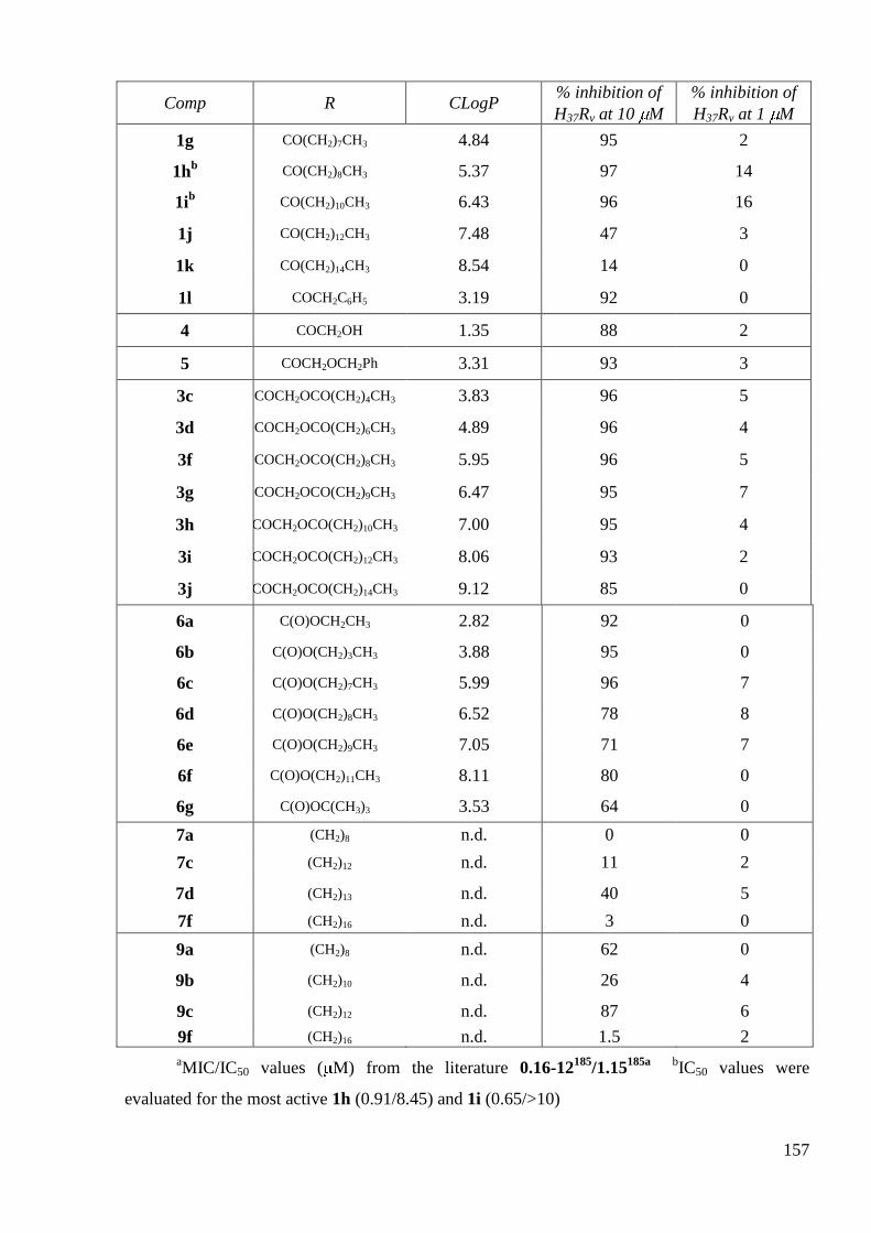
157
Comp R CLogP % inhibition of
H37Rv at 10 M
% inhibition of
H37Rv at 1 M
1g CO(CH2)7CH3 4.84 95 2
1hb CO(CH2)8CH3 5.37 97 14
1ib CO(CH2)10CH3 6.43 96 16
1j CO(CH2)12CH3 7.48 47 3
1k CO(CH2)14CH3 8.54 14 0
1l COCH2C6H5 3.19 92 0
4 COCH2OH 1.35 88 2
5 COCH2OCH2Ph 3.31 93 3
3c COCH2OCO(CH2)4CH3 3.83 96 5
3d COCH2OCO(CH2)6CH3 4.89 96 4
3f COCH2OCO(CH2)8CH3 5.95 96 5
3g COCH2OCO(CH2)9CH3 6.47 95 7
3h COCH2OCO(CH2)10CH3 7.00 95 4
3i COCH2OCO(CH2)12CH3 8.06 93 2
3j COCH2OCO(CH2)14CH3 9.12 85 0
6a C(O)OCH2CH3 2.82 92 0
6b C(O)O(CH2)3CH3 3.88 95 0
6c C(O)O(CH2)7CH3 5.99 96 7
6d C(O)O(CH2)8CH3 6.52 78 8
6e C(O)O(CH2)9CH3 7.05 71 7
6f C(O)O(CH2)11CH3 8.11 80 0
6g C(O)OC(CH3)3 3.53 64 0
7a (CH2)8 n.d. 0 0
7c (CH2)12 n.d. 11 2
7d (CH2)13 n.d. 40 5
7f (CH2)16 n.d. 3 0
9a (CH2)8 n.d. 62 0
9b (CH2)10 n.d. 26 4
9c (CH2)12 n.d. 87 6
9f (CH2)16 n.d. 1.5 2
aMIC/IC50 values ( M) from the literature 0.16-12
185/1.15
185a
bIC50 values were
evaluated for the most active 1h (0.91/8.45) and 1i (0.65/>10)

158
At 10 M, several ―monomer‖ CIP derivatives displayed a percentage of inhibition of
H37Rv growth more than 95% comparable to that of CIP (98%).
Concerning the 7-(4-alkanoyl)piperazin-1-yl) derivatives 1, the percentage of inhibition
was optimal in a ClogP range from 3.78 (1f, n = 5, Mw = 463.5 g.mol-1
) to 6.43 (1i, n = 10,
513.7 g.mol-1
) and decreased for higher ClogP (1j and 1k, owning longer chain length (n = 12
and 14 respectively).
Among the tested 7-(4-(oxoethylalkanoate)piperazin-1-yl) derivatives 3, this optimum
was reached for ClogP ranging from 3.83 (3c, n = 4, Mw = 487.5 g.mol-1
) to 8.06 (3i, n = 12,
599.7 g.mol-1
) and in a similar manner decreased for derivatives with longer chain length (3j, n =
14). The results obtained for compound 4 resulting from the possible hydrolysis of derivatives 3
showed once again that no hydrolysis occured in the cellular media.
7-(4-(Alkoxycarbonyl)piperazin-1-yl) derivatives 6 are less potent as only compounds 6b
(ClogP = 3.88: n = 3, Mw = 431.5 g.mol-1
) and 6c (5.99; n = 7, 487.6 g.mol-1
) exhibited a
percentage of inhibition ≥ 95%.
The more potent CIP derivatives from series 1 and 3 displayed the best percentages of
inhibition of H37Rv growth for CLogP and molecular weight values situated within a relatively
large range (3.78 < ClogP < 8.06 and 463.51 < Mw < 599.7 g.mol-1
) and for a range of chain
length between 4 to 12 methylene groups. For the CIP derivatives from series 6, these ranges of
values were more limited (3.88 < ClogP < 5.99; 3 < n < 7; 431.5 < Mw < 487.6 g.mol-1
) and we
observed a decrease of inhibition of H37Rv growth from a ClogP value of 6.52 (6d, n=8; Mw =
501.6 g.mol-1
).
It could be notice that the ―dimer‖ derivatives 7 and 9 of CIP were few active or inactive
as the highest percentages of inhibition of H37Rv growth for these two series were 40% (7d) and
87% (9c).
At 1 M, most of the ―monomer‖ and ―dimer‖ CIP derivatives were inactive as only
compounds 1h and 1i showed percentages of inhibition around 15% while CIP inhibited the
mycobacterial growth at 95%.
At 0.1 M (data not shown), all compounds including CIP, didn‘t inhibit the
mycobacterial growth.
This first screening has shown that increasing lipophilicity of CIP by a C-7 piperazine
modification does not lead to an increase of the antimycobacterial activity as the more potent
CIP derivatives are less potent than their native parent CIP at 1 M.

159
3.2 In vitro antimycobacterial inhibitory activities of LEV derivatives
Derivatives of LEV were also evaluated for their inhibitory activity against M.
tuberculosis at concentrations of 10, 1 and 0.1 M along with those of LEV. These results are
summarized in Table 73
Table 73 Percentage of inhibition of M. Tuberculosis growth for selected LEV
derivatives
Comp R CLogP % inh.
at 10 M
% inh.
at 1 M
MIC,
M
IC50,
M
LEVa OH -0.51 96 85 2.10
b 0.78
b
14a OCH2OCO(CH2)2CH3 1.67 96 74 3.33 b 1.60
b
14c OCH2OCO(CH2)6CH3 3.79 96 89 3.33 b 1.40
b
14e OCH2OCO(CH2)10CH3 5.68 97 89 3.33 b 1.26
b
14f OCH2OCO(CH2)12CH3 6.74 96 6 2.50 c
1.50 c
14g OCH2OCO(CH2)14CH3 7.79 95 0
14i OCH2OCO(CH2)18CH3 n.d. 0 0
17b NH(CH2)5CH3 3.19 15 0
17d NH(CH2)7CH3 4.25 98 0
17e NH(CH2)9CH3 5.31 97 5 10 5.10 c
17g NH(CH2)12CH3 6.89 97 6 > 10 n.d.
17i NH(CH2)14CH3 7.95 96 6 10 4.00 c
17j NH(CH2)16CH3 9.00 93 3
18b (CH2)5 n.d. 77 5
18c (CH2)6 n.d. 60 0 >10 n.d
18f (CH2)9 n.d. 98 20 5.00 b 1.96
b
18g (CH2)10 n.d. n.d. n.d. 5.00 c
1.90 c
18h (CH2)12 n.d. n.d. n.d. 2.50 c
1.60 c
a MIC/IC50 values from the literature
0.18-2.76
185a, b, 185e, f / 1.8
185a, b Mean of three
experiments c Values from two different experiments
At 10 M, several ―monomer‖ LEV derivatives displayed a percentage of inhibition more
than 95% comparable to that of LEV (96%).

160
Concerning the acyloxymethyl ester LEV derivatives, the percentage of inhibition was
optimal in a ClogP range from 1.67 (14a, n = 2, Mw = 461.7 g.mol-1
) to 7.79 (14g, n = 14, 629.8
g.mol-1
) and the compound with the higher chain length (14h, n = 18, 685.9 g.mol-1
) did not
inhibit at all the growth of M. tuberculosis.
Among the tested alkylcarboxamide LEV derivatives, this optimum was reached for
ClogP ranging from 4.25 (17d, n = 7, Mw = 472.3 g.mol-1
) to 9.00 (17j, n = 16, 598.4 g.mol-1
).
The ―dimer‖ alkylcarboxamide LEV derivatives 18f (n = 9, Mw = 845.0 g.mol-1
)
displayed a percentage of inhibition of the mycobacterial growth of 98%. Compounds with
shorter chain length (18b and 18c, n = 5 and 6) were less potent against M. Tuberculosis.
At 1 M, only three compounds from the acyloxymethyl ester series, 14a (74%), 14c
(89%) and 14e (89%) displayed an activity similar to that of LEV (85%). However, the
possibility of a hydrolysis of these compounds into the parent LEV remained even if this
hydrolysis should be more important for 14a (n = 2) than for 14c (n = 6) or 14e (n = 10).
At 0.1 M (data not shown), all compounds including LEV, didn‘t inhibit the
mycobacterial growth.
3.3 In vitro determination of MIC and IC50 against M. tuberculosis
This first screening permitted us to select some compounds for a determination of their
MIC and/or IC50 values against M. tuberculosis. We have selected, for these tests, the most
potent CIP and LEV derivatives at 10 and 1 M.
For CIP derivatives, only two compounds were selected that were 1h and 1i. As we have
seen, more LEV derivatives were active at 1 M, so MIC and IC50 of several compounds of each
series were evaluated including C-6-(N-acylcarboxamide)-linked LEV dimers 18g and 18h that
were synthesized in the group after the evaluation of inhibitory activities. The results obtained
for these various compounds are reported in Table 72 and Table 73 along with those of CIP and
LEV.
These preliminary studies confirmed the observed results about the inhibition of M.
tuberculosis growth, that were a more potent activity of derivatives of LEV than those of CIP.
The MIC and IC50 values of CIP and LEV against H37Rv are in accordance with those of
literature. Nevertheless, these studies are currently carried out as only one experiment had been
done for some compounds and as derivatives 1h and 1i of CIP showed two very different values
from the two separate experiments.

161
3.4 SAR on CIP derivatives
A SAR study is somewhat difficult to establish as, concerning the antimycobacterial
activity, a C-7 modification of CIP was not beneficial as all ―monomers‖ and ―dimers‖ were less
or as active as than their parent CIP. We also observed that ―monomers‖ were much more active
than the corresponding ―dimers‖.
If we consider the ―monomer‖ CIP derivatives we can conclude the following order of
relative antimycobacterial activity: amides > oxoethylesters > carbamates.
The maximum percentages of inhibition of M. tuberculosis were observed for the most
active compounds for whom ClogP values were included between 3.78 and 8.06, n between 4
and 12 and molecular weights between 463.5 and 599.7 g.mol-1
.
The most active compounds 1h and 1i belong to the amide series with ClogP values of
5.37 and 6.43; n = 8 and 10 and Mw = 485.6 and 513.7 g.mol-1
(Table 74).
Table 74 The most active CIP derivatives against H37Rv strain
Comp n CLogP % inhibition
at 10 M
% inhibition
at 1 M IC50, M
1h 8 5.37 97 14 0.91/8.45
1i 10 6.43 96 16 0.65
SAR on LEV derivatives 1.1
A C-6 modification of LEV was more beneficial than C-7 modification of CIP with
higher number of active compounds. The most potent derivatives of LEV, that were
alkanoyloxymethyl esters LEV derivatives 14 and C-6(N-alkylcarboxamide)-linked LEV
―dimers‖ 18 exhibited IC50 ≤ 2 M and MIC ≥ 2.5 M. Alkylcarboxamide LEV derivatives 17
were the least active. Compound 14e (IC50 = 1.26 M; n = 10; ClogP = 5.68; Mw = 513.7 g.mol-
1) from alkanoyloxymethyl esters of LEV seems to be the more potent against M. tuberculosis
among all ―monomer‖ derivatives of LEV, while compound 18h (IC50 = 1.60 M; n = 12; Mw =
845.0 g.mol-1
) seems to be the more potent against M. tuberculosis among C-6(N-
alkylcarboxamide)-linked LEV ―dimers‖ (Table 75).
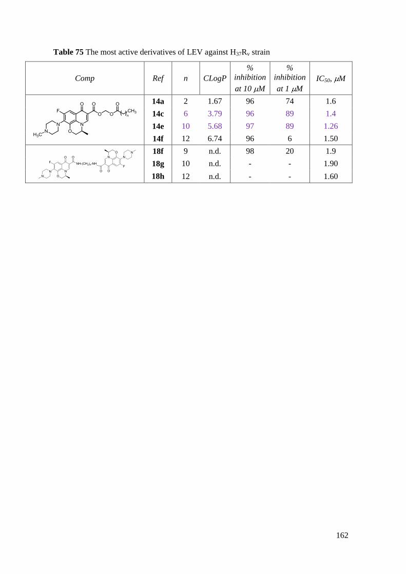
162
Table 75 The most active derivatives of LEV against H37Rv strain
Comp Ref n CLogP
%
inhibition
at 10 M
%
inhibition
at 1 M
IC50, M
14a 2 1.67 96 74 1.6
14c 6 3.79 96 89 1.4
14e 10 5.68 97 89 1.26
14f 12 6.74 96 6 1.50
18f 9 n.d. 98 20 1.9
18g 10 n.d. - - 1.90
18h 12 n.d. - - 1.60
163
4. Antiproliferative activity
The antiproliferative activity of synthesized compounds together with parent LEV and
CIP was evaluated in vitro against five human cancer cell lines. For each of the compounds
under study, nine concentrations were tested on two human apoptosis-resistant glioblastoma
U373-MG186
, non-small cell lung cancer A549187
cell lines and three apoptosis-sensitive prostate
PC-3188
, colorectal LoVo189
, breast MCF-7188
cancer cell lines. We made use of the 3-(4,5-
dimethylthiazol-2-yl)-2,5-diphenyltetrazolium bromide (MTT) colorimetric assay, which
indirectly assesses the effect of the potentially anticancer compounds on the overall growth of
adherent cell lines190
. The IC50 values, which are the concentrations that reduce the mean growth
value of the cell lines by 50% (after having cultured the cells for 5 days in presence of the
investigational compound), were determined for each drug and compared to the mean control
growth value. The in vivo tolerance of some selected derivatives in mice was also determined as
a maximum tolerated dose (MTD) which represents the highest single dose of the compound that
can be administered acutely to healthy animals. This evaluation was conducted by Pr. Robert
Kiss et al., at Laboratoire de Toxicologie, Institut de Pharmacie, Université Libre de Bruxelles,
Belgium.
4.1 In vitro antiproliferative activities of CIP derivatives
4.1.1 In vitro antiproliferative activities of “monomer” derivatives
The IC50 values, which are the concentrations that reduce the mean growth value of the
cells by 50%, were determined for each drug and compared to the mean control growth value
and are listed in Table 76.

164
Table 76 In vitro antiproliferative activity of CIP ―monomer‖ derivatives
IC50 ( M)
Apoptosis-sensitive Apoptosis-resistant
Comp. n CLogP PC-3 LoVo MCF-7 U373-MG A549
CIP - -0.72 143 ± 1 89 ± 2 476 ± 13 96 ± 4 280 ± 11
2 - 0.78 8 ± 0.1 17 ± 0.3 23 ± 0.2 4 ± 0.3 10 ± 0.2
1a 0 1.14 680 ± 33 323 ± 8 803 ± 15 535 ± 13 584 ± 14
1b 1 1.67 352 ± 7 306 ± 6 727 ± 17 279 ± 6 402 ± 21
1c 2 2.19 85 ± 2 148 ± 2 172 ± 9 104 ± 2 697 ± 18
1d - 2.37 246 ± 4 93 ± 5 85 ± 1 67±3 509 ± 11
1e 3 2.72 73 ± 0.7 87 ± 2 316 ± 11 86 ± 2 64 ± 0.8
1f 5 3.78 779 ± 21 793 ± 14 754 ± 12 729 ± 9 808 ± 12
1g 7 4.84 7 ± 0.1 30 ± 0.4 26 ± 0.3 24 ± 0.3 6 ± 0.1
1h 8 5.37 4 ± 0.1 7 ± 0.2 20 ± 0.4 5 ± 0.2 3 ± 0.1
1i 10 6.43 4 ± 0.1 45 ± 0.9 108 ± 2 18 ± 0.2 7 ± 0.1
1j 12 7.48 94 ± 1 101 ± 0.9 96 ± 1 205 ± 3 56 ± 0.4
1k 14 8.54 114 ± 0.6 290 ± 4 145 ± 1 16 ± 0.1 65 ± 0.7
1l - 3.19 243 ± 2 41 ± 0.6 32 ± 0.5 34 ± 0.7 29 ± 0.3
4 - 1.35 433 ± 19 408 ± 12 985 ± 26 294 ± 4 456 ± 8
5 - 3.31 692 96 242 85 82
3a 0 1.71 176 ± 2 268 ± 4 279 ± 6 99 ± 2 373 ± 8
3b 2 2.77 715 ± 10 613 ± 6 1000 ± 11 586 ± 6 456 ± 5
3c 4 3.83 14 ± 0.1 27 ± 0.8 72 ± 2 28 ± 0.7 20 ± 0.3
3d 6 4.89 23 ± 0.3 29 ± 0.4 818 ± 17 53 ± 0.7 16 ± 0.2
3e 7 5.41 257 ± 4 267 ± 2 255 ± 3 272 ± 7 216 ± 1
3f 8 5.95 416 ± 8 317 ± 2 353 ± 4 291 ± 5 273 ± 4
3g 9 6.47 nda 828 ± 13 816 ± 17 >1000 >1000
3h 10 7.00 3 ± 0.1 39 ± 0.7 621 ± 12 49 ± 2 15 ± 0.2
3i 12 8.06 745 ± 14 766 ± 12 828 ± 14 748 ± 23 742 ± 18
3j 14 9.12 30 ± 0.4 75 ± 2 56 ± 0.8 119 ± 0.6 112 ± 1
6a 1 2.34 385 ± 14 272 ± 8 455 ± 12 228 ± 3 >1000
6b 3 3.40 9 ± 0.3 52±1 14 ± 0.2 31 ± 0.5 42 ± 1.3
6c 7 5.52 24 ± 0.6 43 ± 1 23 ± 1 79 ± 0.7 21 ± 0.5
6d 8 6.05 30 ± 0.5 30 ± 0.1 35 ± 2 283 ± 9 38 ± 2
6e 9 6.58 24 ± 0.7 32 ± 0.1 20 ± 1 401 ± 44 44 ± 5
6f 11 7.63 217 ± 2 347 ± 6 200 ± 3 94 ± 7 273 ± 7
6g - 3.06 26 ± 0.6 40 ± 0.4 13 ± 0.3 35 ± 0.9 18 ± 0.2 aNot determined

165
In a general way, no clear correlation between CLogP values or chain length and in vitro
cytotoxic activity was observed for ―monomers‖ CIP derivatives.
The synthesized 7-(4-(substituted)piperazin-1-yl) derivatives and the native
pharmacophore CIP, evaluated in vitro for their antitumor activity, displayed IC50 values ranging
from M to mM concentrations.
Compound 2 (N-(chloroacetyl)-CIP; ClogP = 0.78; Mw = 407.8 g.mol-1
) showed higher
antitumor activity in vitro than CIP with mean IC50 values of 9 M (mean IC50 values = 250 M
for CIP) and exhibited IC50 values 5- to 28-fold lower than that of CIP depending on human
cancer cell lines.
Among the series of 7-(4-(alkanoyl)piperazin-1-yl) CIP derivatives (1a-l) with different
chain lengths from one to fifteen carbon atoms (ClogP ranging from 1.14 to 8.54), only three
compounds (1g (ClogP = 4.84; n = 7; Mw = 471.6 g.mol-1
; mean IC50; 15 M), 1h (5.37; 8;
485.6 g.mol-1
; 5 M) and 1i (6.43; 10; 471.6 g.mol-1
; 10 M)) showed ICs50 15 M (Table 76).
The most potent compound against all cancer cell lines was 1h, which exhibited IC50 values from
13- to 93-fold lower than that of CIP.
Among the series of 7-(4-(2-oxoethylalcanoate)piperazin-1-yl) CIP derivatives (3a-j)
with increasing lipophilicity of the alkyl chain bearing one to fifteen carbon atoms (ClogP
ranging from 1.71 to 9.12), only three derivatives (3c (ClogP = 3.83; n = 4; Mw = 487.5 g.mol-1
;
mean IC50; 26 M); 3d (4.89; 6; 515.6 g.mol-1
; 32 M); 3h (7.00; 10; 571.4 g.mol
-1; 37 M))
showed higher activities than CIP but none of them showed higher activity when compared to
compounds from the ―mono-amide‖ series 1. The most interesting compound of this series was
3h whose IC50 value on the prostate cancer cell line (3 M) was 47-fold lower than that of CIP.
Stability tests of some compounds from series 3 in phosphate buffer at pH 7.4 have shown no
degradation into 4. As compound 4 had a very weak in vitro antitumor activity (mean IC50: 484
M), the observed antitumor activities recorded for 3c, 3d and 3h are thus not related to a
degradation of these compounds.
Compounds from the series of 7-(4-(alkoxycarbonyl)piperazin-1-yl) CIP derivatives (6a-
g) with different length chains from two to twelve carbon atoms (ClogP ranging from 2.34 to
7.63) showed few or no in vitro antitumor activities. Indeed, compounds 6b-e, g (3.40 < ClogP <
6.58; 1< n < 9; 431.46< Mw < 515.62 g.mol-1
; 33 < mean IC50 < 26 M) presented in vitro
antitumor activity against the five cancer cell lines that is weaker than series 1 and 3. The most
active compound 6b (IC50 = 9 M for PC-3) with a n-Butyl chain displayed IC50 values
somewhat similar than its analog 6g with a t-Butyl moiety.

166
In summary, among the three series of ―monomer‖ CIP derivatives, some compounds
displayed increased in vitro antitumor activity in comparison to their parent CIP regardless the
type of cancer cell line (both resistant and sensitive to apoptosis). No clear dependence between
cLogP and cytotoxicity was observed since the most active compounds 2 and 1h greatly differed
with their cLogP values (0.78 and 4.40 respectively). However, there was a strong influence of
the type of attached group on the potency with the following order ―amide‖ > ―oxoethyl ester‖ ≈
―carbamate‖. We can conclude that the modification of the distal nitrogen at C-7 piperazine of
CIP lead to more cytotoxic compounds with a homogenous activity profile for the two more
potent derivatives.
4.1.2 In vivo maximum tolerated dose (MTD) in healthy mice
The in vivo tolerance of CIP, compound 2 and derivatives from series 1 and 6 with the
lowest or highest in vitro activity (1f, 1h, 1k, 6a, 6b, 6c and 6g) was determined as the MTD
index, which represents the highest single dose of the compound that can be administered i.p. to
experimental groups of three healthy mice per group over a maximum period of 28 days without
causing their death191
. All of which displayed MTD indices >80 mg/kg.
4.1.3 In vitro antiproliferative activities of “dimer” derivatives
C-7-((4-substitued)piperazin-1-yl)-linked CIP ―dimers‖ were analogously evaluated in
regards to their antiproliferative activity (Table 77).
Table 77 In vitro antiproliferative activity of CIP ―dimer‖ derivatives
IC50 ( M)
Apoptosis-sensitive Apoptosis-resistant
Comp. n PC-3 LoVo MCF-7 U373-MG A549
CIP – 143 89 476 96 280
7a 8 350 339 826 358 423
7b 10 > 1000 > 1000 > 1000 141 > 1000
7c 12 175 15 5 5 82
7d 13 246 88 81 60 249
7e 14 8 6 8 3 7
7f 16 69 29 28 48 43
9a 8 358 389 > 1000 291 428
9b 10 454 476 > 1000 71 680
9c 12 281 43 9 3 437
9d 13 734 41 694 78 125
9e 14 0.2
0.1 1.0 0.8 8.7
9f 16 785 514 > 1000 516 838

167
The same antiproliferative profile was obtained for ―dimers‖ of CIP 7a-f and 9a-f.
The 7-(4-(alkanoyl)piperazin-1-yl)-linked CIP 7c (n = 12) and 7e (n = 14) and the 7-(4-
(oxoethylalcanoate)piperazin-1-yl)-linked CIP 9c (n = 12) and 9e (n = 14) were the more potent
derivatives of these two series (Table 77). Compounds 7e and 9e displayed ICs50 ≤ 10 M
against all cancer cell lines, whereas compounds 7c and 9c showed an in vitro inhibition of the
human apoptosis-resistant U373-MG (5 and 3 M respectively) and apoptosis-sensitive MCF-7
(5 and 9 M respectively) cancer cell lines. Compounds 7e and 9e exhibited respectively IC50
values from 3 to 8 M and from 0.1 to 8.7 M that are 15- to 40-fold and 32- to 890-fold lower
than that of CIP. For other compounds, no pharmacological benefit, in term of in vitro antitumor
activity, was observed compared to CIP. The ―dimer‖ 9e turned out to be the most potent
compound among the C-7/C-7-linked CIP ―dimers‖.
4.1.4 General conclusion on antiproliferative activity of CIP derivatives
Several of the novel CIP derivatives displayed higher in vitro antitumor activity than
parent CIP. By increasing lipophilicity of CIP, we could expect an increase in the affinity of the
compounds for the cell membrane and thus an easier penetration into the cell. However, if
substitutions on the piperazinyl group of CIP can lead to an increase in antitumor activity, this is
not dependent on the lipophilicity. In comparing each homologous series or each cancer cell line
independently, no correlation between ClogP and IC50 values can be observed.
However, in comparing the same chain length but with a different functional linker to
CIP, the ―monomer‖ alkanoyl derivatives 1 seem to be more potent than the alkoxycarbonyl 6 or
oxoethylalcanoate 3 derivatives. For example, the most potent antiproliferative derivative 1h,
with a chain length of nine carbon atoms, is more active than the corresponding alkoxycarbonyl
6d or oxyethylalcanoate 3f derivatives.
In a general way, piperazinyl-linked CIP ―dimers‖ displayed more potent antiproliferative
effect than the monomers derivatives. For the most interesting ―dimer‖ derivative 9e, the two CIP
moities are tethered by an oxoethylalcanoate linker whereas for the most interesting monomer
derivative 1h, an alkanoyl functionnal group is linked to CIP.
In comparing ―mono-amide‖ versus ―di-amide‖ series, the most active ―mono-amide‖ 1h
(n = 8) showed the same homogeneous antiproliferative profile than the most active ―di-amide‖
derivative 7e (n = 14) (Figure 47, A)
In comparing ―mono-oxoethylester‖ versus ―di-oxoethylester‖ series, the most active
―mono-oxoethylester‖ derivatives (3c (n = 4); 3d (n = 6) and 3h (n = 10)) showed a low activity

168
against all cancer cell lines while the most active ―di-oxoethylester‖ derivative 9e (n = 14)
displayed a homogeneous antiproliferative profile with IC50 values < 10 M (Figure 47, B).
Figure 47 IC50 values ( M) of ―monomer‖ and ―dimer‖ CIP derivatives (A) :
―mono-amide‖ 1h versus ―di-amide‖ 7e CIP derivatives. (B) ―mono-
oxoethylester‖ 3h versus ―di-oxoethylester‖ 9e CIP derivatives
In conclusion, the antiproliferative activity of CIP derivatives decreases in the following
order : ―di-oxoethylester‖ > ―di-amide‖ > ―mono-amide‖ > ―mono-carbamate‖ ~ ―mono-
oxoethylester‖.
The ―di-oxoethylester‖ 9e displayed the highest cytotoxic activity among all CIP
derivatives.

169
4.2 In vitro antiproliferative activities of LEV derivatives
4.2.1 In vitro antiproliferative activities of “monomer” LEV derivatives
Some C-6 substituted LEV derivatives were tested for their antiproliferative activity
against the panel of five cancer cell lines (Table 78).
Table 78 Antiproliferative activity of ―monomer‖ LEV derivatives
IC50 ( M)
Apoptosis-sensitive Apoptosis-resistant
Comp. n CLogP PC-3 LoVo MCF-7 U373-MG A549
LEV - -0.51 238 ± 12 67 ± 5 622 ± 19 188 ± 13 70 ± 6
14a 2 1.67 31 ± 4 5 ± 1 35 ± 3 40 ± 3 3 ± 1
14b 4 2.73 207 ± 12 162 ± 15 183 ± 12 199 ± 22 235 ± 16
14c 6 3.79 230 ± 14 209 ± 12 238 ± 13 243 ± 15 289 ± 12
14d 8 4.84 >1000 >1000 >1000 >1000 >1000
14e 10 5.90 86 ± 6 0.3 ± 0.1 0.3 ± 0.1 0.2 ± 0.1 65 ± 5
14f 12 6.96 71 ± 7 21 ± 3 6 ± 1 8 ± 2 617 ± 15
14g 14 8.02 56 ± 5 6 ± 1 8 ± 1 4 ± 1 46 ± 4
14h 16 nd 100 ± 6 4 ± 1 12 ± 2 0.9 ± 0.2 593 ± 34
17a 3 2.13 30 ± 2 24 ± 3 55 ± 4 113 ± 18 46 ± 1
17b 5 3.19 12 ± 2 5 ± 1 16 ± 2 4 ± 1 6 ± 1
17c 6 3.72 4 ± 1 1 ± 1 5 ± 1 3 ± 1 3 ± 1
17d 7 4.25 1.5 ± 0.2 0.8 ± 0.4 2.1 ± 0.2 2.3 ± 0.1 2.2 ± 0.1
17e 9 5.31 3 ± 1 3 ± 1 5 ± 1 3 ± 1 3 ± 1
17f 11 6.36 5 ± 1 3 ± 1 10 ± 1 5 ± 1 5 ± 1
17h 13 7.42 3 ± 1 3 ± 1 5 ± 1 3 ± 1 5 ± 1
17i 14 7.95 5 ± 1 6 ± 1 15 ± 2 7 ± 1 8 ± 1
Although the ―mono-acyloxymethyl ester‖ derivatives 14 of increasing lipophilicity
(ClogP ranging from 1.67 to 8.02) showed different antiproliferative effects depending on the
substituent grafted on the carboxylic acid, there was no clear-cut relationship between in vitro
antitumor activity and lipophilicity. Among the ―mono-amide‖ derivatives 17 of increasing
lipophilicity (ClogP ranging from 2.13 to 7.95), only the less lipophilic compound 17a displayed
IC50 values >10 M; the introduction of a group with higher lipophilicity led to derivatives 17b-
f, 17h-i that exhibited IC50 values in the micromolar range.
Only three ―mono-esters‖ (14b, 14c, 14d) possessed poor activity (IC50 >100 M) against
all cell lines (Table 78). As far as the A549 lung cell line is concerned, the ―mono-esters‖ (with
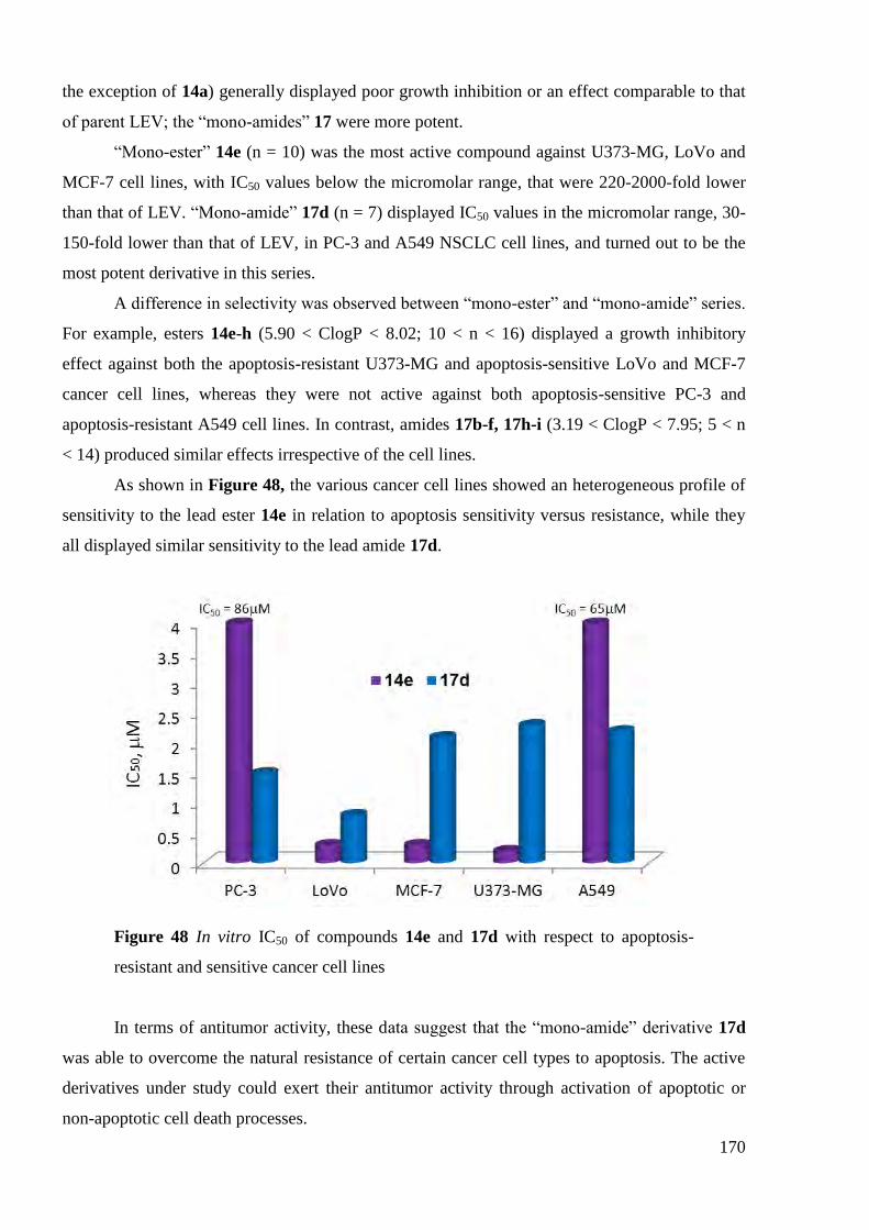
170
the exception of 14a) generally displayed poor growth inhibition or an effect comparable to that
of parent LEV; the ―mono-amides‖ 17 were more potent.
―Mono-ester‖ 14e (n = 10) was the most active compound against U373-MG, LoVo and
MCF-7 cell lines, with IC50 values below the micromolar range, that were 220-2000-fold lower
than that of LEV. ―Mono-amide‖ 17d (n = 7) displayed IC50 values in the micromolar range, 30-
150-fold lower than that of LEV, in PC-3 and A549 NSCLC cell lines, and turned out to be the
most potent derivative in this series.
A difference in selectivity was observed between ―mono-ester‖ and ―mono-amide‖ series.
For example, esters 14e-h (5.90 < ClogP < 8.02; 10 < n < 16) displayed a growth inhibitory
effect against both the apoptosis-resistant U373-MG and apoptosis-sensitive LoVo and MCF-7
cancer cell lines, whereas they were not active against both apoptosis-sensitive PC-3 and
apoptosis-resistant A549 cell lines. In contrast, amides 17b-f, 17h-i (3.19 < ClogP < 7.95; 5 < n
< 14) produced similar effects irrespective of the cell lines.
As shown in Figure 48, the various cancer cell lines showed an heterogeneous profile of
sensitivity to the lead ester 14e in relation to apoptosis sensitivity versus resistance, while they
all displayed similar sensitivity to the lead amide 17d.
Figure 48 In vitro IC50 of compounds 14e and 17d with respect to apoptosis-
resistant and sensitive cancer cell lines
In terms of antitumor activity, these data suggest that the ―mono-amide‖ derivative 17d
was able to overcome the natural resistance of certain cancer cell types to apoptosis. The active
derivatives under study could exert their antitumor activity through activation of apoptotic or
non-apoptotic cell death processes.

171
Mondal et al.192
and Yamakuchi et al.193
evaluated the antiproliferative and apoptotic
activities of LEV on human NSCLC and transitional bladder carcinoma cells in culture
respectively. These investigators demonstrated that LEV caused cellular growth inhibition in a
time-dependent manner. We also investigated the time course of the increase on growth
inhibition of the stable ―mono-amide‖ derivatives that exhibited similar antitumor effect against
all the cell lines under study. In agreement with previous reports,192-193
we observed significant
increases in growth inhibition, on all tumor cell lines, from 3 to 5 days of exposure. For example,
the cell number declines from 30-60% to 1-3% for the ―mono-amide‖ 17d, depending on the cell
line (Figure 49).
Figure 49 Time-dependent growth inhibition observed with LEV and ―mono-
amide‖ 17d in human tumor cell. The cells were cultured for 3 (purple bars) or 5
(blue bars) days with compounds LEV or 17d
4.2.2 In Vivo Maximum Tolerated Dose (MTD) in Healthy Mice
The in vivo tolerance of LEV and ―mono-esters‖ 14 was determined as the MTD index,
all of which displayed MTD indices >80 mg/kg.

172
4.2.3 In vitro antiproliferative activity of “dimer” LEV derivatives
IC50 values of ―di-amide‖ derivatives along with those of LEV are summarized in Table
79.
Table 79 In vitro antiproliferative activity of LEV ―di-amides‖
IC50 ( M)
Apoptosis-sensitive Apoptosis-resistant
Comp. n PC-3 LoVo MCF-7 U373-MG A549
LEV – 238±12 67±5 622±19 188±13 70±6
18a 4 20 ± 2 19± 2 36 ± 2 30 ± 2 64.7 ± 1.5
18b 5 14 ± 2 9 ± 1 33 ± 5 23 ± 1 85 ± 13
18c 6 0.7 ± 0.1 1.2 ± 0.1 1.8± 0.1 2.4 ± 0.1 2.9 ± 0.2
18d 7 1.7 ± 0.1 1.4 ± 0.1 1.4 ± 0.1 2.1 ± 0.1 2.3 ± 0.1
18e 8 1.6 ± 0.1 0.4 ± 0.1 1.8 ± 0.1 2 ± 60 2.2 ± 10
18f 9 0.3 ± 0.1 0.2 ± 0.1 0.5 ± 0.1 0.2 ± 0.1 0.7 ± 0.1
18g 10 120 ± 10 130 ± 20 360 ± 30 270 ± 10 480 ± 20
18h 12 2.8 ± 0.1 2.2 ± 0.1 3.8 ± 0.5 2.4 ± 0.1 4.5 ± 0.6
A homogeneous antiproliferative profile was obtained for C-6-(N-alkylcarboxamide)-
linked LEV ―dimers‖ 18a-h. For each human cancer cell lines, there was a good correlation
between the antiproliferative activities and the alkyl linker length. C-6-(N-butylcarboxamide)-
linked LEV 18a and C-6-(N-pentylcarboxamide)-linked LEV 18b ―dimers‖ exhibited ICs50 ≥ 9
M, whereas IC50 values were in the range from 0.2 to 4.5 M against all cancer cell lines for
compounds 18c-f, h (chain length with n ≥ 6). Thus, IC50 values decreased when the chain length
increased, however we observed a break between C-6-(N-nonylcarboxamide)-linked LEV 18f
which exhibited the most potent antiproliferative activities against all cancer cell lines and C-6-
(N-dodecanoylcarboxamide)-linked LEV 18h as compound C-6-(N-decanoylcarboxamide)-
linked LEV 18g was inactive. These observed results led us to study the solubility of these 3
compounds (18f, 18g, 18h) in the cellular media for 24 hours at 37°C (Figure 50).

173
Figure 50 Solubility tests in cellular media for compound 18f (n=9) at 5 M (A,
no precipitation) and 50 M (A‘), compound 18g (n=10) at 50 M (B) and 500
M (B‘) and compound 18h (n=12) at 5 M (C) and 50 M (C‘)
Compound 18f didn‘t precipitate at concentrations of 5 and 50 M while compound 18h
precipitated at 50 M and also, but with less importance, at 5 M. On the other hand, we
observed a precipitation of compound 18g at 500 or 50 M. These results could explain why
compound 18f exhibited the most potent antiproliferative activities while there was a loss of
activity for compound 18h and no antiproliferative activity for compound 18g. Nevertheless, the
―dimer‖ 18f exhibited the most potent activities against all cancer cell lines with IC50 values
ranging from 0.2 to 0.7 M, that is 100- to 1240-fold lower than that of LEV.
A A’
B B’
C C’
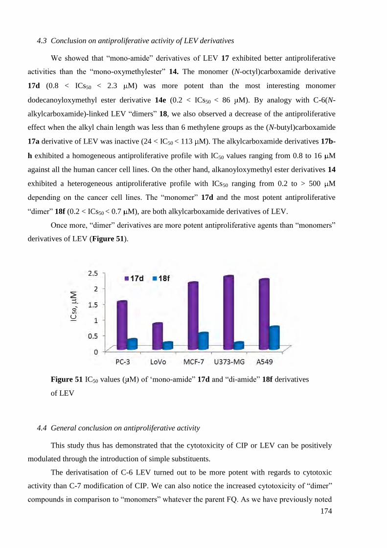
174
4.3 Conclusion on antiproliferative activity of LEV derivatives
We showed that ―mono-amide‖ derivatives of LEV 17 exhibited better antiproliferative
activities than the ―mono-oxymethylester‖ 14. The monomer (N-octyl)carboxamide derivative
17d (0.8 < ICs50 < 2.3 M) was more potent than the most interesting monomer
dodecanoyloxymethyl ester derivative 14e (0.2 < ICs50 < 86 M). By analogy with C-6(N-
alkylcarboxamide)-linked LEV ―dimers‖ 18, we also observed a decrease of the antiproliferative
effect when the alkyl chain length was less than 6 methylene groups as the (N-butyl)carboxamide
17a derivative of LEV was inactive (24 < IC50 < 113 M). The alkylcarboxamide derivatives 17b-
h exhibited a homogeneous antiproliferative profile with IC50 values ranging from 0.8 to 16 M
against all the human cancer cell lines. On the other hand, alkanoyloxymethyl ester derivatives 14
exhibited a heterogeneous antiproliferative profile with ICs50 ranging from 0.2 to > 500 M
depending on the cancer cell lines. The ―monomer‖ 17d and the most potent antiproliferative
―dimer‖ 18f (0.2 < ICs50 < 0.7 M), are both alkylcarboxamide derivatives of LEV.
Once more, ―dimer‖ derivatives are more potent antiproliferative agents than ―monomers‖
derivatives of LEV (Figure 51).
Figure 51 IC50 values ( M) of ‗mono-amide‖ 17d and ―di-amide‖ 18f derivatives
of LEV
4.4 General conclusion on antiproliferative activity
This study thus has demonstrated that the cytotoxicity of CIP or LEV can be positively
modulated through the introduction of simple substituents.
The derivatisation of C-6 LEV turned out to be more potent with regards to cytotoxic
activity than C-7 modification of CIP. We can also notice the increased cytotoxicity of ―dimer‖
compounds in comparison to ―monomers‖ whatever the parent FQ. As we have previously noted

175
for the whole series of synthesized compounds we couldn‘t establish a clear-cut dependence
between the chain length and the antiproliferative activity.
For ―monomers‖ derivatives, we observed a profile of activity which was highly
influenced by the functional group linked to FQ:
The most active ―mono-oxoethylester‖ CIP derivatives (3c, 3d or 3h) and ―mono-
acyloxymethylester‖ derivatives of LEV (14e-h) displayed a heterogeneous
antiproliferative profile of activity towards the different cancer cell lines. However, ―mono-
acyloxymethylester‖ derivatives of LEV were found to be more potent than ―mono-
oxoethylester‖ CIP derivatives.
The most active ―mono-amide‖ derivative of CIP 1h and ―mono-amide‖ derivatives of
LEV (17c-h) both displayed homogeneous antiproliferative. Again, ―mono-amide‖
derivatives of LEV were found to be more potent than ―mono-amide‖ CIP derivatives.
Whatever the parent FQ, the dimerization of monomeric molecules led to an increase of
the antiproliferative activity:
The most active ―di-amide‖ derivative of CIP 7e was more potent than the most active
―mono-amide‖ derivative of CIP 1h. They both showed a homogeneous antiproliferative
profile.
The most active ―di-oxoethylester‖ derivative of CIP 9e was more potent than the most
active ―mono-oxoethylester‖ CIP derivatives (3c, 3d or 3h). ―Monomer‖ derivatives
showed a heterogeneous antiproliferative profile while ―dimer‖ derivatives showed a
homogeneous one.
The most active ―di-amide‖ derivatives of LEV 18c-f, 18h were more potent than the most
active ―mono-amide‖ derivatives of LEV 17c-h. They both showed a homogeneous
antiproliferative profile.
As seen in literature, the mechanisms of action by which some FQ antibiotics exert
antitumor effects on various human cancer cell lines were determined to be through pro-
apoptotic effects.
In terms of antitumor activity, these data suggest that the ―amide‖ CIP derivatives and
LEV and the ―di-oxoethylester‖ derivative of CIP are able to overcome the natural resistance of
certain cancer cell types to apoptosis as they all exhibited a homogeneous antiproliferative

176
profile. The active derivatives could exert their antitumor activity through activation of apoptotic
or non-apoptotic cell death processes.
The most potent antiproliferative compounds are represented in Table 80.
Table 80 In vitro antiproliferative activity of the most active synthesized
derivatives
IC50 ( M)
Apoptosis-sensitive Apoptosis-resistant
Structure PC-3 LoVo MCF-7 U373-
MG A549
4 7 20 5 3
8 6 8 3 7
0.2 0.1 1.0 0.8 8.7
1.5 0.8 2.1 2.3 2.2
0.3 0.2 0.5 0.2 0.7
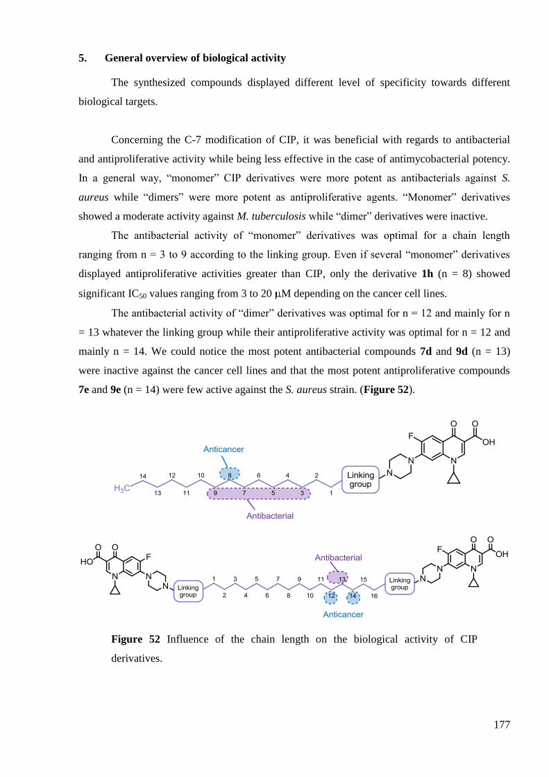
177
5. General overview of biological activity
The synthesized compounds displayed different level of specificity towards different
biological targets.
Concerning the C-7 modification of CIP, it was beneficial with regards to antibacterial
and antiproliferative activity while being less effective in the case of antimycobacterial potency.
In a general way, ―monomer‖ CIP derivatives were more potent as antibacterials against S.
aureus while ―dimers‖ were more potent as antiproliferative agents. ―Monomer‖ derivatives
showed a moderate activity against M. tuberculosis while ―dimer‖ derivatives were inactive.
The antibacterial activity of ―monomer‖ derivatives was optimal for a chain length
ranging from n = 3 to 9 according to the linking group. Even if several ―monomer‖ derivatives
displayed antiproliferative activities greater than CIP, only the derivative 1h (n = 8) showed
significant IC50 values ranging from 3 to 20 M depending on the cancer cell lines.
The antibacterial activity of ―dimer‖ derivatives was optimal for n = 12 and mainly for n
= 13 whatever the linking group while their antiproliferative activity was optimal for n = 12 and
mainly n = 14. We could notice the most potent antibacterial compounds 7d and 9d (n = 13)
were inactive against the cancer cell lines and that the most potent antiproliferative compounds
7e and 9e (n = 14) were few active against the S. aureus strain. (Figure 52).
Figure 52 Influence of the chain length on the biological activity of CIP
derivatives.

178
The C-6 modification of LEV was more beneficial with regards to cytotoxicity than for
antibacterial and antimycobacterial activities.
The antibacterial activity of ―mono-acyloxymethyl esters‖ derivatives was optimal for
chain lengths equal to n = 8 (14d) and 10 (14e) while the antimycobacterial activity was optimal
for n = 6 (14c) and 10 (14e). We could notice that these optimums were close for these two
biological activities. On the other hand, the antiproliferative activity was optimal for longer chain
lengths that were n = 12 (14f), 14 (14g) and 16 (14h) (Figure 47).
Figure 53 Influence of the chain length on the biological activity of LEV ―mono-
acyloxymethyl esters‖
The introduction of an amide function at C-6 position of LEV led to a loss of activity of
―mono-amide‖ derivatives against bacteria strains. The antimycobacterial activity was moderate
as the most potent compound 17i (n = 14) showed an IC50 value against M. tuberculosis of 4 M,
that is 5-fold less potent than LEV. On the other hand, a significant enhancement of the
antiproliferative activity was observed for chain lengths ranging from n = 6 (17c) to n = 14 (17i)
with a maximum of activity for n = 7 (17d) (Figure 54).
Figure 54 Influence of the chain length on the spectrum biological of activity for
LEV ―mono-amides‖
―Di-amide‖ derivatives of LEV were the most interesting compounds whatever the
biological activity. For compounds 18g (n = 10) and mainly 18h (n = 12), a broad spectrum of
activity, as for the parent LEV, against Gram-positive and Gram-negative bacteria strains was

179
observed. A significant antimycobacterial activity was observed from n = 9 (18f) to n = 12 (18h)
with IC50 values against M. tuberculosis < 2 M. The best antiproliferative activities were
observed for chain lengths ranging from n = 6 (18c) to n = 12 (18h) with an optimum for n = 9
(18f) (Figure 55).
Figure 55 Influence of the chain length on the biological activity of LEV ―di-
amides‖
Considering now the enzymatic target depending on the studied biological activity,
remember that FQs corrupt the activities of DNA gyrase and topoisomerase IV. However,
depending upon FQ employed and the bacterial species, either DNA gyrase or topoisomerase IV
serves as the primary cytotoxic target of drug action. For most older FQ, including CIP and LEV,
DNA gyrase in Gram-negative bacteria and topoisomerase IV in Gram-positive bacteria are
often found to be the primary targets. Each quinolone drug appears to have a preferred target and
the target selection can be altered by changes in quinolone structure. Moreover, Kerns et al.
showed that DNA gyrase became the primary target for their synthetized ―dimers‖ in Gram-
positive S. aureus and S. pneumoniae strains74-75
.
DNA gyrase is the only type II topoisomerase present in M. tuberculosis and hence is the
only target for FQ action.
FQs and their derivatives can exert their antitumor activity either by pro-apoptotic or
through non-apoptotic-dependent mechanisms. As outlined in the introduction, FQs can act on
human topoisomerase I or II and on several other biological targets.
We have reported in the following table (Table 81) the level of each biological activity
for the most potent CIP derivatives and LEV against the Gram-positive S. aureus strain.
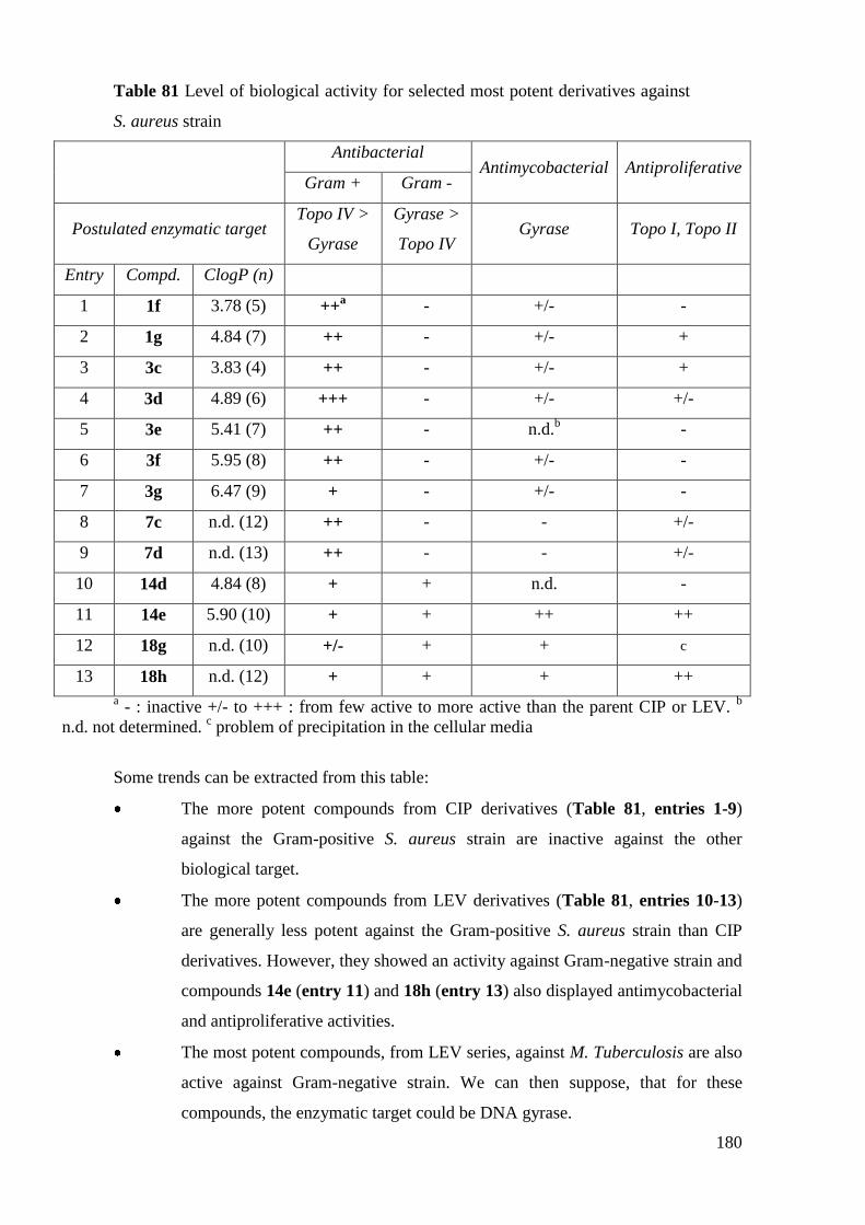
180
Table 81 Level of biological activity for selected most potent derivatives against
S. aureus strain
Antibacterial
Antimycobacterial Antiproliferative Gram + Gram -
Postulated enzymatic target Topo IV >
Gyrase
Gyrase >
Topo IV Gyrase Topo I, Topo II
Entry Compd. ClogP (n)
1 1f 3.78 (5) ++a - +/- -
2 1g 4.84 (7) ++ - +/- +
3 3c 3.83 (4) ++ - +/- +
4 3d 4.89 (6) +++ - +/- +/-
5 3e 5.41 (7) ++ - n.d.b
-
6 3f 5.95 (8) ++ - +/- -
7 3g 6.47 (9) + - +/- -
8 7c n.d. (12) ++ - - +/-
9 7d n.d. (13) ++ - - +/-
10 14d 4.84 (8) + + n.d. -
11 14e 5.90 (10) + + ++ ++
12 18g n.d. (10) +/- + + c
13 18h n.d. (12) + + + ++
a - : inactive +/- to +++ : from few active to more active than the parent CIP or LEV.
b
n.d. not determined. c problem of precipitation in the cellular media
Some trends can be extracted from this table:
The more potent compounds from CIP derivatives (Table 81, entries 1-9)
against the Gram-positive S. aureus strain are inactive against the other
biological target.
The more potent compounds from LEV derivatives (Table 81, entries 10-13)
are generally less potent against the Gram-positive S. aureus strain than CIP
derivatives. However, they showed an activity against Gram-negative strain and
compounds 14e (entry 11) and 18h (entry 13) also displayed antimycobacterial
and antiproliferative activities.
The most potent compounds, from LEV series, against M. Tuberculosis are also
active against Gram-negative strain. We can then suppose, that for these
compounds, the enzymatic target could be DNA gyrase.

181
It would be so interesting to evaluate the IC50 values of these compounds against gyrase
and topoisomerase IV from S. aureus and E. coli. So, we could determine if topoisomerase IV in
Gram-positive and gyrase in Gram-negative remain to be the primary targets for the studied
compounds. And mainly to determine if DNA gyrase didn‘t become the primary target for ―di-
amide‖ LEV derivatives 18g-h (Table 81, entries 12-13) in Gram-positive strain.
In the next table (Table 82) are summarized the level of each biological activity for the
most potent CIP derivatives and LEV against the cancer cell lines.
Table 82 Level of biological activity for selected most potent derivatives against
cancer cell lines
Antibacterial
Antimycobacterial Antiproliferative Gram + Gram -
Postulated enzymatic target Topo IV >
Gyrase
Gyrase >
Topo IV Gyrase Topo I, Topo II
Entry Compd. ClogP (n)
1 1h 5.37 (8) - a
- + ++
2 3h 7.00 (10) +/- - +/- +
3 7c n.d. (12) ++ - - +
4 7e n.d. (14) +/- - - ++
5 9c n.d. (12) +/- - +/- +
6 9e n.d. (14) +/- - n.d. b
+++
7 14e 5.90 (10) + + ++ ++
8 14f 6.96 (12) - - + +
9 17d 4.25 (7) - - +/- +++
10 17e 5.31 (9) - - +/- ++
11 17f 6.36 (11) +/- - n.d. ++
12 17h 7.42 (13) +/- - n.d. ++
13 17i 7.95 (14) - - + ++
14 18c n.d. (6) - - - ++
15 18e n.d. (8) - +/- n.d. ++
16 18f n.d. (9) - +/- + +++
17 18h n.d. (12) + + + ++ a- : inactive +/- to +++ : from few active to more active than the parent CIP or LEV.
bn.d.
not determined.

182
Some trends can be extracted from this table:
Except compound 7c (Table 82, entry 3), no compound possessing an
antibacterial activity showed an antiproliferative activity. Only ―dimer‖ CIP
derivatives 7e and 9e (entries 4, 6) displayed a good antiproliferative activity.
More compounds from LEV derivatives (Table 82, entries 7-17) were found to
have a good antiproliferative activity than an antibacterial one (Table 82, entries
10-13). Only LEV derivatives 14e and 18h showed these two biological activities.
LEV derivatives are generally more potent as antiproliferative agents than CIP
derivatives. However, we can notice that, except for compound 18h (entry 17),
they didn‘t display an antibacterial activity but some of them showed an
antimycobacterial activity.
The mechanism of action by which these derivatives exert their antiproliferative activity
will be more difficult to establish. In fact, the time- and dose-dependent pro-apoptotic effects of
FQ were observed at higher doses than in vitro growth inhibition values. For example, it have
been shown that most FQ required concentrations (IC50s) to inhibit the human topoisomerase II of
HeLa cells that were higher than those needed to inhibit bacterial type II topoisomerases.
Moreover, no significant correlation was observed between the inhibition of HeLa cell
topoisomerase II and that of S. aureus topoisomerase IV as well as E. coli DNA gyrase194
. For
CIP, the IC50 values to inhibit the topoisomerase II of Hela cells was found to be 3 and 478 higher
than those needed to inhibit bacterial type II topoisomerases. So, the human polymerase II test
can only be valuable if our compounds have IC50 values lower than CIP or LEV themselves.
So, mechanistic studies to investigate as whether our lead compounds induce either pro-
apoptotic, pro-autophagic or cell proliferation inhibition should be envisaged. But, before
proceeding with a genomic approach to identify the molecular pathways activated or inhibited by
our compounds, we have to select a lead compound through the characterization of in vivo
activity on a broad panel of biologically aggressive human xenograft models.
In summary, many CIP and LEV derivatives have been synthetized and evaluated for
their biological activity. Some of these derivatives are non-toxic in vivo according to their MTD.
This study thus demonstrates that the biological activity of CIP and LEV could be
positively modulated through the introduction of simple substituents on C-6 or C-7 positions.
Primary investigations have shown that a modification on the N-4 piperazinyl group of
CIP can influence the biological activity: the main activity is specifically oriented against the

183
inhibition of S. aureus growth or oriented against the inhibition of cancer cell lines growth. No
significant activity against M. tuberculosis was observed.
The most potent CIP derivatives against standard and MRSA strains displayed high MIC
against standard E. coli strain and were inactive for the inhibition of human cancer cell lines
growth. On the other hand, the most potent CIP derivatives as antiproliferative agents displayed
moderate to poor activities as antibacterial agents.
The activity against S. aureus is greater for ―monomeric‖ CIP derivatives as the
antiproliferative effect is greater for ―dimeric‖ ones.
A modification on the C-6 carboxylic group of LEV, and especially the substitution of
this group by an alkylcarboxamide one, led to the most potent antiproliferative and
antimycobacterial agents. So, we can conclude that C-6-carboxylic group of LEV is not required
for the antiproliferative and antimycobacterial activities.
In a general way, the derivatives of LEV are less potent as antibacterial agents against S.
aureus than CIP derivatives. However, some of them display an antibacterial activity against E.
coli. For some ―monomer‖ and ―dimer‖ derivatives of LEV, the MICs values against S. aureus
and E. coli are comparable to those of LEV, suggesting that this carboxylic acid modification is
allowed for the antibacterial activity. These results could be useful for the development of new
quinolone-based antibacterial agents.
One LEV derivative, that is C-6-(N-dodecanoylcarboxamide)-linked LEV 18h, was found
be be particulary interesting as it was the only one derivative showing a broad activity on the
three biological target.

184

185
CONCLUSION GÉNÉRALE
En résumé, nous avons synthétisé par une procédure efficace cinq séries de dérivés
―dimères‖ et ―monomères‖ de la LEV et de la CIP. Nous avons mis au point de nouvelles
conditions de réaction pour les dérivés ―dimères‖ de la CIP parce que les conditions utilisées
pour la synthèse des ―monomères‖ de la CIP n‘étaient pas efficaces et ont conduit à de faibles
rendements. L‘optimisation des conditions de réaction pour les "diesters" de CIP ont permis,
avec l'utilisation de TBAI comme un catalyseur, de préparer ces composés avec de bons
rendements. Les "di-amides" de CIP ont été obtenus en utilisant le carbodiimide EDCI soluble
dans l‘eau, ce qui a facilité le processus de purification. Les études les plus approfondies ont été
menées au cours de la synthèse des "monoester" de la LEV lorsque les deux types de conditions
par voie thermique et micro-ondes ont été évaluées. Après un examen détaillé de la réaction dans
le cas des composés à chaîne courte et longue, nous avons obtenu une amélioration du
rendement, un temps de réaction plus court et diminué la formation de sous-produit (conditions
thermiques, DMF, Cs2CO3). Pour les "mono-"et "di-amides" de LEV deux méthodes ont été
comparées: l‘utilisation de l‘agent de couplage HATU et du système TPP / TCA. Le mélange
TPP / TCA s'est avéré être plus pratique avec des réactifs disponibles et par conséquent a été
appliqué pour ces séries de composés.
En ce qui concerne l'activité biologique, notre travail a démontré que l‘activité native
(antibactérienne) de CIP et LEV pouvait être modulée par la simple modification des substituants
en C-6 et en C-7. La modification sur la position C-7 semble être plus bénéfique pour l'activité
antibactérienne, notamment contre S. aureus pour les composés ―monomères‖ et contre les
souches résistantes de S. aureus pour les dérivés ―dimères‖. Contrairement à la position C-7, la
substitution de la LEV en position C-6 a considérablement augmenté l'activité antiproliférative.
L'influence de la fonction introduite a également été démontrée: le groupement amide fourni une
large gamme de composés à spectre d‘activité homogène tandis qu‘un groupement ester a
conduit à des composés à spectre d‘activité hétérogène.
Nos travaux ont démontré le potentiel des modifications en position C-7 et C-3 des FQs
et fourni la base pour de nouvelles recherches dans ce domaine.

186
187
EXPERIMENTAL PART

188
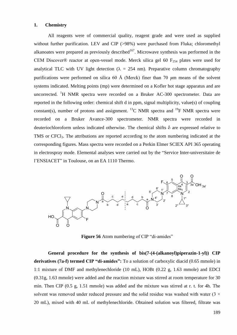
189
1. Chemistry
All reagents were of commercial quality, reagent grade and were used as supplied
without further purification. LEV and CIP (>98%) were purchased from Fluka; chloromethyl
alkanoates were prepared as previously described167
. Microwave synthesis was performed in the
CEM Discover® reactor at open-vessel mode. Merck silica gel 60 F254 plates were used for
analytical TLC with UV light detection ( = 254 nm). Preparative column chromatography
purifications were performed on silica 60 Å (Merck) finer than 70 m means of the solvent
systems indicated. Melting points (mp) were determined on a Kofler hot stage apparatus and are
uncorrected. 1H NMR spectra were recorded on a Bruker AC-300 spectrometer. Data are
reported in the following order: chemical shift d in ppm, signal multiplicity, value(s) of coupling
constant(s), number of protons and assignment. 13
C NMR spectra and 19
F NMR spectra were
recorded on a Bruker Avance-300 spectrometer. NMR spectra were recorded in
deuteriochloroform unless indicated otherwise. The chemical shifts are expressed relative to
TMS or CFCl3. The attributions are reported according to the atom numbering indicated at the
corresponding figures. Mass spectra were recorded on a Perkin Elmer SCIEX API 365 operating
in electrospray mode. Elemental analyses were carried out by the ―Service Inter-universitaire de
l‘ENSIACET‖ in Toulouse, on an EA 1110 Thermo.
Figure 56 Atom numbering of CIP ―di-amides‖
General procedure for the synthesis of bis(7-(4-(alkanoyl)piperazin-1-yl)) CIP
derivatives (7a-f) termed CIP “di-amides”: To a solution of carboxylic diacid (0.65 mmole) in
1:1 mixture of DMF and methylenechloride (10 mL), HOBt (0.22 g, 1.63 mmole) and EDCI
(0.31g, 1.63 mmole) were added and the reaction mixture was stirred at room temperature for 30
min. Then CIP (0.5 g, 1.51 mmole) was added and the mixture was stirred at r. t. for 4h. The
solvent was removed under reduced pressure and the solid residue was washed with water (3 ×
20 mL), mixed with 40 mL of methylenechloride. Obtained solution was filtered, filtrate was
190
evaporated. Crude product was purified by column chromatography on silica gel (40g, CH2Cl2/
MeOH, 10:0.1 – 10:1).
bis(7-(4-Decanoyl)piperazin-1-yl)-1-cyclopropyl-6-fluoro-1,4-dihydro-4-
oxoquinoline-3-carboxylic acid) (7a). Purification provided the desired compound (251 mg,
48% yield) as a pale yellow solid. m.p. >260 ºC (decomp.). Rf 0.19 (CH2Cl2/MeOH, 10/0.75). 1H
NMR (500 MHz): 1.21 (dd, 3Jcis=10.5 Hz,
3Jtrans=6 Hz, 4H, C2‖-H and C3‖-H), 1.35 (m, 8H, Cd-
H, Ce-H), 1.40 (dd, 3Jcis=13.5 Hz,
3Jtrans=6 Hz, 4H, C2‖-H and C3‖-H), 1.66 (quint,
3Jb-c=7.5 Hz,
3Jc-d=6 Hz, 4H, Cc-H), 2.38 (t,
3J=7.5 Hz, 4H, Cb-H), 3.28 (t,
3J=5 Hz, 4H, C3'-H), 3.35 (t,
3J=5
Hz, 4H, C5'-H), 3.54 (sept, 3J=4 and 7 Hz, 2H, C1''-H), 3.71 (t,
3J =5 Hz, 4H, C2'-H), 3.87 (t,
3J =5
Hz, 4H, C6'-H), 7.38 (d, 4JH-F=7 Hz, 2H, C8-H), 8.07 (d,
3JH-F=13 Hz, 2H, C5-H), 8.79 (s, 2H, C2-
H), 14.89 (br s, 2H, O3a'-H). 13
C NMR (125 MHz): 8.3 (C2'', C3''), 25.2, 29.2, 29.4, 33.2 (Cb-e),
35.3 (C1''), 41.1 (C3'), 45.4 (C5'), 49.4 (C6'), 50.4 (C2'), 105.1 (d, 3JC-F = 2.8 Hz, C8), 108.4 (C3),
112.8 (d, 2JC-F = 23.3 Hz, C5), 120.5 (d,
3JC-F = 7.9 Hz, C4a), 139 (C8a), 145.4 (d,
2JC-F = 10.5 Hz,
C7), 147.6 (C2), 153.6 (d, 1JC-F = 249.6 Hz, C6), 166.9 (C3a), 171.8 (Ca), 177.1 (d,
4JC-F = 2.5 Hz,
C4). 19
F NMR (282 MHz): -121.18 – -121.25 (dd, 3JH-F = 12.6 Hz,
4JH-F = 6.9 Hz). MS CI
(DCI/NH3): C44H50F2N6O8, m/z 829.5 [M+H]+. Elemental Anal. (C44H50F2N6O8 · H2O) C, H, N:
calcd, 62.40, 6.19, 9.92; found, 62.41, 6.12, 9.90.
bis(7-(4-Dodecanoyl)piperazin-1-yl)-1-cyclopropyl-6-fluoro-1,4-dihydro-4-
oxoquinoline-3-carboxylic acid) (7b). Purification provided the desired compound (174 mg,
31% yield) as a pale yellow solid. m.p. >260 ºC (decomp.). Rf 0.21 (CH2Cl2/MeOH, 10/0.75). 1H
NMR (500 MHz): 1.21 (dd, 3Jcis=10.5 Hz,
3Jtrans=6.5 Hz, 4H, C2‖-H and C3‖-H), 1.29 – 1.36 (m,
12H, Cd-f-H), 1.40 (dd, 3Jcis = 13.5 Hz,
3Jtrans = 6.5 Hz, 4H, C2‖-H and C3‖-H), 1.65 (quint,
3Jb-c =
8 Hz, 3Jc-d = 7 Hz, 4H, Cc-H), 2.37 (t,
3J = 8 Hz, 4H, Cb-H), 3.28 (t,
3J = 5 Hz, 4H, C3'-H), 3.35 (t,
3J = 4.5 Hz, 4H, C5'-H), 3.54 (sept,
3J = 4 and 7.5 Hz, 2H, C1''-H), 3.72 (t,
3J = 4.5 Hz, 4H, C2'-
H), 3.86 (t, 3J = 5 Hz, 4H, C6'-H), 7.37 (d,
4JH-F = 7 Hz, 2H, C8-H), 8.03 (d,
3JH-F = 12.5 Hz, 2H,
C5-H), 8.77 (s, 2H, C2-H), 14.90 (br s, 2H, O3a'-H). 13
C NMR (125 MHz): 8.3 (C2'', C3''); 25.2,
29.4, 29.4, 33.2 (Cb-f); 35.3 (C1''); 41.1 (C3'); 45.4 (C5'); 49.4 (C6'); 50.3 (C2'); 105.1 (d, 3JC-F = 2.8
Hz, C8); 108.3 (C3); 112.6 (d, 2JC-F = 23.1 Hz, C5); 120.4 (d,
3JC-F = 7.8 Hz, C4a); 138.9 (C8a);
145.4 (d, 2JC-F = 10.4 Hz, C7); 147.6 (C2); 153.5 (d,
1JC-F = 249.7 Hz, C6); 166.9 (C3a); 171.9
(Ca), 177.1 (d, 4JC-F = 2.5 Hz, C4).
19F NMR (282 MHz, CDCl3): -121.14 – -121.21 (dd,
4JF-H =
7.0 Hz, 3JF-H = 12.8 Hz). MS CI (DCI/NH3): C46H54F2N6O8, m/z 857.7 [M+H]
+. Elemental Anal.
(C48H58F2N6O8 · 2.8H2O) C, H, N: calcd, 60.90, 6.58, 9.27; found, 60.93, 6.13, 9.94.
191
bis(7-(4-Tetradecanoyl)piperazin-1-yl)-1-cyclopropyl-6-fluoro-1,4-dihydro-4-
oxoquinoline-3-carboxylic acid) (7c). Purification provided the desired compound (400 mg,
66% yield) as a pale yellow solid. m.p. 250 ºC. Rf 0.22 (CH2Cl2/MeOH, 10/0.75). 1H NMR (300
MHz): 1.20 (m, 4H, C2''-H, C3''-H); 1.26 – 1.32 (m, 16H, Cd-g-H); 1.39 (dd, 3Jcis = 13.8 Hz,
3Jtrans
= 6.9 Hz, 4H, C2''-H, C3''-H); 1.65 (quint, 3Jb-c = 7.8 Hz,
3Jc-d = 6.9 Hz, 4H, Cc-H); 2.37 (t,
3JH-H =
7.8 Hz, 4H, Cb-H); 3.29 (m, 4H, C3‘'-H); 3.35 (m, 4H, C5'-H); 3.55 (m, 2H, C1''-H); 3.7 (t, 3J = 4.5
Hz, 4H, C2'-H); 3.86 (m, 4H, C6'-H); 7.35 (d, 4JH-F = 6.6 Hz, 2H, C8-H); 7.98 (d,
3JH-F = 12.9 Hz,
2H, C5-H); 8.72 (s, 2H, C2-H); 14.88 (br s, 2H, O3a'-H). 13
C NMR (75 MHz): 8.2 (C2'', C3''); 25.2,
29.4, 29.4, 29.5, 33.2 (Cb-g); 35.3 (C1''); 41.0 (C3'); 45.3 (C5'); 49.3 ( C6'); 50.3 ( C2'); 105.0 (d, 3JC-
F = 3.0 Hz, C8); 108.1 (C3); 112.5 (d, 2JC-F = 23.2 Hz, C5); 120.2 (d,
3JC-F = 7.7 Hz, C4a); 138.9
(C8a); 145.4 (d, 2JC-F = 10.2 Hz, C7); 147.5 (C2); 153.5 (d,
1JC-F = 249.7 Hz, C6); 166.8 (C3a);
171.8 (Ca), 176.9 (d, 4JC-F = 2.3 Hz, C4).
19F NMR (282 MHz): -121.04 – -121.11 (dd,
4JF-H = 7.0
Hz, 3JF-H = 12.9 Hz). MS CI (DCI/NH3): C48H58F2N6O8, m/z 885.8 [M+H]
+. Elemental Anal.
(C48H58F2N6O8 · H2O) C, H, N: calcd, 63.86, 6.65, 9.31; found, 63.90, 6.19, 10.04.
bis(7-(4-Pentadecanoyl)piperazin-1-yl)-1-cyclopropyl-6-fluoro-1,4-dihydro-4-
oxoquinoline-3-carboxylic acid) (7d). Purification provided the desired compound (318 mg,
61% yield) as a pale yellow solid. m.p. 178 ºC. Rf 0.20 (CH2Cl2/MeOH, 10/0.75). 1H NMR (300
MHz): 1.19 (m, 4H, C2''-H, C3''-H); 1.24 – 1.30 (m, 18H, Cd-h-H); 1.37 (dd, 3Jcis=13.5 Hz,
3Jtrans=6.9 Hz, 4H, C2''-H, C3''-H); 1.63 (quint,
3Jb-c=7.5 Hz,
3Jc-d=6.9 Hz, 4H, Cc-H); 2.36 (t,
3JH-H
= 7.5 Hz, 4H, Cb-H); 3.29 (m, 4H, C3'); 3.36 (m, 4H, C5'); 3.55 (m, 2H, C1''-H); 3.71 (m, 4H, C2');
3.85 (m, 4H, C6'); 7.33 (d, 4JH-F = 7.5 Hz, 2H, C8-H); 7.87 (d,
3JH-F = 12.9 Hz, 2H, C5-H); 8.65 (s,
2H, C2-H); 14.87 (br s, 2H, O3a'-H). 13
C NMR (75 MHz): 8.2 (C2'', C3''); 25.2, 29.3, 29.4, 29.5,
29.6 (Cc-h); 33.2 (Cb); 35.3 (C1''); 41.0 (C3'); 45.3 (C5'); 49.3 (C6'); 50.2 (C2'); 105.0 (d, 3JC-F = 3.0
Hz, C8); 107.9 (C3); 112.2 (d, 2JC-F = 23.2 Hz, C5); 119.8 (d,
3JC-F = 7.8 Hz, C4a); 138.9 (C8a);
145.3 (d, 2JC-F = 10.3 Hz, C7); 147.3 (C2); 153.5 (d,
1JC-F = 249.7 Hz, C6); 166.7 (C3a); 171.8
(Ca), 176.7 (d, 4JC-F = 2.5 Hz, C4).
19F NMR (282 MHz): -120.93 – -121.00 (dd,
4JF-H = 6.8 Hz,
3JF-H = 12.9 Hz). MS CI (DCI/NH3): C49H60F2N6O8, m/z 899.7 [M+H]
+. Elemental Anal.
(C49H60F2N6O8 · H2O) C, H, N: calcd, 64.19, 6.77, 9.17; found, 64.15, 6.76, 9.41.
bis(7-(4-Hexadecanoyl)piperazin-1-yl)-1-cyclopropyl-6-fluoro-1,4-dihydro-4-
oxoquinoline-3-carboxylic acid) (7e). Purification provided the desired compound (326 mg,
55% yield) as a pale yellow solid. m.p. 144 ºC. Rf 0.20 (CH2Cl2/MeOH, 10/0.75). 1H NMR (300
MHz): 1.20 (m, 4H, C2''-H, C3''-H); 1.24 – 1.32 (m, 20H, Cd-h-H); 1.39 (dd, 3Jcis=13.5 Hz,
3Jtrans=6.9 Hz, 4H, C2''-H, C3''-H); 1.65 (quint,
3Jb-c=7.5 Hz,
3Jc-d=7.0 Hz, 4H, Cc-H); 2.37 (t,
3JH-H

192
= 7.5 Hz, 4H, Cb-H); 3.29 (m, 4H, C3'-H); 3.36 (m, 4H, C5'-H); 3.55 (m, 2H, C1''-H); 3.72 (m, 4H,
C2'-H); 3.86 (m, 4H, C6'-H); 7.35 (d, 4JH-F = 7.2 Hz, 2H, C8-H); 7.94 (d,
3JH-F = 12.9 Hz, 2H, C5-
H); 8.69 (s, 2H, C2-H); 14.88 (br s, 2H, O3a'-H). 13
C NMR (75 MHz): 8.3 (C2'', C3''); 25.2, 29.4,
29.4, 29.4, 29.6, 29.6 (Cc-h); 33.2 (Cb); 35.4 (C1''); 41.1 (C3'); 45.4 (C5'); 49.4 (C6'); 50.3 (C2');
105.1 (d, 3JC-F = 3.0 Hz, C8); 107.9 (C3); 112.4 (d,
2JC-F = 23.2 Hz, C5); 120.1 (d,
3JC-F = 7.8 Hz,
C4a); 139.0 (C8a); 145.4 (d, 2JC-F = 10.3 Hz, C7); 147.5 (C2); 153.5 (d,
1JC-F = 249.8 Hz, C6);
166.8 (C3a); 171.9 (Ca), 176.9 (d, 4JC-F = 2.6 Hz, C4).
19F NMR (282 MHz): -121.00 – -121.07
(dd, 4JF-H = 7.0 Hz,
3JF-H = 12.7 Hz). MS CI (DCI/NH3): C50H62F2N6O8, m/z 913.7 [M+H]
+.
Elemental Anal. (C50H62F2N6O8 · 2H2O) C, H, N: calcd, 63.29, 6.96, 8.86; found, 63.28, 6.85,
9.22.
bis(7-(4-Octadecanoyl)piperazin-1-yl)-1-cyclopropyl-6-fluoro-1,4-dihydro-4-
oxoquinoline-3-carboxylic acid) (7f). Purification provided the desired compound (325 mg,
53% yield) as a pale yellow solid. m.p. 138 ºC. Rf 0.22 (CH2Cl2/MeOH, 10/0.75). 1H NMR (300
MHz): 1.20 (m, 4H, C2''-H, C3''-H); 1.23 – 1.32 (m, 24H, Cd-i-H); 1.39 (dd, 3Jcis=13.5 Hz,
3Jtrans=6.9 Hz, 4H, C2''-H, C3''-H); 1.64 (quint,
3Jb-c=7.5 Hz,
3Jc-d=6.6 Hz, 4H, Cc-H); 2.37 (t,
3JH-H
= 7.5 Hz, 4H, Cb-H); 3.29 (m, 4H, C3'-H); 3.36 (m, 4H, C5'-H); 3.55 (m, 2H, C1''-H); 3.72 (m, 4H,
C2'-H); 3.86 (m, 4H, C6'-H); 7.33 (d, 4JH-F = 6.9 Hz, 2H, C8-H); 7.89 (d,
3JH-F = 12.9 Hz, 2H, C5-
H); 8.66 (s, 2H, C2-H); 14.88 (br s, 2H, O3a'-H). 13
C NMR (75 MHz): 8.2 (C2'', C3''); 25.2, 29.4,
29.4, 29.4, 29.5, 29.6 (Cc-i); 33.2 (Cb); 35.3 (C1''); 41.0 (C3'); 45.3 (C5'); 49.3 (C6'); 50.1 (C2');
105.0 (d, 3JC-F = 3.0 Hz, C8); 107.9 (C3); 112.2 (d,
2JC-F = 23.2 Hz, C5); 119.9 (d,
3JC-F = 7.8 Hz,
C4a); 138.9 ( C8a); 145.3 (d, 2JC-F = 10.3 Hz, C7); 147.4 (C2); 153.5 (d,
1JC-F = 249.8 Hz, C6);
166.7 (C3a); 171.9 (Ca); 176.8 (d, 4JC-F = 2.6 Hz, C4).
19F NMR (282 MHz): -120.94 – -121.01
(4JF-H = 7.0 Hz,
3JF-H = 12.7 Hz). MS CI (DCI/NH3): C52H66F2N6O8, m/z 941.8 [M+H]
+.
Elemental Anal. (C52H66F2N6O8 · 2H2O) C, H, N: calcd, 63.93, 7.17, 8.61; found, 63.94, 7.01,
9.22.
Figure 57 Atom numbering of CIP ―di-oxoethyl esters‖
193
General procedure for the synthesis of bis(7-(4-(2-Oxoethylalcanoate)piperazin-1-
yl)) CIP derivatives (9a-f) termed CIP “di-oxoethyl esters”. To a solution of carboxylic diacid
(0.5 mmole) in DMF (10 mL), Cs2CO3 (0.32 g, 1.00 mmole) was added and the reaction mixture
was stirred at 50-60ºC for 1 hour. Then N-chloroacetylciprofloxacin (0.42 g, 1.03 mmole) and
TBAI (0.37 g, 1.00 mmole) were added and the mixture was stirred at the previously mentioned
temperature for 72h. The solvent was removed under reduced pressure and 20 mL of aqueous
HCl 1M was added to the residue. The obtained solid was filtered and washed with aqueous HCl
1M, water, ethanol and petroleum ether. The remaining solid residue was purified by column
chromatography (CH2Cl2/MeOH from 10/0.3 to 10/0.5).
bis(7-(4-Oxoethyldecanoate)piperazin-1-yl)-1-cyclopropyl-6-fluoro-1,4-dihydro-4-
oxoquinoline-3-carboxylic acid) (9a). Obtained from decanedioïc acid (n=8) as a pale yellow
solid in 42 % yield (200 mg). Due to the low solubility of this compound, even in DMSO-d6, no
13C NMR could be obtained.
1H NMR (300 MHz, DMSO-d6): 1.21-1.34 (m, 12H, C2''-H, C3''-H,
Cf-H, Cg-H); 1.40 (dd, 3Jcis=13.5 Hz,
3Jtrans=6.9 Hz, 4H, C2''-H, C3''-H); 1.67 (quint,
3Jd e=7.5 Hz,
3Je f=7.0, 4H, Ce-H); 2.45 (t,
3JH-H = 7.5Hz, 4H, Cd-H); 3.32 – 3.37 (m, 8H, C3'-H, C5'-H); 3.54
(m, 2H, C1''-H); 3.66 (m, 4H, C2'-H); 3.87 (m, 4H, C6'-H); 4.78 (s, 4H, Cb-H); 7.38 (d, 4JH-F = 6.6
Hz, 2H, C8-H); 8.07 (d, 3JH-F = 12.6Hz, 2H, C5-H); 8.79 (s, 2H, C2-H); 14.86 (br s, 2H, O3a'-H).
19F NMR (282 MHz, DMSO-d6): -121.28 – -121.35 (dd,
3JH-F = 12.7 Hz,
4JH-F = 7.0 Hz). MS CI
(DCI/NH3): C48H54F2N6O12, m/z 945.6 [M+H]+. Elemental Anal. (C48H54F2N6O12 · 1.2 H2O); C,
H, N: calcd, 59.68, 5.88, 8.70; found, 59.65, 5.96, 8.67.
bis(7-(4-Oxoethyldodecanoate)piperazin-1-yl)-1-cyclopropyl-6-fluoro-1,4-dihydro-4-
oxoquinoline-3-carboxylic acid) (9b). Obtained from dodecanedioïc acid (n=10) as a pale
yellow solid in 47 % yield (200 mg). 1H NMR (300 MHz, DMSO-d6): 1.21 – 1.32 (m, 16H, C2''-
H, C3''-H, Cf-h-H); 1.40 (dd, 3Jcis=13.2 Hz,
3Jtrans=6.6 Hz, 4H, C2''-H, C3''-H); 1.70 (quint,
3Jd e=7.5 Hz,
3Je f=7.0 Hz, 4H, Ce-H); 2.45 (t,
3JH-H = 7.5 Hz, 4H, Cd-H); 3.32 – 3.37 (m, 8H, C3'-
H, C5'-H); 3.55 (m, 2H, C1''-H); 3.66 (m, 4H, C2'-H); 3.88 (m, 4H, C6'-H); 4.78 (s, 4H, Cb-H);
7.38 (d, 4JH-F = 6.5 Hz, 2H, C8-H); 8.07 (d,
3JH-F = 12.6 Hz, 2H, C5-H); 8.78 (s, 2H, C2-H); 14.89
(br s, 2H, O3a'-H). 13
C NMR (125 MHz, DMSO-d6): 8.0 (C2'', C3''); 24.9 (Ch); 29.2, 29.1, 28.8 (Ce-
g); 33.7 (Cd); 36.3 (C1''); 41.5 (C3'); 44.5 (C5'); 48.9 (C2'); 49.2 (C6'); 61.6 (Cb); 107.1 (d, 3JC-F = 3
Hz, C8); 107.4 (C3); 111.5 (d, 2JC-F = 24 Hz, C5); 119.7 (d,
3JC-F = 8 Hz, C4a); 139.6 (C8a); 145.3
(d, 2JC-F = 10.4 Hz, C7); 148.5 (C2); 153.4 (d,
1JC-F = 249.0 Hz, C6); 165.5 (Cc); 166.3 (C3a);
172.9 (Ca); 176.8 (d, 4JC-F = 2.3 Hz, C4).
19F NMR (282 MHz, DMSO-d6): -121.31 – -121.38 (dd,

194
3JH-F=12.5 Hz,
4JH-F=6.6Hz). MS CI (DCI/NH3): C50H58F2N6O12, m/z found, 973.6 [M+H]
+
Elemental Anal. (C50H58F2N6O12 · 1.9 H2O); C, H, N: calcd, 59.65, 6.18, 8.35; found, 59.49,
5.94, 8.56.
bis(7-(4-Oxoethyltetradecanoate)piperazin-1-yl)-1-cyclopropyl-6-fluoro-1,4-
dihydro-4-oxoquinoline-3-carboxylic acid) (9c). Obtained from tetradecanedioïc acid (n=12)
as a pale yellow solid in 56% yield (250 mg). 1H NMR (500 MHz): 1.20 – 1.32 (m, 20H, C2''-H,
C3''-H, Cf-i-H); 1.43 (dd, 3Jcis = 13.2 Hz,
3Jtrans = 6.6 Hz, 4H, C2''-H, C3''-H); 1.69 (quint,
3Jd-e=7.5
Hz, 3Je-f = 6.9 Hz, 4H, Ce-H); 2.45 (t,
3JH-H = 7.5 Hz, 4H, Cd-H); 3.32 – 3.37 (2m, 8H, C3'-H, C5'-
H); 3.55 (m, 2H, C1''-H); 3.66 (m, 4H, C2'-H); 3.87 (m, 4H, C6'-H); 4.78 (s, 4H, Cb-H); 7.38 (d,
4JH-F = 6.6 Hz, 2H, C8-H); 8.05 (d,
3JH-F = 12.6 Hz, 2H, C5-H); 8.79 (s, 2H, C2-H); 14.87 (br s,
2H, O3a'-H). 13
C NMR (125 MHz): 8.2 (C2'', C3''); 24.9 (Ci); 29.5, 29.5, 29.4, 29.3 (Ce-h); 33.9
(Cd); 35.3 (C1''); 41.5 (C3'); 44.5 (C5'); 49.3 (C2'); 50.0 (C6'); 58.6 (Cb); 105.3 (d, 3JC-F = 2.9 Hz,
C8); 108.2 (C3); 112.7 (d, 2JC-F = 23.3 Hz, C5); 120.5 (d,
3JC-F = 7.9 Hz, C4a); 138.9 (C8a); 145.2
(d, 2JC-F = 10.5 Hz, C7); 147.6 (C2); 153.5 (d,
1JC-F = 251.4 Hz, C6); 165.3 (Cc); 166.8 (C3a);
173.5 (Ca); 177.1 (d, 4JC-F = 2.5 Hz, C4).
19F NMR (282 MHz): 121.28 – -121.35 (dd,
3JH-F = =
12.9 Hz, 4JH-F = 7.0 Hz). MS CI (DCI/NH3): C52H62F2N6O12, m/z 1001.7 [M+H]
+. Elemental
Anal. (C52H62F2N6O12 · 0.9 H2O); C, H, N: calcd, 61.39, 6.32, 8.26; found, 61.22, 6.24, 8.66.
bis(7-(4-Oxoethylpentadecanoate)piperazin-1-yl)-1-cyclopropyl-6-fluoro-1,4-
dihydro-4-oxoquinoline-3-carboxylic acid) (9d). Obtained from pentadecanedioïc acid (n=13)
as a pale yellow solid in 56% yield (260 mg). 1H NMR (300 MHz): 1.21 – 1.45 (m, 26H, C2''-H,
C3''-H, Cf-j-H,); 1.65 (quint, 3Jd-e = 7.8 Hz,
3Je-f = 7.2 Hz, 4H, Ce-H); 2.45 (t,
3JH-H = 7.8 Hz, 4H,
Cd-H); 3.32 – 3.38 (2m, 8H, C3'-H, C5'-H); 3.56 (m, 2H, C1''-H); 3.66 (m, 4H, C2'-H); 3.87 (m,
4H, C6'-H); 4.78 (s, 4H, Cb-H); 7.37 (d, 4JH-F = 6.9 Hz, 2H, C8-H); 8.00 (d,
3JH-F = 12.6 Hz, 2H,
C5-H); 8.74 (s, 2H, C2-H); 14.87 (br s, 2H, O3a'-H). 13
C NMR (75 MHz): 8.2 (C2'', C3''); 24.7 (Cj);
29.0, 29.2, 29.3, 29.4, 29.5 (Ce-i); 33.9 (Cd); 35.3 (C1''); 41.5 (C3'); 44.2 (C5'); 49.2 (C2'); 49.8 (
C6'); 60.9 (Cb); 105.2 (d, 3JC-F = 2.7 Hz, C8); 107.9 (C3); 112.3 (d,
2JC-F = 23.1 Hz, C5); 120.1 (d,
3JC-F = 7.7 Hz, C4a); 138.9 ( C8a); 145.2 (d,
2JC-F = 10.4 Hz, C7); 147.4 (C2); 153.5 (d,
1JC-F =
249.8 Hz, C6); 165.2 (Cc); 166.6 (C3a); 173.4 (Ca); 176.8 (d, 4JC-F = 2.4 Hz, C4).
19F NMR (282
MHz): -121.06 – -121.13 (dd, 3JH-F = 12.7 Hz,
4JH-F = 6.0 Hz); MS CI (DCI/NH3):
C53H64F2N6O12, m/z 1015.4 [M+H]+. Elemental Anal. (C53H64F2N6O12 · 1H2O) C, H, N: calcd,
61.62, 6.44, 8.13; found, 61.78, 6.56, 8.12.

195
bis(7-(4-Oxoethylhexadecanoate)piperazin-1-yl)-1-cyclopropyl-6-fluoro-1,4-dihydro-
4-oxoquinoline-3-carboxylic acid) (9e). Obtained from hexadecanedioïc acid (n=14) as a pale
yellow solid in 41% yield (190 mg). 1H NMR (500 MHz) : 1.21 (dd,
3Jcis = 10.5 Hz,
3Jtrans = 7.5
Hz, 4H, C2''-H, C3''-H); 1.24 –1.34 (m, 20H, Cf-j-H); 1.40 (dd, 3Jcis = 14.0 Hz,
3Jtrans = 7.5 Hz, 4H,
C2''-H, C3''-H); 1.67 (quint, 3Jd-e = 7.5 Hz,
3Je-f = 7.5 Hz, 4H, Ce-H); 2.45 (t,
3JH-H = 7.5 Hz, 4H,
Cd-H); 3.31 – 3.38 (2 m, 8H, C3'-H, C5'-H); 3.55 (quint, 3J = 3.5 and 7.5 Hz, 2H, C1''-H); 3.66 (m,
4H, C2'-H); 3.87 (m, 4H, C6'-H); 4.78 (s, 4H, Cb-H); 7.37 (d, 4JH-F = 7.0 Hz, 2H, C8-H); 8.00 (d,
2H, 3JH-F = 12.5 Hz, C5-H); 8.74 (s, 2H, C2-H); 14.87 (br s, 2H, O3a'-H).
13C NMR (125 MHz):
8.3 (C2'', C3''); 24.8 (Cj); 29.6, 29.5, 29.4, 29.2, 29.0 (Ce-i); 33.9 (Cd); 35.3 (C1''); 41.0 (C3'); 45.3
(C5'); 49.2 (C2'); 49.9 ( C6'); 60.9 (Cb); 105.2 (d, 3JC-F = 3.0 Hz, C8); 108.2 (C3); 112.6 (d,
2JC-F =
23.1 Hz, C5); 120.4 (d, 3JC-F = 7.8 Hz, C4a); 138.9 (C8a); 145.2 (d,
2JC-F = 10.3 Hz, C7); 147.6
(C2); 153.5 (d, 1JC-F = 249.8 Hz, C6); 165.3 (Cc); 166.8 (C3a); 173.5 (Ca); 177.0 (d,
4JC-F = 2.5 Hz,
C4). 19
F NMR (282 MHz): -121.20 – -121.27 (dd, 3JH-F = 12.7 Hz,
4JH-F = 7.0 Hz); MS CI
(DCI/NH3): C54H66F2N6O12, m/z 1029.6 [M+H]+. Elemental Anal. (C54H66F2N6O12 · 2.6H2O) C,
H, N: calcd, 60.28, 6.67, 7.81; found, 60.20, 6.50, 7.51.
bis(7-(4-Oxoethyloctadecanoate)piperazin-1-yl)-1-cyclopropyl-6-fluoro-1,4-dihydro-
4-oxoquinoline-3-carboxylic acid) (9f). Obtained from octadecanedioïc acid (n=16) as a pale
yellow solid in 75% yield (330 mg). 1H NMR (500 MHz) : 1.21 (dd,
3Jcis = 10.5 Hz,
3Jtrans = 7.0
Hz, 4H, C2''-H, C3''-H); 1.24 – 1.34 (m, 24H, Cf-k-H), 1.40 (dd, 3Jcis = 13.5 Hz,
3Jtrans = 7.0 Hz,
4H, C2''-H, C3''-H), 1.68 (quint, 3Jd-e = 7.5 Hz,
3Je-f = 7.0 Hz, 4H, Ce-H); 2.45 (t,
3JH-H = 7.5 Hz,
4H, Cd-H); 3.31 – 3.38 (2 m, 8H, C3'-H, C5'-H); 3.55 (quint, 3J = 3.5 and 7.0 Hz, 2H, C1''-H); 3.66
(m, 4H, C2'-H); 3.87 (m, 4H, C6'-H); 4.78 (s, 4H, Cb-H); 7.37 (d, 4JH-F = 7.0 Hz, 2H, C8-H); 8.05
(d, 3JH-F = 12.5 Hz, 2H, C5-H); 8.78 (s, 2H, C2-H); 14.87 (br s, 2H, O3a'-H).
13C NMR (125
MHz): 8.3 (C2'', C3''); 24.8 (Ck); 29.1, 29.2, 29.4, 29.5, 29.6, 29.7 (Ce-j); 33.9 (Cd); 35.3 (C1'');
41.5 (C3'); 44.5 (C5'); 49.3 (C2'); 50.0 ( C6'); 60.9 (Cb); 105.2 (d, 3JC-F = 2.9 Hz, C8); 108.3 (C3);
112.7 (d, 2JC-F = 23.1 Hz, C5); 120.6 (d,
3JC-F = 7.8 Hz, C4a); 138.9 (C8a); 145.2 (d,
2JC-F = 10.5
Hz, C7); 147.7 (C2); 153.6 (d, 1JC-F = 249.8 Hz, C6); 165.3 (Cc); 166.8 (C3a); 173.5 (Ca); 177.1
(d, 4JC-F = 2.4 Hz, C4).
19F NMR (282 MHz): -121.23 – -121.30 (dd,
3JH-F = 12.7 Hz,
4JH-F = 7.0
Hz); MS CI (DCI/NH3): C56H70F2N6O12, m/z 1057.6 [M+H]+. Elemental Anal. (C56H70F2N6O12 ·
1H2O) C, H, N: calcd, 62.56, 6.75, 7.82; found, 62.29, 6.78, 7.92.
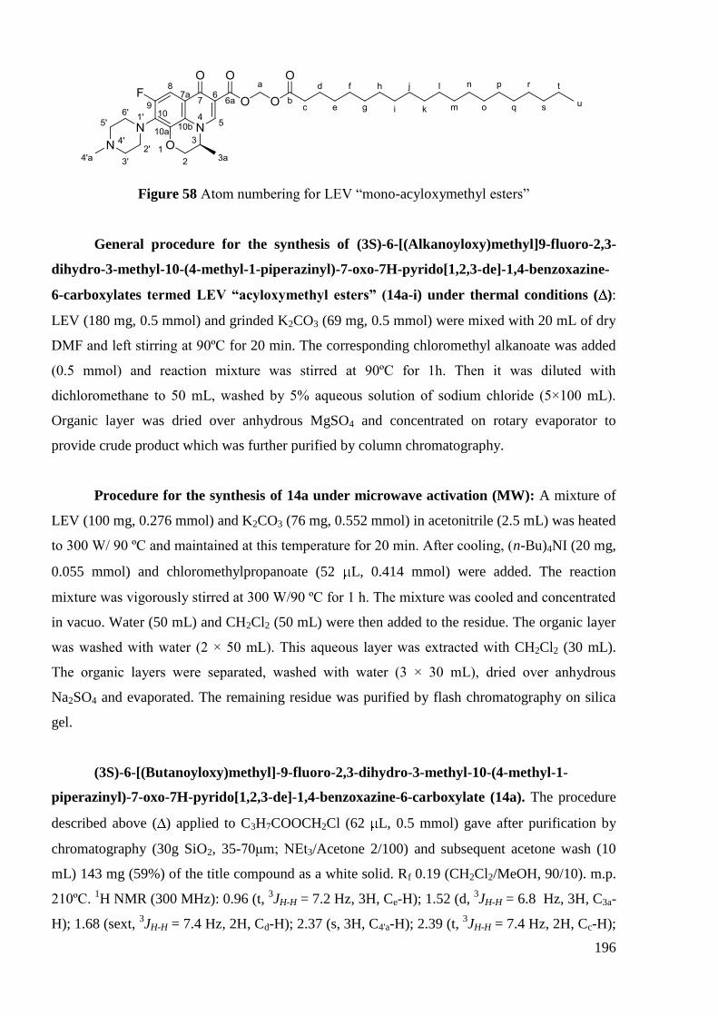
196
Figure 58 Atom numbering for LEV ―mono-acyloxymethyl esters‖
General procedure for the synthesis of (3S)-6-[(Alkanoyloxy)methyl]9-fluoro-2,3-
dihydro-3-methyl-10-(4-methyl-1-piperazinyl)-7-oxo-7H-pyrido[1,2,3-de]-1,4-benzoxazine-
6-carboxylates termed LEV “acyloxymethyl esters” (14a-i) under thermal conditions ( ):
LEV (180 mg, 0.5 mmol) and grinded K2CO3 (69 mg, 0.5 mmol) were mixed with 20 mL of dry
DMF and left stirring at 90ºC for 20 min. The corresponding chloromethyl alkanoate was added
(0.5 mmol) and reaction mixture was stirred at 90ºC for 1h. Then it was diluted with
dichloromethane to 50 mL, washed by 5% aqueous solution of sodium chloride (5×100 mL).
Organic layer was dried over anhydrous MgSO4 and concentrated on rotary evaporator to
provide crude product which was further purified by column chromatography.
Procedure for the synthesis of 14a under microwave activation (MW): A mixture of
LEV (100 mg, 0.276 mmol) and K2CO3 (76 mg, 0.552 mmol) in acetonitrile (2.5 mL) was heated
to 300 W/ 90 ºC and maintained at this temperature for 20 min. After cooling, (n-Bu)4NI (20 mg,
0.055 mmol) and chloromethylpropanoate (52 L, 0.414 mmol) were added. The reaction
mixture was vigorously stirred at 300 W/90 ºC for 1 h. The mixture was cooled and concentrated
in vacuo. Water (50 mL) and CH2Cl2 (50 mL) were then added to the residue. The organic layer
was washed with water (2 × 50 mL). This aqueous layer was extracted with CH2Cl2 (30 mL).
The organic layers were separated, washed with water (3 × 30 mL), dried over anhydrous
Na2SO4 and evaporated. The remaining residue was purified by flash chromatography on silica
gel.
(3S)-6-[(Butanoyloxy)methyl]-9-fluoro-2,3-dihydro-3-methyl-10-(4-methyl-1-
piperazinyl)-7-oxo-7H-pyrido[1,2,3-de]-1,4-benzoxazine-6-carboxylate (14a). The procedure
described above ( ) applied to C3H7COOCH2Cl (62 L, 0.5 mmol) gave after purification by
chromatography (30g SiO2, 35-70μm; NEt3/Acetone 2/100) and subsequent acetone wash (10
mL) 143 mg (59%) of the title compound as a white solid. Rf 0.19 (CH2Cl2/MeOH, 90/10). m.p.
210ºC. 1H NMR (300 MHz): 0.96 (t,
3JH-H = 7.2 Hz, 3H, Ce-H); 1.52 (d,
3JH-H = 6.8 Hz, 3H, C3a-
H); 1.68 (sext, 3JH-H = 7.4 Hz, 2H, Cd-H); 2.37 (s, 3H, C4'a-H); 2.39 (t,
3JH-H = 7.4 Hz, 2H, Cc-H);
197
2.55 (m, 4H, C3'-H, C5'-H); 3.35 (m, 4H, C2'-H, C6'-H); 4.28 – 4.61 (m, 3H, C2-H, C3-H); 5.95
(AB system, 2JH-H = 5.6 Hz, 2H, Ca-H); 7.38 (d,
3JH-F = 12.5 Hz, 1H, C8-H); 8.17 (s, 1H, C5-H).
13C NMR (75 MHz): 13.5 (Ce); 18.1 (Cd); 18.3 (C3a); 35.8 (Cc); 46.4 (C4'a); 50.5 (d,
4JC-F = 4.2
Hz, C2', C6'); 54.9 (C3); 55.7 (C3',C5'); 68.1 (C2); 79.5 (Ca); 105.2 (d, 2JC-F = 24.1 Hz, C8); 107.9
(C6); 122.9 (d, 3JC-F = 8.5 Hz, C7a); 123.5 (d,
4JC-F = 1.4 Hz, C10b); 131.9 (d,
2JC-F = 14.4 Hz,
C10); 139.6 (d, 3JC-F = 6.8 Hz, C10a); 145.6 (C5); 155.7 (d,
1JC-F = 245.9 Hz, C9); 163.4 (C6a);
172.5 (d, 4JC-F = 2.7 Hz, C7); 172.6 (Cb).
19F NMR (282 MHz): -120.9 (d,
3JF-H = 12.4 Hz). MS-
ESI (positive mode, MeOH) m/z 462.5 [M+H+]. Elemental Anal. (C23H28FN3O6); C, H, N: calcd,
59.86, 6.12, 9.11; found, 59.57, 5.88, 9.02.
(3S)-6-[(Hexanoyloxy)methyl]-9-fluoro-2,3-dihydro-3-methyl-10-(4-methyl-1-
piperazinyl)-7-oxo-7H-pyrido[1,2,3-de]-1,4-benzoxazine-6-carboxylate (14b). The procedure
described above ( ) applied to C5H11COOCH2Cl (82 mg, 0.5 mmol) gave after purification by
chromatography (30g SiO2, 35-70μm; NEt3/Acetone 1/100) 171 mg (69%) of the title compound
as a white solid. Rf 0.22 (CH2Cl2/MeOH, 90/10). m.p. 190ºC. 1H NMR (300 MHz): 0.89 (t,
3JH-H
= 6.9 Hz, 3H, Cg-H); 1.22 – 1.37 (m, 4H, Ce-H, Cf-H); 1.53 (d, 3JH-H = 6.8 Hz, 3H, C3a-H); 1.66
(m, 2H, Cd-H); 2.38 (s, 3H, C4'a-H); 2.40 (t, 3JH-H = 7.4 Hz, 2H, Cc-H); 2.57 (m, 4H, C3'-H, C5'-
H); 3.36 (m, 4H, C2'-H, C6'-H); 4.32 – 4.55 (m, 3H, C2-H, C3-H); 5.95 (AB system, 2JH-H = 5.6
Hz, 2H, Ca-H); 7.44 (d, 3JH-F = 12.6 Hz, 1H, C8-H); 8.21 (s, 1H, C5-H).
13C NMR (75 MHz):
13.9 (Cg); 18.3 (C3a); 22.3 (Cf); 24.3 (Ce); 31.2 (Cd); 34.0 (Cc); 46.4 (C4'a); 50.5 (d, 4JC-F = 4.2
Hz, C2', C6'); 54.9 (C3); 55.7 (C3',C5'); 68.1 (C2); 79.5 (Ca); 105.4 (d, 2JC-F = 23.7 Hz, C8); 108.0
(C6); 123.0 (d, 3JC-F = 7.8 Hz, C7a); 123.5 (d,
4JC-F = 1.4 Hz, C10b); 131.9 (d,
2JC-F = 14.2 Hz,
C10); 139.6 (d, 3JC-F = 6.7 Hz, C10a); 145.6 (C5); 155.7 (d,
1JC-F = 245.9 Hz, C9); 163.4 (C6a);
172.5 (d, 4JC-F = 2.3 Hz, C7); 172.8 (Cb).
19F NMR (282 MHz): -121.0 (d,
3JF-H = 11.4 Hz). MS-
ESI (positive mode, MeOH) m/z 490.8 [M+H+]. Elemental Anal. (C25H32FN3O6); C, H, N: calcd,
61.34, 6.59, 8.58; found, 61.52, 6.48, 8.49.
(3S)-6-[(Octanoyloxy)methyl]-9-fluoro-2,3-dihydro-3-methyl-10-(4-methyl-1-
piperazinyl)-7-oxo-7H-pyrido[1,2,3-de]-1,4-benzoxazine-6-carboxylate (14c). The procedure
described above ( ) applied to C7H15COOCH2Cl (96 mg, 0.5 mmol) gave after purification by
chromatography (30g SiO2, 35-70μm; NEt3/Acetone 1/100) 192 mg (75%) of the title compound
as a white solid. Rf 0.23 (CH2Cl2/MeOH, 90/10). m.p. 189ºC. 1H NMR (300 MHz): 0.86 (t,
3JH-H
= 6.8 Hz, 3H, Ci-H); 1.21 – 1.36 (m, 8H, Ce-h-H);1.53 (d, 3JH-H = 6.6 Hz, 3H, C3a-H); 1.60 – 1.70
(m, 2H, Cd-H); 2.38 (s, 3H, C4'a-H); 2.40 (t, 3JH-H = 7.2 Hz, 2H, Cc-H); 2.56 (m, 4H, C3'-H, C5'-
H); 3.36 (m, 4H, C2'-H, C6'-H); 4.33 – 4.54 (m, 3H, C2-H, C3-H); 5.96 (AB system, 2JH-H = 5.7

198
Hz, 2H, Ca-H); 7.41 (d, 3JH-F = 12.0 Hz, 1H, C8-H); 8.19 (s, 1H, C5-H).
13C NMR (75 MHz):
14.0 (Ci); 18.3 (C3a); 22.6 (Ch); 24.6 (Cg); 28.9, 29.0 (Cf, Ce); 31.6 (Cd); 34.0 (Cc); 46.4 (C4'a);
50.5 (d, 4JC-F = 4.1 Hz, C2', C6'); 54.9 (C3); 55.7 (C3',C5'); 68.1 (C2); 79.5 (Ca); 105.3 (d,
2JC-F =
24.0 Hz, C8); 107.9 (C6); 123.0 (d, 3JC-F = 8.5 Hz, C7a); 123.5 (d,
4JC-F = 1.4 Hz, C10b); 131.9 (d,
2JC-F = 14.2 Hz, C10); 139.6 (d,
3JC-F = 6.8 Hz, C10a); 145.6 (C5); 155.7 (d,
1JC-F = 245.9 Hz, C9);
163.4 (C6a); 172.5 (d, 4JC-F = 2.6 Hz, C7); 172.8 (Cb).
19F NMR (282 MHz): -120.9 (d,
3JF-H =
11.3 Hz). MS-ESI (positive mode, MeOH) m/z 518.8 [M+H]+. Elemental Anal. (C27H36FN3O6);
C, H, N: calcd, 62.65, 7.01, 8.12; found, 62.67, 6.86, 8.12.
(3S)-6-[(Decanoyloxy)methyl]-9-fluoro-2,3-dihydro-3-methyl-10-(4-methyl-1-
piperazinyl)-7-oxo-7H-pyrido[1,2,3-de]-1,4-benzoxazine-6-carboxylate (14d). The procedure
described above ( ) applied to C9H19COOCH2Cl (110 mg, 0.5 mmol) gave after purification by
chromatography (30g SiO2, 35-70μm; NEt3/Acetone 1/100) 215 mg (79%) of the title compound
as a white solid. Rf 0.24 (CH2Cl2/MeOH, 90/10). m.p. 189ºC. 1H NMR (300 MHz): 0.86 (t,
3JH-H
= 6.8 Hz, 3H, Ck-H); 1.19 – 1.36 (m, 12H, Ce-j-H);1.53 (d, 3JH-H = 6.9 Hz, 3H, C3a-H); 1.64 (m,
2H, Cd-H); 2.38 (s, 3H, C4'a-H); 2.40 (t, 3JH-H = 7.5 Hz, 2H, Cc-H); 2.57 (m, 4H, C3'-H, C5'-H);
3.36 (m, 4H, C2'-H, C6'-H); 4.32 – 4.55 (m, 3H, C2-H, C3-H); 5.95 (AB system, 2JH-H = 5.7 Hz,
2H, Ca-H); 7.41 (d, 3JH-F = 12.6 Hz, 1H, C8-H); 8.20 (s, 1H, C5-H).
13C NMR (75 MHz): 14.1
(Ck); 18.3 (C3a); 22.6 (Cj); 24.6 (Ci); 29.0, 29.2, 29.2, 29.4 (Ce-h); 31.8 (Cd); 34.0 (Cc); 46.4 (C4'a);
50.5 (d, 4JC-F = 4.1 Hz, C2', C6'); 54.9 (C3); 55.7 (C3',C5'); 68.1 (C2); 79.5 (Ca); 105.3 (d,
2JC-F =
24.1 Hz, C8); 107.9 (C6); 123.0 (d, 3JC-F = 8.5 Hz, C7a); 123.5 (d,
4JC-F = 1.4 Hz, C10b); 131.9 (d,
2JC-F = 14.2 Hz, C10); 139.6 (d,
3JC-F = 6.8 Hz, C10a); 145.6 (C5); 155.7 (d,
1JC-F = 245.9 Hz, C9);
163.4 (C6a); 172.5 (d, 4JC-F = 2.7 Hz, C7); 172.8 (Cb).
19F NMR (282 MHz): -121.0 (d,
3JF-H =
11.3 Hz). MS-ESI (positive mode, MeOH) m/z 546.5 [M+H]+. Elemental Anal. (C29H40FN3O6 ·
1.2H2O); C, H, N: calcd, 61.40, 7.53, 7.41; found, 61.33, 7.00, 7.39.
(3S)-6-[(Dodecanoyloxy)methyl]-9-fluoro-2,3-dihydro-3-methyl-10-(4-methyl-1-
piperazinyl)-7-oxo-7H-pyrido[1,2,3-de]-1,4-benzoxazine-6-carboxylate (14e). The procedure
described above ( ) applied to C11H23COOCH2Cl (124 mg, 0.5 mmol) gave after purification by
chromatography (30g SiO2, 35-70μm; NEt3/Acetone 1/100) 233 mg (79%) of the title compound
as a white solid. Rf 0.25 (CH2Cl2/MeOH, 99/1). m.p. 189ºC. 1H NMR (300 MHz): 0.88 (t,
3JH-H
= 6.6 Hz, 3H, Cm-H); 1.21 – 1.35 (m, 16H, Ce-l-H); 1.53 (d, 3JH-H = 6.9 Hz, 3H, C3a-H); 1.65 (m,
2H, Cd-H); 2.37 (s, 3H, C4'a-H); 2.40 (t, 3JH-H = 7.5 Hz, 2H, Cc-H); 2.56 (m, 4H, C3'-H, C5'-H);
3.36 (m, 4H, C2'-H, C6'-H); 4.32 – 4.54 (m, 3H, C2-H, C3-H); 5.96 (AB system, 2JH-H = 5.4 Hz,
2H, Ca-H); 7.44 (d, 3JH-F = 12.6 Hz, 1H, C8-H); 8.20 (s, 1H, C5-H).
13C NMR (75 MHz): 14.1

199
(Cm); 18.3 (C3a); 22.7 (Cl); 24.6 (Ck); 29.1, 29.3, 29.3, 29.5, 29.6 (Ce-j); 31.9 (Cd); 34.0 (Cc); 46.4
(C4'a); 50.6 (d, 4JC-F = 4.1 Hz, C2', C6'); 54.9 (C3); 55.7 (C3',C5'); 68.1 (C2); 79.5 (Ca); 105.4 (d,
2JC-F = 24.0 Hz, C8); 108.0 (C6); 123.0 (d,
3JC-F = 8.5 Hz, C7a); 123.5 (d,
4JC-F = 1.4 Hz, C10b);
131.9 (d, 2JC-F = 14.2 Hz, C10); 139.6 (d,
3JC-F = 6.8 Hz, C10a); 145.6 (C5); 155.7 (d,
1JC-F = 245.9
Hz, C9); 163.4 (C6a); 172.5 (d, 4JC-F = 2.6 Hz, C7); 172.8 (Cb).
19F NMR (282 MHz): -120.9 (d,
3JF-H = 11.3 Hz). MS-ESI (positive mode, MeOH) m/z 574.7 [M+H]
+. Elemental Anal.
(C31H44FN3O6 · 0.4H2O); C, H, N: calcd, 64.10, 7.77, 7.23; found, 64.01, 7.52, 7.20.
(3S)-6-[(Tetradecanoyloxy)methyl]-9-fluoro-2,3-dihydro-3-methyl-10-(4-methyl-1-
piperazinyl)-7-oxo-7H-pyrido[1,2,3-de]-1,4-benzoxazine-6-carboxylate (14f). The procedure
described above ( ) applied to C13H27COOCH2Cl (137 mg, 0.5 mmol) gave after purification by
chromatography (30g SiO2, 6-35μm; MeOH/NEt3/CH2Cl2 7.5/1/100) and subsequent acetone
wash (10 mL) 234 mg (77%) of the title compound as a white solid. Rf 0.27 (CH2Cl2/MeOH,
90/10). m.p. 184ºC. 1H NMR (300 MHz): 0.88 (t,
3JH-H = 6.8 Hz, 3H, Co-H); 1.20 – 1.37 (m,
20H, Ce-n-H);1.53 (d, 3JH-H = 6.6 Hz, 3H, C3a-H); 1.65 (m, 2H, Cd-H); 2.38 (s, 3H, C4'a-H); 2.40
(t, 3JH-H = 7.5 Hz, 2H, Cc-H); 2.57 (m, 4H, C3'-H, C5'-H); 3.36 (m, 4H, C2'-H, C6'-H); 4.33 – 4.55
(m, 3H, C2-H, C3-H); 5.95 (AB system, 2JH-H = 5.7 Hz, 2H, Ca-H); 7.42 (d,
3JH-F = 12.6 Hz, 1H,
C8-H); 8.20 (s, 1H, C5-H). 13
C NMR (75 MHz): 14.1 (Co); 18.3 (C3a); 22.7 (Cn); 24.6 (Cm); 29.1,
29.3, 29.3, 29.5, 29.6, 29.6, 29.7 (Ce-l); 31.9 (Cd); 34.0 (Cc); 46.4 (C4'a); 50.5 (d, 4JC-F = 4.1 Hz,
C2', C6'); 54.9 (C3); 55.7 (C3',C5'); 68.1 (C2); 79.5 (Ca); 105.4 (d, 2JC-F = 24.0 Hz, C8); 108.0 (C6);
123.0 (d, 3JC-F = 8.4 Hz, C7a); 123.5 (d,
4JC-F = 1.4 Hz, C10b); 131.9 (d,
2JC-F = 14.3 Hz, C10);
139.6 (d, 3JC-F = 6.8 Hz, C10a); 145.6 (C5); 155.7 (d,
1JC-F = 245.9 Hz, C9); 163.4 (C6a); 172.5 (d,
4JC-F = 2.6 Hz, C7); 172.8 (Cb).
19F NMR (282 MHz): -121.0 (d,
3JF-H = 11.3 Hz). MS-ESI
(positive mode, MeOH) m/z 602.5 [M+H]+. Elemental Anal. (C33H48FN3O6); C, H, N: calcd,
65.87, 8.04, 6.98; found, 65.65, 7.79, 6.94.
(3S)-6-[(Hexadecanoyloxy)methyl]-9-fluoro-2,3-dihydro-3-methyl-10-(4-methyl-1-
piperazinyl)-7-oxo-7H-pyrido[1,2,3-de]-1,4-benzoxazine-6-carboxylate (14g). The procedure
described above ( ) applied to C15H31COOCH2Cl (152 mg, 0.5 mmol) gave after purification by
chromatography (30g SiO2, 6-35μm; MeOH/NEt3/CH2Cl2 7.5/1/100) and subsequent acetone
wash (10 mL) 252 mg (79%) of the title compound as a white solid. Rf 0.28 (CH2Cl2/MeOH,
90/10). m.p. 182ºC. 1H NMR (300 MHz): 0.88 (t,
3JH-H = 6.8 Hz, 3H, Cq-H); 1.21 – 1.36 (m,
24H, Ce-p-H); 1.53 (d, 3JH-H = 6.9 Hz, 3H, C3a-H); 1.65 (m, 2H, Cd-H); 2.38 (s, 3H, C4'a-H); 2.40
(t, 3JH-H = 7.2 Hz, 2H, Cc-H); 2.56 (m, 4H, C3'-H, C5'-H); 3.36 (m, 4H, C2'-H, C6'-H); 4.33 – 4.55
(m, 3H, C2-H, C3-H); 5.96 (AB system, 2JH-H = 5.6 Hz, 2H, Ca-H); 7.42 (d,
3JH-F = 12.3 Hz, 1H,
200
C8-H); 8.20 (s, 1H, C5-H). 13
C NMR (75 MHz): 14.1 (Cq); 18.3 (C3a); 22.7 (Cp); 24.6 (Co); 29.1,
29.3, 29.4, 29.5, 29.6, 29.6, 29.7, 29.7 (Ce-n); 31.9 (Cd); 34.0 (Cc); 46.4 (C4'a); 50.5 (d, 4JC-F = 4.1
Hz, C2', C6'); 54.9 (C3); 55.7 (C3',C5'); 68.1 (C2); 79.5 (Ca); 105.4 (d, 2JC-F = 24.0 Hz, C8); 108.0
(C6); 123.0 (d, 3JC-F = 8.4 Hz, C7a); 123.5 (d,
4JC-F = 1.5 Hz, C10b); 131.9 (d,
2JC-F = 14.3 Hz,
C10); 139.6 (d, 3JC-F = 6.8 Hz, C10a); 145.6 (C5); 155.7 (d,
1JC-F = 245.9 Hz, C9); 163.4 (C6a);
172.5 (d, 4JC-F = 2.6 Hz, C7); 172.8 (Cb).
19F NMR (282 MHz): -120.9 (d,
3JF-H = 11.3 Hz). MS-
ESI (positive mode, MeOH) m/z 630.7 [M+H]+. Elemental Anal. (C35H52FN3O6 · 0.2H2O); C, H,
N: calcd, 66.37, 8.34, 6.63; found, 66.29, 8.03, 6.60.
(3S)-6-[(Octadecanoyloxy)methyl]-9-fluoro-2,3-dihydro-3-methyl-10-(4-methyl-1-
piperazinyl)-7-oxo-7H-pyrido[1,2,3-de]-1,4-benzoxazine-6-carboxylate (14h). The procedure
described above ( ) applied to C17H35COOCH2Cl (166 mg, 0.5 mmol) gave after purification by
chromatography (30g SiO2, 6-35μm; MeOH/NEt3/CH2Cl2 5/1/100) and subsequent acetone wash
(10 mL) 266 mg (81 %) of the title compound as a white solid. Rf 0.29 (CH2Cl2/MeOH, 90/10).
m.p. 180ºC. 1H NMR (300 MHz): 0.88 (t,
3JH-H = 6.8 Hz, 3H, Cs-H); 1.20 – 1.36 (m, 28H, Ce-r-
H); 1.52 (d, 3JH-H = 6.6 Hz, 3H, C3a-H); 1.64 (m, 2H, Cd-H); 2.39 (s, 3H, C4'a-H); 2.40 (t,
3JH-H =
7.5 Hz, 2H, Cc-H); 2.58 (m, 4H, C3'-H, C5'-H); 3.37 (m, 4H, C2'-H, C6'-H); 4.33 – 4.57 (m, 3H,
C2-H, C3-H); 5.95 (AB system, 2JH-H = 5.7 Hz, 2H, Ca-H); 7.39 (d,
3JH-F = 12.6 Hz, 1H, C8-H);
8.18 (s, 1H, C5-H). 13
C NMR (75 MHz): 14.1 (Cs); 18.3 (C3a); 22.7 (Cr); 24.6 (Cq); 29.1, 29.3,
29.4, 29.5, 29.6, 29.6, 29.6, 29.7, 29.7 (Ce-p); 31.9 (Cd); 34.0 (Cc); 46.3 (C4'a); 50.4 (d, 4JC-F = 4.1
Hz, C2', C6'); 54.9 (C3); 55.7 (C3',C5'); 68.1 (C2); 79.5 (Ca); 105.3 (d, 2JC-F = 24.0 Hz, C8); 107.9
(C6); 123.0 (d, 3JC-F = 8.4 Hz, C7a); 123.5 (d,
4JC-F = 1.4 Hz, C10b); 131.8 (d,
2JC-F = 14.3 Hz,
C10); 139.6 (d, 3JC-F = 6.8 Hz, C10a); 145.6 (C5); 155.7 (d,
1JC-F = 245.8 Hz, C9); 163.4 (C6a);
172.5 (d, 4JC-F = 2.7 Hz, C7); 172.8 (Cb).
19F NMR (282 MHz): -121.0 (d,
3JF-H = 11.3 Hz). MS-
ESI (positive mode, MeOH) m/z 658.5 [M+H]+. Elemental Anal. (C37H56FN3O6 · 0.5H2O); C, H,
N: calcd, 66.64, 8.62, 6.30; found, 66.53, 8.22, 6.22.
(3S)-6-[(Eicosanoyloxy)methyl]-9-fluoro-2,3-dihydro-3-methyl-10-(4-methyl-1-
piperazinyl)-7-oxo-7H-pyrido[1,2,3-de]-1,4-benzoxazine-6-carboxylate (14i). The procedure
described above ( ) applied to C19H39COOCH2Cl (180 mg, 0.5 mmol) gave after purification by
chromatography (30g SiO2, 6-35μm; MeOH/NEt3/CH2Cl2 7.5/1/100) and subsequent acetone
wash (2 mL) 269 mg (79%) of the title compound as a white solid. Rf 0.32 (CH2Cl2/MeOH,
90/10). m.p. 181ºC. 1H NMR (300 MHz): 0.88 (t,
3JH-H = 6.8 Hz, 3H, Cu-H); 1.19 – 1.36 (m,
32H, Ce-t-H); 1.53 (d, 3JH-H = 6.6 Hz, 3H, C3a-H); 1.65 (m, 2H, Cd-H); 2.38 (s, 3H, C4'a-H); 2.40
(t, 3JH-H = 7.5 Hz, 2H, Cc-H); 2.57 (m, 4H, C3'-H, C5'-H); 3.36 (m, 4H, C2'-H, C6'-H); 4.31 – 4.56

201
(m, 3H, C2-H, C3-H); 5.96 (AB system, 2JH-H = 5.7 Hz, 2H, Ca-H); 7.44 (d,
3JH-F = 12.3 Hz, 1H,
C8-H); 8.20 (s, 1H, C5-H). 13
C NMR (75 MHz): 14.1 (Cu); 18.3 (C3a); 22.7 (Ct); 24.6 (Cs); 29.1,
29.3, 29.4, 29.5, 29.6, 29.7, 29.7, 29.7 (Ce-r); 31.9 (Cd); 34.0 (Cc); 46.4 (C4'a); 50.5 (d, 4JC-F = 4.1
Hz, C2', C6'); 54.9 (C3); 55.7 (C3',C5'); 68.1 (C2); 79.5 (Ca); 105.4 (d, 2JC-F = 24.1 Hz, C8); 108.0
(C6); 123.0 (d, 3JC-F = 8.5 Hz, C7a); 123.5 (d,
4JC-F = 1.5 Hz, C10b); 131.9 (d,
2JC-F = 14.3 Hz,
C10); 139.6 (d, 3JC-F = 6.8 Hz, C10a); 145.6 (C5); 155.7 (d,
1JC-F = 245.9 Hz, C9); 163.4 (C6a);
172.5 (d, 4JC-F = 2.6 Hz, C7); 172.8 (Cb).
19F NMR (282 MHz): -120.9 (d,
3JF-H = 12.5 Hz). MS-
ESI (positive mode, MeOH) m/z 686.5 [M+H]+. Elemental Anal. (C39H60FN3O6 · 0.4H2O); C, H,
N: calcd, 67.58, 8.84, 6.06; found, 67.47, 8.71, 6.02.
Figure 59 Atom numbering for LEV ―mono-amides‖
General procedure for the synthesis of (3S)-N-Alkyl-9-fluoro-2,3-dihydro-3-methyl-
10-(4-methyl-1-piperazinyl)-7-oxo-7H-pyrido[1,2,3-de]-1,4-benzoxazine-6-carboxamides
termed LEV “mono-amides” (17a–j). To a cooled (0 ºC) and stirred mixture of LEV (361 mg,
1 mmol) and TCA (0.2 mL, 2 mmol) in 5 mL of dry CH2Cl2 was added dropwise, under argon, a
solution of TPP (525 mg, 2 mmol) in 1 mL of dry CH2Cl2. The reaction mixture was stirred for 4
h at r.t. to give a yellow residue which was centrifugated, washed with CH2Cl2 (5 mL) and used
without further purification. A suspension of this acyl chloride in 1 mL of dry CH2Cl2 was then
treated with alkylamine (1 mmol) followed by triethylamine (303 mg, 3 mmol). The reaction
mixture was allowed to react for 18 h at r.t. it was diluted to 25 mL with CH2Cl2 and washed
with brine (3 × 50 mL). The organic layer was dried over anhydrous MgSO4, concentrated under
reduced pressure to provide after flash chromatography on silica gel (30g SiO2, 20-45μm;
MeOH/CH2Cl2 7.5/100) the desired compound.
(3S)-N-Butyl-9-fluoro-2,3-dihydro-3-methyl-10-(4-methyl-1-piperazinyl)-7-oxo-7H-
pyrido[1,2,3-de]-1,4-benzoxazine-6-carboxamide (17a). With C4H9NH2 (59 mg, 1 mmol) the
procedure described above provided after purification 143mg (34%) of the title compound as a
yellowish solid. Rf 0.14 (CH2Cl2/MeOH, 90/10). m.p. 160ºC. 1H NMR (300 MHz): 0.95 (t,
3JH-H
= 7.4 Hz, 3H, Ce-H); 1.37 – 1.51 (m, 2H, Cd-H); 1.57 (d, 3JH-H = 6.6 Hz, 3H, C3a-H); 1.58 (m,

202
2H, Cc-H); 2.36 (s, 3H, C4'a-H); 2.56 (m, 4H, C3'-H, C5'-H); 3.28 – 3.51 (m, 6H, C2'-H, C6'-H, Cb-
H); 4.26 – 4.74 (m, 3H, C2-H, C3-H); 7.69 (d, 3JH-F = 12.6 Hz, 1H, C8-H); 8.63 (s, 1H, C5-H),
9.97 (t, 3JH-H = 5.6 Hz, 1H, N-H).
13C NMR (75 MHz, CDCl3): 13.8 (Ce); 18.2 (C3a); 20.3 (Cd);
31.7 (Cc); 38.9 (Cb), 46.4 (C4'a); 50.6 (d, 4JC-F = 4.1 Hz, C2', C6'); 54.8 (C3); 55.7 (C3',C5'); 68.2
(C2); 105.1 (d, 2JC-F = 23.9 Hz, C8); 111.3 (C6); 122.4 (d,
3JC-F = 8.7 Hz, C7a); 124.3 (d,
4JC-F =
1.3 Hz, C10b); 131.8 (d, 2JC-F = 14.6 Hz, C10); 139.4 (d,
3JC-F = 6.7 Hz, C10a); 143.8 (C5); 155.8
(d, 1JC-F = 245.8 Hz, C9); 164.8 (C6a); 175.4 (d,
4JC-F = 3.0 Hz, C7).
19F NMR (282 MHz): -121.2
(d, 3JF-H = 12.4 Hz). MS-ESI (positive mode, MeOH) m/z 417.3 [M+H]
+. Elemental Anal.
(C22H29FN4O3 · 0.3H2O); C, H, N: calcd, 62.63, 7.07, 13.28; found, 62.59, 6.99, 13.09.
(3S)-N-Hexyl-9-fluoro-2,3-dihydro-3-methyl-10-(4-methyl-1-piperazinyl)-7-oxo-7H-
pyrido[1,2,3-de]-1,4-benzoxazine-6-carboxamide (17b). With C6H13NH2 (101 mg, 1 mmol)
the procedure described above provided after purification 166 mg (37%) of the title compound as
a yellowish solid. Rf 0.15 (CH2Cl2/MeOH, 90/10). m.p. 113ºC. 1H NMR (300 MHz): 0.88 (t,
3JH-
H = 6.9 Hz, 3H, Cg-H); 1.25 – 1.47 (m, 6H, Cd-f-H); 1.57 (d, 3JH-H = 6.6 Hz, 3H, C3a-H); 1.62 (m,
2H, Cc-H); 2.36 (s, 3H, C4'a-H); 2.55 (m, 4H, C3'-H, C5'-H); 3.28 – 3.49 (m, 6H, C2'-H, C6'-H, Cb-
H); 4.27 – 4.48 (m, 3H, C2-H, C3-H); 7.68 (d, 3JH-F = 12.6 Hz, 1H, C8-H); 8.64 (s, 1H, C5-H),
9.96 (t, 3JH-H = 5.4 Hz, 1H, N-H).
13C NMR (75 MHz): 14.1 (Cg); 18.3 (C3a); 22.6, 26.8, 29.6,
31.6 (Cc-f); 39.3 (Cb), 46.4 (C4'a); 50.6 (d, 4JC-F = 3.8 Hz, C2', C6'); 54.8 (C3); 55.7 (C3',C5'); 68.2
(C2); 105.1 (d, 2JC-F = 23.3 Hz, C8); 111.3 (C6); 122.4 (d,
3JC-F = 8.3 Hz, C7a); 124.3 (d,
4JC-F =
1.5 Hz, C10b); 131.8 (d, 2JC-F = 14.3 Hz, C10); 139.4 (d,
3JC-F = 6.8 Hz, C10a); 143.8 (C5); 155.8
(d, 1JC-F = 246.0 Hz, C9); 164.8 (C6a); 175.4 (d,
4JC-F = 3.0 Hz, C7).
19F NMR (282 MHz): -121.2
(d, 3JF-H = 12.4 Hz). MS-ESI (positive mode, MeOH) m/z 445.5 [M+H]
+. Elemental Anal.
(C24H33FN4O3); C, H, N: calcd, 64.84, 7.48, 12.60; found, 64.83, 7.35, 12.57.
(3S)-N-Heptyl-9-fluoro-2,3-dihydro-3-methyl-10-(4-methyl-1-piperazinyl)-7-oxo-7H-
pyrido[1,2,3-de]-1,4-benzoxazine-6-carboxamide (17c). With C7H15NH2 (115 mg, 1 mmol) the
procedure described above provided after purification 184 mg (36%) as yellowish solid. Rf 0.16
(CH2Cl2/MeOH, 90/10). m.p. 115 °C. 1H NMR (300 MHz): 0.87 (t,
3JH-H = 6.8 Hz, 3H, Ch-H);
1.20–1.45 (m, 8H, Cd-g-H); 1.57 (d, 3JH-H = 6.7 Hz, 3H, C3a–H); 1.63 (m, 2H, Cc-H); 2.36 (s, 3H,
C4‘a-H); 2.54 (m, 4H, C3‘-H and C5‘-H); 3.28–3.50 (m, 6H, Cb-H, C2‘-H and C6‘-H); 4.27–4.48
(m, 3H, C2-H and C3-H); 7.68 (d, 3JH-F = 12.5 Hz, 1H, C8-H); 8.64 (s, 1H, C5-H); 9.98 (t,
3JH-H =
5.4 Hz, 1H, N–H). 13
C NMR (75 MHz): 14.1 (Ch); 18.2 (C3a); 22.6, 27.1, 29.0, 29.6, 31.7, 39.3
(Cc-g); 46.4 (C4‘a); 50.6 (d, 4JC-F = 3.8 Hz, C2‘ and C6‘); 54.8 (C3); 55.7 (C3‘ and C5‘); 68.2 (C2);
105.1 (d, 2JC-F = 24.0 Hz, C8); 111.3 (C6); 122.4 (d,
3JC-F = 9.0 Hz, C7a); 124.3 (d,
4JC-F = 1.5 Hz,

203
C10b); 131.8 (d, 2JC-F = 14.3 Hz, C10); 139.4 (d,
3JC-F = 6.8 Hz, C10a); 143.8 (C5); 155.8 (d,
1JC-F =
246.0 Hz, C9); 164.8 (C6a); 175.4 (d, 4JC-F = 3.0 Hz, C7).
19F NMR (282 MHz): -121.2 (d,
3JH-F =
12.5 Hz). MS (ESI, positive mode): m/z 459.1 [M+H]+. Elemental Anal. (C25H35FN4O3); C, H,
N: 65.48, 7.69, 12.22; found, 65.41, 7.47, 12.22.
(3S)-N-Octyl-9-fluoro-2,3-dihydro-3-methyl-10-(4-methyl-1-piperazinyl)-7-oxo-7H-
pyrido[1,2,3-de]-1,4-benzoxazine-6-carboxamide (17d). With C8H17NH2 (129 mg, 1 mmol)
the procedure described above provided after purification 168 mg (36%) of the title compound as
a yellowish solid. Rf 0.17 (CH2Cl2/MeOH, 90/10). m.p. 122ºC. 1H NMR (300 MHz): 0.88 (t,
3JH-
H = 6.8 Hz, 3H, Ci-H); 1.21 – 1.47 (m, 10H, Cd-h-H); 1.58 (d, 3JH-H = 6.9 Hz, 3H, C3a-H); 1.63
(m, 2H, Cc-H); 2.37 (s, 3H, C4'a-H); 2.56 (m, 4H, C3'-H, C5'-H); 3.29 – 3.51 (m, 6H, C2'-H, C6'-H,
Cb-H); 4.26 – 4.47 (m, 3H, C2-H, C3-H); 7.71 (d, 3JH-F = 12.3 Hz, 1H, C8-H); 8.64 (s, 1H, C5-H),
9.98 (t, 3JH-H = 5.6 Hz, 1H, N-H).
13C NMR (75 MHz): 14.1 (Ci); 18.2 (C3a); 22.7, 27.2, 29.2,
29.3, 29.7, 31.8 (Cc-h); 39.3 (Cb), 46.4 (C4'a); 50.6 (d, 4JC-F = 4.1 Hz, C2', C6'); 54.8 (C3); 55.7
(C3',C5'); 68.2 (C2); 105.1 (d, 2JC-F = 24.0 Hz, C8); 111.3 (C6); 122.5 (d,
3JC-F = 8.7 Hz, C7a);
124.3 (d, 4JC-F = 1.3 Hz, C10b); 131.8 (d,
2JC-F = 14.6 Hz, C10); 139.4 (d,
3JC-F = 6.7 Hz, C10a);
143.8 (C5); 155.8 (d, 1JC-F = 245.9 Hz, C9); 164.8 (C6a); 175.4 (d,
4JC-F = 2.9 Hz, C7).
19F NMR
(282 MHz): -121.2 (d, 3JF-H = 11.3 Hz). MS-ESI (positive mode, MeOH) m/z 473.2 [M+H]
+.
Elemental Anal. (C26H37FN4O3 · 0.5H2O); C, H, N: calcd, 64.84, 7.95, 11.63; found, 64.87, 7.72,
11.67.
(3S)-N-Decyl-9-fluoro-2,3-dihydro-3-methyl-10-(4-methyl-1-piperazinyl)-7-oxo-7H-
pyrido[1,2,3-de]-1,4-benzoxazine-6-carboxamide (17e). With C10H21NH2 (157 mg, 1 mmol)
the procedure described above provided after purification 176 mg (35%) of the title compound as
a yellowish solid. Rf 0.17 (CH2Cl2/MeOH, 90/10). m.p. 133ºC. 1H NMR (300 MHz): 0.88 (t,
3JH-
H = 6.6 Hz, 3H, Ck-H); 1.19 – 1.47 (m, 14H, Cd-j-H); 1.58 (d, 3JH-H = 6.6 Hz, 3H, C3a-H); 1.64
(m, 2H, Cc-H); 2.37 (s, 3H, C4'a-H); 2.56 (m, 4H, C3'-H, C5'-H); 3.29 – 3.50 (m, 6H, C2'-H, C6'-H,
Cb-H); 4.27 – 4.46 (m, 3H, C2-H, C3-H); 7.71 (d, 3JH-F = 12.6 Hz, 1H, C8-H); 8.64 (s, 1H, C5-H),
9.98 (t, 3JH-H = 5.6 Hz, 1H, N-H).
13C NMR (75 MHz): 14.1 (Ck); 18.2 (C3a); 22.7, 27.2, 29.3,
29.4, 29.5, 29.6, 29.6, 31.9 (Cc-j); 39.3 (Cb), 46.4 (C4'a); 50.6 (d, 4JC-F = 4.1 Hz, C2', C6'); 54.8
(C3); 55.7 (C3',C5'); 68.2 (C2); 105.1 (d, 2JC-F = 24.0 Hz, C8); 111.3 (C6); 122.5 (d,
3JC-F = 8.7 Hz,
C7a); 124.3 (d, 4JC-F = 1.3 Hz, C10b); 131.8 (d,
2JC-F = 14.5 Hz, C10); 139.4 (d,
3JC-F = 6.7 Hz,
C10a); 143.8 (C5); 155.8 (d, 1JC-F = 245.9 Hz, C9); 164.8 (C6a); 175.4 (d,
4JC-F = 2.9 Hz, C7).
19F
NMR (282 MHz): -121.2 (d, 3JF-H = 11.3 Hz). MS-ESI (positive mode, MeOH) m/z 501.5

204
[M+H]+. Elemental Anal. (C28H41FN4O3 · 0.3H2O); C, H, N: calcd, 66.46, 8.29, 11.07; found,
66.35, 7.97, 11.07.
(3S)-N-Dodecyl-9-fluoro-2,3-dihydro-3-methyl-10-(4-methyl-1-piperazinyl)-7-oxo-
7H-pyrido[1,2,3-de]-1,4-benzoxazine-6-carboxamide (17f). With C12H25NH2 (185 mg, 1
mmol) the procedure described above provided after purification 197 mg (37%) of the title
compound as a yellowish solid. Rf 0.19 (CH2Cl2/MeOH, 90/10). m.p. 101ºC. 1H NMR (300
MHz): 0.88 (t, 3JH-H = 6.8 Hz, 3H, Cm-H); 1.19 – 1.47 (m, 18H, Cd-l-H); 1.58 (d,
3JH-H = 6.6 Hz,
3H, C3a-H); 1.63 (m, 2H, Cc-H); 2.37 (s, 3H, C4'a-H); 2.55 (m, 4H, C3'-H, C5'-H); 3.28 – 3.49 (m,
6H, C2'-H, C6'-H, Cb-H); 4.26 – 4.46 (m, 3H, C2-H, C3-H); 7.71 (d, 3JH-F = 12.6 Hz, 1H, C8-H);
8.64 (s, 1H, C5-H), 9.98 (t, 3JH-H = 5.6 Hz, 1H, N-H).
13C NMR (75 MHz): 14.1 (Cm); 18.2 (C3a);
22.7, 27.2, 29.3, 29.4, 29.5, 29.6, 29.6, 29.7, 31.9 (Cc-l); 39.3 (Cb), 46.4 (C4'a); 50.6 (d, 4JC-F = 4.1
Hz, C2', C6'); 54.8 (C3); 55.7 (C3',C5'); 68.2 (C2); 105.1 (d, 2JC-F = 24.0 Hz, C8); 111.3 (C6); 122.5
(d, 3JC-F = 8.8 Hz, C7a); 124.3 (d,
4JC-F = 1.2 Hz, C10b); 131.8 (d,
2JC-F = 14.6 Hz, C10); 139.4 (d,
3JC-F = 6.7 Hz, C10a); 143.8 (C5); 155.8 (d,
1JC-F = 245.9 Hz, C9); 164.8 (C6a); 175.4 (d,
4JC-F =
2.9 Hz, C7). 19
F NMR (282 MHz): -121.2 (d, 3JF-H = 12.4 Hz). MS-ESI (positive mode, MeOH)
m/z 529.6 [M+H]+. Elemental Anal. (C30H45FN4O3); C, H, N: calcd, 68.15, 8.58, 10.60; found,
68.79, 8.06, 10.70.
(3S)-N-Tetradecyl-9-fluoro-2,3-dihydro-3-methyl-10-(4-methyl-1-piperazinyl)-7-oxo-
7H-pyrido[1,2,3-de]-1,4-benzoxazine-6-carboxamide (17h). With C14H29NH2 (213 mg, 1
mmol) the procedure described above provided after purification 216 mg (39%) of the title
compound as a yellowish solid. Rf 0.20 (CH2Cl2/MeOH, 90/10). m.p. 90ºC.1H NMR (300 MHz):
0.89 (t, 3JH-H = 6.8 Hz, 3H, Co-H); 1.20 – 1.47 (m, 22H, Cd-n-H); 1.58 (d,
3JH-H = 6.6 Hz, 3H, C3a-
H); 1.64 (m, 2H, Cc-H); 2.38 (s, 3H, C4'a-H); 2.57 (m, 4H, C3'-H, C5'-H); 3.30 – 3.50 (m, 6H, C2'-
H, C6'-H, Cb-H); 4.27 – 4.47 (m, 3H, C2-H, C3-H); 7.72 (d, 3JH-F = 12.6 Hz, 1H, C8-H); 8.64 (s,
1H, C5-H), 9.98 (t, 3JH-H = 5.6 Hz, 1H, N-H).
13C NMR (75 MHz): 14.1 (Co); 18.2 (C3a); 22.7,
27.2, 29.4, 29.4, 29.5, 29.6, 29.7, 29.7, 29.7, 29.7, 31.9 (Cc-n); 39.3 (Cb), 46.4 (C4'a); 50.6 (d, 4JC-F
= 4.1 Hz, C2', C6'); 54.8 (C3); 55.7 (C3',C5'); 68.2 (C2); 105.1 (d, 2JC-F = 24.0 Hz, C8); 111.3 (C6);
122.5 (d, 3JC-F = 8.8 Hz, C7a); 124.3 (d,
4JC-F = 1.3 Hz, C10b); 131.8 (d,
2JC-F = 14.6 Hz, C10);
139.4 (d, 3JC-F = 6.6 Hz, C10a); 143.8 (C5); 155.8 (d,
1JC-F = 245.9 Hz, C9); 164.8 (C6a); 175.4 (d,
4JC-F = 2.9 Hz, C7).
19F NMR (282 MHz): -121.2 (d,
3JF-H = 14.1 Hz). MS-ESI (positive mode,
MeOH) m/z 557.6 [M+H]+. Elemental Anal. (C32H49FN4O3); C, H, N: calcd, 69.03, 8.87, 10.06;
found, 68.78, 8.67, 9.93.

205
(3S)-N-Pentadecyl-9-fluoro-2,3-dihydro-3-methyl-10-(4-methyl-1-piperazinyl)-7-
oxo-7H-pyrido[1,2,3-de]-1,4-benzoxazine-6-carboxamide (17i). With C15H31NH2 (227 mg, 1
mmol) the procedure described above provided after purification 231 mg (40%) of the title
compound as a yellowish solid. Rf 0.20 (CH2Cl2/MeOH, 90/10). m.p. 87ºC. 1H NMR (300
MHz): 0. 90 (t, 3JH-H = 6.6 Hz, 3H, Cp-H); 1.21 – 1.49 (m, 24H, Cd-o-H); 1.59 (d,
3JH-H = 6.6 Hz,
3H, C3a-H); 1.65 (m, 2H, Cc-H); 2.38 (s, 3H, C4'a-H); 2.57 (m, 4H, C3'-H, C5'-H); 3.29 – 3.52 (m,
6H, C2'-H, C6'-H, Cb-H); 4.28 – 4.49 (m, 3H, C2-H, C3-H); 7.72 (d, 3JH-F = 12.6 Hz, 1H, C8-H);
8.65 (s, 1H, C5-H), 10.00 (t, 3JH-H = 5.7 Hz, 1H, N-H).
13C NMR (75 MHz): 14.1 (Cp); 18.3
(C3a); 22.7, 27.2, 29.4, 29.4, 29.6, 29.7, 29.7, 29.7, 29.7, 29.7, 31.9 (Cc-o); 39.3 (Cb), 46.4 (C4'a);
50.6 (d, 4JC-F = 4.1 Hz, C2', C6'); 54.9 (C3); 55.7 (C3',C5'); 68.2 (C2); 105.1 (d,
2JC-F = 24.0 Hz,
C8); 111.3 (C6); 122.5 (d, 3JC-F = 8.7 Hz, C7a); 124.3 (d,
4JC-F = 1.3 Hz, C10b); 131.9 (d,
2JC-F =
14.6 Hz, C10); 139.4 (d, 3JC-F = 6.8 Hz, C10a); 143.8 (C5); 155.8 (d,
1JC-F = 245.8 Hz, C9); 164.8
(C6a); 175.4 (d, 4JC-F = 2.9 Hz, C7).
19F NMR (282 MHz): -121.2 (d,
3JF-H = 12.4 Hz). MS-ESI
(positive mode, MeOH) m/z 571.6 [M+H]+. Elemental Anal. (C33H51FN4O3 · 0.6H2O);); C, H, N:
calcd, 69.44, 9.01, 9.82; found, 69.10, 8.93, 9.75.
Figure 60 Atom numbering for LEV ―di-amides‖
General procedure for the preparation of bis((3S)-N-alkyl-9-fluoro-2,3-dihydro-3-
methyl-10-(4-methyl-1-piperazinyl)-7-oxo-7H-pyrido[1,2,3-de]-1,4-benzoxazine-6-
carboxamide termed LEV “di-amides” (18a-h). TCA (0.2 mL, 2 mmol) was added dropwise,
to a solution of LEV (295 mg, 0.83 mmol) in dry CH2Cl2 (5 mL). Then, a solution of
triphenylphosphine (511 mg, 2 mmol) in CH2Cl2 (1 mL) was added dropwise, and the reaction
mixture was left stirring from 0°C to r.t. for 4 hours. After filtration, the yellow residue (0.8
mmol of acyl chloride) in 4 mL of dry CH2Cl2 was treated with diamine (0.4 mmol) followed by
triethylamine (0.33 mL, 2.4 mmol). The reaction mixture was allowed to react for 18 h at r.t. The
mixture was concentrated in vacuo, and the residue was washed with water. After filtration, the
obtained residue was diluted with CH2Cl2, then diethyl ether was added to precipitate the desired
compound which, after filtration, was purified by silica gel chromatography (CH2Cl2/MeOH
10/0.3).
206
bis((3S)-N-butyl-9-fluoro-2,3-dihydro-3-methyl-10-(4-methyl-1-piperazinyl)-7-oxo-
7H-pyrido[1,2,3-de]-1,4-benzoxazine-6-carboxamide) (18a). The purification provided the
desired compound (121 mg, 39%) as a pale yellow solid. 1H NMR (300 MHz): 1.56 (d,
3JH-H =
6.6 Hz, 3H, C3a-H); 1.74 (m, 4H, Cc-H); 2.36 (s, 6H, C4'a-H); 2.55 (m, 8H, C3'-H, C5'-H); 3.33 –
3.51 (m, 12H, C2'-H, C6'-H, Cb-H); 4.26 – 4.31 (m, 2H, C3-H); 4.37 – 4.41 (m, 4H, C2-H); 7.67
(d, 3JH-F = 12.6 Hz, 2H, C8-H); 8.61 (s, 2H, C5-H), 9.99 (t,
3JH-H = 5.7 Hz, 2H, N-H).
13C NMR
(75 MHz): 18.2 (C3a); 27.2 (Cc); 38.8 (Cb), 46.3 (C4'a); 50.4 (d, 4JC-F = 4.0 Hz, C2', C6'); 54.7 (C3);
55.6 (C3',C5'); 68.1 (C2); 104.9 (d, 2JC-F = 23.9 Hz, C8); 111.1 (C6); 122.3 (d,
3JC-F = 8.8 Hz, C7a);
124.2 (d, 4JC-F = 0.9 Hz, C10b); 131.6 (d,
2JC-F = 14.5 Hz, C10); 139.3 (d,
3JC-F = 6.7 Hz, C10a);
143.7 (C5); 155.6 (d, 1JC-F = 245.7 Hz, C9); 164.8 (C6a); 175.2 (d,
4JC-F = 2.9 Hz, C7).
19F NMR
(282 MHz): -121.2 (d, 3JF-H = 12.4 Hz). MS CI (DCI/NH3): m/z 775.4 [M+H]
+. Elemental Anal.
(C40H48F2N8O6 · 1.5 H2O); C, H, N: calcd, 59.91, 6.41, 13.97; found, 60.05, 6.16, 13.82.
bis((3S)-N-pentyl-9-fluoro-2,3-dihydro-3-methyl-10-(4-methyl-1-piperazinyl)-7-oxo-
7H-pyrido[1,2,3-de]-1,4-benzoxazine-6-carboxamide) (18b). The purification provided the
desired compound (91 mg, 29%) as a pale yellow solid. 1H NMR (300 MHz): 1.24 (m, 2H, Cd-
H); 1.56 (d, 3J = 6.6 Hz, 6H, C3a-H); 1.68 (tt,
3J = 7.2 Hz, 4H, Cc-H); 2.38 (s, 6H, C4‘a-H); 2.58
(m, 8H, C3'-H, C5'-H); 3.33 – 3.47 (m, 12H, C2'-H, C6'-H, Cb-H); 4.26 – 4.31 (m, 2H, C3); 4.36 –
4.42 (m, 4H, C2); 7.68 (d, 3JH-F = 12.6 Hz, 2H, C8); 8.62 (s, 2H, C5); 9.97 (t,
3J = 5.7 Hz, 2H,
NH). 13
C NMR (75 MHz): 18.2 (C3a); 24.6, 29.3, 39.1 (Cb-d); 46.3 (C4‘a); 50.5 (d, 4JC-F = 3.9 Hz,
C2‘,C6‘); 54.7 (C3); 55.6 (C3‘,C5‘); 68.1 (C2); 105.0 (d, 2JC-F = 24 Hz, C8); 111.1 (C6); 122.3 (d,
3JC-F = 8.8 Hz, C7a); 124.2 (d,
4JC-F = 0.9 Hz, C10b); 131.6 (d,
2JC-F = 14.6 Hz, C10); 139.3 (d,
3JC-
F = 6.7 Hz, C10a); 143.7 (C5); 155.7 (d, 1JC-F = 245.7 Hz, C9); 164.8 (C6a); 175.2 (d,
4JC-F = 2.9
Hz, C7). 19
F NMR (282 MHz): -121.2 (d, 3JH-F = 12.4 Hz). MS CI (DCI/NH3): m/z 789.3
[M+H]+. Elemental Anal. (C41H50F2N8O6 · 2 H2O); C, H, N: calcd, 59.70, 6.60, 13.58; found,
59.67, 6.53, 13.40.
bis((3S)-N-hexyl-9-fluoro-2,3-dihydro-3-methyl-10-(4-methyl-1-piperazinyl)-7-oxo-
7H-pyrido[1,2,3-de]-1,4-benzoxazine-6-carboxamide) (18c). The purification provided the
desired compound (132 mg, 41%) as a pale yellow solid. 1H NMR (300 MHz): 1.43 (m, 4H, Cd-
H); 1.53 (d, 3JH-H = 6.9 Hz, 6H, C3a-H); 1.63 (tt,
3J = 6.6 Hz, 4H, Cc-H); 2.33 (s, 6H, C4'a-H);
2.52 (m, 8H, C3'-H, C5'-H); 3.30 – 3.44 (m, 12H, C2'-H, C6'-H, Cb-H); 4.25 – 4.29 (m, 2H, C3-H);
4.36 – 4.42 (m, 4H, C2-H); 7.65 (d, 3JH-F = 12.6 Hz, 2H, C8-H); 8.60 (s, 2H, C5-H), 9.94 (t,
3JH-H
= 5.7 Hz, 2H, N-H). 13
C NMR (75 MHz): 18.2 (C3a); 26.8 (Cd); 29.5 (Cc); 39.1 (Cb); 46.3 (C4'a);

207
50.5 (d, 4JC-F = 4.0 Hz, C2', C6'); 54.7 (C3); 55.6 (C3',C5'); 68.1 (C2); 105.0 (d,
2JC-F = 24.0 Hz,
C8); 111.2 (C6); 122.4 (d, 3JC-F = 8.7 Hz, C7a); 124.2 (d,
4JC-F = 1.0 Hz, C10b); 131.6 (d,
2JC-F =
14.6 Hz, C10); 139.3 (d, 3JC-F = 6.6 Hz, C10a); 143.7 (C5); 155.7 (d,
1JC-F = 245.7 Hz, C9); 164.7
(C6a); 175.2 (d, 4JC-F = 2.9 Hz, C7).
19F NMR (282 MHz): -121.3 (d,
3JF-H = 12.4 Hz). MS CI
(DCI/NH3): m/z 803.3 [M+H]+. Elemental Anal (C42H52F2N8O6 · 1.5 H2O); C, H, N: calcd, 60.78,
6.68, 13.50; found, 60.43, 6.29, 13.51.
bis((3S)-N-heptyl-9-fluoro-2,3-dihydro-3-methyl-10-(4-methyl-1-piperazinyl)-7-oxo-
7H-pyrido[1,2,3-de]-1,4-benzoxazine-6-carboxamide) (18d). The purification provided the
desired compound (120 mg, 37%) as a pale yellow solid. 1H NMR (300 MHz): 1.40 (m, 6H, Cd,e-
H); 1.56 (d, 3J = 6.9 Hz, 6H, C3a); 1.63 (tt,
3J = 6.6 Hz, 4H, Cc-H); 2.37 (s, 6H, C4‘a); 2.56 (m,
8H, C3'-H, C5'-H); 3.31 – 3.46 (m, 12H, C2'-H, C6'-H, Cb-H); 4.26 – 4.31 (m, 2H, C3-H); 4.37 –
4.41 (m, 4H, C2-H); 7.69 (d, 3JH-F = 12.6 Hz, 2H, C8-H); 8.62 (s, 2H, C5-H); 9.94 (t,
3J = 5.7 Hz,
2H, NH). 13
C NMR (75 MHz): 18.2 (C3a); 26.9, 29, 29.5, 39.2 (Cb-e); 46.3 (C4‘a); 50.5 (d, 4JC-F =
4 Hz, C2', C6'); 54.7 (C3); 55.6 (C3',C5'); 68.1 (C2); 105.0 (d, 2JC-F = 23.9 Hz, C8); 111.2 (C6);
122.4 (d, 3JC-F = 8.8 Hz, C7a); 124.2 (d,
4JC-F = 1.0 Hz, C10b); 131.7 (d,
2JC-F = 14.6 Hz, C10);
139.3 (d, 3
JC-F = 6.8 Hz, C10a); 143.7 (C5); 155.7 (d, 1JC-F = 245.7 Hz, C9); 164.7 (C6a);175.2 (d,
4JC-F = 2.9 Hz, C7).
19F NMR (282 MHz): -121.3 (d,
3JH-F = 12.4 Hz). MS CI (DCI/NH3): m/z
817.4 [M+H]+. Elemental Anal. (C43H54F2N8O6 · 1 H2O); C, H, N: calcd, 61.86, 6.76, 13.42;
found, 61.52, 6.69, 13.21.
bis((3S)-N-octyl-9-fluoro-2,3-dihydro-3-methyl-10-(4-methyl-1-piperazinyl)-7-oxo-
7H-pyrido[1,2,3-de]-1,4-benzoxazine-6-carboxamide) (18e). The purification provided the
desired compound (67 mg, 23%) as a pale yellow solid. 1
H NMR (300 MHz): 1.28 (m, 8H, Cd-H,
Ce-H); 1.56 (d, 3JH-H = 6.6 Hz, 6H, C3a-H); 1.61 (tt,
3J = 7.2 Hz, 4H, Cc-H); 2.39 (s, 6H, C4'a-H);
2.59 (m, 8H, C3'-H, C5'-H); 3.33 – 3.45 (m, 12H, C2'-H, C6'-H, Cb-H); 4.26 – 4.30 (m, 2H, C3-H);
4.37 – 4.41 (m, 4H, C2-H); 7.69 (d, 3JH-F = 12.3 Hz, 2H, C8-H); 8.62 (s, 2H, C5-H), 9.94 (t,
3JH-H
= 5.7 Hz, 2H, N-H). 13
C NMR (75 MHz): 18.2 (C3a); 27.1, 29.3 (Cd, Ce); 29.6 (Cc); 39.3 (Cb),
46.2 (C4'a); 50.4 (d, 4JC-F = 3.8 Hz, C2', C6'); 54.8 (C3); 55.6 (C3',C5'); 68.2 (C2); 105.1 (d,
2JC-F =
24.1 Hz, C8); 111.4 (C6); 122.6 (d, 3JC-F = 9.0 Hz, C7a); 124.3 (d,
4JC-F = 1.0 Hz, C10b); 131.6 (d,
2JC-F = 14.7 Hz, C10); 139.4 (d,
3JC-F = 6.0 Hz, C10a); 143.8 (C5); 155.8 (d,
1JC-F = 246.7 Hz, C9);
164.8 (C6a); 175.3 (d, 4JC-F = 3.0 Hz, C7).
19F NMR (282 MHz): -121.3 (d,
3JF-H = 12.7 Hz). MS
CI (DCI/NH3): m/z 831.4 [M+H]+. Elemental Anal. (C44H56F2N8O6 · 0.5 H2O); C, H, N: calcd,
62.92, 6.84, 13.84; found, 62.74, 6.79, 13.51.

208
bis((3S)-N-nonyl-9-fluoro-2,3-dihydro-3-methyl-10-(4-methyl-1-piperazinyl)-7-oxo-
7H-pyrido[1,2,3-de]-1,4-benzoxazine-6-carboxamide) (18f). The purification provided the
desired compound (63 mg, 20%) as a pale yellow solid. 1H NMR (300 MHz): 1.28 (m, 10H, Cd-f-
H); 1.56 (d, 3JH-H = 6.6 Hz, 6H, C3a-H); 1.61 (tt,
3J = 6.9 Hz, 4H, Cc-H); 2.38 (s, 6H, C4'a-H);
2.58 (m, 8H, C3'-H, C5'-H); 3.33 – 3.45 (m, 12H, C2'-H, C6'-H, Cb-H); 4.25 – 4.30 (m, 2H, C3-H);
4.35 –4.43 (m, 4H, C2-H); 7.69 (d, 3JH-F = 12.6 Hz, 2H, C8-H); 8.62 (s, 2H, C5-H), 9.94 (t,
3JH-H
= 5.7 Hz, 2H, N-H). 13
C NMR (75 MHz): 18.2 (C3a); 27.1, 29.4, 29.5, 29.6 (Cc-f); 39.3 (Cb); 46.2
(C4'a); 50.4 (d, 4JC-F = 3.8 Hz, C2', C6'); 54.8 (C3); 55.6 (C3',C5'); 68.2 (C2); 105.1 (d,
2JC-F = 24.1
Hz, C8); 111.4 (C6); 122.6 (d, 3JC-F = 9.0 Hz, C7a); 124.3 (d,
4JC-F = 1.0 Hz, C10b); 131.6 (d,
2JC-F
= 15.1 Hz, C10); 139.5 (d, 3JC-F = 6.1 Hz, C10a); 143.8 (C5); 155.8 (d,
1JC-F = 246.7 Hz, C9); 164.8
(C6a); 175.3 (d, 4JC-F = 3.0 Hz, C7).
19F NMR (282 MHz): -121.3 (d,
3JF-H = 12.1 Hz). MS CI
(DCI/NH3): m/z 845.4 [M+H]+. Elemental Anal. (C45H58F2N8O6 · 1.9 H2O); C, H, N: calcd,
61.47, 7.08, 12.74; found, 61.34, 6.92, 12.79.
bis((3S)-N-decyl-9-fluoro-2,3-dihydro-3-methyl-10-(4-methyl-1-piperazinyl)-7-oxo-
7H-pyrido[1,2,3-de]-1,4-benzoxazine-6-carboxamide) (18g). The purification provided the
desired compound (94 mg, 20%) as a pale yellow solid. 1H NMR (300 MHz): 1.28 (m, 12H, Cd-f-
H); 1.56 (d, 3J = 6.6 Hz, 6H, C3a-H) ; 1.62 (tt,
3J = 7.1 Hz, 4H, Cc-H); 2.44 (s, 6H, C4'a-H); 2.67
(m, 8H, C3'-H, C5'-H); 3.33 – 3.50 (m, 12H, C2'-H, C6'-H, Cb-H); 4.27 – 4.30 (m, 2H, C3-H); 4.38
– 4.42 (m, 4H, C2-H); 7.70 (d, 3JH-F = 12.3 Hz, 2H, C8-H); 8.63 (s, 2H, C5-H); 9.93 (t,
3J = 5.7
Hz, 2H, N-H). 13
C NMR (75 MHz): 18.2 (C3a); 27.1, 29.3, 29.5, 29.6, 39.3 (Cb-f); 46.1 (C4‘a);
50.2 (d, 4JC-F = 3.8 Hz, C2', C6'); 54.8 (C3); 55.6 (C3',C5'); 68.2(C2); 105.1 (d,
2JC-F = 24.1 Hz, C8);
111.4 (C6); 122.7 (d, 3JC-F = 9 Hz, C7a); 124.3 (d,
4JC-F = 1.0 Hz, C10b); 131.5 (d,
2JC-F = 14.3 Hz,
C10); 139.5 (d, 3
JC-F = 6.8 Hz, C10a); 143.8 (C5); 155.7 (d, 1JC-F = 245.7 Hz, C9); 164.8
(C6a);175.3 (d, 4JC-F = 3 Hz, C7).
19F NMR (282 MHz): -121.4 (d,
3JH-F = 11.8 Hz). MS
(DCI/NH3): m/z 859.4 [M+H]+. Elemental Anal. (C46H60F2N8O6 · 1.5 H2O); C, H, N: calcd,
62.36, 7.17, 12.65; found, 62.31, 7.02, 12.52.
bis((3S)-N-dodecyl-9-fluoro-2,3-dihydro-3-methyl-10-(4-methyl-1-piperazinyl)-7-
oxo-7H-pyrido[1,2,3-de]-1,4-benzoxazine-6-carboxamide) (18h). The purification provided
the desired compound (75 mg, 27%) as a pale yellow solid. 1H NMR (300 MHz): 1.25 (m, 16H,
Cd-g-H); 1.56 (d, 3J = 6.6 Hz, 6H, C3a-H); 1.62 (tt,
3J = 6.9 Hz, 4H, Cc-H); 2.44 (s, 6H, C4'a-H);
2.66 (m, 8H, C3'-H, C5'-H); 3.39 – 3.46 (m, 12H, C2'-H, C6'-H, Cb-H), 4.26 – 4.31 (m, 2H, C3-H);
4.38 – 4.42 (m, 4H, C2-H); 7.70 (d, 3JH-F = 12.3 Hz, 2H, C8-H); 8.63 (s, 2H, C5-H); 9.93 (t,
3J =
6.0 Hz, 2H, N-H). 13
C NMR (75 MHz): 18.2 (C3a); 27.1, 29.3, 29.4, 29.6, 29.6, 39.2 (Cb-g); 46.3

209
(C4‘a); 50.5 (d, 4JC-F = 4 Hz, C2', C6'); 54.7 (C3); 55.6 (C3',C5'); 68.1 (C2); 105 (d,
2JC-F = 23.9 Hz,
C8); 111.2 (C6); 122.4 (d, 3JC-F = 8.7 Hz, C7a); 124.2 (d,
4JC-F = 1.0 Hz, C10b); 131.7 (d,
2JC-F =
14.6 Hz, C10); 139.3 (d, 3
JC-F = 6.7 Hz, C10a); 143.7 (C5); 155.7 (d, 1JC-F = 245.7 Hz, C9); 164.7
(C6a); 175.2 (d, 4JC-F = 2.9 Hz, C7).
19F NMR (282 MHz): -121.2 (d,
3JH-F = 12.4 Hz). MS
(DCI/NH3): m/z 887.4 [M+H]+. Elemental Anal. (C48H64F2N8O6 · 1.5 H2O); C, H, N: calcd,
63.07, 7.39, 12.26; found, 63.05, 7.30, 12.07.
2. Synthesis of octadecanoyloxymethyl ester of LEV 14h
2.1 Reaction monitoring during the synthesis of octadecanoyloxymethyl ester of LEV 14h
HPLC analysis was performed on a Waters system (Waters Associates Inc., Milford, MA,
USA) consisting of a 600 controller pump, a PDA996 diode array detector, a Waters 2420
Evaporative Light Scattering (ELS) detector, a 717 Plus autosampler and a Lisa 30 Ecrosas (ICS)
oven at 30 ºC. The instrument was controlled by the Empower software. The experiments were
carried out on a C18 Luna (Phenomenex®) reverse-phase column (length 100 × 3 mm i.d., 3 m
particle size) eluted with a mobile phase consisting of various linear gradients of Solvent A
(MeOH + 0.1% TFA)/Solvent B (water + 0.1% TFA) at a flow rate of 0.6 mL/min. The eluting
solvents were prepared daily and degassed with helium during analyses. The detection was set at
325 nm. In a typical experiment 100 l of reaction mixture was diluted with CHCl3 (HPLC
grade) to a final volume of 5 mL and analyzed by HPLC (5 l injection volume).
2.1.1 HPLC Conditions
The mobile phase consisted of linear gradient presented in Table 83. Waters 2420
Evaporative Light Scattering (ELS) detector was used for the quantification.

210
Table 83 Mobile phase composition for reaction monitoring
Time, min Aa, % B
b, %
0 20 80
1 20 80
5 0 100
20 0 100
21 20 80
25 20 80
a0.1% trifluoroacetic acid in water
b0.1% trifluoroacetic acid in methanol
2.1.2 Calibration
Compound 14h and LEV were dissolved in the appropriate volume CHCl3 according to
Table 84 to produce the final solutions.
Table 84 Calibration solution preparation for reaction monitoring
Ref Mw mass, mg CHCl3 volume, mL Final concentration, mg/mL
LEV 361.4 19.30 50 0.3860
14h 657.9 40.11 50 0.8022
Each compound in CHCl3 solution (final solution from Table 84) was mixed with
appropriate volume of CHCl3, according to Table 85, to yield 4 separate standard solutions (four
different concentrations per compound) which were further used to build the calibration curves.
Two injections were made for each concentration.
Table 85 Standard solutions for reaction monitoring
№ Compound solution volume CHCl3 volume Degree of dilution
1 1 mL – 1.000
2 2 mL 1 mL 0.6666
3 1 mL 2 mL 0.3333
4 1 mL 9 mL 0.1000
Separate standard solutions were injected using appropriate gradient (Table 83).
Analytical data were processed by the Empower® software, leading to the corresponding
equation:

211
Peak area (LS Units · sec) = A + B × concentration (mg/mL) + C × concentration2 (Table 86)
Table 86 Calibration parameters for reaction monitoring
Reference Retention time, min A · 10-3
B · 10-6
C · 10-6
R2
LEV 0.971 5.91 1.39 14.0 0.9987
14h 5.816 -270 5.12 9.05 0.9997
2.1.3 Reaction monitoring procedure
47.25 mg (0.096 mmol) of cesium salt of levofloxacin and 46.46 mg (0.139 mmol) of
chloromethyloctadecanoate 10h were dissolved in 4.85 mL of dry DMF (H2O < 6ppm) and the
resulted solution was stirred at 90ºC. During the first hour four consequent probes of the reaction
mixture were taken and analyzed (0 min, 20 min, 40 min and 60 min probe). Each probe
consisted of 100 L aliquot which was further diluted by CHCl3 to the final volume of 5 mL
(analyzed and quantified by HPLC, Table 87) and 50 L aliquot which was further diluted by
CDCl3 to the final volume of 0.6 mL (analyzed by NMR). Then another two probes (50 L
aliquot) were taken at 2h and 3h time points and analyzed by NMR. After 3h of stirring the
solvent was removed under reduced pressure. The solid residue was dissolved in 25mL of CHCl3
(Solution 1) and 1mL of it was diluted by CHCl3 to the final volume of 10mL (Solution 2).
Solution 2 was analyzed by HPLC in order to prove the reliability of HPLC data in the
estimation of the yield. The solvent was removed from the rest 24mL of Solution 1 under
reduced pressure and the solid residue was purified by column chromatography on the silica gel
(20g SiO2, 6-35 m; CH2Cl2/MeOH, 10/1) to yield 31 mg (misol) of octadecanoyloxymethyl ester
of LEV 14h.
Table 87 HPLC analysis of aliquots during the reaction monitoring
Probe Concentration, mg/mL
14h LEV
20 min 0.169 0.067
40 min 0.173 0.066
60 min 0.173 0.065

212
2.1.4 Calculation of isolated and determined by HPLC yields
The theoretical amount of octadecanoyloxymethyl ester of LEV 14h if no aliquots were
taken: mtheor 100% = 63mg.
To calculate the theoretical amount of octadecanoyloxymethyl ester of LEV 14h which is
left after all aliquots, we have to take into account the total volume of aliquots taken (100 L × 4
+ 50 L × 6 = 0.7mL) and the 1mL of Solution 1 taken away for HPLC analysis: mtheor = mtheor
100% × (4.85mL – 0.7mL)/4.85mL × (24/25) = 51.8mg.
So the isolated yield misol/mtheor × 100% = 60%.
After the HPLC analysis of Solution 2 we have obtained the following concentration of
octadecanoyloxymethyl ester of LEV 14h: 0.139 mg/mL. So the theoretical amount of
octadecanoyloxymethyl ester of LEV 14h, in the initial reaction mixture after 3h, without taking
into account the aliquots taken and according to the HPLC data was the following: mHPLC =
0.139 mg/mL × 250 mL × 4.85mL/(4.85mL – 0.7mL) = 40.6mg.
So the yield determined by HPLC mHPLC / mtheor 100% × 100% = 64%.
2.2 Reaction optimization experiments for the synthesis of octadecanoyloxymethyl ester of
LEV 14h
HPLC analysis was performed on the previously mentioned Waters system, column and
solvent composition. The detection was set at 325 nm for LEV derivatives. In a typical
experiment the solvent was removed from reaction mixture and a solid residue was dissolved in
50 ml of CHCl3 (HPLC Grade). The resulting solution was further analyzed by HPLC (5 L
injection volume).
2.2.1 HPLC Conditions
The mobile phase consisted of linear gradient presented in Table 88. PDA996 diode
array detector was used for the quantification.

213
Table 88 Mobile phase composition for reaction optimization
Time, min Aa, % B
b, %
0 20 80
8 20 80
12 0 100
18 0 100
19 20 80
23 20 80
a0.1% trifluoroacetic acid in water
b0.1% trifluoroacetic acid in methanol
2.2.2 Calibration
The standard solutions described in the previous section (Table 84, Table 85) were
reinjected using appropriate gradient Table 88. Analytical data were processed by the
Empower® software, leading to the corresponding equation:
Peak area ( V · sec) = A + B × concentration ( mol) (Table 89)
Table 89 Calibration parameters for reaction optimization
Reference Retention time, min λ, nm A · 10-5
B · 10-7
R2
LEV 0.816 325 2.413 1.555 0.9994
14h 8.206 325 -3.167 1.017 0.9993
2.2.3 Reaction optimization procedure
During the series of small-scale experiments the correspondent amount of cesium salt of
LEV and chloromethyl octadecanoate 10h were dissolved in 2ml of appropriate solvent and were
stirred for the fixed number of hours at 90ºC (Table 90).

214
Table 90 Conditions for reaction optimization experiments
Entry Cesium salt of
LEV, mg
10h Solvent Time
Mass, mg Equivalent
1 27.16 27.16 1.48 CH3CN 3h
2 21.11 21.83 1.5 DMF 1h
3 24.24 16.83 1.02 DMF 1h
In each experiment, the solvent was removed under reduced pressure after the reaction
and the solid residue was dissolved in 50mL of CHCl3. The resulted solution was injected using
appropriate gradient (Table 88) and quantified using previously obtained regression model. The
mass of octadecanoyloxymethyl ester of LEV 14h was calculated by the following equation:
concentration obtained by HPLC × 50mL;
3. Stability tests
HPLC analysis was performed on the previously mentioned Waters system, column and
solvent composition. The detection was set at 292 or 325 nm for LEV derivatives, 280 or 345 nm
for CIP derivatives. All injections (10 L) were made in duplicate. In a typical experiment, 1 mL
of a 1 mM solution of tested compound in DMSO was diluted with acetonitrile/phosphate buffer
(pH 7.4) (50:50) to a final volume of 10 mL. An aliquot was discarded and kept at 4 ºC (0 day
probe). The remaining solution placed in a sealed tube was heated in a water bath at 37 ºC for 7
days (7 day probe).
3.1 Detailed procedure for stability investigation of CIP “di-amides”
3.1.1 HPLC Conditions
The mobile phase consisted of linear gradient presented in Table 91.

215
Table 91 Mobile phase composition for CIP ―di-amides‖ 7
Time, min
Gradient for 7 series
Aa, % B
b, %
0 70 30
2 70 30
7 0 100
18 0 100
20 70 30
a0.1% trifluoroacetic acid in water
b0.1% trifluoroacetic acid in methanol
3.1.2 Calibration
PBS preparation: 8.0 g NaCl, 0.2 g KCl, 1.44 g Na2HPO4, 0.24 g KH2PO4 were dissolved
in 800 mL of water/acetonitrile mixture (50/50). Then pH was adjusted to 7.4 by 0.1M HCl and
the resulted solution was diluted by water/acetonitrile to 1 L. The use of acetonitrile was
necessary to obtain homogenous solutions due to the low solubility of tested compounds in PBS.
Compounds 7 were dissolved in the appropriate volume of DMSO according to Table 92.
Resulted DMSO solutions were diluted with water/acetonitrile phosphate-buffered saline (PBS
pH = 7.4) to produce the final concentrations.
Table 92 DMSO solutions for CIP ―di-amides‖
Ref. Mw mass, mg DMSO
voulme, mL Conc. in
DMSO, M
Final conc. in
buffer, M
7b 857.0 9.43 20.0 550a 55
7c 885.0 8.96 10.1 1000 100
7e 913.1 10.12 11.1 1000 100 aNot soluble in DMSO at 1000 M concentration.
Each compound in DMSO solution (Table 92) was mixed with appropriate volume of
PBS, according to Table 93, to yield 4 separate standard solutions (four different concentrations
per compound) which were further used to build the calibration curves. Two injections were
made for each concentration.

216
Table 93 Calibration solutions for CIP ―di-amides‖
№ Compound solution volume PBS volume Degree of dilution
1 1 mL – 1.000
2 2 mL 1 mL 0.6666
3 1 mL 2 mL 0.3333
4 1 mL 9 mL 0.1000
Separate standard solutions were injected using appropriate gradient (Table 91).
Analytical data were processed by the Empower® software, leading to the corresponding
equation:
Peak area ( V · sec) = A + B × concentration ( mol) (Table 94)
Table 94 Calibration parameters for CIP ―di-amides‖
Reference Retention time, min λ, nm A · 10-4
B · 10-4
R2
7b 14.50 280 -5.472 7.092 0.9989
7c 15.08 345a -1.168 1.211 0.9999
7e 15.45 280 1.405 6.793 0.9980 a7c peak was slightly overlapped with impurity peak. Detection wavelength was shifted
to eliminate its contribution to the peak area.
3.1.3 Stability investigation procedure
Fifteen milliliters of each compound solution (final solutions from Table 92) were added
to 20 mL glass vials which were sealed with glass stoppers, covered with teflon film, and placed
on a 37º C shaker water bath and shaken gently for 7 days. Intermediate samples were taken at 3
and 5 days. All collected samples were analyzed by HPLC and quantified using previously
obtained regression models.
To evaluate the rate of degradation of the tested compounds, CIP (degradation product
expected for 7 was injected. This retention time was 2.6 min. For compounds whose stability
was less than 100%, the ratio [compound peak area / (compound peak + degradation product
peak) area] was calculated.

217
3.2 Detailed procedure for stability investigation of LEV “mono-amides” 17 and “mono-
acyloxymethyl esters” 14
3.2.1 HPLC Conditions
The mobile phase consisted of linear gradient presented in Table 95.
Table 95 Mobile phase composition for LEV derivatives
Time, min Gradient for 17 series
Aa, % B
b, %
0 65 35
2 65 35
7 0 100
15 0 100
17 65 35
Time, min Gradient for 14 series
Aa, % B
b, %
0 65 35
2 65 35
7 0 100
18 0 100
20 65 35
a0.1% trifluoroacetic acid in water
b0.1% trifluoroacetic acid in methanol
3.2.2 Calibration
Compounds were dissolved in DMSO according to Table 96. Resulted solutions were
diluted by water/acetonitrile phosphate buffer (pH = 7.4) to produce final concentration.

218
Table 96 DMSO solutions for LEV ―mono-amides‖ 17 and ―mono-
acyloxymethyl esters‖ 14
Ref. Mw mass, mg V(DMSO), mL Conc. in
DMSO, M
Final conc. in
buffer, M
LEV 361.4 3.82 105.8 100 10
17a (n=3) 416.5 5.43 13.5 1000 100
17b (n=6) 444.6 5.26 11.8 1000 100
17d (n=8) 472.6 6.28 13.3 1000 100
17h(n=14) 556.8 5.62 10.1 1000 100
14a (n=2) 461.5 5.45 11.8 1000 100
14c (n=6) 517.6 5.76 11.1 1000 100
14e (n=10) 573.7 5.70 9.95 1000 50a
14g (n=14) 629.8 6.64 10,5 1000 10a
aCorresponding volume of DMSO was added to the final solution to maintain constant
DMSO concentration (10% v/v)
Each DMSO solution was mixed with appropriate amount of PBS, according to Table
97, to yield separate standard solutions (four different concentrations per compound) which were
further used to build the calibration curves. Two injections were made for each concentration.
Table 97 Calibration solutions for LEV ―mono-amides‖ 17 and ―mono-
acyloxymethyl esters‖ 14
№ Compound solution volume PBS volume Degree of dilution
1 1 mL – 1.000
2 2 mL 1 mL 0.6666
3 1 mL 2 mL 0.3333
4 1 9 mL 0.1000
Separate standard solutions were injected using appropriate gradient. Analytical data
were processed by the Empower® software leading to the corresponding equation:
Peak area ( V · sec) = A + B × concentration ( mol) (Table 98)
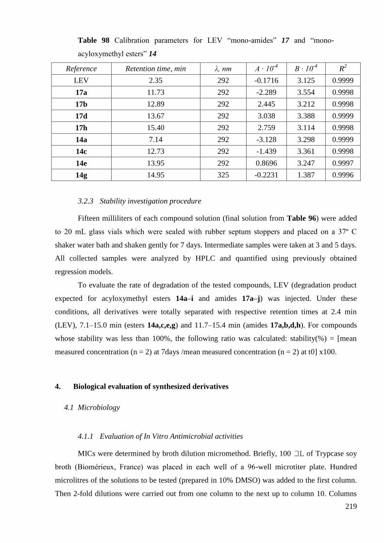
219
Table 98 Calibration parameters for LEV ―mono-amides‖ 17 and ―mono-
acyloxymethyl esters‖ 14
Reference Retention time, min λ, nm A · 10-4
B · 10-4
R2
LEV 2.35 292 -0.1716 3.125 0.9999
17a 11.73 292 -2.289 3.554 0.9998
17b 12.89 292 2.445 3.212 0.9998
17d 13.67 292 3.038 3.388 0.9999
17h 15.40 292 2.759 3.114 0.9998
14a 7.14 292 -3.128 3.298 0.9999
14c 12.73 292 -1.439 3.361 0.9998
14e 13.95 292 0.8696 3.247 0.9997
14g 14.95 325 -0.2231 1.387 0.9996
3.2.3 Stability investigation procedure
Fifteen milliliters of each compound solution (final solution from Table 96) were added
to 20 mL glass vials which were sealed with rubber septum stoppers and placed on a 37º C
shaker water bath and shaken gently for 7 days. Intermediate samples were taken at 3 and 5 days.
All collected samples were analyzed by HPLC and quantified using previously obtained
regression models.
To evaluate the rate of degradation of the tested compounds, LEV (degradation product
expected for acyloxymethyl esters 14a–i and amides 17a–j) was injected. Under these
conditions, all derivatives were totally separated with respective retention times at 2.4 min
(LEV), 7.1–15.0 min (esters 14a,c,e,g) and 11.7–15.4 min (amides 17a,b,d,h). For compounds
whose stability was less than 100%, the following ratio was calculated: stability(%) = [mean
measured concentration (n = 2) at 7days /mean measured concentration (n = 2) at t0] x100.
4. Biological evaluation of synthesized derivatives
4.1 Microbiology
4.1.1 Evaluation of In Vitro Antimicrobial activities
MICs were determined by broth dilution micromethod. Briefly, 100 of Trypcase soy
broth (Biomérieux, France) was placed in each well of a 96-well microtiter plate. Hundred
microlitres of the solutions to be tested (prepared in 10% DMSO) was added to the first column.
Then 2-fold dilutions were carried out from one column to the next up to column 10. Columns

220
11 and 12 were used as a sterility control (without product and without bacteria) and a growth
control (without product, with inoculum) of the medium. Fresh bacterial suspensions were
prepared at 108cells/mL using standard strains (S. aureus CIP 4.83, E. hirae CIP 5855, P.
aeruginosa CIP 82.118 and E. CIP 53126) obtained from the Institut Pasteur Collection (France).
The microplates were inoculated using a multipoint inoculator (Denley) to obtain a final
concentration of 106cells/mL. Plates were then incubated at 37 °C for 18–24 h under aerobic
conditions. MIC was determined as the lowest concentration with no visible growth. Assays
were performed in duplicate.
4.1.2 In vitro inhibition of Mycobacterium Tuberculosis
Susceptibility of M. tuberculosis (strain H37Rv) to the synthetic compounds was tested
by determining the MIC. A colorimetric microassay based on the reduction of MTT to formazan
by metabolically active cells was used. Briefly, serial twofold dilutions of each compound were
prepared in 7H9 broth (Middlebrook 7H9 broth base, Difco) using 96-well microtiter plates and
100 L of M. tuberculosis suspension in 7H9 broth were added to each well. After 6 days
incubation, MTT was added (50 L, 1 mg/mL). After one day incubation, solubilization buffer
was added to each well. The optical densities were measured at 570 nm. The MIC was
determined as the lowest concentration of compound that inhibited bacterial growth (absorbance
from untreated bacilli was taken as a control for growth).
4.2 Evaluation of in vitro cell proliferation by means of the MTT colorimetric assay
The assessment of cell population growth is based on the capability of living cells to
reduce the yellow product MTT (Sigma, Bornem, Belgium) to a blue product, formazan, by a
reduction reaction occurring in the mitochondria190
. The five cell lines were incubated for 24 h in
96-microwell plates (at a concentration of 104 to 3 x 10
4 cells/mL culture medium depending on
the cell type) to ensure adequate plating prior to cell growth determination. The number of living
cells after 120 h of culture in the presence or absence (control) of the various drugs is directly
proportional to the intensity of the blue color, measured by spectrophotometry using a 680XR
microplate reader (Bio-Rad Laboratories Inc, Hercules, CA) at a wavelength of 570 nm (with a
reference at 630 nm). Each experiment was carried out in hexaplicate. Nine concentrations
ranging from 10-3
to 10-7
Mw (with semi-log decrease in concentration) were tested for each of
the compounds under study.

221
4.3 In vivo testing maximum tolerated dose procedure
The acute MTD was determined following single i.p. administration of increasing drug
doses to groups of three healthy B6D2F1 female mice (Charles River, Brussels, Belgium). Five
dose levels (5, 10, 20, 40 and 80 mg/kg) of drug were evaluated. The MTD was defined as the
dose just below the lowest dose level that killed at least one mouse in a treatment group after a
maximum of 28 days.

222

223
BIBLIOGRAPHY
1. Lesher, G. Y.; Froelich, E. J.; Gruett, M. D.; Bailey, J. H.; Brundage, R. P., 1,8-
Naphthyridine Derivatives. A New Class of Chemotherapeutic Agents. J. Med. Pharm. Chem.
1962, 5 (5) 1063-1065.
2. Wagman, A. S.; Wentland, M. P., Quinolone Antibacterial Agents. In
Comprehensive Medicinal Chemistry II, John, B. T.; David, J. T., Eds. Elsevier: Oxford, 2007,
567-596.
3. Oliphant, C. M.; Green, G. M., Quinolones: a comprehensive review. Am. Fam.
Physician 2002, 65 (3) 455-464.
4. Ward-Mcquaid, J. F.; Jichlinski, D.; Macis, R., Nalidixic Acid in Urinary
Infections. Br. Med. J. 1963, 2 (5368) 1311-4.
5. Koga, H.; Itoh, A.; Murayama, S.; Suzue, S.; Irikura, T., Structure-activity
relationships of antibacterial 6,7- and 7,8-disubstituted 1-alkyl-1,4-dihydro-4-oxoquinoline-3-
carboxylic acids. J. Med. Chem. 1980, 23 (12) 1358-1363.
6. (a) Wise, R., Norfloxacin—a review of pharmacology and tissue penetration. J.
Antimicrob. Chemother. 1984, 13 (suppl B) 59-64; (b) Montay, G.; Goueffon, Y.; Roquet, F.,
Absorption, distribution, metabolic fate, and elimination of pefloxacin mesylate in mice, rats,
dogs, monkeys, and humans. Antimicrob. Agents Chemother. 1984, 25 (4) 463-472.
7. Wise, R.; Andrews, J. M.; Edwards, L. J., In vitro activity of Bay 09867, a new
quinoline derivative, compared with those of other antimicrobial agents. Antimicrob. Agents
Chemother. 1983, 23 (4) 559-564.
8. Fass, R. J., In vitro activity of ciprofloxacin (Bay o 9867). Antimicrob. Agents
Chemother. 1983, 24 (4) 568-574.
9. (a) Crump, B.; Wise, R.; Dent, J., Pharmacokinetics and tissue penetration of
ciprofloxacin. Antimicrob. Agents Chemother. 1983, 24 (5) 784-786; (b) Lode, H.; Hoffken, G.;
Olschewski, P.; Sievers, B.; Kirch, A.; Borner, K.; Koeppe, P., Pharmacokinetics of ofloxacin
after parenteral and oral administration. Antimicrob. Agents Chemother. 1987, 31 (9) 1338-1342.
10. Ball, P., Emergent resistance to ciprofloxacin amongst Pseudomonas aeruginosa
and Staphylococcus aureus:clinical significance and therapeutic approaches. J. Antimicrob.
Chemother. 1990, 26 (suppl F) 165-179.
11. Mitscher, L. A.; Sharma, P. N.; Chu, D. T. W.; Shen, L. L.; Pernet, A. G., Chiral
DNA gyrase inhibitors. 2. Asymmetric synthesis and biological activity of the enantiomers of 9-
224
fluoro-3-methyl-10-(4-methyl-1-piperazinyl)-7-oxo-2,3-dihydro-7H-pyrido[1,2,3-de]-1,4-
benzoxazine-6-carboxylic acid (ofloxacin). J. Med. Chem. 1987, 30 (12) 2283-2286.
12. Fu, K. P.; Lafredo, S. C.; Foleno, B.; Isaacson, D. M.; Barrett, J. F.; Tobia, A. J.;
Rosenthale, M. E., In Vitro and In Vivo Antibacterial Activities of Levofloxacin (l-Ofloxacin),
an Optically Active Ofloxacin. Antimicrob. Agents Chemother. 1992, 36 (8) 860-866.
13. Dalhoff, A.; Schmitz, F. J., In Vitro Antibacterial Activity and Pharmacodynamics
of New Quinolones. Eur. J. Clin. Microbiol. Infect. Dis. 2003, 22 (4) 203-221.
14. Coyle, E. A.; Kaatz, G. W.; Rybak, M. J., Activities of Newer Fluoroquinolones
against Ciprofloxacin-Resistant Streptococcus pneumoniae. Antimicrob. Agents Chemother.
2001, 45 (6) 1654-1659.
15. Boswell, F. J.; Andrews, J. M.; Jevons, G.; Wise, R., Comparison of the in vitro
activities of several new fluoroquinolones against respiratory pathogens and their abilities to
select fluoroquinolone resistance. J. Antimicrob. Chemother. 2002, 50 (4) 495-502.
16. Lubasch, A.; Keller, I.; Borner, K.; Koeppe, P.; Lode, H., Comparative
Pharmacokinetics of Ciprofloxacin, Gatifloxacin, Grepafloxacin, Levofloxacin, Trovafloxacin,
and Moxifloxacin after Single Oral Administration in Healthy Volunteers. Antimicrob. Agents
Chemother. 2000, 44 (10) 2600-2603.
17. (a) Spangler, S. K.; Jacobs, M. R.; Appelbaum, P. C., Activity of CP 99,219
compared with those of ciprofloxacin, grepafloxacin, metronidazole, cefoxitin, piperacillin, and
piperacillin-tazobactam against 489 anaerobes. Antimicrob. Agents Chemother. 1994, 38 (10)
2471-2476; (b) Goldstein, E. J. C.; Citron, D. M.; Warren, Y.; Tyrrell, K.; Merriam, C. V., In
Vitro Activity of Gemifloxacin (SB 265805) against Anaerobes. Antimicrob. Agents Chemother.
1999, 43 (9) 2231-2235.
18. Marshall, S. A.; Jones, R. N., In vitro activity of DU-6859a, a new
fluorocyclopropyl quinolone. Antimicrob. Agents Chemother. 1993, 37 (12) 2747-2753.
19. Ozaki, M.; Matsuda, M.; Tomii, Y.; Kimura, K.; Kazuno, K.; Kitano, M.; Kise,
M.; Shibata, K.; Otsuki, M.; Nishino, T., In vitro antibacterial activity of a new quinolone,
NM394. Antimicrob. Agents Chemother. 1991, 35 (12) 2490-2495.
20. Ozaki, M.; Matsuda, M.; Tomii, Y.; Kimura, K.; Segawa, J.; Kitano, M.; Kise,
M.; Shibata, K.; Otsuki, M.; Nishino, T., In vivo evaluation of NM441, a new thiazeto-quinoline
derivative. Antimicrob. Agents Chemother. 1991, 35 (12) 2496-2499.
21. (a) Sugino, A.; Peebles, C. L.; Kreuzer, K. N.; Cozzarelli, N. R., Mechanism of
action of nalidixic acid: Purification of Escherichia coli nalA gene product and its relationship to
DNA gyrase and a novel nicking-closing enzyme. Proc. Natl. Acad. Sci. U. S. A. 1977, 74 (11)
4767-4771; (b) Gellert, M.; Mizuuchi, K.; O'Dea, M. H.; Itoh, T.; Tomizawa, J.-I., Nalidixic acid

225
resistance: A second genetic character involved in DNA gyrase activity. Proc. Natl. Acad. Sci.
U. S. A. 1977, 74 (11) 4772-4776.
22. (a) Kato, J.-i.; Nishimura, Y.; Imamura, R.; Niki, H.; Hiraga, S.; Suzuki, H., New
topoisomerase essential for chromosome segregation in E. coli. Cell 1990, 63 (2) 393-404; (b)
Kato, J.; Suzuki, H.; Ikeda, H., Purification and characterization of DNA topoisomerase IV in
Escherichia coli. J. Biol. Chem. 1992, 267 (36) 25676-25684.
23. Ferrero, L.; Cameron, B.; Manse, B.; Lagneaux, D.; Crouzet, J.; Famechon, A.;
Blanche, F., Cloning and primary structure of Staphylococcus aureus DNA topoisomerase IV: a
primary target of fluoroquinolones. Mol. Microbiol. 1994, 13 (4) 641-653.
24. Dong, K. C.; Berger, J. M., Structural basis for gate-DNA recognition and
bending by type IIA topoisomerases. Nature 2007, 450 (7173) 1201-1205.
25. Wentzell, L. M.; Maxwell, A., The Complex of DNA Gyrase and Quinolone
Drugs on DNA Forms a Barrier to the T7 DNA Polymerase Replication Complex. J. Mol. Biol.
2000, 304 (5) 779-791.
26. (a) Chen, C.-R.; Malik, M.; Snyder, M.; Drlica, K., DNA Gyrase and
Topoisomerase IV on the Bacterial Chromosome: Quinolone-induced DNA Cleavage. J. Mol.
Biol. 1996, 258 (4) 627-637; (b) Pohlhaus, J. R.; Kreuzer, K. N., Norfloxacin-induced DNA
gyrase cleavage complexes block Escherichia coli replication forks, causing double-stranded
breaks in vivo. Mol. Microbiol. 2005, 56 (6) 1416-1429.
27. Laponogov, I.; Sohi, M. K.; Veselkov, D. A.; Pan, X.-S.; Sawhney, R.;
Thompson, A. W.; McAuley, K. E.; Fisher, L. M.; Sanderson, M. R., Structural insight into the
quinolone-DNA cleavage complex of type IIA topoisomerases. Nat. Struct. Mol. Biol. 2009, 16
(6) 667-669.
28. Pan, X.-S.; Fisher, L. M., Streptococcus pneumoniae DNA Gyrase and
Topoisomerase IV: Overexpression, Purification, and Differential Inhibition by
Fluoroquinolones. Antimicrob. Agents Chemother. 1999, 43 (5) 1129-1136.
29. Ledoussal, B.; Almstead, J.-I. K.; Gray, J. L.; Hu, E. X.; Roychoudhury, S.,
Discovery, Structure-Activity Relationships and Unique Properties of Non- Fluorinated
Quinolones (NFQs). Curr. Med. Chem.: Anti-Infect. Agents 2003, 2 (1) 13-25.
30. EARSS annual report http://www.rivm.nl/earss/result/Monitoring_reports/
[Online], 2001, 2005, 2008.
31. Heisig, P., Genetic evidence for a role of parC mutations in development of high-
level fluoroquinolone resistance in Escherichia coli. Antimicrob. Agents Chemother. 1996, 40 (4)
879-885.

226
32. Yoshida, H.; Bogaki, M.; Nakamura, M.; Nakamura, S., Quinolone resistance-
determining region in the DNA gyrase gyrA gene of Escherichia coli. Antimicrob. Agents
Chemother. 1990, 34 (6) 1271-1272.
33. Yoshida, H.; Bogaki, M.; Nakamura, M.; Yamanaka, L. M.; Nakamura, S.,
Quinolone resistance-determining region in the DNA gyrase gyrB gene of Escherichia coli.
Antimicrob. Agents Chemother. 1991, 35 (8) 1647-1650.
34. Takahashi, H.; Kikuchi, T.; Shoji, S.; Fujimura, S.; Lutfor, A. B.; Tokue, Y.;
Nukiwa, T.; Watanabe, A., Characterization of gyrA, gyrB, grlA and grlB mutations in
fluoroquinolone-resistant clinical isolates of Staphylococcus aureus. J. Antimicrob. Chemother.
1998, 41 (1) 49-57.
35. Takei, M.; Fukuda, H.; Kishii, R.; Hosaka, M., Target Preference of 15
Quinolones against Staphylococcus aureus, Based on Antibacterial Activities and Target
Inhibition. Antimicrob. Agents Chemother. 2001, 45 (12) 3544-3547.
36. Pan, X.; Ambler, J.; Mehtar, S.; Fisher, L., Involvement of topoisomerase IV and
DNA gyrase as ciprofloxacin targets in Streptococcus pneumoniae. Antimicrob. Agents
Chemother. 1996, 40 (10) 2321-2326.
37. Gootz, T.; Zaniewski, R.; Haskell, S.; Schmieder, B.; Tankovic, J.; Girard, D.;
Courvalin, P.; Polzer, R., Activity of the new fluoroquinolone trovafloxacin (CP-99,219) against
DNA gyrase and topoisomerase IV mutants of Streptococcus pneumoniae selected in vitro.
Antimicrob. Agents Chemother. 1996, 40 (12) 2691-2697.
38. Fukuda, H.; Hiramatsu, K., Primary Targets of Fluoroquinolones in Streptococcus
pneumoniae. Antimicrob. Agents Chemother. 1999, 43 (2) 410-412.
39. Morris, J. E.; Pan, X.-S.; Fisher, L. M., Grepafloxacin, a Dimethyl Derivative of
Ciprofloxacin, Acts Preferentially through Gyrase in Streptococcus pneumoniae: Role of the C-5
Group in Target Specificity. Antimicrob. Agents Chemother. 2002, 46 (2) 582-585.
40. Pan, X.-S.; Fisher, L. M., DNA Gyrase and Topoisomerase IV Are Dual Targets
of Clinafloxacin Action in Streptococcus pneumoniae. Antimicrob. Agents Chemother. 1998, 42
(11) 2810-2816.
41. Lynch, A. S., Efflux systems in bacterial pathogens: An opportunity for
therapeutic intervention? An industry view. Biochem. Pharmacol. 2006, 71 (7) 949-956.
42. Ubukata, K.; Itoh-Yamashita, N.; Konno, M., Cloning and expression of the norA
gene for fluoroquinolone resistance in Staphylococcus aureus. Antimicrob. Agents Chemother.
1989, 33 (9) 1535-1539.
43. (a) Schmitz, F. J.; Fluit, A. C.; Lückefahr, M.; Engler, B.; Hofmann, B.; Verhoef,
J.; Heinz, H. P.; Hadding, U.; Jones, M. E., The effect of reserpine, an inhibitor of multidrug

227
efflux pumps, on the in-vitro activities of ciprofloxacin, sparfloxacin and moxifloxacin against
clinical isolates of Staphylococcus aureus. J. Antimicrob. Chemother. 1998, 42 (6) 807-810; (b)
Beyer, R.; Pestova, E.; Millichap, J. J.; Stosor, V.; Noskin, G. A.; Peterson, L. R., A Convenient
Assay for Estimating the Possible Involvement of Efflux of Fluoroquinolones by Streptococcus
pneumoniae and Staphylococcus aureus: Evidence for Diminished Moxifloxacin, Sparfloxacin,
and Trovafloxacin Efflux. Antimicrob. Agents Chemother. 2000, 44 (3) 798-801.
44. Tran, J. H.; Jacoby, G. A., Mechanism of plasmid-mediated quinolone resistance.
Proc. Natl. Acad. Sci. U. S. A. 2002, 99 (8) 5638-5642.
45. (a) Nikaido, H.; Thanassi, D. G., Penetration of lipophilic agents with multiple
protonation sites into bacterial cells: tetracyclines and fluoroquinolones as examples. Antimicrob.
Agents Chemother. 1993, 37 (7) 1393-1399; (b) Fresta, M.; Guccione, S.; Beccari, A. R.;
Furneri, P. M.; Puglisi, G., Combining molecular modeling with experimental methodologies:
mechanism of membrane permeation and accumulation of ofloxacin. Bioorg. Med. Chem. 2002,
10 (12) 3871-3889.
46. (a) Piddock, L. J. V.; Jin, Y. F.; Griggs, D. J., Effect of hydrophobicity and
molecular mass on the accumulation of fluoroquinolones by Staphylococcus aureus. J.
Antimicrob. Chemother. 2001, 47 (3) 261-270; (b) Asuquo, A. E.; Piddock, L. J. V.,
Accumulation and killing kinetics of fifteen quinolones for Escherichia coil, Staphylococcus
aureus and Pseudomonas aeruginosa. J. Antimicrob. Chemother. 1993, 31 (6) 865-880; (c) Furet,
Y. X.; Deshusses, J.; Pechere, J. C., Transport of pefloxacin across the bacterial cytoplasmic
membrane in quinolone-susceptible Staphylococcus aureus. Antimicrob. Agents Chemother.
1992, 36 (11) 2506-2511; (d) McCaffrey, C.; Bertasso, A.; Pace, J.; Georgopapadakou, N. H.,
Quinolone accumulation in Escherichia coli, Pseudomonas aeruginosa, and Staphylococcus
aureus. Antimicrob. Agents Chemother. 1992, 36 (8) 1601-1605.
47. Nikaido, H., Molecular Basis of Bacterial Outer Membrane Permeability
Revisited. Microbiol. Mol. Biol. Rev. 2003, 67 (4) 593-656.
48. (a) Mitscher, L. A.; Ma, Z., Structure-activity relationships of quinolones. In
Fluoroquinolone Antibiot., Ronald, A. R.; Low, D., Eds. Birkhaeuser Verlag: 2003, 11-48; (b)
Mitscher, L. A., Bacterial Topoisomerase Inhibitors: Quinolone and Pyridone Antibacterial
Agents. Chem. Rev. 2005, 105 (2) 559-592; (c) Peterson, L. R., Quinolone molecular structure-
activity relationships: what we have learned about improving antimicrobial activity. Clin. Infect.
Dis. 2001, 33 Suppl 3 180-186.
49. Chu, D. T. W.; Fernandes, P. B.; Claiborne, A. K.; Pihuleac, E.; Nordeen, C. W.;
Maleczka, R. E.; Pernet, A. G., Synthesis and structure-activity relationships of novel
arylfluoroquinolone antibacterial agents. J. Med. Chem. 1985, 28 (11) 1558-1564.

228
50. (a) Hayakawa, I.; Atarashi, S.; Yokohama, S.; Imamura, M.; Sakano, K.;
Furukawa, M., Synthesis and antibacterial activities of optically active ofloxacin. Antimicrob.
Agents Chemother. 1986, 29 (1) 163-164; (b) Hoshino, K.; Sato, K.; Akahane, K.; Yoshida, A.;
Hayakawa, I.; Sato, M.; Une, T.; Osada, Y., Significance of the methyl group on the oxazine ring
of ofloxacin derivatives in the inhibition of bacterial and mammalian type II topoisomerases.
Antimicrob. Agents Chemother. 1991, 35 (2) 309-312.
51. Yoshida, T.; Yamamoto, Y.; Orita, H.; Kakiuchi, M.; Takahashi, Y.; Itakura, M.;
Kado, N.; Mitani, K.; Yasuda, S., Studies on quinolone antibacterials. IV. Structure-activity
relationships of antibacterial activity and side effects for 5- or 8-substituted and 5,8-
disubstituted-7-(3-amino-1-pyrrolidinyl)-1-cyclopropyl-1,4-dihydro-4-oxoquinoline-3-
carboxylic acids. Chem. Pharm. Bull. 1996, 44 (5) 1074-1085.
52. Andersson, M. I.; MacGowan, A. P., Development of the quinolones. J.
Antimicrob. Chemother. 2003, 51 (suppl 1) 1-11.
53. Gray, J. L.; Almstead, J.-I. K.; Gallagher, C. P.; Hu, X. E.; Kim, N. K.; Taylor, C.
J.; Twinem, T. L.; Wallace, C. D.; Ledoussal, B., Synthesis and biological testing of non-
Fluorinated analogues of levofloxacin. Bioorg. Med. Chem. Lett. 2003, 13 (14) 2373-2375.
54. Lawrence, L. E.; Wu, P.; Fan, L.; Gouveia, K. E.; Card, A.; Casperson, M.;
Denbleyker, K.; Barrett, J. F., The inhibition and selectivity of bacterial topoisomerases by
BMS-284756 and its analogues. J. Antimicrob. Chemother. 2001, 48 (2) 195-201.
55. Sanchez, J. P.; Domagala, J. M.; Hagen, S. E.; Heifetz, C. L.; Hutt, M. P.;
Nichols, J. B.; Trehan, A. K., Quinolone antibacterial agents. Synthesis and structure-activity
relationships of 8-substituted quinoline-3-carboxylic acids and 1,8-naphthyridine-3-carboxylic
acids. J. Med. Chem. 1988, 31 (5) 983-991.
56. (a) Bertino, J. J.; Fish, D., The safety profile of the fluoroquinolones. Clin. Ther.
2000, 22 (7) 798-817; (b) Domagala, J. M., Structure-activity and structure-side-effect
relationships for the quinolone antibacterials. J. Antimicrob. Chemother. 1994, 33 (4) 685-706.
57. Fukuda, H.; Kishii, R.; Takei, M.; Hosaka, M., Contributions of the 8-Methoxy
Group of Gatifloxacin to Resistance Selectivity, Target Preference, and Antibacterial Activity
against Streptococcus pneumoniae. Antimicrob. Agents Chemother. 2001, 45 (6) 1649-1653.
58. Dalhoff, A., Comparative In Vitro and In Vivo Activity of the C-8 Methoxy
Quinolone Moxifloxacin and the C-8 Chlorine Quinolone BAY y 3118. Clin. Infect. Dis. 2001,
32 (S1) 16-22.
59. Ball, P.; Stahlmann, R.; Kubin, R.; Choudhri, S.; Owens, R., Safety profile of oral
and intravenous moxifloxacin: Cumulative data from clinical trials and postmarketing studies.
Clin. Ther. 2004, 26 (7) 940-950.

229
60. Cecchetti, V.; Fravolini, A.; Palumbo, M.; Sissi, C.; Tabarrini, O.; Terni, P.; Xin,
T., Potent 6-Desfluoro-8-methylquinolones as New Lead Compounds in Antibacterial
Chemotherapy1. J. Med. Chem. 1996, 39 (25) 4952-4957.
61. Vázquez, J. L.; Merino, S.; Domenech, Ò.; Berlanga, M.; Viðas, M.; Montero, M.
T.; Hernández-Borrell, J., Determination of the partition coefficients of a homologous series of
ciprofloxacin: influence of the N-4 piperazinyl alkylation on the antimicrobial activity. Int. J.
Pharm. 2001, 220 (1-2) 53-62.
62. (a) Bermejo, M.; Merino, V.; Garrigues, T. M.; Delfina, J. M. P.; Mulet, A.; Vizet,
P.; Trouiller, G.; Mercier, C., Validation of a Biophysical Drug Absorption Model by the
PATQSAR System. J. Pharm. Sci. 1999, 88 (4) 398-405; (b) Escribano, E.; Calpena, A.;
Garrigues, T.; Freixas, J.; Domenech, J.; Moreno, J., Structure-absorption relationships of a
series of 6-fluoroquinolones. Antimicrob. Agents Chemother. 1997, 41 (9) 1996-2000.
63. Abuo-Rahma, G. E.-D. A. A.; Sarhan, H. A.; Gad, G. F. M., Design, synthesis,
antibacterial activity and physicochemical parameters of novel N-4-piperazinyl derivatives of
norfloxacin. Bioorg. Med. Chem. 2009, 17 (11) 3879-3886.
64. (a) Alovero, F.; Nieto, M.; Mazzieri, M. R.; Then, R.; Manzo, R. H., Mode of
Action of Sulfanilyl Fluoroquinolones. Antimicrob. Agents Chemother. 1998, 42 (6) 1495-1498;
(b) Nieto, M. J.; Alovero, F. d. L.; Manzo, R. H.; Mazzieri, M. R., A new class of
fluoroquinolones: benzenesulfonamidefluoroquinolones (BSFQs), antibacterial activity and SAR
studies. Eur. J. Med. Chem. 1999, 34 (3) 209-214.
65. Alovero, F. L.; Pan, X.-S.; Morris, J. E.; Manzo, R. H.; Fisher, L. M., Engineering
the Specificity of Antibacterial Fluoroquinolones: Benzenesulfonamide Modifications at C-7 of
Ciprofloxacin Change Its Primary Target in Streptococcus pneumoniae from Topoisomerase IV
to Gyrase. Antimicrob. Agents Chemother. 2000, 44 (2) 320-325.
66. Foroumadi, A.; Mohammadhosseini, N.; Emami, S.; Letafat, B.; Faramarzi, M.
A.; Samadi, N.; Shafiee, A., Synthesis and antibacterial activity of new 7-piperazinyl-quinolones
containing a functionalized 2-(furan-3-yl)ethyl moiety. Arch. Pharm. (Weinheim, Ger.) 2007,
340 (1) 47-52.
67. Letafat, B.; Emami, S.; Mohammadhosseini, N.; Faramarzi, M. A.; Samadi, N.;
Shafiee, A.; Foroumadi, A., Synthesis and antibacterial activity of new N-[2-(thiophen-3-
yl)ethyl] piperazinyl quinolones. Chem. Pharm. Bull. 2007, 55 (6) 894-898.
68. Foroumadi, A.; Ghodsi, S.; Emami, S.; Najjari, S.; Samadi, N.; Faramarzi, M. A.;
Beikmohammadi, L.; Shirazi, F. H.; Shafiee, A., Synthesis and antibacterial activity of new
fluoroquinolones containing a substituted N-(phenethyl)piperazine moiety. Bioorg. Med. Chem.
Lett. 2006, 16 (13) 3499-3503.

230
69. Emami, S.; Foroumadi, A.; Samadi, N.; Faramarzi, M. A.; Rajabalian, S.,
Conformationally Constrained Analogs of N-Substituted Piperazinylquinolones: Synthesis and
Antibacterial Activity of N-(2,3-Dihydro-4-hydroxyimino-4H-1-benzopyran-3-yl)-
piperazinylquinolones. Arch. Pharm. (Weinheim, Ger.) 2009, 342 (7) 405-411.
70. Emami, S.; Foroumadi, A.; Faramarzi, M. A.; Samadi, N., Synthesis and
antibacterial activity of quinolone-based compounds containing a coumarin moiety. Arch.
Pharm. (Weinheim, Ger.) 2008, 341 (1) 42-48.
71. Shen, L. L.; Mitscher, L. A.; Sharma, P. N.; O'Donnell, T. J.; Chu, D. W. T.;
Cooper, C. S.; Rosen, T.; Pernet, A. G., Mechanism of inhibition of DNA gyrase by quinolone
antibacterials: a cooperative drug-DNA binding model. Biochemistry 1989, 28 (9) 3886-3894.
72. Kerns, R. J.; Rybak, M. J.; Cheung, C. M., Susceptibility studies of piperazinyl-
cross-linked fluoroquinolone dimers against test strains of Gram-positive and Gram-negative
bacteria. Diagn. Microbiol. Infect. Dis. 2006, 54 (4) 305-310.
73. (a) Kerns, R. J.; Rybak, M. J.; Kaatz, G. W.; Vaka, F.; Cha, R.; Grucz, R. G.;
Diwadkar, V. U.; Ward, T. D., Piperazinyl-linked fluoroquinolone dimers possessing potent
antibacterial activity against drug-resistant strains of Staphylococcus aureus. Bioorg. Med.
Chem. Lett. 2003, 13 (10) 1745-1749; (b) Kerns, R. J.; Rybak, M. J.; Kaatz, G. W.; Vaka, F.;
Cha, R.; Grucz, R. G.; Diwadkar, V. U., Structural features of piperazinyl-linked ciprofloxacin
dimers required for activity against drug-resistant strains of Staphylococcus aureus. Bioorg. Med.
Chem. Lett. 2003, 13 (13) 2109-2112.
74. Zhao, X.; Quinn, B.; Kerns, R.; Drlica, K., Bactericidal activity and target
preference of a piperazinyl-cross-linked ciprofloxacin dimer with Staphylococcus aureus and
Escherichia coli. J. Antimicrob. Chemother. 2006, 58 (6) 1283-1286.
75. Gould, K. A.; Pan, X. S.; Kerns, R. J.; Fisher, L. M., Ciprofloxacin Dimers Target
Gyrase in Streptococcus pneumoniae. Antimicrob. Agents Chemother. 2004, 48 (6) 2108-2115.
76. Chu, D. T.; Fernandes, P. B., Structure-activity relationships of the
fluoroquinolones. Antimicrob. Agents Chemother. 1989, 33 (2) 131-135.
77. Chu, D. T. Tricyclic quinoline and naphthyride antibacterials. U.S. Patent
4767762, 1988.
78. Kohlbrenner, W. E.; Wideburg, N.; Weigl, D.; Saldivar, A.; Chu, D. T., Induction
of calf thymus topoisomerase II-mediated DNA breakage by the antibacterial
isothiazoloquinolones A-65281 and A-65282. Antimicrob. Agents Chemother. 1992, 36 (1) 81-
86.
79. Wiles, J. A.; Wang, Q.; Lucien, E.; Hashimoto, A.; Song, Y.; Cheng, J.; Marlor,
C. W.; Ou, Y.; Podos, S. D.; Thanassi, J. A.; Thoma, C. L.; Deshpande, M.; Pucci, M. J.;

231
Bradbury, B. J., Isothiazoloquinolones containing functionalized aromatic hydrocarbons at the 7-
position: Synthesis and in vitro activity of a series of potent antibacterial agents with diminished
cytotoxicity in human cells. Bioorg. Med. Chem. Lett. 2006, 16 (5) 1272-1276.
80. Wiles, J. A.; Song, Y.; Wang, Q.; Lucien, E.; Hashimoto, A.; Cheng, J.; Marlor,
C. W.; Ou, Y.; Podos, S. D.; Thanassi, J. A.; Thoma, C. L.; Deshpande, M.; Pucci, M. J.;
Bradbury, B. J., Biological evaluation of isothiazoloquinolones containing aromatic heterocycles
at the 7-position: In vitro activity of a series of potent antibacterial agents that are effective
against methicillin-resistant Staphylococcus aureus. Bioorg. Med. Chem. Lett. 2006, 16 (5) 1277-
1281.
81. Wang, Q.; Lucien, E.; Hashimoto, A.; Pais, G. C. G.; Nelson, D. M.; Song, Y.;
Thanassi, J. A.; Marlor, C. W.; Thoma, C. L.; Cheng, J.; Podos, S. D.; Ou, Y.; Deshpande, M.;
Pucci, M. J.; Buechter, D. D.; Bradbury, B. J.; Wiles, J. A., Isothiazoloquinolones with Enhanced
Antistaphylococcal Activities against Multidrug-Resistant Strains: Effects of Structural
Modifications at the 6-, 7-, and 8-Positions. J. Med. Chem. 2007, 50 (2) 199-210.
82. Pucci, M. J.; Cheng, J.; Podos, S. D.; Thoma, C. L.; Thanassi, J. A.; Buechter, D.
D.; Mushtaq, G.; Vigliotti, G. A., Jr.; Bradbury, B. J.; Deshpande, M., In Vitro and In Vivo
Antibacterial Activities of Heteroaryl Isothiazolones against Resistant Gram-Positive Pathogens.
Antimicrob. Agents Chemother. 2007, 51 (4) 1259-1267.
83. Cheng, J.; Thanassi, J. A.; Thoma, C. L.; Bradbury, B. J.; Deshpande, M.; Pucci,
M. J., Dual Targeting of DNA Gyrase and Topoisomerase IV: Target Interactions of Heteroaryl
Isothiazolones in Staphylococcus aureus. Antimicrob. Agents Chemother. 2007, 51 (7) 2445-
2453.
84. Patel, N. B.; Patel, A. L.; Chauhan, H. I., Synthesis of amide derivatives of
quinolone and their antimicrobial studies. Indian J. Chem., Sect. B: Org. Chem. Incl. Med.
Chem. 2007, 46B (1) 126-134.
85. Sultana, N.; Arayne, M. S.; Rizvi, S. B. S.; Mesaik, M. A., Synthesis,
characterization and biological evaluation of a series of levofloxacin carboxamide analogues.
Bull. Korean Chem. Soc. 2009, 30 (10) 2294-2298.
86. (a) Rivers, E. C.; Mancera, R. L., New anti-tuberculosis drugs in clinical trials
with novel mechanisms of action. Drug Discovery Today 2008, 13 (23-24) 1090-1098; (b)
WHO, Global tuberculosis control: epidemiology, strategy, financing 2009.
http://www.who.int/tb/publications/global_report/2009/en/index.html
87. Johnson, R.; Streicher, E. M.; Louw, G. E.; Warren, R. M.; van Helden, P. D.;
Victor, T. C., Drug resistance in Mycobacterium tuberculosis. Curr. Issues Mol. Biol. 2006, 8 (2)
97-111.

232
88. Lowther, J.; Bryskier, A., Fluoroquinolones and tuberculosis. Expert Opin. Invest.
Drugs 2002, 11 (2) 233-258.
89. Loddenkemper, R.; Hauer, B., Drug-resistant tuberculosis: a worldwide epidemic
poses a new challenge. Dtsch. Arztebl. Int. 2010, 107 (1-2) 10-9.
90. WHO, Treatment of Tuberculosis: guidelines for national programmes 2010.
http://whqlibdoc.who.int/publications/2010/9789241547833_eng.pdf.
91. Cole, S. T.; Brosch, R.; Parkhill, J.; Garnier, T.; Churcher, C.; Harris, D.; Gordon,
S. V.; Eiglmeier, K.; Gas, S.; Barry, C. E.; Tekaia, F.; Badcock, K.; Basham, D.; Brown, D.;
Chillingworth, T.; Connor, R.; Davies, R.; Devlin, K.; Feltwell, T.; Gentles, S.; Hamlin, N.;
Holroyd, S.; Hornsby, T.; Jagels, K.; Krogh, A.; McLean, J.; Moule, S.; Murphy, L.; Oliver, K.;
Osborne, J.; Quail, M. A.; Rajandream, M. A.; Rogers, J.; Rutter, S.; Seeger, K.; Skelton, J.;
Squares, R.; Squares, S.; Sulston, J. E.; Taylor, K.; Whitehead, S.; Barrell, B. G., Deciphering
the biology of Mycobacterium tuberculosis from the complete genome sequence. Nature 1998,
393 (6685) 537-544.
92. Aubry, A.; Mark Fisher, L.; Jarlier, V.; Cambau, E., First functional
characterization of a singly expressed bacterial type II topoisomerase: The enzyme from
Mycobacterium tuberculosis. Biochem. Biophys. Res. Commun. 2006, 348 (1) 158-165.
93. Chatterjee, D., The mycobacterial cell wall: structure, biosynthesis and sites of
drug action. Curr. Opin. Chem. Biol. 1997, 1 (4) 579-588.
94. Jarlier, V.; Nikaido, H., Mycobacterial cell wall: Structure and role in natural
resistance to antibiotics. FEMS Microbiol. Lett. 1994, 123 (1-2) 11-18.
95. Daffé, M.; Etienne, G., The capsule of Mycobacterium tuberculosis and its
implications for pathogenicity. Tubercle and Lung Disease 1999, 79 (3) 153-169.
96. Almeida, D.; Nuermberger, E.; Tyagi, S.; Bishai, W. R.; Grosset, J., In Vivo
Validation of the Mutant Selection Window Hypothesis with Moxifloxacin in a Murine Model of
Tuberculosis. Antimicrob. Agents Chemother. 2007, 51 (12) 4261-4266.
97. (a) Kocagoz, T.; Hackbarth, C.; Unsal, I.; Rosenberg, E.; Nikaido, H.; Chambers,
H., Gyrase mutations in laboratory-selected, fluoroquinolone-resistant mutants of
Mycobacterium tuberculosis H37Ra. Antimicrob. Agents Chemother. 1996, 40 (8) 1768-1774;
(b) Takiff, H. E.; Salazar, L.; Guerrero, C.; Philipp, W.; Huang, W. M.; Kreiswirth, B.; Cole, S.
T.; Jacobs, W. R., Jr; Telenti, A., Cloning and nucleotide sequence of Mycobacterium
tuberculosis gyrA and gyrB genes and detection of quinolone resistance mutations. Antimicrob.
Agents Chemother. 1994, 38 (4) 773-780; (c) Cheng, A. F. B.; Yew, W. W.; Chan, E. W. C.;
Chin, M. L.; Hui, M. M. M.; Chan, R. C. Y., Multiplex PCR Amplimer Conformation Analysis

233
for Rapid Detection of gyrA Mutations in Fluoroquinolone-Resistant Mycobacterium
tuberculosis Clinical Isolates. Antimicrob. Agents Chemother. 2004, 48 (2) 596-601.
98. Poole, K., Efflux-Mediated Resistance to Fluoroquinolones in Gram-Positive
Bacteria and the Mycobacteria. Antimicrob. Agents Chemother. 2000, 44 (10) 2595-2599.
99. Banerjee, S. K.; Bhatt, K.; Rana, S.; Misra, P.; Chakraborti, P. K., Involvement of
an Efflux System in Mediating High Level of Fluoroquinolone Resistance inMycobacterium
smegmatis. Biochem. Biophys. Res. Commun. 1996, 226 (2) 362-368.
100. (a) Takiff, H. E.; Cimino, M.; Musso, M. C.; Weisbrod, T.; Martinez, R.;
Delgado, M. B.; Salazar, L.; Bloom, B. R.; Jacobs, W. R., Efflux pump of the proton antiporter
family confers low-level fluoroquinolone resistance in Mycobacterium smegmatis. Proc. Natl.
Acad. Sci. U. S. A. 1996, 93 (1) 362-366; (b) Liu, J.; Takiff, H.; Nikaido, H., Active efflux of
fluoroquinolones in Mycobacterium smegmatis mediated by LfrA, a multidrug efflux pump. J.
Bacteriol. 1996, 178 (13) 3791-3795.
101. Montero, C.; Mateu, G.; Rodriguez, R.; Takiff, H., Intrinsic Resistance of
Mycobacteriumsmegmatis to Fluoroquinolones May Be Influenced by New Pentapeptide Protein
MfpA. Antimicrob. Agents Chemother. 2001, 45 (12) 3387-3392.
102. Hegde, S. S.; Vetting, M. W.; Roderick, S. L.; Mitchenall, L. A.; Maxwell, A.;
Takiff, H. E.; Blanchard, J. S., A Fluoroquinolone Resistance Protein from Mycobacterium
tuberculosis That Mimics DNA. Science 2005, 308 (5727) 1480-1483.
103. (a) Renau, T. E.; Sanchez, J. P.; Shapiro, M. A.; Dever, J. A.; Gracheck, S. J.;
Domagala, J. M., Effect of Lipophilicity at N-1 on Activity of Fluoroquinolones against
Mycobacteria. J. Med. Chem. 1995, 38 (15) 2974-2977; (b) Renau, T. E.; Sanchez, J. P.; Gage, J.
W.; Dever, J. A.; Shapiro, M. A.; Gracheck, S. J.; Domagala, J. M., Structure−Activity
Relationships of the Quinolone Antibacterials against Mycobacteria: Effect of Structural
Changes at N-1 and C-7. J. Med. Chem. 1996, 39 (3) 729-735.
104. Lu, T.; Zhao, X.; Li, X.; Drlica-Wagner, A.; Wang, J. Y.; Domagala, J.; Drlica,
K., Enhancement of Fluoroquinolone Activity by C-8 Halogen and Methoxy Moieties: Action
against a Gyrase Resistance Mutant of Mycobacterium smegmatis and a Gyrase-Topoisomerase
IV Double Mutant of Staphylococcus aureus. Antimicrob. Agents Chemother. 2001, 45 (10)
2703-2709.
105. Dong, Y.; Xu, C.; Zhao, X.; Domagala, J.; Drlica, K., Fluoroquinolone Action
against Mycobacteria: Effects of C-8 Substituents on Growth, Survival, and Resistance.
Antimicrob. Agents Chemother. 1998, 42 (11) 2978-2984.

234
106. Haemers, A.; Leysen, D. C.; Bollaert, W.; Zhang, M. Q.; Pattyn, S. R., Influence
of N substitution on antimycobacterial activity of ciprofloxacin. Antimicrob. Agents Chemother.
1990, 34 (3) 496-497.
107. Almeida, M. V.; Saraiva, M. F.; de Souza, M. V. N.; da Costa, C. F.; Vicente, F.
R. C.; Lourenço, M. C. S., Synthesis and antitubercular activity of lipophilic moxifloxacin and
gatifloxacin derivatives. Bioorg. Med. Chem. Lett. 2007, 17 (20) 5661-5664.
108. (a) Sriram, D.; Yogeeswari, P.; Basha, J. S.; Radha, D. R.; Nagaraja, V., Synthesis
and antimycobacterial evaluation of various 7-substituted ciprofloxacin derivatives. Bioorg.
Med. Chem. 2005, 13 (20) 5774-5778; (b) Sriram, D.; Bal, T. R.; Yogeeswari, P.; Radha, D. R.;
Nagaraja, V., Evaluation of antimycobacterial and DNA gyrase inhibition of fluoroquinolone
derivatives. J. Gen. Appl. Microbiol. 2006, 52 (4) 195-200.
109. Senthilkumar, P.; Dinakaran, M.; Yogeeswari, P.; China, A.; Nagaraja, V.;
Sriram, D., Antimycobacterial activities of novel fluoroquinolones. Biomed. Pharmacother.
2009, 63 (1) 27-35.
110. Dinakaran, M.; Senthilkumar, P.; Yogeeswari, P.; China, A.; Nagaraja, V.;
Sriram, D., Novel ofloxacin derivatives: Synthesis, antimycobacterial and toxicological
evaluation. Bioorg. Med. Chem. Lett. 2008, 18 (3) 1229-1236.
111. (a) Molina-Torres, C. A.; Ocampo-Candiani, J.; Rendon, A.; Pucci, M. J.; Vera-
Cabrera, L., In Vitro Activity of a New Isothiazoloquinolone, ACH-702, against Mycobacterium
tuberculosis and Other Mycobacteria. Antimicrob. Agents Chemother. 2010, 54 (5) 2188-2190;
(b) Pucci, M. J.; Ackerman, M.; Thanassi, J. A.; Shoen, C. M.; Cynamon, M. H., In Vitro
Antituberculosis Activities of ACH-702, a Novel Isothiazoloquinolone, against Quinolone-
Susceptible and Quinolone-Resistant Isolates. Antimicrob. Agents Chemother. 2010, 54 (8)
3478-3480.
112. Drlica, K.; Zhao, X., DNA gyrase, topoisomerase IV, and the 4-quinolones.
Microbiol. Mol. Biol. Rev. 1997, 61 (3) 377-392.
113. Azarova, A. M.; Lyu, Y. L.; Lin, C.-P.; Tsai, Y.-C.; Lau, J. Y.-N.; Wang, J. C.;
Liu, L. F., Roles of DNA topoisomerase II isozymes in chemotherapy and secondary
malignancies. Proc. Natl. Acad. Sci. U. S. A. 2007, 104 (26) 11014-11019.
114. (a) Hussy, P.; Maass, G.; Tummler, B.; Grosse, F.; Schomburg, U., Effect of 4-
quinolones and novobiocin on calf thymus DNA polymerase alpha primase complex,
topoisomerases I and II, and growth of mammalian lymphoblasts. Antimicrob. Agents
Chemother. 1986, 29 (6) 1073-1078; (b) Hoshino, K.; Sato, K.; Une, T.; Osada, Y., Inhibitory
effects of quinolones on DNA gyrase of Escherichia coli and topoisomerase II of fetal calf
thymus. Antimicrob. Agents Chemother. 1989, 33 (10) 1816-1818.

235
115. Barrett, J. F.; Gootz, T. D.; McGuirk, P. R.; Farrell, C. A.; Sokolowski, S. A., Use
of in vitro topoisomerase II assays for studying quinolone antibacterial agents. Antimicrob.
Agents Chemother. 1989, 33 (10) 1697-1703.
116. (a) Robinson, M. J.; Martin, B. A.; Gootz, T. D.; McGuirk, P. R.; Moynihan, M.;
Sutcliffe, J. A.; Osheroff, N., Effects of quinolone derivatives on eukaryotic topoisomerase II. A
novel mechanism for enhancement of enzyme-mediated DNA cleavage. J. Biol. Chem. 1991,
266 (22) 14585-14592; (b) Duan, W.; Rangan, A.; Vankayalapati, H.; Kim, M. Y.; Zeng, Q.;
Sun, D.; Han, H.; Fedoroff, O. Y.; Nishioka, D.; Rha, S. Y.; Izbicka, E.; Von, H. D. D.; Hurley,
L. H., Design and synthesis of fluoroquinophenoxazines that interact with human telomeric G-
quadruplexes and their biological effects. Mol. Cancer Ther. 2001, 1 (2) 103-20.
117. Advani, R. H.; Hurwitz, H. I.; Gordon, M. S.; Ebbinghaus, S. W.; Mendelson, D.
S.; Wakelee, H. A.; Hoch, U.; Silverman, J. A.; Havrilla, N. A.; Berman, C. J.; Fox, J. A.; Allen,
R. S.; Adelman, D. C., Voreloxin, a First-in-Class Anticancer Quinolone Derivative, in
Relapsed/Refractory Solid Tumors: A Report on Two Dosing Schedules. Clin. Cancer Res.
2010, 16 (7) 2167-2175.
118. Quarfloxin in Patients With Low to Intermediate Grade Neuroendocrine
Carcinoma, Phase II 2008. http://clinicaltrials.gov/ct2/show/NCT00780663A.
119. Roca, J., The mechanisms of DNA topoisomerases. Trends Biochem. Sci. 1995,
20 (4) 156-160.
120. Caron, P. R.; Wang, J. C., Appendix II: Alignment of Primary Sequences of DNA
Topoisomerases. In Adv. Pharmacol. (San Diego, CA, U. S.), Leroy, F. L., Ed. Academic Press:
1994, Vol. 29, Part 2, 271-297.
121. Wasserman, R. A.; Austin, C. A.; Fisher, L. M.; Wang, J. C., Use of Yeast in the
Study of Anticancer Drugs Targeting DNA Topoisomerases: Expression of a Functional
Recombinant Human DNA Topoisomerase IIα in Yeast. Cancer Res. 1993, 53 (15) 3591-3596.
122. Nitiss, J. L., DNA topoisomerase II and its growing repertoire of biological
functions. Nat. Rev. Cancer 2009, 9 (5) 327-337.
123. (a) Deweese, J. E.; Osheroff, N., The DNA cleavage reaction of topoisomerase II:
wolf in sheep's clothing. Nucleic Acids Res. 2009, 37 (3) 738-748; (b) Nitiss, J. L., Targeting
DNA topoisomerase II in cancer chemotherapy. Nat. Rev. Cancer 2009, 9 (5) 338-350.
124. Larsen, A. K.; Escargueil, A. E.; Skladanowski, A., Catalytic topoisomerase II
inhibitors in cancer therapy. Pharmacol. Ther. 2003, 99 (2) 167-181.
125. Heisig, P., Type II topoisomerases—inhibitors, repair mechanisms and mutations.
Mutagenesis 2009, 24 (6) 465-469.

236
126. (a) Bromberg, K. D.; Burgin, A. B.; Osheroff, N., Quinolone Action against
Human Topoisomerase IIα: Stimulation of Enzyme-Mediated Double-Stranded DNA Cleavage.
Biochemistry 2003, 42 (12) 3393-3398; (b) Elsea, S. H.; Osheroff, N.; Nitiss, J. L., Cytotoxicity
of quinolones toward eukaryotic cells. Identification of topoisomerase II as the primary cellular
target for the quinolone CP-115,953 in yeast. J. Biol. Chem. 1992, 267 (19) 13150-13153.
127. Coughlin, S. A.; Danz, D. W.; Robinson, R. G.; Klingbeil, K. M.; Wentland, M.
P.; Corbett, T. H.; Waud, W. R.; Zwelling, L. A.; Altschuler, E.; Bales, E.; Rake, J. B.,
Mechanism of action and antitumor activity of (S)-10-(2,6-dimethyl-4-pyridinyl)-9-fluoro-3-
methyl-7-oxo-2,3-dihydro-7H-pyridol [1,2,3-de]-[1,4]benzothiazine-6-carboxylic acid (WIN
58161). Biochem. Pharmacol. 1995, 50 (1) 111-122.
128. Hawtin, R. E.; Stockett, D. E.; Byl, J. A. W.; McDowell, R. S.; Tan, N.; Arkin, M.
R.; Conroy, A.; Yang, W.; Osheroff, N.; Fox, J. A., Voreloxin Is an Anticancer Quinolone
Derivative that Intercalates DNA and Poisons Topoisomerase II. PLoS ONE 2010, 5 (4) e10186.
129. Permana, P. A.; Snapka, R. M.; Shen, L. L.; Chu, D. T. W.; Clement, J. J.;
Plattner, J. J., Quinobenoxazines: A Class of Novel Antitumor Quinolones and Potent
Mammalian DNA Topoisomerase II Catalytic Inhibitors. Biochemistry 1994, 33 (37) 11333-
11339.
130. Pommier, Y., DNA Topoisomerase I Inhibitors: Chemistry, Biology, and
Interfacial Inhibition. Chem. Rev. 2009, 109 (7) 2894-2902.
131. D'Arpa, P.; Beardmore, C.; Liu, L. F., Involvement of Nucleic Acid Synthesis in
Cell Killing Mechanisms of Topoisomerase Poisons. Cancer Res. 1990, 50 (21) 6919-6924.
132. You, Q.-D.; Li, Z.-Y.; Huang, C.-H.; Yang, Q.; Wang, X.-J.; Guo, Q.-L.; Chen,
X.-G.; He, X.-G.; Li, T.-K.; Chern, J.-W., Discovery of a Novel Series of Quinolone and
Naphthyridine Derivatives as Potential Topoisomerase I Inhibitors by Scaffold Modification. J.
Med. Chem. 2009, 52 (18) 5649-5661.
133. Ou, T.-m.; Lu, Y.-j.; Tan, J.-h.; Huang, Z.-s.; Wong, K.-Y.; Gu, L.-q., G-
Quadruplexes: Targets in Anticancer Drug Design. ChemMedChem. 2008, 3 (5) 690-713.
134. Kim, M.-Y.; Duan, W.; Gleason-Guzman, M.; Hurley, L. H., Design, Synthesis,
and Biological Evaluation of a Series of Fluoroquinoanthroxazines with Contrasting Dual
Mechanisms of Action against Topoisomerase II and G-Quadruplexes. J. Med. Chem. 2003, 46
(4) 571-583.
135. Drygin, D.; Siddiqui-Jain, A.; O'Brien, S.; Schwaebe, M.; Lin, A.; Bliesath, J.;
Ho, C. B.; Proffitt, C.; Trent, K.; Whitten, J. P.; Lim, J. K. C.; Von Hoff, D.; Anderes, K.; Rice,
W. G., Anticancer Activity of CX-3543: A Direct Inhibitor of rRNA Biogenesis. Cancer Res.
2009, 69 (19) 7653-7661.

237
136. Morris, P. G.; Fornier, M. N., Microtubule Active Agents: Beyond the Taxane
Frontier. Clin. Cancer Res. 2008, 14 (22) 7167-7172.
137. (a) Risinger, A. L.; Giles, F. J.; Mooberry, S. L., Microtubule dynamics as a target
in oncology. Cancer Treat. Rev. 2009, 35 (3) 255-261; (b) Bhalla, K. N., Microtubule-targeted
anticancer agents and apoptosis. Oncogene 2003, 22 (56) 9075-9086; (c) Cortes, J.; Baselga, J.,
Targeting the Microtubules in Breast Cancer Beyond Taxanes: The Epothilones. Oncologist
2007, 12 (3) 271-280.
138. Rai, S. S.; Wolff, J., Localization of the Vinblastine-binding Site on β-Tubulin. J.
Biol. Chem. 1996, 271 (25) 14707-14711.
139. Li, L.; Wang, H.-K.; Kuo, S.-C.; Wu, T.-S.; Mauger, A.; Lin, C. M.; Hamel, E.;
Lee, K.-H., Antitumor Agents 155. Synthesis and Biological Evaluation of 3',6,7-Substituted 2-
Phenyl-4-quinolones as Antimicrotubule Agents. J. Med. Chem. 1994, 37 (20) 3400-3407.
140. Li, L.; Wang, H.-K.; Kuo, S.-C.; Wu, T.-S.; Lednicer, D.; Lin, C. M.; Hamel, E.;
Lee, K.-H., Antitumor Agents. 150. 2',3',4',5',5,6,7-Substituted 2-Phenyl-4-quinolones and
Related Compounds: Their Synthesis, Cytotoxicity, and Inhibition of Tubulin Polymerization. J.
Med. Chem. 1994, 37 (8) 1126-1135.
141. Lai, Y.-Y.; Huang, L.-J.; Lee, K.-H.; Xiao, Z.; Bastow, K. F.; Yamori, T.; Kuo,
S.-C., Synthesis and biological relationships of 3',6-substituted 2-phenyl-4-quinolone-3-
carboxylic acid derivatives as antimitotic agents. Bioorg. Med. Chem. 2005, 13 (1) 265-275.
142. Chen, Y.-C.; Lu, P.-H.; Pan, S.-L.; Teng, C.-M.; Kuo, S.-C.; Lin, T.-P.; Ho, Y.-F.;
Huang, Y.-C.; Guh, J.-H., Quinolone analogue inhibits tubulin polymerization and induces
apoptosis via Cdk1-involved signaling pathways. Biochem. Pharmacol. 2007, 74 (1) 10-19.
143. Elsea, S. H.; Westergaard, M.; Burden, D. A.; Lomenick, J. P.; Osheroff, N.,
Quinolones Share a Common Interaction Domain on Topoisomerase II with Other DNA
Cleavage-Enhancing Antineoplastic Drugs. Biochemistry 1997, 36 (10) 2919-2924.
144. Elsea, S. H.; McGuirk, P. R.; Gootz, T. D.; Moynihan, M.; Osheroff, N., Drug
features that contribute to the activity of quinolones against mammalian topoisomerase II and
cultured cells: correlation between enhancement of enzyme-mediated DNA cleavage in vitro and
cytotoxic potential. Antimicrob. Agents Chemother. 1993, 37 (10) 2179-2186.
145. Wentland, M. P.; Lesher, G. Y.; Reuman, M.; Gruett, M. D.; Singh, B.; Aldous, S.
C.; Dorff, P. H.; Rake, J. B.; Coughlin, S. A., Mammalian topoisomerase II inhibitory activity of
1-cyclopropyl-6,8-difluoro-1,4-dihydro-7-(2,6-dimethyl-4-pyridinyl)-4-oxo-3-
quinolinecarboxylic acid and related derivatives. J. Med. Chem. 1993, 36 (19) 2801-2809.
146. (a) Tomita, K.; Tsuzuki, Y.; Shibamori, K.-i.; Tashima, M.; Kajikawa, F.; Sato,
Y.; Kashimoto, S.; Chiba, K.; Hino, K., Synthesis and Structure−Activity Relationships of Novel

238
7-Substituted 1,4-Dihydro-4-oxo-1-(2-thiazolyl)-1,8-naphthyridine-3-carboxylic Acids as
Antitumor Agents. Part 1. J. Med. Chem. 2002, 45 (25) 5564-5575; (b) Tsuzuki, Y.; Tomita, K.;
Shibamori, K.-i.; Sato, Y.; Kashimoto, S.; Chiba, K., Synthesis and Structure−Activity
Relationships of Novel 7-Substituted 1,4-Dihydro-4-oxo-1-(2-thiazolyl)-1,8-naphthyridine-3-
carboxylic Acids as Antitumor Agents. Part 2. J. Med. Chem. 2004, 47 (8) 2097-2109.
147. Eissenstat, M. A.; Kuo, G.-H.; Weaver, J. D.; Wentland, M. P.; Robinson, R. G.;
Klingbeil, K. M.; Danz, D. W.; Corbett, T. H.; Coughlin, S. A., 3-benzyl-quinolones: Novel,
potent inhibitors of mammalian topoisomerase II. Bioorg. Med. Chem. Lett. 1995, 5 (9) 1021-
1026.
148. Wentland, M. P.; Aldous, S. C.; Gruett, M. D.; Perni, R. B.; Powles, R. G.; Danz,
D. W.; Klingbeil, K. M.; Peverly, A. D.; Robinson, R. G.; Corbett, T. H.; Rake, J. B.; Coughlin,
S. A., The antitumor activity of novel pyrazoloquinoline derivatives. Bioorg. Med. Chem. Lett.
1995, 5 (4) 405-410.
149. Robinson, M. J.; Martin, B. A.; Gootz, T. D.; McGuirk, P. R.; Osheroff, N.,
Effects of novel fluoroquinolones on the catalytic activities of eukaryotic topoisomerase II:
Influence of the C-8 fluorine group. Antimicrob. Agents Chemother. 1992, 36 (4) 751-756.
150. Suto, M. J.; Domagala, J. M.; Roland, G. E.; Mailloux, G. B.; Cohen, M. A.,
Fluoroquinolones: relationships between structural variations, mammalian cell cytotoxicity and
antimicrobial activity. J. Med. Chem. 1992, 35 (25) 4745-4750.
151. Tsuzuki, Y.; Tomita, K.; Sato, Y.; Kashimoto, S.; Chiba, K., Synthesis and
structure-activity relationships of 3-substituted 1,4-dihydro-4-oxo-1-(2-thiazolyl)-1,8-
naphthyridines as novel antitumor agents. Bioorg. Med. Chem. Lett. 2004, 14 (12) 3189-3193.
152. Wentland, M. P.; Lesher, G. Y.; Reuman, M.; Pilling, G. M.; Saindane, M. T.;
Perni, R. B.; Eissenstat, M. A.; Weaver Iii, J. D.; Singh, B.; Rake, J.; Coughlin, S. A.,
Relationship of structure of bridged (2,6-dimethyl-4-pyridinyl)quinolones to mammalian
topoisomerase II inhibition. Bioorg. Med. Chem. Lett. 1993, 3 (8) 1711-1716.
153. (a) Zeng, Q.; Kwok, Y.; Kerwin, S. M.; Mangold, G.; Hurley, L. H., Design of
New Topoisomerase II Inhibitors Based upon a Quinobenzoxazine Self-Assembly Model. J.
Med. Chem. 1998, 41 (22) 4273-4278; (b) Kwok, Y.; Sun, D.; Clement, J. J.; Hurley, L. H., The
quinobenzoxazines: relationship between DNA binding and biological activity. Anti-Cancer
Drug Des. 1999, 14 (5) 443-450.
154. Kwok, Y.; Zeng, Q.; Hurley, L. H., Structural Insight into a Quinolone-
Topoisomerase II-DNA Complex. J. Biol. Chem. 1999, 274 (24) 17226-17235.
155. (a) Weigt, M.; Wiese, M., A Comparative Molecular Field Analysis of Inhibitors
of Tubulin Polymerization. Quant. Struct.-Act. Relat. 2000, 19 (2) 142-148; (b) Zhang, S.-X.;
239
Feng, J.; Kuo, S.-C.; Brossi, A.; Hamel, E.; Tropsha, A.; Lee, K.-H., Antitumor Agents. 199.
Three-Dimensional Quantitative Structure−Activity Relationship Study of the Colchicine
Binding Site Ligands Using Comparative Molecular Field Analysis. J. Med. Chem. 2000, 43 (2)
167-176.
156. Yamashita, Y.; Ashizawa, T.; Morimoto, M.; Hosomi, J.; Nakano, H., Antitumor
Quinolones with Mammalian Topoisomerase II Mediated DNA Cleavage Activity. Cancer Res.
1992, 52 (10) 2818-2822.
157. Fang, K.-C.; Chen, Y.-L.; Sheu, J.-Y.; Wang, T.-C.; Tzeng, C.-C., Synthesis,
Antibacterial, and Cytotoxic Evaluation of Certain 7-Substituted Norfloxacin Derivatives. J.
Med. Chem. 2000, 43 (20) 3809-3812.
158. Foroumadi, A.; Emami, S.; Rajabalian, S.; Badinloo, M.; Mohammadhosseini, N.;
Shafiee, A., N-Substituted piperazinyl quinolones as potential cytotoxic agents: Structure–
activity relationships study. Biomed. Pharmacother. 2009, 63 (3) 216-220.
159. Rajabalian, S.; Foroumadi, A.; Shafiee, A.; Emami, S., Functionalized N-(2-
oxyiminoethyl)piperazinyl quinolones as new cytotoxic agents. J. Pharm. Pharm. Sci. 2007, 10
(2) 153-158.
160. Holbeck, S. L.; Collins, J. M.; Doroshow, J. H., Analysis of Food and Drug
Administration–Approved Anticancer Agents in the NCI60 Panel of Human Tumor Cell Lines.
Molecular Cancer Therapeutics 2010, 9 (5) 1451-1460.
161. Shaharyar, M.; Ali, M.; Abdullah, M., Synthesis and antiproliferative activity of
1-[(sub)]-6-fluoro-3-[(sub)]-1, 3,4-oxadiazol-2-yl-7-piperazino-1, 4-dihydro-4-quinolinone
derivatives. Med. Chem. Res. 2007, 16 (6) 292-299.
162. Hu, G. Q.; Zhang, Z. Q.; Xie, S. Q.; Huang, W. L., Synthesis and antitumor
evaluation of C3/C3 fluoroquinolone dimers (I): Tethered with a fused heterocyclic s-
triazolo[2,1-b][1,3,4]thiadiazole. Chin. Chem. Lett. 2010, 21 (6) 661-663.
163. Sheehan, J. C.; Hess, G. P., A New Method of Forming Peptide Bonds. J. Am.
Chem. Soc. 1955, 77 (4) 1067-1068.
164. Sheehan, J.; Cruickshank, P.; Boshart, G., Notes- A Convenient Synthesis of
Water-Soluble Carbodiimides. J. Org. Chem. 1961, 26 (7) 2525-2528.
165. (a) Rebek, J.; Feitler, D., Improved method for the study of reaction intermediates.
Mechanism of peptide synthesis mediated by carbodiimides. J. Am. Chem. Soc. 1973, 95 (12)
4052-4053; (b) Rebek, J.; Feitler, D., Mechanism of the carbodiimide reaction. II. Peptide
synthesis on the solid phase. J. Am. Chem. Soc. 1974, 96 (5) 1606-1607.

240
166. Nudelman, A.; Levovich, I.; Cutts, S. M.; Phillips, D. R.; Rephaeli, A., The Role
of Intracellularly Released Formaldehyde and Butyric Acid in the Anticancer Activity of
Acyloxyalkyl Esters. J. Med. Chem. 2005, 48 (4) 1042-1054.
167. Azéma, J.; Guidetti, B.; Malet-Martino, M.; Martino, R.; Roques, C., Efficient
approach to acyloxymethyl esters of nalidixic acid and in vitro evaluation as intra-ocular
prodrugs. Bioorg. Med. Chem. 2006, 14 (8) 2569-2580.
168. (a) Binderup, E.; Hansen, E. T., Chlorosulfates as Reagents in the Synthesis of
Carboxylic Acid Esters Under Phase-Transfer Conditions. Synth. Commun. 1984, 14 (9) 857 -
864; (b) Harada, N.; Hongu, M.; Tanaka, T.; Kawaguchi, T.; Hashiyama, T.; Tsujihara, K., A
Simple Preparation of Chloromethyl Esters of the Blocked Amino Acids. Synth. Commun. 1994,
24 (6) 767 - 772.
169. Iyer, R. P.; Yu, D.; Ho, N.-h.; Agrawal, S., Synthesis of Iodoalkylacylates and
Their Use in the preparation of S-Alkyl Phosphorothiolates. Synth. Commun. 1995, 25 (18) 2739
- 2749.
170. Maeda, Y.; Omoda, K.; Konishi, T.; Takahashi, M.; Kihira, K.; Hibino, S.;
Tsukiai, S., Effects of aluminium-containing antacid on bioavailability of ofloxacin following
oral administration of pivaloyloxymethyl ester of ofloxacin as prodrug. Biol. Pharm. Bull. 1993,
16 (6) 594-9.
171. (a) Bougrin, K.; Loupy, A.; Soufiaoui, M., Microwave-assisted solvent-free
heterocyclic synthesis. J. Photochem. Photobiol., C 2005, 6 (2-3) 139-167; (b) Arya, K.;
Agarwal, M., Microwave prompted multigram synthesis, structural determination, and photo-
antiproliferative activity of fluorinated 4-hydroxyquinolinones. Bioorg. Med. Chem. Lett. 2007,
17 (1) 86-93; (c) Balamurugan, K.; Jeyachandran, V.; Perumal, S.; Manjashetty, T. H.;
Yogeeswari, P.; Sriram, D., A microwave-assisted, facile, regioselective Friedländer synthesis
and antitubercular evaluation of 2,9-diaryl-2,3-dihydrothieno-[3,2-b]quinolines. Eur. J. Med.
Chem. 2010, 45 (2) 682-688.
172. Ortmann, R.; Wiesner, J.; Reichenberg, A.; Henschker, D.; Beck, E.; Jomaa, H.;
Schlitzer, M., Acyloxyalkyl ester prodrugs of FR900098 with improved in vivo anti-malarial
activity. Bioorg. Med. Chem. Lett. 2003, 13 (13) 2163-2166.
173. (a) Carpino, L. A., 1-Hydroxy-7-azabenzotriazole. An efficient peptide coupling
additive. J. Am. Chem. Soc. 1993, 115 (10) 4397-4398; (b) Carpino, L. A.; Imazumi, H.; El-
Faham, A.; Ferrer, F. J.; Zhang, C.; Lee, Y.; Foxman, B. M.; Henklein, P.; Hanay, C.; Mügge,
C.; Wenschuh, H.; Klose, J.; Beyermann, M.; Bienert, M., The Uronium/Guanidinium Peptide
Coupling Reagents: Finally the True Uronium Salts. Angew. Chem., Int. Ed. 2002, 41 (3) 441-
445.
241
174. Jang, D. O.; Park, D. J.; Kim, J., A mild and efficient procedure for the
preparation of acid chlorides from carboxylic acids. Tetrahedron Lett. 1999, 40 (29) 5323-5326.
175. Carpino, L. A.; El-Faham, A., Effect of Tertiary Bases on O-
Benzotriazolyluronium Salt-Induced Peptide Segment Coupling. J. Org. Chem. 1994, 59 (4)
695-698.
176. Carpino, L. A.; Ionescu, D.; El-Faham, A., Peptide Coupling in the Presence of
Highly Hindered Tertiary Amines. J. Org. Chem. 1996, 61 (7) 2460-2465.
177. Li, P.; Xu, J. C., The development of highly efficient onium-type peptide coupling
reagents based upon rational molecular design. J. Pept. Res. 2001, 58 (2) 129-139.
178. Carey, F. A.; Sundberg, R. J., Advanced Organic Chemistry - Part B: Reactions
and Synthesis (5th Edition). Springer - Verlag: 2007.
179. (a) Chantarasriwong, O.; Jang, D. O.; Chavasiri, W., A practical and efficient
method for the preparation of sulfonamides utilizing Cl3CCN/PPh3. Tetrahedron Lett. 2006, 47
(42) 7489-7492; (b) Vágñ, I.; Greiner, I., A useful acylation method using trichloroacetonitrile
and triphenylphosphine for solid phase organic synthesis. Tetrahedron Lett. 2002, 43 (34) 6039-
6041.
180. (a) Di, L.; Kerns, E. H.; Hong, Y.; Chen, H., Development and application of high
throughput plasma stability assay for drug discovery. Int. J. Pharm. 2005, 297 (1-2) 110-119; (b)
Hitchingham, L.; Thomas, V. H., Development of a semi-automated chemical stability system to
analyze solution based formulations in support of discovery candidate selection. J. Pharm.
Biomed. Anal. 2007, 43 (2) 522-526; (c) Kibbey, C. E.; Poole, S. K.; Robinson, B.; Jackson, J.
D.; Durham, D., An integrated process for measuring the physicochemical properties of drug
candidates in a preclinical discovery environment. J. Pharm. Sci. 2001, 90 (8) 1164-1175.
181. Ng, E. Y.; Trucksis, M.; Hooper, D. C., Quinolone resistance mediated by norA:
physiologic characterization and relationship to flqB, a quinolone resistance locus on the
Staphylococcus aureus chromosome. Antimicrob. Agents Chemother. 1994, 38 (6) 1345-1355.
182. (a) Chapman, J. S.; Georgopapadakou, N. H., Routes of quinolone permeation in
Escherichia coli. Antimicrob. Agents Chemother. 1988, 32 (4) 438-442; (b) Marshall, A. J. H.;
Piddock, L. J. V., Interaction of divalent cations, quinolones and bacteria. J. Antimicrob.
Chemother. 1994, 34 (4) 465-483.
183. (a) Jazayeri, S.; Moshafi, M. H.; Firoozpour, L.; Emami, S.; Rajabalian, S.;
Haddad, M.; Pahlavanzadeh, F.; Esnaashari, M.; Shafiee, A.; Foroumadi, A., Synthesis and
antibacterial activity of nitroaryl thiadiazole-gatifloxacin hybrids. Eur. J. Med. Chem. 2009, 44
(3) 1205-1209; (b) Foroumadi, A.; Emami, S.; Mansouri, S.; Javidnia, A.; Saeid-Adeli, N.;

242
Shirazi, F. H.; Shafiee, A., Synthesis and antibacterial activity of levofloxacin derivatives with
certain bulky residues on piperazine ring. Eur. J. Med. Chem. 2007, 42 (7) 985-992.
184. Jefferson, E. A.; Swayze, E. E.; Osgood, S. A.; Miyaji, A.; Risen, L. M.; Blyn, L.
B., Antibacterial activity of quinolone–Macrocycle conjugates. Bioorg. Med. Chem. Lett. 2003,
13 (10) 1635-1638.
185. (a) Aubry, A.; Pan, X. S.; Fisher, L. M.; Jarlier, V.; Cambau, E., Mycobacterium
tuberculosis DNA Gyrase: Interaction with Quinolones and Correlation with Antimycobacterial
Drug Activity. Antimicrob. Agents Chemother. 2004, 48 (4) 1281-1288; (b) Levofloxacin.
Tuberculosis 2008, 88 (2) 119-121; (c) Shandil, R. K.; Jayaram, R.; Kaur, P.; Gaonkar, S.;
Suresh, B. L.; Mahesh, B. N.; Jayashree, R.; Nandi, V.; Bharath, S.; Balasubramanian, V.,
Moxifloxacin, Ofloxacin, Sparfloxacin, and Ciprofloxacin against Mycobacterium tuberculosis:
Evaluation of In Vitro and Pharmacodynamic Indices That Best Predict In Vivo Efficacy.
Antimicrob. Agents Chemother. 2007, 51 (2) 576-582; (d) Abdel-Rahman, H. M.; El-Koussi, N.
A.; Hassan, H. Y., Fluorinated 1,2,4-Triazolo[1,5-a]pyrimidine-6-carboxylic Acid Derivatives as
Antimycobacterial Agents. Arch. Pharm. 2009, 342 (2) 94-99; (e) Rodrìguez, J. C.; Ruiz, M.;
Climent, A.; Royo, G., In vitro activity of four fluoroquinolones against Mycobacterium
tuberculosis. Int. J. Antimicrob. Agents 2001, 17 (3) 229-231; (f) Vernon, A.; Burman, W., New
treatment regimens for drug-sensitive tuberculosis: fluoroquinolones and enhanced rifamycins.
Respirology 2008, 13 S116-S124.
186. (a) Lamoral-Theys, D.; Andolfi, A.; Van Goietsenov
Kornienko, A.; Kiss, R.; Evidente, A., Lycorine, the Main Phenanthridine Amaryllidaceae
Alkaloid, Exhibits Significant Antitumor Activity in Cancer Cells That Display Resistance to
Proapoptotic Stimuli: An Investigation of Structure−Activity Relationship and Mechanistic
Insight. J. Med. Chem. 2009, 52 (20) 6244-6256; (b) Lefranc, F.; Mijatovic, T.; Kondo, Y.;
Sauvage, S.; Roland, I.; Debeir, O.; Krstic, D.; Vasic, V.; Gailly, P.; Kondo, S.; Blanco, G.; Kiss,
R., Targeting the alpha 1 subunit of the sodium pump to combat glioblastoma cells.
Neurosurgery 2008, 62 (1) 211-221; (c) Ingrassia, L.; Lefranc, F.; Dewelle, J.; Pottier, L.;
Mathieu, V. r.; Spiegl-Kreinecker, S.; Sauvage, S. b.; El Yazidi, M.; Dehoux, M. l.; Berger, W.;
Van Quaquebeke, E.; Kiss, R., Structure−Activity Relationship Analysis of Novel Derivatives of
Narciclasine (an Amaryllidaceae Isocarbostyril Derivative) as Potential Anticancer Agents. J.
Med. Chem. 2009, 52 (4) 1100-1114.
187. Mijatovic, T.; Mathieu, V.; Gaussin, J.-F.; De, N. N.; Ribaucour, F.; Van, Q. E.;
Dumont, P.; Darro, F.; Kiss, R., Cardenolide-induced lysosomal membrane permeabilization

243
demonstrates therapeutic benefits in experimental human non-small cell lung cancers. Neoplasia
(Ann Arbor, MI, U. S.) 2006, 8 (5) 402-412.
188. Dumont, P.; Ingrassia, L.; Rouzeau, S.; Ribaucour, F.; Thomas, S.; Roland, I.;
Darro, F.; Lefranc, F.; Kiss, R., The Amaryllidaceae isocarbostyril narciclasine induces
apoptosis by activation of the death receptor and/or mitochondrial pathways in cancer cells but
not in normal fibroblasts. Neoplasia (Ann Arbor, MI, U. S.) 2007, 9 (9) 766-776.
189. Yao, Y.; Jia, X.-Y.; Tian, H.-Y.; Jiang, Y.-X.; Xu, G.-J.; Qian, Q.-J.; Zhao, F.-K.,
Comparative proteomic analysis of colon cancer cells in response to Oxaliplatin treatment.
Biochim. Biophys. Acta, Proteins Proteomics 2009, 1794 (10) 1433-1440.
190. (a) Delfourne, E.; Darro, F.; Portefaix, P.; Galaup, C.; Bayssade, S.; Bouteillé, A.;
Le Corre, L.; Bastide, J.; Collignon, F.; Lesur, B.; Frydman, A.; Kiss, R., Synthesis and In Vitro
Antitumor Activity of Novel Ring D Analogues of the Marine Pyridoacridine Ascididemin:
Structure−Activity Relationship. J. Med. Chem. 2002, 45 (17) 3765-3771; (b) Van Quaquebeke,
E.; Simon, G.; André, A.; Dewelle, J.; Yazidi, M. E.; Bruyneel, F.; Tuti, J.; Nacoulma, O.;
Guissou, P.; Decaestecker, C.; Braekman, J.-C.; Kiss, R.; Darro, F., Identification of a Novel
Cardenolide (2‗‗-Oxovoruscharin) from Calotropis procera and the Hemisynthesis of Novel
Derivatives Displaying Potent in Vitro Antitumor Activities and High in Vivo Tolerance:
Structure−Activity Relationship Analyses. J. Med. Chem. 2005, 48 (3) 849-856.
191. Darro, F.; Decaestecker, C.; Gaussin, J. F.; Mortier, S.; Van Ginckel, R.; Kiss, R.,
Are syngeneic mouse tumor models still valuable experimental models in the field of anti-cancer
drug discovery? Int. J. Oncol. 2005, 27 (3) 607-16.
192. Mondal, E. R.; Das, S. K.; Mukherjee, P., Comparative evaluation of
antiproliferative activity and induction of apoptosis by some fluoroquinolones with a human
non-small cell lung cancer cell line in culture. Asian Pac. J. Cancer Prev. 2004, 5 (2) 196-204.
193. Yamakuchi, M.; Nakata, M.; Kawahara, K.-i.; Kitajima, I.; Maruyama, I., New
quinolones, ofloxacin and levofloxacin, inhibit telomerase activity in transitional cell carcinoma
cell lines. Cancer Lett. 1997, 119 (2) 213-219.
194. Takei, M.; Fukuda, H.; Yasue, T.; Hosaka, M.; Oomori, Y., Inhibitory Activities
of Gatifloxacin (AM-1155), a Newly Developed Fluoroquinolone, against Bacterial and
Mammalian Type II Topoisomerases. Antimicrob. Agents Chemother. 1998, 42 (10) 2678-2681.

244
Dérivés lipophiles de la ciprofloxacine et de la lévofloxacine : synthèse et évaluation de leurs
activités antibactérienne, antimycobactérienne et antiproliférative
La ciprofloxacine (CIP) et la lévofloxacine (LEV) appartiennent à la large famille des
antibiotiques de type fluoroquinolone (FQ). Ce sont des agents antibactériens qui ont également été
recommandés pour le traitement de la tuberculose. Malgré tous leurs avantages, l‘apparition des
phénomènes de résistance dans les souches pathogènes bactériennes et M. tuberculosis devient un
problème majeur. C‘est pourquoi, le besoin de développer de nouveaux agents existe toujours.
Des études ont montré que CIP et LEV ont des activités antiproliférative et apoptotique contre
plusieurs lignées de cellules cancéreuses. Le mécanisme exact de cette activité des FQs est inconnu, mais
il a été prouvé qu‘ils peuvent inhiber la topoisomérase II mammifère, les G-quadruplex, et la
polymérisation de tubuline. De plus, nous avons mis en évidence que certains dérivés lipophiles de la CIP
possèdent une activité antiproliférative. Le greffage de longues chaînes alkyles pourrait donc conduire à
des agents anticancéreux.
Au cours de cette thèse, nous avons développé et synthétisé cinq séries des dérivés lipophiles de
la CIP et de la LEV. Afin de contourner les difficultés synthétiques rencontrées (en particulier la faible
réactivité de l‘acide carboxylique en C-3 du motif quinolone) nous avons réalisé différents essais
d‘optimisation des conditions réactionnelles. L‘influence de différents paramètres a été étudiée (des
réactions ont été conduites sous microondes, en présence ou absence de catalyseur) ce qui nous a permis
d‘améliorer les rendements et de réduire le temps de réaction.
Les activités biologiques des cinq séries de dérivés ont été évaluées in vitro et pour les analogues
les plus actifs les DMT ont été déterminées in vivo. L‘analyse détaillée des activités antibactérienne,
antimycobactérienne et antiproliférative nous a permis de mettre en évidence quelques règles générales de
relation structure-activité concernant l‘influence du type de substitution du noyau quinolone, de la
longueur de la chaîne alkyle et de la structure type dimère ou monomère sur les différentes cibles
biologiques.
Lipophilic ciprofloxacin and levofloxacin derivatives: synthesis and evaluation as antibiotic,
antimycobacterial and antiproliferative agents
Ciprofloxacin (CIP) and levofloxacin (LEV) are members of the large family of fluoroquinolone
(FQ) antibiotics. Though they have proved to be reliable and effective antibacterial broad-spectrum
agents, with several members also approved as second-line drugs for tuberculosis treatment. Nevertheless,
the emerging resistance among key bacterial pathogens and M. tuberculosis hinders their future
effectiveness. So there is a constant need for new compounds which could help to overcome these
obstacles.
In addition, studies have shown that LEV and CIP have antiproliferative and apoptotic activities
against several cancer cell lines. The exact mechanism by which FQs exert their tumour growth inhibitory
activity and lead to cell death is not fully understood but these molecules could inhibit mammalian
topoisomerase II, G-quadruplexes and tubulin polymerization. Moreover, we reported that some 7-(4-
substituted)piperazin-1-yl) derivatives of CIP with increased lipophilicity displayed increased
antiproliferative activity in vitro, suggesting that grafting long alkyl chain could give rise to antitumor
agents.
During the course of the thesis, we have designed and synthesized five series of CIP and LEV
derivatives. A number of optimization runs were conducted in order to overcome the low reactivity of C-3
carboxylic acid on quinolone core. The influence of various parameters was examined (microwave-
assisted synthesis versus thermal conditions, presence or absence of catalyst), which permitted us to
increase the yields and decrease the reaction time.
Biological evaluation of these five series of FQ analogs was conducted in vitro together with in
vivo MTD determination for the most potent ones. The detailed analysis of antimicrobial,
antimycobacterial and antiproliferative activities permitted us to deduce general structure-activity
relationships concerning the influence of substitution type on quinolone core, the length of the grafted
alkyl chain and the ―dimeric‖ or ―monomeric‖ structure on the selectivity and potency against these
various biological targets.