Embed Size (px)
Citation preview
Endonucleases: tools to correct thedystrophin gene
Joel Rousseau1
Pierre Chapdelaine1
Sébastien Boisvert2,3
Luciana P. Almeida4
Jacques Corbeil3
Alexandre Montpetit5
Jacques P. Tremblay1,3*
1Unité de recherche de recherche enGénétique Humaine, Centre derecherche de CHUL, CHUQ, Faculté demédecine, Université Laval, Québec,Canada2Centre de recherche en infectiologie,Centre Hospitalier Universitaire deQuébec (CHUQ), Pavillon CHUL,Québec, Canada3Faculté de médecine, PavillonFerdinand-Vandry, 1050, avenue de laMédecine, Université Laval, Québec,Canada4Department of Biochemistry andImmunology, School of Medicine ofRibeirão Preto, University of SãoPaulo, SP, Brazil5McGill University and GenomeQuebec Innovation Centre, Montreal,Canada
*Correspondence to: J. P. Tremblay,Unité de recherche de recherche enGénétique Humaine, Centre derecherche de CHUL, CHUQ, Facultéde médecine, Université Laval,Québec, Canada.E-mail: [email protected]
Abstract
Background Various endonucleases can be engineered to induce double-strand breaks (DSBs) in chosen DNA sequences. These DSBs are spontaneouslyrepaired by nonhomologous-end-joining, resulting in micro-insertions or micro-deletions (INDELs). We detected, characterized and quantified the frequency ofINDELs produced by one meganuclease (MGN) targeting the RAG1 gene, sixMGNs targeting three introns of the human dystrophin gene and one pair of zincfinger nucleases (ZFNs) targeting exon 50 of the human dystrophin gene. Theexperiments were performed in human cells (i.e. 293T cells, myoblasts andmyotubes).
Methods To analyse the INDELs produced by the endonucleases the targetedregion was polymerase chain reaction amplified and the amplicons weredigested with the Surveyor enzyme, cloned in bacteria or deep sequenced.
Results Endonucleases targeting the dystrophin gene produced INDELs ofdifferent sizes but there were clear peaks in the size distributions. Thepositions of these peaks were similar for MGNs but not for ZFNs in 293T cellsand in myoblasts. The size of the INDELs produced by these endonucleases inthe dystrophin gene would have permitted a change in the reading frame. In asubsequent experiment, we observed that the frequency of INDELs wasincreased by re-exposition of the cells to the same endonuclease.
Conclusions Endonucleases are able to: (i) restore the normal reading of agene with a frame shift mutation; (ii) delete a nonsense codon; and (iii) knock-out a gene. Endonucleases could thus be used to treat Duchenne musculardystrophy and other hereditary diseases that are the result of a nonsensecodon or a frame shift mutation. Copyright © 2011 John Wiley & Sons, Ltd.
Keywords Duchenne muscular dystrophy; dystrophin; endonuclease; meganu-clease; micro-deletion; micro-insertion; reading frame correction; zinc fingernuclease
Introduction
Many hereditary diseases, including Duchenne muscular dystrophy (DMD) arecaused by mutations, which lead to a premature termination of protein trans-lation as a result of the presence of a nonsense mutation or a frame shift mu-tation, which results in a premature stop codon. Internally deleted dystrophinproteins have been shown to have a normal or quasi-normal role because theycan integrate into the dystrophin complex and give rise either to asymptomaticsubjects or to Becker muscular dystrophy with a milder phenotype [1–3].
Gene targeting is the ultimate tool for genetic modifications for eventuallytreating a variety of genetic diseases, but its use is often limited by its low
RESEARCH ARTICLE
Received: 4 July 2011Revised: 23 August 2011Accepted: 15 September 2011
Copyright © 2011 John Wiley & Sons, Ltd.
THE JOURNAL OF GENE MEDICINEJ Gene Med 2011; 13: 522–537.Published online in Wiley Online Library (wileyonlinelibrary.com) DOI: 10.1002/jgm.1611
efficiency. Meganucleases (MGNs), also calledhomingendo-nucleases, are sequence-specific endonucleases, which recog-nize unique large (> 12bp) target sites in living cells [4].Zinc finger nucleases (ZFNs) are another type ofendonucleases, which can also be engineered to targetspecific DNA sequences [5–12]. More recently, a third typeof endonucleases, the Tal effector nucleases (TALENs) hasalso been engineered to mutate specific DNA sequences[13–18]. All three types of endonucleases induce site-specific double-strand breaks (DSBs), which stimulate therate of homologous recombination (HR) up to 10000-foldin cultured cells [19,20]. However, repair by HR requiresthe insertion in the cells of a donor DNA molecule sharinghomologies with the targeted gene. Endonuclease inducedDSB can also be repaired by nonhomologous end-joining(NHEJ), an error prone process, which frequently resultsin micro-insertions or micro-deletions (INDELs) at the siteof the break [21–23]. However, NHEJ has the greatadvantage that it does not require any donor DNA and,thus, when introduced in the cells in vitro or in vivo, onlythe endonuclease protein could produce NHEJ. It is nowpossible to engineer highly specific redesigned endonu-cleases recognizing chosen sequences from almost anychromosomal locus [7,8,10,11,13,15,24–29]. Thus, endonu-cleases represent universal tools to mutate the genome atspecific target sequences. Moreover, it is possible to perma-nently mutate the genome with only the transduction of anendonuclease protein, avoiding the requirement to intro-duce in the cells the DNA coding for the endonucleaseand a donor DNA.
The present study describes three procedures forcharacterizing and quantifying INDELs induced by endo-nucleases at a specific site within a gene. All three proce-dures include a first step consisting in the polymerasechain reaction (PCR) amplification of a short DNAsequence surrounding the chromosomal target site,although they differ in the techniques used to characterizethe resulting amplicons. The first procedure, the Surveyorassay, has already been described [11,28,30]. The secondprocedure, called subtractive colony hybridization (SCH),is the only one to include the cloning of an individualamplicon. PCR products were cloned in bacterial plas-mids, and products containing INDELs were detected bytwo rounds of negative in situ hybridization in bacteria,using a specific oligonucleotide radioactive probecorresponding to the MGN target. This approach permitsa semi-quantitative evaluation of INDELs produced by aMGN inside cells. The third procedure is based on basedon deep barcoded sequencing (DBS). The DBS methodhas previously been used by Meng et al. [31]. It is bothquantitative and extremely sensitive and can thus be usedto compare the efficacy of various procedures for deliver-ing the endonuclease genes or proteins not only in vitro,but also in vivo. Moreover, this procedure permits an exactdetermination of which base pairs have been added ordeleted, and thus determine whether the reading frameof the resulting mRNA has been modified. Our results alsodemonstrate that the INDELs induced by endonucleasesmodify the reading frame of the dystrophin and could
thus be used to restore the normal reading frame of adystrophin gene with an out-of-frame deletion.
Material and methods
Materials
Meganuclease-coding plasmids were provided by CellectisSA Inc. (Romainville, France). The plasmid coding forRAG1, corresponds to the scV3–-V2+(G19S) meganucleasedescribed in a previous study [32]. Six MGNs targetingthree dystrophin introns (MGN2874, MGN3387, MGN3631,MGN3633,MGN3326 andMGN3330)were produced basedon the same scaffold as the RAG1 meganuclease. The RAG1MGN is under the EF1a promoter, whereas the MGNstargeting dystrophin are under the cytomegalovirus (CMV)promoter. The ZFNs targeting the dystrophin gene werepurchased from Compo ZR Inc. (Oakville, ON, Canada).The ZFNs were under the control of a CMV promoter in a4129bp expression vector called pZFN. Targeted sequencesare indicated in Table 1. The plasmid vector containing thedog m-dystrophin fused to a V5-tag and containing aninsertion including the RAG1MGN target sequence has beenpreviously described [33]. Surveyor nuclease enzyme waspurchased from Transgenomic Inc. (Omaha, NE, USA) andused as in accordance with the manufacturer’s instructionsto detect INDELs. Phusion High-Fidelity DNA Polymerase(Finnzymes Qy Inc., Vantaa, Finland) and TAQ DNApolymerase used for PCRwere purchased fromNewEnglandBiolabs Ltd (Pickering, ON, Canada). Qiaquick gel extractionkit and Qiagen PCR cloning kit were from Qiagen Inc.(Valencia, CA, USA). The vector pLenti6/V5-D-TOPO andLipofectamine 2000™ were from Invitrogen Canada Inc.(Burlington, ON, Canada).
Transfection of 293T cells withendonuclease plasmids
Half a million 293 T cells were plated in a six-wellplate. The following day, these cells were transfectedwith 4 mg of each of the seven MGN or Flag-ZFN plas-mids using Lipofectamine 2000™. Three days later,the genomic DNA was extracted from the cells [34]to detect INDELs with the Surveyor enzyme, SCH orDBS procedure. The analysis of INDELs by SCH andDBS was also carried out for 293 T cells transfectedeither one or four times consecutively with MGNRAG1 at a 3-day interval using Lipofectamine 2000™. Insome experiments, the proteins were also extracted fromthe cells and analyzed by western blot with a specificpolyclonal antibody developed against I-CreI MGN thatis able to detect the expression of the different MGN[33]. The expression of the Flag-ZFN was confirmed bywestern blotting using an anti-Flag monoclonal antibody(Sigma-Aldrich Canada Inc., Oakville, Canada).
Endonucleases and dystrophin gene 523
Copyright © 2011 John Wiley & Sons, Ltd. J Gene Med 2011; 13: 522–537.DOI: 10.1002/jgm
Lentivirus constructs coding for MGNs
All the plasmids coding for different MGNs obtained fromCellectis Inc. (Romainville, France) have a similar struc-ture. A simple digestion of MGN vector with restrictionenzymes Nde1 and Age1 liberated a DNA fragment ofapproximately 1.5 kb containing a portion of CMV promoterand a complete MGN coding sequence, which was subse-quently cloned directly in the same restriction enzyme sitesof a lentiviral vector named pLenti6/V5-D-TOPO in whichthe blasticidin resistant gene had been replaced by apuromycin resistant gene. The resulting final constructscontained a MGN coding sequence driven by a CMVpromoter already present in the lentiviral vector. Thelentiviruses coding for different MGNs were produced inthe 293T cells as described below. The supernatantcontaining the virus were then used to infect human myo-blasts. The cells were selected with 2mg/ml puromycin(2days) and propagated. Western blot analysis with aspecific polyclonal antibody to I-CreI MGN, reacting withan epitope common to all these MGNs, was carried out toconfirm the expression of each MGN.
Nucleofection of human myoblasts withan endonuclease plasmid
Human myoblasts were nucleofected with 5mg of an endo-nuclease plasmid. Nucleofection solution adult (NHDF-adult, Lonza, no. CC-2511; ESBE Scientific, Markham, ON,Canada) was used and the nucleofection apparatus (AmaxaInc., Amaxa Nucleofector System, Lonza Walkersville Inc.,Walkersville, MD, USA) was set to program P-022. After thenucleofection of plasmid, the cells were grown during 3daysin MB1 medium (Hyclone Inc., Logan, UT, USA) culture me-dium followed byDNAextraction. The endonuclease-targetedregion was amplified by PCR. The resulting amplicons weretreated with the Surveyor enzyme, as described below.
Western blot analysis of differentendonucleases in 293T cells
Proteins were also extracted from 293T cells nucleofectedwith 5 mg of an endonuclease plasmid and analyzed bywestern blotting with a specific polyclonal antibody
developed against the I-CreI MGN able to detect the ex-pression of the different MGNs [33] or with an antibodyagainst the Flag (Sigma-Aldrich Canada Inc.) for theZFNs. An aliquot of 20 mg of protein was loaded in eachlane, resolved in 12% sodium dodecyl sulphate (SDS)-polyacrylamide gel electrophoresis (PAGE) and electro-transferred onto 0.45 mm nitrocellulose membrane. Themembrane was blocked in 5% (w/v) nonfat dry milkresuspended in phosphate-buffered saline (PBS) contain-ing 0.05% Tween-20 for 1 h and incubated overnight at4 �C with a rabbit polyclonal antibody (dilution 1:5000for MGN) or monoclonal anti-FLAG (dilution 1:1500 forZFN) serum diluted at 1:5000 in PBS-Tween. Themembrane was washed for 10min in PBS with 0.05%Tween three times and incubated with goat anti-rabbit(dilution 1:10000) Ab or rabbit anti-mouse (dilution1:1500) for 1 h at room temperature. After three washesof 10min each, the membranes were incubated with anenhanced chemiluminescent reagent (Renaissancereagent, New England Nuclear Corporation, Perkin-Elmer,Waltham, MA, USA) substrate and exposed to autoradiog-raphy film.
Surveyor enzyme procedure
The surveyor enzyme procedure is explained in Figure 1A.Briefly, 100 ng of genomic DNA were PCR amplified withthe 2X Phusion mix (Finnzymes QY Inc.) in a total volumeof 50 ml, using the primers for RAG1 or for each of the dys-trophin target (Primers 1–14 in Table 2). PCR parameterswere: one step at 98 �C for 1min followed by 30 cycles of98 �C for 30 s, 60 �C for 30 s and 72 �C for 30 s. Ampliconswere purified with the Qiaquick gel extraction kit (QiagenInc.) in accordance with the manufacturers instructions.Purified PCR product (10ml) was submitted to the Sur-veyor enzyme protocol in accordance with the manufac-turer’s instructions.
SCH protocol to identify INDELs causedby endonucleases
A new five-steps procedure, termed SCH (Figure 2A), wasdeveloped to characterize INDELs produced in thegenome of cells treated with different endonucleases.
Table 1. Nucleotide sequences targeted by the different endonucleases
MGN name Dystrophin Targetted sequence
intron/exonRag1 TTGTTCTCAGGTACCTCAGCCAGI-SceI CACGCTAGGGATAACAGGGTAATATMGN2874 Intron 38 GAAACCTCAAGTACCAAATGTAAAMGN3387 Intron 38 GAAACCTCAAGTACCAAATGTAAAMGN3631 Intron 44 AATGTCTGATGTTCAATGTGTTGAMGN3633 Intron 44 AATGTCTGATGTTCAATGTGTTGAMGN3326 Intron 42 CAAATCCTGCCTTAAAGTATCTCAMGN3330 Intron 42 CAAATCCTGCCTTAAAGTATCTCAZFNs Exon 50 CTAGCTCCTGGACTGACCactattGGAGCCTGTAAGTATACTG
Note that there are two different MGNs targeting the same dystrophin sequence.
524 J. Rousseau et al.
Copyright © 2011 John Wiley & Sons, Ltd. J Gene Med 2011; 13: 522–537.DOI: 10.1002/jgm
The first step: 100 ng of genomic DNA was amplified withthe Phusion High-Fidelity DNA Polymerase (New EnglandBiolabs Inc., Ipswich, MA, USA) (one cycle at 98 �C for1min; 25 cycles at 98 �C for 10 s, 60 �C for 20 s and72 �C for 30 s and finally one cycle at 72 �C for 10min)using primers 1–14 specified in Table 2. At the end ofthe PCR reaction, 0.5ml of Taq DNA polymerase (NEB)was added to the PCR medium and brought at 72 �C for10min and then loaded and electrophoresed on a 1.4%agarose gel containing ethidium bromide for DNA visuali-zation under ultraviolet (UV) light. A fluorescent 600-pbband was excised from the gel (Qiagen gel extractionkit) and this amplicon was cloned in the TA cloning vectorpDrive (Qiagen PCR cloning kit). The ligation medium
(10ml: 0.5 ml pDrive (50 ng/ml), 4.5 ml PCR product, 5 ml li-gation 2X) was incubated overnight at 13 �C. The 10ml ofthe ligation final volume was mixed directly with 100 ml ofDH5a bacteria. The transformed bacteria were spread on3 LB-agar plates containing ampicillin (100 mg/ml) andkept at 37 �C during 18 h, giving more than 100 coloniesper plate. Second step: the next day, the bacterial colonieson the agar plate were transferred to a nylon membrane0.45 mm (GE Health Bio-Sciences Inc, Baie-D’Urfé, QC,Canada). The membrane (with the colonies on the upperside) was treated successively for 7min on a membraneWhatman 3 MM CHR chromatography paper (Fisher Sci-entific Inc., Cooksville, ON, Canada) dipped in a solutioncontaining 0.5M NaOH and 1.5M NaCl and twice in 1M
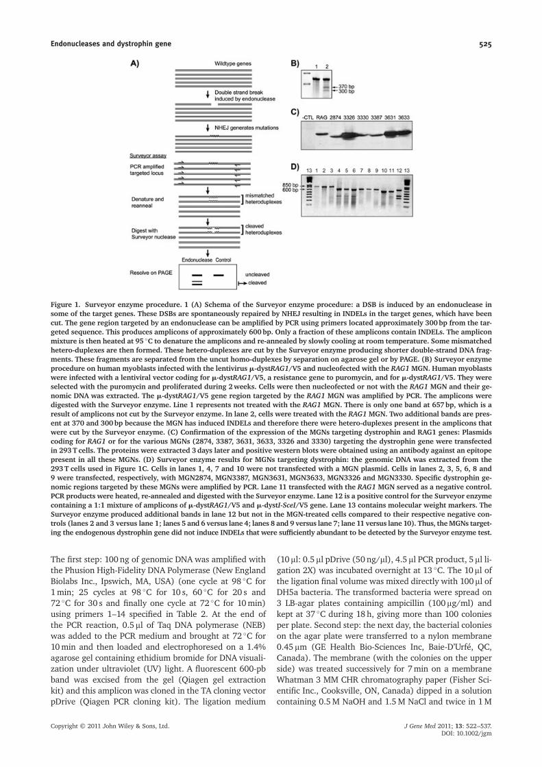
Figure 1. Surveyor enzyme procedure. 1 (A) Schema of the Surveyor enzyme procedure: a DSB is induced by an endonuclease insome of the target genes. These DSBs are spontaneously repaired by NHEJ resulting in INDELs in the target genes, which have beencut. The gene region targeted by an endonuclease can be amplified by PCR using primers located approximately 300bp from the tar-geted sequence. This produces amplicons of approximately 600bp. Only a fraction of these amplicons contain INDELs. The ampliconmixture is then heated at 95 �C to denature the amplicons and re-annealed by slowly cooling at room temperature. Some mismatchedhetero-duplexes are then formed. These hetero-duplexes are cut by the Surveyor enzyme producing shorter double-strand DNA frag-ments. These fragments are separated from the uncut homo-duplexes by separation on agarose gel or by PAGE. (B) Surveyor enzymeprocedure on human myoblasts infected with the lentivirus m-dystRAG1/V5 and nucleofected with the RAG1 MGN. Human myoblastswere infected with a lentiviral vector coding for m-dystRAG1/V5, a resistance gene to puromycin, and for m-dystRAG1/V5. They wereselected with the puromycin and proliferated during 2weeks. Cells were then nucleofected or not with the RAG1 MGN and their ge-nomic DNA was extracted. The m-dystRAG1/V5 gene region targeted by the RAG1 MGN was amplified by PCR. The amplicons weredigested with the Surveyor enzyme. Line 1 represents not treated with the RAG1 MGN. There is only one band at 657bp, which is aresult of amplicons not cut by the Surveyor enzyme. In lane 2, cells were treated with the RAG1 MGN. Two additional bands are pres-ent at 370 and 300bp because the MGN has induced INDELs and therefore there were hetero-duplexes present in the amplicons thatwere cut by the Surveyor enzyme. (C) Confirmation of the expression of the MGNs targeting dystrophin and RAG1 genes: Plasmidscoding for RAG1 or for the various MGNs (2874, 3387, 3631, 3633, 3326 and 3330) targeting the dystrophin gene were transfectedin 293T cells. The proteins were extracted 3days later and positive western blots were obtained using an antibody against an epitopepresent in all these MGNs. (D) Surveyor enzyme results for MGNs targeting dystrophin: the genomic DNA was extracted from the293T cells used in Figure 1C. Cells in lanes 1, 4, 7 and 10 were not transfected with a MGN plasmid. Cells in lanes 2, 3, 5, 6, 8 and9 were transfected, respectively, with MGN2874, MGN3387, MGN3631, MGN3633, MGN3326 and MGN3330. Specific dystrophin ge-nomic regions targeted by these MGNs were amplified by PCR. Lane 11 transfected with the RAG1 MGN served as a negative control.PCR products were heated, re-annealed and digested with the Surveyor enzyme. Lane 12 is a positive control for the Surveyor enzymecontaining a 1:1 mixture of amplicons of m-dystRAG1/V5 and m-dystI-SceI/V5 gene. Lane 13 contains molecular weight markers. TheSurveyor enzyme produced additional bands in lane 12 but not in the MGN-treated cells compared to their respective negative con-trols (lanes 2 and 3 versus lane 1; lanes 5 and 6 versus lane 4; lanes 8 and 9 versus lane 7; lane 11 versus lane 10). Thus, theMGNs target-ing the endogenous dystrophin gene did not induce INDELs that were sufficiently abundant to be detected by the Surveyor enzyme test.
Endonucleases and dystrophin gene 525
Copyright © 2011 John Wiley & Sons, Ltd. J Gene Med 2011; 13: 522–537.DOI: 10.1002/jgm
Tris-HCl (pH 8.0) with 1.5M NaCl each time for 7min. Af-ter a light air-drying at room temperature, the membranewas exposed to a UV light during 3min and hybridized witha radioactive 32P-oligonucleotide in hybridization solution(6� SSC (1� SSC being 0.15M NaCl/0.015M sodium cit-rate, pH 7.0), 2.5�Denhardt’s, salmon sperm DNA(300mg/ml) and SDS 1%) during at least 4–18h at 60 �Cin a water bath. The oligonucleotide probe was 24 nucleo-tides long and complementary to the sequence targeted bya given MGN. It was radioactively labelled with T4 polynu-cleotide kinase (Invitrogen Inc.) in presence of 32P-g ATP(6000Ci/mmol) at 37 �C and then purified. After hybridiza-tion, the membrane was washed several times in 1� SSCsolution at 60 �C, air-dried and exposed to X-ray film at�80 �C overnight. The next day, the developed filmwas su-perposed to the original agar plate and bacteria colonies
presenting no radioactive hybridization signal were pickedup and seeded in order on a second agar plate divided intosquares and also at the same time in the LB medium to begrown overnight at 37 �C. Third step: a second hybridiza-tion was carried out (i.e. the colonies on the secondagar plate were transferred to a nylon membrane, hy-bridized with the same radioactive probe and exposedto a film to confirm that the colonies picked in step 2were really negative). Fourth step: the plasmids wereprepared using the bacteria grown in the LB mediumbut only for the colonies, which were still negative instep 3. These plasmids were digested with EcoR1 andthe digestion products were analyzed on a 1.4% agarosegel to confirm the presence inside the pDrive vector of an in-sert of a size similar to the amplicon obtained in the firststep. Fifth step: these plasmids with an insert were then
Table 2. Primer sequences
Primer Forward/ Gene Nucleotide Sequence Amplicon
# reverse positions length1 Forward/ m-dystRag1 1027–1051 GACAGTTATCAAACAGCTTTGGAAG 6572 Reverse m-dystRag1 1684–1673 GTAATCTGTGGGTGTCTTGTAAAAGA3 Forward/ m-dystI-SceI 1027–1051 GACAGTTATCAAACAGCTTTGGAAG 6574 Reverse m-dystI-SceI 1684–1673 GTAATCTGTGGGTGTCTTGTAAAAGA5 Forward/ RAG1a 9969–9988 GGGAGGCAAAGATGAATCAA 5746 Reverse RAG1a 10542–10523 GGGCTTTTAACAATGGCTGA7 Forward/ dystrophin intron 38 997822–997843 TCTTGCAGCCTAAAGGAACAAA 7978 Reverse dystrophin intron 38 998618–998597 TCCTCTCGCTTTCTCTCATCTG9 Forward/ dystrophin intron 42 1036576–1036599 GCAGAGCTAGAGAAGAATGAGAAA 66810 Reverse dystrophin intron 42 1037243–1037220 TTTGTTATTGGTTGAGGTTTGCTG11 Forward/ dystrophin intron 44 1130033–1130054 GAACAGGTGGTATTACTAGCCA 66912 Reverse dystrophin intron 44 1949881–1949860 GGTTGCAGTGAGCTGAGATCAT13 Forward/ dystrophin exon 50 1524446–1524469 TTCACCAAATGGATTAAGATGTTC 40614 Reverse dystrophin exon 50 1524845–1524826 ACTCCCCATATCCCGTTGTC15 Deep Forward/ dystrophin intron 38 998314–998336 AATGATACGGCGACCACCGAGATCTA
CACTCTTTCCCTACACGACGCTCTTCCGATCTNNNGTGTTCTTTACAACTAGATGGGA
219
16 Deep Reverse dystrophin intron 38 998414–998390 CAAGCAGAAGACGGCATACGAGATCGGTCTCGGCATTCCTGCTGAACCGCTCTTCCGATCTNNNCATGGAATGCTATTATAAGATACTG
17 Deep Forward/ dystrophin intron 42 1036800–1036823 AATGATACGGCGACCACCGAGATCTACACTCTTTCCCTACACGACGCTCTTCCGATCTNNNATTTTCCAAAATATAATTACTGTG
222
18 Deep Reverse dystrophin intron 42 1036896–1036874 AAGCAGAAGACGGCATACGAGATCGGTCTCGGCATTCCTGCTGAACCGCTCTTCCGATCTNNNAACTTACGATGCTAAGAAATTCT
19 Deep Forward/ dystrophin intron 44 1130279–1130302 AATGATACGGCGACCACCGAGATCTACACTCTTTCCCTACACGACGCTCTTCCGATCTNNNTAACAAAAGCCTCCTGGCAAGGAT
249
20 Deep Reverse dystrophin intron 44 1130409–1130386 AAGCAGAAGACGGCATACGAGATCGGTCTCGGCATTCCTGCTGAACCGCTCTTCCGATCTNNNCTGCCTGAAACACCTGAAACACTT
21 Deep Forward/ RAG1a 10234–10258 AATGATACGGCGACCACCGAGATCTACACTCTTTCCCTACACGACGCTCTTCCGATCTNNNGATATTGATATTGGTCTTAATATGA
218
22 Deep Reverse RAG1a 10326–10301 CAAGCAGAAGACGGCATACGAGATCGGTCTCGGCATTCCTGCTGAACCGCTCTTCCGATCTNNNTGAGTCCCAAGGTGGGTGGGAAAGA
23 Deep Forward/ dystrophin exon 50 1524579–1524601 AATGATACGGCGACCACCGAGATCTACACTCTTTCCCTACACGACGCTCTTCCGATCTNNNAAGAGCTGAGGGCAAAGCAGCCT
214
24 Deep Reverse dystrophin exon 50 1524674–1524652 CAAGCAGAAGACGGCATACGAGATCGGTCTCGGCATTCCTGCTGAACCGCTCTTCCGATCTNNNATAGCTAGAGCCAAAGAGAATGG
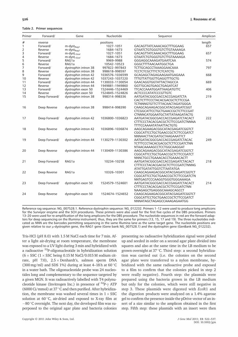
Reference rag sequence: NG_007528.1. Reference dystrophin sequence: NG_012232. Primers 1–12 were used to produce long ampliconsfor the Surveyor enzyme and the SCH procedures. These primers were also used for the first five cycles of the DBS procedure. Primers13–20 were used for re-amplification of the long amplicons for the DBS procedure. The nucleotide sequences in red are the forward adap-ters for deep sequencing on the Illumina instrument; thus, they are the same for primers (13, 15, 17 and 19). The three nucleotides indi-cated as NNN are the barcodes permitting sequencing in the same Illumina lane as the same target gene. The nucleotide positions aregiven relative to our m-dystrophin gene, the RAG1 gene (Gene bank NG_007528.1) and the dystrophin gene (GenBank NG_012232).
526 J. Rousseau et al.
Copyright © 2011 John Wiley & Sons, Ltd. J Gene Med 2011; 13: 522–537.DOI: 10.1002/jgm
sequenced with the dideoxy method (Sanger) to detectthe presence of INDELs produced by the MGN. Thesequencing was done using fluorescent dye sequencing[i.e. the sample (recombinant plasmid (mini-prep)]was electrophoresed on capillaries with a sequencingapparatus of type ABI 3730/XL.
DNA preparation for DBS
Genomic DNA was extracted from 293T or human myo-blasts. For DBS, 100 ng of genomic DNA were PCR ampli-fied for only five cycles using the same PCR primers andamplification parameters as for the Surveyor enzyme orSCH procedures. Five microlitres of PCR medium (firststep) were then used for a second PCR amplification with
a new set of forward and reverse primers containingnucleotides (23–25 nts) complementary to the target gene(primers 15–24 in Table 2). These primers contained intheir 5′ end additional nucleotides (58 nts for Deepforward and 61 nts for Deep reverse primers (adaptersrequired for the IlluminaW sequencing technology) [35]followed by a three nucleotides barcode. It should benoted that our barcoding procedure differs from the onepreviously described by Illumina Inc. (San Diego, CA,USA) [36]. For the PCR amplification (50ml final volume),100ng of the DBS primers reverse and forward mixed with5ml of the first PCR reaction were used as follows: onecycle at 98 �C for 1min followed by five cycles at 98 �Cfor 5 s, 55 �C for 10 s and 72 �C for 10 s and 28 cycles at98 �C for 5 s, 65 �C for 10 s and 72 �C for 10 s. The ampli-cons resulting from the second PCR were electrophoresed
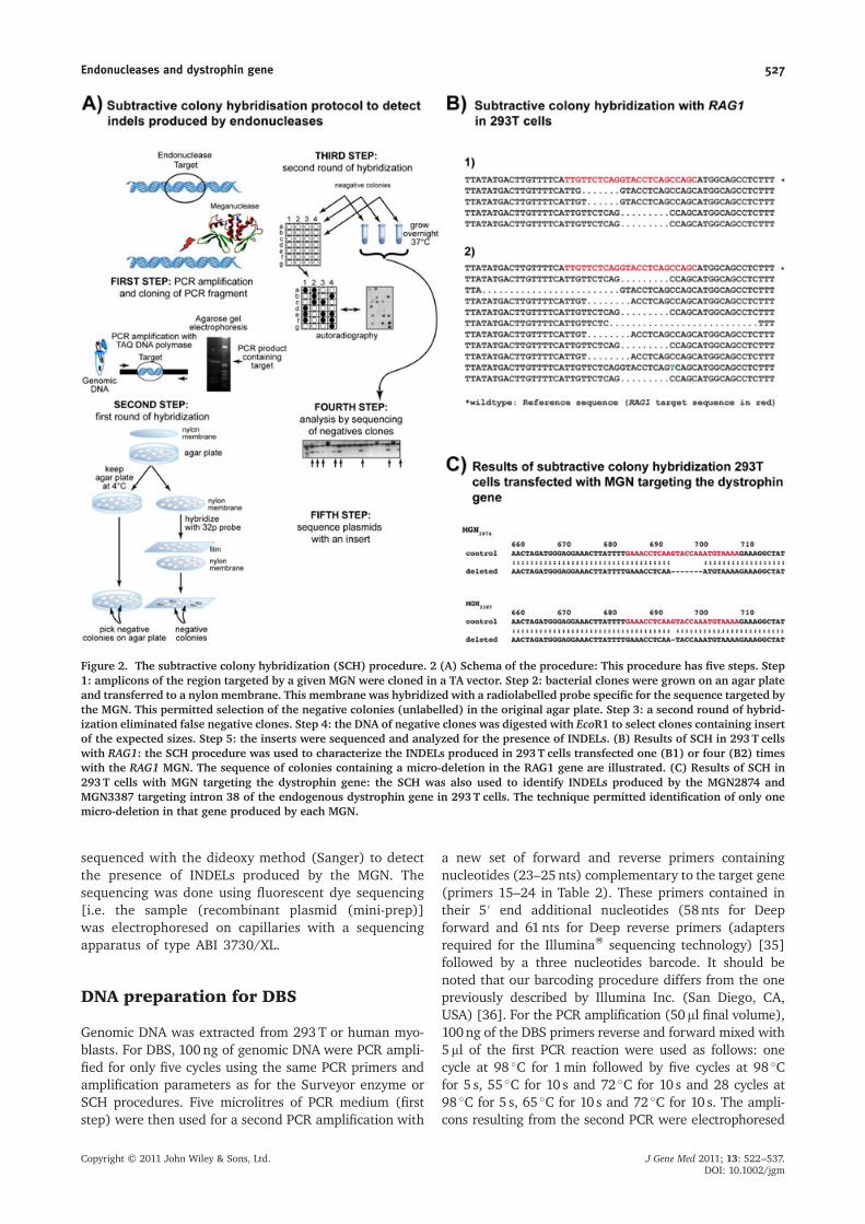
Figure 2. The subtractive colony hybridization (SCH) procedure. 2 (A) Schema of the procedure: This procedure has five steps. Step1: amplicons of the region targeted by a given MGN were cloned in a TA vector. Step 2: bacterial clones were grown on an agar plateand transferred to a nylon membrane. This membrane was hybridized with a radiolabelled probe specific for the sequence targeted bythe MGN. This permitted selection of the negative colonies (unlabelled) in the original agar plate. Step 3: a second round of hybrid-ization eliminated false negative clones. Step 4: the DNA of negative clones was digested with EcoR1 to select clones containing insertof the expected sizes. Step 5: the inserts were sequenced and analyzed for the presence of INDELs. (B) Results of SCH in 293T cellswith RAG1: the SCH procedure was used to characterize the INDELs produced in 293T cells transfected one (B1) or four (B2) timeswith the RAG1 MGN. The sequence of colonies containing a micro-deletion in the RAG1 gene are illustrated. (C) Results of SCH in293T cells with MGN targeting the dystrophin gene: the SCH was also used to identify INDELs produced by the MGN2874 andMGN3387 targeting intron 38 of the endogenous dystrophin gene in 293T cells. The technique permitted identification of only onemicro-deletion in that gene produced by each MGN.
Endonucleases and dystrophin gene 527
Copyright © 2011 John Wiley & Sons, Ltd. J Gene Med 2011; 13: 522–537.DOI: 10.1002/jgm
on agarose gel stained with ethidium bromide and thenpurified with QIAquick gel extraction kit (Qiagen Inc.) inaccordance with the manufacturer’s instructions. Theresulting size of the amplicon issue from the second PCRwith DBS primers was smaller than the amplicons obtainedin the first PCR used for Surveyor enzyme or SCH proce-dures (Table 2); this second PCR being a nested PCR.Because only 76 nucleotide sequences were obtained byDBS, the primer sequences complementary to the targetgene were positioned at approximately the same distancefrom the sequence targeted by a given endonuclease(Table 1). The concentrations of the purified ampliconswere evaluated with PicoGreenW dsDNA QuantificationReagent (Molecular Probes Inc., Eugene, OR, USA). Theresulting fluorescence was read (Ex 502nm/Em 523nm)on Victor3 multi-label reader (Perkin-Elmer). An equalamount of the different amplicons were pooled togetherto obtain a final DNA concentration of 10ng/ml and sentfor DBS at the McGill Genome Center. Pools containingapproximately 0.02pmol of each sample and phiX controlDNA were used for cluster generation on the cBot andpaired-end sequencing on one lane of the Illumina GenomeAnalyser IIx (Illumina Inc.) for 2�76 cycles.
Analysis of the sequences obtained withthe IlluminaW instrument
The Illumina Genome Analyser IIx permits approximately25 million sequences of 32–200 bp per lane to beobtained. A flow cell contains eight lanes producing atotal of 200 million sequences. The number of sequencesobtained per lane was much more than needed for thepresent study. We modified the Illumina sequencing adap-ters to permit multiplexing to analyse simultaneously, inthe same lane, the activity of different endonucleasesunder different experimental conditions. Because thenumber of sequences needed for our experiments waslow compared to the sequencing capacity of the Illuminainstrument, our samples were usually mixed with othersamples of yeast genome sequencing. Our samples repre-sented only 1% of the total DNA loaded in one lane. Theindividual amplicon sequences were retrieved from thepooled reads using the 3-bp barcode and the first 8 bp ofthe specific primers used from each amplicon. Between15,000–65 000 paired reads of 76 bases were obtainedfor each of our specimens. These sequences included the3-bp barcode at each end, the forward or reverse primersequences, which varied between 23 and 25 pb (Table 2).Thus there were between 47 and 53 bp in the target genewhere INDELs could be detected. Therefore, 72 or 73 bp(excluding the barcode, but including the forward primeror the reverse primer plus the 47–53 bp of the targetgene) were aligned onto the wild-type human referencegene (RAG1 or dystrophin) using BLAST software [37].This allowed the determination of how many sequenceshad mutations and their separation from the wild-typesequences for further analyses. Each of the mutatedsequence was then compared with the wild-type sequence
(NG_007528.1 for the RAG1 gene and NG_012232 for thedystrophin gene) using software that developed for thepresent study. This was carried out using the 3 bp ofmultiplex tag sequence and the first 8 bp of the specificprimers used form each amplicon. The BLASTALL soft-ware was then used to align selected sequences with thereference gene permitting to detect the exact positionand composition of each INDEL. This software permittedthe determination of how many base pairs were insertedor deleted, as well as the exact position of the event. Forthe final result compilation, the primer sequences weremasked. We calculated that the overall sequencing errorrate of the Illumina instrument was approximately 0.6%by aligning phiX to its genome using the GERALD toolfrom Illumina. To eliminate these sequencing errors, weused forward and reverse sequences, which were overlap-ping, and only the matching sequences were retained forfurther analysis. This pairing of the forward and reversesequences eliminated the sequencing errors made by theIllumina instrument. However, this pairing of the forwardand the reverse sequences could not be performed forMGN3631 and MGN3633 because a long stretch of thymi-dine near the targeted sequence precludes the design areverse primer that was sufficiently close to the forwardprimer to obtain an amplicon of approximately 76 bp,permitting the overlap of the forward and the reversesequences obtained with the Illumina instrument. Thus,for these two MGNs some of the 1-bp mutations may besequencing errors of the Illumina instrument, whichproduces an approximately 0.6% bp identification errorbut never produces INDELs.
ZFNs
A pair of ZFNs targeting a sequence in exon 50 of humandystrophin (Table 1) was purchased from Compo ZR Inc.
Results
Detection of INDELs with the Surveyorenzyme
Figure 1A shows a schema on the Surveyor enzyme meth-odology used to detect the activity of an endonucleasetargeting a specific sequence in the genome of a cell.The DNA break caused by an endonuclease is subse-quently repaired within the cell by NHEJ generating amutation of the target gene. The initial experiments toestablish the Surveyor enzyme method in our laboratorywere performed with human myoblasts infected with alentivirus coding for a modified micro-dystrophin genecontaining the target sequence for the RAG1 MGN and aV5 flag at the 3′ end (m-dystRAG1/V5) [33]. These cellswere then nucleofected with a plasmid coding for theRAG1 MGN. The sequence within the m-dystRAG1/V5 genetargeted by this MGN was then amplified by PCR (using
528 J. Rousseau et al.
Copyright © 2011 John Wiley & Sons, Ltd. J Gene Med 2011; 13: 522–537.DOI: 10.1002/jgm
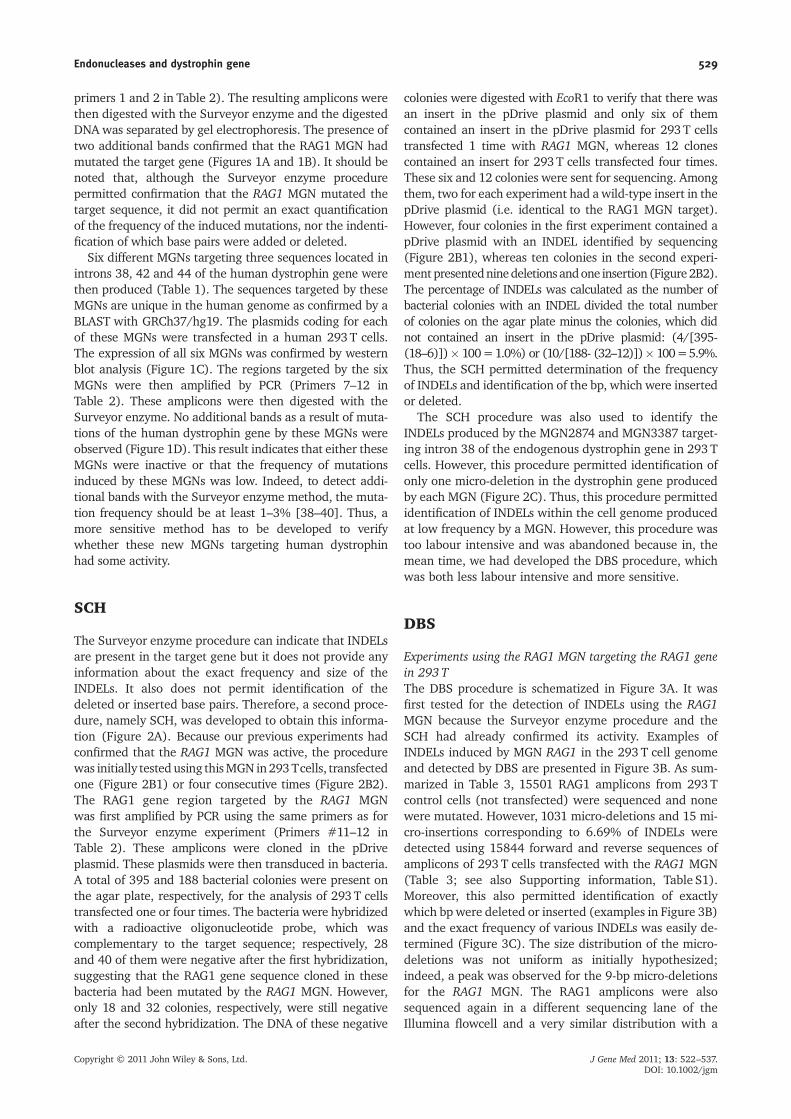
primers 1 and 2 in Table 2). The resulting amplicons werethen digested with the Surveyor enzyme and the digestedDNA was separated by gel electrophoresis. The presence oftwo additional bands confirmed that the RAG1 MGN hadmutated the target gene (Figures 1A and 1B). It should benoted that, although the Surveyor enzyme procedurepermitted confirmation that the RAG1 MGN mutated thetarget sequence, it did not permit an exact quantificationof the frequency of the induced mutations, nor the indenti-fication of which base pairs were added or deleted.
Six different MGNs targeting three sequences located inintrons 38, 42 and 44 of the human dystrophin gene werethen produced (Table 1). The sequences targeted by theseMGNs are unique in the human genome as confirmed by aBLAST with GRCh37/hg19. The plasmids coding for eachof these MGNs were transfected in a human 293T cells.The expression of all six MGNs was confirmed by westernblot analysis (Figure 1C). The regions targeted by the sixMGNs were then amplified by PCR (Primers 7–12 inTable 2). These amplicons were then digested with theSurveyor enzyme. No additional bands as a result of muta-tions of the human dystrophin gene by these MGNs wereobserved (Figure 1D). This result indicates that either theseMGNs were inactive or that the frequency of mutationsinduced by these MGNs was low. Indeed, to detect addi-tional bands with the Surveyor enzyme method, the muta-tion frequency should be at least 1–3% [38–40]. Thus, amore sensitive method has to be developed to verifywhether these new MGNs targeting human dystrophinhad some activity.
SCH
The Surveyor enzyme procedure can indicate that INDELsare present in the target gene but it does not provide anyinformation about the exact frequency and size of theINDELs. It also does not permit identification of thedeleted or inserted base pairs. Therefore, a second proce-dure, namely SCH, was developed to obtain this informa-tion (Figure 2A). Because our previous experiments hadconfirmed that the RAG1 MGN was active, the procedurewas initially tested using thisMGN in293Tcells, transfectedone (Figure 2B1) or four consecutive times (Figure 2B2).The RAG1 gene region targeted by the RAG1 MGNwas first amplified by PCR using the same primers as forthe Surveyor enzyme experiment (Primers #11–12 inTable 2). These amplicons were cloned in the pDriveplasmid. These plasmids were then transduced in bacteria.A total of 395 and 188 bacterial colonies were present onthe agar plate, respectively, for the analysis of 293T cellstransfected one or four times. The bacteria were hybridizedwith a radioactive oligonucleotide probe, which wascomplementary to the target sequence; respectively, 28and 40 of them were negative after the first hybridization,suggesting that the RAG1 gene sequence cloned in thesebacteria had been mutated by the RAG1 MGN. However,only 18 and 32 colonies, respectively, were still negativeafter the second hybridization. The DNA of these negative
colonies were digested with EcoR1 to verify that there wasan insert in the pDrive plasmid and only six of themcontained an insert in the pDrive plasmid for 293T cellstransfected 1 time with RAG1 MGN, whereas 12 clonescontained an insert for 293T cells transfected four times.These six and 12 colonies were sent for sequencing. Amongthem, two for each experiment had a wild-type insert in thepDrive plasmid (i.e. identical to the RAG1 MGN target).However, four colonies in the first experiment contained apDrive plasmid with an INDEL identified by sequencing(Figure 2B1), whereas ten colonies in the second experi-ment presentedninedeletions andone insertion (Figure2B2).The percentage of INDELs was calculated as the number ofbacterial colonies with an INDEL divided the total numberof colonies on the agar plate minus the colonies, which didnot contained an insert in the pDrive plasmid: (4/[395-(18–6)])� 100=1.0%) or (10/[188- (32–12)])� 100=5.9%.Thus, the SCH permitted determination of the frequencyof INDELs and identification of the bp, which were insertedor deleted.
The SCH procedure was also used to identify theINDELs produced by the MGN2874 and MGN3387 target-ing intron 38 of the endogenous dystrophin gene in 293Tcells. However, this procedure permitted identification ofonly one micro-deletion in the dystrophin gene producedby each MGN (Figure 2C). Thus, this procedure permittedidentification of INDELs within the cell genome producedat low frequency by a MGN. However, this procedure wastoo labour intensive and was abandoned because in, themean time, we had developed the DBS procedure, whichwas both less labour intensive and more sensitive.
DBS
Experiments using the RAG1 MGN targeting the RAG1 genein 293 TThe DBS procedure is schematized in Figure 3A. It wasfirst tested for the detection of INDELs using the RAG1MGN because the Surveyor enzyme procedure and theSCH had already confirmed its activity. Examples ofINDELs induced by MGN RAG1 in the 293T cell genomeand detected by DBS are presented in Figure 3B. As sum-marized in Table 3, 15501 RAG1 amplicons from 293Tcontrol cells (not transfected) were sequenced and nonewere mutated. However, 1031 micro-deletions and 15 mi-cro-insertions corresponding to 6.69% of INDELs weredetected using 15844 forward and reverse sequences ofamplicons of 293 T cells transfected with the RAG1 MGN(Table 3; see also Supporting information, Table S1).Moreover, this also permitted identification of exactlywhich bp were deleted or inserted (examples in Figure 3B)and the exact frequency of various INDELs was easily de-termined (Figure 3C). The size distribution of the micro-deletions was not uniform as initially hypothesized;indeed, a peak was observed for the 9-bp micro-deletionsfor the RAG1 MGN. The RAG1 amplicons were alsosequenced again in a different sequencing lane of theIllumina flowcell and a very similar distribution with a
Endonucleases and dystrophin gene 529
Copyright © 2011 John Wiley & Sons, Ltd. J Gene Med 2011; 13: 522–537.DOI: 10.1002/jgm
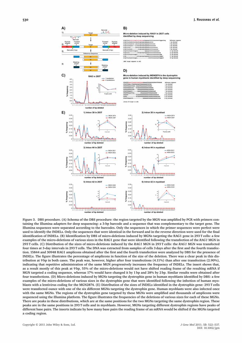
Figure 3. DBS procedure. (A) Schema of the DBS procedure: the region targeted by the MGN was amplified by PCR with primers con-taining the Illumina adapters for deep sequencing: a 3-bp barcode and a sequence that was complementary to the target gene. TheIllumina sequences were separated according to the barcodes. Only the sequences in which the primer sequences were perfect wereused to identify the INDELs. Only the sequences that were identical in the forward and in the reverse direction were used for the finalidentification of INDELs. (B) Identification by DBS of micro-deletions induced by MGNs targeting the RAG1 gene in 293T cells: a fewexamples of the micro-deletions of various sizes in the RAG1 gene that were identified following the transfection of the RAG1MGN in293T cells. (C) Distribution of the sizes of micro-deletions induced by the RAG1 MGN in 293T cells: the RAG1 MGN was transfectedfour times at 3-day intervals in 293T cells. The DNA was extracted from samples of cells 3 days after the first and the fourth transfec-tion. 15844 and 30948 RAG1 amplicons obtained after the first and the fourth transfection were analyzed by DBS for the presence ofINDELs. The figure illustrates the percentage of amplicons in function of the size of the deletion. There was a clear peak in this dis-tribution at 9 bp in both cases. The peak was, however, higher after four transfections (4.11%) than after one transfection (2.90%),indicating that repetitive administration of the same MGN progressively increases the frequency of INDELs. The insert shows that,as a result mostly of this peak at 9 bp, 55% of the micro-deletions would not have shifted reading frame of the resulting mRNA ifMGN targeted a coding sequence, whereas 17% would have changed it by 1bp and 28% by 2bp. Similar results were obtained afterfour transfections. (D) Micro-deletions induced by MGNs targeting the dystrophin gene in human myoblasts identified by DBS: a fewexamples of the micro-deletions of various sizes in the dystrophin gene that were identified following the infection of human myo-blasts with a lentivirus coding for the MGN2874. (E) Distribution of the sizes of INDELs identified in the dystrophin gene: 293T cellswere transfected ounce with one of the six different MGNs targeting the dystrophin gene. Human myoblasts were also infected oncewith the same MGNs. The regions of the dystrophin gene targeted by these MGNs were amplified and thousands of amplicons weresequenced using the Illumina platform. The figure illustrates the frequencies of the deletions of various sizes for each of these MGNs.There are peaks in these distributions, which are at the same positions for the two MGNs targeting the same dystrophin region. Thesepeaks are in the same positions in 293T cells and in myoblasts. However, MGNs targeting different dystrophin regions have peaks ofdifferent base pairs. The inserts indicate by howmany base pairs the reading frame of an mRNAwould be shifted if the MGNs targeteda coding region.
530 J. Rousseau et al.
Copyright © 2011 John Wiley & Sons, Ltd. J Gene Med 2011; 13: 522–537.DOI: 10.1002/jgm
peak at 9 bp was obtained. The DBS experiment was alsorepeated with 293T cells that were transfected four timesat 3-day intervals with the plasmid coding for the RAG1MGN. The percentage of INDELs was increased to 8.4%,indicating that repetitive administrations of the sameMGN progressively increased the frequency of INDELs.The distribution of the size of the micro-deletions weresimilar, with a deletion peak at 9 bp (Figure 3C), whichwas higher after foru transfections (4.11%) than afterone transfection (2.90%) These last results demonstratethat the sensitivity and precision of the DBS analysis ofINDELs is better than those of the SCH. It is importantto note that the large majority of INDELs produced byRAG1 are micro-deletions and that micro-insertions wereonly 1.2–1.4% of the INDELs.
Experiments using six MGNs targetingthe human dystrophin gene
Experiment in 293 T cellsFollowing the establishment of the DBS procedure withthe RAG1 MGN, we tested it with the six new MGNstargeting three dystrophin introns (Table 1) for whichno INDELs were detected with the Surveyor procedureand only one INDEL was detected by the SCH procedurefor MGN2874 and MGN3384. The detection of moreINDELs by the SCH procedure would have required muchmore work. The initial experiments were carried out in293 T cells that were transfected separately with differentplasmids each coding for one of the MGNs targetingintrons of the human dystrophin gene. Amplicons wereobtained as described in the Materials and methods (forsequences of primers, see Table 2). Note that, for the
two different MGNs targeting the same dystrophinintrons, different barcodes were used to permit simulta-neous sequencing in the same lane of the Illumina flow-cell. This permitted sequencing of 49 bp of the dystrophingene, which were between the primer sequences. INDELswere detected in the dystrophin gene of 293 T cells trans-fected with each one of the six MGN plasmids. When the293T cells were transfected with the RAG1 MGN plasmid(i.e. an irrelevant MGN for the dystrophin gene), onlyvery low percentages (0.00% to 0.04%) of INDELs weredetected in the dystrophin introns 38, 42 and 44 (Table 3).The MGN3631 was the most active with 1.77% INDELs,while MGN3330 was the least active, with only 0.14%INDELs. It is important, however, to note that thefrequency of INDELs measured for MGN3631 andMGN3633 targeting intron 44 is based only on a forwardsequence of the amplicons. This is because there was along series of thymidines after the 3′ end of the intronsequence targeted by these MGNs. This did not permitthe design of primers to obtain the amplified sequencebetween 43 and 52 bp as for the other MGNs; the ampli-fied sequences were 83 bp long and thus longer sequenceswould have been necessary to obtain overlapping forwardand reverse sequences. Thus, our results for these twoMGNs may contain some erroneous INDELs, especiallythose of only 1 bp. We also calculated the distribution ofINDELs of various numbers of base pairs for these sixMGNs targeting dystrophin in 293T cells (Figures 3E1,E3 and E5). The distributions of the size of INDELs werenot uniform, with peaks in different positions for thesesix MGNs. If the MGN3387 had targeting a codingsequence, 62% of the INDELs that it produced would haveshifted their reading by �1 (Figure 3E1, insert). It isimportant to note that, as for the RAG1 MGN, the
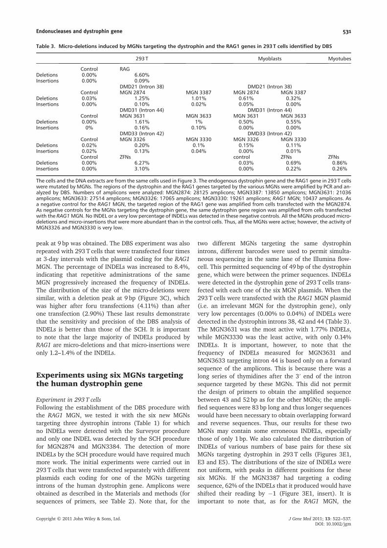
Table 3. Micro-deletions induced by MGNs targeting the dystrophin and the RAG1 genes in 293 T cells identified by DBS
293 T Myoblasts Myotubes
Control RAGDeletions 0.00% 6.60%Insertions 0.00% 0.09%
DMD21 (Intron 38) DMD21 (Intron 38)Control MGN 2874 MGN 3387 MGN 2874 MGN 3387
Deletions 0.03% 1.25% 1.01% 0.61% 0.32%Insertions 0.00% 0.10% 0.02% 0.05% 0.00%
DMD31 (Intron 44) DMD31 (Intron 44)Control MGN 3631 MGN 3633 MGN 3631 MGN 3633
Deletions 0.00% 1.61% 1% 0.50% 0.55%Insertions 0% 0.16% 0.10% 0.00% 0.00%
DMD33 (Intron 42) DMD33 (Intron 42)Control MGN 3326 MGN 3330 MGN 3326 MGN 3330
Deletions 0.02% 0.20% 0.1% 0.15% 0.11%Insertions 0.02% 0.13% 0.04% 0.00% 0.01%
Control ZFNs control ZFNs ZFNsDeletions 0.00% 6.27% 0.03% 0.69% 0.86%Insertions 0.00% 3.10% 0.00% 0.22% 0.26%
The cells and the DNA extracts are from the same cells used in Figure 3. The endogenous dystrophin gene and the RAG1 gene in 293 T cellswere mutated by MGNs. The regions of the dystrophin and the RAG1 genes targeted by the various MGNs were amplified by PCR and an-alyzed by DBS. Numbers of amplicons were analyzed: MGN2874: 28125 amplicons; MGN3387: 13850 amplicons; MGN3631: 21036amplicons; MGN3633: 27514 amplicons; MGN3326: 17065 amplicons; MGN3330: 19261 amplicons; RAG1 MGN; 10437 amplicons. Asa negative control for the RAG1 MGN, the targeted region of the RAG1 gene was amplified from cells transfected with the MGN2874.As negative controls for the MGNs targeting the dystrophin gene, the same dystrophin gene region was amplified from cells transfectedwith the RAG1MGN. No INDEL or a very low percentage of INDELs was detected in these negative controls. All the MGNs produced micro-deletions and micro-insertions that were more abundant than in the control cells. Thus, all the MGNs were active; however, the activity ofMGN3326 and MGN3330 is very low.
Endonucleases and dystrophin gene 531
Copyright © 2011 John Wiley & Sons, Ltd. J Gene Med 2011; 13: 522–537.DOI: 10.1002/jgm
percentages of INDELs that were micro-insertions werelower than micro-deletions for MGNs targeting the dystro-phin gene, being 7.4% for MGN2874, 2.0% for MGN3387,9.3% for MGN3631, 8.7% for MGN3633, 41.7% forMGN3326 and 22.9% for MGN3330. The last two MGNs,however, had a very low total mutagenic activity.
Experiments in human myoblastsHuman myoblasts are difficult to transfect; therefore, sixlentivirus vectors were thus constructed, each onecontaining the gene coding for one of the six MGNstargeting the dystrophin gene and a puromycin resistancegene. Myoblast cultures were infected with each of theselentiviruses, selected with puromycin during 48 h andpropagated for 2weeks. The expression of the MGN pro-teins by the infected myoblasts was confirmed by westernblotting. Myoblasts infected with a lentiviral vector codingfor EGFP were used as negative control. As for the exper-iment carried out with 293T cells, the dystrophin regionstargeted by the MGNs were analyzed by DBS. Lowfrequency INDELs were detected in myoblasts demon-strating the high sensitivity of this technique and indicat-ing that these MGNs were able to reach and mutate theendogenous dystrophin gene in these primary humanmyoblast cultures. Examples of micro-deletions producedby MGN 2874 in human myoblasts are presented inFigure 3D and the results are summarized in Table 3.The MGN2874 was the most active in myoblasts. Theseresults indicate that the Surveyor enzyme and the SCHprocedures were not sufficiently sensitive to detect theseINDELs. The sizes of the INDELs produced by the MGNstargeting dystrophin in myoblasts (Figures 3E2, E4 andE6) were also not uniformly distributed. The peaks inthose distributions were frequently at the same positionsfor the two MGNs targeting the same dystrophin region.It is important to note that the peaks are at the same num-ber of base pairs in both 293T cells and in myoblasts. Ifthe target sites would had been located in codingsequences, reading frame shifts would have beenproduced (Figure 3E, inserts). Again, the percentage ofmicro-insertions produced by the MGNs targeting thedystrophin gene were a low percentage of the totalINDELs, being 2.9% for MGN2874, 0% for MGN3387,0% for MGN3631, 0% for MGN3633, 0% for MGN3326and 10.0% for MGN3330.
Mutations of the dystrophin gene withZFNs targeting exon 50
The previous experiments were made with MGNs target-ing dystrophin introns. However, to restore the readingframe of the dystrophin gene in DMD patients that haveout-of-frame deletions by inducing INDELs, it is exons thathave to be targeted. We thus obtained (from Compo ZR) apair of ZFNs targeting the end of exon 50. Each of theZFNs contained six fingers. The sequences targeted by thispair of ZFNs are: CTAGCTCCTGGACTGACC andGGAGCCTGTAAGTATACTG. We initially tested these
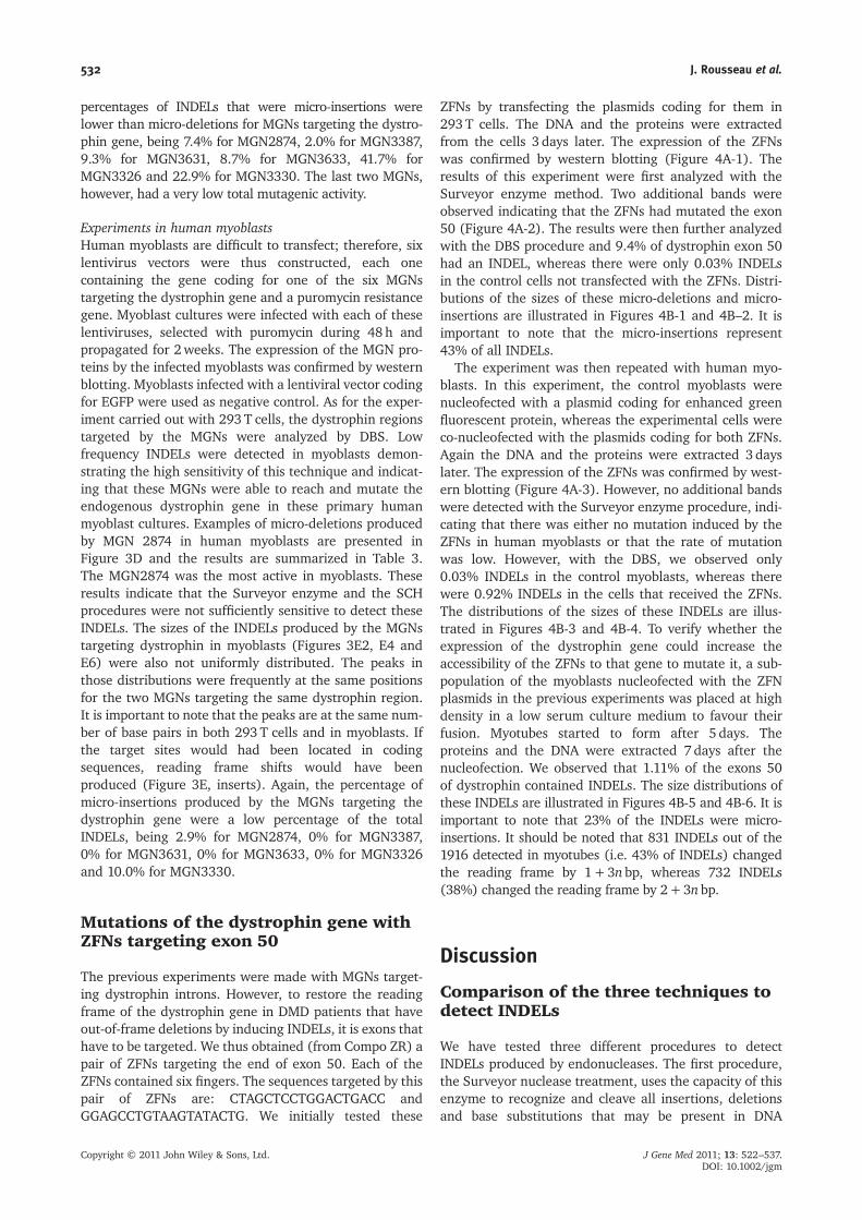
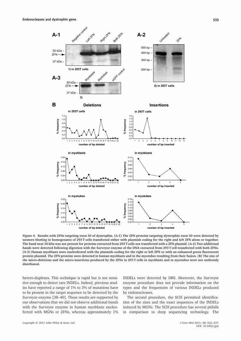
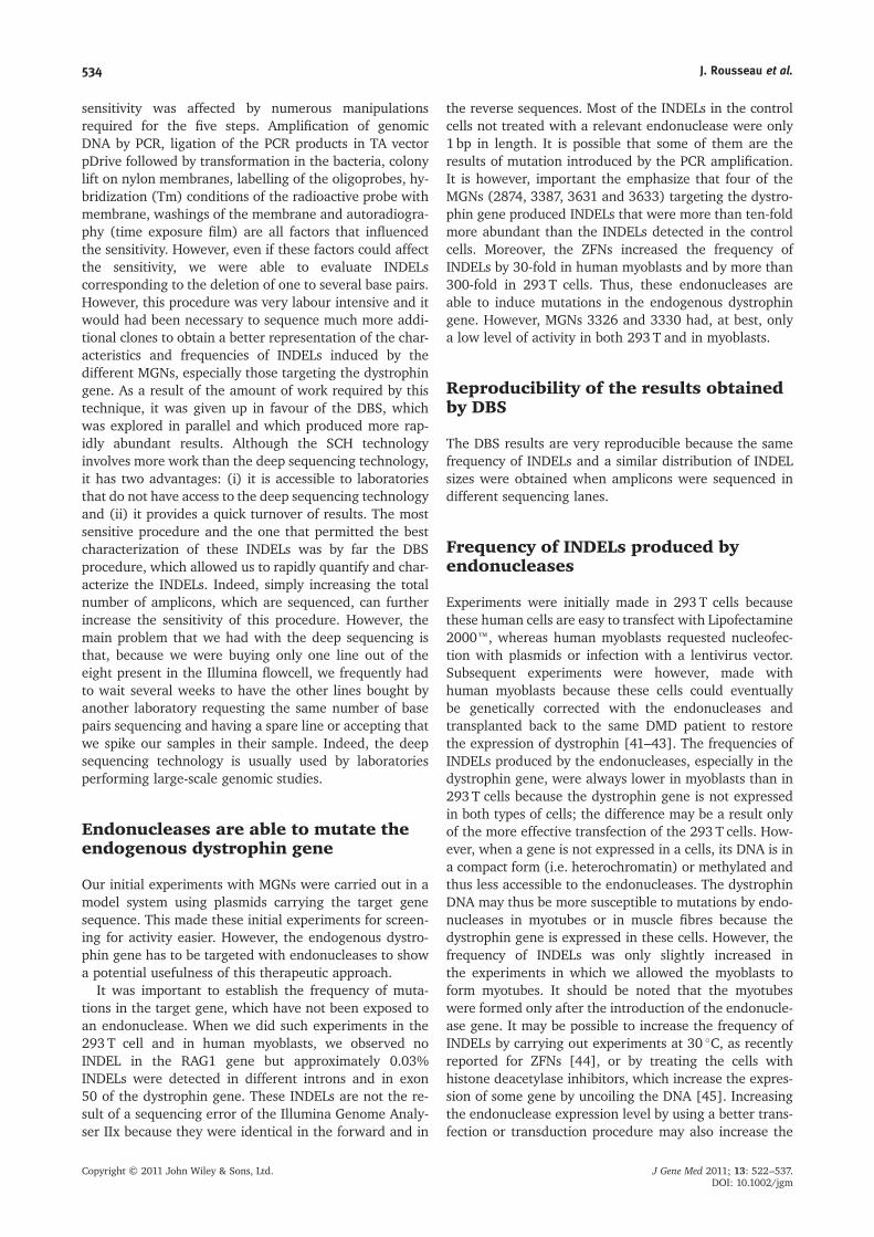
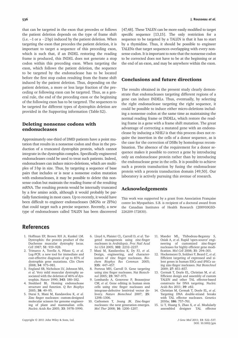
ZFNs by transfecting the plasmids coding for them in293T cells. The DNA and the proteins were extractedfrom the cells 3 days later. The expression of the ZFNswas confirmed by western blotting (Figure 4A-1). Theresults of this experiment were first analyzed with theSurveyor enzyme method. Two additional bands wereobserved indicating that the ZFNs had mutated the exon50 (Figure 4A-2). The results were then further analyzedwith the DBS procedure and 9.4% of dystrophin exon 50had an INDEL, whereas there were only 0.03% INDELsin the control cells not transfected with the ZFNs. Distri-butions of the sizes of these micro-deletions and micro-insertions are illustrated in Figures 4B-1 and 4B–2. It isimportant to note that the micro-insertions represent43% of all INDELs.
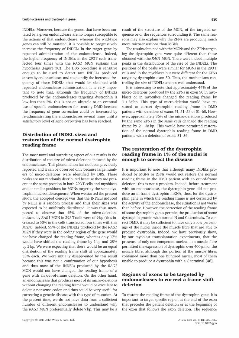
The experiment was then repeated with human myo-blasts. In this experiment, the control myoblasts werenucleofected with a plasmid coding for enhanced greenfluorescent protein, whereas the experimental cells wereco-nucleofected with the plasmids coding for both ZFNs.Again the DNA and the proteins were extracted 3 dayslater. The expression of the ZFNs was confirmed by west-ern blotting (Figure 4A-3). However, no additional bandswere detected with the Surveyor enzyme procedure, indi-cating that there was either no mutation induced by theZFNs in human myoblasts or that the rate of mutationwas low. However, with the DBS, we observed only0.03% INDELs in the control myoblasts, whereas therewere 0.92% INDELs in the cells that received the ZFNs.The distributions of the sizes of these INDELs are illus-trated in Figures 4B-3 and 4B-4. To verify whether theexpression of the dystrophin gene could increase theaccessibility of the ZFNs to that gene to mutate it, a sub-population of the myoblasts nucleofected with the ZFNplasmids in the previous experiments was placed at highdensity in a low serum culture medium to favour theirfusion. Myotubes started to form after 5 days. Theproteins and the DNA were extracted 7 days after thenucleofection. We observed that 1.11% of the exons 50of dystrophin contained INDELs. The size distributions ofthese INDELs are illustrated in Figures 4B-5 and 4B-6. It isimportant to note that 23% of the INDELs were micro-insertions. It should be noted that 831 INDELs out of the1916 detected in myotubes (i.e. 43% of INDELs) changedthe reading frame by 1+3nbp, whereas 732 INDELs(38%) changed the reading frame by 2+3nbp.
Discussion
Comparison of the three techniques todetect INDELs
We have tested three different procedures to detectINDELs produced by endonucleases. The first procedure,the Surveyor nuclease treatment, uses the capacity of thisenzyme to recognize and cleave all insertions, deletionsand base substitutions that may be present in DNA
532 J. Rousseau et al.
Copyright © 2011 John Wiley & Sons, Ltd. J Gene Med 2011; 13: 522–537.DOI: 10.1002/jgm
hetero-duplexes. This technique is rapid but is not sensi-tive enough to detect rare INDELs. Indeed, previous stud-ies have reported a range of 1% to 3% of mutations haveto be present in the target sequence to be detected by theSurveyor enzyme [38–40]. These results are supported byour observations that we did not observe additional bandswith the Surveyor enzyme in human myoblasts nucleo-fected with MGNs or ZFNs, whereas approximately 1%
INDELs were detected by DBS. Moreover, the Surveyorenzyme procedure does not provide information on thetypes and the frequencies of various INDELs producedby endonucleases.
The second procedure, the SCH permitted identifica-tion of the sizes and the exact sequences of the INDELsinduced by MGNs. The SCH procedure has several pitfallsin comparison to deep sequencing technology. The
Figure 4. Results with ZFNs targeting exon 50 of dystrophin. (A-1) The ZFN proteins targeting dystrophin exon 50 were detected bywestern blotting in homogenates of 293T cells transfected either with plasmids coding for the right and left ZFN alone or together.The band near 50kDa was not present for proteins extracted from 293Tcells not transfected with a ZFN plasmid. (A-2) Two additionalbands were detected following digestion with the Surveyor enzyme of the DNA extracted from 293T cell transfected with both ZFNs.(A-3) Human myoblasts were nucleofected with the plasmids coding for the right or left ZFN or with an enhanced green fluorescentprotein plasmid. The ZFN proteins were detected in human myoblasts and in the myotubes resulting from their fusion. (B) The size ofthe micro-deletions and the micro-insertions produced by the ZFNs in 293T cells in myoblasts and in myotubes were not uniformlydistributed.
Endonucleases and dystrophin gene 533
Copyright © 2011 John Wiley & Sons, Ltd. J Gene Med 2011; 13: 522–537.DOI: 10.1002/jgm
sensitivity was affected by numerous manipulationsrequired for the five steps. Amplification of genomicDNA by PCR, ligation of the PCR products in TA vectorpDrive followed by transformation in the bacteria, colonylift on nylon membranes, labelling of the oligoprobes, hy-bridization (Tm) conditions of the radioactive probe withmembrane, washings of the membrane and autoradiogra-phy (time exposure film) are all factors that influencedthe sensitivity. However, even if these factors could affectthe sensitivity, we were able to evaluate INDELscorresponding to the deletion of one to several base pairs.However, this procedure was very labour intensive and itwould had been necessary to sequence much more addi-tional clones to obtain a better representation of the char-acteristics and frequencies of INDELs induced by thedifferent MGNs, especially those targeting the dystrophingene. As a result of the amount of work required by thistechnique, it was given up in favour of the DBS, whichwas explored in parallel and which produced more rap-idly abundant results. Although the SCH technologyinvolves more work than the deep sequencing technology,it has two advantages: (i) it is accessible to laboratoriesthat do not have access to the deep sequencing technologyand (ii) it provides a quick turnover of results. The mostsensitive procedure and the one that permitted the bestcharacterization of these INDELs was by far the DBSprocedure, which allowed us to rapidly quantify and char-acterize the INDELs. Indeed, simply increasing the totalnumber of amplicons, which are sequenced, can furtherincrease the sensitivity of this procedure. However, themain problem that we had with the deep sequencing isthat, because we were buying only one line out of theeight present in the Illumina flowcell, we frequently hadto wait several weeks to have the other lines bought byanother laboratory requesting the same number of basepairs sequencing and having a spare line or accepting thatwe spike our samples in their sample. Indeed, the deepsequencing technology is usually used by laboratoriesperforming large-scale genomic studies.
Endonucleases are able to mutate theendogenous dystrophin gene
Our initial experiments with MGNs were carried out in amodel system using plasmids carrying the target genesequence. This made these initial experiments for screen-ing for activity easier. However, the endogenous dystro-phin gene has to be targeted with endonucleases to showa potential usefulness of this therapeutic approach.
It was important to establish the frequency of muta-tions in the target gene, which have not been exposed toan endonuclease. When we did such experiments in the293T cell and in human myoblasts, we observed noINDEL in the RAG1 gene but approximately 0.03%INDELs were detected in different introns and in exon50 of the dystrophin gene. These INDELs are not the re-sult of a sequencing error of the Illumina Genome Analy-ser IIx because they were identical in the forward and in
the reverse sequences. Most of the INDELs in the controlcells not treated with a relevant endonuclease were only1 bp in length. It is possible that some of them are theresults of mutation introduced by the PCR amplification.It is however, important the emphasize that four of theMGNs (2874, 3387, 3631 and 3633) targeting the dystro-phin gene produced INDELs that were more than ten-foldmore abundant than the INDELs detected in the controlcells. Moreover, the ZFNs increased the frequency ofINDELs by 30-fold in human myoblasts and by more than300-fold in 293T cells. Thus, these endonucleases areable to induce mutations in the endogenous dystrophingene. However, MGNs 3326 and 3330 had, at best, onlya low level of activity in both 293T and in myoblasts.
Reproducibility of the results obtainedby DBS
The DBS results are very reproducible because the samefrequency of INDELs and a similar distribution of INDELsizes were obtained when amplicons were sequenced indifferent sequencing lanes.
Frequency of INDELs produced byendonucleases
Experiments were initially made in 293T cells becausethese human cells are easy to transfect with Lipofectamine2000™, whereas human myoblasts requested nucleofec-tion with plasmids or infection with a lentivirus vector.Subsequent experiments were however, made withhuman myoblasts because these cells could eventuallybe genetically corrected with the endonucleases andtransplanted back to the same DMD patient to restorethe expression of dystrophin [41–43]. The frequencies ofINDELs produced by the endonucleases, especially in thedystrophin gene, were always lower in myoblasts than in293T cells because the dystrophin gene is not expressedin both types of cells; the difference may be a result onlyof the more effective transfection of the 293T cells. How-ever, when a gene is not expressed in a cells, its DNA is ina compact form (i.e. heterochromatin) or methylated andthus less accessible to the endonucleases. The dystrophinDNA may thus be more susceptible to mutations by endo-nucleases in myotubes or in muscle fibres because thedystrophin gene is expressed in these cells. However, thefrequency of INDELs was only slightly increased inthe experiments in which we allowed the myoblasts toform myotubes. It should be noted that the myotubeswere formed only after the introduction of the endonucle-ase gene. It may be possible to increase the frequency ofINDELs by carrying out experiments at 30 �C, as recentlyreported for ZFNs [44], or by treating the cells withhistone deacetylase inhibitors, which increase the expres-sion of some gene by uncoiling the DNA [45]. Increasingthe endonuclease expression level by using a better trans-fection or transduction procedure may also increase the
534 J. Rousseau et al.
Copyright © 2011 John Wiley & Sons, Ltd. J Gene Med 2011; 13: 522–537.DOI: 10.1002/jgm
INDELs. Moreover, because the genes, that have been mu-tated by a given endonuclease are no longer susceptible tothe actions of that endonuclease, whereas the wild-typegenes can still be mutated, it is possible to progressivelyincrease the frequency of INDELs in the target gene byrepeated administration of the endonuclease. Indeed,the higher frequency of INDELs in the 293T cells trans-fected four times with the RAG1 MGN sustains thishypothesis (Figure 3C). The DBS procedure is sensitiveenough to be used to detect rare INDELs producedin vivo by endonucleases and to quantify the increased fre-quency of these INDELs that would be obtained withrepeated endonuclease administration. It is very impor-tant to note that, although the frequency of INDELsproduced by the endonucleases targeting dystrophin islow less than 2%, this is not an obstacle to an eventualuse of specific endonucleases for treating DMD becausethe frequency of gene correction could be increased byre-administrating the endonucleases several times until asatisfactory level of gene correction has been reached.
Distribution of INDEL sizes andrestoration of the normal dystrophinreading frame
The most novel and surprising aspect of our results is thedistribution of the size of micro-deletions induced by theendonucleases. This phenomenon has not been previouslyreported and it can be observed only because large numb-ers of micro-deletions were identified by DBS. Thesepeaks are not randomly distributed because they are pres-ent at the same position in both 293T cells and myoblastsand at similar positions for MGNs targeting the same dys-trophin nucleotide sequence. When we started the presentstudy, the accepted concept was that the INDELs inducedby NHEJ is a random process and thus their sizes wasexpected to be uniformly distributed. It was thus unex-pected to observe that 45% of the micro-deletionsinduced by RAG1 MGN in 293T cells were of 9 bp (this in-creased to 50% in the cells transfected four times with thisMGN). Indeed, 55% of the INDELs produced by the RAG1MGN if they were in the coding region of the gene wouldnot have changed the reading frame, whereas only 17%would have shifted the reading frame by 1 bp and 28%by 2 bp. We were expecting that there would be an equaldistribution of the reading frame shift at approximately33% each. We were initially disappointed by this resultbecause this was not a confirmation of our hypothesisand thus most of the INDELs produced by the RAG1MGN would not have changed the reading frame of agene with an out-of-frame deletion. On the other hand,an endonuclease that produces most of its micro-deletionswithout changing the reading frame would be excellent todelete a nonsense codon and thus could be very useful forcorrecting a genetic disease with this type of mutation. Atthe present time, we do not have data from a sufficientnumber of different endonucleases to understand whythe RAG1 MGN preferentially delete 9 bp. This may be a
result of the structure of the MGN, of the targeted se-quence or of the sequences surrounding it. The same rea-sons may also explain why the ZFNs are producing muchmore micro-insertions than MGNs.
The results obtained with theMGNs and the ZFNs target-ing the dystrophin gene were quite different than thoseobtained with the RAG1MGN. There were indeed multiplepeaks in the distributions of the size of the INDELs. Thepositions of the peaks were similar for MGNs in the 293Tcells and in the myoblasts but were different for the ZFNstargeting dystrophin exon 50. Thus, the mechanisms con-trolling the size of INDELs are not well understood.
It is interesting to note that approximately 44% of themicro-deletions produced by the ZFNs in exon 50 in myo-blasts or in myotubes changed the reading frame by1+3n bp. This type of micro-deletion would have re-stored to correct dystrophin reading frame in DMDpatients with deletions of exons 51, 51–53 or 51–60. How-ever, approximately 36% of the micro-deletions producedby the same ZFNs in the same cells changed the readingframe by 2+3n bp. This would have permitted restora-tion of the normal dystrophin reading frame in DMDpatients with a deletion of exons 51–56.
The restoration of the dystrophinreading frame in 1% of the nuclei isenough to correct the disease
It is important to note that although many INDELs pro-duced by MGNs or ZFNs would not restore the normalreading frame in the DMD patient with an out-of-framedeletion; this is not a problem. Indeed, before treatmentwith an endonuclease, the dystrophin gene did not pro-duce an in-frame dystrophin mRNA; thus, for the dystro-phin gene in which the reading frame is not corrected bythe activity of the endonuclease, the situation is not worsethan before. However, the correction of the reading frameof some dystrophin genes permits the production of somedystrophin protein with normal N and C terminals. To cor-rect DMD, it may be sufficient to have only a low percent-age of the nuclei inside the muscle fibre that are able toproduce dystrophin. Indeed, we have previously show,by our myoblast transplantation experiments, that thepresence of only one competent nucleus in a muscle fibrepermitted the expression of dystrophin over 400 mm of themuscle fibre, although this portion of the muscle fibrescontained more than one hundred nuclei, most of themunable to produce a dystrophin with a C terminal [46].
Regions of exons to be targeted byendonucleases to correct a frame shiftdeletion
To restore the reading frame of the dystrophin gene, it isimportant to target specific region at the end of the exonthat precedes the patient deletion or at the beginning ofthe exon that follows the exon deletion. The sequence
Endonucleases and dystrophin gene 535
Copyright © 2011 John Wiley & Sons, Ltd. J Gene Med 2011; 13: 522–537.DOI: 10.1002/jgm
that can be targeted in the exon that precedes or followsthe patient deletion depends on the type of frame shift(i.e. –1 or a �2 bp) induced by the patient deletion. Whentargeting the exon that precedes the patient deletion, it isimportant to target a sequence of this preceding exon,which is such that, if an INDEL restoring the readingframe is produced, this INDEL does not generate a stopcodon within this preceding exon. When targeting theexon, which follows the patient deletion, the sequenceto be targeted by the endonuclease has to be locatedbefore the first stop codon resulting from the frame shiftinduced by the patient deletion. Thus, depending on thepatient deletion, a more or less large fraction of the pre-ceding or following exon can be targeted. Thus, as a gen-eral rule, the end of the preceding exon or the beginningof the following exon has to be targeted. The sequences tobe targeted for different types of dystrophin deletion areprovided in the Supporting information (Table S2).
Deleting nonsense codons withendonucleases
Approximately one-third of DMD patients have a point mu-tation that results in a nonsense codon and thus in the pro-duction of a truncated dystrophin protein, which cannotintegrate in the dystrophin complex. Specifically engineeredendonucleases could be used to treat such patients. Indeed,endonucleases can induce micro-deletions, which are multi-ples of 3 bp in size. Thus, by targeting a sequence of basepairs that includes or is near a nonsense codon mutationwith endonucleases, it may be possible to delete this non-sense codon but maintain the reading frame of the resultingmRNA. The resulting protein would be internally truncatedby a few amino acids, although it would probably be par-tially functioning inmost cases. Up to recently, it would havebeen difficult to engineer endonucleases (MGNs or ZFNs)that could target such a precise sequence. Recently, a newtype of endonucleases called TALEN has been discovered
[47,48]. These TALEN can be more easily modified to targetspecific sequence [13,15]. The only restriction for asequence to be targeted by a TALEN is that it has to startby a thymidine. Thus, it should be possible to engineerTALENs that target sequences overlapping with every non-sense codon. It is important to note that the nonsense codonto be corrected does not have to be at the beginning or atthe end of an exon, and may be anywhere within the exon.
Conclusions and future directions
The results obtained in the present study clearly demon-strate that endonucleases targeting different regions of agene can induce INDELs. Thus, eventually, by selectingthe right endonuclease targeting the right sequence, itcould be possible to induce either micro-deletions includ-ing a nonsense codon at the same time as maintaining thenormal reading frame or INDELs, which restore the read-ing frame in a gene with a frame shift mutation. The greatadvantage of correcting a mutated gene with an endonu-clease by inducing a NHEJ is that this process does not re-quire the insertion in the cells of a donor sequence, as isthe case for the correction of DSBs by homologous recom-bination. The absence of the requirement for a donor se-quence makes it possible to correct a gene by introducingonly an endonuclease protein rather than by introducingthe endonuclease gene in the cells. It is possible to achievesuch a protein transduction by fusing the endonucleaseprotein with a protein transduction domain [49,50]. Ourlaboratory is actively pursuing this avenue of research.
Acknowledgements
This work was supported by a grant from Association Françaisecontre les Myopathies. S.B. is recipient of a doctoral award fromthe Canadian Institutes of Health Research (200910GSD-226209-172830).
References
1. Hoffman EP, Brown RH Jr, Kunkel LM.Dystrophin: the protein product of theDuchenne muscular dystrophy locus.Cell 1987; 51: 919–928.
2. Trimarco A, Torella A, Piluso G, et al.Log-PCR: a new tool for immediate andcost-effective diagnosis of up to 85% ofdystrophin gene mutations. Clin Chem2008; 54: 973–981.
3. England SB, Nicholson LV, Johnson MA,et al. Very mild muscular dystrophy as-sociated with the deletion of 46% of dys-trophin. Nature 1990; 343: 180–182.
4. Stoddard BL. Homing endonucleasestructure and function. Q Rev Biophys2005; 38: 49–95.
5. Durai S, Mani M, Kandavelou K, et al.Zinc finger nucleases: custom-designedmolecular scissors for genome engineer-ing of plant and mammalian cells.Nucleic Acids Res 2005; 33: 5978–5990.
6. Lloyd A, Plaisier CL, Carroll D, et al. Tar-geted mutagenesis using zinc-fingernucleases in Arabidopsis. Proc Natl AcadSci USA 2005; 102: 2232–2237.
7. Mani M, Kandavelou K, Dy FJ, et al.Design, engineering, and character-ization of zinc finger nucleases. Bio-chem Biophys Res Commun 2005;335: 447–457.
8. Porteus MH, Carroll D. Gene targetingusing zinc finger nucleases. Nat Biotech-nol 2005; 23: 967–973.
9. Lombardo A, Genovese P, BeausejourCM, et al. Gene editing in human stemcells using zinc finger nucleases andintegrase-defective lentiviral vector de-livery. Nature Biotechnol 2007; 25:1298–1306.
10. Cathomen T, Joung JK. Zinc-fingernucleases: the next generation emerges.Mol Ther 2008; 16: 1200–1207.
11. Maeder ML, Thibodeau-Beganny S,Osiak A, et al. Rapid ‘open-source’ engi-neering of customized zinc-fingernucleases for highly efficient gene modi-fication. Mol Cell 2008; 31: 294–301.
12. Hockemeyer D, Soldner F, Beard C, et al.Efficient targeting of expressed and si-lent genes in human ESCs and iPSCs us-ing zinc-finger nucleases. Nat Biotechnol2009; 27: 851–857.
13. Cermak T, Doyle EL, Christian M, et al.Efficient design and assembly of customTALEN and other TAL effector-basedconstructs for DNA targeting. NucleicAcids Res 2011; 39: e82.
14. Christian M, Cermak T, Doyle EL, et al.Targeting DNA double-strand breakswith TAL effector nucleases. Genetics2010a; 186: 757–761.
15. Li T, Huang S, Zhao X, et al. Modularlyassembled designer TAL effector
536 J. Rousseau et al.
Copyright © 2011 John Wiley & Sons, Ltd. J Gene Med 2011; 13: 522–537.DOI: 10.1002/jgm
nucleases for targeted gene knockoutand gene replacement in eukaryotes.Nucleic Acids Res 2011; 39: 6315–6325.
16. Mahfouz MM, Li L, Shamimuzzaman M,et al. De novo-engineered transcriptionactivator-like effector (TALE) hybridnuclease with novel DNA binding speci-ficity creates double-strand breaks. ProcNatl Acad Sci USA 2011; 108: 2623–2628.
17. Miller JC, Tan S, Qiao G, et al. A TALEnuclease architecture for efficient ge-nome editing. Nat Biotechnol 2010; 29:143–148.
18. Morbitzer R, Elsaesser J, Hausner J,et al. Assembly of custom TALE-type DNAbinding domains by modular cloning.Nucleic Acids Res 2011;39: 5790–5799.
19. Choulika A, Perrin A, Dujon B, et al. In-duction of homologous recombination inmammalian chromosomes by using theI-SceI system of Saccharomyces cerevisiae.Mol Cell Biol 1995; 15: 1968–1973.
20. Rouet P, Smih F, Jasin M. Introductionof double-strand breaks into the genomeof mouse cells by expression of a rare-cutting endonuclease. Mol Cell Biol1994; 14: 8096–8106.
21. Paques F, Haber JE. Multiple pathwaysof recombination induced by double-strand breaks in Saccharomycescerevisiae. Microbiol Mol Biol Rev 1999;63: 349–404.
22. Beumer K, Bhattacharyya G, BibikovaM, et al. Efficient gene targeting in Dro-sophila with zinc-finger nucleases. Ge-netics 2006; 172: 2391–2403.
23. Bibikova M, Golic M, Golic KG, et al. Tar-geted chromosomal cleavage and muta-genesis in Drosophila using zinc-fingernucleases.Genetics 2002; 161: 1169–1175.
24. Doyon JB, Pattanayak V, Meyer CB, et al.Directed evolution and substrate specific-ity profile of homing endonuclease I-SceI.J Am Chem Soc 2006; 128: 2477–2484.
25. Ashworth J, Havranek JJ, Duarte CM,et al. Computational redesign of endo-nuclease DNA binding and cleavagespecificity. Nature 2006; 441: 656–659.
26. Arnould S, Chames P, Perez C, et al. En-gineering of large numbers of highlyspecific homing endonucleases that in-duce recombination on novel DNA tar-gets. J Mol Biol 2006; 355: 443–458.
27. Smith J, Grizot S, Arnould S, et al. Acombinatorial approach to create artifi-cial homing endonucleases cleaving
chosen sequences. Nucleic Acids Res2006; 34: e149.
28. Miller JC, Holmes MC, Wang J, et al. Animproved zinc-finger nuclease architecturefor highly specific genome editing.Nat Bio-technol 2007; 25: 778–785.
29. Foley JE, Yeh JR, Maeder ML, et al.Rapid mutation of endogenous zebrafishgenes using zinc finger nucleases madeby Oligomerized Pool ENgineering(OPEN). PLoS One 2009; 4: e4348.
30. Perez EE, Wang J, Miller JC, et al. Estab-lishment of HIV-1 resistance in CD4+ Tcells by genome editing using zinc-fingernucleases. Nature Biotechnol 2008; 26:808–816.
31. Meng X, Noyes MB, Zhu LJ, et al. Tar-geted gene inactivation in zebrafish us-ing engineered zinc-finger nucleases.Nat Biotechnol 2008; 26: 695–701.
32. Grizot S, Smith J, Daboussi F, et al. Effi-cient targeting of a SCID gene by anengineered single-chain homing endo-nuclease. Nucleic Acids Res 2009; 37:5405–5419.
33. Chapdelaine P, Pichavant C, Rousseau J,et al. Meganucleases can restore thereading frame of a mutated dystrophin.Gene Ther 2010; 17: 846–858.
34. Chapdelaine P, Delahaye S, Gauthier E,et al. A one-hour procedure for the prep-aration of genomic DNA from frozen tis-sues. Biotechniques 1993; 14: 163–164.
35. Bentley DR, Balasubramanian S,Swerdlow HP, et al. Accurate whole hu-man genome sequencing using revers-ible terminator chemistry. Nature 2008;456: 53–59.
36. Craig DW, Pearson JV, Szelinger S, et al.Identification of genetic variants usingbar-coded multiplexed sequencing. NatMethods 2008; 5: 887–893.
37. Altschul SF, Madden TL, Schaffer AA,et al. Gapped BLAST and PSI-BLAST: anew generation of protein databasesearch programs. Nucleic Acids Res1997; 25: 3389–3402.
38. Guschin DY, Waite AJ, Katibah GE, et al.A rapid and general assay for monitor-ing endogenous gene modification.MethMol Biol 2010; 649: 247–256.
39. Jiang Y, Palma JF, Agus DB, et al. Detec-tion of androgen receptor mutations incirculating tumor cells in castration-resistant prostate cancer. Clin Chem2010; 56: 1492–1495.
40. Pilch J, Asman M, Jamroz E, et al. Sur-veyor nuclease detection of mutationsand polymorphisms of mtDNA inchildren. Pediatr Neurol 2010; 43:325–330.
41. Skuk D, Goulet M, Roy B, et al. Dystro-phin expression in muscles of duchennemuscular dystrophy patients after high-density injections of normal myogeniccells. J Neuropathol Exp Neurol 2006;65: 371–386.
42. Quenneville SP, Chapdelaine P, Skuk D,et al. Autologous transplantation of mus-cle precursor cells modified with a lenti-virus for muscular dystrophy: humancells and primate models. Mol Ther2007; 15: 431–438.
43. Skuk D, Goulet M, Roy B, et al. First testof a ‘high-density injection’ protocol formyogenic cell transplantation through-out large volumes of muscles in a Du-chenne muscular dystrophy patient:18months follow-up. Neuromuscul Dis-ord 2007; 17: 38–46.
44. Doyon Y, Choi VM, Xia DF, et al. Tran-sient cold shock enhances zinc-fingernuclease-mediated gene disruption. NatMethods 2010; 7: 459–460.
45. Thiagalingam S, Cheng KH, Lee HJ,et al. Histone deacetylases: uniqueplayers in shaping the epigenetic histonecode. Ann NY Acad Sci 2003; 983:84–100.
46. Kinoshita I, Vilquin JT, Asselin I, et al.Transplantation of myoblasts froma transgenic mouse overexpressingdystrophin produced only a relativelysmall increase of dystrophin-positivemembrane. Muscle Nerve 1998; 21:91–103.
47. Boch J, Scholze H, Schornack S, et al.Breaking the code of DNA binding speci-ficity of TAL-type III effectors. Science2009; 326: 1509–1512.
48. Christian M, Cermak T, Doyle EL, et al.Targeting DNA double-strand breakswith TAL effector nucleases. Genetics2010b; 186: 757–761.
49. Tilstra J, Rehman KK, Hennon T, et al.Protein transduction: identification,characterization and optimization. Bio-chem Soc Trans 2007; 35: 811–815.
50. Chauhan A, Tikoo A, Kapur AK, et al.The taming of the cell penetrating do-main of the HIV Tat: myths and realities.J Control Release 2007; 117: 148–162.
Endonucleases and dystrophin gene 537
Copyright © 2011 John Wiley & Sons, Ltd. J Gene Med 2011; 13: 522–537.DOI: 10.1002/jgm


![IPv6-Gene[1].. Chap7](https://img.pdfslide.fr/doc/110x75/563dbb0d550346aa9aa9dcfc/ipv6-gene1-chap7.jpg)