Embed Size (px)
Citation preview
Identification and characterization of some Aspergillus pectinolytic glycoside
hydrolases
Promotor: prof. dr. A.G.J. Voragen Hoogleraar in de levensmiddelenchemie Wageningen Universiteit Co-promotor: dr. Gerrit Beldman
Universitair docent, departement Agrotechnologie en Voedingswetenschappen Wageningen Universiteit
Promotiecommissie: Prof. Dr. S.C. de Vries (Wageningen Universiteit)
Prof. Dr. J. van der Oost (Wageningen Universiteit) Prof. Dr. J.-F. Thibault (INRA, Nantes) Dr. A.A. van Dijk (DSM Food Specialties, Delft)
Dit onderzoek is uitgevoerd binnen de onderzoekschool VLAG (Voeding, levensmiddelenctechnologie, Agrotechnologie en Gezondheid)
Identification and characterization of some Aspergillus pectinolytic glycoside
hydrolases
Joris S. Zandleven
Proefschrift ter verkrijging van de graad van doctor
op gezag van de rector magnificus van Wageningen Universiteit,
prof. dr. M.J. Kropff, in het openbaar te verdedigen op dinsdag 12 december 2006
des namiddags te vier uur in de Aula
ISBN 90-8504-551-7 Zandleven, Joris S. Identification and characterization of some Aspergillus pectinolytic glycoside hydrolases Thesis Wageningen University, The Netherlands with summary in Dutch
Abstract
Abstract Zandleven, J.S. Identification and characterization of some Aspergillus pectinolytic glycoside
hydrolases Ph.D. Thesis Wageningen University , Wageningen, The Netherlands 2006 Key words: Aspergillus niger, Arabidopsis thaliana, homogalacturonan,
rhamnogalacturonan, xylogalacturonan, xylogalacturonan hydrolase, exo-polygalacturonase
Pectinases are used for many food applications, in particular for the manufacture of fruit
juices. However, the array of pectin modifying enzymes as available today is insufficient
to completely degrade pectic polysaccharides from plants, which consequently can cause
problems in food processing. As the genome sequence of Aspergillus niger indicated the
presence of more pectin modifying enzymes than previously known, research was carried
out to identify, produce, and characterize novel pectinases from this species.
From the complete inventory of the pectinolytic glycoside hydrolase family 28 of A.
niger a new gene group of seven exo-acting enzymes was found. Three of these enzymes
(PGXA, PGXB, PGXC) were biochemically identified from which it was demonstrated
that PGXB and PGXC act as an exo-polygalacturonase while PGXA rather acts like an
exo-xylogalacturonan hydrolase.
The xylogalacturonan hydrolase (XGH) was thoroughly investigated for its action
towards a xylogalacturonan (XGA) derived from gum tragacanth by isolation and
characterization of the produced oligosaccharides. Also XGH activity towards XGA in
the saponified modified ‘hairy’ regions (MHR-s) of pectin from apples and potatoes was
investigated. The enzyme predominantly released the di-saccharide GalAXyl from these
substrates which illustrates the preference of XGH to act between two xylosylated GalA
residues. However this enzyme was also able to release low substituted XGA
oligosaccharides as well as linear GalA oligosaccharides, which shows its tolerance for
unsubstituted GalA residues in its active site.
By using XGH as analytical tool, the presence of XGA could also be demonstrated in the
stem and the leaves of Arabidopsis thaliana, which shows that the presence of this
polymer is not strictly confined to storage tissues or reproductive organs of plants as was
previously thought to be the case.

Contents
Contents
Abstract
Chapter 1 General Introduction 1
Chapter 2 A new group of exo-acting family 28 glycoside hydrolases
of Aspergillus niger that are involved in pectin degradation 31
Chapter 3 Mode of action of xylogalacturonan hydrolase towards xylogalacturonan and xylogalacturonan oligosaccharides 57
Chapter 4 Enzymatic degradation studies of xylogalacturonans from apple and potato, using xylogalacturonan hydrolase 75
Chapter 5 Xylogalacturonan exists in cell walls from various tissues of Arabidopsis thaliana 97
Chapter 6 General discussion 115
Summary 139
Samenvatting 143
Acknowledgements 147
Curriculum Vitae 151
List of Publications 153
Addendum 155
Overview of completed training activities 157

Aan mijn ouders

Chapter 1
1
General Introduction
Chapter 1
2
1.1 Project background
The research presented in this thesis forms part of a larger project (Carbnet) which deals with
the discovery of new carbohydrate modifying enzymes (CMEs) from Aspergillus niger and
was funded by IOP (“Innovatief Onderzoeks Programma”) Genomics. Within this project the
laboratory of Food Chemistry focused on the exploration of novel pectinases.
At the start of the project, seven polygalacturonases (Parenicová et al., 2000b) and two
rhamnogalacturonases (Sykerbuyk et al., 1997) from A. niger were known, which have been
cloned, over-expressed and biochemically characterized. These enzymes are usually
components of commercial Aspergillus pectinase preparations which are used by industry for
high yield juice extraction, clarification, maceration, and liquefaction of fruit tissues.
However, the range of pectic enzymes present in commercial preparations is still insufficient
and after their application in processing of plant materials, still parts of undegradable pectin
are left, for which no enzymes have been found yet. For instance, during ultra filtration of
depectinized fruit juices, which has become a standard operation in juice clarification in order
to produce sparkling clear juices such as apple juice (Rao et al., 1987; Schobinger, 1988),
fouling of the membranes occurs which is caused by the accumulation of highly branched
parts of pectins (Schols et al., 1990b). Another example is pectin enriched soybean meal
which is commonly used as feed for livestocks. Pectin in soybean is poorly utilized by
monogastric animals, and partial degradation of these polysaccharides by the addition of
pectinases may improve the utilization by these animals. However so far, large parts of the
pectin polysaccharides from soybean appeared to be resistant towards the existing spectrum of
pectinases (Huisman et al., 2001).
Recently the genome of A. niger has been sequenced by DSM Food Specialties (Delft, The
Netherlands) from which it became clear that only a fraction of the potential of carbohydrate
modifying enzymes (CMEs) produced by this fungus was currently explored. As a result, the
Carbnet project was founded which aims at the discovery of all CMEs which include
pectinases, amylases and fructanases. Also the regulation of gene expression of these enzymes
was studied. Detailed information regarding the Carbnet project can also be found at URL:
www.senternovem.nl/mmfiles/factsheet%20Aspergillus%20(IGE01021) _tcm24-34630.pdf.
With the use of functional genomics tools we are able to investigate the set of these CMEs
present in A. niger in a rapid and efficient way compared to the conventional techniques used
General introduction
3
in the past. The choice to scrutinize for new CMEs of A. niger was supported by the fact that
this species is easy to handle when cultivating, and that it secretes its enzymes into the
medium from which they are easy to recover. Moreover the newly found CMEs from A.
niger, which have a “Generally Recognized As Safe” (GRAS) status, can be used safely for
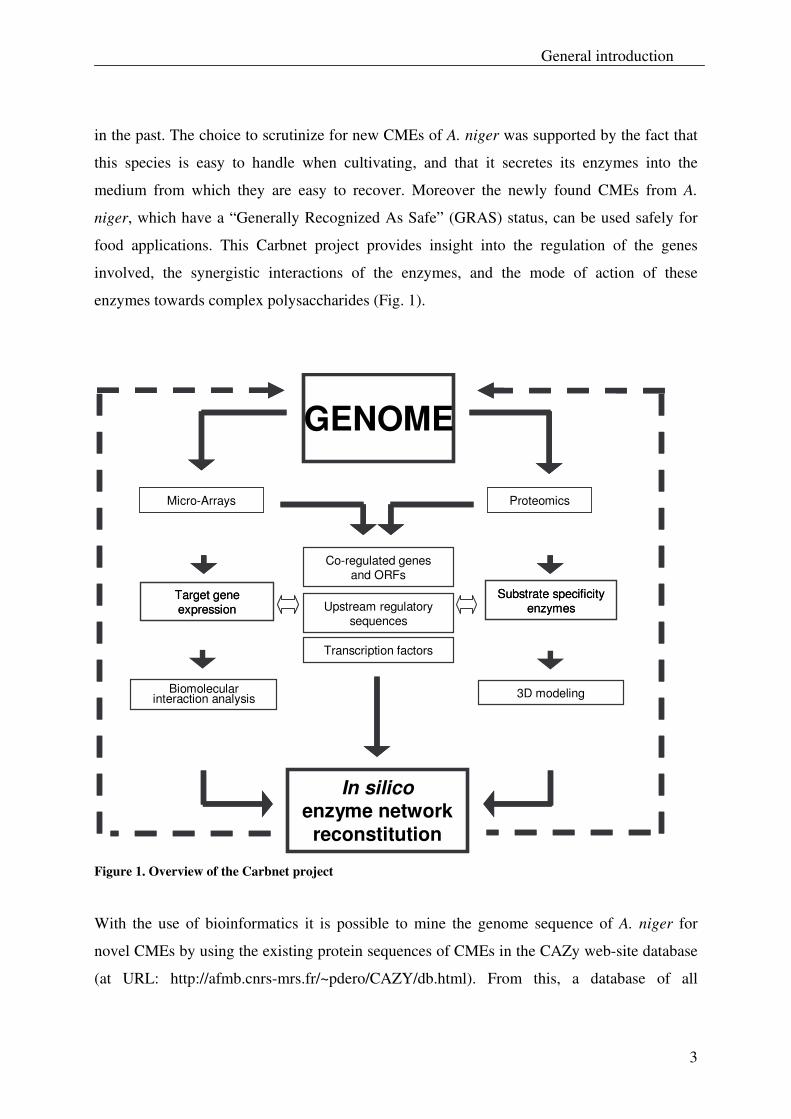
food applications. This Carbnet project provides insight into the regulation of the genes
involved, the synergistic interactions of the enzymes, and the mode of action of these
enzymes towards complex polysaccharides (Fig. 1).
Substrate specificityenzymes
Target gene expression
Substrate specificityenzymes
Target gene expression Upstream regulatory
sequences
Transcription factors
Co-regulated genesand ORFs
GENOME
In silicoenzyme network reconstitution
Biomolecular interaction analysis
Proteomics
3D modeling
Micro-Arrays
Figure 1. Overview of the Carbnet project
With the use of bioinformatics it is possible to mine the genome sequence of A. niger for
novel CMEs by using the existing protein sequences of CMEs in the CAZy web-site database
(at URL: http://afmb.cnrs-mrs.fr/~pdero/CAZY/db.html). From this, a database of all
Chapter 1
4
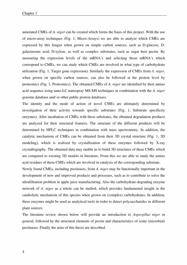
annotated CMEs of A. niger can be created which forms the basis of this project. With the use
of micro-array techniques (Fig. 1, Micro-Arrays) we are able to analyze which CMEs are
expressed by this fungus when grown on simple carbon sources, such as D-glucose, D-
galacturonic acid, D-xylose, as well as complex substrates, such as sugar beet pectin. By
measuring the expression levels of the mRNA’s and selecting those mRNA’s which
correspond to CMEs, we can study which CMEs are involved in what type of carbohydrate
utilization (Fig. 1, Target gene expression). Similarly the expression of CMEs from A. niger,
when grown on specific carbon sources, can also be followed at the protein level by
proteomics (Fig. 1, Proteomics). The obtained CMEs of A. niger are identified by their amino
acid sequence using nano-LC-nanospray MS-MS techniques in combination with the A. niger
genome database and/ or other public protein databases.
The identity and the mode of action of novel CMEs are ultimately determined by
investigation of their activity towards specific substrates (Fig. 1, Substrate specificity
enzymes). After incubation of CMEs with these substrates, the obtained degradation products
are analyzed for their structural features. The structure of the different products will be
determined by HPLC techniques in combination with mass spectrometry. In addition, the
catalytic mechanism of CMEs can be obtained from their 3D crystal structure (Fig. 1, 3D
modeling), which is realized by crystallization of these enzymes followed by X-ray
crystallography. The obtained data may enable us to build 3D structures of these CMEs which
are compared to existing 3D models in literature. From this we are able to study the amino
acid residues of these CMEs which are involved in catalysis of the corresponding substrate.
Newly found CMEs, including pectinases, from A. niger may be functionally important in the
development of new and improved products and processes, such as to contribute to solve the
ultrafiltration problem in apple juice manufacturing. Also the carbohydrate-degrading enzyme
network of A. niger as a whole can be studied, which provides fundamental insight in the
catabolytic mechanism of this species when grown on (complex) carbohydrates. In addition,
these enzymes might be used as analytical tools in order to detect polysaccharides in different
plant sources.
The literature review shown below will provide an introduction to Aspergillus niger in
general, followed by the structural elements of pectin and characteristics of some (microbial)
pectinases. Finally the aims of this thesis are described.
General introduction
5
1.2 Aspergillus
The fungus Aspergillus niger (A. niger) is a member of the Deuteromycetes (Fungi
Imperfecti) which stands for those fungi that reproduce asexually. This species belongs to the
A. niger section Nigri, which are also known as the black aspergilli (Abarca et al., 2004). The
presence of this species has been reported in field situations and in stored foods such as
peanuts, pecans, corn, apples, pears, peaches, grapes, strawberries, tomatoes and melons (Pitt
and Hocking, 1985; Pitt and Hocking, 1997).
A. niger is extensively used by industry for the production of enzymes (for instance β-
galactosidases, lipases, proteases, hemicellulases, cellulases, and pectinases) and organic
acids. While pectinases are used for processing plant materials to food products, the organic
acids, such as citric acid, are used in food and feed industry as flavor enhancers, acidifiers,
stabilizers or preservatives (Magnuson and Lasure, 2004).
Since 1917, Aspergillus niger has been used for the production of citric acid (Raper and
Fennel, 1965). Nowadays, around 700.000 - 1.000.000 tons of citric acid are produced per
annum, which makes it the most important organic acid in quantitative terms (Ikram-ul et al.,
2004; Magnuson and Lasure, 2004; Prado et al., 2005; Vandenberghe et al., 2000). Around 70
% of total citric acid manufactured is used in diverse food and beverage products, while the
remainder is mostly used for pharmaceutical formulations (Magnuson and Lasure, 2004;
Prado et al., 2005; Vandenberghe et al., 2000).
Pectinases from fungal sources, especially from A. niger, are used commonly in the fruit juice
industry and account, next to cellulases and hemicellulases, for approximately 20% of the one
billion US dollar annual sales of all industrial enzymes (Bhat, 2000; Kashyap et al., 2001).
These enzymes are used in the production of sparkling clear juices, such as apple juice, pear
juice, berrie juice, and grape juice, but also for the manufacture of juices with stable clouds,
such as citrus juices and prune juices.
A. niger has been utilized for decades in the food industry without any negative impact on
human health (Dijck et al., 2003; Schuster et al., 2002). Moreover, A. niger products are
considered “Generally Regarded As Safe” (GRAS) by the United States Food and Drug
Administration (FDA) (Oxenbøll, 1994; Abarca et al., 2004; Schuster et al., 2002). Another
reason for A. niger’s success in the food industry is that it grows rapidly on cheap substrates
Chapter 1
6
and secretes its enzymes into the medium, from which they are easy to recover (Oxenbøll,
1994).
To understand the function of pectinases from fungal sources, such as A. niger, towards pectic
polysaccharides, first a general overview will be given of the plant cell polysaccharides, with
emphasis on pectic polysaccharides. Then the function of pectinolytic enzymes will be
described with special attention to those that belong to the glycoside hydrolase family 28 of A.
niger.
2. Plant cell wall polysaccharides
Plant cells are surrounded by a “wall”, which stands out from the plasma membrane. These
cell walls enable regulation of cell expansion and strengthen them to osmotic stress. They also
protect these cells from insects as well as from invading pathogenic fungi and bacteria
(Carpita and Gibeaut, 1993).
Cell walls of growing plant tissues are predominantly composed of primary walls and middle
lamella (Fig. 2). The primary cell wall mainly consists of polysaccharides and to a lesser
extent of glycoproteins, phenolic esters, minerals and enzymes, while the middle lamella is a
polysaccharide rich region between primary cell walls of adjacent cells (Fry, 1988; O' Neill
and York, 2003). The most important polysaccharides in primary walls are cellulose,
hemicellulose and pectin (O' Neill and York, 2003; Albersheim et al., 1996; Gibeaut and
Carpita, 1994; Zablackis et al., 1995).
Cellulose is the most abundant plant polysaccharide which consist of β-(1→ 4)-linked D-
glucan chains. These chains arrayed in cellulose microfibrils, are coated with hemicelluloses,
such as xyloglycan and arabinoxylan. The hemicelluloses cross-link the cellulose microfibrils,
which creates a cellulose/hemicellulose network (Albersheim et al., 1996; Gibeaut and
Carpita, 1994; O' Neill and York, 2003).
Pectin covers a diverse group of polysaccharides which are present in primary cell walls and
middle lamella of plant cell walls (Voragen et al., 2001). These polysaccharides function as
“glue” that holds the cellulose microfibrils, hemicellulose and proteins together (Carpita and
Gibeaut, 1993), however the exact nature of how these pectin polysaccharides are linked to
other polymers is yet unknown (Mort, 2002).
General introduction
7
2.1 Structural elements of pectin
Pectin is probably the most complex class of plant cell wall polysaccharides and consist of
several monosaccharides (Vincken et al., 2003). Generally pectin is comprised of two families
of acidic polymers (Voragen et al., 2001):
• Galacturonan, which include pectin molecules with exclusively α-(1,4)-linked D-
galacturonic acid (GalA) residues in the backbone, such as homogalacturonan,
xylogalacturonan and rhamnogalacturonan II. Rhamnogalacturonan II is a highly
branched homogalacturonan which contains rare sugar residues (see further) and its
name is therefore somewhat misleading, because it suggests that it contains a
backbone of alternating rhamnose and D-galacturonic acid residues.
• Rhamnogalacturonan I, which has a backbone of alternating α-(1,2)-linked L-
rhamnose and α-(1,4)-linked D-galacturonic acid residues.
Chapter 1
8
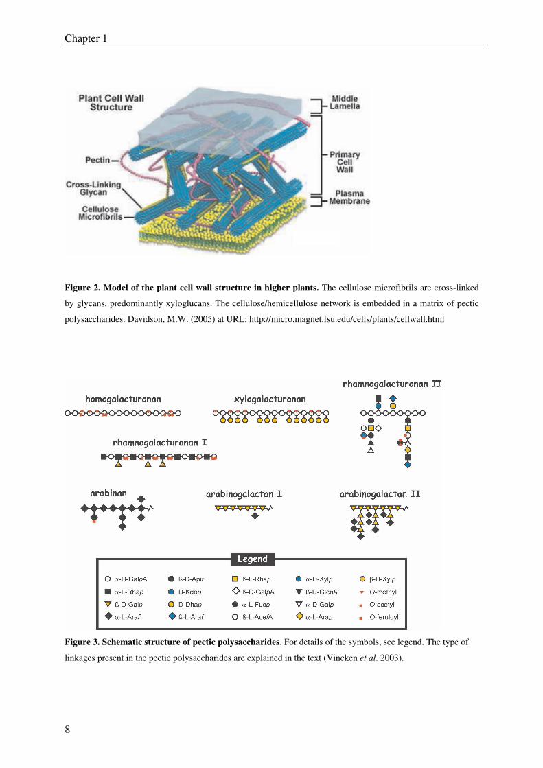
Figure 2. Model of the plant cell wall structure in higher plants. The cellulose microfibrils are cross-linked
by glycans, predominantly xyloglucans. The cellulose/hemicellulose network is embedded in a matrix of pectic
polysaccharides. Davidson, M.W. (2005) at URL: http://micro.magnet.fsu.edu/cells/plants/cellwall.html
Figure 3. Schematic structure of pectic polysaccharides. For details of the symbols, see legend. The type of
linkages present in the pectic polysaccharides are explained in the text (Vincken et al. 2003).
General introduction
9
2.1.1 Homogalacturonan
Homogalacturonan (HG) is a linear chain of α-(1, 4)-linked D-galacturonic acid residues (Fig.
3) in which some of these residues can be methyl-esterified at the carboxylic acid group (e.g.
C-6 position), and can carry acetyl groups on C-2 and C-3 (Albersheim et al., 1996; O' Neill
and York, 2003; Vincken et al., 2003). It is the most predominant biopolymer in primary cell
walls of dicotyledonous plants. Highly methyl-esterified HG polymers are also known as
“pectin” while HG polymers with a low or no methyl-esterification are referred to as “pectic
acid” (O' Neill and York, 2003). Particularly, methyl-esterified pectins have gained a lot of
interest in the food industry (see section 2.2), because of their excellent gelling and stabilizing
properties which are governed by their degree and pattern of methyl-esterification (Vincken et
al., 2003).
2.1.2 Xylogalacturonan
Xylogalacturonan (XGA) consist of a linear chain of α-(1, 4)-linked D-galacturonic acid
residues in which β-D-xylose residues are β- (1, 3)-linked to part of the GalA residues (Fig.
3). This biopolymer has been detected in the walls of reproductive plant organs such as
soybeans, kidney beans, peas, apple fruit, pear fruit, onions, carrot, pine pollen, cotton seed,
and watermelon (Bouveng, 1965; Kikuchi et al., 1996; Schols et al., 1995; Schols and
Voragen, 1996; Weightman et al., 1994; Yu and Mort, 1996). Xylogalacturonan accounts for
approximately 21% and 4% (w/w) of soybean pectin and apple pectin respectively (Voragen
et al., 2001), while it accounts for about 1.8 % (w/w) of the pea hulls (Le Goff et al., 2001).
The degree of xylosylation in XGA varies between 25% (as isolated from watermelon) and
75% (as isolated from apple), whereas the degree of methylation ranges between 40-90%
(Voragen et al., 2001).
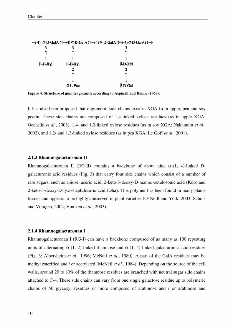
Exudates from gum tragacanth (from the Astralagus species) also contain a XGA-like
polysaccharide (Fig. 4), which contains besides single β-D-xylose residues, also the
disaccharide units α-L-fucose-(1, 2)-D-xylose and β-D-galactose-(1, 2)-D-xylose (Aspinall
and Baillie, 1963).
Chapter 1
10
4) -α-D-GalA (1 4) α-D-GalA(1 4) α-D-GalA(1 4) α-D-GalA(1 3
1β-D-Xyl
3
1β-D-Xyl
2
1α-L-Fuc
3
1β-D-Xyl
2
1β-D-Gal
4) -α-D-GalA (1 4) α-D-GalA(1 4) α-D-GalA(1 4) α-D-GalA(1 3
1β-D-Xyl
3
1β-D-Xyl
2
1α-L-Fuc
3
1β-D-Xyl
2
1β-D-Gal
→ → → → →
↑ ↑ ↑
↑ ↑
4) -α-D-GalA (1 4) α-D-GalA(1 4) α-D-GalA(1 4) α-D-GalA(1 3
1β-D-Xyl
3
1β-D-Xyl
2
1α-L-Fuc
3
1β-D-Xyl
2
1β-D-Gal
4) -α-D-GalA (1 4) α-D-GalA(1 4) α-D-GalA(1 4) α-D-GalA(1 3
1β-D-Xyl
3
1β-D-Xyl
2
1α-L-Fuc
3
1β-D-Xyl
2
1β-D-Gal
→ → → → →
↑ ↑ ↑
↑ ↑
Figure 4. Structure of gum tragacanth according to Aspinall and Baillie (1963).
It has also been proposed that oligomeric side chains exist in XGA from apple, pea and soy
pectin. These side chains are composed of 1,4-linked xylose residues (as in apple XGA;
Oechslin et al., 2003), 1,4- and 1,2-linked xylose residues (as in soy XGA; Nakamura et al.,
2002), and 1,2- and 1,3-linked xylose residues (as in pea XGA; Le Goff et al., 2001).
2.1.3 Rhamnogalacturonan II
Rhamnogalacturonan II (RG-II) contains a backbone of about nine α-(1, 4)-linked D-
galacturonic acid residues (Fig. 3) that carry four side chains which consist of a number of
rare sugars, such as apiose, aceric acid, 2-keto-3-deoxy-D-manno-octulosonic acid (Kdo) and
2-keto-3-deoxy-D-lyxo-heptulosaric acid (Dha). This polymer has been found in many plants
tissues and appears to be highly conserved in plant varieties (O' Neill and York, 2003; Schols
and Voragen, 2002; Vincken et al., 2003).
2.1.4 Rhamnogalacturonan I
Rhamnogalacturonan I (RG-I) can have a backbone composed of as many as 100 repeating
units of alternating α-(1, 2)-linked rhamnose and α-(1, 4)-linked galacturonic acid residues
(Fig. 3; Albersheim et al., 1996; McNeil et al., 1980). A part of the GalA residues may be
methyl esterified and / or acetylated (McNeil et al., 1984). Depending on the source of the cell
walls, around 20 to 80% of the rhamnose residues are branched with neutral sugar side chains
attached to C-4. These side chains can vary from one single galactose residue up to polymeric
chains of 50 glycosyl residues or more composed of arabinose and / or arabinose and
General introduction
11
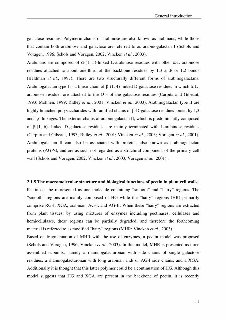
galactose residues. Polymeric chains of arabinose are also known as arabinans, while those
that contain both arabinose and galactose are referred to as arabinogalactan I (Schols and
Voragen, 1996; Schols and Voragen, 2002; Vincken et al., 2003).
Arabinans are composed of α-(1, 5)-linked L-arabinose residues with other α-L arabinose
residues attached to about one-third of the backbone residues by 1,3 and/ or 1,2 bonds
(Beldman et al., 1997). There are two structurally different forms of arabinogalactans.
Arabinogalactan type I is a linear chain of β-(1, 4)-linked D-galactose residues in which α-L-
arabinose residues are attached to the O-3 of the galactose residues (Carpita and Gibeaut,
1993; Mohnen, 1999; Ridley et al., 2001; Vincken et al., 2003). Arabinogalactan type II are
highly branched polysaccharides with ramified chains of β-D-galactose residues joined by 1,3
and 1,6 linkages. The exterior chains of arabinogalactan II, which is predominantly composed
of β-(1, 6)- linked D-galactose residues, are mainly terminated with L-arabinose residues
(Carpita and Gibeaut, 1993; Ridley et al., 2001; Vincken et al., 2003; Voragen et al., 2001).
Arabinogalactan II can also be associated with proteins, also known as arabinogalactan
proteins (AGPs), and are as such not regarded as a structural component of the primary cell
wall (Schols and Voragen, 2002; Vincken et al., 2003; Voragen et al., 2001) .
2.1.5 The macromolecular structure and biological functions of pectin in plant cell walls
Pectin can be represented as one molecule containing “smooth” and “hairy” regions. The
“smooth” regions are mainly composed of HG while the “hairy” regions (HR) primarily
comprise RG-I, XGA, arabinan, AG-I, and AG-II. When these “hairy” regions are extracted
from plant tissues, by using mixtures of enzymes including pectinases, cellulases and
hemicellulases, these regions can be partially degraded, and therefore the forthcoming
material is referred to as modified “hairy” regions (MHR; Vincken et al., 2003).
Based on fragmentation of MHR with the use of enzymes, a pectin model was proposed
(Schols and Voragen, 1996; Vincken et al., 2003). In this model, MHR is presented as three
assembled subunits, namely a rhamnogalacturonan with side chains of single galactose
residues, a rhamnogalacturonan with long arabinan and/ or AG-I side chains, and a XGA.
Additionally it is thought that this latter polymer could be a continuation of HG. Although this
model suggests that HG and XGA are present in the backbone of pectin, it is recently
Chapter 1
12
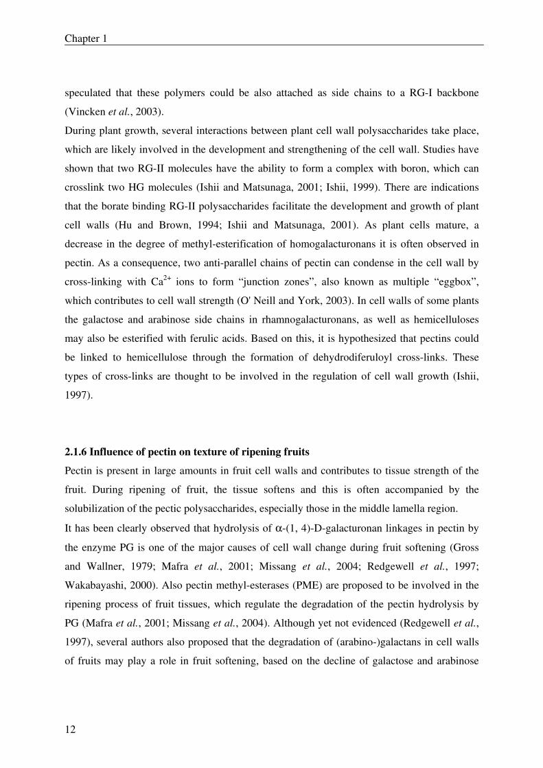
speculated that these polymers could be also attached as side chains to a RG-I backbone
(Vincken et al., 2003).
During plant growth, several interactions between plant cell wall polysaccharides take place,
which are likely involved in the development and strengthening of the cell wall. Studies have
shown that two RG-II molecules have the ability to form a complex with boron, which can
crosslink two HG molecules (Ishii and Matsunaga, 2001; Ishii, 1999). There are indications
that the borate binding RG-II polysaccharides facilitate the development and growth of plant
cell walls (Hu and Brown, 1994; Ishii and Matsunaga, 2001). As plant cells mature, a
decrease in the degree of methyl-esterification of homogalacturonans it is often observed in
pectin. As a consequence, two anti-parallel chains of pectin can condense in the cell wall by
cross-linking with Ca2+ ions to form “junction zones”, also known as multiple “eggbox”,
which contributes to cell wall strength (O' Neill and York, 2003). In cell walls of some plants
the galactose and arabinose side chains in rhamnogalacturonans, as well as hemicelluloses
may also be esterified with ferulic acids. Based on this, it is hypothesized that pectins could
be linked to hemicellulose through the formation of dehydrodiferuloyl cross-links. These
types of cross-links are thought to be involved in the regulation of cell wall growth (Ishii,
1997).
2.1.6 Influence of pectin on texture of ripening fruits
Pectin is present in large amounts in fruit cell walls and contributes to tissue strength of the
fruit. During ripening of fruit, the tissue softens and this is often accompanied by the
solubilization of the pectic polysaccharides, especially those in the middle lamella region.
It has been clearly observed that hydrolysis of α-(1, 4)-D-galacturonan linkages in pectin by
the enzyme PG is one of the major causes of cell wall change during fruit softening (Gross
and Wallner, 1979; Mafra et al., 2001; Missang et al., 2004; Redgewell et al., 1997;
Wakabayashi, 2000). Also pectin methyl-esterases (PME) are proposed to be involved in the
ripening process of fruit tissues, which regulate the degradation of the pectin hydrolysis by
PG (Mafra et al., 2001; Missang et al., 2004). Although yet not evidenced (Redgewell et al.,
1997), several authors also proposed that the degradation of (arabino-)galactans in cell walls
of fruits may play a role in fruit softening, based on the decline of galactose and arabinose
General introduction
13
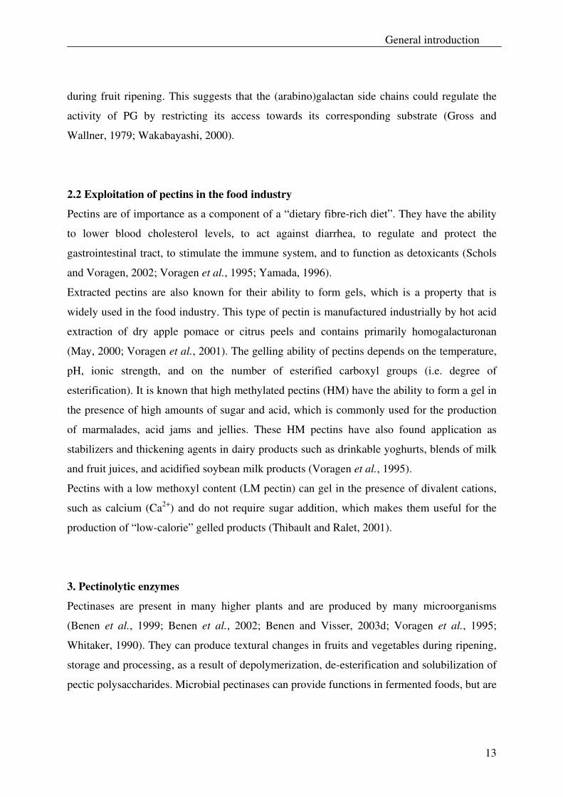
during fruit ripening. This suggests that the (arabino)galactan side chains could regulate the
activity of PG by restricting its access towards its corresponding substrate (Gross and
Wallner, 1979; Wakabayashi, 2000).
2.2 Exploitation of pectins in the food industry
Pectins are of importance as a component of a “dietary fibre-rich diet”. They have the ability
to lower blood cholesterol levels, to act against diarrhea, to regulate and protect the
gastrointestinal tract, to stimulate the immune system, and to function as detoxicants (Schols
and Voragen, 2002; Voragen et al., 1995; Yamada, 1996).
Extracted pectins are also known for their ability to form gels, which is a property that is
widely used in the food industry. This type of pectin is manufactured industrially by hot acid
extraction of dry apple pomace or citrus peels and contains primarily homogalacturonan
(May, 2000; Voragen et al., 2001). The gelling ability of pectins depends on the temperature,
pH, ionic strength, and on the number of esterified carboxyl groups (i.e. degree of
esterification). It is known that high methylated pectins (HM) have the ability to form a gel in
the presence of high amounts of sugar and acid, which is commonly used for the production
of marmalades, acid jams and jellies. These HM pectins have also found application as
stabilizers and thickening agents in dairy products such as drinkable yoghurts, blends of milk
and fruit juices, and acidified soybean milk products (Voragen et al., 1995).
Pectins with a low methoxyl content (LM pectin) can gel in the presence of divalent cations,
such as calcium (Ca2+) and do not require sugar addition, which makes them useful for the
production of “low-calorie” gelled products (Thibault and Ralet, 2001).
3. Pectinolytic enzymes
Pectinases are present in many higher plants and are produced by many microorganisms
(Benen et al., 1999; Benen et al., 2002; Benen and Visser, 2003d; Voragen et al., 1995;
Whitaker, 1990). They can produce textural changes in fruits and vegetables during ripening,
storage and processing, as a result of depolymerization, de-esterification and solubilization of
pectic polysaccharides. Microbial pectinases can provide functions in fermented foods, but are
Chapter 1
14
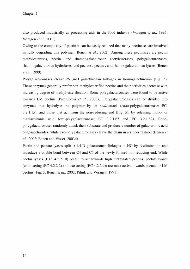
also produced industrially as processing aids in the food industry (Voragen et al., 1995;
Voragen et al., 2001).
Owing to the complexity of pectin it can be easily realized that many pectinases are involved
in fully degrading this polymer (Benen et al., 2002). Among these pectinases are pectin
methylesterases, pectin- and rhamnogalacturonan acetylesterases, polygalacturonases,
rhamnogalacturonan hydrolases, and pectate-, pectin-, and rhamnogalacturonan lyases (Benen
et al., 1999).
Polygalacturonases cleave α-1,4-D galacturonan linkages in homogalacturonan (Fig. 5).
These enzymes generally prefer non-methylesterified pectins and their activities decrease with
increasing degree of methyl-esterification. Some polygalacturonases were found to be active
towards LM pectins (Parenicová et al., 2000a). Polygalacturonases can be divided into
enzymes that hydrolyse the polymer by an endo-attack (endo-polygalacturonase; EC.
3.2.1.15), and those that act from the non-reducing end (Fig. 5), by releasing mono- or
digalacturonic acid (exo-polygalacturonase; EC 3.2.1.67 and EC 3.2.1.82). Endo-
polygalacturonases randomly attack their substrate and produce a number of galacturonic acid
oligosaccharides, while exo-polygalacturonases cleave the chain in a zipper fashion (Benen et
al., 2002; Benen and Visser, 2003d).
Pectin and pectate lyases split α-1,4-D galacturonan linkages in HG by β-elimination and
introduce a double bond between C4 and C5 of the newly formed non-reducing end. While
pectin lyases (E.C. 4.2.2.10) prefer to act towards high methylated pectins, pectate lyases
(endo acting (EC 4.2.2.2) and exo-acting (EC 4.2.2.9)) are most active towards pectate or LM
pectins (Fig. 5; Benen et al., 2002; Pilnik and Voragen, 1991).
General introduction
15
XylAcMe
GalGalARha
Smooth regions
Hairy Regions regions
homogalacturonan
Rhamnogalacturonan Ixylogalacturonan
Pectin methyl esterase Polygalacturonase
Pectate lyase
Pectin acetyl esterase
RG-hydrolase RG-lyase
RG-acetyl esterase
pectinlyase
Xylogalacturonan hydrolase
RG-galacturonohydrolase
RG-rhamnohydrolase
Exo-poly-galacturonase
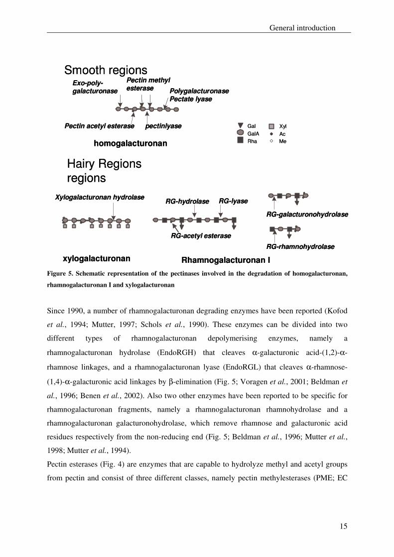
Figure 5. Schematic representation of the pectinases involved in the degradation of homogalacturonan,
rhamnogalacturonan I and xylogalacturonan
Since 1990, a number of rhamnogalacturonan degrading enzymes have been reported (Kofod
et al., 1994; Mutter, 1997; Schols et al., 1990). These enzymes can be divided into two
different types of rhamnogalacturonan depolymerising enzymes, namely a
rhamnogalacturonan hydrolase (EndoRGH) that cleaves α-galacturonic acid-(1,2)-α-
rhamnose linkages, and a rhamnogalacturonan lyase (EndoRGL) that cleaves α-rhamnose-
(1,4)-α-galacturonic acid linkages by β-elimination (Fig. 5; Voragen et al., 2001; Beldman et
al., 1996; Benen et al., 2002). Also two other enzymes have been reported to be specific for
rhamnogalacturonan fragments, namely a rhamnogalacturonan rhamnohydrolase and a
rhamnogalacturonan galacturonohydrolase, which remove rhamnose and galacturonic acid
residues respectively from the non-reducing end (Fig. 5; Beldman et al., 1996; Mutter et al.,
1998; Mutter et al., 1994).
Pectin esterases (Fig. 4) are enzymes that are capable to hydrolyze methyl and acetyl groups
from pectin and consist of three different classes, namely pectin methylesterases (PME; EC
Chapter 1
16
3.1.1.11), pectin acetylesterases (PAE; EC 3.1.1.6) and rhamnogalacturonan acetyl esterases
(RGAE).
Pectin methylesterases cleave off methyl esters which are present at the carboxylic function
(C-6) in the backbone of homogalacturonan, which results in the formation of free carboxylic
acid and methanol. The acetylesterases split off acetyl groups from C-2 and/ or C-3 from
GalA residues and can be grouped in those that are restricted to homogalacturonan (PAE) and
rhamnogalacturonan (RGAE) respectively (Fig. 5; Benen et al., 2003c; Benen et al., 2002;
Voragen et al., 1995; Voragen et al., 2001).
Also a novel type of pectinase, xylogalacturonan hydrolase, has been found in Aspergillus
tubingensis. This enzyme acts on xylogalacturonan by cleaving α-1,4-D galacturonan
linkages in an endo-fashion (Fig. 5) and has a requirement for xylosyl side chains (Van der
Vlugt-Bergmans et al., 2000).
3.1 Microbial pectinases
Several pectinases have been isolated from microorganisms such as Erwinia chrysanthemi, E.
carotovora, Aspergillus niger, A. tubingensis and A. aculeatus (Benen et al., 2002). A list of
different types of known microbial pectinases is presented in Table 1, along with their family
number according to Coutinho and Henrissat (1999). The enzymes presented in Table 1 can
also be found at the following URL: http://afmb.cnrs-mrs.fr/~pdero/CAZY/db.html. It should
be noted that the assignment of these microbial pectinases into family numbers is based on
amino acid sequence similarities rather than biochemical properties.
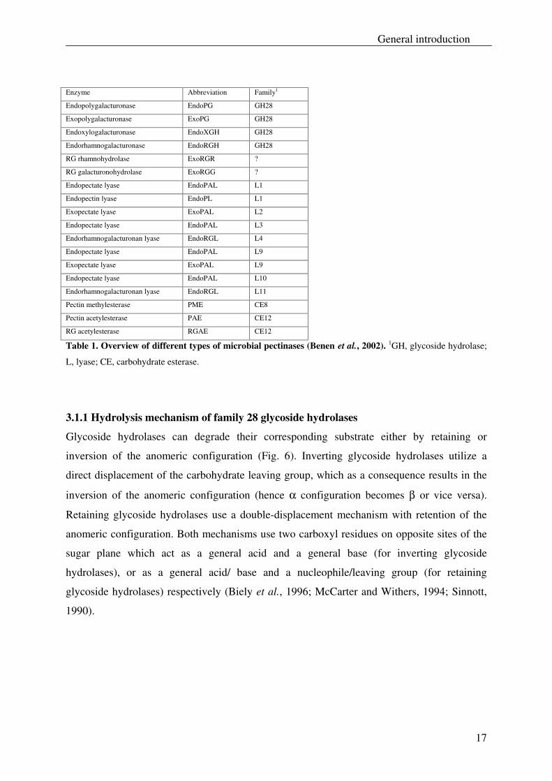
As shown in Table 1, the hydrolytic pectinases are grouped into glycoside hydrolase family
28 (GH28). As the amino acid sequence information of enzymes ExoRGR and ExoRGG is
still unknown, these enzymes have still to be attributed to a proper family. The pectic lyases
are more diverse and have been grouped into six different families. The pectin esterase PME
has been grouped into family number CE8, whereas the pectin esterases PAE and RGAE have
both been assigned into family number CE12.
General introduction
17
Enzyme Abbreviation Family1
Endopolygalacturonase EndoPG GH28
Exopolygalacturonase ExoPG GH28
Endoxylogalacturonase EndoXGH GH28
Endorhamnogalacturonase EndoRGH GH28
RG rhamnohydrolase ExoRGR ?
RG galacturonohydrolase ExoRGG ?
Endopectate lyase EndoPAL L1
Endopectin lyase EndoPL L1
Exopectate lyase ExoPAL L2
Endopectate lyase EndoPAL L3
Endorhamnogalacturonan lyase EndoRGL L4
Endopectate lyase EndoPAL L9
Exopectate lyase ExoPAL L9
Endopectate lyase EndoPAL L10
Endorhamnogalacturonan lyase EndoRGL L11
Pectin methylesterase PME CE8
Pectin acetylesterase PAE CE12
RG acetylesterase RGAE CE12
Table 1. Overview of different types of microbial pectinases (Benen et al., 2002). 1GH, glycoside hydrolase;
L, lyase; CE, carbohydrate esterase.
3.1.1 Hydrolysis mechanism of family 28 glycoside hydrolases
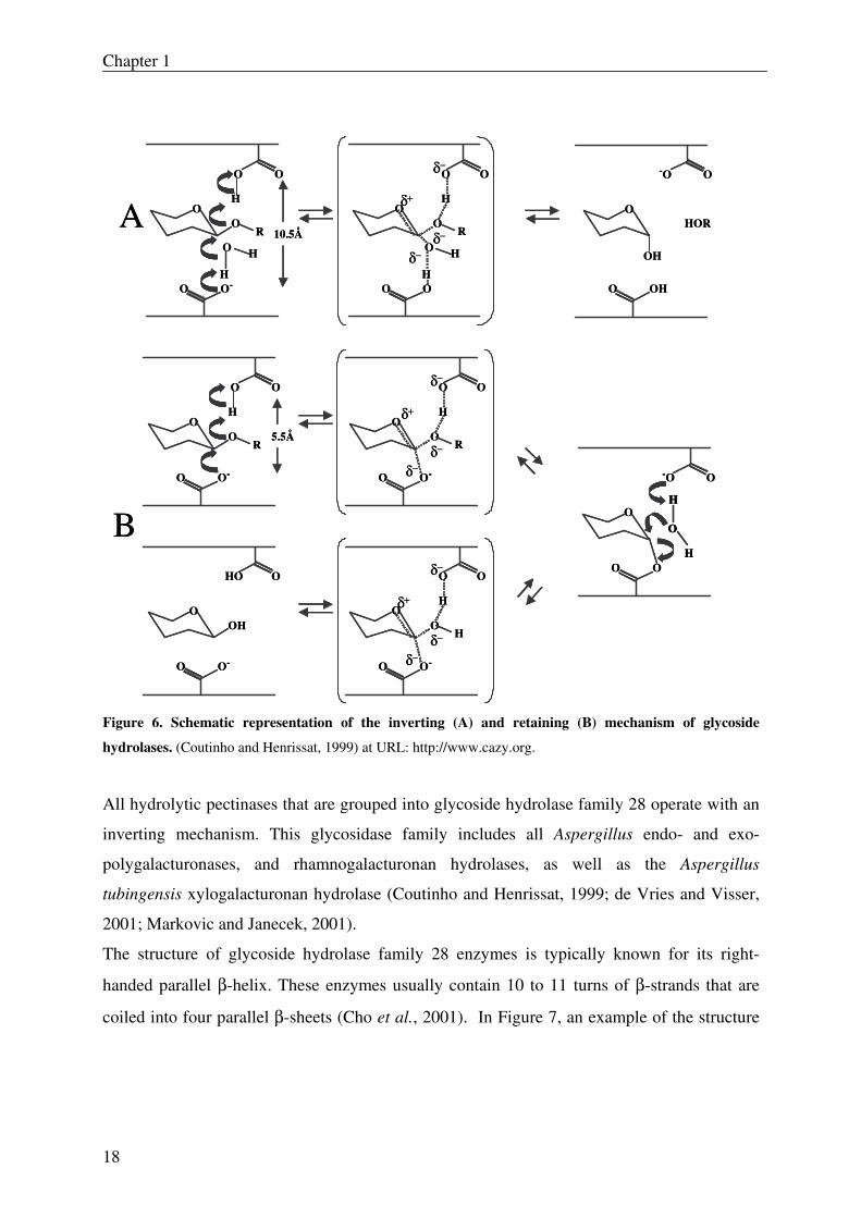
Glycoside hydrolases can degrade their corresponding substrate either by retaining or
inversion of the anomeric configuration (Fig. 6). Inverting glycoside hydrolases utilize a
direct displacement of the carbohydrate leaving group, which as a consequence results in the
inversion of the anomeric configuration (hence α configuration becomes β or vice versa).
Retaining glycoside hydrolases use a double-displacement mechanism with retention of the
anomeric configuration. Both mechanisms use two carboxyl residues on opposite sites of the
sugar plane which act as a general acid and a general base (for inverting glycoside
hydrolases), or as a general acid/ base and a nucleophile/leaving group (for retaining
glycoside hydrolases) respectively (Biely et al., 1996; McCarter and Withers, 1994; Sinnott,
1990).
Chapter 1
18
OOH
HO O
O-O
O
O O
O-O
O
δ−
H
Hδ−
δ+
δ−
O
O O
O-O
O
O O
O-O
O
δ−
H
Rδ−
δ+
δ−
OR
O
-O O
OO
H
H
O
H
O
O O
O-O
O
O O
OO
O
δ−
H
Rδ−
δ+
δ−
OR
HO
OH
-O O
OHO
A
B
H
H
O H
H
O
HOR10.5Å
5.5Å
OOH
HO O
O-O
O
O O
O-O
O
δ−
H
Hδ−
δ+
δ−
O
O O
O-O
O
O O
O-O
O
δ−
H
Rδ−
δ+
δ−
OR
O
-O O
OO
H
H
O
H
H
O
H
O
O O
O-O
O
O O
OO
O
δ−
H
Rδ−
δ+
δ−
OR
HO
OH
-O O
OHO
A
B
H
H
O H
H
O
HOR10.5Å
5.5Å
Figure 6. Schematic representation of the inverting (A) and retaining (B) mechanism of glycoside
hydrolases. (Coutinho and Henrissat, 1999) at URL: http://www.cazy.org.
All hydrolytic pectinases that are grouped into glycoside hydrolase family 28 operate with an
inverting mechanism. This glycosidase family includes all Aspergillus endo- and exo-
polygalacturonases, and rhamnogalacturonan hydrolases, as well as the Aspergillus
tubingensis xylogalacturonan hydrolase (Coutinho and Henrissat, 1999; de Vries and Visser,
2001; Markovic and Janecek, 2001).
The structure of glycoside hydrolase family 28 enzymes is typically known for its right-
handed parallel β-helix. These enzymes usually contain 10 to 11 turns of β-strands that are
coiled into four parallel β-sheets (Cho et al., 2001). In Figure 7, an example of the structure
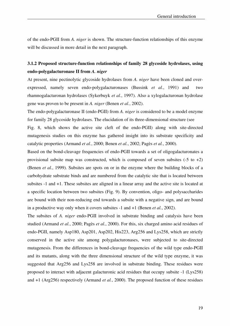
General introduction
19
of the endo-PGII from A. niger is shown. The structure-function relationships of this enzyme
will be discussed in more detail in the next paragraph.
3.1.2 Proposed structure-function relationships of family 28 glycoside hydrolases, using
endo-polygalacturonase II from A. niger
At present, nine pectinolytic glycoside hydrolases from A. niger have been cloned and over-
expressed, namely seven endo-polygalacturonases (Bussink et al., 1991) and two
rhamnogalacturonan hydrolases (Sykerbuyk et al., 1997). Also a xylogalacturonan hydrolase
gene was proven to be present in A. niger (Benen et al., 2002).
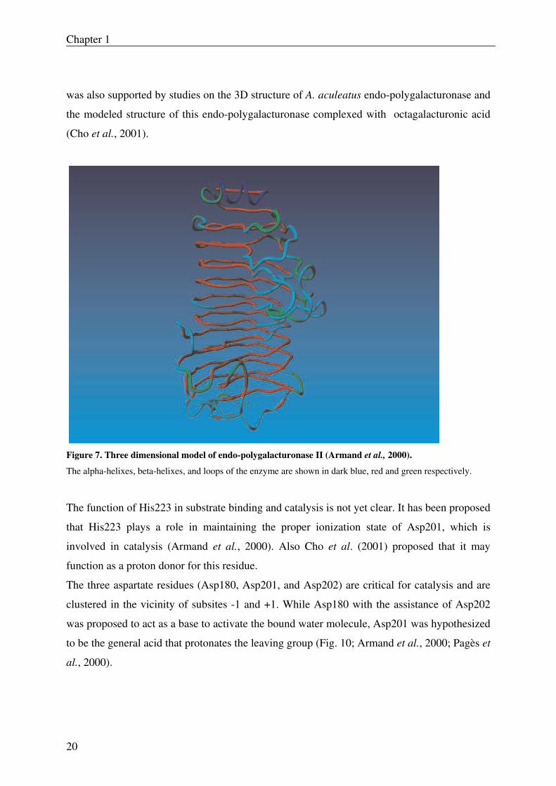
The endo-polygalacturonase II (endo-PGII) from A. niger is considered to be a model enzyme
for family 28 glycoside hydrolases. The elucidation of its three-dimensional structure (see
Fig. 8, which shows the active site cleft of the endo-PGII) along with site-directed
mutagenesis studies on this enzyme has gathered insight into its substrate specificity and
catalytic properties (Armand et al., 2000; Benen et al., 2002; Pagès et al., 2000).
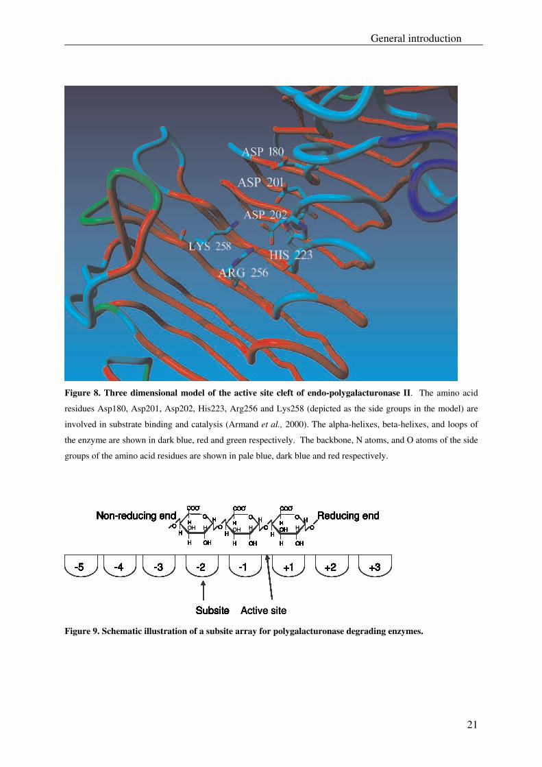
Based on the bond-cleavage frequencies of endo-PGII towards a set of oligogalacturonates a
provisional subsite map was constructed, which is composed of seven subsites (-5 to +2)
(Benen et al., 1999). Subsites are spots on or in the enzyme where the building blocks of a
carbohydrate substrate binds and are numbered from the catalytic site that is located between
subsites -1 and +1. These subsites are aligned in a linear array and the active site is located at
a specific location between two subsites (Fig. 9). By convention, oligo- and polysaccharides
are bound with their non-reducing end towards a subsite with a negative sign, and are bound
in a productive way only when it covers subsites -1 and +1 (Benen et al., 2002).
The subsites of A. niger endo-PGII involved in substrate binding and catalysis have been
studied (Armand et al., 2000; Pagès et al., 2000). For this, six charged amino acid residues of
endo-PGII, namely Asp180, Asp201, Asp202, His223, Arg256 and Lys258, which are strictly
conserved in the active site among polygalacturonases, were subjected to site-directed
mutagenesis. From the differences in bond-cleavage frequencies of the wild type endo-PGII
and its mutants, along with the three dimensional structure of the wild type enzyme, it was
suggested that Arg256 and Lys258 are involved in substrate binding. These residues were
proposed to interact with adjacent galacturonic acid residues that occupy subsite -1 (Lys258)
and +1 (Arg256) respectively (Armand et al., 2000). The proposed function of these residues
Chapter 1
20
was also supported by studies on the 3D structure of A. aculeatus endo-polygalacturonase and
the modeled structure of this endo-polygalacturonase complexed with octagalacturonic acid
(Cho et al., 2001).
Figure 7. Three dimensional model of endo-polygalacturonase II (Armand et al., 2000). The alpha-helixes, beta-helixes, and loops of the enzyme are shown in dark blue, red and green respectively.
The function of His223 in substrate binding and catalysis is not yet clear. It has been proposed
that His223 plays a role in maintaining the proper ionization state of Asp201, which is
involved in catalysis (Armand et al., 2000). Also Cho et al. (2001) proposed that it may
function as a proton donor for this residue.
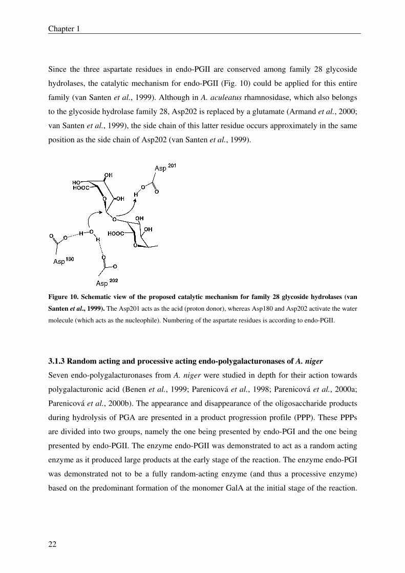
The three aspartate residues (Asp180, Asp201, and Asp202) are critical for catalysis and are
clustered in the vicinity of subsites -1 and +1. While Asp180 with the assistance of Asp202
was proposed to act as a base to activate the bound water molecule, Asp201 was hypothesized
to be the general acid that protonates the leaving group (Fig. 10; Armand et al., 2000; Pagès et
al., 2000).
General introduction
21
Figure 8. Three dimensional model of the active site cleft of endo-polygalacturonase II. The amino acid
residues Asp180, Asp201, Asp202, His223, Arg256 and Lys258 (depicted as the side groups in the model) are
involved in substrate binding and catalysis (Armand et al., 2000). The alpha-helixes, beta-helixes, and loops of
the enzyme are shown in dark blue, red and green respectively. The backbone, N atoms, and O atoms of the side
groups of the amino acid residues are shown in pale blue, dark blue and red respectively.
-5 -4 -3 -2 -1 +1 +2 +3
Subsite Active site
COO-
HO
HO
HO
H
HO
H
O
OH
OH H
H
HO
OH
OH H
H
H
COO-
O
OH
OH H
H
H
COO-
Reducing endNon-reducing end
-5 -4 -3 -2 -1 +1 +2 +3
Subsite
COO-
HO
HO
HO
H
HO
H
O
OHH
HO
OHH
H
COO-
O
OH
OH
H
H
COO-COO-
HO
HO
HO
H
HO
H
O
OHH
HO
OHH
H
COO-
O
OH
OH
H
H
COO-
Reducing endNon-reducing end
-5 -4 -3 -2 -1 +1 +2 +3
Subsite
COO-
HO
HO
HO
H
HO
H
O
OHH
HO
OHH
H
COO-
O
OH
OH
H
H
COO-COO-
HO
HO
HO
H
HO
H
O
OHH
HO
OHH
H
COO-
O
OH
OH
H
H
COO-
Reducing endNon-reducing end
-5 -4 -3 -2 -1 +1 +2 +3
Subsite
COO-
HO
HO
HO
H
HO
H
O
OHH
HO
OHH
H
COO-
O
OH
OH
H
H
COO-COO-
HO
HO
HO
H
HO
H
O
OHH
HO
OHH
H
COO-
O
OH
OH
H
H
COO-
Reducing endNon-reducing end
-5 -4 -3 -2 -1 +1 +2 +3
Subsite Active site
COO-
HO
HO
HO
H
HO
H
O
OHH
HO
OHH
H
COO-
O
OH
OH
H
H
COO-COO-
HO
HO
HO
H
HO
H
O
OHH
HO
OHH
H
COO-
O
OH
OH
H
H
COO-
Reducing endNon-reducing end
-5 -4 -3 -2 -1 +1 +2 +3
Subsite
COO-
HO
HO
HO
H
HO
H
O
OHH
HO
OHH
H
COO-
O
OH
OH
H
H
COO-COO-
HO
HO
HO
H
HO
H
O
OHH
HO
OHH
H
COO-
O
OH
OH
H
H
COO-
Reducing endNon-reducing end
-5 -4 -3 -2 -1 +1 +2 +3
Subsite
COO-
HO
HO
HO
H
HO
H
O
OHH
HO
OHH
H
COO-
O
OH
OH
H
H
COO-COO-
HO
HO
HO
H
HO
H
O
OHH
HO
OHH
H
COO-
O
OH
OH
H
H
COO-
Reducing endNon-reducing end
-5 -4 -3 -2 -1 +1 +2 +3
Subsite
COO-
HO
HO
HO
H
HO
H
O
OHH
HO
OHH
H
COO-
O
OH
OH
H
H
COO-COO-
HO
HO
HO
H
HO
H
O
OHH
HO
OHH
H
COO-
O
OH
OH
H
H
COO-
Reducing endNon-reducing end
-5 -4 -3 -2 -1 +1 +2 +3
Subsite Active site
COO-
HO
HO
HO
H
HO
H
O
OH
OH H
H
HO
OH
OH H
H
H
COO-
O
OH
OH H
H
H
COO-
Reducing endNon-reducing end
-5 -4 -3 -2 -1 +1 +2 +3
Subsite
COO-
HO
HO
HO
H
HO
H
O
OHH
HO
OHH
H
COO-
O
OH
OH
H
H
COO-COO-
HO
HO
HO
H
HO
H
O
OHH
HO
OHH
H
COO-
O
OH
OH
H
H
COO-
Reducing endNon-reducing end
-5 -4 -3 -2 -1 +1 +2 +3
Subsite
COO-
HO
HO
HO
H
HO
H
O
OHH
HO
OHH
H
COO-
O
OH
OH
H
H
COO-COO-
HO
HO
HO
H
HO
H
O
OHH
HO
OHH
H
COO-
O
OH
OH
H
H
COO-
Reducing endNon-reducing end
-5 -4 -3 -2 -1 +1 +2 +3
Subsite
COO-
HO
HO
HO
H
HO
H
O
OHH
HO
OHH
H
COO-
O
OH
OH
H
H
COO-COO-
HO
HO
HO
H
HO
H
O
OHH
HO
OHH
H
COO-
O
OH
OH
H
H
COO-
Reducing endNon-reducing end
-5 -4 -3 -2 -1 +1 +2 +3
Subsite Active site
COO-
HO
HO
HO
H
HO
H
O
OHH
HO
OHH
H
COO-
O
OH
OH
H
H
COO-COO-
HO
HO
HO
H
HO
H
O
OHH
HO
OHH
H
COO-
O
OH
OH
H
H
COO-
Reducing endNon-reducing end
-5 -4 -3 -2 -1 +1 +2 +3
Subsite
COO-
HO
HO
HO
H
HO
H
O
OHH
HO
OHH
H
COO-
O
OH
OH
H
H
COO-COO-
HO
HO
HO
H
HO
H
O
OHH
HO
OHH
H
COO-
O
OH
OH
H
H
COO-
Reducing endNon-reducing end
-5 -4 -3 -2 -1 +1 +2 +3
Subsite
COO-
HO
HO
HO
H
HO
H
O
OHH
HO
OHH
H
COO-
O
OH
OH
H
H
COO-COO-
HO
HO
HO
H
HO
H
O
OHH
HO
OHH
H
COO-
O
OH
OH
H
H
COO-
Reducing endNon-reducing end
-5 -4 -3 -2 -1 +1 +2 +3
Subsite
COO-
HO
HO
HO
H
HO
H
O
OHH
HO
OHH
H
COO-
O
OH
OH
H
H
COO-COO-
HO
HO
HO
H
HO
H
O
OHH
HO
OHH
H
COO-
O
OH
OH
H
H
COO-
Reducing endNon-reducing end
Figure 9. Schematic illustration of a subsite array for polygalacturonase degrading enzymes.
Chapter 1
22
Since the three aspartate residues in endo-PGII are conserved among family 28 glycoside
hydrolases, the catalytic mechanism for endo-PGII (Fig. 10) could be applied for this entire
family (van Santen et al., 1999). Although in A. aculeatus rhamnosidase, which also belongs
to the glycoside hydrolase family 28, Asp202 is replaced by a glutamate (Armand et al., 2000;
van Santen et al., 1999), the side chain of this latter residue occurs approximately in the same
position as the side chain of Asp202 (van Santen et al., 1999).
Figure 10. Schematic view of the proposed catalytic mechanism for family 28 glycoside hydrolases (van
Santen et al., 1999). The Asp201 acts as the acid (proton donor), whereas Asp180 and Asp202 activate the water
molecule (which acts as the nucleophile). Numbering of the aspartate residues is according to endo-PGII.
3.1.3 Random acting and processive acting endo-polygalacturonases of A. niger
Seven endo-polygalacturonases from A. niger were studied in depth for their action towards
polygalacturonic acid (Benen et al., 1999; Parenicová et al., 1998; Parenicová et al., 2000a;
Parenicová et al., 2000b). The appearance and disappearance of the oligosaccharide products
during hydrolysis of PGA are presented in a product progression profile (PPP). These PPPs
are divided into two groups, namely the one being presented by endo-PGI and the one being
presented by endo-PGII. The enzyme endo-PGII was demonstrated to act as a random acting
enzyme as it produced large products at the early stage of the reaction. The enzyme endo-PGI
was demonstrated not to be a fully random-acting enzyme (and thus a processive enzyme)
based on the predominant formation of the monomer GalA at the initial stage of the reaction.
General introduction
23
Also the action of this enzyme towards a hexamer (GalA6) confirmed a processive behavior
(Benen et al., 1999; Benen et al., 2003b).
With the aid of the amino acid sequence alignments of the A. niger endo-polygalacturonases,
as well as the structure of endo-PGII, it was found that an arginine at position 96 (Arg96) in
the endo-PGI group was probably accountable for the processive behavior of these enzymes.
For the random acting enzymes (endo-PGII group) the Ser91 residue was found at the
equivalent position of the processive enzymes. By replacement of Arg96 for a Ser96 in endo-
PGI and Ser91 for a Arg91 in endo-PGII the processive behavior was interchanged between
these two enzymes. While endo-PGI-R95S behaved as a random acting enzyme, endo-PGII-
S91R possessed properties of a processive enzyme (van Pouderoyen et al., 2003).
3.2 Function of pectinases in the food industry
Pectinases from food-grade fungi, such as A. niger, are used predominantly as processing aids
in the manufacture of fruit juices. The application of these exogenous pectinolytic enzymes
improves and influences the efficiency of the fruit juice process, which cannot be achieved
alone by the endogenous enzymes that occur naturally in the fruit. Upon grinding of the raw
fruit, pectinases are added which reduce the viscosity of the pectin-rich crude juice, also
known as pulp enzyming, and therefore improve the processing capacity and the yield of the
fruit juice (Benen and Voragen, 2003a).
While in the early 1930s enzyme mixtures of various glycosidases were used, nowadays it
becomes possible to use tailor made enzyme preparations for specific applications. For
instance, food-grade fungal enzyme preparations which mainly contain endo-
polygalacturonase activity can be used to produce pulpy nectars in a process called
maceration. These nectars are known for their higher content of fruit solids, pigments and
nutrients compared to those which are processed thermo-mechanically (Voragen et al., 1992).
The production of fruit juices can also be achieved by liquefaction of the fruit tissues. In this
process the fruit tissues are solubilized by using a broad spectrum of polysaccharide
degrading enzymes, such as pectin, hemicellulose, and cellulose degrading enzymes (Grassin
and Fauquembergue, 1996; Uhlig, 1998; Benen and Voragen, 2003a). The liquefaction
technique is relatively simple and economical for the manufacture of high yields of clear or
cloudy juices. It is particularly useful for those fruits, such as mango, guava and banana, from
Chapter 1
24
which no juice can be obtained by pressing (Benen and Voragen, 2003a). Liquefaction also
limits losses of nutrients which occurs when mechanical pressing is applied, as for instance
for the manufacture of carrot juice (Voragen et al., 1992).
4 Aim and outline of this thesis
The aim of this thesis was to biochemically identify and characterize pectinases, which belong
to the glycoside hydrolase family 28 of A. niger, that became available in the Carbnet project.
Special emphasis was put on the characterization of the xylogalacturonan hydrolase (XGH).
This enzyme was discovered in A. tubingenisis (Van der Vlugt-Bergmans et al., 2000), and
subsequently cloned and over-expressed in the A. niger “PlugBug” (van Dijck, 1999).
Although XGH originates from A. tubingensis, similar characteristics are also expected for
XGH from A. niger as these enzymes have an amino acid sequence identity of 97 % (Chapter
two).
Identification and characterization of the pectinolytic enzymes from this family was realized
by investigating their activity towards several different pectin substrates which were
chemically and/ or enzymatically modified. Other pectinolytic enzymes from A. niger such as
pectin, pectate lyases, rhamnogalacturonan lyases, as well as accessory enzymes of this
species, such as pectin esterases, endo- and exo-arabinases, were not covered in this thesis.
The entire set of the pectinolytic glycoside hydrolase family 28 from A. niger, as obtained by
bioinformatics tools, micro-array techniques, protein sequencing, and biochemical
identification methods using pectin substrates, is illustrated in Chapter two. As described in
Chapter three, the enzyme xylogalacturonan hydrolase (XGH) was further investigated for its
action towards a xylogalacturonan (XGA) derived from gum tragacanth in order to understand
its mode of action towards this substrate analogue. As a side project of Carbnet, the action of
XGH was further studied towards XGA in the saponified modified “hairy” regions of apple
and potato pectin (Chapter four) which also enabled us to study the structural features of this
polymer from both sources. Also the presence of XGA in plant cell walls (Arabidopsis
thaliana), other than storage or reproductive tissues, was demonstrated by using this enzyme
(Chapter five).
General introduction
25
Abarca, M.L., Accensi, F., Cano, J., Cabañes, F.J. 2004. Taxonomy and significance of black aspergilli. Antonie van Leeuwenhoek 86:33-49.
Albersheim, P., Darvill, A.G., O'Neill, M.A., Schols, H.A., and Voragen, A.G.J. 1996. An Hypothesis: The Same Six Polysaccharides are Components of the Primary Cell Walls of All Higher Plants, p. 47-55, In J. Visser, Voragen, A.G.J., ed. Pectins and Pectinases. Elsevier Science.
Armand, S., Wagemaker, M.J.M., Sánchez-Torres, P., Kester, H.C.M., van Santen, Y., Dijkstra, B.W., Visser, J., Benen, J.A.E. 2000. The Active Site Topology of Aspergillus niger Endopolygalacturonase II as Studied by Site-directed Mutagenisis. The Journal of Biological Chemistry 275:691-696.
Aspinall, G.O., Baillie, J. 1963. Gum Tragacanth. Part I. Fractionation of the gum and the structure of tragacanthic acid. Journal of the chemical society 318:1702-1714.
Beldman, G., Mutter, M., Searle-van Leeuwen, M.J.F., van den Broek, L.A.M., Schols, H.A., Voragen, A.G.J. 1996. New enzymes active towards pectic structures, p. 231-245, In J. Visser, Voragen, A.G.J., ed. Pectins and Pectinases, Vol. 14. Elsevier Science, Amsterdam.
Beldman, G.S., H.A., Pitson, S.M., Searle-van Leeuwen, M.J.F., and Voragen, A.G.J. 1997. Arabinans and arabinan degrading enzymes, p. 1-64, In R. J. Sturgeon, ed. Advances in Macromolecular Carbohydrate Research, Vol. 1. JAI Press INC.
Benen, J.A.E., and Voragen, A.G.J. 2003a. Pectic Enzymes, p. 845-848, In J. R. Whitaker, Voragen, A.G.J., Wong, D.W.S., ed. Handbook of Food Enzymology. Marcel Dekker, Inc, New York.
Benen, J.A.E., Kester, H.C.H., Visser, J. 1999. Kinetic characterization of Aspergillus niger N400 endopolygalacturonases I, II and C. European Journal of Biochemistry 259:72-80.
Benen, J.A.E., van Alebeek, G.-J.W.M., Voragen, A.G.J., Visser, J. 2003b. Mode of Action Analysis And Structure-Function Relationships Of Aspergillus Niger Pectinolytic Enzymes, p. 235-256, In A. G. J. Voragen, Schols, H., Visser, R., ed. Advances in Pectin and Pectinase Research. Kluwer Academic Publishers, Dordrecht.
Benen, J.A.E., van Alebeek, G.-J.W.M., Voragen, A.G.J., Visser, J. 2003c. Pectic Esterases, p. 849-856, In J. R. Whitaker, Voragen, A.G.J., Wong, D.W.S., ed. Handbook of Food Enzymology. Marcel Dekker, Inc, New York.
Benen, J.A.E., Vincken, J.-P., van Alebeek, G.-J.W.M. 2002. Microbial Pectinases, In G. B. Seymour, Knox, J.P., ed. Pectins and their Manipulation. Blackwell Publishing, Oxford.
Benen, J.A.E., Visser, J. 2003d. Polygalacturonases, p. 857-866, In J. R. Whitaker, Voragen, A.G.J., Wong, D.W.S., ed. Handbook of Food Enzymology. Marcel Dekker, Inc., New York.
Bhat, M.K. 2000. Research review paper: Cellulases and related enzymes in biotechnology. Biotechnology Advances 18:355-383.
Biely, P., Benen, J., Heinrichovia, K., Kester, H.C.M, Visser, J. 1996. Inversion of configuration during hydrolysis of α-1,4-galacturonidic linkage by three Aspergillus polygalacturonases. FEBS Letters 382:249-255.
Bouveng, H.O. 1965. Polysaccharides in pollen. 2. Xylogalacturonan from mountain pine (Pinus mugoturra) pollen. Acta Chemica Scandinavica 19.
Chapter 1
26
Bussink, H.J., Buxton, F.P., Fraaye, B.A., de Graaf, L.H., Visser, J. 1991. The polygalacturonases of Aspergillus niger are encoded by a family of diverged genes. European Journal of Biochemistry 208:83-90.
Carpita, N., Gibeaut, D.M. 1993. Structural models of primary cell walls in flowering plants: consistency of molecular structure with the physical properties of the walls during growth. The Plant Journal 3:1-30.
Chaudhary, K., Ethiray, S., Lakshminarayana, K., Tauro, P. 1978. Citric Acid Production from Indian Cane Molasses by Aspergillus niger under solid state Fermentation Conditions. Fermentation Technology 65:554-557.
Cho, S., S. Lee, W. Shin, S.W. Cho, S. Lee, and W. Shin. 2001. The X-ray structure of Aspergillus aculeatus polygalacturonase and a modeled structure of the polygalacturonase-octagalacturonate complex. Journal of Molecular Biology 311:863-878.
Coutinho, P.M., Henrissat, B. 1999. Carbohydrate-active enzymes: an integrated database approach, p. 3-12, In G. D. H.J. Gilbert, B. Henrissat and B. Svensson, ed. Recent Advances in Carbohydrate Bioengineering. The Royal Society of Chemistry, Cambridge.
de Vries, R.P., Visser, J. 2001. Aspergillus Enzymes Involved in Degradation of Plant Cell Wall Polysaccharides. American Society for Microbiology 65:497-522.
Dijck, P.W.M., Selten, G.C.M., Hempenius, R.A. 2003. On the safety of a new generation of DSM Aspergillus niger enzyme production strains. Regulatory Toxicology and Pharmacology 38:27-35.
Fry, S.C. 1988. The growing Plant Cell Wall: Chemical and Metabolic Analysis Longman Scientific & Technical, Essex.
Gibeaut, D.M., Carpita, N.C. 1994. Biosynthesis of plant cell wall polysaccharides. The FASEB Journal 8:904-915.
Grassin, C., Fauquembergue, P. 1996. Application of Pectinases in Beverages, p. 453-462, In J. Visser, Voragen, A.G.J., ed. Pectins and Pectinases. Elsevier Science.
Gross, K.C., and Wallner, S.J. 1979. Degradation of Cell Wall Polysaccharides during Tomato Fruit Ripening. Plant Physiology 63:117-120.
Hu, H., Brown, P.H. 1994. Localization of Boron in Cell Walls of Squas and Tobacco and Its Association with Pectin. Plant Physiology 105:681-689.
Huisman, M.M.H., Brüll, L.P., Thomas-Oates, J.E., Haverkamp, J. 2001. The occurrence of internal (1, 5)-linked arabinofuranose and arabinopyranose residues in arabinogalactan side chains from soybean pectic substances. Carbohydrate Research 330:103-114.
Ikram-ul, H., Ali, S., Qadeer, M.A., Iqbal, J. 2004. Citric acid production by selected mutants of Aspergillus niger from cane molasses. Bioresource Technology 93:125-130.
Ishii, T. 1997. Structure and functions of feruloyated polysaccharides. Plant Science 127:111-127.
Ishii, T., Matsunaga, T. 2001. Pectic polysaccharide rhamnogalacturonan II is covalently linked to homogalacturonan. Phytochemistry 57:969-974.
Ishii, T., Matsunaga, T., Pellerin, P., O' Neill, M.A., Darvill, A., Albersheim, P. 1999. The plant cell wall polysaccharide rhamnogalacturonan II self-assembles into a covalently cross-linked dimer. Journal of Biological Chemistry 274:13098-13104.
Kashyap, D.R., Vohra, P.K., Chopra, S., Tewari, R. 2001. Applications of pectinases in the commercial sector: a review. Bioresource Technology 77:215-227.
General introduction
27
Kikuchi, A., Edashige, Y., Ishii, T., Satoh, S. 1996. A xylogalacturonan whose level is dependent on the size of cell clusters is present in the pectin from cultured carrot cells. Planta 200.
Kofod, L.V., Kauppinen, S., Christgau, S., Andersen, L.N., Heldt-Hansen, H.P., Dorreich, K., Dalbφge, H. 1994. Cloning and characterization of two structurally and functionally divergent rhamnogalacturonases from Aspergillus aculeatus. Journal of Biological Chemistry 269:29182-29189.
Le Goff, A., C.M.G.C. Renard, E. Bonnin, and J.F. Thibault. 2001. Extraction, purification and chemical characterisation of xylogalacturonans from pea hulls. Carbohydrate Polymers 45:325-334.
Mafra, I., Lanza, B., Reis, A., Marsilio, V., Campestre, C., De Angelis, M., Coimbra, M.A. 2001. Effect of ripening on texture, microtexture and cell wall polysaccharide composition of olive fruit (olea europaea). Physiologia Plantarum 111:439-447.
Magnuson, J.K., Lasure, L.L. 2004. Organic Acid Production by Filamentous Fungi, In J. S. Tkacz, Lange, L., ed. Advances in Fungal Biotechnology for Industry, Agriculture, and Medicine. Springer.
Markovic, O., Janecek, S. 2001. Pectin degrading glycoside hydrolases of family 28: sequence-structural features, specificities and evolution. Protein Engineering 14:615-631.
May, C.D. 2000. Pectins, p. 169-188, In G. O. Philips, and Williams, P.A., ed. Handbook of hydrocolloids. Woodhead Publishing Limited, Boca Raton-Boston-New York-Washington DC.
McCarter, J.D., Withers, S.G. 1994. Mechanisms of enzymatic glycoside hydrolysis. Current Opinion in Structural Biology 4:885-892.
McNeil, M., Darvill, A.G., Albersheim, P. 1980. Structure of plant cell walls. XII. Identification of seven differently linked glycosyl residues attached to O-4 of the 2,4 linked L-rhamnosyl residues of rhamnogalacturonan I. Plant Physiology 70:1586-1591.
McNeil, M., Darvill, A.G., Fry, S.C., Albersheim, P. 1984. Structure and function of the primary cell walls of plants. Annual Review of Biochemistry 53:625-663.
Missang, C.E., Baron, A., Renard, C.M.G.C. 2004. Cell wall-degrading enzymes and changes in cell wall polysaccharides during ripening and storage of bush butter (Dacryodes edulis (G. Don) H.J. Lam) fruit. Journal of Horticultural Science & Biotechnology 79:797-805.
Mohnen, D. 1999. Biosynthesis of pectins and galactomannans, p. 497-527, In D. Barton, Nakanishi, K., Meth-Cohn, O., ed. Comprehensive Natural Products Chemistry, Vol. 3. Elsevier.
Mort, A.J. 2002. Interactions between pectins and other polymers, p. 30-51, In G. B. Seymour, Know, J.P., ed. Pectins and their manipulation. Blackwell Publishing.
Mutter, M. 1997. New rhamnogalacturonan degrading enzymes from Aspergillus aculeatus. pp141. The Wageningen Agricultural University, Wageningen, Netherlands.
Mutter, M., Beldman, G., Pitson, S.M., Schols, H.A. and Voragen, A.G.J. 1998. Rhamnogalacturonan α-D-Galactopyranosyluronohydrolase. An enzyme that specifically removes the terminal nonreducing galacturonosyl residue in rhamnogalacturonan regions of pectin. Plant Physiology 117:153-163.
Mutter, M., Beldman, G., Schols, H.A. and Voragen, A.G.J. 1994. Rhamnogalacturonan α-L-rhamnopyranosylhydrolase. A novel enzyme specific for the terminal non-reducing
Chapter 1
28
rhamnosyl unit in rhamnogalacturonan regions of pectin. Plant Physiology 106:241-250.
O' Neill, M.A., York, W.S. 2003. The composition and structure of plant primary cell walls, p. 1-54, In J. K. C. Rose, ed. The Plant Cell Wall, Vol. 8. Blackwell Publishing, Ithaca, New York, USA.
Oechslin, R., Lutz, M.V., Amado, R. 2003. Pectic substances isolated from apple cellulosic residue: structural characterisation of a new type of rhamnogalacturonan I. Carbohydrate Polymers 51:301-310.
Oxenbøll, K. 1994. Aspergillus enzymes and industrial uses, p. 147-154, In K. A. Powell, Renwick, A., Peberdy, J.F., ed. The Genus Aspergillus: From Taxonomy and Genetics to Industrial Applications. Plenum Press, London.
Pagès, S., Heijne, W.H.M., Kester, H.C.M., Visser, J., Benen, J.A.E. 2000. Subsite Mapping of Aspergillus niger Endopolygalacturonase II by Site-directed Mutagenisis. The Journal of Biological Chemistry 275:29348-29353.
Parenicová, L., Benen, J.A.E., Kester, H.C.M., Visser, J. 1998. pgaE encodes a fourth member of the endopolygalacturonase gene family from Aspergillus niger. European Journal of Biochemistry 251:72-80.
Parenicová, L., Benen, J.A.E., Kester, H.C.M., Visser, J. 2000a. pgaA and pgaB encode constitutively expressed members of the Aspergillus niger endopolygalacturonase gene family. Biochemical Journal 345:637-644.
Parenicová, L., Kester, H.C.M., Benen, J.A.E., Visser, J. 2000b. Characterization of a novel endopolygalacturonase from Aspergillus niger with unique kinetic properties. FEBS Letters 467:333-336.
Pilnik, W., Voragen, A.G.J. 1991. The significance of endogenous and exogenous pectic enzymes in fruit and vegetable processing, p. 303-336, In P. F. Fox, ed. Food Enzymology, Vol. 1. Elsevier Applied Science, London.
Pitt, J.I., Hocking, A.D. 1985. Fungi And Food Spoilage Academic Press Australia, Sydney. Pitt, J.I., Hocking, A.D. 1997. Fungi and Food Spoilage Blackie Academic & Professional,
London, UK. Prado, F.C., Vandenberghe, L.P.S., Soccol, C.R. 2005. Relation between Citric acid
Production by Solid-State Fermentation from Cassava Bagasse and Respiration of Aspergillus niger LPB 21 in Semi-Pilot Scale. Brazilian Archives of Biology and Technology 48:29-36.
Rao, M.A., Acree, T.E., Colley, H.J., and Ennis, R.W. 1987. Clarification of Apple Juice by Hollow Fiber Ultrafiltration: Fluxes and Retention of Odor-Active Volatiles. Journal of Food Science 52:375-377.
Raper, K.B., Fennell, D.I. 1965. The Genus Aspergillus Williams and Wilkins, U.S.A. Redgewell, R.J., Fischer, M., Kendal, E., MacRae, E.A. 1997. Galactose loss and fruit
ripening: high-molecular-weight arabinogalactans in the pectic polysaccharides of fruit cell walls. Planta 203:174-181.
Ridley, B.L., O'Neill, M.A., Mohnen, D. 2001. Pectins: structure, biosynthesis, and oligogalacturonide-related signaling. Phytochemistry 57:929-967.
Schobinger, U. 1988. Möglichkeiten und Grenzen der Ultrafiltration in der Klärung von Apfelsaft. Flüssiges Obst 55:614-620.
Schols, H.A., E.J. Bakx, D. Schipper, and A.G.J. Voragen. 1995. A xylogalacturonan subunit present in the modified hairy regions of apple pectin. Carbohydrate Research. Dec 279:265-279.
General introduction
29
Schols, H.A., Geraeds, C.C.J.M., Searle-van Leeuwen, M.J.F., Kormelink, F.J.M., Voragen, A.G.J. 1990. Rhamnogalacturonase: a novel enzyme that degrades the hairy regions of pectins. Carbohydrate Research 206:105-115.
Schols, H.A., Voragen, A.G.J. 1996. Complex Pectins: Structure elucidation using enzymes, p. 3-19, In J. Visser, Voragen, A.G.J., ed. Pectins and pectinases, Vol. 14. Elsevier Science B.V., Amsterdam.
Schols, H.A., Voragen, A.G.J. 2002. The chemical structure of pectins, p. 1-29, In G. B. Seymour, Knox, J.P., ed. Pectins and their Manipulation. Blackwell Science.
Schuster, E., Dunn-Colemann, N., Frisvad, J.C., van Dijck, P.W.M. 2002. On the safety of Aspergillus niger- a review. Applied Microbiology and Biotechnology 59:426-435.
Sinnott, M.L. 1990. Catalytic mechanisms of enzymic glycoside transfer 90:1170-1202. Sykerbuyk, M.E.G., Kester, H.C.M., Schaap, P.J., Stam, H., Musters, W., Visser, J. 1997.
Cloning and characterization of two rhamnogalacturonan hydrolase genes from Aspergillus niger. Applied and Environmental Microbiology 63:2507-2515.
Thibault, J.-F., Ralet, M.-C. 2001. Pectins, their Origin, Structure and Functions, p. 369- 378, In B. V. McCleary, Prosky, L., ed. Advanced Dietary Fibre Technology. Blackwell Science, Oxford.
Uhlig, H. 1998. Industrial enzymes and their applications John Wiley & Sons, New York. Van der Vlugt-Bergmans, C.J.B., Meeuwsen, P.J.A., Voragen, A.G.J., and van Ooyen, A.J.J.
2000. Endo-Xylogalacturonan hydrolase, a Novel Pectinolytic Enzyme. Applied and Environmental Microbiology 66:36-41.
van Dijck, P.W.M. 1999. Chymosin and Phytase. Made by genetic engineering (No. 10 in a series of articles to promote a better understanding of the use of genetic engineering). Journal of Biotechnology 67:77-80.
van Pouderoyen, G., Snijder, H.J., Benen, J.A.E., Dijkstra, B.W. 2003. Structural insights into the processivity of endopolygalacturonase I from Aspergillus niger. FEBS Letters 554:462-466.
van Santen, Y., Benen, J.A.E., Schröter, K.-H., Kalk, K.H., Armand, S., Visser, J., Dijkstra, B.W. 1999. 1.68-Å Crystal Structure of Endopolygalacturonase II from Aspergillus niger and Identification of Active Site Residues by Site-directed Mutagenisis. The Journal of Biological Chemistry 274:30474-30480.
Vandenberghe, L.P.S.L., Soccol, C.R., Pandey, A., Lebault, J.-M. 2000. Solid-state fermentation for the synthesis of citric acid by Aspergillus niger. Bioresource Technology 74:175-178.
Versari, A., S. Biesenbruch, D. Barbanti, P.J. Farnell, and S. Galassi. 1999. HPAEC-PAD analysis of oligogalacturonic acids in strawberry juice. Food Chemistry 66:257-261.
Vincken, J.-P., Schols, H.A., Oomen, R.J.F.J., Beldman, G., Visser, R.G.F., Voragen, A.G.J. 2003. Pectin-The hairy thing, p. 47-59, In A. G. J. Voragen, Schols, H., Visser, R., ed. Advances in Pectin and Pectinase Research. Kluwer Academic Publishers, Dordrecht.
Voragen, A.G.J., and Pilnik, W., Thibault, J.-F., Axelos, M.A.V., Renard, C.M.G.C. 1995. Pectins, p. 287-339, In A. M. Stephen, ed. Food Polysaccharides. Marcel Dekker, Inc, New York-Basel-Hong Kong.
Voragen, A.G.J., Beldman, G., Schols, H. 2001. Chemistry and Enzymology of Pectins, p. 379-397, In B. V. McCleary, Prosky, L., ed. Advanced Dietary Fibre Technology. Blackwell Science, Oxford.
Voragen, A.G.J., Schols, H.A., and Beldman, G. 1992. Maßgeschneiderte Enzyme in der Fruchtsaftherstellung. Flüssiges Obst 7:404-410.
Chapter 1
30
Wakabayashi, K. 2000. Changes in Cell Wall Polysaccharides During Fruit Ripening. Journal of Plant Research 113:231-237.
Weightman, R.M., Renard, C.M.G.C., Thibault, J.-F. 1994. Structure and properties of the polysaccharides from pea hulls. Part I: Chemical extraction and fractionation of the polysaccharides. Carbohydrate Polymers 24:139-148.
Whitaker, J.R. 1990. Microbial pectolytic enzymes, p. 133-176, In W. M. Fogarty, Kelly, C.T., ed. Microbial enzymes and biotechnology, 2 ed. Elsevier Applied Science, London and New York.
Yamada, H. 1996. Contribution of pectins on health care, p. 173-190, In J. Visser, Voragen, A.G.J., ed. Pectins and Pectinases, Vol. 14. Elsevier Science, Amsterdam.
Yu, L., Mort, A.J. 1996. Partial characterization of xylogalacturonans from cell walls of ripe watermelon fruit: inhibition of endopolygalacturonase activiy by xylosilation, p. 79-88, In J. Visser, Voragen, A.G.J., ed. Pectins and Pectinases, Vol. 14. Elsevier Science, Amsterdam.
Zablackis, E., Huang, J., Muller, B., Darvill, A.G., Albersheim, P. 1995. Characterization of the Cell-Wall Polysaccharides of Arabidopsis thaliana Leaves. Plant Physiology 107:1129-1138.
Chapter 2
31
A new group of exo-acting family 28 glycoside hydrolases of
Aspergillus niger that are involved in pectin degradation
E.S. Martens-Uzunova1, J.S. Zandleven1, J.A.E. Benen, H. Awad, H.J. Kools, G. Beldman, A.G.J. Voragen, J.A.
van den Berg, and P.J. Schaap
1 These authors contributed equally to this work.
Abstract
The fungus Aspergillus niger is an industrial producer of pectin degrading enzymes. The
recent solving of the genomic sequence of A. niger allowed an inventory of the entire genome
of the fungus for potential carbohydrate degrading enzymes. By applying bioinformatics tools
12 new genes putatively encoding family 28 glycoside hydrolases were identified. Seven of
the newly discovered genes form a new gene group, which we show to encode exo-acting
pectinolytic glycoside hydrolases. This group includes four exo-polygalacturonan hydrolases
(PGAX, PGXA, PGXB and PGXC) and three putative exo-rhamnogalacturonan hydrolases
(RGXA, RGXB and RGXC). Biochemical identification using polygalacturonic acid and
xylogalacturonan as substrates demonstrated that indeed PGXB and PGXC act as exo-
polygalacturonases while PGXA acts as an exo-xylogalacturonan hydrolase.
The expression levels of all 21 genes were assessed by microarray analysis. The results from
this study demonstrate that exo-acting glycoside hydrolases play a prominent role in pectin
degradation.
This chapter has been accepted for publication in Biochemical Journal
Chapter 2
32
Introduction
Pectin is a complex heteropolymer present in the middle lamella of the primary cell wall of
plants. This biopolymer accounts for about one-third of the total cell wall material [1] and as
such represents an important carbon source for bacteria and fungi. Pectin is composed of a
number of distinct polysaccharides [2], such as homogalacturonan (HG), xylogalacturonan
(XGA), rhamnogalacturonan I (RGI), and rhamnogalacturonan II (RGII).
Rhamnogalacturonan carries short and long, linear and branched side chains which are built
up from neutral sugars (e.g. arabinose, galactose).
Two pectin models exist, namely a pectin structure in which HG is an extension of RGI or
vice versa and has neutral sugar side chains [3], and a pectin structure that is built from a
backbone of RGI where HG and neutral sugars form the side chains [2].
HG, also known as the smooth region of pectin, is an unbranched polymer composed of 1,4-
linked �-D-galacturonic acid residues. XGA, RGI and RGII are part of the branched or
“hairy” regions of pectin [4]. In XGA xylose is linked at O-3 of the galacturonyl residues of
the HG backbone. Rhamnogalacturonan I (RGI) is composed of a backbone of alternating α-
(1, 2)-linked rhamnosyl and α-(1, 4)-linked galactosyluronic acid residues [2]. The rhamnosyl
residues are branched with O-4 attached neutral sugar side chains which can vary from a
single galactose residue up to polymeric chains of glycosyl residues composed of arabinose
and / or arabinose and galactose residues. Rhamnogalacturonan II (RGII) is comprised of
a backbone of approximately nine α-(1, 4)-linked D-galactosyluronic acid residues that carry
four side chains, containing rare sugars, such as apiose and aceric acid [2].
The biodegradation of this complex and heterogeneous structure of pectin requires many
different enzymatic activities. Exo- and endo-polygalacturonan hydrolases (exo- and
endoPG), pectin lyases (PL) and pectate lyases (PLY) degrade HG. XGA can be degraded by
xylogalacturonan hydrolase [5] and exo-PG [6, 7], whereas rhamnogalacturonan hydrolases
(RGH) [8] and rhamnogalacturonan lyases (RGL) [9, 10] degrade RGI. In addition, the
complete enzymatic depolymerisation of pectin requires the presence of different types of
esterase activities [11].
The industrially used saprobic fungus Aspergillus niger is an excellent producer of
pectinolytic enzymes which, unlike those produced by many other fungi of the same genus,
enjoy a GRAS status. So far 9 genes encoding pectinolytic glycoside hydrolases produced by
Exo-acting family 28 glycoside hydrolases of A. niger
33
A. niger have been cloned and the corresponding enzymes have been characterised in detail.
The list includes seven different endopolygalacturonan hydrolases [12, and references herein],
and two rhamnogalacturonan hydrolases [8], all belonging to family 28 of the general
classification system of glycoside hydrolases [13].
The structures of two A. niger endo-polygalacturonan hydrolases, PGI and PGII, have been
solved [14, 15]. This, together with the biochemical data obtained from site directed
mutagenesis of strictly conserved amino acids, allowed the identification of the residues
involved in catalysis, substrate binding, substrate specificity and mode of action of PGII [16-
18].
After the recent sequencing of the genome of A. niger, it became clear that only a part of the
pectinase spectrum is currently explored. To obtain a complete inventory of pectinolytic
glycoside hydrolases produced by A. niger, we applied bioinformatics and functional
genomics tools. We were able to identify a total of 21 genes which belong to family 28
glycoside hydrolases. Their transcriptional levels were assessed using custom made
Affymetrix gene chip DNA microarrays. Proteins from four newly discovered genes,
encoding for novel exo-activities, have been overexpressed and their activities have been
biochemically identified.
Chapter 2
34
Materials and methods:
Bioinformatics
Genome mining
The genome of Aspergillus niger strain CBS513.88, which is a natural derivative of strain
NRRL3122, has been recently sequenced [19]. A list of accession numbers for currently
available protein sequences belonging to family 28 glycoside was obtained from the CAZy
web-server [20] at http://afmb.cnrs-mrs.fr/CAZY/. The corresponding 309 protein sequences
were retrieved from the SWISS-PROT database [21] at http://www.expasy.org/sprot/ and
used to build a hidden Markov model profile using the HMMER package [22] from
http://hmmer.wustl.edu/. The genome of A. niger was screened with the obtained profile using
the WISE 2 package [23] from http://www.ebi.ac.uk/Wise2/.
Sequence analyses
2D alignments of identified protein sequences were performed using the T-coffee program
[24] and manually curated. Dendrograms and distance measurements were performed using
the Mega 3 software package [25]. Sequences were compared using the pairwise analysis
algorithm with distances only computed setting. Gaps/Missing Data were calculated by the
pairwise deletion algorithm. Substitution model used was amino p-distance with all
substitutions included. Homogeneous pattern among lineages and uniform rates among sites
were applied. Dendrograms were constructed by using the Neighbour-Joining Method with
the same distance parameters as described above. The nucleotide sequences reported in this
paper have been submitted to the DDBJ, EMBL and GenBank® Nucleotide Sequence
Databases under the accession numbers DQ374422 to DQ374431, DQ417225 and
DQ417226.
Verification of intron positions by RT-PCR
Total RNA was isolated from frozen ground mycelia using TRIZOL® Reagent (Invitrogen™)
according to the instructions of the supplier. RNA concentrations were estimated using a
NanoDrop® ND-1000 Spectrophotometer. cDNAs of pgxC and rgxA were amplified by two
step RT-PCR. In the first step, 1.5 µg total RNA from 2 h transfer cultures on D-galacturonic
acid and L-rhamnose, respectively were used as a template in the reverse transcription
reaction. Omniscript® reverse transcriptase (QIAGEN) was used as described by the supplier.
Second step RT-PCR was performed with 1µl product from the reverse transcription reaction
Exo-acting family 28 glycoside hydrolases of A. niger
35
using 2.5 units SuperTaq Plus polymerase (Sphaero Q, Leiden, The Netherlands) under
standard PCR conditions. Prior to sequencing, two overlapping fragments per messenger were
amplified with the following primers: for the amplification of pgxC 5’fragment: pgxCAFw
5’-CGTCATGTCTGTCTTCAAGG-3’ and pgxCBRv 5’-TGATACTGTCGGTGATCTGG-
3’; for the amplification of pgxC 3’fragment: pgxCDFw 5’-
CACTGCGGTGCAGAATATAG-3’ and pgxCCRv 5’-GCGTTCATGCAGATCACACT-3’;
for the amplification of rgxA 5’fragment: rgxAAFw 5’-GGTATCGAGGTGAGCCAGGA-3’
and rgxABRv 5’-TATGGTCGGATCGGTACGTG-3’; for the amplification of rgxA
3’fragment: rgxADFw 5’-GGCTGTTCTTGTCGATTCCT -3’ and rgxACRv 5’-
CAGCGACAGCTCACGAATTG -3’; RTPCR products were gel purified with QIAquick®
gel extraction kit (QIAGEN) and outsourced to BaseClear Labservices (BaseClear B.V.,
Leiden, The Netherlands) for sequence determination.
Transcriptional profiling
Strains and Growth Conditions
Wild type strain Aspergillus niger N400 (CBS 120.49) was used in all transcriptional
profiling experiments.
300 ml minimal medium [26], pH6.0, containing 0.1% (w/v) yeast extract and Vishniac trace
elements [27] with 2% (w/v) D-fructose as a sole carbon source was inoculated with 106
spores/ml and cultivated at 30°C at 250 rpm in an orbital shaker. After 18 h of incubation
mycelium was harvested on a Büchner funnel with nylon gauze, washed once with sterile
0.9% (w/v) NaCl and aliquots of 1.5 g (wet weight) mycelium were transferred to 50 ml
minimal medium, pH6.0, 0.1% (w/v) yeast extract, Vishniac trace element solution and 1%
(w/v) of the various sole carbon sources: D-fructose (Merck), D-glucose (Merck), D-
galacturonic acid (Fluka Chemica), L- rhamnose (ACROS organics), D-xylose (Merck), D-
sorbitol (Merck), polygalacturonic acid (United States, Biochemical Corp.) and sugar beet
pectin (GENU, Copenhagen pectin). At 2, 4, 8 and 24 h after transfer mycelium was harvested
on a Büchner funnel with nylon gauze and immediately stored at -70°C. The amount of
monomeric sugars remaining in the culture fluid was assessed by standard HPLC techniques
[7].
Chapter 2
36
RNA manipulations and microarray processing
Before and during microarray processing RNA quality was verified by analysing aliquots with
1% TAE agarose gel electrophoresis and Agilent Bioanalyzer “Lab on chip” system (Agilent
Technologies, Palo Alto, CA). Messenger RNA levels were assessed using custom made
“dsmM_ANIGERa_coll” Affymetrix GeneChip® Microarrays which were kindly provided
by DSM Food Specialties (Delft, The Netherlands).
Probe labeling, hybridisation and scanning
Total RNA from mycelium samples was amplified, labeled and hybridized strictly following
the Affymetrix protocols for “Eukaryotic Target Preparation” and “Eukaryotic Target
Hybridization”. For probe array wash and stain, the “Antibody Amplification Washing and
Staining Protocol” was used. Probe arrays were scanned with Agillent technologies G2500A
Gene Array Scanner at pixel value 3 µm and wavelength 570 nm. Raw intensity
measurements and present/absent calls were derived in Microarray Suite Software version
MAS5 (Affymetrix, UK Ltd) after applying “Mask all outliers” algorithm. All chip data were
scaled to an arbitrary target gene intensity of 500.
Cloning and overexpression of enzymes
PGXA, PGXB and RGXB were overexpressed in A. niger PlugBug® [28] and kindly
supplied by DSM Food Specialities (Delft, The Netherlands).
Cloning and overexpression of PGXC
Molecular work was essentially carried out using standard techniques [29]. All enzymes were
used according to the instructions provided by the supplier. A. niger genomic DNA was
isolated as described [30] 1.5 kb PCR fragment containing the rgxC gene was amplified using
primers PGXCFw 5’-TGG CAT GGC AAT TGG AGA CC-3’ and PGXCRv 5’-GTG TGG
CTT CCT GTG GAT GG-3’. The obtained product was used as a template in a subsequent
nested PCR reaction using primers PGXC3NsiIFw 5’-GCA TCG TCA TGC ATG TCT TC-
3’and PGXCNotIRv 5’-TGG CGG CGG CTG GAT GGC TTA-3’ for the introduction of
restriction sites NsiI and NotI at the ATG start codon and 73 bp downstream the stop codon of
the pgxC gene respectively. Both PCR and nested PCR were performed using Pfu Turbo
Polymerase (Stratagene). The nested PCR product was NsiI/NotI digested, gel-purified using
Exo-acting family 28 glycoside hydrolases of A. niger
37
QIAquick® gel extraction kit (QIAGEN) and ligated in plasmid pIM3710 [31] resulting in
pgxC overexpression vector pIM5150.
Transformation of A. niger NW188 (cspA1, pyrA6, leuA1, prtF28, goxC17) was performed as
described [32] using 1 µg of pGW635 [33] and 20 µg of co-transforming pIM5150. Control
transformation with 1 µg of pGW635 was performed in parallel. Positive transformants
determination and enzyme production were performed as described [34] with the following
modifications: STREAMLINE SP and Citrate buffer pH 3.8 were used instead of Sephadex
DEAE and 10 mM piperazine/HCl. Following dialysis further purification steps were
performed as described below.
Enzyme purification and gel electrophoresis
PGXA, PGXB, PGXC, and RGXB, were purified by dialysis overnight at 4°C against 50 mM
NaAc (pH 5.7) and subsequent size-exclusion chromatography on a Hiload 16/60 Superdex
75 column (Amersham Pharmacia Biotech, Uppsalsa, Sweden) equilibrated with 50 mM
NaAc (pH 5.7). The elution was carried out with the same buffer at a flow rate of 1 mL/min,
and fractions of 2 mL were collected. Protein homogeneity was determined by SDS-PAGE on
a 8-25% polyacrylamide gradient gel under reducing conditions, using the PhastSystem and
the protocol of the supplier (Amersham Pharmacia Biotech, Uppsala, Sweden). The gels were
stained by Coomassie Brilliant Blue. Low molecular mass standard proteins (14.4-94 kDa)
were used to determine the molecular mass of the purified enzymes.
Substrates
PGA and XGA
Xylogalacturonan (XGA-25) was prepared from gum tragacanth by treatment with alkali and
trifluoro acetic acid (TFA) and had a Xyl:GalA ratio of 1:4 [35]. Polygalacturonic acid (PGA)
was from ICN Biomedials Inc., (Ohio, US). As substrates for rhamnosidase activity, p-
nitrophenyl-α-L-rhamnopyranoside (pnp-Rha; Sigma Chemical Co, ST Louis, USA), naringin
(Sigma Aldrich, Steinheim, Germany), and hesperidin (Fluka, Steinheim, Germany) were
used.
Chapter 2
38
Amidated and methylated pectin
Three commercial pectins, A, B and E extracted from lemon peels were obtained from
Degussa Texturant Systems SAS (Baupte France). These pectins are partially methyl-
esterified with a degree of methyl-esterification (DM) of 74%, 72 % and 30% respectively,
where the distribution of methyl-esters is blockwise for pectin A and random for pectin B
[36].
Two other commercial pectins, O5 and O27, were obtained from DANISCO. These pectins
have a degree of amidation (DA) of 5% and 27% respectively. Prior to use, the methyl esters
were removed from O5 and O27 by saponification [37]. Saponified amidated pectins are
referred to as O5sap and O27sap.
Linear rhamnogalacturonan oligosaccharides
Saponified apple MHR (a-MHR-S) was prepared as described [38]. Enzymatic liquefaction of
apples for the preparation of a-MHR-S was performed with a commercial enzyme preparation
(Ultra-SP; Novozymes, Copenhagen, Denmark). This substrate was further treated with
rhamnogalacturonan hydrolase (RGH) and fractionated by preparative size exclusion
chromatography (HPSEC) as described [39]. Fractions 45-60, which contained oligomeric
products, as determined by HPSEC analysis, were pooled and freeze-dried. As this material
still contained some polymeric residue, further fractionation was performed by gel filtration
on a Sephadex G50 column (GE Health Care, Uppsala, Sweden), using Millipore water as
eluent at a flow rate of 1 mL/min. Fractions 89-100, which contained primarily
oligosaccharides, as determined by HPSEC analysis, were pooled and subsequently freeze-
dried. This pool is referred to as a-MHR-S-fr1. Since the activity of a RG-rhamnohydrolase
and a RG-galacturonohydrolase towards rhamnogalacturonan oligosaccharides is hindered by
Gal side chains [40], a-MHR-S-fr1 was treated with β-galactosidase [41] for 19 h at 40°C in
20 mM bis-Tris (pH 5). The final protein concentration was 2.2 µg/ml. The enzyme was
inactivated by heating for 10 min at 100ºC. Subsequently, β-galactosidase treated a-MHR-S-
fr1 (a-MHR-S-fr1-g) was desalted on a Biogel P2 column (Bio-Rad, CA, U.S.A.) using
Millipore water as eluens at a flow rate of 1 mL/min. After desalting, a-MHR-S-fr1-g was
analyzed by MALDI-TOF MS [39], for the presence of rhamnogalacturonan oligosaccharides.
Four linear rhamnogalacturonan oligosaccharides were detected at m/z 539, m/z 685, m/z 861
and m/z 1007. Based on the mode action of RG hydrolase towards rhamnogalacturonan it is
Exo-acting family 28 glycoside hydrolases of A. niger
39
concluded that the produced rhamnogalacturonan oligosaccharides with a m/z at 685 and m/z
at 1007 correspond to Rha-GalA-Rha-GalA and GalA-Rha-GalA-Rha-GalA respectively. The
rhamnogalacturonan oligosaccharides at m/z 539 and at m/z 861 correspond to GalA-Rha-
GalA and GalA-Rha-GalA-Rha-GalA. As these latter two oligosaccharides cannot be
produced directly from RG hydrolase treated rhamnogalacturonan it is assumed that these
oligosaccharides are formed due to side activities in the RG hydrolase preparation, in
particular RG-rhamnosidase activity
Enzyme activity measurements
PGXA, PGXB, PGXC and RGXB were analyzed for their activity towards 0.25% (w/v) PGA,
0.25% (w/v) XGA-25, and 0.25% (w/v) of the pectins, encoded as A, B, C, O5sap, and
O27sap. Exo-PG from Aspergillus tubingensis [42] was included as a reference. The action of
RGXB was also studied towards 0.25% (w/v) narigin, 0.25% (w/v) hespiridin, 0.02% (w/v) α-
L-pnp-Rha, and 0.5% (w/v) a-MHR-S-Fr1-g. For comparison, this latter substrate was also
treated with rhamnogalacturonan galacturonohydrolase from A. aculeatus [43]. All digests
were made in 50 mM NaAc (pH 4.0) for 16 h at 30°C. The final protein concentration was 1
µg/mL. Enzymes were inactivated by heating the reaction mixtures for 10 min at 100°C. All
digests were analyzed by HPSEC and HPAEC, as described [44], except for RG-
galacturonohydrolase or RGXB treated a-MHR-S-Fr1-g which were analyzed by MALDI-
TOF MS as described [39]. Products released from XGA-25 and PGA after 1 h of incubation
with PGXA, PGXB, PGXC or Exo-PG were also analyzed by HPAEC.
The specific activity of PGXA, PGXB, PGXC and Exo-PG was determined towards 0.25%
(w/v) PGA and 0.25% (w/v) XGA-25, as described [34]. Specific activity is in units per mg of
enzyme (U/mg), where one unit is defined as one micromole of reducing sugar released per
minute. The influence of pH on enzyme activity was determined as described [34].
Chapter 2
40
Identification of RGXC protein sequence by MS Analysis
Tryptic digest of RG-galacturonohydrolase from A. aculeatus
Protein concentration of RG-galacturonohydrolase [43] was determined by the Bradford assay
(Bio-Rad, Hercules, CA) and this enzyme was digested with trypsin as described [45]. The
tryptic digest was subsequently acidified to a pH 3.0 by the addition of 10% formic acid. The
sample was then analysed by nano-RPLC/MS/MS.
Nano-LC- nano-spray mass spectrometry
LC-MS/MS analysis of the tryptic peptides was performed on an integrated
Famos/Switchos/Ultimate 1D/2D nano-flow HPLC system (LC Packings, a division of
Dionex, San Francisco, CA, USA), as described [46] with the following adaptations: The
peptides were first trapped on a LC Packings C18 precolumn (300 µm i.d. x 5 mm, 100 Å)
and eluted after desalting onto a Pep Map C18 resolving column (3 µm, 100 Å, 75 µm I.D. x
15 cm, LC Packings, Dionex) using the following elution profile: a linear gradient of
increasing acetonitrile concentration in water (5-30%) over 40 min, isocratic at 30% for 5
min, a linear gradient (30-95%) over 30 min, isocratic at 95% for 5 min. All eluents contained
0.05% formic acid as the ion-pairing agent.
The electrospray needle was operated with a voltage differential of 3.0 kV, and the heated
desolvation capillary was held at 180oC. The specific mass-to-charge value of each peptide
sequenced by tandem mass spectrometry was excluded dynamically from reanalysis for 2
min.
Analysis of MS/MS and MS3 data
Data analysis of each raw tandem spectrum was performed as described [47], with
adaptations. Processed tandem mass spectra were correlated with the various public protein
databases and the A. niger protein database (DSM, Delft, The Netherlands) using the program
Bio Works 3.1 (Thermo Finnigan). The allowed mass tolerance range between expected and
observed masses for search was +/- 1.0 Da for MS peaks, and +/- 0.1 Da for MS/MS fragment
ions. The correlation results were then filtered using the value of the cross-correlation score
and the matched sequence for each spectrum. For singly charged peptides, spectra with a
cross-correlation score to a tryptic peptide ≥ 1.5 were retained. For multiply charged peptides,
spectra with a cross-correlation to a tryptic peptide ≥ 2 were retained. All spectra with cross-
correlation scores not meeting these criteria were eliminated from further consideration.
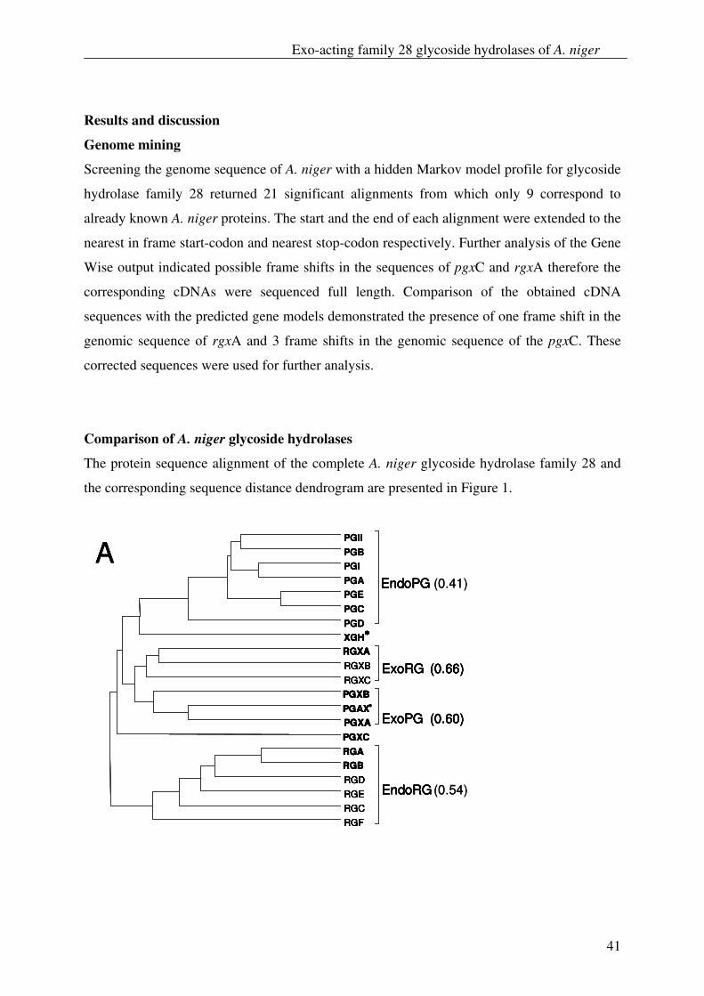
Exo-acting family 28 glycoside hydrolases of A. niger
41
Results and discussion
Genome mining
Screening the genome sequence of A. niger with a hidden Markov model profile for glycoside
hydrolase family 28 returned 21 significant alignments from which only 9 correspond to
already known A. niger proteins. The start and the end of each alignment were extended to the
nearest in frame start-codon and nearest stop-codon respectively. Further analysis of the Gene
Wise output indicated possible frame shifts in the sequences of pgxC and rgxA therefore the
corresponding cDNAs were sequenced full length. Comparison of the obtained cDNA
sequences with the predicted gene models demonstrated the presence of one frame shift in the
genomic sequence of rgxA and 3 frame shifts in the genomic sequence of the pgxC. These
corrected sequences were used for further analysis.
Comparison of A. niger glycoside hydrolases
The protein sequence alignment of the complete A. niger glycoside hydrolase family 28 and
the corresponding sequence distance dendrogram are presented in Figure 1.
PGIIPGBPGIPGAPGEPGCPGD
EndoPG (0.41)
XGH∗∗∗∗
RGXARGXBRGXC
ExoRG (0.66)
PGXBPGAX∗∗∗∗
PGXA ExoPG (0.60)PGXCRGARGBRGDRGERGCRGF
EndoRG(0.54)
APGIIPGBPGIPGAPGEPGCPGD
EndoPG
XGH∗∗∗∗
RGXARGXBRGXC
ExoRG (0.66)
PGXBPGAX∗∗∗∗
PGXA ExoPG (0.60)PGXCRGARGBRGDRGERGCRGF
EndoRG
PGIIPGBPGIPGAPGEPGCPGD
EndoPG
XGH∗∗∗∗
RGXARGXBRGXC
ExoRG (0.66)
PGXBPGAX∗∗∗∗
PGXA ExoPG (0.60)PGXCRGARGBRGDRGERGCRGF
EndoRG
APGIIPGBPGIPGAPGEPGCPGD
EndoPG (0.41)
XGH∗∗∗∗
RGXARGXBRGXC
ExoRG (0.66)
PGXBPGAX∗∗∗∗
PGXA ExoPG (0.60)PGXCRGARGBRGDRGERGCRGF
EndoRG(0.54)
APGIIPGBPGIPGAPGEPGCPGD
EndoPG
XGH∗∗∗∗
RGXARGXBRGXC
ExoRG (0.66)
PGXBPGAX∗∗∗∗
PGXA ExoPG (0.60)PGXCRGARGBRGDRGERGCRGF
EndoRG
PGIIPGBPGIPGAPGEPGCPGD
EndoPG
XGH∗∗∗∗
RGXARGXBRGXC
ExoRG (0.66)
PGXBPGAX∗∗∗∗
PGXA ExoPG (0.60)PGXCRGARGBRGDRGERGCRGF
EndoRG
A
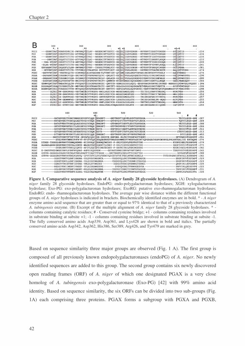
Chapter 2
42
340 360 380 400 420 440*. * .** . +1 +1 . . +1-1 .
PGII ---GGHNTDAFDVGNSVGVNIIK-PWVHNQDDCLAV--NSGENIWFTGGTCI-GGHGLSIGSVGDRSN--NVVKNVTIEHSTVSNSEN -----AVRIKTIS--------- :234PGI ---GGHNTDGFDISESTGVYISG-ATVKNQDDCIAI--NSGESISFTGGTCS-GGHGLSIGSVGGRDD--NTVKNVTISDSTVSNSAN -----GVRIKTIY--------- :236PGA ---GGHNTDGFDISESTYITITG-ATVKNQDDCVAI--NSGENIYFSGGTCS-GGHGLSIGSVGGRDD--NTVKNVTFIDSTVSDSEN -----GVRIKTVY--------- :235PGB ----GHNTDAFDIGQSTYITIDG-ATVYNQDDCLAI--NSGEHITFTNGYCD-GGHGLSIGSIGGRSD--NTVNDVTISNSKVLNSQN -----GVRIKTIY--------- :233PGE --EAA-NTDGFDIGDSTYITITG-ANVYNQDDCVAV--NSGENIYFSGGVCS-GGHGLSIGSVGGRSD--NTVKNVTFYDSEIKSSQN -----GVRIKTIY--------- :240PGC -DLAA-NTDGFDIGESTYITITG-AEIYNQDDCVAI--NSGENIYFSASVCS-GGHGLSIGSVGGRDD--NTVKNVTFYDVNVLKSQQ -----AIRIKTIY--------- :241PGD GDPAAHNTDGFDIKQSDFLTLSN-SWVHNQDDCVAV--TSGSSIVVDNLYCY-GGHGLSIGSIGGKSN--NTVDGVTFSNSQVINSEN -----GCRIKSNA--------- :381XGH --NPPKNTDGFDIGESTYVTITE-VTVVNDDDCVALKPSS-NYVTVDTISCT-GSHGISVGSLGKSSD--DSVKNIYVTGATMINSTK -----AAGIKTYPSGG------ :274PGXC -GSFPFNTDGFD-VEGTNIQITD-SIMYNGDDAIAVGADSHDTLFTRNTIGY-QTHGMSIGSLGKDPTDFANISNIRFDDVTVVDGLY -----AARFKSWSG-------- :275PGXA -DAPAKNTDGWDIYRSSNIVIQD-SRIVNTDDCVSFKPNSTQIVI-QNLDCT-GSHGISVGSLGQYQGETDIVEDLYIYNISMTDASD -----VARIKVWPGVPA----- :294PGAX -DNEAKNTDGWDTYRSNNIVIQN-SVINNGDDCVSFKPNSTNILV-QNLHCN-GSHGISVGSLGQYKDEVDIVENVYVYNISMFNASD -----MARIKVWPGTPS----- :289PGXB -SSDPANTDGMDSLDVDGVSFTN-MRIDVGDDCFSPKPNTTNIFV-QNMWCN-NTHGVSMGSIGQYAGEMDIIENVYIENVTLLNGQN -----GARLKAWAGQDV----- :306RGXC GDNWVQNTDGFDTMDATNIQLTN-FVYQGGDDCIAIKPRSYNIDI-QNVTCR-GGNGIAIGSLGQYL-EDSSVANIRVDKVNIIRYNED -MHNSAYLKTWVGALVPQSSY : 284RGXB DGGSAQNTDGANTIYSKNITLTN-WEVVNGDDSISTKANSTDITI-ANCTFT-SGLGIAIGSIGQYNGAFETVERLKISNITYEKTTH -----AVYFKTWTGDQV----- :306RGXA EGEWVQNTDGIDTYRSDHITVTN-WVYQGGDDAVAFKGNSTNIHV-ENVTVY-GGPGIAFGSLGQYPDRTDI VENVTVRNVRVQPSFQRAMNSGVYF KSWIGVNY----- :288RGA ------GLDGIDVW-GSNIWVHD-VEVTNKDECVTVKSPA-NNILVESIYCN-WSGGCAMGSLGAD ----TDITDILYRNVYTWSSNQ -----MYMIKSN---------- :256RGB ------GLDGIDVW-SNNIWVHD-VEVTNKDECVTVKSPS-KNILIESIYCN-WSGGCGMGSFGSD ----TNVSDITYRNIYTWSSNN-----MMLIKSN---------- :256RGD ------GLDGIDIS-GANYWIHD-VEVTNGDECVTVKSPS-ANVRVENVFCN-HSGGCAMGSLGTD ----TNISNIEFENIYTYNSTQ -----MYMIKSN---------- :257RGC ------ATDAIDVW-GENMWFHD-IEVSNGDECVTVKSPA-HNYLIENIYCN-LSGGTAIGSLGTG ----TNISDIHYRNLYM-NQAD-----ACFLKSN---------- :255RGE ------GLDGIDLY-GGNIWIHL-VMVTNKDECVTSKTNS-HNFLIENIYCN-PSGGCAIGSLGSS ----VNVTNILYRNVYTWDSNQ -----MMMIKTN---------- :258RGF ------SYDGIDAI-GTNYYIHD-NEVTNRDECVSVKSPS-HHALVENLVCNQAGSGVSIGSLNVS ----AEISNIEARNISIIQGNN -----IAFIKTYP--------- :255
460 480 500 520 540 . +1. . . . # #
PGII ---------GATGSVSEITYSNIVMSGISDYGVVIQQD YEDGKPT----GKPTNGVTIQDVKLESVTGSVDSGA ----------------------- TEIYLLCGS-GSC :307PGI ---------KETGDVSEITYSNIQLSGITDYGIVIEQD YENGSPT----GTPSTGIPITDVTVDGVTGTLEDDA ----------------------- TQVYILCGD-GSC :309PGA ---------DATGTVEDITYSNIQLSGISDYGIVIEQD YENGDPT----GTPSNGVTISDVTLEDITGSVDSDA ----------------------- VEIYILCGD-GSC :308PGB ---------GKTGTVENVKFEDITLSDISKYGIVVEQD YENGSPT----GTPTNGVKVEDITFKKVTGSVKSSG ----------------------- TDIYILCGS-GSC :306PGE ---------GDTGSVSEVTYKEITLSDITDYGIVVEQN YDDTS------KSPTDGITIEDFVLDNVQGSVESSG ----------------------- TNIYIVCGS-DSC :311PGC ---------GDTGSVSEVTYHEIAFSDATDYGIVIEQN YDDTS------KTPTTGVPITDFVLENIVGTCEDDD ---------------------- CTEVYIACGD-GSC :313PGD ---------DTTGEVYNVKYENITLSGISDYGIDIQQD YENGGAT----GDPTNGVKIENISFVNVKGTMSDGK ------------------------ DYYILCGD-GSC :453XGH --------DHGTSTVSNVTFTDFTVDN -SDYAFQIQSCYGE-DDDYCE-ENPGNA-KLTDIVVSSFSGTTSDKY--------------------- DPVVANIDCGSDGTC :351PGXC ----------GTGLVKNVTWNNIRVFN VT-FPIFVTQSYSDQGASRSGTVNASSAVMMEDFTWSDFAGSINTYQPGDGSCVSDPCWYNVGLPNLKHTEALIIE CHTAQSC :374PGXA DTSGS-TSGGGLGRVRNVTYEHMQSENND -HIISVSQCYESKNQTMCDSY-P-SKLVIEDVLFKDFKGTTSK --------------------- KYDPEIGELTCSSPDVC :379PGAX ALSADLQGGGGSGSVKNITYDTALIDNVD -WAIEITQCYGQKNTTLCNEY-P-SSLTISDVHIKNFRGTTSG --------------------- SEDPYVGTIVCSSPDTC :375PGXB ----------GYGRINNVTYKNIQIQNTD -APIVLDQCYFDINATECAKY-P-SAVNITNILFENIWGSSSG --------------------- KDGKIVADLVCSPDAVC :382RGXC E-SAGVPRGDGWGSIRNVLFSNFNVQGAS -AGPSISQDSGDN-----GSYAGTSKMSISNVAFVNFTGWVD ---------------------- TEKSVVSTVSCSEVHPC :365RGXB GYPPN-GGGGGLGYASDIVATNLKTNNLKGAPFTISQCTTFSGAS ---GNCTNSKFQIRDLVFTDISGTTDS ------------------------ SDVASFQCSAVAPC :388RGXA GVPPN-GGGGGHGYVRNVSVENLRLKDVQ -LPVYIDTCLSYLFSENITQYCDTSTYE FEDLHFRNISGNGLA--------------TVTDYPGKNISFAVALLCSEKAPC :382RGA ---------GGSGTVNNTVLENFIGHGNA -YSLDVDSYWSSMTA------VDGDGVQLSNITFKNWKGTEADGA --------------------- ERGPIKVVCSDTAPC :329RGB ---------GGSGFVENVLLENFIGHGNA -YSLDIDSYWASMSA------VDGDGVQLSNITVKNWK GTEAYGA--------------------- ERGPVKVVCADGAPC :329RGD ---------GGNGTVTNCSFKNFIGYSNA -YMLDLDTYWGDES--------DGDGIKYENIGFENWKGTSSNGI --------------------- QRSPIRILCPDANPC :328RGC ---------NGDGIVKNIIWENVIVHGGP -YPLAIDEAWGDDRG--------SVGVQVSNLTFRNWHGESVSA ----------------------SRPVIRLQCDSDVPC :325RGE ---------GGLGNVSNIVFENFIGHGNV -NSLDLDSYWSSMNA------IDGVGIYYHNITIYNWTGTAIDGE --------------------- TRPPIRVICPEDMPC :331RGF ---------GGSGYVKDVTFENFR SLNSL-YGLDINQYWQNTWE------PDTGSVTLSNLVFKNFSGSVADGA --------------------- LRPPLYLFASDLTFA :328
B 340 360 380 400 420 440*. * .** . +1 +1 . . +1-1 .
PGII ---GGHNT AFDVGNSVGVNIIK-PWVHNQDDCLAV--NSGENIWFTGGTCI-GGHGLSIGSVGDRSN--NVVKNVTIEHSTVSNSEN -----AVRIKTIS--------- :234PGI ---GGHNT GFDISESTGVYISG-ATVKNQDDCIAI--NSGESISFTGGTCS-GGHGLSIGSVGGRDD--NTVKNVTISDSTVSNSAN -----GVRIKTIY--------- :236PGA ---GGHNT GFDISESTYITITG-ATVKNQDDCVAI--NSGENIYFSGGTCS-GGHGLSIGSVGGRDD--NTVKNVTFIDSTVSDSEN -----GVRIKTVY--------- :235PGB ----GHNTDAFDIGQSTYITIDG-ATVYNQDDCLAI--NSGEHITFTNGYCD-GGHGLSIGSIGGRSD--NTVNDVTISNSKVLNSQN -----GVRIKTIY--------- :233PGE --EAA-NTDGFDIGDSTYITITG-ANVYNQDDCVAV--NSGENIYFSGGVCS-GGHGLSIGSVGGRSD--NTVKNVTFYDSEIKSSQN -----GVRIKTIY--------- :240PGC -DLAA-NTDGFDIGESTYITITG-AEIYNQDDCVAI--NSGENIYFSASVCS-GGHGLSIGSVGGRDD--NTVKNVTFYDVNVLKSQQ -----AIRIKTIY--------- :241PGD GDPAAHNTDGFDIKQSDFLTLSN-SWVHNQDDCVAV--TSGSSIVVDNLYCY-GGHGLSIGSIGGKSN--NTVDGVTFSNSQVINSEN -----GCRIKSNA--------- :381XGH --NPPKNTDGFDIGESTYVTITE-VTVVNDDDCVALKPSS-NYVTVDTISCT-GSHGISVGSLGKSSD--DSVKNIYVTGATMINSTK -----AAGIKTYPSGG------ :274PGXC -GSFPFNTDGFD-VEGTNIQITD-SIMYNGDDAIAVGADSHDTLFTRNTIGY-QTHGMSIGSLGKDPTDFANISNIRFDDVTVVDGLY -----AARFKSWSG-------- :275PGXA -DAPAKNTDGWDIYRSSNIVIQD-SRIVNTDDCVSFKPNSTQIVI-QNLDCT-GSHGISVGSLGQYQGETDIVEDLYIYNISMTDASD -----VARIKVWPGVPA----- :294PGAX -DNEAKNTDGWDTYRSNNIVIQN-SVINNGDDCVSFKPNSTNILV-QNLHCN-GSHGISVGSLGQYKDEVDIVENVYVYNISMFNASD -----MARIKVWPGTPS----- :289PGXB -SSDPANTDGMDSLDVDGVSFTN-MRIDVGDDCFSPKPNTTNIFV-QNMWCN-NTHGVSMGSIGQYAGEMDIIENVYIENVTLLNGQN -----GARLKAWAGQDV----- :306RGXC GDNWVQNTDGFDTMDATNIQLTN-FVYQGGDDCIAIKPRSYNIDI-QNVTCR-GGNGIAIGSLGQYL-EDSSVANIRVDKVNIIRYNED -MHNSAYLKTWVGALVPQSSY : 284RGXB DGGSAQNTDGANTIYSKNITLTN-WEVVNGDDSISTKANSTDITI-ANCTFT-SGLGIAIGSIGQYNGAFETVERLKISNITYEKTTH -----AVYFKTWTGDQV----- :306RGXA EGEWVQNTDGIDTYRSDHITVTN-WVYQGGDDAVAFKGNSTNIHV-ENVTVY-GGPGIAFGSLGQYPDRTDI VENVTVRNVRVQPSFQRAMNSGVYF KSWIGVNY----- :288RGA ------GLDGIDVW-GSNIWVHD-VEVTNKDECVTVKSPA-NNILVESIYCN-WSGGCAMGSLGAD ----TDITDILYRNVYTWSSNQ -----MYMIKSN---------- :256RGB ------GLDGIDVW-SNNIWVHD-VEVTNKDECVTVKSPS-KNILIESIYCN-WSGGCGMGSFGSD ----TNVSDITYRNIYTWSSNN-----MMLIKSN---------- :256RGD ------GLDGIDIS-GANYWIHD-VEVTNGDECVTVKSPS-ANVRVENVFCN-HSGGCAMGSLGTD ----TNISNIEFENIYTYNSTQ -----MYMIKSN---------- :257RGC ------AT AIDVW-GENMWFHD-IEVSNGDECVTVKSPA-HNYLIENIYCN-LSGGTAIGSLGTG ----TNISDIHYRNLYM-NQAD-----ACFLKSN---------- :255RGE ------GL GIDLY-GGNIWIHL-VMVTNKDECVTSKTNS-HNFLIENIYCN-PSGGCAIGSLGSS ----VNVTNILYRNVYTWDSNQ -----MMMIKTN---------- :258RGF ------SY GIDAI-GTNYYIHD-NEVTNRDECVSVKSPS-HHALVENLVCNQAGSGVSIGSLNVS ----AEISNIEARNISIIQGNN -----IAFIKTYP--------- :255
460 480 500 520 540 . +1. . . . # #
PGII ---------GATGSVSEITYSNIVMSGISDYGVVIQQD YEDGKPT----GKPTNGVTIQDVKLESVTGSVDSGA ----------------------- TEIYLLCGS-GSC :307PGI ---------KETGDVSEITYSNIQLSGITDYGIVIEQD YENGSPT----GTPSTGIPITDVTVDGVTGTLEDDA ----------------------- TQVYILCGD-GSC :309PGA ---------DATGTVEDITYSNIQLSGISDYGIVIEQD YENGDPT----GTPSNGVTISDVTLEDITGSVDSDA ----------------------- VEIYILCGD-GSC :308PGB ---------GKTGTVENVKFEDITLSDISKYGIVVEQD YENGSPT----GTPTNGVKVEDITFKKVTGSVKSSG ----------------------- TDIYILCGS-GSC :306PGE ---------GDTGSVSEVTYKEITLSDITDYGIVVEQN YDDTS------KSPTDGITIEDFVLDNVQGSVESSG ----------------------- TNIYIVCGS-DSC :311PGC ---------GDTGSVSEVTYHEIAFSDATDYGIVIEQN YDDTS------KTPTTGVPITDFVLENIVGTCEDDD ---------------------- CTEVYIACGD-GSC :313PGD ---------DTTGEVYNVKYENITLSGISDYGIDIQQD YENGGAT----GDPTNGVKIENISFVNVKGTMSDGK ------------------------ DYYILCGD-GSC :453XGH --------DHGTSTVSNVTFTDFTVDN -SDYAFQIQSCYGE-DDDYCE-ENPGNA-KLTDIVVSSFSGTTSDKY--------------------- DPVVANIDCGSDGTC :351PGXC ----------GTGLVKNVTWNNIRVFN VT-FPIFVTQSYSDQGASRSGTVNASSAVMMEDFTWSDFAGSINTYQPGDGSCVSDPCWYNVGLPNLKHTEALIIE CHTAQSC :374PGXA DTSGS-TSGGGLGRVRNVTYEHMQSENND -HIISVSQCYESKNQTMCDSY-P-SKLVIEDVLFKDFKGTTSK --------------------- KYDPEIGELTCSSPDVC :379PGAX ALSADLQGGGGSGSVKNITYDTALIDNVD -WAIEITQCYGQKNTTLCNEY-P-SSLTISDVHIKNFRGTTSG --------------------- SEDPYVGTIVCSSPDTC :375PGXB ----------GYGRINNVTYKNIQIQNTD -APIVLDQCYFDINATECAKY-P-SAVNITNILFENIWGSSSG --------------------- KDGKIVADLVCSPDAVC :382RGXC E-SAGVPRGDGWGSIRNVLFSNFNVQGAS -AGPSISQDSGDN-----GSYAGTSKMSISNVAFVNFTGWVD ---------------------- TEKSVVSTVSCSEVHPC :365RGXB GYPPN-GGGGGLGYASDIVATNLKTNNLKGAPFTISQCTTFSGAS ---GNCTNSKFQIRDLVFTDISGTTDS ------------------------ SDVASFQCSAVAPC :388RGXA GVPPN-GGGGGHGYVRNVSVENLRLKDVQ -LPVYIDTCLSYLFSENITQYCDTSTYE FEDLHFRNISGNGLA--------------TVTDYPGKNISFAVALLCSEKAPC :382RGA ---------GGSGTVNNTVLENFIGHGNA -YSLDVDSYWSSMTA------VDGDGVQLSNITFKNWKGTEADGA --------------------- ERGPIKVVCSDTAPC :329RGB ---------GGSGFVENVLLENFIGHGNA -YSLDIDSYWASMSA------VDGDGVQLSNITVKNWK GTEAYGA--------------------- ERGPVKVVCADGAPC :329RGD ---------GGNGTVTNCSFKNFIGYSNA -YMLDLDTYWGDES--------DGDGIKYENIGFENWKGTSSNGI --------------------- QRSPIRILCPDANPC :328RGC ---------NGDGIVKNIIWENVIVHGGP -YPLAIDEAWGDDRG--------SVGVQVSNLTFRNWHGESVSA ----------------------SRPVIRLQCDSDVPC :325RGE ---------GGLGNVSNIVFENFIGHGNV -NSLDLDSYWSSMNA------IDGVGIYYHNITIYNWTGTAIDGE --------------------- TRPPIRVICPEDMPC :331RGF ---------GGSGYVKDVTFENFR SLNSL-YGLDINQYWQNTWE------PDTGSVTLSNLVFKNFSGSVADGA --------------------- LRPPLYLFASDLTFA :328
B 340 360 380 400 420 440*. * .** . +1 +1 . . +1-1 .
PGII ---GGHNTDAFDVGNSVGVNIIK-PWVHNQDDCLAV--NSGENIWFTGGTCI-GGHGLSIGSVGDRSN--NVVKNVTIEHSTVSNSEN -----AVRIKTIS--------- :234PGI ---GGHNTDGFDISESTGVYISG-ATVKNQDDCIAI--NSGESISFTGGTCS-GGHGLSIGSVGGRDD--NTVKNVTISDSTVSNSAN -----GVRIKTIY--------- :236PGA ---GGHNTDGFDISESTYITITG-ATVKNQDDCVAI--NSGENIYFSGGTCS-GGHGLSIGSVGGRDD--NTVKNVTFIDSTVSDSEN -----GVRIKTVY--------- :235PGB ----GHNTDAFDIGQSTYITIDG-ATVYNQDDCLAI--NSGEHITFTNGYCD-GGHGLSIGSIGGRSD--NTVNDVTISNSKVLNSQN -----GVRIKTIY--------- :233PGE --EAA-NTDGFDIGDSTYITITG-ANVYNQDDCVAV--NSGENIYFSGGVCS-GGHGLSIGSVGGRSD--NTVKNVTFYDSEIKSSQN -----GVRIKTIY--------- :240PGC -DLAA-NTDGFDIGESTYITITG-AEIYNQDDCVAI--NSGENIYFSASVCS-GGHGLSIGSVGGRDD--NTVKNVTFYDVNVLKSQQ -----AIRIKTIY--------- :241PGD GDPAAHNTDGFDIKQSDFLTLSN-SWVHNQDDCVAV--TSGSSIVVDNLYCY-GGHGLSIGSIGGKSN--NTVDGVTFSNSQVINSEN -----GCRIKSNA--------- :381XGH --NPPKNTDGFDIGESTYVTITE-VTVVNDDDCVALKPSS-NYVTVDTISCT-GSHGISVGSLGKSSD--DSVKNIYVTGATMINSTK -----AAGIKTYPSGG------ :274PGXC -GSFPFNTDGFD-VEGTNIQITD-SIMYNGDDAIAVGADSHDTLFTRNTIGY-QTHGMSIGSLGKDPTDFANISNIRFDDVTVVDGLY -----AARFKSWSG-------- :275PGXA -
PGE --EAA-NTDGFDIGDSTYITITG-ANVYNQDDCVAV--NSGENIYFSGGVCS-GGHGLSIGSVGGRSD--NTVKNVTFYDSEIKSSQN -----GVRIKTIY--------- :240PGC -DLAA-NTDGFDIGESTYITITG-AEIYNQDDCVAI--NSGENIYFSASVCS-GGHGLSIGSVGGRDD--NTVKNVTFYDVNVLKSQQ -----AIRIKTIY--------- :241PGD GDPAAHNTDGFDIKQSDFLTLSN-SWVHNQDDCVAV--TSGSSIVVDNLYCY-GGHGLSIGSIGGKSN--NTVDGVTFSNSQVINSEN -----GCRIKSNA--------- :381XGH --NPPKNTDGFDIGESTYVTITE-VTVVNDDDCVALKPSS-NYVTVDTISCT-GSHGISVGSLGKSSD--DSVKNIYVTGATMINSTK -----AAGIKTYPSGG------ :274PGXC -GSFPFNTDGFD-VEGTNIQITD-SIMYNGDDAIAVGADSHDTLFTRNTIGY-QTHGMSIGSLGKDPTDFANISNIRFDDVTVVDGLY -----AARFKSWSG-------- :275PGXA -DAPAKNTDGWDIYRSSNIVIQD-SRIVNTDDCVSFKPNSTQIVI-QNLDCT-GSHGISVGSLGQYQGETDIVEDLYIYNISMTDASD -----VARIKVWPGVPA----- :294PGAX -DNEAKNTDGWDTYRSNNIVIQN-SVINNGDDCVSFKPNSTNILV-QNLHCN-GSHGISVGSLGQYKDEVDIVENVYVYNISMFNASD -----MARIKVWPGTPS----- :289PGXB -SSDPANTDGMDSLDVDGVSFTN-MRIDVGDDCFSPKPNTTNIFV-QNMWCN-NTHGVSMGSIGQYAGEMDIIENVYIENVTLLNGQN -----GARLKAWAGQDV----- :306RGXC GDNWVQNTDGFDTMDATNIQLTN-FVYQGGDDCIAIKPRSYNIDI-QNVTCR-GGNGIAIGSLGQYL-EDSSVANIRVDKVNIIRYNED -MHNSAYLKTWVGALVPQSSY : 284RGXB DGGSAQNTDGANTIYSKNITLTN-WEVVNGDDSISTKANSTDITI-ANCTFT-SGLGIAIGSIGQYNGAFETVERLKISNITYEKTTH -----AVYFKTWTGDQV----- :306RGXA EGEWVQNTDGIDTYRSDHITVTN-WVYQGGDDAVAFKGNSTNIHV-ENVTVY-GGPGIAFGSLGQYPDRTDI VENVTVRNVRVQPSFQRAMNSGVYF KSWIGVNY----- :288RGA ------GLDGIDVW-GSNIWVHD-VEVTNKDECVTVKSPA-NNILVESIYCN
DAPAKNTDGWDIYRSSNIVIQD-SRIVNTDDCVSFKPNSTQIVI-QNLDCT-GSHGISVGSLGQYQGETDIVEDLYIYNISMTDASD -----VARIKVWPGVPA----- :294PGAX -DNEAKNTDGWDTYRSNNIVIQN-SVINNGDDCVSFKPNSTNILV-QNLHCN-GSHGISVGSLGQYKDEVDIVENVYVYNISMFNASD -----MARIKVWPGTPS----- :289PGXB -SSDPANTDGMDSLDVDGVSFTN-MRIDVGDDCFSPKPNTTNIFV-QNMWCN-NTHGVSMGSIGQYAGEMDIIENVYIENVTLLNGQN -----GARLKAWAGQDV----- :306RGXC GDNWVQNTDGFDTMDATNIQLTN-FVYQGGDDCIAIKPRSYNIDI-QNVTCR-GGNGIAIGSLGQYL-EDSSVANIRVDKVNIIRYNED -MHNSAYLKTWVGALVPQSSY : 284RGXB DGGSAQNTDGANTIYSKNITLTN-WEVVNGDDSISTKANSTDITI-ANCTFT-SGLGIAIGSIGQYNGAFETVERLKISNITYEKTTH -----AVYFKTWTGDQV----- :306RGXA EGEWVQNTDGIDTYRSDHITVTN-WVYQGGDDAVAFKGNSTNIHV-ENVTVY-GGPGIAFGSLGQYPDRTDI VENVTVRNVRVQPSFQRAMNSGVYF KSWIGVNY----- :288RGA ------GLDGIDVW-GSNIWVHD-VEVTNKDECVTVKSPA-NNILVESIYCN-WSGGCAMGSLGAD ----TDITDILYRNVYTWSSNQ -----MYMIKSN---------- :256RGB ------GLDGIDVW-SNNIWVHD-VEVTNKDECVTVKSPS-KNILIESIYCN-WSGGCGMGSFGSD ----TNVSDITYRNIYTWSSNN-----MMLIKSN---------- :256RGD ------GLDGIDIS-GANYWIHD-VEVTNGDECVTVKSPS-ANVRVENVFCN-HSGGCAMGSLGTD ----TNISNIEFENIYTYNSTQ -----MYMIKSN---------- :257RGC ------ATDAIDVW-GENMWFHD-IEVSNGDECVTVKSPA-HNYLIENIYCN-LSGGTAIGSLGTG ----TNISDIHYRNLYM-NQAD-----ACFLKSN---------- :255RGE ------GLDGIDLY-GGNIWIHL-VMVTNKDECVTSKTNS-HNFLIENIYCN-PSGGCAIGSLGSS ----VNVTNILYRNVYTWDSNQ -----MMMIKTN---------- :258RGF ------SYDGIDAI-GTNYYIHD-NEVTNRDECVSVKSPS-HHALVENLVCNQAGSGVSIGSLNVS ----AEISNIEARNISIIQGNN -----IAFIKTYP--------- :255
460 480 500 520 540 . +1. . . . # #
PGII ---------
-WSGGCAMGSLGAD ----TDITDILYRNVYTWSSNQ -----MYMIKSN---------- :256RGB ------GLDGIDVW-SNNIWVHD-VEVTNKDECVTVKSPS-KNILIESIYCN-WSGGCGMGSFGSD ----TNVSDITYRNIYTWSSNN-----MMLIKSN---------- :256RGD ------GLDGIDIS-GANYWIHD-VEVTNGDECVTVKSPS-ANVRVENVFCN-HSGGCAMGSLGTD ----TNISNIEFENIYTYNSTQ -----MYMIKSN---------- :257RGC ------ATDAIDVW-GENMWFHD-IEVSNGDECVTVKSPA-HNYLIENIYCN-LSGGTAIGSLGTG ----TNISDIHYRNLYM-NQAD-----ACFLKSN---------- :255RGE ------GLDGIDLY-GGNIWIHL-VMVTNKDECVTSKTNS-HNFLIENIYCN-PSGGCAIGSLGSS ----VNVTNILYRNVYTWDSNQ -----MMMIKTN---------- :258RGF ------SYDGIDAI-GTNYYIHD-NEVTNRDECVSVKSPS-HHALVENLVCNQAGSGVSIGSLNVS ----AEISNIEARNISIIQGNN -----IAFIKTYP--------- :255
460 480 500 520 540 . +1. . . . # #
PGII ---------GATGSVSEITYSNIVMSGISDYGVVIQQD YEDGKPT----GKPTNGVTIQDVKLESVTGSVDSGA ----------------------- TEIYLLCGS-GSC :307PGI ---------KETGDVSEITYSNIQLSGITDYGIVIEQD YENGSPT----GTPSTGIPITDVTVDGVTGTLEDDA ----------------------- TQVYILCGD-GSC :309PGA ---------DATGTVEDITYSNIQLSGISDYGIVIEQD YENGDPT----GTPSNGVTISDVTLEDITGSVDSDA ----------------------- VEIYILCGD-GSC :308PGB ---------GKTGTVENVKFEDITLSDISKYGIVVEQD YENGSPT----GTPTNGVKVEDITFKKVTGSVKSSG ----------------------- TDIYILCGS-GSC :306PGE ---------GDTGSVSEVTYKEITLSDITDYGIVVEQN YDDTS------KSPTDGITIEDFVLDNVQGSVESSG ----------------------- TNIYIVCGS-DSC :311PGC ---------GDTGSVSEVTYHEIAFSDATDYGIVIEQN YDDTS------KTPTTGVPITDFVLENIVGTCEDDD ---------------------- CTEVYIACGD-GSC :313PGD ---------DTTGEVYNVKYENITLSGISDYGIDIQQD YENGGAT----GDPTNGVKIENISFVNVKGTMSDGK ------------------------ DYYILCGD-GSC :453XGH --------DHGTSTVSNVTFTDFTVDN -SDYAFQIQSCYGE-DDDYCE-ENPGNA-KLTDIVVSSFSGTTSDKY--------------------- DPVVANIDCGSDGTC :351PGXC ----------GTGLVKNVTWNNIRVFN VT-FPIFVTQSYSDQGASRSGTVNASSAVMMEDFTWSDFAGSINTYQPGDGSCVSDPCWYNVGLPNLKHTEALIIE CHTAQSC :374PGXA DTSGS-TSGGGLGRVRNVTYEHMQSENND -HIISVSQCYESKNQTMCDSY-P-SKLVIEDVLFKDFKGTTSK --------------------- KYDPEIGELTCSSPDVC :379PGAX ALSADLQGGGGSGSVKNITYDTALIDNVD -WAIEITQC
GATGSVSEITYSNIVMSGISDYGVVIQQD YEDGKPT----GKPTNGVTIQDVKLESVTGSVDSGA ----------------------- TEIYLLCGS-GSC :307PGI ---------KETGDVSEITYSNIQLSGITDYGIVIEQD YENGSPT----GTPSTGIPITDVTVDGVTGTLEDDA ----------------------- TQVYILCGD-GSC :309PGA ---------DATGTVEDITYSNIQLSGISDYGIVIEQD YENGDPT----GTPSNGVTISDVTLEDITGSVDSDA ----------------------- VEIYILCGD-GSC :308PGB ---------GKTGTVENVKFEDITLSDISKYGIVVEQD YENGSPT----GTPTNGVKVEDITFKKVTGSVKSSG ----------------------- TDIYILCGS-GSC :306PGE ---------GDTGSVSEVTYKEITLSDITDYGIVVEQN YDDTS------KSPTDGITIEDFVLDNVQGSVESSG ----------------------- TNIYIVCGS-DSC :311PGC ---------GDTGSVSEVTYHEIAFSDATDYGIVIEQN YDDTS------KTPTTGVPITDFVLENIVGTCEDDD ---------------------- CTEVYIACGD-GSC :313PGD ---------DTTGEVYNVKYENITLSGISDYGIDIQQD YENGGAT----GDPTNGVKIENISFVNVKGTMSDGK ------------------------ DYYILCGD-GSC :453XGH --------DHGTSTVSNVTFTDFTVDN -SDYAFQIQSCYGE-DDDYCE-ENPGNA-KLTDIVVSSFSGTTSDKY--------------------- DPVVANIDCGSDGTC :351PGXC ----------GTGLVKNVTWNNIRVFN VT-FPIFVTQSYSDQGASRSGTVNASSAVMMEDFTWSDFAGSINTYQPGDGSCVSDPCWYNVGLPNLKHTEALIIE CHTAQSC :374PGXA DTSGS-TSGGGLGRVRNVTYEHMQSENND -HIISVSQCYESKNQTMCDSY-P-SKLVIEDVLFKDFKGTTSK --------------------- KYDPEIGELTCSSPDVC :379PGAX ALSADLQGGGGSGSVKNITYDTALIDNVD -WAIEITQCYGQKNTTLCNEY-P-SSLTISDVHIKNFRGTTSG --------------------- SEDPYVGTIVCSSPDTC :375PGXB ----------GYGRINNVTYKNIQIQNTD -APIVLDQCYFDINATECAKY-P-SAVNITNILFENIWGSSSG --------------------- KDGKIVADLVCSPDAVC :382RGXC E-SAGVPRGDGWGSIRNVLFSNFNVQGAS -AGPSISQDSGDN-----GSYAGTSKMSISNVAFVNFTGWVD ---------------------- TEKSVVSTVSCSEVHPC :365RGXB GYPPN-GGGGGLGYASDIVATNLKTNNLKGAPFTISQCTTFSGAS ---GNCTNSKFQIRDLVFTDISGTTDS ------------------------ SDVASFQCSAVAPC :388RGXA GVPPN-GGGGGHGYVRNVSVENLRLKDVQ -LPVYIDTCLSYLFSENITQYCDTSTYE FEDLHFRNISGNGLA--------------TVTDYPGKNISFAVALLCSEKAPC :382RGA ---------GGSGTVNNTVLENFIGHGNA -YSLDVDSYWSSMTA------VDGDGVQLSNITFKNWKGTEADGA --------------------- ERGPIKVVCSDTAPC :329RGB ---------GGSGFVENVLLENFIGHGNA -YSLDIDSYWASMSA------VDGDGVQLSNITVKNWK GTEAYGA--------------------- ERGPVKVVCADGAPC :329RGD ---------GGNGTVTNCSFKNFIGYSNA -YMLDLDTYWGDES--------DGDGIKYENIGFENWKGTSSNGI --------------------- QRSPIRILCPDANPC :328RGC ---------NGDGIVKNIIWENVIVHGGP -YPLAIDEAWGDDRG--------SVGVQVSNLTFRNWHGESVSA ----------------------SRPVIRLQCDSDVPC :325RGE ---------GGLGNVSNIVFENFIGHGNV -NSLDLDSYWSSMNA------IDGVGIYYHNITIYNWTGTAIDGE --------------------- TRPPIRVICPEDMPC :331RGF ---------GGSGYVKDVTFENFR SLNSL-YGLDINQYWQNTWE------PDTGSVTLSNLVFKNFSGSVADGA --------------------- LRPPLYLFASDLTFA :328
B 340 360 380 400 420 440*. * .** . +1 +1 . . +1-1 .
PGII ---GGHNT AFDVGNSVGVNIIK-PWVHNQDDCLAV--NSGENIWFTGGTCI-GGHGLSIGSVGDRSN--NVVKNVTIEHSTVSNSEN -----AVRIKTIS--------- :234PGI ---GGHNT GFDISESTGVYISG-ATVKNQDDCIAI--NSGESISFTGGTCS-GGHGLSIGSVGGRDD--NTVKNVTISDSTVSNSAN -----GVRIKTIY--------- :236PGA ---GGHNT GFDISESTYITITG-ATVKNQDDCVAI--NSGENIYFSGGTCS-GGHGLSIGSVGGRDD--NTVKNVTFIDSTVSDSEN -----GVRIKTVY--------- :235PGB ----GHNTDAFDIGQSTYITIDG-ATVYNQDDCLAI--NSGEHITFTNGYCD-GGHGLSIGSIGGRSD--NTVNDVTISNSKVLNSQN -----GVRIKTIY--------- :233PGE --EAA-NTDGFDIGDSTYITITG-ANVYNQDDCVAV--NSGENIYFSGGVCS-GGHGLSIGSVGGRSD--NTVKNVTFYDSEIKSSQN -----GVRIKTIY--------- :240PGC -DLAA-NTDGFDIGESTYITITG-AEIYNQDDCVAI--NSGENIYFSASVCS-GGHGLSIGSVGGRDD--NTVKNVTFYDVNVLKSQQ -----AIRIKTIY--------- :241PGD GDPAAHNTDGFDIKQSDFLTLSN-SWVHNQDDCVAV--TSGSSIVVDNLYCY-GGHGLSIGSIGGKSN--NTVDGVTFSNSQVINSEN -----GCRIKSNA--------- :381XGH --NPPKNTDGFDIGESTYVTITE-VTVVNDDDCVALKPSS-NYVTVDTISCT-GSHGISVGSLGKSSD--DSVKNIYVTGATMINSTK -----AAGIKTYPSGG------ :274PGXC -GSFPFNTDGFD-VEGTNIQITD-SIMYNGDDAIAVGADSHDTLFTRNTIGY-QTHGMSIGSLGKDPTDFANISNIRFDDVTVVDGLY -----AARFKSWSG-------- :275PGXA -DAPAKNTDGWDIYRSSNIVIQD-SRIVNTDDCVSFKPNSTQIVI-QNLDCT-GSHGISVGSLGQYQGETDIVEDLYIYNISMTDASD -----VARIKVWPGVPA----- :294PGAX -DNEAKNTDGWDTYRSNNIVIQN-SVINNGDDCVSFKPNSTNILV-QNLHCN-GSHGISVGSLGQYKDEVDIVENVYVYNISMFNASD -----MARIKVWPGTPS----- :289PGXB -SSDPANTDGMDSLDVDGVSFTN-MRIDVGDDCFSPKPNTTNIFV-QNMWCN-NTHGVSMGSIGQYAGEMDIIENVYIENVTLLNGQN -----GARLKAWAGQDV----- :306RGXC GDNWVQNTDGFDTMDATNIQLTN-FVYQGGDDCIAIKPRSYNIDI-QNVTCR-GGNGIAIGSLGQYL-EDSSVANIRVDKVNIIRYNED -MHNSAYLKTWVGALVPQSSY : 284RGXB DGGSAQNTDGANTIYSKNITLTN-WEVVNGDDSISTKANSTDITI-ANCTFT-SGLGIAIGSIGQYNGAFETVERLKISNITYEKTTH -----AVYFKTWTGDQV----- :306RGXA EGEWVQNTDGIDTYRSDHITVTN-WVYQGGDDAVAFKGNSTNIHV-ENVTVY-GGPGIAFGSLGQYPDRTDI VENVTVRNVRVQPSFQRAMNSGVYF KSWIGVNY----- :288RGA ------GLDGIDVW-GSNIWVHD-VEVTNKDECVTVKSPA-NNILVESIYCN-WSGGCAMGSLGAD ----TDITDILYRNVYTWSSNQ -----MYMIKSN---------- :256RGB ------GLDGIDVW-SNNIWVHD-VEVTNKDECVTVKSPS-KNILIESIYCN-WSGGCGMGSFGSD ----TNVSDITYRNIYTWSSNN-----MMLIKSN---------- :256RGD ------GLDGIDIS-GANYWIHD-VEVTNGDECVTVKSPS-ANVRVENVFCN-HSGGCAMGSLGTD ----TNISNIEFENIYTYNSTQ -----MYMIKSN---------- :257RGC ------AT AIDVW-GENMWFHD-IEVSNGDECVTVKSPA-HNYLIENIYCN-LSGGTAIGSLGTG ----TNISDIHYRNLYM-NQAD-----ACFLKSN---------- :255RGE ------GL GIDLY-GGNIWIHL-VMVTNKDECVTSKTNS-HNFLIENIYCN-PSGGCAIGSLGSS ----VNVTNILYRNVYTWDSNQ -----MMMIKTN---------- :258RGF ------SY GIDAI-GTNYYIHD-NEVTNRDECVSVKSPS-HHALVENLVCNQAGSGVSIGSLNVS ----AEISNIEARNISIIQGNN -----IAFIKTYP--------- :255
460 480 500 520 540 . +1. . . . # #
PGII ---------GATGSVSEITYSNIVMSGISDYGVVIQQD YEDGKPT----GKPTNGVTIQDVKLESVTGSVDSGA ----------------------- TEIYLLCGS-GSC :307PGI ---------KETGDVSEITYSNIQLSGITDYGIVIEQD YENGSPT----GTPSTGIPITDVTVDGVTGTLEDDA ----------------------- TQVYILCGD-GSC :309PGA ---------DATGTVEDITYSNIQLSGISDYGIVIEQD YENGDPT----GTPSNGVTISDVTLEDITGSVDSDA ----------------------- VEIYILCGD-GSC :308PGB ---------GKTGTVENVKFEDITLSDISKYGIVVEQD YENGSPT----GTPTNGVKVEDITFKKVTGSVKSSG ----------------------- TDIYILCGS-GSC :306PGE ---------GDTGSVSEVTYKEITLSDITDYGIVVEQN YDDTS------KSPTDGITIEDFVLDNVQGSVESSG ----------------------- TNIYIVCGS-DSC :311PGC ---------GDTGSVSEVTYHEIAFSDATDYGIVIEQN YDDTS------KTPTTGVPITDFVLENIVGTCEDDD ---------------------- CTEVYIACGD-GSC :313PGD ---------DTTGEVYNVKYENITLSGISDYGIDIQQD YENGGAT----GDPTNGVKIENISFVNVKGTMSDGK ------------------------ DYYILCGD-GSC :453XGH --------DHGTSTVSNVTFTDFTVDN -SDYAFQIQSCYGE-DDDYCE-ENPGNA-KLTDIVVSSFSGTTSDKY--------------------- DPVVANIDCGSDGTC :351PGXC ----------GTGLVKNVTWNNIRVFN VT-FPIFVTQSYSDQGASRSGTVNASSAVMMEDFTWSDFAGSINTYQPGDGSCVSDPCWYNVGLPNLKHTEALIIE CHTAQSC :374PGXA DTSGS-TSGGGLGRVRNVTYEHMQSENND -HIISVSQCYESKNQTMCDSY-P-SKLVIEDVLFKDFKGTTSK --------------------- KYDPEIGELTCSSPDVC :379PGAX ALSADLQGGGGSGSVKNITYDTALIDNVD -WAIEITQC
GATGSVSEITYSNIVMSGISDYGVVIQQD YEDGKPT----GKPTNGVTIQDVKLESVTGSVDSGA ----------------------- TEIYLLCGS-GSC :307PGI ---------KETGDVSEITYSNIQLSGITDYGIVIEQD YENGSPT----GTPSTGIPITDVTVDGVTGTLEDDA ----------------------- TQVYILCGD-GSC :309PGA ---------DATGTVEDITYSNIQLSGISDYGIVIEQD YENGDPT----GTPSNGVTISDVTLEDITGSVDSDA ----------------------- VEIYILCGD-GSC :308PGB ---------GKTGTVENVKFEDITLSDISKYGIVVEQD YENGSPT----GTPTNGVKVEDITFKKVTGSVKSSG ----------------------- TDIYILCGS-GSC :306PGE ---------GDTGSVSEVTYKEITLSDITDYGIVVEQN YDDTS------KSPTDGITIEDFVLDNVQGSVESSG ----------------------- TNIYIVCGS-DSC :311PGC ---------GDTGSVSEVTYHEIAFSDATDYGIVIEQN YDDTS------KTPTTGVPITDFVLENIVGTCEDDD ---------------------- CTEVYIACGD-GSC :313PGD ---------DTTGEVYNVKYENITLSGISDYGIDIQQD YENGGAT----GDPTNGVKIENISFVNVKGTMSDGK ------------------------ DYYILCGD-GSC :453XGH --------DHGTSTVSNVTFTDFTVDN -SDYAFQIQSCYGE-DDDYCE-ENPGNA-KLTDIVVSSFSGTTSDKY--------------------- DPVVANIDCGSDGTC :351PGXC ----------GTGLVKNVTWNNIRVFN VT-FPIFVTQSYSDQGASRSGTVNASSAVMMEDFTWSDFAGSINTYQPGDGSCVSDPCWYNVGLPNLKHTEALIIE CHTAQSC :374PGXA DTSGS-TSGGGLGRVRNVTYEHMQSENND -HIISVSQCYESKNQTMCDSY-P-SKLVIEDVLFKDFKGTTSK --------------------- KYDPEIGELTCSSPDVC :379PGAX ALSADLQGGGGSGSVKNITYDTALIDNVD -WAIEITQCYGQKNTTLCNEY-P-SSLTISDVHIKNFRGTTSG --------------------- SEDPYVGTIVCSSPDTC :375PGXB ----------GYGRINNVTYKNIQIQNTD -APIVLDQCYFDINATECAKY-P-SAVNITNILFENIWGSSSG --------------------- KDGKIVADLVCSPDAVC :382RGXC E-SAGVPRGDGWGSIRNVLFSNFNVQGAS -AGPSISQDSGDN-----GSYAGTSKMSISNVAFVNFTGWVD ---------------------- TEKSVVSTVSCSEVHPC :365RGXB GYPPN-GGGGGLGYASDIVATNLKTNNLKGAPFTISQCTTFSGAS ---GNCTNSKFQIRDLVFTDISGTTDS ------------------------ SDVASFQCSAVAPC :388RGXA GVPPN-GGGGGHGYVRNVSVENLRLKDVQ -LPVYIDTCLSYLFSENITQYCDTSTYE FEDLHFRNISGNGLA--------------TVTDYPGKNISFAVALLCSEKAPC :382RGA ---------GGSGTVNNTVLENFIGHGNA -YSLDVDSYWSSMTA------VDGDGVQLSNITFKNWKGTEADGA --------------------- ERGPIKVVCSDTAPC :329RGB ---------GGSGFVENVLLENFIGHGNA -YSLDIDSYWASMSA------VDGDGVQLSNITVKNWK GTEAYGA--------------------- ERGPVKVVCADGAPC :329RGD ---------GGNGTVTNCSFKNFIGYSNA -YMLDLDTYWGDES--------DGDGIKYENIGFENWKGTSSNGI --------------------- QRSPIRILCPDANPC :328RGC ---------NGDGIVKNIIWENVIVHGGP -YPLAIDEAWGDDRG--------SVGVQVSNLTFRNWHGESVSA ----------------------SRPVIRLQCDSDVPC :325RGE ---------GGLGNVSNIVFENFIGHGNV -NSLDLDSYWSSMNA------IDGVGIYYHNITIYNWTGTAIDGE --------------------- TRPPIRVICPEDMPC :331RGF ---------GGSGYVKDVTFENFR SLNSL-YGLDINQYWQNTWE------PDTGSVTLSNLVFKNFSGSVADGA --------------------- LRPPLYLFASDLTFA :328
B
Figure 1. Comparative sequence analysis of A. niger family 28 glycoside hydrolases. (A) Dendrogram of A. niger family 28 glycoside hydrolases. EndoPG: endo-polygalacturonan hydrolases; XGH: xylogalacturonan hydrolase; Exo-PG: exo-polygalacturonan hydrolases; ExoRG: putative exo-rhamnogalacturonan hydrolases; EndoRG: endo- rhamnogalacturonan hydrolases. The average pair wise distance within the different functional groups of A. niger hydrolases is indicated in brackets. Biochemically identified enzymes are in bold. * - A niger enzyme amino acid sequence that are greater than or equal to 97% identical to that of a previously characterized A. tubingensis enzyme. (B) Excerpt of the multiple alignment of A. niger family 28 glycoside hydrolases. * - columns containing catalytic residues; # - Conserved cysteine bridge; +1 - columns containing residues involved in substrate binding at subsite +1; -1 - columns containing residues involved in substrate binding at subsite -1. The fully conserved amino acids Asp339, Asp361, and Lys428 are shown in bold and italics. The partially conserved amino acids Asp342, Asp362, His386, Ser389, Arg426, and Tyr479 are marked in grey.
Based on sequence similarity three major groups are observed (Fig. 1 A). The first group is
composed of all previously known endopolygalacturonases (endoPG) of A. niger. No newly
identified sequences are added to this group. The second group contains six newly discovered
open reading frames (ORF) of A. niger of which one designated PGAX is a very close
homolog of A. tubingensis exo-polygalacturonase (Exo-PG) [42] with 99% amino acid
identity. Based on sequence similarity, the six ORFs can be divided into two sub-groups (Fig.
1A) each comprising three proteins. PGAX forms a subgroup with PGXA and PGXB,
Exo-acting family 28 glycoside hydrolases of A. niger
43
therefore it is very likely that PGXA and PGXB are also exo-polygalacturonan hydrolases.
RGXA, RGXB and RGXC form the second distinct subgroup and evidence is presented that
they are similar to rhamnogalacturonan galacturonohydrolase [43] (see below). The
conservation of intron positions within ORFs of both sub-groups further underpins this
subdivision. The most deviating sequence from all 21 identified proteins is PGXC. Also, there
are no introns in the encoding gene pgxC to support its classification. However, exo-
polygalacturonan hydrolase activity was determined for the encoded enzyme (see below). The
last group includes two previously characterised endo-acting A. niger proteins –
rhamnogalacturonan hydrolase A (RGHA) and rhamnogalacturonan hydrolase B (RGHB) as
well as 4 putative enzymes which we propose to be endo-rhamnogalacturonan hydrolases.
Finally XGH, the strongly conserved homolog (97% amino acid identity) of xylogalacturonan
hydrolase of A. tubingensis [5] did not group together with any of the other sequences.
Detailed inspection of the amino acid conservation between all 21 sequences revealed that
only one of the 4 disulfide bridges between columns 544 and 550 of the multiple alignment
(Fig. 1 B) demonstrated by the protein structures of PGII of A. niger [14] and RGA of A.
aculeatus [48] is well conserved within family 28 glycoside hydrolases of A. niger. The other
three cysteine bridges are well conserved in the groups of endoPGs and endoRGs but not in
the exo-hydrolase group. Inspection of the catalytic residues revealed that all previously
identified catalytic Asp residues are conserved within the A. niger family except for Asp362
(Fig. 1B). In the group of rhamnogalacturonan hydrolases Asp362 is replaced by Glu which is
characteristic for this type of enzymatic activity [48]. The substrate binding Lys at alignment
position 428 is 100% conserved while the second substrate binding residue Arg426 is present
in all endoPG sequences and in only four of the seven proteins from the exo-group, viz.
PGAX, PGXA, PGXB and PGXC. Armand and co-workers [16] have demonstrated by site
directed mutagenesis that the corresponding Arg of A. niger PGII (Arg256) is essential for the
proper orientation of the substrate in the catalytic cleft and mutations of this residue
dramatically affect the substrate affinity of the enzyme. Interestingly, other amino acids
shown to be involved in substrate binding at subsite +1, His386 and Ser389, are conserved
within the same subset of sequences. The same is true for the Tyr479, which, in PGII, is
involved in the stabilisation of the substrate at subsite +1 [16, 49]. The partial conservation of
amino acids responsible for substrate specificity and binding within the exo-hydrolase group
Chapter 2
44
further supports the suggestion that only PGXA, PGXB, PGXC and PGAX have exo-
polygalacturonan hydrolase activity.
Identification of RGXC as a close homolog of A. aculeatus rhamnogalacturonan
galacturonohydrolase
A rhamnogalacturonan galacturonohydrolase (RG-galacturonohydrolase), which specifically
removes terminal galacturonosyl residues from rhamnogalacturonan I, has been isolated from
the commercial mixture Pectinex Ultra SP produced by A. aculeatus [43]. In order to identify
a possible A. niger homolog of this enzyme a tryptic digest of the A. aculeatus RG-
galacturonohydrolase was analysed by MS. The obtained peptide masses were used to search
the inferred A. niger proteome. At least 6 peptides perfectly matched the sequence of RGXC
resulting in a total of 29% overall coverage of the protein (data not shown). Based on
sequence similarity, RGXC is grouped together with RGXA and RGXB indicating that there
are three candidate genes encoding proteins with exo-rhamnogalacturonan hydrolase activity.
Transcriptional profiling
The complex substrate sugar beet pectin (SBP) and polygalacturonic acid (PGA) were chosen
as primary carbon sources to investigate expression of selected pectinolytic genes.
Galacturonic acid, rhamnose and xylose represent the most abundant sugar residues present in
homogalacturonan, rhamnogalacturonan and xylogalacturonan and were used to asses the
effects on gene expression caused by simple well defined carbon sources. Fructose as a strong
repressor of the expression of genes that are under carbon catabolite regulation (CCR) and
sorbitol as a non-inducing sugar like alcohol, which does not affect the CCR mechanisms
were chosen as control substrates. After transfer from 18 h pre-culture on 2 % fructose,
mycelium was sampled at four time points during 24 h growth on the different substrates. The
corresponding total RNA samples were used in microarray experiments to monitor the mRNA
levels of family 28 glycoside hydrolase encoding genes (Fig 2).
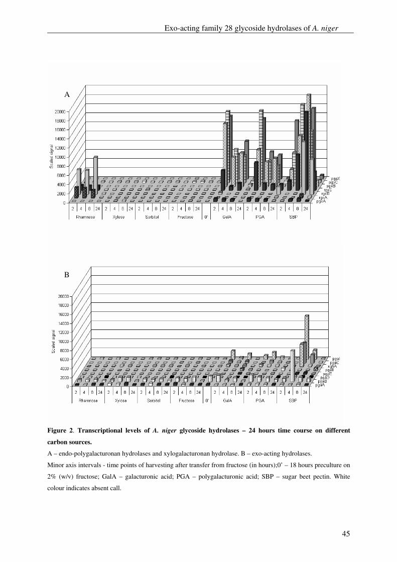
Exo-acting family 28 glycoside hydrolases of A. niger
45
Figure 2. Transcriptional levels of A. niger glycoside hydrolases – 24 hours time course on different
carbon sources.
A – endo-polygalacturonan hydrolases and xylogalacturonan hydrolase. B – exo-acting hydrolases.
Minor axis intervals - time points of harvesting after transfer from fructose (in hours);0’ – 18 hours preculture on
2% (w/v) fructose; GalA – galacturonic acid; PGA – polygalacturonic acid; SBP – sugar beet pectin. White
colour indicates absent call.
A
B
Chapter 2
46
During growth on non-pectic carbon sources, viz. fructose and sorbitol, none of the genes
encoding for enzymes from the exo-hydrolase group was transcribed. In contrast, transfer
from fructose to galacturonic acid and PGA caused a strong induction peak in the first 2 h
after transfer for pgaX, pgxB and pgxC followed by a decrease to relatively moderate
expression levels during the next 8 h and further reduction of the signal to background levels
24 h after transfer, coinciding with the depletion of carbon source. Although less strongly
induced, rgxC displayed a similar transcriptional profile on galacturonic acid and PGA. This
gene was also moderately expressed together with rgxA and rgxB after transfer to rhamnose
(Fig. 2B). Interestingly pgxA showed only low expression levels on these carbon sources.
Transfer to SBP caused a more complex transcriptional pattern. PgaX was quickly induced
and remained highly expressed reaching a maximum 8 h after transfer. PgxC messenger levels
reached a maximum 4 h after transfer. PgxB and rgxC levels gradually increased and reached
a maximum 8 h after transfer. The signals obtained for rgxC were much higher on sugar beet
pectin compared to rhamnose or galacturonic acid as sole carbon source. SBP caused a similar
effect on the induction of the rgxB gene. RgxA messenger was hardly detected 8 h after
transfer to SBP.
In contrast to the strong induction observed for genes from the exo-group, the transfer to
galacturonic acid and PGA did not cause a strong effect on the expression of
endopolygalacturonases. SBP moderately induced pgaI, pgaB and pgaC 4 h after transfer,
coinciding with the slight accumulation of extra-cellular free galacturonic acid (data not
shown).
PgaA and pgaB have been reported to be constitutively expressed [50] which is in agreement
with the data obtained in this study (Fig. 2A). Yet a slight increase of pgaB messenger was
observed coinciding with the depletion of fructose during the time course of incubation
suggesting that, although weak, pgaB is under CCR control. In contrast, when pectic
substrates were used, the signal of pgaB decreased in time to reach a background level at 24 h
(Fig. 2A). Instead, low levels of pgaD messenger were detected. PGD, the protein encoded by
pgaD gene, has been proposed to be a cell wall attached oligogalacturonase with high
tolerance towards methyl-esterified pectins [12]. HPLC measurements (data not shown)
demonstrated that at the time points of expression of pgaD free sugars are being depleted
from the culture fluid, which together with its functional properties, suggests that the

Exo-acting family 28 glycoside hydrolases of A. niger
47
expression of pgaD may in part be due to carbon starvation. Still, during incubation on
galacturonic acid and sugar beet pectin pgaD messengers could be detected slightly earlier, 8
h after incubation.
Despite many efforts including several genetic screens [51], no specific transcriptional factors
regulating the expression of pectinolytic glycoside hydrolases have been identified so far. The
strong discrepancy between the general expression profiles observed for the exo- and the
endo-hydrolase encoding genes suggests separate or loosely linked regulation mechanisms for
both groups. Exo-polygalacturonases are well induced by galacturonic acid and the observed
expression levels on pectin, although slightly more elevated, do not differ much from the
levels observed on its monomeric substitute. Unexpectedly, galacturonic acid and PGA as a
sole carbon source appeared to be only weak inducers for endo-polygalacturonases, while
SBP caused profoundly higher expression of endo-PG. In addition, the expression pattern of
all studied genes on SBP has a much more complex character compared to monomeric sugars,
which suggests that a multi-factorial regulation system and not just galacturonic acid, is
responsible for the induction of pectinolytic genes.
Biochemical identification of PGXA, PGXB, PGXC and RGXB
Purified PGXA, PGXB, PGXC and RGXB appeared as a single band upon SDS-PAGE
(results not shown) and had an apparent molecular mass of 78 kDa, 67 kDa, 79 kDa, and 82
kDa respectively (Table 1). The difference between the measured and calculated molecular
masses of these enzymes is most probably caused by a high degree of glycosylation, which
has also been demonstrated previously for the exo-polygalacturonase (Exo-PG) from A.
tubingensis [42].
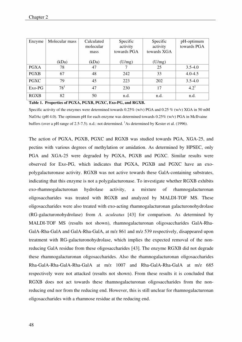
Chapter 2
48
Table 1. Properties of PGXA, PGXB, PGXC, Exo-PG, and RGXB.
Specific activity of the enzymes were determined towards 0.25% (w/v) PGA and 0.25 % (w/v) XGA in 50 mM
NaOAc (pH 4.0). The optimum pH for each enzyme was determined towards 0.25% (w/v) PGA in McIlvaine
buffers (over a pH range of 2.5-7.5). n.d.: not determined. 1As determined by Kester et al. (1996).
The action of PGXA, PGXB, PGXC and RGXB was studied towards PGA, XGA-25, and
pectins with various degrees of methylation or amidation. As determined by HPSEC, only
PGA and XGA-25 were degraded by PGXA, PGXB and PGXC. Similar results were
observed for Exo-PG, which indicates that PGXA, PGXB and PGXC have an exo-
polygalacturonase activity. RGXB was not active towards these GalA-containing substrates,
indicating that this enzyme is not a polygalacturonase. To investigate whether RGXB exhibits
exo-rhamnogalacturonan hydrolase activity, a mixture of rhamnogalacturonan
oligosaccharides was treated with RGXB and analyzed by MALDI-TOF MS. These
oligosaccharides were also treated with exo-acting rhamnogalacturonan galacturonohydrolase
(RG-galacturonohydrolase) from A. aculeatus [43] for comparison. As determined by
MALDI-TOF MS (results not shown), rhamnogalacturonan oligosaccharides GalA-Rha-
GalA-Rha-GalA and GalA-Rha-GalA, at m/z 861 and m/z 539 respectively, disappeared upon
treatment with RG-galacturonohydrolase, which implies the expected removal of the non-
reducing GalA residue from these oligosaccharides [43]. The enzyme RGXB did not degrade
these rhamnogalacturonan oligosaccharides. Also the rhamnogalacturonan oligosaccharides
Rha-GalA-Rha-GalA-Rha-GalA at m/z 1007 and Rha-GalA-Rha-GalA at m/z 685
respectively were not attacked (results not shown). From these results it is concluded that
RGXB does not act towards these rhamnogalacturonan oligosaccharides from the non-
reducing end nor from the reducing end. However, this is still unclear for rhamnogalacturonan
oligosaccharides with a rhamnose residue at the reducing end.
Enzyme Molecular mass
(kDa)
Calculated molecular
mass
(kDa)
Specific activity
towards PGA
(U/mg)
Specific activity
towards XGA
(U/mg)
pH-optimum
towards PGA
PGXA 78 47 7 25 3.5-4.0 PGXB 67 48 242 33 4.0-4.5
PGXC 79 45 223 202 3.5-4.0
Exo-PG 781 47 230 17 4.21
RGXB 82 50 n.d. n.d. n.d.
Exo-acting family 28 glycoside hydrolases of A. niger
49
As RGXB was able to degrade pnp-Rha (results not shown), for now this enzyme will be
named a pnp-rhamnohydrolase. In contrast to the pnp-rhamnohydrolase from A. aculeatus
[40], RGXB did not hydrolyze hespiridin or naringin.
The enzymes PGXA, PGXB and PGXC were optimally active towards PGA at pH 3.5-4.0,
pH 4.0-4.5 and pH 3.5-4.0, respectively (Table 1). pH optima for exo-polygalacturonases
close to this value were previously reported by several investigators [42, 52, 53].
The specific activities of PGXA, PGXB, PGXC towards PGA and XGA-25 are presented in
Table 1. Also the specific activity of A. tubingensis Exo-PG towards these polymers is
included in this table for comparison. PGXB, PGXC and Exo-PG have a similar activity
towards PGA. The specific activity of PGXB and Exo-PG towards XGA were less than
towards PGA, which shows that these enzymes have a higher preference for PGA. However
PGXC has no preference as it was equally active on both substrates. Compared to the other
enzymes, PGXA has a significant lower specific activity towards PGA and it is the only
enzyme that has a higher specific activity towards XGA-25 than towards PGA. This was also
demonstrated by HPAEC analysis (see further).
As determined by HPSEC analysis (results not shown), PGA and XGA-25 were degraded by
PGXA, PGXB and PGXC, respectively, without a dramatic decrease in the molecular weight
of these polymers. These results were comparable to those obtained for Exo-PG and imply
that PGXA, PGXB and PGXC degrade both polymers in an exo-fashion. This is further
substantiated by analysis of the products by HPAEC. PGXA, PGXB and PGXC
predominantly produce GalA from PGA, and a mixture of GalA and the disaccharide GalA-
Xyl from XGA-25 at the early stage of the reaction (i.e. after 1 hour of incubation, see Fig. 3)
as well as after prolonged incubation (results not shown). These results were similar to those
observed for Exo-PG, thus, again confirming the exolytic mode of action of these enzymes as
predicted from the sequence alignment.
A detailed inspection of the HPAEC results demonstrates that PGXA acts differently towards
PGA and XGA-25 than PGXB, PGXC and Exo-PG. As shown in Figure 3A, the production
of GalA from PGA (after 1 hour of incubation) was relatively low for PGXA, when compared
to PGXB, PGXC and Exo-PG. These results are also in accordance with the specific activities
of these enzymes towards PGA (Table 1). As illustrated in Figure 3B, compared to the other
enzymes, PGXA also produced significantly more GalA-Xyl in ratio to GalA from XGA-25.
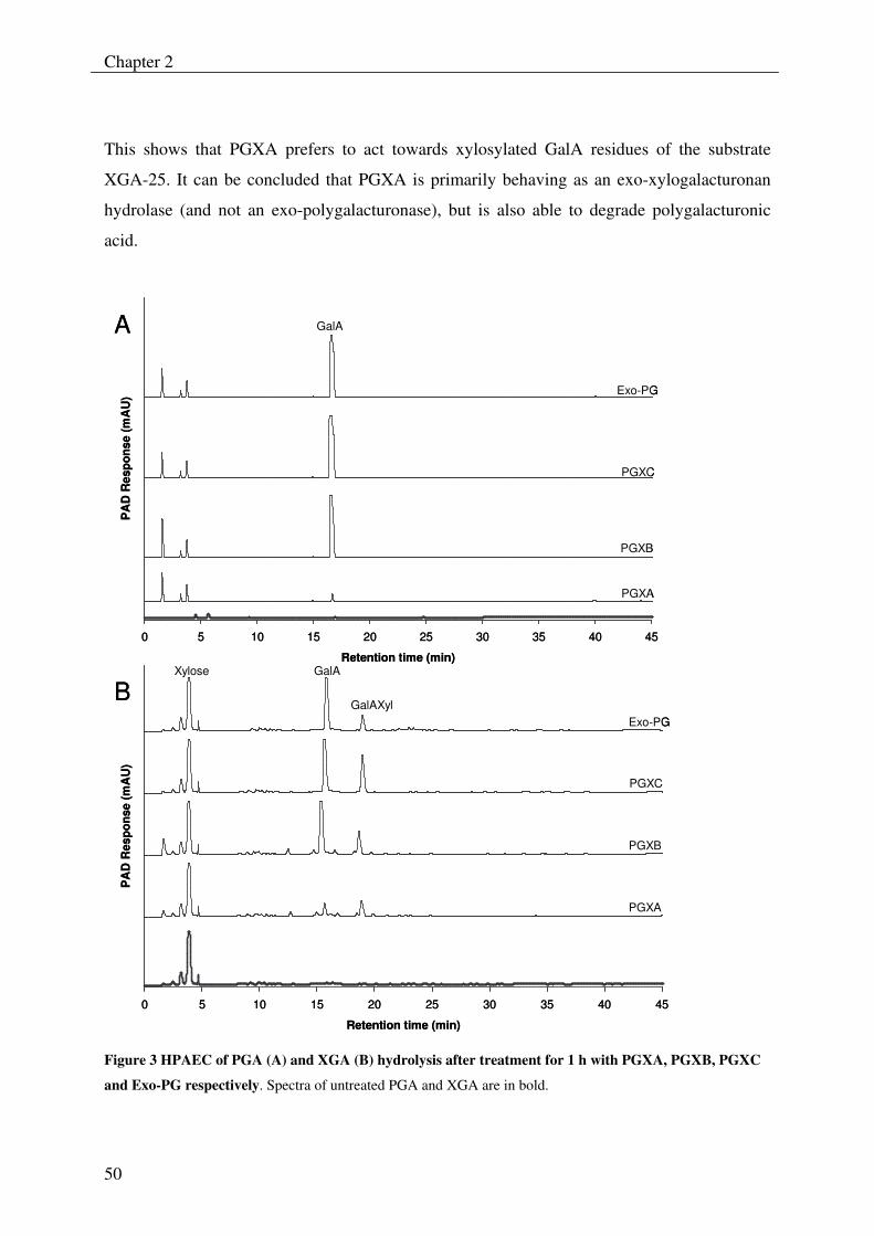
Chapter 2
50
This shows that PGXA prefers to act towards xylosylated GalA residues of the substrate
XGA-25. It can be concluded that PGXA is primarily behaving as an exo-xylogalacturonan
hydrolase (and not an exo-polygalacturonase), but is also able to degrade polygalacturonic
acid.
0 5 10 15 20 25 30 35 40 45
Retention time (min)
PA
D R
espo
nse
(mA
U)
GalA
PGXA
PGXB
PGXC
Exo-PG
0 5 10 15 20 25 30 35 40 45
Retention time (min)
PA
D R
espo
nse
(mA
U)
GalA
GalAXyl
Xylose
PGXA
PGXB
PGXC
Exo-PG
A
B
0 5 10 15 20 25 30 35 40 45
Retention time (min)
PA
D R
espo
nse
(mA
U)
GalA
PGXA
PGXB
PGXC
Exo-PG
0 5 10 15 20 25 30 35 40 45
Retention time (min)
PA
D R
espo
nse
(mA
U)
GalA
GalAXyl
Xylose
PGXA
PGXB
PGXC
Exo-PG
A
B
Figure 3 HPAEC of PGA (A) and XGA (B) hydrolysis after treatment for 1 h with PGXA, PGXB, PGXC
and Exo-PG respectively. Spectra of untreated PGA and XGA are in bold.
Exo-acting family 28 glycoside hydrolases of A. niger
51
Summary and conclusions
This paper presents for the first time an overview of the entire set of pectinolytic glycoside
hydrolases encoded in the genome of Aspergillus niger.
In addition to the already well-studied gene group of seven endo-polygalacturonan
hydrolases, we were able to extend the number of endo-rhamnogalacturonan hydrolases to
six. Furthermore, a new gene group comprising seven genes encoding for exo-acting
pectinolytic glycoside hydrolases was identified. In addition one gene encoding for a
pectinolytic glycoside hydrolase was identified to be a xylogalacturonan hydrolase.
Subsequent sequence analysis of the new gene group in combination with transcriptional data
allowed for the prediction of two subgroups of enzyme activities. Four genes were assessed as
encoding the exo-polygalacturonan hydrolases – PGAX, PGXA, PGXB, PGXC and three
genes as encoding the exo-rhamnogalacturonan hydrolases RGXA, RGXB and RGXC.
PGAX encoded by the A. niger gene pgaX is almost identical with the earlier described Exo-
PG of A. tubingensis.
Biochemical characterisation of the three proposed exo-polygalacturonan hydrolases PGXA,
PGXB and PGXC demonstrated that indeed all three enzymes were active towards
homogalacturonan in an exo-fashion. Yet, while the biochemical properties of PGXB do not
differ much from those of the Exo-PG of A. tubingensis, the data obtained for PGXA strongly
indicate that this enzyme is a novel exo-xylogalacturonan hydrolase. Interestingly, PGXC
appears to be an enzyme with reduced substrate specificity and remains unaffected in its
activity when GalA residues of the substrate are substituted with xylose.
Additionally, the MS analysis of a tryptic digest of the RG galacturono-hydrolase produced
by A. aculeatus, a protein for which no gene sequence is known, allowed the identification of
its A. niger homolog RGXC.
Although RGXB showed some activity towards pnp-rhamnose we were not able to identify
the natural substrate of this enzyme. Additional analysis is required to investigate the possible
function of RGXA and RGXB. Yet, both enzymes posses the catalytic residues and the proper
spacing required for the inverting mechanism characteristic for enzymes from family 28
glycoside hydrolases. The expression of the genes encoding these two enzymes on rhamnose
and sugar beet pectin suggests their involvement in the degradation of pectin regions that
contain rhamnose, most probably rhamnogalacturonan I.
Chapter 2
52
The generally higher expression levels observed for genes encoding exo–acting glycoside
hydrolases compared to those of genes encoding endo-activities, the strong initial rates of the
reactions catalysed by them and the fact that A. niger is able to utilise galacturonic acid in its
monomeric form suggest that exo-activities play a much more important physiological role
than expected previously during pectin degradation.
Acknowledgements
This research was supported by Senter IOP, The Netherlands (IGE01021).
We would like to thank Esther Tichon for cloning the pgxC gene. We also thank Margaret
Bosveld for the preparation of linear rhamnogalacturonan oligosaccharides.
Exo-acting family 28 glycoside hydrolases of A. niger
53
References 1 Carpita, N. C. and Gibeaut, D. M. (1993) Structural models of primary cell walls in
flowering plants: consistency of molecular structure with the physical properties of the walls during growth. Plant J. 3, 1-30
2 Vincken, J.-P., Schols, H.A., Oomen, R.J.F.J., Beldman, G., Visser, R.G.F., Voragen, A.G.J. (2003) Pectin-The hairy thing. In Advances in Pectin and Pectinase Research (Voragen, A. G. J., Schols, H., Visser, R., ed.), pp. 47-59, Kluwer Academic Publishers, Dordrecht
3 Voragen, A. G. J., Pilnik, W., Thibault, J. -F., Axelos, M. A. V., and Renard, C. M. G. C. (1995) Pectins. In Food Polysaccharides (Stephen, A. M., ed.), pp. 287-339, Marcel Dekker, Inc, New York-Basel-Hing Kong
4 Schols, H. A., Voragen, A.G.J. (1996) Complex Pectins: Structure elucidation using enzymes. In Pectins and pectinases (Visser, J., Voragen, A.G.J., ed.), pp. 3-19, Elsevier Science B.V., Amsterdam
5 Van der Vlugt-Bergmans, C. J. B., Meeuwsen, P.J.A., Voragen, A.G.J., and van Ooyen, A.J.J. (2000) Endo-Xylogalacturonan hydrolase, a Novel Pectinolytic Enzyme. Appl. Environ. Microbiol. 66, 36-41
6 Beldman, G., van den Broek, L.A.M., Schols, H.A., Searle-van Leeuwen, M.J.F., van Laere, K.M.J., Voragen, A.G.J. (1996) An exogalacturonase from Aspergillus aculeatus able to degrade Xylogalacturonan. Biotechnol. Lett. 18, 707-712
7 Kester, H. C., Benen, J. A. and Visser, J. (1999) The exopolygalacturonase from Aspergillus tubingensis is also active on xylogalacturonan. Biotechnol. Appl. Biochem. 30, 53-57
8 Suykerbuyk, M. E., Kester, H. C., Schaap, P. J., Stam, H., Musters, W. and Visser, J. (1997) Cloning and characterization of two rhamnogalacturonan hydrolase genes from Aspergillus niger. Appl. Environ. Microbiol. 63, 2507-2515
9 Kofod, L. V., Kauppinen, S., Christgau, S., Andersen, L. N., Heldt-Hansen, H. P., Dorreich, K. and Dalboge, H. (1994) Cloning and characterization of two structurally and functionally divergent rhamnogalacturonases from Aspergillus aculeatus. J. Biol. Chem. 269, 29182-29189
10 Azadi, P., O'Neill, M. A., Bergmann, C., Darvill, A. G. and Albersheim, P. (1995) The backbone of the pectic polysaccharide rhamnogalacturonan I is cleaved by an endohydrolase and an endolyase. Glycobiol. 5, 783-789
11 de Vries, R. P. and Visser, J. (2001) Aspergillus Enzymes Involved in Degradation of Plant Cell Wall Polysaccharides. Microbiol. Mol. Biol. Rev. 65, 497-522
12 Parenicova, L., Kester, H. C., Benen, J. A. and Visser, J. (2000) Characterization of a novel endopolygalacturonase from Aspergillus niger with unique kinetic properties. FEBS Lett. 467, 333-336
13 Henrissat, B. and Davies, G. (1997) Structural and sequence-based classification of glycoside hydrolases. Curr. Opin. Struc. Biol. 7, 637
14 van Santen, Y., Benen, J. A. E., Schroter, K.-H., Kalk, K. H., Armand, S., Visser, J. and Dijkstra, B. W. (1999) 1.68-A Crystal Structure of Endopolygalacturonase II from Aspergillus niger and Identification of Active Site Residues by Site-directed Mutagenesis. J. Biol. Chem. 274, 30474-30480
Chapter 2
54
15 van Pouderoyen, G., Snijder, H. J., Benen, J. A. E. and Dijkstra, B. W. (2003) Structural insights into the processivity of endopolygalacturonase I from Aspergillus niger. FEBS Lett. 554, 462
16 Armand, S., Wagemaker, M. J. M., Sanchez-Torres, P., Kester, H. C. M., van Santen, Y., Dijkstra, B. W., Visser, J. and Benen, J. A. E. (2000) The Active Site Topology of Aspergillus niger Endopolygalacturonase II as Studied by Site-directed Mutagenesis. J. Biol. Chem. 275, 691-696
17 Pages, S., Heijne, W. H., Kester, H. C., Visser, J. and Benen, J. A. (2000) Subsite mapping of Aspergillus niger endopolygalacturonase II by site-directed mutagenesis. J. Biol. Chem. 275, 29348-29353
18 Pages, S., Kester, H. C., Visser, J. and Benen, J. A. (2001) Changing a single amino acid residue switches processive and non-processive behavior of Aspergillus niger endopolygalacturonase I and II. J. Biol. Chem. 276, 33652-33656
19 Groot, G., Peij, N. v. and Ooyen, A. v. (2002) Exploring and exploiting the Aspergillus niger genome. In Annual Meeting of the Society for Industrial Microbiology August 2002
20 Coutinho, P. M. H., B. (1999) Carbohydrate-Active Enzymes server at URL: <http://afmb.cnrs-mrs.fr/CAZY/index.html>.
21 Bairoch, A. and Apweiler, R. (2000) The SWISS-PROT protein sequence database and its supplement TrEMBL in 2000. Nucl. Acids Res. 28, 45-48
22 Durbin, R., Eddy, S., Krogh, A. and Mitchison, G. (1998) Biological Sequence Analysis: Probabilistic Models of Proteins and Nucleic Acids. A tutorial introduction to hidden Markov models and other probabilistic modelling approaches in computational sequence analysis. Cambridge University Press
23 Birney, E., Clamp, M. and Durbin, R. (2004) GeneWise and Genomewise. Genome Res. 14, 988-995
24 Notredame, C., Higgins, D. G. and Heringa, J. (2000) T-coffee: a novel method for fast and accurate multiple sequence alignment. J. Mol. Biol. 302, 205
25 Kumar, S., Tamura, K. and Nei, M. (2004) MEGA3: Integrated software for Molecular Evolutionary Genetics Analysis and sequence alignment. Brief. Bioinfor. 5, 150-163
26 Pontecorvo, G., Roper, J. A., Hemmons, L. M., Macdonald, K. D. and Bufton, A. W. J. (1953) The genetics of Aspergillus nidulans. Adv. Genet. 5, 141
27 Vishniac, W. and Santer, M. (1957) The thiobacilli. Bacteriological Reviews 21, 195-213
28 van Dijck, P. W. M. (1999) Made by Genetic Engineering: A series of articles to promote a better understanding of the use of genetic engineering. J. Biotechnol. 67, 77
29 Sambrook, J., Fritsch, E. F. and Maniatis, T. (1989) Molecular Cloning: A Laboratory Manual. Cold Spring Harbor Laboratory Press, Cold Spring Harbor, NY
30 de Graaff, L., van den Broek, H. and Visser, J. (1988) Isolation and transformation of the pyruvate kinase gene of Aspergillus nidulans. Curr. Genet. 13, 315-321
31 Benen, J. A. E., Kester, H. C. M. and Visser, J. (1999) Kinetic characterization of Aspergillus niger N400 endopolygalacturonases I, II and C. Eur. J. Biochem. 259, 577-585
32 Kusters-van Someren, M. A., Harmsen, J. A., Kester, H. C. and Visser, J. (1991) Structure of the Aspergillus niger pelA gene and its expression in Aspergillus niger and Aspergillus nidulans. Curr. Genet. 20, 293
Exo-acting family 28 glycoside hydrolases of A. niger
55
33 Goosen, T., Bloemheuvel, G., Gysler, C., de Bie, D. A., van den Broek, H. W. and Swart, K. (1987) Transformation of Aspergillus niger using the homologous orotidine-5'-phosphate-decarboxylase gene. Curr. Genet. 11, 499
34 Parenicova, L., Benen, J. A., Kester, H. C. and Visser, J. (1998) pgaE encodes a fourth member of the endopolygalacturonase gene family from Aspergillus niger. Eur. J. Biochem. 251, 72-80
35 Beldman, G., Vincken, J.-P., Meeuwsen, P.J.A., Herweijer, M., Voragen, A.G.J. (2003) Degradation of differently substituted Xylogalacturonans by Endoxylogalacturonan Hydrolase and Endopolygalacturonases. Biocatal. Biotransform. 21, 189-198
36 Guillotin, S. E., Bakx, E. J., Boulenguer, P., Mazoyer, J., Schols, H. A. and Voragen, A. G. J. (2005) Populations having different GalA blocks characteristics are present in commercial pectins which are chemically similar but have different functionalities. Carbohydr. Polym. 60, 391
37 Guillotin, S. E. (2005) Studies on the intra- and intermolecular distributions of substituents in commercial pectins. pp. 155, Wageningen University, Wageningen
38 Schols, H. A., Posthumus, M.A., Voragen, A.G.J. (1990) Hairy (ramified) regions of pectins. PartI. Structural features of hairy regions of pectins isolated from apple juice produced by the liquefaction process. Carbohydr. Res. 206, 117-129
39 Zandleven, J., Beldman, G., Bosveld, M., Benen, J., Voragen, A. (2006) Enzymatic degradation studies of xylogalacturonans from apple and potato, using XGH. Carbohydr. Polym. 65, 495-503
40 Mutter, M., Beldman, G., Schols, H.A. and Voragen, A.G.J. (1994) Rhamnogalacturonan α-L-rhamnopyranosylhydrolase. A novel enzyme specific for the terminal non-reducing rhamnosyl unit in rhamnogalacturonan regions of pectin. Plant Physiol. 106, 241-250
41 van de Vis, J. W. (1994) Characterization and mode of action of enzymes degrading galactan structures of arabinegalactans. In Wageningen University, pp. 161, Wageningen University, Wageningen
42 Kester, H. C., Kusters-van Someren, M. A., Muller, Y. and Visser, J. (1996) Primary structure and characterization of an exopolygalacturonase from Aspergillus tubingensis. Eur. J. Biochem. 240, 738-746
43 Mutter, M., Beldman, G., Pitson, S.M., Schols, H.A. and Voragen, A.G.J. (1998) Rhamnogalacturonan α-D-Galactopyranosyluronohydrolase. An enzyme that specifically removes the terminal nonreducing galacturonosyl residue in rhamnogalacturonan regions of pectin. Plant Physiol. 117, 153-163
44 Zandleven, J., Beldman, G., Bosveld, M., Benen, J., Voragen, A. (2005) Mode of action of Xylogalacturonan hydrolase towards xylogalacturonan and xylogalacturonan oligosaccharides. Biochem. J. 387, 719-725
45 Gao, J., Opiteck, G. J., Friedrichs, M. S., Dongre, A. R. and Hefta, S. A. (2003) Changes in the Protein Expression of Yeast as a Function of Carbon Source. J. Proteome Res. 2, 643-649
46 VerBerkmoes, N. C., Hervey, W. J., Shah, M., Land, M., Hauser, L., Larimer, F. W., VanBerkel, G. J. and Goeringer, D. E. (2005) Evaluation of "Shotgun" Proteomics for Identification of Biological Threat Agents in Complex Environmental Matrixes: Experimental Simulations. Anal. Chem. 77, 923-932
Chapter 2
56
47 Link, A. J., Eng, J., Schieltz, D. M., Carmack, E., Mize, G. J., Morris, D. R., Garvik, B. M. and Yates, J. R. (1999) Direct analysis of protein complexes using mass spectrometry. Nat. Biotech. 17, 676-682
48 Petersen, T. N., Kauppinen, S. and Larsen, S. (1997) The crystal structure of rhamnogalacturonase A from Aspergillus aculeatus: a right-handed parallel beta-helix. Struc. 5, 533
49 Cho, S., Lee, S., Shin, W., Cho, S. W., Lee, S. and Shin, W. (2001) The X-ray structure of Aspergillus aculeatus polygalacturonase and a modeled structure of the polygalacturonase-octagalacturonate complex. J. Mol. Biol. 311, 863-878
50 Parenicova, L., Benen, J. K., H. C., Kester Hc Fau - Visser, J. and Visser, J. (2000) pgaA and pgaB encode two constitutively expressed endopolygalacturonases of Aspergillus niger. Biochem. J. 345, 637–644
51 Parenicová, L. (2000) Pectinases of Aspergillus niger: a molecular and biochemical characterisation. pp. 200, Wageningen University, Wageningen
52 Hara, T., Lim, J.Y. & Ueda, S. (1984) Purification and some properties of exopolygalacturonase from Aspergillus niger cultured in the medium containing satsuma mandarin peel. J. Jpn. Soc. Food Sci. Technol.-Nippon shok 31, 581-586
53 Sakamoto, T., Bonnin, E., Quemener, B., Thibault, J.-F. (2002) Purification and characterisation of two exo-polygalacturonases from Aspergillus niger able to degrade xylogalacturonan and acetylated homogalacturonan. Biochim. Biophys. Acta 1572, 10-18
Chapter 3
57
Mode of action of Xylogalacturonan hydrolase towards xylogalacturonan and xylogalacturonan oligosaccharides J. S. Zandleven, G. Beldman, M. Bosveld, J.A.E. Benen and A.G.J. Voragen Abstract Xylogalacturonan hydrolase (XGH, GH 28) is an enzyme that is able to degrade
xylogalacturonan (XGA), which is a polymer of α-D-galacturonic acid highly substituted with
β-D-xylose. XGA is present in cell walls of various plants and exudates such as gum
tragacanth.
XGA oligosaccharides were derived from a XGH digestion of gum tragacanth, then
fractionated, and analyzed for their sugar composition and structure by MALDI-TOF MS and
Nano-spray MS.
Several oligosaccharides from XGA were identified with different galacturonic acid/ xylose
ratios including five oligosaccharide isomers. Although XGH can act as an endo-enzyme,
product progression profiling showed that the disaccharide GalAXyl was predominantly
produced from XGA by XGH, which indicated also an exolytic action. The latter was further
supported by degradation studies of purified oligosaccharide GalA4Xyl3. It was shown that
XGH acted from the non-reducing end towards the reducing end of this oligosaccharide
showing the processive character of XGH.
The results from this study further show that although XGH prefers to act between two
xylosidated GalA units it tolerates unsubstituted GalA units in its -1 and +1 subsites.
This chapter has been published as: Zandleven, J. et al. (2005). "Mode of action of Xylogalacturonan hydrolase towards xylogalacturonan and xylogalacturonan oligosaccharides." Biochemical Journal 387(3): 719-725.
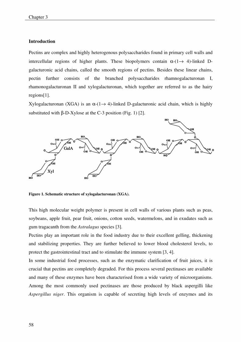
Chapter 3
58
Introduction Pectins are complex and highly heterogenous polysaccharides found in primary cell walls and
intercellular regions of higher plants. These biopolymers contain α-(1→ 4)-linked D-
galacturonic acid chains, called the smooth regions of pectins. Besides these linear chains,
pectin further consists of the branched polysaccharides rhamnogalacturonan I,
rhamonogalacturonan II and xylogalacturonan, which together are referred to as the hairy
regions[1].
Xylogalacturonan (XGA) is an α-(1→ 4)-linked D-galacturonic acid chain, which is highly
substituted with β-D-Xylose at the C-3 position (Fig. 1) [2].
OOHC
OH
OO
OOH
HO
C
OH
OO
O
OOH
HOHO
OOHC
OHO
O
OOH
HO
C
OH
OO
O
OOH
HOHO
O
OOHC
OHO
O
OOH
O
C
OH
OO
HO
OOH
HOHO
GalA
Xyl
OOHC
OH
OO
OOH
HO
C
OH
OO
O
OOH
HOHO
OOHC
OHO
O
OOH
HO
C
OH
OO
O
OOH
HOHO
O
OOHC
OHO
O
OOH
O
C
OH
OO
HO
OOH
HOHO
GalA
Xyl
Figure 1. Schematic structure of xylogalacturonan (XGA).
This high molecular weight polymer is present in cell walls of various plants such as peas,
soybeans, apple fruit, pear fruit, onions, cotton seeds, watermelons, and in exudates such as
gum tragacanth from the Astralagus species [3].
Pectins play an important role in the food industry due to their excellent gelling, thickening
and stabilizing properties. They are further believed to lower blood cholesterol levels, to
protect the gastrointestinal tract and to stimulate the immune system [3, 4].
In some industrial food processes, such as the enzymatic clarification of fruit juices, it is
crucial that pectins are completely degraded. For this process several pectinases are available
and many of these enzymes have been characterised from a wide variety of microorganisms.
Among the most commonly used pectinases are those produced by black aspergilli like
Aspergillus niger. This organism is capable of secreting high levels of enzymes and its
Mode of action of xylogalacturonan hydrolase
59
products are ‘generally recognized as safe’ allowing them to be used in food applications [5,
6].
Several pectinases have been isolated from A. niger such as endo-polygalacturonases, endo-
pectate lyases, endo-pectin lyases, pectin methyl esterases, which act on the smooth region of
pectin, as well as rhamnogalacturonan acetylesterase, rhamnogalacturonases, arabinases and
galactanases that act on the hairy regions of pectin. Most of these enzymes have been
thoroughly characterised with respect to their mode of action on their corresponding
substrates [7-10].
Despite the rich collection of pectinases in technical enzyme preparations obtained from A.
niger, parts of the hairy regions of pectin remain resistant to degradation and cause membrane
fouling in the ultrafiltration process for fruit juice clarification [10]. Being part of the pectic
hairy regions, fouling is largely influenced by XGA. Little is known about pectinases that can
degrade XGA, with the exception of exo-polygalacturonases from Aspergillus sp. [9, 11, 12]
Recently, the enzyme xylogalacturonan hydrolase (XGH) was discovered in Aspergillus
tubigensis, which acts specifically on XGA by cleaving the galacturonic acid backbone in an
endo-fashion [5, 13]. This enzyme belongs to the pectin degrading glycoside hydrolase family
28 (GH 28), which includes polygalacturonases (PGs) and exo-polygalacturonases (exo-PGs),
based on its amino acid sequence similarity towards this family [14, 15]. In addition the
amino acid sequence of XGH contains functionally important residues equivalent to the four
conserved active-site segments of PGs [15-17]. All family 28 members, including XGH, act
with an inverting mechanism [18].
XGH has been characterised with respect to its kinetic parameters, temperature- and pH-
effects and degradation of differently substituted (xylo-) galacturonans [11].
Here we report on the mode of action of XGH on its substrate XGA. This was realized by
degradation of XGA by XGH, followed by fractionation and quantitation and analysis of the
obtained oligosaccharides for their sugar abundance, composition and structure. Furthermore,
the degradation of a defined XGA oligosaccharide was studied in a time course in order to
determine the direction of action of XGH.
Chapter 3
60
Experimental
Substrate
Two xylogalacturonans (XGAs) derived from acid-modified and alkali saponified gum
tragacanth, were used. These XGAs had a Xyl:GalA ratio of 0.29 (XGA-29) and 0.47 (XGA-
47) respectively [11].
Enzyme
The enzyme xylogalacturonan hydrolase (XGH) from Aspergillus tubingensis was cloned [5]
and expressed in the A. niger “PlugBug” of DSM Food Specialities (Delft, the Netherlands).
This crude enzyme preparation was purified [11]. The crude and purified enzyme preparation
had a specific activity of 77 U/mg and 150 U/mg respectively.
Enzyme incubations
XGH was used to digest XGA-29 (final concentration of 1 mg/ml) for one h at 30 ºC. The
final enzyme concentration was 3.17 µg/ml. The substrate was dissolved in water in a total
volume of 10 ml and had a final pH of 3.6 without additional buffering. The enzyme was
inactivated by heating the reaction mixture for 10 min at 100 ºC. The digested XGA was
concentrated to 5 mg/ml, by freeze drying and redissolving in water, and was analyzed by
HPSEC as described previously [19]. Subsequently, the digest was analyzed by HPAEC at pH
12 using pulsed amperometric detection (PAD) as described previously [20]. The elution of
the XGA oligosaccharides by HPAEC was adapted as follows: a combination of two linear
gradients was used starting with 0-600 mM sodium acetate in 100 mM NaOH for 50 min,
followed by 600-1000 mM sodium acetate in 100 mM NaOH for 5 min.
Preparation of XGA oligosaccharides
For the preparation of XGA oligosaccharides, 300 mg XGA-29 was digested, as described
above, and concentrated to 25 mg/ml prior to fractionation by HPAEC. All fractions were
desalted by H+-Dowex AG 50W X8 (Biorad, Hercules, CA) treatment. Subsequently, the
desalted fractions were analyzed by HPAEC for their purity and compared with the complete
XGA digest for assignment. A GalA3 standard (5mg/ml) was used for quantification.
Mode of action of xylogalacturonan hydrolase
61
MALDI-TOF mass spectrometry
The fractionated XGA oligosaccharides were analyzed by MALDI-TOF as described
previously [19]. Two types of MS spectrometers were used: Voyager-DE RP Biospectrometry
workstation (PerSeptive Biosystems Inc., Framingham, MA, USA) and Ultraflex TOF MS
(Bruker Daltonics, Hamburg, Germany). The concentration of the XGA oligosaccharides
ranged from 0.25 mg/ml to 1 mg/ml. The samples were mixed with a matrix solution (1 µl of
sample in 1 µl of matrix) on a silver plate. The matrix solution was prepared by dissolving 10
mg of 2,5-dihydroxybenzoic acid in 1-ml mixture of acetonitrile/water (500:500 µl).
XGA oligosaccharides were identified based on their theoretical m/z masses. These masses
were based on the GalA/Xyl ratio’s, assuming total protonation of GalA and addition of one
Na+ or K+.
Labeling
Labeling of the individual XGA oligosaccharides with 18O at their reducing ends was
performed as described [21]. The XGA samples were incubated at 40 ºC for 12 days. The
progress of labeling was followed by MALDI-TOF MS and the labeled XGA
oligosaccharides were further structurally characterized by post-source decay (PSD) using the
Ultraflex-TOF (Bruker Daltonics, Hamburg, Germany).
Nano-spray mass spectrometry
Dynamic nanospray MS was performed on a LCQ Ion-trap (Finnigan MAT 95, San Jose, CA)
as described previously [19]. For MS analysis of XGA oligosaccharides settings were adapted
as follows: the flow was set at 5 µl/min, the spray voltage at 4.5 kV and 20-30% relative
collision energy for MS2 and higher was applied.
Product progression profiling of XGH
A XGA-29 solution (1 mg/ml) was incubated with purified XGH (0.35 µg/ml, final
concentration) at 30 °C. The digest had a total volume of 20 ml. At different time intervals (0-
48 h) a sample was taken and incubated for 10 min at 100 ºC to terminate the reaction. An
extra enzyme dose (0.70 µg/ml, final concentration) was added to the digest after 24 h.
Chapter 3
62
By freeze drying and redissolving in water, samples were concentrated five times and were
subsequently analyzed by HPAEC along with identified XGA oligosaccharides and GalA3 (5
mg/ml).
Preparation and degradation of oligosaccharide GalA4Xyl3
Another batch of XGA from gum tragacanth batch, similar to XGA-47, as described by
Beldman et al. (2003), was degraded by XGH using the same conditions as above. This digest
was fractionated by HPAEC as described before, and was isolated.
GalA4Xyl3 was labeled and structurally characterized by MALDI-TOF PSD. Subsequently,
GalA4Xyl3 was degraded with purified XGH using the conditions as described previously. At
different intervals (0-24 h) a sample was taken and incubated for 10 min at 100 ºC to
terminate the reaction. An extra enzyme dose (0.70 µg/ml, final concentration) was added to
the digest after 8 h. The progress of degradation in all samples was analyzed by MALDI-TOF
MS using the Ultraflex TOF MS (Bruker Daltonics, Hamburg, Germany) as described.
Mode of action of xylogalacturonan hydrolase
63
Results and discussion
HPLC analysis of xylogalacturonan digest
Xylogalacturonan (XGA-29) was incubated with xylogalacturonan hydrolase (XGH) for 24 h
and subsequently analyzed by HPSEC. An endo type of degradation of XGA was observed as
shown previously [11], although analysis by HPAEC (Fig. 2) revealed the presence of more
XGA oligosaccharides than reported before. This is explained by the application of higher
concentrations of the degraded substrate onto the column. In addition, to the previously
observed oligosaccharides, high molecular weight XGA products (shown as peaks 5-20) were
noticeable after one hour of enzymatic degradation of XGA.
Identification of xylogalacturonan oligosaccharides
An XGA-29 digest was preparatively fractionated by HPAEC in order to isolate the individual
oligomeric products. All fractions were subjected to HPAEC again to check their purity. By
MALDI-TOF MS these purified XGA oligosaccharides were analyzed for their sugar
composition. Figure 2 shows the HPAEC profile of the XGA digest from which eighteen
XGA oligosaccharides were identified. Also the previously observed products Xyl and GalA
as well as the di-mer GalAXyl were observed.
The XGA oligosaccharides identified have various GalA/Xyl ratios. In addition, five pairs of
oligosaccharide isomers were found having a similar sugar composition but a different elution
time upon HPAEC analysis. This can be explained by a different distribution of Xyl over the
GalA backbone as the xylose side chain may be linked to a different GalA residue or to a
different –OH group (C2 or C3) on the same GalA residue. These pairs of oligosaccharide
isomers are shown as peaks 3 and 6, 7 and 10, 11 and 14, 14 and 16, and 15 and 18. To avoid
confusion, the isomers are accentuated as shown in the table.
A measurable amount of xylose was present in the digest. This indicates that some xylose was
released by enzymatic or chemical hydrolysis from XGA and/or the oligosaccharides during
the digestion. The total amount of free xylose however was estimated to be only about 2 % of
the total amount present in the substrate.
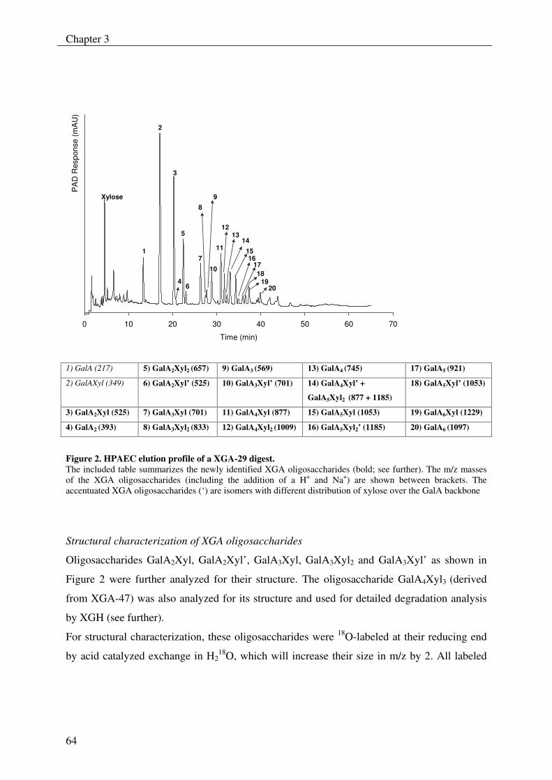
Chapter 3
64
0 10 20 30 40 50 60 70
Time (min)
PA
D R
espo
nse
(mA
U)
1
2
3
5
6
7
8
10
11
1213
14 1516
1718
1920
Xylose
4
9
Figure 2. HPAEC elution profile of a XGA-29 digest. The included table summarizes the newly identified XGA oligosaccharides (bold; see further). The m/z masses of the XGA oligosaccharides (including the addition of a H+ and Na+) are shown between brackets. The accentuated XGA oligosaccharides (‘) are isomers with different distribution of xylose over the GalA backbone
Structural characterization of XGA oligosaccharides
Oligosaccharides GalA2Xyl, GalA2Xyl’, GalA3Xyl, GalA3Xyl2 and GalA3Xyl’ as shown in
Figure 2 were further analyzed for their structure. The oligosaccharide GalA4Xyl3 (derived
from XGA-47) was also analyzed for its structure and used for detailed degradation analysis
by XGH (see further).
For structural characterization, these oligosaccharides were 18O-labeled at their reducing end
by acid catalyzed exchange in H218O, which will increase their size in m/z by 2. All labeled
1) GalA (217) 5) GalA2Xyl2 (657) 9) GalA3 (569) 13) GalA4 (745) 17) GalA5 (921)
2) GalAXyl (349) 6) GalA2Xyl’ (525) 10) GalA3Xyl’ (701) 14) GalA4Xyl’ +
GalA5Xyl2 (877 + 1185)
18) GalA5Xyl’ (1053)
3) GalA2Xyl (525) 7) GalA3Xyl (701) 11) GalA4Xyl (877) 15) GalA5Xyl (1053) 19) GalA6Xyl (1229)
4) GalA2 (393) 8) GalA3Xyl2 (833) 12) GalA4Xyl2 (1009) 16) GalA5Xyl2’ (1185) 20) GalA6 (1097)
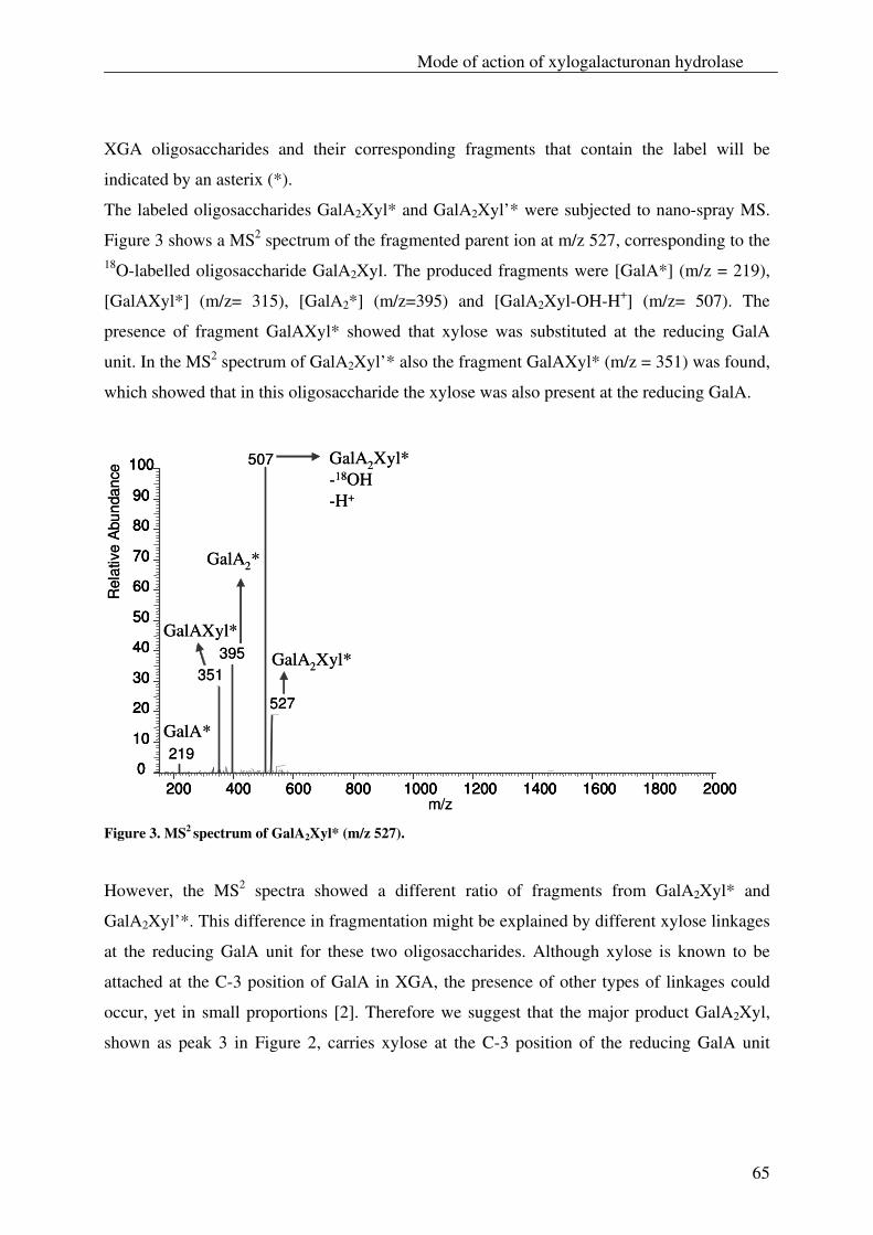
Mode of action of xylogalacturonan hydrolase
65
XGA oligosaccharides and their corresponding fragments that contain the label will be
indicated by an asterix (*).
The labeled oligosaccharides GalA2Xyl* and GalA2Xyl’* were subjected to nano-spray MS.
Figure 3 shows a MS2 spectrum of the fragmented parent ion at m/z 527, corresponding to the 18O-labelled oligosaccharide GalA2Xyl. The produced fragments were [GalA*] (m/z = 219),
[GalAXyl*] (m/z= 315), [GalA2*] (m/z=395) and [GalA2Xyl-OH-H+] (m/z= 507). The
presence of fragment GalAXyl* showed that xylose was substituted at the reducing GalA
unit. In the MS2 spectrum of GalA2Xyl’* also the fragment GalAXyl* (m/z = 351) was found,
which showed that in this oligosaccharide the xylose was also present at the reducing GalA.
200 400 600 800 1000200 400 600 800 1000 1200 1400 1600 1800 20000
10
20
30
40
50
60
70
80
90
100
1200 1400 1600 1800 2000m/z
0
10
20
30
40
50
60
70
80
90
100
Rel
ativ
eA
bund
ance
507
395351
527
219GalA*
GalAXyl*
GalA2*
GalA2Xyl*-18OH-H+
GalA2Xyl*
200 400 600 800 1000200 400 600 800 1000 1200 1400 1600 1800 20000
10
20
30
40
50
60
70
80
90
100
1200 1400 1600 1800 2000m/z
0
10
20
30
40
50
60
70
80
90
100
Rel
ativ
eA
bund
ance
507
395351
527
219GalA*
GalAXyl*
GalA2*
GalA2Xyl*-18OH-H+
GalA2Xyl*
Figure 3. MS2 spectrum of GalA2Xyl* (m/z 527).
However, the MS2 spectra showed a different ratio of fragments from GalA2Xyl* and
GalA2Xyl’*. This difference in fragmentation might be explained by different xylose linkages
at the reducing GalA unit for these two oligosaccharides. Although xylose is known to be
attached at the C-3 position of GalA in XGA, the presence of other types of linkages could
occur, yet in small proportions [2]. Therefore we suggest that the major product GalA2Xyl,
shown as peak 3 in Figure 2, carries xylose at the C-3 position of the reducing GalA unit
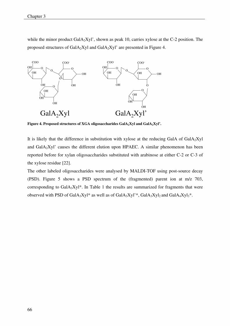
Chapter 3
66
while the minor product GalA2Xyl’, shown as peak 10, carries xylose at the C-2 position. The
proposed structures of GalA2Xyl and GalA2Xyl’ are presented in Figure 4.
OOH
COO-
OH
O
O
OH
OH
OH
OOH
OH
OH
COO-
O
GalA2Xyl GalA2Xyl’
OOH
OH
OH
O
COO-
O
OH
COO-
OOH
O
OH
OH
OH
Figure 4. Proposed structures of XGA oligosaccharides GalA2Xyl and GalA2Xyl’.
It is likely that the difference in substitution with xylose at the reducing GalA of GalA2Xyl
and GalA2Xyl’ causes the different elution upon HPAEC. A similar phenomenon has been
reported before for xylan oligosaccharides substituted with arabinose at either C-2 or C-3 of
the xylose residue [22].
The other labeled oligosaccharides were analysed by MALDI-TOF using post-source decay
(PSD). Figure 5 shows a PSD spectrum of the (fragmented) parent ion at m/z 703,
corresponding to GalA3Xyl*. In Table 1 the results are summarized for fragments that were
observed with PSD of GalA3Xyl* as well as of GalA3Xyl’*, GalA3Xyl2 and GalA4Xyl3*.
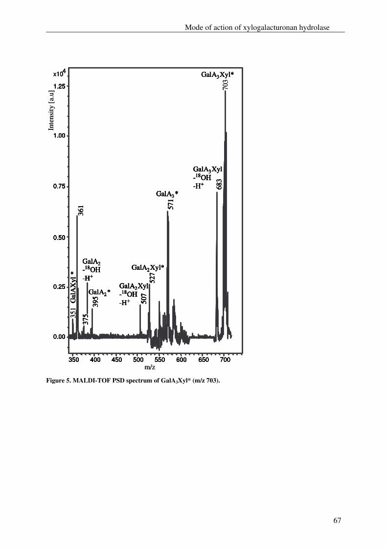
Mode of action of xylogalacturonan hydrolase
67
x10
683
361 57
1
527
395 507
375
0.00
0.25
0.50
0.75
1.00
1.25
4
350 400 450 500 550 600 650 700
GalA3*
GalA2*
Gal
AX
yl*
GalA2Xyl*
GalA2Xyl-18OH-H+
GalA2-18OH-H+
GalA3Xyl-18OH-H+
x10 GalA3Xyl*GalA3Xyl*
683
361 57
1
527
395 507
375
0.00
0.25
0.50
0.75
1.00
1.25
4
350 400 450 500 550 600 650 700
GalA3*
GalA2*
Gal
AX
yl*
GalA2Xyl*
GalA2Xyl-18OH-H+
GalA2-18OH-H+
GalA3Xyl-18OH-H+
703
683
361 57
1
527
395 507
351
375
0.00
0.25
0.50
0.75
1.00
1.25
4
350 400 450 500 550 600 650 700
GalA3*
GalA2*
Gal
AX
yl*
GalA2Xyl*
GalA2Xyl-18OH-H+
GalA2-18OH-H+
GalA3Xyl-18OH-H+
Inte
nsity
[a.
u]
m/z
x10
683
361 57
1
527
395 507
375
0.00
0.25
0.50
0.75
1.00
1.25
4
350 400 450 500 550 600 650 700
GalA3*
GalA2*
Gal
AX
yl*
GalA2Xyl*
GalA2Xyl-18OH-H+
GalA2-18OH-H+
GalA3Xyl-18OH-H+ 68
3
361 57
1
527
395 507
375
0.00
0.25
0.50
0.75
1.00
1.25
4
350 400 450 500 550 600 650 700
GalA3*
GalA2*
Gal
AX
yl*
GalA2Xyl*
GalA2Xyl-18OH-H+
GalA2-18OH-H+
GalA3Xyl-18OH-H+
x10 GalA3Xyl*GalA3Xyl*GalA3Xyl*GalA3Xyl*
683
361 57
1
527
395 507
375
0.00
0.25
0.50
0.75
1.00
1.25
4
350 400 450 500 550 600 650 700
GalA3*
GalA2*
Gal
AX
yl*
GalA2Xyl*
GalA2Xyl-18OH-H+
GalA2-18OH-H+
GalA3Xyl-18OH-H+
703
683
361 57
1
527
395 507
351
375
0.00
0.25
0.50
0.75
1.00
1.25
4
350 400 450 500 550 600 650 700
GalA3*
GalA2*
Gal
AX
yl*
GalA2Xyl*
GalA2Xyl-18OH-H+
GalA2-18OH-H+
GalA3Xyl-18OH-H+
Inte
nsity
[a.
u]
m/z Figure 5. MALDI-TOF PSD spectrum of GalA3Xyl* (m/z 703).
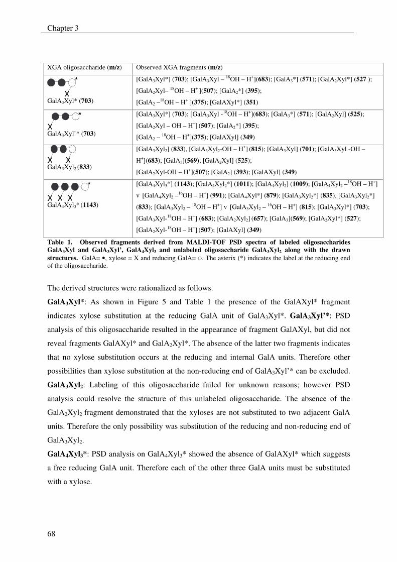
Chapter 3
68
XGA oligosaccharide (m/z) Observed XGA fragments (m/z)
GalA3Xyl* (703)
[GalA3Xyl*] (703); [GalA3Xyl – 18OH – H+](683); [GalA3*] (571); [GalA2Xyl*] (527 );
[GalA2Xyl– 18OH – H+ ](507); [GalA2*] (395);
[GalA2 –18OH – H+ ](375); [GalAXyl*] (351)
GalA3Xyl’* (703)
[GalA3Xyl*] (703); [GalA3Xyl -18OH – H+](683); [GalA3*] (571); [GalA2Xyl] (525);
[GalA2Xyl – OH – H+] (507); [GalA2*] (395);
[GalA2 – 18OH – H+](375); [GalAXyl] (349)
GalA3Xyl2 (833)
[GalA3Xyl2] (833), [GalA3Xyl2-OH – H+] (815); [GalA3Xyl] (701); [GalA3Xyl -OH –
H+](683); [GalA3](569); [GalA2Xyl] (525);
[GalA2Xyl-OH – H+](507); [GalA2] (393); [GalAXyl] (349)
GalA4Xyl3* (1143)
[GalA4Xyl3*] (1143); [GalA4Xyl2*] (1011); [GalA4Xyl2] (1009); [GalA4Xyl2 –18OH – H+]
v [GalA4Xyl2 –16OH – H+] (991); [GalA4Xyl*] (879); [GalA3Xyl2*] (835), [GalA3Xyl2*]
(833); [GalA3Xyl2 – 18OH – H+] v [GalA3Xyl2 – 16OH – H+] (815); [GalA3Xyl*] (703);
[GalA3Xyl-18OH – H+] (683); [GalA2Xyl2] (657); [GalA3](569); [GalA2Xyl*] (527);
[GalA2Xyl-18OH – H+] (507); [GalAXyl] (349)
Table 1. Observed fragments derived from MALDI-TOF PSD spectra of labeled oligosaccharides GalA3Xyl and GalA3Xyl’, GalA4Xyl3 and unlabeled oligosaccharide GalA3Xyl2 along with the drawn structures. GalA= •, xylose = X and reducing GalA= �. The asterix (*) indicates the label at the reducing end of the oligosaccharide.
The derived structures were rationalized as follows.
GalA3Xyl*: As shown in Figure 5 and Table 1 the presence of the GalAXyl* fragment
indicates xylose substitution at the reducing GalA unit of GalA3Xyl*. GalA3Xyl’*: PSD
analysis of this oligosaccharide resulted in the appearance of fragment GalAXyl, but did not
reveal fragments GalAXyl* and GalA2Xyl*. The absence of the latter two fragments indicates
that no xylose substitution occurs at the reducing and internal GalA units. Therefore other
possibilities than xylose substitution at the non-reducing end of GalA3Xyl’* can be excluded.
GalA3Xyl2: Labeling of this oligosaccharide failed for unknown reasons; however PSD
analysis could resolve the structure of this unlabeled oligosaccharide. The absence of the
GalA2Xyl2 fragment demonstrated that the xyloses are not substituted to two adjacent GalA
units. Therefore the only possibility was substitution of the reducing and non-reducing end of
GalA3Xyl2.
GalA4Xyl3*: PSD analysis on GalA4Xyl3* showed the absence of GalAXyl* which suggests
a free reducing GalA unit. Therefore each of the other three GalA units must be substituted
with a xylose.
X
*
X*
XX
XXX*
Mode of action of xylogalacturonan hydrolase
69
Product progression profiling of XGH
The appearance of products during the degradation of XGA by XGH was studied. This was
done by degrading XGA-29 with purified XGH for different time intervals followed by
HPAEC analysis and quantification (Fig. 6). The co-eluting XGA oligosaccharides were
plotted together in the product progression profiles.
It was assumed that the response factors of the individual oligosaccharides under the
conditions of HPAEC were not substantially different from each other. This was based on
HPAEC analysis of GalA and GalA oligosaccharides with a DP of 2 to DP 5 in equal molar
concentrations for which no substantial differences in response factors were found (results not
shown). In addition, from the literature it is known that only a small difference in response
factor for GalA and GalAXyl exist [9]. This demonstrates that xylose substitution has no
remarkable affect on the response factor of xylogalacturonan oligosaccharides.
The product progression profiles show a predominant production of GalAXyl with a minor
production of linear oligosaccharides like GalA2, GalA3 and GalA4 indicating that XGH
prefers to act between two xylosylated GalA units. This would include that subsites –1 and +1
of XGH have a high affinity for xylosylated GalA units. Linear oligosaccharides were not
degraded after prolonged digestion, which shows the dependency of XGH for xylose
substitution as well as the absence of other galacturonosidases (i.e. exo-PG). To our
knowledge only exo-PGs from Aspergillus sp. are known to accept xylose substituted
galacturonic acid in its subsite –1, because they produce the dimer GalAXyl from XGA [12,
23]. Although exo-PG has a pronounced sequence similarity with XGH, this latter enzyme
only contains a part of the conserved regions characteristic for exo-PGs. This indicates the
uniqueness of XGH [17].
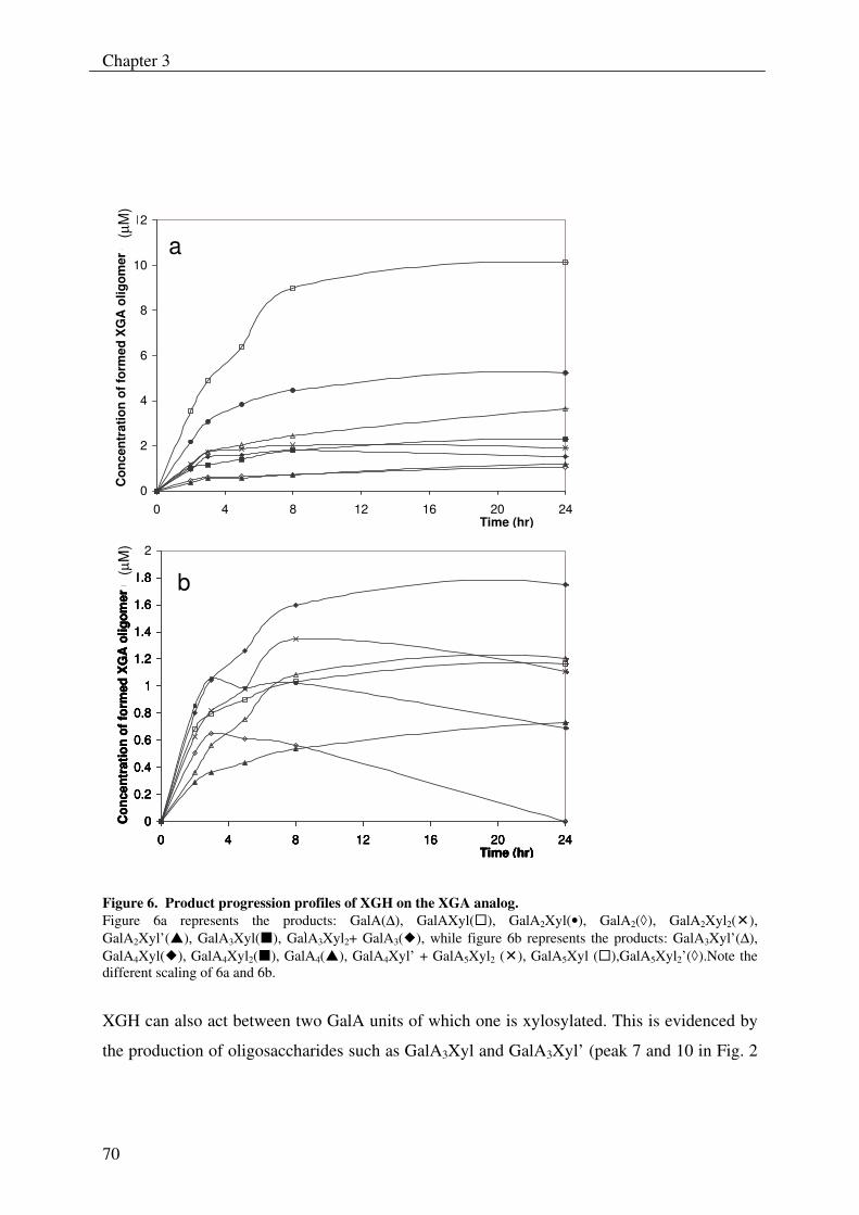
Chapter 3
70
0
2
4
6
8
10
12
0 4 8 12 16 20 24
Con
cent
ratio
n of
form
ed X
GA
olig
omer
(�M
)
Time (hr)
0
0.2
0.4
0.6
0.8
1
1.2
1.4
1.6
1.8
2
0 4 8 12 16 20 24
Con
cent
ratio
n of
form
ed X
GA
olig
omer
(�M
)
Time (hr)
a
b
0
2
4
6
8
10
12
0 4 8 12 16 20 24
Con
cent
ratio
n of
form
ed X
GA
olig
omer
(�M
)
Time (hr)
0
0.2
0.4
0.6
0.8
1
1.2
1.4
1.6
1.8
2
0 4 8 12 16 20 24
Con
cent
ratio
n of
form
ed X
GA
olig
omer
(�M
)
Time (hr)
a
b
(µM
)(µ
M)
0
2
4
6
8
10
12
0 4 8 12 16 20 24
Con
cent
ratio
n of
form
ed X
GA
olig
omer
(�M
)
Time (hr)
0
0.2
0.4
0.6
0.8
1
1.2
1.4
1.6
1.8
2
0 4 8 12 16 20 24
Con
cent
ratio
n of
form
ed X
GA
olig
omer
(�M
)
Time (hr)
a
b
0
2
4
6
8
10
12
0 4 8 12 16 20 24
Con
cent
ratio
n of
form
ed X
GA
olig
omer
(�M
)
Time (hr)
0
0.2
0.4
0.6
0.8
1
1.2
1.4
1.6
1.8
2
0 4 8 12 16 20 24
Con
cent
ratio
n of
form
ed X
GA
olig
omer
(�M
)
Time (hr)
a
b
(µM
)(µ
M)
Figure 6. Product progression profiles of XGH on the XGA analog. Figure 6a represents the products: GalA(∆), GalAXyl(�), GalA2Xyl(•), GalA2(�), GalA2Xyl2(�), GalA2Xyl’(�), GalA3Xyl(�), GalA3Xyl2+ GalA3(�), while figure 6b represents the products: GalA3Xyl’(∆), GalA4Xyl(�), GalA4Xyl2(�), GalA4(�), GalA4Xyl’ + GalA5Xyl2 (�), GalA5Xyl (�),GalA5Xyl2’(�).Note the different scaling of 6a and 6b.
XGH can also act between two GalA units of which one is xylosylated. This is evidenced by
the production of oligosaccharides such as GalA3Xyl and GalA3Xyl’ (peak 7 and 10 in Fig. 2
Mode of action of xylogalacturonan hydrolase
71
respectively), that have no xylose substitution at the non-reducing or at the reducing end,
respectively (Table 1).
As shown in Figure 6, GalA4Xyl2 was further degraded, which demonstrates that XGH prefers
to act on XGA oligosaccharides with a backbone of at least four GalA units. In addition,
GalA5Xyl2’ was degraded more efficiently than GalA4Xyl2, which demonstrates that an
increase of the GalA backbone by one GalA unit also significantly increases the XGH
activity. Furthermore, the rapid degradation of GalA5Xyl2’ is in line with the decline of the
GalA4Xyl’-GalA5Xyl2 pair in the progression profiles. GalA4Xyl and GalA5Xyl were not
further degraded, which demonstrates that increased xylose substitution enhances XGH
activity.
Degradation of GalA4Xyl3
In order to study the direction of action of XGH, a highly substituted oligosaccharide with at
least four GalA units in the backbone that can potentially be hydrolyzed more than once by
XGH is required. Furthermore, this oligosaccharide should have an asymmetrical structure
with respect to xylose substitution for the recognition of the reducing end. As none of the
oligosaccharides that were produced from XGA-29 met this requirement, XGA-47 with a
higher degree of substitution with xylose was used to prepare the desired oligosaccharide.
Partial degradation of this substrate resulted in the production of significant amounts of
oligosaccharide GalA4Xyl3, which appeared suitable for further degradation studies. This
oligosaccharide was purified to near homogeneity (>95%) from the XGA digest by
preparative HPAEC and structurally characterized by MALDI-TOF PSD as described in the
previous paragraph.
GalA4Xyl3 was degraded with purified XGH for different time intervals followed by MALDI-
TOF MS analysis (results not shown). Degradation of the oligosaccharide GalA4Xyl3 for 5
min resulted in the formation of significant amounts of GalAXyl and GalA3Xyl2. After 24 hrs
of incubation the main products left were GalA2Xyl and GalAXyl. This shows that XGH acts
mainly from the non-reducing end of GalA4Xyl3 towards the reducing end, cleaving off
GalAXyl step-by-step. Furthermore, the smallest substrate hydrolysable appeared to be
GalA3Xyl2. Since a small amount of GalA2Xyl2 was also produced, it can be concluded that
XGH also acts, however less favorably, at the internal glycosidic bond of GalA4Xyl3. This
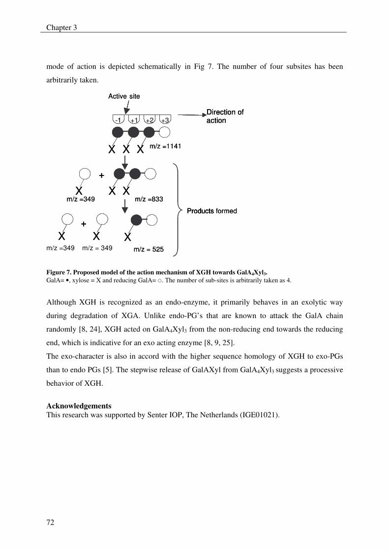
Chapter 3
72
mode of action is depicted schematically in Fig 7. The number of four subsites has been
arbitrarily taken.
- 1 +1 +2 +3
Active site
m/z =1141
m/z =349 m/z = 349 m/z = 525
Products Products formed
m/z =349 m/z =833 X X X
X X X
X
+
X
+
Direction of action - 1
Active site
m/z =1141
m/z = 525
Products Products formed
m/z =349 m/z =833 X X X
X X X
X
+
X
+
Direction of action Direction of action
X X
Figure 7. Proposed model of the action mechanism of XGH towards GalA4Xyl3. GalA= •, xylose = X and reducing GalA= �. The number of sub-sites is arbitrarily taken as 4.
Although XGH is recognized as an endo-enzyme, it primarily behaves in an exolytic way
during degradation of XGA. Unlike endo-PG’s that are known to attack the GalA chain
randomly [8, 24], XGH acted on GalA4Xyl3 from the non-reducing end towards the reducing
end, which is indicative for an exo acting enzyme [8, 9, 25].
The exo-character is also in accord with the higher sequence homology of XGH to exo-PGs
than to endo PGs [5]. The stepwise release of GalAXyl from GalA4Xyl3 suggests a processive
behavior of XGH.
Acknowledgements This research was supported by Senter IOP, The Netherlands (IGE01021).
Mode of action of xylogalacturonan hydrolase
73
References
1 Vincken, J.-P., Schols, H.A., Oomen, R.J.F.J., Beldman, G., Visser, R.G.F., Voragen, A.G.J. (2003) Pectin-The hairy thing. In Advances in pectin and Pectinase Research (Voragen, A. G. J., Schols, H., Visser, R., ed.), pp. 47-59, Kluwer Academic Publishers, Dordrecht
2 Aspinall, G. O., Baillie, J. (1963) Gum Tragacanth. Part I. Fractionation of the gum and the structure of tragacanthic acid, J. Chem. Soc. 318, 1702-1714
3 Thibault, J.-F., Ralet, M.-C. (2001) Pectins, their Origin, Structure and Functions. In Advanced Dietary Fibre Technology (McCleary, B. V., Prosky, L., ed.), pp. 369- 378, Blackwell Science, Oxford
4 Yamada, H. (1996) Contribution of pectins on health care. In Pectins and Pectinases (Visser, J., Voragen, A.G.J., ed.), pp. 173-190, Elsevier Science, Amsterdam
5 Van der Vlugt-Bermans, C. J. B., Meeuwsen, P.J.A., Voragen, A.G.J., and van Ooyen, A.J.J. (2000) Endo-Xylogalacturonan hydrolase, a Novel Pectinolytic Enzyme, Appl. Environ. Microbiol. 66, 36-41
6 De Vries, R. P. (2003) Regulation of Aspergillus genes encoding plant cell wall polysaccharide-degrading enzymes; relevance for industrial production, Appl. Microbiol. Biot. 61, 10-20
7 Searle-van Leeuwen, M. J. F., Van den Broek L.A.M., Schols, H.A., Beldman, G., Voragen, A.G.J. (1992) Rhamnogalacturonan acetylesterase: a novel enzyme from Aspergillus aculeatus, specific for the deacetylation of hairy (ramified) regions of pectins, Appl. Microbiol. Biotechnol. 38, 347-349
8 Benen, J. A. E., van Alebeek, G.-J.W.M., Voragen, A.G.J., Visser, J. (2003) Mode of Action Analysis And Structure-Function Relationships Of Aspergillus Niger Pectinolytic Enzymes. In Advances in Pectin and Pectinase Research (Voragen, A. G. J., Schols, H., Visser, R., ed.), pp. 235-256, Kluwer Academic Publishers, Dordrecht
9 Sakamoto, T., Bonnin, E., Quemener, B., Thibault, J.-F. (2002) Purification and characterisation of two exo-polygalacturonases from Aspergillus niger able to degrade xylogalacturonan and acetylated homogalacturonan, Biochim. Biophys. Acta 1572, 10-18
10 Vincken, J.-P., Voragen, A.G.J., Beldman, G. (2003) Enzymes Degrading Rhamnogalacturonan and Xylogalacturonan. In Handbook of Food Enzymology (Whitaker, J. R., Voragen, A.G.J., Wong, D.W.S., ed.), pp. 931-941, Marcel Dekker, New York
11 Beldman, G., Vincken, J.-P., Schols, H.A., Meeuwsen, P.J.A., Herweijer, M., Voragen, A.G.J. (2003) Degradation of differently substituted Xylogalacturonans by Endoxylogalacturonan Hydrolase and Endopolygalacturonases, Biocatal. Biotranfor. 21, 189-198
12 Kester, H., Benen, J., Visser, J. (1999) The exo-polygalacturonase from Aspergillus tubingensis is also active on xylogalacturonan, Biot. and Appl. Biochem. 30, 53-57
13 Herweijer, M. A., Vincken, J.-P., Meeuwsen, P.J.A, van der Vlugt-Bergmans, C.J.B., Beldman, G., van Ooyen, A.J.J., Voragen, A.G.J. (2003) Endo-Xylogalacturonan Hydrolase. In Advances in Pectin and Pectinase Research (Voragen, A. G. J., Schols, H., Visser, R., ed.), pp. 257-266, Kluwer Academic Publishers, Dordrecht
Chapter 3
74
14 Henrissat, B. (1991) A classification of glycosyl hydrolases based on amino acid sequence similarities, Biochem. J. 280, 309-316
15 Coutinho, P. M. H., B. (1999) Carbohydrate-Active Enzymes server at URL: http://afmb.cnrs-mrs.fr/CAZY/
16 Coutinho, P. M., & Henrissat, B. (1999) Carbohydrate-active enzymes: an integrated database approach. In Recent Advances in Carbohydrate Bioengineering (Gilbert, H. J., Davies, G., Henrissat, B., Svensson, B., ed.), pp. 3-12, The Royal Society of Chemistry, Cambridge
17 Markovic, O., Janecek, S. (2001) Pectin degrading glycoside hydrolases of family 28: sequence-structural features, specificities and evolution, Protein Eng. 14, 615-631
18 Henrissat, B., Davies, G. (1997) Structural and sequence-based classification of glycoside hydrolases, Curr. Opin. Struct. Biol. 7, 637-644
19 Kabel, M. A., Schols, H. A. and Voragen, A. G. J. (2002) Complex xylo-oligosaccharides identified from hydrothermally treated Eucalyptus wood and brewery's spent grain, Carbohydr. Polym. 50, 191-200
20 Van Alebeek, G.-J., Christensen, T.M.I.E., Schols, H.A., Mikkelsen, J.D. Voragen, A.G.J. (2002) Mode of Action of Pectin Lyase A of Aspergillus niger on Differently C6-substituted Oligogalacturonides, J. Biol. Chem. 277, 25929-25936
21 Van Alebeek, G.-J. W. M., Zabotina, O. Beldman, G., Schols, H.A., Voragen, A.G.J. (2000) Structural analysis of (methyl-esterified) oligogalacturonides using post-source decay matrix-assisted laser desorption/ionization time-of-flight mass spectrometry, J. Mass Spectrom. 35, 831-840
22 Van Laere, K. M. J., Beldman, G., Voragen, A.G.J. (1997) A new arabinofuranohydrolase from Bifidobacterium adolescentis able to remove arabinosyl residues from double-substituted xylose units in arabinoxylan, Appl. Microbiol. Biot. 47, 231-235
23 Beldman, G., van den Broek, L.A.M., Schols, H.A., Searle-van Leeuwen, M.J.F., van Laere, K.M.J., Voragen, A.G.J. (1996) An exogalacturonase from Aspergillus aculeatus able to degrade Xylogalacturonan, Biotechnol Lett 18, 707-712
24 Bonnin, E., Le Goff, A., Korner, R., Van, A. G., Christensen, T., Voragen, A. G. J., Roepstorff, P., Caprari, C. and Thibault, J. F. (2001) Study of the mode of action of endopolygalacturonase from Fusarium moniliforme, Biochim. Biophys. Acta 15, 301-309
25 Chellegatti, M. A. D., Fonseca, M.J.V., Said, S. (2002) Purification and partial characterization of exopolygalacturonase I from Penicillium frequentans, Microbiol. Res. 157, 19-24
Chapter 4
75
Enzymatic degradation studies of xylogalacturonans from apple and
potato, using xylogalacturonan hydrolase
J. S. Zandleven, G. Beldman, M. Bosveld, H.A. Schols, and A.G.J. Voragen
Abstract
Action of xylogalacturonan hydrolase (XGH) towards xylogalacturonan (XGA) present in the
alkali saponified “modified hairy regions” from potato- and apple pectin was studied.
Analysis of enzymatic degradation products from XGA in these complex pectins
demonstrated that the degradable xylogalacturonans from both sources have a similar xylose
side chain distribution. The disaccharide β-D-Xyl-(1,3)-GalA was the predominant product
from these substrates.
The number of different enzymatic degradation products from xylogalacturonan present in
apple and potato pectin was much lower than the number of different products from a
xylogalacturonan derived from gum tragacanth. This suggests a relatively uniform distribution
of xylose in the degradable part of XGA from apple and potato pectin. In addition, dimeric
side chains of xylose were observed in digests of XGA from both pectins, which apparently
did not hinder the action of XGH. From this it is assumed that Xyl-Xyl as well as Xyl
substituted GalA residues are accepted in subsite –1 and +1 of xylogalacturonan hydrolase.
This chapter has been published as: Zandleven, J. et al. (2006). "Enzymatic degradation studies of xylogalacturonans from apple and potato, using xylogalacturonan hydrolase.” Carbohydrate Polymers 65: 495-503.
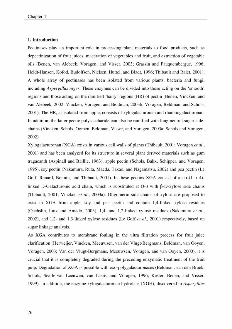
Chapter 4
76
1. Introduction
Pectinases play an important role in processing plant materials to food products, such as
depectinization of fruit juices, maceration of vegetables and fruit, and extraction of vegetable
oils (Benen, van Alebeek, Voragen, and Visser, 2003; Grassin and Fauquembergue, 1996;
Heldt-Hansen, Kofod, Budolfsen, Nielsen, Huttel, and Bladt, 1996; Thibault and Ralet, 2001).
A whole array of pectinases has been isolated from various plants, bacteria and fungi,
including Aspergillus niger. These enzymes can be divided into those acting on the ‘smooth’
regions and those acting on the ramified ‘hairy’ regions (HR) of pectin (Benen, Vincken, and
van Alebeek, 2002; Vincken, Voragen, and Beldman, 2003b; Voragen, Beldman, and Schols,
2001). The HR, as isolated from apple, consists of xylogalacturonan and rhamnogalacturonan.
In addition, the latter pectic polysaccharide can also be ramified with long neutral sugar side-
chains (Vincken, Schols, Oomen, Beldman, Visser, and Voragen, 2003a; Schols and Voragen,
2002)
Xylogalacturonan (XGA) exists in various cell walls of plants (Thibault, 2001; Voragen et al.,
2001) and has been analyzed for its structure in several plant derived materials such as gum
tragacanth (Aspinall and Baillie, 1963), apple pectin (Schols, Bakx, Schipper, and Voragen,
1995), soy pectin (Nakamura, Ruta, Maeda, Takao, and Nagamatsu, 2002) and pea pectin (Le
Goff, Renard, Bonnin, and Thibault, 2001). In these pectins XGA consist of an α-(1→ 4)-
linked D-Galacturonic acid chain, which is substituted at O-3 with β-D-xylose side chains
(Thibault, 2001; Vincken et al., 2003a). Oligomeric side chains of xylose are proposed to
exist in XGA from apple, soy and pea pectin and contain 1,4-linked xylose residues
(Oechslin, Lutz and Amado, 2003), 1,4- and 1,2-linked xylose residues (Nakamura et al.,
2002), and 1,2- and 1,3-linked xylose residues (Le Goff et al., 2001) respectively, based on
sugar linkage analysis.
As XGA contributes to membrane fouling in the ultra filtration process for fruit juice
clarification (Herweijer, Vincken, Meeuwsen, van der Vlugt-Bergmans, Beldman, van Ooyen,
Voragen, 2003; Van der Vlugt-Bergmans, Meeuwsen, Voragen, and van Ooyen, 2000), it is
crucial that it is completely degraded during the preceding enzymatic treatment of the fruit
pulp. Degradation of XGA is possible with exo-polygalacturonases (Beldman, van den Broek,
Schols, Searle-van Leeuwen, van Laere, and Voragen, 1996; Kester, Benen, and Visser,
1999). In addition, the enzyme xylogalacturonan hydrolase (XGH), discovered in Aspergillus
Degradation studies of xylogalacturonan from apple and potato
77
tubingensis, is able to degrade XGA in an endo fashion (van der Vlugt-Bergmans, Meeuwsen,
Voragen, and van Ooyen, 2000). The corresponding gene for XGH from A. tubingensis has
been over-expressed in the A. niger “PlugBug” (van Dijck, 1999) and the forthcoming
enzyme was subsequently purified (Beldman, Vincken, Meeuwsen, Herweijer, and Voragen,
2003). This enzyme has been studied for its pH- and temperature stability as well as its
activity towards xylogalacturonans with different degrees of xylose substitution (Beldman et
al. 2003). Additionally, the mode of action of this enzyme towards XGA from alkali and acid
modified gum tragacanth has been investigated by analysis of the structure of the degradation
products (Zandleven, Beldman, Bosveld, Benen, and Voragen, 2005). In this study, XGH is
investigated with repect to its activity towards XGA present in apple and potato MHR. Also
some aspects about the subsite model of XGH are discussed based on the structure of XGA
oligosaccharides formed.
Chapter 4
78
2. Experimental
2.1. Substrates
Saponified potato MHR (p-MHR-S) and saponified apple MHR (a-MHR-S) were prepared as
described (Schols, Posthumus, and Voragen, 1990). Enzymatic liquefaction of apples for the
preparation of a-MHR-S was performed with a commercial enzyme preparation (Ultra-SP;
Novozymes, Copenhagen, Denmark). An amount of 7.5 g of this substrate was further treated
with rhamnogalacturonan hydrolase (RGH) and fractionated by preparative size exclusion
chromatography (SEC) in order to obtain a XGA enriched a-MHR-S substrate. This procedure
was done as follows: a-MHR-S (5 mg/ml final concentration) was treated with purified RGH
(Novozymes, Copenhagen, Denmark; Kofod, Kauppinen, Christgau, Andersen, Heldt-
Hansen, Dorreich, Dalbfge, 1994; Schols et al., 1990) for 24 hrs at 40 °C in 50 mM NaOAc
buffer (pH 5.6). The final enzyme concentration was 3.33 µg/ml. The enzyme was inactivated
by heating the reaction mixture for 10 min at 100 ºC. Subsequently, the RGH treated a-MHR-
S was dialyzed against water. A yield of 6.15 g RGH treated a-MHR-S was obtained after
freeze-drying. A portion of 6 g from this material was applied onto a Superdex 30 PG column
(5.5 L, 70 x 10 cm) using millipore water as eluent with a flow rate of 25 ml⋅min-1. Two
fractions, a-MHR-S-frA (pooled fractions 18-27) and a-MHR-S-frB (pooled fractions 27-41)
respectively, were collected (Fig. 1) and subsequently freeze-dried. The fractions a-MHR-S-
frA and a-MHR-S-frB gave a yield of 1.15 g and 2.0 g, respectively.
2.2. Neutral sugar composition and uronic acid content
p-MHR-S and a-MHR-S-frB were analyzed for their neutral sugar composition by gas
chromatography (Englyst & Cummings, 1984), using inositol as an intern standard. The
samples were treated with 72% (w/w) H2SO4 for 1 h at 30 °C, followed by hydrolysis with 1
M H2SO4 for 3 h at 100 °C. Prior to analysis by gas chromatography, the released neutral
sugars were converted to their alditol acetates.
The uronic acid content of p-MHR-S and a-MHR-S-frB was determined by an automated m-
hydroxydiphenyl assay using an autoanalyser (Skalar Analytical BV, Breda, The Netherlands;
Blumenkrantz and Asboe-Hansen, 1973; Thibault and Robin 1975).
Degradation studies of xylogalacturonan from apple and potato
79
2.3. HPSEC analysis
High-performance size-exclusion chromatography was performed on three TSK columns (7.8
mm ID x 30 cm per column) in a series of TSKGel G4000 PWXL, TSKGel G3000 PWXL
and TSK2500 PWXL, in combination with a PWX-guard column. All columns were from
TosoHaas (Japan).
Elution took place at 30 ºC using 0.2 M sodium nitrate at a flow rate of 0.8 ml/min. The eluate
was monitored using a refractive index (RI) detector. The calibration was performed using
pullulan standards with a molecular weight of 404, 112, 47.3, 22.8, 5.9 kDa and GalA (0.196
kDa).
2.4 HPAEC at pH 12
High performance anion-exhange chromatography was performed on a Dionex system
equipped with a CarboPac PA-1 column (4 mm ID x 250 mm) in combination with a
CarboPac PA Guard column (3 mm ID x 25 mm) and pulsed amperometric detection (PAD).
The elution (1 ml/min) of the oligosaccharides was performed by using a combination of two
linear gradients, starting with 0-600 mM sodium acetate in 100 mM NaOH for 50 min,
followed by 600-1000 mM sodium acetate in 100 mM NaOH for 5 min.
2.5. MALDI-TOF mass spectrometry
Prior to mass spectrometry analysis, samples were desalted by treatment with H+-Dowex AG
50W X8 (Biorad, Hercules, CA), using a final concentration of 350 mg H+-Dowex per ml
digest. The desalted digests were mixed with a matrix solution (1 µl of sample in 1 µl of
matrix) and applied on a MALDI sample plate. The matrix solution was prepared by
dissolving 9 mg of 2,5- dihydroxybenzoic acid in a 1-ml mixture of acetonitrile:water (300 µl:
700 µl).
MALDI-TOF MS (Matrix-Assisted Laser Desorption/ Ionisation Time-Of-Flight Mass
Spectrometry) analysis was performed using an Ultraflex workstation (Bruker Daltonics,
Hamburg, Germany) equipped with a nitrogen laser of 337 nm. The mass spectrometer was
selected for positive ions, which were accelerated by an electric field of 12 kV, after a delayed
Chapter 4
80
extraction time of 200 ns. The ions were detected in the reflector mode. External calibration
of the mass spectrometer was performed using a mixture of maltodextrines (mass range 365-
2309).
2.6 Electrospray mass spectrometry
Electrospray MS was performed on a LTQ Ion-trap (Thermo, San Jose, CA). Sample was
running through a transferring capillary (100 µm ID) and a spraying capillary with an ID of
50 µm at a flow rate of 1 µl/min. MS analysis was carried out in the positive mode using a
spray voltage of 4.5 kV and a capillary temperature of 200 ºC. The capillary voltage and tube
lens were set at 33 V and 225 V respectively. MS2 and higher was executed using a window
of 1 m/z and a relative collision energy of 20-30%.
2.7. Enzyme incubations
Purified XGH, prepared as described (Beldman et al., 2003), was used to degrade a-MHR-S-
frA, a-MHR-S-frB and p-MHR-S (final concentration of 5 mg/ml). These substrates were
dissolved in 50 mM NaOAc (pH 3.5) and incubated with XGH (final concentration of 0.35
µg/ml) for 20 h at 30 ºC. The enzyme had a specific activity of 150 U/mg. Subsequently, the
enzyme was inactivated by heating the reaction mixtures for 10 min at 100 ºC. The digested
substrates including their controls (i.e. a-MHR-S-frB and p-MHR-S, which were not treated
with XGH) were desalted by treatment with H+-Dowex AG 50W X8 (Biorad, Hercules, CA),
using a final concentration of 350 mg H+-Dowex per ml digest. The desalted samples were
subsequently analyzed by HPSEC and HPAEC using a set of known XGA-oligosaccharides
for identification (Zandleven et al., 2005).
2.8. Product analysis of XGH degraded a-MHR-S-frB and p-MHR-S at different time
intervals
Oligosaccharide digests were prepared from a-MHR-S-frB and p-MHR-S as described above.
At 0 min (blank), 30 min, 1h, and 20 h, a sample was taken and incubated at 100 ºC for 10
Degradation studies of xylogalacturonan from apple and potato
81
min to inactivate the enzyme. The obtained oligosaccharides were desalted and subsequently
analyzed by HPAEC, and by MALDI-TOF MS or electrospray MS.
2.9 Fractionation of XGH degraded a-MHR-S-frB and p-MHR-S
A-MHR-S-frB and p-MHR-S were degraded by XGH using the same conditions as described
in section “Enzyme incubations”. These digests were fractionated by HPAEC on the
Carbopac PA-1 column as described. Samples of the fractionated XGA oligosaccharides were
desalted and analyzed by MALDI-TOF MS. Fractions containing GalAXyl2 were pooled,
desalted by H+-Dowex AG 50W X8 (Biorad, Hercules, CA) and freezedried until further use.
2.10 Degradation of GalAXyl2 by ββββ-xylosidase
GalAXyl2, as isolated from 25 mg of p-MHR-S and a-MHR-S-frB respectively, was degraded
by β-(1,4)-xylosidase (E.C.3.2.1.37; GH 3) from Aspergillus awamori. The conditions were
as follows: the total yield of GalAXyl2 was dissolved in 500 µl 50 mM NaOAC (pH 4.0) and
incubated with β-(1,4)-xylosidase (final concentration of 0.85 µg/ml) for 20 h at 30 °C. The
enzyme preparation had a specific activity of 1.45 U/mg.
Degradation of GalAXyl2 was analyzed by HPAEC and MALDI-TOF MS, as described in
section 2.4 and 2.5 respectively.
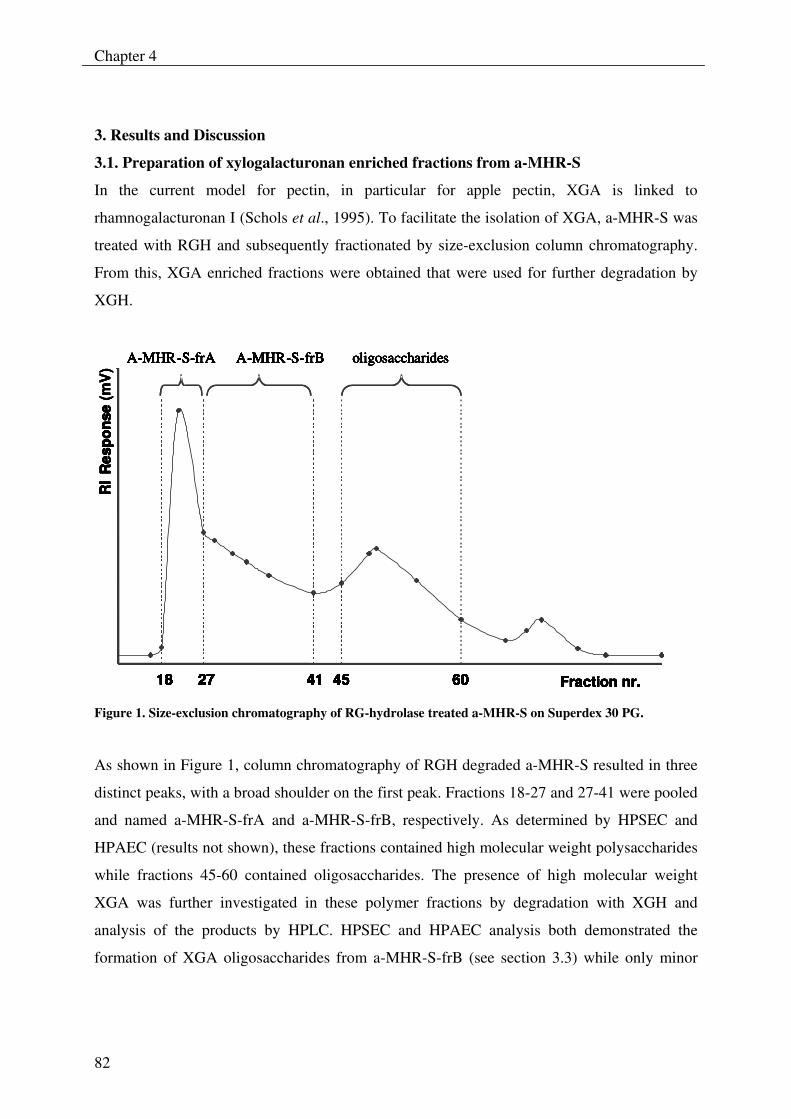
Chapter 4
82
3. Results and Discussion
3.1. Preparation of xylogalacturonan enriched fractions from a-MHR-S
In the current model for pectin, in particular for apple pectin, XGA is linked to
rhamnogalacturonan I (Schols et al., 1995). To facilitate the isolation of XGA, a-MHR-S was
treated with RGH and subsequently fractionated by size-exclusion column chromatography.
From this, XGA enriched fractions were obtained that were used for further degradation by
XGH.
Fraction nr.18 41
oligosaccharides
27 45 60
RI
Res
pons
e
18 4127 45 60
A-MHR-S-frA A-MHR-S-frB
Fraction nr.18 41
oligosaccharides
27 45 60
RI
Res
pons
e (m
V)
18 4127 45 60
A-MHR-S-frA A-MHR-S-frB
Fraction nr.18 41
oligosaccharides
27 45 60
RI
Res
pons
e
18 4127 45 60
A-MHR-S-frA A-MHR-S-frB
Fraction nr.18 41
oligosaccharides
27 45 60
RI
Res
pons
e (m
V)
18 4127 45 60
A-MHR-S-frA A-MHR-S-frB
27 45 60
A-MHR-S-frA A-MHR-S-frB
Fraction nr.18 41
oligosaccharides
27 45 60
RI
Res
pons
e
18 4127 45 60
A-MHR-S-frA A-MHR-S-frB
Fraction nr.18 41
oligosaccharides
27 45 60
RI
Res
pons
e (m
V)
18 4127 45 60
A-MHR-S-frA A-MHR-S-frB
27 45 60
A-MHR-S-frA A-MHR-S-frB
Fraction nr.18 41
oligosaccharides
27 45 60
RI
Res
pons
e
18 4127 45 60
A-MHR-S-frA A-MHR-S-frB
Fraction nr.18 41
oligosaccharides
27 45 60
RI
Res
pons
e (m
V)
18 4127 45 60
A-MHR-S-frA A-MHR-S-frB
27 45 60
A-MHR-S-frA A-MHR-S-frB
Figure 1. Size-exclusion chromatography of RG-hydrolase treated a-MHR-S on Superdex 30 PG.
As shown in Figure 1, column chromatography of RGH degraded a-MHR-S resulted in three
distinct peaks, with a broad shoulder on the first peak. Fractions 18-27 and 27-41 were pooled
and named a-MHR-S-frA and a-MHR-S-frB, respectively. As determined by HPSEC and
HPAEC (results not shown), these fractions contained high molecular weight polysaccharides
while fractions 45-60 contained oligosaccharides. The presence of high molecular weight
XGA was further investigated in these polymer fractions by degradation with XGH and
analysis of the products by HPLC. HPSEC and HPAEC analysis both demonstrated the
formation of XGA oligosaccharides from a-MHR-S-frB (see section 3.3) while only minor
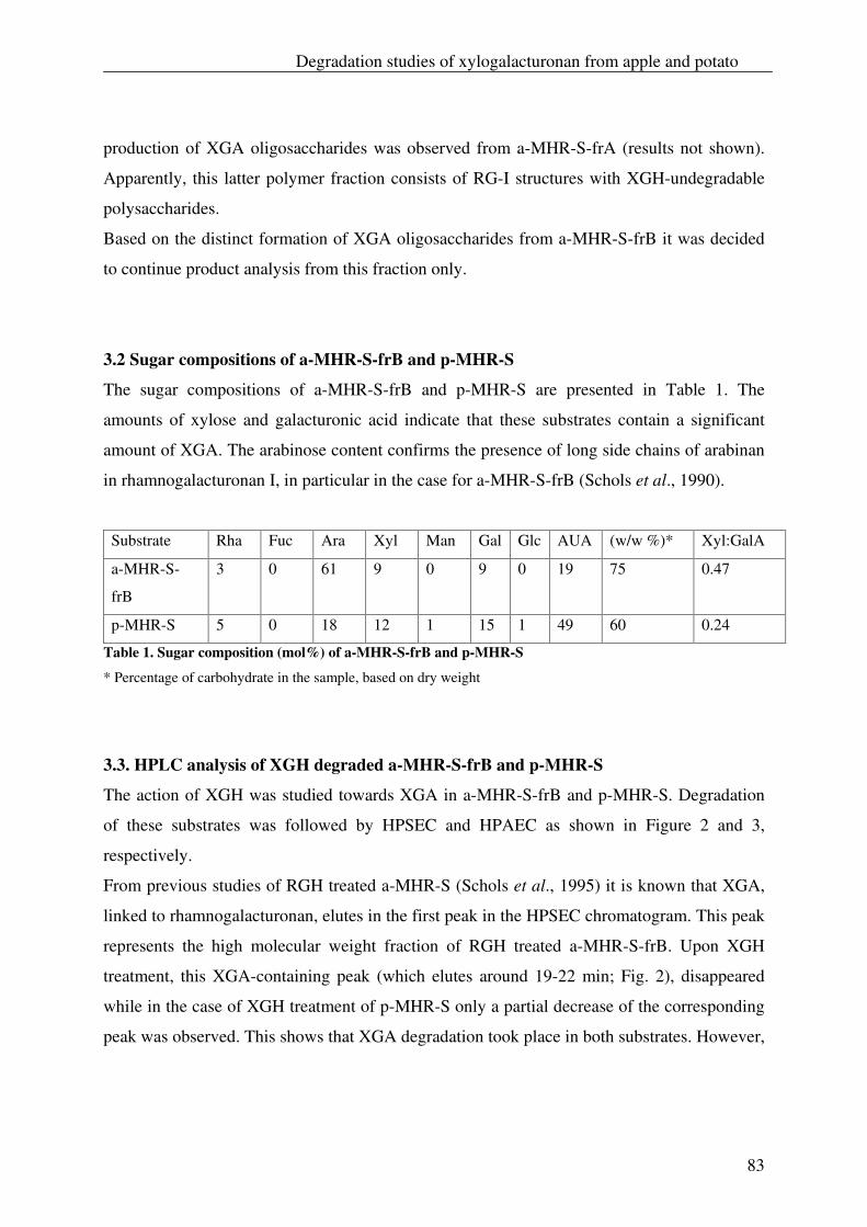
Degradation studies of xylogalacturonan from apple and potato
83
production of XGA oligosaccharides was observed from a-MHR-S-frA (results not shown).
Apparently, this latter polymer fraction consists of RG-I structures with XGH-undegradable
polysaccharides.
Based on the distinct formation of XGA oligosaccharides from a-MHR-S-frB it was decided
to continue product analysis from this fraction only.
3.2 Sugar compositions of a-MHR-S-frB and p-MHR-S
The sugar compositions of a-MHR-S-frB and p-MHR-S are presented in Table 1. The
amounts of xylose and galacturonic acid indicate that these substrates contain a significant
amount of XGA. The arabinose content confirms the presence of long side chains of arabinan
in rhamnogalacturonan I, in particular in the case for a-MHR-S-frB (Schols et al., 1990).
Substrate Rha Fuc Ara Xyl Man Gal Glc AUA (w/w %)* Xyl:GalA
a-MHR-S-
frB
3 0 61 9 0 9 0 19 75 0.47
p-MHR-S 5 0 18 12 1 15 1 49 60 0.24
Table 1. Sugar composition (mol%) of a-MHR-S-frB and p-MHR-S
* Percentage of carbohydrate in the sample, based on dry weight
3.3. HPLC analysis of XGH degraded a-MHR-S-frB and p-MHR-S
The action of XGH was studied towards XGA in a-MHR-S-frB and p-MHR-S. Degradation
of these substrates was followed by HPSEC and HPAEC as shown in Figure 2 and 3,
respectively.
From previous studies of RGH treated a-MHR-S (Schols et al., 1995) it is known that XGA,
linked to rhamnogalacturonan, elutes in the first peak in the HPSEC chromatogram. This peak
represents the high molecular weight fraction of RGH treated a-MHR-S-frB. Upon XGH
treatment, this XGA-containing peak (which elutes around 19-22 min; Fig. 2), disappeared
while in the case of XGH treatment of p-MHR-S only a partial decrease of the corresponding
peak was observed. This shows that XGA degradation took place in both substrates. However,
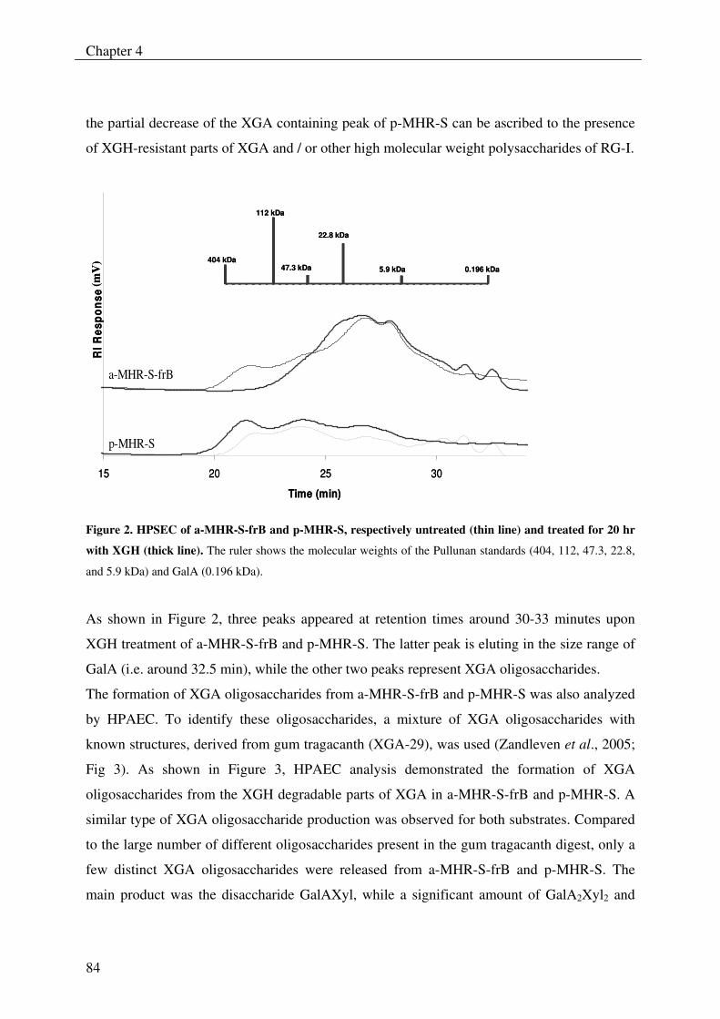
Chapter 4
84
the partial decrease of the XGA containing peak of p-MHR-S can be ascribed to the presence
of XGH-resistant parts of XGA and / or other high molecular weight polysaccharides of RG-I.
15 20 25 30
Time (min)
RI R
espo
nse
a-MHR-S-frB
p-MHR-S
404 kDa
112 kDa
47.3 kDa
22.8 kDa
5.9 kDa 0.196 kDa
(mV
)
15 20 25 30
Time (min)
RI R
espo
nse
a-MHR-S-frB
p-MHR-S
404 kDa
112 kDa
47.3 kDa
22.8 kDa
5.9 kDa 0.196 kDa404 kDa
112 kDa
47.3 kDa
22.8 kDa
5.9 kDa 0.196 kDa
(mV
)
Figure 2. HPSEC of a-MHR-S-frB and p-MHR-S, respectively untreated (thin line) and treated for 20 hr
with XGH (thick line). The ruler shows the molecular weights of the Pullunan standards (404, 112, 47.3, 22.8,
and 5.9 kDa) and GalA (0.196 kDa).
As shown in Figure 2, three peaks appeared at retention times around 30-33 minutes upon
XGH treatment of a-MHR-S-frB and p-MHR-S. The latter peak is eluting in the size range of
GalA (i.e. around 32.5 min), while the other two peaks represent XGA oligosaccharides.
The formation of XGA oligosaccharides from a-MHR-S-frB and p-MHR-S was also analyzed
by HPAEC. To identify these oligosaccharides, a mixture of XGA oligosaccharides with
known structures, derived from gum tragacanth (XGA-29), was used (Zandleven et al., 2005;
Fig 3). As shown in Figure 3, HPAEC analysis demonstrated the formation of XGA
oligosaccharides from the XGH degradable parts of XGA in a-MHR-S-frB and p-MHR-S. A
similar type of XGA oligosaccharide production was observed for both substrates. Compared
to the large number of different oligosaccharides present in the gum tragacanth digest, only a
few distinct XGA oligosaccharides were released from a-MHR-S-frB and p-MHR-S. The
main product was the disaccharide GalAXyl, while a significant amount of GalA2Xyl2 and
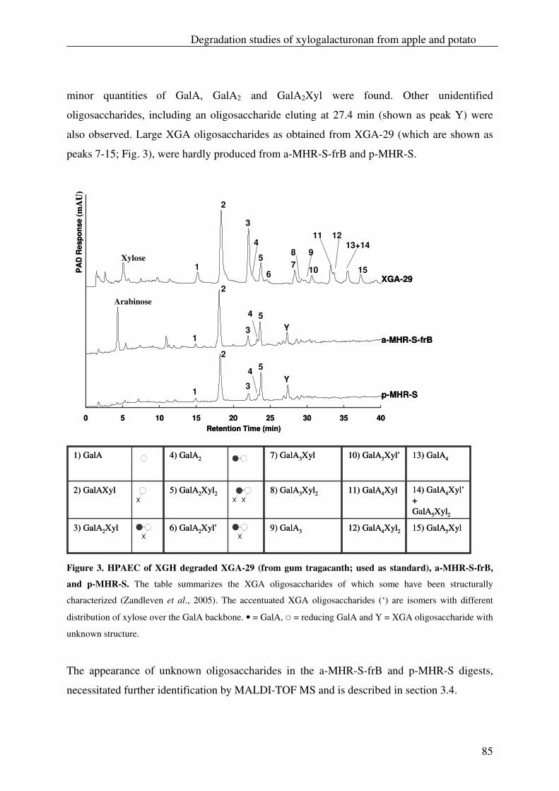
Degradation studies of xylogalacturonan from apple and potato
85
minor quantities of GalA, GalA2 and GalA2Xyl were found. Other unidentified
oligosaccharides, including an oligosaccharide eluting at 27.4 min (shown as peak Y) were
also observed. Large XGA oligosaccharides as obtained from XGA-29 (which are shown as
peaks 7-15; Fig. 3), were hardly produced from a-MHR-S-frB and p-MHR-S.
0 5 10 15 20 25 30 35 40Retention Time (min)
PA
D R
espo
nse
XGA-29
a-MHR-S-frB
p-MHR-S
Arabinose
Xylose 1
1
1
2
2
2
3
3
3
4
4
4
5
5
5
7 6
8 9
10
11 1213+14
15
Y
Y
(mA
U)
0 5 10 15 20 25 30 35 40Retention Time (min)
PA
D R
espo
nse
XGA-29
a-MHR-S-frB
p-MHR-S
Arabinose
Xylose 1
1
1
2
2
2
3
3
3
4
4
4
5
5
5
7 6
8 9
10
11 1213+14
15
Y
Y
(mA
U)
Figure 3. HPAEC of XGH degraded XGA-29 (from gum tragacanth; used as standard), a-MHR-S-frB,
and p-MHR-S. The table summarizes the XGA oligosaccharides of which some have been structurally
characterized (Zandleven et al., 2005). The accentuated XGA oligosaccharides (‘) are isomers with different
distribution of xylose over the GalA backbone. • = GalA, � = reducing GalA and Y = XGA oligosaccharide with
unknown structure.
The appearance of unknown oligosaccharides in the a-MHR-S-frB and p-MHR-S digests,
necessitated further identification by MALDI-TOF MS and is described in section 3.4.
XX15) GalA5Xyl 12) GalA4Xyl2 9) GalA3 6) GalA2Xyl’3) GalA2Xyl
14) GalA4Xyl’+ GalA5Xyl2
11) GalA4Xyl 8) GalA3Xyl2 5) GalA2Xyl2 2) GalAXyl
13) GalA4 10) GalA3Xyl’7) GalA3Xyl 4) GalA2 1) GalA
15) GalA5Xyl 12) GalA4Xyl2 9) GalA3 6) GalA2Xyl’3) GalA2Xyl
14) GalA4Xyl’+ GalA5Xyl2
11) GalA4Xyl 8) GalA3Xyl2 5) GalA2Xyl2 2) GalAXyl
13) GalA4 10) GalA3Xyl’7) GalA3Xyl 4) GalA2 1) GalA
X XX
Chapter 4
86
Free arabinose was also found in the a-MHR-S-frB digest. As the XGH preparation does not
contain arabinofuranosidase, this arabinose is probably liberated by acid hydrolysis during the
incubation temperature and inactivation step at 100 °C (pH 3.5) of the digestion mixture.
Indeed a lower amount of free arabinose (about 60 % reduction) was detected by HPAEC
analysis of a-MHR-S-frB which was not treated at 100 °C (results not shown). The formation
of free arabinose was even further reduced (about 90 % reduction) when a-MHR-S-frB was
incubated at pH 6.0.
The susceptibility for hydrolysis of the arabinosyl linkages at a low pH has also been reported
before (Beldman, Schols, Pitson, Searle-van Leeuwen, Voragen, 1997). Also free arabinose
was expected in the p-MHR-S digest, but could however not be detected by HPAEC analysis
(results not shown). This is probably related to the lower arabinose content of this substrate
(Table 1) as well as to structural differences of this arabinan (Beldman et al., 1997).
3.4. Product analysis of XGH degradable parts of XGA from a-MHR-frB and p-MHR-S
at different time intervals
a-MHR-S-frB and p-MHR-S were degraded by XGH for different time intervals and analyzed
by HPAEC (Fig. 4) and MALDI-TOF MS (Fig. 5) for the presence of XGA oligosaccharides.
Degradation of a-MHR-S-frB and p-MHR-S during 30 min and 1 h resulted mainly in the
production of the disaccharide GalAXyl (Fig. 4), which was also reported before for XGH
treatment of a-MHR-S (Beldman et al., 2003) and XGA-29 from gum tragacanth (Zandleven
et al., 2005) respectively. In addition, a significant amount of GalA2Xyl2 was observed as
degradation product from a-MHR-S-frB and p-MHR-S.
Comparing the HPAEC profiles of Figure 3 and 4 for the corresponding samples of a-MHR-
S-frB and p-MHR-S (20 h of treatment with XGH) only some minor differences were
observed, which falls within the range of experimental variance.
The HPAEC results were confirmed by MALDI-TOF MS analysis of the products from p-
MHR-S (Fig. 5) and a-MHR-S-frB (results not shown) for the samples after 20 h of
incubation. The MS spectra of both degraded substrates showed the main product GalAXyl,
and significant amounts of GalA2Xyl2, GalA2 and GalA2Xyl. Two novel XGA
oligosaccharides, GalAXyl2, with a m/z of 481, and GalA2Xyl3, with a m/z of 789, were
observed as products from both substrates. The formation of GalAXyl2 and GalA2Xyl3 by
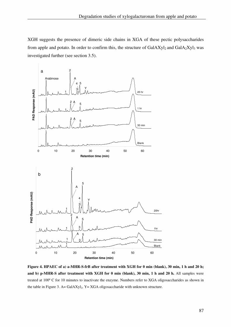
Degradation studies of xylogalacturonan from apple and potato
87
XGH suggests the presence of dimeric side chains in XGA of these pectic polysaccharides
from apple and potato. In order to confirm this, the structure of GalAXyl2 and GalA2Xyl3 was
investigated further (see section 3.5).
0 10 20 30 40 50 60
Retention time (min)
PA
D R
espo
nse
(mA
U)
Blank
30 min
1 hr
20 hr 1
1
2
2
2
3
4
5
5
A
A
A
a
5
Arabinose
Y
0 10 20 30 40 50 60
Retention time (min)
PA
D R
espo
nse
(mA
U)
20hr
1hr
30 min
Blank
2
2
2
3
4
5
5
A
A
A
3
3
1
1 1 1
1
5
b
Y
Figure 4. HPAEC of a) a-MHR-S-frB after treatment with XGH for 0 min (blank), 30 min, 1 h and 20 h;
and b) p-MHR-S after treatment with XGH for 0 min (blank), 30 min, 1 h and 20 h. All samples were
treated at 100º C for 10 minutes to inactivate the enzyme. Numbers refer to XGA oligosaccharides as shown in
the table in Figure 3. A= GalAXyl2, Y= XGA oligosaccharide with unknown structure.
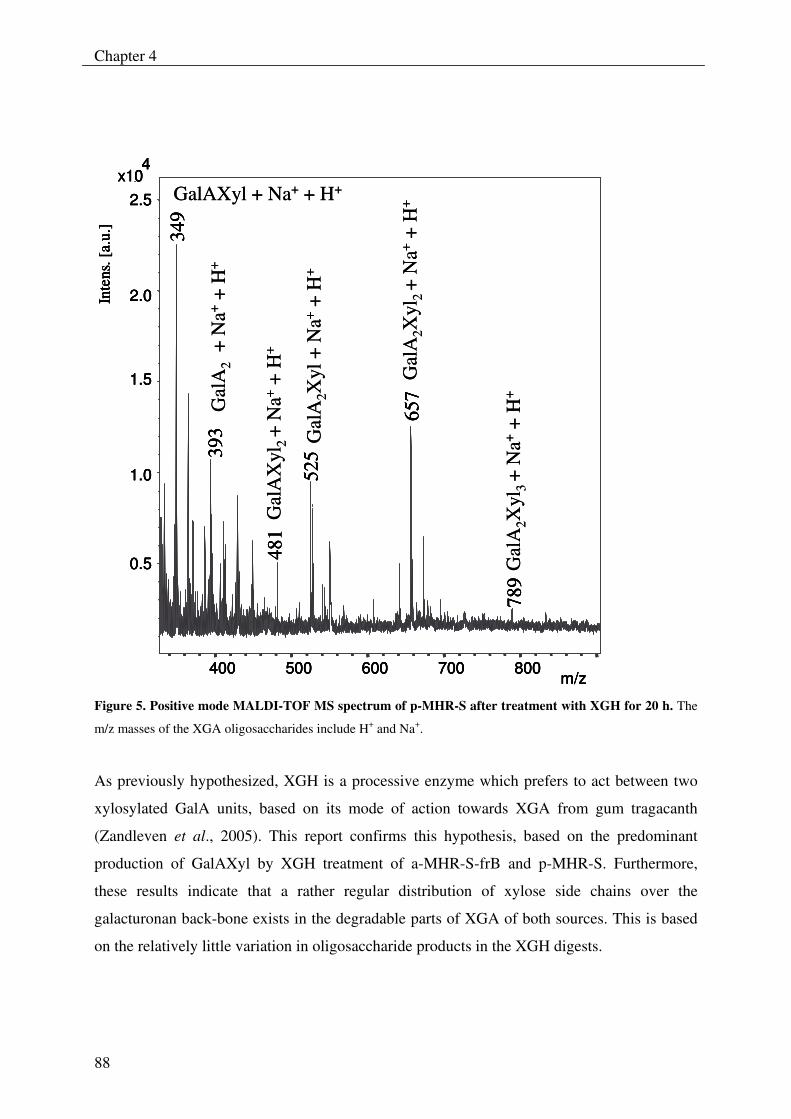
Chapter 4
88
34
9
657
525
481
393
789
0.5
1.0
1.5
2.0
2.5
4x10
Inte
ns. [
a.u.
]
400 500 600 700 800m/z
Gal
AX
yl2
+ N
a++
H+
Gal
A2X
yl +
Na+
+ H
+
349
657
525
481
393
789
0.5
1.0
1.5
2.0
2.5
4x10
Inte
ns. [
a.u.
]
400 500 600 700 800m/z
GalAXyl + Na+ + H+
Gal
A2
+ N
a++
H+
Gal
A2X
yl2
+ N
a++
H+
Gal
A2X
yl3
+ N
a++
H+
349
657
525
481
393
789
0.5
1.0
1.5
2.0
2.5
4x10
Inte
ns. [
a.u.
]
400 500 600 700 800m/z
Gal
AX
yl2
+ N
a++
H+
Gal
A2X
yl +
Na+
+ H
+
349
657
525
481
393
789
0.5
1.0
1.5
2.0
2.5
4x10
Inte
ns. [
a.u.
]
400 500 600 700 800m/z
GalAXyl + Na+ + H+
Gal
A2
+ N
a++
H+
Gal
A2X
yl2
+ N
a++
H+
Gal
A2X
yl3
+ N
a++
H+
Figure 5. Positive mode MALDI-TOF MS spectrum of p-MHR-S after treatment with XGH for 20 h. The
m/z masses of the XGA oligosaccharides include H+ and Na+.
As previously hypothesized, XGH is a processive enzyme which prefers to act between two
xylosylated GalA units, based on its mode of action towards XGA from gum tragacanth
(Zandleven et al., 2005). This report confirms this hypothesis, based on the predominant
production of GalAXyl by XGH treatment of a-MHR-S-frB and p-MHR-S. Furthermore,
these results indicate that a rather regular distribution of xylose side chains over the
galacturonan back-bone exists in the degradable parts of XGA of both sources. This is based
on the relatively little variation in oligosaccharide products in the XGH digests.
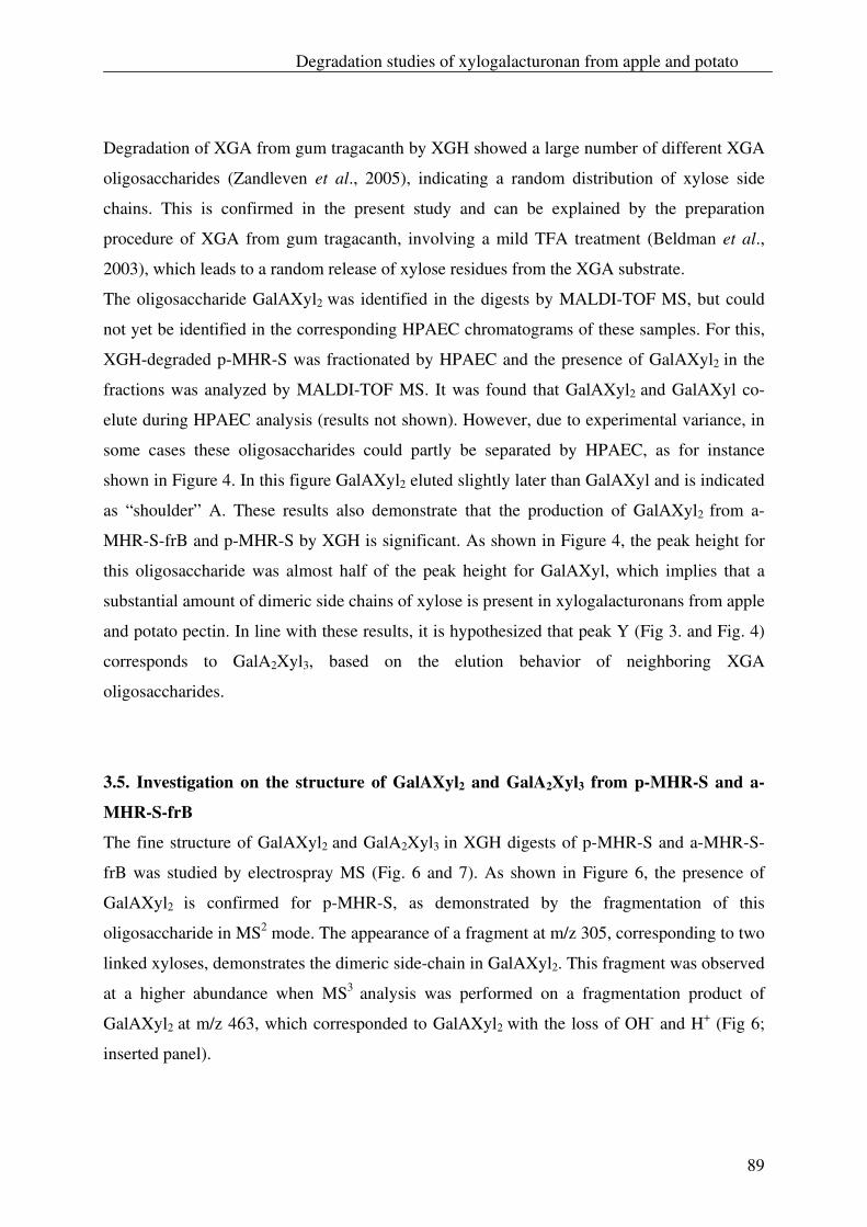
Degradation studies of xylogalacturonan from apple and potato
89
Degradation of XGA from gum tragacanth by XGH showed a large number of different XGA
oligosaccharides (Zandleven et al., 2005), indicating a random distribution of xylose side
chains. This is confirmed in the present study and can be explained by the preparation
procedure of XGA from gum tragacanth, involving a mild TFA treatment (Beldman et al.,
2003), which leads to a random release of xylose residues from the XGA substrate.
The oligosaccharide GalAXyl2 was identified in the digests by MALDI-TOF MS, but could
not yet be identified in the corresponding HPAEC chromatograms of these samples. For this,
XGH-degraded p-MHR-S was fractionated by HPAEC and the presence of GalAXyl2 in the
fractions was analyzed by MALDI-TOF MS. It was found that GalAXyl2 and GalAXyl co-
elute during HPAEC analysis (results not shown). However, due to experimental variance, in
some cases these oligosaccharides could partly be separated by HPAEC, as for instance
shown in Figure 4. In this figure GalAXyl2 eluted slightly later than GalAXyl and is indicated
as “shoulder” A. These results also demonstrate that the production of GalAXyl2 from a-
MHR-S-frB and p-MHR-S by XGH is significant. As shown in Figure 4, the peak height for
this oligosaccharide was almost half of the peak height for GalAXyl, which implies that a
substantial amount of dimeric side chains of xylose is present in xylogalacturonans from apple
and potato pectin. In line with these results, it is hypothesized that peak Y (Fig 3. and Fig. 4)
corresponds to GalA2Xyl3, based on the elution behavior of neighboring XGA
oligosaccharides.
3.5. Investigation on the structure of GalAXyl2 and GalA2Xyl3 from p-MHR-S and a-
MHR-S-frB
The fine structure of GalAXyl2 and GalA2Xyl3 in XGH digests of p-MHR-S and a-MHR-S-
frB was studied by electrospray MS (Fig. 6 and 7). As shown in Figure 6, the presence of
GalAXyl2 is confirmed for p-MHR-S, as demonstrated by the fragmentation of this
oligosaccharide in MS2 mode. The appearance of a fragment at m/z 305, corresponding to two
linked xyloses, demonstrates the dimeric side-chain in GalAXyl2. This fragment was observed
at a higher abundance when MS3 analysis was performed on a fragmentation product of
GalAXyl2 at m/z 463, which corresponded to GalAXyl2 with the loss of OH- and H+ (Fig 6;
inserted panel).
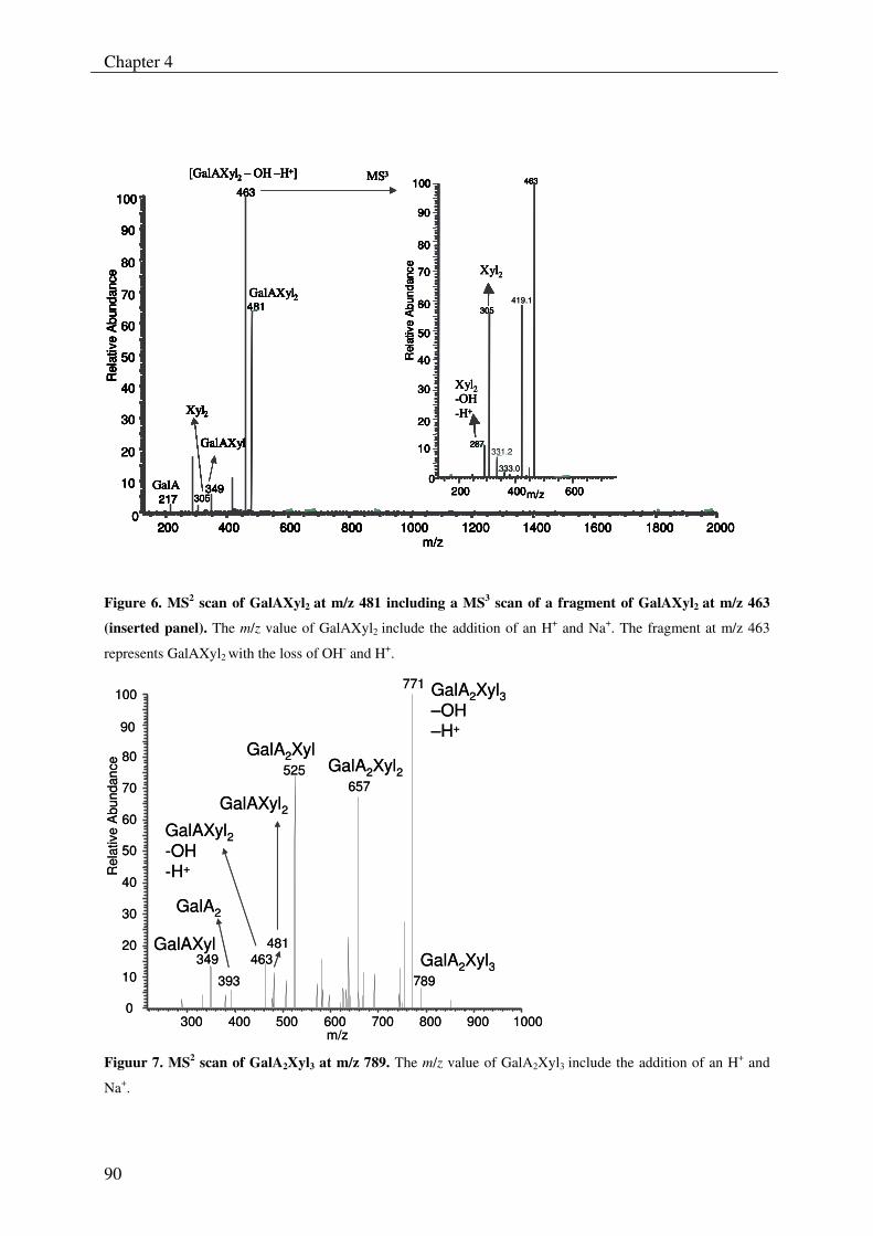
Chapter 4
90
200 400 600 800
20
30
40
50
60
70
80
90
100 463
481
349217
1000 1200 1400 1600 1800 20000
GalAXyl
Xyl2
10
Rel
ativ
eA
bund
ance
349217
m/z
GalAXyl2
MS3[GalAXyl2 – OH –H+]
GalAXyl
GalA305
Xyl2
200 400 600m/z0
10
20
30
40
50
60
70
80
90
100
Rel
ativ
eA
bund
ance
463
287331.2
305
331.2
Xyl2
Xyl2-OH-H+
200 400 600 800200 400 600 800
20
30
40
50
60
70
80
90
100 463
481
349217
1000 1200 1400 1600 1800 20000
GalAXyl
Xyl2
10
Rel
ativ
eA
bund
ance
349217
m/z
GalAXyl2
MS3[GalAXyl2 – OH –H+]
GalAXyl
GalA305
Xyl2
200 400 600m/z0
10
20
30
40
50
60
70
80
90
100
Rel
ativ
eA
bund
ance
463
287
419.1305
333.0
Xyl2
Xyl2-OH-H+
200200 400 600 800
20
30
40
50
60
70
80
90
100
400 600 800
20
30
40
50
60
70
80
90
100 463
481
349217
1000 1200 1400 1600 1800 20000
463
481
349217
1000 1200 1400 1600 1800 20000
GalAXyl
Xyl
GalAXyl
Xyl22
10
Rel
ativ
eA
bund
ance
10
Rel
ativ
eA
bund
ance
349217
m/z
349217
m/z
GalAXyl2
MS3[GalAXyl2 – OH –H+]
GalAXyl
GalA305
Xyl2
GalAXyl2
MS3[GalAXyl2 – OH –H+]
GalAXyl
GalA305
Xyl2
200 400 600m/z0
10
20
30
40
50
60
70
80
90
100
Rel
ativ
eA
bund
ance
200 400 600m/z0
10
20
30
40
50
60
70
80
90
100
Rel
ativ
eA
bund
ance
463
287331.2
463
287331.2
305
331.2
Xyl2
Xyl2-OH-H+
200 400 600 800200 400 600 800200 400 600 800200 400 600 800
20
30
40
50
60
70
80
90
100
20
30
40
50
60
70
80
90
100 463
481
349217
1000 1200 1400 1600 1800 20000
463
481
349217
1000 1200 1400 1600 1800 20000
GalAXyl
Xyl2
GalAXyl
Xyl2
10
Rel
ativ
eA
bund
ance
10
Rel
ativ
eA
bund
ance
349217
m/z
349217
m/z
GalAXyl2
MS3[GalAXyl2 – OH –H+]
GalAXyl
GalA305
Xyl2
GalAXyl2
MS3[GalAXyl2 – OH –H+]
GalAXyl
GalA305
Xyl2
200 400 600m/z0
10
20
30
40
50
60
70
80
90
100
Rel
ativ
eA
bund
ance
200 400 600m/z0
10
20
30
40
50
60
70
80
90
100
Rel
ativ
eA
bund
ance
463
287
419.1305
333.0
Xyl2
Xyl2-OH-H+
Figure 6. MS2 scan of GalAXyl2 at m/z 481 including a MS3 scan of a fragment of GalAXyl2 at m/z 463
(inserted panel). The m/z value of GalAXyl2 include the addition of an H+ and Na+. The fragment at m/z 463
represents GalAXyl2 with the loss of OH- and H+.
300 400 500 600 700 800 900 1000m/z
0
10
20
30
40
50
60
70
80
90
100
Rel
ativ
eA
bund
ance
771
525657
463349481
789393GalA2Xyl3
GalA2Xyl3–OH–H+
GalA2Xyl2GalA2Xyl
GalAXyl2GalAXyl2-OH-H+
GalAXyl
GalA2
300 400 500 600 700 800 900 1000m/z
0
10
20
30
40
50
60
70
80
90
100
Rel
ativ
eA
bund
ance
771
525657
463349481
789393GalA2Xyl3
GalA2Xyl3–OH–H+
GalA2Xyl2GalA2Xyl
GalAXyl2GalAXyl2-OH-H+
GalAXyl
GalA2
Figuur 7. MS2 scan of GalA2Xyl3 at m/z 789. The m/z value of GalA2Xyl3 include the addition of an H+ and
Na+.
Degradation studies of xylogalacturonan from apple and potato
91
An identical fragmentation spectrum was obtained for GalAXyl2 that was formed by
fragmentation of GalA2Xyl3 in the MS2 mode (for the latter, see Fig. 7). This confirms that a
dimeric side chain of xylose also exists in the XGA oligosaccharide GalA2Xyl3.
Similar results were acquired for GalAXyl2 and GalA2Xyl3 present in the a-MHR-S-frB digest
(results not shown), which points out that these dimeric side-chains of xylose are also present
in XGA from apple pectin.
Based on linkage analysis, the existence of side chains longer than one xylose unit in XGA
has been indicated for apple- (Oechslin et al., 2003) and pea pectin (Le Goff et al., 2001). In
this report, the occurrence of dimeric side chains of xylose in XGA in apple- and potato pectin
is demonstrated based on the structure of the oligosaccharides GalAXyl2 and GalA2Xyl3.
It is generally accepted that the single terminal xylose is in the β-form and is linked at the O-3
position of GalA in XGA (Beldman et al., 2003; Le Goff et al., 2001; Schols et al., 1995;
Vincken et al., 2003a). From this, we assume that the dimeric side chains in XGA from a-
MHR-S-frB and p-MHR-S are also linked at this position.
The nature of the linkage between the xyloses in the dimeric side chains in XGA from apple
and potato pectin is unknown. In order to obtain information about the anomeric form of the
terminal xylose residue, we purified the oligosaccharide GalAXyl2 from a p-MHR-S and a-
MHR-S-frB digest to near homogeneity (>95%) by analytical HPAEC. Subsequently it was
treated with β-(1,4) xylosidase from Aspergillus awamori and analyzed by HPAEC and
MALDI-TOF MS (results not shown). GalAXyl2 from both sources was degraded by this
enzyme to free xylose and GalAXyl, which implies that the two xyloses are β-linked.
Linkage analysis of apple pectin (Schols et al., 1995) and cellulase degraded cellulosic
residues of apples (Oechslin et al., 2003) respectively, show that 1,4 linked xylose residues
prevail as non-terminal xylose in apple pectin. Schols et al. (1995) found that 90% of the total
amount of non-terminal xylose residues exist as 1,4 linked xylose residues, whereas Oechslin
et al. (2003) showed that exclusively 1,4 linked xylose residues are present. From these
studies it is reasonable to assume that the dimeric side chains of xylose present in a-MHR-S-
frB could also be 1,4 linked.
The fact that a-MHR-S-frB and p-MHR-S were successfully degraded by XGH with
concomitant production of GalAXyl2 and GalA2Xyl3 implies that the dimeric side chains in
XGA from these sources did not hinder the action of the enzyme. We already showed that
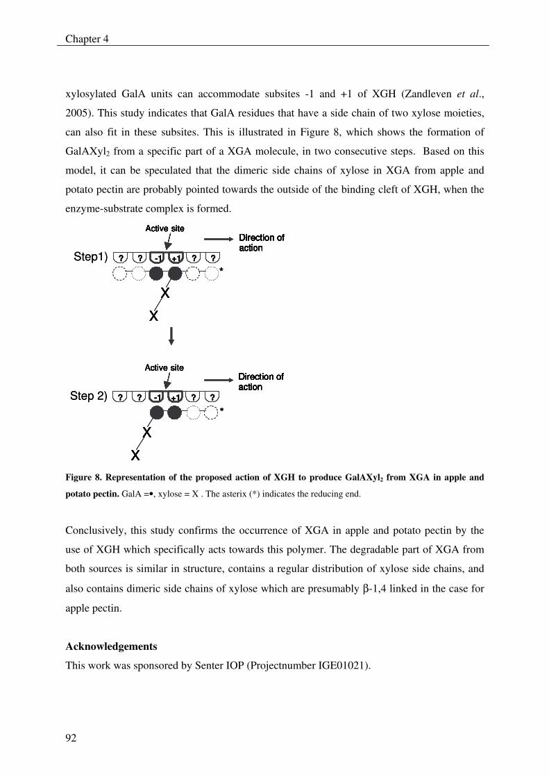
Chapter 4
92
xylosylated GalA units can accommodate subsites -1 and +1 of XGH (Zandleven et al.,
2005). This study indicates that GalA residues that have a side chain of two xylose moieties,
can also fit in these subsites. This is illustrated in Figure 8, which shows the formation of
GalAXyl2 from a specific part of a XGA molecule, in two consecutive steps. Based on this
model, it can be speculated that the dimeric side chains of xylose in XGA from apple and
potato pectin are probably pointed towards the outside of the binding cleft of XGH, when the
enzyme-substrate complex is formed.
Direction of actionDirection of actionDirection of action
Active siteActive site
? ? -1 +1 ? ?
XX
XX
*
Active siteDirection of action
Active siteDirection of actionDirection of action
? ? -1 +1 ? ?
XX
XX
*
Step1)
Step 2)
Direction of actionDirection of actionDirection of action
Active siteActive site
? ? -1 +1 ? ?
XX
XX
*
Direction of actionDirection of actionDirection of action
Active siteActive site
? ? -1 +1 ? ?
XX
XX
*
Active siteDirection of action
Active siteDirection of actionDirection of action
? ? -1 +1 ? ?
XX
XX
*
Active siteDirection of action
Active siteDirection of actionDirection of action
? ? -1 +1 ? ?
XX
XX
*
Step1)
Step 2)
Figure 8. Representation of the proposed action of XGH to produce GalAXyl2 from XGA in apple and
potato pectin. GalA =•, xylose = X . The asterix (*) indicates the reducing end.
Conclusively, this study confirms the occurrence of XGA in apple and potato pectin by the
use of XGH which specifically acts towards this polymer. The degradable part of XGA from
both sources is similar in structure, contains a regular distribution of xylose side chains, and
also contains dimeric side chains of xylose which are presumably β-1,4 linked in the case for
apple pectin.
Acknowledgements
This work was sponsored by Senter IOP (Projectnumber IGE01021).
Degradation studies of xylogalacturonan from apple and potato
93
References
Aspinall G.O., Baillie, J. (1963) Gum Tragacanth. Part I. Fractionation of the Gum and the
structure of tragacanthic acid. Journal of the chemical society 318, 1702-1714.
Beldman G., Schols, H.A., Pitson, S.M., Searle-van Leeuwen, M.J.F., and Voragen, A.G.J.
(1997) Arabinans and arabinan degrading enzymes. In Advances in Macromolecular
Carbohydrate Research (Edited by Sturgeon R. J.), Vol. 1, p. 1-64. JAI Press INC.
Beldman G., van den Broek, L.A.M., Schols, H.A., Searle-van Leeuwen, M.J.F., van Laere,
K.M.J., Voragen, A.G.J. (1996) An exogalacturonase from Aspergillus aculeatus able to
degrade Xylogalacturonan. Biotechnology Letters 18, 707-712.
Beldman G., Vincken, J.-P., Meeuwsen, P.J.A., Herweijer, M., Voragen, A.G.J. (2003)
Degradation of differently substituted Xylogalacturonans by Endoxylogalacturonan
Hydrolase and Endopolygalacturonases. Biocatalysis and Biotransformation 21, 189-198.
Benen J.A.E., van Alebeek, G.-J.W.M., Voragen, A.G.J., Visser, J. (2003) Mode of Action
Analysis And Structure-Function Relationships Of Aspergillus Niger Pectinolytic Enzymes.
In Advances in Pectin and Pectinase Research (Edited by Voragen A.G.J., Schols, H.A.,
Visser, R.), p. 235-256. Kluwer Academic Publishers, Dordrecht.
Benen J.A.E., Vincken, J.-P., van Alebeek, G.-J.W.M. (2002) Microbial Pectinases. In Pectins
and their Manipulation (Edited by Seymour G.B., Knox, J.P.). Blackwell Publishing,
Oxford.
Blumenkrantz N., Asboe-Hansen, G. (1973) New method for quantitative determination of
uronic acids. Analytical Biochemistry 54, 484-489.
Englyst H.N., Cummings, J.H. (1984) Simplified method for the measurement of total non-
starch polysaccharides by gas-liquid chromatography of constituent sugars as alditol
acetates. Analyst 109, 937-942.
Grassin, C., Fauquembergue, P. (1996) Application of Pectinases in Beverages. In Pectins
and Pectinases (Edited by Visser J., Voragen, A.G.J.), p. 453-462. Elsevier Science.
Heldt-Hansen, H.P., Kofod, L.V., Budolfsen, G., Nielsen, P.M., Huttel, S., Bladt, T. (1996)
Application of Tailormade Pectinases. In Pectins and Pectinases (Edited by Visser J.,
Voragen, A.G.J.), p. 463-474. Elsevier Science.
Herweijer M.A., Vincken, J.-P., Meeuwsen, P.J.A, van der Vlugt-Bergmans, C.J.B., Beldman,
G., van Ooyen, A.J.J., Voragen, A.G.J. (2003) Endo-Xylogalacturonan Hydrolase. In
Chapter 4
94
Advances in Pectin and Pectinase Research (Edited by Voragen A.G.J., Schols, H.A.,
Visser, R.), p. 257-266. Kluwer Academic Publishers, Dordrecht.
Kester H., Benen, J., Visser, J. (1999) The exo-polygalacturonase from Aspergillus
tubingensis is also active on xylogalacturonan. Biotechnology and Applied Biochemistry 30,
53-57.
Kofod L.V., Kauppinen, S., Christgau, S., Andersen, L.N., Heldt-Hansen, H.P., Dorreich, K.,
Dalbfge, H. (1994) Cloning and characterization of two structurally and functionally
divergent rhamnogalacturonases from Aspergillus aculeatus. Journal of Biological
Chemistry 269, 29182-29189.
Le Goff A., Renard C.M.G.C., Bonnin E., and Thibault J.-F. (2001) Extraction, purification
and chemical characterisation of xylogalacturonans from pea hulls. Carbohydrate Polymers.
45, 325-334.
Nakamura, A., Furuta, H., Maeda, H., Takao, T., Nagamatsu, Y. (2002) Analysis of the
Molecular Construction of Xylogalacturonan Isolated from Soluble Soybean
Polysaccharides. Bioscience Biotechnology and Biochemistry. 66, 1155-1158.
Oechslin R., Lutz, M.V., Amado, R. (2003) Pectic substances isolated from apple cellulosic
residue: structural characterisation of a new type of rhamnogalacturonan I. Carbohydrate
Polymers 51, 301-310.
Schols H.A., Bakx E. J., Schipper D., and Voragen A.G. J. (1995) A xylogalacturonan subunit
present in the modified hairy regions of apple pectin. Carbohydrate Research. 279, 265-279.
Schols H.A., Posthumus, M.A., Voragen, A.G.J. (1990) Hairy (ramified) regions of pectins.
PartI. Structural features of hairy regions of pectins isolated from apple juice produced by
the liquefaction process. Carbohydrate Research 206, 117-129.
Schols H.A., Voragen, A.G.J. (2002) The chemical structure of pectins. In Pectins and their
Manipulation (Edited by Seymour G. B., Knox, J.P.), p. 1-29. Blackwell Science.
Thibault J.-F., Robin, J.-P. (1975) Automatisation du dosage des acides uroniques par la
méthode au carbazol. Application au cas de matieres pectiques. Annales de technologie
agricole 24, 99-110.
Thibault J.-F. and Ralet, M.-C. (2001) Pectins, Their Origin, Structure and Functions. In
Advanced Dietary Fibre Technology (Edited by McCleary, B. V., Prosky, L.), p. 369- 378.
Blackwell Science, Oxford.
Degradation studies of xylogalacturonan from apple and potato
95
Van der Vlugt-Bergmans, C.J.B., Meeuwsen, P.J.A., Voragen, A.G.J., and van Ooyen, A.J.J.
(2000) Endo-Xylogalacturonan hydrolase, a Novel Pectinolytic Enzyme. Applied and
Environmental Microbiology 66, 36-41.
Van Dijck, P.W.M. (1999). Chymosin and Phytase. Made by genetic engineering (No. 10 in a
series of articles to promote a better understanding of the use of genetic engineering).
Journal of Biotechnology 67, 77-80.
Vincken J.-P., Schols, H.A., Oomen, R.J.F.J., Beldman, G., Visser, R.G.F., Voragen, A.G.J.
(2003a) Pectin-The hairy thing. In Advances in Pectin and Pectinase Research (Edited by
Voragen A.G.J., Schols, H.A., and Visser, R.), p. 47-59. Kluwer Academic Publishers,
Dordrecht.
Vincken J.-P., Voragen, A.G.J., Beldman, G. (2003b) Enzymes Degrading
Rhamnogalacturonan and Xylogalacturonan. In Handbook of Food Enzymology (Edited by
Whitaker J. R., Voragen, A.G.J., Wong, D.W.S.), New York.
Voragen A.G.J., Beldman, G., Schols, H.A. (2001) Chemistry and Enzymology of Pectins. In
Advanced Dietary Fibre Technology (Edited by McCleary, B.V., Prosky, L.), p. 379-397.
Blackwell Science, Oxford.
Zandleven J., Beldman, G., Bosveld, M., Benen, J., Voragen, A. (2005) Mode of action of
Xylogalacturonan hydrolase towards xylogalacturonan and xylogalacturonan
oligosaccharides. Biochemical Journal 387, 719-725.

Chapter 4
96
Chapter 5
97
Xylogalacturonan exists in cell walls from various tissues of Arabidopsis
thaliana
J.S. Zandleven, S.O. Sørensen, J. Harholt, G. Beldman, H.A. Schols, H.V. Scheller, and A.G.J. Voragen
Abstract
Evidence is presented for the presence of xylogalacturonan (XGA) in Arabidopsis thaliana.
This evidence was obtained by extraction of pectin from the seeds, root, stem, young leaves
and mature leaves of A. thaliana, followed by treatment of these pectin extracts with
xylogalacturonan hydrolase (XGH). Upon enzymatic treatment, XGA oligosaccharides were
primarily produced from pectin extracts obtained from the young and mature leaves and to a
lesser extent from those originating from the stem of A. thaliana. The oligosaccharide
GalA3Xyl was predominantly formed from these pectin extracts. No XGA oligosaccharides
were detected in digests of pectin extracts from the seeds and roots.
A low number of different XGA oligosaccharides were obtained from pectins of A. thaliana;
as was also observed in digests of XGA from apple and potato pectins. This indicates a
uniform distribution of xylose in XGA from A. thaliana. The predominant production of
GalA3Xyl, as well as the release of linear GalA oligosaccharides pointed to a lower degree of
xylose substitution in XGA from A. thaliana than in XGA from apple and potato.
The estimated amount of XGA accounted for approximately 2.5 %, 7 % and 6 % (w/w) of the
total carbohydrate in the pectin fraction of the stem, young leaves and mature leaves
respectively.
A modified version of this chapter will be submitted to Phytochemistry
Chapter 5
98
1. Introduction
Primary cell walls are the major textural components of plant-derived foods. The most
important polysaccharides that account for 90 to 100% of the structural polymers of these cell
walls are cellulose, hemicellulose and pectin (Albersheim et al., 1996).
Cellulose is comprised of β-(1→ 4)-linked D-glucan while hemicelluloses primarily consists
of xyloglucan and arabinoxylan (Albersheim et al., 1996). Pectin is a heteropolysaccharide,
which contains α-(1→ 4)-linked D-galacturonic acid chains (also known as the smooth
regions of pectins) and the branched polysaccharides rhamnogalacturonan I,
rhamnogalacturonan II, and xylogalacturonan (referred to as the “hairy” regions) (Benen et
al., 2002; Schols and Voragen, 1996; Vincken et al., 2003).
Cellulose exists in plant cell walls as microfibrils, which are composed of long
semicrystalline β-glucan chains (Gibeaut and Carpita, 1994). These microfibrils are cross-
linked by hemicelluloses, such as xyloglucans that are believed to be a major factor in
controlling the rate of cell-wall expansion (Gibeaut and Carpita, 1994; O'Neill and York,
2003; Zablackis et al., 1995). The load-bearing cellulose-hemicellulose network is embedded
in a matrix of pectic polysaccharides, which form a hydrated and crosslinked three-
dimensional network (Gibeaut and Carpita, 1994; Knox, 2002).
Xylogalacturonan (XGA) is a chain of α-(1→ 4)-linked D-galacturonic acid, which is
substituted with β-D-xylose at the O-3 position. It is suggested that this biopolymer is a side
chain of RG-I (rhamnogalacturonan I) in the “hairy” regions of pectin (Vincken et al., 2003).
The presence of XGA in plants has been reported in storage tissues of reproductive organs
such as in cell walls of peas, soybeans, watermelons, apples, pears, onions, potato’s, pine
pollen, and cotton seeds (Albersheim et al., 1996; Le Goff et al., 2001; Nakamura et al., 2002;
Schols et al., 1995; Thibault and Ralet, 2001; Vincken et al., 2003; Voragen et al., 2001;
Zandleven et al., 2006; Huisman et al., 1999). Its presence has also been reported in exudates
from trees, such as gum tragacanth from the Astralagus species (Aspinall and Baillie, 1963).
Previous studies demonstrated that leaf primary cell walls of A. thaliana contain
homogalacturonan, RG-I, RG-II, xylan, xyloglucan and cellulose (Zablackis et al., 1995).
These polymeric structures were also suggested to be present in cell walls of the stem of this
plant species (Gardner et al., 2002). Additionally, mannan polysaccharides have also been
reported in the cell walls of leaves and stem of A. thaliana (Handford et al., 2003).
Xylogalacturonan exists in Arabidopsis thaliana
99
So far, the presence of other polysaccharides, such as XGA, has not been demonstrated in A.
thaliana (Zablackis et al., 1995), although it has been suggested that this polymer exists in
root caps of this plant species. This was based on immunocytochemical analysis using an
LM8 antibody that was raised against pea XGA (Willats et al., 2004). Also Gardner et al.
(2002) mentioned XGA as a probable xylose-containing polysaccharide in the stem of A.
thaliana; however this was only based on the sugar composition of the alcohol insoluble
residue (AIR) of the stems.
Recently, a gene (At5g33290) expected to encode a β-xylosyl transferase was identified in A.
thaliana. Pectin isolated from a T-DNA insertion line having a T-DNA insertion in this
particular gene was found to contain less xylose compared to the wild type (Sørensen et al.,
unpublished data). We therefore hypothesized that pectin from A. thaliana may contain
regions of XGA. To investigate this, pectin was extracted from cell wall material, prepared
from the seeds, roots, young leaves and mature leaves of A. thaliana. The pectin fractions
were analysed for their sugar composition and subsequently treated with xylogalacturonan
hydrolase (XGH) to determine the presence of XGA. Based on the obtained XGA hydrolysis
products, some structural characteristics of A. thaliana XGA are hypothesized. Also the
amount of XGA liberated from these pectin fractions was estimated.
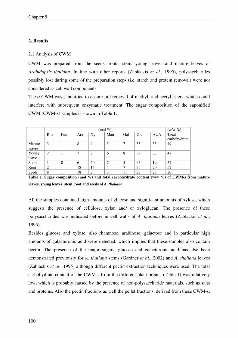
Chapter 5
100
2. Results
2.1 Analysis of CWM
CWM was prepared from the seeds, roots, stem, young leaves and mature leaves of
Arabidopsis thaliana. In line with other reports (Zablackis et al., 1995), polysaccharides
possibly lost during some of the preparation steps (i.e. starch and protein removal) were not
considered as cell wall components.
These CWM was saponified to ensure full removal of methyl- and acetyl esters, which could
interfere with subsequent enzymatic treatment. The sugar composition of the saponified
CWM (CWM-s) samples is shown in Table 1.
(mol %) Rha Fuc Ara Xyl Man Gal Glc AUA
(w/w %) Total carbohydrate
Mature leaves
3 1 8 9 5 7 33 35 49
Young leaves
2 1 7 8 6 8 37 33 47
Stem 1 0 6 20 7 5 43 19 57 Root 2 1 18 14 4 7 35 20 52 Seeds 8 1 18 8 3 11 27 25 20 Table 1. Sugar composition (mol %) and total carbohydrate content (w/w %) of CWM-s from mature
leaves, young leaves, stem, root and seeds of A. thaliana
All the samples contained high amounts of glucose and significant amounts of xylose, which
suggests the presence of cellulose, xylan and/ or xyloglucan. The presence of these
polysaccharides was indicated before in cell walls of A. thaliana leaves (Zablackis et al.,
1995).
Besides glucose and xylose, also rhamnose, arabinose, galactose and in particular high
amounts of galacturonic acid were detected, which implies that these samples also contain
pectin. The presence of the major sugars, glucose and galacturonic acid has also been
demonstrated previously for A. thaliana stems (Gardner et al., 2002) and A. thaliana leaves
(Zablackis et al., 1995) although different pectin extraction techniques were used. The total
carbohydrate content of the CWM-s from the different plant organs (Table 1) was relatively
low, which is probably caused by the presence of non-polysaccharide materials, such as salts
and proteins. Also the pectin fractions as well the pellet fractions, derived from these CWM-s,
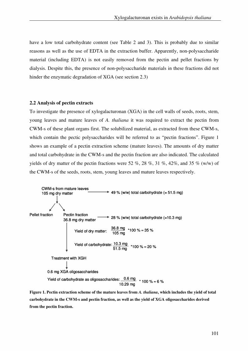
Xylogalacturonan exists in Arabidopsis thaliana
101
have a low total carbohydrate content (see Table 2 and 3). This is probably due to similar
reasons as well as the use of EDTA in the extraction buffer. Apparently, non-polysaccharide
material (including EDTA) is not easily removed from the pectin and pellet fractions by
dialysis. Despite this, the presence of non-polysaccharide materials in these fractions did not
hinder the enzymatic degradation of XGA (see section 2.3)
2.2 Analysis of pectin extracts
To investigate the presence of xylogalacturonan (XGA) in the cell walls of seeds, roots, stem,
young leaves and mature leaves of A. thaliana it was required to extract the pectin from
CWM-s of these plant organs first. The solubilized material, as extracted from these CWM-s,
which contain the pectic polysaccharides will be referred to as “pectin fractions”. Figure 1
shows an example of a pectin extraction scheme (mature leaves). The amounts of dry matter
and total carbohydrate in the CWM-s and the pectin fraction are also indicated. The calculated
yields of dry matter of the pectin fractions were 52 %, 28 %, 31 %, 42%, and 35 % (w/w) of
the CWM-s of the seeds, roots, stem, young leaves and mature leaves respectively.
CWM-s from mature leaves105 mg dry matter
Pellet fraction Pectin fraction36.8 mg dry matter
49 % (w/w) total carbohydrate (= 51.5 mg)
28 % (w/w) total carbohydrate (=10.3 mg)
Treatment with XGH
0.6 mg XGA oligosaccharides
0.6 mg10.29 mg
* 100 % ≈ 6 %
36.8 mg105 mg
*100 % ≈ 35 %Yield of dry matter:
Yield of carbohydrate: 10.3 mg51.5 mg *100 % ≈ 20 %
Yield of carbohydrate as oligosaccharides:
Figure 1. Pectin extraction scheme of the mature leaves from A. thaliana, which includes the yield of total
carbohydrate in the CWM-s and pectin fraction, as well as the yield of XGA oligosaccharides derived
from the pectin fraction.
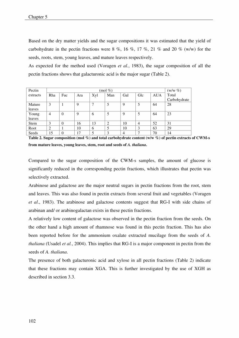
Chapter 5
102
Based on the dry matter yields and the sugar compositions it was estimated that the yield of
carbohydrate in the pectin fractions were 8 %, 16 %, 17 %, 21 % and 20 % (w/w) for the
seeds, roots, stem, young leaves, and mature leaves respectively.
As expected for the method used (Voragen et al., 1983), the sugar composition of all the
pectin fractions shows that galacturonic acid is the major sugar (Table 2).
(mol %) Pectin extracts Rha Fuc Ara Xyl Man Gal Glc AUA
(w/w %) Total Carbohydrate
Mature leaves
3 1 9 7 5 9 5 64 28
Young leaves
4 0 9 6 5 9 5 64 23
Stem 3 0 16 13 2 10 4 52 31 Root 2 1 10 6 5 10 3 63 29 Seeds 15 0 17 5 3 4 7 70 14 Table 2. Sugar composition (mol %) and total carbohydrate content (w/w %) of pectin extracts of CWM-s
from mature leaves, young leaves, stem, root and seeds of A. thaliana.
Compared to the sugar composition of the CWM-s samples, the amount of glucose is
significantly reduced in the corresponding pectin fractions, which illustrates that pectin was
selectively extracted.
Arabinose and galactose are the major neutral sugars in pectin fractions from the root, stem
and leaves. This was also found in pectin extracts from several fruit and vegetables (Voragen
et al., 1983). The arabinose and galactose contents suggest that RG-I with side chains of
arabinan and/ or arabinogalactan exists in these pectin fractions.
A relatively low content of galactose was observed in the pectin fraction from the seeds. On
the other hand a high amount of rhamnose was found in this pectin fraction. This has also
been reported before for the ammonium oxalate extracted mucilage from the seeds of A.
thaliana (Usadel et al., 2004). This implies that RG-I is a major component in pectin from the
seeds of A. thaliana.
The presence of both galacturonic acid and xylose in all pectin fractions (Table 2) indicate
that these fractions may contain XGA. This is further investigated by the use of XGH as
described in section 3.3.
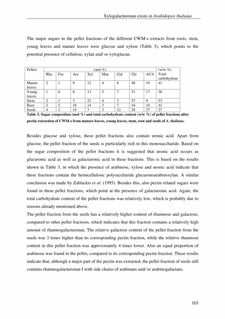
Xylogalacturonan exists in Arabidopsis thaliana
103
The major sugars in the pellet fractions of the different CWM-s extracts from roots, stem,
young leaves and mature leaves were glucose and xylose (Table 3), which points to the
potential presence of cellulose, xylan and/ or xyloglucan.
(mol %) Pellets Rha Fuc Ara Xyl Man Gal Glc AUA
(w/w %) Total carbohydrate
Mature leaves
2 1 9 12 4 6 48 19 41
Young leaves
1 0 8 13 5 7 51 17 38
Stem 2 1 3 23 4 3 57 9 53 Root 2 2 18 14 3 7 44 10 41 Seeds 4 1 19 7 3 12 28 27 27 Table 3. Sugar composition (mol %) and total carbohydrate content (w/w %) of pellet fractions after
pectin extraction of CWM-s from mature leaves, young leaves, stem, root and seeds of A. thaliana.
Besides glucose and xylose, these pellet fractions also contain uronic acid. Apart from
glucose, the pellet fraction of the seeds is particularly rich in this monosaccharide. Based on
the sugar composition of the pellet fractions it is suggested that uronic acid occurs as
glucuronic acid as well as galacturonic acid in these fractions. This is based on the results
shown in Table 3, in which the presence of arabinose, xylose and uronic acid indicate that
these fractions contain the hemicellulosic polysaccharide glucuronoarabinoxylan. A similar
conclusion was made by Zablackis et al. (1995). Besides this, also pectin related sugars were
found in these pellet fractions, which point at the presence of galacturonic acid. Again, the
total carbohydrate content of the pellet fractions was relatively low, which is probably due to
reasons already mentioned above.
The pellet fraction from the seeds has a relatively higher content of rhamnose and galactose,
compared to other pellet fractions, which indicates that this fraction contains a relatively high
amount of rhamnogalacturonan. The relative galactose content of the pellet fraction from the
seeds was 3 times higher than its corresponding pectin fraction, while the relative rhamnose
content in this pellet fraction was approximately 4 times lower. Also an equal proportion of
arabinose was found in the pellet, compared to its corresponding pectin fraction. These results
indicate that, although a major part of the pectin was extracted, the pellet fraction of seeds still
contains rhamnogalacturonan I with side chains of arabinans and/ or arabinogalactans.
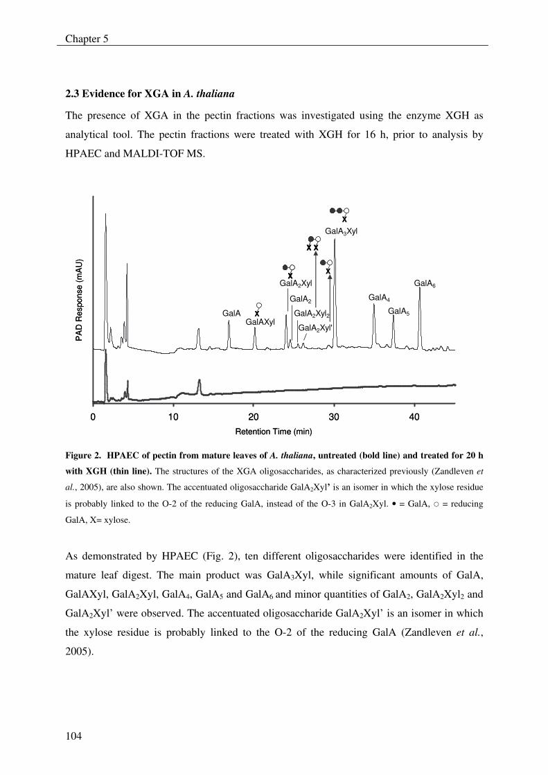
Chapter 5
104
2.3 Evidence for XGA in A. thaliana
The presence of XGA in the pectin fractions was investigated using the enzyme XGH as
analytical tool. The pectin fractions were treated with XGH for 16 h, prior to analysis by
HPAEC and MALDI-TOF MS.
0 10 20 30 40Retention Time (min)
PA
D R
espo
nse
(mA
U)
GalAGalAXyl
GalA2Xyl
GalA2
GalA2Xyl2
GalA2Xyl'
GalA3Xyl
GalA4
GalA5
GalA6
XX
XX
XX XXXX
XX
XX
0 10 20 30 40Retention Time (min)
PA
D R
espo
nse
(mA
U)
GalAGalAXyl
GalA2Xyl
GalA2
GalA2Xyl2
GalA2Xyl'
GalA3Xyl
GalA4
GalA5
GalA6
XX
XXXX
XXX XXXX
XX
XX
Figure 2. HPAEC of pectin from mature leaves of A. thaliana, untreated (bold line) and treated for 20 h
with XGH (thin line). The structures of the XGA oligosaccharides, as characterized previously (Zandleven et
al., 2005), are also shown. The accentuated oligosaccharide GalA2Xyl’ is an isomer in which the xylose residue
is probably linked to the O-2 of the reducing GalA, instead of the O-3 in GalA2Xyl. • = GalA, � = reducing
GalA, X= xylose.
As demonstrated by HPAEC (Fig. 2), ten different oligosaccharides were identified in the
mature leaf digest. The main product was GalA3Xyl, while significant amounts of GalA,
GalAXyl, GalA2Xyl, GalA4, GalA5 and GalA6 and minor quantities of GalA2, GalA2Xyl2 and
GalA2Xyl’ were observed. The accentuated oligosaccharide GalA2Xyl’ is an isomer in which
the xylose residue is probably linked to the O-2 of the reducing GalA (Zandleven et al.,
2005).
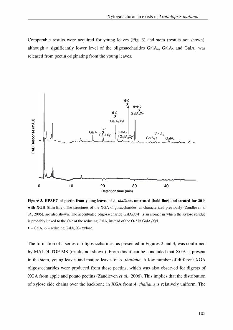
Xylogalacturonan exists in Arabidopsis thaliana
105
Comparable results were acquired for young leaves (Fig. 3) and stem (results not shown),
although a significantly lower level of the oligosaccharides GalA4, GalA5 and GalA6 was
released from pectin originating from the young leaves.
0 10 20 30 40Retention time (min)
PA
D R
espo
nse
(mA
U)
GalAGalAXyl
GalA2Xyl
GalA2
GalA2Xyl'
GalA3Xyl
GalA4
GalA5
GalA6
XX
XX
XX
XX
0 10 20 30 40Retention time (min)
PA
D R
espo
nse
(mA
U)
GalAGalAXyl
GalA2Xyl
GalA2
GalA2Xyl'
GalA3Xyl
GalA4
GalA5
GalA6
XX
XXXX
XXXX
XX
Figure 3. HPAEC of pectin from young leaves of A. thaliana, untreated (bold line) and treated for 20 h
with XGH (thin line). The structures of the XGA oligosaccharides, as characterized previously (Zandleven et
al., 2005), are also shown. The accentuated oligosaccharide GalA2Xyl’ is an isomer in which the xylose residue
is probably linked to the O-2 of the reducing GalA, instead of the O-3 in GalA2Xyl.
• = GalA, � = reducing GalA, X= xylose.
The formation of a series of oligosaccharides, as presented in Figures 2 and 3, was confirmed
by MALDI-TOF MS (results not shown). From this it can be concluded that XGA is present
in the stem, young leaves and mature leaves of A. thaliana. A low number of different XGA
oligosaccharides were produced from these pectins, which was also observed for digests of
XGA from apple and potato pectins (Zandleven et al., 2006). This implies that the distribution
of xylose side chains over the backbone in XGA from A. thaliana is relatively uniform. The
Chapter 5
106
degree of xylose substitution in XGA from pectin from the stem and the leaves is lower than
that of XGA from apple pectin and potato pectin. While XGH mainly released the
disaccharide GalAXyl from these latter two sources, a predominant production of GalA3Xyl
was seen for the A. thaliana pectins. Also the relative amount of linear oligosaccharides in
relation to branched GalA oligosaccharides was higher in the A. thaliana pectins than in
pectins from apple and potato (Zandleven et al., 2006).
The enzyme XGH was also able to release linear GalA oligosaccharides from the A. thaliana
pectin fractions. Based on the fact that XGH degrades neither polygalacturonic acid (Beldman
et al., 2003) nor galacturonic acid oligosaccharides (Zandleven et al., 2005) it is concluded
that these linear GalA oligosaccharides are also products from XGA. Linear GalA
oligosaccharides can be produced by XGH from unsubstituted regions of XGA by hydrolysis
of the linkage in the galacturonan back-bone, next to a xylosylated GalA.
Although we already illustrated that subsites -1 and +1 of XGH can accommodate xylosylated
GalA units (Zandleven et al., 2005), this study indicates that these subsites show a preference
for these xylosylated residues. This can be seen from the formation of XGA oligosaccharides,
which all have a xylose substitution at the reducing end. It is possible that other subsites of the
enzyme do not require or may not accept xylosylated GalA residues. This can be deduced
from the significant formation of GalA oligosaccharides, especially from pectin of the mature
leaves (Fig. 1). The production of the linear oligosaccharides GalA4, GalA5 and GalA6 (which
was especially high from the pectin of the mature leaves) implies that short regions of HG
together with XGA exist in pectin from A. thaliana. The indication that XGA segments are
interrupting the HG structural elements was also mentioned by Vincken et al. (2003).
By using HPAEC and MALDI-TOF MS analysis (results not shown), the presence of XGA
could neither be demonstrated in pectin from the seeds nor from the roots of A. thaliana,
although it has been indicated that XGA exists in root caps of A. thaliana (Willats et al.,
2004). Also the pellet fractions from the different plant organs contained some residual pectin
material, but no XGA oligosaccharides were detected by HPAEC and MALDI-TOF MS
analysis (results not shown) of these XGH-treated fractions. It is possible that the
concentration of XGA is too low in these pectin samples to be detected by our methods. If this
is the case, it is possible that xylose in these samples originates from xylose containing
polysaccharides other than XGA. Alternatively, it is also possible that XGH-resistant XGA
Xylogalacturonan exists in Arabidopsis thaliana
107
exists in these pectin fractions. The occurrence of XGH-resistant XGA has also been observed
before for pea XGA (Beldman et al., 2003).
The total amount of liberated XGA from the pectin fractions of the stem, young leaves and
mature leaves was estimated. For this, the amount of each XGA oligosaccharide in the
different pectin digests was quantified and the total amount determined by summation of these
values. Linear GalA oligosaccharides, which were regarded as products from XGA, were also
included in the calculations. An example of the yield of XGA oligosaccharides (% w/w) from
the mature leaves is illustrated in Figure 1.
Approximately 2.5 %, 7 % and 6 % (w/w) of XGA related products could be liberated from
the total carbohydrate present in the pectin fraction from the stem, young leaves and mature
leaves respectively. Only the degradable part of XGA is taken into account for the calculation
of the yield of XGA oligosaccharides. It cannot be ruled out that XGH resistant parts of XGA
are also present in these pectin samples, which imply an underestimation of these values.
The results from this study clearly demonstrate the presence of XGA in the stem, young
leaves and mature leaves of A. thaliana, although a relative lower amount of this polymer was
detected in the stem compared to the leaves.
To our knowledge this is the first time that the occurrence of XGA has been demonstrated in
plant material other than storage or reproductive tissues, and root caps. The outcome of this
study may initiate investigations on the presence of XGA in other plant varieties, as well as
the relationship between XGA structure and its function in different plant tissues.
Chapter 5
108
3 Experimental
3.1. Plant material
Arabidopsis thaliana (L) Heyn. Ecotype Col-0 was used for all experiments.
Plants were grown in peat at an 8 h photoperiod at 100-120 µmol photons m-2 s-1, 20 oC, 70 %
relative humidity and watered using tap water when necessary.
Plant material (green tissue) was harvested as young leaves (small rosettes 10 to 12 leaf
stage), mature leaves (12 weeks), stems (including flowers and silliques) and seeds. Roots
were obtained from plants grown hydroponically according to Husted et al. (2002).
3.2 Preparation of cell wall material (CWM)
Cell wall material was prepared as follows: alcohol insoluble residue (AIR) was prepared as
described (Fry, 1988) with adaptations. Tissue of interest was ground in liquid nitrogen with a
mortar and pestle and boiled in 96% ethanol for 30 min. The supernatant was removed after
centrifugation at 10.000 g for 5 min. The pellet was washed with 70% ethanol with
subsequent centrifugation until it appeared free of chlorophyll. A final wash with 100%
acetone was performed and the pellet was dried under vacuum.
Alcohol insoluble residue (AIR) was treated with enzymes for the removal of starch. For this,
AIR was suspended in 10 volumes of a solution that had been pre-heated to 95°C containing
10 mM potassium phosphate buffer (pH 6.5), 1 mM CaCl2, and 0.05% NaN3. Starch was
allowed to gelatinize for 30 s before addition of thermostable α-amylase (Megazyme, Bray,
Ireland) to a final concentration of 1 U/ml. The suspension was incubated at 85°C for 15 min.
After the incubation the sample was cooled to 25 �°C� and amyloglucosidase and pullulanase
(both from Megazyme) were added to a final concentration of 1 U/ml. The suspension was
incubated for 16 h at 25 °C, with continuous shaking at 500 rpm. The suspension was
centrifuged for 10 min at 6000 g. The pellet was washed with 50 ml of a solution containing
10 mM potassium phosphate (pH 6.5), 1 mM CaCl2, and 0.05% NaN3, centrifuged again at
6000 g for 10 min, and finally dried under vacuum. The de-starched cell wall material was
extracted with phenol:acetic acid:water (2:1:1, v/v/v) for 3 h (1:10 (w/v) ratio between AIR
Xylogalacturonan exists in Arabidopsis thaliana
109
and phenol:acetic acid:water 2:1:1) at room temperature followed by centrifugation at 6000 g
for 5 min. The pellet (CWM) was washed three times with water to remove phenol and
extracted proteins and finally dried under vacuum.
3.3. Saponification of CWM
Pectin, as present in CWM from seeds, root, stem, young leaves and mature leaves was
saponified in 0.1 M sodium hydroxide for 24 h at 4 ºC and subsequently neutralized with 0.1
M acetic acid. After neutralization, samples were dialyzed overnight against MilliQ water and
freeze-dried until further use. Saponified CWM is referred to as CWM-s.
3.4. Pectin extraction from CWM-s
Pectin was extracted from 200 mg, 100 mg, 130 mg, 20 mg, and 105 mg of CWM-s from
seeds, root, stem, young leaves and mature leaves of A. thaliana according to the method of
Voragen et al. (1983). For this extraction a cold solution of 5 mM EDTA in 50 mM NaOH (4
°C) was used.
The obtained pectin and pellet fractions from each type of CWM-s were dialyzed overnight
against distilled water. Aliquots of these fractions were freeze-dried prior to analysis of their
sugar compositions (see section 2.5). The total concentration of soluble polysaccharides in the
pectin fractions as well as the total concentration of the residue in the pellet fractions was
calculated using the yield of dry material from the freeze dried aliquots of these fractions.
The remainder of the pectin fractions as well as the pellet fractions were dialyzed against 50
mM NaOAc (pH 3.5) prior to treatment by XGH, see section 2.7.
3.5. Neutral sugar composition and uronic acid content
CWM-s, as well as the corresponding pectin and pellet fractions, were analyzed for their
neutral sugar compositions using gas chromatography, after derivatization to alditol acetates
(Englyst and Cummings, 1984). Inositol was used as internal standard. The samples were
hydrolyzed in 72% (w/w) H2SO4 for 1 h at 30 °C, followed by a treatment in 1 M H2SO4 for 3
Chapter 5
110
h at 100 °C. The uronic acid content of the samples was determined by an automated m-
hydroxydiphenyl assay (Blumenkrantz and Asboe-Hansen, 1973; Thibault and Robin, 1975).
3.6 Substrates and enzymes
Xylogalacturonan (XGA-29) was prepared from gum tragacanth by treatment with alkali and
trifluoro acetic acid (TFA) as described (Beldman et al., 2003). This XGA had a Xyl:GalA
ratio of 0.29.
A set of XGA oligosaccharides with known structures and with different GalA/ Xyl ratios
were obtained as described (Zandleven et al., 2005).
The enzyme xylogalacturonan hydrolase (XGH) from Aspergillus tubingensis was cloned
(Van der Vlugt-Bergmans et al., 2000) and expressed in the A. niger “PlugBug” (DSM Food
Specialities, Delft, the Netherlands). This enzyme was purified as described (Beldman et al.,
2003) and had a specific activity of 150 U/mg.
3.7. Enzyme incubations
One ml from the dialyzed pectin or pellet fraction (ranging in concentration between 1 to 6
mg/ml) from the different plant organs was incubated with XGH for 20 h at 30 ºC. The final
enzyme concentration was 0.35 µg/ml, which should be able to degrade all XGA possibly
present in these samples during the incubation period. Subsequently, the enzyme was
inactivated by heating the reaction mixtures for 10 min at 100 ºC. The XGH treated samples
were analyzed by HPAEC using a set of known XGA-oligosaccharides for identification
(Zandleven et al., 2005). GalA, GalA2, and GalA3, (all 10 mM) were taken as standards to
calculate the concentration of the corresponding oligosaccharides, as well as of the
xylogalacturonan oligosaccharides. For the calculation of the XGA oligosaccharides it was
assumed that xylose substitution has no significant effect on the response factor of
galacturonan oligosaccharides (Sakamoto et al., 2002). The standard GalA3 was also used to
calculate concentrations of oligosaccharides with a degree of polymerization (DP) of 4 and
higher, based on the experience that the differences in response factors for these larger
oligosaccharides are relatively small.
Xylogalacturonan exists in Arabidopsis thaliana
111
3.8 MALDI-TOF mass spectrometry
XGH-treated pectin samples were desalted by treatment with H+-Dowex AG 50W X8
(Biorad, Hercules, CA), using a final concentration of 350 mg H+-Dowex per ml digest. The
desalted digests were mixed with a matrix solution (1 µl of sample in 1 µl of matrix) and
applied on a MALDI sample plate. The matrix solution was prepared by dissolving 9 mg of
2,5- dihydroxybenzoic acid in a 1-ml mixture of acetonitrile:water (300 µl: 700 µl).
MALDI-TOF MS analysis was performed using an Ultraflex workstation (Bruker Daltonics,
Hamburg, Germany) equipped with a nitrogen laser of 337 nm. The mass spectrometer was
selected for positive ions, which were accelerated by an electric field of 12 kV, after a delayed
extraction time of 200 ns. The ions were detected in the reflector mode. External calibration
of the mass spectrometer was performed using a mixture of maltodextrines (mass range 365 to
2309).
Chapter 5
112
Albersheim P., Darvill, A.G., O'Neill, M.A., Schols, H.A., and Voragen, A.G.J. (1996) An Hypothesis: The Same Six Polysaccharides are Components of the Primary Cell Walls of All Higher Plants. In Pectins and Pectinases, Visser J., Voragen, A.G.J. (eds.), Elsevier Science, p. 47-55.
Aspinall G.O., and Baillie, J. (1963) Gum Tragacanth. Part I. Fractionation of the gum and the structure of tragacanthic acid. J. chem. soc. 318, 1702-1714.
Beldman G., Vincken, J.-P., Schols, H.A., Meeuwsen, P.J.A., Herweijer, M., and Voragen, A.G.J. (2003) Degradation of differently substituted Xylogalacturonans by Endoxylogalacturonan Hydrolase and Endopolygalacturonases. Biocatal.Biotransform. 21, 189-198.
Benen J.A.E., Vincken, J.-P., van Alebeek, G.-J.W.M. (2002) Microbial Pectinases. In Pectins and their Manipulation, Seymour G. B., Knox, J.P. (eds.), Blackwell Publishing, Oxford.
Blumenkrantz N., and Asboe-Hansen, G. (1973) New method for quantitative determination of uronic acids. Anal.Biochem. 54, 484-489.
Englyst H. N., and Cummings, J.H. (1984) Simplified method for the measurement of total non-starch polysaccharides by gas-liquid chromotography of constituent sugars as alditol acetates. Anal. 109, 937-942.
Fry S. C. (1988) The growing Plant Cell Wall: Chemical and Metabolic Analysis. Longman Scientific & Technical, Essex.
Gardner S., Burrell, M.M., and Fry, S.C. (2002) Screening of Arabidopsis thaliana stems for variation in cell wall polysaccharides. Phytochem. 60, 241-254.
Gibeaut D. M., and Carpita, N.C. (1994) Biosynthesis of plant cell wall polysaccharides. FASEB J. 8, 904-915.
Handford, M.G., Baldwin, T.C., Goubet, F., Prime, T., Miles, J., Yu, X., and Dupree, P. (2003). Localisation and characterization of cell wall mannan polysaccharides in Arabidopsis thaliana. Planta 218, 27-36.
Huisman, M.M.H., Schols, H.A., and Voragen, A.G.J. (1999). Enzymatic Degradation of Cell Wall Polysaccharides from Soybean Meal. Carbohydr. Polym. 38, 299-307.
Husted S., Mattsson, M., Möllers, C., Wallbraun, M., and Schjørring, J.K. (2002) Photorespiratory NH4
+ production in leaves of wild-type and glutamine synthetase 2 antisense oilseed rape. Plant Physiol. 130, 989-998.
Knox J. P. (2002) Cell and developmental biology of pectins. In Pectins and their manipulation, Seymour G. B., Know, J.P. (eds.), Blackwell Publishing, p. 131-149.
Le Goff A., Renard C.M.G.C., Bonnin E. and Thibault J.-F. (2001) Extraction, purification and chemical characterisation of xylogalacturonans from pea hulls. Carbohydr. Polym. 45, 325-334.
Nakamura A., Furuta, H., Maeda, H., Takao, T., and Nagamatsu, Y. (2002) Analysis of the Molecular Construction of Xylogalacturonan Isolated from Soluble Soybean Polysaccharides. Biosci. Biotechnol., Biochem. 66, 1155-1158.
O'Neill M. A., and York, W.S. (2003) The composition and structure of plant primary cell walls. In The Plant Cell Wall, Rose, J.K.C (ed.), Vol. 8. Blackwell Publishing, Ithaca, New York, USA, p. 1-54.
Sakamoto T., Bonnin, E., Quemener, B., and Thibault, J.-F. (2002) Purification and characterisation of two exo-polygalacturonases from Aspergillus niger able to degrade xylogalacturonan and acetylated homogalacturonan. Biochimica et Biophysica Acta 1572, 10-18.
Xylogalacturonan exists in Arabidopsis thaliana
113
Schols H.A., Bakx E.J., Schipper D., and Voragen A.G.J. (1995) A xylogalacturonan subunit present in the modified hairy regions of apple pectin. Carbohydr. Res. 279, 265-279.
Schols H.A., Voragen, A.G.J. (1996) Complex Pectins: Structure elucidation using enzymes. In Pectins and pectinases, Visser J., Voragen, A.G.J. (eds.), Vol. 14. Elsevier Science B.V., Amsterdam, p. 3-19.
Sørensen, S.O., Jensen, J.K., Harholt, J., Sakuraki, Y., Møller, I., Zandleven, J., Bernal, A.J., Sørensen, C., Pauly, M., Beldman, G., Willats, W.G.T., Scheller, H.V.
XYLOGALACTURONAN DEFICIENT I is a Putative Xylosyltransferase Involved in Biosynthesis of Xylogalacturonan in Arabidopsis, unpublished data
Thibault J.-F., and Robin, J.-P. (1975) Automatisation du dosage des acides uroniques par la méthode au carbazol. Application au cas de matieres pectiques. Ann. technol. agric. 24, 99-110.
Thibault J.-F., and Ralet, M.-C. (2001) Pectins, their Origin, Structure and Functions. In Advanced Dietary Fibre Technology, McCleary B. V., Prosky, L. (eds.), Blackwell Science, Oxford, p. 369- 378.
Usadel B., Kushinsky, A.M., Rosso, M.G., Eckermann, N., and Pauly, M. (2004) RHM2 Is Involved in Mucilage Pectin Synthesis and Is Required for the Development of the Seed Coat in Arabidopsis. Plant Physiol. 134, 286-295.
Van der Vlugt-Bergmans C.J.B., Meeuwsen, P.J.A., Voragen, A.G.J., and van Ooyen, A.J.J. (2000) Endo-Xylogalacturonan hydrolase, a Novel Pectinolytic Enzyme. Appl. Environ. Microbiol. 66, 36-41.
Vincken J.-P., Schols, H.A., Oomen, R.J.F.J., Beldman, G., Visser, R.G.F., and Voragen, A.G.J. (2003) Pectin-The hairy thing. In Advances in Pectin and Pectinase Research, Voragen A. G. J., Schols, H., Visser, R. (eds.), Kluwer Academic Publishers, Dordrecht, p 47-59.
Voragen A. G. J., Beldman, G., Schols, H. (2001) Chemistry and Enzymology of Pectins. In Advanced Dietary Fibre Technology, McCleary B. V., Prosky, L. (eds.), Blackwell Science, Oxford, p.379-397.
Voragen A. G. J., Timmers, J.P.J., Linssen, J.P.H., Schols, H.A., and Pilnik, W. (1983) Methods of Analysis for Cell-Wall Polysaccharides of Fruits and Vegetables. Z. lebensm. unters. forsch. 177, 251-256.
Willats, W., McCartney, L., Steele-King, C., Marcus, S., Mort, A., Huisman, M., van Alebeek, G.-J., Schols, H.A., Voragen, A.G.J., le Goff, A., Bonnin, E., Thibault, J.-F., and Knox, J.P. (2004). A xylogalacturonan epitope is specifically associated with plant cell detachment. Planta 218, 673-681.
Zablackis E., Huang, J., Muller, B., Darvill, A.G., and Albersheim, P. (1995) Characterization of the Cell-Wall Polysaccharides of Arabidopsis thaliana Leaves. Plant Physiol. 107, 1129-1138.
Zandleven J., Beldman, G., Bosveld, M., Benen, J., Voragen, A. (2005) Mode of action of Xylogalacturonan hydrolase towards xylogalacturonan and xylogalacturonan oligosaccharides. Biochem. J. 387, 719-725.
Zandleven J., Beldman, G., Bosveld, M., Schols, H.A., Voragen, A. (2006) Enzymatic degradation studies of xylogalacturonans from apple and potato, using XGH. Carbohydr. Polym. 65, 495-503
Chapter 5
114
Chapter 6
115
General discussion
Chapter 6
116
Pectinases are used in processing of plant materials to food products, for instance fruit juices
such as apple juice (Herweijer et al., 2003). However, the array of pectin modifying enzymes
as available today is still insufficient to degrade pectin completely. For example, highly
branched parts of apple pectin remain resistant towards these pectinases and, as a
consequence, cause fouling problems of the membranes during ultrafiltration of the apple
juice (Schols et al., 1990).
The genome of A. niger, which has been recently sequenced by DSM Food Specialties,
showed that probably more carbohydrate modifying enzymes are present in this fungus than
known so far, which implies that also the number of pectinases in A. niger is possibly
underestimated. Therefore the goal of this thesis, which forms a part of the Carbnet project
(Chapter 1), was to investigate the complete collection of pectinases from A. niger, with
special attention to the Glycoside hydrolase family 28. Newly found pectinases were
identified by functional genomics and proteomics, and subsequently biochemically
characterized, particularly with respect to their substrate specificity and mode of action
(Chapter 2). Also emphasis was put on the characterization of the xylogalacturonan hydrolase
(XGH) towards a xylogalacturonan (XGA) derived from gum tragacanth (Chapter 3), and
XGA in apple and potato pectin (Chapter 4). This enzyme was also used to detect XGA in
plant tissues of Arabidopsis thaliana (Chapter 5). The results described in this thesis will be
discussed in more detail in the following paragraphs.
The glycoside hydrolase family 28 of Aspergillus niger
Seven different endo-polygalacturonases (Parenicová et al., 2000) and two
rhamnogalacturonases from A. niger (Sykerbuyk et al., 1997) have already been cloned and
characterized in detail. These enzymes, which belong to the glycoside hydrolase family 28 of
A. niger, were found by the use of Southern and western analysis (Parenicová et al., 1998;
Sykerbuyk et al., 1997).
Recently the genome of A. niger became available to the Carbnet project and enabled us to
use bioinformatics, microarray technology and proteomics, and provided a clear overview of
all possible pectinolytic glycoside hydrolase family 28 enzymes of A. niger. This approach is
more efficient compared to the conventional Southern and western blot techniques which are
General discussion
117
labour-intensive and usually not always effective to discover novel enzymes; in this case for
A. niger (Sykerbuyk et al., 1997).
Besides the existing seven endo-polygalacturonases and 2 rhamnogalacturonan hydrolases
(see Fig. 1 of Chapter 2) four new enzymes with possible rhamnogalacturonan hydrolase
activity have been found. Also a new group of seven exo-acting enzymes were annotated of
which four were identified as exo-polygalacturonases and three as exo-rhamnogalacturonases.
Also one enzyme annotated as xylogalacturonan hydrolase (XGH) was found in this species.
Besides the A. tubingensis Exo-PG and XGH, also a known rhamnogalacturonan
galacturonohydrolase (Mutter et al., 1998a) from A. aculeatus enabled the identification of its
A. niger homolog RGXC. As a consequence the RGXA and RGXB were annotated as exo-
rhamnogalacturonases based on their sequence similarities with RGXC.
Subsequently the identity of these pectinases had to be confirmed by the use of biochemical
methods in which the activity of these enzymes is tested towards a set of different pectin
substrates. Indeed the enzymes PGXB and PGXC were identified as exo-polygalacturonases
which agrees with their predicted function. However PGXA, which was predicted to be an
exo-polygalacturonase, behaves rather like an exo-xylogalacturonan hydrolase as shown from
its preference to act towards XGA (see Table 1 and Figure 3A, B of Chapter 2). Also PGXC,
which was predicted to be an exo-polygalacturonase, appears to have a similar activity
towards both PGA and XGA (see Table 1 of Chapter 2), which makes it debatable whether
this enzyme is an exo-polygalacturonase or an exo-xylogalacturonase. This confirms that
annotation based on bioinformatics is usually not sufficient for identification of enzymes.
Speculations on the role of some partly conserved amino acids among family 28
glycoside hydrolases of A. niger
While there are several different endo-acting enzymes known to degrade PGA and RG-I,
apparently only one endo-acting enzyme (XGH) from A. niger is known that can degrade
XGA. This signifies that XGH is rather a unique enzyme within the glycoside hydrolase
family 28. XGH contains the amino acids Asp339, Asp361, Asp362, His386, and Lys428
(equivalent to Asp180, Asp201, Asp202, His223, and Lys258; endo-PGII numbering) that are
conserved among all polygalacturonases (Fig. 1).
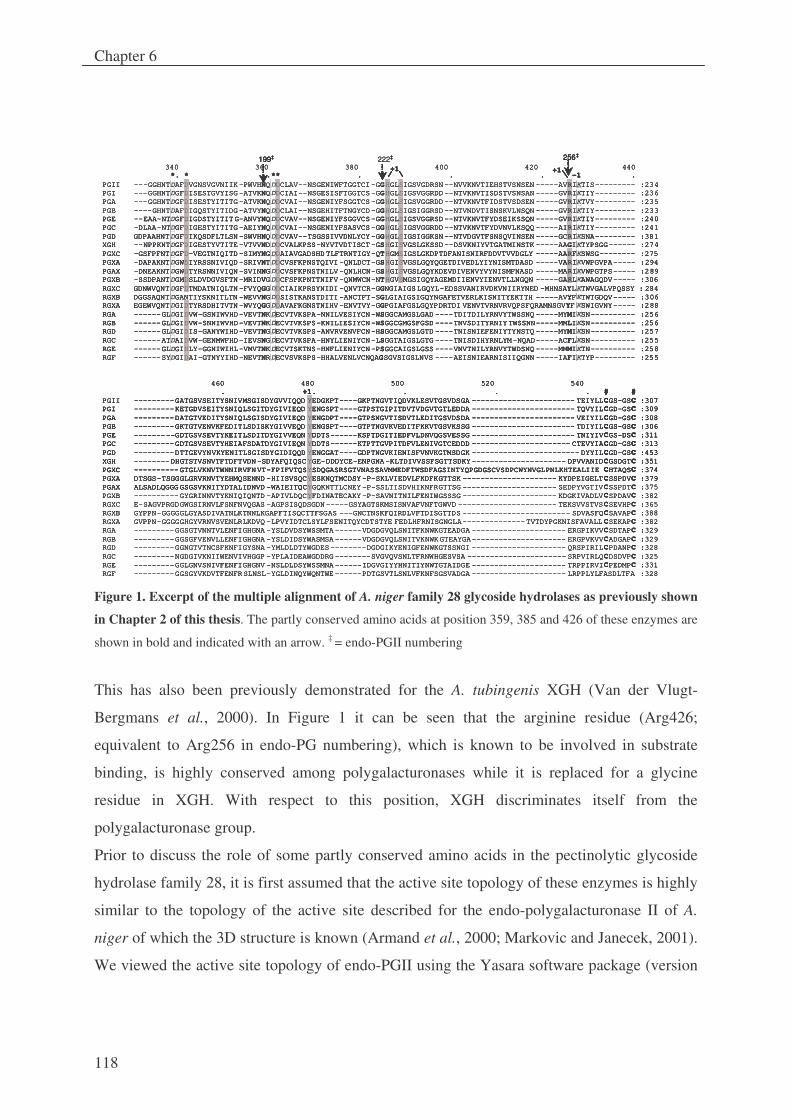
Chapter 6
118
340 360 380 400 420 440*. * .** . . . -1 .
PGII ---GGHNTDAFDVGNSVGVNIIK-PWVHNQDDCLAV--NSGENIWFTGGTCI-GGHGLSIGSVGDRSN--NVVKNVTIEHSTVSNSEN -----AVRIKTIS--------- :234PGI ---GGHNTDGFDISESTGVYISG-ATVKNQDDCIAI--NSGESISFTGGTCS-GGHGLSIGSVGGRDD--NTVKNVTISDSTVSNSAN -----GVRIKTIY--------- :236PGA ---GGHNTDGFDISESTYITITG-ATVKNQDDCVAI--NSGENIYFSGGTCS-GGHGLSIGSVGGRDD--NTVKNVTFIDSTVSDSEN -----GVRIKTVY--------- :235PGB ----GHNTDAFDIGQSTYITIDG-ATVYNQDDCLAI--NSGEHITFTNGYCD-GGHGLSIGSIGGRSD--NTVNDVTISNSKVLNSQN -----GVRIKTIY--------- :233PGE --EAA-NTDGFDIGDSTYITITG-ANVYNQDDCVAV--NSGENIYFSGGVCS-GGHGLSIGSVGGRSD--NTVKNVTFYDSEIKSSQN -----GVRIKTIY--------- :240PGC -DLAA-NTDGFDIGESTYITITG-AEIYNQDDCVAI--NSGENIYFSASVCS-GGHGLSIGSVGGRDD--NTVKNVTFYDVNVLKSQQ -----AIRIKTIY--------- :241PGD GDPAAHNTDGFDIKQSDFLTLSN-SWVHNQDDCVAV--TSGSSIVVDNLYCY-GGHGLSIGSIGGKSN--NTVDGVTFSNSQVINSEN -----GCRIKSNA--------- :381XGH --NPPKNTDGFDIGESTYVTITE-VTVVNDDDCVALKPSS-NYVTVDTISCT-GSHGISVGSLGKSSD--DSVKNIYVTGATMINSTK -----AAGIKTYPSGG------ :274PGXC -GSFPFNTDGFD-VEGTNIQITD-SIMYNGDDAIAVGADSHDTLFTRNTIGY-QTHGMSIGSLGKDPTDFANISNIRFDDVTVVDGLY -----AARFKSWSG-------- :275PGXA -DAPAKNTDGWDIYRSSNIVIQD-SRIVNTDDCVSFKPNSTQIVI-QNLDCT-GSHGISVGSLGQYQGETDIVEDLYIYNISMTDASD -----VARIKVWPGVPA----- :294PGAX -DNEAKNTDGWDTYRSNNIVIQN-SVINNGDDCVSFKPNSTNILV-QNLHCN-GSHGISVGSLGQYKDEVDIVENVYVYNISMFNASD -----MARIKVWPGTPS----- :289PGXB -SSDPANTDGMDSLDVDGVSFTN-MRIDVGDDCFSPKPNTTNIFV-QNMWCN-NTHGVSMGSIGQYAGEMDIIENVYIENVTLLNGQN -----GARLKAWAGQDV----- :306RGXC GDNWVQNTDGFDTMDATNIQLTN-FVYQGGDDCIAIKPRSYNIDI-QNVTCR-GGNGIAIGSLGQYL-EDSSVANIRVDKVNIIRYNED -MHNSAYLKTWVGALVPQSSY : 284RGXB DGGSAQNTDGANTIYSKNITLTN -WEVVNGDDSISTKANSTDITI-ANCTFT-SGLGIAIGSIGQYNGAFETVERLKISNITYEKTTH -----AVYFKTWTGDQV----- :306RGXA EGEWVQNTDGIDTYRSDHITVTN-WVYQGGDDAVAFKGNSTNIHV-ENVTVY-GGPGIAFGSLGQYPDRTDI VENVTVRNVRVQPSFQRAMNSGVYF KSWIGVNY----- :288RGA ------GLDGIDVW-GSNIWVHD-VEVTNKDECVTVKSPA-NNILVESIYCN-WSGGCAMGSLGAD ----TDITDILYRNVYTWSSNQ -----MYMIKSN---------- :256RGB ------GLDGIDVW-SNNIWVHD-VEVTNKDECVTVKSPS-KNILIESIYCN-WSGGCGMGSFGSD ----TNVSDITYRNIYTWSSNN-----MMLIKSN---------- :256RGD ------GLDGIDIS-GANYWIHD-VEVTNGDECVTVKSPS-ANVRVENVFCN-HSGGCAMGSLGTD ----TNISNIEFENIYTYNSTQ -----MYMIKSN---------- :257RGC ------ATDAIDVW-GENMWFHD-IEVSNGDECVTVKSPA-HNYLIENIYCN-LSGGTAIGSLGTG ----TNISDIHYRNLYM-NQAD-----ACFLKSN---------- :255RGE ------GLDGIDLY-GGNIWIHL-VMVTNKDECVTSKTNS-HNFLIENIYCN-PSGGCAIGSLGSS ----VNVTNILYRNVYTWDSNQ -----MMMIKTN---------- :258RGF ------SYDGIDAI-GTNYYIHD-NEVTNRDECVSVKSPS-HHALVENLVCNQAGSGVSIGSLNVS ----AEISNIEARNISIIQGNN -----IAFIKTYP--------- :255
460 480 500 520 540 . +1. . . . # #
PGII ---------GATGSVSEITYSNIVMSGISDYGVVIQQD YEDGKPT----GKPTNGVTIQDVKLESVTGSVDSGA ----------------------- TEIYLLCGS-GSC :307PGI ---------KETGDVSEITYSNIQLSGITDYGIVIEQD YENGSPT----GTPSTGIPITDVTVDGVTGTLEDDA ----------------------- TQVYILCGD-GSC :309PGA ---------DATGTVEDITYSNIQLSGISDYGIVIEQD YENGDPT----GTPSNGVTISDVTLEDITGSVDSDA ----------------------- VEIYILCGD-GSC :308PGB ---------GKTGTVENVKFEDITLSDISKYGIVVEQD YENGSPT----GTPTNGVKVEDITFKKVTGSVKSSG ----------------------- TDIYILCGS-GSC :306PGE ---------GDTGSVSEVTYKEITLSDITDYGIVVEQN YDDTS------KSPTDGITIEDFVLDNVQGSVESSG ----------------------- TNIYIVCGS-DSC :311PGC ---------GDTGSVSEVTYHEIAFSDATDYGIVIEQN YDDTS------KTPTTGVPITDFVLENIVGTCEDDD ---------------------- CTEVYIACGD-GSC :313PGD ---------DTTGEVYNVKYENITLSGISDYGIDIQQD YENGGAT----GDPTNGVKIENISFVNVKGTMSDGK ------------------------ DYYILCGD-GSC :453XGH --------DHGTSTVSNVTFTDFTVDN-SDYAFQIQSCYGE-DDDYCE-ENPGNA-KLTDIVVSSFSGTTSDKY--------------------- DPVVANIDCGSDGTC :351PGXC ----------GTGLVKNVTWNNIRVFNVT-FPIFVTQSYSDQGASRSGTVNASSAVMMEDFTWSDFAGSINTYQPGDGSCVSDPCWYNVGLPNLKHTEALIIE CHTAQSC :374PGXA DTSGS-TSGGGLGRVRNVTYEHMQSENND -HIISVSQCYESKNQTMCDSY-P-SKLVIEDVLFKDFKGTTSK --------------------- KYDPEIGELTCSSPDVC :379PGAX ALSADLQGGGGSGSVKNITYDTALIDNVD -WAIEITQCYGQKNTTLCNEY-P-SSLTISDVHIKNFRGTTSG --------------------- SEDPYVGTIVCSSPDTC :375PGXB ----------GYGRINNVTYKNIQIQNTD -APIVLDQCYFDINATECAKY-P-SAVNITNILFENIWGSSSG --------------------- KDGKIVADLVCSPDAVC :382RGXC E-SAGVPRGDGWGSIRNVLFSNFNVQGAS -AGPSISQDSGDN-----GSYAGTSKMSISNVAFVNFTGWVD ---------------------- TEKSVVSTVSCSEVHPC :365RGXB GYPPN-GGGGGLGYASDIVATNLKTNNLKGAPFTISQCTTFSGAS ---GNCTNSKFQIRDLVFTDISGTTDS ------------------------ SDVASFQCSAVAPC :388RGXA GVPPN-GGGGGHGYVRNVSVENLRLKDVQ -LPVYIDTCLSYLFSENITQYCDTSTYE FEDLHFRNISGNGLA--------------TVTDYPGKNISFAVALL CSEKAPC :382RGA ---------GGSGTVNNTVLENFIGHGNA -YSLDVDSYWSSMTA------VDGDGVQLSNITFKNWKGTEADGA --------------------- ERGPIKVVCSDTAPC :329RGB ---------GGSGFVENVLLENFIGHGNA -YSLDIDSYWASMSA------VDGDGVQLSNITVKNWKGTEAYGA--------------------- ERGPVKVVCADGAPC :329RGD ---------GGNGTVTNCSFKNFIGYSNA -YMLDLDTYWGDES--------DGDGIKYENIGFENWKGTSSNGI --------------------- QRSPIRILCPDANPC :328RGC ---------NGDGIVKNIIWENVIVHGGP -YPLAIDEAWGDDRG--------SVGVQVSNLTFRNWHGESVSA ---------------------- SRPVIRLQCDSDVPC :325RGE ---------GGLGNVSNIVFENFIGHGNV -NSLDLDSYWSSMNA------IDGVGIYYHNITIYNWTGTAIDGE --------------------- TRPPIRVICPEDMPC :331RGF ---------GGSGYVKDVTFENFRSLNSL-YGLDINQYWQNTWE------PDTGSVTLSNLVFKNFSGSVADGA --------------------- LRPPLYLFASDLTFA :328
340 360 380 400 420 440*. * .** . . . -1 .
PGII ---GGHNT AFDVGNSVGVNIIK-PWVHNQDDCLAV--NSGENIWFTGGTCI-GGHGLSIGSVGDRSN--NVVKNVTIEHSTVSNSEN -----AVRIKTIS--------- :234PGI ---GGHNT GFDISESTGVYISG-ATVKNQDDCIAI--NSGESISFTGGTCS-GGHGLSIGSVGGRDD--NTVKNVTISDSTVSNSAN -----GVRIKTIY--------- :236PGA ---GGHNT GFDISESTYITITG-ATVKNQDDCVAI--NSGENIYFSGGTCS-GGHGLSIGSVGGRDD--NTVKNVTFIDSTVSDSEN -----GVRIKTVY--------- :235PGB ----GHNTDAFDIGQSTYITIDG-ATVYNQDDCLAI--NSGEHITFTNGYCD-GGHGLSIGSIGGRSD--NTVNDVTISNSKVLNSQN -----GVRIKTIY--------- :233PGE --EAA-NTDGFDIGDSTYITITG-ANVYNQDDCVAV--NSGENIYFSGGVCS-GGHGLSIGSVGGRSD--NTVKNVTFYDSEIKSSQN -----GVRIKTIY--------- :240PGC -DLAA-NTDGFDIGESTYITITG-AEIYNQDDCVAI--NSGENIYFSASVCS-GGHGLSIGSVGGRDD--NTVKNVTFYDVNVLKSQQ -----AIRIKTIY--------- :241PGD GDPAAHNTDGFDIKQSDFLTLSN-SWVHNQDDCVAV--TSGSSIVVDNLYCY-GGHGLSIGSIGGKSN--NTVDGVTFSNSQVINSEN -----GCRIKSNA--------- :381XGH --NPPKNTDGFDIGESTYVTITE-VTVVNDDDCVALKPSS-NYVTVDTISCT-GSHGISVGSLGKSSD--DSVKNIYVTGATMINSTK -----AAGIKTYPSGG------ :274PGXC -GSFPFNTDGFD-VEGTNIQITD-SIMYNGDDAIAVGADSHDTLFTRNTIGY-QTHGMSIGSLGKDPTDFANISNIRFDDVTVVDGLY -----AARFKSWSG-------- :275PGXA -DAPAKNTDGWDIYRSSNIVIQD-SRIVNTDDCVSFKPNSTQIVI-QNLDCT-GSHGISVGSLGQYQGETDIVEDLYIYNISMTDASD -----VARIKVWPGVPA----- :294PGAX -DNEAKNTDGWDTYRSNNIVIQN-SVINNGDDCVSFKPNSTNILV-QNLHCN-GSHGISVGSLGQYKDEVDIVENVYVYNISMFNASD -----MARIKVWPGTPS----- :289PGXB -SSDPANTDGMDSLDVDGVSFTN-MRIDVGDDCFSPKPNTTNIFV-QNMWCN-NTHGVSMGSIGQYAGEMDIIENVYIENVTLLNGQN -----GARLKAWAGQDV----- :306RGXC GDNWVQNTDGFDTMDATNIQLTN-FVYQGGDDCIAIKPRSYNIDI-QNVTCR-GGNGIAIGSLGQYL-EDSSVANIRVDKVNIIRYNED -MHNSAYLKTWVGALVPQSSY : 284RGXB DGGSAQNTDGANTIYSKNITLTN -WEVVNGDDSISTKANSTDITI-ANCTFT-SGLGIAIGSIGQYNGAFETVERLKISNITYEKTTH -----AVYFKTWTGDQV----- :306RGXA EGEWVQNTDGIDTYRSDHITVTN-WVYQGGDDAVAFKGNSTNIHV-ENVTVY-GGPGIAFGSLGQYPDRTDI VENVTVRNVRVQPSFQRAMNSGVYF KSWIGVNY----- :288RGA ------GLDGIDVW-GSNIWVHD-VEVTNKDECVTVKSPA-NNILVESIYCN-WSGGCAMGSLGAD ----TDITDILYRNVYTWSSNQ -----MYMIKSN---------- :256RGB ------GLDGIDVW-SNNIWVHD-VEVTNKDECVTVKSPS-KNILIESIYCN-WSGGCGMGSFGSD ----TNVSDITYRNIYTWSSNN-----MMLIKSN---------- :256RGD ------GLDGIDIS-GANYWIHD-VEVTNGDECVTVKSPS-ANVRVENVFCN-HSGGCAMGSLGTD ----TNISNIEFENIYTYNSTQ -----MYMIKSN---------- :257RGC ------AT AIDVW-GENMWFHD-IEVSNGDECVTVKSPA-HNYLIENIYCN-LSGGTAIGSLGTG ----TNISDIHYRNLYM-NQAD-----ACFLKSN---------- :255RGE ------GL GIDLY-GGNIWIHL-VMVTNKDECVTSKTNS-HNFLIENIYCN-PSGGCAIGSLGSS ----VNVTNILYRNVYTWDSNQ -----MMMIKTN---------- :258RGF ------SY GIDAI-GTNYYIHD-NEVTNRDECVSVKSPS-HHALVENLVCNQAGSGVSIGSLNVS ----AEISNIEARNISIIQGNN -----IAFIKTYP--------- :255
460 480 500 520 540 . +1. . . . # #
PGII ---------GATGSVSEITYSNIVMSGISDYGVVIQQD YEDGKPT----GKPTNGVTIQDVKLESVTGSVDSGA ----------------------- TEIYLLCGS-GSC :307PGI ---------KETGDVSEITYSNIQLSGITDYGIVIEQD YENGSPT----GTPSTGIPITDVTVDGVTGTLEDDA ----------------------- TQVYILCGD-GSC :309PGA ---------DATGTVEDITYSNIQLSGISDYGIVIEQD YENGDPT----GTPSNGVTISDVTLEDITGSVDSDA ----------------------- VEIYILCGD-GSC :308PGB ---------GKTGTVENVKFEDITLSDISKYGIVVEQD YENGSPT----GTPTNGVKVEDITFKKVTGSVKSSG ----------------------- TDIYILCGS-GSC :306PGE ---------GDTGSVSEVTYKEITLSDITDYGIVVEQN YDDTS------KSPTDGITIEDFVLDNVQGSVESSG ----------------------- TNIYIVCGS-DSC :311PGC ---------GDTGSVSEVTYHEIAFSDATDYGIVIEQN YDDTS------KTPTTGVPITDFVLENIVGTCEDDD ---------------------- CTEVYIACGD-GSC :313PGD ---------DTTGEVYNVKYENITLSGISDYGIDIQQD YENGGAT----GDPTNGVKIENISFVNVKGTMSDGK ------------------------ DYYILCGD-GSC :453XGH --------DHGTSTVSNVTFTDFTVDN-SDYAFQIQSCYGE-DDDYCE-ENPGNA-KLTDIVVSSFSGTTSDKY--------------------- DPVVANIDCGSDGTC :351PGXC ----------GTGLVKNVTWNNIRVFNVT-FPIFVTQSYSDQGASRSGTVNASSAVMMEDFTWSDFAGSINTYQPGDGSCVSDPCWYNVGLPNLKHTEALIIE CHTAQSC :374PGXA DTSGS-TSGGGLGRVRNVTYEHMQSENND -HIISVSQCYESKNQTMCDSY-P-SKLVIEDVLFKDFKGTTSK --------------------- KYDPEIGELTCSSPDVC :379PGAX ALSADLQGGGGSGSVKNITYDTALIDNVD -WAIEITQCYGQKNTTLCNEY-P-SSLTISDVHIKNFRGTTSG --------------------- SEDPYVGTIVCSSPDTC :375PGXB ----------GYGRINNVTYKNIQIQNTD -APIVLDQCYFDINATECAKY-P-SAVNITNILFENIWGSSSG --------------------- KDGKIVADLVCSPDAVC :382RGXC E-SAGVPRGDGWGSIRNVLFSNFNVQGAS -AGPSISQDSGDN-----GSYAGTSKMSISNVAFVNFTGWVD ---------------------- TEKSVVSTVSCSEVHPC :365RGXB GYPPN-GGGGGLGYASDIVATNLKTNNLKGAPFTISQCTTFSGAS ---GNCTNSKFQIRDLVFTDISGTTDS ------------------------ SDVASFQCSAVAPC :388RGXA GVPPN-GGGGGHGYVRNVSVENLRLKDVQ -LPVYIDTCLSYLFSENITQYCDTSTYE FEDLHFRNISGNGLA--------------TVTDYPGKNISFAVALL CSEKAPC :382RGA ---------GGSGTVNNTVLENFIGHGNA -YSLDVDSYWSSMTA------VDGDGVQLSNITFKNWKGTEADGA --------------------- ERGPIKVVCSDTAPC :329RGB ---------GGSGFVENVLLENFIGHGNA -YSLDIDSYWASMSA------VDGDGVQLSNITVKNWKGTEAYGA--------------------- ERGPVKVVCADGAPC :329RGD ---------GGNGTVTNCSFKNFIGYSNA -YMLDLDTYWGDES--------DGDGIKYENIGFENWKGTSSNGI --------------------- QRSPIRILCPDANPC :328RGC ---------NGDGIVKNIIWENVIVHGGP -YPLAIDEAWGDDRG--------SVGVQVSNLTFRNWHGESVSA ---------------------- SRPVIRLQCDSDVPC :325RGE ---------GGLGNVSNIVFENFIGHGNV -NSLDLDSYWSSMNA------IDGVGIYYHNITIYNWTGTAIDGE --------------------- TRPPIRVICPEDMPC :331RGF ---------GGSGYVKDVTFENFRSLNSL-YGLDINQYWQNTWE------PDTGSVTLSNLVFKNFSGSVADGA --------------------- LRPPLYLFASDLTFA :328
+1 +1199‡ 222‡ 256‡
+1 +1199‡ ‡ 256‡
340 360 380 400 420 440*. * .** . . . -1 .
PGII ---GGHNTDAFDVGNSVGVNIIK-PWVHNQDDCLAV--NSGENIWFTGGTCI-GGHGLSIGSVGDRSN--NVVKNVTIEHSTVSNSEN -----AVRIKTIS--------- :234PGI ---GGHNTDGFDISESTGVYISG-ATVKNQDDCIAI--NSGESISFTGGTCS-GGHGLSIGSVGGRDD--NTVKNVTISDSTVSNSAN -----GVRIKTIY--------- :236PGA ---GGHNTDGFDISESTYITITG-ATVKNQDDCVAI--NSGENIYFSGGTCS-GGHGLSIGSVGGRDD--NTVKNVTFIDSTVSDSEN -----GVRIKTVY--------- :235PGB ----GHNTDAFDIGQSTYITIDG-ATVYNQDDCLAI--NSGEHITFTNGYCD-GGHGLSIGSIGGRSD--NTVNDVTISNSKVLNSQN -----GVRIKTIY--------- :233PGE --EAA-NTDGFDIGDSTYITITG-ANVYNQDDCVAV--NSGENIYFSGGVCS-GGHGLSIGSVGGRSD--NTVKNVTFYDSEIKSSQN -----GVRIKTIY--------- :240PGC -DLAA-NTDGFDIGESTYITITG-AEIYNQDDCVAI--NSGENIYFSASVCS-GGHGLSIGSVGGRDD--NTVKNVTFYDVNVLKSQQ -----AIRIKTIY--------- :241PGD GDPAAHNTDGFDIKQSDFLTLSN-SWVHNQDDCVAV--TSGSSIVVDNLYCY-GGHGLSIGSIGGKSN--NTVDGVTFSNSQVINSEN -----GCRIKSNA--------- :381XGH --NPPKNTDGFDIGESTYVTITE-VTVVNDDDCVALKPSS-NYVTVDTISCT-GSHGISVGSLGKSSD--DSVKNIYVTGATMINSTK -----AAGIKTYPSGG------ :274PGXC -GSFPFNTDGFD-VEGTNIQITD-SIMYNGDDAIAVGADSHDTLFTRNTIGY-QTHGMSIGSLGKDPTDFANISNIRFDDVTVVDGLY -----AARFKSWSG-------- :275PGXA -
PGE --EAA-NTDGFDIGDSTYITITG-ANVYNQDDCVAV--NSGENIYFSGGVCS-GGHGLSIGSVGGRSD--NTVKNVTFYDSEIKSSQN -----GVRIKTIY--------- :240PGC -DLAA-NTDGFDIGESTYITITG-AEIYNQDDCVAI--NSGENIYFSASVCS-GGHGLSIGSVGGRDD--NTVKNVTFYDVNVLKSQQ -----AIRIKTIY--------- :241PGD GDPAAHNTDGFDIKQSDFLTLSN-SWVHNQDDCVAV--TSGSSIVVDNLYCY-GGHGLSIGSIGGKSN--NTVDGVTFSNSQVINSEN -----GCRIKSNA--------- :381XGH --NPPKNTDGFDIGESTYVTITE-VTVVNDDDCVALKPSS-NYVTVDTISCT-GSHGISVGSLGKSSD--DSVKNIYVTGATMINSTK -----AAGIKTYPSGG------ :274PGXC -GSFPFNTDGFD-VEGTNIQITD-SIMYNGDDAIAVGADSHDTLFTRNTIGY-QTHGMSIGSLGKDPTDFANISNIRFDDVTVVDGLY -----AARFKSWSG-------- :275PGXA -DAPAKNTDGWDIYRSSNIVIQD-SRIVNTDDCVSFKPNSTQIVI-QNLDCT-GSHGISVGSLGQYQGETDIVEDLYIYNISMTDASD -----VARIKVWPGVPA----- :294PGAX -DNEAKNTDGWDTYRSNNIVIQN-SVINNGDDCVSFKPNSTNILV-QNLHCN-GSHGISVGSLGQYKDEVDIVENVYVYNISMFNASD -----MARIKVWPGTPS----- :289PGXB -SSDPANTDGMDSLDVDGVSFTN-MRIDVGDDCFSPKPNTTNIFV-QNMWCN-NTHGVSMGSIGQYAGEMDIIENVYIENVTLLNGQN -----GARLKAWAGQDV----- :306RGXC GDNWVQNTDGFDTMDATNIQLTN-FVYQGGDDCIAIKPRSYNIDI-QNVTCR-GGNGIAIGSLGQYL-EDSSVANIRVDKVNIIRYNED -MHNSAYLKTWVGALVPQSSY : 284RGXB DGGSAQNTDGANTIYSKNITLTN -WEVVNGDDSISTKANSTDITI-ANCTFT-SGLGIAIGSIGQYNGAFETVERLKISNITYEKTTH -----AVYFKTWTGDQV----- :306RGXA EGEWVQNTDGIDTYRSDHITVTN-WVYQGGDDAVAFKGNSTNIHV-ENVTVY-GGPGIAFGSLGQYPDRTDI VENVTVRNVRVQPSFQRAMNSGVYF KSWIGVNY----- :288RGA ------GLDGIDVW-GSNIWVHD-VEVTNKDECVTVKSPA-NNILVESIYCN
DAPAKNTDGWDIYRSSNIVIQD-SRIVNTDDCVSFKPNSTQIVI-QNLDCT-GSHGISVGSLGQYQGETDIVEDLYIYNISMTDASD -----VARIKVWPGVPA----- :294PGAX -DNEAKNTDGWDTYRSNNIVIQN-SVINNGDDCVSFKPNSTNILV-QNLHCN-GSHGISVGSLGQYKDEVDIVENVYVYNISMFNASD -----MARIKVWPGTPS----- :289PGXB -SSDPANTDGMDSLDVDGVSFTN-MRIDVGDDCFSPKPNTTNIFV-QNMWCN-NTHGVSMGSIGQYAGEMDIIENVYIENVTLLNGQN -----GARLKAWAGQDV----- :306RGXC GDNWVQNTDGFDTMDATNIQLTN-FVYQGGDDCIAIKPRSYNIDI-QNVTCR-GGNGIAIGSLGQYL-EDSSVANIRVDKVNIIRYNED -MHNSAYLKTWVGALVPQSSY : 284RGXB DGGSAQNTDGANTIYSKNITLTN -WEVVNGDDSISTKANSTDITI-ANCTFT-SGLGIAIGSIGQYNGAFETVERLKISNITYEKTTH -----AVYFKTWTGDQV----- :306RGXA EGEWVQNTDGIDTYRSDHITVTN-WVYQGGDDAVAFKGNSTNIHV-ENVTVY-GGPGIAFGSLGQYPDRTDI VENVTVRNVRVQPSFQRAMNSGVYF KSWIGVNY----- :288RGA ------GLDGIDVW-GSNIWVHD-VEVTNKDECVTVKSPA-NNILVESIYCN-WSGGCAMGSLGAD ----TDITDILYRNVYTWSSNQ -----MYMIKSN---------- :256RGB ------GLDGIDVW-SNNIWVHD-VEVTNKDECVTVKSPS-KNILIESIYCN-WSGGCGMGSFGSD ----TNVSDITYRNIYTWSSNN-----MMLIKSN---------- :256RGD ------GLDGIDIS-GANYWIHD-VEVTNGDECVTVKSPS-ANVRVENVFCN-HSGGCAMGSLGTD ----TNISNIEFENIYTYNSTQ -----MYMIKSN---------- :257RGC ------ATDAIDVW-GENMWFHD-IEVSNGDECVTVKSPA-HNYLIENIYCN-LSGGTAIGSLGTG ----TNISDIHYRNLYM-NQAD-----ACFLKSN---------- :255RGE ------GLDGIDLY-GGNIWIHL-VMVTNKDECVTSKTNS-HNFLIENIYCN-PSGGCAIGSLGSS ----VNVTNILYRNVYTWDSNQ -----MMMIKTN---------- :258RGF ------SYDGIDAI-GTNYYIHD-NEVTNRDECVSVKSPS-HHALVENLVCNQAGSGVSIGSLNVS ----AEISNIEARNISIIQGNN -----IAFIKTYP--------- :255
460 480 500 520 540 . +1. . . . # #
PGII ---------
-WSGGCAMGSLGAD ----TDITDILYRNVYTWSSNQ -----MYMIKSN---------- :256RGB ------GLDGIDVW-SNNIWVHD-VEVTNKDECVTVKSPS-KNILIESIYCN-WSGGCGMGSFGSD ----TNVSDITYRNIYTWSSNN-----MMLIKSN---------- :256RGD ------GLDGIDIS-GANYWIHD-VEVTNGDECVTVKSPS-ANVRVENVFCN-HSGGCAMGSLGTD ----TNISNIEFENIYTYNSTQ -----MYMIKSN---------- :257RGC ------ATDAIDVW-GENMWFHD-IEVSNGDECVTVKSPA-HNYLIENIYCN-LSGGTAIGSLGTG ----TNISDIHYRNLYM-NQAD-----ACFLKSN---------- :255RGE ------GLDGIDLY-GGNIWIHL-VMVTNKDECVTSKTNS-HNFLIENIYCN-PSGGCAIGSLGSS ----VNVTNILYRNVYTWDSNQ -----MMMIKTN---------- :258RGF ------SYDGIDAI-GTNYYIHD-NEVTNRDECVSVKSPS-HHALVENLVCNQAGSGVSIGSLNVS ----AEISNIEARNISIIQGNN -----IAFIKTYP--------- :255
460 480 500 520 540 . +1. . . . # #
PGII ---------GATGSVSEITYSNIVMSGISDYGVVIQQD YEDGKPT----GKPTNGVTIQDVKLESVTGSVDSGA ----------------------- TEIYLLCGS-GSC :307PGI ---------KETGDVSEITYSNIQLSGITDYGIVIEQD YENGSPT----GTPSTGIPITDVTVDGVTGTLEDDA ----------------------- TQVYILCGD-GSC :309PGA ---------DATGTVEDITYSNIQLSGISDYGIVIEQD YENGDPT----GTPSNGVTISDVTLEDITGSVDSDA ----------------------- VEIYILCGD-GSC :308PGB ---------GKTGTVENVKFEDITLSDISKYGIVVEQD YENGSPT----GTPTNGVKVEDITFKKVTGSVKSSG ----------------------- TDIYILCGS-GSC :306PGE ---------GDTGSVSEVTYKEITLSDITDYGIVVEQN YDDTS------KSPTDGITIEDFVLDNVQGSVESSG ----------------------- TNIYIVCGS-DSC :311PGC ---------GDTGSVSEVTYHEIAFSDATDYGIVIEQN YDDTS------KTPTTGVPITDFVLENIVGTCEDDD ---------------------- CTEVYIACGD-GSC :313PGD ---------DTTGEVYNVKYENITLSGISDYGIDIQQD YENGGAT----GDPTNGVKIENISFVNVKGTMSDGK ------------------------ DYYILCGD-GSC :453XGH --------DHGTSTVSNVTFTDFTVDN-SDYAFQIQSCYGE-DDDYCE-ENPGNA-KLTDIVVSSFSGTTSDKY--------------------- DPVVANIDCGSDGTC :351PGXC ----------GTGLVKNVTWNNIRVFNVT-FPIFVTQSYSDQGASRSGTVNASSAVMMEDFTWSDFAGSINTYQPGDGSCVSDPCWYNVGLPNLKHTEALIIE CHTAQSC :374PGXA DTSGS-TSGGGLGRVRNVTYEHMQSENND -HIISVSQCYESKNQTMCDSY-P-SKLVIEDVLFKDFKGTTSK --------------------- KYDPEIGELTCSSPDVC :379PGAX ALSADLQGGGGSGSVKNITYDTALIDNVD -WAIEITQC
GATGSVSEITYSNIVMSGISDYGVVIQQD YEDGKPT----GKPTNGVTIQDVKLESVTGSVDSGA ----------------------- TEIYLLCGS-GSC :307PGI ---------KETGDVSEITYSNIQLSGITDYGIVIEQD YENGSPT----GTPSTGIPITDVTVDGVTGTLEDDA ----------------------- TQVYILCGD-GSC :309PGA ---------DATGTVEDITYSNIQLSGISDYGIVIEQD YENGDPT----GTPSNGVTISDVTLEDITGSVDSDA ----------------------- VEIYILCGD-GSC :308PGB ---------GKTGTVENVKFEDITLSDISKYGIVVEQD YENGSPT----GTPTNGVKVEDITFKKVTGSVKSSG ----------------------- TDIYILCGS-GSC :306PGE ---------GDTGSVSEVTYKEITLSDITDYGIVVEQN YDDTS------KSPTDGITIEDFVLDNVQGSVESSG ----------------------- TNIYIVCGS-DSC :311PGC ---------GDTGSVSEVTYHEIAFSDATDYGIVIEQN YDDTS------KTPTTGVPITDFVLENIVGTCEDDD ---------------------- CTEVYIACGD-GSC :313PGD ---------DTTGEVYNVKYENITLSGISDYGIDIQQD YENGGAT----GDPTNGVKIENISFVNVKGTMSDGK ------------------------ DYYILCGD-GSC :453XGH --------DHGTSTVSNVTFTDFTVDN-SDYAFQIQSCYGE-DDDYCE-ENPGNA-KLTDIVVSSFSGTTSDKY--------------------- DPVVANIDCGSDGTC :351PGXC ----------GTGLVKNVTWNNIRVFNVT-FPIFVTQSYSDQGASRSGTVNASSAVMMEDFTWSDFAGSINTYQPGDGSCVSDPCWYNVGLPNLKHTEALIIE CHTAQSC :374PGXA DTSGS-TSGGGLGRVRNVTYEHMQSENND -HIISVSQCYESKNQTMCDSY-P-SKLVIEDVLFKDFKGTTSK --------------------- KYDPEIGELTCSSPDVC :379PGAX ALSADLQGGGGSGSVKNITYDTALIDNVD -WAIEITQCYGQKNTTLCNEY-P-SSLTISDVHIKNFRGTTSG --------------------- SEDPYVGTIVCSSPDTC :375PGXB ----------GYGRINNVTYKNIQIQNTD -APIVLDQCYFDINATECAKY-P-SAVNITNILFENIWGSSSG --------------------- KDGKIVADLVCSPDAVC :382RGXC E-SAGVPRGDGWGSIRNVLFSNFNVQGAS -AGPSISQDSGDN-----GSYAGTSKMSISNVAFVNFTGWVD ---------------------- TEKSVVSTVSCSEVHPC :365RGXB GYPPN-GGGGGLGYASDIVATNLKTNNLKGAPFTISQCTTFSGAS ---GNCTNSKFQIRDLVFTDISGTTDS ------------------------ SDVASFQCSAVAPC :388RGXA GVPPN-GGGGGHGYVRNVSVENLRLKDVQ -LPVYIDTCLSYLFSENITQYCDTSTYE FEDLHFRNISGNGLA--------------TVTDYPGKNISFAVALL CSEKAPC :382RGA ---------GGSGTVNNTVLENFIGHGNA -YSLDVDSYWSSMTA------VDGDGVQLSNITFKNWKGTEADGA --------------------- ERGPIKVVCSDTAPC :329RGB ---------GGSGFVENVLLENFIGHGNA -YSLDIDSYWASMSA------VDGDGVQLSNITVKNWKGTEAYGA--------------------- ERGPVKVVCADGAPC :329RGD ---------GGNGTVTNCSFKNFIGYSNA -YMLDLDTYWGDES--------DGDGIKYENIGFENWKGTSSNGI --------------------- QRSPIRILCPDANPC :328RGC ---------NGDGIVKNIIWENVIVHGGP -YPLAIDEAWGDDRG--------SVGVQVSNLTFRNWHGESVSA ---------------------- SRPVIRLQCDSDVPC :325RGE ---------GGLGNVSNIVFENFIGHGNV -NSLDLDSYWSSMNA------IDGVGIYYHNITIYNWTGTAIDGE --------------------- TRPPIRVICPEDMPC :331RGF ---------GGSGYVKDVTFENFRSLNSL-YGLDINQYWQNTWE------PDTGSVTLSNLVFKNFSGSVADGA --------------------- LRPPLYLFASDLTFA :328
340 360 380 400 420 440*. * .** . . . -1 .
PGII ---GGHNT AFDVGNSVGVNIIK-PWVHNQDDCLAV--NSGENIWFTGGTCI-GGHGLSIGSVGDRSN--NVVKNVTIEHSTVSNSEN -----AVRIKTIS--------- :234PGI ---GGHNT GFDISESTGVYISG-ATVKNQDDCIAI--NSGESISFTGGTCS-GGHGLSIGSVGGRDD--NTVKNVTISDSTVSNSAN -----GVRIKTIY--------- :236PGA ---GGHNT GFDISESTYITITG-ATVKNQDDCVAI--NSGENIYFSGGTCS-GGHGLSIGSVGGRDD--NTVKNVTFIDSTVSDSEN -----GVRIKTVY--------- :235PGB ----GHNTDAFDIGQSTYITIDG-ATVYNQDDCLAI--NSGEHITFTNGYCD-GGHGLSIGSIGGRSD--NTVNDVTISNSKVLNSQN -----GVRIKTIY--------- :233PGE --EAA-NTDGFDIGDSTYITITG-ANVYNQDDCVAV--NSGENIYFSGGVCS-GGHGLSIGSVGGRSD--NTVKNVTFYDSEIKSSQN -----GVRIKTIY--------- :240PGC -DLAA-NTDGFDIGESTYITITG-AEIYNQDDCVAI--NSGENIYFSASVCS-GGHGLSIGSVGGRDD--NTVKNVTFYDVNVLKSQQ -----AIRIKTIY--------- :241PGD GDPAAHNTDGFDIKQSDFLTLSN-SWVHNQDDCVAV--TSGSSIVVDNLYCY-GGHGLSIGSIGGKSN--NTVDGVTFSNSQVINSEN -----GCRIKSNA--------- :381XGH --NPPKNTDGFDIGESTYVTITE-VTVVNDDDCVALKPSS-NYVTVDTISCT-GSHGISVGSLGKSSD--DSVKNIYVTGATMINSTK -----AAGIKTYPSGG------ :274PGXC -GSFPFNTDGFD-VEGTNIQITD-SIMYNGDDAIAVGADSHDTLFTRNTIGY-QTHGMSIGSLGKDPTDFANISNIRFDDVTVVDGLY -----AARFKSWSG-------- :275PGXA -DAPAKNTDGWDIYRSSNIVIQD-SRIVNTDDCVSFKPNSTQIVI-QNLDCT-GSHGISVGSLGQYQGETDIVEDLYIYNISMTDASD -----VARIKVWPGVPA----- :294PGAX -DNEAKNTDGWDTYRSNNIVIQN-SVINNGDDCVSFKPNSTNILV-QNLHCN-GSHGISVGSLGQYKDEVDIVENVYVYNISMFNASD -----MARIKVWPGTPS----- :289PGXB -SSDPANTDGMDSLDVDGVSFTN-MRIDVGDDCFSPKPNTTNIFV-QNMWCN-NTHGVSMGSIGQYAGEMDIIENVYIENVTLLNGQN -----GARLKAWAGQDV----- :306RGXC GDNWVQNTDGFDTMDATNIQLTN-FVYQGGDDCIAIKPRSYNIDI-QNVTCR-GGNGIAIGSLGQYL-EDSSVANIRVDKVNIIRYNED -MHNSAYLKTWVGALVPQSSY : 284RGXB DGGSAQNTDGANTIYSKNITLTN -WEVVNGDDSISTKANSTDITI-ANCTFT-SGLGIAIGSIGQYNGAFETVERLKISNITYEKTTH -----AVYFKTWTGDQV----- :306RGXA EGEWVQNTDGIDTYRSDHITVTN-WVYQGGDDAVAFKGNSTNIHV-ENVTVY-GGPGIAFGSLGQYPDRTDI VENVTVRNVRVQPSFQRAMNSGVYF KSWIGVNY----- :288RGA ------GLDGIDVW-GSNIWVHD-VEVTNKDECVTVKSPA-NNILVESIYCN-WSGGCAMGSLGAD ----TDITDILYRNVYTWSSNQ -----MYMIKSN---------- :256RGB ------GLDGIDVW-SNNIWVHD-VEVTNKDECVTVKSPS-KNILIESIYCN-WSGGCGMGSFGSD ----TNVSDITYRNIYTWSSNN-----MMLIKSN---------- :256RGD ------GLDGIDIS-GANYWIHD-VEVTNGDECVTVKSPS-ANVRVENVFCN-HSGGCAMGSLGTD ----TNISNIEFENIYTYNSTQ -----MYMIKSN---------- :257RGC ------AT AIDVW-GENMWFHD-IEVSNGDECVTVKSPA-HNYLIENIYCN-LSGGTAIGSLGTG ----TNISDIHYRNLYM-NQAD-----ACFLKSN---------- :255RGE ------GL GIDLY-GGNIWIHL-VMVTNKDECVTSKTNS-HNFLIENIYCN-PSGGCAIGSLGSS ----VNVTNILYRNVYTWDSNQ -----MMMIKTN---------- :258RGF ------SY GIDAI-GTNYYIHD-NEVTNRDECVSVKSPS-HHALVENLVCNQAGSGVSIGSLNVS ----AEISNIEARNISIIQGNN -----IAFIKTYP--------- :255
460 480 500 520 540 . +1. . . . # #
PGII ---------GATGSVSEITYSNIVMSGISDYGVVIQQD YEDGKPT----GKPTNGVTIQDVKLESVTGSVDSGA ----------------------- TEIYLLCGS-GSC :307PGI ---------KETGDVSEITYSNIQLSGITDYGIVIEQD YENGSPT----GTPSTGIPITDVTVDGVTGTLEDDA ----------------------- TQVYILCGD-GSC :309PGA ---------DATGTVEDITYSNIQLSGISDYGIVIEQD YENGDPT----GTPSNGVTISDVTLEDITGSVDSDA ----------------------- VEIYILCGD-GSC :308PGB ---------GKTGTVENVKFEDITLSDISKYGIVVEQD YENGSPT----GTPTNGVKVEDITFKKVTGSVKSSG ----------------------- TDIYILCGS-GSC :306PGE ---------GDTGSVSEVTYKEITLSDITDYGIVVEQN YDDTS------KSPTDGITIEDFVLDNVQGSVESSG ----------------------- TNIYIVCGS-DSC :311PGC ---------GDTGSVSEVTYHEIAFSDATDYGIVIEQN YDDTS------KTPTTGVPITDFVLENIVGTCEDDD ---------------------- CTEVYIACGD-GSC :313PGD ---------DTTGEVYNVKYENITLSGISDYGIDIQQD YENGGAT----GDPTNGVKIENISFVNVKGTMSDGK ------------------------ DYYILCGD-GSC :453XGH --------DHGTSTVSNVTFTDFTVDN-SDYAFQIQSCYGE-DDDYCE-ENPGNA-KLTDIVVSSFSGTTSDKY--------------------- DPVVANIDCGSDGTC :351PGXC ----------GTGLVKNVTWNNIRVFNVT-FPIFVTQSYSDQGASRSGTVNASSAVMMEDFTWSDFAGSINTYQPGDGSCVSDPCWYNVGLPNLKHTEALIIE CHTAQSC :374PGXA DTSGS-TSGGGLGRVRNVTYEHMQSENND -HIISVSQCYESKNQTMCDSY-P-SKLVIEDVLFKDFKGTTSK --------------------- KYDPEIGELTCSSPDVC :379PGAX ALSADLQGGGGSGSVKNITYDTALIDNVD -WAIEITQC
GATGSVSEITYSNIVMSGISDYGVVIQQD YEDGKPT----GKPTNGVTIQDVKLESVTGSVDSGA ----------------------- TEIYLLCGS-GSC :307PGI ---------KETGDVSEITYSNIQLSGITDYGIVIEQD YENGSPT----GTPSTGIPITDVTVDGVTGTLEDDA ----------------------- TQVYILCGD-GSC :309PGA ---------DATGTVEDITYSNIQLSGISDYGIVIEQD YENGDPT----GTPSNGVTISDVTLEDITGSVDSDA ----------------------- VEIYILCGD-GSC :308PGB ---------GKTGTVENVKFEDITLSDISKYGIVVEQD YENGSPT----GTPTNGVKVEDITFKKVTGSVKSSG ----------------------- TDIYILCGS-GSC :306PGE ---------GDTGSVSEVTYKEITLSDITDYGIVVEQN YDDTS------KSPTDGITIEDFVLDNVQGSVESSG ----------------------- TNIYIVCGS-DSC :311PGC ---------GDTGSVSEVTYHEIAFSDATDYGIVIEQN YDDTS------KTPTTGVPITDFVLENIVGTCEDDD ---------------------- CTEVYIACGD-GSC :313PGD ---------DTTGEVYNVKYENITLSGISDYGIDIQQD YENGGAT----GDPTNGVKIENISFVNVKGTMSDGK ------------------------ DYYILCGD-GSC :453XGH --------DHGTSTVSNVTFTDFTVDN-SDYAFQIQSCYGE-DDDYCE-ENPGNA-KLTDIVVSSFSGTTSDKY--------------------- DPVVANIDCGSDGTC :351PGXC ----------GTGLVKNVTWNNIRVFNVT-FPIFVTQSYSDQGASRSGTVNASSAVMMEDFTWSDFAGSINTYQPGDGSCVSDPCWYNVGLPNLKHTEALIIE CHTAQSC :374PGXA DTSGS-TSGGGLGRVRNVTYEHMQSENND -HIISVSQCYESKNQTMCDSY-P-SKLVIEDVLFKDFKGTTSK --------------------- KYDPEIGELTCSSPDVC :379PGAX ALSADLQGGGGSGSVKNITYDTALIDNVD -WAIEITQCYGQKNTTLCNEY-P-SSLTISDVHIKNFRGTTSG --------------------- SEDPYVGTIVCSSPDTC :375PGXB ----------GYGRINNVTYKNIQIQNTD -APIVLDQCYFDINATECAKY-P-SAVNITNILFENIWGSSSG --------------------- KDGKIVADLVCSPDAVC :382RGXC E-SAGVPRGDGWGSIRNVLFSNFNVQGAS -AGPSISQDSGDN-----GSYAGTSKMSISNVAFVNFTGWVD ---------------------- TEKSVVSTVSCSEVHPC :365RGXB GYPPN-GGGGGLGYASDIVATNLKTNNLKGAPFTISQCTTFSGAS ---GNCTNSKFQIRDLVFTDISGTTDS ------------------------ SDVASFQCSAVAPC :388RGXA GVPPN-GGGGGHGYVRNVSVENLRLKDVQ -LPVYIDTCLSYLFSENITQYCDTSTYE FEDLHFRNISGNGLA--------------TVTDYPGKNISFAVALL CSEKAPC :382RGA ---------GGSGTVNNTVLENFIGHGNA -YSLDVDSYWSSMTA------VDGDGVQLSNITFKNWKGTEADGA --------------------- ERGPIKVVCSDTAPC :329RGB ---------GGSGFVENVLLENFIGHGNA -YSLDIDSYWASMSA------VDGDGVQLSNITVKNWKGTEAYGA--------------------- ERGPVKVVCADGAPC :329RGD ---------GGNGTVTNCSFKNFIGYSNA -YMLDLDTYWGDES--------DGDGIKYENIGFENWKGTSSNGI --------------------- QRSPIRILCPDANPC :328RGC ---------NGDGIVKNIIWENVIVHGGP -YPLAIDEAWGDDRG--------SVGVQVSNLTFRNWHGESVSA ---------------------- SRPVIRLQCDSDVPC :325RGE ---------GGLGNVSNIVFENFIGHGNV -NSLDLDSYWSSMNA------IDGVGIYYHNITIYNWTGTAIDGE --------------------- TRPPIRVICPEDMPC :331RGF ---------GGSGYVKDVTFENFRSLNSL-YGLDINQYWQNTWE------PDTGSVTLSNLVFKNFSGSVADGA --------------------- LRPPLYLFASDLTFA :328
+1 +1199‡ 222‡ 256‡
+1 +1199‡ ‡ 256‡
Figure 1. Excerpt of the multiple alignment of A. niger family 28 glycoside hydrolases as previously shown
in Chapter 2 of this thesis. The partly conserved amino acids at position 359, 385 and 426 of these enzymes are
shown in bold and indicated with an arrow. ‡ = endo-PGII numbering
This has also been previously demonstrated for the A. tubingenis XGH (Van der Vlugt-
Bergmans et al., 2000). In Figure 1 it can be seen that the arginine residue (Arg426;
equivalent to Arg256 in endo-PG numbering), which is known to be involved in substrate
binding, is highly conserved among polygalacturonases while it is replaced for a glycine
residue in XGH. With respect to this position, XGH discriminates itself from the
polygalacturonase group.
Prior to discuss the role of some partly conserved amino acids in the pectinolytic glycoside
hydrolase family 28, it is first assumed that the active site topology of these enzymes is highly
similar to the topology of the active site described for the endo-polygalacturonase II of A.
niger of which the 3D structure is known (Armand et al., 2000; Markovic and Janecek, 2001).
We viewed the active site topology of endo-PGII using the Yasara software package (version
General discussion
119
6.7.15) and have replaced the arginine at position 256 (equivalent to Arg426 in Fig 1) in the
3D structure of this enzyme for a glycine residue (hence Gly256) in order to use it as a
structure for XGH (see Fig. 2).
The Arg256 residue, which is present in all polygalacturonases, is involved in substrate
binding in which the carboxyl group of GalA is hydrogen-bonded to the side chain of Arg256
(Cho et al., 2001). In XGH, the glycine residue at this position does not posses any side chain
and can therefore not form a hydrogen bond with a GalA residue. This may be one of the
reasons why XGH lost its affinity for polygalacturonic acid.
It is debatable whether the Gly256 residue in XGH (Fig. 2) is responsible for the enzyme’s
interaction with xylosylated homogalacturonan to be able to act on the galacturonan
backbone. However, because Gly256 in XGH does not contain a side chain it might create
space in the active site cleft of the enzyme. This space may be sufficient to harbor xylosylated
galacturonic acid residues, or even dimeric side chains of xylose (as discussed later). However
we know that exo-polygalacturonases that, just like endo-polygalacturonases, posses the
Arg256 residue can still act towards XGA. This suggests that an other amino acid (or amino
acids) may also be involved in allowing the enzyme to accept xylosylated Gal residues in its
active site. Such an amino acid should exist among XGH as well as exo-polygalacturonases
and not in endo-polygalacturonases. As shown in Figure 1, at amino acid position 385
(equivalent to position 222 in endo-PGII numbering) all endo-polygalacturonases contain a
glycine, while the exo-polygalacturonases possess either a serine or a threonine, and the XGH
a serine. Serine and threonine are both amino acids with a hydroxyl group in a short side
chain.
To study the possible roles of glycine and serine at position 222, we have viewed the structure
of endo-PGII, by using the Yasarah software, and have replaced the Gly222 residue for a
Ser222 residue (see Fig. 2). Unlike Gly222, which is common in all endo-polygalacturonases,
a Ser 222 (as in XGH and some exo-polygalacturonases) is probably able to form a hydrogen
bond with the side chain of asparigine199 (Asn199, see Fig 2). A similar hydrogen bond can
also be suggested when Gly222 is replaced by a threonine (which was found in some exo-
polygalacturonases). Although the Asp199 residue (equivalent to Asp359, see Fig. 1) appears
to be present in most of the polygalacturonases, the enzyme PGXB forms an exception to this.
Chapter 6
120
This enzyme contains a valine residue instead of an asparagine residue at position 199. In this
case it is unlikely that a hydrogen bond can be formed between Val-199 and Thr222.
Although we suggest that the presence of a Ser or a Thr residue at position 222 in exo-
polygalacturonases and XGH may be involved in changing the topological structure of the
active site pockets in these enzymes, and thereby allowing these enzymes to accept
xylosylated GalA, we still have no clear idea to which extent the active site of these enzymes
is changed and how the 3D structure will look like.
In rhamnogalacturonan degrading enzymes of A. niger also a correlation can be made
between the presence of glycine, serine and threonine at position 222 (equivalent to position
385; see Fig. 1) and the proposed functions of these enzymes. The enzymes RGXA, RGXB
and RGXC, which are proposed to be exo-acting rhamnogalacturonases carry a glycine at
position 222 while the proposed rhamnogalacturonan hydrolases (except for RGF which also
possesses a glycine at position 222) carry a serine at this position. It is known that exo-acting
rhamnogalacturonases are more active towards linear rhamnogalacturonan substrates (Mutter
et al., 1998a; Mutter et al., 1994), while rhamnogalacturonan hydrolases can act towards
galactosylated rhamnogalacturonan (Mutter et al., 1994; Mutter et al., 1998b; Schols et al.,
1994).
From this it can be speculated that pectinolytic glycoside hydrolases which possess the amino
acids Ser222 or Thr222 (equivalent to Ser385 or Thr385; Fig 1) are involved in degrading
branched substrates such as galactose substituted RG-I (as for rhamnogalacturonan
hydrolases) or xylose substituted HG (as for exo-polygalacturonases and XGH), while those
that contain a Gly222 (equivalent to Gly385; Fig. 1) are involved in the degradation of linear
substrates such as RG-I (as for the exo-rhamnogalacturonases) and PGA (as for the endo-
polygalacturonases). Although the proposed rhamnogalacturonan hydrolase RGF which
contains a glycine at this position, forms an exception, it is possible that this enzyme
preferably degrades non-galactosylated RG-I.
The suggested roles of these partially conserved residues at position 222 among glycoside
hydrolase family 28 enzymes of A. niger (Fig. 1) must still be regarded as highly speculative.
In order to clarify this, it is necessary to perform site-directed mutagenesis of the amino acids
of interest. Particularly the Gly222 residue (as in endo-polygalacturonases and exo-
rhamnogalacturonan hydrolases) should be replaced for a serine or a threonine, and the
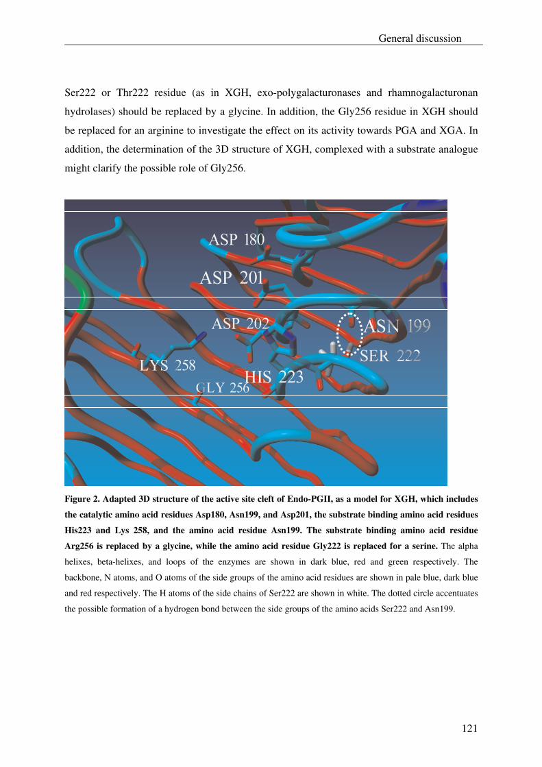
General discussion
121
Ser222 or Thr222 residue (as in XGH, exo-polygalacturonases and rhamnogalacturonan
hydrolases) should be replaced by a glycine. In addition, the Gly256 residue in XGH should
be replaced for an arginine to investigate the effect on its activity towards PGA and XGA. In
addition, the determination of the 3D structure of XGH, complexed with a substrate analogue
might clarify the possible role of Gly256.
Figure 2. Adapted 3D structure of the active site cleft of Endo-PGII, as a model for XGH, which includes
the catalytic amino acid residues Asp180, Asn199, and Asp201, the substrate binding amino acid residues
His223 and Lys 258, and the amino acid residue Asn199. The substrate binding amino acid residue
Arg256 is replaced by a glycine, while the amino acid residue Gly222 is replaced for a serine. The alpha
helixes, beta-helixes, and loops of the enzymes are shown in dark blue, red and green respectively. The
backbone, N atoms, and O atoms of the side groups of the amino acid residues are shown in pale blue, dark blue
and red respectively. The H atoms of the side chains of Ser222 are shown in white. The dotted circle accentuates
the possible formation of a hydrogen bond between the side groups of the amino acids Ser222 and Asn199.
Chapter 6
122
Mode of action of xylogalacturonan hydrolase
The enzyme xylogalacturonan hydrolase was discovered in A. tubingensis as described by
Van der Vlugt-Bergmans et al. (2000). This enzyme was reported to act on the galacturonic
acid backbone by cleaving α-1,4-D galacturonan linkages in an endo-fashion and has a
requirement for xylose side chains (Van der Vlugt-Bergmans et al., 2000). Although this
enzyme has been characterized with respect to its pH optimum, temperature optimum and
kinetic properties (Beldman et al., 2003), it was unclear how the xylose distribution on the
galacturonic acid backbone affects the action of XGH. In order to clarify this, we studied the
mode of action of XGH towards a XGA derived from gum tragacanth (Chapter 3), as well as
towards XGA in the saponified modified ‘hairy’ regions (MHR-s) of apple pectin and potato
pectin (Chapter 4). The demonstration of XGA in cell wall material from different tissues of
Arabidopsis thaliana, which was achieved by the use of XGH, also enables us to make a few
conclusions on the mode of action of this enzyme (Chapter 5). In this thesis research the mode
of action of XGH is investigated by analysis of the structure of XGA oligosaccharides it
releases from these substrates as well as to which extend these oligosaccharides are produced.
As XGA from gum tragacanth was already studied for its degradability by XGH (Beldman et
al., 2003), we commenced our work on this substrate. Upon degradation of a XGA derived
from gum tragacanth (XGA-29) by the enzyme XGH, the released oligosaccharides were first
preparatively separated by HPAEC and subsequently analyzed for their composition and
structure by mass spectrometry (Chapter 3). Then the XGA-oligosaccharide composition was
determined by HPAEC using the XGA-29 digest for assignment of the different peaks. All
XGA products derived from XGA-29 with known structures are summarized in Table 1.
The XGA-29 digest was also used for identification of XGA oligosaccharides in digests of
XGA in MHR-s of apple pectin and potato pectin (Chapter 4), and in cell wall material from
the leaves and the stem of A. thaliana (Chapter 5). The released XGA oligosaccharides with
known structures from these substrates are also summarized in Table 1.
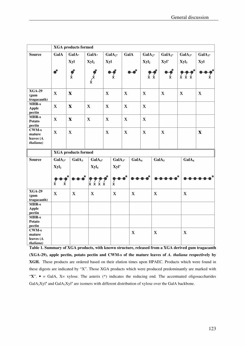
General discussion
123
XGA products formed
Source GalA
GalA-
Xyl
GalA-
Xyl2
GalA2-
Xyl
GalA GalA2-
Xyl2
GalA2-
Xyl’
GalA3-
Xyl3
GalA3-
Xyl
XGA-29 (gum tragacanth)
x x x x x x x x
MHR-s Apple pectin
x x x x x x
MHR-s Potato pectin
x x x x x x
CWM-s mature leaves (A. thaliana)
x x x x x x x
XGA products formed
Source GalA3-
Xyl2
GalA3 GalA4-
Xyl4
GalA3-
Xyl’
GalA4 GalA5 GalA6
XGA-29 (gum tragacanth)
x x x x x x x
MHR-s Apple pectin
MHR-s Potato pectin
CWM-s mature leaves (A. thaliana)
x x x
Table 1. Summary of XGA products, with known structure, released from a XGA derived gum tragacanth
(XGA-29), apple pectin, potato pectin and CWM-s of the mature leaves of A. thaliana respectively by
XGH. These products are ordered based on their elution times upon HPAEC. Products which were found in
these digests are indicated by “X”. Those XGA products which were produced predominantly are marked with
“X”. • = GalA, X= xylose. The asterix (*) indicates the reducing end. The accentuated oligosaccharides
GalA2Xyl’ and GalA3Xyl’ are isomers with different distribution of xylose over the GalA backbone.
X X X
X XX X X X X X X X X
X XX X X X X X * * * * * * *
X XX X XX X X X XX X X X
*
*
*
*
*
*
*
* *
Chapter 6
124
It is clear that XGH tolerates XGAs which vary in their degree of xylose substitution as well
as in the distribution of the xyloses. It can be concluded that XGA-29 has a random
distribution of xyloses over the GalA backbone because of the high number of different XGA
products (twenty, including five isomers) which were formed from this substrate (Chapter 3).
The XGA in MHR-s of apple and potato pectin must have a more regular distribution of
xylose over the GalA backbone as indicated by the limited number of different XGA products
that were produced from these sources. XGA present in the cell wall of the leaves and the
stem of A. thaliana have their xyloses also regularly distributed, however, compared to XGA-
29 and XGA in MHR-s of apple and potato pectin, the degree of xylose substitution of XGAs
from A. thaliana is rather low. This is based on the predominant release of the oligosaccharide
GalA3Xyl as well as the relatively high production of linear GalA oligosaccharides from this
latter source.
Based on the products formed, a Xyl:GalA ratio between 1:2 to 1:3 can be estimated for
XGA-29 (see Table 1) while a Xyl:GalA ratio of almost 1:1 is assessed for XGA in MHR-s of
apple pectin. This latter ratio can also be estimated for XGA in MHR-s in potato pectin,
because the XGA oligosaccharide production from this pectin source was similar to that of
apple pectin. The XGA present in the cell wall of leaves and the stem of A. thaliana may have
a Xyl:GalA ratio between 1:4 to 1:5.
Analysis of the XGA products released from XGA-29 and from XGA in MHR-s of apple and
potato pectin shows that XGH prefers to act between two xylosylated GalA units, as deduced
from the high production of the disaccharide GalAXyl (Chapter 3). The formation of XGA
oligosaccharides that have no xylose substitution at the non-reducing end or at the reducing
end, such as GalA3Xyl, and GalA3Xyl’ (see Table 1), show that XGH can also act between
two GalA residues of which only one is xylosylated. In addition, XGH also released GalA and
linear GalA oligosaccharides, which were significantly present in digests from XGA-29 and
in particular in XGA digests from the cell wall of the mature leaves (see Fig. 2 of Chapter 5)
and the stem of A. thaliana. Although this suggests that XGH can also act between two
unsubstituted GalA residues, no XGA oligosaccharides have been identified yet which only
contain xylose substitution at the internal GalA residue(s). It has also been demonstrated from
the product progression profiles (Chapter 3) that linear GalA oligosaccharides were not
further degraded by XGH, as well as that polygalacturonic acid (PGA) is hardly degraded by
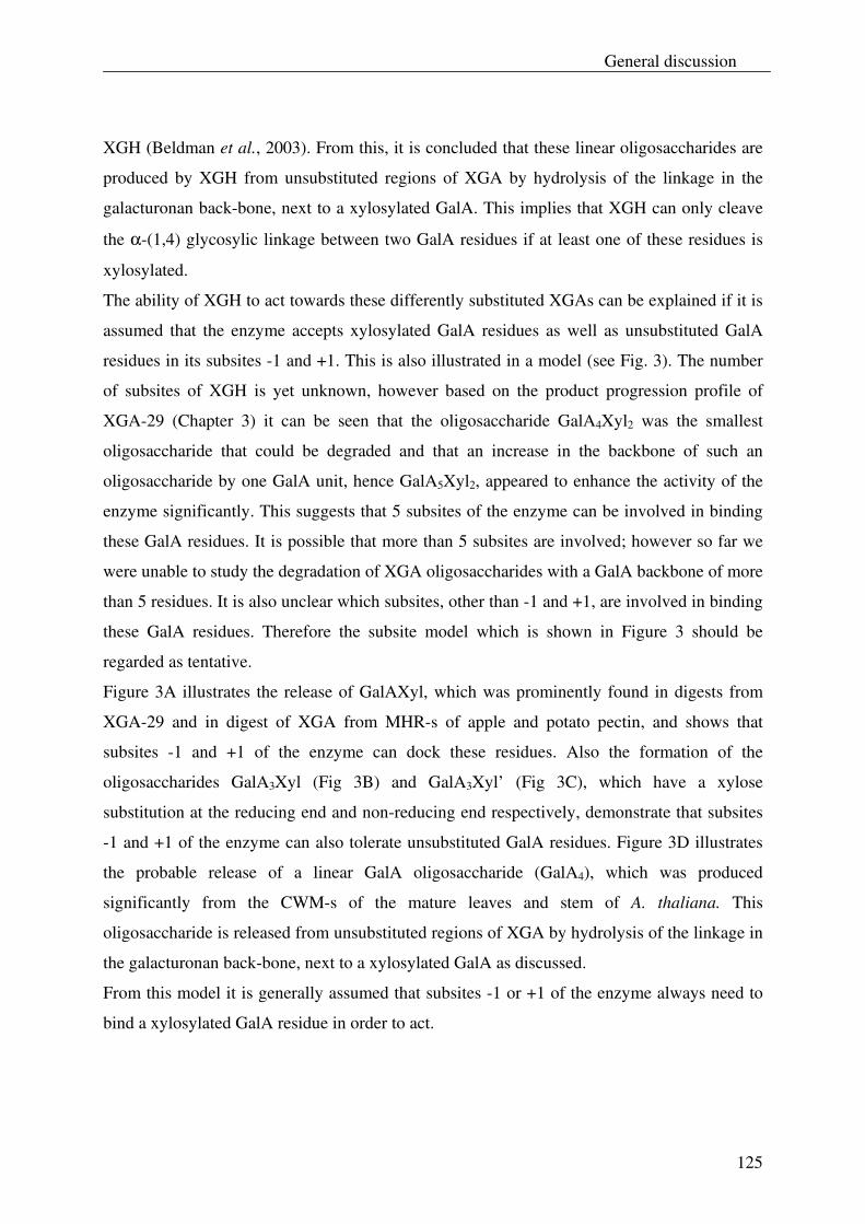
General discussion
125
XGH (Beldman et al., 2003). From this, it is concluded that these linear oligosaccharides are
produced by XGH from unsubstituted regions of XGA by hydrolysis of the linkage in the
galacturonan back-bone, next to a xylosylated GalA. This implies that XGH can only cleave
the α-(1,4) glycosylic linkage between two GalA residues if at least one of these residues is
xylosylated.
The ability of XGH to act towards these differently substituted XGAs can be explained if it is
assumed that the enzyme accepts xylosylated GalA residues as well as unsubstituted GalA
residues in its subsites -1 and +1. This is also illustrated in a model (see Fig. 3). The number
of subsites of XGH is yet unknown, however based on the product progression profile of
XGA-29 (Chapter 3) it can be seen that the oligosaccharide GalA4Xyl2 was the smallest
oligosaccharide that could be degraded and that an increase in the backbone of such an
oligosaccharide by one GalA unit, hence GalA5Xyl2, appeared to enhance the activity of the
enzyme significantly. This suggests that 5 subsites of the enzyme can be involved in binding
these GalA residues. It is possible that more than 5 subsites are involved; however so far we
were unable to study the degradation of XGA oligosaccharides with a GalA backbone of more
than 5 residues. It is also unclear which subsites, other than -1 and +1, are involved in binding
these GalA residues. Therefore the subsite model which is shown in Figure 3 should be
regarded as tentative.
Figure 3A illustrates the release of GalAXyl, which was prominently found in digests from
XGA-29 and in digest of XGA from MHR-s of apple and potato pectin, and shows that
subsites -1 and +1 of the enzyme can dock these residues. Also the formation of the
oligosaccharides GalA3Xyl (Fig 3B) and GalA3Xyl’ (Fig 3C), which have a xylose
substitution at the reducing end and non-reducing end respectively, demonstrate that subsites
-1 and +1 of the enzyme can also tolerate unsubstituted GalA residues. Figure 3D illustrates
the probable release of a linear GalA oligosaccharide (GalA4), which was produced
significantly from the CWM-s of the mature leaves and stem of A. thaliana. This
oligosaccharide is released from unsubstituted regions of XGA by hydrolysis of the linkage in
the galacturonan back-bone, next to a xylosylated GalA as discussed.
From this model it is generally assumed that subsites -1 or +1 of the enzyme always need to
bind a xylosylated GalA residue in order to act.
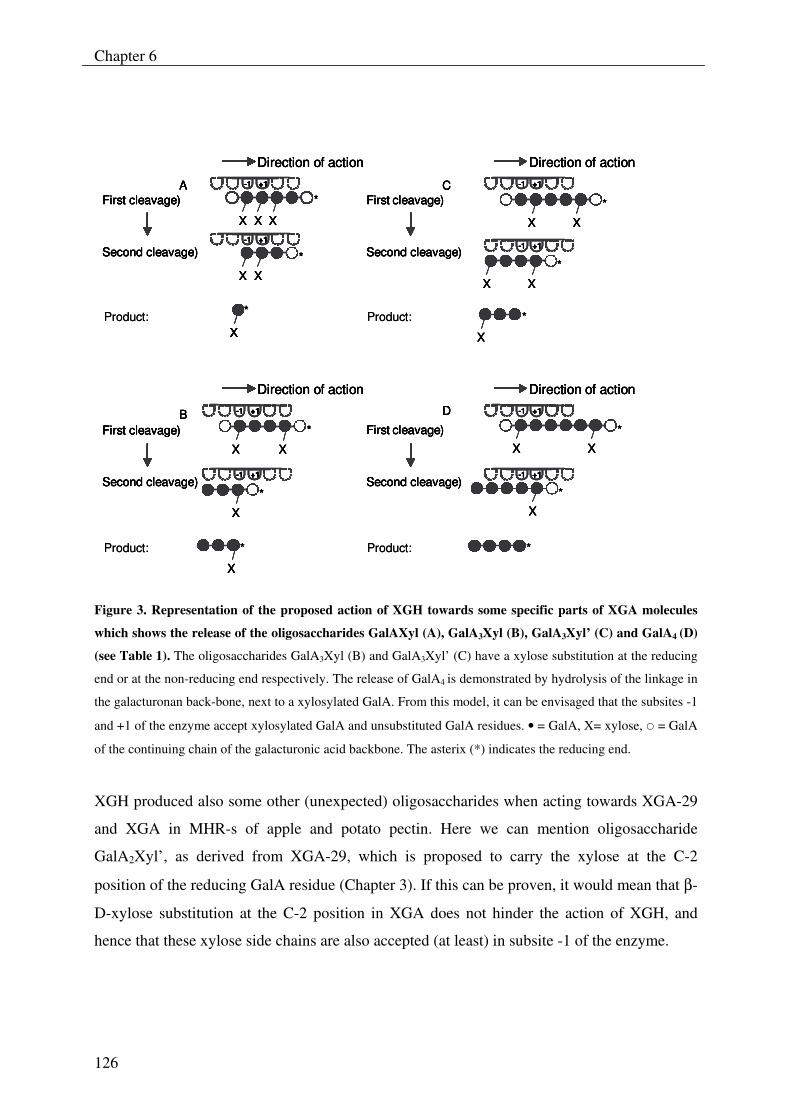
Chapter 6
126
Direction of action
Direction of action
Direction of action
Direction of action
X X X
X X
*
*
X
*
X
X
*
X
*X X
First cleavage)
Second cleavage)
Second cleavage)
First cleavage)
Second cleavage)
First cleavage)
Second cleavage)
A
B
C
D
*
X X
First cleavage)
*
X
*X
Direction of action
Direction of action
Direction of action
Direction of action
X X X
X X
*
*
X
*
X
X
*
X
*X X
First cleavage)
Second cleavage)
Second cleavage)
First cleavage)
Second cleavage)
First cleavage)
Second cleavage)
A
B
C
D
*
X X
First cleavage)
*
X
*X
-1 +1+1
-1 +1+1
-1 +1+1
-1 +1+1
-1 +1+1
-1 +1+1
-1 +1+1
-1 +1+1
XXProduct: *
XX
*Product:
XX
*Product:
*Product:
Direction of action
Direction of action
Direction of action
Direction of action
X X X
X X
*
*
X
*
X
X
*
X
*X X
First cleavage)
Second cleavage)
Second cleavage)
First cleavage)
Second cleavage)
First cleavage)
Second cleavage)
A
B
C
D
*
X X
First cleavage)
*
X
*X
Direction of action
Direction of action
Direction of action
Direction of action
X X X
X X
*
*
X
*
X
X
*
X
*X X
First cleavage)
Second cleavage)
Second cleavage)
First cleavage)
Second cleavage)
First cleavage)
Second cleavage)
A
B
C
D
*
X X
First cleavage)
*
X
*X
-1 +1+1-1 +1+1
-1 +1+1-1 +1+1
-1 +1+1-1 +1+1
-1 +1+1-1 +1+1
-1 +1+1-1 +1+1
-1 +1+1-1 +1+1
-1 +1+1-1 +1+1
-1 +1+1-1 +1+1
XXXXProduct: *
XX
*Product:
XX
*Product:
*Product:
Figure 3. Representation of the proposed action of XGH towards some specific parts of XGA molecules
which shows the release of the oligosaccharides GalAXyl (A), GalA3Xyl (B), GalA3Xyl’ (C) and GalA4 (D)
(see Table 1). The oligosaccharides GalA3Xyl (B) and GalA3Xyl’ (C) have a xylose substitution at the reducing
end or at the non-reducing end respectively. The release of GalA4 is demonstrated by hydrolysis of the linkage in
the galacturonan back-bone, next to a xylosylated GalA. From this model, it can be envisaged that the subsites -1
and +1 of the enzyme accept xylosylated GalA and unsubstituted GalA residues. • = GalA, X= xylose, � = GalA
of the continuing chain of the galacturonic acid backbone. The asterix (*) indicates the reducing end.
XGH produced also some other (unexpected) oligosaccharides when acting towards XGA-29
and XGA in MHR-s of apple and potato pectin. Here we can mention oligosaccharide
GalA2Xyl’, as derived from XGA-29, which is proposed to carry the xylose at the C-2
position of the reducing GalA residue (Chapter 3). If this can be proven, it would mean that β-
D-xylose substitution at the C-2 position in XGA does not hinder the action of XGH, and
hence that these xylose side chains are also accepted (at least) in subsite -1 of the enzyme.
General discussion
127
The enzyme also released the XGA oligosaccharides GalAXyl2 and GalA2Xyl3 from XGA in
MHR-s of apple and potato. Both oligosaccharides contain a dimeric side chain of xylose
from which it is concluded that, besides xylose substituted GalA residues, also dimeric side
chains of xylose are accepted in subsites -1 and +1 of XGH (Chapter 4). The dimeric side
chains of xylose in XGA from MHR-s of apple and potato pectin may be pointed towards the
outside of the binding cleft of XGH which facilitates the formation of the enzyme-substrate
complex. However there may also be another explanation for the acceptance of these dimeric
side chains. The glycine residue, which is present at position 256 of XGH (see Fig. 2) has no
side chains and may therefore create sufficient space in the active site cleft of this enzyme to
harbor, besides Xyl substituted GalA residues, also Xyl-Xyl substituted GalA residues.
The XGH described in this study shows some properties of both an endo-polygalacturonase
and an exo-polygalacturonase. XGH degrades XGA in an endo fashion as demonstrated from
the rapid decrease in the molecular weight of this substrate (Beldman et al., 2003). This is
also observed when polygalacturonic acid (PGA) is degraded by endo-polygalacturonases
(endo-PGs). Besides this, processive endo-PGs are known, like endo-PGI, which
predominantly produce GalA from PGA (Benen et al., 1999; Benen et al., 2003). It is possible
that XGH is also a processive enzyme based on the predominant release of the disaccharide
GalAXyl. Although XGH shows some similar properties with endo-polygalacturonases, these
latter enzymes cannot degrade XGA while XGH and exo-polygalacturonases can. During
degradation of XGA, the enzyme XGH behaves as an exo-acting enzyme, as illustrated by the
stepwise degradation of the XGA oligosaccharide GalA4Xyl3 from the non-reducing end
towards the reducing end (see Fig. 7 of Chapter 3).
Presence of XGA in different tissues of Arabidopsis thaliana
In this thesis it is evidenced that the presence of XGA is not strictly confined to storage
tissues or reproductive organs, and root caps of plants (Albersheim et al., 1996; Willats et al.,
2004). By using the well characterized xylogalacturonan hydrolase (XGH) it is possible to
detect XGA oligosaccharides in pectin fractions obtained from the stem, young leaves and
mature leaves of Arabidopsis thaliana (Chapter 5). No XGA is detected in pectin fractions
from the roots and seeds of this plant species, which is probably due to the absence or a too
Chapter 6
128
low concentration. However, using immunochemical techniques, Willats et al. (2004)
indicated that XGA is present in root caps of A. thaliana. For this, a monoclonal antibody
LM8 was used, which was originally raised against XGA from pea testea (Le Goff et al.,
2001). This anti-body was specifically bound to the root caps of A. thaliana which suggests
that XGA is solely associated with these cells of the root and not with the root body. As these
root caps are known to be loosely attached cells (Willats et al., 2004) it is possible that these
cells were lost during the preparation of CWM from the roots and may therefore, at least
partly, explain why we did not detect XGA in the pectin fraction of this tissue.
In general, XGH can be used as a powerful tool to confirm the presence of XGA in plant cell
walls. In addition, this enzyme also provides clues on the structure of XGA (e.g. degree of
substitution and distribution of xyloses), which is based on the produced XGA
oligosaccharides. However it should be remembered that with this method only the
degradable part of XGA in the plant cell wall is investigated.
Since XGA has been detected for the first time in plant cell walls of the stem, young leaves
and mature leaves of A. thaliana it may open up possibilities to generate anti-bodies towards
XGA in these tissues. From this, the location of XGA in the cell wall of these tissues can be
studied which may help us to understand the role of this polymer. Although the LM8 anti-
body might also be useful for this, this anti-body appears to be specific for highly substituted
XGA (Willats et al., 2004). From this, it is unlikely that it will bind to a XGA with a low
degree of xylose substitution as found in pectin from the stem, young leaves and mature
leaves of A. thaliana. To generate anti-bodies with specificity for such a XGA, it is first
necessary to isolate XGA from the pectin fractions of these tissues. This can be achieved by
enzymatic degradation of non-XGA regions in pectin as described previously for the isolation
of XGA from pea testea (Le Goff et al., 2001) or from the MHR-s of apple pectin (Schols et
al., 1995). Fractions which are enriched in XGA can be identified by XGH degradation, and
can be finally used for the generation of anti-bodies as described previously (Willats et al.,
2004).
Similarly XGA can be demonstrated in cell walls of several tissues of other plants using XGH
or anti-bodies that are raised against XGA (e.g. from A. thaliana) as rapid screening tools.
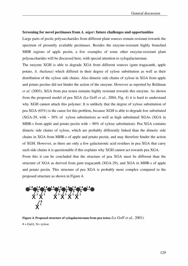
General discussion
129
Screening for novel pectinases from A. niger: future challenges and opportunities
Large parts of pectic polysaccharides from different plant sources remain resistant towards the
spectrum of presently available pectinases. Besides the enzyme-resistant highly branched
MHR regions of apple pectin, a few examples of some other enzyme-resistant plant
polysaccharides will be discussed here, with special attention to xylogalacturonan.
The enzyme XGH is able to degrade XGA from different sources (gum tragacanth, apple
potato, A. thaliana) which differed in their degree of xylose substitution as well as their
distribution of the xylose side chains. Also dimeric side chains of xylose in XGA from apple
and potato pectins did not hinder the action of the enzyme. However as reported by Beldman
et al. (2003), XGA from pea testea remains highly resistant towards this enzyme. As shown
from the proposed model of pea XGA (Le Goff et al., 2004; Fig. 4) it is hard to understand
why XGH cannot attack this polymer. It is unlikely that the degree of xylose substitution of
pea XGA (65%) is the cause for this problem, because XGH is able to degrade low substituted
(XGA-29, with ~ 30% of xylose substitution) as well as high substituted XGAs (XGA in
MHR-s from apple and potato pectin with ~ 90% of xylose substitution). Pea XGA contains
dimeric side chains of xylose, which are probably differently linked than the dimeric side
chains in XGA from MHR-s of apple and potato pectin, and may therefore hinder the action
of XGH. However, as there are only a few galacturonic acid residues in pea XGA that carry
such side chains it is questionable if this explains why XGH cannot act towards pea XGA.
From this it can be concluded that the structure of pea XGA must be different than the
structure of XGA as derived from gum tragacanth (XGA-29), and XGA in MHR-s of apple
and potato pectin. This structure of pea XGA is probably more complex compared to the
proposed structure as shown in Figure 4.
X13
X12
X13
X13
X13
X13
X13
1 4 1 4 1 4 1 4 1 4 1 4 1 4 1 4 1 4
X13
X12
X12
X13
X13
X13
X13
X13
X13
X13
X13
X13
X13
1 4 1 4 1 4 1 4 1 4 1 4 1 4 1 4 1 4
Figure 4. Proposed structure of xylogalacturonan from pea testea (Le Goff et al., 2001).
• = GalA, X= xylose.
Chapter 6
130
Willats et al. (2004) demonstrated that an exo-polygalacturonase from A. aculeatus was
indeed able to release XGA products (GalA and GalAXyl) from pea XGA, however only little
degradation of this polymer was observed. Probably the dimeric side chains of xylose in pea
XGA hinder the action of this enzyme.
XGA regions in soybean are also hardly degraded by XGH (Huisman et al., 2001). This is
probably caused by the presence of oligomeric side chains of xylose in soybean XGA
(Huisman et al., 2001; Nakamura et al., 2002), which hinder the enzyme to interact with this
polymer. In addition it was also reported that further substitution of xylose residues with
fucose may also be responsible for the resistance of soybean XGA towards XGH (Huisman,
2000a). An exo-polygalacturonase from A. aculeatus was shown to be more effective in the
degradation of soybean XGA than XGH. Yet a large part of the XGA remains resistant
towards this exo-polygalacturonase (Huisman et al., 2001) which is probably due to similar
reasons as described for XGH.
Besides XGA, most of the other pectic polysaccharides present in cell walls of soybean
remain resistant to enzymatic degradation. For instance Huisman et al. (2000b) described a
hypothetical structure of the rhamnogalacturonan regions of CDTA-extractable soybean
pectic substances for which no enzymes are currently available to degrade it (Fig. 5).
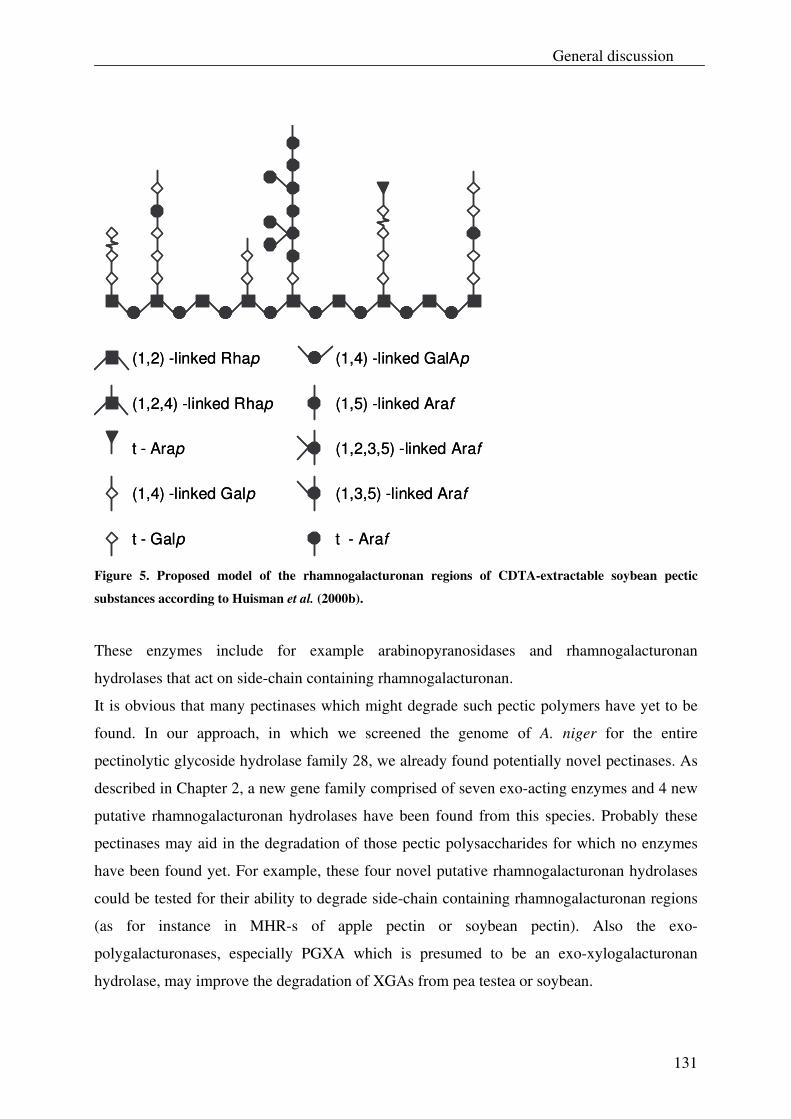
General discussion
131
(1,2,4) -linked Rhap
(1,2) -linked Rhap
t - Arap
(1,4) -linked Galp
t - Galp
(1,4) -linked GalAp
(1,5) -linked Araf
(1,2,3,5) -linked Araf
(1,3,5) -linked Araf
t - Araf
(1,2,4) -linked Rhap
(1,2) -linked Rhap
t - Arap
(1,4) -linked Galp
t - Galp
(1,4) -linked GalAp
(1,5) -linked Araf
(1,2,3,5) -linked Araf
(1,3,5) -linked Araf
t - Araf
Figure 5. Proposed model of the rhamnogalacturonan regions of CDTA-extractable soybean pectic
substances according to Huisman et al. (2000b).
These enzymes include for example arabinopyranosidases and rhamnogalacturonan
hydrolases that act on side-chain containing rhamnogalacturonan.
It is obvious that many pectinases which might degrade such pectic polymers have yet to be
found. In our approach, in which we screened the genome of A. niger for the entire
pectinolytic glycoside hydrolase family 28, we already found potentially novel pectinases. As
described in Chapter 2, a new gene family comprised of seven exo-acting enzymes and 4 new
putative rhamnogalacturonan hydrolases have been found from this species. Probably these
pectinases may aid in the degradation of those pectic polysaccharides for which no enzymes
have been found yet. For example, these four novel putative rhamnogalacturonan hydrolases
could be tested for their ability to degrade side-chain containing rhamnogalacturonan regions
(as for instance in MHR-s of apple pectin or soybean pectin). Also the exo-
polygalacturonases, especially PGXA which is presumed to be an exo-xylogalacturonan
hydrolase, may improve the degradation of XGAs from pea testea or soybean.
Chapter 6
132
In a similar way, the genome of A. niger can be explored for instance for the presence of
assessory enzymes, which can aid in the degradation of arabinogalactan side chains in
rhamnogalacturonan regions of soybean pectin (Fig. 5; Huisman, 2000b), such as
arabinopyranosidases. Similarly the presence of xylosidases and fucosidases in this species
can be investigated which may degrade the polymeric side chains in soybean XGA. As a
consequence such a modified soybean XGA may become better degradable for XGH and/ or
exo-polygalacturonases. A similar result may also be expected for pea XGA.
Alternatively it is possible to grow A. niger specifically on pectic fragments for which no
enzymes have been found yet, for instance for those pectic polysaccharides as described
above. Degradation of these substrates during fermentation can be readily followed by
HPSEC analysis and those pectinases which are responsible for this can be obtained from the
growth media. Also the expression levels of genes, which may correspond to pectinases that
are involved in degradation of these substrates, can be measured by using micro-array
techniques in a similar experiment as described in Chapter 2. Subsequently these genes which
encode for putative novel pectinases can be cloned and expressed in a similar fashion as
performed before (Chapter 2). Finally these (still putative) expressed pectinases can be
biochemically identified and characterized by using specific pectic substrates.
These are just a few examples of this approach to search for those pectinases that are still
needed for the complete degradation of pectin from different plant sources. This underpins the
powerful use of the genome of A. niger to screen for such enzymes. These newly found
enzymes from A. niger, which have a GRAS status, can be readily used for industrial
applications.
Although A. niger is a potential source for novel pectinases, it cannot be excluded that this
species lacks pectinases that are still required to fully degrade pectic material. It may therefore
also be useful to investigate (novel) pectinases in other (fungal) sources such as Aspergillus
oryzae. This species also has the advantage of having a GRAS status (Tailor and Richardson,
1979) as well as that its genome sequence is known (Coutinho and Henrissat, 1999). As A.
oryzae is commonly used for the fermentation of soybeans (Hong et al., 2004; Ling and Chou,
1996), to produce soy sauce, it may be an interesting source for pectinases that can degrade
soybean pectin. In addition, in principle all other organisms of which the genome sequence is
known can be mined for novel pectinases.
General discussion
133
Ultimately, by obtaining the complete array of pectinases we can improve formulation of
tailor-made enzyme preparations. As a result, pectins can be modified in a controlled manner
which can be subsequently used for particular food applications.
Chapter 6
134
Albersheim, P., Darvill, A.G., O'Neill, M.A., Schols, H.A., and Voragen, A.G.J. 1996. An Hypothesis: The Same Six Polysaccharides are Components of the Primary Cell Walls of All Higher Plants, p. 47-55, In J. Visser, Voragen, A.G.J., ed. Pectins and Pectinases. Elsevier Science.
Armand, S., Wagemaker, M.J.M., Sánchez-Torres, P., Kester, H.C.M., van Santen, Y., Dijkstra, B.W., Visser, J., Benen, J.A.E. 2000. The Active Site Topology of Aspergillus niger Endopolygalacturonase II as Studied by Site-directed Mutagenisis. The Journal of Biological Chemistry 275:691-696.
Beldman, G., Vincken, J.-P., Schols, H.A., Meeuwsen, P.J.A., Herweijer, M., Voragen, A.G.J. 2003. Degradation of differently substituted Xylogalacturonans by Endoxylogalacturonan Hydrolase and Endopolygalacturonases. Biocatalysis and Biotransformation 21:189-198.
Benen, J.A.E., Kester, H.C.H., Visser, J. 1999. Kinetic characterization of Aspergillus niger N400 endopolygalacturonases I, II and C. European Journal of Biochemistry 259:72-80.
Benen, J.A.E., van Alebeek, G.-J.W.M., Voragen, A.G.J., Visser, J. 2003. Mode of Action Analysis And Structure-Function Relationships Of Aspergillus Niger Pectinolytic Enzymes, p. 235-256, In A.G.J. Voragen, Schols, H., Visser, R., ed. Advances in Pectin and Pectinase Research. Kluwer Academic Publishers, Dordrecht.
Cho, S., S. Lee, W. Shin, S.W. Cho, S. Lee, and W. Shin. 2001. The X-ray structure of Aspergillus aculeatus polygalacturonase and a modeled structure of the polygalacturonase-octagalacturonate complex. Journal of Molecular Biology 311:863-878.
Coutinho, P.M., Henrissat, B. 1999. Carbohydrate-active enzymes: an integrated database approach, p. 3-12, In H.J. Gilbert, Davies, G., Henrissat, B., and Svensson, B., ed. Recent Advances in Carbohydrate Bioengineering. The Royal Society of Chemistry, Cambridge.
Herweijer, M.A., Vincken, J.-P., Meeuwsen, P.J.A, van der Vlugt-Bergmans, C.J.B., Beldman, G., van Ooyen, A.J.J., Voragen, A.G.J. 2003. Endo-Xylogalacturonan Hydrolase, p. 257-266, In A. G. J. Voragen, Schols, H., Visser, R., ed. Advances in Pectin and Pectinase Research. Kluwer Academic Publishers, Dordrecht.
Hong, K.-J., Lee, C.-H., Kim, S.W. 2004. Aspergillus oryzae GB-107 Fermentation Improves Nutritional Quality of Food Soybeans and Feed Soybean Meals. Journal of Medicinal Food 7:430-435.
Huisman, M.M.H. 2000a. Elucidation of the chemical fine structure of polysaccharides from soybean and maize kernel cell walls. pp. 159, Wageningen University, Wageningen.
Huisman, M.M.H., Brüll, L.P., Thomas-Oates, J.E., Haverkamp, J. 2000b. The occurrence of internal (1, 5)-linked arabinofuranose and arabinopyranose residues in arabinogalactan side chains from soybean pectic substances. Carbohydrate Research 330:103-114.
Huisman, M.M.H., Fransen, C.T.M., Kamerling, J.P., Vliegenthart, J.F.G., Schols, H.A., Voragen, A.G.J. 2001. The CDTA-soluble pectic substances from soybean meal are composed of rhamnogalacturonan and xylogalacturonan but not homogalacturonan. Biopolymers 58:279-294.
Le Goff, A., C.M.G.C. Renard, E. Bonnin, and J.-F. Thibault. 2001. Extraction, purification and chemical characterisation of xylogalacturonans from pea hulls. Carbohydrate Polymers. 45:325-334.
General discussion
135
Ling, M.-Y., and Chou, C.-C. 1996. Biochemical changes during the preparation of soy sauce koji with extruded and traditional raw materials. International Journal of Food Science and Technology 31:511-517.
Markovic, O., Janecek, S. 2001. Pectin degrading glycoside hydrolases of family 28: sequence-structural features, specificities and evolution. Protein Engineering 14:615-631.
Mutter, M., Beldman, G., Pitson, S.M., Schols, H.A. and Voragen, A.G.J. 1998a. Rhamnogalacturonan α-D-Galactopyranosyluronohydrolase. An enzyme that specifically removes the terminal nonreducing galacturonosyl residue in rhamnogalacturonan regions of pectin. Plant Physiology 117:153-163.
Mutter, M., Beldman, G., Schols, H.A. and Voragen, A.G.J. 1994. Rhamnogalacturonan α-L-rhamnopyranosylhydrolase. A novel enzyme specific for the terminal non-reducing rhamnosyl unit in rhamnogalacturonan regions of pectin. Plant Physiology 106:241-250.
Mutter, M., Renard, C.M.G.C., Beldman, G., Schols, H.A., Voragen, A.G.J. 1998b. Mode of action of RG-hydrolase and RG-lyase toward rhamnogalacturonan oligomers. Characterization of degradation products using RG-rhamnohydrolase and RG-galacturonohydrolase. Carbohydrate Research 311:155-164.
Nakamura, A., Furuta, H., Maeda, H., Takao, T., Nagamatsu, Y. 2002. Analysis of the Molecular Construction of Xylogalacturonan Isolated from Soluble Soybean Polysaccharides. Bioscience Biotechnology and Biochemistry. 66:1155-1158.
Parenicová, L., Benen, J.A.E., Kester, H.C.M., Visser, J. 1998. pgaE encodes a fourth member of the endopolygalacturonase gene family from Aspergillus niger. European Journal of Biochemistry 251:72-80.
Parenicová, L., Kester, H.C.M., Benen, J.A.E., Visser, J. 2000. Characterization of a novel endopolygalacturonase from Aspergillus niger with unique kinetic properties. FEBS Letters 467:333-336.
Schols, H.A., E.J. Bakx, D. Schipper, and A.G.J. Voragen. 1995. A xylogalacturonan subunit present in the modified hairy regions of apple pectin. Carbohydrate Research. Dec 279:265-279.
Schols, H.A., Posthumus, M.A., Voragen, A.G.J. 1990. Structural features of hairy regions of pectins isolated from apple juice produced by the liquefaction process. Carbohydrate Research 206:117-129.
Schols, H.A., Voragen, A.G.J., Colquhoun, I.J. 1994. Isolation and characterization of rhamnogalacturonan oligomers, liberated during degradation of pectic hairy regions by rhamnogalacturonase. Carbohydrate Research 256:97-111.
Sykerbuyk, M.E.G., Kester, H.C.M., Schaap, P.J., Stam, H., Musters, W., Visser, J. 1997. Cloning and characterization of two rhamnogalacturonan hydrolase genes from Aspergillus niger. Applied and Environmental Microbiology 63:2507-2515.
Tailor, M.J., and Richardson, T. 1979. Application of microbial enzymes in food systems and in biotechnology. Advances in Applied Microbiology 25:7-35.
Van der Vlugt-Bergmans, C.J.B., Meeuwsen, P.J.A., Voragen, A.G.J., and van Ooyen, A.J.J. 2000. Endo-Xylogalacturonan hydrolase, a Novel Pectinolytic Enzyme. Applied and Environmental Microbiology 66:36-41.
Willats, W., McCartney, L., Steele-King, C., Marcus, S., Mort, A., Huisman, M., van Alebeek, G.-J., Schols, H.A., Voragen, A.G.J., le Goff, A., Bonnin, E., Thibault, J.-F.,
Chapter 6
136
Knox, J.P. 2004. A xylogalacturonan epitope is specifically associated with plant cell detachment. Planta 218:673-681.
General discussion
137

Chapter 6
138
Summary
139
Summary
The subject of this thesis was to biochemically identify and characterize (novel) pectin
degrading enzymes that belong to the glycoside hydrolase family 28 of Aspergillus niger.
These pectin degrading enzymes became available by exploring the genome of A. niger for
these enzymes, using functional genomics techniques.
Biochemical identification and characterization of these enzymes was executed by analysis of
their activity towards a set of pectinolytic substrates. Special attention has been given to the
characterization of the xylogalacturonan hydrolase towards a XGA derived from gum
tragacanth and XGA from other plant sources.
In Chapter 2, a complete inventory of the pectinolytic glycoside hydrolase family 28 of A.
niger is presented, which was obtained by applying bioinformatics tools on the genome
sequence of this species. Besides the seven existing endo-polygalacturonases and two
rhamnogalacturonases in A. niger, a new gene group of seven exo-acting enzymes was found,
which included four exo-polygalacturonan hydrolases and three putative exo-
rhamnogalacturonan hydrolases. Also four novel putative rhamnogalacturonan hydrolases
were found in this species. Three exo-acting enzymes PGXA, PGXB and PGXC were
biochemically identified by using PGA and XGA as substrates for which it was demonstrated
that PGXB and PGXC act as an exo-polygalacturonase, while PGXA behaves as an exo-
xylogalacturonan hydrolase.
The xylogalacturonan hydrolase (XGH) was investigated for its mode of action towards a
XGA derived from gum tragacanth (XGA-29; Chapter 3). This was achieved by analysis of
the structure of the XGA fragments released from XGA-29 as well as to which extend these
oligosaccharides were produced. Upon degradation of XGA-29, several XGA
oligosaccharides were formed with different galacturonic acid/ xylose ratios, including five
oligosaccharide isomers.
Although XGH is known to be an endo-acting enzyme, product progression profiling
illustrated a predominant production of the di-saccharide GalAXyl, which also indicates an
exolytic action of this enzyme. The latter was also supported when XGH was investigated for
its action towards a defined XGA oligosaccharide GalA4Xyl3 in a time course experiment.
Summary
140
From this, XGH was demonstrated to act from the non-reducing end towards the reducing end
of this oligosaccharide.
Based on the released XGA products from XGA-29, in particular GalAXyl, it was shown that
although XGH prefers to act between two xylosidated GalA units, it can also tolerate
unsubstituted GalA units in its -1 and +1 subsites.
The action of XGH was also investigated towards XGAs in the saponified modified ‘hairy’
regions (MHR-s) of apple pectin and potato pectin (Chapter 4). Product analysis in digests of
these XGAs illustrated that the degradable parts of XGA from both sources have a similar
xylose side chain distribution. Once again, the di-saccharide GalAXyl was predominantly
formed which underpins the preference of XGH to act between two xylosylated GalA units as
well as its exolytic behavior. A significant lower number of different XGA products was
released from these sources, compared to the number of different XGA products derived from
XGA-29. This suggests a regular distribution of xylose over the galacturonan backbone in the
degradable parts of XGA in MHR-s of apple and potato pectin. Also the oligosaccharides
GalAXyl2 and GalA2Xyl3 were observed in the digests of XGA from MHR-s of apple and
potato pectin, which shows the presence of dimeric side chains of xylose (Xyl-Xyl) in these
sources. These side chains did not hinder the action of XGH which points out that this
enzyme can also accept Xyl-Xyl substituted GalA residues in subsites -1 and +1.
With the aid of XGH it was possible to detect XGA in different tissues of Arabidopsis
thaliana (Chapter 5). This was achieved by extraction of the pectin from the seeds, roots,
stem, young leaves and mature leaves of this species, followed by treatment of these pectin
extracts with XGH. From this, XGA oligosaccharides were only detected in the stem and the
leaves of A. thaliana. The estimated amount of XGA accounted for approximately 2.5 %, 7 %
and 6 % (w/w) of the total carbohydrate in the pectin fraction of the stem, young leaves and
mature leaves respectively. A low number of different XGA oligosaccharides was detected in
pectin hydrolysates from the cell wall of different tissues of A. thaliana. This suggests a
regular distribution of xylose in the degradable parts of XGA of these tissues, which was
similar to our findings for XGA in MHR-s from apple and potato pectin. Also the XGA from
these A. thaliana pectins contained a lower degree of xylose substitution compared to XGA in
MHR-s from apple and potato pectin. The oligosaccharide GalA3Xyl was predominantly
released from these A. thaliana pectins, and the amount of linear GalA oligosaccharides in
Summary
141
relation to branched GalA oligosaccharides was higher in these A. thaliana pectins than in
MHR-s from apple and potato.
The results described in this thesis are discussed in Chapter 6 with emphasis on the mode of
action of XGH. Also some speculations are made on the roles of some partial conserved
amino acids in the pectinolytic glycoside hydrolase family 28 of A. niger. In addition, the use
of XGH as a functional tool for the detection of XGA in other plant varieties is discussed.
Finally the usefulness of our methods to explore the genome of A. niger for novel pectin
degrading enzymes is discussed. Our approach (Chapter 2) is attractive to explore enzymes
that may degrade those pectic polysaccharides, as derived for instance from soya and pea,
which are still highly resistant towards the collection of pectin modifying enzymes as
available today. As a consequence, the improved degradation of these polysaccharides may
improve their utilization in industrial applications.

Summary
142
Samenvatting
143
Samenvatting Het in dit proefschrift beschreven onderzoek betrof de biochemische identificatie en
karakterisatie van (nieuwe) pectine modificerende enzymen die behoren tot de familie 28
glycosyl hydrolasen van Aspergillus niger. Deze pectine modificerende enzymen kwamen tot
onze beschikking door het genoom van A. niger te onderzoeken naar deze enzymen, met
gebruikmaking van “functional genomics” technieken. Deze enzymen werden vervolgens
biochemisch geïdentificeerd en gekarakteriseerd aan de hand van hun activiteit op bepaalde
pectine substraten. Met nadruk was er aandacht besteed aan de karakterisatie van het enzym
xylogalacturonaan hydrolase op een xylogalacturonaan (XGA) dat verkregen was van gum
tragacanth, en op XGA uit andere plantenbronnen.
In Hoofdstuk 2 is de gehele inventaris van familie 28 glycosyl hydrolasen van A. niger
gepresenteerd. Dit was gerealiseerd door gebruikmaking van “bioinformatics tools” op het
genoom van A. niger. Naast de zeven bestaande endo-polygalacturonaan hydrolasen en de
twee rhamnogalacturonaan hydrolasen van A. niger was er een nieuwe groep van zeven exo-
enzymen gevonden. Deze nieuwe groep bestond uit vier exo-polygalacturonaan hydrolasen en
drie vermoedelijke exo-rhamnogalacturonaan hydrolasen. Er waren ook vier nieuwe
mogelijke rhamnogalacturonaan hydrolasen gevonden. Drie exo-enzymen (PGXA, PGXB en
PGXC) werden biochemisch geïdentificeerd door hun activiteit te toetsen op
polygalacturonaan (PGA) en xylogalacturonaan (XGA). Daaruit is gebleken dat PGXB en
PGXC beiden werken als een exo-polygalacturonaan hydrolase, terwijl PGXA zich als een
exo-xylogalacturonaan hydrolase gedraagt.
Xylogalacturonaan hydrolase (XGH) was bestudeerd voor zijn “mode of action” op
xylogalacturonaan dat verkregen was uit gum tragacanth (XGA-29; Hoofdstuk 3). Dit werd
gerealiseerd door de structuur van de gevormde XGA oligosacchariden te bepalen, evenals de
mate waarin deze oligosacchariden werden gevormd. Na behandeling van XGA-29 met XGH
werden er verscheidene XGA oligosacchariden geproduceerd met verschillende
galacturonzuur/ xylose ratio’s, inclusief vijf isomeren van deze oligosacchariden.
Ondanks dat XGH bekend staat als een endo-enzym, werd, zoals aangegeven in de product
progressie profielen, het di-saccharide GalAXyl voornamelijk gevormd waaruit blijkt dat dit
enzym ook exolytische eigenschappen bezit. Dit laatste werd nog eens bevestigd door de
Samenvatting
144
wijze waarop XGH het XGA oligosaccharide GalA4Xyl3 afbrak. Het enzym brak dit
oligosaccharide af in de richting van het niet-reducerend eind naar het reducerend eind.
Gebaseerd op de gevormde produkten uit XGA-29, met GalAXyl in het bijzonder, werd het
duidelijk dat hoewel XGH prefereert om twee galacturonzuur eenheden te splitsen die beiden
gesubstitueerd zijn met een xylose, dit enzym ook niet gesubstitueerde galacturonzuur
eenheden kan accepteren in zijn -1 en +1 subsites.
De activiteit van XGH was ook onderzocht op XGA in verzeept “modified hairy regions”
(MHR-s) van appel en aardappel pectine (Hoofdstuk 4). Uit product analyse van digesten van
deze xylogalacturonanen was gedemonstreerd dat de afbreekbare delen van XGA uit beide
bronnen eenzelfde distributie van xylose zijketens hebben. Ook werd het di-saccharide
GalAXyl voornamelijk geproduceerd wat bevestigt dat XGH prefereert om twee ge-
xylosyleerde galacturonzuur eenheden te splitsen en dat dit enzym exolytische eigenschappen
bezit. XGH produceerde een laag aantal verschillende XGA oligosacchariden uit MHR-s van
appel en aardappel pectine, vergeleken bij het aantal verschillende XGA oligosacchariden dat
werd geproduceerd uit XGA-29. Daaruit kan worden verondersteld dat de afbreekbare delen
van XGA uit MHR-s van appel en aardappel pectine een regelmatige distributie van xylose
zijketens hebben.
Ook werden de oligosacchariden GalAXyl2 en GalA2Xyl3 gevonden in digesten van XGA uit
MHR-s van appel en aardappel pectine dat wees op de aanwezigheid van xylose di-meren als
zijketens in XGA. Deze zijketens hadden geen remmende werking op de activiteit van XGH
waaruit wordt verondersteld dat het enzym ook Xyl-Xyl gesubstitueerde galacturonzuur
eenheden kan accepteren in zijn -1 en +1 subsites.
Met behulp van XGH was het mogelijk om XGA aan te kunnen tonen in verschillende
weefsels van Arabidopsis thaliana (Hoofdstuk 5). Dit werd gedaan door eerst pectine selectief
te extraheren uit de zaden, wortel, stam, jonge bladeren, en volwassen bladeren van deze
plant. De pectine extracten uit deze weefsels werden vervolgens behandeld met het enzym.
Hieruit werd geconstateerd dat alleen XGA oligosacchariden werden gevormd uit pectine
extracten van de stam en de bladeren van A. thaliana. De geschatte hoeveelheid XGA was
verantwoordelijk voor ongeveer 2.5 %, 7 %, en 6% (w/w) van de totale hoeveelheid
carbohydraat in de pectine extracten van respectievelijk de stam, de jonge bladeren en de
volwassen bladeren.
Samenvatting
145
In de pectine hydrolysaten van de stam, jonge bladeren en volwassen bladeren van A. thaliana
was een laag aantal verschillende XGA oligosacchariden gevonden. Dit wijst uit dat de
afbreekbare delen van XGA, afkomstig uit deze weefsels, een regelmatige distributie van
xylose bezitten, wat vergelijkbaar was met onze bevindingen voor XGA uit MHR-s van appel
en aardappel pectine. Het XGA uit de pectine extracten van A. thaliana had ook een lagere
graad van xylose substitutie vergeleken met XGA uit MHR-s van appel en aardappel pectine.
Het oligosaccharide GalA3Xyl was voornamelijk geproduceerd uit de pectine extracten van A.
thaliana. Ook de hoeveelheid lineaire GalA oligosacchariden in verhouding met vertakte
GalA oligosacchariden was hoger in deze pectine extracten vergeleken met de gevormde
XGA oligosacchariden uit MHR-s van appel en aardappel pectine.
De resultaten in dit proefschrift, in het bijzonder de “mode of action” van XGH, zijn
bediscussieerd in Hoofdstuk 6. Ook is er gespeculeerd over de rol van een paar gedeeltelijk
geconserveerde aminozuren in de familie 28 glycosyl hydrolasen van A. niger. Tevens is het
gebruik van XGH als middel voor de detectie van XGA in andere plantensoorten besproken.
Ten slotte word het nut van onze aanpak, dat beschreven staat in Hoofdstuk 2, om het genoom
van A. niger te onderzoeken naar nieuwe pectine afbrekende enzymen bediscussieerd. Deze
aanpak is aantrekkelijk om enzymen op te sporen die pectines kunnen afbreken, uit
bijvoorbeeld soya en erwt, die momenteel nog steeds moeilijk afbreekbaar zijn. Een
verbetering in de afbraak van zulke pectines heeft als gevolg dat deze pectines beter zouden
kunnen worden gebruikt voor industriële toepassingen.
Samenvatting
146
Acknowledgements
147
Acknowledgements
Beste lezer, wat is de overeenkomst tussen het kopje van een asperge en de stellingen plus het
dankwoord van het proefschrift? Men begint meestal hier mee en het is vaak ook het meest
aantrekkelijke van het geheel. De rest is namelijk wat taaier om door te komen.
Na zo’n vier en een half jaar ben ik er toch in geslaagd een proefschrift te kunnen fabriceren
wat mij natuurlijk nooit is gelukt zonder de hulp en steun van velen om mij heen.
Allereerst bedank ik mijn promoter Fons Voragen die altijd kritisch naar mijn werk heeft
gekeken en mij nog altijd weet te boeien in de wereld van pectines en pectinases. Jij bent een
uitstekende motivator!
Ook wil ik graag mijn co-promoter Gerrit Beldman bedanken voor het corrigeren van mijn
werk en zijn “nuchtere” kritiek op mijn (lange en soms niet “to the point” komende)
zinsconstructies. Ook je positieve kijk op het onderzoek heeft mij veel geholpen om door te
zetten.
Jean-Paul, bedankt dat je mij hebt opgevangen in de eerste zes maanden van mijn AIO-schap
toen Gerrit en Willemiek beiden ziek waren. Willemiek, ook al heb ik niet veel met je te
maken gehad, wil ik je toch bedanken voor alle gezellige gesprekken die ik met je heb gehad
tijdens het werk en de (koffie) pauzes.
Henk, jij hebt ook een flink aandeel geleverd in dit proefschrift. Jouw kritiek heeft veel
bijdrage geleverd aan mijn werk en jij vormde samen met Gerrit en uitstekend “dream” team.
Je bent altijd geïnteresseerd geweest in mijn werk en liet me daarom van tijd tot tijd even
weer herinneren dat ik jou natuurlijk niet buiten mijn werk moest sluiten (het “Denk-aan-
Henk” principe).
Natuurlijk wil ik ook Elena, Peter en Jacques bedanken. Ik heb met plezier met jullie kunnen
samenwerken wat tevens een fantastisch artikel heeft opgeleverd. Harrie (K), ook bedankt
voor je bijdrage hieraan. Jacques, ook al was je niet meer bij ons halverwege het projekt, ik
heb jouw bijdrage aan mijn werk zeer gewaardeerd. Peter en Elena, samenwerken met jullie
ging vaak samen met veel ironie (ook tijdens de project meetings) waar ik zeer veel lol aan
heb beleefd.
Acknowledgements
148
Hanem, I’d also like to thank you for your help and contribution to our publication as well.
Henk-Jan, jij wordt bedankt voor het maken van de plaatjes van de 3D structuur van endo-
PGII voor mijn proefschrift.
Margaret en Edwin, ik bedank jullie voor de (technische) hulp die jullie mij hebben gegeven
bij de massa spectrometrie en HPAEC analyses. Margaret, jij hebt ook een belangrijk aandeel
geleverd bij mijn eerste en tweede publikatie. Mirjam, ook heb ik jouw technische hulp zeer
gewaardeerd tijdens de HPAEC analyses. Jan, bedankt voor je hulp bij de gas chromatografie
analyses en voor de uitstekende dosis humor.
Gerd-Jan, bedankt voor de bijzondere humor en stellingen die je hebt, meestal over vrouwen
(en je eigen vrouw)! Ook je anti-linkse politieke standpunten waren mij niet ontgaan. Hauke,
jij vormt ook een goede partij genoot van Gerd-Jan en ik heb erg genoten van jouw
opmerkingen en humor over de alledaagse dingen (o.a. nederlands zorgstelsel, spelling van
het woord “Voetbal”). Bedankt voor het organiseren van de trip naar Hamburg!
René (V), bedankt voor de gezellige sfeer in ons kantoor. Ik hoop dat je het nu net zo gezellig
hebt bij Henk! Ik bedank je ook voor het organiseren van o.a. de vele wandelweekenden.
Lyn, I also thank you for the nice talks in our office as well as the sweets (chocolate, biscuits)
and tea you shared with us. My gratitude also goes to my students Charlotte, Mauricio and
Stefan. Without their help I could never have come this far in this project.
Graag wil ik ook de gehele organisatie bedanken die het mogelijk heeft gemaakt om een
fantastische AIO reis te kunnen meemaken naar de VS en Japan. Wat heb ik toch geluk gehad
(en velen anderen) om zo iets mee te kunnen maken.
Stephanie (P), Laurice, Natalie, Lyn, Chen, Eva, Hanem, Junrung, Francesca, Mingwei,
Takao, Tai, Carlos, and Yvonne, thank you for the nice talks we had during lunch, coffee
breaks. Valle, Michal, Junrung, Fernando (F), and Fernando (C), I have had a lot of fun with
your jokes and all the internal dinners we had. I hope seeing you again in the future. En
natuurlijk wil ik ook graag Sandra, Wil, Bas & Julia, Koen, Karin, Maaike, Evelien, Aagje,
Ben, Jolan, Toos, René (K), Fons, Jean-Paul, Gerrit (B), Henk, Peter (W), Jan, Peter (I),
Kerensa, Gerrit (v.K), Mark, Harrie (G), Mirjam, Jozef, Raymond, Walter (T), Edwin, Teun,
René (V), Jacques, Margaret, Renate, Neleke, Jendo, Chantal (D), Daan, Jolanda, en Miranda,
bedanken voor de gezellige gesprekken gedurende de (koffie) pauzes en/of de PhD reis (VS,
Japan).
Acknowledgements
149
Julia en Renate, het was leuk om met jullie te kunnen salsa dansen. Misschien doet een
dergelijke gelegenheid zich nog een keer voor.
Ten slotte wil ik graag naast mijn familie ook zeker mijn ouders en mijn broer bedanken voor
hun steun tijdens mijn AIO periode. Ook al hadden jullie niet veel kennis van mijn onderzoek,
jullie hebben me toch altijd vermaakt met vele grappen en grollen over Aspergillus niger
(“zwarte asperges”) wat we even deed beseffen dat er nog meer dingen zijn dan alleen serieus
werk.
Acknowledgements
150
Curriculum Vitae
151
Curriculum Vitae
Joris Zandleven werd geboren op 1 mei 1976 te Eindhoven. Na het behalen van zijn HAVO-
diploma aan het Van Mearlant Lyceum te Eindhoven begon hij met zijn studie Medische
Microbiologie aan de Fontys Hogeschool Eindhoven in 1995. Tijdens deze studie heeft hij
stage gelopen bij Numico Research B.V. te Wageningen en een afstudeerstage gelopen aan
het EC Slater instituut te Amsterdam. In juli 1999 heeft hij zijn Bachelor diploma behaald.
Vervolgens begon hij in augustus 2000 met een MSc studie aan de Strathclyde University in
Glasgow waarbij hij een afstudeerstage heeft gelopen aan het Scottisch Crops Research
Institute in Dundee. In september 2001 heeft hij zijn MSc diploma behaald.
In de periode van april 2002 tot en met augustus 2006 deed hij een promotie-onderzoek bij de
leerstoelgroep Levensmiddelenchemie aan de Wageningen Universiteit, onder begeleiding
van Prof. Dr. A.G.J. Voragen en Dr. Gerrit Beldman. Dit onderzoek dat werd uitgevoerd in
deze periode is beschreven in dit proefschrift.
Curriculum Vitae
152
List of Publications
153
List of Publications
Full papers
• Zandleven J., Beldman, G., Bosveld, M., Benen, J., Voragen, A. (2005) Mode of
action of Xylogalacturonan hydrolase towards xylogalacturonan and xylogalacturonan
oligosaccharides. Biochemical Journal 387, 719-725.
• Zandleven, J., Beldman, G., Bosveld, M., Schols, H.A., Voragen, A.G.J. (2006)
Enzymatic degradation studies of xylogalacturonans from apple and potato, using
xylogalacturonan hydrolase. Carbohydrate Polymers 65, 495-503
• Zandleven, J., Oxenbøll Sørensen, S., Harholt, J., Beldman, B., Schols, H.A., Scheller,
H.V., Voragen, A.G.J. Xylogalacturonan exists in cell walls from various tissues of
Arabidopsis thaliana. Submitted to phytochemistry
• Martens-Uzunova, E1., Zandleven, J.S1., Benen, J.A.E., Awad, H., Kools, H.J.,
Beldman, G., Voragen, A.G.J., van den Berg, J.A., and Schaap, P.J. Exo-Acting
Glycoside Hydrolases of Aspergillus niger Involved in Pectin Degradation.
Biochemical Journal. In press. 1Both authors contributed equally to this paper.
List of Publications
154
Abstracts
• Zandleven, J., van Oostveen, W., Vincken, J.-P., Beldman, G., Voragen, A.G.J. (2002)
Disclosing the carbohydrate modifying network of Aspergillus niger by functional
genomics. VLAG Summer Course Glycosciences, Wageningen.
• Zandleven, J., van Oostveen, W., Awad, H., Beldman, G., Voragen, A.G.J. (2004)
Mode of action of xylogalacturonan hydrolase towards xylogalacturonan and
xylogalacturonan oligomers. X Cell wall meeting, Sorrento, Italy.
• Sørensen, S.O., Zandleven, J., Harholt, J., Bernal, A.J., Jensen, J.K., Pauly, M.,
Willats, W.G.T., Beldman, G., and Scheller, H.V. Identification of a putative
xylosyltransferase involved in formation of xylogalacturonan in Arabidopsis thaliana.
At URL: http://xyloglucan.prl.msu.edu/cgi-bin/meetingAbstractpub.pl?email=
[email protected]&order=1&lName=S%C3%B8rensen&fName=Susanne%20Oxen
b%C3%B8ll
Addendum
155
Addendum
The work described in this thesis has been carried out with the financial support of Senter IOP
(“Innovatief Onderzoeks Programma”; The Netherlands) with the project number IGE01021.
Addendum
156
157
Overview of completed training activities
Discipline specific activities:
Courses:
VLAG Summer school glycosciences (Wageningen, June 2002) Scientific Writing (2004) Food Enzymology (2004)
Conferences:
Carbohydrate Bioengineering meeting (Groningen, 2003) Seminar ICAT technique (Leiden, 2002) Seminar 2D gel electrophoresis (Utrecht, 2002) X-Cell Wall meeting (Sorrento, Italy, 2004) Seminar Millipore (2004) Seminar Leuven University (2004) General Courses: Food Chemistry PhD trip (USA, November 2002) Food Chemistry PhD trip (Japan, December 2004) PhD student week VLAG (2003) Career Perspectives Course, Meijer & Meijaard, Wageningen, (2005) Additional activities: Preparation PhD proposal Food Chemistry Seminars (Wageningen, 2002-2006) Food Chemistry Colloquia (Wageningen, 2002-2006) Carbnet meetings (Amersfoort, Utrecht, Wageningen, Haren, 2002-2006)

158

159

160