Embed Size (px)
Citation preview
31
http://journals.tubitak.gov.tr/biology/
Turkish Journal of Biology Turk J Biol(2015) 39: 31-48© TÜBİTAKdoi:10.3906/biy-1402-69
Identification and pathogenicity of bacteria in the Mediterranean corn borerSesamia nonagrioides Lefebvre (Lepidoptera: Noctuidae)
Ardahan ESKİ1, Filiz ÖZKAN ÇAKICI1, Mustafa GÜLLÜ2, Hacer MURATOĞLU3, Zihni DEMİRBAĞ1, İsmail DEMİR1,*1Department of Biology, Faculty of Sciences, Karadeniz Technical University, Trabzon, Turkey
2Department of Entomology, Biological Control Research Station, Adana, Turkey3Department of Molecular Biology and Genetics, Faculty of Sciences, Karadeniz Technical University, Trabzon, Turkey
* Correspondence: [email protected]
1. IntroductionCorn is the largest grown cereal crop in the world with doubled grain yield per unit area compared to wheat and barley. Although corn production holds second place in Mediterranean agricultural production, there has been a sharp decline in this value due to infestation by the Mediterranean corn borer (Özcan, 2009). The Mediterranean corn borer, Sesamia nonagrioides (Lefebvre) (Lepidoptera: Noctuidae), is a major multivoltine pest of maize in Mediterranean countries (Tsitsipis, 1988; Alexandri and Tsitsipis, 1990). S. nonagrioides is a polyphagous species with a fairly wide range of host plants, including corn, sorghum, millet, rice, sugar cane, grasses, melon, asparagus, palms, and banana (Uygun and Kayapınar, 1993; Sannino et al., 2004). The population levels of this species, which has considerable potential to establish itself in an area and become abundant, may therefore depend on the abundance of these hosts (Eizaguirre and Fantinou, 2012). If insecticides are not
applied, losses could rise to 100% in late and second crop production (Alexandri and Tsitsipis, 1990).
Application of chemical insecticides against this harmful insect is recommended 2–3 times per growing season. However, yield losses could rise to 30% during seasons with severe insect outbreaks, even after 4–5 insecticide applications (Özcan, 2009). As well as causing significant maize crop losses, the insects reduce the nitrogen/protein content of grain by tunneling into the stem and cobs, where they are likely to interfere with the uptake of plant nutrients (Bayram, 2003). Furthermore, mycotoxigenic fungi associated with the pest often invade wound sites and can greatly depreciate the crop value (Avantaggiato et al., 2003). Although several control methods have been previously applied to this pest, damage is still pervasive. Chemical insecticides have been frequently used against it. While chemicals decrease pest population, they cause major threats to the environment and human health. Mechanical, cultural, and biological
Abstract: Sesamia nonagrioides (Lep.: Noctuidae) is one of the most serious pests of corn in Turkey and other Mediterranean countries. Although various cultural, chemical, and biological methods are used to control this pest, its damage still continues in all Mediterranean countries. In this study, to find an effective bacterium that can be used as a biocontrol agent against S. nonagrioides, we isolated 15 bacteria from S. nonagrioides larvae and evaluated the larvicidal potency of all isolates on the pest. According to their morphological, physiological, biochemical, and molecular properties, the isolates were identified as Achromobacter insolitus (Sn1), Morganella morganii (Sn2), Klebsiella pneumoniae (Sn3), Citrobacter freundii (Sn4), Arthrobacter protophormiae (Sn5), Chryseobacterium indologenes (Sn6), Bacillus thuringiensis (Sn7), Bacillus safensis (Sn8), Bacillus thuringiensis (Sn9), Bacillus thuringiensis (Sn10), Klebsiella pneumoniae (Sn11), Staphylococcus sciuri (Sn12), Enterobacter kobei (Sn13), Serratia marcescens (Sn14), and Microbacterium arborescens (Sn15). The results of the larvicidal activities of these isolates indicated that the mortality value obtained from all treatments varied from 25% to 93%, reaching 93% with B. thuringiensis (Sn10) for the third-instar larvae within 10 days of the application of 1.89 × 10⁹ cfu/mL bacterial concentration at 25 °C in laboratory conditions. However, the dose-response experiments showed that increasing the concentration of bacteria gradually increased larval mortality, which reached 100% with a 2-fold concentration of Sn10. The findings of this study indicate that this isolate appears to be a promising biocontrol agent for use against S. nonagrioides.
Key words: Sesamia nonagrioides, bacterial flora, Bacillus thuringiensis, biocontrol
Received: 27.02.2014 Accepted: 01.07.2014 Published Online: 02.01.2015 Printed: 30.01.2015
Research Article
ESKİ et al. / Turk J Biol
32
methods have also been used to control this pest. For example, it has been found that a natural enemy of S. nonagrioides is Telenomus busseolae, an egg parasitoid (Sertkaya and Kornoşor, 2003). This does not offer control and is not currently used in any biocontrol programs.
The current methods are not adequate to control S. nonagrioides. The control of this pest is faced with major problems considering the epidemics it causes every year. In recent years, an alternative control method has appeared in the place of chemical control, which involves organic farming studies in corn fields. Microbial control is distinguished by a delimitation of natural enemies and a decrease in the population of the pest. Thus there is still need to find microbes that can be used to control S. nonagrioides.
The use of microbial agents against harmful insects is the most accepted approach in the world, as they limit pest infestations. Microorganisms used in pest control are not essentially toxic and pathogenic to nontarget organisms (Uygun et al., 2010). Microbial agents (entomopathogenic viruses, bacteria, fungi, nematodes, and protozoans) and their products are successfully applied in different parts of the world. Considering environmental and biological conditions, the most appropriate microbial control agent against S. nonagrioides, which causes crop loss in Turkey, should originate from entomopathogenic bacteria. To our knowledge, no study regarding utilization of entomopathogenic bacteria in the microbial control of S. nonagrioides has been conducted until now.
In the present study, we focus on the determination of the culturable bacterial flora of Sesamia nonagrioides in order to find virulent and safe biocontrol agents against the pest and to provide suitable symbiotic bacteria that can be genetically modified to express poisonous proteins to kill this or other pests. For this purpose, we isolated 15 bacteria from S. nonagrioides larvae and identified them in detail using morphological, biochemical, physiological, and molecular techniques. Additionally, we tested the larvicidal potency of all bacterial isolates against the larvae of the pest, and we found that 2 of them are highly toxic to the larvae. The isolation and determination of these pathogenic strains could be important for the future development of biotechnological strategies aimed at reducing the economic losses caused by S. nonagrioides.
2. Materials and methods2.1. Collection of larvaeS. nonagrioides larvae were collected from infested corn fields in the area of Adana, Turkey. Corn stems containing S. nonagrioides larvae were collected during the 2011/2012 seasons. Collected stems were carefully dissected and larvae were removed and put into plastic boxes with perforated covers to permit airflow. Fresh corn stems were provided
as food and the boxes were immediately transported to the laboratory. Following transportation, we continued to feed healthy larvae with fresh stems at room temperature under a 12:12 photoperiod for 1 week. Finally, larvae were used for bacterial isolation.2.2. Isolation of bacteriaBacterial isolation was performed from larvae. Twenty field-collected larvae were surface-sterilized with 70% ethanol for 5 min (Poinar and Thomas, 1978) and washed 3 times in sterile distilled water. The larval bodies were homogenized in nutrient broth using a glass tissue grinder, and the homogenate was filtered through 2 layers of cheese muslin into sterile tubes to remove larval debris. Afterwards, 10, 25, and 50 µL of larval extracts were plated on nutrient agar and incubated at 30 °C for 2–3 days. The remaining mixtures were incubated at 30 °C for 3–4 h to enrich the number of bacteria that had low concentration. From these mixtures, 10, 25, and 50 µL were also plated on nutrient agar and incubated at 30 °C for 2–3 days. Finally, the incubated larval suspensions were heated in a water bath at 80 °C for 10 min to eliminate nonspore-forming bacteria (Ohba and Aizawa, 1986). After heat inactivation, 10, 25, and 50 µL of the heated suspensions were plated on nutrient agar and incubated at 30 °C for 2–3 days. Isolates were distinguished based on colony color and morphology. Pure cultures of bacterial colonies were prepared and were stocked in 20% glycerol at –80 °C in the Microbiology Laboratory, Department of Biology, Faculty of Sciences, Karadeniz Technical University, Trabzon, Turkey. Bacterial cultures were identified according to their morphology, nutritional features, and biochemical and molecular characteristics.2.3. Phenotypic and biochemical identification of the isolatesPhenotypic and biochemical characterization of isolates was conducted and evaluated according to Bergey’s Manual of Systematic Bacteriology, Vols. 1 and 2 (Krieg and Holt, 1986; Sneath et al., 1986). Colony morphologies of bacterial isolates, plated out on nutrient agar plates, were inspected by direct observation and under a stereomicroscope. The shape of the bacterial isolates was also determined using a light microscope at 1000× magnification. Gram staining was performed according to the procedure described by Claus (1992). Endospore staining was carried out with the method of Prescott et al. (1996). The API 20E, API 50CH, and VITEK-2 systems were also used for biochemical characterization of the bacterial isolates. API test results were evaluated using IdBactv. 1.1 software by G Kronvall, with Matrix for API20E and API 50CHB from bioMerieux, France. VITEK-2 is an automated microbial identification system that provides highly accurate and reproducible results with its colorimetric cards and associated hardware and software advances.
ESKİ et al. / Turk J Biol
33
2.4. 16S rRNA gene sequencingPartial sequencing of the 16S rRNA gene was used to confirm isolate identification. Total genomic DNAs of bacterial isolates were extracted according to the standard protocol of Sambrook et al. (1989) and stored at –20 °C until use.
PCR amplification of 16S rRNA genes of the bacterial isolates was performed with universal primers. UNI16S-F (5’-ATTCTAGAGTTTGATCATGGCTCA-3’ andUNI16S-R (5’-ATGGTACCGTGTGACGGGCGGTGT-GTA-3’) primers were used as forward and reverse primers, respectively (Weisburg et al., 1991). PCR amplification was carried out in a thermocycler (Eppendorf, Mastercycler Gradient, Hamburg, Germany) for 36 reaction cycles. Reactions were routinely performed in 50 µL: 1.5 µL of 10 mM dNTP mix, 1.5 µL of 10 pmol each of the opposing amplification primers, 0.5 µL of 5 U/µL of Taq DNA polymerase, 3 µL of 2.5 mM MgCl2, 10 µL of 5X reaction buffer, 2 µL of 250 ng/µL genomic DNA, and 32 µL dH2O. The PCR amplification program of 16S rRNA primers included an initial denaturation of the template DNA at 94 °C for 2 min followed by 35 cycles with a denaturation step at 94 °C for 1 min, annealing at 56 °C for 1 min, and extension at 72 °C for 2 min, and final extension at 72 °C for 10 min. PCR products were separated on 1.0% agarose gels, stained with ethidium bromide, and viewed under UV light. After confirming the PCR products, amplified fragments were cloned into the pGEM-T Easy Vector (Promega Co., USA). The ligation mixture was transformed into the Escherichia coli JM101 strain (Sambrook et al., 1989). After amplification, plasmid DNA samples were digested by EcoRI restriction enzyme (Promega) for confirming, and confirmed plasmids were sent to MACROGEN (the Netherlands) for sequencing.
The obtained sequences were used to perform BLAST searches (Altschul et al., 1990) using the NCBI GenBank database. Additionally, they were used for phylogenetic analysis to confirm isolates identification.
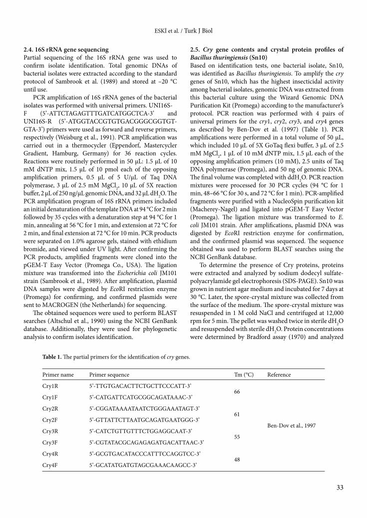
2.5. Cry gene contents and crystal protein profiles of Bacillus thuringiensis (Sn10)Based on identification tests, one bacterial isolate, Sn10, was identified as Bacillus thuringiensis. To amplify the cry genes of Sn10, which has the highest insecticidal activity among bacterial isolates, genomic DNA was extracted from this bacterial culture using the Wizard Genomic DNA Purification Kit (Promega) according to the manufacturer’s protocol. PCR reaction was performed with 4 pairs of universal primers for the cry1, cry2, cry3, and cry4 genes as described by Ben-Dov et al. (1997) (Table 1). PCR amplifications were performed in a total volume of 50 µL, which included 10 µL of 5X GoTaq flexi buffer, 3 µL of 2.5 mM MgCl2, 1 µL of 10 mM dNTP mix, 1.5 µL each of the opposing amplification primers (10 mM), 2.5 units of Taq DNA polymerase (Promega), and 50 ng of genomic DNA. The final volume was completed with ddH2O. PCR reaction mixtures were processed for 30 PCR cycles (94 °C for 1 min, 48–66 °C for 30 s, and 72 °C for 1 min). PCR-amplified fragments were purified with a NucleoSpin purification kit (Macherey-Nagel) and ligated into pGEM-T Easy Vector (Promega). The ligation mixture was transformed to E. coli JM101 strain. After amplifications, plasmid DNA was digested by EcoRI restriction enzyme for confirmation, and the confirmed plasmid was sequenced. The sequence obtained was used to perform BLAST searches using the NCBI GenBank database.
To determine the presence of Cry proteins, proteins were extracted and analyzed by sodium dodecyl sulfate-polyacrylamide gel electrophoresis (SDS-PAGE). Sn10 was grown in nutrient agar medium and incubated for 7 days at 30 °C. Later, the spore-crystal mixture was collected from the surface of the medium. The spore-crystal mixture was resuspended in 1 M cold NaCl and centrifuged at 12,000 rpm for 5 min. The pellet was washed twice in sterile dH2O and resuspended with sterile dH2O. Protein concentrations were determined by Bradford assay (1970) and analyzed
Table 1. The partial primers for the identification of cry genes.
Primer name Primer sequence Tm (°C) Reference
Cry1R 5’-TTGTGACACTTCTGCTTCCCATT-3’66
Ben-Dov et al., 1997
Cry1F 5’-CATGATTCATGCGGCAGATAAAC-3’
Cry2R 5’-CGGATAAAATAATCTGGGAAATAGT-3’61
Cry2F 5’-GTTATTCTTAATGCAGATGAATGGG-3’
Cry3R 5’-CATCTGTTGTTTCTGGAGGCAAT-3’55
Cry3F 5’-CGTATACGCAGAGAGATGACATTAAC-3’
Cry4R 5’-GCGTGACATACCCATTTCCAGGTCC-3’48
Cry4F 5’-GCATATGATGTAGCGAAACAAGCC-3’
ESKİ et al. / Turk J Biol
34
in 10% SDS-PAGE as described by Laemmli (1970). Silver staining was used to detect proteins after electrophoretic separation on polyacrylamide gel, and protein molecular weight was estimated by comparison to protein molecular weight standards (Novex prestained protein marker).2.6. PhylogenyNucleotide sequences of 16S rRNA genes and cry genes were edited with BioEdit and aligned by ClustalW (Hall, 1999). Phylogenetic analysis was performed using the neighbor-joining (NJ) method, as carried out by MEGA 5.0 software (Tamura et al., 2011). The NJ analysis was based on the Kimura 2-parameter test. Alignment gaps were treated as missing data. The reliability of the phylograms was tested by bootstrap analysis with 1000 replicates using MEGA 5.0.2.7. BioassaysBacterial isolates were streaked on nutrient agar plates to obtain single colonies for each isolate. The obtained single colonies were inoculated into nutrient broth medium and incubated overnight at 30 °C. Several isolates were incubated at 30 °C for 2 days due to their slow growth. After incubation, bacterial density was measured at OD600 (optical density) and adjusted to 1.8 × 10⁹ cfu/mL (Ben-Dov et al., 1995). Five milliliters of this culture was centrifuged at 3000 rpm for 10 min. Afterwards, the pellet was resuspended in 5 mL of sterile phosphate-buffered saline and used in bioassays.
The susceptibility of S. nonagrioides larvae to each bacterial isolate was tested under laboratory conditions. The third-instar of healthy larvae obtained from corn fields in the vicinity of Adana were randomly selected and used in bioassays. Fresh corn stems were used as diet in infections. One milliliter of bacterial suspension for each isolate, prepared as described above, was saturated on corn stems
and placed in individual plastic boxes, each containing a single bacterial isolate. Thirty third-instar larvae were placed on the diet in containers and kept at 30 ± 2 °C and 60% relative humidity on a 12:12 photoperiod (Mitchell and Smith, 1985). Larvae mortality was recorded every 24 h and all dead larvae were removed from the containers. All bioassays were repeated 3 times on different occasions.
Three isolates (Sn8, Sn10, and Sn14) that had high mortality against S. nonagrioides were used in dose-response experiments. Three different bacterial concentrations (0.9 × 10⁹ cfu/mL, 1.8 × 10⁹ cfu/mL, and 3.6 × 10⁹ cfu/mL, based on OD600 values) of these isolates were used in the dose-response experiments, and bioassays were performed as described earlier.2.8. Data analysisPercent mortality was corrected according to Abbott’s formula (Abbott, 1925). The data were subjected to ANOVA and subsequently to least significant difference (LSD) multiple comparison tests to compare test isolates with each other and the control group. The lethal concentrations (LC50 and LC95) were estimated by probit analysis. All analyses were performed using SPSS 20.0 software.
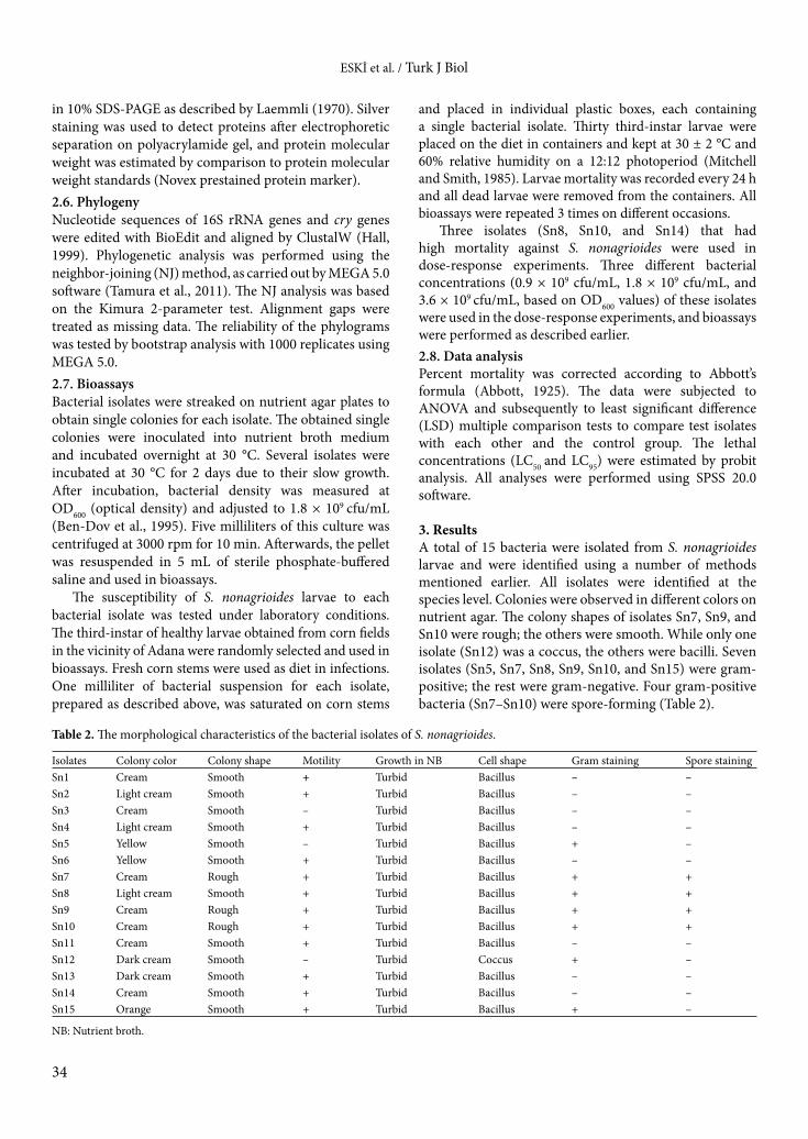
3. ResultsA total of 15 bacteria were isolated from S. nonagrioides larvae and were identified using a number of methods mentioned earlier. All isolates were identified at the species level. Colonies were observed in different colors on nutrient agar. The colony shapes of isolates Sn7, Sn9, and Sn10 were rough; the others were smooth. While only one isolate (Sn12) was a coccus, the others were bacilli. Seven isolates (Sn5, Sn7, Sn8, Sn9, Sn10, and Sn15) were gram-positive; the rest were gram-negative. Four gram-positive bacteria (Sn7–Sn10) were spore-forming (Table 2).
Table 2. The morphological characteristics of the bacterial isolates of S. nonagrioides.
Isolates Colony color Colony shape Motility Growth in NB Cell shape Gram staining Spore stainingSn1 Cream Smooth + Turbid Bacillus – –Sn2 Light cream Smooth + Turbid Bacillus – –Sn3 Cream Smooth – Turbid Bacillus – –Sn4 Light cream Smooth + Turbid Bacillus – –Sn5 Yellow Smooth – Turbid Bacillus + –Sn6 Yellow Smooth + Turbid Bacillus – –Sn7 Cream Rough + Turbid Bacillus + +Sn8 Light cream Smooth + Turbid Bacillus + +Sn9 Cream Rough + Turbid Bacillus + +Sn10 Cream Rough + Turbid Bacillus + +Sn11 Cream Smooth + Turbid Bacillus – –Sn12 Dark cream Smooth – Turbid Coccus + –Sn13 Dark cream Smooth + Turbid Bacillus – –Sn14 Cream Smooth + Turbid Bacillus – –Sn15 Orange Smooth + Turbid Bacillus + –
NB: Nutrient broth.
ESKİ et al. / Turk J Biol
35
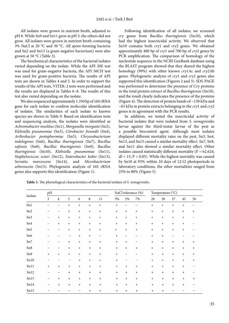
All isolates were grown in nutrient broth, adjusted to pH 8. While Sn9 and Sn11 grew at pH 3, the others did not grow. All isolates were grown in nutrient broth containing 3% NaCl at 20 °C and 30 °C. All spore-forming bacteria and Sn2 and Sn13 (a gram-negative bacterium) were also grown at 50 °C (Table 3).
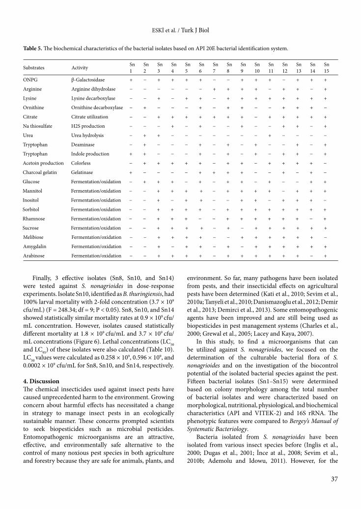
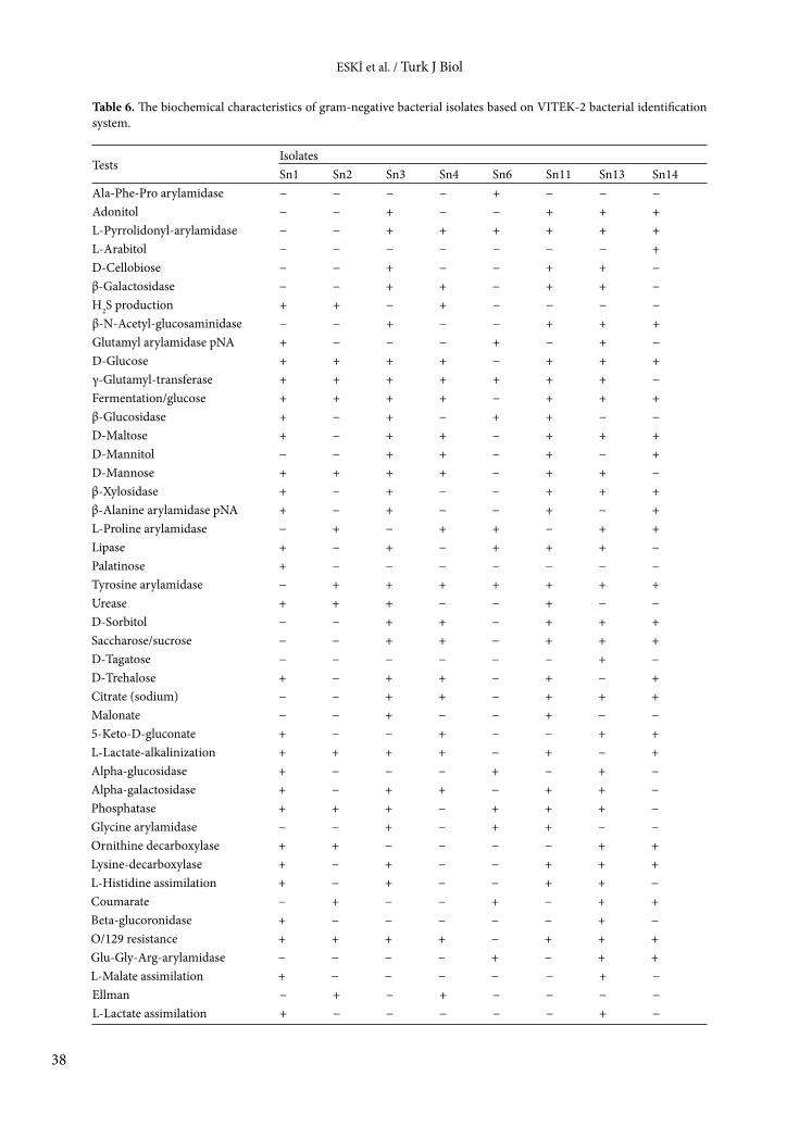
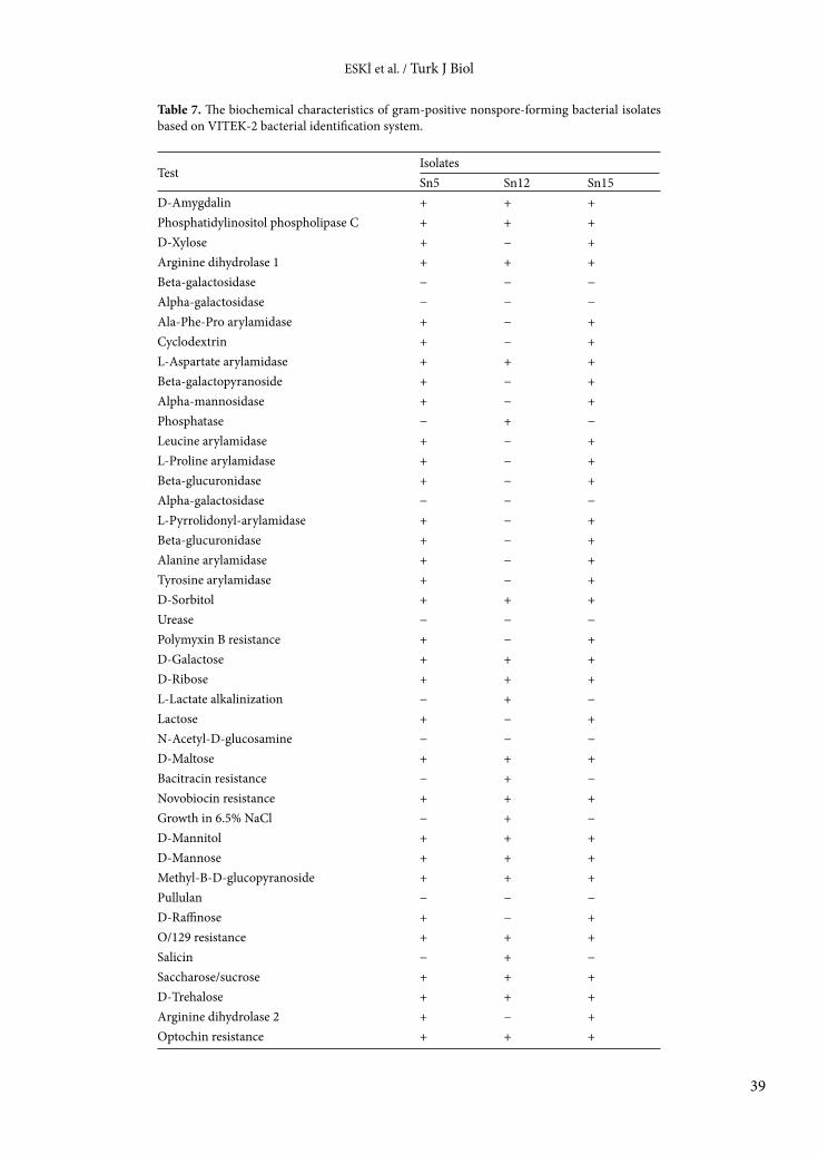
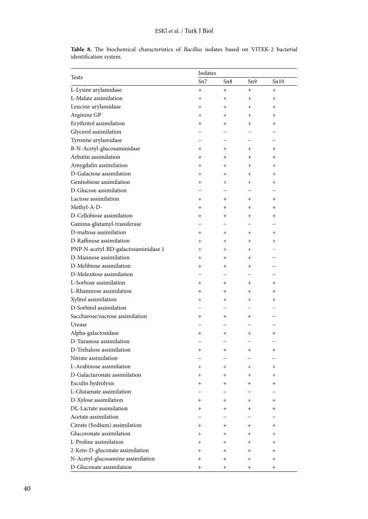
The biochemical characteristics of the bacterial isolates varied depending on the isolate. While the API 20E test was used for gram-negative bacteria, the API 50CH test was used for gram-positive bacteria. The results of API tests are shown in Tables 4 and 5. In order to support the results of the API tests, VITEK-2 tests were performed and the results are displayed in Tables 6–8. The results of this test also varied depending on the isolate.
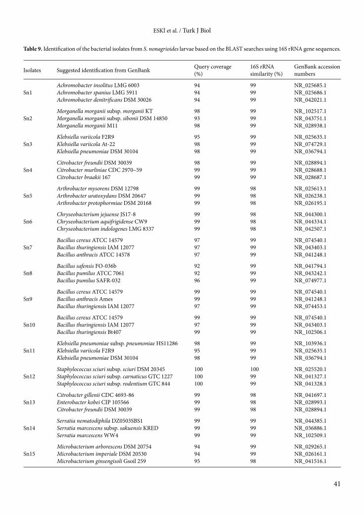
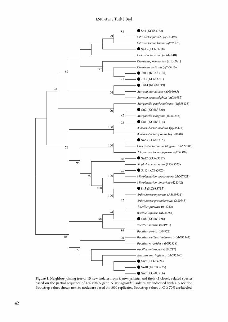
We also sequenced approximately 1.350 bp of 16S rRNA gene for each isolate to confirm molecular identification of isolates. The similarities of each isolate to known species are shown in Table 9. Based on identification tests and sequencing analysis, the isolates were identified as Achromobacter insolitus (Sn1), Morganella morganii (Sn2), Klebsiella pneumoniae (Sn3), Citrobacter freundii (Sn4), Arthrobacter protophormiae (Sn5), Chryseobacterium indologenes (Sn6), Bacillus thuringiensis (Sn7), Bacillus safensis (Sn8), Bacillus thuringiensis (Sn9), Bacillus thuringiensis (Sn10), Klebsiella pneumoniae (Sn11), Staphylococcus sciuri (Sn12), Enterobacter kobei (Sn13), Serratia marcescens (Sn14), and Microbacterium arborescens (Sn15). Phylogenetic analysis of 16S rRNA genes also supports this identification (Figure 1).
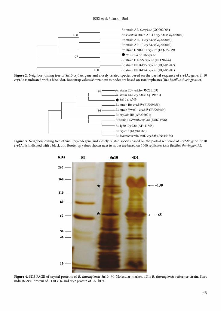
Following identification of all isolates, we screened cry genes from Bacillus thuringiensis (Sn10), which had the highest insecticidal activity. We observed that Sn10 contains both cry1 and cry2 genes. We obtained approximately 400 bp of cry1 and 700 bp of cry2 genes by PCR amplification. The comparison of homology of the nucleotide sequence in the NCBI GenBank database using the BLAST program showed that they shared the highest homology (99%) with other known cry1Ac and cry2Ab genes. Phylogenetic analysis of cry1 and cry2 genes also supported this identification (Figures 2 and 3). SDS-PAGE was performed to determine the presence of Cry proteins in the total protein extract of Bacillus thuringiensis (Sn10), and the result clearly indicates the presence of the proteins (Figure 4). The detection of protein bands of ~130 kDa and ~65 kDa in protein extracts belonging to the cry1 and cry2 genes is in agreement with the PCR results.
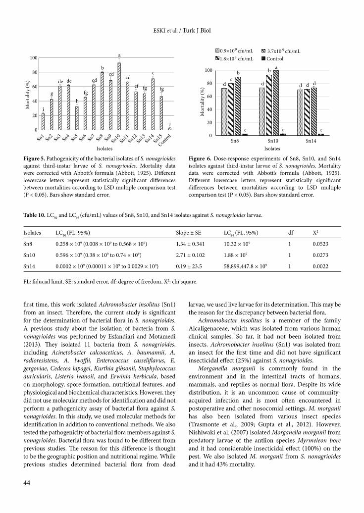
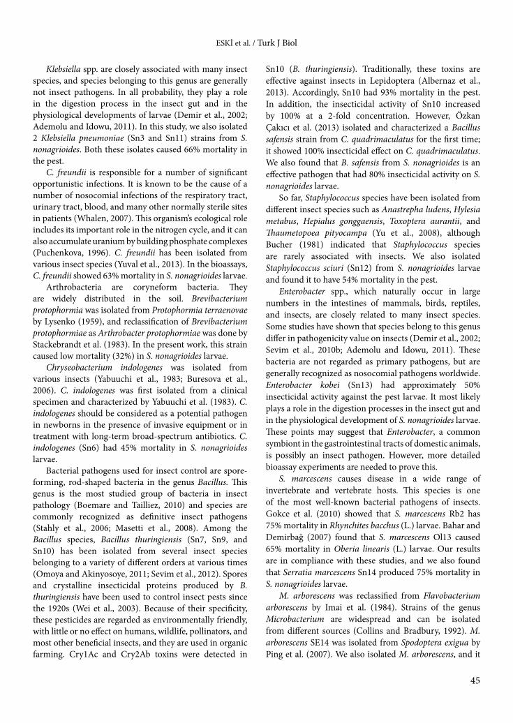
In addition, we tested the insecticidal activity of bacterial isolates that were isolated from S. nonagrioides larvae against the third-instar larvae of the pest as a possible biocontrol agent. Although most isolates displayed different mortality rates on the pest, Sn3, Sn4, Sn13, and Sn15 caused a similar mortality effect. Sn7, Sn9, and Sn11 also showed a similar mortality effect. Other isolates caused statistically different mortality (F = 62.624; df = 15; P < 0.05). While the highest mortality was caused by Sn10 at 93% within 10 days of 12:12 photoperiods in laboratory conditions, the other mortalities ranged from 25% to 80% (Figure 5).
Table 3. The physiological characteristics of the bacterial isolates of S. nonagrioides.
IsolatepH NaCl tolerance (%) Temperature (°C)
3 4 5 6 8 11 3% 5% 7% 20 30 37 45 50
Sn1 − − + + + + + − − + + + + −
Sn2 − − + + + + + + + + + + + +
Sn3 − + + + + + + + + + + + + −
Sn4 − + + + + + + + + + + + + −
Sn5 − − + + + + + + + + + + + −
Sn6 − − − + + − + − − + + − − −
Sn7 − − + + + + + + − + + + + +
Sn8 − − + + + + + + + + + + + +
Sn9 + + + + + + + − − + + + + +
Sn10 − − − + + + + − − + + + + +
Sn11 + + + + + + + + + + + + + −
Sn12 − − + + + + + + + + + + + −
Sn13 − + + + + + + + + + + + + +
Sn14 − + + + + + + + + + + + + −
Sn15 − − − − + + + + + + + − − −

ESKİ et al. / Turk J Biol
36
Table 4. The biochemical characteristics of the gram-positive bacterial isolates based on API 50 CHB bacterial identification system.
TestsIsolatesSn5 Sn7 Sn8 Sn9 Sn10 Sn12 Sn15
Glycerol − − − − − − −Erythritol − − + − + − −D-Arabinose − − − − − − −L-Arabinose − − − − − − −D-Ribose − − + − − − −D-Xylose + + + − + − −L-Xylose − − + − − − −D-Adonitol − − + − − − +Methyl-β-D-xylopyranoside − − + − − − +D-Galactose − − + − − − +D-Glucose − − + − − − −D-Fructose + + + + + + +D-Mannose + + − + + + +L-Sorbose − − − − + − −L-Rhamnose − − − − − − −Dulcitol − − + − − − −Inositol − − − − − − −D-Mannitol − − + − − − −D-Sorbitol − − + − − − −Methyl-αD-mannopyranoside + − + − − − −Methyl-αD-glucopyranoside + − − − + − −N-Acetylglucosamine + − + − + − −Amygdalin + + + + + + +Arbutin − − + − − − −Esculin-ferric citrate − − − + + + +Salicin − − + + + + +D-Cellobiose + + + + + + +D-Maltose + + + + + + +D-Lactose (bovine origin) + + + + + + +D-Melibiose + + − − − − −D-Saccharose (sucrose) + − − − − −D-Trehalose − − + + + + +Inulin − − − + + + +D-Melezitose − − − − + − −D-Raffinose + + + − − − −Glycogen + − + + + + +Xylitol − − + + + + +Gentiobiose + + + − + − −D-Turanose − − + − + − −D-Lyxose − − − − − − −D-Tagatose + − + − − − −D-Fucose + − + − − − −L-Fucose + − − − − − −D-Arabitol − − + − − − −L-Arabitol − − + − − − −Potassium gluconate + − + − − − −Potassium 2-ketogluconate + − + − − − −Potassium 5-ketogluconate − − + − − + +
ESKİ et al. / Turk J Biol
37
Finally, 3 effective isolates (Sn8, Sn10, and Sn14) were tested against S. nonagrioides in dose-response experiments. Isolate Sn10, identified as B. thuringiensis, had 100% larval mortality with 2-fold concentration (3.7 × 10⁹
cfu/mL) (F = 248.34; df = 9; P < 0.05). Sn8, Sn10, and Sn14 showed statistically similar mortality rates at 0.9 × 10⁹ cfu/mL concentration. However, isolates caused statistically different mortality at 1.8 × 10⁹ cfu/mL and 3.7 × 10⁹ cfu/mL concentrations (Figure 6). Lethal concentrations (LC50 and LC95) of these isolates were also calculated (Table 10). LC50 values were calculated as 0.258 × 10⁹, 0.596 × 10⁹, and 0.0002 × 10⁹ cfu/mL for Sn8, Sn10, and Sn14, respectively.
4. DiscussionThe chemical insecticides used against insect pests have caused unprecedented harm to the environment. Growing concern about harmful effects has necessitated a change in strategy to manage insect pests in an ecologically sustainable manner. These concerns prompted scientists to seek biopesticides such as microbial pesticides. Entomopathogenic microorganisms are an attractive, effective, and environmentally safe alternative to the control of many noxious pest species in both agriculture and forestry because they are safe for animals, plants, and
environment. So far, many pathogens have been isolated from pests, and their insecticidal effects on agricultural pests have been determined (Kati et al., 2010; Sevim et al., 2010a; Tanyeli et al., 2010; Danismazoglu et al., 2012; Demir et al., 2013; Demirci et al., 2013). Some entomopathogenic agents have been improved and are still being used as biopesticides in pest management systems (Charles et al., 2000; Grewal et al., 2005; Lacey and Kaya, 2007).
In this study, to find a microorganisms that can be utilized against S. nonagrioides, we focused on the determination of the culturable bacterial flora of S. nonagrioides and on the investigation of the biocontrol potential of the isolated bacterial species against the pest. Fifteen bacterial isolates (Sn1–Sn15) were determined based on colony morphology among the total number of bacterial isolates and were characterized based on morphological, nutritional, physiological, and biochemical characteristics (API and VITEK-2) and 16S rRNA. The phenotypic features were compared to Bergey’s Manual of Systematic Bacteriology.
Bacteria isolated from S. nonagrioides have been isolated from various insect species before (Inglis et al., 2000; Dugas et al., 2001; İnce at al., 2008; Sevim et al., 2010b; Ademolu and Idowu, 2011). However, for the
Table 5. The biochemical characteristics of the bacterial isolates based on API 20E bacterial identification system.
Substrates Activity Sn1
Sn2
Sn3
Sn4
Sn5
Sn6
Sn7
Sn8
Sn9
Sn10
Sn11
Sn12
Sn13
Sn14
Sn15
ONPG β-Galactosidase + − + + + + − − + + + − + + +
Arginine Arginine dihydrolase − − − − − − + + + + − + + − +
Lysine Lysine decarboxylase − − + − + + − + + + + + + + +
Ornithine Ornithine decarboxylase − + − − − + − + + − − + + + −
Citrate Citrate utilization − − + + + + + + + − + + + + +
Na thiosulfate H2S production − − − + − + − − + − − + + − +
Urea Urea hydrolysis − + + − − − − − − − + − − − −
Tryptophan Deaminase − + − − − + − + − + − − + − +
Tryptophan Indole production + + − − − + − + − + − + + − +
Acetoin production Colorless − + + + + + − + + − + + + + −
Charcoal gelatin Gelatinase + − − − − + + + + − − + − + −
Glucose Fermentation/oxidation − + + + − + − + + − + − − + +
Mannitol Fermentation/oxidation − − + + + + − + + + + − + + +
Inositol Fermentation/oxidation − − + − + + − − + + − + + + −
Sorbitol Fermentation/oxidation − − + + + + − + + + + + + + +
Rhamnose Fermentation/oxidation − − + + + − − + + + + + + − +
Sucrose Fermentation/oxidation − − + + + + − + − + + + + + +
Melibiose Fermentation/oxidation − − + + + + − − + + + + + + −
Amygdalin Fermentation/oxidation − − + − + + − + − + + + + + +
Arabinose Fermentation/oxidation − − + + + + − + + + + + + − +
ESKİ et al. / Turk J Biol
38
Table 6. The biochemical characteristics of gram-negative bacterial isolates based on VITEK-2 bacterial identification system.
TestsIsolatesSn1 Sn2 Sn3 Sn4 Sn6 Sn11 Sn13 Sn14
Ala-Phe-Pro arylamidase − − − − + − − −Adonitol − − + − − + + +L-Pyrrolidonyl-arylamidase − − + + + + + +L-Arabitol − − − − − − − +D-Cellobiose − − + − − + + −β-Galactosidase − − + + − + + −H2S production + + − + − − − −β-N-Acetyl-glucosaminidase − − + − − + + +Glutamyl arylamidase pNA + − − − + − + −D-Glucose + + + + − + + +γ-Glutamyl-transferase + + + + + + + −Fermentation/glucose + + + + − + + +β-Glucosidase + − + − + + − −D-Maltose + − + + − + + +D-Mannitol − − + + − + − +D-Mannose + + + + − + + −β-Xylosidase + − + − − + + +β-Alanine arylamidase pNA + − + − − + − +L-Proline arylamidase − + − + + − + +Lipase + − + − + + + −Palatinose + − − − − − − −Tyrosine arylamidase − + + + + + + +Urease + + + − − + − −D-Sorbitol − − + + − + + +Saccharose/sucrose − − + + − + + +D-Tagatose − − − − − − + −D-Trehalose + − + + − + − +Citrate (sodium) − − + + − + + +Malonate − − + − − + − −5-Keto-D-gluconate + − − + − − + +L-Lactate-alkalinization + + + + − + − +Alpha-glucosidase + − − − + − + −Alpha-galactosidase + − + + − + + −Phosphatase + + + − + + + −Glycine arylamidase − − + − + + − −Ornithine decarboxylase + + − − − − + +Lysine-decarboxylase + − + − − + + +L-Histidine assimilation + − + − − + + −Coumarate − + − − + − + +Beta-glucoronidase + − − − − − + −O/129 resistance + + + + − + + +Glu-Gly-Arg-arylamidase − − − − + − + +L-Malate assimilation + − − − − − + −Ellman − + − + − − − −L-Lactate assimilation + − − − − − + −
ESKİ et al. / Turk J Biol
39
Table 7. The biochemical characteristics of gram-positive nonspore-forming bacterial isolates based on VITEK-2 bacterial identification system.
TestIsolatesSn5 Sn12 Sn15
D-Amygdalin + + +Phosphatidylinositol phospholipase C + + +D-Xylose + − +Arginine dihydrolase 1 + + +Beta-galactosidase − − −Alpha-galactosidase − − −Ala-Phe-Pro arylamidase + − +Cyclodextrin + − +L-Aspartate arylamidase + + +Beta-galactopyranoside + − +Alpha-mannosidase + − +Phosphatase − + −Leucine arylamidase + − +L-Proline arylamidase + − +Beta-glucuronidase + − +Alpha-galactosidase − − −L-Pyrrolidonyl-arylamidase + − +Beta-glucuronidase + − +Alanine arylamidase + − +Tyrosine arylamidase + − +D-Sorbitol + + +Urease − − −Polymyxin B resistance + − +D-Galactose + + +D-Ribose + + +L-Lactate alkalinization − + −Lactose + − +N-Acetyl-D-glucosamine − − −D-Maltose + + +Bacitracin resistance − + −Novobiocin resistance + + +Growth in 6.5% NaCl − + −D-Mannitol + + +D-Mannose + + +Methyl-B-D-glucopyranoside + + +Pullulan − − −D-Raffinose + − +O/129 resistance + + +Salicin − + −Saccharose/sucrose + + +D-Trehalose + + +Arginine dihydrolase 2 + − +Optochin resistance + + +
ESKİ et al. / Turk J Biol
40
Table 8. The biochemical characteristics of Bacillus isolates based on VITEK-2 bacterial identification system.
TestsIsolatesSn7 Sn8 Sn9 Sn10
L-Lysine arylamidase + + + +L-Malate assimilation + + + +Leucine-arylamidase + + + +Arginine GP + + + +Erythritol assimilation + + + +Glycerol assimilation − − − −Tyrosine arylamidase − − − −Β-N-Acetyl-glucosaminidase + + + +Arbutin assimilation + + + +Amygdalin assimilation + + + +D-Galactose assimilation + + + +Gentiobiose assimilation + + + +D-Glucose assimilation − − − −Lactose assimilation + + + +Methyl-A-D- + + + +D-Cellobiose assimilation + + + +Gamma-glutamyl-transferase − − − −D-maltose assimilation + + + +D-Raffinose assimilation + + + +PNP-N-acetyl-BD-galactosaminidase 1 + + + −D-Mannose assimilation + + + −D-Melibiose assimilation + + + −D-Melezitose assimilation − − − −L-Sorbose assimilation + + + +L-Rhamnose assimilation + + + +Xylitol assimilation + + + +D-Sorbitol assimilation − − − −Saccharose/sucrose assimilation + + + −Urease − − − −Alpha-galactosidase + + + +D-Turanose assimilation − − − −D-Trehalose assimilation + + + +Nitrate assimilation − − − −L-Arabinose assimilation + + + +D-Galacturonate assimilation + + + +Esculin hydrolysis + + + +L-Glutamate assimilation − − − −D-Xylose assimilation + + + +DL-Lactate assimilation + + + +Acetate assimilation − − − −Citrate (Sodium) assimilation + + + +Glucoronate assimilation + + + +L-Proline assimilation + + + +2-Keto-D-gluconate assimilation + + + +N-Acetyl-glucosamine assimilation + + + +D-Gluconate assimilation + + + +
ESKİ et al. / Turk J Biol
41
Table 9. Identification of the bacterial isolates from S. nonagrioides larvae based on the BLAST searches using 16S rRNA gene sequences.
Isolates Suggested identification from GenBank Query coverage (%)
16S rRNAsimilarity (%)
GenBank accessionnumbers
Sn1Achromobacter insolitus LMG 6003 Achromobacter spanius LMG 5911Achromobacter denitrificans DSM 30026
949494
999999
NR_025685.1NR_025686.1NR_042021.1
Sn2Morganella morganii subsp. morganii KT Morganella morganii subsp. sibonii DSM 14850Morganella morganii M11
989398
999999
NR_102517.1NR_043751.1NR_028938.1
Sn3Klebsiella variicola F2R9 Klebsiella variicola At-22Klebsiella pneumoniae DSM 30104
959898
999999
NR_025635.1NR_074729.1NR_036794.1
Sn4Citrobacter freundii DSM 30039Citrobacter murliniae CDC 2970–59Citrobacter braakii 167
989999
999999
NR_028894.1NR_028688.1 NR_028687.1
Sn5Arthrobacter mysorens DSM 12798Arthrobacter uratoxydans DSM 20647Arthrobacter protophormiae DSM 20168
999999
989898
NR_025613.1NR_026238.1NR_026195.1
Sn6Chryseobacterium jejuense JS17-8 Chryseobacterium aquifrigidense CW9 Chryseobacterium indologenes LMG 8337
999999
989898
NR_044300.1NR_044334.1NR_042507.1
Sn7Bacillus cereus ATCC 14579Bacillus thuringiensis IAM 12077Bacillus anthracis ATCC 14578
979797
999999
NR_074540.1NR_043403.1 NR_041248.1
Sn8Bacillus safensis FO-036b Bacillus pumilus ATCC 7061Bacillus pumilus SAFR-032
929296
999999
NR_041794.1NR_043242.1NR_074977.1
Sn9Bacillus cereus ATCC 14579Bacillus anthracis AmesBacillus thuringiensis IAM 12077
999997
999999
NR_074540.1NR_041248.1NR_074453.1
Sn10Bacillus cereus ATCC 14579Bacillus thuringiensis IAM 12077Bacillus thuringiensis Bt407
999799
999999
NR_074540.1NR_043403.1NR_102506.1
Sn11Klebsiella pneumoniae subsp. pneumoniae HS11286Klebsiella variicola F2R9 Klebsiella pneumoniae DSM 30104
989598
999999
NR_103936.1NR_025635.1NR_036794.1
Sn12Staphylococcus sciuri subsp. sciuri DSM 20345 Staphylococcus sciuri subsp. carnaticus GTC 1227Staphylococcus sciuri subsp. rodentium GTC 844
100100100
1009999
NR_025520.1NR_041327.1NR_041328.1
Sn13Citrobacter gillenii CDC 4693-86Enterobacter kobei CIP 105566Citrobacter freundii DSM 30039
999999
989898
NR_041697.1NR_028993.1NR_028894.1
Sn14Serratia nematodiphila DZ0503SBS1 Serratia marcescens subsp. sakuensis KREDSerratia marcescens WW4
999999
999999
NR_044385.1NR_036886.1NR_102509.1
Sn15Microbacterium arborescens DSM 20754Microbacterium imperiale DSM 20530Microbacterium ginsengisoli Gsoil 259
949495
999998
NR_029265.1NR_026161.1NR_041516.1
ESKİ et al. / Turk J Biol
42
Sn4 (KC683722)
Citrobacter freundii (aj233408)
Citrobacter werkmanii (af025373)
Sn13 (KC683718)
Enterobacter kobei (ab616140)
Klebsiella pneumoniae (af130981)
Klebsiella variicola (aj783916)
Sn11 (KC683726)
Sn3 (KC683721)
Sn14 (KC683719)
Serratia marcescens (ab061685)
Serratia nematodiphila (eu036987)
Morganella psychrotolerans (dq358135)
Sn2 (KC683720)
Morganella morganii (ab089243)
Sn1 (KC683714)
Achromobacter insolitus (jq746423)
Achromobacter spanius (ay170848)
Sn6 (KC683715)
Chryseobacterium indologenes (ab517708)
Chryseobacterium jejuense (ef591303)
Sn12 (KC683717)
Staphylococcus sciuri (17385625)
Sn15 (KC683726)
Microbacterium arborescens (ab007421)
Microbacterium imperiale (d21342)
Sn5 (KC683715)
Arthrobacter mysorens (AJ639831)
Arthrobacter protophormiae (X80745)
Bacillus pumilus (043242)
Bacillus safensis (af234854)
Sn8 (KC683728)
Bacillus subtilis (024931)
Bacillus cereus (004722)
Bacillus weihenstephanensis (ab592543)
Bacillus mycoides (ab592538)
Bacillus anthracis (ab190217)
Bacillus thuringiensis (ab592540)
Sn9 (KC683724)
Sn10 (KC683725)
Sn7 (KC683716)
89
94
98
90
72
100
100
100
96
72100
100
100
93100
76
96
74
9290
7894
71
9787
8389
Figure 1. Neighbor-joining tree of 15 new isolates from S. nonagrioides and their 41 closely related species based on the partial sequence of 16S rRNA gene. S. nonagrioides isolates are indicated with a black dot. Bootstrap values shown next to nodes are based on 1000 replicates. Bootstrap values of C ≥ 70% are labeled.
ESKİ et al. / Turk J Biol
43
Bt. strain AR-6 cry1Ac (GQ202005)Bt. kurstaki strain AR-12 cry1Ac (GQ202004) Bt. strain AR-14 cry1Ac (GQ202003)Bt. strain AR-10 cry1Ac (GQ202002)Bt. strain DNB-Bt1 cry1Ac (DQ785779)
Bt. strain Sn10 cry1AcBt. strain BT-AS cry1Ac (JN120764) Bt. strain DNB-Bt5 cry1Ac (DQ785782) Bt. strain DNB-Bt4 cry1Ac (DQ785781)100
100
97
Bt. strain FB cry2Ab (JN226103)Bt. strain 14-1 cry2Ab (DQ119823)
Sn10 cry2AbBt. strain Bts cry2Ab (EU909455)Bt. strain Ywc5-4 cry2Ab (EU909454)Bt. cry2ab-HB (AY297091)Bt.strain LSZ9408 cry2Ab (EU623976)
Bt. ly30 Cry2Ab (AF441855)Bt. cry2Ab (DQ361266)Bt. kurstaki strain MnD cry2Ab (JN415485)
98
98
Figure 2. Neighbor-joining tree of Sn10 cry1Ac gene and closely related species based on the partial sequence of cry1Ac gene. Sn10 cry1Ac is indicated with a black dot. Bootstrap values shown next to nodes are based on 1000 replicates (Bt.: Bacillus thuringiensis).
Figure 3. Neighbor-joining tree of Sn10 cry2Ab gene and closely related species based on the partial sequence of cry2Ab gene. Sn10 cry2Ab is indicated with a black dot. Bootstrap values shown next to nodes are based on 1000 replicates (Bt.: Bacillus thuringiensis).
Figure 4. SDS-PAGE of crystal proteins of B. thuringiensis Sn10. M: Molecular marker, 4D1: B. thuringiensis reference strain. Stars indicate cry1 protein of ~130 kDa and cry2 protein of ~65 kDa.
ESKİ et al. / Turk J Biol
44
first time, this work isolated Achromobacter insolitus (Sn1) from an insect. Therefore, the current study is significant for the determination of bacterial flora in S. nonagrioides. A previous study about the isolation of bacteria from S. nonagrioides was performed by Esfandiari and Motamedi (2013). They isolated 11 bacteria from S. nonagrioides, including Acinetobacter calcoaceticus, A. baumannii, A. radioresistens, A. lwoffii, Enterococcus casseliflavus, E. gergoviae, Cedecea lapagei, Kurthia gibsonii, Staphylococcus auricularis, Listeria ivanoii, and Erwinia herbicula, based on morphology, spore formation, nutritional features, and physiological and biochemical characteristics. However, they did not use molecular methods for identification and did not perform a pathogenicity assay of bacterial flora against S. nonagrioides. In this study, we used molecular methods for identification in addition to conventional methods. We also tested the pathogenicity of bacterial flora members against S. nonagrioides. Bacterial flora was found to be different from previous studies. The reason for this difference is thought to be the geographic position and nutritional regime. While previous studies determined bacterial flora from dead
larvae, we used live larvae for its determination. This may be the reason for the discrepancy between bacterial flora.
Achromobacter insolitus is a member of the family Alcaligenaceae, which was isolated from various human clinical samples. So far, it had not been isolated from insects. Achromobacter insolitus (Sn1) was isolated from an insect for the first time and did not have significant insecticidal effect (25%) against S. nonagrioides.
Morganella morganii is commonly found in the environment and in the intestinal tracts of humans, mammals, and reptiles as normal flora. Despite its wide distribution, it is an uncommon cause of community-acquired infection and is most often encountered in postoperative and other nosocomial settings. M. morganii has also been isolated from various insect species (Trasmonte et al., 2009; Gupta et al., 2012). However, Nishiwaki et al. (2007) isolated Morganella morganii from predatory larvae of the antlion species Myrmeleon bore and it had considerable insecticidal effect (100%) on the pest. We also isolated M. morganii from S. nonagrioides and it had 43% mortality.
i
g
de de
h fg
cd
b cd
a
cd ef fg
c
fg
j 0
20
40
60
80
100
Mor
talit
y (%
)
Isolates
Figure 5. Pathogenicity of the bacterial isolates of S. nonagrioides against third-instar larvae of S. nonagrioides. Mortality data were corrected with Abbott’s formula (Abbott, 1925). Different lowercase letters represent statistically significant differences between mortalities according to LSD multiple comparison test (P < 0.05). Bars show standard error.
Figure 6. Dose-response experiments of Sn8, Sn10, and Sn14 isolates against third-instar larvae of S. nonagrioides. Mortality data were corrected with Abbott’s formula (Abbott, 1925). Different lowercase letters represent statistically significant differences between mortalities according to LSD multiple comparison test (P < 0.05). Bars show standard error.
d d d c
b
d
b a
d
e e e 0
20
40
60
80
100
Sn8 Sn10 Sn14
Mor
talit
y (%
)
Isolates
Control0.9×109 cfu/mL
1.8×10⁹ cfu/mL 3.7x10 9 cfu/mL
Table 10. LC50 and LC95 (cfu/mL) values of Sn8, Sn10, and Sn14 isolates against S. nonagrioides larvae.
Isolates LC50 (FL, 95%) Slope ± SE LC95 (FL, 95%) df X2
Sn8 0.258 × 10⁹ (0.008 × 10⁹ to 0.568 × 10⁹) 1.34 ± 0.341 10.32 × 10⁹ 1 0.0523
Sn10 0.596 × 10⁹ (0.38 × 10⁹ to 0.74 × 10⁹) 2.71 ± 0.102 1.88 × 10⁹ 1 0.0273
Sn14 0.0002 × 10⁹ (0.00011 × 10⁹ to 0.0029 × 10⁹) 0.19 ± 23.5 58,899,447.8 × 109 1 0.0022
FL: fiducial limit, SE: standard error, df: degree of freedom, X2: chi square.
ESKİ et al. / Turk J Biol
45
Klebsiella spp. are closely associated with many insect species, and species belonging to this genus are generally not insect pathogens. In all probability, they play a role in the digestion process in the insect gut and in the physiological developments of larvae (Demir et al., 2002; Ademolu and Idowu, 2011). In this study, we also isolated 2 Klebsiella pneumoniae (Sn3 and Sn11) strains from S. nonagrioides. Both these isolates caused 66% mortality in the pest.
C. freundii is responsible for a number of significant opportunistic infections. It is known to be the cause of a number of nosocomial infections of the respiratory tract, urinary tract, blood, and many other normally sterile sites in patients (Whalen, 2007). This organism’s ecological role includes its important role in the nitrogen cycle, and it can also accumulate uranium by building phosphate complexes (Puchenkova, 1996). C. freundii has been isolated from various insect species (Yuval et al., 2013). In the bioassays, C. freundii showed 63% mortality in S. nonagrioides larvae.
Arthrobacteria are coryneform bacteria. They are widely distributed in the soil. Brevibacterium protophormia was isolated from Protophormia terraenovae by Lysenko (1959), and reclassification of Brevibacterium protophormiae as Arthrobacter protophormiae was done by Stackebrandt et al. (1983). In the present work, this strain caused low mortality (32%) in S. nonagrioides larvae.
Chryseobacterium indologenes was isolated from various insects (Yabuuchi et al., 1983; Buresova et al., 2006). C. indologenes was first isolated from a clinical specimen and characterized by Yabuuchi et al. (1983). C. indologenes should be considered as a potential pathogen in newborns in the presence of invasive equipment or in treatment with long-term broad-spectrum antibiotics. C. indologenes (Sn6) had 45% mortality in S. nonagrioides larvae.
Bacterial pathogens used for insect control are spore-forming, rod-shaped bacteria in the genus Bacillus. This genus is the most studied group of bacteria in insect pathology (Boemare and Tailliez, 2010) and species are commonly recognized as definitive insect pathogens (Stahly et al., 2006; Masetti et al., 2008). Among the Bacillus species, Bacillus thuringiensis (Sn7, Sn9, and Sn10) has been isolated from several insect species belonging to a variety of different orders at various times (Omoya and Akinyosoye, 2011; Sevim et al., 2012). Spores and crystalline insecticidal proteins produced by B. thuringiensis have been used to control insect pests since the 1920s (Wei et al., 2003). Because of their specificity, these pesticides are regarded as environmentally friendly, with little or no effect on humans, wildlife, pollinators, and most other beneficial insects, and they are used in organic farming. Cry1Ac and Cry2Ab toxins were detected in
Sn10 (B. thuringiensis). Traditionally, these toxins are effective against insects in Lepidoptera (Albernaz et al., 2013). Accordingly, Sn10 had 93% mortality in the pest. In addition, the insecticidal activity of Sn10 increased by 100% at a 2-fold concentration. However, Özkan Çakıcı et al. (2013) isolated and characterized a Bacillus safensis strain from C. quadrimaculatus for the first time; it showed 100% insecticidal effect on C. quadrimaculatus. We also found that B. safensis from S. nonagrioides is an effective pathogen that had 80% insecticidal activity on S. nonagrioides larvae.
So far, Staphylococcus species have been isolated from different insect species such as Anastrepha ludens, Hylesia metabus, Hepialus gonggaensis, Toxoptera aurantii, and Thaumetopoea pityocampa (Yu et al., 2008), although Bucher (1981) indicated that Staphylococcus species are rarely associated with insects. We also isolated Staphylococcus sciuri (Sn12) from S. nonagrioides larvae and found it to have 54% mortality in the pest.
Enterobacter spp., which naturally occur in large numbers in the intestines of mammals, birds, reptiles, and insects, are closely related to many insect species. Some studies have shown that species belong to this genus differ in pathogenicity value on insects (Demir et al., 2002; Sevim et al., 2010b; Ademolu and Idowu, 2011). These bacteria are not regarded as primary pathogens, but are generally recognized as nosocomial pathogens worldwide. Enterobacter kobei (Sn13) had approximately 50% insecticidal activity against the pest larvae. It most likely plays a role in the digestion processes in the insect gut and in the physiological development of S. nonagrioides larvae. These points may suggest that Enterobacter, a common symbiont in the gastrointestinal tracts of domestic animals, is possibly an insect pathogen. However, more detailed bioassay experiments are needed to prove this.
S. marcescens causes disease in a wide range of invertebrate and vertebrate hosts. This species is one of the most well-known bacterial pathogens of insects. Gokce et al. (2010) showed that S. marcescens Rb2 has 75% mortality in Rhynchites bacchus (L.) larvae. Bahar and Demirbağ (2007) found that S. marcescens Ol13 caused 65% mortality in Oberia linearis (L.) larvae. Our results are in compliance with these studies, and we also found that Serratia marcescens Sn14 produced 75% mortality in S. nonagrioides larvae.
M. arborescens was reclassified from Flavobacterium arborescens by Imai et al. (1984). Strains of the genus Microbacterium are widespread and can be isolated from different sources (Collins and Bradbury, 1992). M. arborescens SE14 was isolated from Spodoptera exigua by Ping et al. (2007). We also isolated M. arborescens, and it
ESKİ et al. / Turk J Biol
46
showed 46% mortality in S. nonagrioides larvae.In conclusion, we isolated and characterized 15
bacteria from S. nonagrioides larvae and tested their effectiveness against it. Some of the isolates appeared to be significant candidates for the biological control of this pest. B. thuringiensis (Sn10) and B. safensis (Sn8) are especially the most promising isolates for the microbial control of S. nonagrioides. Findings from this study showed that further
studies should include field efficacy of the isolates Sn10 and Sn8 and an investigation of the predisposition of those isolates in terms of mass production.
AcknowledgmentThis research was supported by the Scientific Research and Project Coordinator (8761) of Karadeniz Technical University.
References
Abbott WS (1925). A method of computing the effectiveness of an insecticide. J Econ Entomol 18: 265–267.
Ademolu KO, Idowu AB (2011). Occurrence and distribution of microflora in the gut regions of the variegated grasshopper Zonocerus variegatus (Orthoptera: Pyrgomorphidae) during development. Zool Stud 50: 409–415.
Albernaz KC, Merlin BL, Martinelli S, Head GP, Omoto C (2013). Baseline susceptibility to Cry1Ac insecticidal protein in Heliothis virescens (Lepidoptera: Noctuidae) populations in Brazil. J Econ Entomol 106: 1819–1824.
Alexandri MP, Tsitsipis JA (1990). Influence of the egg parasitoid Platytelenomus busseolae (Hym.: Scelionidae) on the population of Sesamia nonagrioides (Lep.: Noctuidae) in central Greece. Entomophaga 35: 61–70.
Avantaggiato G, Quaranta F, Desiderio E, Visconti A (2003). Fumonisin contamination of maize hybrids visibly damaged by Sesamia. J Sci Food Agr 83: 13–18.
Bahar AA, Demirbağ, Z (2007). Isolation of pathogenic bacteria from Obera linearis (Coleoptera: Cerambycidae). Biologia 6: 1–6.
Bayram A (2003). Development of economic injury levels for Sesamia nonagriodes Lefevbre (Lepidoptera: Noctuidae) and some biological features of its egg parasitod Telenomus Busseolae Gahan (Hymenoptera: Sceliontoae). PhD, Çukurova University, Adana, Turkey.
Ben-Dov E, Boussiba S, Zaritsky A (1995). Mosquito larvicidal activity of Escherichia coli with combinations of genes from Bacillus thuringiensis subsp. israelensis. J Bacteriol 177: 2851–2857.
Boemare N, Tailliez P (2010). Molecular approaches and techniques for the study of entomopathogenic bacteria. In: Stock SP, Vanderberg J, Boemare N, Glazer I, editors. Insect Pathogens Molecular Approaches and Techniques. Wallingford, UK: CAB International, pp. 32–45.
Bradford MM (1976). A rapid and sensitive method for quantification of microgram quantities of protein utilizing the principle of protein-dye binding. Anal Biochem 72: 248–254.
Bucher GE (1981). Identification of bacteria found in insects. In: Burges HD, editor. Microbial Control of Pests and Plant Diseases. New York, NY, USA: Academic Press, pp. 7–33.
Buresova V, Franta Z, Kopacek P (2006). A comparison of Chryseobacterium indologenes pathogenicity to the soft tick Ornithodoros moubata and hard tick Ixodes ricinus. J Invertebr Pathol 93: 96–104.
Charles JF, Delecluse A, Nielsen-LeRoux C (2000). Entomopathogenic Bacteria: From Laboratory to Field Application. Dordrecht, the Netherlands: Kluwer Academic Publishers.
Claus M (1992). A standardized Gram staining procedure. World J Microbiol Biotechnol 8: 451–452.
Collins MD, Bradbury JF (1992). The genera Agromyces, Aureobacterium, Clavibacter, Curtobacterium, and Microbacterium. In: Balows A, Trüper HG, Dworkin M, Harder W, Schleifer KH, editors. The Prokaryotes. New York, NY, USA: Springer-Verlag, pp. 1355–1368.
Danismazoglu M, Demir İ, Sevim A, Demirbag Z, Nalcacioglu R (2012). Highly pathogenic bacteria for the control of Agriotes sp. (Coleoptera: Elateridae). Crop Protection 40: 1–7.
Demir İ, Nalçacıoğlu R, Mohammad Gholizad L, Demirbağ Z (2013). Characterization of a new isolate of Malacosoma neustria nucleopolyhedrovirus (ManeNPV) from Turkey. Turk J Biol 37: 385–391.
Demir İ, Sezen K, Demirbağ Z (2002). The first study on bacterial flora and biological control agent of Anoplus roboris (Coleoptera: Curculionidae). J Microbiol 40: 104–108.
Demirci M, Sevim E, Demir İ, Sevim A (2013). Culturable bacterial microbiota of Plagiodera versicolora (L.) (Coleoptera: Chrysomelidae) and virulence of the isolated strains. Folia Microbiol 58: 201–210.
Dugas JE, Zurek L, Paster BJ, Keddie BA, Leadbetter ER (2001). Isolation and characterization of a Chryseobacterium strain from the gut of the American cockroach, Periplaneta americana. Arch Microbiol 175: 259–262.
Esfandiari M, Motamedi H (2013). Bacteria isolated from the stem borer Sesamia nonagrioides (Lepidoptera: Noctuidae) in Iran. Mun Ent Zool 8: 180–184.
Gokce C, Sevim A, Demirbag Z, Demir I (2010). Isolation, characterization and pathogenicity of bacteria from Rhynchites bacchus (Coleoptera: Rhynchitidae). Biocont Sci Technol 20: 973–982.
Grewal PS, Ehlers RU, Shapiro-Ilan DI (2005). Nematodes as Biological Control Agents. Boston, MA, USA: CABI.
ESKİ et al. / Turk J Biol
47
Gupta AK, Nayduch D, Verma P, Shah B, Ghate HV, Patole MS, Shouche YS (2012). Phylogenetic characterization of bacteria in the gut of house flies (Musca domestica L.). FEMS Microbiol Ecol 79: 581–593.
Hall TA (1999). BioEdit: A user-friendly biological sequence alignment editor and analysis program for windows 95/98/Nt. Nucleic Acids Symp 41: 95–98.
Imai K, Takeuchi M, Banno I (1984). Reclassification of Flavobacterium arborescens (Frankland and Frankland) Bergey et al. in the genus Microbacterium (Orla-Jensen) Collins et al., as Microbacterium arborescens comb. nov., nom. rev. Curr Microbiol 11: 281–284.
İnce İA, Katı H, Yılmaz H, Demir İ, Demirbağ Z (2008). Isolation and identification of bacteria from Thaumetopoea pityocampa Den. and Schiff. (Lepidoptera: Thaumetopoeidae) and determination of their biocontrol potential. World J Microbiol Biotechnol 24: 3005–3015.
Inglis GD, Lawrence AM, Davis FM (2000). Pathogens associated with southwestern corn borers and southern corn stalk borers (Lepidoptera: Crambidae). J Econ Entomol 93: 1619–26.
Katı H, İnce İA, Demir İ, Demirbağ Z (2010). Brevibacterium pityocampae sp. nov., isolated from caterpillars of Thaumetopoea pityocampa (Lepidoptera, Thaumetopoeidae). Int J Syst Evol Microbiol 60: 312–316.
Krieg NR, Holt JG (1986). Gram-negative aerobic rods and cocci. In: Palleroni NJ, editor. Bergey’s Manual of Systematic Bacteriology. Baltimore, MD, USA: Williams and Wilkins, pp. 140–218.
Lacey A, Kaya HK (2007). Field Manual of Techniques in Invertebrate Pathology. 2nd ed. Dordrecht, the Netherlands: Springer.
Laemmli UK (1970). Cleavage of structural proteins during the assembly of the head of bacteriophage T4. Nature 227: 680–685.
Lau WL, Jumars PA, Ambrust EV (2002). Genetic diversity of attached bacteria in hindgut of the deposit-feeding shrimp Neotrypaea californiensis (Decapoda: Thalassinidae). Microb Ecol 43: 455–466.
Li H, Medina F, Vinson SB, Coates CJ (2005). Isolation, characterization, and molecular identification of bacteria from the red imported fire ant (Solenopsis invicta) midgut. J Invertebr Pathol 89: 203–209.
Lysenko O (1959). The occurrence of species of the genus Brevibacterium in insects. J Insect Pathol 1: 34–42.
Masetti A, De Luigi V, Burgio G (2008). Effects of nucleopolyhedrovirus based product on Spodoptera littoralis. Bull Insect 61: 299–302.
Nishiwaki H, Ito K, Shimomura M, Nakashima K, Matsuda K (2007). Insecticidal bacteria isolated from predatory larvae of the antlion species Myrmeleon bore (Neuroptera: Myrmeleontidae). J Invertebr Pathol 96: 80–88.
Ohba M, Aizawa K (1986). Distribution of Bacillus thuringiensis in soil of Japan. J Invertebr Pathol 47: 277–287.
Omoya FO, Akinyosoye FA (2011). Evaluation of larvicidal potency of some entomopathogenic bacteria isolated from insect cadavers on Anopheles arabiensis larvae in Nigeria. Int J Pharm Biomed Res 2: 145–148.
Osborn F, Berlioz L, Vitelli-Flores J, Monsalve W, Dorta B, Lemoine VR (2002). Pathogenic effects of bacteria isolated from larvae of Hylesia metabus Crammer (Lepidoptera: Saturniidae). J Invertebr Pathol 80: 7–12.
Özcan S (2009). Modern dünyanın vazgeçilmez bitkisi mısır: genetiği değiştirilmiş (transgenik) mısırın tarımsal üretime katkısı. Türk Bilimsel Derlemeler Dergisi 2: 1–34 (in Turkish).
Özkan Çakıcı F, Sevim, A, Demirbağ Z, Demir İ (2014). Investigating internal bacteria of Spodoptera littoralis (Boisd.) (Lepidoptera: Noctuidae) larvae and some Bacillus strains as biocontrol agents. Turk J Agric For 38: 99–110.
Ping L, Büchler R, Mithöfer A, Svatos A, Spiteller D, Dettner K, Gmeiner S, Piel J, Schlott B, Boland W (2007). A novel Dps-type protein from insect gut bacteria catalyses hydrolysis and synthesis of N-acyl amino acids. Environ Microbiol 9: 1572–1583.
Poinar GO, Thomas GM (1978). Diagnostic Manual for the Identification of Insect Pathogens. New York, NY, USA: Plenum Press.
Prescott LM, Harley JP, Klein DA (1996). Microbiology. Dubuque, IA, USA: William C. Brown Publishers.
Puchenkova SG (1996). Enterobacteria in areas of water along the Crimean coast. Microbiol Z 58: 3–7.
Sambrook J, Fritsch EF, Maniatis T (1989). Molecular Cloning: A Laboratory Manual. Cold Spring Harbor, NY, USA: Cold Spring Harbor Laboratory Press.
Sannino L, Espinosa B, Campese B (2004). Melon: a new host of Sesamia nonagrioides (Lefebvre). Informatore Fitopatologico 54: 32–34.
Sertkaya E, Kornoşor S (2003). Some biological aspects of the egg parasitoid, Telenomus busseolae (Gahan) (Hym., Scelionidae) on the Sesamia nonagrioides Lef. (Lep., Noctuidae) eggs. Turk Entomol Derg-Tu 27: 231–239.
Sevim A, Demir İ, Demirbağ Z (2010a). Molecular characterization and virulence of Beauveria spp. from the pine processionary moth, Thaumetopoea pityocampa (Lepidoptera: Thaumetopoeidae). Mycopathologia 170: 269–277.
Sevim A, Demirbağ Z, Demir İ (2010b). A new study on the bacteria of Agrotis segetum Schiff. (Lepidoptera: Noctuidae) and their insecticidal activities. Turk J Agric For 34: 333–342.
Sevim A, Eryüzlü E, Demirbağ Z, Demir İ (2012). A novel cry2Ab gene from the indigenous isolate Bacillus thuringiensis subsp. kurstaki. J Microbiol Biotechnol 22: 137–144.
Sneath PHA, Mair NS, Sharpe ME, Holt JG (1986). Regular, nonsporing gram-positive rods. In: Kandler O, Weiss N, editors. Bergey’s Manual of Systematic Bacteriology. Baltimore, MD, USA: Williams and Wilkins, pp. 1208–1260.
ESKİ et al. / Turk J Biol
48
Stackebrandt E, Fowler VJ, Fiedler F, Seiler H (1983). Taxonomic studies on Arthrobacter nicotianae and related taxa: description of Arthrobacter uratoxydans sp. nov. and Arthrobacter sulfureus sp. nov. and reclassification of Brevibacterium protophormiae as Arthrobacter protophormiae comb. nov. Syst Appl Microbiol 4: 470–486.
Stahly DP, Andrews RE, Yousten AA (2006). The genus Bacillus-insect pathogens. Prokaryotes 4: 563–608.
Tamura K, Peterson D, Peterson N, Stecher G, Nei M, Kumar S (2011). MEGA5: Molecular evolutionary genetics analysis using maximum likelihood, evolutionary distance, and maximum parsimony methods. Mol Biol Evol 28: 2731–2739.
Tanyeli E, Sevim A, Demirbag Z, Eroglu M, Demir I (2010). Isolation and virulence of entomopathogenic fungi against the great spruce bark beetle, Dendroctonus micans (Kugelann) (Coleoptera: Scolytidae). Biocontrol Sci Techn 20: 695–701.
Trasmonte A, Garcia Y, Humbaria L, Garcia de Humbaria L, Cazorla D (2009). Isolation of enterobacteria from Musca domestica in Coro, Falcon state, Venezuela. Bol Mal Salud Amb 49: 275–281.
Tsitsipis JA (1988). The corn stalk borer, Sesamia nonagrioides: forecasting, crop loss assessment and pest management. In: Cavalloro R, Sunderland KS, editors. Integrated Crop Protection in Cereals. Leiden, the Netherlands: Balkema, pp. 171–177.
Uygun N, Kayapınar A (1993). A new pest on banana: corn stalk borer, Sesamia nonagrioides Lefebvre (Lep.: Noctuidae) in South Anatolia. Turk Entomol Derg-Tu 1: 33–40.
Uygun N, Ulusoy MR, Satar S (2010). Biological control. Turk Biyo Muc Derg 1: 1–14.
Wei JZ, Hale K, Carta L, Platzer E, Wong C, Fang SC, Aroian RV (2003). Bacillus thuringiensis crystal proteins that target nematodes. P Natl Acad Sci USA 100: 2760–2765.
Weisburg WG, Barns SM, Pelletier DA, Lane DJ (1991). 16S ribosomal DNA amplification for phylogenetic study. J Bacteriol 173: 697–703.
Whalen JG, Mully TW, English JC (2007). Spontaneous Citrobacter freundii infection in an immunocompetent patient. Arch Dermatol 143: 124–125.
Yabuuchi E, Kaneko T, Yano I, Moss CW, Miyoshi N (1983). Sphingobacterium gen. nov., Sphingobacterium spiritivorum comb. nov., Sphingobacterium multivorum comb. nov., Sphingobacterium mizutae sp. nov., and Flavobacterium indologenes sp. nov.: glucose nonfermenting, gram-negative rods in CDC group IIk-2 and lIb. Int J Syst Bacteriol 33: 580–98.
Yu H, Wang Z, Liu L, Xia Y, Cao Y, Yin Y (2008). Analysis of intestinal microflora in Hepialus gonggaensis (Lepidoptera: Hepialidae). Curr Microbiol 56: 391–396.