Embed Size (px)
Citation preview
Identification and quantitation of xenobiotics by1H NMR spectroscopy in poisoning cases
M. Imbenottea,*, N. Azaroualb, B. Cartignyc,G. Vermeerschb, M. Lhermittea,c
aLaboratoire de Toxicologie, Faculte des Sciences Pharmaceutiques et Biologiques, BP 83, 59006 Lille, FrancebLaboratoire de Physique, UPRESA CNRS 8009, Laboratoire d’Application RMN, Lille, France
cLaboratoire de Biochimie et Biologie Moleculaire, Hopital Calmette, Lille, France
Received 31 October 2002; received in revised form 18 January 2003; accepted 18 January 2003
Abstract
In order to analyse a wide range of xenobiotics and their metabolites present in biological fluids, NMR spectroscopy can be
used. A large variety of xenobiotics (therapeutic agents, pesticides, solvents, alcohols) can be characterized and quantitated
directly, without sample preparation. NMR investigations were applied to acute poisoning cases, involving drugs such as
salicylates and valproic acid (VPA). In a salicylate poisoning case, the three major metabolites of acetylsalicylic acid have been
detected in crude urine, and rapid identification of lysine revealed the origin of the intoxication, namely lysine acetylsalicylate
(Aspegic1). Valproic acid as its glucuronide was identified in urine samples from two poisoned patients. 1H NMR was also used
to identify and quantitate paraquat (Gramoxone1) in urine owing to its two aromatic signals at 8.49 and 9.02 ppm, in two acutely
poisoned patients (183 and 93 mg/l). An intentional poisoning case with tetrahydrofuran (THF) was also investigated. Serum and
urine samples were collected. THF was characterized by its resonances at 1.90 and 3.76 ppm, and quantified at 813 and 850 mg/l
in the two biological fluids, respectively. Moreover, two other compounds were detected: lactate and g-hydroxybutyric acid
(GHB). 1H NMR spectroscopic analysis of serum samples from three poisoned patients revealed methanol in one case and
ethylene glycol in the two others. Moreover, in the same spectrum, the corresponding metabolites formate and glycolate were
found. Compared with the reference chromatographic or spectrophotometric methods, requiring time-consuming extraction and/
or derivatization steps, NMR spectroscopy allows the determination of many exogenous and endogenous compounds, without
any pre-selection of the analytes.
# 2003 Elsevier Science Ireland Ltd. All rights reserved.
Keywords: Biological fluids; 1H NMR spectroscopy; Poisoning; Xenobiotics
1. Introduction
Forensic testing of xenobiotics in biological fluids involves
several analytical methods, the most commonly used requir-
ing chromatographic techniques after extraction hydrolysis of
conjugated forms and sometimes derivatization. NMR spec-
troscopy can be applied to identify compounds in seized drugs
[1], or to precise the mechanisms of postmortem decomposi-
tion [2]. It has been proved to be a very useful complementary
technique to the above-mentioned separative ones, to identify
and quantitate xenobiotics directly invarious biological fluids.
The aim of this paper is to present some benefits linked to
the use of 1H NMR spectroscopy in analytical toxicology. As
no hypothesis has to be made with regards to the compounds
screened for, a wide range of xenobiotics can be analysed,
such as therapeutic agents, pesticides, solvents and alcohols.
2. Material and methods
1H NMRpectra were recorded at 300 MHz on a Bruker
DPX 300 spectrometer using BBI inverse gradient probe.
Forensic Science International 133 (2003) 132–135
* Corresponding author. Tel.: þ33-3-20-964040.
E-mail address: [email protected] (M. Imbenotte).
0379-0738/03/$ – see front matter # 2003 Elsevier Science Ireland Ltd. All rights reserved.
doi:10.1016/S0379-0738(03)00059-8
Prior to Fourier transform, an exponential apodization func-
tion was applied to the signal, corresponding to a line
broadening of 0.3 Hz. A presaturation sequence was used
to suppress the intense water signal. Depending on the
sample concentration, 128–512 transients were collected
into a 16 k data point computer, with a spectral width of
3200 Hz and a 308 pulse.
Chemical standards and b-glucuronidase were obtained
from Sigma–Aldrich (Saint-Quentin Fallavier, France).
Other chemicals were of the highest purity commercially
available.
3. Results
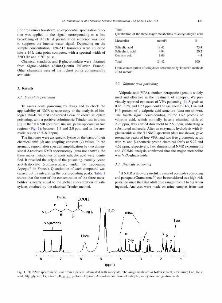
3.1. Salicylate poisoning
To assess acute poisoning by drugs and to check the
applicability of NMR spectroscopy to the analysis of bio-
logical fluids, we first considered a case of known salicylate
poisoning, with a positive colorimetric Trinder test in urine
[3]. In the 1H NMR spectrum, unusual peaks appeared in two
regions (Fig. 1): between 1.4 and 2.0 ppm and in the aro-
matic region (6.5–8.0 ppm).
The first ones were assigned to lysine on the basis of their
chemical shift (d) and coupling constant (J) values. In the
aromatic region, after spectral simplification by two dimen-
sional J-resolved NMR spectroscopy (data not shown), the
three major metabolites of acetylsalicylic acid were identi-
fied. It revealed the origin of the poisoning, namely lysine
acetylsalicylate (commercialized under the trade-name
Aspegic1 in France). Quantitation of each compound was
carried out by integrating the corresponding peaks. Table 1
shows that the sum of the concentration of the three meta-
bolites is nearly equal to the global concentration of sali-
cylates obtained by the classical Trinder method.
3.2. Valproic acid poisoning
Valproic acid (VPA), another therapeutic agent, is widely
used and effective in the treatment of epilepsy. We pre-
viously reported two cases of VPA poisoning [4]. Signals at
0.85, 1.28, and 1.53 ppm could be assigned to H-5, H-4 and
H-3 protons of a valproic acid structure (data not shown).
The fourth signal corresponding to the H-2 protons of
valproic acid, which normally have a chemical shift of
2.23 ppm, was shifted downfield to 2.53 ppm, indicating a
substituted molecule. After an enzymatic hydrolysis with b-
glucuronidase, the 1H NMR spectrum (data not shown) gave
resonance peaks of free VPA, and two free glucuronic acids
with a- and b-anomeric proton chemical shifts at 5.22 and
4.62 ppm, respectively. Two dimensional NMR experiments
and GC/MS analysis confirmed that the major metabolite
was VPA-glucuronide.
3.3. Pesticide poisoning
1H NMR is also very useful in cases of pesticides poisoning
and paraquat (Gramoxone1) can be considered as a high-risk
pesticide since the fatal adult dose ranges from 3 to 6 g when
ingested. Analyses were made on urine samples from two
Fig. 1. 1H NMR spectrum of urine from a patient intoxicated with salicylate. The assignments are as follows: creat, creatinine; Lac, lactic
acid; Gly, glycine; Ci, citrate.; Ha,b,g,d,e, protons of lysine; Ar-protons are those of salicylic, salicyluric and gentisic acids.
Table 1
Quantitation of the three major metabolites of acetylsalicylic acid
Metabolite mmol/l %
Salicylic acid 18.42 75.4
Salicyluric acid 4.94 20.2
Gentisic acid 1.06 4.4
Total 24.42 100
Urine concentration of salicylates determined by Trinder’s method:
22.81 mmol/l.
M. Imbenotte et al. / Forensic Science International 133 (2003) 132–135 133
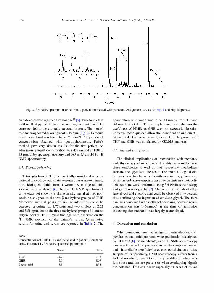
suicide cases who ingested Gramoxone1 [5]. Two doublets at
8.49 and 9.02 ppm with the same coupling constant of 6.3 Hz,
corresponded to the aromatic paraquat protons. The methyl
resonance appeared as a singlet at 4.48 ppm (Fig. 2). Paraquat
quantitation limit was found to be 25 mmol/l. Comparison of
concentration obtained with spectrophotometric Fuke’s
method gave very similar results: for the first patient, on
admission, parquat concentration was determined at 1081�33 mmol/l by spectrophotometry and 985 � 85 mmol/l by 1H
NMR spectroscopy.
3.4. Solvent poisoning
Tetrahydrofuran (THF) is essentially considered in occu-
pational toxicology, and acute poisoning cases are extremely
rare. Biological fluids from a woman who ingested this
solvent were analysed [6]. In the 1H NMR spectrum of
urine (data not shown), a characteristic signal at 1.90 ppm
could be assigned to the two b-methylene groups of THF.
Moreover, unusual peaks of similar intensities could be
detected: a quintet at 1.77 ppm and two triplets at 2.22
and 3.58 ppm, due to the three methylene groups of 4-amino
butyric acid (GHB). Similar findings were observed on the1H NMR spectrum of the patient’s serum. Quantitative
results for urine and serum are reported in Table 2. The
quantitation limit was found to be 0.1 mmol/l for THF and
0.4 mmol/l for GHB. This example strongly emphasizes the
usefulness of NMR, as GHB was not expected. No other
universal technique can allow the identification and quanti-
tation of GHB in the same analysis as THF. The presence of
THF and GHB was confirmed by GC/MS analyses.
3.5. Alcohol and glycols
The clinical implications of intoxication with methanol
and ethylene glycol are serious and fatality can result because
these xenobiotics as well as their respective metabolites,
formate and glycolate, are toxic. The main biological dis-
turbance is metabolic acidosis with an anionic gap. Analyses
of serum and urine samples from three patients in a metabolic
acidosis state were performed using 1H NMR spectroscopy
and gas chromatography [7]. Characteristic signals of ethy-
lene glycol and glycolic acid could be observed in two cases,
thus confirming the ingestion of ethylene glycol. The third
case was concerned with methanol poisoning: formate serum
concentration was 146 mmol/l at the time of admission
indicating that methanol was largely metabolised.
4. Discussion and conclusion
Other compounds such as analgesics, antiepileptics, anti-
psychotics and antidepressants were previously investigated
by 1H NMR [8]. Some advantages of 1H NMR spectroscopy
can be established: no pretreatment of the sample is needed
and it has reliable specificity based on spectral characteristics.
In spite of its specificity, NMR spectroscopy suffers from a
lack of sensitivity: quantitation may be difficult when very
low concentrations are present or when overlapping signals
are detected. This can occur especially in cases of mixed
Fig. 2. 1H NMR spectrum of urine from a patient intoxicated with paraquat. Assignments are as for Fig. 1 and Hip, hippurate.
Table 2
Concentrations of THF, GHB and lactic acid in patient’s serum and
urine, measured by 1H NMR spectroscopy (mmol/l)
Serum Urine
THF 11.3 11.8
GHB 2.3 28.6
Lactic acid 3.8 25.4
134 M. Imbenotte et al. / Forensic Science International 133 (2003) 132–135
poisoning. Spectral simplification can be achieved using
either multidimensional sequences or 31P NMR spectroscopy,
as in the case of organophosphorus poisonings [9]. As it is a
non-destructive technique, the samples can be later analysed
by complementary methods. Compared with chromato-
graphic or spectrophotometric methods, 1H NMR spectro-
scopy is rapid, only needs a small sample volume (500 ml)
and does not require time-consuming extraction and/or deri-
vatization steps. Moreover, in a single spectrum, three kinds
of information can be collected: the nature of the xenobiotic
compounds, the possible presence of their metabolites, even
of unexpected species such as GHB in the case of THF
poisoning, and finally the consecutive metabolic distur-
bances. NMR spectroscopy can document cases from direct
analysis of biological fluids and when possible, analysis of
the xenobiotic suspected to be at the origin of the poisoning.
In each presented case, quantitative data were validated
and satisfactorily correlated with reference methods, both
for xenobiotic compounds and endogenous abnormalities.
The inherent relatively low sensitivity of 1H NMR spectro-
scopy is counterbalanced by its simplicity and specificity.
This technique is not commonly used in hospital labora-
tories. In spite of this, physicians should be aware of this
laboratory method every time they face an acute poisoning
case in toxicological or forensic science fields.
References
[1] A.J. Poortman, E. Lock, Analytical profile of 4-methylthioam-
phetamine (4-MTA), a new street drug, Forensic Sci. Int. 100
(1999) 221–233.
[2] S.C. Szathmary, L. Von Tamaska, A. Steigel, Postmortem
decomposition of neutral lipids: use of modern methods of
analysis (HPLC, capillary GC, GC–MS and NMR) in
adipocere formation, Z. Rechtsmed. 94 (1985) 273–287.
[3] S. Maschke, N. Azaroual, M. Imbenotte, G. Vermeersch, F.
Leclerc, M. Lhermitte, Salicylate poisoning: two-dimensional
J-resolved NMR urinalysis, NMR Biomed. 8 (1995) 19–
24.
[4] N. Azaroual, M. Imbenotte, B. Cartigny, F. Leclerc, L. Vallee,
M. Lhermitte, G. Vermeersch, Valproic acid intoxication
identified by 1H and 1H–13C correlated NMR spectroscopy in
urine samples, MAGMA 10 (2000) 177–182.
[5] M. Imbenotte, N. Azaroual, D. Mathieu, B. Cartigny, G.
Vermeersch, M. Lhermitte, Determination by 1H NMR
spectroscopy of paraquat in urine from acutely poisoned
patients: comparison with second derivative spectroscopy
method, J. Anal. Toxicol. 23 (1999) 586–590.
[6] B. Cartigny, N. Azaroual, M. Imbenotte, N. Sadeg, F. Testart,
J. Richecoeur, G. Vermeersch, M. Lhermitte, 1H NMR
spectroscopy investigation of serum and urine in a case of
acute poisoning with tetrahydrofuran, J. Anal. Toxicol. 25
(2001) 270–274.
[7] A. Wahl, N. Azaroual, M. Imbenotte, D. Mathieu, G. Forzy, B.
Cartigny, G. Vermeersch, M. Lhermitte, Poisoning with
methanol and ethylene glycol; 1H NMR spectroscopy as an
effective clinical tool for diagnosis and quantification,
Toxicology 128 (1998) 73–81.
[8] E.M. Komoroski, R.A. Komoroski, J.L. Valentine, J.M. Pearce,
G.L. Kearns, The use of nuclear magnetic resonance spectro-
scopy in the detection of drug intoxication, J. Anal. Toxicol. 24
(2000) 180–186.
[9] A. Wahl, N. Azaroual, M. Imbenotte, D. Mathieu, G.
Vermeersch, C. Mereau, M. Lhermitte, 1H and 31P spectro-
scopy for determination of glycophosphate in human urine and
serum, J. Magn. Res. Anal. 4 (1998) 17–20.
M. Imbenotte et al. / Forensic Science International 133 (2003) 132–135 135


![Detection of Histamine Dihydrochloride at Low ...€¦ · spectroscopy, biological applications (bioimaging, biosensing, drug delivery), and catalysis [21,22] Histamine is a relevant](https://img.pdfslide.fr/doc/110x75/5ea0c82e88c5854e9a580eca/detection-of-histamine-dihydrochloride-at-low-spectroscopy-biological-applications.jpg)