Embed Size (px)
Citation preview
Illuminating the landscape of host–pathogeninteractions with the bacteriumListeria monocytogenesPascale Cossart1
Unité des Interactions Bactéries-Cellules, Département de Biologie Cellulaire et Infection, Institut Pasteur, F-75015 Paris, France; Institut National de la Santéet de la Recherche Médicale U604, F-75015 Paris, France; and Institut National de la Recherche Agronomique USC2020, F-75015 Paris, France
This contribution is part of the special series of Inaugural Articles by members of the National Academy of Sciences elected in 2009.
Contributed by Pascale Cossart, October 19, 2011 (sent for review June 20, 2011)
Listeria monocytogenes has, in 25 y, become a model in infectionbiology. Through the analysis of both its saprophytic life and in-fectious process, new concepts in microbiology, cell biology, andpathogenesis have been discovered. This review will update ourknowledge on this intracellular pathogen and highlight the mostrecent breakthroughs. Promising areas of investigation such as theincreasingly recognized relevance for the infectious process, ofRNA-mediated regulations in the bacterium, and the role of bac-terially controlled posttranslational and epigenetic modificationsin the host will also be discussed.
bacterial invasion | mitochondria | posttranslational modifications |epigenetics
Listeria monocytogenes was discovered in 1926 during an epi-demic that affected rabbits and guinea pigs (1). It was later
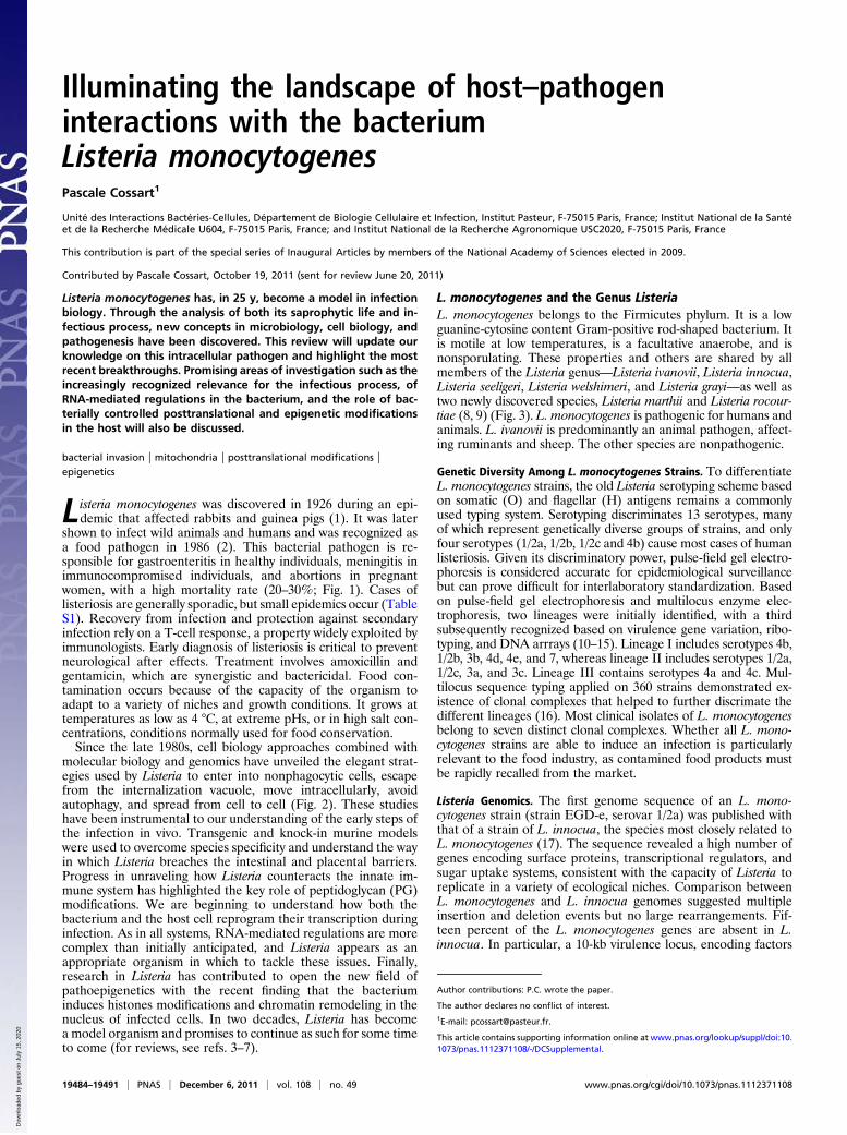
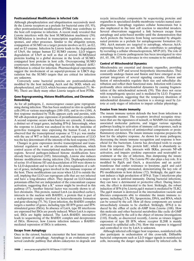
shown to infect wild animals and humans and was recognized asa food pathogen in 1986 (2). This bacterial pathogen is re-sponsible for gastroenteritis in healthy individuals, meningitis inimmunocompromised individuals, and abortions in pregnantwomen, with a high mortality rate (20–30%; Fig. 1). Cases oflisteriosis are generally sporadic, but small epidemics occur (TableS1). Recovery from infection and protection against secondaryinfection rely on a T-cell response, a property widely exploited byimmunologists. Early diagnosis of listeriosis is critical to preventneurological after effects. Treatment involves amoxicillin andgentamicin, which are synergistic and bactericidal. Food con-tamination occurs because of the capacity of the organism toadapt to a variety of niches and growth conditions. It grows attemperatures as low as 4 °C, at extreme pHs, or in high salt con-centrations, conditions normally used for food conservation.Since the late 1980s, cell biology approaches combined with
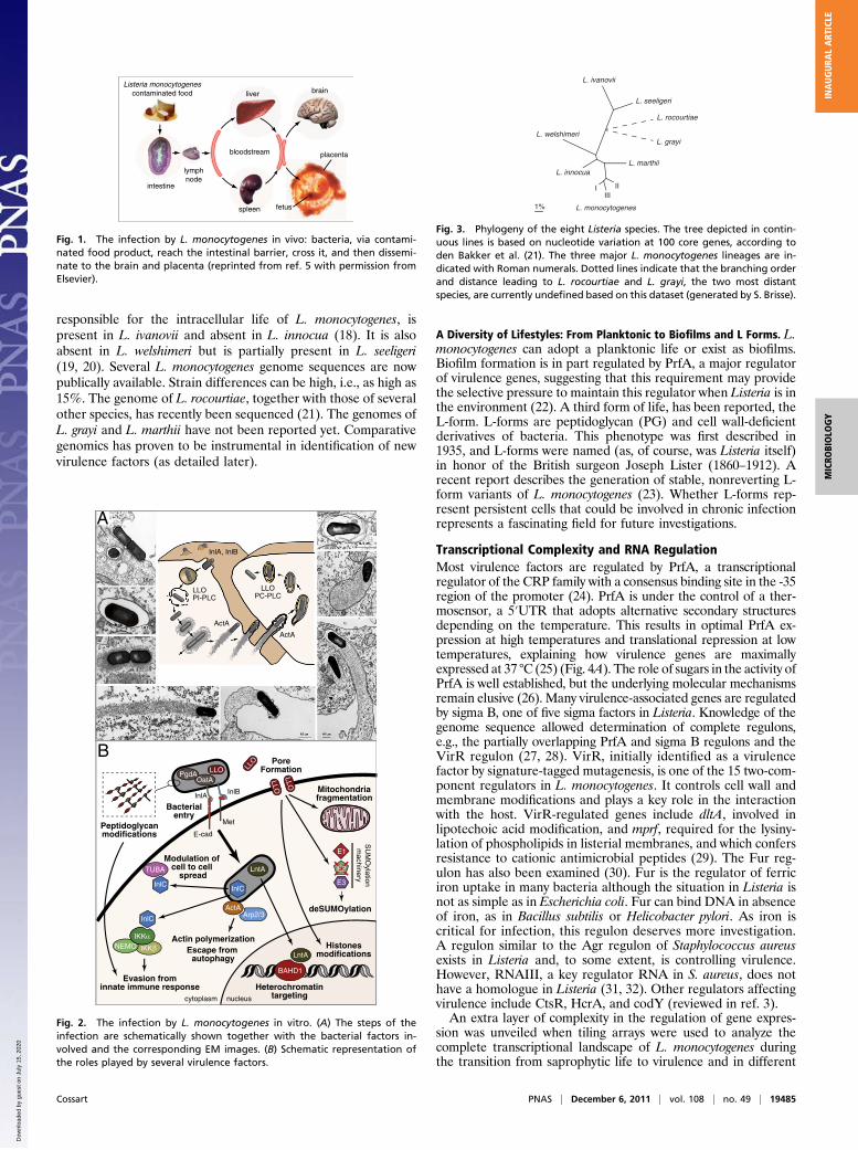
molecular biology and genomics have unveiled the elegant strat-egies used by Listeria to enter into nonphagocytic cells, escapefrom the internalization vacuole, move intracellularly, avoidautophagy, and spread from cell to cell (Fig. 2). These studieshave been instrumental to our understanding of the early steps ofthe infection in vivo. Transgenic and knock-in murine modelswere used to overcome species specificity and understand the wayin which Listeria breaches the intestinal and placental barriers.Progress in unraveling how Listeria counteracts the innate im-mune system has highlighted the key role of peptidoglycan (PG)modifications. We are beginning to understand how both thebacterium and the host cell reprogram their transcription duringinfection. As in all systems, RNA-mediated regulations are morecomplex than initially anticipated, and Listeria appears as anappropriate organism in which to tackle these issues. Finally,research in Listeria has contributed to open the new field ofpathoepigenetics with the recent finding that the bacteriuminduces histones modifications and chromatin remodeling in thenucleus of infected cells. In two decades, Listeria has becomea model organism and promises to continue as such for some timeto come (for reviews, see refs. 3–7).
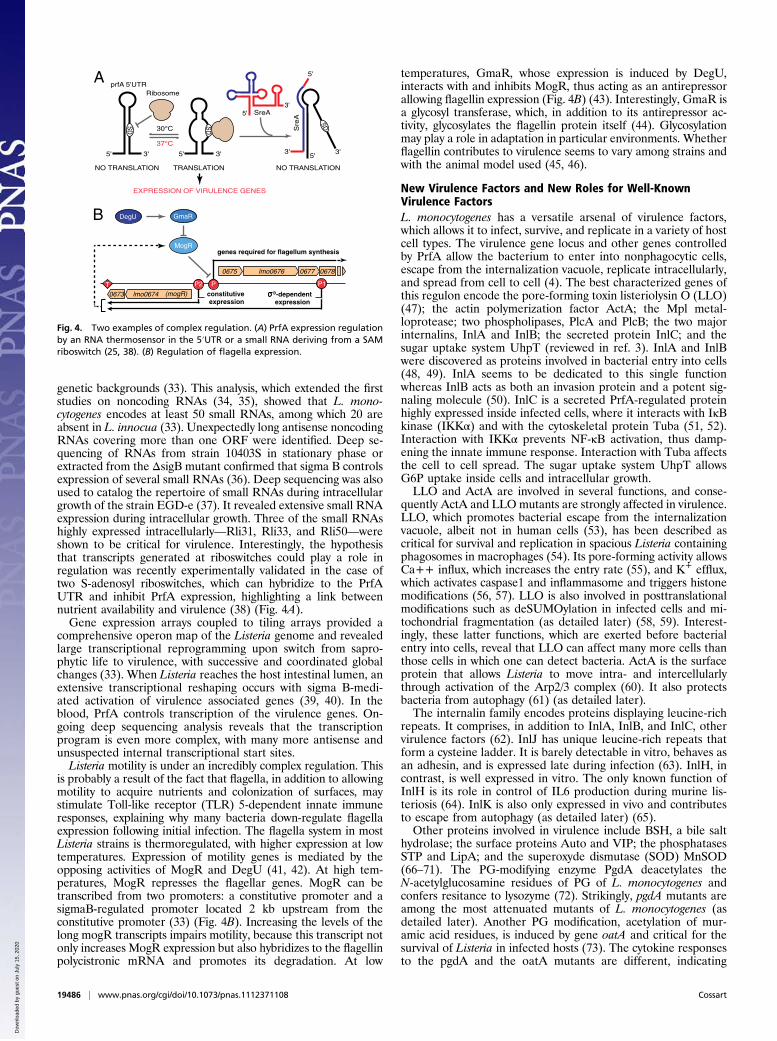
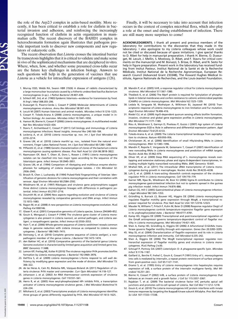
L. monocytogenes and the Genus ListeriaL. monocytogenes belongs to the Firmicutes phylum. It is a lowguanine-cytosine content Gram-positive rod-shaped bacterium. Itis motile at low temperatures, is a facultative anaerobe, and isnonsporulating. These properties and others are shared by allmembers of the Listeria genus—Listeria ivanovii, Listeria innocua,Listeria seeligeri, Listeria welshimeri, and Listeria grayi—as well astwo newly discovered species, Listeria marthii and Listeria rocour-tiae (8, 9) (Fig. 3). L. monocytogenes is pathogenic for humans andanimals. L. ivanovii is predominantly an animal pathogen, affect-ing ruminants and sheep. The other species are nonpathogenic.
Genetic Diversity Among L. monocytogenes Strains. To differentiateL. monocytogenes strains, the old Listeria serotyping scheme basedon somatic (O) and flagellar (H) antigens remains a commonlyused typing system. Serotyping discriminates 13 serotypes, manyof which represent genetically diverse groups of strains, and onlyfour serotypes (1/2a, 1/2b, 1/2c and 4b) cause most cases of humanlisteriosis. Given its discriminatory power, pulse-field gel electro-phoresis is considered accurate for epidemiological surveillancebut can prove difficult for interlaboratory standardization. Basedon pulse-field gel electrophoresis and multilocus enzyme elec-trophoresis, two lineages were initially identified, with a thirdsubsequently recognized based on virulence gene variation, ribo-typing, and DNA arrrays (10–15). Lineage I includes serotypes 4b,1/2b, 3b, 4d, 4e, and 7, whereas lineage II includes serotypes 1/2a,1/2c, 3a, and 3c. Lineage III contains serotypes 4a and 4c. Mul-tilocus sequence typing applied on 360 strains demonstrated ex-istence of clonal complexes that helped to further discrimate thedifferent lineages (16). Most clinical isolates of L. monocytogenesbelong to seven distinct clonal complexes. Whether all L. mono-cytogenes strains are able to induce an infection is particularlyrelevant to the food industry, as contamined food products mustbe rapidly recalled from the market.
Listeria Genomics. The first genome sequence of an L. mono-cytogenes strain (strain EGD-e, serovar 1/2a) was published withthat of a strain of L. innocua, the species most closely related toL. monocytogenes (17). The sequence revealed a high number ofgenes encoding surface proteins, transcriptional regulators, andsugar uptake systems, consistent with the capacity of Listeria toreplicate in a variety of ecological niches. Comparison betweenL. monocytogenes and L. innocua genomes suggested multipleinsertion and deletion events but no large rearrangements. Fif-teen percent of the L. monocytogenes genes are absent in L.innocua. In particular, a 10-kb virulence locus, encoding factors
Author contributions: P.C. wrote the paper.
The author declares no conflict of interest.1E-mail: [email protected].
This article contains supporting information online at www.pnas.org/lookup/suppl/doi:10.1073/pnas.1112371108/-/DCSupplemental.
19484–19491 | PNAS | December 6, 2011 | vol. 108 | no. 49 www.pnas.org/cgi/doi/10.1073/pnas.1112371108
Dow
nloa
ded
by g
uest
on
July
15,
202
0
responsible for the intracellular life of L. monocytogenes, ispresent in L. ivanovii and absent in L. innocua (18). It is alsoabsent in L. welshimeri but is partially present in L. seeligeri(19, 20). Several L. monocytogenes genome sequences are nowpublically available. Strain differences can be high, i.e., as high as15%. The genome of L. rocourtiae, together with those of severalother species, has recently been sequenced (21). The genomes ofL. grayi and L. marthii have not been reported yet. Comparativegenomics has proven to be instrumental in identification of newvirulence factors (as detailed later).
A Diversity of Lifestyles: From Planktonic to Biofilms and L Forms. L.monocytogenes can adopt a planktonic life or exist as biofilms.Biofilm formation is in part regulated by PrfA, a major regulatorof virulence genes, suggesting that this requirement may providethe selective pressure to maintain this regulator when Listeria is inthe environment (22). A third form of life, has been reported, theL-form. L-forms are peptidoglycan (PG) and cell wall-deficientderivatives of bacteria. This phenotype was first described in1935, and L-forms were named (as, of course, was Listeria itself)in honor of the British surgeon Joseph Lister (1860–1912). Arecent report describes the generation of stable, nonreverting L-form variants of L. monocytogenes (23). Whether L-forms rep-resent persistent cells that could be involved in chronic infectionrepresents a fascinating field for future investigations.
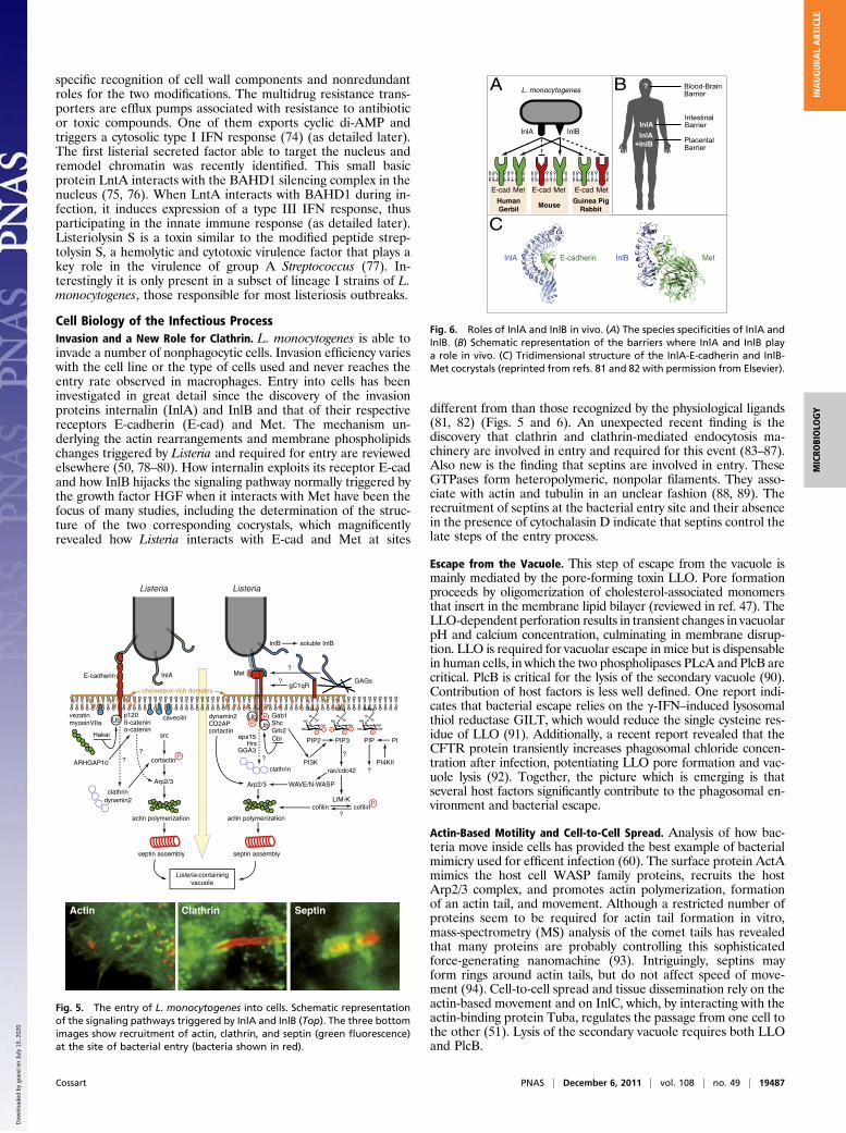
Transcriptional Complexity and RNA RegulationMost virulence factors are regulated by PrfA, a transcriptionalregulator of the CRP family with a consensus binding site in the -35region of the promoter (24). PrfA is under the control of a ther-mosensor, a 5′UTR that adopts alternative secondary structuresdepending on the temperature. This results in optimal PrfA ex-pression at high temperatures and translational repression at lowtemperatures, explaining how virulence genes are maximallyexpressed at 37 °C (25) (Fig. 4A). The role of sugars in the activity ofPrfA is well established, but the underlying molecular mechanismsremain elusive (26). Many virulence-associated genes are regulatedby sigma B, one of five sigma factors in Listeria. Knowledge of thegenome sequence allowed determination of complete regulons,e.g., the partially overlapping PrfA and sigma B regulons and theVirR regulon (27, 28). VirR, initially identified as a virulencefactor by signature-tagged mutagenesis, is one of the 15 two-com-ponent regulators in L. monocytogenes. It controls cell wall andmembrane modifications and plays a key role in the interactionwith the host. VirR-regulated genes include dltA, involved inlipotechoic acid modification, and mprf, required for the lysiny-lation of phospholipids in listerial membranes, and which confersresistance to cationic antimicrobial peptides (29). The Fur reg-ulon has also been examined (30). Fur is the regulator of ferriciron uptake in many bacteria although the situation in Listeria isnot as simple as in Escherichia coli. Fur can bind DNA in absenceof iron, as in Bacillus subtilis or Helicobacter pylori. As iron iscritical for infection, this regulon deserves more investigation.A regulon similar to the Agr regulon of Staphylococcus aureusexists in Listeria and, to some extent, is controlling virulence.However, RNAIII, a key regulator RNA in S. aureus, does nothave a homologue in Listeria (31, 32). Other regulators affectingvirulence include CtsR, HcrA, and codY (reviewed in ref. 3).An extra layer of complexity in the regulation of gene expres-
sion was unveiled when tiling arrays were used to analyze thecomplete transcriptional landscape of L. monocytogenes duringthe transition from saprophytic life to virulence and in different
Listeria monocytogenescontaminated food
intestine
lymphnode
liver
spleen
brain
fetus
placentabloodstream
Fig. 1. The infection by L. monocytogenes in vivo: bacteria, via contami-nated food product, reach the intestinal barrier, cross it, and then dissemi-nate to the brain and placenta (reprinted from ref. 5 with permission fromElsevier).
NEMO
OatA
InlBInlA
Met
E-cad
LLOPore
Formation
E3
E1
InlC
LntA
Histonesmodifications
Bacterialentry
Actin polymerizationEscape from
autophagy
Heterochromatintargeting
deSUMOylation
PgdA
LLO
LLO
LLO
Peptidoglycanmodifications
E2
SU
MO
ylationm
achinery
LntA
ActAArp2/3
Mitochondriafragmentation
InlC
Evasion frominnate immune response
B
BAHD1
A
LLOPI-PLC
ActA
ActA
LLOPC-PLC
InlA, InlB
cytoplasm nucleus
Modulation of cell to cell
spreadTUBA
InlC
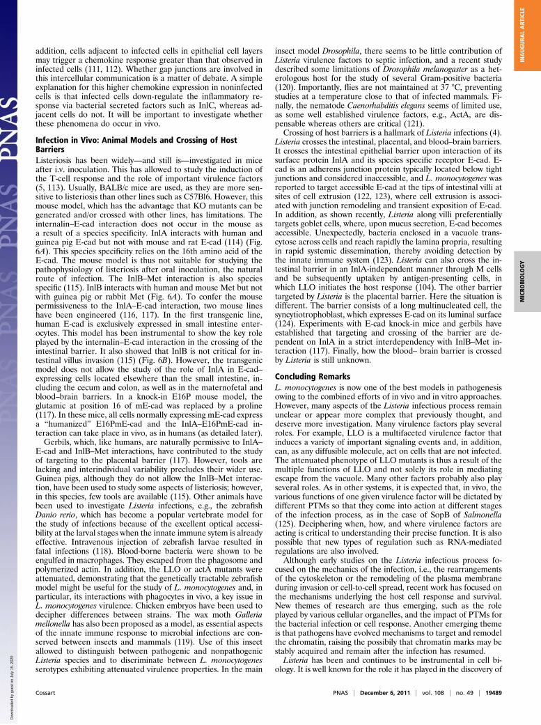
Fig. 2. The infection by L. monocytogenes in vitro. (A) The steps of theinfection are schematically shown together with the bacterial factors in-volved and the corresponding EM images. (B) Schematic representation ofthe roles played by several virulence factors.
L. seeligeri
L. marthii
L. monocytogenes
L. innocua
L. ivanovii
L. welshimeri
1%
I IIIII
L. rocourtiae
L. grayi
Fig. 3. Phylogeny of the eight Listeria species. The tree depicted in contin-uous lines is based on nucleotide variation at 100 core genes, according toden Bakker et al. (21). The three major L. monocytogenes lineages are in-dicated with Roman numerals. Dotted lines indicate that the branching orderand distance leading to L. rocourtiae and L. grayi, the two most distantspecies, are currently undefined based on this dataset (generated by S. Brisse).
Cossart PNAS | December 6, 2011 | vol. 108 | no. 49 | 19485
MICRO
BIOLO
GY
INAUGURA
LART
ICLE
Dow
nloa
ded
by g
uest
on
July
15,
202
0
genetic backgrounds (33). This analysis, which extended the firststudies on noncoding RNAs (34, 35), showed that L. mono-cytogenes encodes at least 50 small RNAs, among which 20 areabsent in L. innocua (33). Unexpectedly long antisense noncodingRNAs covering more than one ORF were identified. Deep se-quencing of RNAs from strain 10403S in stationary phase orextracted from the ΔsigB mutant confirmed that sigma B controlsexpression of several small RNAs (36). Deep sequencing was alsoused to catalog the repertoire of small RNAs during intracellulargrowth of the strain EGD-e (37). It revealed extensive small RNAexpression during intracellular growth. Three of the small RNAshighly expressed intracellularly—Rli31, Rli33, and Rli50—wereshown to be critical for virulence. Interestingly, the hypothesisthat transcripts generated at riboswitches could play a role inregulation was recently experimentally validated in the case oftwo S-adenosyl riboswitches, which can hybridize to the PrfAUTR and inhibit PrfA expression, highlighting a link betweennutrient availability and virulence (38) (Fig. 4A).Gene expression arrays coupled to tiling arrays provided a
comprehensive operon map of the Listeria genome and revealedlarge transcriptional reprogramming upon switch from sapro-phytic life to virulence, with successive and coordinated globalchanges (33). When Listeria reaches the host intestinal lumen, anextensive transcriptional reshaping occurs with sigma B-medi-ated activation of virulence associated genes (39, 40). In theblood, PrfA controls transcription of the virulence genes. On-going deep sequencing analysis reveals that the transcriptionprogram is even more complex, with many more antisense andunsuspected internal transcriptional start sites.Listeria motility is under an incredibly complex regulation. This
is probably a result of the fact that flagella, in addition to allowingmotility to acquire nutrients and colonization of surfaces, maystimulate Toll-like receptor (TLR) 5-dependent innate immuneresponses, explaining why many bacteria down-regulate flagellaexpression following initial infection. The flagella system in mostListeria strains is thermoregulated, with higher expression at lowtemperatures. Expression of motility genes is mediated by theopposing activities of MogR and DegU (41, 42). At high tem-peratures, MogR represses the flagellar genes. MogR can betranscribed from two promoters: a constitutive promoter and asigmaB-regulated promoter located 2 kb upstream from theconstitutive promoter (33) (Fig. 4B). Increasing the levels of thelong mogR transcripts impairs motility, because this transcript notonly increases MogR expression but also hybridizes to the flagellinpolycistronic mRNA and promotes its degradation. At low
temperatures, GmaR, whose expression is induced by DegU,interacts with and inhibits MogR, thus acting as an antirepressorallowing flagellin expression (Fig. 4B) (43). Interestingly, GmaR isa glycosyl transferase, which, in addition to its antirepressor ac-tivity, glycosylates the flagellin protein itself (44). Glycosylationmay play a role in adaptation in particular environments. Whetherflagellin contributes to virulence seems to vary among strains andwith the animal model used (45, 46).
New Virulence Factors and New Roles for Well-KnownVirulence FactorsL. monocytogenes has a versatile arsenal of virulence factors,which allows it to infect, survive, and replicate in a variety of hostcell types. The virulence gene locus and other genes controlledby PrfA allow the bacterium to enter into nonphagocytic cells,escape from the internalization vacuole, replicate intracellularly,and spread from cell to cell (4). The best characterized genes ofthis regulon encode the pore-forming toxin listeriolysin O (LLO)(47); the actin polymerization factor ActA; the Mpl metal-loprotease; two phospholipases, PlcA and PlcB; the two majorinternalins, InlA and InlB; the secreted protein InlC; and thesugar uptake system UhpT (reviewed in ref. 3). InlA and InlBwere discovered as proteins involved in bacterial entry into cells(48, 49). InlA seems to be dedicated to this single functionwhereas InlB acts as both an invasion protein and a potent sig-naling molecule (50). InlC is a secreted PrfA-regulated proteinhighly expressed inside infected cells, where it interacts with IκBkinase (IKKα) and with the cytoskeletal protein Tuba (51, 52).Interaction with IKKα prevents NF-κB activation, thus damp-ening the innate immune response. Interaction with Tuba affectsthe cell to cell spread. The sugar uptake system UhpT allowsG6P uptake inside cells and intracellular growth.LLO and ActA are involved in several functions, and conse-
quently ActA and LLOmutants are strongly affected in virulence.LLO, which promotes bacterial escape from the internalizationvacuole, albeit not in human cells (53), has been described ascritical for survival and replication in spacious Listeria containingphagosomes in macrophages (54). Its pore-forming activity allowsCa++ influx, which increases the entry rate (55), and K+ efflux,which activates caspase1 and inflammasome and triggers histonemodifications (56, 57). LLO is also involved in posttranslationalmodifications such as deSUMOylation in infected cells and mi-tochondrial fragmentation (as detailed later) (58, 59). Interest-ingly, these latter functions, which are exerted before bacterialentry into cells, reveal that LLO can affect many more cells thanthose cells in which one can detect bacteria. ActA is the surfaceprotein that allows Listeria to move intra- and intercellularlythrough activation of the Arp2/3 complex (60). It also protectsbacteria from autophagy (61) (as detailed later).The internalin family encodes proteins displaying leucine-rich
repeats. It comprises, in addition to InlA, InlB, and InlC, othervirulence factors (62). InlJ has unique leucine-rich repeats thatform a cysteine ladder. It is barely detectable in vitro, behaves asan adhesin, and is expressed late during infection (63). InlH, incontrast, is well expressed in vitro. The only known function ofInlH is its role in control of IL6 production during murine lis-teriosis (64). InlK is also only expressed in vivo and contributesto escape from autophagy (as detailed later) (65).Other proteins involved in virulence include BSH, a bile salt
hydrolase; the surface proteins Auto and VIP; the phosphatasesSTP and LipA; and the superoxyde dismutase (SOD) MnSOD(66–71). The PG-modifying enzyme PgdA deacetylates theN-acetylglucosamine residues of PG of L. monocytogenes andconfers resitance to lysozyme (72). Strikingly, pgdA mutants areamong the most attenuated mutants of L. monocytogenes (asdetailed later). Another PG modification, acetylation of mur-amic acid residues, is induced by gene oatA and critical for thesurvival of Listeria in infected hosts (73). The cytokine responsesto the pgdA and the oatA mutants are different, indicating
37°C
30°C
SD
prfA 5'UTRRibosome
5' 3'
NO TRANSLATION
Sre
A
5'
3'5'
3'
NO TRANSLATION
5' 3'
TRANSLATION
EXPRESSION OF VIRULENCE GENES
SreA5'3'
lmo0674 (mogR)0673
0675
MogR
GmaR
constitutive expression
B-dependentexpression
genes required for flagellum synthesis
B
SD
A
SD
DegU
lmo0676 0677 0678
P2 P P1T
Fig. 4. Two examples of complex regulation. (A) PrfA expression regulationby an RNA thermosensor in the 5′UTR or a small RNA deriving from a SAMriboswitch (25, 38). (B) Regulation of flagella expression.
19486 | www.pnas.org/cgi/doi/10.1073/pnas.1112371108 Cossart
Dow
nloa
ded
by g
uest
on
July
15,
202
0
specific recognition of cell wall components and nonredundantroles for the two modifications. The multidrug resistance trans-porters are efflux pumps associated with resistance to antibioticor toxic compounds. One of them exports cyclic di-AMP andtriggers a cytosolic type I IFN response (74) (as detailed later).The first listerial secreted factor able to target the nucleus andremodel chromatin was recently identified. This small basicprotein LntA interacts with the BAHD1 silencing complex in thenucleus (75, 76). When LntA interacts with BAHD1 during in-fection, it induces expression of a type III IFN response, thusparticipating in the innate immune response (as detailed later).Listeriolysin S is a toxin similar to the modified peptide strep-tolysin S, a hemolytic and cytotoxic virulence factor that plays akey role in the virulence of group A Streptococcus (77). In-terestingly it is only present in a subset of lineage I strains of L.monocytogenes, those responsible for most listeriosis outbreaks.
Cell Biology of the Infectious ProcessInvasion and a New Role for Clathrin. L. monocytogenes is able toinvade a number of nonphagocytic cells. Invasion efficiency varieswith the cell line or the type of cells used and never reaches theentry rate observed in macrophages. Entry into cells has beeninvestigated in great detail since the discovery of the invasionproteins internalin (InlA) and InlB and that of their respectivereceptors E-cadherin (E-cad) and Met. The mechanism un-derlying the actin rearrangements and membrane phospholipidschanges triggered by Listeria and required for entry are reviewedelsewhere (50, 78–80). How internalin exploits its receptor E-cadand how InlB hijacks the signaling pathway normally triggered bythe growth factor HGF when it interacts with Met have been thefocus of many studies, including the determination of the struc-ture of the two corresponding cocrystals, which magnificentlyrevealed how Listeria interacts with E-cad and Met at sites
different from than those recognized by the physiological ligands(81, 82) (Figs. 5 and 6). An unexpected recent finding is thediscovery that clathrin and clathrin-mediated endocytosis ma-chinery are involved in entry and required for this event (83–87).Also new is the finding that septins are involved in entry. TheseGTPases form heteropolymeric, nonpolar filaments. They asso-ciate with actin and tubulin in an unclear fashion (88, 89). Therecruitment of septins at the bacterial entry site and their absencein the presence of cytochalasin D indicate that septins control thelate steps of the entry process.
Escape from the Vacuole. This step of escape from the vacuole ismainly mediated by the pore-forming toxin LLO. Pore formationproceeds by oligomerization of cholesterol-associated monomersthat insert in the membrane lipid bilayer (reviewed in ref. 47). TheLLO-dependent perforation results in transient changes in vacuolarpH and calcium concentration, culminating in membrane disrup-tion. LLO is required for vacuolar escape in mice but is dispensablein human cells, in which the two phospholipases PLcA and PlcB arecritical. PlcB is critical for the lysis of the secondary vacuole (90).Contribution of host factors is less well defined. One report indi-cates that bacterial escape relies on the γ-IFN–induced lysosomalthiol reductase GILT, which would reduce the single cysteine res-idue of LLO (91). Additionally, a recent report revealed that theCFTR protein transiently increases phagosomal chloride concen-tration after infection, potentiating LLO pore formation and vac-uole lysis (92). Together, the picture which is emerging is thatseveral host factors significantly contribute to the phagosomal en-vironment and bacterial escape.
Actin-Based Motility and Cell-to-Cell Spread. Analysis of how bac-teria move inside cells has provided the best example of bacterialmimicry used for efficent infection (60). The surface protein ActAmimics the host cell WASP family proteins, recruits the hostArp2/3 complex, and promotes actin polymerization, formationof an actin tail, and movement. Although a restricted number ofproteins seem to be required for actin tail formation in vitro,mass-spectrometry (MS) analysis of the comet tails has revealedthat many proteins are probably controlling this sophisticatedforce-generating nanomachine (93). Intriguingly, septins mayform rings around actin tails, but do not affect speed of move-ment (94). Cell-to-cell spread and tissue dissemination rely on theactin-based movement and on InlC, which, by interacting with theactin-binding protein Tuba, regulates the passage from one cell tothe other (51). Lysis of the secondary vacuole requires both LLOand PlcB.
Actin Clathrin
Cbl
Gab1Shc
PI3K
PIP2 PIP3
Grb2eps15
HrsGGA3
dynamin2CD2APcortactin
clathrin
WAVE/N-WASP
rac/cdc42
LIM-Kcofilin cofilin
?
?
InlB
Met
gC1qR?
cholesterol-rich domains
GAGs
?
p120-catenin-catenin
ARHGAP10
Arp2/3
actin polymerization
myosinVIIavezatin
src
cortactinP
E-cadherin InlA
clathrindynamin2
?
PIP
PI4KII
PI
?
soluble InlB
??
caveolin
Listeria Listeria
Listeria-containingvacuole
Hakai
septin assembly
Ub
actin polymerization
septin assembly
UbP
Arp2/3
PUb O
OP
O
OHHO
HO
OH
O
P
O
OP
O
OH
HO
O
PPP
O
OP
O
OHHO
HO
O
PP
Septin
P
Fig. 5. The entry of L. monocytogenes into cells. Schematic representationof the signaling pathways triggered by InlA and InlB (Top). The three bottomimages show recruitment of actin, clathrin, and septin (green fluorescence)at the site of bacterial entry (bacteria shown in red).
InlA E-cadherin InlB Met
Blood-Brain Barrier
IntestinalBarrier
PlacentalBarrier
InlA
InlA+InlB
?A
C
BL. monocytogenes
InlA InlB
MetE-cad
HumanGerbil
MetE-cad
Mouse
MetE-cad
Guinea PigRabbit
Fig. 6. Roles of InlA and InlB in vivo. (A) The species specificities of InlA andInlB. (B) Schematic representation of the barriers where InlA and InlB playa role in vivo. (C) Tridimensional structure of the InlA-E-cadherin and InlB-Met cocrystals (reprinted from refs. 81 and 82 with permission from Elsevier).
Cossart PNAS | December 6, 2011 | vol. 108 | no. 49 | 19487
MICRO
BIOLO
GY
INAUGURA
LART
ICLE
Dow
nloa
ded
by g
uest
on
July
15,
202
0
Posttranslational Modifications in Infected CellsAlthough phosphorylation and ubiquitination successively mod-ify the Listeria receptors as a prelude for bacterial entry (83, 95),other posttranslational modifications (PTMs) may contribute tothe host cell response to infection. A recent study revealed thatListeria interferes with the host SUMOylation machinery (58).SUMOylation is involved in transcription regulation, stress re-sponses, and other processes. Similar to the ubiquitin system,conjugation of SUMO on a target protein involves an E1, an E2,and an E3 enzyme. Infection by Listeria leads to the degradationof Ubc9, the unique human E2 SUMO enzyme. LLO triggersdegradation of Ubc9 as well as that of several SUMOylatedproteins leading to a global decrease in the levels of SUMO-conjugated host proteins in host cells. Overexpressing SUMOcounteracts infection revealing that bacterially induced deSU-MOylation is critical for infection. Pharmacological experimentssuggest the involvement of an aspartyl-protease in UBC9 deg-radation but the SUMO targets that are critical for infectionare unknown.Conversely some bacterial proteins are posttranslationally
modified by the host including ActA and the SOD, which arephosphorylated, and LLO, which becomes ubiquitinated (71, 96–98). There are likely many other Listeria targets of host PTMs.
Gene Reprogramming, Histone Modifications, and ChromatinRemodelingAs for all pathogens, L. monocytogenes causes gene reprogram-ming during infection. This has been analyzed in vitro in epithelialcells (99) or various macrophages (100–103), and in vivo (104). Afirst response mediated by TLRs appears early and persists, withNF-κB–dependent gene expression of proinflammatory cytokines.A second response occurs when bacteria are cytosolic. It inducesa distinct set of target genes, including IFN-β. Interestingly, whenthe response to Listeria was analyzed in vivo in the instestine ofgerm-free transgenic mice expressing the human E-cad, it wasobserved that the transcriptional response at 72 h p.i. was similarwith the use of WT or InlA mutant, and that expression of LLOwas the key determinant of the intensity of the host response (104).Changes in gene expression involve transcriptional and trans-
lational regulators as well as chromatin modifications, whichcontrol access of the transcriptional machinery to the promoterregions. Several pathogen-induced histone modifications havealready been reported (105). Interestingly, Listeria also induceshistone modifications during infection (56). Dephosphorylationof serine 10 of histone H3 and deacetylation of H4 were shown tobe LLO-dependent and to lead to the down-regulation of a sub-set of genes, including genes involved in the immune response ofthe host. These modifications can occur when LLO is outside thecell, implying that LLO can reprogram cells that are not infectedand have a long-distance effect. They depend on LLO-inducedpotassium efflux but are independent of the concomitant caspaseactivation, suggesting that a K+ sensor might be involved in thispathway (57). Another listerial factor was recently shown to af-fect chromatin. This protein, named LntA (as described earlier),interacts in the nucleus with BAHD1, a protein that is part ofa multiprotein complex involved in heterochromatin formationand gene silencing (75, 76). Upon infection, the BAHD1 complextargets a number of genes, including type III IFN genes and IFN-stimulated genes (ISGs). In absence of LntA, type III interferonsare expressed but ISGs are poorly induced. When LntA is expres-sed, ISGs are highly induced. The LntA–BAHD1 interactionleads to sequestering of the BAHD1 complex and derepressionof ISGs. However, how Listeria infection induces a BAHD1-mediated repression of ISGs is unknown.
Escape from AutophagyOnce in the cytosol, bacteria encounter the host innate surveil-lance system of autophagy. Autophagy is an evolutionary con-served catabolic pathway that allows eukaryotes to degrade and
recycle intracellular components by sequestering proteins andorganelles in specialized double-membrane vesicles named auto-phagosomes. Autophagy regulates cellular homeostasis but isalso implicated in the host cell reaction to microbial invaders.Several observations suggested a link between escape fromautophagy and actin-based motility until the demonstration thatActA protects the bacteria from being surrounded by ubiquiti-nated proteins as a prelude for autophagosome formation (61,106). ActA mutants are targeted to autophagy, whereas ActA-expressing bacteria are not. InlK also contributes to autophagyby recruiting a cellular ribonucleoprotein, MVP (65). The role ofautophagy in Listeria pathogenesis has been investigated in vitro(61, 65, 106, 107). Its relevance in vivo remains to be established.
Control of Mitochondrial DynamicsMitochondria are essential and dynamic organelles, providingmost cellular ATP and several biosynthetic intermediates. Theyconstantly undergo fusion and fission and have emerged as im-portant integrators of several signaling cascades. Fusion andfission of mitochondria regulate their size and subcellular dis-tribution and reflect their functional state. Infection with Listeriaprofoundly alters mitochondrial dynamics by causing fragmen-tation of the mitochondrial network (59). This does not occurwith nonpathogenic species and is mediated by LLO. Fragmen-tation is transient, suggesting that the temporary shutdown ofmitochondrial dynamics and function is a strategy used by Lis-teria at early stages of infection to impair cellular physiology.
Evasion from Innate ImmunityThe innate immune system defends the host from infections ina nonspecific manner. The receptors involved recognize struc-tures that are the signatures of nonself, or MAMPs for microbial-associated molecular patterns, and trigger various signalingpathways leading to activation of immune cell responses, such asexpression and secretion of antimicrobial components or proin-flammatory cytokines. The innate immune response prepares thehost for the adaptive response. As shown for many pathogens,decreasing the innate immune recognition and response is ben-eficial for the bacterium. Listeria has developed tools to escapefrom this response. The protein InlC, which is abundantly se-creted in infected cells, interacts with IKKα, prevents NF-κBactivation, and impairs cytokine expression and neutrophil re-cruitment at the site of infection, thus dampening the innateimmune response (52). The Listeria PG also plays a key role. It ismodified by PgdA and OatA, a deacetylase and an acetyl-transferase that confer resistance to lysozyme. pgdA and oatAmutants are strongly attenuated, demonstrating the key role ofPG modifications in host defense (72). Strikingly, the pgdA mu-tant induces a high production of IFN-β. Type I interferons playa major role in antiviral immunity. During bacterial infections,they can have a detrimental or protective effect. During listeri-osis, the effect is detrimental to the host. Strikingly, the robustinduction of IFN-β by Listeria pgdAmutant is mediated by TLR2.The pgdA mutant is destroyed in the internalization vacuole andliberates components able to stimulate the innate immune re-sponse, including LTA, PG, lipoproteins, DNA, and RNA, whichcan be sensed by the cell. How all these components are sensedintracellularly remains to be clarified. Strikingly, IFN-β is in-duced by the efflux of cyclic di-AMP from Listeria (108). Howthis molecule and other bacterial metabolites like cyclic di-GMP(109) are sensed by the cell is the object of intense investigations(110). Finally, as discovered recently, Listeria as viruses triggersexpression of type III IFN, whose receptor IL28R is mainlyexpressed in epithelial cells (76). How this response is triggeredand controlled in vivo by LntA is unknown.Although infected cells trigger host responses, noninfected cells
also participate in orchestration of this cross-talk. Diffusiblebacterial components such as LLO trigger signals in noninfectedcells, increasing the danger signals induced by infected cells. In
19488 | www.pnas.org/cgi/doi/10.1073/pnas.1112371108 Cossart
Dow
nloa
ded
by g
uest
on
July
15,
202
0
addition, cells adjacent to infected cells in epithelial cell layersmay trigger a chemokine response greater than that observed ininfected cells (111, 112). Whether gap junctions are involved inthis intercellular communication is a matter of debate. A simpleexplanation for this higher chemokine expression in noninfectedcells is that infected cells down-regulate the inflammatory re-sponse via bacterial secreted factors such as InlC, whereas ad-jacent cells do not. It will be important to investigate whetherthese phenomena do occur in vivo.
Infection in Vivo: Animal Models and Crossing of HostBarriersListeriosis has been widely—and still is—investigated in miceafter i.v. inoculation. This has allowed to study the induction ofthe T-cell response and the role of important virulence factors(5, 113). Usually, BALB/c mice are used, as they are more sen-sitive to listeriosis than other lines such as C57Bl6. However, thismouse model, which has the advantage that KO mutants can begenerated and/or crossed with other lines, has limitations. Theinternalin–E-cad interaction does not occur in the mouse asa result of a species specificity. InlA interacts with human andguinea pig E-cad but not with mouse and rat E-cad (114) (Fig.6A). This species specificity relies on the 16th amino acid of theE-cad. The mouse model is thus not suitable for studying thepathophysiology of listeriosis after oral inoculation, the naturalroute of infection. The InlB–Met interaction is also speciesspecific (115). InlB interacts with human and mouse Met but notwith guinea pig or rabbit Met (Fig. 6A). To confer the mousepermissiveness to the InlA–E-cad interaction, two mouse lineshave been engineered (116, 117). In the first transgenic line,human E-cad is exclusively expressed in small intestine enter-ocytes. This model has been instrumental to show the key roleplayed by the internalin–E-cad interaction in the crossing of theintestinal barrier. It also showed that InlB is not critical for in-testinal villus invasion (115) (Fig. 6B). However, the transgenicmodel does not allow the study of the role of InlA in E-cad–expressing cells located elsewhere than the small intestine, in-cluding the cecum and colon, as well as in the maternofetal andblood–brain barriers. In a knock-in E16P mouse model, theglutamic at position 16 of mE-cad was replaced by a proline(117). In these mice, all cells normally expressing mE-cad expressa “humanized” E16PmE-cad and the InlA–E16PmE-cad in-teraction can take place in vivo, as in humans (as detailed later).Gerbils, which, like humans, are naturally permissive to InlA–
E-cad and InlB–Met interactions, have contributed to the studyof targeting to the placental barrier (117). However, tools arelacking and interindividual variability precludes their wider use.Guinea pigs, although they do not allow the InlB–Met interac-tion, have been used to study some aspects of listeriosis; however,in this species, few tools are available (115). Other animals havebeen used to investigate Listeria infections, e.g., the zebrafishDanio rerio, which has become a popular vertebrate model forthe study of infections because of the excellent optical accessi-bility at the larval stages when the innate immune sytem is alreadyeffective. Intravenous injection of zebrafish larvae resulted infatal infections (118). Blood-borne bacteria were shown to beengulfed in macrophages. They escaped from the phagosome andpolymerized actin. In addition, the LLO or actA mutants wereattenuated, demonstrating that the genetically tractable zebrafishmodel might be useful for the study of L. monocytogenes and, inparticular, its interactions with phagocytes in vivo, a key issue inL. monocytogenes virulence. Chicken embryos have been used todecipher differences between strains. The wax moth Galleriamellonella has also been proposed as a model, as essential aspectsof the innate immune response to microbial infections are con-served between insects and mammals (119). Use of this insectallowed to distinguish between pathogenic and nonpathogenicListeria species and to discriminate between L. monocytogenesserotypes exhibiting attenuated virulence properties. In the main
insect model Drosophila, there seems to be little contribution ofListeria virulence factors to septic infection, and a recent studydescribed some limitations of Drosophila melanogaster as a het-erologous host for the study of several Gram-positive bacteria(120). Importantly, flies are not maintained at 37 °C, preventingstudies at a temperature close to that of infected mammals. Fi-nally, the nematode Caenorhabditis elegans seems of limited use,as some well established virulence factors, e.g., ActA, are dis-pensable whereas others are critical (121).Crossing of host barriers is a hallmark of Listeria infections (4).
Listeria crosses the intestinal, placental, and blood–brain barriers.It crosses the intestinal epithelial barrier upon interaction of itssurface protein InlA and its species specific receptor E-cad. E-cad is an adherens junction protein typically located below tightjunctions and considered inaccessible, and L. monocytogenes wasreported to target accessible E-cad at the tips of intestinal villi atsites of cell extrusion (122, 123), where cell extrusion is associ-ated with junction remodeling and transient exposition of E-cad.In addition, as shown recently, Listeria along villi preferentiallytargets goblet cells, where, upon mucus secretion, E-cad becomesaccessible. Unexpectedly, bacteria enclosed in a vacuole trans-cytose across cells and reach rapidly the lamina propria, resultingin rapid systemic dissemination, thereby avoiding detection bythe innate immune system (123). Listeria can also cross the in-testinal barrier in an InlA-independent manner through M cellsand be subsequently uptaken by antigen-presenting cells, inwhich LLO initiates the host response (104). The other barriertargeted by Listeria is the placental barrier. Here the situation isdifferent. The barrier consists of a long multinucleated cell, thesyncytiotrophoblast, which expresses E-cad on its luminal surface(124). Experiments with E-cad knock-in mice and gerbils haveestablished that targeting and crossing of the barrier are de-pendent on InlA in a strict interdependency with InlB–Met in-teraction (117). Finally, how the blood– brain barrier is crossedby Listeria is still unknown.
Concluding RemarksL. monocytogenes is now one of the best models in pathogenesisowing to the combined efforts of in vivo and in vitro approaches.However, many aspects of the Listeria infectious process remainunclear or appear more complex that previously thought, anddeserve more investigation. Many virulence factors play severalroles. For example, LLO is a multifaceted virulence factor thatinduces a variety of important signaling events and, in addition,can, as any diffusible molecule, act on cells that are not infected.The attenuated phenotype of LLO mutants is thus a result of themultiple functions of LLO and not solely its role in mediatingescape from the vacuole. Many other factors probably also playseveral roles. As in other systems, it is expected that, in vivo, thevarious functions of one given virulence factor will be dictated bydifferent PTMs so that they come into action at different stagesof the infection process, as in the case of SopB of Salmonella(125). Deciphering when, how, and where virulence factors areacting is critical to understanding their precise function. It is alsopossible that new types of regulation such as RNA-mediatedregulations are also involved.Although early studies on the Listeria infectious process fo-
cused on the mechanics of the infection, i.e., the rearrangementsof the cytoskeleton or the remodeling of the plasma membraneduring invasion or cell-to-cell spread, recent work has focused onthe mechanisms underlying the host cell response and survival.New themes of research are thus emerging, such as the roleplayed by various cellular organelles, and the impact of PTMs forthe bacterial infection or cell response. Another emerging themeis that pathogens have evolved mechanisms to target and remodelthe chromatin, raising the possibily that chromatin marks may bestably acquired and remain after the infection has resumed.Listeria has been and continues to be instrumental in cell bi-
ology. It is well known for the role it has played in the discovery of
Cossart PNAS | December 6, 2011 | vol. 108 | no. 49 | 19489
MICRO
BIOLO
GY
INAUGURA
LART
ICLE
Dow
nloa
ded
by g
uest
on
July
15,
202
0
the role of the Arp2/3 complex in actin-based motility. More re-cently, it has been critical to establish a role for clathrin in bac-terial invasion and adhesion, and reinforcing the increasinglyrecognized function of clathrin in actin organization in mam-malian cells. The recent discovery of the BAHD1 complex inheterochromatin formation again illustrates that pathogens pro-vide important tools to discover new components and new regu-lators of eukaryotic cells.The recent observation that Listeria crosses the intestinal barrier
by transcytosis highlights that it is critical to validate andmake sensein vivo of the sophisticatedmechanisms that are deciphered in vitro.Where, when, how, and whether some presumed cross-talks occurare the future key challenges in infection biology. Answers tosuch questions will help in the generation of vaccines that useListeria as a vehicle for intracellular expression of antigens (126).
Finally, it will be necessary to take into account that infectionoccurs in the context of complex microbial flora, which also playa role at the onset and during establishment of infection. Thereare still many more surprises to come!
ACKNOWLEDGMENTS. I thank all present and previous members of thelaboratory for contributions to the discoveries that they made in thelaboratory. I also apologize to my Listeria colleagues whose work couldnot be cited or discussed because of space imitations. I give special thanksto D. Ribet for help in manuscript preparation. I thank H. Bierne, O. Dussur-get, M. Lecuit, J. Mellin, S. Mostowy, D. Ribet, and F. Stavru for critical com-ments on the manuscript and M. Bonazzi, S. Brisse, D. Ribet, and N. Sesto forhelp in figure preparation. Present work in the laboratory has received fund-ing from Insitut Pasteur, Institut National de la Santé et de la RechercheMédicale, Institut National de la Recherche Agronomique, European Re-search Council (Advanced Grant 233348), The Howard Hughes Medical In-stitute, Agence Nationale de Recherches, and the Louis-Jeantet Foundation.
1. Murray EGD, Webb RA, Swann HBR (1926) A disease of rabbits characterized bya large mononuclear leucoytosis caused by a hitherto undescribed bacillus Bacteriummonocytogenes (n.sp.). J Pathol Bacteriol 29:407–439.
2. Schlech WF, 3rd, et al. (1983) Epidemic listeriosis—evidence for transmission by food.N Engl J Med 308:203–206.
3. Dussurget O, Pizarro-Cerda J, Cossart P (2004) Molecular determinants of Listeriamonocytogenes virulence. Annu Rev Microbiol 58:587–610.
4. Lecuit M (2007) Human listeriosis and animal models. Microbes Infect 9:1216–1225.5. Cossart P, Toledo-Arana A (2008) Listeria monocytogenes, a unique model in in-
fection biology: An overview. Microbes Infect 10:1041–1050.6. Hamon M, Bierne H, Cossart P (2006) Listeria monocytogenes: A multifaceted model.
Nat Rev Microbiol 4:423–434.7. Stavru F, Archambaud C, Cossart P (2011) Cell biology and immunology of Listeria
monocytogenes infections: Novel insights. Immunol Rev 240:160–184.8. Leclercq A, et al. (2010) Listeria rocourtiae sp. nov. Int J Syst Evol Microbiol 60:
2210–2214.9. Graves LM, et al. (2010) Listeria marthii sp. nov., isolated from the natural envi-
ronment, Finger Lakes National Forest. Int J Syst Evol Microbiol 60:1280–1288.10. Piffaretti JC, et al. (1989) Genetic characterization of clones of the bacterium Listeria
monocytogenes causing epidemic disease. Proc Natl Acad Sci USA 86:3818–3822.11. Rasmussen OF, Beck T, Olsen JE, Dons L, Rossen L (1991) Listeria monocytogenes
isolates can be classified into two major types according to the sequence of thelisteriolysin gene. Infect Immun 59:3945–3951.
12. Graves LM, et al. (1994) Comparison of ribotyping and multilocus enzyme electro-phoresis for subtyping of Listeria monocytogenes isolates. J Clin Microbiol 32:2936–2943.
13. Brosch R, Chen J, Luchansky JB (1994) Pulsed-field fingerprinting of listeriae: Iden-tification of genomic divisions for Listeria monocytogenes and their correlation withserovar. Appl Environ Microbiol 60:2584–2592.
14. Wiedmann M, et al. (1997) Ribotypes and virulence gene polymorphisms suggestthree distinct Listeria monocytogenes lineages with differences in pathogenic po-tential. Infect Immun 65:2707–2716.
15. Doumith M, et al. (2004) New aspects regarding evolution and virulence of Listeriamonocytogenes revealed by comparative genomics and DNA arrays. Infect Immun72:1072–1083.
16. Ragon M, et al. (2008) A new perspective on Listeria monocytogenes evolution. PLoSPathog 4:e1000146.
17. Glaser P, et al. (2001) Comparative genomics of Listeria species. Science 294:849–852.18. Gouin E, Mengaud J, Cossart P (1994) The virulence gene cluster of Listeria mono-
cytogenes is also present in Listeria ivanovii, an animal pathogen, and Listeria see-ligeri, a nonpathogenic species. Infect Immun 62:3550–3553.
19. Hain T, et al. (2006) Whole-genome sequence of Listeria welshimeri reveals commonsteps in genome reduction with Listeria innocua as compared to Listeria mono-cytogenes. J Bacteriol 188:7405–7415.
20. Steinweg C, et al. (2010) Complete genome sequence of Listeria seeligeri, a non-pathogenic member of the genus Listeria. J Bacteriol 192:1473–1474.
21. den Bakker HC, et al. (2010) Comparative genomics of the bacterial genus Listeria:Genome evolution is characterized by limited gene acquisition and limited gene loss.BMC Genomics 11:688.
22. Lemon KP, Freitag NE, Kolter R (2010) The virulence regulator PrfA promotes biofilmformation by Listeria monocytogenes. J Bacteriol 192:3969–3976.
23. Dell’Era S, et al. (2009) Listeria monocytogenes L-forms respond to cell wall de-ficiency by modifying gene expression and the mode of division. Mol Microbiol 73:306–322.
24. de las Heras A, Cain RJ, Bielecka MK, Vázquez-Boland JA (2011) Regulation of Lis-teria virulence: PrfA master and commander. Curr Opin Microbiol 14:118–127.
25. Johansson J, et al. (2002) An RNA thermosensor controls expression of virulencegenes in Listeria monocytogenes. Cell 110:551–561.
26. Herro R, et al. (2005) How seryl-phosphorylated HPr inhibits PrfA, a transcriptionactivator of Listeria monocytogenes virulence genes. J Mol Microbiol Biotechnol 9:224–234.
27. Milohanic E, et al. (2003) Transcriptome analysis of Listeria monocytogenes identifiesthree groups of genes differently regulated by PrfA. Mol Microbiol 47:1613–1625.
28. Mandin P, et al. (2005) VirR, a response regulator critical for Listeria monocytogenesvirulence. Mol Microbiol 57:1367–1380.
29. Thedieck K, et al. (2006) The MprF protein is required for lysinylation of phospho-lipids in listerial membranes and confers resistance to cationic antimicrobial peptides(CAMPs) on Listeria monocytogenes. Mol Microbiol 62:1325–1339.
30. Ledala N, Sengupta M, Muthaiyan A, Wilkinson BJ, Jayaswal RK (2010) Tran-scriptomic response of Listeria monocytogenes to iron limitation and Fur mutation.Appl Environ Microbiol 76:406–416.
31. Riedel CU, et al. (2009) AgrD-dependent quorum sensing affects biofilm formation,invasion, virulence and global gene expression profiles in Listeria monocytogenes.Mol Microbiol 71:1177–1189.
32. Rieu A, Weidmann S, Garmyn D, Piveteau P, Guzzo J (2007) Agr system of Listeriamonocytogenes EGD-e: Role in adherence and differential expression pattern. ApplEnviron Microbiol 73:6125–6133.
33. Toledo-Arana A, et al. (2009) The Listeria transcriptional landscape from saprophy-tism to virulence. Nature 459:950–956.
34. Christiansen JK, et al. (2006) Identification of small Hfq-binding RNAs in Listeriamonocytogenes. RNA 12:1383–1396.
35. Mandin P, Repoila F, Vergassola M, Geissmann T, Cossart P (2007) Identification ofnew noncoding RNAs in Listeria monocytogenes and prediction of mRNA targets.Nucleic Acids Res 35:962–974.
36. Oliver HF, et al. (2009) Deep RNA sequencing of L. monocytogenes reveals over-lapping and extensive stationary phase and sigma B-dependent transcriptomes, in-cluding multiple highly transcribed noncoding RNAs. BMC Genomics 10:641.
37. Mraheil MA, et al. (2011) The intracellular sRNA transcriptome of Listeria mono-cytogenes during growth in macrophages. Nucleic Acids Res 39:4235–4238.
38. Loh E, et al. (2009) A trans-acting riboswitch controls expression of the virulenceregulator PrfA in Listeria monocytogenes. Cell 139:770–779.
39. Garner MR, Njaa BL, Wiedmann M, Boor KJ (2006) Sigma B contributes to Listeriamonocytogenes gastrointestinal infection but not to systemic spread in the guineapig infection model. Infect Immun 74:876–886.
40. Gahan CG, Hill C (2005) Gastrointestinal phase of Listeria monocytogenes infection.J Appl Microbiol 98:1345–1353.
41. Gründling A, Burrack LS, Bouwer HG, Higgins DE (2004) Listeria monocytogenesregulates flagellar motility gene expression through MogR, a transcriptional re-pressor required for virulence. Proc Natl Acad Sci USA 101:12318–12323.
42. Mauder N, Williams T, Fritsch F, Kuhn M, Beier D (2008) Response regulator DegU ofListeria monocytogenes controls temperature-responsive flagellar gene expressionin its unphosphorylated state. J Bacteriol 190:4777–4781.
43. Kamp HD, Higgins DE (2009) Transcriptional and post-transcriptional regulation ofthe GmaR antirepressor governs temperature-dependent control of flagellar mo-tility in Listeria monocytogenes. Mol Microbiol 74:421–435.
44. Shen A, Kamp HD, Gründling A, Higgins DE (2006) A bifunctional O-GlcNAc trans-ferase governs flagellar motility through anti-repression. Genes Dev 20:3283–3295.
45. Way SS, et al. (2004) Characterization of flagellin expression and its role in Listeriamonocytogenes infection and immunity. Cell Microbiol 6:235–242.
46. Shen A, Higgins DE (2006) The MogR transcriptional repressor regulates non-hierarchal expression of flagellar motility genes and virulence in Listeria mono-cytogenes. PLoS Pathog 2:e30.
47. Schnupf P, Portnoy DA (2007) Listeriolysin O: A phagosome-specific lysin. MicrobesInfect 9:1176–1187.
48. Gaillard JL, Berche P, Frehel C, Gouin E, Cossart P (1991) Entry of L. monocytogenesinto cells is mediated by internalin, a repeat protein reminiscent of surface antigensfrom gram-positive cocci. Cell 65:1127–1141.
49. Dramsi S, et al. (1995) Entry of Listeria monocytogenes into hepatocytes requiresexpression of inIB, a surface protein of the internalin multigene family. Mol Mi-crobiol 16:251–261.
50. Bierne H, Cossart P (2002) InlB, a surface protein of Listeria monocytogenes thatbehaves as an invasin and a growth factor. J Cell Sci 115:3357–3367.
51. Rajabian T, et al. (2009) The bacterial virulence factor InlC perturbs apical celljunctions and promotes cell-to-cell spread of Listeria. Nat Cell Biol 11:1212–1218.
52. Gouin E, et al. (2010) The Listeria monocytogenes InlC protein interferes with innateimmune responses by targeting the IkappaB kinase subunit IKKalpha. Proc Natl AcadSci USA 107:17333–17338.
19490 | www.pnas.org/cgi/doi/10.1073/pnas.1112371108 Cossart
Dow
nloa
ded
by g
uest
on
July
15,
202
0
53. Portnoy DA, Jacks PS, Hinrichs DJ (1988) Role of hemolysin for the intracellulargrowth of Listeria monocytogenes. J Exp Med 167:1459–1471.
54. Birmingham CL, et al. (2008) Listeriolysin O allows Listeria monocytogenes replica-tion in macrophage vacuoles. Nature 451:350–354.
55. Dramsi S, Cossart P (2003) Listeriolysin O-mediated calcium influx potentiates entryof Listeria monocytogenes into the human Hep-2 epithelial cell line. Infect Immun71:3614–3618.
56. Hamon MA, et al. (2007) Histone modifications induced by a family of bacterialtoxins. Proc Natl Acad Sci USA 104:13467–13472.
57. Hamon MA, Cossart P (2011) K+ efflux, is required for histone-H3 dephosphorylationby Listeria LLO and other pore forming toxins. Infect Immun 79:2839–2846.
58. Ribet D, et al. (2010) Listeria monocytogenes impairs SUMOylation for efficient in-fection. Nature 464:1192–1195.
59. Stavru F, Bouillaud F, Sartori A, Ricquier D, Cossart P (2011) Listeria monocytogenestransiently alters mitochondrial dynamics during infection. Proc Natl Acad Sci USA108:3612–3617.
60. Gouin E, Welch MD, Cossart P (2005) Actin-based motility of intracellular pathogens.Curr Opin Microbiol 8:35–45.
61. Yoshikawa Y, et al. (2009) Listeria monocytogenes ActA-mediated escape from au-tophagic recognition. Nat Cell Biol 11:1233–1240.
62. Bierne H, Sabet C, Personnic N, Cossart P (2007) Internalins: A complex family ofleucine-rich repeat-containing proteins in Listeria monocytogenes.Microbes Infect 9:1156–1166.
63. Sabet C, et al. (2008) The Listeria monocytogenes virulence factor InlJ is specificallyexpressed in vivo and behaves as an adhesin. Infect Immun 76:1368–1378.
64. Personnic N, et al. (2010) The stress-induced virulence protein InlH controls in-terleukin-6 production during murine listeriosis. Infect Immun 78:1979–1989.
65. Dortet L, et al. (2011) Recruitment of the major vault protein by InlK: A Listeriamonocytogenes strategy to avoid autophagy. PLoS Pathog 7:e1002168.
66. Dussurget O, et al.; European Listeria Genome Consortium (2002) Listeria mono-cytogenes bile salt hydrolase is a PrfA-regulated virulence factor involved in theintestinal and hepatic phases of listeriosis. Mol Microbiol 45:1095–1106.
67. Cabanes D, Dussurget O, Dehoux P, Cossart P (2004) Auto, a surface associated au-tolysin of Listeria monocytogenes required for entry into eukaryotic cells and viru-lence. Mol Microbiol 51:1601–1614.
68. Cabanes D, et al. (2005) Gp96 is a receptor for a novel Listeria monocytogenes vir-ulence factor, Vip, a surface protein. EMBO J 24:2827–2838.
69. Archambaud C, Gouin E, Pizarro-Cerda J, Cossart P, Dussurget O (2005) Translationelongation factor EF-Tu is a target for Stp, a serine-threonine phosphatase involvedin virulence of Listeria monocytogenes. Mol Microbiol 56:383–396.
70. Kastner R, et al. (2011) LipA, a tyrosine and lipid phosphatase involved in the viru-lence of Listeria monocytogenes. Infect Immun 79:2489–2498.
71. Archambaud C, Nahori MA, Pizarro-Cerda J, Cossart P, Dussurget O (2006) Control ofListeria superoxide dismutase by phosphorylation. J Biol Chem 281:31812–31822.
72. Boneca IG, et al. (2007) A critical role for peptidoglycan N-deacetylation in Listeriaevasion from the host innate immune system. Proc Natl Acad Sci USA 104:997–1002.
73. Aubry C, et al. (2011) OatA, a peptidoglycan O-acetyltransferase involved in Listeriamonocytogenes immune escape, is critical for virulence. J Infect Dis 204:731–740.
74. Crimmins GT, et al. (2008) Listeria monocytogenes multidrug resistance transportersactivate a cytosolic surveillance pathway of innate immunity. Proc Natl Acad Sci USA105:10191–10196.
75. Bierne H, et al. (2009) Human BAHD1 promotes heterochromatic gene silencing. ProcNatl Acad Sci USA 106:13826–13831.
76. Lebreton A, et al. (2011) A bacterial protein targets the BAHD1 chromatin complexto stimulate type III interferon response. Science 331:1319–1321.
77. Cotter PD, et al. (2008) Listeriolysin S, a novel peptide haemolysin associated witha subset of lineage I Listeria monocytogenes. PLoS Pathog 4:e1000144.
78. Pizarro-Cerdá J, Cossart P (2006) Bacterial adhesion and entry into host cells. Cell 124:715–727.
79. Pizarro-Cerdá J, Cossart P (2009) Listeria monocytogenes membrane trafficking andlifestyle: The exception or the rule? Annu Rev Cell Dev Biol 25:649–670.
80. Seveau S, Pizarro-Cerda J, Cossart P (2007) Molecular mechanisms exploited byListeria monocytogenes during host cell invasion. Microbes Infect 9:1167–1175.
81. Niemann HH, et al. (2007) Structure of the human receptor tyrosine kinase met incomplex with the Listeria invasion protein InlB. Cell 130:235–246.
82. Schubert WD, et al. (2002) Structure of internalin, a major invasion protein of Listeriamonocytogenes, in complex with its human receptor E-cadherin. Cell 111:825–836.
83. Veiga E, Cossart P (2005) Listeria hijacks the clathrin-dependent endocytic machineryto invade mammalian cells. Nat Cell Biol 7:894–900.
84. Veiga E, Cossart P (2006) The role of clathrin-dependent endocytosis in bacterialinternalization. Trends Cell Biol 16:499–504.
85. Veiga E, et al. (2007) Invasive and adherent bacterial pathogens co-opt host clathrinfor infection. Cell Host Microbe 2:340–351.
86. Bonazzi M, et al. (2011) Clathrin phosphorylation is required for actin recruitment atsites of bacterial adhesion and internalization. J Cell Biol 195:525–536.
87. Pizarro-Cerdá J, Bonazzi M, Cossart P (2010) Clathrin-mediated endocytosis: Whatworks for small, also works for big. Bioessays 32:496–504.
88. Mostowy S, et al. (2009) Septins regulate bacterial entry into host cells. PLoS ONE 4:e4196.
89. Mostowy S, et al. (2009) Septin 11 restricts InlB-mediated invasion by Listeria. J BiolChem 284:11613–11621.
90. Vazquez-Boland JA, et al. (1992) Nucleotide sequence of the lecithinase operon ofListeria monocytogenes and possible role of lecithinase in cell-to-cell spread. InfectImmun 60:219–230.
91. Singh R, Jamieson A, Cresswell P (2008) GILT is a critical host factor for Listeriamonocytogenes infection. Nature 455:1244–1247.
92. Radtke AL, et al. (2011) Listeria monocytogenes exploits cystic fibrosis trans-membrane conductance regulator (CFTR) to escape the phagosome. Proc Natl AcadSci USA 108:1633–1638.
93. Van Troys M, et al. (2008) The actin propulsive machinery: The proteome of Listeriamonocytogenes tails. Biochem Biophys Res Commun 375:194–199.
94. Mostowy S, et al. (2010) Entrapment of intracytosolic bacteria by septin cage-likestructures. Cell Host Microbe 8:433–444.
95. Bonazzi M, Veiga E, Pizarro-Cerdá J, Cossart P (2008) Successive post-translationalmodifications of E-cadherin are required for InlA-mediated internalization of Lis-teria monocytogenes. Cell Microbiol 10:2208–2222.
96. Brundage RA, Smith GA, Camilli A, Theriot JA, Portnoy DA (1993) Expression andphosphorylation of the Listeria monocytogenes ActA protein in mammalian cells.Proc Natl Acad Sci USA 90:11890–11894.
97. Chong R, et al. (2009) Regulatory mimicry in Listeria monocytogenes actin-basedmotility. Cell Host Microbe 6:268–278.
98. Schnupf P, Portnoy DA, Decatur AL (2006) Phosphorylation, ubiquitination anddegradation of listeriolysin O in mammalian cells: Role of the PEST-like sequence.Cell Microbiol 8:353–364.
99. Baldwin DN, Vanchinathan V, Brown PO, Theriot JA (2003) A gene-expression pro-gram reflecting the innate immune response of cultured intestinal epithelial cells toinfection by Listeria monocytogenes. Genome Biol 4:R2.
100. Nau GJ, et al. (2002) Human macrophage activation programs induced by bacterialpathogens. Proc Natl Acad Sci USA 99:1503–1508.
101. McCaffrey RL, et al. (2004) A specific gene expression program triggered by Gram-positive bacteria in the cytosol. Proc Natl Acad Sci USA 101:11386–11391.
102. Leber JH, et al. (2008) Distinct TLR- and NLR-mediated transcriptional responses toan intracellular pathogen. PLoS Pathog 4:e6.
103. Tchatalbachev S, Ghai R, Hossain H, Chakraborty T (2010) Gram-positive pathogenicbacteria induce a common early response in human monocytes. BMC Microbiol 10:275.
104. Lecuit M, Sonnenburg JL, Cossart P, Gordon JI (2007) Functional genomic studies ofthe intestinal response to a foodborne enteropathogen in a humanized gnotobioticmouse model. J Biol Chem 282:15065–15072.
105. Hamon MA, Cossart P (2008) Histone modifications and chromatin remodelingduring bacterial infections. Cell Host Microbe 4:100–109.
106. Birmingham CL, et al. (2007) Listeria monocytogenes evades killing by autophagyduring colonization of host cells. Autophagy 3:442–451.
107. Mostowy S, et al. (2011) p62 and NDP52 proteins target intracytosolic Shigella andListeria to different autophagy pathways. J Biol Chem 286:26987–26995.
108. Woodward JJ, Iavarone AT, Portnoy DA (2010) c-di-AMP secreted by intracellularListeria monocytogenes activates a host type I interferon response. Science 328:1703–1705.
109. McWhirter SM, et al. (2009) A host type I interferon response is induced by cytosolicsensing of the bacterial second messenger cyclic-di-GMP. J Exp Med 206:1899–1911.
110. Burdette DL, et al. (2011) STING is a direct innate immune sensor of cyclic di-GMP.Nature 478:515–518.
111. Dolowschiak T, et al. (2010) Potentiation of epithelial innate host responses by in-tercellular communication. PLoS Pathog 6:e1001194.
112. Kasper CA, et al. (2010) Cell-cell propagation of NF-κB transcription factor and MAPkinase activation amplifies innate immunity against bacterial infection. Immunity33:804–816.
113. Pamer EG (2004) Immune responses to Listeria monocytogenes. Nat Rev Immunol 4:812–823.
114. Lecuit M, et al. (1999) A single amino acid in E-cadherin responsible for host speci-ficity towards the human pathogen Listeria monocytogenes. EMBO J 18:3956–3963.
115. Khelef N, Lecuit M, Bierne H, Cossart P (2006) Species specificity of the Listeriamonocytogenes InlB protein. Cell Microbiol 8:457–470.
116. Lecuit M, et al. (2001) A transgenic model for listeriosis: Role of internalin in crossingthe intestinal barrier. Science 292:1722–1725.
117. Disson O, et al. (2008) Conjugated action of two species-specific invasion proteins forfetoplacental listeriosis. Nature 455:1114–1118.
118. Levraud JP, et al. (2009) Real-time observation of listeria monocytogenes-phagocyteinteractions in living zebrafish larvae. Infect Immun 77:3651–3660.
119. Mukherjee K, et al. (2010) Galleria mellonella as a model system for studying Listeriapathogenesis. Appl Environ Microbiol 76:310–317.
120. Jensen RL, Pedersen KS, Loeschcke V, Ingmer H, Leisner JJ (2007) Limitations in theuse of Drosophila melanogaster as a model host for gram-positive bacterial in-fection. Lett Appl Microbiol 44:218–223.
121. Thomsen LE, Slutz SS, Tan MW, Ingmer H (2006) Caenorhabditis elegans is a modelhost for Listeria monocytogenes. Appl Environ Microbiol 72:1700–1701.
122. Pentecost M, Otto G, Theriot JA, Amieva MR (2006) Listeria monocytogenes invadesthe epithelial junctions at sites of cell extrusion. PLoS Pathog 2:e3.
123. Nikitas G, et al. (2011) Transcytosis of Listeria monocytogenes across the intestinalbarrier upon specific targeting of goblet cell accessible E-cadherin. J Exp Med 208:2263–2277.
124. Lecuit M, et al. (2004) Targeting and crossing of the human maternofetal barrier byListeria monocytogenes: Role of internalin interaction with trophoblast E-cadherin.Proc Natl Acad Sci USA 101:6152–6157.
125. Patel JC, Hueffer K, Lam TT, Galan JE (2009) Diversification of a Salmonella virulenceprotein function by ubiquitin-dependent differential localization. Cell 137:283–294.
126. Paterson Y, Guirnalda PD, Wood LM (2010) Listeria and Salmonella bacterial vectorsof tumor-associated antigens for cancer immunotherapy. Semin Immunol 22:183–189.
Cossart PNAS | December 6, 2011 | vol. 108 | no. 49 | 19491
MICRO
BIOLO
GY
INAUGURA
LART
ICLE
Dow
nloa
ded
by g
uest
on
July
15,
202
0