Embed Size (px)
Citation preview
Spectroscopy 26 (2011) 79–92 79DOI 10.3233/SPE-2011-0532IOS Press
Interaction between Plectranthus barbatusherbal tea components and human serumalbumin and lysozyme: Binding andactivity studies
Pedro L.V. Falé a,b, Lia Ascensão a, Maria L.M. Serralheiro b and Parvez I. Haris c,∗
a Centro de Biotecnologia Vegetal, Faculdade de Ciências da Universidade de Lisboa, Lisboa, Portugalb Centro Química e Bioquímica da Faculdade de Ciências da Universidade de Lisboa, Lisboa, Portugalc Faculty of Health and Life Sciences, De Montfort University, Leicester, UK
Abstract. Anti-cholinesterase and antioxidant active constituents of Plectranthus barbatus aqueous extract were found inplasma of rats after its administration – rosmarinic acid, luteolin and apigenin. The aim of the present work is to determineif the extract components can interact with human plasma proteins, namely albumin and lysozyme. Protein intrinsic fluores-cence analysis showed that the plant phenolic compounds may bind to albumin, the main transport protein in plasma, and tolysozyme. The estimated thermodynamic parameters suggest that the main intermolecular interaction is hydrophobic associa-tion. FTIR analysis of the protein amide bands showed that the plant extract components do not alter the secondary structure ofeither albumin or lysozyme, however the rate of hydrogen–deuterium exchange suggests that tertiary structure changes mighthave occurred. An increase of hydrogen deuterium exchange suggests that rosmarinic acid may bind to the fatty acid bindingsites in albumin, while luteolin and apigenin may bind to the drug binding sites. The plant extract components also inhibitlysozyme activity with IC50 values around 100 µM. Therefore P. barbatus herbal tea, rosmarinic acid, luteolin and apigenininteract and may be transported by albumin and lysozyme. The inhibition of lysozyme activity may be an additional mechanismfor its anti-inflammatory activity.
Keywords: Human serum albumin, lysozyme, spectroscopy, Plectranthus barbatus, rosmarinic acid
1. Introduction
Plasma proteins play an important role in transportation and deposition of substances in the circula-tory system, such as fatty acids, hormones and medicinal drugs. Therefore it is important to reveal theinteraction between drugs and proteins in the bloodstream as it may affect the bioavailability, distribu-tion and elimination of pharmaceutical or nutraceutical active compounds. Albumin is the main proteinof plasma, and its main function is the regulation of colloidal osmotic pressure and the binding andtransport of substances in the bloodstream [14]. The interaction of human serum albumin with chem-ically synthesized drugs used in medicine may influence their bioavailability and their effectiveness,and so many recent studies have focused on these interactions [25]. The interaction between HSA andplant secondary metabolites traditionally taken by people as “natural medicines” has also been subject to
*Correponding author: Dr. P.I. Haris, Faculty of Health and Life Sciences, De Montfort University, The Gateway,Leicester LE1 9BH, UK. Tel.: +44 116 2506306; E-mail: [email protected].
0712-4813/11/$27.50 © 2011 – IOS Press and the authors. All rights reserved
80 P.L.V. Falé et al. / Interaction between P. barbatus herbal tea components and human serum albumin
many reports [19]. Lysozyme is also known to play a role in the transportation of drugs [9], although itsmain function is to hydrolyse peptidoglycans, which are found in bacterial cell walls (especially Gram-positive), as part of the innate immune system [11]. The most dramatic lysozyme-related conditions arecaused by the decrease or lack of its activity [11], however its excessive activity is known to be relatedto allergic conditions and aggravation of inflammation in the immune response to pathogens [16,21,26].
Plectranthus barbatus (Lamiaceae) is drunk as an herbal tea to treat a wide range of diseases, and isalso used in food recipes, particularly in South America, Africa and Eastern regions [15]. Previous workdemonstrated that this tea has antiacetylcholinesterase as well as antioxidant activity [3], which is relatedto its main components: rosmarinic acid, flavonoid glucuronides and abietane diterpenoids. The enzymeinhibition was shown to occur after the administration of the extract by in vitro and in vivo models [4,18].After the oral administration of the plant extract to rats, its main component, rosmarinic acid, was foundin the plasma, as well as the flavonoids apigenin, luteolin and acacetin, in both the glucuronated and asaglycon forms [4].
The aim of the present study was to find if P. barbatus water extract compounds and metabolites thatwere previously found circulating in the plasma can interact with human albumin and lysozyme, bybinding and being transported by these proteins, or affecting lysozyme activity.
2. Materials and methods
2.1. Instruments and chemicals
Human serum albumin (HSA), lysozyme from human milk (EC 3.2.1.17), lyophilized Micrococ-cus lysodeiktcus, apigenin, luteolin and rosmarinic acid were obtained from Sigma. Trizma base andK-phosphate buffer salts were obtained from Merck.
2.2. Plant extract
The aqueous P. barbatus extract, prepared as a decoction, was the same used in previous studies[18]. Briefly, 10 g of ground fresh leaves boiled for 10 min at 100◦C, in 100 ml of distilled water andfiltered through grade 1 Whatman paper. The extract was lyophilised and the yield of extraction wasapproximately 140 mg of extract/g of plant.
The extract was analysed by HPLC and MS and its composition was as previously reported [18].
2.3. Fluorescence measurements
The fluorescence quenching study on HSA was performed in 3 ml solutions containing 1 × 10⁻⁸ MHSA in Tris-HCl buffer (0.20 M, pH 7.4) containing 0.10 M NaCl and the appropriate quantities ofquencher – P. barbatus extract, rosmarinic acid, luteolin or apigenin [28].
Lysozyme intrinsic fluorescence quenching was studied in 3 ml solutions containing 0.02 mg · ml−1
and the appropriate amount of quencher in K-phosphate buffer (0.01 M, pH 7.4). All samples werepre-incubated for 1 h at 37◦C [28].
All fluorescence measurements were performed on a LS55 spectrofluorometer (Perkin-Elmer, UK)in a 1 cm quartz cell. Excitation and emission bandwidths were set to 10 nm. Emission spectra wererecorded from 300 to 500 nm, under excitation wavelength of 280 nm. The fluorescence intensity wasmeasured at 335 nm under the same excitation wavelength, at different temperatures (293, 298 and303 K).
P.L.V. Falé et al. / Interaction between P. barbatus herbal tea components and human serum albumin 81
2.4. FTIR measurements
FTIR measurements were carried out at 21◦C using a Bruker Vector 22 Spectrometer. Each spectrumwas acquired by averaging between 32 and 300 scans in the spectral range of 400–4000 cm−1 at 4 cm−1
resolution.Fifty microliters of reaction mixture were transferred into a CaF2 infrared cell, fitted with a 50 µm
path Teflon spacer and placed in the spectrometer. Spectra were acquired every 2 min for 1 h. For eachsample a spectrum was acquired after 2 h, averaging 300 scans.
The hydrogen–deuterium exchange was initiated when HSA was dissolved in TRIS buffer, 50 mMpH 7.4, containing 25 mM NaCl, prepared in deuterium oxide, and the compounds in study in a finalconcentration of 500 µM HSA, 500 µM of standard compounds or 10 mg/ml of P. barbatus extract.
For the measurements with lysozyme, the hydrogen–deuterium exchange was started when lysozymewas dissolved in K-phosphate buffer 10 mM pH 7.4, prepared in deuterium oxide, containing lysozymein the final concentration of 2 mM, standard compounds in the same concentration or P. barbatus extractin the final concentration of 10 mg · ml−1.
The FTIR spectra recorded were analysed using the Bruker OPUS software. FTIR spectra of the bufferwere recorded under identical conditions and the OPUS software was used to subtract the spectrum ofthe buffer from the spectrum of the protein in buffer using previously described procedures [5]. Sub-sequently, second-derivative analysis was carried on the absorbance spectra to reveal the overlappingamide I components (see [5]).
2.5. Lysozyme activity measurements
Lysozyme activity was measured with an adaptation of a method previously described [11]. Five hun-dred microliters of M. lysodeikticus suspension in K-phosphate buffer (0.10 M, pH 7.4) were added to500 µl of a solution containing lysozyme (0.4 mg) and an appropriate amount of inhibitor – P. bar-batus extract, rosmarinic acid, luteolin or apigenin – in the same buffer. The decrease in absorbancewas followed at 570 nm for the first five minutes of reaction and compared with the appropriate con-trols (without inhibitor and without enzyme). All assays were done in triplicate and IC50 values werecalculated.
3. Results
3.1. Binding of P. barbatus to albumin and lysozyme
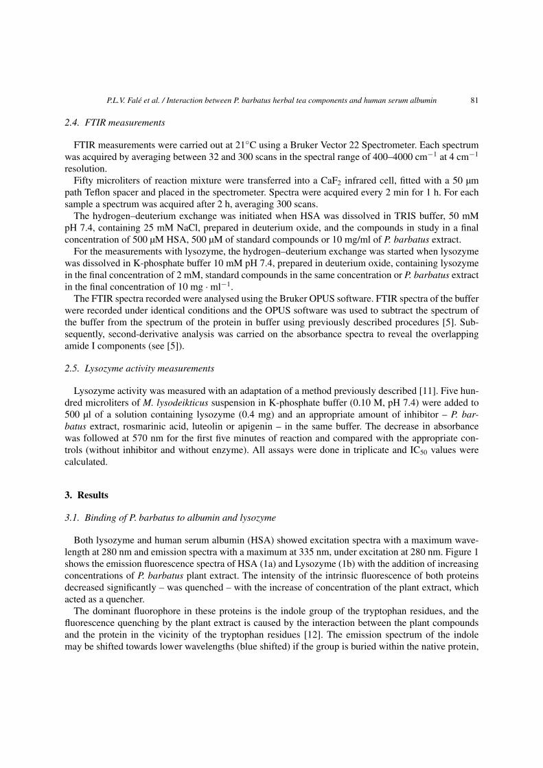
Both lysozyme and human serum albumin (HSA) showed excitation spectra with a maximum wave-length at 280 nm and emission spectra with a maximum at 335 nm, under excitation at 280 nm. Figure 1shows the emission fluorescence spectra of HSA (1a) and Lysozyme (1b) with the addition of increasingconcentrations of P. barbatus plant extract. The intensity of the intrinsic fluorescence of both proteinsdecreased significantly – was quenched – with the increase of concentration of the plant extract, whichacted as a quencher.
The dominant fluorophore in these proteins is the indole group of the tryptophan residues, and thefluorescence quenching by the plant extract is caused by the interaction between the plant compoundsand the protein in the vicinity of the tryptophan residues [12]. The emission spectrum of the indolemay be shifted towards lower wavelengths (blue shifted) if the group is buried within the native protein,
82 P.L.V. Falé et al. / Interaction between P. barbatus herbal tea components and human serum albumin
(a)
(b)
Fig. 1. Fluorescence emission spectra of HSA (a) and lysozyme (b) with the addition of P. barbatus aqueous extract. Arrowpoints to increasing concentrations of P. barbatus plant extract, ranging 0; 0.5; 0.75; 1; 2.5; 5; 7.5; 100 µg · ml−1.
or its emission may be shifted towards larger wavelengths (red shifted) when the protein is unfolded[12]. These effects were not observed in the present study (Fig. 1a and b), suggesting that, although theflavonoids may bind to the proteins in close proximity of the tryptophan residues for the fluorescencequenching to occur, they do not change the tryptophan residue exposition by altering the secondary ortertiary structure of the protein.
The fluorescence data were analysed by the Stern–Volmer equation (Eq. (1)), which allows one tocalculate the Stern–Volmer quenching constant (KSV) and the quenching rate constant (Kq) of the fluo-rescence quenching reaction:
F0/F = 1 + KSV[Q] = 1 + Kqτ0[Q], (1)
where F0 and F are the steady state fluorescence in the absence and presence of quencher, respectively,[Q] the concentration of quencher, and τ0 the average lifetime of the protein fluorescence in the absenceof quencher.
P.L.V. Falé et al. / Interaction between P. barbatus herbal tea components and human serum albumin 83
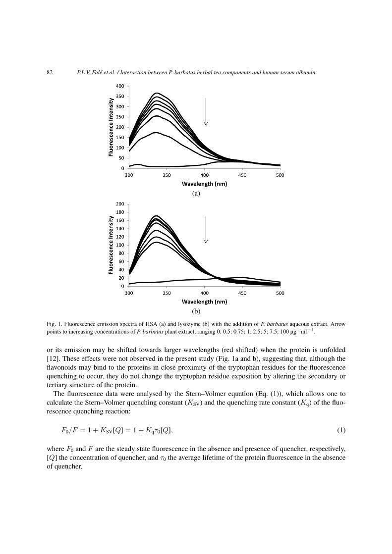
Fig. 2. Stern–Volmer plots of HSA and lysozyme with P. barbatus aqueous extract. [Q] is the concentration of P. barbatus inµg · ml−1.
The Stern–Volmer plots for the fluorescence quenching of HSA and Lysozyme by P. barbatus extractcan be found in Fig. 2, where it is shown that the fluorescence quenching by the plant extract followsthe behaviour of single-compound binding for both proteins. As the extract is composed of many com-pounds, the concentration is expressed in µg · ml−1 and the KSV values cannot be calculated in M−1,just estimated in l · mg−1. The molarity of the P. barbatus extract was estimated, based on the concen-tration of rosmarinic acid, luteolin glucuronide and apigenin glucuronide as 1.1122 mmol per gram ofplant extract, calculated by the HPLC chromatogram peak areas [18]. The protein intrinsic fluorescencequenching was also analysed for the main component of the plant extract, rosmarinic acid, and for lu-teolin and apigenin, which were also found as aglycons in the plasma of rats after the administration ofP. barbatus herbal tea [4].
The values for the Stern–Volmer quenching constant (KSV) and the quenching rate constant (Kq)for HSA and Lysozyme in the presence of rosmarinic acid, apigenin or luteolin are shown in Table 1.The estimated KSV and Kq for the P. barbatus extract were, respectively, 461,518 M−1 and 4.61 ×10¹³ M−1s−1 for HSA, and 68,513 M−1 and 6.85 × 10¹² M−1s−1 for lysozyme, which were approximateto the values found for the extract’s main component, rosmarinic acid. The maximum scatter collisionquenching constant (Kq) value of various quenchers with a biopolymer is reported to be 2 × 10¹⁰ l ·M−1s−1 [13]. As the Kq values obtained in the present work were higher, ranging from 1011 to 1013,than the Kq obtained for a scatter mechanism, it is implied that the quenching was not initiated bydynamic collision but rather originated by the formation of a complex.
3.2. Analysis of binding equilibria
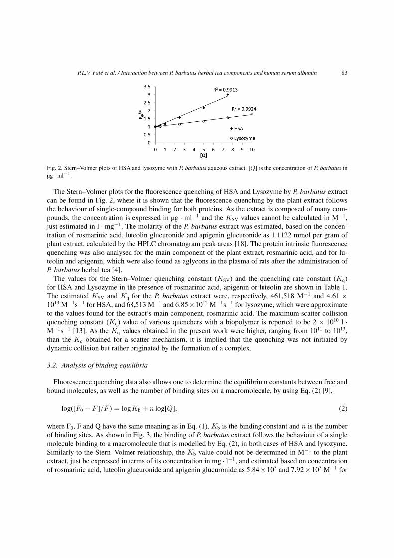
Fluorescence quenching data also allows one to determine the equilibrium constants between free andbound molecules, as well as the number of binding sites on a macromolecule, by using Eq. (2) [9],
log([F0 − F ]/F ) = log Kb + n log[Q], (2)
where F0, F and Q have the same meaning as in Eq. (1), Kb is the binding constant and n is the numberof binding sites. As shown in Fig. 3, the binding of P. barbatus extract follows the behaviour of a singlemolecule binding to a macromolecule that is modelled by Eq. (2), in both cases of HSA and lysozyme.Similarly to the Stern–Volmer relationship, the Kb value could not be determined in M−1 to the plantextract, just be expressed in terms of its concentration in mg · l−1, and estimated based on concentrationof rosmarinic acid, luteolin glucuronide and apigenin glucuronide as 5.84 × 10⁵ and 7.92 × 10⁵ M−1 for
84 P.L.V. Falé et al. / Interaction between P. barbatus herbal tea components and human serum albumin
Table 1
Binding parameters (KSV, Kq, Kb, n) and thermodynamic parameters (ΔH◦, ΔS◦, ΔG◦) for the binding of P. barbatus extract,rosmarinic acid (RA), luteolin (Lut) and apigenin (Api) to HSA and to lysozyme (R2 > 0.99 to all linear regressions), rates ofamide II/amide I variation and IC50 values for the inhibition of lysozyme activity
T(K) Human serum albumin Lysozyme
P. barbatus RA Lut Api P. barbatus RA Lut Api
KSV (M−1) 293 – 245,192 194,500 28,029 – 87,179 4191.9 5344.4
298 0.5133 (a) 355,856 250,748 8090.5 0.0762 (a) 98,081 3381.4 3805.1
303 – 482,757 326,653 5886.5 – 143,377 3533.7 3250.8
Kq (M−1s−1) 293 – 2.45E+13 1.95E+13 2.8E+12 – 8.72E+12 4.19E+11 5.34E+11
298 5.13E+7 (b) 3.56E+13 2.51E+13 8.09E+11 7.62E+6 (b) 9.81E+12 3.38E+11 3.81E+11
303 – 4.83E+13 3.27E+13 5.89E+11 – 1.43E+13 3.53E+11 3.25E+11
Kb (M−1) 293 – 1.32E+08 3.03E+08 3.07E+04 – 3,069,729 2760.57 7084.35
298 0.6493 (a) 3.96E+08 1.12E+09 1.85E+06 0.8806 (a) 205,541.7 3498.64 642.98
303 – 7.79E+08 6.92E+09 1.61E+09 – 84,781.29 4926.06 363.74
n 293 – 1.4998 1.5849 1.0101 – 1.1132 0.9551 0.9935
298 1.4643 1.5796 1.5403 1.4283 1.4667 1.0673 0.9839 0.8253
303 – 1.6267 1.6267 2.0964 – 0.9617 0.9789 0.7814
ΔH0 – – 131.16 230.67 96.35 – −265.66 42.69 −219.90
(kJ · mol−1)
ΔS0 – – 603.68 948.93 338.72 – −784.91 211.45 −679.24
(J · mol−1K−1)
ΔG0 293 – −45.551 −47.576 −25.172 – −36.389 −19.302 −21.598
(kJ · mol−1)
298 – −49.048 −51.619 −35.757 – −36.389 −20.218 −16.021
303 – −51.577 −57.081 −53.413 – −28.588 −21.420 −14.855
Rates of 298 −0.979 −1.071 −0.071 −0.715 −1.425 −1.820 −0.086 −0.449
amide II/amide I
intensity ratios
(mAU/min)
IC50 (µM) 298 – – – – 9.02 ± 0.40 (c) 97.2 ± 3.9 106.5 ± 9.6 91.0 ± 1.4
Notes: The rates of amide II/amide I variation were determined in the presence of similar amount of RA, Api and Lut, or10 mg · ml−1 of P. barbatus extract. Without ligand the rates of amide II/amide I variation were −0.716 for HSA and −1.815for lysozyme. For P. barbatus extract the values are expressed in l · mg−1 (a), l · mg−1s−1 (b) and mg · l−1 (c).
HSA and lysozyme, respectively. The fluorescence quenching effects by rosmarinic acid, luteolin andapigenin standards were analysed independently, and the values of Kb and n can be found in Table 1.The n value estimated for the interaction of P. barbatus extract with HSA was within the range of the onefound for the isolated compounds, while the Kb value was lower, suggesting that the extract componentsmay interfere with the binding of each other to HSA. For lysozyme, the plant extract seemed to bind tomore than one binding site (n = 1.5, Table 1) while for the isolated compounds n is approximately 1(Table 1), which may be the cause of the higher Kb value estimated for the plant extract than for theisolated compounds (Table 1).
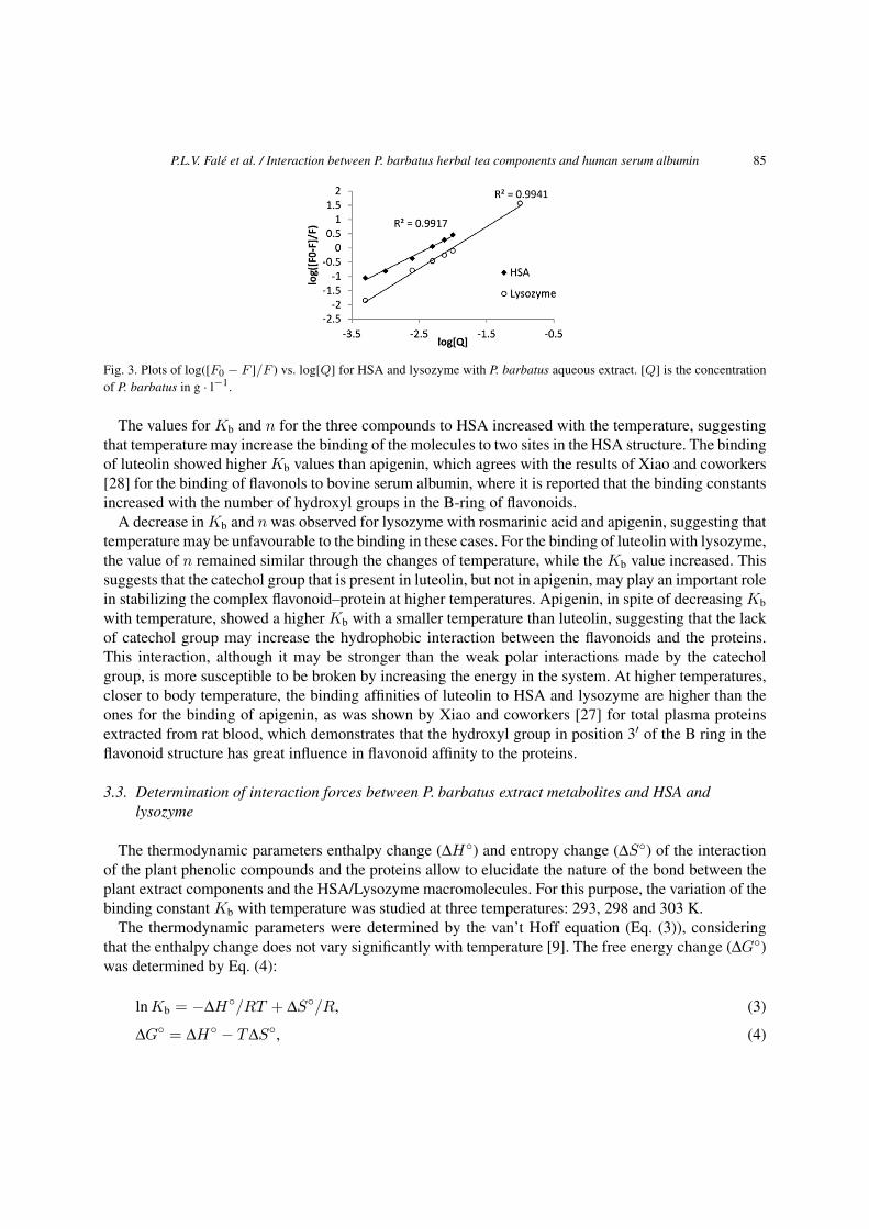
P.L.V. Falé et al. / Interaction between P. barbatus herbal tea components and human serum albumin 85
Fig. 3. Plots of log([F0 − F ]/F ) vs. log[Q] for HSA and lysozyme with P. barbatus aqueous extract. [Q] is the concentrationof P. barbatus in g · l−1.
The values for Kb and n for the three compounds to HSA increased with the temperature, suggestingthat temperature may increase the binding of the molecules to two sites in the HSA structure. The bindingof luteolin showed higher Kb values than apigenin, which agrees with the results of Xiao and coworkers[28] for the binding of flavonols to bovine serum albumin, where it is reported that the binding constantsincreased with the number of hydroxyl groups in the B-ring of flavonoids.
A decrease in Kb and n was observed for lysozyme with rosmarinic acid and apigenin, suggesting thattemperature may be unfavourable to the binding in these cases. For the binding of luteolin with lysozyme,the value of n remained similar through the changes of temperature, while the Kb value increased. Thissuggests that the catechol group that is present in luteolin, but not in apigenin, may play an important rolein stabilizing the complex flavonoid–protein at higher temperatures. Apigenin, in spite of decreasing Kb
with temperature, showed a higher Kb with a smaller temperature than luteolin, suggesting that the lackof catechol group may increase the hydrophobic interaction between the flavonoids and the proteins.This interaction, although it may be stronger than the weak polar interactions made by the catecholgroup, is more susceptible to be broken by increasing the energy in the system. At higher temperatures,closer to body temperature, the binding affinities of luteolin to HSA and lysozyme are higher than theones for the binding of apigenin, as was shown by Xiao and coworkers [27] for total plasma proteinsextracted from rat blood, which demonstrates that the hydroxyl group in position 3′ of the B ring in theflavonoid structure has great influence in flavonoid affinity to the proteins.
3.3. Determination of interaction forces between P. barbatus extract metabolites and HSA andlysozyme
The thermodynamic parameters enthalpy change (ΔH◦) and entropy change (ΔS◦) of the interactionof the plant phenolic compounds and the proteins allow to elucidate the nature of the bond between theplant extract components and the HSA/Lysozyme macromolecules. For this purpose, the variation of thebinding constant Kb with temperature was studied at three temperatures: 293, 298 and 303 K.
The thermodynamic parameters were determined by the van’t Hoff equation (Eq. (3)), consideringthat the enthalpy change does not vary significantly with temperature [9]. The free energy change (ΔG◦)was determined by Eq. (4):
ln Kb = −ΔH◦/RT + ΔS◦/R, (3)
ΔG◦ = ΔH◦ − TΔS◦, (4)
86 P.L.V. Falé et al. / Interaction between P. barbatus herbal tea components and human serum albumin
where R is the gas constant and T – the temperature. The values of the thermodynamic parameters of theinteraction of rosmarinic acid, apigenin and luteolin with HSA and Lysozyme can be found in Table 1.
The negative values of ΔG◦ indicate that the binding processes occurred spontaneously in all studiedcases.
From the view-point of water structure, a positive ΔS◦ value is typical evidence of hydrophobic in-teraction [29]. In the case of HSA, both ΔH◦ and ΔS◦ values were positive (Table 1), suggesting thathydrophobic association is the dominant form of interaction of the tested plant phenolics with HSA [22].Previous reports on the interaction of apigenin with HSA [30], and luteolin with bovine serum albumin[29], also showed positive values for ΔS◦. However, Yang and coworkers [29] presented a negative valuefor ΔH◦, which may be due to a strengthening of the interaction by other forces, such as van der Waalsor hydrogen bonds, introduced by hydrophobic effect in higher temperature conditions than the intervalused in the present work [22].
The values of ΔH◦ and ΔS◦ in the interaction of rosmarinic acid and apigenin with Lysozyme werenegative, suggesting van der Waals interactions between phenolic compounds and certain domains oflysozyme [22]. In these cases, the major contribution to ΔG◦ arises from the ΔH◦ term rather than fromΔS◦, which implies that the binding processes are enthalpy driven. In the interaction of luteolin andlysozyme, both ΔH◦ and ΔS◦ values were positive, which points to hydrophobic association as the mainform interaction between the molecules. The difference in thermodynamic parameters found betweenthe apigenin and luteolin may have been caused by the higher polarity and solubility in water of luteolin[10], that arises from the higher hydroxylation of the B-ring. The higher affinity to water of luteolin maycause a weaker hydrophobic association that did not induce the electrostatic interactions in our intervalof temperatures [22].
3.4. Determination of protein structure changes caused by P. barbatus extract and its plasmametabolites by FTIR
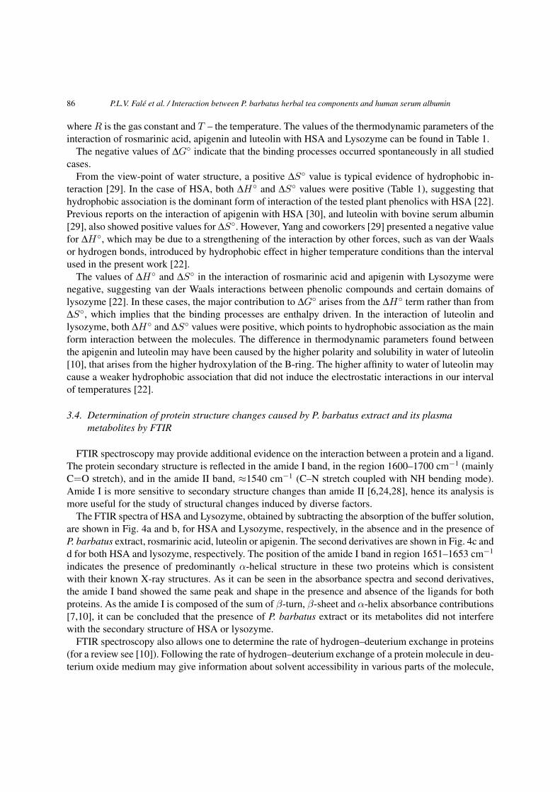
FTIR spectroscopy may provide additional evidence on the interaction between a protein and a ligand.The protein secondary structure is reflected in the amide I band, in the region 1600–1700 cm−1 (mainlyC=O stretch), and in the amide II band, ≈1540 cm−1 (C–N stretch coupled with NH bending mode).Amide I is more sensitive to secondary structure changes than amide II [6,24,28], hence its analysis ismore useful for the study of structural changes induced by diverse factors.
The FTIR spectra of HSA and Lysozyme, obtained by subtracting the absorption of the buffer solution,are shown in Fig. 4a and b, for HSA and Lysozyme, respectively, in the absence and in the presence ofP. barbatus extract, rosmarinic acid, luteolin or apigenin. The second derivatives are shown in Fig. 4c andd for both HSA and lysozyme, respectively. The position of the amide I band in region 1651–1653 cm−1
indicates the presence of predominantly α-helical structure in these two proteins which is consistentwith their known X-ray structures. As it can be seen in the absorbance spectra and second derivatives,the amide I band showed the same peak and shape in the presence and absence of the ligands for bothproteins. As the amide I is composed of the sum of β-turn, β-sheet and α-helix absorbance contributions[7,10], it can be concluded that the presence of P. barbatus extract or its metabolites did not interferewith the secondary structure of HSA or lysozyme.
FTIR spectroscopy also allows one to determine the rate of hydrogen–deuterium exchange in proteins(for a review see [10]). Following the rate of hydrogen–deuterium exchange of a protein molecule in deu-terium oxide medium may give information about solvent accessibility in various parts of the molecule,
P.L.V. Falé et al. / Interaction between P. barbatus herbal tea components and human serum albumin 87
(a) (b)
(c) (d)
Fig. 4. FTIR spectra of the proteins alone, with P. barbatus extract, rosmarinic acid (RA), luteolin (Lut) and apigenin (Api). Theabsorbance spectra obtained for HSA are shown in (a) and the second derivatives are in (c). The absorbance spectra obtainedfor lysozyme are shown in (b) and the second derivatives are in (d).
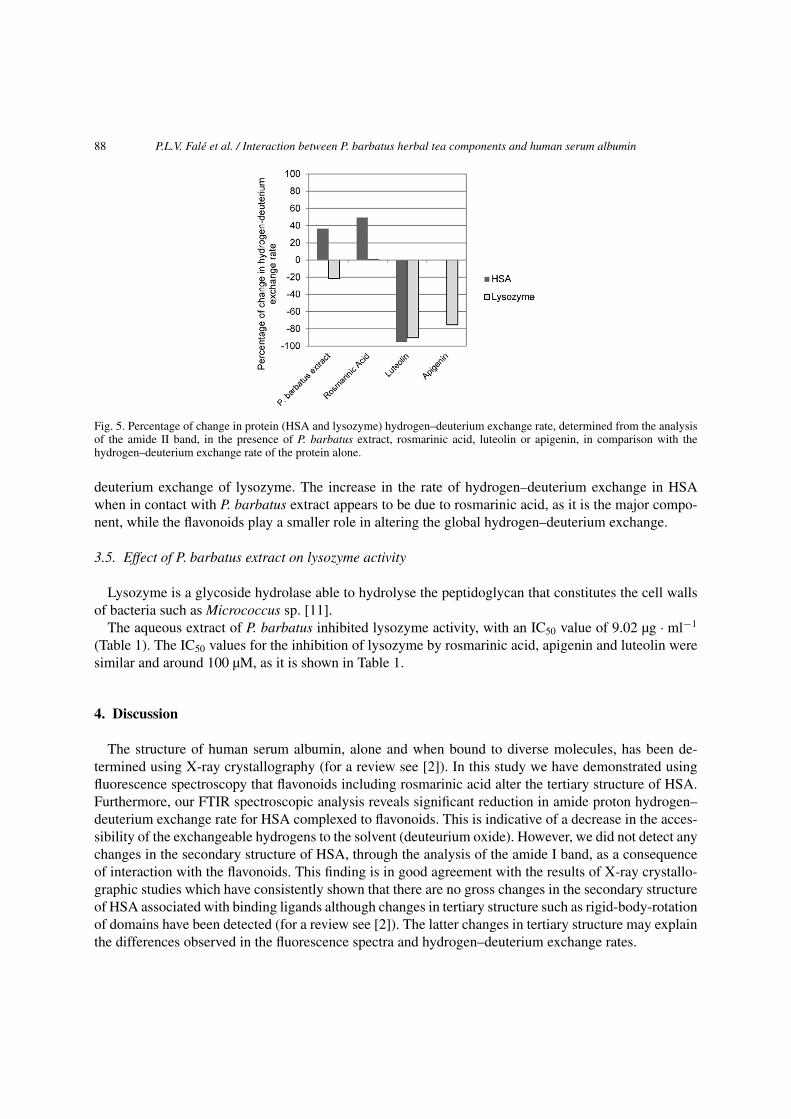
which reflects the tertiary structure of the protein [5]. The hydrogen–deuterium exchange can be fol-lowed by a decrease in amide II band during the first moments of contact of the protein with deuteriumoxide [6].
When P. barbatus extract (10 mg·ml−1) was in contact with the proteins under study, the rate of hydro-gen/deuterium exchange increased by 36.2% for HSA and decreased by 21.5% for lysozyme (Fig. 5).The effects of rosmarinic acid, luteolin and apigenin were analysed separately for each of the com-pounds in the same concentration of the protein. Rosmarinic acid caused a 49.6% increase in the rateof hydrogen–deuterium exchange of HSA, while the increase in lysozyme was negligible (0.3%). Theflavonoids luteolin and apigenin caused a decrease in hydrogen–deuterium exchange in both HSA andlysozyme. The highest decrease was observed for luteolin (90.1% and 95.3% for HSA and lysozyme,respectively), while apigenine caused a 75.3% decrease in the hydrogen–deuterium exchange rate oflysozyme, and a negligible decrease in HSA (0.1%) (Fig. 5). There was no apparent difference in theoverall shape and position of amide I bands, as seen from both the absorbance and second-derivativespectra (Fig. 4), of HSA and lysozyme in the presence of P. barbatus extract. This suggests that the in-teraction does not alter the secondary structure of these two proteins. Therefore, the differences found inthe rate of hydrogen–deuterium exchange may be due, predominantly to changes in the tertiary structureof the proteins [6].
The decrease of lysozyme hydrogen–deuterium exchange by P. barbatus extract may be due toits flavonoid components, since its main component, rosmarinic acid, does not affect the hydrogen–
88 P.L.V. Falé et al. / Interaction between P. barbatus herbal tea components and human serum albumin
Fig. 5. Percentage of change in protein (HSA and lysozyme) hydrogen–deuterium exchange rate, determined from the analysisof the amide II band, in the presence of P. barbatus extract, rosmarinic acid, luteolin or apigenin, in comparison with thehydrogen–deuterium exchange rate of the protein alone.
deuterium exchange of lysozyme. The increase in the rate of hydrogen–deuterium exchange in HSAwhen in contact with P. barbatus extract appears to be due to rosmarinic acid, as it is the major compo-nent, while the flavonoids play a smaller role in altering the global hydrogen–deuterium exchange.
3.5. Effect of P. barbatus extract on lysozyme activity
Lysozyme is a glycoside hydrolase able to hydrolyse the peptidoglycan that constitutes the cell wallsof bacteria such as Micrococcus sp. [11].
The aqueous extract of P. barbatus inhibited lysozyme activity, with an IC50 value of 9.02 µg · ml−1
(Table 1). The IC50 values for the inhibition of lysozyme by rosmarinic acid, apigenin and luteolin weresimilar and around 100 µM, as it is shown in Table 1.
4. Discussion
The structure of human serum albumin, alone and when bound to diverse molecules, has been de-termined using X-ray crystallography (for a review see [2]). In this study we have demonstrated usingfluorescence spectroscopy that flavonoids including rosmarinic acid alter the tertiary structure of HSA.Furthermore, our FTIR spectroscopic analysis reveals significant reduction in amide proton hydrogen–deuterium exchange rate for HSA complexed to flavonoids. This is indicative of a decrease in the acces-sibility of the exchangeable hydrogens to the solvent (deuteurium oxide). However, we did not detect anychanges in the secondary structure of HSA, through the analysis of the amide I band, as a consequenceof interaction with the flavonoids. This finding is in good agreement with the results of X-ray crystallo-graphic studies which have consistently shown that there are no gross changes in the secondary structureof HSA associated with binding ligands although changes in tertiary structure such as rigid-body-rotationof domains have been detected (for a review see [2]). The latter changes in tertiary structure may explainthe differences observed in the fluorescence spectra and hydrogen–deuterium exchange rates.
P.L.V. Falé et al. / Interaction between P. barbatus herbal tea components and human serum albumin 89
Of the different compounds tested, luteolin induced the highest decrease of hydrogen–deuterium ex-change and it also presented the highest values for the binding constants and lowest ΔG◦ values, sug-gesting a higher stability for the HSA-luteolin complex compared to the HSA-apigenin complex. The in-creased stability of the protein associated with the bonding between the flavonoids and the HSA moleculemakes it more difficult for the amide protons to be substituted with deuterium. Furthermore, the interac-tion may result in the shielding of certain segments of the protein from being accessible to solvent. Allthis explains the significant reduction in hydrogen–deuterium exchange upon binding of the flavonoidsto HSA. Rosmarinic acid, like luteolin, had no effect on the secondary structure of HSA. However, un-like luteolin, rosmarinic acid increased the hydrogen–deuterium exchange of HSA, suggesting that thenature of its interaction with the transport protein is different. Our results suggest that hydrophobic re-gions of the protein becomes accessible to solvent due to alterations in the protein tertiary structure [17]without any changes in the secondary structure. The nature of the change could be a large rigid-bodyrotation of the subunits that have been shown to occur on fatty acid binding to the main fatty acid bind-ing sites in HSA, as was extensively reviewed by Curry [2]. X-ray crystallographic analysis has shownthat fatty acid binding results in a HSA molecule that is 10 Å wider than the defatted HSA. It is thuspossible that whilst the binding may reduce hydrogen–deuterium exchange for molecules that are inter-acting with rosmarinic acid but a much larger change such as widening of the molecule could expose agreater number of amino acid residues to solvent resulting in an overall increase in hydrogen–deuteriumexchange. We propose that rosmarinic acid may induce a similar type of tertiary structural change asinduced by binding of fatty acids to HSA. In five of the seven known fatty acid binding sites the lipid isanchored by the interaction of the carboxylic group with a basic or polar group at the pocket entrance.Some non-lipid acidic compounds known to interact with these binding sites, specifically by interactingwith the carboxylic acid binding residues [2]. Rosmarinic acid and fatty acids both share a commonfunctional group (carboxylic acid group) which is not the case for luteolin and this may explain as towhy they behave differently in altering the structure of the HSA molecule.
Flavonoids have been reported to bind to the drug binding sites [19,30], which are different from thefatty acid binding sites. The drug binding sites 1 and 2 comprise largely apolar cavities with defines polarfeatures located in sub-domains IIA and IIIA, respectively. Most drugs bind to the drug binding site 1,which has preference for flat aromatic compounds that fit between Leu238 and Ala291 in the centre ofthe pocket [2]. The planar structure of flavonoids may be responsible for their binding predominantly tothe drug site 1, as it was reported for apigenin [30] and quercetin [19]. Usually small adjustments in theside chains in the site 1 cavity can happen upon the binding of the compounds. These changes are not soextensive that may lead to changes in the secondary structure of HSA [2], but may be the cause of thedecrease in hydrogen–deuterium exchange rate in the binding of luteolin to HSA.
It is interesting that like with HSA, luteolin and apigenin interaction with lysozyme results in a de-crease in amide proton hydrogen–deuterium exchange rate. This can be due to one or more of the fol-lowing reasons:
(i) due to the amide protons participating in stronger H-bonds within the secondary structural ele-ments as a consequence of interaction with these compounds. This will make it more difficult tobreak the H-bonds for the deuterium substitution to occur;
(ii) due to amide protons forming H-bonds to other groups including luteolin and apigenin making itmore difficult for the hydrogen–deuterium exchange reaction to occur;
(iii) or it could be due to movement of domains/regions of protein that results in a reduction in solventaccessibility so that deuterium oxide is unable to penetrate into the core of the protein to replacethe amide protons with deuterium.
90 P.L.V. Falé et al. / Interaction between P. barbatus herbal tea components and human serum albumin
Similarly to HSA, luteolin induced a greater decrease in lysozyme hydrogen deuterium exchange(Table 1), and presented a higher binding constant and lower ΔG◦ value at room temperature, suggestingthat the binding of luteolin to lysozyme is more stable at room temperature than the binding of apigenin.
Once again, like with what we saw for HSA, the interaction of rosmarinic acid with lysozyme isdifferent to that observed for luteolin. Although, there is no significant increase in hydrogen–deuteriumexchange, it at least does not reduce the hydrogen–deuterium exchange. This could be due to weakerinteraction between rosmarinic acid and lysozyme or that the nature of the binding does not alter thetertiary structure in a way that would make the amide protons more vulnerable to hydrogen–deuteriumexchange.
Although we have detected changes in hydrogen–deuterium exchange, no significant alteration in thesecondary structure was detected. However, other studies concerning the interaction of flavonoids de-tected small secondary structure changes, as well as tertiary structure changes, by the flavonoids alpinetinand cardamonin [7]. Our findings reveal that alterations in protein secondary structure were not foundwith either luteolin or apigenin, by FTIR or fluorescence spectrometry, suggesting that the alterations inrate of hydrogen deuterium exchange is mainly due to changes in the tertiary structure.
As the proteins and the plant phenolic compounds seemed to interact by weak forces by fluorescencespectrometry, the bonds formed may be reversible and the compounds released under certain conditions.Studies with total plasma protein extracted from rat blood report that the affinity of flavonoids to thetotal protein may be even lower than to purified albumin, which may be due to presence of metallicions such as Zn2+, Cu2+, Ca2+ and Mg2+ [27]. The weak and reversible interaction between theseplant compounds and plasma proteins is in agreement with in vivo studies that show a decrease in theconcentration of plant phenolic compounds in plasma, due to metabolism and excretion, as was observedin P. barbatus extract components [4], rosmarinic acid [1,4] and the flavonoids apigenin and luteolin[23]. Therefore it is important that the structural changes in plasma proteins when bound to compoundsare not so drastic that their function may be compromised after the release of the compounds.
Lysozyme, apart from drug transport, has an important function related to the immune response pro-cess [11]. When degranulation occurs, after neutrophils reach the injured tissue by margination, adhesionand emigration, lysozyme is discharged from lysosomes of neutrophils and destroys not only the phago-somes but also damages the animal tissue itself, thus aggravating the response to inflammation [21,26].Previous reports showed that phenolic compounds from Leggera species extracts decreased the releaseof lysozyme from the neutrophils to the serum [26]. In the present work, P. barbatus extract pheno-lic compounds inhibited directly lysozyme activity. It is long known that some flavonoids may act aslysozyme inhibitors [20]. Previous works also reported that plant extracts mainly composed of apigenin,luteolin and luteolin glucosides decreased allergy symptoms caused by egg-white lysozyme sensitiza-tion in rats [8]. The same anti-allergic effect was observed for Perilla frutescens water extract, and by itsmajor component, rosmarinic acid [16].
As P. barbatus components bind to lysozyme with weak interactions, our data suggests that the en-zyme does not undergo major conformational changes, the plant compounds are eliminated from plasmain a short period of time [4], and the antibacterial activity of lysozyme is not permanently compromised.Therefore, these compounds may be helpful in decreasing the damage caused by a high lysozyme activ-ity, in response to pathogens or in allergic conditions.
P. barbatus extract has also proved to have antioxidant activity as a radical scavenger more powerfulthan the commercial antioxidant BHT [3]. This activity is mainly due to its main component, rosmarinicacid. The present results suggest that P. barbatus water extract may be useful to treat inflammatory con-ditions as its components and metabolites may be transported in circulatory system binding to albumin,
P.L.V. Falé et al. / Interaction between P. barbatus herbal tea components and human serum albumin 91
the most abundant transport protein, and to lysozyme, and decrease the inflammation process by twomechanism: as free radical scavengers, and as lysozyme inhibitors.
In conclusion, protein intrinsic fluorescence quenching proved that the P. barbatus extract componentsand its metabolites found in rat plasma are able to bind to the human transport proteins albumin andlysozyme, allowing them to be carried in the bloodstream to organs where they can have a beneficialactivity. The spectroscopic data suggest that the interaction of the plant phenolic compounds and theproteins in this study is made by weak interactions, causing some changes in protein tertiary structure,but not in the secondary structure. This suggests that the compounds may be released from the complexesthey form with the proteins, without compromising the albumin or lysozyme function in plasma.
P. barbatus extract components and metabolites also inhibit lysozyme activity, which may be helpfulin decreasing the aggravation of the inflammation caused by the immune system in response to pathogensand in allergies. This mechanism can be added to the already well known radical scavenger capacity ofthe extract components, making the anti-inflammatory activity of plant extract very promising.
Acknowledgements
The authors thank Fundação para a Ciência e Tecnologia, FCT, for financial support. Pedro L. V. Faléthanks FCT for the PhD Grant SFRH/BD/37547/2007.
References
[1] S. Baba, N. Osakabe, M. Natsume and J. Terao, Orally administered rosmarinic acid is present as the conjugated and/ormethylated forms in plasma, and is degraded and metabolized to conjugated forms of caffeic acid, ferulic acid and m-coumaric acid, Life Sciences 75 (2004), 165–178.
[2] S. Curry, Lessons from the crystallographic analysis of small molecule binding to human serum albumin, DrugMetabolism and Pharmacokinetics 24 (2009), 342–357.
[3] P.L. Falé, C. Borges, P.J.A. Madeira, L. Ascensão, M.E.M. Araújo, M.H. Florêncio and M.L.M. Serralheiro, Rosmarinicacid, scutellarein 4′-methyl ether 7-O-glucuronide and (16S)-coleon E are the main compounds responsible for the anti-acetylcholinesterase and antioxidant activity in herbal tea of Plectranthus barbatus (“falso boldo”), Food Chemistry 114(2009), 798–805.
[4] P.L.V. Falé, P.J.A. Madeira, M.H. Florêncio, L. Ascensão and M.L.M. Serralheiro, Function of Plectranthus barbatusherbal tea as neuronal acetylcholinesterase inhibitor, Food and Function 2 (2011), 130–136.
[5] P.I. Haris, D. Chapman, R.A. Harrison, K.F. Smith and S.J. Perkins, Conformational transition between native and reactivecenter cleaved forms of α1-antitrypsin by Fourier transform infrared spectroscopy and small-angle neutron scattering,Biochemistry 29 (1990), 1377–1380.
[6] P.I. Haris and F. Severcan, FTIR spectroscopic characterization of protein structure in aqueous and non-aqueous media,Journal Molecular Catalysis B: Enzymatic 7 (1999), 207–221.
[7] W. He, Y. Li, J. Tang, F. Luan, J. Jin and Z. Hu, Comparison of the characterization on binding of alpinetin and cardamoninto lysozyme by spectroscopic methods, International Journal of Biological Macromolecules 39 (2006), 165–173.
[8] E. Iwaoka, H. Oku, M. Iinuma and K. Ishiguro, Allergy-preventive effects of the flowers of Impatiens textori, Biologicaland Pharmaceutical Bulletin 33 (2010), 714–716.
[9] J. Jin and X. Zhang, Spectrophotometric studies on the interaction between pazufloxacin mesilate and human serumalbumin or lysozyme, Journal of Luminescence 128 (2010), 81–86.
[10] H. Kim, H. Kim and S. Jung, Aqueous solubility enhancement of some flavones by complexation with cyclodextrins,Bulletin of the Korean Chemical Society 29 (2008), 590–594.
[11] N.J. Laible and G.R. Germaine, Bactericidal activity of human lysozyme, muramidase-inactive lysozyme, and cationicpolypeptides against Streptococcus sanguis and Streptococcus faecalis: inhibition by chitin oligosaccharides, Infectionand Immunity 48 (1985), 720–728.
[12] J.R. Lakowicz, Principles of Fluorescence Spectroscopy, 3rd edn, Plenum Press, New York, 2006.[13] J.R. Lakowicz and G. Weber, Quenching of fluorescence by oxygen. A probe for structural fluctuations in macromolecules,
Biochemistry 12 (1973), 4161–4170.
92 P.L.V. Falé et al. / Interaction between P. barbatus herbal tea components and human serum albumin
[14] Y. Li, W.Y. He, H. Liu, X. Yao and Z. Hu, Daidzein interaction with human serum albumin studied using optical spec-troscopy and molecular modeling methods, Journal of Molecular Structure 831 (2007), 144–150.
[15] C.W. Lukhoba, M.S.J. Simmonds and A.J. Paton, Plectranthus: a review of ethnobotanical uses, Journal Ethnopharma-cology 103 (2006), 1–24.
[16] T. Makino, Y. Furuta, H. Wakushima, H. Fujii, K. Saito and Y. Kano, Anti-allergic effect of Perilla frutescens and itsactive constituents, Phytotherapy Research 17 (2003), 240–243.
[17] V. Militello, C. Casarino, A. Emanuele, A. Giostra, F. Pullara and M. Leone, Aggregation kinetics of bovine serumalbumin studied by FTIR spectroscopy and light scattering, Biophysical Chemistry 107 (2004), 175–187.
[18] S. Porfírio, P.L.V. Falé, P.J.A. Madeira, M.H. Florêncio, L. Ascensão and M.L.M. Serralheiro, Antiacetylcholinesteraseand antioxidant activities of Plectranthus barbatus tea, after in vitro gastrointestinal metabolism, Food Chemistry 122(2010), 179–187.
[19] H.M. Rawel, S.K. Frey, K. Meidtner, J. Kroll and F.J. Schweigert, Determining the binding affinities of phenolic com-pounds to proteins by quenching of the intrinsic tryptophan fluorescence, Molecular Nutrition and Food Research 50(2006), 705–713.
[20] G. Rodney, A.L. Swanson, L.M. Wheeler, G.N. Smith and C.S. Worrel, The effect of a series of flavonoids onhyaluronidase and some other related enzymes, Journal Biological Chemistry 183 (1950), 739–747.
[21] F. Ronca, L. Palmieri, P. Panicucci and G. Ronca, Anti-inflammatory activity of chondroitin sulphate, Osteoarthritis andCartilage 6 (1998), 14–21.
[22] P.D. Ross and S. Subramanian, Thermodynamics of protein association reactions: forces contributing to stability, Bio-chemistry 20 (1981), 3096–3102.
[23] R. Shi, S. Qiao, D. Yu, X. Shi, M. Liu, X. Jiang, Q. Wang and L. Zhang, Simultaneous determination of five flavonoidsfrom Scutellaria barbata extract in rat plasma by LC-MS/MS and its application to pharmacokinetic study, Journal ofChromatography B: Analytical Technologies in the Biomedical and Life Sciences 879(19) (2011), 1625–1632.
[24] R. Tantipolphan, T. Rades, A.J. McQuillan and N.J. Medlicott, Adsorption of bovine serum albumin (BSA) onto lecithinstudied by attenuated total reflectance Fourier transform infrared (ATR-FTIR) spectroscopy, International Journal Phar-maceutics 337 (2007), 40–47.
[25] A. Varshney, P. Sen, E. Ahmad, M. Rehan, N. Subbarao and R.H. Khan, Ligand binding strategies of human serumalbumin: How can the cargo be utilized?, Chirality 22 (2010), 77–87.
[26] Y. Wu, C. Zhou, L. Song, X. Li, S. Snhi, J. Mo, H. Chen, H. Bai, X. Wu, J. Zhao, R. Zhang, X. Hao, H. Sun and Y. Zhao,Effect of total phenolics from Laggera alata on acute and chronic inflammation models, Journal Ethnopharmacology 108(2006), 243–250.
[27] J. Xiao, H. Cao, T. Chen, F. Yang, C. Liu and X. Xu, Molecular property-binding affinity relationship of flavonoids forcommon rat plasma proteins in vitro, Biochimie 93 (2011), 134–140.
[28] J. Xiao, M. Suzuki, X. Jiang, X. Chen, K. Yamamoto, F. Ren and M. Xu, Influence of B-Ring hydroxylation on interactionsof flavonols with bovine serum albumin, Journal of Agricultural and Food Chemistry 56 (2008), 2350–2356.
[29] Y. Yang, Q. Hua, Y. Fan and H. Shen, Study on the binding of luteolin to bovine serum albumin, Spectrochimica Acta A:Molecular Biomolecular Spectroscopy 69 (2008), 432–436.
[30] J. Yuan, Z. lv, Z. Liu, Z. Hu and G. Zou, Study on interaction between apigenin and human serum albumin by spectroscopyand molecular modelling, Journal of Photochemistry and Photobiology A: Chemistry 191 (2007), 104–113.
Submit your manuscripts athttp://www.hindawi.com
Hindawi Publishing Corporationhttp://www.hindawi.com Volume 2014
Inorganic ChemistryInternational Journal of
Hindawi Publishing Corporation http://www.hindawi.com Volume 2014
International Journal ofPhotoenergy
Hindawi Publishing Corporationhttp://www.hindawi.com Volume 2014
Carbohydrate Chemistry
International Journal of
Hindawi Publishing Corporationhttp://www.hindawi.com Volume 2014
Journal of
Chemistry
Hindawi Publishing Corporationhttp://www.hindawi.com Volume 2014
Advances in
Physical Chemistry
Hindawi Publishing Corporationhttp://www.hindawi.com
Analytical Methods in Chemistry
Journal of
Volume 2014
Bioinorganic Chemistry and ApplicationsHindawi Publishing Corporationhttp://www.hindawi.com Volume 2014
SpectroscopyInternational Journal of
Hindawi Publishing Corporationhttp://www.hindawi.com Volume 2014
The Scientific World JournalHindawi Publishing Corporation http://www.hindawi.com Volume 2014
Medicinal ChemistryInternational Journal of
Hindawi Publishing Corporationhttp://www.hindawi.com Volume 2014
Chromatography Research International
Hindawi Publishing Corporationhttp://www.hindawi.com Volume 2014
Applied ChemistryJournal of
Hindawi Publishing Corporationhttp://www.hindawi.com Volume 2014
Hindawi Publishing Corporationhttp://www.hindawi.com Volume 2014
Theoretical ChemistryJournal of
Hindawi Publishing Corporationhttp://www.hindawi.com Volume 2014
Journal of
Spectroscopy
Analytical ChemistryInternational Journal of
Hindawi Publishing Corporationhttp://www.hindawi.com Volume 2014
Journal of
Hindawi Publishing Corporationhttp://www.hindawi.com Volume 2014
Quantum Chemistry
Hindawi Publishing Corporationhttp://www.hindawi.com Volume 2014
Organic Chemistry International
ElectrochemistryInternational Journal of
Hindawi Publishing Corporation http://www.hindawi.com Volume 2014
Hindawi Publishing Corporationhttp://www.hindawi.com Volume 2014
CatalystsJournal of


![WELCOME [new.airmauritius.com]...Mauritian Vanilla Tea, Earl Grey, Jasmine Tea, Green Tea Camomile Infusion, Mint, Green Anise and Verbena Infusion Nescafe Sélection, Espresso, Decaffeinated](https://img.pdfslide.fr/doc/110x75/5fb551cd65f89e3e906f4019/welcome-new-mauritian-vanilla-tea-earl-grey-jasmine-tea-green-tea-camomile.jpg)