Embed Size (px)
Citation preview
Les modalités d'images médicales
Vincent [email protected] GMCAO(http://www-timc.imag.fr/gmcao)
IICAO, Introduction à l’imagerie médicale, 1
ENSIMAG/IRVM
Buts
• Connaitre les différentes modalités d’imagerie médicale.
• Comprendre les bases de ces modalités et leurs caractéristiques principales.
• Décrire l’utilisation/l’indication pour chaque modalité.
• Comprendre le concept d’imagerie 4D et son applicationapplication.
• Connaitre certaines applications en planification et en chirurgie.
IICAO, Introduction à l’imagerie médicale, 2
Exemples de modalités d’imageries et applications ?
IICAO, Introduction à l’imagerie médicale, 3
Exemples de modalités d’imageries et applications
• Echographie,• Radiographie,g p ,• CT scanner,• PET scan,• IRM,• Endoscopie.
IICAO, Introduction à l’imagerie médicale, 4
• Introduction• Lumière et endoscopie
Plan
Lumière et endoscopie• Ultra-sons et échographie• Rayons X et radiographie• Autres imageries• Applications
IICAO, Introduction à l’imagerie médicale, 5
Introduction
• Imagerie médicale : mettre en lumière l’invisible- Anatomie (os, tissus mous, ...)- Mouvement (cœur, poumon, ...)- Mesures physiologiques (débit sanguin, ...)- Métabolisme (biochimie : utilisation de traceurs
radioactifs)
IICAO, Introduction à l’imagerie médicale, 6
Introduction
• Imagerie médicale : mettre en lumière l’invisible- Anatomie (os, tissus mous, ...)- Mouvement (cœur, poumon, ...)- Mesures physiologiques (débit sanguin, ...)- Métabolisme (biochimie : utilisation de traceurs
radioactifs)
• Principe de formation d’une image médicale :• Principe de formation d’une image médicale :
Agent physique
Interactionavec lamatière
Détection Image(s)
IICAO, Introduction à l’imagerie médicale, 7
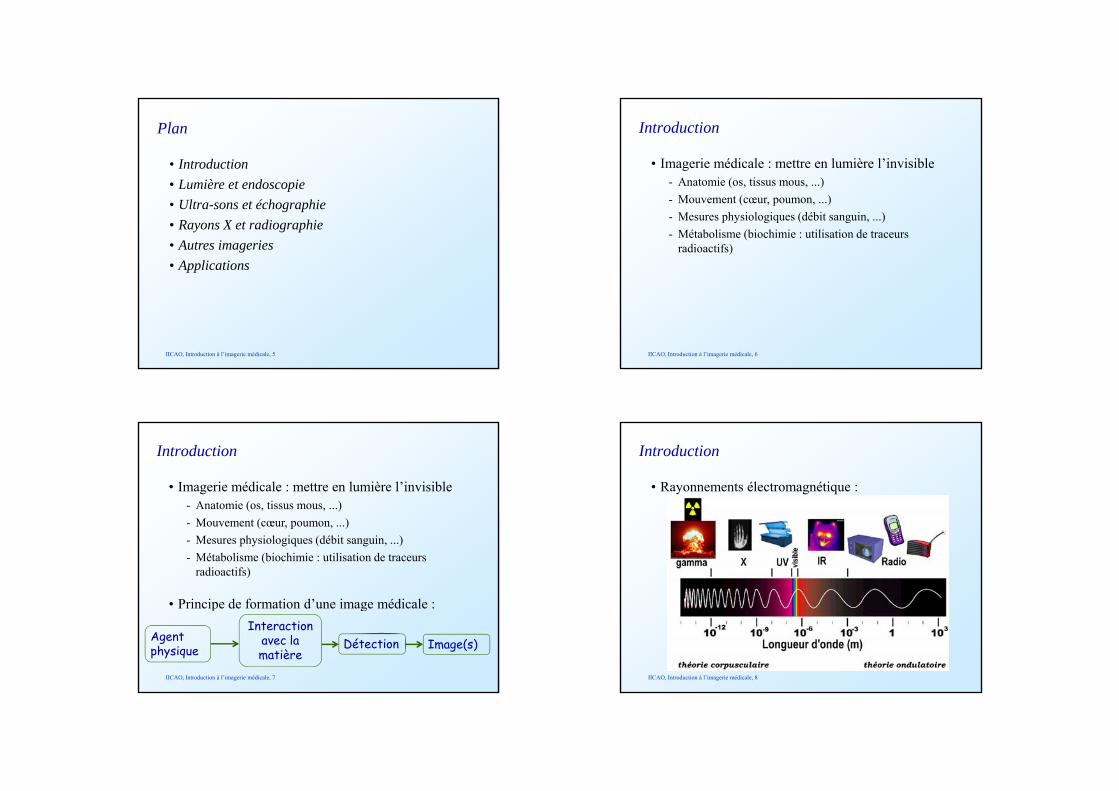
Introduction
• Rayonnements électromagnétique :
IICAO, Introduction à l’imagerie médicale, 8
Introduction
• Différentes techniques suivant les agents :
IICAO, Introduction à l’imagerie médicale, 9
Introduction
• Utilisation des images médicales:- Diagnostique,- Planning de traitement (chirurgie, radiothérapie),- Suivi du traitement des pathologies,- Interventions guidées en temps réel : imagerie
interventionnelle,- Formation / Enseignement.
IICAO, Introduction à l’imagerie médicale, 10
• Introduction• Lumière et endoscopie
Plan
Lumière et endoscopie• Ultra-sons et échographie• Rayons X et radiographie• Autres imageries• Applications
IICAO, Introduction à l’imagerie médicale, 11
Lumière et endoscopie
• Première modalité d’imagerie médicale• Historique :q
- Usage de speculum, autour de -600.
IICAO, Introduction à l’imagerie médicale, 12
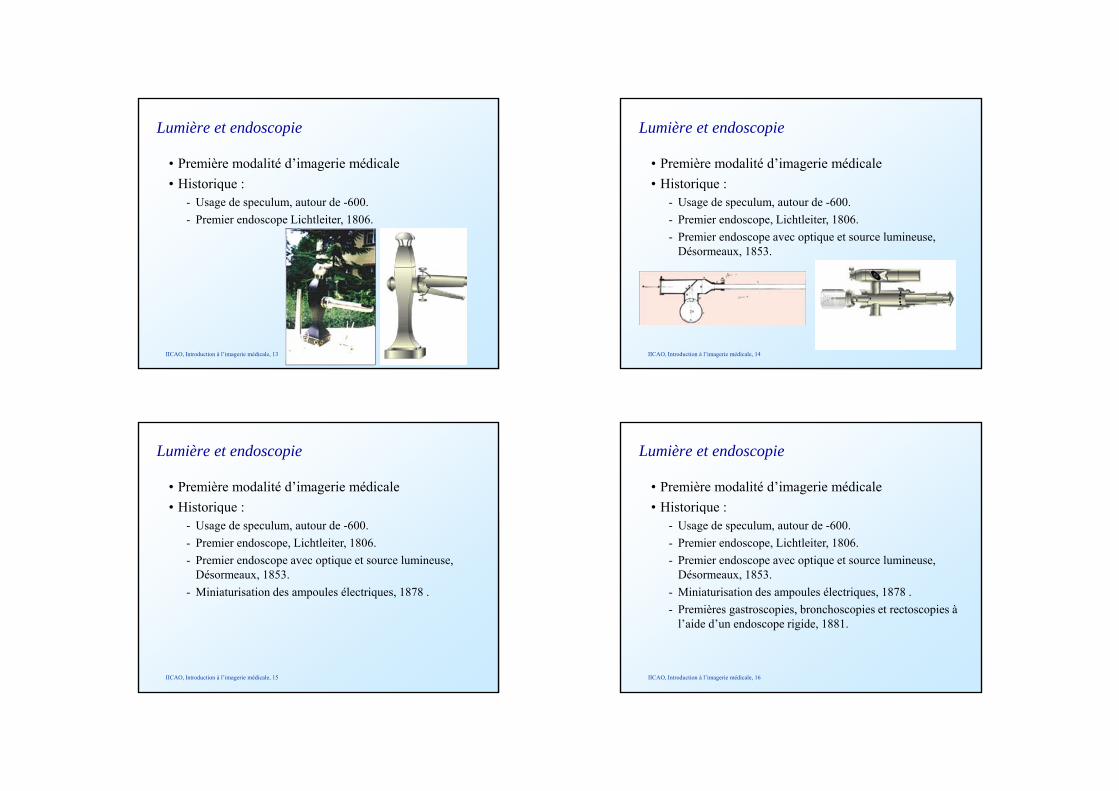
Lumière et endoscopie
• Première modalité d’imagerie médicale• Historique :q
- Usage de speculum, autour de -600.- Premier endoscope Lichtleiter, 1806.
IICAO, Introduction à l’imagerie médicale, 13
Lumière et endoscopie
• Première modalité d’imagerie médicale• Historique :q
- Usage de speculum, autour de -600.- Premier endoscope, Lichtleiter, 1806.- Premier endoscope avec optique et source lumineuse,
Désormeaux, 1853.
IICAO, Introduction à l’imagerie médicale, 14
Lumière et endoscopie
• Première modalité d’imagerie médicale• Historique :q
- Usage de speculum, autour de -600.- Premier endoscope, Lichtleiter, 1806.- Premier endoscope avec optique et source lumineuse,
Désormeaux, 1853. - Miniaturisation des ampoules électriques, 1878 .
IICAO, Introduction à l’imagerie médicale, 15
Lumière et endoscopie
• Première modalité d’imagerie médicale• Historique :q
- Usage de speculum, autour de -600.- Premier endoscope, Lichtleiter, 1806.- Premier endoscope avec optique et source lumineuse,
Désormeaux, 1853. - Miniaturisation des ampoules électriques, 1878 .- Premières gastroscopies, bronchoscopies et rectoscopies à
l’aide d’un endoscope rigide, 1881.
IICAO, Introduction à l’imagerie médicale, 16
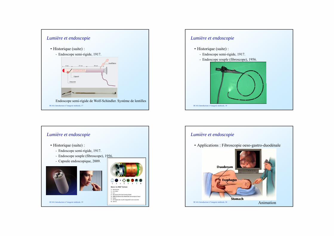
Lumière et endoscopie
• Historique (suite) :- Endoscope semi-rigide, 1917.
Endoscope semi-rigide de Wolf-Schindler. Système de lentillesIICAO, Introduction à l’imagerie médicale, 17
Lumière et endoscopie
• Historique (suite) :- Endoscope semi-rigide, 1917.- Endoscope souple (fibroscope), 1956.
IICAO, Introduction à l’imagerie médicale, 18
Lumière et endoscopie
• Historique (suite) :- Endoscope semi-rigide, 1917.- Endoscope souple (fibroscope), 1956.- Capsule endoscopique, 2009.
IICAO, Introduction à l’imagerie médicale, 19
Lumière et endoscopie
• Applications : Fibroscopie oeso-gastro-duodénale
IICAO, Introduction à l’imagerie médicale, 20 Animation
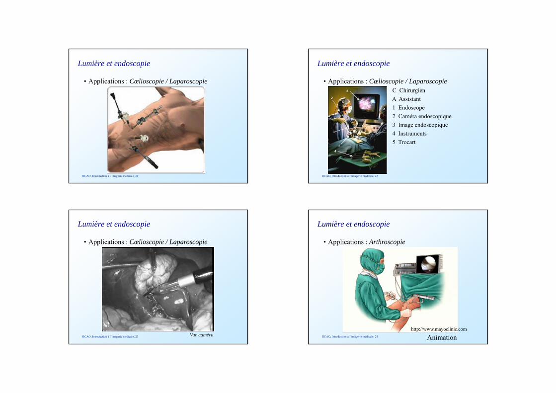
Lumière et endoscopie
• Applications : Cœlioscopie / Laparoscopie
IICAO, Introduction à l’imagerie médicale, 21
Lumière et endoscopie
• Applications : Cœlioscopie / LaparoscopieC ChirurgienA Assistant1 Endoscope2 Caméra endoscopique3 Image endoscopique4 Instruments5 Trocart
IICAO, Introduction à l’imagerie médicale, 22
Lumière et endoscopie
• Applications : Cœlioscopie / Laparoscopie
IICAO, Introduction à l’imagerie médicale, 23 Vue caméra
Lumière et endoscopie
• Applications : Arthroscopie
IICAO, Introduction à l’imagerie médicale, 24
http://www.mayoclinic.com
Animation
Lumière et endoscopie
• Bilan :- Avantages
• Minimalement invasif,• Anesthésie locale dans certains cas,• Cicatrisation rapide et peu de risque de surinfection.
- Inconvénients• Vue très partielle,• Besoin d’un assistant pour orienter l’endoscope,• Perte de la sensation tactile,• Vues 2D.
IICAO, Introduction à l’imagerie médicale, 25
Lumière et endoscopie
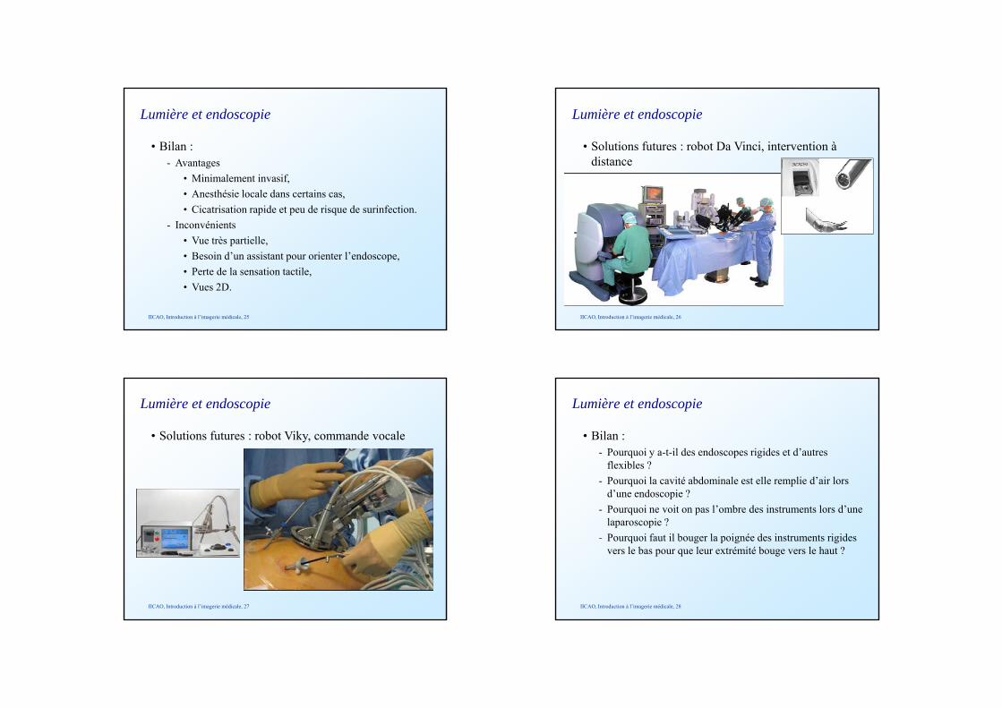
• Solutions futures : robot Da Vinci, intervention à distance
IICAO, Introduction à l’imagerie médicale, 26
Lumière et endoscopie
• Solutions futures : robot Viky, commande vocale
IICAO, Introduction à l’imagerie médicale, 27
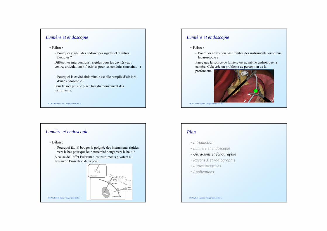
Lumière et endoscopie
• Bilan :- Pourquoi y a-t-il des endoscopes rigides et d’autres
flexibles ?- Pourquoi la cavité abdominale est elle remplie d’air lors
d’une endoscopie ?- Pourquoi ne voit on pas l’ombre des instruments lors d’une
laparoscopie ?- Pourquoi faut il bouger la poignée des instruments rigidesPourquoi faut il bouger la poignée des instruments rigides
vers le bas pour que leur extrémité bouge vers le haut ?
IICAO, Introduction à l’imagerie médicale, 28
Lumière et endoscopie
• Bilan :- Pourquoi y a-t-il des endoscopes rigides et d’autres
flexibles ?Différentes interventions : rigides pour les cavités (ex : ventre, articulations), flexibles pour les conduits (intestins…)
- Pourquoi la cavité abdominale est elle remplie d’air lors d’une endoscopie ?d une endoscopie ?
Pour laisser plus de place lors du mouvement des instruments.
IICAO, Introduction à l’imagerie médicale, 29
Lumière et endoscopie
• Bilan :- Pourquoi ne voit on pas l’ombre des instruments lors d’une
laparoscopie ?Parce que la source de lumière est au même endroit que la caméra. Cela crée un problème de perception de la profondeur.
IICAO, Introduction à l’imagerie médicale, 30
Lumière et endoscopie
• Bilan :- Pourquoi faut il bouger la poignée des instruments rigides
vers le bas pour que leur extrémité bouge vers le haut ?A cause de l’effet Fulcrum : les instruments pivotent au niveau de l’insertion de la peau.
IICAO, Introduction à l’imagerie médicale, 31
• Introduction• Lumière et endoscopie
Plan
Lumière et endoscopie• Ultra-sons et échographie• Rayons X et radiographie• Autres imageries• Applications
IICAO, Introduction à l’imagerie médicale, 32
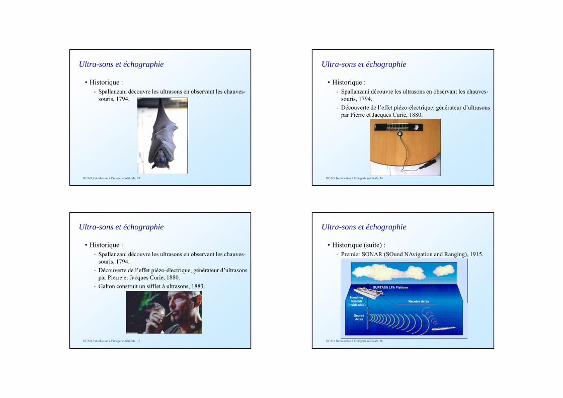
Ultra-sons et échographie
• Historique :- Spallanzani découvre les ultrasons en observant les chauves-
souris, 1794.
IICAO, Introduction à l’imagerie médicale, 33
Ultra-sons et échographie
• Historique :- Spallanzani découvre les ultrasons en observant les chauves-
souris, 1794.- Découverte de l’effet piézo-électrique, générateur d’ultrasons
par Pierre et Jacques Curie, 1880.
IICAO, Introduction à l’imagerie médicale, 34
Ultra-sons et échographie
• Historique :- Spallanzani découvre les ultrasons en observant les chauves-
souris, 1794.- Découverte de l’effet piézo-électrique, générateur d’ultrasons
par Pierre et Jacques Curie, 1880.- Galton construit un sifflet à ultrasons, 1883.
IICAO, Introduction à l’imagerie médicale, 35
Ultra-sons et échographie
• Historique (suite) :- Premier SONAR (SOund NAvigation and Ranging), 1915.
IICAO, Introduction à l’imagerie médicale, 36
Ultra-sons et échographie
• Historique (suite) :- Premier SONAR (SOund NAvigation and Ranging), 1915, et
application à la médecine.
IICAO, Introduction à l’imagerie médicale, 37
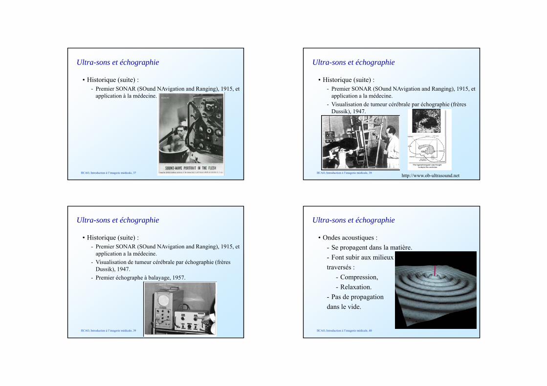
Ultra-sons et échographie
• Historique (suite) :- Premier SONAR (SOund NAvigation and Ranging), 1915, et
application a la médecine.- Visualisation de tumeur cérébrale par échographie (frères
Dussik), 1947.
IICAO, Introduction à l’imagerie médicale, 38http://www.ob-ultrasound.net
Ultra-sons et échographie
• Historique (suite) :- Premier SONAR (SOund NAvigation and Ranging), 1915, et
application a la médecine.- Visualisation de tumeur cérébrale par échographie (frères
Dussik), 1947.- Premier échographe à balayage, 1957.
IICAO, Introduction à l’imagerie médicale, 39
Ultra-sons et échographie
• Ondes acoustiques :- Se propagent dans la matière.p p g- Font subir aux milieux traversés :
- Compression,- Relaxation.
- Pas de propagation dans le vide.
IICAO, Introduction à l’imagerie médicale, 40
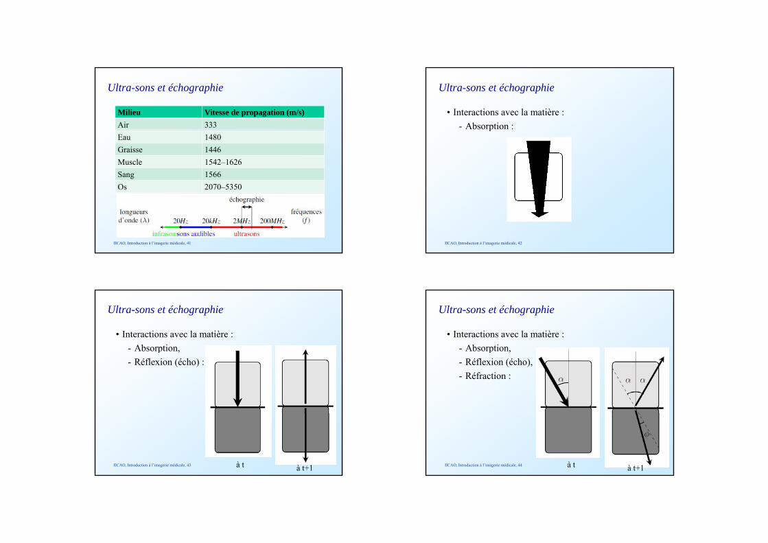
Ultra-sons et échographie
Milieu Vitesse de propagation (m/s)Air 333Eau 1480Graisse 1446Muscle 1542–1626Sang 1566Os 2070–5350
IICAO, Introduction à l’imagerie médicale, 41
Ultra-sons et échographie
• Interactions avec la matière :- Absorption :p
IICAO, Introduction à l’imagerie médicale, 42
Ultra-sons et échographie
• Interactions avec la matière :- Absorption,p ,- Réflexion (écho) :
IICAO, Introduction à l’imagerie médicale, 43 à t à t+1
Ultra-sons et échographie
• Interactions avec la matière :- Absorption,p ,- Réflexion (écho),- Réfraction :
IICAO, Introduction à l’imagerie médicale, 44 à t à t+1
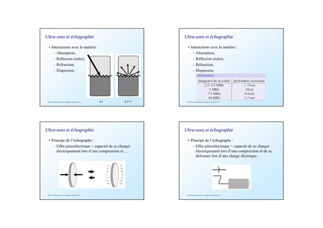
Ultra-sons et échographie
• Interactions avec la matière :- Absorption,p ,- Réflexion (écho),- Réfraction,- Dispersion :
IICAO, Introduction à l’imagerie médicale, 45 à t à t+1
Ultra-sons et échographie
• Interactions avec la matière :- Absorption,p ,- Réflexion (écho),- Réfraction,- Dispersion.
IICAO, Introduction à l’imagerie médicale, 46
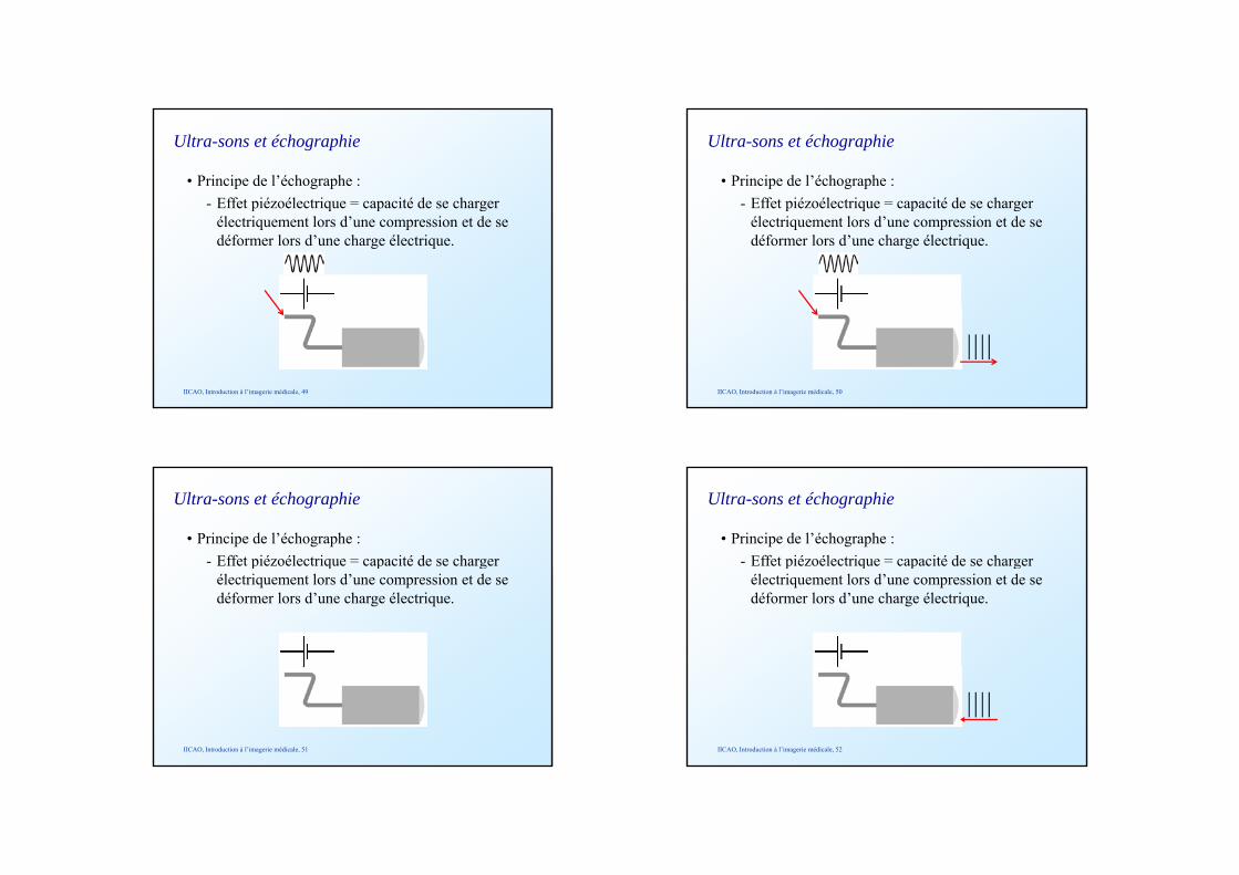
Ultra-sons et échographie
• Principe de l’échographe :- Effet piézoélectrique = capacité de se charger p q p g
électriquement lors d’une compression et ...
IICAO, Introduction à l’imagerie médicale, 47
Ultra-sons et échographie
• Principe de l’échographe :- Effet piézoélectrique = capacité de se charger p q p g
électriquement lors d’une compression et de se déformer lors d’une charge électrique.
IICAO, Introduction à l’imagerie médicale, 48
Ultra-sons et échographie
• Principe de l’échographe :- Effet piézoélectrique = capacité de se charger p q p g
électriquement lors d’une compression et de se déformer lors d’une charge électrique.
IICAO, Introduction à l’imagerie médicale, 49
Ultra-sons et échographie
• Principe de l’échographe :- Effet piézoélectrique = capacité de se charger p q p g
électriquement lors d’une compression et de se déformer lors d’une charge électrique.
IICAO, Introduction à l’imagerie médicale, 50
Ultra-sons et échographie
• Principe de l’échographe :- Effet piézoélectrique = capacité de se charger p q p g
électriquement lors d’une compression et de se déformer lors d’une charge électrique.
IICAO, Introduction à l’imagerie médicale, 51
Ultra-sons et échographie
• Principe de l’échographe :- Effet piézoélectrique = capacité de se charger p q p g
électriquement lors d’une compression et de se déformer lors d’une charge électrique.
IICAO, Introduction à l’imagerie médicale, 52
Ultra-sons et échographie
• Principe de l’échographe :- Effet piézoélectrique = capacité de se charger p q p g
électriquement lors d’une compression et de se déformer lors d’une charge électrique.
IICAO, Introduction à l’imagerie médicale, 53
Ultra-sons et échographie
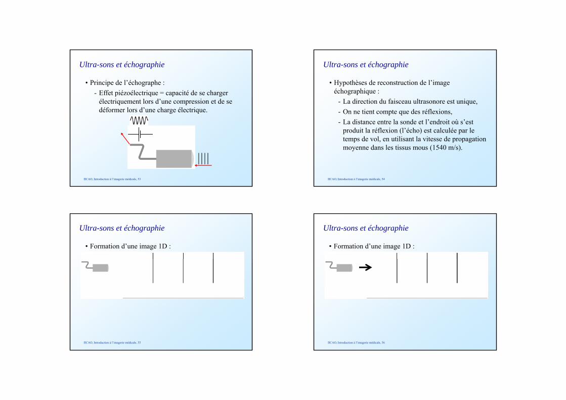
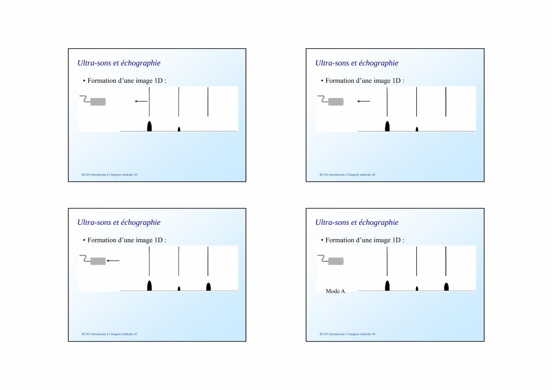
• Hypothèses de reconstruction de l’image échographique :
- La direction du faisceau ultrasonore est unique,- On ne tient compte que des réflexions,- La distance entre la sonde et l’endroit où s’est
produit la réflexion (l’écho) est calculée par le temps de vol, en utilisant la vitesse de propagationtemps de vol, en utilisant la vitesse de propagation moyenne dans les tissus mous (1540 m/s).
IICAO, Introduction à l’imagerie médicale, 54
Ultra-sons et échographie
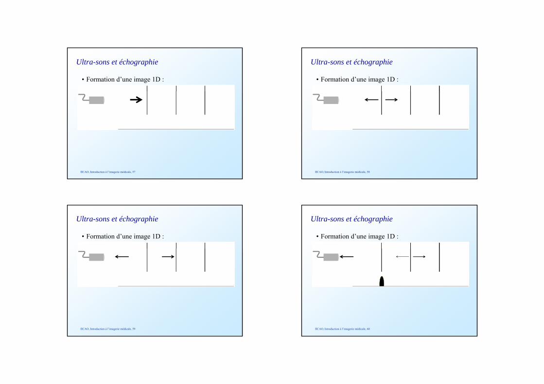
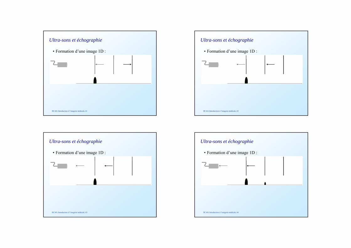
• Formation d’une image 1D :
IICAO, Introduction à l’imagerie médicale, 55
Ultra-sons et échographie
• Formation d’une image 1D :
IICAO, Introduction à l’imagerie médicale, 56
Ultra-sons et échographie
• Formation d’une image 1D :
IICAO, Introduction à l’imagerie médicale, 57
Ultra-sons et échographie
• Formation d’une image 1D :
IICAO, Introduction à l’imagerie médicale, 58
Ultra-sons et échographie
• Formation d’une image 1D :
IICAO, Introduction à l’imagerie médicale, 59
Ultra-sons et échographie
• Formation d’une image 1D :
IICAO, Introduction à l’imagerie médicale, 60
Ultra-sons et échographie
• Formation d’une image 1D :
IICAO, Introduction à l’imagerie médicale, 61
Ultra-sons et échographie
• Formation d’une image 1D :
IICAO, Introduction à l’imagerie médicale, 62
Ultra-sons et échographie
• Formation d’une image 1D :
IICAO, Introduction à l’imagerie médicale, 63
Ultra-sons et échographie
• Formation d’une image 1D :
IICAO, Introduction à l’imagerie médicale, 64
Ultra-sons et échographie
• Formation d’une image 1D :
IICAO, Introduction à l’imagerie médicale, 65
Ultra-sons et échographie
• Formation d’une image 1D :
IICAO, Introduction à l’imagerie médicale, 66
Ultra-sons et échographie
• Formation d’une image 1D :
IICAO, Introduction à l’imagerie médicale, 67
Ultra-sons et échographie
• Formation d’une image 1D :
Mode A
IICAO, Introduction à l’imagerie médicale, 68
Ultra-sons et échographie
• Formation d’une image 1D :
IICAO, Introduction à l’imagerie médicale, 69
Ultra-sons et échographie
• Formation d’une image 1D :
Mode A
IICAO, Introduction à l’imagerie médicale, 70
Ultra-sons et échographie
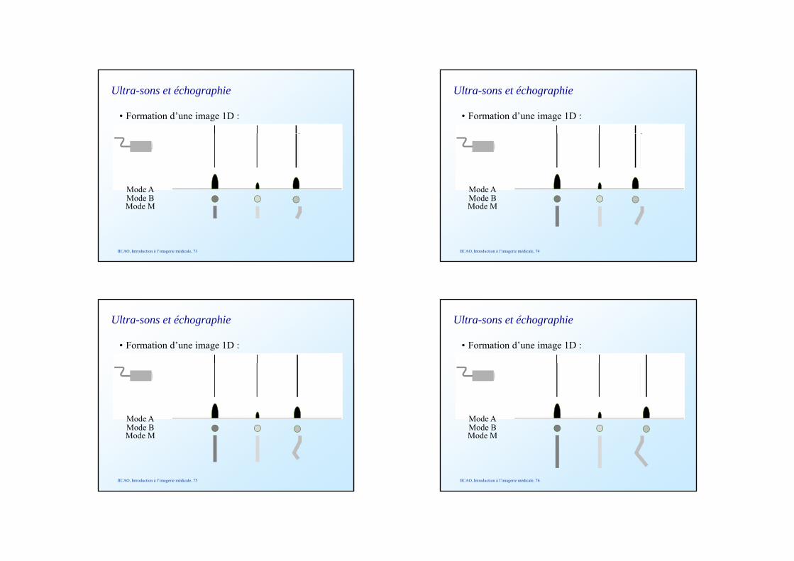
• Formation d’une image 1D :
Mode AMode B
IICAO, Introduction à l’imagerie médicale, 71
Mode B
Ultra-sons et échographie
• Formation d’une image 1D :
Mode AMode B
IICAO, Introduction à l’imagerie médicale, 72
Mode BMode M
Ultra-sons et échographie
• Formation d’une image 1D :
Mode AMode B
IICAO, Introduction à l’imagerie médicale, 73
Mode BMode M
Ultra-sons et échographie
• Formation d’une image 1D :
Mode AMode B
IICAO, Introduction à l’imagerie médicale, 74
Mode BMode M
Ultra-sons et échographie
• Formation d’une image 1D :
Mode AMode B
IICAO, Introduction à l’imagerie médicale, 75
Mode BMode M
Ultra-sons et échographie
• Formation d’une image 1D :
Mode AMode B
IICAO, Introduction à l’imagerie médicale, 76
Mode BMode M
Ultra-sons et échographie
• Formation d’une image 1D :
Mode AMode B
IICAO, Introduction à l’imagerie médicale, 77
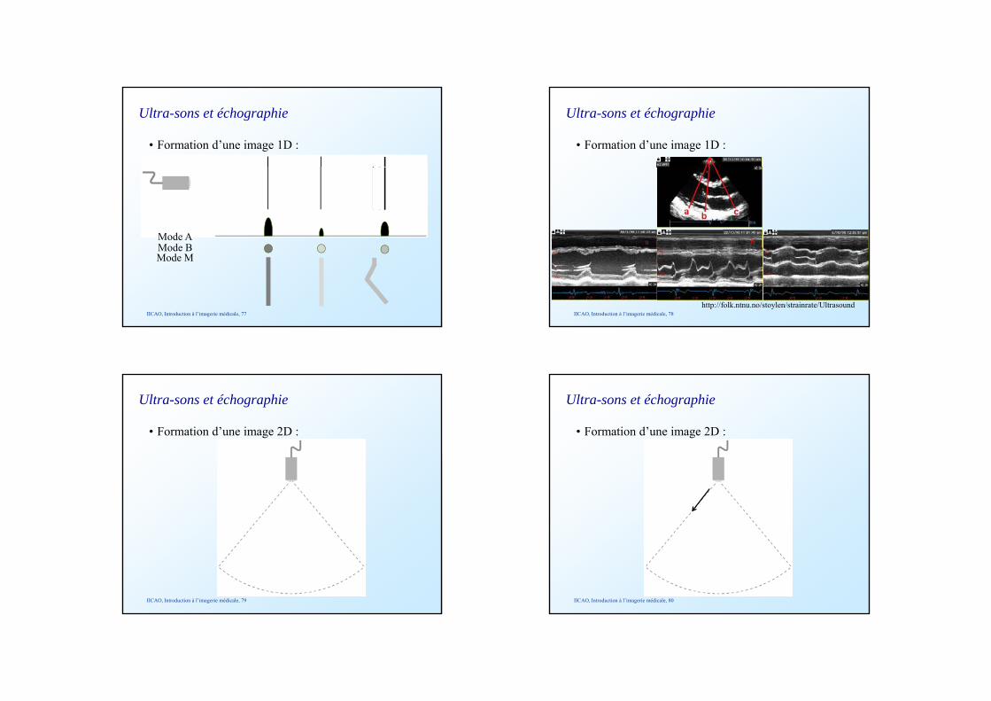
Mode BMode M
Ultra-sons et échographie
• Formation d’une image 1D :
a b c
IICAO, Introduction à l’imagerie médicale, 78http://folk.ntnu.no/stoylen/strainrate/Ultrasound
Ultra-sons et échographie
• Formation d’une image 2D :
IICAO, Introduction à l’imagerie médicale, 79
Ultra-sons et échographie
• Formation d’une image 2D :
IICAO, Introduction à l’imagerie médicale, 80
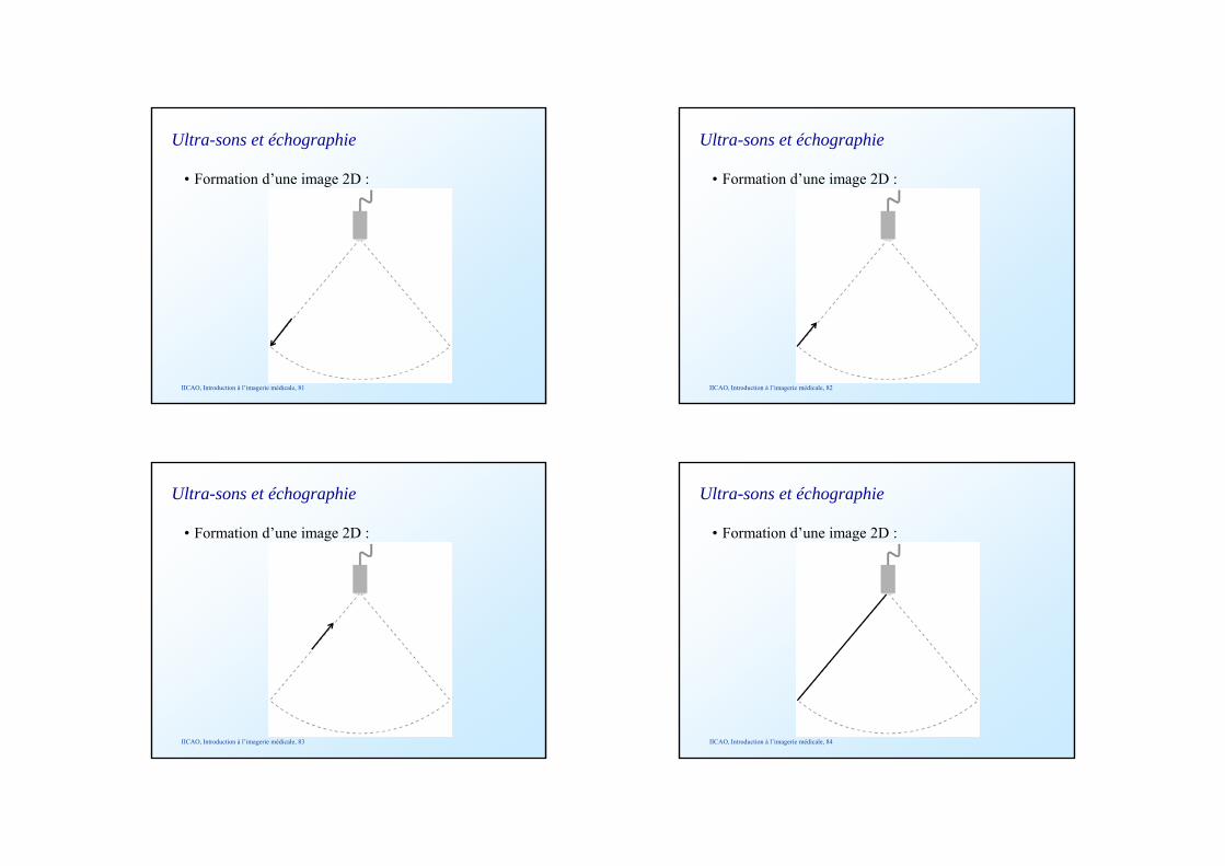
Ultra-sons et échographie
• Formation d’une image 2D :
IICAO, Introduction à l’imagerie médicale, 81
Ultra-sons et échographie
• Formation d’une image 2D :
IICAO, Introduction à l’imagerie médicale, 82
Ultra-sons et échographie
• Formation d’une image 2D :
IICAO, Introduction à l’imagerie médicale, 83
Ultra-sons et échographie
• Formation d’une image 2D :
IICAO, Introduction à l’imagerie médicale, 84
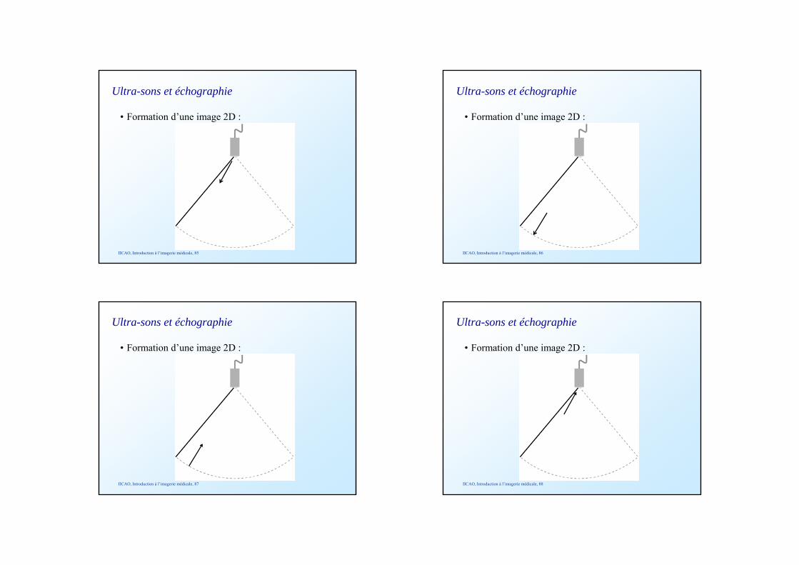
Ultra-sons et échographie
• Formation d’une image 2D :
IICAO, Introduction à l’imagerie médicale, 85
Ultra-sons et échographie
• Formation d’une image 2D :
IICAO, Introduction à l’imagerie médicale, 86
Ultra-sons et échographie
• Formation d’une image 2D :
IICAO, Introduction à l’imagerie médicale, 87
Ultra-sons et échographie
• Formation d’une image 2D :
IICAO, Introduction à l’imagerie médicale, 88
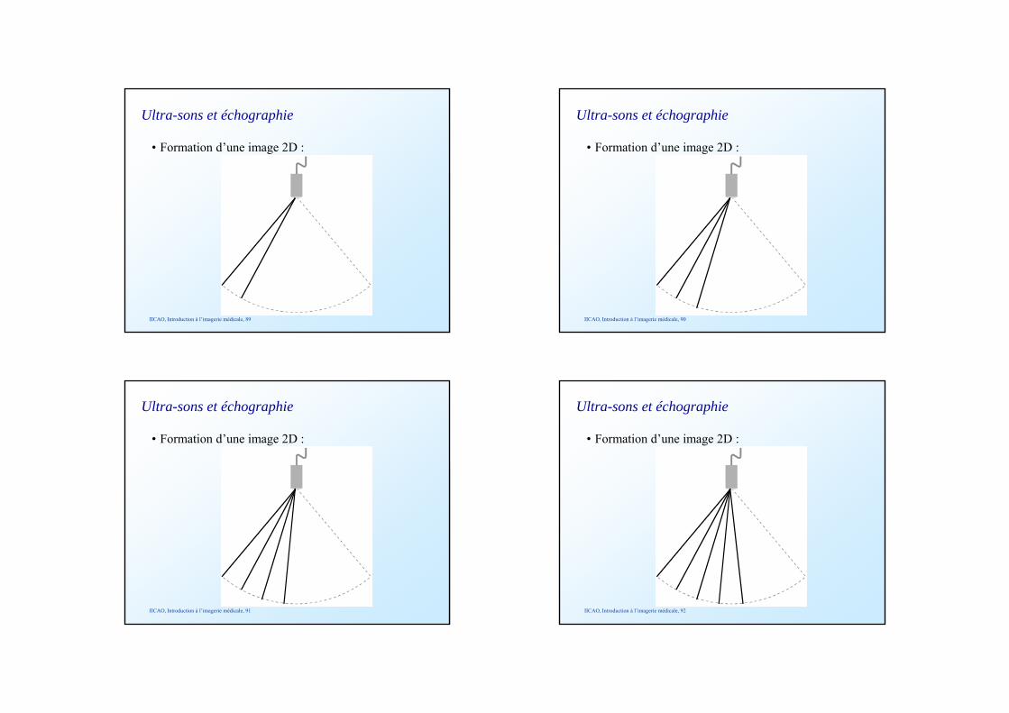
Ultra-sons et échographie
• Formation d’une image 2D :
IICAO, Introduction à l’imagerie médicale, 89
Ultra-sons et échographie
• Formation d’une image 2D :
IICAO, Introduction à l’imagerie médicale, 90
Ultra-sons et échographie
• Formation d’une image 2D :
IICAO, Introduction à l’imagerie médicale, 91
Ultra-sons et échographie
• Formation d’une image 2D :
IICAO, Introduction à l’imagerie médicale, 92
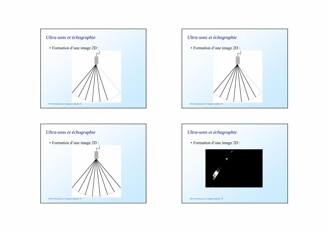
Ultra-sons et échographie
• Formation d’une image 2D :
IICAO, Introduction à l’imagerie médicale, 93
Ultra-sons et échographie
• Formation d’une image 2D :
IICAO, Introduction à l’imagerie médicale, 94
Ultra-sons et échographie
• Formation d’une image 2D :
IICAO, Introduction à l’imagerie médicale, 95
Ultra-sons et échographie
• Formation d’une image 2D :
IICAO, Introduction à l’imagerie médicale, 96
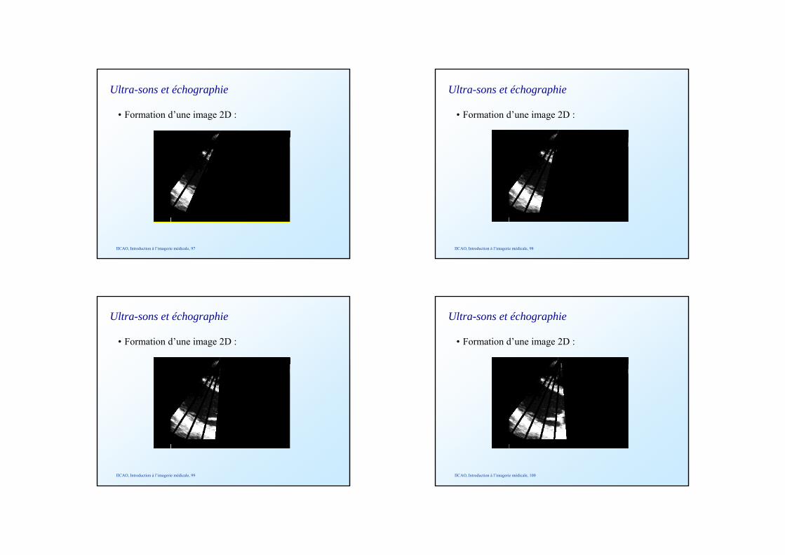
Ultra-sons et échographie
• Formation d’une image 2D :
IICAO, Introduction à l’imagerie médicale, 97
Ultra-sons et échographie
• Formation d’une image 2D :
IICAO, Introduction à l’imagerie médicale, 98
Ultra-sons et échographie
• Formation d’une image 2D :
IICAO, Introduction à l’imagerie médicale, 99
Ultra-sons et échographie
• Formation d’une image 2D :
IICAO, Introduction à l’imagerie médicale, 100
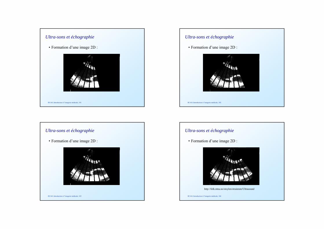
Ultra-sons et échographie
• Formation d’une image 2D :
IICAO, Introduction à l’imagerie médicale, 101
Ultra-sons et échographie
• Formation d’une image 2D :
IICAO, Introduction à l’imagerie médicale, 102
Ultra-sons et échographie
• Formation d’une image 2D :
IICAO, Introduction à l’imagerie médicale, 103
Ultra-sons et échographie
• Formation d’une image 2D :
IICAO, Introduction à l’imagerie médicale, 104
http://folk.ntnu.no/stoylen/strainrate/Ultrasound

Ultra-sons et échographie
• Image 2D + temps :
IICAO, Introduction à l’imagerie médicale, 105
http://www-sante.ujf-grenoble.fr/sante/CardioCD/cardio/video.htm
Animation
Ultra-sons et échographie
• Image 3D :
IICAO, Introduction à l’imagerie médicale, 106
Ultra-sons et échographie
• Image 3D :
IICAO, Introduction à l’imagerie médicale, 107
Ultra-sons et échographie
• Image 3D :
IICAO, Introduction à l’imagerie médicale, 108
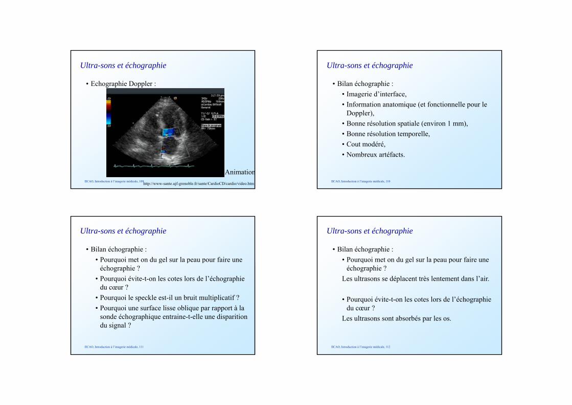
Ultra-sons et échographie
• Echographie Doppler :
IICAO, Introduction à l’imagerie médicale, 109http://www-sante.ujf-grenoble.fr/sante/CardioCD/cardio/video.htm
Animation
Ultra-sons et échographie
• Bilan échographie :• Imagerie d’interface,g ,• Information anatomique (et fonctionnelle pour le
Doppler),• Bonne résolution spatiale (environ 1 mm),• Bonne résolution temporelle,
C t dé é• Cout modéré,• Nombreux artéfacts.
IICAO, Introduction à l’imagerie médicale, 110
Ultra-sons et échographie
• Bilan échographie :• Pourquoi met on du gel sur la peau pour faire une q g p p
échographie ?• Pourquoi évite-t-on les cotes lors de l’échographie
du cœur ?• Pourquoi le speckle est-il un bruit multiplicatif ?• Pourquoi une surface lisse oblique par rapport à la• Pourquoi une surface lisse oblique par rapport à la
sonde échographique entraine-t-elle une disparition du signal ?
IICAO, Introduction à l’imagerie médicale, 111
Ultra-sons et échographie
• Bilan échographie :• Pourquoi met on du gel sur la peau pour faire une q g p p
échographie ?Les ultrasons se déplacent très lentement dans l’air.
• Pourquoi évite-t-on les cotes lors de l’échographie du cœur ?du cœur ?
Les ultrasons sont absorbés par les os.
IICAO, Introduction à l’imagerie médicale, 112
Ultra-sons et échographie
• Bilan échographie :• Pourquoi le speckle est-il un bruit multiplicatif ?q p pIl s’agit d’interférences entre les ondes ultrasonores.
• Pourquoi une surface lisse oblique par rapport à la sonde échographique entraine-t-elle une disparition du signal ?du signal ?
Du fait de la réfraction, aucun son ne revient directement sur la sonde après la réflexion oblique, et le faisceau change de direction après la réfraction.
IICAO, Introduction à l’imagerie médicale, 113
• Introduction• Lumière et endoscopie
Plan
Lumière et endoscopie• Ultra-sons et échographie• Rayons X et radiographie• Autres imageries• Applications
IICAO, Introduction à l’imagerie médicale, 114
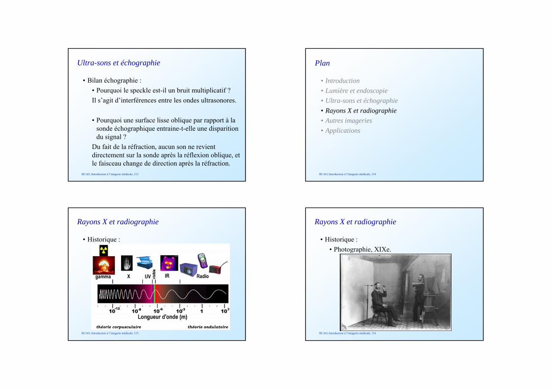
Rayons X et radiographie
• Historique :
IICAO, Introduction à l’imagerie médicale, 115
Rayons X et radiographie
• Historique :• Photographie, XIXe.g p ,
IICAO, Introduction à l’imagerie médicale, 116
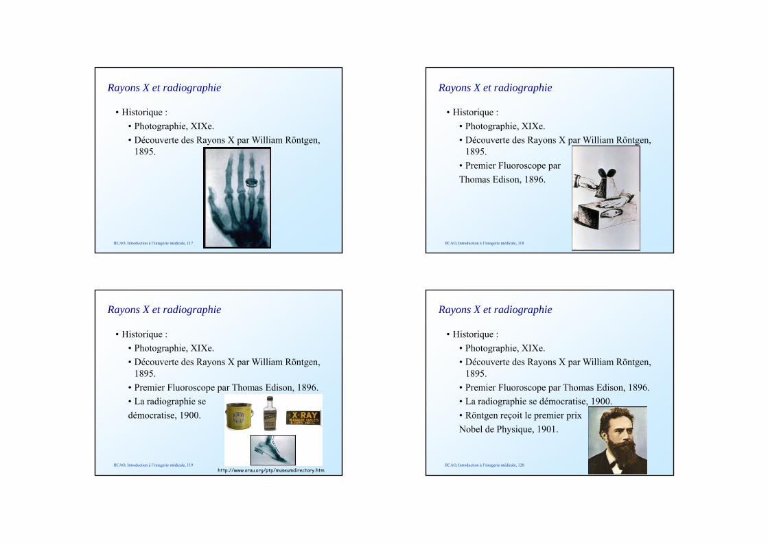
Rayons X et radiographie
• Historique :• Photographie, XIXe.g p ,• Découverte des Rayons X par William Rӧntgen,
1895.
IICAO, Introduction à l’imagerie médicale, 117
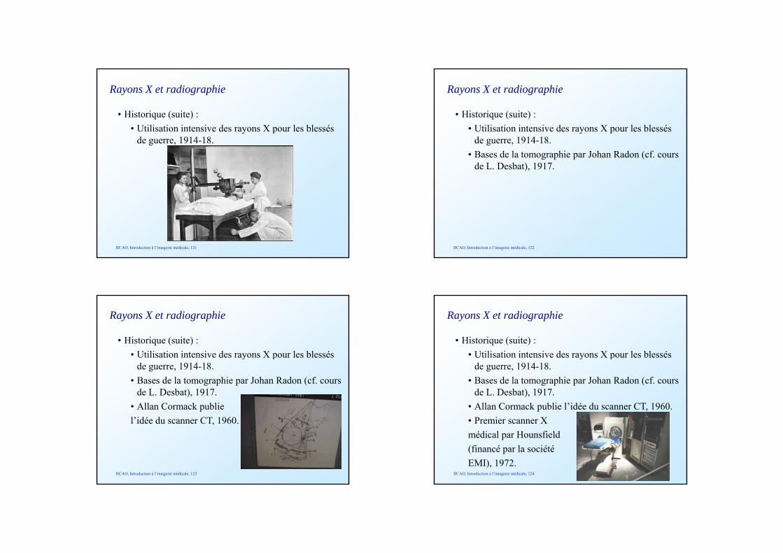
Rayons X et radiographie
• Historique :• Photographie, XIXe.g p ,• Découverte des Rayons X par William Rӧntgen,
1895.• Premier Fluoroscope par Thomas Edison, 1896.
IICAO, Introduction à l’imagerie médicale, 118
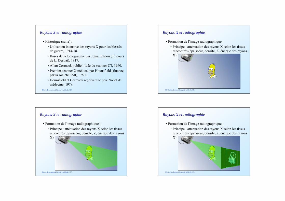
Rayons X et radiographie
• Historique :• Photographie, XIXe.g p ,• Découverte des Rayons X par William Rӧntgen,
1895.• Premier Fluoroscope par Thomas Edison, 1896.• La radiographie se dé ti 1900démocratise, 1900.
IICAO, Introduction à l’imagerie médicale, 119http://www.orau.org/ptp/museumdirectory.htm
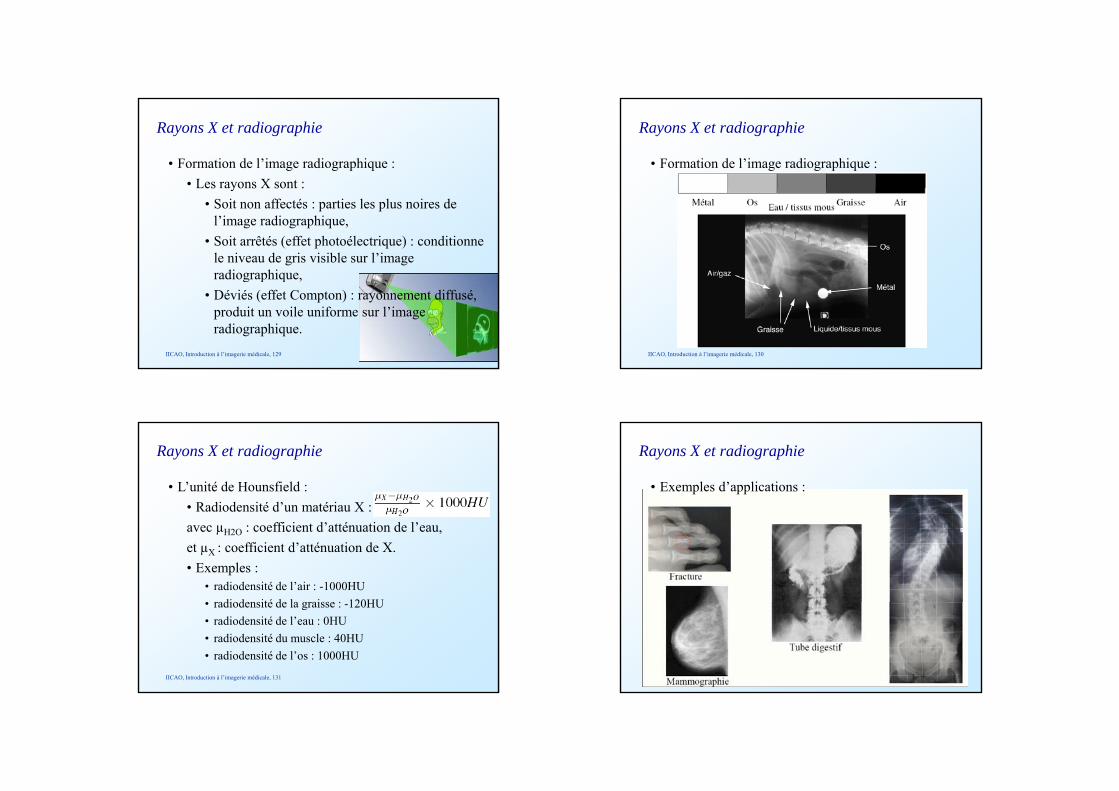
Rayons X et radiographie
• Historique :• Photographie, XIXe.g p ,• Découverte des Rayons X par William Rӧntgen,
1895.• Premier Fluoroscope par Thomas Edison, 1896.• La radiographie se démocratise, 1900.
Rӧ t it l i i• Rӧntgen reçoit le premier prix Nobel de Physique, 1901.
IICAO, Introduction à l’imagerie médicale, 120
Rayons X et radiographie
• Historique (suite) :• Utilisation intensive des rayons X pour les blessés y p
de guerre, 1914-18.
IICAO, Introduction à l’imagerie médicale, 121
Rayons X et radiographie
• Historique (suite) :• Utilisation intensive des rayons X pour les blessés y p
de guerre, 1914-18.• Bases de la tomographie par Johan Radon (cf. cours
de L. Desbat), 1917.
IICAO, Introduction à l’imagerie médicale, 122
Rayons X et radiographie
• Historique (suite) :• Utilisation intensive des rayons X pour les blessés y p
de guerre, 1914-18.• Bases de la tomographie par Johan Radon (cf. cours
de L. Desbat), 1917.• Allan Cormack publie l’idée du scanner CT 1960l idée du scanner CT, 1960.
IICAO, Introduction à l’imagerie médicale, 123
Rayons X et radiographie
• Historique (suite) :• Utilisation intensive des rayons X pour les blessés y p
de guerre, 1914-18.• Bases de la tomographie par Johan Radon (cf. cours
de L. Desbat), 1917.• Allan Cormack publie l’idée du scanner CT, 1960.• Premier scanner X• Premier scanner X médical par Hounsfield (financé par la société EMI), 1972.
IICAO, Introduction à l’imagerie médicale, 124
Rayons X et radiographie
• Historique (suite) :• Utilisation intensive des rayons X pour les blessés y p
de guerre, 1914-18.• Bases de la tomographie par Johan Radon (cf. cours
de L. Desbat), 1917.• Allan Cormack publie l’idée du scanner CT, 1960.• Premier scanner X médical par Hounsfield (financé• Premier scanner X médical par Hounsfield (financé
par la société EMI), 1972.• Hounsfield et Cormack reçoivent le prix Nobel de
médecine, 1979.IICAO, Introduction à l’imagerie médicale, 125
Rayons X et radiographie
• Formation de l’image radiographique :• Principe : atténuation des rayons X selon les tissus p y
rencontrés (épaisseur, densité, Z, énergie des rayons X)
IICAO, Introduction à l’imagerie médicale, 126
Rayons X et radiographie
• Formation de l’image radiographique :• Principe : atténuation des rayons X selon les tissus p y
rencontrés (épaisseur, densité, Z, énergie des rayons X)
IICAO, Introduction à l’imagerie médicale, 127
Rayons X et radiographie
• Formation de l’image radiographique :• Principe : atténuation des rayons X selon les tissus p y
rencontrés (épaisseur, densité, Z, énergie des rayons X)
IICAO, Introduction à l’imagerie médicale, 128
Rayons X et radiographie
• Formation de l’image radiographique :• Les rayons X sont :y
• Soit non affectés : parties les plus noires de l’image radiographique,
• Soit arrêtés (effet photoélectrique) : conditionne le niveau de gris visible sur l’image radiographique,radiographique,
• Déviés (effet Compton) : rayonnement diffusé, produit un voile uniforme sur l’image radiographique.
IICAO, Introduction à l’imagerie médicale, 129
Rayons X et radiographie
• Formation de l’image radiographique :
IICAO, Introduction à l’imagerie médicale, 130
Rayons X et radiographie
• L’unité de Hounsfield :• Radiodensité d’un matériau X :avec µH2O : coefficient d’atténuation de l’eau, et µX : coefficient d’atténuation de X.• Exemples :
• radiodensité de l’air : -1000HU• radiodensité de la graisse : 120HU• radiodensité de la graisse : -120HU• radiodensité de l’eau : 0HU• radiodensité du muscle : 40HU• radiodensité de l’os : 1000HU
IICAO, Introduction à l’imagerie médicale, 131
Rayons X et radiographie
• Exemples d’applications :
IICAO, Introduction à l’imagerie médicale, 132
Rayons X et radiographie
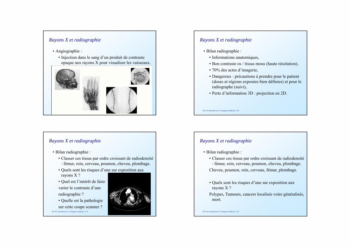
• Angiographie :• Injection dans le sang d’un produit de contraste j g p
opaque aux rayons X pour visualiser les vaisseaux.
IICAO, Introduction à l’imagerie médicale, 133
Rayons X et radiographie
• Bilan radiographie :• Informations anatomiques,q ,• Bon contraste os / tissus mous (haute résolution),• 70% des actes d’imagerie,• Dangereux : précautions à prendre pour le patient
(doses et régions exposées bien définies) et pour le radiographe (suivi)
IICAO, Introduction à l’imagerie médicale, 134
radiographe (suivi),• Perte d’information 3D : projection en 2D.
Rayons X et radiographie
• Bilan radiographie :• Classer ces tissus par ordre croissant de radiodensité p
: fémur, rein, cerveau, poumon, cheveu, plombage.• Quels sont les risques d’une sur exposition aux
rayons X ?• Quel est l’intérêt de faire varier le contraste d’une
IICAO, Introduction à l’imagerie médicale, 135
varier le contraste d une radiographie ?• Quelle est la pathologie sur cette coupe scanner ?
Rayons X et radiographie
• Bilan radiographie :• Classer ces tissus par ordre croissant de radiodensité p
: fémur, rein, cerveau, poumon, cheveu, plombage.Cheveu, poumon, rein, cerveau, fémur, plombage.
• Quels sont les risques d’une sur exposition aux rayons X ?
IICAO, Introduction à l’imagerie médicale, 136
rayons X ?Polypes, Tumeurs, cancers localisés voire généralisés,
mort.
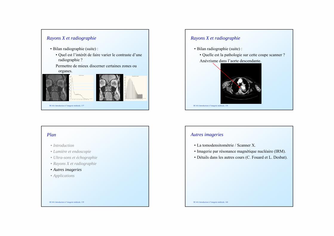
Rayons X et radiographie
• Bilan radiographie (suite) :• Quel est l’intérêt de faire varier le contraste d’une Q
radiographie ?Permettre de mieux discerner certaines zones ou
organes.
IICAO, Introduction à l’imagerie médicale, 137
Rayons X et radiographie
• Bilan radiographie (suite) :• Quelle est la pathologie sur cette coupe scanner ?Q p g pAnévrisme dans l’aorte descendante.
IICAO, Introduction à l’imagerie médicale, 138
• Introduction• Lumière et endoscopie
Plan
Lumière et endoscopie• Ultra-sons et échographie• Rayons X et radiographie• Autres imageries• Applications
IICAO, Introduction à l’imagerie médicale, 139
Autres imageries
• La tomodensitométrie / Scanner X.• Imagerie par résonance magnétique nucléaire (IRM).g p g q ( )• Détails dans les autres cours (C. Fouard et L. Desbat).
IICAO, Introduction à l’imagerie médicale, 140
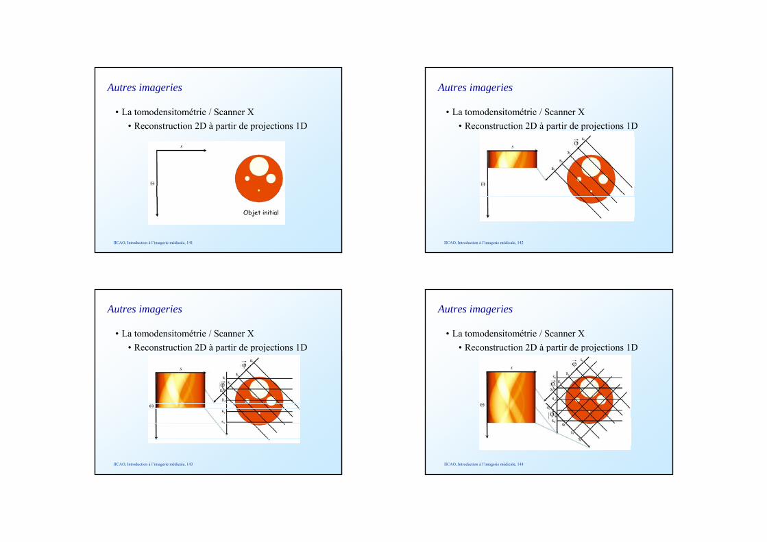
Autres imageries
• La tomodensitométrie / Scanner X• Reconstruction 2D à partir de projections 1Dp p j
IICAO, Introduction à l’imagerie médicale, 141
Objet initial
Autres imageries
• La tomodensitométrie / Scanner X• Reconstruction 2D à partir de projections 1Dp p j
IICAO, Introduction à l’imagerie médicale, 142
Autres imageries
• La tomodensitométrie / Scanner X• Reconstruction 2D à partir de projections 1Dp p j
IICAO, Introduction à l’imagerie médicale, 143
Autres imageries
• La tomodensitométrie / Scanner X• Reconstruction 2D à partir de projections 1Dp p j
IICAO, Introduction à l’imagerie médicale, 144
Autres imageries
• La tomodensitométrie / Scanner X• Reconstruction 2D à partir de projections 1Dp p j
IICAO, Introduction à l’imagerie médicale, 145
Autres imageries
• La tomodensitométrie / Scanner X• Reconstruction 2D à partir de projections 1Dp p j
Transformée de Radon
IICAO, Introduction à l’imagerie médicale, 146
Image de l’objet initial
Autres imageries
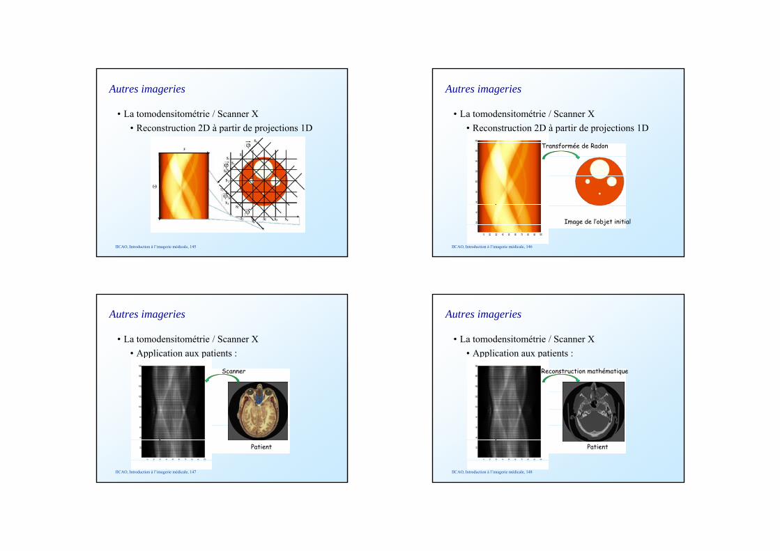
• La tomodensitométrie / Scanner X• Application aux patients :
Scanner
pp p
IICAO, Introduction à l’imagerie médicale, 147
Patient
• La tomodensitométrie / Scanner X• Application aux patients :
Autres imageries
pp p
Reconstruction mathématique
IICAO, Introduction à l’imagerie médicale, 148
Patient
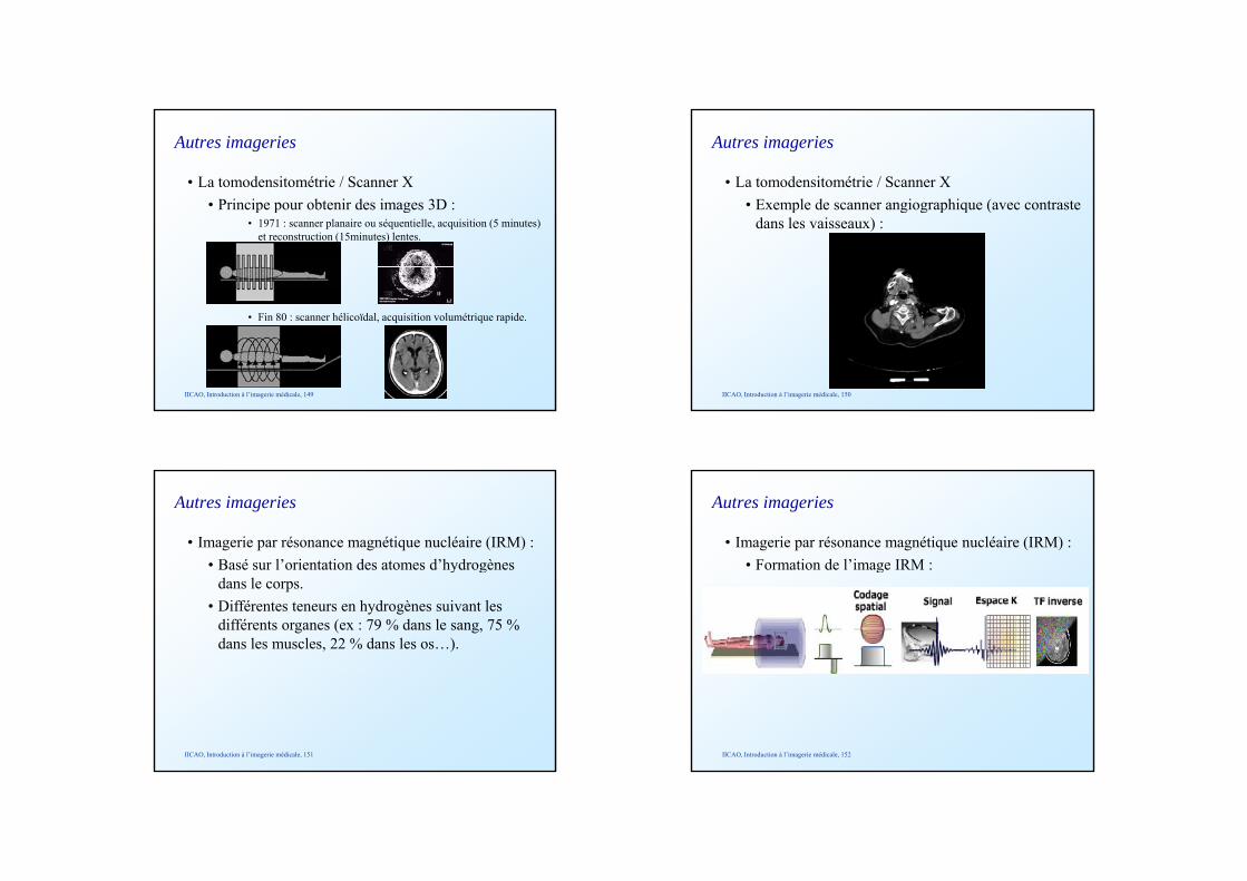
• La tomodensitométrie / Scanner X• Principe pour obtenir des images 3D :
Autres imageries
p p g• 1971 : scanner planaire ou séquentielle, acquisition (5 minutes)
et reconstruction (15minutes) lentes.
• Fin 80 : scanner hélicoïdal, acquisition volumétrique rapide.
IICAO, Introduction à l’imagerie médicale, 149
• La tomodensitométrie / Scanner X• Exemple de scanner angiographique (avec contraste
Autres imageries
p g g p q (dans les vaisseaux) :
IICAO, Introduction à l’imagerie médicale, 150
Autres imageries
• Imagerie par résonance magnétique nucléaire (IRM) :• Basé sur l’orientation des atomes d’hydrogènes y g
dans le corps. • Différentes teneurs en hydrogènes suivant les
différents organes (ex : 79 % dans le sang, 75 % dans les muscles, 22 % dans les os…).
IICAO, Introduction à l’imagerie médicale, 151
Autres imageries
• Imagerie par résonance magnétique nucléaire (IRM) :• Formation de l’image IRM :g
IICAO, Introduction à l’imagerie médicale, 152
Autres imageries
• Imagerie par résonance magnétique nucléaire (IRM) :• Exemple d’IRM :p
IICAO, Introduction à l’imagerie médicale, 153
• Introduction• Lumière et endoscopie
Plan
Lumière et endoscopie• Ultra-sons et échographie• Rayons X et radiographie• Autres imageries• Applications
IICAO, Introduction à l’imagerie médicale, 154
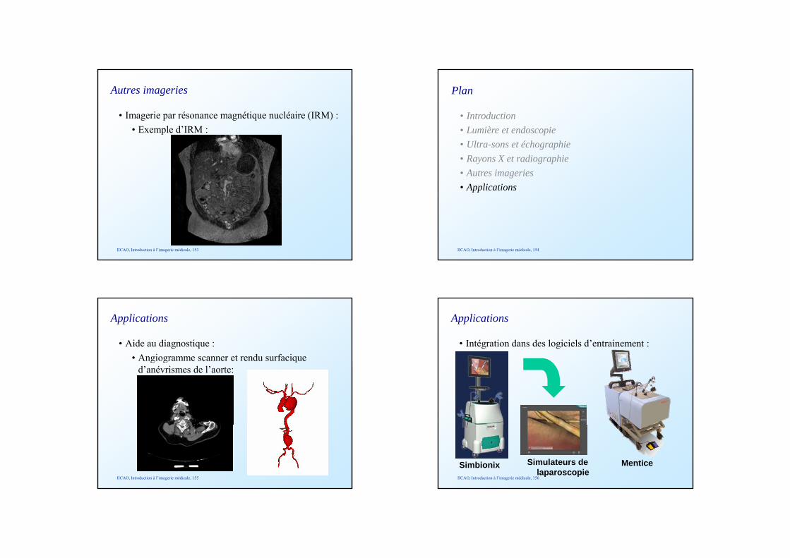
Applications
• Aide au diagnostique :• Angiogramme scanner et rendu surfacique g g q
d’anévrismes de l’aorte:
IICAO, Introduction à l’imagerie médicale, 155
Applications
• Intégration dans des logiciels d’entrainement :
IICAO, Introduction à l’imagerie médicale, 156
Simbionix MenticeSimulateurs de laparoscopie
Applications
• Besoin de traitement des images pour les différentes applications :
• Prétraitement (lissage, réduction du bruit…),• Segmentation (extraction du contours des organes),• Post-traitement (création de surfaces 3D, de
volume…).
IICAO, Introduction à l’imagerie médicale, 157
Résumé
• Les différentes modalités d’imagerie médicale,• Les bases de ces modalités et leurs caractéristiques
principales,• L’utilisation/l’indication pour chaque modalité,• Le concept d’imagerie 4D et son application,• Certaines applications en planification et en
chirurgie.
IICAO, Introduction à l’imagerie médicale, 158