Embed Size (px)
Citation preview
Vet. Res. (2008) 39:54 www.vetres.orgDOI: 10.1051/vetres:2008030
C© INRA, EDP Sciences, 2008 Review article
Membrane markers of the immune cellsin swine: an update
Laurence Piriou-Guzylack1, Henri Salmon2*
1 Institute of Virology and Immunoprophylaxis, CH-3147 Mittelhäusern, Switzerland2 Institut National de la Recherche Agronomique, INRA, UR1282, Infectiologie Animale et Santé Publique
(IASP), Equipe Lymphocyte et Immunité des Muqueuses, Nouzilly, F-37380, France
(Received 6 February 2008; accepted 16 July 2008)
Abstract – Besides their breeding value, swine are increasingly used as biomedical models. As reportedin three international swine clusters of differentiation (CD) workshops and in the animal homologuesection of the last workshop for the determination of human leukocyte differentiation antigens (HLDA 8),characterisation of leukocyte surface antigens by monoclonal antibodies and other molecular studies havedetermined the cell lineages and blood leukocyte subsets implicated in the immune response, including celladhesion molecules involved in cell trafficking. This review focusses on the current state of knowledgeof porcine leukocyte differentiation and major histocompatibility complex (SLA) molecules. Examples ofporcine particularities such as the double-positive T lymphocytes with the phenotype CD4+CD8low andCD4−CD8low �� T cell subsets and the persistence of SLA class II after T-lymphocyte activation areillustrated, as well as the shared characteristics of the Artiodactyla group, such as the high proportion of�� TcR (T cell receptor) T cells in blood and other lymphoid tissues. Furthermore, discrepancies betweenswine and humans, such as CD16 expression on dendritic cells and CD11b (wCD11R1) tissue distributionare outlined. The rapidly growing information should facilitate manipulation of the swine immune systemtowards improving disease control, and open new avenues for biomedical research using the pig asa model.
cluster of differentiation (CD) / monoclonal antibody / immune system / histocompatibility system /pig
Table of contents
1. Introduction . . . . . . . . . . . . . . . . . . . . . . . . . . . . . . . . . . . . . . . . . . . . . . . . . . . . . . . . . . . . . . . . . . . . . . . . . . . . . . . . . . . . . . . . . . . . . . . . 22. Lymphoid and myeloid cells . . . . . . . . . . . . . . . . . . . . . . . . . . . . . . . . . . . . . . . . . . . . . . . . . . . . . . . . . . . . . . . . . . . . . . . . . . . . . . 4
2.1. Markers of T and/or NK (natural killer) cells . . . . . . . . . . . . . . . . . . . . . . . . . . . . . . . . . . . . . . . . . . . . . . . . . . . . 42.1.1. CD2 . . . . . . . . . . . . . . . . . . . . . . . . . . . . . . . . . . . . . . . . . . . . . . . . . . . . . . . . . . . . . . . . . . . . . . . . . . . . . . . . . . . . . . . . 42.1.2. CD3-T cell complex . . . . . . . . . . . . . . . . . . . . . . . . . . . . . . . . . . . . . . . . . . . . . . . . . . . . . . . . . . . . . . . . . . . . . . . 4
2.2. Markers of TcR-�� T cells and ‘null cells’ . . . . . . . . . . . . . . . . . . . . . . . . . . . . . . . . . . . . . . . . . . . . . . . . . . . . . . . 52.2.1. SWC4, SWC5 and SWC6 . . . . . . . . . . . . . . . . . . . . . . . . . . . . . . . . . . . . . . . . . . . . . . . . . . . . . . . . . . . . . . . . 52.2.2. Anti-WC1 . . . . . . . . . . . . . . . . . . . . . . . . . . . . . . . . . . . . . . . . . . . . . . . . . . . . . . . . . . . . . . . . . . . . . . . . . . . . . . . . . . 5
2.3. Accessory molecules . . . . . . . . . . . . . . . . . . . . . . . . . . . . . . . . . . . . . . . . . . . . . . . . . . . . . . . . . . . . . . . . . . . . . . . . . . . . . . 62.3.1. CD4 . . . . . . . . . . . . . . . . . . . . . . . . . . . . . . . . . . . . . . . . . . . . . . . . . . . . . . . . . . . . . . . . . . . . . . . . . . . . . . . . . . . . . . . . 62.3.2. CD5 . . . . . . . . . . . . . . . . . . . . . . . . . . . . . . . . . . . . . . . . . . . . . . . . . . . . . . . . . . . . . . . . . . . . . . . . . . . . . . . . . . . . . . . . 6
* Corresponding author: [email protected]
Article available at http://www.vetres.org or http://dx.doi.org/10.1051/vetres:2008030
Vet. Res. (2008) 39:54 L. Piriou-Guzylack and H. Salmon
2.3.3. CD6 . . . . . . . . . . . . . . . . . . . . . . . . . . . . . . . . . . . . . . . . . . . . . . . . . . . . . . . . . . . . . . . . . . . . . . . . . . . . . . . . . . . . . . . . 62.3.4. CD8 . . . . . . . . . . . . . . . . . . . . . . . . . . . . . . . . . . . . . . . . . . . . . . . . . . . . . . . . . . . . . . . . . . . . . . . . . . . . . . . . . . . . . . . . 62.3.5. SWC2 . . . . . . . . . . . . . . . . . . . . . . . . . . . . . . . . . . . . . . . . . . . . . . . . . . . . . . . . . . . . . . . . . . . . . . . . . . . . . . . . . . . . . . 7
2.4. Differentiation antigens of B cells . . . . . . . . . . . . . . . . . . . . . . . . . . . . . . . . . . . . . . . . . . . . . . . . . . . . . . . . . . . . . . . . 72.4.1. CD1 . . . . . . . . . . . . . . . . . . . . . . . . . . . . . . . . . . . . . . . . . . . . . . . . . . . . . . . . . . . . . . . . . . . . . . . . . . . . . . . . . . . . . . . . 72.4.2. wCD21 (complement receptor 2, CR2) . . . . . . . . . . . . . . . . . . . . . . . . . . . . . . . . . . . . . . . . . . . . . . . . . . 72.4.3. SWC7 . . . . . . . . . . . . . . . . . . . . . . . . . . . . . . . . . . . . . . . . . . . . . . . . . . . . . . . . . . . . . . . . . . . . . . . . . . . . . . . . . . . . . . 72.4.4. Cross-reacting anti-human (anti-h) B cell mAb. . . . . . . . . . . . . . . . . . . . . . . . . . . . . . . . . . . . . . . . . . 7
2.5. Differentiation antigens of monocytes, macrophages and dendritic cells . . . . . . . . . . . . . . . . . . . . . . . 82.5.1. CD14 . . . . . . . . . . . . . . . . . . . . . . . . . . . . . . . . . . . . . . . . . . . . . . . . . . . . . . . . . . . . . . . . . . . . . . . . . . . . . . . . . . . . . . . 82.5.2. CD16 . . . . . . . . . . . . . . . . . . . . . . . . . . . . . . . . . . . . . . . . . . . . . . . . . . . . . . . . . . . . . . . . . . . . . . . . . . . . . . . . . . . . . . . 82.5.3. CD123 . . . . . . . . . . . . . . . . . . . . . . . . . . . . . . . . . . . . . . . . . . . . . . . . . . . . . . . . . . . . . . . . . . . . . . . . . . . . . . . . . . . . . 82.5.4. CD163 . . . . . . . . . . . . . . . . . . . . . . . . . . . . . . . . . . . . . . . . . . . . . . . . . . . . . . . . . . . . . . . . . . . . . . . . . . . . . . . . . . . . . 82.5.5. CD172a . . . . . . . . . . . . . . . . . . . . . . . . . . . . . . . . . . . . . . . . . . . . . . . . . . . . . . . . . . . . . . . . . . . . . . . . . . . . . . . . . . . . 92.5.6. CD203 . . . . . . . . . . . . . . . . . . . . . . . . . . . . . . . . . . . . . . . . . . . . . . . . . . . . . . . . . . . . . . . . . . . . . . . . . . . . . . . . . . . . . 92.5.7. SWC8 . . . . . . . . . . . . . . . . . . . . . . . . . . . . . . . . . . . . . . . . . . . . . . . . . . . . . . . . . . . . . . . . . . . . . . . . . . . . . . . . . . . . . . 92.5.8. Other anti-macrophage mAb. . . . . . . . . . . . . . . . . . . . . . . . . . . . . . . . . . . . . . . . . . . . . . . . . . . . . . . . . . . . . . 9
2.6. Differentiation antigen of activated cells . . . . . . . . . . . . . . . . . . . . . . . . . . . . . . . . . . . . . . . . . . . . . . . . . . . . . . . . . 92.7. Common T or B cell Ag . . . . . . . . . . . . . . . . . . . . . . . . . . . . . . . . . . . . . . . . . . . . . . . . . . . . . . . . . . . . . . . . . . . . . . . . . . . 9
2.7.1. CD45 and CD45R . . . . . . . . . . . . . . . . . . . . . . . . . . . . . . . . . . . . . . . . . . . . . . . . . . . . . . . . . . . . . . . . . . . . . . . . . 92.7.2. SWC1 . . . . . . . . . . . . . . . . . . . . . . . . . . . . . . . . . . . . . . . . . . . . . . . . . . . . . . . . . . . . . . . . . . . . . . . . . . . . . . . . . . . . . . 10
2.8. Costimulatory molecules . . . . . . . . . . . . . . . . . . . . . . . . . . . . . . . . . . . . . . . . . . . . . . . . . . . . . . . . . . . . . . . . . . . . . . . . . . 102.8.1. wCD40 . . . . . . . . . . . . . . . . . . . . . . . . . . . . . . . . . . . . . . . . . . . . . . . . . . . . . . . . . . . . . . . . . . . . . . . . . . . . . . . . . . . . . 102.8.2. CD69 . . . . . . . . . . . . . . . . . . . . . . . . . . . . . . . . . . . . . . . . . . . . . . . . . . . . . . . . . . . . . . . . . . . . . . . . . . . . . . . . . . . . . . . 102.8.3. CD80/86 and CTLA-4 (CD152) . . . . . . . . . . . . . . . . . . . . . . . . . . . . . . . . . . . . . . . . . . . . . . . . . . . . . . . . . . 10
2.9. Cell adhesion molecules . . . . . . . . . . . . . . . . . . . . . . . . . . . . . . . . . . . . . . . . . . . . . . . . . . . . . . . . . . . . . . . . . . . . . . . . . . . 112.9.1. Molecules of the integrin family . . . . . . . . . . . . . . . . . . . . . . . . . . . . . . . . . . . . . . . . . . . . . . . . . . . . . . . . . . 112.9.2. Selectins . . . . . . . . . . . . . . . . . . . . . . . . . . . . . . . . . . . . . . . . . . . . . . . . . . . . . . . . . . . . . . . . . . . . . . . . . . . . . . . . . . . 122.9.3. Molecules of the immunoglobulin superfamily . . . . . . . . . . . . . . . . . . . . . . . . . . . . . . . . . . . . . . . . . . 132.9.4. CD44 . . . . . . . . . . . . . . . . . . . . . . . . . . . . . . . . . . . . . . . . . . . . . . . . . . . . . . . . . . . . . . . . . . . . . . . . . . . . . . . . . . . . . . . 14
3. Major histocompatibility complex antigens (MHC) in swine . . . . . . . . . . . . . . . . . . . . . . . . . . . . . . . . . . . . . . . . . . . 143.1. Introduction . . . . . . . . . . . . . . . . . . . . . . . . . . . . . . . . . . . . . . . . . . . . . . . . . . . . . . . . . . . . . . . . . . . . . . . . . . . . . . . . . . . . . . . . 143.2. Major histocompatibility complex class I . . . . . . . . . . . . . . . . . . . . . . . . . . . . . . . . . . . . . . . . . . . . . . . . . . . . . . . . 143.3. Major histocompatibility complex class II . . . . . . . . . . . . . . . . . . . . . . . . . . . . . . . . . . . . . . . . . . . . . . . . . . . . . . . 15
4. Conclusion . . . . . . . . . . . . . . . . . . . . . . . . . . . . . . . . . . . . . . . . . . . . . . . . . . . . . . . . . . . . . . . . . . . . . . . . . . . . . . . . . . . . . . . . . . . . . . . . . 17
1. INTRODUCTION
Specific identification of the various sub-populations of leukocytes enables improvedinvestigations of the immune response to var-ious porcine infections such as Actinobacilluspleuropneumoniae, African swine fever virus,classical swine fever virus, porcine reproduc-tive and respiratory syndrome virus (PRRSV)and Aujeszky disease virus [6, 41, 76, 110,116, 127, 137, 213]. An understanding of theseinteractions is essential for the development
of new generations of vaccines. This field hasalso been promoted by the potential valuesof the pig as a model for biomedical studiesdue to anatomical and physiological similari-ties with humans and as an important source oftissues or organs for xenotransplantation[106, 178]. This has led to an increase in thenumber of scientists interested in this species,well beyond the restricted numbers focussingon the pig due to its economical importance.The unique aspect of T cell biology in thepig makes this species particularly suitable for
Page 2 of 28 (page number not for citation purpose)
Swine CD Vet. Res. (2008) 39:54
studying the generation of T cell subset diver-sity and tissue distribution. Pigs have beenused, since 1966, to study the ontogenesis ofthe immune response [41, 156, 201, 227] with-in a foetal development not influenced by ma-ternal antibodies and antigens. These studiesconcluded that piglets are immunocompetentat birth [22], albeit with a largely ‘immature’immune system that has not been previouslyin contact with antigens, resulting in a primaryimmune response, a less developed mucosalimmune response and lower repertoire diver-sity than in adults [182]. Piglets also constitutea unique material for studying the develop-ment of the postnatal immune response [120]without any other antigenic stimulus, sincethey can be reared in germ-free [179, 222] andantigen-free environments [108, 129]. How-ever, certain characteristics of the porcine lym-phoid system may affect the immune system,and thus require the development of appro-priate cellular and molecular reagents andtools [184].
The recent development of monoclonalantibodies (mAb) directed against membranemolecules of porcine leukocytes has made itpossible to improve the characterisation of thephenotype and functions of various porcineleukocyte populations. Various internationalworkshops have been organised for the iden-tification of porcine cell surface proteins,classified as clusters of differentiation (CD)using mAb. The first international workshopon differentiation markers was held in 1992in Budapest (Hungary), the second in 1995in Davis, CA (USA) and the third in 1998 inLudhiana (India) [89, 118, 122, 157, 174].The different workshops made it possible toproduce an inventory of the various antibodiesand molecular reagents available for the samereactivity cluster or differentiation group,specifically in pigs (Table A, available onlineonly at www.vetres.org)1. Cross-reactivitystudies have been made with well defined
1 Informative websites on swine immune markers:Porcine Immunology and Nutrition (PIN) data-base [online] http://www.ars.usda.gov/Services/docs.htm?docid=6065 [consulted 08/04/2008];Veterinary Immune Reagent Network [online]
mAb directed against human leukocytedifferentiation antigens showing species-overlapping reactivities [33, 171]. In thesestudies, care was taken to include a broadpanel of lymphoid cells and specialised cellpopulations in order to ensure that the mAbwere clustered appropriately. Thus, in additionto analyses of peripheral blood mononuclearcells (PBMC) and lymphoid tissues, analy-ses of representative cell subsets and celllines, such as B-cell lines, [98, 99], alveolarmacrophages, �� TCR-expressing cell linesfoetal liver cells, SLA I-transfected mousefibroblasts and T-cell lines have been carriedout [164].
The assignment of mAb reactivity to CDhas been standardised, using the followingcriteria: (i) the binding to the cluster of two ormore mAb resulted in a pattern of reactivitythat was typical of the same CD on humancells; (ii) the molecular weight (MW) of theantigen was similar to that of the humanantigen; (iii) finally, the reactivity with thegene product or functional studies of theidentified molecule were required to obtainofficial swine CD number assignment [122].Antigens recognised by mAb without ful-filling all of these criteria were labelled withthe prefix ‘w’ (workshop, see Table A).If the pattern of cell binding reactivity forthe cluster of pig mAb differed from thatof any known human CD antigen, then thecluster of mAb was assigned a swineworkshop cluster number (SWC number,see Table A). Where detailed epitope analyseswere performed, identified epitopes weredenoted by a letter following the CD orSWC number (e.g. CD172a or CD8a, b,c, Table A) [122, 160, 174, 251]. The lackof a letter means that no epitope was
http://www.umass.edu/vetimm/swine/ index.html[consulted 08/04/2008]; and Porcine ImmunologyResources [online] http://eis.bris.ac.uk/∼lvkh/donors.htm [consulted 08/04/2008]; commer-cial websites such as RDI [online] http://www.researchd.com/pigcdabs/pigcdabs.htm [consulted08/04/2008]; and eBioscience [online] http://www.ebioscience.com/ebioscience/whatsnew/humancd-chart.htm [consulted 08/04/2008].
(page number not for citation purpose) Page 3 of 28
Vet. Res. (2008) 39:54 L. Piriou-Guzylack and H. Salmon
assigned for that mAb. It is important tonote that cross reactivity with CD fromother species does not necessarily imply thatpositive mAb recognise the identical CD inswine [33, 171].
2. LYMPHOID AND MYELOID CELLS
2.1. Markers of T and/or NK (natural killer)cells
2.1.1. CD2
Swine CD2 (LFA2), a 50 kDa type I trans-membrane glycoprotein2 [167] is an adhe-sion molecule, the ligand of which LFA-3(CD58) is found on many cells [34]. It is orig-inally the sheep red blood cell receptor [167]and the anti-CD2 mAb block formation ofthe T-cell rosette [83]. In peripheral blood,most of the TcR-�� T cells express CD2,like CD2+CD4+CD8−, CD2+CD4+CD8low,CD2+CD8lowCD4−, CD2+CD8highCD4−, incontrast to TcR-�� T cells which areCD2−CD4−CD8−, CD2+CD4−CD8low andCD2+CD8−CD4− [92, 168, 239]; in addition,there is a large proportion of non-T (CD3−),non-B (sIg-) lymphocytes expressing CD2,(CD2+CD4−CD8low) and exhibiting a nat-ural killer activity [239], as well as a sub-set of B lymphocytes albeit with CD2 atlow level [196]. In foetus cells with a highlevel of MHC (major histocompatibility com-plex) class II and a low level of CD2 andCD25 expression may represent B cell pre-cursors [194, 197]. On the contrary, in thespleen, most TcR-�� T cells express CD2and/or CD8low whilst in the blood the major-ity of TcR-�� T cells are CD2−CD8− [239].
2.1.2. CD3-T cell complex
The CD3-TcR is a multi-polypeptide mem-brane complex on T lymphocytes that is com-posed of the highly variable, antigen-binding
2 The Human Protein Reference Database [online]http://www.hprd.org/ [consulted 08/04/2008] andHuman Cell Differentiation Molecules (HCDM)[online] http://www.hcdm.org/ [consulted 08/04/2008] also provide background information onprotein structure, function and expression.
TcR heterodimer (�� or ��) and invariantsignalling CD3 peptide chains [113]. How-ever, the assembly of the CD3 complex on�� T cells is probably more complex, sinceunlike the conventional anti-CD3, the mAbanti-purified porcine CD3 molecule of �� Tcells reacted specifically with peripheral ��T cells but not with �� T cells and failed toinduce antigenic modulation, T cell prolifer-ation and CD3-redirected cytotoxicity [243].However, no anti-pig �� T cell mAb havebeen produced. The NK cells are enriched inthe CD3−CD21−CD172a− fraction [150] andtheir activities stimulated by IL-2/IL-12/IL-18cytokine, inducing IFN-gamma, perforin pro-duction and cytotoxicity against target cells.
2.1.2.1. CD3�
The porcine CD3� chain has been cloned,sequenced and transiently expressed in COScells (cells being CV-1 (simian) in Origin, andcarrying the SV40 genetic material) [111]. Apanel of 14 mAb (PPT 1-14) directed againstthe porcine CD3 molecule [142, 238] definessix groups of CD3� epitopes; they coprecipi-tate two types of TcR expressed on the surfaceof TcR-�� and TcR-�� T cells which differ inantigenicity, signal transduction potential andstructure. They revealed that the density ofCD3 on CD2+ or CD8+ cells is relatively lowand heterogeneous, whereas the CD2−, CD8−or SWC6+ T cells express CD3 at a higherand more homogeneous level [238]. Based ondiffering mitogenic effects, the 14 anti-CD3mAb can be divided into three groups [241] (a)PPT3 requires both epitope ligation and someunknown additional signal(s); (b) PPT5, 6, 9,12, etc. only requires epitope ligation, eitherby monocytes or by immobilisation; (c) PPT7requires neither epitope ligation nor participa-tion of APC (Ag-presenting cells).
2.1.2.2. TcR � and �-chain
Biochemical analysis revealed that one TcR�-chain of 40 kDa MW and three distinctTcR �-chains of 37, 38 and 46 kDa MW aredistributed in different subsets of porcine ��T cells [92, 168, 221].
Page 4 of 28 (page number not for citation purpose)
Swine CD Vet. Res. (2008) 39:54
MAb immunoprecipitating heterodimers of37 and 40 kDa MW [60, 61, 242] recogniseda constant region of the �-chain [61] and firstdemonstrated that the majority of ‘null cells’(CD2−sIg−) are TcR-�� T cells [23, 243].Some other mAb recognise different epitopesof the �-chain [61], whereas 7G3 was demon-strated suitable for high-quality immunostain-ing on frozen sections [218]. In addition,PG83A and 86D specifically recognise theporcine �-chain of the TcR [61]. Furthermorethe expression of a phylogenetically conservedexternal epitope of TcR-�� subdivides porcine�� T cell lymphocytes into a minor 86D+ anda major 86D− subset [92].
2.2. Markers of TcR-�� T cells and ‘null cells’
In swine, as well as in ruminants, thereis a high proportion of null cells (≥30%in the blood of piglets [134, 180]). Thesecells were originally defined as cells whichdid not form a rosette with sheep redblood cells – thus devoid of CD2 andof membrane immunoglobulin (CD2- sIg-lymphocytes) [25, 180]. One population issimilar in phenotype and distribution tothe population identified in other mammals.The second population is distinguished byexpression of WC1. Analysis of these twopopulations of �� T cells have shown theydiffer in expression of other lineage restrictedmolecules as well, including CD2, CD6, CD8and molecules with no known human or rodentequivalent [60].
It is now clear that this intriguing cellpopulation comprises several distinct subsetsencompassing �� T cells, although not all�� T cells are null cells. In fact, CD3+CD2−CD4−CD8−, CD3+CD2+CD4−CD8low
and CD3+CD2+CD4−CD8−�� T cell subsetshave been identified [216]. Interestingly, asubset of these circulating �� T cells displaysa phenotype similar to professional antigenpresenting cells and are able to take upand present soluble antigen to CD4+ T cellsin a direct cell-cell interaction via MHCclass II [217].
2.2.1. SWC4, SWC5 and SWC6
MAb that form the SWC4 cluster immuno-precipitate two heterodimeric molecules of270–280 kDa, the largest of which is alsorecognised by the anti-SWC6 mAb. They labelthe majority of ‘null’ (CD2−SIg−) lympho-cytes [23, 25, 60, 61].
MAb against SWC5 [24, 61] also bind todeterminants found on null lymphocytes.
A single mAb Mac320 defines SWC6, adisulphide-linked heterodimer that is presentin two isoforms of 270 and 280 kDa. Inreducing conditions, it immunoprecipitated2 or 3 polypeptide chains at 130–160 kDaMW the largest of which is also precipitatedby MAC319 assigned as anti-SWC4 mAb[25,61]. It effectively identifies all null T lym-phocytes in the blood and the majority ofthese SWC6+ cells also express the ortho-logue of WC1 [25, 60]. The second pop-ulation is negative for SWC4, SWC5 andSWC6 and similar in phenotype and distribu-tion to the WC1− population in cattle [59]but expresses CD2 and CD6. A subset co-expresses CD8. As in cattle, this populationis low in concentration in peripheral bloodand most lymphoid organs but high in thespleen [61].
2.2.2. Anti-WC1
The anti-bovine WC1 mAb CC101 recog-nises a conserved determinant of WC1 ofsheep and cattle and cross-reacts with porcinelymphocytes [24,61]. While WC1 is expressedon the majority of ruminant �� T cells onlythe CD2− subset of porcine �� T cells arelabeled [44]. Porcine WC1 was identifiedas a new member of the scavenger-receptorcysteine-rich (SRCR) superfamily containingup to six extra-cellular SRCR domains, andbeing highly homologous to other membersof the family. Interestingly, a striking featureof the porcine and ruminant WC1 gene is itspresence as a multigene family with extensivesequence diversity, both at the nucleotide andpredicted protein levels [101].
(page number not for citation purpose) Page 5 of 28
Vet. Res. (2008) 39:54 L. Piriou-Guzylack and H. Salmon
2.3. Accessory molecules
2.3.1. CD4
CD4 is expressed on 50% of thymocytesand extra-thymic Th (helper T cells) lym-phocytes [144]. Furthermore, blood plasma-cytoid DC (dendritic cells) (or NIPC naturalinterferon-producing cells) express high levelsof CD4 in contrast to conventional (myeloid)DC [208]. Anti-CD4a epitope antibodiesinhibit binding to MHC class II and block theactivation of Th lymphocytes [140, 144].
CD4/CD8 double-positive (DP) lympho-cytes were found to increase gradually in pro-portion with age (30–55% by 3 years of age)and were able to proliferate in response tostimulation with recall viral antigen consis-tent with the hypothesis that this populationin swine includes memory/effector T cells[52, 173, 177, 248, 249].
Interestingly, a novel antigen recog-nised by mAb 2E3 is selectively expressedin the periphery by a subset of porcineCD4+ T cells, on both CD4+CD8�− andCD4+CD8�low. CD4+2E3+ T cells showphenotypical and functional characteristics ofnaive T cells with the majority of them beingCD29lowCD45RAhighCD49low [152]. Accord-ingly, after mitogen activation CD4+2E3+ Tcells express high levels of IL-2 mRNA, butonly traces of IFN-� or IL-4 mRNA.
2.3.2. CD5
CD5 is expressed on most thymocytes(92–97%), both immature and mature thy-mocytes at low and high levels respectivelyon 54–97% of peripheral blood T lympho-cytes [161] with a heterogeneous distribu-tion [166]; high levels of CD5 are foundon CD4+Th cells, CD4+CD8+ memory Tcells and CD4−CD8high Tc (T cytotoxic)cells. In contrast, CD4−CD8−CD2−�� T cellsexpress low levels while CD4−CD8− andCD4−CD8low NK cells do not express CD5.
Thus, CD5 can also be used to dis-tinguish MHC class I-restricted cytolyticT cells (CD4−CD5+CD8+) from MHC-unrestricted, spontaneous cytotoxic NKcells (CD4−CD5−CD8+) [138]. In addition,
10–30% of porcine blood B lymphocytes areCD5+ of low density [161]. This populationcould represent the B1 cells of mice, manand other species based on frequency andlymphoid organ distribution and the highfrequency in neonates [7].
2.3.3. CD6
The homologous of human CD6 [159] hasthree cystein-rich domains and belongs to thefamily of SRCR as CD5 and other peptide-binding receptors [135, 138]. Differences inN- and O-glycosylation sites may accountfor variations in MW between porcine CD6molecules.
All thymocytes with the exception ofCD4−CD8− cells, and 39–76% of peripheralblood T cells express CD6. CD6 is co-expressed with CD4+CD8− Th cells andCD4−CD8high Tc cells whereas CD4−CD8low
and CD4−CD8−�� T are devoid of CD6.Thus, CD6+ T lymphocytes are responsiblefor MHC class I-restricted T-cell cytotoxicity(TcR ��) whereas the CD6− T lymphocytessupport spontaneous and un-restricted MHCcytotoxicity [138, 173]. CD6 is not expressednor by B cells nor by cells of the myeloidlineage.
2.3.4. CD8
CD8 is expressed as a homo-dimer (��)or a hetero-dimer (��) [97, 144]. CD8� isexpressed on the surface of most thymocytes,and is present at high (CD8�high) or low(CD8�low) cell density on porcine T cells.CD8�high cells are generally Tc cells whereasthe co-expression of low levels of CD8�with CD4+ identifies memory Th cells [145,146, 175, 177, 246]. Most �� T cells areCD8− with a minor subset CD8�low [239,240]. The mAb specific for the �-chain canbe used to distinguish between CD8low andCD8high cells, with only the latter expressingCD8� [251]. MAb directed against the CD8aor b epitopes, block cell-mediated lysis inallogeneic T cell responses [97, 144, 162].MAb that recognise the wCD8c epitope bindto only the CD4−CD8�high representing the
Page 6 of 28 (page number not for citation purpose)
Swine CD Vet. Res. (2008) 39:54
classical Tc cells and not the CD4+CD8�low
DP (double positive) Th-cell subset [251].
2.3.5. SWC2
SWC2 cluster closely to the CD6group [162, 170]. The role of thesemolecules of 49–51 kDa [162] still remainsunknown.
2.4. Differentiation antigens of B cells
B cells in mammals are lymphocytes thatmature in the bone marrow and/or ilealPeyer patch [41] and, when stimulated byfree antigen in solution, differentiate intoplasma cells that secrete immunoglobulins(Ig) that inactivate/eliminate the antigens. Thispopulation is characterised by expression ofthe BcR representing a stable (i.e. not elutable)membrane immunoglobulin [180] that canbe recognised by antibodies against the Iglight chain [193]. Extensive work on theantibody repertoire development was done andreviewed by the Butler laboratory [42].
2.4.1. CD1
MAb anti-CD1 antibody 76-7-4 [86] recog-nises the product of porcine pCD1. One gene,a class I-like protein, with a 40 kDa heavychain and a 12 kDa light chain [86], thushighly similar to human CD1a [55]. In bothhumans and pigs, CD1 group I (CD1 a, band c) markers are expressed on corticalthymocytes, a fraction of B cells, thymicdendritic cells, some macrophages andLangerhans cells [147, 167, 183].
2.4.2. wCD21 (complement receptor 2, CR2)
wCD21 is expressed primarily on B cellsand follicular dendritic cells. It is recognisedby the mAb IAH-CCR1, a cross-reactinganti-bovine CD21 [133, 171], BB6-11C9, andC35 [29, 63]. The broad species-overlappingreactivity of mAb directed against CD21 aswell as CD9, CD11, CD14, CD18, CD29,CD44, CD45, CD47, CD49d, CD61, CD86,CD91, CD172a, is interesting, indicatingevolutionary highly conserved epitopes on
these surface molecules but reactivity alone isnot sufficient. These data have to be confirmedby molecular analyses, e.g. immunoprecipi-tation studies and/or analyses on transfec-tants [171].
2.4.3. SWC7
SWC7 is expressed on a subset of Bcells and on follicular dendritic cells inthe germinal centers of different lymphoidorgans (tonsil, lymph-node, spleen, Peyerpatches [37]) and on a large fraction of Bcells in the thymus and bone marrow [192];but it is not expressed on resting circulationB cells [37]. Nevertheless, after phorbol esteractivation, SWC7 is induced on most blood Bcells and on a subset of T cells [37, 192].
Although anti-SWC7 mAb are related tobovine CD19 according to patterns of flowcytometry profiles these mAb immunoprecipi-tate a 40 kDa molecule instead of the expected90 kDa molecule [62, 63].
2.4.4. Cross-reacting anti-human (anti-h) B cellmAb
2.4.4.1. CD19
CD19 is a type I transmembrane proteinof the immunoglobulin super family (IgSF). Itis present on B lymphocytes but not plasmacells and it is a co-receptor involved in B cellsignalling. The swine CD19 gene has beencloned, sequenced and expressed in bacteria;only 60% sequence similarity was in theextracellular region explaining that only one of17 anti-hCD19 mAb recognised swine B cells(B-D3 from Diaclone reactivity with CD19 toT1) [212].
2.4.4.2. CD79
Human CD79a and CD79b are two small22 kDa type I IgSF proteins that are associatedwith the BCR. The commercial mAb anti-human CD79� seems to label the intracellularportion of the swine molecule in cytometrybut not in immunohistochemistry [75]. It isclaimed as a pan-B cell but as ascertained byphenotypes warrants further confirmation.
(page number not for citation purpose) Page 7 of 28
Vet. Res. (2008) 39:54 L. Piriou-Guzylack and H. Salmon
2.4.4.3. CD72
Swine CD72 is highly transcribed inthe lymph-node, thymus, lung tissues andpulmonary alveolar macrophages, perhaps dueto the known expression of this gene intoB cells, some T cell macrophages and DCcells [18]. Compared to CD72 sequences fromother species, the extracellular part of theswine polypeptide is less conserved than theintracellular part, explaining the absence ofidentified cross-reacting mAb [18].
2.5. Differentiation antigens of monocytes,macrophages and dendritic cells
2.5.1. CD14
Swine CD14 is a marker of monocytesand macrophages [68, 85, 209, 220] witha maturation-dependent expression for themonocytic-bone marrow haematopoietic cellpopulation [210]. The intensity of CD14labelling depends on the cell type: PMN(polymorph nuclear leukocytes) neutrophilsexpress low levels of CD14, whereas mono-cytes/macrophages (macrophages less thanmonocytes [128]) express higher levels ofCD14. CD14 can also be found on monocyte-and bone marrow-derived GM-CSF-drivenDC, although functional differences are foundwhen compared to monocyte CD14. Further-more, the continued presence of CD14 andCD16 [87] on mature and immature porcineDC was a notable difference with other speciesincluding humans [46].
The mAb anti-hCD14 cross-react withporcine CD14 expressed on alveolarmacrophages [107], on 60–70% swine mono-cytes but few granulocytes (6–13%) [69].Inversely mAb anti-swine CD14 cross-reactwith human monocytes and granulocytes, in apattern similar to that of human CD14 [209].The gene encoding CD14 has been recentlycloned in swine [148], showing that the differ-ent reactivity patterns described by anti-CD14mAb may be due to differences in affinity ofthese antibodies [68].
2.5.2. CD16
The CD16 gene has been cloned, and thegene product is recognised by the mAb G7 [57,82, 109, 214]. MAb has been shown to abolishPBL (porcine blood leukocytes)-mediated Ab-dependent cellular cytotoxicity almost entirelyand to inhibit PMN-mediated Ab-dependentcellular cytotoxicity by about 50% [232]. Allblood monocytes and all NK lymphocytes bearCD16 [185, 232]. This receptor is also foundon immature and mature monocyte-derivedDC as well as on blood DC in pigs, contrastingto the human expression pattern [46] and in gutDC [87].
2.5.3. CD123
CD123, a type I cytokine receptor family,associates with the common CD131 signallingchain. CD123 is expressed on haematopoieticprogenitors and most myeloid cells includingbasophiles and mast cells. Although noCD123 cross-reactive mAb was identified, themolecule can be detected using his-taggedIL-3 [208]. IL-3 is required for the survival ofthe DC subset as blood myeloid DC and NIPC,the latter expressing particularly high levels ofthis receptor [208].
2.5.4. CD163
The mAb anti-swine CD163 recognisesa 120 kDa protein with a sequence similarto that of human CD163 [38, 185, 220].This receptor is restricted to cells of theporcine monocyte/macrophage lineage andmore specifically, to macrophages and tis-sue DC [53] and confers susceptibility toPRRSV [43]. CD163+ monocytes producemore TNF-�, express high levels of adhesionmolecules and are better at presenting anti-gens to T cells when compared to CD163−monocytes [48]. In addition, the DC derivedfrom CD163+ monocytes, express higherlevels of MHC class II and CD80/86 andare more efficient antigen presentingcells [49].
Page 8 of 28 (page number not for citation purpose)
Swine CD Vet. Res. (2008) 39:54
2.5.5. CD172a
CD172a, an � member of SIRP (signal reg-ulatory protein) of 90–115 kDa MW [4, 5]also known as CD172a [28, 147, 220] is asso-ciated with the protein-tyrosine phosphataseSHP-1 after tyrosine phosphorylation. Thiscluster is present on monocytes/macrophages,neutrophils [86], bone-marrow derived DC,blood monocyte-derived DC and plasmacytoidDC [46], thymic DC [181] skin DC [14] gutDC [21, 90] and as NIPC [126, 155]. Mono-cytes express high levels of CD172a, whereasthe two blood DC, conventional and plasma-cytoid DC express only low levels [208].
2.5.6. CD203
Swine CD203, (SWC9) of 130 and>205 kDa MW is orthologue of humanCD203a (NPP1/CD203a) [148]. It is presenton thymocytes but disappears during T celldevelopment, since most mature peripherallymphocytes do not express this marker. Asmonocytes differentiate into macrophages,they rapidly start to express CD203 [12].On this basis, CD203 has been proposedas a useful marker for studies of myeloiddifferentiation or maturation. Most prominentexpression of CD203a was found in lungmacrophages and liver sinusoids.
2.5.7. SWC8
SWC8 [85, 91] is present on all PMN cellsin blood, macrophages and PMN cells in thegut lamina propria [220]. This antigen is notrestricted to CD172a+ myeloid cells, sinceMIL3 also labels B cells, a subset of CD8high
T cells, epithelial and fibroblastic cells [91].SWC8 has been used to discriminate bloodand bone-marrow granulocytes (SWC8+)from monocytes (SWC8−). This epitope isassociated with the differentiation of cells,such as monocytes and macrophages [192].A minor fraction of CD4+ T cells alsoexpresses this antigen at a low level [91].Through SWC3/SWC8 double-labelling, threecell populations committed to the myeloidlineage can be discriminated: (i) early myeloidprogenitors are CD172alowSWC8−; (ii) cells
committed to the granulocytic lineage areCD172a+SWC8+ and (iii) monocytic cells areCD172a+SWC8− [210].
2.5.8. Other anti-macrophage mAb
Two mAb (clones 2G6 and 2B10) directedagainst porcine macrophages [17], immuno-precipitating under non-reducing conditionsa 140–150 kDa and a 140–145 kDa antigen,respectively, identify cell populations of themononuclear phagocytic system. While 2G6detected tissue macrophages, 2B10 stainedscattered cells in the lymph node and in thelung interstitium.
2.6. Differentiation antigen of activated cells
CD25 is expressed on activated T andB lymphocytes [11, 143] and on regulatoryT cells [29, 103] (personal communication,C. LeGuern3).
2.7. Common T or B cell Ag
2.7.1. CD45 and CD45R
As in almost all species, anti-CD45antibodies recognise common epitopes presentin all isoforms (named CD45), whereasanti-CD45R antibodies react with restrictedantigenic determinants CD45RA, CD45RB orCD45RC encoded by exons A, B and Cdisplaying alternative splicing. The isoformdevoid of all these antigenic determinants iscalled CD45RO.
MAb anti-CD45 (or leukocyte commonantigen) were based on their broad reac-tivity patterns with lymphoid and myeloidcells and their ability to immunoprecipitatethree polypeptides with an apparent MW of226, 210 and 190 kDa whereas mAb anti-CD45R were based on their restricted reactiv-ity against lymphoid and myeloid target cells,and their ability to immunoprecipitate either
3 LeGuern C., Role of MHC Class II in regulatorytolerance to class II-matched transplants, Trans-plantation Biology Research Center, MassachusettsGeneral Hospital and Harvard Medical School,Boston, MA 02129, USA.
(page number not for citation purpose) Page 9 of 28
Vet. Res. (2008) 39:54 L. Piriou-Guzylack and H. Salmon
two polypeptides with an apparent molecu-lar weight of 226 and 210 kDa or a sin-gle polypeptide with an apparent molecularweight of 210 kDa [52, 247].
As a prelude to defining the specificityof anti-porcine CD45 mAb for this purpose,Chinese hamster ovary cells were transfectedwith constructs containing cDNA encoding theextracellular and transmembrane domains offour pig CD45 isoforms, CD45RO, CD45RC,CD45RAC and CD45RA [188].
CD45 is present on more than 95%of PBL, 90% of thymocytes and 95% ofgranulocytes and monocytes in pigs. CD45RA(exon A) reacted with 65–85% of PBL andonly 25% of thymocytes, but is absent fromgranulocytes [250]. Only between 10% and50% of monocytes express this marker. All Bcells express CD45RA, whereas most CD4+T cells express this marker only weakly (tentimes lower), if at all. A reverse patternwas observed with CD45RC (exon C): asignificant proportion of CD4+ T cells wasstained at a level 10 times higher thanthat of B cells whereas in humans it isthe CD45RB which is expressed at a highlevel onto CD4+ T cells [250]. In miceand humans, CD45RA identifies naive cellswhereas CD45RO (i.e. an isoform devoidof all these antigenic determinants) identifiesmemory cells. Accordingly, CD45RA isgradually down-regulated after the stimulationof swine T cells with a mitogen [39, 70].The polymorphism of CD45 was used todetect donor lymphocytes during migrationexperiments [20, 26].
2.7.2. SWC1
SWC1 has two subunits, of 41 and about15 kDa, under reducing conditions [124, 169].SWC1 is expressed on thymocytes [163],resting T cells, monocytes and neutrophilsbut is absent from B lymphocytes [144].This relates to the observation that monocytesgradually downregulate SWC1 during theirdifferentiation to macrophages [12]. Similarly,T lymphocytes also down-regulated SWC1expression following in vitro activation [163].
2.8. Costimulatory molecules
2.8.1. wCD40
Human mAb CD40 cross-react withporcine cells and show similar cellularreactivity onto B cells, B cell line L14, pul-monary alveolar macrophages and endothelialcells with increased expression on activatedcells [29]; however the MW reported bythe mAb STH224 of 35 kDa is lower thanthat reported for human CD40 (48 kDa), andone-color flow cytometry results of PBMCand mesenteric lymph node (MLN) cellsshowed only partial co-expression of the lig-ands, hence wCD49 [19,29,86]. Cholera toxinpromoted the development of semi-matureDC phenotype with decreased levels of MHCclass II and CD40, but increased CD80/86expression [19]. Porcine membrane CD40L,a ligand of the CD40 receptor, was clonedand sequenced [231] with 88% AA sequencesimilarity to human CD40L. Human solubleCD40L may be used to stimulate the CD40+swine cells [16].
2.8.2. CD69
Swine CD69 mRNA was detected in acti-vated PBL, NK cells, macrophages, mono-cytes and granulocytes, but not in resting cells.These results indicate that CD69 can be usedas an activation marker in pig cells of innateas well as acquired immune systems [244].No antibody against porcine CD69 is yetavailable.
2.8.3. CD80/86 and CTLA-4 (CD152)
Porcine co-stimulatory molecules CD86(also known as B7-2) soluble ( [74] or trans-membrane [54, 125] and CD80 (also knownas B7-1) soluble [58] or transmembrane [215,228] have been characterised molecularly,structurally and functionally, as well as theirporcine receptor CTLA-4 (CD152) [226].Sequence analysis showed a high degreeof conservation in residues involved inpCD80/86 with hCD28 and CTLA-4 so thatto detect expression of CD86, human CTLA-4mu-Ig fusion protein was used [19, 132].
Page 10 of 28 (page number not for citation purpose)
Swine CD Vet. Res. (2008) 39:54
Cross-reacting anti-hCD80 blocks the stimula-tion of human lymphocytes by porcine spleniclymphocytes [215].
2.9. Cell adhesion molecules
Adhesion molecules belong to four mainprotein families, integrins, selectins and pro-teoglycans and members of the immunoglob-ulin superfamily. In this chapter, we willfocus on adhesion molecules more specificallyimplicated in leukocyte trafficking [20] andsusceptible to play a role in xenotransplanta-tion [71, 187, 191].
2.9.1. Molecules of the integrin family
Integrin heterodimers composed of non-covalently linked � and � transmembranesubunits act as cell surface receptors thatmediate cell-cell interactions and attachmentto the endothelium.
2.9.1.1. The �2 integrin subfamily CD11a-c/CD18
MAb anti-hCD18 or anti-pCD18 [109]recognise an epitope CD18a on the commonintegrin �2-chain (CD18, 95 kDa) and reactwith 80–96% of porcine PBMC and PMN.In association with CD11a (180 kDa), CD11b(170 kDa) or CD11c 150 kDa) CD18 form theheterodimers CD11a/CD18, CD11b/CD18,CD11c/CD18 which differ in their patternof expression and in cellular distribution ascompared to humans [67, 220].
2.9.1.1.1. CD11a/CD18CD11a, (LFA-1, lymphocyte-functional
antigen-1) is found on all leukocytes withhigh levels on monocytes, granulocytes anda CD8+ subset but low levels on B cellsand various levels inversely correlated withCD3 expression on T cells, CD3lowCD11ahigh
and CD3highCD11alow [3]. Memory Th cells(CD45RA−) express higher levels of CD11athan naive T cells probably in relation totheir greater capacity to migrate from thelymphoid tissues to inflammatory sites [3].MAb anti-CD11a inhibit the ConA mitogenicresponse, the NK cell-cell mediated lysis
of K-562 cells and the mixed lymphocytereaction [3].
2.9.1.1.2. wCD11R1/CD18The cross-reactive mAb anti-hCD11b
(TGM6-5) had a similar pattern of reactivityto mAb MIL-4 raised against porcine leuko-cytes [85]: both mAb label ∼50% porcinePMN, all eosinophils, but not monocytesor alveolar macrophages [67, 85, 204, 220],whereas in humans CD11b is expressedon all monocytes and macrophages andgranulocytes. Consequently, the moleculerecognised by the cross-reactive anti-hCD11bwas named wCD11R1. A recent study usingthe cannulated pseudo-afferent lymph model,has demonstrated that large numbers of DC,expressing CD11R1 (CD11b) and CD172are found in efferent lymph [21] and arephenotypically similar to DC from the diffuselymphoid tissue [21, 87].
Inversely, mAb anti-swine CD11b definesa cluster of differentiation that correspondsto the expression of CD11b similar tothat observed on human cells but with adifferent MW so that they were designed aswCD11R3 [67, 69, 209, 220].
The CD11b/CD18 herodimer binds to afragment of the complement (iC3b) andcontributes to PMN and monocyte adhesion tothe endothelium, so that mAb anti-wCD11R3inhibits phagocytosis of iC3b opsonisedparticles and adherence of activated PMN toplastic [35].
2.9.1.1.3. CD11c/CD18While in humans CD11c is found on all
myelo-monocytic cells, NK cells and some Tand B cell subsets, in the pig the distributionwith cross-reactive anti-hCD11c is different,with expression on most monocytes but noton granulocytes [67, 86, 204]. Hence, we callthe prefix w for wCD11R2 recognised by thecross-reactive anti-hCD11c (S-cHl3).
2.9.1.2. The �1 integrin subfamily, CD49a-f/CD29 heterodimers: the VLA family
The structure of CD29 (the �1 integrin sub-unit) is highly conserved among species and
(page number not for citation purpose) Page 11 of 28
Vet. Res. (2008) 39:54 L. Piriou-Guzylack and H. Salmon
includes 12 potential N-glycosylation sites.Punctual changes between human and swineCD29 molecules within the ligand bindingdomain, and/or the regulatory domain sug-gest potential differences between human andporcine CD29 relative to the human CD29ligand [94]. CD29 mRNA were expressedin a variety of porcine tissues with differentintensities [95] and CD29 integrin is widelydistributed and found on all the cell linestested in one study [91]. The anti-hCD29 mAbcross-react with porcine cells and have beenassigned to the wCD29 group due to dif-ferences in MW [91]. In immunohistochem-istry, rabbit antibody anti-porcine recombinantCD29 displayed a morphological pattern asso-ciated with smooth muscle, epithelium andmyeloid cells [136].
The CD29 non-covalently associated to oneof the six � integrin subunits (CD49 a to f,150 to 200 kDa) form the VLA heterodimer,which stands for ‘very late activation antigens’reflecting their late upregulation after activa-tion [205].
VLA-4 (the �4�1 integrins, CD49d/CD29)forms the adhesion molecule, which medi-ates adhesion to the extracellular matrix com-ponents and provides an important stimu-lus during interactions between B and Tlymphocytes, or between Th and Tc cells.Furthermore this adhesion molecule is themajor ligand for VCAM-1 (vascular celladhesion molecule-1), which is expressedon inflamed endothelia and certain endothe-lial cells of the respiratory tract [30]. Inall organs except the Peyer patches, �4�1(heterodimer detected by mAb anti-h �4�1PAG) has been shown to be more frequentlyexpressed on sIgA+ and CD3+ T cells of thepharyngeal than palatine tonsil [31]. Varia-tions in the level of wCD49d expression [9]demonstrate a relationship between the activ-ity of blood and spleen Th cells, but notTc cells. Ag- or IL-2-activated Th CD25+and memory Th cells display higher lev-els of wCD49d than naive cells. Differ-ences in CD49d expression between bloodand lymph node cells, and between Tc cellsfrom different organs, show that CD49d lev-els are high on blood T cells emigrating from
the lymph nodes and spleen [9]. The anti-hCD49e (Sam-1) was the only mAb cross-reacting with the majority of swine monocyticcells, but not other bone-marrow hematopoi-etic cells [209].
2.9.1.3. �3 integrin: CD61
The mAb anti-CD61 [115] shows a broadexpression in all tissues of the pig with astrongest expression in epithelial cells fromtubules in the kidney [130].
2.9.1.4. �7 integrin
�7 integrin associates non-covalently to �4to form the homing receptor of T and B lym-phocytes for the gut-associated lymphoid tis-sue and mammary gland [30, 31, 219]. It bindsand mediates cell attachment to MAdCAM-1(mucosal cell adhesion molecule-1) [15]. ThemAb against murine �7 integrin and as wellas the mAb against an epitope present onthe human �4�7 dimeric molecule cross-react with �7 and �4�7 in swine [30, 31].Similar to murine studies, retinoic acid alsomediates the induction of �4�7 on porcinelymphocytes [186].
2.9.2. Selectins
Selectins (L-, E-, and P-selectins) belongto a family of cell adhesion molecules andare type I transmembrane proteins with alectin, an epidermal growth factor and variablenumbers of SCR (short consensus repeat)domains. Initial leukocyte rolling on thevascular endothelium is mediated by selectins.
2.9.2.1. L-selectin
L-selectin, CD62L or leukocyte endothe-lial cell adhesion molecule-1 (LECAM-1) isa glycoprotein that is constitutively expressedand functional on all leukocytes. An anti-body directed anti-h L-selectin LAM1-3([102] labels porcine lymphocytes [31].L-selectin ligands include glycosylated celladhesion molecule-1 (Glycam-1), glycosy-lated MadCAM-1 and CD34, a sialomucin
Page 12 of 28 (page number not for citation purpose)
Swine CD Vet. Res. (2008) 39:54
of 120 kDa which is expressed on vascularendothelial cells and appears as the swineL-selectin binding receptor [190]. MECA-79,an anti-rat mAb reacting with peripheral nodeaddressin (the counter-receptor for L-selectinon lymph node high endothelium venules),cross-react with porcine lymph node highendothelium venules [31, 230].
2.9.2.2. P-selectin
P-selectin, CD62P or platelet granulemembrane protein-40 is stored preformedin endothelial cells. The porcine P-selectincDNA has been cloned and used to generatemAb 12C5 [206]. The high degree ofconservation of this lectin and EGF domain– particularly in regions involved in ligandbinding – explains the ability of humanleukocytes to bind to P-selectin. MAb anti-hP-selectin (G3) cross-reacts with porcineP-selectin and labels 30–60% of bloodmacrophages and granulocytes [114].
2.9.2.3. E-selectin
Swine E-selectin, CD62E or ELAM-1 is aglycoprotein of 92 kDa and 71% homologouswith human E-selectin but missing SCR. Itis recognised by the human cross-reactingmAb 12B6 [105, 224]. It is expressed on theactivated vascular endothelium, and mediatesattachment of neutrophils, monocytes andsome lymphocytes [27, 112, 224].
2.9.3. Molecules of the immunoglobulinsuperfamily
The immunoglobulin superfamily adhe-sion molecules are involved in leukocyte-endothelial cell interactions. These proteinsinclude ICAM (intercellular adhesion mole-cule), VCAM-1 and PECAM-1 (plateletendothelial cell adhesion molecule). ICAM-1,-2 and -3 are a group of type I transmembranemolecules within the IgSF having a variablenumber of Ig constant region-like domains,which are expressed by endothelial cells andbind to the �2 (leukocytes) integrins, LFA-1and Mac-I.
2.9.3.1. ICAM
In swine, the gene structure and endothelialexpression of pig ICAM-2 (CD102) are strik-ingly similar to the human and mouse coun-terparts: mRNA transcripts were detected incultured pig endothelial cells and in the lung,spleen, kidney, liver and heart [79]. However,in contrast to humans and mice, ICAM-2 is notdown-regulated on cultured endothelial cellsafter treatment with inflammatory cytokinessuch as IL-1� and TNF-�. The mature proteinsequence is 55% identical to human ICAM-2, with conservation of 5 out of 6 residuescritical for binding of the human protein toits ligand LFA-1 [79]. The anti-hCD102 mAb(CBR-IC2/2) labels blood leukocytes and Tlymphoblasts [114].
Other mAb directed against human CD54(ICAM-1, with ∼41% degree of identitybetween human and porcine ICAM-1 [207]),CD31 (PECAM-1), CD50 (ICAM-3) donot cross-react with pig cells [114]. How-ever, specific mAb anti-swine ICAM-1 havebeen developed [207]. HEV (high-endothelialvenule) express higher levels of ICAM-1 [20].
Similarly, the expression of CD50(ICAM-3), although ubiquitous for themajority of human leukocytes, was morerestricted on pig cells with the cross-reactinganti-hCD50 (HP2/19). Most pig granulocyteswere negative, but monocytes were positiveand subsets of T and B cells showed vari-able expression. This work illustrates theimportance of carrying out a thorough charac-terisation of reagents raised and characterisedfor cells of one species for use in anotherspecies [86]. However, specific mAb anti-swine ICAM-1 have been developed [207].
2.9.3.2. VCAM-1
Porcine VCAM-1 (CD106) has five Igdomains and an overall 77% homology withthe human protein [223]. Several mAb havebeen developed against porcine VCAM-1to specifically block leukocyte adhesion inxenografts [84,131,154]. It is highly expressedafter cytokine activation of cultured vas-cular endothelial cells [13, 223], and by
(page number not for citation purpose) Page 13 of 28
Vet. Res. (2008) 39:54 L. Piriou-Guzylack and H. Salmon
skin endothelial cells during inflammatoryskin reactions [84]. As a vascular addressin,VCAM-1 is present constitutively in pharyn-geal tonsil and lymph-nodes, but neither in thepalatine tonsil nor in Peyer patches and intesti-nal vascular cells [30, 31].
2.9.4. CD44
Swine CD44 (also known as Pgp-1, Hermesantigen or H-CAM, gp 85) is a type Itransmembrane glycoprotein of 80 to 90 kDaMW with a soluble form present in porcineintestinal efferent lymph [235]. Variants existdue to alternative RNA splicing [104]. FourmAb have been shown to react with theporcine equivalent of human CD44 [236].The human mAb Z062 recognise anotherepitope (wCD44a) [247]. CD44 is stronglyexpressed by nucleated cells (leukocytes, [9]fibroblasts, epithelial cells) but not on redblood cells (at variance with humans, [247])and platelets. The expression of CD44 in thelymphoid tissues tested appeared to be relatedto their level of cell migration capacity [234].Although CD44 has been ascribed as areceptor involved in lymphocyte recirculationby binding to high endothelial venules, cell-cell and cell-extracellular matrix interactions,in swine it is not directly involved in suchbinding [233].
3. MAJOR HISTOCOMPATIBILITYCOMPLEX ANTIGENS (MHC) IN SWINE
3.1. Introduction
CD3+�� T lymphocytes recognise antigensonly if they are presented in the MHC con-text of Ag-presenting cells. The T cell recep-tor reacts simultaneously with MHC antigenicpeptides (MHC-Ag peptides) and with CD4or CD8 molecules. Depending on their originand size, antigenic peptides are either pre-sented by MHC-class II and recognised byTh CD4 lymphocytes or presented by MHC-class I and recognised by CD8 Tc lympho-cytes. MHC-class I presents antigenic peptidesof eight to nine amino-acids following endoge-nous degradation in the cytosol, whereas
MHC-class II present peptides of 12 to 25amino-acids following exogenous degradationin phagolysosomes [65, 200]. Using syntheticpentadecapeptides, classical swine fever virus-specific T cell epitopes of SLAd/d mini-swine were identified. These represent classII- and class I-restricted Th and cytolytic Tcell epitopes, respectively [8]. Moreover, footand mouth disease virus-specific pentapep-tides which stimulated class II-restricted Thcells were identified [78] as well as nonamericpeptides able to reconstruct the swine SLAclass I protein complex [77].
MHC class I is expressed on all cells ofthe body, except those of the neural systemand red blood cells, whereas in the adult,MHC class II is expressed on the surface ofAPC such as macrophages, B lymphocytes,microglial and dendritic cells [12, 46, 225]. Indeveloping organisms, MHC II expression isalso observed on other types of cells such asendothelium cells of blood vessels and thymicepithelial cells [90].
The pig MHC4 or swine leukocyte antigen(SLA) has been mapped on both sides ofthe centromere of chromosome 7. The SLAcomplex is about 2 000 kilobases (kb) long,with the SLA class II region locus spanning500 kb and the SLA class I and SLA classIII regions spanning about 1 500 kb. Despitetheir division by the centromere, the spatialrelationships between regions II and III andbetween the well conserved class I and non-class I are similar to those observed for thehuman HLA complex [50].
3.2. Major histocompatibility complex class I
MHC class I is a dimer (� and �subunits) of non-covalently associated typeI transmembrane proteins – the � (44 kDa)
4 Table A and text also report information availablethrough websites: for SLA the Immuno Poly-morphism Database-MHC (IPD-MHC) website[online] http://www.ebi.ac.uk/ipd/mhc/sla/stats.html[consulted 24/06/2008]; for Swine genome project[online] http://www.ncbi.nlm.nih.gov/sites/entrez?db=genomeprj&cmd=Retrieve&dopt=Overview&list_uids=10718 [consulted 24/06/08].
Page 14 of 28 (page number not for citation purpose)
Swine CD Vet. Res. (2008) 39:54
and � (�2-microglobulin, 12 kDa) chainsexpressed on most nucleated cells.
The classical class-I SLA genes aredesignated as SLA-1, SLA-2 and SL-3; thenon-classical as SLA-6, SLA-7 and SLA-8 andthe pseudogene SLA-4, SLA-5, SLA-9 andSLA-11. The SLA-1, SLA-2 and SLA-3 locusare highly polymorphic. For the SLA-1 locus,there are 44 alleles. The SLA-3 locus has 26alleles and the SLA-2 locus has 46 alleles.
In contrast, the SLA-6 locus has limitedpolymorphim. Only two polymorphisms occurwithin the alpha-1 or alpha-2 domains. Thelimited polymorphism of SLA-6 is similar tothe limited polymorphism of ‘non-classical’MHC class loci, such as HLA-E, HLA-F andHLA-G in humans [199]. SLA-6 mRNA hasbeen found in many tissues with the highestlevel found in lymphoid tissue, which is apattern of expression more similar to HLA-Ethan to HLA-F or HLA-G [72].
Lunney [119] and Ivanoska et al. [93]summarise the large set of mAb to SLA Iand II antigens. Two antibodies [145] reactwith SLA class I. MAb 74-11-10 is specificfor a polymorphic MHC class I determinant(haplotype d), and mAb 76-3-2, precipitatestwo chains of 43 kDa and 11 kDa and �2-microglobulin. Haverson et al. [91] describedseveral monoclonal antibodies that appeared torecognise SLA class I antigens (1D10, 4B7/8,UCP1E9 and UCP1F9).
3.3. Major histocompatibility complex class II
SLA class II antigens, the second majorgroup of histocompatibility antigens, aredimers of � (33–35 kDa) and � (28–30 kDa)type I transmembrane proteins. The two chainsare non-covalently associated.
The SLA class II region has been fullysequenced [51, 151, 198]. The SLA classII genes demonstrate high strong sequencehomology with their human leukocyte antigen(HLA) counterparts. The overall arrangementof genes in the class II region is very similarto the HLA class II region, except thatthe length of the region is much shorter,there are no DP genes and it is separatedfrom the class III region by the centromere.
Two independent groups of class II MHCmolecules are found in pigs: SLA-DR, a28/35 kDa heterodimer similar to the murineI-E complex and human HLA-DR, and SLA-DQ, a 27/34 kDa heterodimer correspondingto human HLA-DQ.
The class II loci comprise the SLA-D regionand, consistent with human nomenclature, aredesignated as SLA DRA, SLA-DRB1, SLA-DQA (�-chain) and SLA-DQB1 (�-chain). Inthe SLA-DRA locus, only two polymorphicsites were found in the �1 domain. Thelimited polymorphism in this region is ofinterest because the DRA locus in mostspecies does not show any polymorphism inthe peptide-binding portion of the protein,the �1 domain. In contrast, the SLA-DRB1alleles show a highly polymorphic locuswith 82 published alleles. The SLA-DQAcorresponds to a moderate polymorphic locuswith 20 representing alleles belonging to atleast 4 groups. The SLA-DQB1 is a highlypolymorphic locus with 44 published DNAsequences that represent alleles belonging toat least 9 groups [199].
SLA class II is expressed on dendriticcells, B cells, monocytes and macrophagesand certain T cell subsets, whether restingor activated [49, 80, 197]. Expression maybe induced or downregulated following theactivation of epithelial and endothelial cells.Whilst the CD4−CD8− TcR-�� and theCD4+CD8− TcR-�� T cells lack MHC II, theCD4−CD8+ and CD4+CD8+�� T cell subsets(the latter of which is unique to swine) doexpress MHC II. As opposed to human T cells,expression of porcine MHC II is not transientand restricted to lymphoblasts but is immanentin small, resting T lymphocytes of the twoCD8+ subsets [170, 172, 176].
Mab MSA3 recognises a monomorphicdeterminant on the 28/30 kDa SLA-DRw het-erodimer [91], whereas mAb 2F4 and FQ1D7recognise 33 kDa antigens that probably cor-respond to the � chain. An overview ofthe various mAb reacting with SLA class IIantigens has been provided by Lunney andPescovitz [121] and these antigens have alsobeen the subject of another review [117, 119].
(page number not for citation purpose) Page 15 of 28
Vet.
Res.(2008)
39:54L
.Piriou-Guzylack
andH
.Salmon
γδ/TcR T cell, (Null cells)
CD3ε+ T cells
Helper T cells
B cells
Cytolytic T cells
αβ/ TcR, T cell CD6+
DP CD4+CD8+ cells
DENDRITIC cells
PMN MONOCYTES MACROPHAGES
SWC1+ SWC1-
CD2lo/- CD2- CD2+
CD3-
CD5lo
CD6-
MHC IIhi
sIg+ CD45RA+
CD21+
CD49dlo
CD5-
CD6-
CD8ααlo CD4- CD16+ CD11bhi
CD11c+ CD56+ CD49dhi
CD3ε++ CD5med
CD6- γ δ TcR+
86D+/- CD4- CD8-/lo CD11ahi
CD49d+/-
SWC3+
CD6-
SWC3+
SWC1+
SWC8+
CD14lo
SWC9-
CD163-
SWC3hi
SWC1+
CD11ahi wCD11R1lo/-
CD14hi
MHC II+/-
SWC8- SWC9- CD163+/-
CD49dhi
CD49ehi
SWC3+
SWC1+
wCD11R1-
CD16+
CD163-
SWC3+
SWC1+/- wCD11R1-
CD14lo
MHC II+
SWC8-
SWC9+
CD163+
CD5hi
CD8αhiβ+ CD4-(2E3-) CD45RC+
MHC II+ CD11alo
CD49dlo
CD5hi
CD4+(2E3+) CD8α-
CD45RC+/-
MHC II+/- CD11alo
CD49dlo
CD3ε- NK cells
γδ /TcR T cells , CD2+, CD6-
CD3ε++ CD5med
CD4- CD8αlo/-
CD49d+/-
MHC II+
CD80/86+
CD1+/- CD4-
CD14-
CD16-/+ SWC3lo
MHC IIlo
CD80/86lo
CD1-/+
CD4++
CD14- CD16-/lo
SWC3lo
CD8 -/lo
CD45RA-/lo
pDC (NIPC) cDC
MYELOID CELLS LYMPHOID CELLS
CD5+
CD4+ CD8αβ+
MHC II+ CD11b-
CD16-
wCD29hi
Cytolytic T cells
Memory Helper T cells
CD5+
CD4+(2E3-) CD8α+/lo
CD45RC -/lo
MHC II+ CD11ahi
CD49dhi
wCD29hi
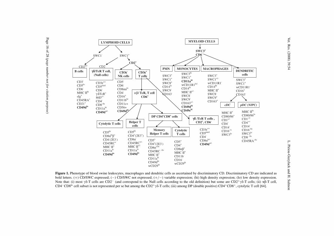
Figure 1. Phenotype of blood swine leukocytes, macrophages and dendritic cells as ascertained by discriminatory CD. Discriminatory CD are indicated asbold letters. (+) CD/SWC expressed; (−) CD/SWC not expressed; (+/−) variable expression; (hi) high density expression; (lo) low density expression.Note that: (i) most ��-T cells are CD2− (and correspond to the Null cells according to the old definition) but some are CD2+��-T cells; (ii) ��-T cell,CD4−CD8lo cell subset is not represented per se but among the CD2+��-T cells; (iii) among DP (double positive) CD4+CD8+, cytolytic T cell [64].
Page16
of28
(pagenum
bernotfor
citationpurpose)
Swine CD Vet. Res. (2008) 39:54
4. CONCLUSION
Even with the limited resources availablein porcine immunology, the various mAbidentifying porcine CD on cells of the immunesystem listed in Table A dress the differ-ences in binding distribution with respect toother species including humans and mice,and demonstrate the impact of new SWCmarker on cell lineage and on functioncharacterisations (Fig. 1). The various swineCD workshops have facilitated the develop-ment of collaborative interactions betweenimmunologists, enabling the designation ofnew mAb as well as cross-reacting mAb toparticular CD and SWC. This effort continuesto improve porcine biomedical research, inparticular a better knowledge of the porcineimmune system. Furthermore, during therecent HLDA8 many more cross-reactivemAb have been identified as being directedagainst a wide range of CD antigens of dif-ferent species including CD1b, CD9, CD14,CD18, CD41, CD44, CD47, CD59, CD68,CD80, CD86, CD91, CD95, CD163, CD172a,CD247 [158, 171]. On the contrary, certaincommercially available human polyclonalantisera may also represent useful tools [195].This will improve our understanding ofporcine immunology.
Acknowledgements. We thank Arthur Summerfieldfor valuable criticisms.
REFERENCES
[1] Aasted B., Viuff B., Reactivity of monoclonalantibodies to human CD antigens with cells from mink,Vet. Immunol. Immunopathol. (2007) 119:27–37.
[2] Ackermann M.R., DeBey B.M., Stabel T.J., GoldJ.H., Register K.B., Meehan J.T., Distribution of anti-CD68 (EBM11) immunoreactivity in formalin-fixed,paraffin-embedded bovine tissues, Vet. Pathol. (1994)31:340–348.
[3] Alvarez B., Domenech N., Alonso F., Sanchez C.,Gomez del Moral M., Ezquerra A., et al., Molecularand functional characterization of porcine LFA-1using monoclonal antibodies to CD11a and CD18,Xenotransplantation (2000) 7:258–266.
[4] Alvarez B., Gomez N., Jose G.J., Yerle M.,Revilla C., Chamorro S., et al., Molecular cloning
characterization and expression of porcine immunore-ceptor SIRPalpha, Dev. Comp. Immunol. (2007)31:307–318.
[5] Alvarez B., Sanchez C., Bullido R., Marina A.,Lunney J., Alonso F., et al., A porcine cell surfacereceptor identified by monoclonal antibodies to SWC3is a member of the signal regulatory protein familyand associates with protein-tyrosine phosphataseSHP-1, Tissue Antigens (2000) 55:342–351.
[6] Appleyard G.D., Furesz S.E., Wilkie B.N., Bloodlymphocyte subsets in pigs vaccinated and challengedwith Actinobacillus pleuropneumoniae, Vet. Immunol.Immunopathol. (2002) 86:221–228.
[7] Appleyard G.D., Wilkie B.N., Characterization ofporcine CD5 and CD5+ B cells, Clin. Exp. Immunol.(1998) 111:225–230.
[8] Armengol E., Wiesmuller K.H., Wienhold D.,Buttner M., Pfaff E., Jung G., et al., Identificationof T-cell epitopes in the structural and non-structuralproteins of classical swine fever virus, J. Gen. Virol.(2002) 83:551–560.
[9] Arriens M.A., Summerfield A., McCullough K.C.,Differential adhesion molecule expression on porcinemononuclear cell populations, Scand. J. Immunol.(1998) 47:487–495.
[10] Aversa G.G., Bishop G.A., Suranyi M.G., HallB.M., RPA-2.10: an anti-CD2 monoclonal antibodythat inhibits alloimmune responses and monitors T cellactivation, Transplant. Proc. (1987) 19:277–278.
[11] Bailey M., Stevens K., Bland P.W., Stokes C.R.,A monoclonal antibody recognising an epitope asso-ciated with pig interleukin-2 receptors, J. Immunol.Methods (1992) 153:85–91.
[12] Basta S., Knoetig S.M., Spagnuolo-WeaverM., Allan G., McCullough K.C., Modulation ofmonocytic cell activity and virus susceptibility duringdifferentiation into macrophages, J. Immunol. (1999)162:3961–3969.
[13] Batten P., Yacoub M.H., Rose M.L., Effect ofhuman cytokines (IFN-gamma, TNF-alpha, IL-1 beta,IL-4) on porcine endothelial cells: induction of MHCand adhesion molecules and functional significance ofthese changes, Immunology (1996) 87:127–133.
[14] Bautista E.M., Gregg D., Golde W.T., Character-ization and functional analysis of skin-derived den-dritic cells from swine without a requirement forin vitro propagation, Vet. Immunol. Immunopathol.(2002) 88:131–148.
[15] Berg E.L., Goldstein L.A., Jutila M.A., NakacheM., Picker L.J., Streeter P.R., et al., Homing receptorsand vascular addressins: cell adhesion molecules thatdirect lymphocyte traffic 23, Immunol. Rev. (1989)108:5–18.
(page number not for citation purpose) Page 17 of 28
Vet. Res. (2008) 39:54 L. Piriou-Guzylack and H. Salmon
[16] Bergamin F., Vincent I.E., Summerfield A.,McCullough K.C., Essential role of antigen-presentingcell-derived BAFF for antibody responses, Eur. J.Immunol. (2007) 37:3122–3130.
[17] Berndt A., Heller M., Methner U., Kosmehl H.,Muller G., Monoclonal antibodies against porcinemacrophages, Vet. Immunol. Immunopathol. (2000)74:163–177.
[18] Bie P.C., Nygard A.B., Fredholm M., AastedB., Salomonsen J., Various domains of the B-cellregulatory molecule CD72 has diverged at differentrates in mammals: cloning, transcription and mappingof porcine CD72, Dev. Comp. Immunol. (2007)31:530–538.
[19] Bimczok D., Rau H., Wundrack N., NaumannM., Rothkötter H.J., McCullough K., et al., Choleratoxin promotes the generation of semi-mature porcinemonocyte-derived dendritic cells that are unable tostimulate T cells, Vet. Res. (2007) 38:597–612.
[20] Bimczok D., Rothkötter H.J., Lymphocyte migra-tion studies, Vet. Res. (2006) 37:325–338.
[21] Bimczok D., Sowa E.N., Faber-Zuschratter H.,Pabst R., Rothkotter H.J., Site-specific expressionof CD11b and SIRPalpha (CD172a) on dendriticcells: implications for their migration patterns in thegut immune system, Eur. J. Immunol. (2005) 35:1418–1427.
[22] Binns R.M., Bone marrow and lymphoid cellinjection of the pig foetus resulting in transplantationtolerance or immunity, and immunoglobulin produc-tion, Nature (1967) 214:179–180.
[23] Binns R.M., The Null/gamma delta TCR+ T cellfamily in the pig, Vet. Immunol. Immunopathol. (1994)43:69–77.
[24] Binns R.M., Bischof R., Carr M.M., DavisW.C., Licence S.T., Misfeldt M., et al., Report onthe behaviour of monoclonal antibodies in the FirstInternational Pig CD Workshop identifying the Nullcell families, Vet. Immunol. Immunopathol. (1994)43:279–287.
[25] Binns R.M., Duncan I.A., Powis S.J., HutchingsA., Butcher G.W., Subsets of null and gamma deltaT-cell receptor+ T lymphocytes in the blood of youngpigs identified by specific monoclonal antibodies,Immunology (1992) 77:219–227.
[26] Binns R.M., Licence S.T., Whyte A., Wilby M.,Rothkotter H.J., Bacon M., Genetically determinedCD45 variant of value in leucocyte tracing in vivo inthe pig, Immunology (1995) 86:25–33.
[27] Binns R.M., Whyte A., Licence S.T., HarrisonA.A., Tsang Y.T., Haskard D.O., et al., The role ofE-selectin in lymphocyte and polymorphonuclear cellrecruitment into cutaneous delayed hypersensitivity
reactions in sensitized pigs, J. Immunol. (1996)157:4094–4099.
[28] Blecha F., Kielian T., McVey D.S., Lunney J.K.,Walker K., Stokes C.R., et al., Workshop studieson monoclonal antibodies reactive against porcinemyeloid cells, Vet. Immunol. Immunopathol. (1994)43:269–272.
[29] Boersma W.J., Zwart R.J., Sinkora J., RehakovaZ., Haverson K., Bianchi A.T., Summary of workshopfindings for porcine B-cell markers, Vet. Immunol.Immunopathol. (2001) 80:63–78.
[30] Bourges D., Chevaleyre C., Wang C., Berri M.,Zhang X., Nicaise L., et al., Differential expressionof adhesion molecules and chemokines between nasaland small intestinal mucosae: implications for T-and sIgA+ B-lymphocyte recruitment, Immunology(2007) 122:551–561.
[31] Bourges D., Wang C.H., Chevaleyre C., SalmonH., T and IgA B lymphocytes of the pharyngeal andpalatine tonsils: differential expression of adhesionmolecules and chemokines, Scand. J. Immunol. (2004)60:338–350.
[32] Bozic F., Lackovic G., Stokes C.R., Valpotic I.,Recruitment of intestinal CD45RA+ and CD45RC+cells induced by a candidate oral vaccine againstporcine post-weaning colibacillosis, Vet. Immunol.Immunopathol. (2002) 86:137–146.
[33] Brodersen R., Bijlsma F., Gori K., JensenK.T., Chen W., Dominguez J., et al., Analysis ofthe immunological cross reactivities of 213 wellcharacterized monoclonal antibodies with specificitiesagainst various leucocyte surface antigens of humanand 11 animal species, Vet. Immunol. Immunopathol.(1998) 64:1–13.
[34] Brossay A., Hube F., Moreau T., Bardos P., WatierH., Porcine CD58: cDNA cloning and moleculardissection of the porcine CD58-human CD2 interface,Biochem. Biophys. Res. Commun. (2003) 309:992–998.
[35] Bullido R., Alonso F., Gomez del Moral M.,Ezquerra A., Alvarez B., Ortuno, et al., Monoclonalantibody 2F4/11 recognizes the alpha chain of aporcine beta 2 integrin involved in adhesion andcomplement mediated phagocytosis, J. Immunol.Methods (1996) 195:125–134.
[36] Bullido R., Domenech N., Alvarez B., AlonsoF., Babin M., Ezquerra A., et al., Characterization offive monoclonal antibodies specific for swine class IImajor histocompatibility antigens and crossreactivitystudies with leukocytes of domestic animals, Dev.Comp. Immunol. (1997) 21:311–322.
[37] Bullido R., Domenech N., Gomez del Moral M.,Alonso F., Ezquerra A., et al., Monoclonal antibodies2F6/8 and 2A10/8 recognize a porcine antigen (SWC7)
Page 18 of 28 (page number not for citation purpose)
Swine CD Vet. Res. (2008) 39:54
expressed on B cells and activated T cells, J. Immunol.Methods (1999) 222:1–11.
[38] Bullido R., Gomez del Moral M., AlonsoF., Ezquerra A., Zapata A., Sanchez C., et al.,Monoclonal antibodies specific for porcine mono-cytes/macrophages: macrophage heterogeneity in thepig evidenced by the expression of surface antigens,Tissue Antigens (1997) 49:403–413.
[39] Bullido R., Gomez del Moral M., Domenech N.,Alonso F., Ezquerra A., et al., Monoclonal antibodiesto a high molecular weight isoform of porcine CD45:biochemical and tissue distribution analyses, Vet.Immunol. Immunopathol. (1997) 56:151–162.
[40] Bullido R., Perez de la Lastra J., Almazan F.,Ezquerra A., Llanes D., Alonso F., et al., Induction ofaggregation in porcine lymphoid cells by antibodiesto CD46, Vet. Immunol. Immunopathol. (2000)73:73–81.
[41] Butler J.E., Sinkora M., Wertz N., HoltmeierW., Lemke C.D., Development of the neonatal B andT cell repertoire in swine: implications for compar-ative and veterinary immunology, Vet. Res. (2006)37:417–441.
[42] Butler J.E., Sun J., Wertz N., Sinkora M., Anti-body repertoire development in swine, Dev. Comp.Immunol. (2006) 30:199–221.
[43] Calvert J.G., Slade D.E., Shields S.L., JolieR., Mannan R.M., Ankenbauer R.G., et al., CD163expression confers susceptibility to porcine reproduc-tive and respiratory syndrome viruses, J. Virol. (2007)81:7371–7379.
[44] Carr M.M., Howard C.J., Sopp P., Manser J.M.,Parsons K.R., Expression on porcine gamma deltalymphocytes of a phylogenetically conserved surfaceantigen previously restricted in expression to ruminantgamma delta T lymphocytes, Immunology (1994)81:36–40.
[45] Carr M.M., Lunney J.K., Arn S., Aasted B.,Binns R.M., Davis W.C., et al., Summary of first roundcluster analysis: complete antibody panel 386, Vet.Immunol. Immunopathol. (1994) 43:211–217.
[46] Carrasco C.P., Rigden R.C., Schaffner R.,Gerber H., Neuhaus V., Inumaru S., et al., Porcinedendritic cells generated in vitro: morphological,phenotypic and functional properties, Immunology(2001) 104:175–184.
[47] Carrillo A., Chamorro S., Rodriguez-Gago M.,Alvarez B., Molina M.J., Rodriguez-Barbosa J.I.,et al., Isolation and characterization of immortalizedporcine aortic endothelial cell lines, Vet. Immunol.Immunopathol. (2002) 89:91–98.
[48] Chamorro S., Revilla C., Alvarez B., Alonso F.,Ezquerra A., Dominguez J., Phenotypic and functional
heterogeneity of porcine blood monocytes and its rela-tion with maturation, Immunology (2005) 114:63–71.
[49] Chamorro S., Revilla C., Gomez N., Alvarez B.,Alonso F., Ezquerra A., et al., In vitro differentiationof porcine blood CD163- and CD163+ monocytesinto functional dendritic cells, Immunobiology (2004)209:57–65.
[50] Chardon P., Renard C., Gaillard C.R., VaimanM., The porcine major histocompatibility complexand related paralogous regions: a review, Genet. Sel.Evol. (2000) 32:109–128.
[51] Chardon P., Renard C., Vaiman M., The majorhistocompatibility complex in swine, Immunol. Rev.(1999) 167:179–192.
[52] Charerntantanakul W., Roth J.A., Biology ofporcine T lymphocytes, Anim. Health Res. Rev.(2006) 7:81–96.
[53] Chen L., Zwart R., Yang P., Kijlstra A.,Macrophages and MHC class II positive dendriti-form cells in the iris and choroid of the pig, Curr. EyeRes. (2003) 26:291–296.
[54] Choi I., Cho B., Kim S.D., Park D., Kim J.Y.,Park C.G., et al., Molecular cloning, expression andfunctional characterization of miniature swine CD86,Mol. Immunol. (2006) 43:480–486.
[55] Chun T., Wang K., Zuckermann F.A., GaskinsH.R., Molecular cloning and characterization of anovel CD1 gene from the pig, J. Immunol. (1999)162:6562–6571.
[56] Dato M.E., Kim Y.B., Characterization andutilization of a monoclonal antibody inhibiting porcinenatural killer cell activity for isolation of natural killerand killer cells, J. Immunol. (1990) 144:4452–4462.
[57] Dato M.E., Wierda W.G., Kim Y.B., A triggeringstructure recognized by G7 monoclonal antibody onporcine lymphocytes and granulocytes, Cell. Immunol.(1992) 140:468–477.
[58] Davis T.A., Craighead N., Williams A.J., ScadronA., June C.H., Lee K.P., Primary porcine endothelialcells express membrane-bound B7-2 (CD86) and asoluble factor that co-stimulate cyclosporin A-resistantand CD28-dependent human T cell proliferation, Int.Immunol. (1996) 8:1099–1111.
[59] Davis W.C., Brown W.C., Hamilton M.J., WyattC.R., Orden J.A., Khalid A.M., et al., Analysis of mon-oclonal antibodies specific for the gamma delta TcR,Vet. Immunol. Immunopathol. (1996) 52:275–283.
[60] Davis W.C., Haverson K., Saalmuller A., YangH., Lunney J.K., Hamilton M.J., et al., Analysisof monoclonal antibodies reacting with moleculesexpressed on gammadelta T-cells, Vet. Immunol.Immunopathol. (2001) 80:53–62.
(page number not for citation purpose) Page 19 of 28
Vet. Res. (2008) 39:54 L. Piriou-Guzylack and H. Salmon
[61] Davis W.C., Zuckermann F.A., Hamilton M.J.,Barbosa J.I., Saalmuller A., Binns R.M., et al., Analy-sis of monoclonal antibodies that recognize gammadelta T/null cells, Vet. Immunol. Immunopathol.(1998) 60:305–316.
[62] Denham S., Shimizu M., Bianchi A.T., Zwart R.J.,Carr M.M., Parkhouse R.M., Monoclonal antibodiesrecognising differentiation antigens on porcine B cells,Vet. Immunol. Immunopathol. (1994) 43:259–267.
[63] Denham S., Zwart R.J., Whittall J.T., PampuschM., Corteyn A.H., Bianchi A.T., et al., Monoclonalantibodies putatively identifying porcine B cells, Vet.Immunol. Immunopathol. (1998) 60:317–328.
[64] Denyer M.S., Wileman T.E., Stirling C.M., ZuberB., Takamatsu H.H., Perforin expression can defineCD8 positive lymphocyte subsets in pigs allowingphenotypic and functional analysis of natural killer,cytotoxic T, natural killer T and MHC un-restrictedcytotoxic T-cells, Vet. Immunol. Immunopathol.(2006) 110:279–292.
[65] Diaz G., Canas B., Vazquez J., Nombela C.,Arroyo J., Characterization of natural peptide ligandsfrom HLA-DP2: new insights into HLA-DP peptide-binding motifs, Immunogenetics (2005) 56:754–759.
[66] Domenech N., Rodriguez-Carreno M.P., FilgueiraP., Alvarez B., Chamorro S., Dominguez J., Identifi-cation of porcine macrophages with monoclonal anti-bodies in formalin-fixed, paraffin-embedded tissues,Vet. Immunol. Immunopathol. (2003) 94:77–81.
[67] Dominguez J., Alvarez B., Alonso F., ThackerE., Haverson K., McCullough K., et al., Workshopstudies on monoclonal antibodies in the myeloid panelwith CD11 specificity, Vet. Immunol. Immunopathol.(2001) 80:111–119.
[68] Dominguez J., Ezquerra A., Alonso F., BullidoR., McCullough K., Summerfield A., et al., Workshopstudies with monoclonal antibodies identifying a novelporcine differentiation antigen, SWC9, Vet. Immunol.Immunopathol. (1998) 60:343–349.
[69] Dominguez J., Ezquerra A., Alonso F., McCul-lough K., Summerfield A., Bianchi A., et al., Porcinemyelomonocytic markers: summary of the SecondInternational Swine CD Workshop, Vet. Immunol.Immunopathol. (1998) 60:329–341.
[70] Donovan J.A., Koretzky G.A., CD45 and theimmune response, J. Am. Soc. Nephrol. (1993)4:976–985.
[71] Dorling A., Delikouras A., Nohadani M., PolakJ., Lechler R.I., In vitro accommodation of porcineendothelial cells by low dose human anti-pig antibody:reduced binding of human lymphocytes by accom-modated cells associated with increased nitric oxideproduction, Xenotransplantation (1998) 5:84–92.
[72] Ehrlich R., Lifshitz R., Pescovitz M.D., RudikoffS., Singer D.S., Tissue-specific expression and struc-ture of a divergent member of a class I MHC genefamily, J. Immunol. (1987) 139:593–602.
[73] Evans P.C., Taylor E.R., Kilshaw P.J., Signal-ing through CD31 protects endothelial cells fromapoptosis, Transplantation (2001) 71:457–460.
[74] Faas S.J., Giannoni M.A., Mickle A.P., KieseckerC.L., Reed D.J., Wu D.Y., et al., Primary structureand functional characterization of a soluble, alter-natively spliced form of B7-1, J. Immunol. (2000)164:6340–6348.
[75] Faldyna M., Samankova P., Leva L., Cerny J.,Oujezdska J., Rehakova Z., et al., Cross-reactiveanti-human monoclonal antibodies as a tool forB-cell identification in dogs and pigs, Vet. Immunol.Immunopathol. (2007) 119:56–62.
[76] Feng W.H., Tompkins M.B., Xu J.S., BrownT.T., Laster S.M., Zhang H.X., et al., Thymocyte andperipheral blood T lymphocyte subpopulation changesin piglets following in utero infection with porcinereproductive and respiratory syndrome virus, Virology(2002) 302:363–372.
[77] Gao F.S., Fang Q.M., Li Y.G., Li X.S., Hao H.F.,Xia C., Reconstruction of a swine SLA-I protein com-plex and determination of binding nonameric peptidesderived from the foot-and-mouth disease virus, Vet.Immunol. Immunopathol. (2006) 113:328–338.
[78] Gerner W., Denyer M.S., Takamatsu H.H.,Wileman T.E., Wiesmuller K.H., Pfaff E., et al.,Identification of novel foot-and-mouth disease virusspecific T-cell epitopes in c/c and d/d haplotypeminiature swine, Virus Res. (2006) 121:223–228.
[79] Godwin J.W., d’Apice A.J., Cowan P.J., Charac-terization of pig intercellular adhesion molecule-2 andits interaction with human LFA-1, Am. J. Transplant.(2004) 4:515–525.
[80] Gonzalez J.M., Mebus C.A., Pan R., RevillaY., Alonso J.M., Lunney J.K., Swine leukocyteantigen and macrophage marker expression on bothAfrican swine fever virus-infected and non-infectedprimary porcine macrophage cultures, Vet. Immunol.Immunopathol. (1992) 32:243–259.
[81] Gutierrez M., Forster F.I., McConnell S.A.,Cassidy J.P., Pollock J.M., Bryson D.G., The detectionof CD2+, CD4+, CD8+, and WC1+ T lymphocytes,B cells and macrophages in fixed and paraffin embed-ded bovine tissue using a range of antigen recoveryand signal amplification techniques, Vet. Immunol.Immunopathol. (1999) 71:321–334.
[82] Halloran P.J., Sweeney S.E., Strohmeier C.M.,Kim Y.B., Molecular cloning and identification of theporcine cytolytic trigger molecule G7 as a Fc gamma
Page 20 of 28 (page number not for citation purpose)
Swine CD Vet. Res. (2008) 39:54
RIII alpha (CD16) homologue, J. Immunol. (1994)153:2631–2641.
[83] Hammerberg C., Schurig G.G., Characterizationof monoclonal antibodies directed against swineleukocytes, Vet. Immunol. Immunopathol. (1986)11:107–121.
[84] Harrison A.A., Stocker C.J., Chapman P.T., TsangY.T., Huehns T.Y., Gundel R.H., et al., Expressionof vascular cell adhesion molecule-1 by vascularendothelial cells in immune and nonimmune inflam-matory reactions in the skin, J. Immunol. (1997)159:4546–4554.
[85] Haverson K., Bailey M., Higgins V.R., BlandP.W., Stokes C.R., Characterization of monoclonalantibodies specific for monocytes, macrophagesand granulocytes from porcine peripheral bloodand mucosal tissues, J. Immunol. Methods (1994)170:233–245.
[86] Haverson K., Bailey M., Stokes C.R., Simon A.,LeFlufy L., Banfield G., et al., Monoclonal antibodiesraised to human cells–specificity for pig leukocytes,Vet. Immunol. Immunopathol. (2001) 80:175–186.
[87] Haverson K., Riffault S., Antigen presenting cellsin mucosal sites of veterinary species, Vet. Res. (2006)37:339–358.
[88] Haverson K., Saalmuller A., Alvarez B., AlonsoF., Bailey M., Bianchi A.T., et al., Overview of thethird international workshop on swine leukocyte dif-ferentiation antigens, Vet. Immunol. Immunopathol.(2001) 80:5–23.
[89] Haverson K., Saalmuller A., Chen Z., HuangC.A., Simon A., Seebach J., et al., Summary of thefirst round analyses of the third international workshopon swine leukocyte differentiation antigens, Vet.Immunol. Immunopathol. (2001) 80:25–34.
[90] Haverson K., Singha S., Stokes C.R., Bailey M.,Professional and non-professional antigen-presentingcells in the porcine small intestine, Immunology(2000) 101:492–500.
[91] Haverson K., Zuckermann F., Saalmuller A., LippJ., Aasted B., Stokes C.R., Summary of workshopfindings for porcine adhesion molecule subgroup, Vet.Immunol. Immunopathol. (1998) 60:351–365.
[92] Hirt W., Saalmuller A., Reddehase M.J., Distinctgamma/delta T cell receptors define two subsets ofcirculatingX porcine CD2-CD4-CD8- T lymphocytes,Eur. J. Immunol. (1990) 20:265–269.
[93] Ivanoska D., Sun D.C., Lunney J.K., Productionof monoclonal antibodies reactive with polymorphicand monomorphic determinants of SLA class I geneproducts, Immunogenetics (1991) 33:220–223.
[94] Jimenez-Marin A., Garrido J.J., de Andres-Cara D.F., Morera L., Barbancho M.J., Llanes D.,
Molecular cloning and characterization of the pighomologue to human CD29, the integrin beta1subunit, Transplantation (2000) 70:649–655.
[95] Jimenez-Marin A., Moreno A., de la MulasJ.M., Millan Y., Morera L., Barbancho M., et al.,Localization of porcine CD29 transcripts and proteinin pig cells and tissues by RT-PCR and immuno-histochemistry, Vet. Immunol. Immunopathol. (2005)104:281–288.
[96] Jones M., Cordell J.L., Beyers A.D., TseA.G., Mason D.Y., Detection of T and B cellsin many animal species using cross-reactive anti-peptide antibodies 32, J. Immunol. (1993) 150:5429–5435.
[97] Jonjic S., Koszinowski U.H., Monoclonal anti-bodies reactive with swine lymphocytes. I. Antibodiesto membrane structures that define the cytolytic Tlymphocyte subset in the swine, J. Immunol. (1984)133:647–652.
[98] Kaeffer B., Bottreau E., Marcon D., Olivier M.,Lantier I., Salmon H., Histocompatible miniature pig(d/d haplotype): generation of hybridomas secretingA or M monoclonal antibody, Hybridoma (1991)10:731–744.
[99] Kaeffer B., Bottreau E., Phan T.L., OlivierM., Salmon H., Histocompatible miniature, boarmodel: selection of transformed cell lines of B andT lineages producing retrovirus, Int. J. Cancer (1990)46:481–488.
[100] Kamata T., Puzon W., Takada Y., Identificationof putative ligand-binding sites of the integrin alpha4 beta 1 (VLA-4, CD49d/CD29), Biochem. J. (1995)305:945–951.
[101] Kanan J.H., Nayeem N., Binns R.M., ChainB.M., Mechanisms for variability in a member ofthe scavenger-receptor cysteine-rich superfamily,Immunogenetics (1997) 46:276–282.
[102] Kansas G.S., Spertini O., Stoolman L.M.,Tedder T.F., Molecular mapping of functional domainsof the leukocyte receptor for endothelium, LAM-1,J. Cell Biol. (1991) 114:351–358.
[103] Kaser T., Gerner W., Hammer S.E., Patzl M.,Saalmuller A., Phenotypic and functional characterisa-tion of porcine CD4(+)CD25(high) regulatory T cells,Vet. Immunol. Immunopathol. (2008) 122:153–158.
[104] Kasper M., Bierhaus A., Whyte A., Binns R.M.,Schuh D., Muller M., Expression of CD44 isoformsduring bleomycin-or radiation-induced pulmonaryfibrosis in rats and mini-pigs, Histochem. Cell Biol.(1996) 105:221–230.
[105] Keelan E.T., Harrison A.A., Chapman P.T.,Binns R.M., Peters A.M., Haskard D.O., Imagingvascular endothelial activation: an approach using
(page number not for citation purpose) Page 21 of 28
Vet. Res. (2008) 39:54 L. Piriou-Guzylack and H. Salmon
radiolabeled monoclonal antibodies against theendothelial cell adhesion molecule E-selectin, J. Nucl.Med. (1994) 35:276–281.
[106] Kenmochi T., Mullen Y., Miyamoto M., Stein E.,Swine as an allotransplantation model, Vet. Immunol.Immunopathol. (1994) 43:177–183.
[107] Kielian T., McVey D.S., Davis W.C., KimY.B., Blecha F., Competitive binding analysis ofmonoclonal antibodies reactive with porcine alveolarmacrophages using anti-CD14 and anti-CD18, Vet.Immunol. Immunopathol. (1994) 43:273–278.
[108] Kim Y.B., Developmental immunity in thepiglet, Birth Defects Orig. Artic. Ser. (1975) 11:549–557.
[109] Kim Y.B., Zhang J., Davis W.C., Lunney J.K.,CD11/CD18 panel report for swine CD workshop,Vet. Immunol. Immunopathol. (1994) 43:289–291.
[110] Kimman T.G., Bianchi A.T., Wensvoort G., deBruin T.G., Meliefste C., Cellular immune responseto hog cholera virus (HCV): T cells of immune pigsproliferate in vitro upon stimulation with live HCV,but the E1 envelope glycoprotein is not a major T-cellantigen, J. Virol. (1993) 67:2922–2927.
[111] Kirkham P.A., Takamatsu H., Yang H., Park-house R.M., Porcine CD3 epsilon: its characterization,expression and involvement in activation of porcine Tlymphocytes, Immunology (1996) 87:616–623.
[112] Kirkiles-Smith N.C., Tereb D.A., Kim R.W.,McNiff J.M., Schechner J.S., Lorber M.I., et al.,Human TNF can induce nonspecific inflammatory andhuman immune-mediated microvascular injury of pigskin xenografts in immunodeficient mouse hosts 13,J. Immunol. (2000) 164:6601–6609.
[113] Kuhns M.S., Davis M.M., Garcia K.C., Decon-structing the form and function of the TCR/CD3complex, Immunity (2006) 24:133–139.
[114] Kumagai-Braesch M., Schacter B., Yan Z.,Michaelson J., Arn S., Smith M., et al., Identificationof swine and primate cellular adhesion molecules(CAM) using mouse anti-human monoclonal antibod-ies, Xenotransplantation (1995) 2:88–97.
[115] Llanes D., Arce C., de la Lastra J.P., de A.D.,Barbancho M., Morera L., et al., Swine platelet anti-gens: section report, Vet. Immunol. Immunopathol.(2001) 80:131–142.
[116] Lopez F.L., Domenech N., Alvarez B., EzquerraA., Dominguez J., Castro J.M., et al., Analysis of cel-lular immune response in pigs recovered from porcinerespiratory and reproductive syndrome infection,Virus Res. (1999) 64:33–42.
[117] Lunney J., Pescovitz M.D., Differentiationantigens in swine lymphoid tissues, in: Miyasaka M.,
Trnka Z. (Eds.), Differentiation antigens in lympho-hemopoietic tissues, Marcel Dekker, New-York andBasel, 1998, pp. 421–454.
[118] Lunney J.K., Characterization of swine leuko-cyte differentiation antigens, Immunol. Today (1993)14:147–148.
[119] Lunney J.K., Current status of the swine leuko-cyte antigen complex, Vet. Immunol. Immunopathol.(1994) 43:19–28.
[120] Lunney J.K., Fossum C., Alm G.V., SteinbachF., Wattrang E., Veterinary immunology: opportunitiesand challenges, Trends Immunol. (2002) 23:4–6.
[121] Lunney J.K., Pescovitz M.D., Phenotypicand functional characterization of pig lymphocytepopulations, Vet. Immunol. Immunopathol. (1987)17:135–144.
[122] Lunney J.K., Walker K., Goldman T., AastedB., Bianchi A., Binns R., et al., Overview of theFirst International Workshop to Define Swine Leuko-cyte Cluster of Differentiation (CD) Antigens, Vet.Immunol. Immunopathol. (1994) 43:193–206.
[123] Mackay C.R., Marston W.L., Dudler L., SpertiniO., Tedder T.F., Hein W.R., Tissue-specific migrationpathways by phenotypically distinct subpopula-tions of memory T cells, Eur. J. Immunol. (1992) 22:887–895.
[124] Magyar A., Mihalik R., Characterisation ofthe swine swC1 antigen, Acta Vet. Hung. (1997)45:17–31.
[125] Maher S.E., Karmann K., Min W., HughesC.C., Pober J.S., Bothwell A.L., Porcine endothelialCD86 is a major costimulator of xenogeneic humanT cells: cloning, sequencing, and functional expres-sion in human endothelial cells, J. Immunol. (1996)157:3838–3844.
[126] Makala L.H., Haverson K., Stokes C.R., BaileyM., Bland P.W., Isolation and characterisation ofpig Peyer’s patch dendritic cells, Vet. Immunol.Immunopathol. (1998) 61:67–81.
[127] Martins C.L., Lawman M.J., Scholl T., MebusC.A., Lunney J.K., African swine fever virus specificporcine cytotoxic T cell activity, Arch. Virol. (1993)129:211–225.
[128] McCullough K.C., Schaffner R., Natale V.,Kim Y.B., Summerfield A., Phenotype of porcinemonocytic cells: modulation of surface moleculeexpression upon monocyte differentiation intomacrophages, Vet. Immunol. Immunopathol. (1997)58:265–275.
[129] Mehrazar K., Kim Y.B., Total parenteralnutrition in germfree colostrum-deprived neonatalminiature piglets: a unique model to study the
Page 22 of 28 (page number not for citation purpose)
Swine CD Vet. Res. (2008) 39:54
ontogeny of the immune system, J. Parenter. EnteralNutr. (1988) 12:563–568.
[130] Merono A., Lucena C., Lopez A., GarridoJ.J., Perez de L.L., Llanes D., Immunohistochemicalanalysis of beta3 integrin (CD61): expression in pigtissues and human tumors, Histol. Histopathol. (2002)17:347–352.
[131] Mueller J.P., Evans M.J., Cofiell R., Rother R.P.,Matis L.A., Elliott E.A., Porcine vascular cell adhesionmolecule (VCAM) mediates endothelial cell adhesionto human T cells. Development of blocking antibodiesspecific for porcine VCAM, Transplantation (1995)60:1299–1306.
[132] Murray A.G., Khodadoust M.M., Pober J.S.,Bothwell A.L.M., Porcine Aortic Endothelial-CellsActivate Human T-Cells – Direct Presentation of MhcAntigens and Costimulation by Ligands for HumanCd2 and Cd28, Immunity (1994) 1:57–63.
[133] Naessens J., Newson J., McHugh N., HowardC.J., Parsons K., Jones B., Characterization of abovine leucocyte differentiation antigen of 145,000MW restricted to B lymphocytes, Immunology (1990)69:525–530.
[134] Outteridge P.M., Binns R.M., Licence S.T.,Subpopulations of pig blood E-rosette-forminglymphocytes and thymus-dependent null cells: sep-aration by nylon wool columns, rosette formationand macrophage-dependent mitogen and antigenresponsiveness, Int. Arch. Allergy Appl. Immunol.(1982) 67:18–24.
[135] Panja A., Barone A., Mayer L., Stimulation oflamina propria lymphocytes by intestinal epithelialcells: evidence for recognition of nonclassical restric-tion elements, J. Exp. Med. (1994) 179:943–950.
[136] Panos G., Moreno A., Jimenez-Marin A., Gar-rido J.J., Martin de la Mulas J., Ordas J., et al., Analy-sis of swine beta1 integrin (CD29) epitopes throughmonoclonal antibodies developed using two immu-nization strategies, Hybrid. Hybridomics (2004) 23:271–278.
[137] Pauly T., Elbers K., Konig M., Lengsfeld T.,Saalmuller A., Thiel H.J., Classical swine fever virus-specific cytotoxic T lymphocytes and identification ofa T cell epitope, J. Gen. Virol. (1995) 76:3039–3049.
[138] Pauly T., Weiland E., Hirt W., Dreyer-Bux C.,Maurer S., Summerfield A., et al., Differentiationbetween MHC-restricted and non-MHC-restrictedporcine cytolytic T lymphocytes, Immunology (1996)88:238–246.
[139] Pescovitz M.D., Aasted B., Canals A.,Dominguez J., Vizcaino J.S., Pospisil R., et al.,Analysis of monoclonal antibodies reactive with theporcine CD2 antigen, Vet. Immunol. Immunopathol.(1994) 43:229–232.
[140] Pescovitz M.D., Aasted B., Canals A.,Dominguez J., Vizcaino J.S., Pospisil R., et al.,Analysis of monoclonal antibodies reactive with theporcine CD4 antigen, Vet. Immunol. Immunopathol.(1994) 43:233–236.
[141] Pescovitz M.D., Book B.K., Aasted B.,Dominguez J., Bullido R., Trebichavsky I., et al.,Analyses of monoclonal antibodies reacting withporcine CD5: results from the Second InternationalSwine CD Workshop, Vet. Immunol. Immunopathol.(1998) 60:269–273.
[142] Pescovitz M.D., Book B.K., Aasted B.,Dominguez J., Ezquerra A., Trebichavsky I., et al.,Analyses of monoclonal antibodies reacting withporcine CD3: results from the Second InternationalSwine CD Workshop, Vet. Immunol. Immunopathol.(1998) 60:261–268.
[143] Pescovitz M.D., Book B.K., Aasted B.,Dominguez J., Ezquerra A., Trebichavsky I., et al.,Summary of workshop findings for antibodies reactingwith porcine T-cells and activation antigens: resultsfrom the Second International Swine CD Workshop,Vet. Immunol. Immunopathol. (1998) 60:251–260.
[144] Pescovitz M.D., Lunney J.K., Sachs D.H.,Preparation and characterization of monoclonal anti-bodies reactive with porcine PBL, J. Immunol. (1984)133:368–375.
[145] Pescovitz M.D., Lunney J.K., Sachs D.H.,Murine anti-swine T4 and T8 monoclonal antibodies:distribution and effects on proliferative and cytotoxicT cells, J. Immunol. (1985) 134:37–44.
[146] Pescowitz D., Sakopoulos A.G., Gaddy J.A.,Husmann R.J., Zuckermann F.A., Porcine peripheralblood CD4+/CD8+ dual expressing T-cells, Vet.Immunol. Immunopathol. (1994) 43:53–62.
[147] Pescowitz M.D., Hsu S.M., Katz S.I., Lun-ney J.K., Shimada S., Sachs D.H., Characterizationof a porcine CD1-specific mAb that distinguishesCD4/CD8 double-positive thymic from peripheral Tlymphocytes, Tissue Antigens (1990) 35:151–156.
[148] Petersen C.B., Nygard A.B., Viuff B., Fred-holm M., Aasted B., Salomonsen J., Porcine ecto-nucleotide pyrophosphatase/phosphodiesterase 1(NPP1/CD203a): cloning, transcription, expression,mapping, and identification of an NPP1/CD203a epi-tope for swine workshop cluster 9 (SWC9) monoclonalantibodies, Dev. Comp. Immunol. (2007) 31:618–631.
[149] Petruzzelli L., Maduzia L., Springer T.A., Acti-vation of lymphocyte function-associated molecule-1(CD11a/CD18) and Mac-1 (CD11b/CD18) mimickedby an antibody directed against CD18, J. Immunol.(1995) 155:854–866.
[150] Pintaric M., Gerner W., Saalmuller A., Syner-gistic effects of IL-2, IL-12 and IL-18 on cytolytic
(page number not for citation purpose) Page 23 of 28
Vet. Res. (2008) 39:54 L. Piriou-Guzylack and H. Salmon
activity, perforin expression and IFN-gamma pro-duction of porcine natural killer cells, Vet. Immunol.Immunopathol. (2007) 121:68–82.
[151] Renard C., Hart E., Sehra H., Beasley H.,Coggill P., Howe K., et al., The genomic sequenceand analysis of the swine major histocompatibilitycomplex, Genomics (2006) 88:96–110.
[152] Revilla C., Chamorro S., Alvarez B., Perez C.,Ezquerra A., Alonso F., et al., Analysis of functionalheterogeneity of porcine memory CD4+ T cells, Dev.Comp. Immunol. (2005) 29:479–488.
[153] Revilla C., Rodriguez-Carreno M.P., AlvarezB., Chamorro S., Alonso L.M., Ezquerra A., et al.,2E3, a new marker that selectively identifies porcineCD4+ naive T cells, Dev. Comp. Immunol. (2004)28:239–250.
[154] Richard C., Charreau B., Vusio P., Soulillou J.P.,Bouhours J.F., Characterization of two monoclonalantibodies against porcine VCAM-1, Hybridoma(1999) 18:159–165.
[155] Riffault S., Carrat C., van Reeth K., PensaertM., Charley B., Interferon-alpha-producing cellsare localized in gut-associated lymphoid tissues intransmissible gastroenteritis virus (TGEV) infectedpiglets, Vet. Res. (2001) 32:71–79.
[156] Rothkotter H.J., Ulbrich H., Pabst R., The post-natal development of gut lamina propria lymphocytes:number, proliferation, and T and B cell subsets inconventional and germ-free pigs, Pediatr. Res. (1991)29:237–242.
[157] Saalmuller A., Characterization of swine leuko-cyte differentiation antigens, Immunol. Today (1996)17:352–354.
[158] Saalmuller A., Aasted B., Summary of theanimal homologue section of HLDA8, Vet. Immunol.Immunopathol. (2007) 119:2–13.
[159] Saalmuller A., Aasted B., Canals A., DominguezJ., Goldman T., Lunney J.K., et al., Analyses of mon-oclonal antibodies reactive with porcine CD6, Vet.Immunol. Immunopathol. (1994) 43:243–247.
[160] Saalmuller A., Aasted B., Canals A., DominguezJ., Goldman T., Lunney J.K., et al., Analyses ofmAb reactive with porcine CD8, Vet. Immunol.Immunopathol. (1994) 43:249–254.
[161] Saalmuller A., Aasted B., Canals A., DominguezJ., Goldman T., Lunney J.K., et al., Analyses of mon-oclonal antibodies reactive with porcine CD5, Vet.Immunol. Immunopathol. (1994) 43:237–242.
[162] Saalmuller A., Aasted B., Canals A., DominguezJ., Goldman T., Lunney J.K., et al., Summary ofworkshop findings for porcine T-lymphocyte anti-gens, Vet. Immunol. Immunopathol. (1994) 43:219–228.
[163] Saalmuller A., Aasted B., Canals A., DominguezJ., Goldman T., Lunney J.K., et al., Analysis of mAbreactive with the porcine SWC1, Vet. Immunol.Immunopathol. (1994) 43:255–258.
[164] Saalmuller A., Bryant J., Characteristics ofporcine T lymphocytes and T-cell lines, Vet. Immunol.Immunopathol. (1994) 43:45–52.
[165] Saalmuller A., Denham S., Haverson K., DavisB., Dominguez J., Pescovitz M.D., et al., The SecondInternational Swine CD Workshop, Vet. Immunol.Immunopathol. (1996) 54:155–158.
[166] Saalmuller A., Hirt W., Maurer S., Weiland E.,Discrimination between two subsets of porcine CD8+cytolytic T lymphocytes by the expression of CD5antigen, Immunology (1994) 81:578–583.
[167] Saalmuller A., Hirt W., Reddehase M.J., Pheno-typic discrimination between thymic and extrathymicCD4-CD8- and CD4+CD8+ porcine T lymphocytes,Eur. J. Immunol. (1989) 19:2011–2016.
[168] Saalmuller A., Hirt W., Reddehase M.J., Porcinegamma/delta T lymphocyte subsets differing intheir propensity to home to lymphoid tissue, Eur. J.Immunol. (1990) 20:2343–2346.
[169] Saalmuller A., Jonjic S., Buhring H.J., Redde-hase M.J., Koszinowski U.H., Monoclonal antibodiesreactive with swine lymphocytes. II. Detection ofan antigen on resting T cells down-regulated afteractivation, J. Immunol. (1987) 138:1852–1857.
[170] Saalmuller A., Kuebart G., Hollemweguer E.,Chen Z., Nielsen J., Zuckermann F., et al., Summary ofworkshop findings for porcine T-lymphocyte-specificmonoclonal antibodies, Vet. Immunol. Immunopathol.(2001) 80:35–52.
[171] Saalmuller A., Lunney J.K., Daubenberger C.,Davis W., Fischer U., Gobel T.W., et al., Summaryof the animal homologue section of HLDA8, Cell.Immunol. (2005) 236:51–58.
[172] Saalmuller A., Maurer S., Major histocompati-bility antigen class II expressing resting porcine T lym-phocytes are potent antigen-presenting cells in mixedleukocyte culture, Immunobiology (1994) 190:23–34.
[173] Saalmuller A., Pauly T., Hohlich B.J., Pfaff E.,Characterization of porcine T lymphocytes and theirimmune response against viral antigens, J. Biotechnol.(1999) 73:223–233.
[174] Saalmuller A., Pauly T., Lunney J.K., BoydP., Aasted B., Sachs D.H., et al., Overview of theSecond International Workshop to define swinecluster of differentiation (CD) antigens, Vet. Immunol.Immunopathol. (1998) 60:207–228.
[175] Saalmuller A., Reddehase M.J., Buhring H.J.,Jonjic S., Koszinowski U.H., Simultaneous expressionof CD4 and CD8 antigens by a substantial proportion
Page 24 of 28 (page number not for citation purpose)
Swine CD Vet. Res. (2008) 39:54
of resting porcine T lymphocytes, Eur. J. Immunol.(1987) 17:1297–1301.
[176] Saalmuller A., Weiland F., Reddehase M.J.,Resting porcine T lymphocytes expressing class IImajor histocompatibility antigen, Immunobiology(1991) 183:102–114.
[177] Saalmuller A., Werner T., Fachinger V.,T-helper cells from naive to committed, Vet. Immunol.Immunopathol. (2002) 87:137–145.
[178] Sachs D.H., Immunologic tolerance to organtransplants, J. Gastrointest. Surg. (1999) 3:105–110.
[179] Saif L.J., Ward L.A., Yuan L., Rosen B.I., ToT.L., The gnotobiotic piglet as a model for studiesof disease pathogenesis and immunity to humanrotaviruses, Arch. Virol. Suppl. (1996) 12:153–161.
[180] Salmon H., Surface markers of porcine lympho-cytes and distribution in various lymphoid organs, Int.Arch. Allergy Appl. Immunol. (1979) 60:262–274.
[181] Salmon H., Swine thymic dendritic cell:an another means to induce the transplantationtolerance: Characterization, isolation and cell cul-ture, in: Laplace J.P., Fevrier C., Barbeau A. (Eds.),Digestive Physiology in pigs, 1997, pp. 661–666.
[182] Salmon H., Mammary gland immunologyand neonate protection in pigs. Homing of lympho-cytes into the MG, Adv. Exp. Med. Biol. (2000)480:279-86:279–286.
[183] Salmon H., Johnson I., Germana S., HallerG.W., Sachs D.H., Leguern C., Dendritic cellsenriched from swine thymus co-express CD1, CD2and major histocompatibility complex class II andactively stimulate alloreactive T lymphocytes, Scand.J. Immunol. (2000) 52:164–172.
[184] Salmon H., Vaiman M., Scheuer D., Immuno-logie du porc, in: Pastoret P.P., Govaerts A., BazinH. (Eds.), Immunologie animale, Flammarion,Médecine-sciences, Paris, 1-1-1990, pp. 565–578.
[185] Sanchez C., Domenech N., Vazquez J., AlonsoF., Ezquerra A., Dominguez J., The porcine 2A10antigen is homologous to human CD163 and relatedto macrophage differentiation, J. Immunol. (1999)162:5230–5237.
[186] Saurer L., McCullough K.C., SummerfieldA., In vitro induction of mucosa-type dendriticcells by all-trans retinoic acid, J. Immunol. (2007)179:3504–3514.
[187] Schneider M.K., Forte P., Seebach J.D., Adhe-sive interactions between human NK cells and porcineendothelial cells, Scand. J. Immunol. (2001) 54:70–75.
[188] Schnitzlein W.M., Zuckermann F.A., Determina-tion of the specificity of CD45 and CD45R monoclonalantibodies through the use of transfected hamster cells
producing individual porcine CD45 isoforms, Vet.Immunol. Immunopathol. (1998) 60:389–401.
[189] Schutt C., Schilling T., Stelter F., Furll B.,Kruger C., Witt S., et al., Modalities of endotoxinbinding to CD14, Prog. Clin. Biol. Res. (1995)392:339-48:339–348.
[190] Shailubhai K., Streeter P.R., Smith C.E., JacobG.S., Sulfation and sialylation requirements for aglycoform of CD34, a major endothelial ligandfor L-selectin in porcine peripheral lymph nodes,Glycobiology (1997) 7:305–314.
[191] Simon A.R., Warrens A.N., Sykes M., Efficacyof adhesive interactions in pig-to-human xenotrans-plantation, Immunol. Today (1999) 20:323–330.
[192] Sinkora J., Rehakova Z., Haverson K., SinkoraM., et al., Monoclonal antibodies putatively recog-nising activation and differentiation antigens, Vet.Immunol. Immunopathol. (2001) 80:143–164.
[193] Sinkora J., Rehakova Z., Samankova L., Haver-son K., Butler J.E., Zwart R., et al., Characterization ofmonoclonal antibodies recognizing immunoglobulinkappa and lambda chains in pigs by flow cytometry,Vet. Immunol. Immunopathol. (2001) 80:79–91.
[194] Sinkora J., Rehakova Z., Sinkora M., CukrowskaB., Tlaskalova-Hogenova H., Bianchi A.T., et al.,Expression of CD2 on porcine B lymphocytes,Immunology (1998) 95:443–449.
[195] Sinkora M., Butler J.E., Holtmeier W., Sinko-rova J., Lymphocyte development in fetal piglets: factsand surprises, Vet. Immunol. Immunopathol. (2005)108:177–184.
[196] Sinkora M., Sinkora J., Rehakova Z., SplichalI., Yang H., Parkhouse R.M., et al., Prenatal ontogenyof lymphocyte subpopulations in pigs, Immunology(1998) 95:595–603.
[197] Sinkora M., Sinkorova J., Butler J.E., B celldevelopment and VDJ rearrangement in the fetal pig,Vet. Immunol. Immunopathol. (2002) 87:341–346.
[198] Smith D.M., Lunney J.K., Ho C.S., MartensG.W., Ando A., Lee J.H., et al., Nomenclature forfactors of the swine leukocyte antigen class II system,2005, Tissue Antigens (2005) 66:623–639.
[199] Smith D.M., Lunney J.K., Martens G.W., AndoA., Lee J.H., Ho C.S., et al., Nomenclature for factorsof the SLA class-I system, 2004, Tissue Antigens(2005) 65:136–149.
[200] Soderholm J., Ahlen G., Kaul A., Frelin L.,Alheim M., Barnfield C., et al., Relation between viralfitness and immune escape within the hepatitis C virusprotease, Gut (2006) 55:266–274.
[201] Solano-Aguilar G.I., Vengroski K.G., BeshahE., Douglass L.W., Lunney J.K., Characterization of
(page number not for citation purpose) Page 25 of 28

Vet. Res. (2008) 39:54 L. Piriou-Guzylack and H. Salmon
lymphocyte subsets from mucosal tissues in neonatalswine, Dev. Comp. Immunol. (2001) 25:245–263.
[202] Sonnenberg A., Linders C.J., Daams J.H., Ken-nel S.J., The alpha 6 beta 1 (VLA-6) and alpha 6 beta 4protein complexes: tissue distribution and biochemicalproperties, J. Cell Sci. (1990) 96:207–217.
[203] Sopp P., Kwong L.S., Howard C.J., Identifica-tion of bovine CD14, Vet. Immunol. Immunopathol.(1996) 52:323–328.
[204] Sopp P., Redknap L., Howard C., Cross-reactivity of human leucocyte differentiation antigenmonoclonal antibodies on porcine cells, Vet. Immunol.Immunopathol. (1998) 60:403–408.
[205] Steeber D.A., Venturi G.M., Tedder T.F., Anew twist to the leukocyte adhesion cascade: intimatecooperation is key, Trends Immunol. (2005) 26:9–12.
[206] Stocker C.J., Sugars K.L., Harari O.A., LandisR.C., Morley B.J., Haskard D.O., TNF-alpha, IL-4,and IFN-gamma regulate differential expression of P-and E-selectin expression by porcine aortic endothelialcells, J. Immunol. (2000) 164:3309–3315.
[207] Stocker C.J., Sugars K.L., Yarwood H., Delik-ouras A., Lechler R.I., Dorling A., et al., Cloningof porcine intercellular adhesion molecule-1 andcharacterization of its induction on endothelial cellsby cytokines, Transplantation (2000) 70:579–586.
[208] Summerfield A., Guzylack-Piriou L., SchaubA., Carrasco C.P., Tache V., Charley B., et al., Porcineperipheral blood dendritic cells and natural interferon-producing cells, Immunology (2003) 110:440–449.
[209] Summerfield A., Haverson K., Thacker E.,McCullough K.C., Differentiation of porcine myeloidbone marrow haematopoietic cell populations, Vet.Immunol. Immunopathol. (2001) 80:121–129.
[210] Summerfield A., McCullough K.C., Porcinebone marrow myeloid cells: phenotype and adhe-sion molecule expression, J. Leukoc. Biol. (1997)62:176–185.
[211] Summerfield A., Saalmuller A., Interleukin-2dependent selective activation of porcine gamma deltaT lymphocytes by an extract from the leaves of Acan-thospermum hispidum, Int. J. Immunopharmacol.(1998) 20:85–98.
[212] Sun J., Sinkora J., Wertz N., Moravkova A.,Butler J.E., Characterization of porcine CD19 andanti-CD19 monoclonal antibodies, Mol. Immunol.(2004) 41:929–938.
[213] Susa M., Konig M., Saalmuller A., ReddehaseM.J., Thiel H.J., Pathogenesis of classical swine fever:B-lymphocyte deficiency caused by hog cholera virus,J. Virol. (1992) 66:1171–1175.
[214] Sweeney S.E., Kim Y.B., Identification of anovel Fc gamma RIIIa alpha-associated molecule thatcontains significant homology to porcine cathelin,J. Immunol. (2004) 172:1203–1212.
[215] Tadaki D.K., Williams A., Lee K.P., Kirk A.D.,Harlan D.M., Porcine CD80: cloning, characteri-zation, and evidence for its role in direct humanT-cell activation, Xenotransplantation (2003) 10:252–258.
[216] Takamatsu H.H., Denyer M.S., Stirling C., CoxS., Aggarwal N., Dash P., et al., Porcine gammadeltaT cells: possible roles on the innate and adaptiveimmune responses following virus infection, Vet.Immunol. Immunopathol. (2006) 112:49–61.
[217] Takamatsu H.H., Denyer M.S., Wileman T.E.,A sub-population of circulating porcine gammadelta Tcells can act as professional antigen presenting cells,Vet. Immunol. Immunopathol. (2002) 87:223–224.
[218] Tang W.R., Shioya N., Eguchi T., Ebata T.,Matsui J., Takenouchi H., et al., Characterization ofnew monoclonal antibodies against porcine lympho-cytes: molecular characterization of clone 7G3, anantibody reactive with the constant region of the T-cellreceptor delta-chains, Vet. Immunol. Immunopathol.(2005) 103:113–127.
[219] Tanneau G.M., Hibrand-Saint O.L., Cheva-leyre C.C., Salmon H.P., Differential recruitmentof T- and IgA B-lymphocytes in the developingmammary gland in relation to homing receptors andvascular addressins, J. Histochem. Cytochem. (1999)47:1581–1592.
[220] Thacker E., Summerfield A., McCullough K.,Ezquerra A., Dominguez J., Alonso F., et al., Summaryof workshop findings for porcine myelomonocyticmarkers, Vet. Immunol. Immunopathol. (2001)80:93–109.
[221] Thome M., Hirt W., Pfaff E., Reddehase M.J.,Saalmuller A., Porcine T-cell receptors: molecularand biochemical characterization, Vet. Immunol.Immunopathol. (1994) 43:3–18.
[222] Travnicek J., Mandel L., Trebichavsky I.,Talafantova M., Immunological state of adult germfreeminiature Minnesota pigs, Folia Microbiol. (Praha)(1989) 34:157–164.
[223] Tsang Y.T., Haskard D.O., Robinson M.K.,Cloning and expression kinetics of porcine vascularcell adhesion molecule, Biochem. Biophys. Res.Commun. (1994) 201:805–812.
[224] Tsang Y.T., Stephens P.E., Licence S.T., HaskardD.O., Binns R.M., Robinson M.K., Porcine E-selectin:cloning and functional characterization, Immunology(1995) 85:140–145.
Page 26 of 28 (page number not for citation purpose)
Swine CD Vet. Res. (2008) 39:54
[225] Van der Stede Y., Verdonck F., Verfaillie T.,Goddeeris B.M., Cox E., Porcine-specific CpG-oligodeoxynucleotide activates B-cells and increasesthe expression of MHC-II molecules on lympho-cytes28, Vet. Immunol. Immunopathol. (2005)105:115–124.
[226] Vaughan A.N., Malde P., Rogers N.J., JacksonI.M., Lechler R.I., Dorling A., Porcine CTLA4-Iglacks a MYPPPY motif, binds inefficiently to humanB7 and specifically suppresses human CD4+ T cellresponses costimulated by pig but not human B7,J. Immunol. (2000) 165:3175–3181.
[227] Vega-Lopez M.A., Bailey M., Telemo E., StokesC.R., Effect of early weaning on the developmentof immune cells in the pig small intestine 20, Vet.Immunol. Immunopathol. (1995) 44:319–327.
[228] Wada M., Amae S., Sasaki H., Ishii T., Sano N.,Nio M., et al., The functional roles of porcine CD80molecule and its ability to stimulate and regulatehuman anti-pig cellular response, Transplantation(2003) 75:1887–1894.
[229] Warnecke G., Avsar M., Morancho M., PetersC., Thissen S., Kruse B., et al., Preoperative low-doseirradiation promotes long-term allograft acceptanceand induces regulatory T cells in a porcine model ofpulmonary transplantation, Transplantation (2006)82:93–101.
[230] Whyte A., Wooding P., Nayeem N., WatsonS.R., Rosen S.D., Binns R.M., The L-selectin counter-receptor in porcine lymph nodes, Biochem. Soc.Trans. (1995) 23:159S
[231] Wienhold D., Berger N., Armengol E., ButtnerM., Saalmuller A., Pfaff E., Cloning, sequencing andexpression of porcine CD40 ligand in Escherichiacoli and human and porcine cells, Cytokine (2002)20:274–282.
[232] Wierda W.G., Johnson B.D., Dato M.E., KimY.B., Induction of porcine granulocyte-mediatedtumor cytotoxicity by two distinct monoclonal anti-bodies against lytic trigger molecules (PNK-E/G7),J. Immunol. (1993) 151:7117–7127.
[233] Yang H., Binns R.M., CD44 is not directlyinvolved in the binding of lymphocytes to culturedhigh endothelial cells from peripheral lymph nodes,Immunology (1993) 79:418–424.
[234] Yang H., Binns R.M., Expression and regulationof the porcine CD44 molecule, Cell. Immunol. (1993)149:117–129.
[235] Yang H., Binns R.M., Isolation and character-ization of the soluble and membrane-bound porcineCD44 molecules, Immunology (1993) 78:547–554.
[236] Yang H., Hutchings A., Binns R.M., Preparationand reactivities of anti-porcine CD44 monoclonalantibodies, Scand. J. Immunol. (1993) 37:490–498.
[237] Yang H., Lan C., Xiao Y., Chen Y.H., Antibodyto CD14 like CXCR4-specific antibody 12G5 couldinhibit CXCR4-dependent chemotaxis and HIVEnv-mediated cell fusion, Immunol. Lett. (2003)88:27–30.
[238] Yang H., Oura C.A., Kirkham P.A., ParkhouseR.M., Preparation of monoclonal anti-porcine CD3antibodies and preliminary characterization of porcineT lymphocytes, Immunology (1996) 88:577–585.
[239] Yang H., Parkhouse R.M., Phenotypic clas-sification of porcine lymphocyte subpopulations inblood and lymphoid tissues, Immunology (1996)89:76–83.
[240] Yang H., Parkhouse R.M., Differential expres-sion of CD8 epitopes amongst porcine CD8-positivefunctional lymphocyte subsets, Immunology (1997)92:45–52.
[241] Yang H., Parkhouse R.M., Differential activationrequirements associated with stimulation of T cellsvia different epitopes of CD3, Immunology (1998)93:26–32.
[242] Yang H., Parkhouse R.M., Characterization ofthe porcine gammadelta T-cell receptor structure andcellular distribution by monoclonal antibody PPT27,Immunology (2000) 99:504–509.
[243] Yang H., Parkhouse R.M., Wileman T., Mon-oclonal antibodies that identify the CD3 moleculesexpressed specifically at the surface of porcinegammadelta-T cells, Immunology (2005) 115:189–196.
[244] Yim D., Sotiriadis J., Kim K.S., Shin S.C.,Jie H.B., Rothschild M.F., et al., Molecular cloning,expression pattern and chromosomal mapping of pigCD69, Immunogenetics (2002) 54:276–281.
[245] Ziegler-Heitbrock H.W., Appl B., KafferleinE., Loffler T., Jahn-Henninger H., Gutensohn W.,et al., The antibody MY4 recognizes CD14 on porcinemonocytes and macrophages, Scand. J. Immunol.(1994) 40:509–514.
[246] Zuckermann F.A., Extrathymic CD4/CD8double positive T cells, Vet. Immunol. Immunopathol.(1999) 72:55–66.
[247] Zuckermann F.A., Binns R.M., Husmann R.,Yang H., Carr M.M., Kim Y.B., et al., Analyses ofmonoclonal antibodies reactive with porcine CD44and CD45, Vet. Immunol. Immunopathol. (1994)43:293–305.
(page number not for citation purpose) Page 27 of 28
Vet. Res. (2008) 39:54 L. Piriou-Guzylack and H. Salmon
[248] Zuckermann F.A., Gaskins H.R., Distributionof porcine CD4/CD8 double-positive T lymphocytesin mucosa-associated lymphoid tissues, Immunology(1996) 87:493–499.
[249] Zuckermann F.A., Husmann R.J., Functionaland phenotypic analysis of porcine peripheral bloodCD4/CD8 double-positive T cells, Immunology(1996) 87:500–512.
[250] Zuckermann F.A., Peavey C., Schnitzlein W.M.,Schabacker D., Husmann R.J., Yang H., et al., Def-inition of the specificity of monoclonal antibodiesagainst porcine CD45 and CD45R: report from theCD45/CD45R and CD44 subgroup of the SecondInternational Swine CD Workshop, Vet. Immunol.Immunopathol. (1998) 60:367–387.
[251] Zuckermann F.A., Pescovitz M.D., AastedB., Dominguez J., Trebichavsky I., Novikov B.,et al., Report on the analyses of mAb reactive withporcine CD8 for the second international swine CDworkshop, Vet. Immunol. Immunopathol. (1998)60:291–303.
[252] Zuckermann F.A., Schabacker D., Binns R.M.,Biochemical analysis of molecules reactive withmonoclonal antibodies specific for porcine CD45, Vet.Immunol. Immunopathol. (1994) 43:307–313.
[253] Zuckermann F.A., Schnitzlein W.M., ThackerE., Sinkora J., Haverson K., Characterization ofmonoclonal antibodies assigned to the CD45 subgroupof the Third International Swine CD Workshop, Vet.Immunol. Immunopathol. (2001) 80:165–174.
Page 28 of 28 (page number not for citation purpose)