Embed Size (px)
Citation preview
Review ArticleMolecular Biomarkers Related to Oral Carcinoma: Clinical TrialOutcome Evaluation in a Literature Review
Gabriele Cervino,1 Luca Fiorillo ,1 Alan Scott Herford,2 Umberto Romeo ,3
Alberto Bianchi,4 Salvatore Crimi ,4 Cesare D’Amico,1 Rosa De Stefano ,1
Giuseppe Troiano ,5 Rossella Santoro,6 Luigi Laino,6 Gregorio Laino,6 and Marco Cicciù 1
1Department of Biomedical and Dental Sciences and Morphological and Functional Imaging, Messina University,Messina 98100, Italy2Department of Maxillofacial Surgery, Loma Linda University, Loma Linda, CA 92354, USA3Department of Oral and Maxillofacial Sciences, Paediatric Dentistry Unit, “Sapienza” University of Rome, Roma 00161, Italy4Department of Maxillofacial Surgery and Surgical Disciplines, University of Catania, Catania 98100, Italy5Departments of Clinical and Experimental Medicine, University of Foggia, 71121 Foggia, Italy6Multidisciplinary Department of Medical-Surgical and Odontostomatological Specialties, University of Campania “Luigi Vanvitelli”,80121 Naples 80121, Italy
Correspondence should be addressed to Marco Cicciù; [email protected]
Received 23 October 2018; Revised 2 December 2018; Accepted 15 January 2019; Published 25 March 2019
Guest Editor: Biagio Ricciuti
Copyright © 2019 Gabriele Cervino et al. This is an open access article distributed under the Creative Commons AttributionLicense, which permits unrestricted use, distribution, and reproduction in any medium, provided the original work isproperly cited.
Backgrounds. The objective of the present research was to systematically revise the international literature about the geneticbiomarkers related to oral cancer (OC) evaluating the recent findings in clinical studies. Methods. A comprehensive review ofthe current literature was conducted according to the PRISMA guidelines by accessing the NCBI PubMed database. The authorsconducted the search of articles in the English language published from 2008 to 2018. The present systematic review includedonly papers with significant results about correlation between wound healing, genetic alteration, and OC. Prognostic capacity ofgenetic markers was not evaluated in vivo. Results. The first analysis with filters recorded about 1884 published papers. Beyondreading and consideration of suitability, only 20 and then 8 papers, with case report exclusion, were recorded for the revision.Conclusion. All the researches recorded the proteomic and genetic alterations in OC human biopsy cells. The gene modificationlevel in the different studies, compared with samples of healthy tissues, has always been statistically significant, but it is notpossible to associate publications with each other because each job is based on the measurement of different biomarkers andgene targets. Further investigations should be required in order to state scientific evidence about a clear advantage of using thesebiomarkers for diagnostic purpose.
1. Introduction
Oral cancer (OC) is today considered one of the principalcauses of deaths with an increasing distribution located inthe developing countries. The difficulty in performing a quickdiagnosis and prompt management seems to be the reasonfor this high mortality and morbidity. Recently, several
investigation methods and modern instruments have beenanalyzed in order to help clinicians in doing noninvasiveanalysis and fast recognition of this kind of oral pathologicallesions [1–9].
OC is a highly relevant problem of global public health,especially for dental surgeons. It is among the top 10 mostfrequent cancers, and though current research in the field
HindawiDisease MarkersVolume 2019, Article ID 8040361, 11 pageshttps://doi.org/10.1155/2019/8040361
discovered new therapies and treatment options, the survivalstill remains low representing a continuing challenge for theclinicians [7, 9–18].
A quick diagnosis is crucial in order to control a possiblemalignant transformation of oral premalignant diseases andfor increasing the overall survival rate of the patients.Numerous techniques and methods like scraping the surfaceof the lesion analyzing the cytological characteristics of theoral premalignant lesions are essential for doing the rightdiagnosis. It is hard to state but clinicians should be able torecognize the features of the oral lesions just by doing a sim-ple view and without touching the lesions avoiding possiblemodifications in the cells of the tissue [2–10, 16–24].
Nowadays, though the current standard of performingdiagnosis in oral pathology is related to incisional biopsy withhistology, this method is painful for patients and involves adelay in the diagnosis, although histology is fully done. Anew technique for doing noninvasive analysis of a soft tis-sue lesion is the autofluorescence. It can be used as a help-ful method useful to find oral precursor malignant lesionsand the correct location for taking biopsies within thealtered mucosa. However, the main limitation of this proce-dure is related to the possibility of frequently occurringfalse-positive results [1, 3, 18–20].
A novel issue in the OC diagnosis is connected tothe molecular biology investigations. This procedure is ableto highlight any modification at a molecular stage muchbefore using a microscope and much before clinicalchanges happen.
Moreover, their molecular features can also classify orallesions. So it is possible to predict malignant potential of orallesions decreasing the incidence and to improve early diag-nosis and treatment of OC [13, 21–29].
The progress into the understanding of human genomeand the numerous possibilities of genetic and molecularresearches can be used as diagnostic and prognostic toolsfor performing quick diagnosis and management of orallesion by doing molecular investigation.
Molecular detection instruments can be classified intonucleic acid-based and protein-based markers. Nucleicacid-based modifications happen due to preceding epigeneticprocesses or existing genetic mutations, amplifications, andpolymorphisms. These mechanisms lead to aberrant expres-sions of genes [30–36]. Unlike nucleic acid-based techniques,protein-based early detection tools detect posttranscriptionaland posttranslational changes that may take place as a resultof carcinogenesis. The reason of investigating the oral bio-markers available in the clinical study is related to the possi-bility of evaluating the soft tissue healing phases. In oralpathology, the wound healing physiological steps involve acomplex interplay of cells, mediators, growth factors, andcytokines. The cascade of this inflammatory process startswith clotting and recruitment of inflammatory cells, andthen, it proceeds to a highly proliferative state. At thistime, fibroblasts are involved in the collagen matrix syn-thesis and remodelling. The keratinocytes spread acrossthe wound to form a new epithelial layer, and angiogenesisoccurs, regulating the tissue healing. A close correlationbetween specific OC biomarkers and wound healing should
be significant in the whole health recovering inflammatoryprocesses [1, 4, 7, 19, 36].
In this article, the authors will discuss genetic and molec-ular pathways as possible genesis of oral carcinoma. Clinicalreports related to the soft tissue healing will be selected inorder to determine useful prognostic and diagnostic factorsfor OC.
Moreover, the objective of the present revision is to over-view the recent literature clinical trials based on diagnosticand prognostic possibilities of genetic and proteomic bio-markers of oral cancer.
2. Materials and Methods
2.1. Application Protocol and Website Recording Data. Theinclusion parameters for the current research was collectedin a protocol and then submitted in advance and docu-mented in the CRD York website PROSPERO, an interna-tional prospective register of systematic reviews: applicationID number: CRD 86658 (registration in progress).
The data of this systematic investigation observed thePreferred Reporting Items for Systematic Review accordinglywith the PRISMA statement [37, 38].
2.2. Outcome Questions. The following next two questionswere sentenced and structured according to the PICOstudy design:
(i) Are there some molecular biomarkers for oral carci-noma wound healing process?
(ii) What is the diagnosis method for oral carcinoma,and what biomarkers are they using on clinical trials?
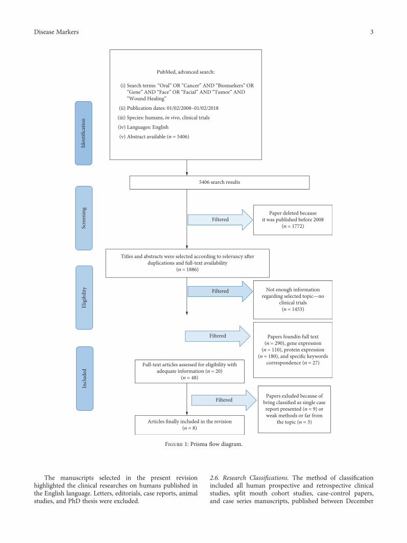
2.3. Searches. The PubMed-Medline resource database wasexplored through advanced searches. The keywords andsearch inquiries used during the first selection stage were asfollows: “oral cancer biomarker”, “oral cancer gene”, and“soft tissue wound healing”. Additional manually selectedarticles were included following the eligibility criteria.Figure 1 represents the flow diagram of the selected studiesaccording to guidelines and following the criteria for theinvestigated papers choice.
2.4. Data Recorded from the Selected Manuscripts. The Med-ical Subject Headings (MeSH) was applied for finding thekeywords used in the present revision. The selected key-words: “oral” OR “facial” AND “cancer” OR “tumor” AND“biomarkers” AND “gene” AND “clinical” AND “woundhealing”, were recorded for collecting the data.
2.5. Selections of the Papers. Four independent reviewers ofdifferent Italian Universities (Messina, Foggia, Catania, andNaples) singularly investigated the obtained full-text papersin order to select inclusion and exclusion criteria as fol-lows. Reviewers compared decisions and resolved differ-ences through discussion and consulting a third partywhen consensus could not be reached. For the stage ofreviewing of full-text articles, a complete independent dualreview was undertaken.
2 Disease Markers
The manuscripts selected in the present revisionhighlighted the clinical researches on humans published inthe English language. Letters, editorials, case reports, animalstudies, and PhD thesis were excluded.
2.6. Research Classifications. The method of classificationincluded all human prospective and retrospective clinicalstudies, split mouth cohort studies, case-control papers,and case series manuscripts, published between December
5406 search results
Titles and abstracts were selected according to relevancy a�erduplications and full-text availability
(n = 1886)
Paper deleted becauseit was published before 2008
(n = 1772)Filtered
Full-text articles assessed for eligibility withadequate information (n = 20)
(n = 48)
Filtered Not enough informationregarding selected topic—no
clinical trials(n = 1453)
Articles finally included in the revision(n = 8)
Filtered
Papers foundin full text(n = 290), gene expression
(n = 110), protein expression(n = 180), and specific keywords
correspondence (n = 27)
Iden
tifica
tion
Scre
enin
gEl
igib
ility
Incl
uded
Filtered
Papers exluded because ofbring classified as single casereport presented (n = 9) orweak methods or far from
the topic (n = 3)
PubMed, advanced search:
Search terms: “Oral” OR “Cancer” AND “Biomarkers” OR“Gene” AND “Face” OR “Facial” AND “Tumor” AND“Wound Healing”
Publication dates: 01/02/2008–01/02/2018
Species: humans, in vivo, clinical trials
Languages: English
Abstract available (n = 5406)
(i)
(ii)
(iii)
(iv)
(v)
Figure 1: Prisma flow diagram.
3Disease Markers
2008 and January 2018, on biomarkers for oral cancer andwound healing.
2.7. Statement of the Problem. The sentence case of “oral can-cer biomarkers clinical trials wound healing” was searchedover each selected papers.
2.8. Exclusion and Inclusion Criteria. The applied inclusioncriteria for the studies were created as follows:
(i) English language
(ii) Clinical human studies of oral cancer and molecularbiomarkers
(iii) Last ten-year data of publishing
The following types of articles were excluded as follows:
(i) In vivo/in vitro studies
(ii) Studies of testing medication and/or new treatmentmethodologies
(iii) Studies of cancer in locations other than mentioned
(iv) Studies not relevant to our selected diagnosticmethods
(v) Animal studies
(vi) Literature review articles published prior to Febru-ary 1st, 2008
(vii) No access to the title and abstract in the Englishlanguage
2.9. Strategy for Collecting Data. Following the initial liter-ature search, all the article titles were screened in order toeliminate irrelevant publications, review articles, casereports, and animal studies. Next, studies were excludedbased on data obtained from screening the abstracts. Thefinal stage of screening involved reading the full texts con-firming each study’s eligibility based on the inclusion andexclusion criteria.
2.10. Data Extraction from the Collected Papers. The data andthe results of the full-text manuscript screened were com-pared. The conclusions were used for assembling the data,according to the aims and themes of the present revision, aslisted onwards.
The following key criteria were used as guidelines foragglomerating the data and then structured following theschemes:
(i) “Author (year)”—revealed the first author and theyear of publication
(ii) “Type of study”—indicated the method of theresearch
(iii) “Sample origin”—describes the number of particu-lar investigated samples in the study and its origin
(e.g., BS: blood sample; SS: saliva sample; and TT:tumor tissue)
(iv) “Follow-up”—yes/no described the duration of theobserved outcomes
(v) “Result”—indicates the parameters that were coher-ent with alterations of particular biomarkers inprognostic studies
2.11. Risk of Bias Assessment. The grade of bias risk was inde-pendently considered and in duplicate by the two indepen-dent reviewers at the moment of data extraction process.
The quality of all included studies was assessed duringthe data extraction process. The quality appraisal involvedevaluating the methodological elements that might influencethe outcomes of each study. According to Moher et al. andHiggins et al., this revision followed the Cochrane Collabora-tion’s two-part tool for assessing risk of bias and PRISMAstatement [37, 38].
Risk of bias (e.g., absence of information or selectivereports on variables of interest) was assessed on a study level.The risks were indicated as lack of precise information ofinterest related to the keywords selected.
This method applied by the four reviewers was valu-able for giving to each study a level of bias. Then, theselected papers were classified with low, moderate, high,and unclear risk.
3. Results
3.1. Manuscript Collection. Manuscript choice and analyzingdata process followed the PRISMA flow diagram (Figure 1).The first electronic and hand search performed onPubMed-Medline and Dentistry and Oral Sciences Sourceresulted with a total of 5406 papers. 1772 papers wereexcluded because they were published prior to February 1st,2008. Then, the other 1886 papers were not involved in therevision because they were not available in full text. Then,the other 1453 papers were not selected because they werenot directly developed as clinical trials. At this point, 290titles and abstracts were evaluated: then, the papers were clas-sified into papers that revealed gene expression n = 110 andprotein expression n = 180; 27 articles were selected as havingsignificant data regarding “Oral Cancer Tumor BiomarkersClinical Trials Wound Healing” topic. 20 articles were deter-mined as full-text papers, 8 of which were incorporated inthis work. Some researches were excluded because of beingclassified as a single case report presented (n = 9) or weakmethods or far from the topic (n = 3).
3.2. Statistical Analysis. No meta-analyses could be per-formed due to the heterogeneity between the studies (differ-ent study designs, control groups, and observation periods).
3.3. Study Characteristics. After the manuscript selection, anew time for screening related to the kind of gene expressionor protein expression has been performed:
(i) Gene expression (n = 110)
4 Disease Markers
(ii) Protein expression (n = 180)
The final clinical papers in full text selected were num-bered as 8.
3.4. Possible Bias of the Selected Studies. The possible risk ofbias was evaluated for each selected papers. The final numberof the selected papers was limited to eight papers. The inclu-sion criteria were really restrictive and for this reason also,the risk of bias was low. Seven studies were considered ashaving a low risk of bias [39–45]; another one was classifiedas moderate risk [46].
Current analysis of the data extracted from studies writ-ten in English only could introduce a publication bias. Aboutpossible bias, some of the selected papers did not specify theinclusion criteria of the patient selection. Another keyparameter that can be assumed as bias is related to the eval-uation of the clinical condition for selecting the patient. Somestudies referred “patients with oral preneoplastic lesions,”while another study wrote about “patients with neoplasticlesion” [39, 43]. The soft tissue healing after the surgical exci-sion was not evaluated in all the selected studies. Moreover,data recorded from the eight studies pointed out the hetero-geneity of the research methods, selections of the patients,and therapeutic options. One paper started the investigationnot directly from the patient but from immortalized humanOSCC-derived cell lines (HSC-2, HSC-3, HSC-4, Ca9-22,
Sa3, HO-1-u-1, and KON) obtained from the Human Sci-ence Research Resources Bank (Osaka, Japan) or the RIKENBRC (Ibaraki, Japan) through the National BioResource Pro-ject of theMinistry of Education, Culture, Sports, Science andTechnology, and this is another bias [46].
Tables 1 and 2 resume the studies selected and theirresults related to the altered biomarkers and to the bio-marker measurements.
3.5. Genetic Alterations in Oral Cancer and Wound Healing.The chosen clinical papers evaluated the alterations in somegene expressions able to influence a predisposition by thepatient on developing oral cancer and consequently the pos-sibility on having a better healing. In the selected clinicalstudies, the oral cancer soft tissue biopsies have beenrecorded and then, the genetic expression of these biopsieswas evaluated, highlighting any possible alterations. Alter-ations in the EGFR gene copy number, or alterations inmiR-7, miR-21, mRNA-KIFGA, OPN, DEPDC1B, EZH2,deltaNp63, and DNMT3B were significant for early evalua-tion and correlation with oral cancer. It is fundamental tounderline how sometimes the quick presumptive diagnosisof preoral cancer lesion and the stage of diagnosis remainthe fundamental steps on recording positive oral cancer diag-nosis. In the final 8 studies, the degree of significance of thesedata was never higher than p < 0 05. In one paper, the corre-lation between the patient’s degree of survival and the
Table 1: Altered biomarkers in OC.
# Year AuthorSubjects
(n)Sampleorigin∗
Gene marker∗∗ Result P value
1 2010Taoudi
Benchekroun et al.162 HB EGFR (U)
An increased EGFR gene copy number increasesthe risk of OSCC
P = 0 062
2 2012 Jung et al. 17 TB134 different miRNA
(see image 1)Keratinization and high miR-21 levels are important
indicators of oral cancer patient prognosisP < 0 05
3 2013 Minakawa et al. 106 TB KIFGA (U)Results showed that KIFGA is overexpressed
in OCP < 0 05
4 2015 Luo et al. 121 HB OPN (osteopontin)Tumor OPN plays an important role in tumordevelopment particularly in tumor invasion
and metastasisP = 0 002
5 2014 Su et al. 7 HB DEPDC1B (U)
DEPDC1B is highly expressed in oral cancer tissue,compared to adjacent tissue. The overexpression in
cells promotes cell migration and induces cellinvasion in cancer cell lines
/
6 2011 Cao et al. 76 TB EZM2(D)EZH2 expression is an independent predictor forOSCC. EZH2 may serve as a biomarker for oral
cancer riskP = 0 05
7 2009 Saintigny et al. 162 HBdeltaNp63 (U), EIC(U), podoplanin (U)
Hazard risk of OC with upregulated genes isaugmented. Considering all three biomarkers,OC patient survival rate is strikingly higher
compared with no, one, or two positive biomarkers
P < 0 0001
8 2011 Saintigny et al. 162 HBHas-miR-101 (D),deltaNp63 (U), P63(U), DNMT3B (U)
It demonstrated the value of gene expressionprofiles in predicting oral cancer development
in OPL patients. The microRNA-based strategiesmight therefore be considered in future
chemoprevention studies
/
∗Type of sample: HU: human biopsy; TB: tissue bank sample. ∗∗Type of altered gene regulation: D: downregulation, diminution; U: upregulation,augmentation.
5Disease Markers
expression of miR-21 is also considered. If the miR-21 valuesare high, the patient’s chances of survival are lower. In onestudy, the degree of dysplasia is evaluated based on theexpression of the EZH2 gene. Another study illustrated thepossibility of evaluating the predisposition to the formationof OC by evaluating deltaNp63 and EIC, also using theexpression of podoplanin [39–46].
In oncology, the tumor markers or tumor indicators areclassified as substances that can be found in the blood or lessoften in the ascitic fluid, which show a significant increase intheir concentration in some types of neoplasia. A high level ofa tumor marker may indicate the presence of cancer,although other causes of raising those values may exist. Somemarkers are specific to certain tumors while others increasein many neoplasms. Tumor markers can be produceddirectly from tumor cells or from normal cells. The tumormarkers, on the other hand, are more useful when they areused to monitor a possible recurrence of cancer after thetreatment (surgical or medical) of the primary tumor. Manyproteins are known to regulate programmed cell death (orapoptosis), and members of the Bcl-2 family are the mostimportant example. This group includes at least 15 differentproteins both with antiapoptotic function (Bcl-2, Bcl-X)and proapoptotic (Bax, Bak), and it represents the balancebetween these two activities determining cell fate. Regardingtheir role in the forms of OSCC, an increase in the levels ofBcl-2 and Bcl-X expression was observed, both in dysplasticoral lesions and in oral cancer [47]. p53 is a tumor suppressor
involved in several mechanisms including cell cycle progres-sion, differentiation, DNA repair, and apoptotic process reg-ulation. p53, also known as tumor protein 53 (TP53 gene), isa transcription factor that regulates cell cycle and coverstumor suppressor function. It intervenes in many antitumormechanisms, activates the repair of damaged DNA (if theDNA is repairable), and can initiate apoptosis, inducing thetranscription of Noxa, in case DNA damage is irreparable;if the DNA is repaired, p53 is degraded and there is a recov-ery of the cell cycle. Some pathogens can instead directlyaffect the p53 protein. An example is the human papilloma-virus (HPV), which encodes a protein which binds p53 inac-tivating it. This, in synergy with the inactivation of anothercell cycle regulator, the p105RB, allows repeated cell divisionsthat occur in the clinical form of the wart. The introductionof p53 into cells with protein deficiency has shown to causea rapid death of cancer cells or a block of cell division. Thisphenomenon reflects the possibility on having good thera-peutic prognosis. For this reason, it is one of the most widelystudied oral cavity biomarkers. The gene encoding is mutatedin the 50% of the tumor forms, particularly in 25-69% ofOSCC cases [48]. A high expression of p53 was observed in40-67% of cases of carcinoma of the head and neck, and thisvariability is related to problems inherent in the method.Some authors [49, 50] have observed a direct relationshipbetween overexpression of p53 and a poor prognosis in termsof survival. In other works, on the contrary, a correlationbetween p53 overexpression and survival did not clearly
Table 2: Biomarker measurement.
# Year AuthorSubjects
(n)Sampleorigin∗
Gene marker∗∗ Sample preparation Method
1 2010Taoudi
Benchekroun et al.162 HB EGFR (U)
Human OC biopsy formalin fixedand paraffin-embedded
FISH
2 2012 Jung et al. 17 TB134 different miRNAs
(see image 1)
Cell culture and transfectionof oral cancer cells and normal
cell biopsy
mirVana™, microarraygene expression,
qRT-PCR
3 2013 Minakawa et al. 106 TB KIFGA (U)
Immortalized humanOSCC-derived
cell lines obtained from the tissuebank. Human biopsy fixed in 20%buffered formaldehyde solution
qRT-PCR
4 2015 Luo et al. 121 HB OPN (osteopontin)Human OC biopsy formalin fixed
and paraffin-embeddedWestern blot
5 2014 Su et al. 7 HB DEPDC1B (U) Human biopsyImmunoprecipitation,
Northern blot,Western blot
6 2011 Cao et al. 76 TB EZM2(D)Human biopsy sample paraffinincluded and sectioned. Colored
with H&EWestern blot
7 2009 Saintigny et al. 162 HBdeltaNp63 (U), EIC(U), podoplanin (U)
Human OC biopsy formalin fixedand paraffin-embedded
Cell membraneimmunoreactivity,
microscope
8 2011 Saintigny 162 HBHas-miR-101 (D),deltaNp63 (U), P63(U), DNMT3B (U)
Whole biopsy including both theepithelial cells and the underlying
stroma
Microarray geneexpression
∗Type of sample: HU: human biopsy; TB: tissue bank sample. ∗∗Type of altered gene regulation: D: downregulation, diminution; U: upregulation,augmentation.
6 Disease Markers
emerge, while an important role of p53 in the carcinogenesisprocess was highlighted, as an early event of malignant trans-formation, and of the histological progression of the tumor[51, 52]. The expression of p53 above the basal layer is con-sidered an early event of the oral carcinogenesis process. Itis an indicator of the development of carcinoma, even beforethe definite morphological changes of the involved tissue.The inactivation of this protein or the alteration of the codinggene could therefore play an important role in the genesis ofOC. This could certainly represent a parameter (biomarker)to be taken into consideration during the diagnostic or inter-ceptive phase of the tumor. Inactivated p53 is not able to stopthe reproduction of cells with damaged DNA. This could be astarting point for OC. The Rb (retinoblastoma) pathway alsoplays a key role in regulating cell cycle progression, and thisactivity can be inhibited by specific mutations. Although Rbmutations are rare in oral cancer, its loss of expression wasseen in 66% of OSCC cases and in 64% of premalignantlesions [22]. Another possible marker of oral cancer is Survi-vin, an apoptotic process inhibitor, expressed in about 80% ofthe forms of squamous cell oral carcinoma and whoseexpression is related with an aggressive phenotype [53]. Ithas been shown that miRNAs can have specific expressionprofiles for developmental stages, tissues, and various pathol-ogies. Studies on several forms of cancer, including oral can-cer, have shown an altered expression of miRNA in tumortissue compared to healthy tissue, suggesting the involve-ment of these molecules in carcinogenesis [54–56]. Humancells have a limited capacity for self-replication and, afternumerous cell divisions, cease to grow and enter on senes-cence phase. Cells with carcinogenic characteristics need tobe immortal in order to replicate infinitely and succeed inmaintaining the length of their telomeres unaltered.
Since tumor growth is limited to 1-2mm3 in the absenceof adequate perfusion, solid tumors require substantial bloodsupply to be able to grow and metastasize [57]. The angio-genic phenomenon is the result of the opposing action ofproangiogenic signals (vascular endothelial growth factor(VEGF), platelet-derived growth factor (PDGF), and inter-leukin 8 (IL-8)) and antiangiogenic signals (interferons andproteolytic fragments such as angiostatin and endostatin).Oral squamous cell cancer has an important local invasivecapacity and a high predisposition to metastasize in the cer-vical lymph nodes. The invasive and metastatic phenomenaare the result of a series of processes involving cell adhesion,cytoskeletal rearrangement, cell migration and degradationof the basement membrane, passage and survival in thebloodstream, and the ability to escape from this and colonizedistant sites with the formation of new vessels.
3.6. Proteomic Changes of Oral Cancer. A total of eight clini-cal studies, in which samples were analyzed, described pro-tein biomarkers and evaluated the wound healing of the siteafter the surgery. In biology, a biomarker is a molecule thatidentifies the presence of a tissue. The marker can be of anynature, but substantially it is a protein, or otherwise polypep-tide, since it is the proteins that are translated by DNA. Forthis reason, a marker is such: it is a molecule that is producedmainly by that type of cell. If the marker is used as a disease
index, it should only be produced in the presence of this dis-ease. Few markers however meet these needs. The majorproblem is given by tumor cell markers: as cells, however,are not completely extraneous to the body, neoplastic cellsdo not translate for molecules that make their dosage accu-rate method. From a molecular point of investigation, studiesinvolved evaluated the aberrant expressions of candidateprotein biomarkers and their quantitative yield in speci-mens. The protein modification is related to the genetic orepigenetic alterations. In some cases, the marker can be rep-resented by high-density lipoprotein components, HDLs,and HDL-cholesterol, [16, 41–48] or even by genetic alter-ations such as those found in some solid tumors [50–54].Proteins are fundamental for physiological cell functioningand life. Aberrant genetic expressions of potential proteinsalter cell division, proliferation, immune response, tissuegrowth, and finally metastasis [48–55]. As for other kindOC cancers, typical patterns of protein expression or indi-vidual proteins with specific features have been recordedand classified as oral cancer biomarkers in order to performdiagnosis and therapy.
4. Discussion
The purpose of this review was to systematically overviewpublished studies restricted to “clinical trials” concerninggenetic and proteomic biomarkers for detection and progno-sis of OC and their relation to wound healing.
Luo et al. [39] evaluated the role of osteopontin (OPN)in chemosensitivity in locally advanced oral squamous cellcarcinoma (OSCC) in humans. Authors considered 121patients and validated the role of OPN in cell proliferation.The recombinant human OPN was executed to SAS cells(human tongue carcinoma cell line) to investigate if theincreased OPN protein could influence a proliferative advan-tage to SAS cells. The presence of OPN is related to boneresorption, wound repair, immune function, and angiogen-esis. However, it is particularly strongly associated withtumorigenesis also. The authors demonstrated that the pro-liferation percentage was significantly increased in matricel-lular OPN in a dose-dependent manner in SAS cells. Thisresult demonstrates that one of the major roles of OPN isto promote growth of OSCC cells. Moreover, it was con-cluded how OPN-mediated cisplatin resistance contributesto a poorer clinical outcome and local wound healing inpatients with locally advanced inoperable OSCC treated withcisplatin-based IC and CCRT.
Taoudi Benchekroun et al. [40] performed a study inves-tigating oral premalignant lesions. The authors obtained dataindicating that an increased EGFR gene copy number is com-mon. Therefore, it is associated with OSCC development inpatients with oral premalignant lesions (OPLs) expressinghigh EGFR, particularly OSCC developing at the site of ahigh-expression OPL; the authors also suggested that EGFRinhibitors might prevent oral cancer in patients with OPLshaving an increased EGFR gene copy number. Moreover,the authors also demonstrated that an increased EGFR genecopy number in OPLs is a precursor to EGFR gene amplifica-tion in HNSCC (as is chromosome 7 increased copy number)
7Disease Markers
and an important oncogenesis-driving effector in oral onco-genesis reducing the possibility of having healing at the sur-gical site and final good prognosis for the patient.
Jung et al. [41] identified deregulated miRNAs in oralcancer and further focus on specific miRNAs that wererelated to patient survival. Authors reported that miRNAexpression profiling provided more precise informationwhen oral squamous cell carcinomas were subcategorizedon the basis of clinic pathological criteria. Data extractedfrom their research highlighted that the interpretation ofmiRNA expression patterns could be better resolved whenone takes into consideration clinical pathological data ofOSCC subtypes. Patient survival data demonstrated that thekeratinization and the high miR-21 levels were significantfactors of OC patient prognosis. Moreover, miR-7 andmiR-21, two keratinization-associated miRNAs, could influ-ence the modification of the tumor suppressor gene RECKin OC. Even if the 17 analyzed tumors clinically showed sim-ilar features, unique miRNA expression patterns were gener-ated for specific subtypes of OSCCs. Finally, the recordeddata underlined that different clinicopathological featuresand miRNA expression profiles could be used as specific sig-natures of individual subtypes of oral tumors with differentfinal prognoses and healing possibilities.
Minakawa et al. [46] assumed that Kinesin family mem-ber 4 (KIF4A) is involved in oral squamous cell carcinomas(OSCCs) pathogenesis by the activation of the spindle assem-bly checkpoint (SAC). KIF4A is overexpressed frequently inOSCC, which suggests interference in the function of thespindle checkpoint proteins such as BUB1, MAD2, andCDC20. KIF4A expression was correlated with tumor sizein KIF4A-positive cases, suggesting that SAC activation playsa significant role in cellular proliferation in OSCC. Theauthors concluded that KIF4A expression is likely to be akey regulator of carcinogenesis progression in OSCCs.
Su et al. [42] studied how the DEPDC1B (defined likeguanine nucleotide exchange factor) induced both cellmigration in a cultured embryonic fibroblast cell line. More-over, it was recorded to favor anchorage-independent growthin oral cancer cells. It was demonstrated that DEPDC1Bexerts a biological function by regulating Rac1. To determinewhether DEPDC1B played a role in the induction of cell pro-liferation, contributing to faster wound healing, the authorsevaluated the growth rate of cells expressing DEPDC1B andcontrol cells founding no substantial difference between thegrowth rates of DEPDC1B-expressing cells and control cells.
However, the authors concluded that oral cancer samplesoverexpressed DEPDC1B proteins, compared with normaladjacent tissue, and so DEPDC1B plays a role in the develop-ment of oral cancer.
Cao et al. [43] investigated the role of the transcriptionalrepressor named Enhancer of Zeste Homolog 2 (EZH2) inoral carcinogenesis and its clinical implication as an OSCCrisk predictor. The study revealed how at 5 years after diag-nosis, the 80% of patients whose OLs expressed strongEZH2 developed OSCC. In Leuk-1 cells, EZH2 downregula-tion resulted in G1 arrest, decreased invasion capability,decreased anchorage independent growth, downregulationof cyclin D1, and upregulation of p15INK4B. The recorded
data suggested that EZH2 seems to have a fundamental rolein OL malignant transformation and may be a biomarker inpredicting OSCC development in patients with OLs. More-over, classifying the EZH2 expression in three stages as weak,moderate, and strong, the authors correlated this situationwith better or not clinical healing, patient survival, and finalprognosis. Quick diagnosis results are fundamental in orderto approach the right therapy and for long survival.
Saintigny et al. [44] considered deltaNp63 as homolog ofthe p53 tumor suppressor and frequently amplified and over-expressed in squamous cell carcinomas, including head andneck squamous cell carcinoma. The authors were able todetermine, in a relatively large population from whom OPLsamples had been collected in a prospective longitudinalmanner, how the level of overexpression of deltaNp63 aloneor in combination with other molecular and morphologicfeatures can be associated with a high risk to develop oralcancer. This investigation referred only oral cancers thatdeveloped in the same site as the OPL; 25% of the patientspositive for podoplanin developed cancer, compared with4% of the patients negative for podoplanin; 24% of thepatients positive for deltaNp63 developed cancer, comparedwith 7% of the patients negative for deltaNp63; and 40% ofthe patients positive for all the biomarkers developed oralcancer, compared with 9% of the patients with no, one, ortwo positive biomarkers. The authors concluded that becausethe measurement of the three biomarkers can be done in rou-tine pathology laboratories, it can be useful for evaluating softtissue healing after OC removal and then patient survival.
Saintigny et al. [45] in a next investigation tried to deter-mine the value of gene expression profiling in predicting oralcancer development. Gene expression profile was measuredin 86 of 162 OPL patients who were enrolled in a clinical che-moprevention trial that used the incidence of oral cancerdevelopment as a prespecified endpoint. The results showedthat gene expression profiles might improve the predictionof oral cancer risk in OPL patients. Moreover, the significantgenes identified may serve as potential targets for oral cancerchemoprevention. Tumor progression from normal mucosato dysplastic mucosa and eventually cancer is the result of aseries of gene modifications affecting the normal functionsof genes such as protooncogenes and tumor suppressors.Such alterations can be partly inherited but most are muta-tions that develop ex novo and accumulate in precancerousand cancerous tissue. These mutations can cause alterationsin cell cycle regulation, differentiation, proliferation, DNArepair mechanisms, and cellular immunity. Chromosomalaberrations such as deletions, amplifications, and structuralrearrangements are common in neoplasms and therefore alsoin head and neck cancer.
All those clinical studies evaluated an alteration of geno-mic proteins leading a tissue transformation directed to theOC formation. The possibility of quickly knowing thoseentire factors such as oral premalignant lesions (OPLs) mayhelp in quick diagnosis and management.
Unfortunately, among the studies taken into consider-ation, few of those evaluate the same markers; thus, the riskof bias of this review study is classified as “high.” It is not pos-sible to make a real report of the statistics of the different
8 Disease Markers
studies, which, however, despite the small number of patientshave satisfactory statistical results. It is very interesting toconsider the possibility that these biomarkers represent a fac-tor to intervene early in the pathology so as to make complexreconstructions more rare [57]. Specifically in those cases,clinicians should avoid the use of complex rehabilitationsplacing dental implant fixtures increasing chronic inflamma-tory process of the jaws and exposing the patient to a risk[58]. In this way, the risk of psychological complicationscan be reduced and it can affect the patients’ oral healthand quality of life [59]. It is interesting to highlight anomaliesin the crevicular fluid associated with the inflammatory stateof the mucosa therefore with precancerous lesions, benignlesions, or OC [60, 61].
5. Conclusions
It is estimated that in the world, the annual cases of squa-mous cell head/neck neoplasia are more than 640,000 (with350,000 deaths). After the success in HER2-positive metasta-tic breast cancer, lapatinib (a small oral molecule that is theresult of GSK research) has also opened an important pathin the treatment of head and neck cancer. These results tellus that the use of a dual tyrosine kinase inhibitor such aslapatinib may be clinically important not only in breast can-cer but also probably in other tumors such as the head andneck, where EGFR is overexpressed. Surely, the possibilityof identifying markers for a diagnosis of a primary oral cavitytumor or a relapse, especially if early, can save the life ofnumerous patients. The possibility of having a set of bio-markers that represent a certain risk for OC and above allthe ease of sampling may constitute real screening for allpatients at risk (genetic predisposition or family history) orexposed to environmental risks (alcohol, smoking, etc.).The present systematic review of clinical studies discov-ered genes and proteins associated with OC and strictlyrelated with the wound healing, the prognosis, and patients’long-term survival. Due to high heterogeneity of theresearches, it was not possible to perform meta-analysis forcomparing the data of the selected papers. Due to poor mate-rials and several parameters recorded, it is not possible toestablish biomarkers specific for oral cancer. The diagnosticcapabilities are also not sufficiently developed and used toallow the use of these markers. However, the highlightedpapers demonstrated how the high, low, or moderate markerexpression might influence the clinical status and the finalprognosis of the patients. At this stage, it seems not possibleto define standard genetic patterns of tumor cells.
Conflicts of Interest
The authors report no conflicts of interest related to thisstudy.
Authors’ Contributions
G.C. is the author responsible for writing the paper. A.S.H.,U.R., and G.L. are the chief reviewers for the collected dataand responsible for the language proof and revision. L.F.,
S.C., and A.B. are responsible for the collection of data andtables. C.D. and R.D. are responsible for funding acquisitiondata. G.T., R.S., L.L., and G.L. are responsible for editing,original data, and text preparation. M.C. is responsible forthe supervision.
References
[1] M. Cicciù, A. S. Herford, G. Cervino, G. Troiano, F. Lauritano,and L. Laino, “Tissue fluorescence imaging (VELscope) forquick non-invasive diagnosis in oral pathology,” Journal ofCraniofacial Surgery, vol. 28, no. 2, pp. e112–e115, 2017.
[2] M.Mascolo, M. Siano, G. Ilardi et al., “Epigenetic disregulationin oral cancer,” International Journal of Molecular Sciences,vol. 13, no. 2, pp. 2331–2353, 2012.
[3] M. Galeano, D. Altavilla, D. Cucinotta et al., “Recombinanthuman erythropoietin stimulates angiogenesis and woundhealing in the genetically diabetic mouse,” Diabetes, vol. 53,no. 9, pp. 2509–2517, 2004.
[4] S. B. S. M. Siriwardena, T. Tsunematsu, G. Qi, N. Ishimaru,and Y. Kudo, “Invasion-related factors as potential diagnosticand therapeutic targets in oral squamous cell carcinoma—areview,” International Journal of Molecular Sciences, vol. 19,no. 5, p. 1462, 2018.
[5] H. A. Sahibzada, Z. Khurshid, R. S. Khan et al., “Salivary IL-8,IL-6 and TNF-α as potential diagnostic biomarkers for oralcancer,” Diagnostics, vol. 7, no. 2, p. 21, 2017.
[6] J.-Y. Chuang, Y.-L. Huang, W.-L. Yen, I.-P. Chiang,M.-H. Tsai, and C.-H. Tang, “Syk/JNK/AP-1 signaling path-way mediates interleukin-6-promoted cell migration in oralsquamous cell carcinoma,” International Journal of MolecularSciences, vol. 15, no. 1, pp. 545–559, 2014.
[7] M. T. Ho, H. S. Kang, J. S. Huh, Y. M. Kim, Y. Lim,and M. Cho, “Effects of the novel compound DK223([1E,2E-1,2-bis(6-methoxy-2H-chromen-3-yl)methylene]hy-drazine) on migration and proliferation of human kera-tinocytes and primary dermal fibroblasts,” InternationalJournal of Molecular Sciences, vol. 15, no. 7, pp. 13091–13110, 2014.
[8] A. Hameedaldeen, J. Liu, A. Batres, G. S. Graves, and D. T.Graves, “FOXO1, TGF-β regulation and wound healing,”International Journal of Molecular Sciences, vol. 15, no. 9,pp. 16257–16269, 2014.
[9] G. Cervino, L. Fiorillo, L. Laino et al., “Oral health impactprofile in celiac patients: analysis of recent findings in aliterature review,” Gastroenterology Research and Practice,vol. 2018, Article ID 7848735, 9 pages, 2018.
[10] J. Majtan and M. Jesenak, “β-Glucans: multi-functional mod-ulator of wound healing,” Molecules, vol. 23, no. 4, p. 806,2018.
[11] P. De Souza Bonfim-Mendonça, I. R. G. Capoci, F. K.Tobaldini-Valerio, M. Negri, and T. I. E. Svidzinski, “Over-view of β-glucans from Laminaria spp.: immunomodula-tion properties and applications on biologic models,”International Journal of Molecular Sciences, vol. 18, no. 9,p. 1629, 2017.
[12] K. M. I. Bashir and J.-S. Choi, “Clinical and physiologicalperspectives of β-glucans: the past, present, and future,”International Journal of Molecular Sciences, vol. 18, no. 9,p. 1906, 2017.
9Disease Markers
[13] A. A. Farooqi, C.-W. Shu, H.-W. Huang et al., “TRAIL, Wnt,sonic hedgehog, TGFβ, and miRNA signalings are potentialtargets for oral cancer therapy,” International Journal ofMolecular Sciences, vol. 18, no. 7, p. 1523, 2017.
[14] T.-H. Wang, S.-M. Hsia, Y.-H. Shih, and T.-M. Shieh, “Associ-ation of Smoking, alcohol use, and betel quid chewing withepigenetic aberrations in cancers,” International Journal ofMolecular Sciences, vol. 18, no. 6, p. 1210, 2017.
[15] A. I. Irimie, C. Ciocan, D. Gulei et al., “Current insights intooral cancer epigenetics,” International Journal of MolecularSciences, vol. 19, no. 3, p. 670, 2018.
[16] G. Y. Seo, C. Hyun, D. Koh et al., “A novel synthetic material,BMM, accelerates wound repair by stimulatingre-epithelialization and fibroblast activation,” InternationalJournal of Molecular Sciences, vol. 19, no. 4, p. 1164, 2018.
[17] D. Ligi, L. Croce, G. Mosti, J. D. Raffetto, and F. Mannello,“Chronic venous insufficiency: transforming growth factor-βisoforms and soluble endoglin concentration in different statesof wound healing,” International Journal of Molecular Sciences,vol. 18, no. 10, p. 2206, 2017.
[18] H.-C. Horng, W.-H. Chang, C.-C. Yeh et al., “Estrogen effectson wound healing,” International Journal of Molecular Sci-ences, vol. 18, no. 11, p. 2325, 2017.
[19] R. C. Dedhia, K. J. Smith, J. T. Johnson, and M. Roberts, “Thecost-effectiveness of community-based screening for oral can-cer in high-risk males in the United States: a Markov decisionanalysis approach,” The Laryngoscope, vol. 121, no. 5, pp. 952–960, 2011.
[20] H. Deneo-Pellegrini, E. de Stefani, P. Boffetta et al., “Mate con-sumption and risk of oral cancer: case-control study in Uru-guay,” Head & Neck, vol. 35, no. 8, pp. 1091–1095, 2012.
[21] M. C. Downer, D. R. Moles, S. Palmer, and P. M. Speight,“A systematic review of test performance in screening fororal cancer and precancer,” Oral Oncology, vol. 40, no. 3,pp. 264–273, 2004.
[22] P. C. Edwards, “Oral cancer screening for asymptomaticadults: do the United States preventive services task force draftguidelines miss the proverbial forest for the trees?,” Oral Sur-gery, Oral Medicine, Oral Pathology and Oral Radiology,vol. 116, no. 2, pp. 131–134, 2013.
[23] S. Y. Liu, C. L. Lu, C. T. Chiou et al., “Surgical outcomes andprognostic factors of oral cancer associated with betel quidchewing and tobacco smoking in Taiwan,” Oral Oncology,vol. 46, no. 4, pp. 276–282, 2010.
[24] S. Shingaki, T. Kobayashi, I. Suzuki, M. Kohno, andT. Nakajima, “Surgical treatment of stage I and II oral squa-mous cell carcinomas: analysis of causes of failure,” The BritishJournal of Oral &Maxillofacial Surgery, vol. 33, no. 5, pp. 304–308, 1995.
[25] R. W. Hinerman, W. M. Mendenhall, C. G. Morris, R. J.Amdur, J. W. Werning, and D. B. Villaret, “Postoperative irra-diation for squamous cell carcinoma of the oral cavity: 35-yearexperience,” Head & Neck, vol. 26, no. 11, pp. 984–994, 2004.
[26] G. W. Sun, E. Y. Tang, X. D. Yang, and Q. G. Hu, “Salvagetreatment for recurrent oral squamous cell carcinoma,” TheJournal of Craniofacial Surgery, vol. 20, no. 4, pp. 1093–1096,2009.
[27] J. A. Jullien, M. C. Downer, J. M. Zakrzewska, and P. M.Speight, “Evaluation of a screening test for the early detectionof oral cancer and precancer,” Community Dental Health,vol. 12, no. 1, pp. 3–7, 1995.
[28] S. B. Baylin and P. A. Jones, “A decade of exploring the cancerepigenome—biological and translational implications,”Nature Reviews Cancer, vol. 11, no. 10, pp. 726–734, 2011.
[29] P. Pisani, F. Bray, and D. M. Parkin, “Estimates of theworld-wide prevalence of cancer for 25 sites in the adult pop-ulation,” International Journal of Cancer, vol. 97, no. 1, pp. 72–81, 2002.
[30] S. Papillon-Cavanagh, C. Lu, T. Gayden et al., “ImpairedH3K36 methylation defines a subset of head and neck squa-mous cell carcinomas,” Nature Genetics, vol. 49, no. 2,pp. 180–185, 2017.
[31] S. B. Baylin and P. A. Jones, “Epigenetic determinants of can-cer,” Cold Spring Harbor Perspectives in Biology, vol. 8, no. 9,article a019505, 2016.
[32] N. Vigneswaran andM. D. Williams, “Epidemiologic trends inhead and neck cancer and aids in diagnosis,” Oral and Maxil-lofacial Surgery Clinics of North America, vol. 26, no. 2,pp. 123–141, 2014.
[33] G. Pannone, A. Santoro, S. Papagerakis, L. lo Muzio, G. deRosa, and P. Bufo, “The role of human papillomavirus in thepathogenesis of head & neck squamous cell carcinoma: anoverview,” Infectious Agents and Cancer, vol. 6, no. 1, p. 4,2011.
[34] V. Damiani, E. Falvo, G. Fracasso et al., “Therapeutic efficacyof the novel stimuli-sensitive nano-ferritins containing doxo-rubicin in a head and neck cancer model,” International Jour-nal of Molecular Sciences, vol. 18, no. 7, p. 1555, 2017.
[35] R. M. Castilho, C. H. Squarize, and L. O. Almeida, “Epigeneticmodifications and head and neck cancer: implications fortumor progression and resistance to therapy,” InternationalJournal of Molecular Sciences, vol. 18, no. 7, article 1506, 2017.
[36] S.-Y. Chen, G.-H. Liu, W.-Y. Chao et al., “Piperlonguminesuppresses proliferation of human oral squamous cell carci-noma through cell cycle arrest, apoptosis and senescence,”International Journal of Molecular Sciences, vol. 17, no. 4,p. 616, 2016.
[37] D. Moher, A. Liberati, J. Tetzlaff, D. G. Altman, and PRISMAGroup, “Preferred reporting items for systematic reviews andmeta-analyses: the PRISMA statement,” Journal of ClinicalEpidemiology, vol. 62, no. 10, pp. 1006–1012, 2009.
[38] J. P. T. Higgins, D. G. Altman, P. C. Gotzsche et al., “TheCochrane Collaboration’s tool for assessing risk of bias in ran-domised trials,” BMJ, vol. 343, article d5928, 2011.
[39] S.-D. Luo, Y.-J. Chen, C.-T. Liu et al., “Osteopontin involvescisplatin resistance and poor prognosis in oral squamous cellcarcinoma,” BioMed Research International, vol. 2015, ArticleID 508587, 13 pages, 2015.
[40] M. Taoudi Benchekroun, P. Saintigny, S. M. Thomas et al.,“Epidermal growth factor receptor expression and gene copynumber in the risk of oral cancer,” Cancer PreventionResearch, vol. 3, no. 7, pp. 800–809, 2010.
[41] H. M. Jung, B. L. Phillips, R. S. Patel et al., “Keratinization-associated miR-7 and miR-21 regulate tumor suppressorreversion-inducing cysteine-rich protein with kazal motifs(RECK) in oral cancer,” Journal of Biological Chemistry,vol. 287, no. 35, pp. 29261–29272, 2012.
[42] Y.-F. Su, C. Y. Liang, C. Y. Huang et al., “A putative novel pro-tein, DEPDC1B, is overexpressed in oral cancer patients, andenhanced anchorage-independent growth in oral cancer cellsthat is mediated by Rac1 and ERK,” Journal of Biomedical Sci-ence, vol. 21, no. 1, p. 67, 2014.
10 Disease Markers
[43] W. Cao, R. H. Younis, J. Li et al., “EZH2 promotes malignantphenotypes and is a predictor of oral cancer development inpatients with oral leukoplakia,” Cancer Prevention Research,vol. 4, no. 11, pp. 1816–1824, 2011.
[44] P. Saintigny, A. K. el-Naggar, V. Papadimitrakopoulou et al.,“DeltaNp63 overexpression, alone and in combination withother biomarkers, predicts the development of oral cancer inpatients with leukoplakia,” Clinical Cancer Research, vol. 15,no. 19, pp. 6284–6291, 2009.
[45] P. Saintigny, L. Zhang, Y. H. Fan et al., “Gene expression pro-filing predicts the development of oral cancer,” Cancer Preven-tion Research, vol. 4, no. 2, pp. 218–229, 2011.
[46] Y. Minakawa, A. Kasamatsu, H. Koike et al., “Kinesin fam-ily member 4A: a potential predictor for progression ofhuman oral cancer,” PLoS One, vol. 8, no. 12, article e85951,2013.
[47] A. Rousseau, M. S. Lim, Z. Lin, and R. C. Jordan, “Frequentcyclin D1 gene amplification and protein overexpression inoral epithelial dysplasias,” Oral Oncology, vol. 37, no. 3,pp. 268–275, 2001, 275..
[48] T. Tsuji, Y. Mimura, S. Wen et al., “The significance of PCNAand p53 protein in some oral tumors,” International Journal ofOral and Maxillofacial Surgery, vol. 24, no. 3, pp. 221–225,1995.
[49] M. J. Caminero, F. Nunez, C. Suarez, P. Ablanedo, J. R.Riera, and F. Dominguez, “Detection of p53 protein in oro-pharyngeal carcinoma. Prognostic implications,” Archives ofOtolaryngology – Head & Neck Surgery, vol. 122, no. 7,pp. 769–772, 1996.
[50] B. Popovic, B. Jekic, I. Novakovic et al., “Bcl-2 expression inoral squamous cell carcinoma,” Annals of the New York Acad-emy of Sciences, vol. 1095, no. 1, pp. 19–25, 2007.
[51] S. C. Girod, P. Pfeiffer, J. Ries, and H. D. Pape, “Proliferativeactivity and loss of function of tumour suppressor genes as‘biomarkers’ in diagnosis and prognosis of benign and preneo-plastic oral lesions and oral squamous cell carcinoma,” BritishJournal of Oral and Maxillofacial Surgery, vol. 36, no. 4,pp. 252–260, 1998.
[52] P. Pande, M. Mathur, N. K. Shukla, and R. Ralhan, “pRb andp16 protein alterations in human oral tumorigenesis,” OralOncology, vol. 34, no. 5, pp. 396–403, 1998.
[53] G. A. Calin and C. M. Croce, “MicroRNA-cancer connection:the beginning of a new tale,” Cancer Research, vol. 66, no. 15,pp. 7390–7394, 2006, 66.
[54] J. Takamizawa, H. Konishi, K. Yanagisawa et al., “Reducedexpression of the let-7 microRNAs in human lung cancers inassociation with shortened postoperative survival,” CancerResearch, vol. 64, no. 11, pp. 3753–3756, 2004, 64.
[55] G. A. Calin, C. D. Dumitru, M. Shimizu et al., “Frequentdeletions and down-regulation of micro- RNA genes miR15and miR16 at 13q14 in chronic lymphocytic leukemia,” Pro-ceedings of the National Academy of Sciences of the UnitedStates of America, vol. 99, no. 24, pp. 15524–15529, 2002,15529..
[56] J. Folkman, “What is the evidence that tumors are angiogene-sis dependent?,” Journal of the National Cancer Institute,vol. 82, no. 1, pp. 4–7, 1990.
[57] M. Cicciù, G. Cervino, A. Herford et al., “Facial bone recon-struction using both marine or non-marine bone substitutes:evaluation of current outcomes in a systematic literaturereview,” Marine Drugs, vol. 16, no. 1, 2018.
[58] G. Matarese, L. Ramaglia, L. Fiorillo, G. Cervino, F. Lauritano,and G. Isola, “Implantology and periodontal disease: the pan-acea to problem solving?,” The Open Dentistry Journal, vol. 11,no. 1, pp. 460–465, 2017.
[59] A. Bruno, G. Pandolfo, G. Scimeca et al., “Anger in health,benign breast disease and breast cancer: a prospectivecase-control study,” In Vivo, vol. 28, no. 5, pp. 973–977, 2014.
[60] L. Fiorillo, G. Cervino, A. S. Herford et al., “Interferon crevic-ular fluid profile and correlation with periodontal disease andwound healing: a systemic review of recent data,” InternationalJournal of Molecular Sciences, vol. 19, no. 7, article E1908,2018.
[61] G. Isola, M. Cicciù, L. Fiorillo, and G. Matarese, “Associationbetween odontoma and impacted teeth,” The Journal of Cra-niofacial Surgery, vol. 28, no. 3, pp. 755–758, 2017.
11Disease Markers

Stem Cells International
Hindawiwww.hindawi.com Volume 2018
Hindawiwww.hindawi.com Volume 2018
MEDIATORSINFLAMMATION
of
EndocrinologyInternational Journal of
Hindawiwww.hindawi.com Volume 2018
Hindawiwww.hindawi.com Volume 2018
Disease Markers
Hindawiwww.hindawi.com Volume 2018
BioMed Research International
OncologyJournal of
Hindawiwww.hindawi.com Volume 2013
Hindawiwww.hindawi.com Volume 2018
Oxidative Medicine and Cellular Longevity
Hindawiwww.hindawi.com Volume 2018
PPAR Research
Hindawi Publishing Corporation http://www.hindawi.com Volume 2013Hindawiwww.hindawi.com
The Scientific World Journal
Volume 2018
Immunology ResearchHindawiwww.hindawi.com Volume 2018
Journal of
ObesityJournal of
Hindawiwww.hindawi.com Volume 2018
Hindawiwww.hindawi.com Volume 2018
Computational and Mathematical Methods in Medicine
Hindawiwww.hindawi.com Volume 2018
Behavioural Neurology
OphthalmologyJournal of
Hindawiwww.hindawi.com Volume 2018
Diabetes ResearchJournal of
Hindawiwww.hindawi.com Volume 2018
Hindawiwww.hindawi.com Volume 2018
Research and TreatmentAIDS
Hindawiwww.hindawi.com Volume 2018
Gastroenterology Research and Practice
Hindawiwww.hindawi.com Volume 2018
Parkinson’s Disease
Evidence-Based Complementary andAlternative Medicine
Volume 2018Hindawiwww.hindawi.com
Submit your manuscripts atwww.hindawi.com


![EUROPEAN CONSENSUS-BASED RECOMMENDATIONS ... KD...children and adolescents only[16] (Supplementary Table S1). Articles were assessed using pre-specified inclusion/exclusion criteria](https://img.pdfslide.fr/doc/110x75/60f68c768464673906276281/european-consensus-based-recommendations-kd-children-and-adolescents-only16.jpg)

![AOZIZ of INCLUSION - presentation [FR] of INCLUSION... · AOZIZ of INCLUSION - Présentation 2020 Institut CALEM - Ludovic-Mohamed Zahed Ludovic Mohamed Zahed est le recteur de l'Institut](https://img.pdfslide.fr/doc/110x75/610ddd2bf17c9732e21e1a9c/aoziz-of-inclusion-presentation-fr-of-inclusion-aoziz-of-inclusion-prsentation.jpg)