Embed Size (px)
Citation preview
Université de Montréal
Neogenin function and modulation in spinal motor neuron
development
Par
Louis-Philippe Croteau
Programmes de biologie moléculaire
Faculté de Médecine
Thèse présentée à la Faculté de médecine
en vue de l’obtention du grade de PhD
en biologie moléculaire
Septembre 2017
© Louis-Philippe Croteau, 2017
i
Résumé
Le système nerveux est la structure fonctionnelle la plus complexe que l’on
connaisse. Cette structure permet aux organismes multicellulaires de percevoir leur
existence en tant qu’entités individuelles. Le fonctionnement du système nerveux
repose sur l’assemblage précis des circuits neuronaux. Pour ce faire, pendant le
développement, les projections axonales des neurones sont guidées par des signaux
de guidages. Ceux-ci se trouvent dans l’environnement extracellulaire et permettent
aux neurones de trouver leurs cibles respectives. Pour détecter et pouvoir répondre
aux signaux de guidage, les cônes de croissance à la pointe des axones expriment
des récepteurs de signaux de guidage. Ceux-ci, lorsqu’ils se lient à leurs ligands,
induisent des cascades de signalisation en aval, menant à des modulations dans la
poussée et la direction des axones. Bien que nous ayons fait de grands progrès dans
la compréhension des effets qu’ont les signaux de guidage sur les axones, on en sait
beaucoup moins sur la façon dont ces signaux agissent en concert.
Les neurones moteurs qui résident dans la colonne moteur latérale (LMC) de
la moelle épinière projettent leurs axones dans les membres, s'appuient sur une
multitude de signaux de guidage. Cette thèse démontre que le récepteur
transmembranaire Neogenin est impliqué dans plusieurs aspects du développement
des neurones LMC, y compris la différenciation et la ségrégation des sous-types de
neurones LMC ainsi que le guidage de leurs axones.
Notre laboratoire a précédemment démontré que les axones LMC sont
guidés de manière synergique par Netrin-1 et les ephrins. Je vous ferai part de mes
ii
résultats explorant le mécanisme sous-jacent à l'activité synergique de Netrin-1 et
d’ephrin-A5 dans le guidage des axones LMC. Je démontre que ephrin-A5 sensibilise
un sous-ensemble d'axones LMC à Netrin-1. Je propose que cela est la conséquence
d'une augmentation induite par ephrin-A5 dans l'abondance de Neogenin conduisant
à une liaison améliorée de Netrin-1 dans les cônes de croissance LMC. En outre, je
montre que la modulation de ephrin-A5 des niveaux de Neogenin dépend de
l'interaction entre ephrin-A5 et son récepteur EphA4. Je montre également que
contrairement à la répulsion des axones de ephrin-A5, la sensibilisation à Netrin-1 se
produit indépendamment de la queue cytoplasmique d'EphA4. Ces résultats
suggèrent que la répulsion des axones induite par ephrin-A5 et la sensibilisation à
Netrin-1 se produisent dans des voies moléculairement distinctes.
Netrin-1, Neogenin et les ephrins sont vastement impliqués lors du
développement du système nerveux et au-delà. Notre démonstration que les
interactions ephrin-A5 / EphA4 modulent l'abondance de Neogenin et la sensibilité à
Netrin-1 ouvre la possibilité a ce que les ephrins puissent avoir un impact profond sur
les processus cellulaires dépendants de la signalisation Netrin-1 et Neogenin.
Mots-clés : Neogenin, Netrin-1 ephrin, guidage axonal, neurones moteurs
iii
Abstract
The nervous system is the most exquisitely complex functional structure in the
known universe. The nervous system is what enables multicellular organisms to
experience life as individual entities. The functionality of the nervous system relies
upon the precise assembly of neuronal circuits. To achieve this, during development,
the extending axons of neurons are guided by cues in the extracellular environment
that enable neurons to find their respective targets. To sense and respond to
extracellular cues, growth cones at the tip of axons express guidance cue receptors.
The interaction between guidance cues and their receptors induce downstream
signaling cascades which lead to modulations in axon outgrowth and directionality.
Although we have made great progress in understanding how individual cues guide
axons, much less is known about how these cues act in concert.
Motor neurons that reside within the lateral motor column (LMC) of the spinal
cord extend axons that innervate the limbs, rely on a multitude of guidance cues. The
evidence presented in this thesis shows that the transmembrane receptor Neogenin
is implicated in several aspects of LMC neuron development including LMC subtype
differentiation and segregation as well as the guidance of their axons.
Our lab has previously shown that LMC axons are guided synergistically by
Netrin-1 and ephrins. I will be presenting results exploring the mechanism underlying
the synergistic activity of Netrin-1 and ephrin-A5 in LMC axon guidance. I provide
evidence that ephrin-A5 sensitizes a subset of LMC axons to Netrin-1. I propose that
this is a consequence of an ephrin-A5 induced increase in the abundance of
iv
Neogenin leading to enhanced Netrin-1 binding in LMC growth cones. Furthermore, I
show that the ephrin-A5 modulation of Neogenin levels is dependent on the
interaction between ephrin-A5 and its receptor EphA4. I also show that contrarily to
the repulsion from ephrin-A5, sensitization to Netrin-1 occurs independently from the
cytoplasmic tail of EphA4. These results suggest that the ephrin-A5 induced axon
repulsion and sensitization to Netrin-1 occur in molecularly distinct pathways.
Netrin-1, Neogenin and ephrins are vastly implicated during the development
of the nervous system and beyond. Our demonstration that ephrin-A5/EphA4
interactions modulate the abundance of Neogenin and sensitivity to Netrin-1 brings
forth the possibility that ephrins may have a profound impact on cellular processes
implicating Netrin-1 and Neogenin.
Keywords: Neogenin, Netrin-1, ephrin, axon guidance, motor neuron
v
Table of contents Résumé ------------------------------------------------------------------------------------------------------- i
Abstract ----------------------------------------------------------------------------------------------------- iii
List of abbreviations ------------------------------------------------------------------------------------ viii
List of figures ----------------------------------------------------------------------------------------------- x
Acknowledgements ------------------------------------------------------------------------------------- xii
CHAPTER I - INTRODUCTION --------------------------------------------------------------------- 15
1 An introduction to axon guidance ----------------------------------------------------------------- 16
2 The Growth Cone ------------------------------------------------------------------------------------ 18
3 Guidance cues ---------------------------------------------------------------------------------------- 20
4 Netrin-1 ----------------------------------------------------------------------------------------------- 21
4.1 Netrin-1 in commissural axon guidance ----------------------------------------------------- 21
4.2 Netrin-1 receptors ------------------------------------------------------------------------------- 23
4.3 DsCAM ------------------------------------------------------------------------------------------ 23
4.4 Dcc ------------------------------------------------------------------------------------------------ 24
4.5 Neogenin ----------------------------------------------------------------------------------------- 25
4.6 Neogenin as a receptor for RGMs ------------------------------------------------------------ 25
4.7 Neogenin as a receptor for Netrin-1 ---------------------------------------------------------- 27
4.8 Chick Neogenin --------------------------------------------------------------------------------- 28
4.9 Neogenin/Dcc structure ------------------------------------------------------------------------ 30
5 Netrin-1 signaling ------------------------------------------------------------------------------------ 32
6 Modulators of Netrin-1 signaling ------------------------------------------------------------------ 34
6.1 Heparin sulfate proteoglycans ---------------------------------------------------------------- 34
6.2 Slit/Robo ----------------------------------------------------------------------------------------- 35
6.3 Draxin -------------------------------------------------------------------------------------------- 37
6.4 Dcc/Neogenin cleavage ------------------------------------------------------------------------ 38
7 An introduction to Eph/ephrin signaling --------------------------------------------------------- 40
7.1 Eph/ephrin signaling in axon guidance ------------------------------------------------------ 42
8. Spinal motor neuron development ---------------------------------------------------------------- 44
8.1 LMC neuron differentiation ------------------------------------------------------------------- 48
9 LMC axon guidance --------------------------------------------------------------------------------- 49
10 Dorso-ventral LMC guidance -------------------------------------------------------------------- 51
vi
10.1 Eph/ephrin forward signaling---------------------------------------------------------------- 52
10.2 Eph/ephrin reverse signaling ---------------------------------------------------------------- 53
10.3 Eph/ephrin cis attenuation ------------------------------------------------------------------- 55
10.4 GDNF / GFRa1/RET ------------------------------------------------------------------------- 56
10.5 Npn-2/Sema3F --------------------------------------------------------------------------------- 57
11 Netrin-1 in LMC guidance ------------------------------------------------------------------------ 58
11.1 In vivo requirement for Netrin-1 in LMC axon guidance ------------------------------- 59
11.2 LMCm and LMCl axons respond to Netrin-1 in vitro ----------------------------------- 60
12 Synergy between Netrin-1 and ephrins in LMC guidance ----------------------------------- 61
CHAPTER II - MATERIALS AND METHODS --------------------------------------------------- 65
CHAPTER III - RESULTS ---------------------------------------------------------------------------- 74
1. Neogenin in the responsiveness of LMCl axons to Netrin-1 --------------------------------- 75
Rationale ---------------------------------------------------------------------------------------------- 75
Neogenin is required for the responsiveness of LMCl axons towards Netrin-1------------ 75
Dcc rescues the loss of LMCl responsiveness to Netrin-1 ------------------------------------ 76
2. Netrin-1 and ephrin-B2 collapse LMCm growth cones synergistically --------------------- 78
Rationale ---------------------------------------------------------------------------------------------- 78
3. Neogenin in LMC differentiation ----------------------------------------------------------------- 80
Rationale ---------------------------------------------------------------------------------------------- 81
Expression of Neogenin, RGMa and RGMb during LMC neuron development ---------- 81
Knockdown of Neogenin alters LMC neuron subtype differentiation ----------------------- 83
LMC neuron subtype distribution within the LMC is altered in Neogenin KD embryos - 88
Investigating the requirement for RGMa and RGMb in LMC subtype differentiation --- 90
Knockdown of Neogenin alters lumbar LMC axon projections ------------------------------ 92
Neogenin may be dispensable for the subtype differentiation of LMC neurons in mice - 94
4. Sensitization of spinal motor neurons to Netrin-1 by ephrin-A5 ----------------------------- 96
Rational ----------------------------------------------------------------------------------------------- 96
Netrin-1 induces a protein kinase A-dependent re-localization of Neogenin in LMC
growth cones ----------------------------------------------------------------------------------------- 97
Ephrin-A5 increases Neogenin protein levels in LMC growth cones ---------------------- 104
Ephrin-A5 induces an increase in the co-localization of Neogenin and EphA4 ---------- 107
In LMC growth cones ----------------------------------------------------------------------------- 107
vii
Ephrin-A5 increases Neogenin growth cone surface levels ---------------------------------- 107
Ephrin-A5 enhances Netrin-1 binding in growth cones -------------------------------------- 110
Binding of ephrin-A5 to EphA4 is necessary for the ephrin-A5 induction of Neogenin in
growth cones ---------------------------------------------------------------------------------------- 112
Overexpression of EphA4 potentiates LMC growth cones to the ephrin-A5-dependent
increase in Neogenin abundance ----------------------------------------------------------------- 114
The ephrin-A5-induced Neogenin upregulation is independent of PKA, Src family kinase,
protein synthesis, proteosomal or γ-secretase degradation ----------------------------------- 115
The intracellular domain of EphA4 is dispensable for potentiating the ephrin-A5 induced
increase in Neogenin abundance ----------------------------------------------------------------- 117
Ephrin-A5 sensitization of lateral LMC axons to Netrin-1 ---------------------------------- 119
The cytoplasmic tail of EphA4 is dispensable for the sensitization of LMCl axons to
Netrin-1 by ephrin-A5 ----------------------------------------------------------------------------- 122
CHAPTER IV - DISCUSSION --------------------------------------------------------------------- 124
Neogenin in LMC neuron differentiation ---------------------------------------------------------- 125
Neogenin in LMC subtype segregation --------------------------------------------------------- 125
Neogenin in LMC subtype differentiation ------------------------------------------------------ 127
Sensitization of LMCl axons to Netrin-1 by ephrin-A5 ----------------------------------------- 129
Netrin-1 increases and redistributes Neogenin to the growth cone periphery ------------- 129
Ephrin-A5 on receptor dynamics in LMC growth cones ------------------------------------- 130
Ephrin-A5/EphA4 attenuation of Neogenin cleavage as a possible mechanism underlining
the sensitization to Netrin-1 ----------------------------------------------------------------------- 132
CHAPTER V – CONTRIBUTIONS AND PERSPECTIVES -------------------------------- 137
Neogenin as a mediator of attractive Netrin-1 signaling in LMC neurons ------------------- 138
Speculations on the requirement for Neogenin dependent transcriptional regulation for the
expression of Lim1 by LMC neurons -------------------------------------------------------------- 138
Optimization of the chick in ovo electroporation protocol -------------------------------------- 139
The sensitization of LMC neurons to Netrin-1 by ephrin-A5 ---------------------------------- 140
A potential for ephrin-A5 induced upregulation of Neogenin in various cell types --------- 140
A potential requirement for ephrin-A/EphA induction of Neogenin for neural tube closure
----------------------------------------------------------------------------------------------------------- 142
Speculations on the requirement for ephrin/EphA4 dependent upregulation of Dcc/Neogenin
for LTP ------------------------------------------------------------------------------------------------- 143
BIBLIOGRAPHY -------------------------------------------------------------------------------------- 144
viii
List of abbreviations
ADAM A disintegrin and metalloproteinase
APP Amyloid precursor protein
BC Boundary cap
BMP Bone morphogenic protein
CNS Central nervous system
Dcc Deleted in colorectal cancer
DsCAM Down syndrome cell adhesion molecule
E Chick Embryonic day
F-actin Filamentous actin
FAK Focal adhesion kinase
FNIII Fibronectin type III
GAP GTPase activating protein
GDNF Glial-cell-line-derived neurotrophic factor
GEF Guanine nucleotide exchange factor
GPI Glycosylphosphatidylinositol
GFRα1 GDNF receptor alpha-1
HH st. Hamburger-Hamilton stage
HMC Hypaxial motor column
HRP Horseradish peroxidase
HSPG Heparin sulfate proteoglycan
IF Immunofluorescence
ICD Intracellular domain
Ig Immunoglobulin
KD Knock down
LMC Lateral motor column
ix
LMCl Lateral LMC
LMCm Medial LMC
LMO4 Lim-only protein 4
MMC Medial motor column
MN Motor neuron
MT Microtubule
Np Neuropilin
OE Olfactory epithelium
PGC Preganglionic motor column
PKA Protein kinase A
PS1 Presenilin-1
RA Retinoic acid
Raldh2 Retinaldehyde dehydrogenase-2
RGM Repulsive guidance molecule
RGC Retinal ganglion cell
RTK Receptor tyrosine kinase
Sema Semaphorin
SFK Src family kinase
SpMN Spinal motor neuron
TCA Thalamocortical axon
WNT Wingless-type MMTV integration site
WRC Wave regulatory complex
WT Wild type
x
List of figures
Figure 1 The structural and spatial divisions in a typical growth cone .................................... 19
Figure 2 Chick Neogenin expression in the spinal cord closely reassembles the combined
expression of Dcc and Neogenin in the mouse ......................................................................... 29
Figure 3 Netrin-1 signaling ....................................................................................................... 31
Figure 4 Early development of spinal motor neurons of the LMC ........................................... 47
Table 1 LMC dorso-ventral axon guidance summary .............................................................. 52
Figure 5 Model for the guidance of LMC axons by Netrin-1 ................................................... 58
Figure 8 Netrin-1 and ephrins are integrated synergistically by LMCm and LMCl axons ...... 62
Figure 6 Neogenin is required for LMCl axon growth preference for Netrin-1 ....................... 77
Figure 7 Dcc can functionally substitute for chick Neogenin in LMCl axon preference for
Netrin-1 ..................................................................................................................................... 78
Figure 9 Netrin-1 and ephrin-B2 act synergistically on LMCm growth cone collapse ............ 80
Figure 10 Expression of Neogenin, RGMa and RGMb during LMC neuron development ..... 83
Figure 11 Knockdown of Neogenin alters LMC subtype differentiation ................................. 85
Figure 12 Neogenin is required for establishing Lim1 expression in LMC neurons................ 87
Figure 13 The Distribution of LMCm and LMCl neurons is altered in [Neo]siRNA embryos 89
Figure 14 A possible implication for RGMb in LMC subtype differentiation ......................... 92
Figure 15 Knockdown of Neogenin alters LMC axon projections ........................................... 93
Figure 16 LMC neuron subtype differentiation in mice lacking Neogenin appears normal .... 95
Figure 17 Netrin-1 induces a re-localization of Neogenin in LMC growth cones ................... 98
Figure 18 Netrin-1 at 10 ng/mL is sufficient in redistributing Neogenin within LMC growth
cones ......................................................................................................................................... 99
Figure 19 Netrin-1 induced re-localization of Neogenin is PKA dependent .......................... 100
Figure 20 Netrin-1 induces specific changes in protein levels and distribution at the growth
cone ......................................................................................................................................... 103
Figure 21 The effects of ephrin-A5 on protein levels and distribution at the growth cone .... 105
Figure 22 Ephrin-A5 increases the proportion of fluorescence overlap between Neogenin and
EphA4 ..................................................................................................................................... 108
Figure 23 Ephrin-A5 increases surface enriched Neogenin in growth cones ......................... 109
Figure 24 Ephrin-A5 enhances Netrin-1 binding in growth cones ......................................... 111
Figure 25 Blocking ephrin-A5/EphA4 interactions inhibits the ephrin-A5 induced increase in
Neogenin levels and overlap with EphA4 .............................................................................. 113
Figure 26 Overexpression of EphA4 enhances the ephrin-A5 induced increase in Neogenin
abundance ............................................................................................................................... 115
Figure 27 The ephrin-A5 induced Neogenin upregulation prevails despite inhibiting specific
cell functions ........................................................................................................................... 117
Figure 28 The cytoplasmic tail of EphA4 is dispensable in potentiating the ephrin-A5 induced
increase in Neogenin abundance ............................................................................................. 119
Figure 29 LMCl axons are sensitized to Netrin-1 by ephrin-A5 ............................................ 121
xi
Figure 30 The cytoplasmic tail of EphA4 is dispensable in the sensitization of LMC axons to
Netrin-1 by ephrin-A5............................................................................................................. 123
Figure 31 Tentative model for the sensitization of LMCl axons to Netrin-1 ......................... 134
Supplementary Figure 1 Growth cone area measurements..................................................... 136
xii
Acknowledgements
First and foremost, I would like to thank my PhD supervisor, Dr. Artur Kania,
for providing me with the opportunity to pursue graduate studies as a member in his
lab. Over the years, he has dedicated an enormous amount of time discussing my
experiments with me and I am grateful for all the scientific wisdom he has shared. I
am also grateful for his encouragement in having high scientific standards and for
participating in conferences. Thank you Artur for seeing the opportunity in turning an
unexpected electroporation artifact into my first scientific publication. You have also
provided me the opportunity of collaborating with several labs, experiences that have
enriched my skill set and scientific career. Above all, thank you Artur for listening to
what I had to say and taking my opinions into consideration.
I would also like to acknowledge Meirong Liang, our lab technician, for all the
help and support she has provided over the years (and for having to deal with a
bunch of students). I also want to acknowledge past Kania lab members who have
helped my out over the years. I would like to express my gratitude to Dayana
Krawchuk and Elena Palmesino for helping out when I first joined the lab. I would also
like to thank Chris Law for making my life easier by introducing me to ImageJ macros
and for providing valuable feedback during lab meetings. Special thanks to Tzu-Jen
Kao for collaborating with me on the stripe assays presented in chapter I and chapter
III, I literally couldn’t have done it without you. Thanks to Dominic Fillion for the
numerous troubleshooting sessions with the microscopes and for programing the
MATLAB application used to determine the distribution of immunofluorescence in

xiii
growth cones. I would also like to acknowledge the members of the Neuro labs and
Michel Cayouette, Frédéric Charron and Hideto Takahashi for providing me with
valuable feedback during my Club Neuro presentations.
This thesis is dedicated to my love, Rose-Marie Lacroix. Your unconditional
moral support throughout this journey has been the force behind my sustained efforts.
You have always been understanding of me working long hours and over the week-
ends and have been an invaluable support during difficult times. I aspire to one day
become the man you deserve to be with and hope that our son, Laurent Croteau, will
be as beautiful of a person as you are.

15
CHAPTER I - INTRODUCTION
16
1 An introduction to axon guidance
To achieve functional circuitry, neurons must establish precise connections
with their cellular targets. To do so, a neuron extends a cellular process termed axon,
that navigates through surrounding tissue until the target is reached. Depending on
the neuron type and the size of the organism, axons may span just a few
micrometers, to several meters, as is the case for some axons in large vertebrates
such as whales (Smith, 2009). The field of axon guidance investigates the
mechanisms enabling axonal targeting during development. Understanding these
mechanisms may be crucial for achieving functional recovery post-injury as well as in
the prevention and recovery in neurodegenerative disorders. The father of
contemporary neurobiology, Ramon Y Cajal S., first described the specialized cellular
structure at the tip of extending axons termed the growth cone (Ramon, 1890).
Thought to be inspired by evidence of chemotaxis in leukocytes guided by diffusible
bacterial toxins, Cajal postulated the Neurotrophic theory whereby growth cones are
proposed to be endowed with chemotactic sensitivities to factors secreted by their
cells enabling axons to reach their targets (Metchnikoff, 1892; Ramon, 1892). Strong
in vivo evidence for the neurotrophic theory came much later with the seminal
experiments by Roger Sperry where Xenopus tadpole eyes were surgically reoriented
and axonal fibers were able to reach their original targets despite the reorientation
(Sperry, 1963). This demonstrated that the target cells were providing positional
information rather than this information being intrinsic to the projecting neurons.
17
The proper targeting of axons is regulated by a multitude of factors that can
ultimately be categorized as having attractive or repulsive effects on extending axons
(Kolodkin and Tessier-Lavigne, 2011). Different modalities of guidance have been
described including chemotaxis, whereby axons are guided towards or away from
guidance cue gradients secreted from the target tissue. Guidance cues are integrated
into positional information through the expression of guidance cue receptors at the
growth cone surface (Kolodkin and Tessier-Lavigne, 2011). Other axon guidance
mechanisms have since been described such as haptotaxis, whereby the guidance
cue provides adhesion and mechanical traction which promotes the outgrowth of
axons along the path of guidance cue expression (Carter, 1965; Varadarajan et al.,
2017). Conversely, surround repulsion, whereby the axon outgrowth path is restricted
by cells expressing repulsive cues that establish permissive corridors (Keynes et al.,
1997). Axon fasciculation also contributes to axon pathfinding, axons often tend to
extend as bundles and the impediment of axon fasciculation can result in certain
axons to stray away (Landmesser et al., 1988; Landmesser et al., 1990).
Furthermore, later born neurons sometimes rely on pre-existing axon tracts to reach
their targets (Gallarda et al., 2008; Wang et al., 2011; Wang et al., 2014). The final
guidance decision is target recognition and is crucial for axons to know when to stop
extending and to begin forming synapses with the target cells (Timofeev et al., 2012).
18
2 The Growth Cone
The growth cone consists of a dynamic subcellular structure located at the tip
of axons that is highly sensitive and responsive to the extracellular environment.
Growth cones are responsible for guiding axons to their targets by enabling axonal
steering, extension as well as retraction (Lowery and Van Vactor, 2009). Its function
requires the tight orchestration and crosstalk between different processes such as
actin cytoskeleton dynamics, adhesion complex assembly and disassembly as well as
membrane trafficking (Vitriol and Zheng, 2012). The growth cone has a complex
architecture that can be subdivided into different domains based on its cytoskeletal
components (Figure 1) (Lowery and Van Vactor, 2009). The peripheral domain (P) is
the most dynamic and consists of filopodia which can be described as finger-like
projections containing long bundles of actin filaments (F-actin) as well as lamellipodia,
located between filopodia, consisting of mesh-like branched F-actin networks (Figure
1). Individual microtubules (MT) can also be seen within filipodia. The central (C)
domain consists of stable microtubule bundles originating from the axon shaft as well
as various vesicles organelles and F-actin bundles (Figure 1). Positioned at the
interface between the central and peripheral domains is the transition zone where
actomyosin contractile structures are formed and allow F-actin retrograde flow, a
process required for axon elongation (Figure 1) (Lowery and Van Vactor, 2009).
19
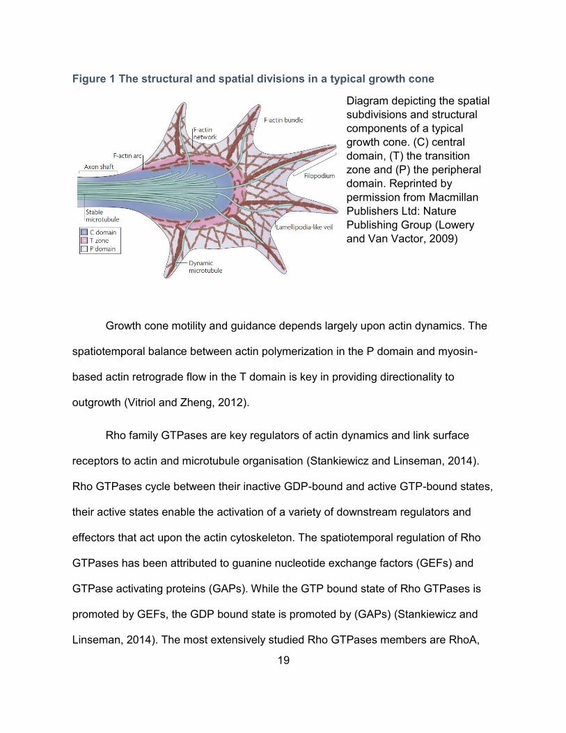
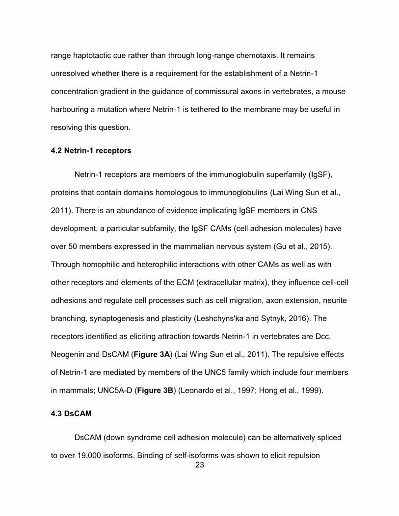
Figure 1 The structural and spatial divisions in a typical growth cone
Diagram depicting the spatial
subdivisions and structural
components of a typical
growth cone. (C) central
domain, (T) the transition
zone and (P) the peripheral
domain. Reprinted by
permission from Macmillan
Publishers Ltd: Nature
Publishing Group (Lowery
and Van Vactor, 2009)
Growth cone motility and guidance depends largely upon actin dynamics. The
spatiotemporal balance between actin polymerization in the P domain and myosin-
based actin retrograde flow in the T domain is key in providing directionality to
outgrowth (Vitriol and Zheng, 2012).
Rho family GTPases are key regulators of actin dynamics and link surface
receptors to actin and microtubule organisation (Stankiewicz and Linseman, 2014).
Rho GTPases cycle between their inactive GDP-bound and active GTP-bound states,
their active states enable the activation of a variety of downstream regulators and
effectors that act upon the actin cytoskeleton. The spatiotemporal regulation of Rho
GTPases has been attributed to guanine nucleotide exchange factors (GEFs) and
GTPase activating proteins (GAPs). While the GTP bound state of Rho GTPases is
promoted by GEFs, the GDP bound state is promoted by (GAPs) (Stankiewicz and
Linseman, 2014). The most extensively studied Rho GTPases members are RhoA,
20
Rac1 and Cdc42. Rac1/Cdc42 are typically attributed to growth cone development
and axon extension while RhoA is typically attributed to axon retraction and growth
cone collapse (Stankiewicz and Linseman, 2014). Nevertheless, evidence suggests
that the activation balance of Rho GTPases rather than the activation state of a
member is required for proper guidance (Barallobre et al., 2005).
3 Guidance cues
There are four classical families of guidance cues which comprise of the
Netrins, ephrins, semaphorins and slits (Kolodkin and Tessier-Lavigne, 2011). Slits
are secreted cues, ephrins are membrane bound and Netrins and semaphorins can
be either membrane bound or secreted (Kolodkin and Tessier-Lavigne, 2011; Lai
Wing Sun et al., 2011) . Whereas Netrins, ephrins and semaphorins can act as
bidirectional cues, capable of eliciting both attraction and repulsion in a context
dependent manner, slits have been solely associated with repulsion (Morales and
Kania, 2016). More recently, a growing list of morphogens as well as growth factors
and cell-adhesion molecules have also been implicated in axon guidance (Kolodkin
and Tessier-Lavigne, 2011). Guidance molecules function in other neural functions
such as cell migration and synaptogenesis as well as in non-neuronal tissues in
processes such as angiogenesis, organogenesis, immune function as well as cancer
biology (Hinck, 2004; Kolodkin and Tessier-Lavigne, 2011). The present work will
focus on Netrins and ephrins for their relevance in the original work described in
chapters III - Results.
21
4 Netrin-1
Netrins are a family of laminin-related extracellular proteins with homologues in
all assayed organisms with bilateral symmetry (Lai Wing Sun et al., 2011). Mammals
express three secreted netrins (Netrin-1-3) and two GPI-anchored netrins (Netrin-G1,
Netrin-G2) (Lai Wing Sun et al., 2011). Netrins have been implicated in a variety of
neural processes including cell and axon migration, axon arborisation,
synaptogenesis as well as oligodendrocyte development (Lai Wing Sun et al., 2011).
Outside the nervous system, Netrins have been implicated in the development of the
pancreas, lungs, mammary gland as well as in angiogenesis (Cirulli and Yebra,
2007). Netrin-1, the most extensively studied Netrin member, can illicit axon attraction
or repulsion depending on the expression of Netrin-1 receptors by neurons
(Colamarino and Tessier-Lavigne, 1995; Hong et al., 1999; Lai Wing Sun et al., 2011;
Poliak et al., 2015). The first ascribed function for Netrin-1 was the guidance of
commissural axons (Kennedy et al., 1994; Serafini et al., 1994; Serafini et al., 1996).
4.1 Netrin-1 in commissural axon guidance
Commissural neurons are responsible for connecting both sides of the CNS. In
vertebrates, Netrin-1 was shown to be required for commissural neurons in the
developing brain and dorsal spinal cord to extend axons to an intermediate target, the
ventral floor-plate (Kennedy et al., 1994; Serafini et al., 1994; Serafini et al., 1996).
The guidance effect of Netrin-1 on commissural neurons was originally proposed to
occur through long-range chemotaxis whereby secretion of Netrin-1 by floor plate
22
cells was proposed to establish a gradient of Netrin-1 ranging from high ventral to low
dorsal. The long-range chemotaxis theory for Netrin-1 attraction was substantiated by
in vitro culture experiments showing dorsal commissural axons being attracted
towards floor-plate explants, positioned as far as ~250 μm away. This experiment
suggested that Netrin-1 could act at a distance by diffusing away from its source cells
(Tessier-Lavigne et al., 1988; Serafini et al., 1994). More recently, the requirement for
long-range diffusion of Netrin-1 from the floor-plate has been challenged. Evidence in
Drosophila shows that in both the midline and the visual system, a tethered Netrin is
sufficient to rescue Netrin loss of function phenotypes (Brankatschk and Dickson,
2006; Timofeev et al., 2012). It was also demonstrated that the mechanical
attachment of Netrin-1 to a substrate is required for axon outgrowth for mouse
commissural axons in vitro (Moore et al., 2012). Furthermore, biochemical analysis of
Netrin-1 shows that it is tightly bound to the membrane fraction and can only be
extracted at very high salt concentrations, making it unlikely to be able to diffuse to
long-distance within tightly packed cells, at least in the context of passive diffusion
(Serafini et al., 1994). Recently, two independent groups demonstrated that
progenitors at the ventricular zone, rather than cells from the floor plate, were the
major source of Netrin-1 in guiding commissural axons (Dominici et al., 2017;
Varadarajan et al., 2017). Both groups also highlight that Netrin-1 produced by
progenitors at the ventricular zone is concentrated at the pial surface that lines the
periphery of the spinal cord as well as on the progenitor radial processes encountered
by commissural axons when projecting dorsally. Taken together, the evidence
suggests that in the context of commissural axon guidance, Netrin-1 acts as a short-
23
range haptotactic cue rather than through long-range chemotaxis. It remains
unresolved whether there is a requirement for the establishment of a Netrin-1
concentration gradient in the guidance of commissural axons in vertebrates, a mouse
harbouring a mutation where Netrin-1 is tethered to the membrane may be useful in
resolving this question.
4.2 Netrin-1 receptors
Netrin-1 receptors are members of the immunoglobulin superfamily (IgSF),
proteins that contain domains homologous to immunoglobulins (Lai Wing Sun et al.,
2011). There is an abundance of evidence implicating IgSF members in CNS
development, a particular subfamily, the IgSF CAMs (cell adhesion molecules) have
over 50 members expressed in the mammalian nervous system (Gu et al., 2015).
Through homophilic and heterophilic interactions with other CAMs as well as with
other receptors and elements of the ECM (extracellular matrix), they influence cell-cell
adhesions and regulate cell processes such as cell migration, axon extension, neurite
branching, synaptogenesis and plasticity (Leshchyns'ka and Sytnyk, 2016). The
receptors identified as eliciting attraction towards Netrin-1 in vertebrates are Dcc,
Neogenin and DsCAM (Figure 3A) (Lai Wing Sun et al., 2011). The repulsive effects
of Netrin-1 are mediated by members of the UNC5 family which include four members
in mammals; UNC5A-D (Figure 3B) (Leonardo et al., 1997; Hong et al., 1999).
4.3 DsCAM
DsCAM (down syndrome cell adhesion molecule) can be alternatively spliced
to over 19,000 isoforms. Binding of self-isoforms was shown to elicit repulsion
24
required for dendrite and axon self-avoidance (Schmucker et al., 2000; Wojtowicz et
al., 2007; Hattori et al., 2009). Although there is evidence for DsCAM being implicated
in axon attraction to Netrin-1 in Drosophila and in chick, complete loss of DsCAM in
the mouse does not result in any commissural guidance defects in vivo, or alters
commissural axon responses to Netrin-1 in vitro (Andrews et al., 2008; Ly et al., 2008;
Liu et al., 2009; Palmesino et al., 2012). These discrepancies may reflect different
requirements for DsCAM among species. It is also possible that the full knock-out of
DsCAM in the mouse may bring about compensatory mechanisms not available in the
context of chick DsCAM knockdown by shRNA.
4.4 Dcc
Unlike DsCAM, loss of Dcc dramatically alters commissural axon guidance,
closely phenocopying mice lacking Netrin-1 (Fazeli et al., 1997). Because of defects
in commissural axon guidance, human individuals with mutations in Dcc display
mirror movements whereby contralateral involuntary movements mirror voluntary
ones (Srour et al., 2010). Dcc (deleted in colorectal carcinoma) was named for its
absence in most colorectal carcinomas and was proposed to be a putative tumor
suppressor gene (Fearon et al., 1990). It’s role as a tumor suppressor has been
substantiated by evidence of Dcc being a death receptor, in prolonged absence of
Netrin-1, Dcc initiates a caspase cascade leading to apoptosis (Mehlen et al., 1998;
Castets et al., 2011). It’s role as a dependence receptor is not ubiquitous, in mice
completely lacking Netrin-1, no difference in apoptosis can be seen within the
25
developing CNS (Bin et al., 2015). It’s role as a dependence receptor likely requires
specific adaptors as exemplified by DIP13α, proposed to modulate the pro-apoptotic
effects of Dcc (Liu et al., 2002).
4.5 Neogenin
Closely related to Dcc is Neogenin, first identified in the chicken and named so
for its high expression during neural differentiation (Vielmetter et al., 1994). Neogenin
is a receptor for members of the repulsive guidance molecule (RGM) family which in
vertebrates includes RGMa, RGMb and RGMc, as well as a receptor for Netrins
(Keino-Masu et al., 1996; Rajagopalan et al., 2004; Siebold et al., 2017).
4.6 Neogenin as a receptor for RGMs
During mouse development, whereas RGMa and RGMb are expressed in and
out of the CNS in a rather complementary fashion, RGMc expression is excluded
from the CNS (Niederkofler et al., 2004; Oldekamp et al., 2004). RGMs are not only
ligands for Neogenin but have also been shown to be BMP co-receptors (Babitt et al.,
2005; Samad et al., 2005; Babitt et al., 2006). The binding of RGMa to Neogenin in
cis is proposed to enhance BMP downstream signaling (Healey et al., 2015).
BMP/RGM/Neogenin have been involved in a variety of biological processes such as
iron homeostasis, bone formation and astrocyte differentiation (Babitt et al., 2006;
Zhou et al., 2010; Huang et al., 2016). RGMc/Neogenin signalling is required for iron
26
homeostasis, both Neogenin and RGMc null mice suffer from hepatic iron overload,
reduced hepcidin expression and defective BMP signaling (Lee et al., 2010).
In the chick optic tectum and the Xenopus forebrain, RGMa acts as a repulsive
cue for Neogenin expressing axons (Monnier et al., 2002; Rajagopalan et al., 2004;
Wilson and Key, 2006). RGMa induced growth cone collapse is independent of BMP
signaling and involves RhoA, the Rho-associated kinase ROCK as well as PKC
(Conrad et al., 2007). Furthermore, RGMa induced growth cone collapse was shown
to require Unc5b as well as the GEF LARG (Hata et al., 2009). Unc5 proteins are
involved in the repulsion from both Netrin-1 and RGMa by associating with Dcc and
Neogenin respectively (Hong et al., 1999; Hata et al., 2009). The convergence of
Netrin-1 and RGMa signalling pathways at Unc5 receptors suggests a possible
enhancement of repulsive signaling where both cues overlap. In mammals, RGMa
inhibits axon outgrowth following spinal cord injury, RGMa inhibition using a blocking
antibody results in an increase in axon outgrowth and improves functional recovery
(Hata et al., 2006). In contrast, RGMa was shown to have neuroprotective capabilities
for retinal ganglion cells (RGCs), following optic nerve transection, the intraocular
injection of RGMa reduces RGC death (Koeberle et al., 2010). RGMa/Neogenin were
also shown to be required for neural tube closure in Xenopus and mice by regulating
neuroepithelium morphology (Niederkofler et al., 2004; Kee et al., 2008).
Misexpression of RGMa in the developing chick hind-brain was shown to influence
neural differentiation through an unknown mechanism (Matsunaga et al., 2006).
27
RGMb is the least studied member of the RGM family. There is evidence
RGMb acting as both a tumor suppressor as well as promoting cancer progressing by
regulating BMP signaling(Shi et al., 2015; Li et al., 2016). In the developing olfactory
epithelium (OE), loss of RGMb and Neogenin both result in neural differentiation
defects (Kam et al., 2016). The authors propose that progenitors expressing
Neogenin bind RGMb on neighbouring cells and that these interactions regulate cell
cycle kinetics and exit (Kam et al., 2016).
Thus, RGM/Neogenin signaling is involved in a wide variety of processes
during development and beyond. RGM/Neogenin signalling outside the CNS often
involves the regulation of BMP signaling, which to my knowledge, has not been
demonstrated within the CNS. Whereas Netrin-1 binds Neogenin on its fibronectin
type III (FNIII) domains 4 and 5, RGMs bind Neogenin on the FNIII domains 5 and 6
(Figure 3) (Yang et al., 2008; Xu et al., 2014). In vitro, Netrin-1 inhibits RGMa
induced growth cone collapse of dorsal root ganglion neurons, suggesting that Netrin-
1 and RGMa compete in binding Neogenin (Bell et al., 2013). This provides evidence
for the cross-regulation potential of Netrin-1 and RGMa signalling pathways.
4.7 Neogenin as a receptor for Netrin-1
In mice, whereas the loss of Neogenin alone seemingly does not result in
spinal commissural guidance defects, the loss of both Neogenin and Dcc exacerbates
the guidance phenotype of Dcc mutants to a degree comparable to Netrin-1 mutants.
This suggests that both Dcc and Neogenin collaborate in guiding commissural axons
28
in the mouse (Xu et al., 2014). Neogenin has been shown to be implicated in adult
neurogenesis in mice. Loss of Neogenin results in neuroblast migration defects that
are presumed to result from a loss of Netrin-1 attraction as well as aberrant
differentiation resulting from impaired cell cycle kinetics (O'Leary et al., 2015). In the
supraoptic tract of the developing Xenopus forebrain, knock down of either Neogenin
or Netrin-1 results in similar guidance defects, suggesting they may be acting as a
ligand/receptor pair in guiding supraoptic tract axons (Wilson and Key, 2006).
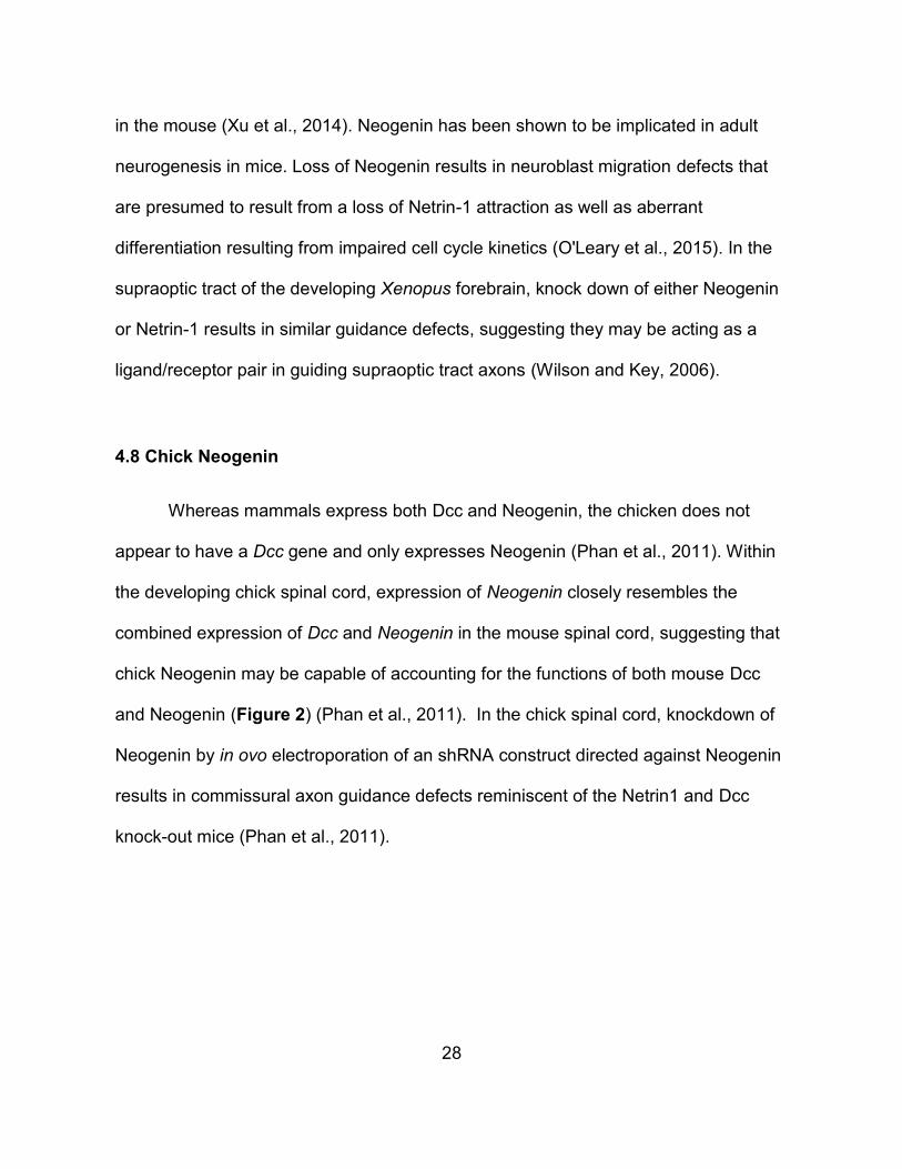
4.8 Chick Neogenin
Whereas mammals express both Dcc and Neogenin, the chicken does not
appear to have a Dcc gene and only expresses Neogenin (Phan et al., 2011). Within
the developing chick spinal cord, expression of Neogenin closely resembles the
combined expression of Dcc and Neogenin in the mouse spinal cord, suggesting that
chick Neogenin may be capable of accounting for the functions of both mouse Dcc
and Neogenin (Figure 2) (Phan et al., 2011). In the chick spinal cord, knockdown of
Neogenin by in ovo electroporation of an shRNA construct directed against Neogenin
results in commissural axon guidance defects reminiscent of the Netrin1 and Dcc
knock-out mice (Phan et al., 2011).
29
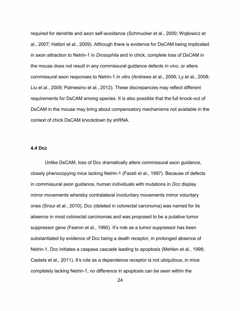
(A–H) In situ hybridization
experiments for Neogenin (A–
C, G–I) and Dcc (D–F) on
transverse sections of the
spinal cord of E10.5 (A, D)
and E11.5 (B, C, E, F) mouse
embryos and HH stage 20
(G), 23 (E) and 26 (I) chicken
embryos. (A–C) In mouse,
Neogenin is expressed first in
the intermediate spinal cord
(bracket, A). By E11.5,
Neogenin is present at high
levels in the ventral
ventricular zone as well as
broadly in motor neurons
(brackets, B, C) (D–F) Mouse
Dcc is expressed at highest
levels in the dorsal-most
neural progenitors
(arrowhead, D, E) and in
post-mitotic neurons
(brackets, D–F) throughout the dorsal spinal cord as well as at lower levels in a broad
population of motor neurons. (G–I) The distribution of chicken Neogenin is a
composite of the expression patterns of both mouse Dcc and Neogenin. At all stages,
the highest levels of Neogenin expression is in the dorsal-most spinal cord, in dorsal
neural progenitors (arrowheads, G, I) and in a population of post-mitotic dorsal
neurons whose position is consistent with their being commissural neurons
(arrowhead, H). Neogenin is also present at lower levels in both the ventral ventricular
zone and in motor neurons (brackets, H, I). Scale bar: A, D, G: 30 mm, B, C, E, F, H,
I: 40 mm. Taken from (Phan et al., 2011)
Figure 2 Chick Neogenin expression in the spinal cord closely reassembles the combined expression of Dcc and Neogenin in the mouse
30
4.9 Neogenin/Dcc structure
Dcc and Neogenin form a subfamily of IgSFs and share the same overall
structure with their extracellular portions containing four immunoglobulin domains
followed by six fibronectin type III repeats and three intracellular P domains (P1-3)
(Figure 3) (Keino-Masu et al., 1996). Whereas the expression of Dcc is mostly
restricted to the CNS, Neogenin is highly expressed in the CNS and in mesoderm
derived tissue during development as well as in adulthood (Meyerhardt et al., 1997).
Netrin-1 has two binding sites for Dcc and Neogenin and both receptors have 2
binding sites for Netrin-1 within their FNIII4 and FNIII5 domains (Figure 3A)
(Meyerhardt et al., 1997; Xu et al., 2014). The crystal structure of Netrin-1 bound to
Dcc and Neogenin reveals that two distinct architectures are possible depending on
splice variants, 2:2 heteromers or a continuous ligand/receptor assembly (Figure 3C,
D) (Xu et al., 2014). The Netrin-1 induced multimerization of Dcc results in the
association of Dcc intracellular P3 domains and is required for Netrin-1 induced
attraction (Stein et al., 2001).
31
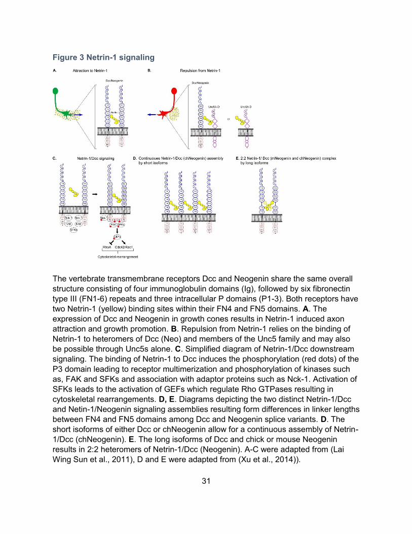
Figure 3 Netrin-1 signaling
The vertebrate transmembrane receptors Dcc and Neogenin share the same overall
structure consisting of four immunoglobulin domains (Ig), followed by six fibronectin
type III (FN1-6) repeats and three intracellular P domains (P1-3). Both receptors have
two Netrin-1 (yellow) binding sites within their FN4 and FN5 domains. A. The
expression of Dcc and Neogenin in growth cones results in Netrin-1 induced axon
attraction and growth promotion. B. Repulsion from Netrin-1 relies on the binding of
Netrin-1 to heteromers of Dcc (Neo) and members of the Unc5 family and may also
be possible through Unc5s alone. C. Simplified diagram of Netrin-1/Dcc downstream
signaling. The binding of Netrin-1 to Dcc induces the phosphorylation (red dots) of the
P3 domain leading to receptor multimerization and phosphorylation of kinases such
as, FAK and SFKs and association with adaptor proteins such as Nck-1. Activation of
SFKs leads to the activation of GEFs which regulate Rho GTPases resulting in
cytoskeletal rearrangements. D, E. Diagrams depicting the two distinct Netrin-1/Dcc
and Netin-1/Neogenin signaling assemblies resulting form differences in linker lengths
between FN4 and FN5 domains among Dcc and Neogenin splice variants. D. The
short isoforms of either Dcc or chNeogenin allow for a continuous assembly of Netrin-
1/Dcc (chNeogenin). E. The long isoforms of Dcc and chick or mouse Neogenin
results in 2:2 heteromers of Netrin-1/Dcc (Neogenin). A-C were adapted from (Lai
Wing Sun et al., 2011), D and E were adapted from (Xu et al., 2014)).
32
5 Netrin-1 signaling
Netrin-1 dependent signaling has been extensively studied, mostly focusing on
Dcc as its receptor. A thorough review of the literature investigating Netrin-1
downstream signaling events would exceed the scope of this thesis, nevertheless,
here are some of the key findings. Stimulation with Netrin-1 induces the tyrosine
phosphorylation of Dcc in its P3 domain resulting in receptor multimerization (Figure
3C) (Meriane et al., 2004; Lai Wing Sun et al., 2011; Xu et al., 2014). In rodent
neuroblastoma cells (NG108-15), ectopic expression of Dcc induces a Netrin-1
dependent increase in surface area and filipodia number. These effects were shown
to require the activation of the Rho GTPases Cdc42 and Rac1 (Shekarabi and
Kennedy, 2002). Two Rho GEFs, Trio and Dock180, have been implicated in
mediating Rac1 activation downstream of Netrin-1/Dcc (Briancon-Marjollet et al.,
2008; Li et al., 2008). Furthermore, through a Dcc dependent mechanism, Netrin-1
inhibits RhoA which in turn increases the levels of Dcc at the plasma membrane,
thereby sensitizing rat commissural neurons to Netrin-1 outgrowth and guidance in
vitro (Moore et al., 2008). The tyrosine kinase adaptor protein Nck-1 binds directly to
Dcc in rat commissural neurons, the expression of a dominant negative Nck-1 inhibits
Dcc’s ability to induce Netrin-1 dependent neurite outgrowth and Rac1 activation in
N1E-115 cells and fibroblasts respectively (Li et al., 2002).
Three independent groups provided evidence that focal adhesion kinase
(FAK), a cytosolic protein tyrosine kinase, binds to Dcc and is phosphorylated upon
Netrin-1 treatment and is required for Netrin-1 dependent outgrowth and attraction (Li
33
et al., 2004; Liu et al., 2004; Ren et al., 2004). FAK has also been implicated in
signaling downstream of Neogenin in the context of growth cone collapse and
myogenesis (Bae et al., 2009; Endo and Yamashita, 2009). FAK itself is regulated by
phosphorylation of multiple tyrosine residues in and outside the nervous system, it is
implicated in establishing cell morphology, maturation of adhesive structures and
migration (Chacon and Fazzari, 2011). FAK has been implicated in signaling
downstream of semaphorins, ephrins as well as Netrins and is involved in both axon
attraction and repulsion (Chacon and Fazzari, 2011). Furthermore, FAK has been
shown to interact with both activators and inhibitors of Rho GTPases and can
phosphorylate as well as be phosphorylated by Src family kinases (SFK) (Mitra et al.,
2005).
SFKs have been implicated in multiple aspects of neural development
including differentiation, axon outgrowth, fasciculation and guidance (Morse et al.,
1998; Hoffman-Kim et al., 2002; Kuo et al., 2005; Robles et al., 2005; Kao et al.,
2009). Members of SFKs bind Dcc and are phosphorylated upon Netrin-1 stimulation,
and SFK function is required for Netrin-1 dependent commissural axon outgrowth and
guidance in vitro (Li et al., 2004; Meriane et al., 2004).
To summarize, Netrin-1 binding to Dcc/Neogenin induces the tyrosine
phosphorylation within their P3 domains and receptor multimerization. These events
enable the activation of adaptor proteins such as Nck-1 and signaling kinases such as
FAK and SFKs. Association with FAK promotes traction to the extracellular
environment whereas SFKs induce the activation of GEFs such as Trio and Dock180.
34
The spatiotemporal regulation of GEF activation in turn regulates Rho GTPases and
the downstream cytoskeletal remodeling behind growth cone turning and axon
outgrowth (Figure 3C).
6 Modulators of Netrin-1 signaling
6.1 Heparin sulfate proteoglycans
Heparin sulfate proteoglycans (HSPG) are polysaccharides of the
glycosaminoglycan family and have been shown to bind to hundreds of proteins
(Esko and Lindahl, 2001). HSPGs consist of core proteins mainly consisting of
syndecans, glypicans, perlecan and agrin, on to which are attached highly charged
heparin sulfate side chains (Lee and Chien, 2004). HSPGs can regulate the
distribution of growth factors as well as enhance the binding of several ligands
implicated in axon guidance (Lee and Chien, 2004). Netrin-1 binds heparin with high
affinity and its extraction from the heparin fraction during chromatography requires
high salt concentrations. (Serafini et al., 1994). Dcc also interacts with heparin via its
fifth fibronectin domain, a domain also implicated in binding Netrin-1(Bennett et al.,
1997; Xu et al., 2014). Spinal commissural neurons have a cell-autonomous
requirement for heparin sulfate in vivo and in vitro. Dorsal spinal neurons that are
genetically deficient for Heparin sulfate have commissural guidance defects
comparable to Netrin-1-/- and Dcc-/- mice in vivo and are unresponsive to Netrin-1
dependent outgrowth in vitro (Matsumoto et al., 2007). It has been suggested that
heparin sulfate could act as a coreceptor for Dcc in binding Netrin-1, therefore the
35
expression and distribution of HSPGs may play a vital role in modulating the strength
of Netrin-1 signaling by promoting Dcc/Netrin-1 interactions (Matsumoto et al., 2007).
6.2 Slit/Robo Once spinal commissural neurons reach the floor-plate, they continue
extending contralaterally, away from the floor-plate, before turning rostrally and
extend along the rostral-caudal axis of the spinal cord. Post-crossing neurons
expressing the receptors Robo1/2 and Neuropilin-2, are repelled from the midline by
Slit and Semaphorin3B respectively, expressed by the floor-plate (Zou et al., 2000;
Long et al., 2004). To avoid stalling at the midline and recrossing the floor plate,
commissural axons must become desensitized to Netrin-1 still expressed at the
midline (Stein and Tessier-Lavigne, 2001). Xenopus spinal axons in culture are
attracted towards a Netrin-1 gradient, when Slit is added to the Netrin-1 gradient,
axons become unresponsive to Netrin-1 attraction while retaining the axon outgrowth
promoting effect of Netrin-1 (Stein and Tessier-Lavigne, 2001). Furthermore,
Xenopus spinal neurons exogenously expressing a truncated Robo receptor, without
its intracellular tail, remain attracted towards Netrin-1 in the presence of Slit whereas
axons expressing full-length Robo remain unresponsive (Stein and Tessier-Lavigne,
2001) These result suggest that Slit binding to Robo somehow desensitizes axons to
Netrin-1 attraction. Experiments where Xenopus axons express chimeric Robo and
Dcc receptors, revealed that the cytoplasmic tails of Robo and Dcc were required in
Slit dependent desensitization to Netrin-1. Furthermore, co-immunoprecipitation
experiments show that Dcc Robo interaction is Slit dependent. Together, these in
36
vitro results suggest that spinal commissural axons expressing Robo receptors
become insensitive to Netrin-1 attraction when they encounter Slit at the floor-plate
(Stein and Tessier-Lavigne, 2001).
In vitro, thalamocortical axons (TCAs) are repelled by Slit1 and are
unresponsive to Netrin-1 (Bielle et al., 2011). For a subset of TCAs, the rostral TCAs,
Slit1 and Netrin-1 provided in combination results in attraction towards Netrin-1, even
when Slit1 is homogeneously distributed (Bielle et al., 2011; Dupin et al., 2015). Slit-1
induced attraction to Netrin-1 is PKA dependent and proposed to result from an
increase in Dcc surface levels (Leyva-Diaz et al., 2014). The permissive effect of Slit1
on rostral TCA attraction to Netrin-1 requires sub-threshold concentrations of Slit1,
insufficient for inducing repulsion (Dupin et al., 2015). This effect requires the
expression of the Slit1 receptor Robo1 and its coreceptor FLRT3 by rostral TCAs
(Leyva-Diaz et al., 2014).
FLRT proteins can promote cell adhesion through homophilic interactions and
promote repulsion by binding Unc5 proteins in trans (Karaulanov et al., 2009;
Seiradake et al., 2014). They have also been implicated in FGF signaling and
synaptogenesis as well as neural migration. (Bottcher et al., 2004; O'Sullivan et al.,
2012; Del Toro et al., 2017). Slit1/Robo interactions silence Netrin-1 attraction in
commissural axons and allow Netrin-1 attraction in TCAs. The mechanism behind
these opposite responses is currently unknown but may by in part due to differences
in the expression of coreceptors such as FLRT.
37
6.3 Draxin
Draxin is a secreted protein recently identified as a guidance cue that shares
no homology with any other guidance cue families (Islam et al., 2009). Draxin-/- mice
display a mild fasciculation defects in spinal commissural axons and severe forebrain
commissural axon guidance defects comparable to Dcc-/- mice (Fazeli et al., 1997;
Islam et al., 2009). Draxin was shown to bind to the Netrin-1 receptors Dcc,
Neogenin, DSCAM and Unc5A-C as well as to Netrin-1 (Ahmed et al., 2011; Gao et
al., 2015). It is also noteworthy that Draxin and Netrin-1 have distinct binding domains
on Dcc (Ahmed et al., 2011) In vitro, depending on the concentration and neuronal
type, Draxin can either stimulate axonal outgrowth or inhibit outgrowth and induce
growth cone collapse (Islam et al., 2009; Ahmed et al., 2011; Shinmyo et al., 2015).
Draxin-/- mice also have thalamocortical projection defects and these defects are
comparable in severity to the defects seen in (Draxin+/- ; Dcc-/- ), (Draxin+/- ; Neo1GT/G),
or (Dcc-/- ; Neo1GT/G), suggesting that Draxin genetically interacts with Dcc and
Neogenin in the context of axon guidance (Shinmyo et al., 2015). The role of Draxin
in thalamocortical projections occurs independently from Netrin-1 since Netrin-1-/-
mice don’t have thalamocortical projections defects (Shinmyo et al., 2015). On the
other hand, the similar forebrain commissure phenotype seen in Netrin-1-/-, Dcc-/-, and
Draxin-/- mice raises the possibility that Draxin, by binding to both Netrin-1 and Dcc,
may enhance Netrin-1/Dcc interactions (Ahmed et al., 2011; Gao et al., 2015)
38
6.4 Dcc/Neogenin cleavage
Dcc immunoprecipitations done on supernatant collected from cell cultures
overexpressing Dcc reveals that Dcc’s ectodomain is cleaved near the
transmembrane domain (Galko and Tessier-Lavigne, 2000). Furthermore, it was
shown that the in vitro ectodomain cleavage of Dcc can be blocked by a broad
spectrum metalloprotease inhibitor (Galko and Tessier-Lavigne, 2000). The in vitro
treatment of dorsal spinal explants with metalloprotease inhibitors results in an
increase in Dcc expression and a potentiation towards Netrin-1 dependent axonal
outgrowth (Galko and Tessier-Lavigne, 2000). It was also demonstrated that Netrin-1
treatment increases the ectodomain cleavage of Dcc (Bai et al., 2011).
The ectodomain of Neogenin can be cleaved by the protease ADAM17 and
was proposed to modulate RGMa induced repulsion by regulating surface Neogenin
(Okamura et al., 2011). Lrig2, a transmembrane protein that binds Neogenin, can
regulate ADAM17 proteolysis of Neogenin (van Erp et al., 2015). The binding of
RGMa to Neogenin inhibits Lrig2/Neogenin interactions and allows ADAM17 cleavage
of Neogenin. In vitro, Lrig2 is required for the RGMa dependent RhoA activation and
growth cone collapse. In vivo, knockdown of Ligr2 results in the promotion of optic
nerve outgrowth following experimental optic nerve crush (van Erp et al., 2015).
Considering the homology between Dcc and Neogenin, as well as evidence of Dcc
cleavage by metalloproteases, it is likely that Dcc may also undergo ectodomain
cleavage by ADAM17 (Galko and Tessier-Lavigne, 2000).
39
Several transmembrane proteins such as E and N-Cadherin, Notch and
amyloid precursor protein (APP), have their ectodomains cleaved, which
subsequently leads to the cleavage of their remaining intracellular fragments by γ-
secretase (De Strooper et al., 1998; Naruse et al., 1998; De Strooper et al., 1999;
Struhl and Greenwald, 1999; Marambaud et al., 2002). Dcc and Neogenin both
undergo cleavage by γ-secretase, resulting in the release of their intracellular
domains (ICD) (Taniguchi et al., 2003; Goldschneider et al., 2008).The ICDs of Dcc
and Neogenin are capable of translocating to the nucleus and have been suggested
to act as transcription regulators (Taniguchi et al., 2003; Goldschneider et al., 2008).
In Drosophila, the ICD of the Dcc/Neogenin homologue Frazzled, acts as a
transcriptional activator of commissurless, an antagonist to Slit/Robo midline
repulsion (Neuhaus-Follini and Bashaw, 2015). The ICD of Neogenin interacts directly
with the transcription coactivator LMO4 (LIM-only protein 4) and can translocate to
the nucleus where it can presumably regulate transcription (Goldschneider et al.,
2008; Banerjee et al., 2016). RGMa induced outgrowth inhibition of RGC axons is lost
when LMO4 is knocked down. This suggests that NeoICD/LMO4 may in part regulate
repulsion from RGMa by regulating transcription (Banerjee et al., 2016)
Presenilin-1 (PS1) is an essential catalytic component of the γ-secretase
complex, PS1-/- mice have a subset of spinal commissural axons that fail to cross the
floor-plate, accompanied by spinal motor neurons that aberrantly extend axons to and
across the midline (Bai et al., 2011). In early post-mitotic motor neurons, the
40
coexpression of Slit and Robo by motor neurons is proposed to silence motor neuron
axon attraction to Netrin-1 at the midline. In Robo1/2-/- mice, a subset of motor neuron
axons aberrantly cross the floor-plate as in PS1-/- mice (Bai et al., 2011). Co-
immunoprecipitation experiments show that Robo1 interacts with full-length but not to
a truncated Dcc, lacking its extracellular fragment, referred to as Dcc stub (Bai et al.,
2011) In PS1-/- mice, the accumulation of Dcc stubs that bind to full-length Dcc at the
membrane is thought to inhibit Robo/Dcc interaction, thereby enabling Netrin/Dcc
signaling and attraction of motor neuron axons to the midline (Bai et al., 2011)
Thus, the strength and outcome of Netrin-1 signaling can be modulated by cell
extrinsic factors, such as the presence of Draxin and Slit in the extracellular
environment, as well as cell intrinsic factors, such as the expression of HSPGs,
Robo1, FLRT3 and members of the Unc5 protein family. Netrin-1 signaling may also
be modulated by the availability of downstream effectors such as Src, FAK, GEFs and
GAPs. Exposure to guidance cues that share common signaling components could
potentially attenuate Netrin-1 signaling by sequestering common downstream
effectors. Conversely, exposure to other cues could potentiate Netrin-1 signaling by
bringing downstream effectors in proximity to Netrin-1 signaling complexes.
7 An introduction to Eph/ephrin signaling
Eph receptors are a subfamily of the receptor tyrosine kinases (RTKs) that
include 14 members sub classified into two groups, A and B, based on their general
preference for binding either GPI anchored ligands (ephrin-As) or transmembrane
41
ligands (ephrin-Bs) (Gale et al., 1996; Kania and Klein, 2016). Exceptions to these
classifications are EphB2 which can bind ephrin-A5 and EphA4, capable of binding
ephrin-Bs (Gale et al., 1996; Himanen et al., 2004). Eph/ephrin signaling has vast
implications in neural and non-neural tissues during development and adulthood as
well as in cancer and neurodegenerative diseases (Kania and Klein, 2016)
A particularity of these receptor ligand pairs is that ephrin ligands also act as
receptors for Ephs and that Eph/ephrin interactions can elicit downstream signaling
cascades in the cells expressing ephrins (Klein, 2004). Forward signaling describes
signaling occurring downstream of an Eph subsequent to ephrin binding whereas
reverse signaling describes the reciprocal situation. Efficient signaling requires
clustering of Ephs and ephrins and activation induces receptor multimerization (Gale
et al., 1996; Taylor et al., 2017).
Both forward and reverse signaling involves the phosphorylation of conserved
tyrosine residues and the activation of kinases such as SFKs and the modulation of
Rho GTPases through GEFs and GAPs (Klein, 2004). Eph/ephrin forward and
reverse signaling have been shown to elicit the cleavage of ephrins and Eph by
members of the A disintegrin and metalloproteinase (ADAM) and by matrix
metalloproteinases (MMPs) respectively (Hattori et al., 2000; Janes et al., 2005; Lin et
al., 2008). Eph/ephrin signaling also leads to the trans-endocytosis of Eph/ephrin
complexes in both forward and reverse signaling (Zimmer et al., 2003). Eph/ephrin
endocytosis regulates cell-cell detachment and in some contexts, is required to
terminate signaling (Marston et al., 2003; Zimmer et al., 2003; Janes et al., 2009).
42
Although most Eph/ephrin interactions result in cellular segregation, axon repulsion
and growth cone collapse, there are several Eph/ephrin signaling events resulting in
cell adhesion, axon outgrowth and arborisation (Castellani et al., 1998; Holmberg et
al., 2000; Kania and Klein, 2016)
Ephs and ephrins are membrane tethered, therefore, their actions are thought
to require cell-cell interactions (Kania and Klein, 2016). Recently, a novel Eph/ephrin
signaling mechanism was described whereby cells secreting extracellular vesicles
containing EphB2 illicit Eph/ephrin reverse signaling in nearby cells expressing
ephrins. This raises the possibility that Eph/ephrin signalling may occur at a distance,
without the need for direct cell-cell contact (Gong et al., 2016).
7.1 Eph/ephrin signaling in axon guidance
Ephs/ephrins are the prototypical example of proteins regulating topographic
projections. Many axonal projections form topographic maps whereby the spatial
relation among neuron cell bodies is maintained at their axonal targets. Topographic
projections enable the efficient processing and integration of spatial relations.
Topographic projections are exemplified by the projections of RGC axons from the
retina to the optic tectum and the superior colliculus in the chick and mouse
respectively (Triplett, 2014). Eph/ephrin signaling plays a crucial role in establishing
topography in this system. To briefly summarize, RGCs in the retina display a graded
expression of EphAs and EphBs, while the optic tectum and superior colliculus
display graded expressions of ephrin-As and ephrin-Bs. These reciprocal expression
patterns contribute in RGCs maintaining their relative positions at their target sites
43
and involves both forward and reverse signaling (Frisen et al., 1998; Thakar et al.,
2011; McLaughlin et al., 2014).
Eph/ephrin signaling has also been implicated in establishing the axonal
projections required for binocular vision. Whereas most RGCs project contralaterally
across the midline at a region where axons from both eyes converge, termed the
optic chiasm, a sub-population of RGCs project ipsilaterally in animals endowed with
binocular vision (Petros et al., 2008). The expression of EphBs by a subset RGCs
allows projecting axons to be repelled by ephrin-Bs at the optic chiasm thereby
inhibiting midline crossing and allowing these axons to continue projecting ipsilaterally
(Williams et al., 2003).
Like Netrin-1, ephrins have also been implicated in regulating axonal midline
crossing in the spinal cord. The brain and spinal cord are connected via ascending
and descending spinal tracts. Loss of EphA4 and similarly, loss of ephrin-B3, results
in the aberrant crossing of the midline of both ascending and descending axonal
tracts (Paixao et al., 2013). The expression of EphA4 on ascending and descending
axons allows them to be repelled from ephrin-B3 expressed at the midline (Paixao et
al., 2013). Functionally, EphA4 KO mice display a kangaroo-like hopping gait (Dottori
et al., 1998). Similarly to guidance defects described for ascending and descending
axonal tracts, the hopping phenotype was shown to originate from the aberrant
midline crossing of excitatory spinal neurons, no longer repelled by ephrin-B3
expressed at the midline (Kullander et al., 2003; Borgius et al., 2014). The
44
involvement of Eph/ephrins in LMC axon guidance will be discussed in detail in the
sections 10.1-10.3.
8. Spinal motor neuron development
Spinal motor neurons (SpMNs) are situated in the ventral portion of the spinal
cord and make synaptic connections with muscle fibers in the periphery. The
orchestrated activity of spinal motor neurons is behind the muscle contractions
required for movement. SpMNs are cholinergic and receive inputs from motor
neurons situated in the cortex, sensory neurons as well as by spinal interneurons
(Stifani, 2014). SpMNs subtypes are arranged in discrete columns along the
rostral/caudal axis of the spinal cord and are positioned vis-à-vis their peripheral
targets (Bonanomi and Pfaff, 2010). Medial motor column (MMC) neurons span the
entire spinal cord and innervate axial muscles. Hypaxial motor column (HMC)
neurons innervate body wall muscles. Preganglionic motor column (PGC) neurons
innervate sympathetic ganglia and are mainly restricted to the thoracic spinal cord.
Lateral motor column (LMC) neurons innervate the forelimbs and hindlimbs and are
positioned at the brachial and lumbar levels respectively (Figure 4 A, B) (Bonanomi
and Pfaff, 2010).
During development, the neural tube, derived from endoderm, defines the
structure that eventually gives rise to the spinal cord. The neural tube is populated by
neural progenitors that differentiate into the diverse neurons that make up the spinal
cord (Jessell, 2000). The mechanisms by which early neural subtype specification is
45
achieved have been studied in great detail (Jessell, 2000; Briscoe and Small, 2015).
Within the neural tube, members of the wingless-type MMTV integration site family
(WNT) and of the bone morphogenic protein family (BMPs) as well as some of their
regulators, are expressed in a decreasing dorsal to ventral gradient. This is
accompanied by a decreasing ventral to dorsal gradient of the morphogen sonic
hedgehog (shh) expressed by the notochord and neural tube floor plate (Jessell,
2000). Along with retinoic acid (RA) provided by the paraxial mesoderm surrounding
the neural tube, exposure to variable concentrations of morphogens results in the
differential expression of homeodomain transcription factors by progenitor domains
along the dorso-ventral axis of the neural tube (Jessell, 2000). The class identity of
post-mitotic neurons is determined by the specific expression of homeodomain
transcription factors expressed by the progenitors from which they derive (Jessell,
2000). All SpMNs originate from the same progenitor domain (pMN) defined by the
expression of a combination of transcriptions factors including NKX6.1, PAX6, Olig2
and Hb9 (Figure 4C, D) (Briscoe and Ericson, 2001; Briscoe and Small, 2015).
Expression and cross-repression of homeobox (Hox) proteins along the
rostral/caudal axis have been implicated in defining the columnar extent of motor
neuron subtypes. For example, Hox6 paralogs define the rostral/caudal extent of the
brachial LMC, Hox9 paralogs define the PGC at thoracic levels while Hox10 paralogs
define the extent of lumbar LMC (Dasen et al., 2003; Bonanomi and Pfaff, 2010).
` Neural progenitors lining the ventricular zone extend radial processes that
terminate at the outer limits of the neural tube. At the apical surface of the ventricular
46
zone, neural progenitors remain adhered through N-Cadherin containing adherens
junctions while their end-feet remain adhered to the periphery through integrin-laminin
interactions (Meng and Takeichi, 2009; Rousso et al., 2012). Once they exit the cell
cycle, new daughter neurons detach from the neuroepithelium at the ventricular zone
and differentiate while migrating away perpendicularly from ventricular zone.
The expression of Hox genes leads to the expression of accessory factors by
pMNs such as the forkhead transcription factors Foxp2 and Foxp4 (Rousso et al.,
2012). In the chick neural tube, premature expression of Foxp2/4 leads to early
detachment and differentiation of pMNs, while silencing Foxp2/4 results in a
differentiation delay (Rousso et al., 2012). The Foxp2/4 induced detachment of pMNs
from the neuroepithelium was shown to occur via the downregulation of N-Cadherin
at the ventricular zone through direct binding of Foxp4 to a regulatory region of the N-
Cadherin gene (Rousso et al., 2012). N-Cadherin, a homophilic adhesion molecule
promoting cell-cell adhesion, was also shown to be downregulated as neural
progenitors delaminate from the ventricular zone in the cortex (Zhang et al., 2010) As
LMC neurons settle into the most ventrolateral portion of the neural tube, they begin
expressing Foxp1 (Figure 4C) (Dasen et al., 2008; Rousso et al., 2008). Although
LMC neurons maintain a generic motor neuron identity in Foxp1 KO mutants, their
axonal projections within the developing limbs appears randomized (Dasen et al.,
2008)
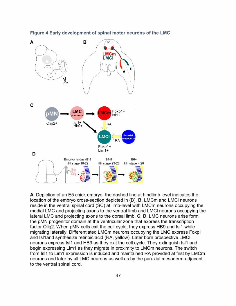
47
Figure 4 Early development of spinal motor neurons of the LMC
A. Depiction of an E5 chick embryo, the dashed line at hindlimb level indicates the location of the embryo cross-section depicted in (B). B. LMCm and LMCl neurons reside in the ventral spinal cord (SC) at limb-level with LMCm neurons occupying the medial LMC and projecting axons to the ventral limb and LMCl neurons occupying the lateral LMC and projecting axons to the dorsal limb. C, D. LMC neurons arise form the pMN progenitor domain at the ventricular zone that express the transcription factor Olig2. When pMN cells exit the cell cycle, they express HB9 and Isl1 while migrating laterally. Differentiated LMCm neurons occupying the LMC express Foxp1 and Isl1and synthesize retinoic acid (RA, yellow). Later born prospective LMCl neurons express Isl1 and HB9 as they exit the cell cycle. They extinguish Isl1 and begin expressing Lim1 as they migrate in proximity to LMCm neurons. The switch from Isl1 to Lim1 expression is induced and maintained RA provided at first by LMCm neurons and later by all LMC neurons as well as by the paraxial mesoderm adjacent to the ventral spinal cord.
48
8.1 LMC neuron differentiation
LMC neurons differentiate into two subtypes, neurons occupying the medial
portion of the LMC (LMCm), project axons to the ventral portion of developing limbs,
neurons occupying the lateral portion of the LMC (LMCl), project axons into the dorsal
part of the developing limb (Figure 4B) (Landmesser, 1978b). LMCm neurons are
generated first, the later born LMCl neurons migrate past LMCm neurons and settle at
the most ventrolateral position of the neural tube, adjacent to LMCm neurons (Figure
4C, D) (Hollyday and Hamburger, 1977). LMC subtype specification involves the
differential expression of LIM homeodomain transcription factors. LMCm and LMCl
neurons express Isl1 and Isl2 as they exit the cell cycle. While LMCm neurons retain
the expression of Isl1/2, LMCl neurons replace expression of Isl1 with Lim1 as LMCl
neurons migrate past LMCm neurons (Figure 4C, D) (Tsuchida et al., 1994). Retinoic
acid signaling provided by LMCm neurons as well as by the paraxial mesoderm
adjacent to the neural tube is necessary for LMCl neurons to switch from Isl1 to Lim1
expression (Figure 4C, D) (Sockanathan and Jessell, 1998; Ji et al., 2006). Lim1 and
Isl1 are cross repressive, overexpression of either in chick LMC neurons leads to the
loss of the other (Kania and Jessell, 2003). Lim1 and Isl1 expression contributes to
the settling positions of LMC neurons as evident by the medialization and
lateralization of LMC neurons overexpressing Isl1 and Lim1 respectively (Kania and
Jessell, 2003).
Later in development, LMC neurons become subdivided into motor pools with
each pool innervating a specific muscle (Landmesser, 2001). Muscle innervation
49
occurs in a topographic fashion, the relative position of muscles is mirrored by the
relative positions of the motor pools they are innervated by (Landmesser, 2001). The
mechanisms behind MN pool specification are still not fully understood but also relies
in part on the combinatorial expression of Hox proteins and Cadherins (Dasen et al.,
2005; Demireva et al., 2011; Lacombe et al., 2013).
9 LMC axon guidance
Evidence that the tissue targeted by LMC axons is instructive in guiding LMC
neurons was first provided by transplantation experiments. When short sections of
LMC containing neural tube are reversed on their anterior-posterior axis, thereby
increasing the distance between LMC neurons and their original targets, LMC axons
retain their ability to find their way to their original targets (Lance-Jones and
Landmesser, 1981). This ability is lost when longer sections of neural tube are
flipped, suggesting that molecular cues in the tissue surrounding the neurons are
instructive in guiding LMC axons, although limited in the distance they can do so
(Lance-Jones and Landmesser, 1981). Furthermore, when limb buds are rotated on
their dorso-ventral axis prior to innervation, LMCm and LMCl axons retain their ability
to innervate the ventral and dorsal limb respectively (Ferguson, 1983). These findings
were later confirmed through genetic manipulations in mice that alter the dorso-
ventral patterning of the limb. In the absence of the LIM homeodomain transcription
factor Lmx1b., dorsal limb mesenchyme adopts a ventral identity, in such mutants,
LMC axons innervate the limb in a seemingly random fashion (Chen et al., 1998;
50
Kania et al., 2000; Kania and Jessell, 2003). Conversely, loos of BMP signaling via
the loss of bmpr1A, ventral limb mesenchyme adopts a dorsal identity. The
dorsalization of the ventral limb compartment results in the innervation of LMCl axons
in both dorsal compartments and a failure of LMCm axons to innervate the limbs (Ahn
et al., 2001; Luria and Laufer, 2007; Luria et al., 2008). Genetic ablation of Lim1 also
results in LMCl axons innervating the limbs seemingly randomly as seen in Lmx1b
mutants (Kania et al., 2000). It is therefore apparent that LIM homeodomain
transcription factors expressed by LMC neurons as well as by limb mesenchyme is
crucial in providing the means by which LMC axons find their targets (Kania et al.,
2000).
LMC axon guidance occurs in a stepwise fashion, first, LMC axons must exit
the spinal cord through the ventral root. A subset of Boundary cap (BC) cells, derived
from neural crest cells, settle adjacent to ventral root (Niederlander and Lumsden,
1996; Golding and Cohen, 1997). BC cells are required for maintaining LMC
columnar integrity, in their absence, LMC neurons evade the spinal cord through the
ventral root (Vermeren et al., 2003). At the lumbar level of the spinal cord, the
expression of Neuropilin-2 and PlexinA1/A2 receptor complexes by motor neurons
contributes to their repulsion from BC cells expressing Sema6a (Bron et al., 2007). At
the brachial level, loss of Netrin-5 expression by BC cells and loss of Dcc by LMC
neurons also results in MNs evading the spinal cord through the ventral root (Garrett
et al., 2016).
51
LMC axons exiting the ventral root extend in tight fascicules, the cell adhesion
molecules L1 and NCAM as well as polysialic acid contribute in fasciculating LMC
axons (Tang et al., 1992; Tang et al., 1994). Peripheral nerves, including sensory and
motor axons, are channeled into the anterior portion of the somites, giving the
peripheral nerves a segmented appearance (Keynes and Stern, 1984).
Chemorepellents expressed in the posterior and chemoattractant expressed in the
anterior portion of the somites have been proposed to restrict nerve outgrowth
(Oakley and Tosney, 1991; Fredette and Ranscht, 1994; Krull et al., 1995; Ebens et
al., 1996; Wang and Anderson, 1997; Vermeren et al., 2000; Tzarfati-Majar et al.,
2001; Roffers-Agarwal and Gammill, 2009). Loss of Sema3A in the forelimb
mesenchyme, or loss of its receptor neuropilin-1 (Npn-1) in LMC neurons, results in
fasciculation deficiencies as well as premature innervation of the limb (Huber et al.,
2005).
10 Dorso-ventral LMC guidance
Once LMC axons extend past the somites, axons from a common plexus
converge into a tight fascicule that extends to the base of the limbs. At the base of the
limb, LMC axons are confronted to project either dorsally (LMCl axons) or ventrally
(LMCm axons) (Figure 4B). Several guidance cue families have been implicated in
this binary choice, a review of the literature describing the mechanisms underlying
this seemingly simple guidance decision will be described below (Table 1).
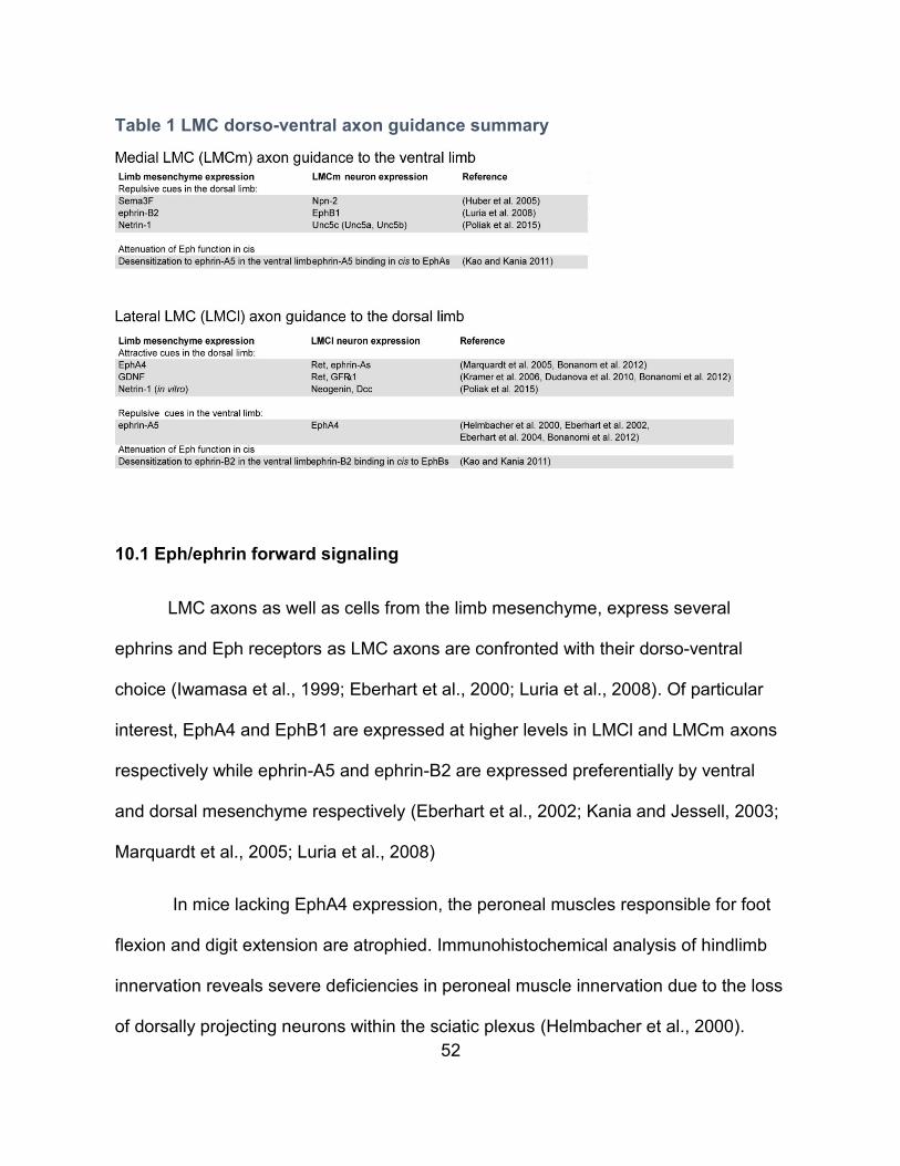
52
Table 1 LMC dorso-ventral axon guidance summary
10.1 Eph/ephrin forward signaling
LMC axons as well as cells from the limb mesenchyme, express several
ephrins and Eph receptors as LMC axons are confronted with their dorso-ventral
choice (Iwamasa et al., 1999; Eberhart et al., 2000; Luria et al., 2008). Of particular
interest, EphA4 and EphB1 are expressed at higher levels in LMCl and LMCm axons
respectively while ephrin-A5 and ephrin-B2 are expressed preferentially by ventral
and dorsal mesenchyme respectively (Eberhart et al., 2002; Kania and Jessell, 2003;
Marquardt et al., 2005; Luria et al., 2008)
In mice lacking EphA4 expression, the peroneal muscles responsible for foot
flexion and digit extension are atrophied. Immunohistochemical analysis of hindlimb
innervation reveals severe deficiencies in peroneal muscle innervation due to the loss
of dorsally projecting neurons within the sciatic plexus (Helmbacher et al., 2000).
53
Furthermore, overexpression of EphA4 in chick lumbar LMC neurons results in LMCm
axons misprojecting into the dorsal limb (Eberhart et al., 2000). As stated previously,
the expression of Lim1 by LMCl is required for their guidance, absence of Lim1 in
LMCl results in randomized axon projections (Kania and Jessell, 2003). Mice lacking
Lim1 expression show lower levels of EphA4 expression in LMCl neurons and
overexpression of Lim1 in the chick results in an increase in EphA4 expression in
LMC neurons (Table 1) (Kania and Jessell, 2003)
Conversely, LMCm neurons expressing EphB1, are repulsed from ephrin-B2
enriched in the dorsal limb (Luria et al., 2008). In ephrin-B2 conditional KO mice,
LMCm neurons misproject axons into the dorsal limb and the overexpression of
ephrin-B2 in the ventral chick limb bud results in the misprojection of LMCm neurons
to the dorsal limb mesenchyme (Table 1) (Luria et al., 2008).
10.2 Eph/ephrin reverse signaling
The above described a requirement for Eph/ephrin forward signaling,
Eph/ephrin reverse signaling has also been described in LMC axon guidance. As
previously mentioned, ephrin-As are GPI-anchored and lack an intracellular signaling
domain. Therefore, signaling downstream of ephrinAs in cis, requires ephrinA
coreceptors. In RGCs, the neurotrophic receptors TrkB and p75 can interact with
ephrin-As in cis and are required for EphA induced reduction in axon branching and
axon repulsion respectively (Lim et al., 2008; Marler et al., 2008).As LMC axons are
confronted with their dorso-ventral choice, LMC neurons express ephrin-A5/A2 while
54
EphA4 is expressed in dorsal limb mesenchyme (Iwamasa et al., 1999; Eberhart et
al., 2000; Kania and Jessell, 2003). LMCl axons are attracted to EphAs in vitro and a
subset of LMCl axons misproject ventrally in mice carrying limb-specific EphA4
deletions (Table 1) (Marquardt et al., 2005; Dudanova et al., 2012). Although LMCl
neurons express p75 and TrkB, theses receptors are not required in conveying
Eph/ephrin reverse signaling in LMCl guidance (Bonanomi et al., 2012). The
transmembrane tyrosine kinase receptor Ret interacts with ephrin-A5/A2 and was
identified as an obligate coreceptor for EphA/ as seen in RGCs ephrin-A reverse
signaling in LMCl guidance (Table 1) (Bonanomi et al., 2012). Contrary to RGCs,
EphA/ephrin-A reverse signaling in LMCl axon guidance is attractive rather than
repulsive, this may be the result of ephrin-A5 associating with Ret rather than with
p75 or TrkB (Bonanomi et al., 2012). The mechanisms governing the preferential
association of ephrin-A5 with specific coreceptors despite their coexpression remains
unknown (Bonanomi et al., 2012).
The regulation of both EphA4/ephrin-A forward and reverse signaling,
occurring simultaneously in parallel, was proposed to rely on the lateral segregation
of EphA and ephrin-As into distinct membrane domains in LMCl growth cones
(Dudanova et al., 2012). To my knowledge, no requirement for EphB/ephrinB reverse
signaling has been ascribed in LMCm axon guidance to date.
During vertebrate development, sensory neuron axons trail behind MN axons
and use them as scaffolds to reach their targets. Mutations causing MN axon
guidance defects can result in sensory axons aberrantly extending over misrouted
55
MN axons (Gallarda et al., 2008; Wang et al., 2011; Wang et al., 2014). The
requirement for MN axon tracts for sensory axons may be a consequence of motor
circuits arising prior to sensory circuits during evolution (Wang et al., 2014). Sensory
axons expressing ephrin-As track along epaxial MN axons expressing EphA3/4. Loss
of EphA3/4 in MNs results in sensory axon defects that can be rescued by the
expression of truncated EphA4, incapable of forward signaling. These results suggest
that sensory axons employ EphA/ephrin-A reverse signaling to navigate over MN
axons (Wang et al., 2011)
10.3 Eph/ephrin cis attenuation
RGC neurons coexpress EphAs and ephrin-As, in vitro, overexpression of
ephrin-As in RGCs decreases their sensitivity to exogenous ephrin-As. Conversely,
the removal of endogenous ephrin-As through PI-PLC treatment sensitizes RGCs to
exogenous ephrin-As (Hornberger et al., 1999). Ephrins bind in cis and trans on
different Eph domains. Ephrin-As binding in cis inhibits EphA tyrosine
phosphorylation, possibly by blocking Eph receptor clustering (Carvalho et al., 2006).
In addition to being expressed in the ventral limb mesenchyme, ephrin-A5 is
also expressed by LMCm neurons that navigate within the ventral limb. Conversely,
ephrin-B2 expressed by the dorsal limb mesenchyme, is expressed in LMCl neurons
(Table 1) (Kao and Kania, 2011). Knockdown of ephrin-A5 and ephrin-B2 in chick
LMC neurons via in ovo electroporation of siRNAs results in misprojections of LMCm
and LMCl axons respectively (Kao and Kania, 2011). In LMCm neurons, expression
56
of ephrin-A5 at the growth cone is thought to desensitize LMCm axons expressing
EphA receptors to ephrin-A5 expressed in the ventral limb. Conversely, expression of
ephrin-B2 on LMCl growth cones desensitizes them to ephrin-B2 in the dorsal limb
(Table 1) (Kao and Kania, 2011). Thus Eph/ephrin cis attenuation enables LMC
axons to fine-tune their responsiveness to exogenous ephrins through the expression
of endogenous ephrins.
10.4 GDNF / GFRa1/RET
In EphA4 mutant mice, the severity of the peroneal nerve thinning varies
depending on the mouse genetic background and epha4 allele studied, suggesting
that their may be other guidance cues instructing LMCl axons (Helmbacher et al.,
2000; Kullander et al., 2001; Leighton et al., 2001). Glial-cell-line-derived neurotrophic
factor (GDNF) promotes axon outgrowth, synaptogenesis and survival in various
neurons (Ibanez and Andressoo, 2017). Secreted GDNF binds to GDNF Family
Receptor alpha 1 (GFRα1), a GPI-anchored receptor that like ephrin-A5, requires
association with coreceptors such as Ret (Airaksinen and Saarma, 2002). LMC
neurons express GFRα1 as well as Ret while GDNF is expressed in the dorsal limb
mesenchyme, adjacent to the dorso-ventral branchpoint (Kramer et al., 2006). Mice
lacking GDNF have dorsal hindlimb innervation deficiencies with LMCl axons
aberrantly innervating the ventral limb, reminiscent of the LMCl guidance defects
seen in EphA4 deficient mice (Table 1) (Kramer et al., 2006). In vitro, GDNF
enhances the outgrowth of LMCm and LMCl axons in a concentration dependent
57
manner, but only LMCl axons show chemotaxis towards GDNF. These results
suggest that the LMCl axon guidance defects arise from a lack of attraction rather
than resulting from an lack of outgrowth (Dudanova et al., 2010). The requirement for
Ret in LMCl guidance is two-fold, it conveys EphA-ephrin-A reverse signaling as well
as attraction to GDNF via interactions with GFRα1 (Table 1). In vitro, exposure to low
doses of GDNF, strongly enhances EphA induced LMCl axon outgrowth (Bonanomi
et al., 2012). GDNF treatment results in the re-localization of Ret into lipid rafts
containing ephrin-A5 , this was proposed to contribute to the GDNF induced
sensitization of LMCl axons towards EphAs (Bonanomi et al., 2012).
10.5 Npn-2/Sema3F
In chick and mice forelimbs, the repulsive cue Sema3F is expressed in the
dorsal limb mesenchyme while its receptor Npn-2 is expressed in a subset of LMCm
neurons (Huber et al., 2005). In Mice lacking either Npn-2 or Sema3F, a subset of
LMCm axons aberrantly innervate the dorsal forelimb (Table 1) (Huber et al., 2005).
Furthermore, overexpression of Npn-2 in the chick, via in ovo electroporation, results
in LMCl axons innervating the ventral forelimb (Huber et al., 2005). The expression
pattern of Npn-2 and Sema3F in lumbar LMC neurons and in hindlimb mesenchyme
suggests that Npn-2/Sema3F are not implicated in guiding lumbar level LMC neurons
(A.K. unpublished comment).
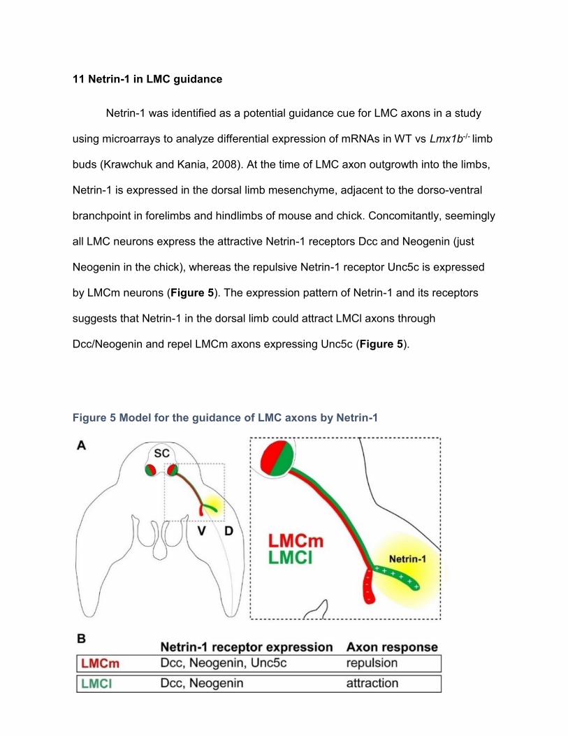
58
11 Netrin-1 in LMC guidance
Netrin-1 was identified as a potential guidance cue for LMC axons in a study
using microarrays to analyze differential expression of mRNAs in WT vs Lmx1b-/- limb
buds (Krawchuk and Kania, 2008). At the time of LMC axon outgrowth into the limbs,
Netrin-1 is expressed in the dorsal limb mesenchyme, adjacent to the dorso-ventral
branchpoint in forelimbs and hindlimbs of mouse and chick. Concomitantly, seemingly
all LMC neurons express the attractive Netrin-1 receptors Dcc and Neogenin (just
Neogenin in the chick), whereas the repulsive Netrin-1 receptor Unc5c is expressed
by LMCm neurons (Figure 5). The expression pattern of Netrin-1 and its receptors
suggests that Netrin-1 in the dorsal limb could attract LMCl axons through
Dcc/Neogenin and repel LMCm axons expressing Unc5c (Figure 5).
Figure 5 Model for the guidance of LMC axons by Netrin-1
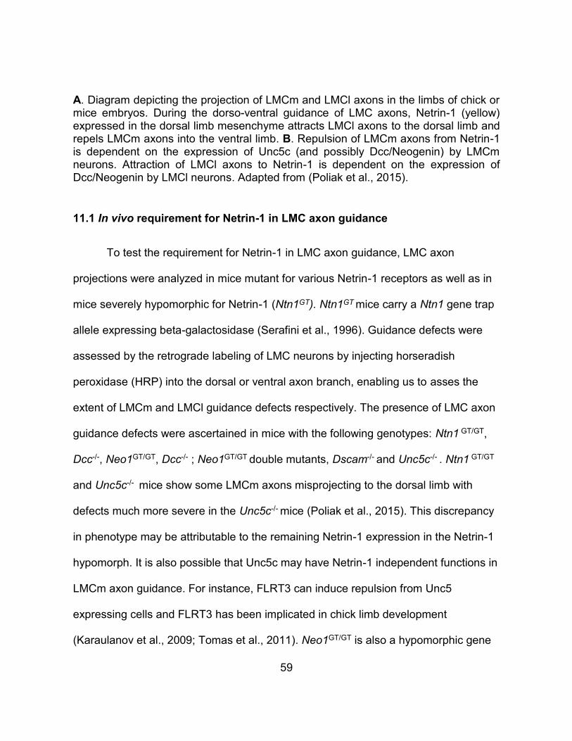
59
A. Diagram depicting the projection of LMCm and LMCl axons in the limbs of chick or mice embryos. During the dorso-ventral guidance of LMC axons, Netrin-1 (yellow) expressed in the dorsal limb mesenchyme attracts LMCl axons to the dorsal limb and repels LMCm axons into the ventral limb. B. Repulsion of LMCm axons from Netrin-1 is dependent on the expression of Unc5c (and possibly Dcc/Neogenin) by LMCm neurons. Attraction of LMCl axons to Netrin-1 is dependent on the expression of Dcc/Neogenin by LMCl neurons. Adapted from (Poliak et al., 2015).
11.1 In vivo requirement for Netrin-1 in LMC axon guidance
To test the requirement for Netrin-1 in LMC axon guidance, LMC axon
projections were analyzed in mice mutant for various Netrin-1 receptors as well as in
mice severely hypomorphic for Netrin-1 (Ntn1GT). Ntn1GT mice carry a Ntn1 gene trap
allele expressing beta-galactosidase (Serafini et al., 1996). Guidance defects were
assessed by the retrograde labeling of LMC neurons by injecting horseradish
peroxidase (HRP) into the dorsal or ventral axon branch, enabling us to asses the
extent of LMCm and LMCl guidance defects respectively. The presence of LMC axon
guidance defects were ascertained in mice with the following genotypes: Ntn1 GT/GT,
Dcc-/-, Neo1GT/GT, Dcc-/- ; Neo1GT/GT double mutants, Dscam-/- and Unc5c-/- . Ntn1 GT/GT
and Unc5c-/- mice show some LMCm axons misprojecting to the dorsal limb with
defects much more severe in the Unc5c-/- mice (Poliak et al., 2015). This discrepancy
in phenotype may be attributable to the remaining Netrin-1 expression in the Netrin-1
hypomorph. It is also possible that Unc5c may have Netrin-1 independent functions in
LMCm axon guidance. For instance, FLRT3 can induce repulsion from Unc5
expressing cells and FLRT3 has been implicated in chick limb development
(Karaulanov et al., 2009; Tomas et al., 2011). Neo1GT/GT is also a hypomorphic gene
60
trap allele and has variable residual Neogenin expression (Bae et al., 2009). It is
therefore possible that the residual Neogenin expression in Neo1GT/GT mice is
sufficient for attracting LMCl axons towards Netrin-1. Alternatively, Netrin-1 may be
required for LMCl guidance events other than the dorso-ventral branch choice, such
as the rostral-caudal extend of the dorsal branch. Unlike LMCl axons which are
attracted to EphA and GDNF in the dorsal limb, no attractive cue has been attributed
to guiding LMCm axons. LMCm axons may therefore by more dependent upon
Netrin-1 than LMCl axons (Table 1). Since Ret is required for both EphA and GDNF
attraction of LMCl axons, perhaps the involvement Netrin-1 in LMCl axon guidance in
vivo could be revealed by comparing Ret-/-; Ntn1 GT/GT double mutants to Ret-/- single
mutants (Bonanomi et al., 2012).
11.2 LMCm and LMCl axons respond to Netrin-1 in vitro
In vitro stripe assay experiments were carried out to assess if Netrin-1 can
directly influence the behavior of LMC axons. Stripe assays consist of challenging
axonal outgrowth of neurons cultured upon alternating stripes containing different
proteins, followed by a quantitative assessment of stripe choice (Weschenfelder et al.,
2013). When chick LMC explants are challenged to grow over either a control stripe
containing the Fc fragment of IgG (Fc) or Netrin-1, LMCl axons show a clear
preference for Netrin-1 containing stripes whereas LMCm axons avoid Netrin-1
stripes (Poliak et al., 2015). To assess the requirement for Unc5c in LMCm axons
avoidance of Netrin-1 stripes, stripe assays were carried out with LMC explants
electroporated with Unc5c siRNAs. Knockdown of Unc5c results in LMCm axons no
61
longer avoiding Netrin-1 stripes. Avoidance of Netrin-1 stripes can be rescued by
overexpressing mUnc5c in conjunction with Unc5c siRNAs, ruling out siRNA induced
off target effects in enabling Netrin-1 loss of avoidance (Poliak et al., 2015).
12 Synergy between Netrin-1 and ephrins in LMC guidance
We have demonstrated that both LMCm and LMCl axons are responsive to
Netrin-1 and ephrins, to determine whether Netrin-1 and ephrin act additively or
synergistically on LMC axon guidance, stripe-assay were performed. When LMCm
axons are challenged to grow over stripes containing a low concentration of Netrin-1
vs. Fc or a low concentration of ephrin-B2 vs. Fc, no outgrowth preference can be
seen (Figure 8B, C). Strikingly, when LMCm axons are challenged to grow over
stripes containing a combination of low concentrations of (Netrin-1+ephrin-B2) vs. Fc,
LMCm axons show a robust preference for Fc stripes (Figure 8D) (Poliak et al., 2015)
. Theses results suggest that Netrin-1 and ephrin-B2 are integrated synergistically by
LMCm axons. Similarly, when LMCl axons are challenged to grow over stripes
containing a low concentration of Netrin-1 vs. Fc or a low concentration of ephrin-A5
vs. Fc, no outgrowth preference can be seen (Figure 8F, G). When LMCl axons are
challenged by alternating stripes containing a low concentration of Netrin-1 vs. a low
concentration of ephrin-A5, LMCl axons show a robust preference for Netrin-1
containing stripes, suggesting synergy between Netrin-1 and ephrin-A5 in LMCl
guidance (Figure 8H) (Poliak et al., 2015) .
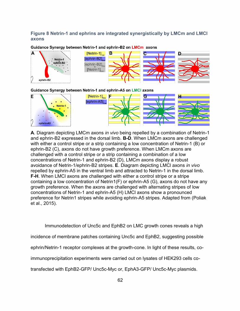
62
Figure 8 Netrin-1 and ephrins are integrated synergistically by LMCm and LMCl axons
A. Diagram depicting LMCm axons in vivo being repelled by a combination of Netrin-1 and ephrin-B2 expressed in the dorsal limb. B-D. When LMCm axons are challenged with either a control stripe or a strip containing a low concentration of Netrin-1 (B) or ephrin-B2 (C), axons do not have growth preference. When LMCm axons are challenged with a control stripe or a strip containing a combination of a low concentrations of Netrin-1 and ephrin-B2 (D), LMCm axons display a robust avoidance of Netrin-1/ephrin-B2 stripes. E. Diagram depicting LMCl axons in vivo repelled by ephrin-A5 in the ventral limb and attracted to Netrin-1 in the dorsal limb. F-H. When LMCl axons are challenged with either a control stripe or a stripe containing a low concentration of Netrin1(F) or ephrin-A5 (G), axons do not have any growth preference. When the axons are challenged with alternating stripes of low concentrations of Netrin-1 and ephrin-A5 (H) LMCl axons show a pronounced preference for Netrin1 stripes while avoiding ephrin-A5 stripes. Adapted from (Poliak et al., 2015).
Immunodetection of Unc5c and EphB2 on LMC growth cones reveals a high
incidence of membrane patches containing Unc5c and EphB2, suggesting possible
ephrin/Netrin-1 receptor complexes at the growth-cone. In light of these results, co-
immunoprecipitation experiments were carried out on lysates of HEK293 cells co-
transfected with EphB2-GFP/ Unc5c-Myc or, EphA3-GFP/ Unc5c-Myc plasmids.
63
Interestingly, anti-GFP antibody precipitated Un5c when cells were transfected
EphB2-GFP but not EphA3-GFP. Furthermore, treatment of HEK293 cells with
ephrin-B2 increased the association between Ephb2-GFP and Unc5c-Myc and this
association is decreased when cells are transfected with EphB2-KD-GFP, a kinase
dead version of EphB2. In line with these results, in a cell line stably expressing
EphB2 and transfected with Unc5c-Myc, Netrin-1 treatment together with ephrin-B2
increases the amount phosphorylated EphB2 in comparison with ephrin-B2 treatment
alone. Thus, the synergistic activity of Netrin-1 with ephrin-B2 seen in LMCm axons in
the context of the stripe-assay and the collapse assay may rely on an ephrin-B2
enhancement of EphB2/Unc5c interaction and the Netrin-1 enhancement of EphB2
phosphorylation.
Considering the evidence for SFKs involvement in both Netrin-1 and Eph
downstream signaling, SFKs were considered as a possible molecular link enabling
synergy between the two pathways (Zisch et al., 1998; Knoll and Drescher, 2004; Liu
et al., 2004; Williams et al., 2006; Kao et al., 2009). To test this idea, levels of SFK-
activating phosphorylation (pSFK) were assay in collapsed growth cones of cultured
LMC explants that had been exposed to ephrin-B2 and Netrin-1 individually or in
combination. The simultaneous treatment with both cues resulted in an increase in
pSFK levels in collapsed LMC growth cones. Furthermore, blocking SFK function
reduced growth cone collapse of LMC growth cones exposed to low concentrations of
Netrin + ephrin-B2. In the context of the stripe assay, blocking SFK function
attenuates LMCm axon stripe choice avoidance of stripes containing either cue alone
64
or in combination. Although these experiments demonstrate a requirement for SFKs
in the combined activity of Netrin-1 and ephrin-B2 on LMC neurons, the loss of
synergistic responsiveness when blocking SFKs could result from its requirement in
individual pathways rather than being at the root of the supra-additive effects.
Our results demonstrate that both LMCm and LMCl neurons respond
synergistically to Netrin-1 and ephrins. In LMCl axons, Netrin-1/ephrin-A5 synergize
attractive-repulsive (opposing) forces, whereas in LMCm axons, Netrin-1/ephrin-B2
synergize repulsive-repulsive (congruent) forces (Poliak et al., 2015). It is tempting to
speculate that Netrin-1/ephrin synergy likely arises from a common mechanism in
LMCm and LMCl neurons but our experiments have not addressed this issue directly.
It also remains to be determined whether Netrin-1 sensitizes axons to ephrins or if
ephrins sensitizes axons to Netrin-1 or rather by mutual sensitization of both
pathways. In Chapter III - C, I will provide evidence that furthers our understanding of
the mechanism behind Netrin-1/ephrin synergy by focussing on the effects of Netrin-1
and ephrin-A5 on receptor dynamics in LMC growth cones.

65
CHAPTER II - MATERIALS AND METHODS

66
Chick tissue
Fertilized eggs (Couvoir Simetin, Mirabel, QC) were incubated (Lyon
Technologies, model PRFWD) at 39 ◦C according to standard protocols (Hamburger
and Hamilton, 1951). Embryos were cut transversely at approximately 3 segments
anterior to the hindlimbs, and fixed in a 4% solution of cold paraformaldehyde (Sigma)
in PB (1.0 M phosphate buffer: 20.66% (w/v) Na2HPO4·7H2O, 3.2% (w/v),
NaH2PO4·H2O, in H2O) for 75 min at 4 ◦C. Embryos were then washed 3
consecutive times in PBS followed by 3 washes of 5 min at 4 ◦C on a slowly shaking
mixer (VMR International), and cryoprotected with 30% sucrose in PB overnight. The
next day, embryos were embedded in O.C.T. (Sakura Finetek), and stored at −80 ◦C.
12 μm sections were collected using a Leica cryostat microtome (model CM3050S)
and stored at −80 ◦C.
In situ mRNA detection
In situ hybridizations were performed as described previously (Law et al.,
2016). cRNA probes were generated as follows: target sequence amplification
primers were designed using Primer3 version 0.4.0 software (Untergasser et al.,
2012) with a probe size set at 600–800 bp. One-step RT-PCR was performed
(QIAGEN) using the appropriate primers containing T7 polymerase promoters
(Invitrogen) to make and amplify cDNA template from mouse E11.5 pooled brain
RNA. The PCR product was purified by gel electrophoresis in 1% agarose gel and gel
extraction using QIAquick gel extraction kit (QIAGEN). The purified DNA was then
reamplified by PCR. The yield of DNA was estimated using the low DNA mass ladder
67
(Invitrogen) after gel electrophoresis. DIG-labeled RNA probes were synthesized by
in vitro transcription with T7 RNA polymerase using DIG RNA labeling kit (Roche). All
probes were verified by sequencing. Then, 12 μm tissue sections were fixed in 4%
PFA, rinsed 3x in PBS, treated with 1 μg/ml proteinase K (in 6.25 mM EDTA, pH 8.0,
Invitrogen + 50 mM Tris, pH 7.5; Fisher Scientific) for 15 min, post fixed in 4% PFA,
rinsed in PBS, and then acetylated for 10 min in a mixture of 6 ml of triethanolamine
(Sigma-Aldrich), 500 ml of distilled H2O, and 1.3 ml of acetic acid (Sigma-Aldrich).
Samples were then rinsed and incubated in hybridization buffer containing 50%
formamide (Sigma-Aldrich), 5μ SSC (20μSSC is 3 M NaCl, 0.3 M NaAc), 5x
Denhardt’s (Sigma-Aldrich), and 500 μg/ml salmon sperm DNA (Roche) inH2Ofor 2 h,
followed by overnight hybridization at 72°C with DIG-labeled probes at a
concentration of 2–5 ng/μl. After hybridization, samples were immersed in 5xSSC at
72°C, followed by 2 45 min washes in 0.2x SSC at 72°C and 1 5 min wash in 0.2x
SSC at room temperature. Tissues were then rinsed with B1 buffer containing 0.1 M
Tris, pH 7.5, and 0.15 M NaCl (Fisher Scientific) for 5 min, blocked with B2 buffer
(10% heat inactivated horse serum in B1) for 1 h at room temperature, and incubated
with anti-DIG antibody (1:5000 in B2; Roche) overnight at 4°C. Samples were then
rinsed with B1 and equilibrated with B3 buffer containing 0.1 M Tris, pH 9.5, 0.1 M
NaCl, and 0.05 M MgCl2 (Fisher Scientific). To detect bound anti-DIG antibodies,
samples were incubated with B4 buffer containing [100 mg/ml NBT, 50 mg/ml BCIP
(Roche), and 400 mM levamisol (Sigma-Aldrich) in B3 in the dark. The reaction was
stopped by immersion in H2O.
Chick and mouse section Immunohistochemistry
68
Sectioned tissue was allowed to air dry, and then washed with PBS, incubated
in blocking solution (1% heat inactivated horse serum in 0.1% Triton-X/PBS (Sigma))
for 5 min, followed by incubation overnight at 4 ◦C with primary antibodies diluted in
blocking solution. The following primary antibodies and dilutions were used: guinea-
pig anti-Foxp1 (1:16,000) (Dasen et al., 2008), sheep anti-GFP (1:1000) (Molecular
Probes), mouse anti-Neurofilament (3A10) (1:100), anti-Isl1 (39.3f7) (1:100)
(Developmental Studies Hybridoma Bank), rabbit anti-Lim1/2 (1:2500) (Tsuchida et
al., 1994), Goat anti-Neogenin (R&D). Samples were incubated overnight with
primary antibodies followed by three 5 min washes in PBS and 1 h incubation with the
appropriate secondary antibody. Slides were then rinsed several times with PBS and
mounted with Mowiol mounting medium (9.6%, w/v Mowiol (Calbiochem), 9.6% (v/v)
1 M Tris–HCl (Fisher Scientific), 19.2% (v/v) Glycerol (Fisher Scientific), in H2O). The
following secondary antibodies were used: Cy3-conjugated AffiniPure donkey anti-
rabbit and anti-mouse IgG (1:1000), Cy5-conjugated AffiniPure donkey anti-guinea-
pig IgG (1:500) (Jackson ImmunoResearch Laboratory), Alexa Fluo 488-conjugated
donkey anti-sheep and anti-mouse IgG (1:1000, Molecular Probes).
LMC Explant Immunohistochemistry
Prior to immunostaining, explants were fixed by replacing half of the culture
media with a 37°C solution of 4% PFA, 3% sucrose in PBS for 20 min. at RT and
washed repeatedly with PBS. Primary antibodies were incubated in blocking solution
(1% heat-inactivated horse serum in 0.1% Triton-X/PBS; Sigma) either 1 hour at 37°C
69
or overnight at 4°C. The following primary antibodies were used: goat anti-Neogenin
(1:300; R&D), rabbit anti-EphA4 (1:1000; Santa Cruz Biotechnology), rabbit anti-
Netrin1 (1:1000; Abcam), guinea-pig anti-Unc5c (1:500; Thomas Jessell lab.) , mouse
anti-Neuronal Class III β-Tubulin (Tuj1) (1:2000; Covance), mouse anti-chicken BEN
(1:00; Developmental Studies Hybridoma Bank). For surfaced enriched staining,
antibodies were added simultaneously with the cue treatment solution. Explants were
washed repeatedly with PBS prior to incubation with appropriate secondary
antibodies in blocking solution for 1 h at RT. The following secondary antibodies were
used: Cy3- (or Cy-5)-conjugated AffiniPure donkey anti-mouse (rabbit, goat, or guinea
pig) IgG (1:1000 for Cy3, 1:500 for Cy5 secondary antibodies; Jackson
ImmunoResearch Laboratory), Alexa Fluor 488 donkey anti-mouse (rabbit or sheep)
IgG (1:1000; Invitrogen). Explants were then washed repeatedly with PBS and
mounted with Mowiol.
Chick in ovo siRNA electroporation
Chick in ovo spinal cord electroporation was performed as described (Croteau
and Kania, 2011). For Figure 11-6, chick spinal cords were electroporated with the
pN2-eGFP expression plasmids and siRNAs (Invitrogen) at a 1:10 molar ratio at HH
st.18–19 and incubated at 39◦C until harvesting at either E4 (HH st. 23-24) or E5 (HH
st. 25–26). For Figure 25, Chick embryos were co-electroporated with a pN2-eGFP
expression plasmid and pCAGGS-mEphA4 (Gatto, 2014) at a 1:4 molar ratio, and
electroporated with pN2-chEphA4-GFP or pN1-chEphA4ΔICD- GFP (lacking
intracellular domain) for Figure 27 and Figure 29.
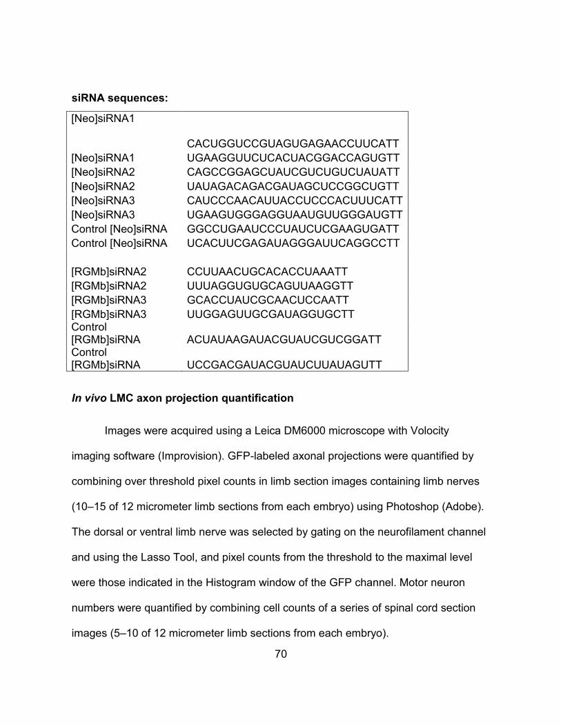
70
siRNA sequences:
[Neo]siRNA1 CACUGGUCCGUAGUGAGAACCUUCATT
[Neo]siRNA1 UGAAGGUUCUCACUACGGACCAGUGTT
[Neo]siRNA2 CAGCCGGAGCUAUCGUCUGUCUAUATT
[Neo]siRNA2 UAUAGACAGACGAUAGCUCCGGCUGTT
[Neo]siRNA3 CAUCCCAACAUUACCUCCCACUUUCATT
[Neo]siRNA3 UGAAGUGGGAGGUAAUGUUGGGAUGTT
Control [Neo]siRNA GGCCUGAAUCCCUAUCUCGAAGUGATT
Control [Neo]siRNA UCACUUCGAGAUAGGGAUUCAGGCCTT
[RGMb]siRNA2 CCUUAACUGCACACCUAAATT
[RGMb]siRNA2 UUUAGGUGUGCAGUUAAGGTT
[RGMb]siRNA3 GCACCUAUCGCAACUCCAATT
[RGMb]siRNA3 UUGGAGUUGCGAUAGGUGCTT Control [RGMb]siRNA ACUAUAAGAUACGUAUCGUCGGATT Control [RGMb]siRNA UCCGACGAUACGUAUCUUAUAGUTT
In vivo LMC axon projection quantification
Images were acquired using a Leica DM6000 microscope with Volocity
imaging software (Improvision). GFP-labeled axonal projections were quantified by
combining over threshold pixel counts in limb section images containing limb nerves
(10–15 of 12 micrometer limb sections from each embryo) using Photoshop (Adobe).
The dorsal or ventral limb nerve was selected by gating on the neurofilament channel
and using the Lasso Tool, and pixel counts from the threshold to the maximal level
were those indicated in the Histogram window of the GFP channel. Motor neuron
numbers were quantified by combining cell counts of a series of spinal cord section
images (5–10 of 12 micrometer limb sections from each embryo).
71
Explant culture and treatment
LMC explants were collected from st. 24-25 lumbar spinal cords and incubated
for about 18 hours as previously described (Kao and Kania, 2011). For drug
treatments, half of the motor neuron media in the explant cultures was replaced with
media containing the drug and incubated for 20 min. prior to cue treatment. The
following drugs were used: KT5720, KT5823, γ-Secretase Inhibitor IX (DAPT),
Anisomycin and SU6656 were purchased from Calbiochem. MG132, Chloroquine
diphosphate and the KYL peptide were purchased from Tocris. For cue treatment,
half of the culture media was replaced with media alone or media containing
recombinant mouse Netrin1, recombinant human ephrin-A5-Fc or Fc (R&D systems).
Prior to explant treatment, recombinant human ephrin-A5-Fc was preclustered in a
5:1 molar ratio with either mouse or goat anti-human Fc (Sigma-Aldrich) for 30 min. at
37°C.
In vitro stripe assay
In vitro stripe assay using explants of spinal motor columns were performed as
described (Kao and Kania, 2011). In brief, carpets of alternating stripes of Netrin-1,
ephrin-A5-Fc, or Fc only as controls were prepared using silicon matrices with micro-
well system (provided by Dr. Martin Bastmeyer’s laboratory). E5 chick spinal motor
column was dissected using sharp tungsten needles (World Precision Instruments)
and collected in motor neuron medium [Neurobasal (Invitrogen), B-27 supplement
(1:50, GIBCO), L-Glutamate (0.5 mM, Sigma-Aldrich), L-Glutamine (25mM, GIBCO),
72
and Penicillin-Streptomycin (1:100, Wisent)]. The excised motor column was then
trimmed into explants with the size of 1/4 width of motor column, and 20 explants
were plated on laminin (20 μg/ml, Invitrogen) coated culture dishes containing
different combinations of stripe carpets in motor neuron medium and incubated in
95% air and 5% CO2 at 37oC overnight. Following incubation, motor column explants
were fixed with 1:1 mixture of 4% paraformaldehyde (Sigma) and 30% sucrose in
PBS for 5 minutes followed by 4% paraformaldehyde for 5 minutes. After PBS
washes, explants were incubated with selected primary antibodies diluted in blocking
solution containing 20% serum in 0.3% Triton-X/PBS(Sigma) for 2 hrs at room
temperature (RT). Following PBS washes, explants were incubated with secondary
antibodies diluted in the same blocking solution for 2 hrs at RT.
Growth cone image acquisition and quantification:
Images were acquired using LSM700 and LSM710 Zeiss confocal
microscopes. Regions of interests (ROIs) delimitating growth cones and subsequent
analysis was done using Imagej. To assess fluorescence distribution within the
growth cones, a Matlab application was designed and programmed by Dr. Dominic
Fillion. The application divides growth cones into 100 bins, with bin 1 at the geometric
centre and bin 100 following the outer perimeter of the growth cone. The mean
fluorescence value for each bin was determined and plotted, bin#100 was omitted
from the analysis due to the region of interest (ROI) occasionally slightly over
representing the growth cone resulting in the absence of signal in bin#100. To
73
determine differences in fluorescence distribution regardless of differences in overall
fluorescence, the fluorescence value for each bin was divided by the mean
fluorescence value of bins 1-99 and termed relative fluorescence distribution. The
protein aggregate analysis was done using the particle analysis tool in Imagej. The
analysis of fluorescence overlap, the fluorescence signal in individual channels was
thresholded and an image representing signal overlap between two channels was
generated and quantified. To determine the change in overlap, the data was treated
as such ((% signal overlap with cue treatment) / (%signal overlap control treatment).
Statistical analysis
Data from the experimental replicate sets were evaluated using Microsoft
Excel and Aabel (Gigawiz). Unless stated otherwise, means were compared with
Student’s unpaired t-test with the threshold for statistical significance set at 0.05,
error-bars represent S.E.M. A minimum of three independent experiments were
carried out for each treatment condition and between 20 and 35 growth cones were
analysed in each experimental replicate

74
CHAPTER III - RESULTS
75
1. Neogenin in the responsiveness of LMCl axons to Netrin-1
Rationale
Upon my arrival in the Kania lab, my initial task was to investigate the
requirement for chick Neogenin in LMC axon guidance. By then, most experiments
implicating mouse genetics that would eventually be published in Poliak et al. had
been completed and the requirement for Netrin-1 in LMC axon guidance had been
established. In the chick, Netrin-1 related expression patterns and results obtained
from the misexpression of Unc5c in LMC neurons suggested that Netrin-1 could be
guiding chick LMC axons as it does in mice. The residual Neogenin expression in
NeogeninGT/GT mice and the possible redundancy between Dcc and Neogenin in LMC
axon guidance in mice prompted us to explore Neogenin function in the chick.
Neogenin is required for the responsiveness of LMCl axons towards Netrin-1
Through the stripe assay, we have previously demonstrated that chick LMCl
axons display attraction towards Netrin-1(Poliak et al., 2015). LMC neurons express
the attractive Netrin-1 receptor Neogenin as LMCl axons encounter Netrin-1
expressed in the Dorsal limb. To investigate the requirement for Neogenin in LMCl
axon responsiveness towards Netrin-1, a Neogenin antibody that antagonizes the
binding of Netrin-1 to Neogenin was added to stripe assays challenging LMCl axons
with either a control stripe or Netrin-1 stripes. In the presence of the Neogenin
antibody, LMCl axons loose their preference for Netrin-1 stripes, demonstrating the
necessity of Neogenin in responsiveness of LMCl axons to Netrin-1 (Figure 6).
76
Dcc rescues the loss of LMCl responsiveness to Netrin-1
In the chick spinal cord, knockdown of Neogenin by in ovo electroporation of an
shRNA construct directed against Neogenin results in commissural axon guidance
defects reminiscent of the Netrin1 and Dcc knock-out mice suggesting that chick
Neogenin can substitute for Dcc in mediating attraction towards Netrin-1 (Phan et al.,
2011). To ensure that the loss of LMCl axon responsiveness towards Netrin-1 in the
presence of Neogenin antibody was not due to off target effects, I attempted to rescue
responsiveness to Netrin-1 by overexpressing Dcc in LMCl axons in the presence of
Neogenin antibody. Overexpression of full-length rat Dcc but not a truncated rat Dcc,
lacking the cytoplasmic signaling tail (DCCDICD), rescued the preference of LMCl
axons for Netrin-1 stripes in the presence of Neogenin antibody (Figure 7). These
results suggest that the loss of LMCl axon responsiveness to Netrin-1 is not the result
of off-target effects of the anti-Neogenin antibody. It also confirms previous studies
suggesting that chick Neogenin could functionally substitute for Dcc in attraction
towards Netrin-1 (Phan et al., 2011).
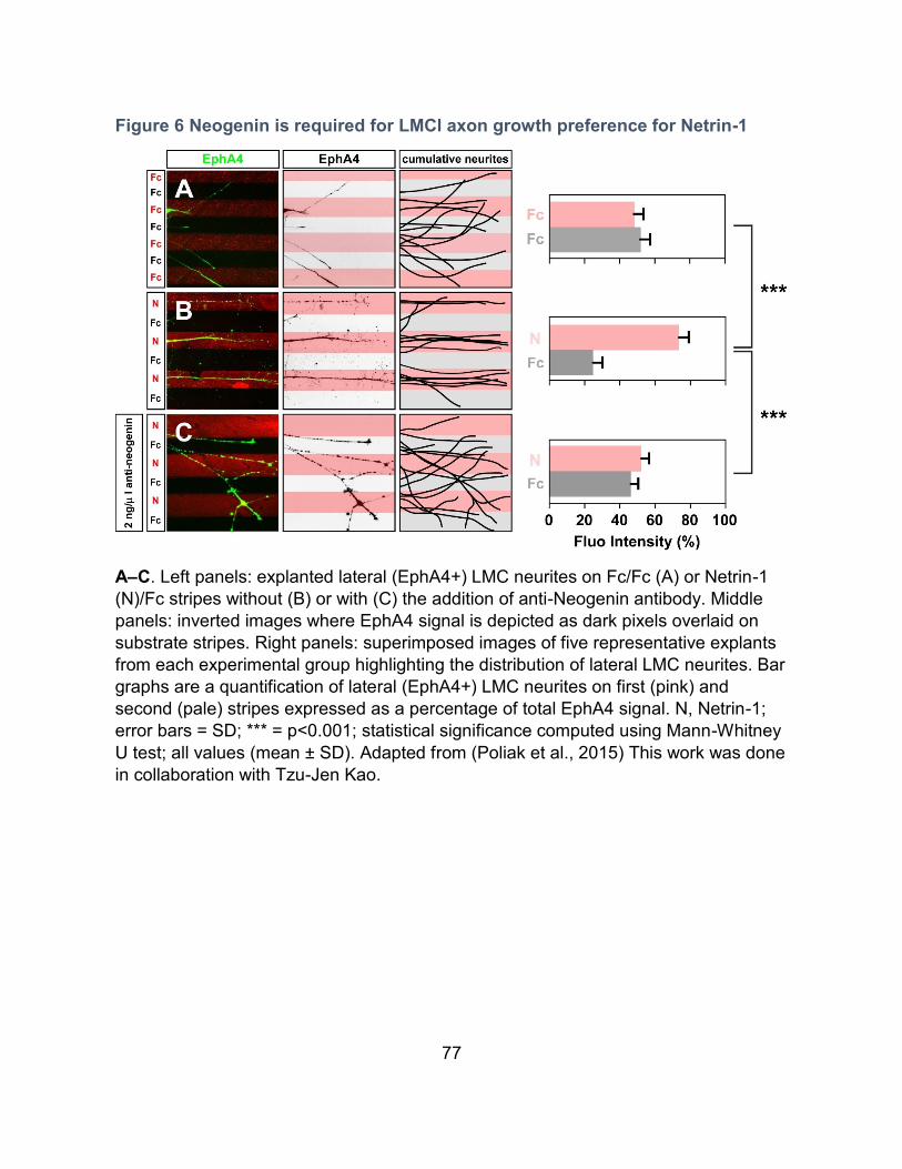
77
Figure 6 Neogenin is required for LMCl axon growth preference for Netrin-1
A–C. Left panels: explanted lateral (EphA4+) LMC neurites on Fc/Fc (A) or Netrin-1
(N)/Fc stripes without (B) or with (C) the addition of anti-Neogenin antibody. Middle
panels: inverted images where EphA4 signal is depicted as dark pixels overlaid on
substrate stripes. Right panels: superimposed images of five representative explants
from each experimental group highlighting the distribution of lateral LMC neurites. Bar
graphs are a quantification of lateral (EphA4+) LMC neurites on first (pink) and
second (pale) stripes expressed as a percentage of total EphA4 signal. N, Netrin-1;
error bars = SD; *** = p<0.001; statistical significance computed using Mann-Whitney
U test; all values (mean ± SD). Adapted from (Poliak et al., 2015) This work was done
in collaboration with Tzu-Jen Kao.
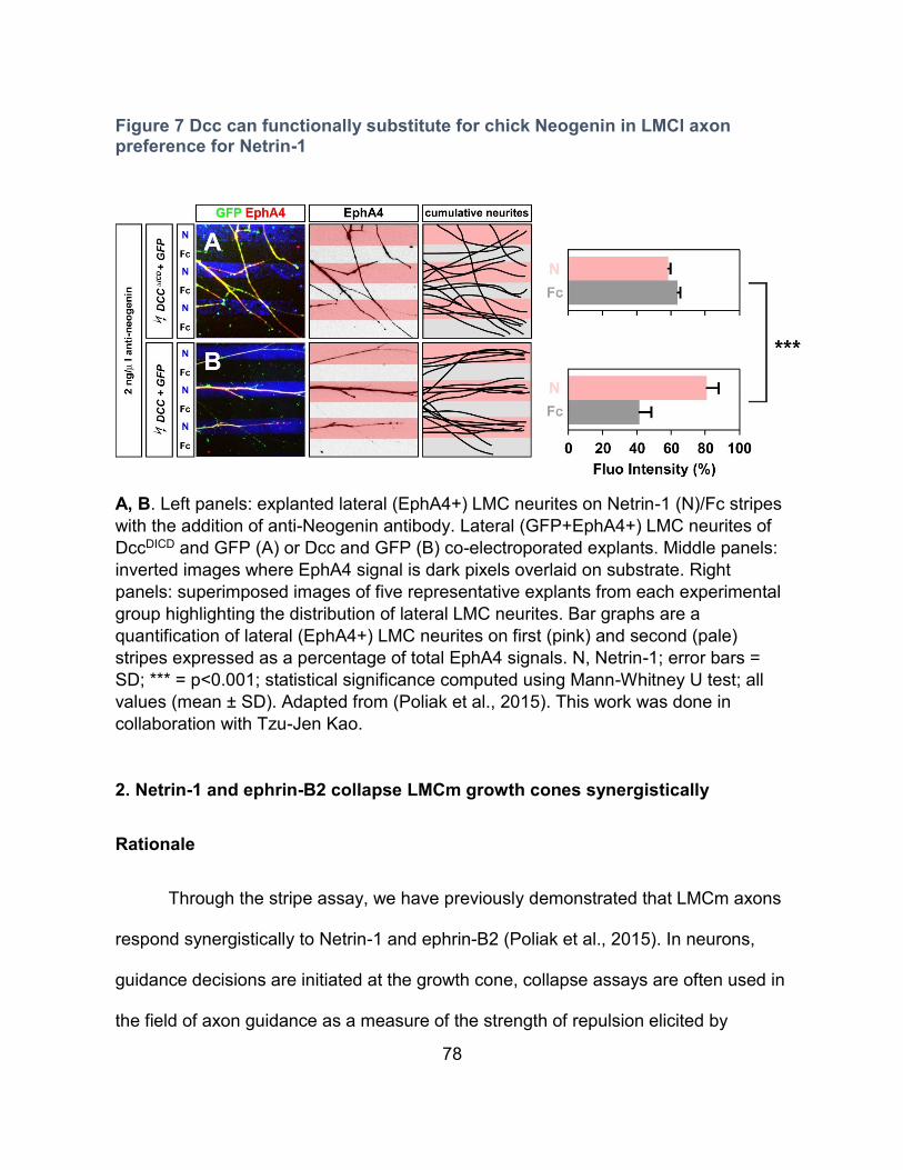
78
Figure 7 Dcc can functionally substitute for chick Neogenin in LMCl axon preference for Netrin-1
A, B. Left panels: explanted lateral (EphA4+) LMC neurites on Netrin-1 (N)/Fc stripes
with the addition of anti-Neogenin antibody. Lateral (GFP+EphA4+) LMC neurites of
DccDICD and GFP (A) or Dcc and GFP (B) co-electroporated explants. Middle panels:
inverted images where EphA4 signal is dark pixels overlaid on substrate. Right
panels: superimposed images of five representative explants from each experimental
group highlighting the distribution of lateral LMC neurites. Bar graphs are a
quantification of lateral (EphA4+) LMC neurites on first (pink) and second (pale)
stripes expressed as a percentage of total EphA4 signals. N, Netrin-1; error bars =
SD; *** = p<0.001; statistical significance computed using Mann-Whitney U test; all
values (mean ± SD). Adapted from (Poliak et al., 2015). This work was done in
collaboration with Tzu-Jen Kao.
2. Netrin-1 and ephrin-B2 collapse LMCm growth cones synergistically
Rationale
Through the stripe assay, we have previously demonstrated that LMCm axons
respond synergistically to Netrin-1 and ephrin-B2 (Poliak et al., 2015). In neurons,
guidance decisions are initiated at the growth cone, collapse assays are often used in
the field of axon guidance as a measure of the strength of repulsion elicited by
79
guidance cues. Since LMCm axons avoid both Netrin-1 and ephrin-B2, I performed
collapse assay experiments to determine the effects of these cues on LMCm growth
cones.
To monitor LMCm growth cones specifically, chicken LMC neurons were
electroporated with the medial LMC marker e[Isl1]::GFP prior to culture (Kao et al.,
2009). LMC explants were exposed to bath applied Netrin-1 and/or ephrin-B2 for 30
min., followed by a quantification of LMCm growth cone status (Figure 9). Whereas
exposure to low concentrations of either Netrin-1 or ephrin-B2 fails to induce LMCm
growth cone collapse, the simultaneous exposure to both cues results in robust
collapse (Figure 9A) (Poliak et al., 2015). These results suggest the both cues are
integrated synergistically within LMCm growth cones. Since ephrin-A5 and Netrin-1
have opposing effects on LMCl axons, a collapse assay paradigm may not be
adequate for investigating Netrin-1/ephrin-A5 LMCl axon guidance synergy.
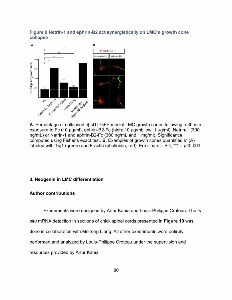
80
Figure 9 Netrin-1 and ephrin-B2 act synergistically on LMCm growth cone collapse
A. Percentage of collapsed e[Isl1]::GFP medial LMC growth cones following a 30 min exposure to Fc (10 µg/ml), ephrin-B2-Fc (high: 10 µg/ml; low: 1 µg/ml), Netrin-1 (300 ng/mL) or Netrin-1 and ephrin-B2-Fc (300 ng/mL and 1 mg/ml). Significance computed using Fisher’s exact test. B. Examples of growth cones quantified in (A) labeled with Tuj1 (green) and F-actin (phalloidin, red). Error bars = SD; *** = p<0.001.
3. Neogenin in LMC differentiation
Author contributions
Experiments were designed by Artur Kania and Louis-Philippe Croteau. The in
situ mRNA detection in sections of chick spinal cords presented in Figure 10 was
done in collaboration with Meirong Liang. All other experiments were entirely
performed and analyzed by Louis-Philippe Croteau under the supervision and
resources provided by Artur Kania.
81
Rationale
With the objective of revealing an in vivo requirement for Neogenin in LMC
axon guidance, Neogenin was knocked down (KD) in LMC neurons by way of chick in
ovo electroporation of siRNAs against Neogenin ([Neo]siRNA) (Croteau and Kania,
2011). KD of Neogenin resulted in alterations in LMC subtype differentiation, a
process that occurs prior and during the dorso-ventral LMC axon branch choice
(Landmesser, 1978a; Tosney and Landmesser, 1985). Neogenin/RGM signaling has
been implicated in neuronal differentiation in various regions of the CNS including the
midbrain, hindbrain, cortex and olfactory bulb (Matsunaga et al., 2006; O'Leary et al.,
2013; O'Leary et al., 2015; Kam et al., 2016). In the chick, Neogenin, as well as its
ligands RGMa and RGMb, are expressed by LMC neurons during their subtype
differentiation suggesting that Neogenin/RGM interactions are implicated in
differentiation of LMC neurons. We therefore decided to further investigate the
potential role of Neogenin in LMC differentiation.
Expression of Neogenin, RGMa and RGMb during LMC neuron development
To investigate the requirement for Neogenin in LMC development, in situ
mRNA detection of Neogenin as well as its ligands RGMa and RGMb was performed
in chick lumbar spinal cord sections. The developmental window in which LMC
neurons become post-mitotic and differentiate into their lateral and medial subtypes
occurs between chick embryonic (E) days 3-6 (Figure 10A) (Hollyday and
Hamburger, 1977; Landmesser, 1978b). At Hamburger-Hamilton stage (HH st.) 18,
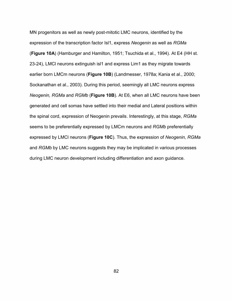
82
MN progenitors as well as newly post-mitotic LMC neurons, identified by the
expression of the transcription factor Isl1, express Neogenin as well as RGMa
(Figure 10A) (Hamburger and Hamilton, 1951; Tsuchida et al., 1994). At E4 (HH st.
23-24), LMCl neurons extinguish isl1 and express Lim1 as they migrate towards
earlier born LMCm neurons (Figure 10B) (Landmesser, 1978a; Kania et al., 2000;
Sockanathan et al., 2003). During this period, seemingly all LMC neurons express
Neogenin, RGMa and RGMb (Figure 10B). At E6, when all LMC neurons have been
generated and cell somas have settled into their medial and Lateral positions within
the spinal cord, expression of Neogenin prevails. Interestingly, at this stage, RGMa
seems to be preferentially expressed by LMCm neurons and RGMb preferentially
expressed by LMCl neurons (Figure 10C). Thus, the expression of Neogenin, RGMa
and RGMb by LMC neurons suggests they may be implicated in various processes
during LMC neuron development including differentiation and axon guidance.
83
Figure 10 Expression of Neogenin, RGMa and RGMb during LMC neuron development
In situ mRNA detection and immunohistochemistry on wild-type chick lumbar-level
spinal cord sections. A. At E3, (HH st. 18), post-mitotic LMC neurons (Isl1+) express
Neogenin and RGMa. B. At E4 (HH st. 23,24), when LMCl neurons begin expressing
Lim1, seemingly all LMC neurons express Neogenin, RGMa and RGMb. C. At E6,
Neogenin is expressed by seemingly all LMC neurons, RGMa is preferentially
expressed by LMCm neurons and RGMb is preferentially expressed by LMCl
neurons.
Knockdown of Neogenin alters LMC neuron subtype differentiation
To investigate the implication of Neogenin in LMC development, HH st. 17-18
embryos were electroporated with a combination of three siRNAs directed against
Neogenin ([Neo]siRNA), along with a GFP expression plasmid to select for
84
electroporated cells (Figure 11A) (Croteau and Kania, 2011). In situ mRNA detection
of Neogenin in sections of lumbar spinal cord of electroporated embryos shows lower
levels of Neogenin on the electroporated side of the spinal cord (Figure 11B). To
determine if the KD of Neogenin affects the total amount of LMC neurons generated,
the amount of cells expressing the generic LMC marker Foxp1 were counted and
compared to embryos electroporated with a GFP plasmid combined with a
scrammbled version of one the Neogenin siRNAs (controls) (Rousso et al., 2008).
Results indicate that the KD of Neogenin at E3 does not affect the generation of
generic LMC neurons (Figure 11C).
To investigate the possible involvement of Neogenin in LMC subtype
differentiation, a quantitative comparison of LMC subtype composition between
[Neo]siRNA and control embryos was carried out. KD of Neogenin results in an
increase in the relative proportion LMCm (Isl1+) neurons (Figure 11D, E).
Furthemore, the increase in Isl1+ neurons is accompanied by an decrease in LMC
neurons expressing the LMCl marker Lim1 (Figure 11F, G).
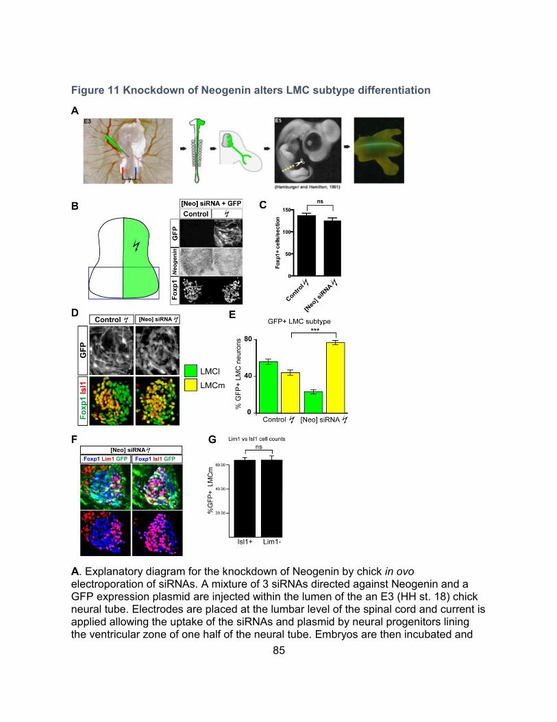
85
Figure 11 Knockdown of Neogenin alters LMC subtype differentiation
A. Explanatory diagram for the knockdown of Neogenin by chick in ovo electroporation of siRNAs. A mixture of 3 siRNAs directed against Neogenin and a GFP expression plasmid are injected within the lumen of the an E3 (HH st. 18) chick neural tube. Electrodes are placed at the lumbar level of the spinal cord and current is applied allowing the uptake of the siRNAs and plasmid by neural progenitors lining the ventricular zone of one half of the neural tube. Embryos are then incubated and
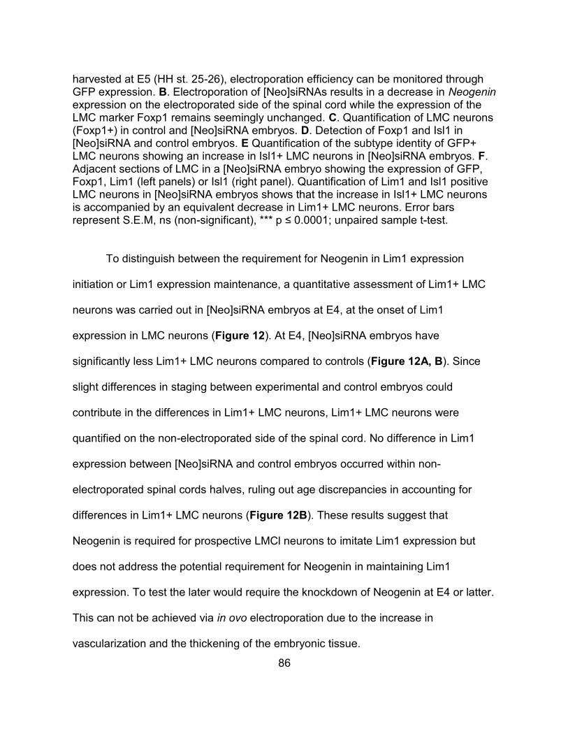
86
harvested at E5 (HH st. 25-26), electroporation efficiency can be monitored through GFP expression. B. Electroporation of [Neo]siRNAs results in a decrease in Neogenin expression on the electroporated side of the spinal cord while the expression of the LMC marker Foxp1 remains seemingly unchanged. C. Quantification of LMC neurons (Foxp1+) in control and [Neo]siRNA embryos. D. Detection of Foxp1 and Isl1 in [Neo]siRNA and control embryos. E Quantification of the subtype identity of GFP+ LMC neurons showing an increase in Isl1+ LMC neurons in [Neo]siRNA embryos. F. Adjacent sections of LMC in a [Neo]siRNA embryo showing the expression of GFP, Foxp1, Lim1 (left panels) or Isl1 (right panel). Quantification of Lim1 and Isl1 positive LMC neurons in [Neo]siRNA embryos shows that the increase in Isl1+ LMC neurons is accompanied by an equivalent decrease in Lim1+ LMC neurons. Error bars represent S.E.M, ns (non-significant), *** p ≤ 0.0001; unpaired sample t-test.
To distinguish between the requirement for Neogenin in Lim1 expression
initiation or Lim1 expression maintenance, a quantitative assessment of Lim1+ LMC
neurons was carried out in [Neo]siRNA embryos at E4, at the onset of Lim1
expression in LMC neurons (Figure 12). At E4, [Neo]siRNA embryos have
significantly less Lim1+ LMC neurons compared to controls (Figure 12A, B). Since
slight differences in staging between experimental and control embryos could
contribute in the differences in Lim1+ LMC neurons, Lim1+ LMC neurons were
quantified on the non-electroporated side of the spinal cord. No difference in Lim1
expression between [Neo]siRNA and control embryos occurred within non-
electroporated spinal cords halves, ruling out age discrepancies in accounting for
differences in Lim1+ LMC neurons (Figure 12B). These results suggest that
Neogenin is required for prospective LMCl neurons to imitate Lim1 expression but
does not address the potential requirement for Neogenin in maintaining Lim1
expression. To test the later would require the knockdown of Neogenin at E4 or latter.
This can not be achieved via in ovo electroporation due to the increase in
vascularization and the thickening of the embryonic tissue.
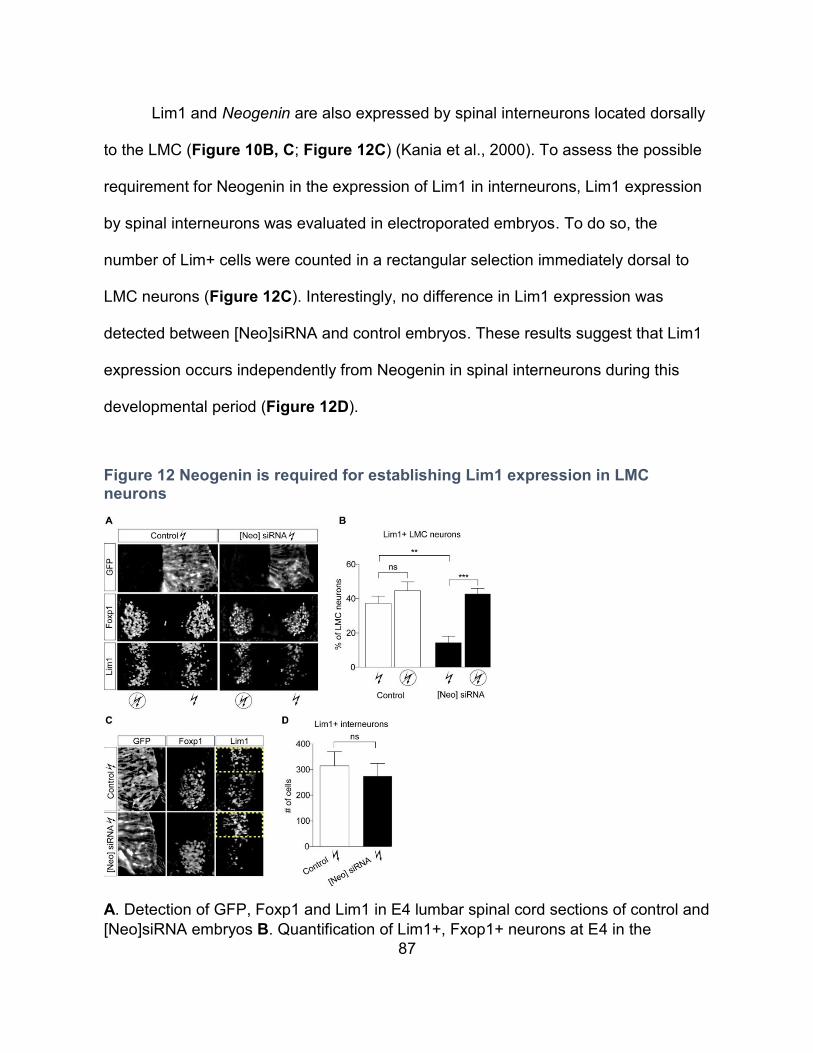
87
Lim1 and Neogenin are also expressed by spinal interneurons located dorsally
to the LMC (Figure 10B, C; Figure 12C) (Kania et al., 2000). To assess the possible
requirement for Neogenin in the expression of Lim1 in interneurons, Lim1 expression
by spinal interneurons was evaluated in electroporated embryos. To do so, the
number of Lim+ cells were counted in a rectangular selection immediately dorsal to
LMC neurons (Figure 12C). Interestingly, no difference in Lim1 expression was
detected between [Neo]siRNA and control embryos. These results suggest that Lim1
expression occurs independently from Neogenin in spinal interneurons during this
developmental period (Figure 12D).
Figure 12 Neogenin is required for establishing Lim1 expression in LMC neurons
A. Detection of GFP, Foxp1 and Lim1 in E4 lumbar spinal cord sections of control and
[Neo]siRNA embryos B. Quantification of Lim1+, Fxop1+ neurons at E4 in the

88
electroporated and non-electroporated side of the spinal cord. C. Detection of GFP,
Foxp1 and Lim1 in E4 lumbar spinal cord sections of control and [Neo]siRNA
embryos, the yellow boxes depict the region where Lim1 expression by interneurons
was quantified in D. D. Quantification of Lim1 expression by spinal interneurons
immediately dorsal to LMC neurons. ** p = 0.0038, *** p= 0.0004.
LMC neuron subtype distribution within the LMC is altered in Neogenin KD embryos
In [Neo]siRNA embryos, the shift in the ratio of LMCm/LMCl neurons is
accompanied by a decrease in LMCm/LMCl segregation and a high occurrence of
LMCm neurons occupying areas normally occupied by LMCl neurons (Figure 11D).
To gain insight in the alteration in LMC subtype distribution in [Neo]siRNA embryos,
serial sections spanning the lumbar spinal cord were digitally reproduced to highlight
the gross occupancy of LMCm/LMCl subdivisions (Figure 13). In control embryos, the
LMCl forms an L shape with the most ventral portion of the LMC generally occupied
by LMCl neurons (Figure 13A, B). In [Neo]siRNA embryos, LMCl is no longer L
shaped, while the dorso-lateral portion of the LMCl appears mostly unaffected, the
most ventral portion of the LMC is occupied by LMCm neurons (Figure 13C, D).
These results suggest that Neogenin may be involved in LMC neuron migration
and/or LMC subtype segregation.
89
Figure 13 The Distribution of LMCm and LMCl neurons is altered in [Neo]siRNA embryos
A, C. Detection of Foxp1, Isl1 and GFP in lumbar spinal cord sections of control (A) and [Neo]siRNAs (B) electroporated embryos. B, D. LMC subtype occupancy within the lumbar spinal cord of three control (B) and three [Neo]siRNAs (D) electroporated embryos was digitally reproduced based on the gross occupation of LMCm neurons (Isl1+) within the LMC (Foxp1+). KD of Neogenin results in the expansion of the territory occupied by LMCm neurons, particularly in the most ventral portion of the LMC.
90
Investigating the requirement for RGMa and RGMb in LMC subtype differentiation
The expression of RGMa and RGMb during LMC neuron development in
conjunction with pre-existing evidence of RGMs implicated in neuronal differentiation,
suggest that Neogenin may regulate LMC subtype differentiation through interacting
with RGMa and/or RGMb (Figure 10) (Matsunaga et al., 2006; O'Leary et al., 2013;
O'Leary et al., 2015; Kam et al., 2016).
RGMa is expressed by MN progenitors as well as differentiated LMC neurons
(Figure 10), to investigate the possible implication of RGMa in LMC subtype
differentiation, a combination of two siRNAs directed against RGMa as well as a GFP
expression plasmid ([RGMa]siRNA) were electroporated in E3 chick lumbar spinal
cords. RGMa expression was assessed qualitatively by in situ mRNA detection in
lumbar spinal cord sections of electroporated embryos. The siRNAs were not
effective at knocking-down RGMa and LMC neuron differentiation appeared normal in
[RGMa]siRNA embryos (data not shown). As an alternative strategy, RGMa was
overexpressed in chick lumbar spinal cord through the electroporation of a bi-
histrionic plasmid for the expression of RGMa and β-galactosidase. Although the
expression of β-galactosidase and increased RGMa expression suggest that RGMa
was overexpressed, no apparent LMC differentiation defects were observed (data not
shown). These results do not rule out an implication of RGMa in LMC subtype
differentiation, effective knockdown of RGMa using alternative siRNAs or the use of a
CRISPR/Cas9 system targeting RGMa could potentially reveal such a role.
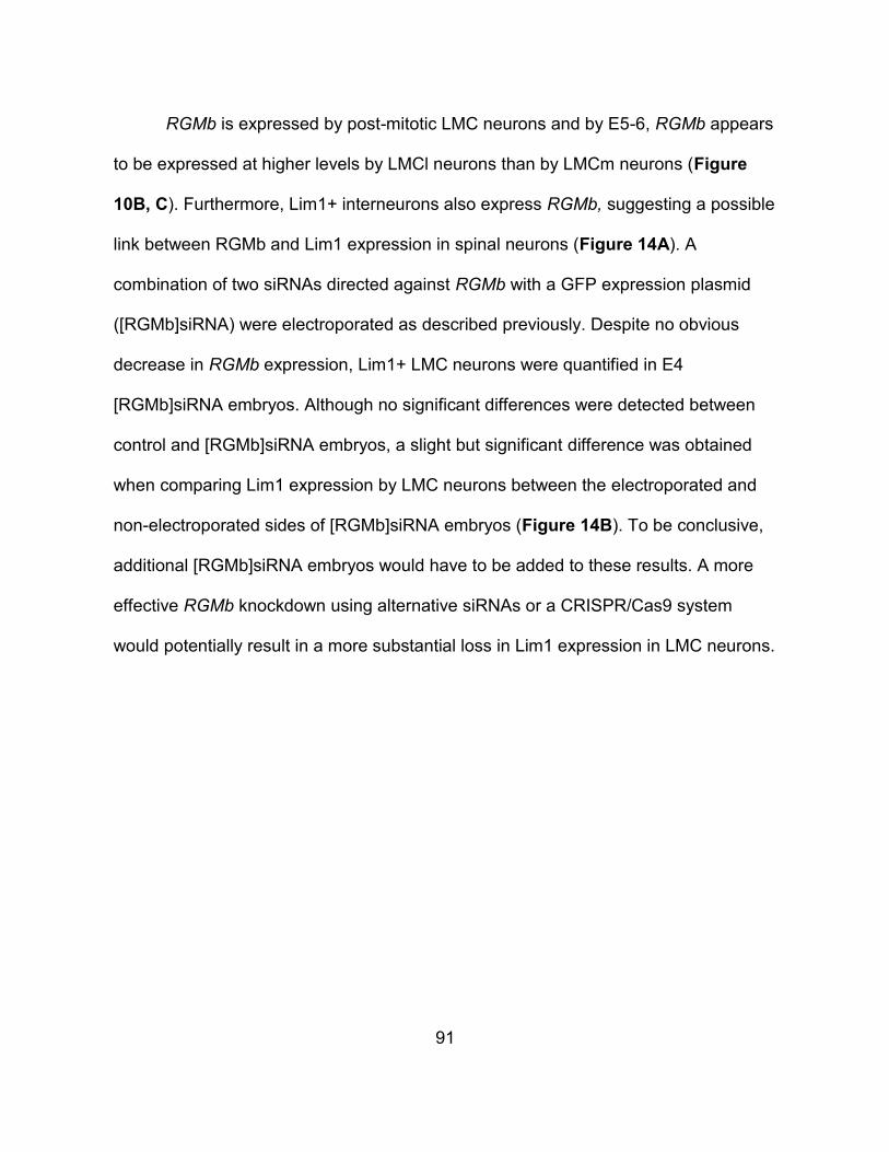
91
RGMb is expressed by post-mitotic LMC neurons and by E5-6, RGMb appears
to be expressed at higher levels by LMCl neurons than by LMCm neurons (Figure
10B, C). Furthermore, Lim1+ interneurons also express RGMb, suggesting a possible
link between RGMb and Lim1 expression in spinal neurons (Figure 14A). A
combination of two siRNAs directed against RGMb with a GFP expression plasmid
([RGMb]siRNA) were electroporated as described previously. Despite no obvious
decrease in RGMb expression, Lim1+ LMC neurons were quantified in E4
[RGMb]siRNA embryos. Although no significant differences were detected between
control and [RGMb]siRNA embryos, a slight but significant difference was obtained
when comparing Lim1 expression by LMC neurons between the electroporated and
non-electroporated sides of [RGMb]siRNA embryos (Figure 14B). To be conclusive,
additional [RGMb]siRNA embryos would have to be added to these results. A more
effective RGMb knockdown using alternative siRNAs or a CRISPR/Cas9 system
would potentially result in a more substantial loss in Lim1 expression in LMC neurons.
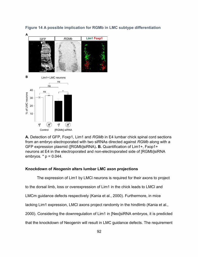
92
Figure 14 A possible implication for RGMb in LMC subtype differentiation
A. Detection of GFP, Foxp1, Lim1 and RGMb in E4 lumbar chick spinal cord sections
from an embryo electroporated with two siRNAs directed against RGMb along with a
GFP expression plasmid ([RGMb]siRNA). B. Quantification of Lim1+, Fxop1+
neurons at E4 in the electroporated and non-electroporated side of [RGMb]siRNA
embryos. * p = 0.044.
Knockdown of Neogenin alters lumbar LMC axon projections
The expression of Lim1 by LMCl neurons is required for their axons to project
to the dorsal limb, loss or overexpression of Lim1 in the chick leads to LMCl and
LMCm guidance defects respectively (Kania et al., 2000). Furthermore, in mice
lacking Lim1 expression, LMCl axons project randomly in the hindlimb (Kania et al.,
2000). Considering the downregulation of Lim1 in [Neo]siRNA embryos, it is predicted
that the knockdown of Neogenin will result in LMC guidance defects. The requirement
93
for Neogenin in LMC development may be two-fold, first through the regulation of
Lim1 expression and secondly, by enabling LMCl axons to be attracted by Netrin-1 in
the dorsal limb (Figure 5) (Poliak et al., 2015). To gain insight in the requirement for
Neogenin in LMC axon guidance, GFP+ axon projections were quantified in
[Neo]siRNA and control embryos by measuring the GFP fluorescence intensities in
the dorsal and ventral axonal branches of hindlimbs (Figure 15). [Neo]siRNA
embryos displayed altered ratios of dorso-ventral axon projections with more ventrally
projecting axons, consistent with an increase in the ratio of LMCm/LMCl neurons
(Figure 15B). To address guidance defects by LMCl or LMCm axons specifically
would require the retrograde labeling of LMC neurons by injecting HRP into the
ventral and dorsal nerves branches respectively (Poliak et al., 2015).
Figure 15 Knockdown of Neogenin alters LMC axon projections
A. Detection of neurofilament and GFP in LMC axons in E5 sciatic plexus of control
and [Neo]siRNA embryos, the dorsal and ventral branches are circled in green and
red respectively. B. Quantification of GFP+ LMC axons in the hindlimbs of control and
[Neo]siRNA embryos. D: dorsal, V: ventral, *** p ≤ 0.0001.
94
Neogenin may be dispensable for the subtype differentiation of LMC neurons in mice
Neogenin is expressed by developing LMC neurons in the mouse (Poliak et al.,
2015). To investigate the requirement for Neogenin in LMC neuron subtype
differentiation in mice, the expression of Lim1 by LMC neurons in e11.5 lumbar spinal
cord was quantified in Neo1-/- mice and compared to WT and Neo1+/- littermates,
generously provided by the Cloutier laboratory (Kam et al., 2016). Although only a few
embryos were analyzed and a thorough analysis would require more embryos, the
distribution and total number of lumbar LMC neurons as well as the number of Lim1
expressing LMC neurons was not significantly altered in Neo1-/- mice (Figure 16B,
D). The lack of an LMC differentiation phenotype may reflect a different requirement
for Neogenin between chick and mice. It is also possible that the expression of Dcc
by LMC neurons in the mouse compensates for the loss of Neogenin. Furthermore,
the complete absence of Neogenin at the onset of embryogenesis may allow for the
establishment of compensatory mechanisms not available in the context of a partial
knockdown of Neogenin via siRNAs.
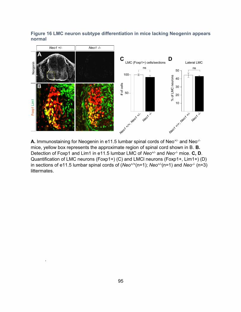
95
Figure 16 LMC neuron subtype differentiation in mice lacking Neogenin appears normal
A. Immunostaining for Neogenin in e11.5 lumbar spinal cords of Neo+/- and Neo-/-
mice, yellow box represents the approximate region of spinal cord shown in B. B.
Detection of Foxp1 and Lim1 in e11.5 lumbar LMC of Neo+/- and Neo-/- mice. C, D.
Quantification of LMC neurons (Foxp1+) (C) and LMCl neurons (Foxp1+, Lim1+) (D)
in sections of e11.5 lumbar spinal cords of (Neo+/+(n=1); Neo+/-(n=1) and Neo-/- (n=3)
littermates.
.
96
4. Sensitization of spinal motor neurons to Netrin-1 by ephrin-A5
Author contributions
Experiments were designed by Louis-Philippe Croteau under the supervision
and input from Artur Kania. All the in ovo electroporations of chick embryos were
performed by Meirong Liang. The stripe assay experiments in Figures III-13 and III-14
were performed and analyzed by Tzu-Jen Kao. All other experiments were
performed and analyzed by Louis-Philippe Croteau. The MATLAB application used to
analyze the immunofluorescence distribution within growth cones was programmed
by Dominic Fillion. The ImageJ macro used to define the region of interest within
analyzed images was programmed by Chris Law. The results section was partially
edited by Artur Kania. Resources were provided by grants obtained by Artur Kania.
Rational
In the chick, LMCl neurons that innervate the dorsal hindlimb are attracted to
Netrin-1 through the expression of the Netrin receptor Neogenin. LMCm neurons that
extend axons ventrally avoid Netrin-1 through the expression of the repulsive Netrin
receptor Unc5c (Poliak et al., 2015). Furthermore, mice hypomorphic for Netrin-1 as
well as Unc5c null mice display LMCm guidance defects (Poliak et al., 2015). LMCm
and LMCl axons are also guided by ephrins in the limbs through the expression of
Eph receptors (Helmbacher et al., 2000; Eberhart et al., 2002; Eberhart et al., 2004;
97
Luria et al., 2008; Bonanomi et al., 2012). In vivo, LMCl axons expressing the ephrin
receptor EphA4, are repelled from ephrin-A5 and ephrin-A2 expressed in the ventral
hindlimb (Helmbacher et al., 2000; Eberhart et al., 2004; Bonanomi et al., 2012).
We have previously demonstrated that Netrin-1 acts synergistically with
ephrins in LMC axon guidance; In vitro assays demonstrate that both LMCm and
LMCl axons integrate Netrin-1 and ephrins in a supra-additive fashion (Poliak et al.,
2015). To gain insight in the molecular mechanism underlining the synergistic
integration of Netrin-1 and ephrins in LMC axons, in vitro experiments were carried
out focussing on the effects of Netrin-1 and ephrin-A5 on receptor dynamics in LMC
growth cones.
Netrin-1 induces a protein kinase A-dependent re-localization of Neogenin in LMC growth cones
We reasoned that changes in growth cone receptor dynamics could underlie
Netrin-1/ephrin-A5 LMC axon guidance synergy and thus we first assessed the
effects of Netrin-1 on Neogenin dynamics in LMC growth cones. To do this, we
explanted chick lumbar LMC neurons between Hamburger–Hamilton stages (HH st.)
24 and 26, the developmental period encompassing the dorso-ventral guidance of
lumbar LMC axons (Hamburger and Hamilton, 1951; Tosney and Landmesser, 1985;
Kania et al., 2000). LMC explants were cultured for ~18 hrs and exposed for 15
minutes to a bath application of motor neuron culture media (MN media) containing
Netrin-1 at 300 ng/mL or MN media alone as control, followed by an assessment of F-
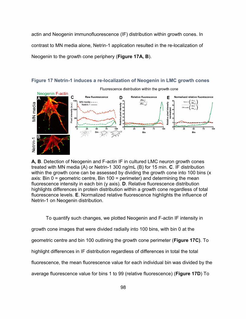
98
actin and Neogenin immunofluorescence (IF) distribution within growth cones. In
contrast to MN media alone, Netrin-1 application resulted in the re-localization of
Neogenin to the growth cone periphery (Figure 17A, B).
Figure 17 Netrin-1 induces a re-localization of Neogenin in LMC growth cones
A, B. Detection of Neogenin and F-actin IF in cultured LMC neuron growth cones treated with MN media (A) or Netrin-1 300 ng/mL (B) for 15 min. C. IF distribution within the growth cone can be assessed by dividing the growth cone into 100 bins (x axis: Bin 0 = geometric centre, Bin 100 = perimeter) and determining the mean fluorescence intensity in each bin (y axis). D. Relative fluorescence distribution highlights differences in protein distribution within a growth cone regardless of total fluorescence levels. E. Normalized relative fluorescence highlights the influence of Netrin-1 on Neogenin distribution.
To quantify such changes, we plotted Neogenin and F-actin IF intensity in
growth cone images that were divided radially into 100 bins, with bin 0 at the
geometric centre and bin 100 outlining the growth cone perimeter (Figure 17C). To
highlight differences in IF distribution regardless of differences in total the total
fluorescence, the mean fluorescence value for each individual bin was divided by the
average fluorescence value for bins 1 to 99 (relative fluorescence) (Figure 17D) To
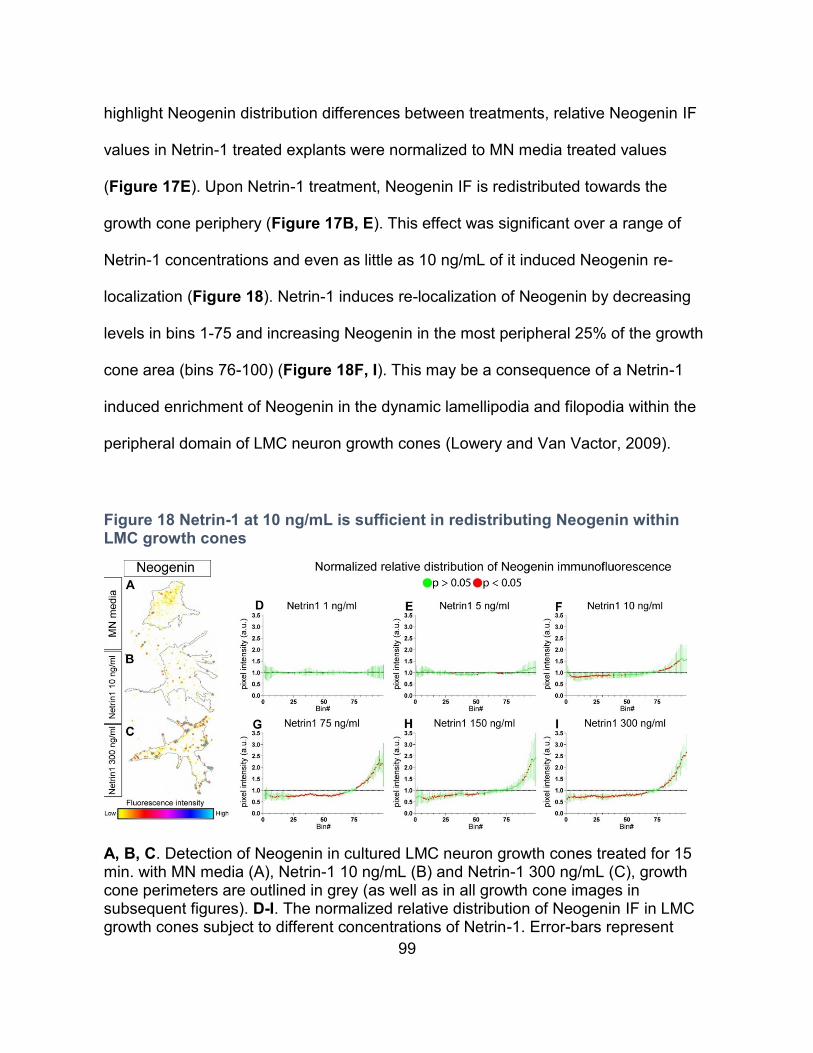
99
highlight Neogenin distribution differences between treatments, relative Neogenin IF
values in Netrin-1 treated explants were normalized to MN media treated values
(Figure 17E). Upon Netrin-1 treatment, Neogenin IF is redistributed towards the
growth cone periphery (Figure 17B, E). This effect was significant over a range of
Netrin-1 concentrations and even as little as 10 ng/mL of it induced Neogenin re-
localization (Figure 18). Netrin-1 induces re-localization of Neogenin by decreasing
levels in bins 1-75 and increasing Neogenin in the most peripheral 25% of the growth
cone area (bins 76-100) (Figure 18F, I). This may be a consequence of a Netrin-1
induced enrichment of Neogenin in the dynamic lamellipodia and filopodia within the
peripheral domain of LMC neuron growth cones (Lowery and Van Vactor, 2009).
Figure 18 Netrin-1 at 10 ng/mL is sufficient in redistributing Neogenin within LMC growth cones
A, B, C. Detection of Neogenin in cultured LMC neuron growth cones treated for 15 min. with MN media (A), Netrin-1 10 ng/mL (B) and Netrin-1 300 ng/mL (C), growth cone perimeters are outlined in grey (as well as in all growth cone images in subsequent figures). D-I. The normalized relative distribution of Neogenin IF in LMC growth cones subject to different concentrations of Netrin-1. Error-bars represent
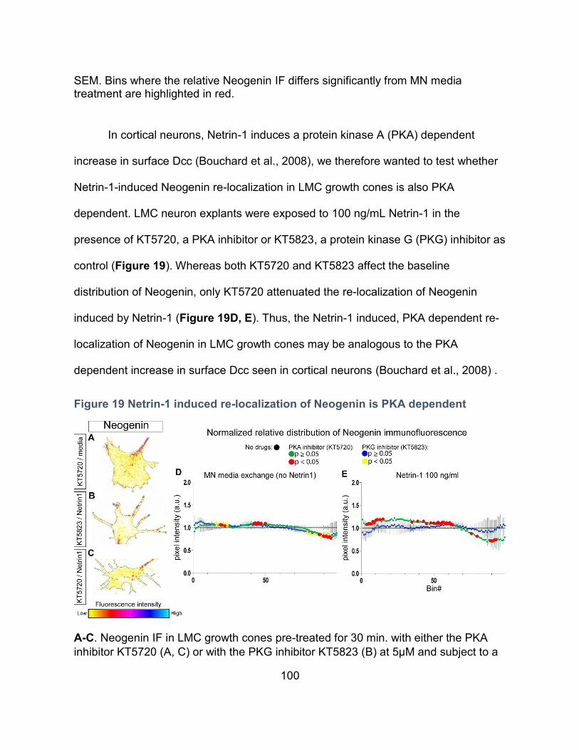
100
SEM. Bins where the relative Neogenin IF differs significantly from MN media treatment are highlighted in red.
In cortical neurons, Netrin-1 induces a protein kinase A (PKA) dependent
increase in surface Dcc (Bouchard et al., 2008), we therefore wanted to test whether
Netrin-1-induced Neogenin re-localization in LMC growth cones is also PKA
dependent. LMC neuron explants were exposed to 100 ng/mL Netrin-1 in the
presence of KT5720, a PKA inhibitor or KT5823, a protein kinase G (PKG) inhibitor as
control (Figure 19). Whereas both KT5720 and KT5823 affect the baseline
distribution of Neogenin, only KT5720 attenuated the re-localization of Neogenin
induced by Netrin-1 (Figure 19D, E). Thus, the Netrin-1 induced, PKA dependent re-
localization of Neogenin in LMC growth cones may be analogous to the PKA
dependent increase in surface Dcc seen in cortical neurons (Bouchard et al., 2008) .
Figure 19 Netrin-1 induced re-localization of Neogenin is PKA dependent
A-C. Neogenin IF in LMC growth cones pre-treated for 30 min. with either the PKA
inhibitor KT5720 (A, C) or with the PKG inhibitor KT5823 (B) at 5µM and subject to a
101
20 min. treatment of either MN media (A) or Netrin-1 100 ng/mL (B, C). D, E.
Quantification of the relative distribution of Neogenin IF normalized to (No drug)
treatment without Netrin-1 (D) or after a 20 min. exposure to Netrin-1 at 100 ng/mL
(E).
The effects of Netrin-1 on protein levels and distribution in LMC neuron growth cones
We reasoned that Netrin-1 exposure of LMC growth cones might result in
wide-spread dynamic changes in cell membrane protein distribution or abundance,
and thus contribute to Netrin-1-ephrin-A5 synergistic axon guidance. For example,
increased expression of EphA4, the ephrin-A5 receptor required for LMC axon
guidance, could result in greater sensitivity to ephrin-A5 (Helmbacher et al., 2000;
Eberhart et al., 2002). To explore this possibility, LMC explants where exposed to
Netrin-1, and IF levels and distribution within growth cones were determined for
Neogenin, the repulsive Netrin-1 receptor Unc5c, EphA4 and BEN, a surface
glycoprotein marker of motor neurons (Pourquie et al., 1990) (Figure 20). Compared
to controls, exposure to Netrin-1 led to a significant increase in total Neogenin signal
in LMC growth cones (43% increase; p<0.001), without any changes in EphA4, Unc5c
or BEN levels (Figure 20A, F-M; p>0.5). Since the IF values quantified are a
compilation of the mean IF value in each growth cone, differences in Neogenin IF
could be the result of a Netrin-1 induced decrease in growth cone area. To ensure
this was not the case, growth cone area measurements were compiled and results
show that the average size of the growth cones included in the analysis did not differ
between treatments (Supplementary Figure 1). Because the Neogenin antibody
102
antagonizes the binding of Netrin-1 to Neogenin, the 43% increase in Neogenin when
subject to Netrin-1 is likely an underestimation of the actual Netrin-1 induced
increase. A quantification of the relative distribution of IF revealed that while Netrin-1
induces Neogenin re-localization towards the periphery (Figure 20B), Unc5c showed
a tendency to be decreased at the periphery, EphA4 and BEN distribution remained
largely unaffected (Figure 20C-E), suggesting that Netrin-1-mediated Neogenin re-
localization is not a consequence of a large-scale protein re-distribution in growth
cones.
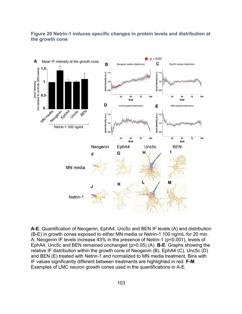
103
Figure 20 Netrin-1 induces specific changes in protein levels and distribution at the growth cone
A-E. Quantification of Neogenin, EphA4, Unc5c and BEN IF levels (A) and distribution (B-E) in growth cones exposed to either MN media or Netrin-1 100 ng/mL for 20 min. A. Neogenin IF levels increase 43% in the presence of Netrin-1 (p<0.001), levels of EphA4, Unc5c and BEN remained unchanged (p>0.05) (A). B-E. Graphs showing the relative IF distribution within the growth cone of Neogenin (B), EphA4 (C), Unc5c (D) and BEN (E) treated with Netrin-1 and normalized to MN media treatment, Bins with IF values significantly different between treatments are highlighted in red. F-M. Examples of LMC neuron growth cones used in the quantifications in A-E.
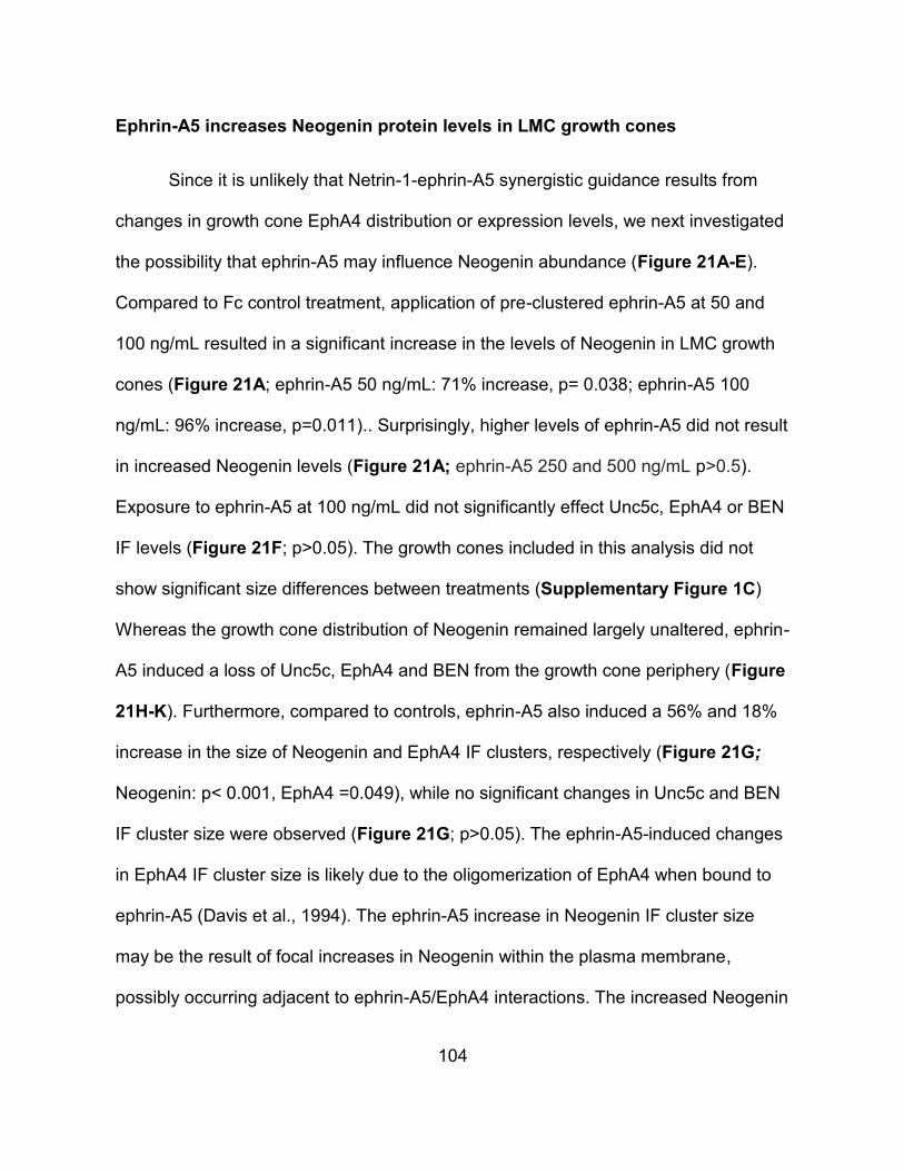
104
Ephrin-A5 increases Neogenin protein levels in LMC growth cones
Since it is unlikely that Netrin-1-ephrin-A5 synergistic guidance results from
changes in growth cone EphA4 distribution or expression levels, we next investigated
the possibility that ephrin-A5 may influence Neogenin abundance (Figure 21A-E).
Compared to Fc control treatment, application of pre-clustered ephrin-A5 at 50 and
100 ng/mL resulted in a significant increase in the levels of Neogenin in LMC growth
cones (Figure 21A; ephrin-A5 50 ng/mL: 71% increase, p= 0.038; ephrin-A5 100
ng/mL: 96% increase, p=0.011).. Surprisingly, higher levels of ephrin-A5 did not result
in increased Neogenin levels (Figure 21A; ephrin-A5 250 and 500 ng/mL p>0.5).
Exposure to ephrin-A5 at 100 ng/mL did not significantly effect Unc5c, EphA4 or BEN
IF levels (Figure 21F; p>0.05). The growth cones included in this analysis did not
show significant size differences between treatments (Supplementary Figure 1C)
Whereas the growth cone distribution of Neogenin remained largely unaltered, ephrin-
A5 induced a loss of Unc5c, EphA4 and BEN from the growth cone periphery (Figure
21H-K). Furthermore, compared to controls, ephrin-A5 also induced a 56% and 18%
increase in the size of Neogenin and EphA4 IF clusters, respectively (Figure 21G;
Neogenin: p< 0.001, EphA4 =0.049), while no significant changes in Unc5c and BEN
IF cluster size were observed (Figure 21G; p>0.05). The ephrin-A5-induced changes
in EphA4 IF cluster size is likely due to the oligomerization of EphA4 when bound to
ephrin-A5 (Davis et al., 1994). The ephrin-A5 increase in Neogenin IF cluster size
may be the result of focal increases in Neogenin within the plasma membrane,
possibly occurring adjacent to ephrin-A5/EphA4 interactions. The increased Neogenin
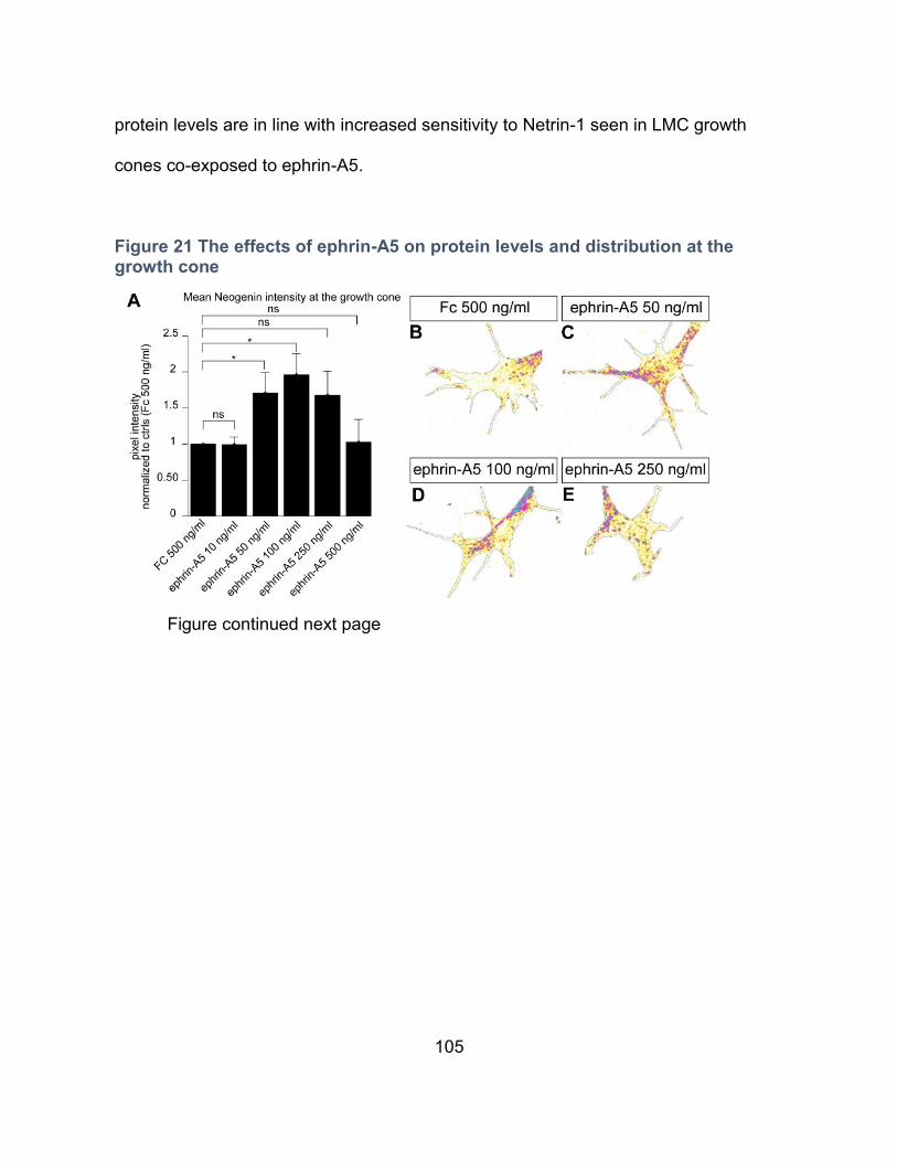
105
protein levels are in line with increased sensitivity to Netrin-1 seen in LMC growth
cones co-exposed to ephrin-A5.
Figure 21 The effects of ephrin-A5 on protein levels and distribution at the growth cone
Figure continued next page
106
A. Cultured LMC explants were subject to a 15 min. exposure to a control solution
containing Fc at 500 ng/mL or ephrin-A5 at concentrations ranging from 10 to 500
ng/mL, followed by a quantification of Neogenin IF in growth cones. Ephrin-A5 at 50
and 100 ng/mL results increased levels of Neogenin IF (ephrin-A5 50 ng/mL: 71%
increase, p= 0.038; ephrin-A5 100ng/mL: 96% increase, p=0.011). B-E. Examples of
Neogenin IF in growth cones quantified in A. F-K. Quantification of Neogenin, EphA4,
Unc5c and BEN IF levels (F), aggregate size (G) and distribution (H-K) in growth
cones exposed to either MN media or ephrin-A5 at 100 ng/mL for 20 min. F. Ephrin-
A5 induced a 64% increase in Neogenin (p<0.001), and 94 % increase in EphA4
(p<0.001) IF levels while Unc5c and BEN levels remained unchanged. G. Ephrin-A5
induced a 56% increase in the average size of Neogenin immunofluorescent
aggregates (p<0.001) and a 18% increase in the average size of EphA4
immunofluorescent aggregates (p=0.049) while Unc5c and BEN average aggregate
size did not differ significantly (p>0.5 and p= 0.061 respectively). H-K. Graphs
showing the relative IF distribution within the growth cone of Neogenin (H), Unc5c (I),
EphA4 (J) and BEN (K) treated with ephnrin-A5 and normalized to MN media
treatment, bins with IF values significantly different between treatments are
highlighted in red. L-S. Examples of LMC neuron growth cones used in the
quantifications in F-K.
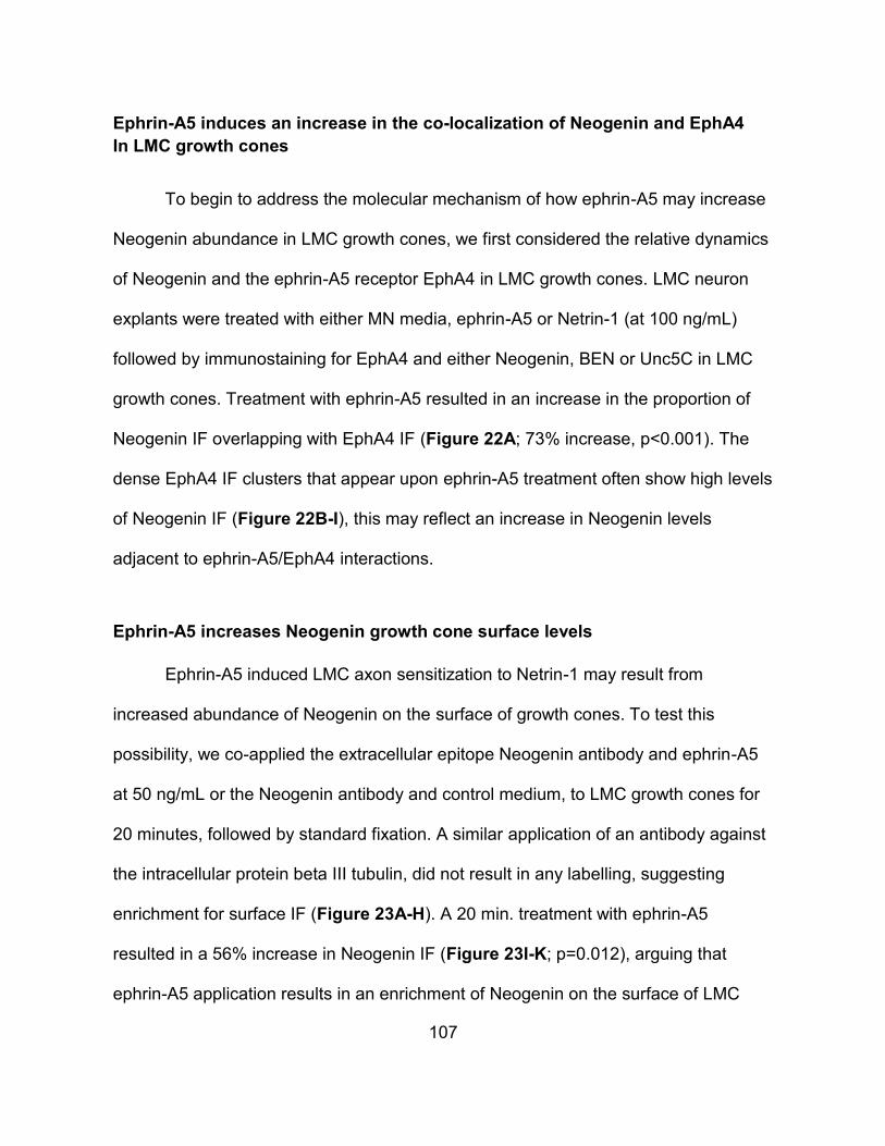
107
Ephrin-A5 induces an increase in the co-localization of Neogenin and EphA4
In LMC growth cones
To begin to address the molecular mechanism of how ephrin-A5 may increase
Neogenin abundance in LMC growth cones, we first considered the relative dynamics
of Neogenin and the ephrin-A5 receptor EphA4 in LMC growth cones. LMC neuron
explants were treated with either MN media, ephrin-A5 or Netrin-1 (at 100 ng/mL)
followed by immunostaining for EphA4 and either Neogenin, BEN or Unc5C in LMC
growth cones. Treatment with ephrin-A5 resulted in an increase in the proportion of
Neogenin IF overlapping with EphA4 IF (Figure 22A; 73% increase, p<0.001). The
dense EphA4 IF clusters that appear upon ephrin-A5 treatment often show high levels
of Neogenin IF (Figure 22B-I), this may reflect an increase in Neogenin levels
adjacent to ephrin-A5/EphA4 interactions.
Ephrin-A5 increases Neogenin growth cone surface levels
Ephrin-A5 induced LMC axon sensitization to Netrin-1 may result from
increased abundance of Neogenin on the surface of growth cones. To test this
possibility, we co-applied the extracellular epitope Neogenin antibody and ephrin-A5
at 50 ng/mL or the Neogenin antibody and control medium, to LMC growth cones for
20 minutes, followed by standard fixation. A similar application of an antibody against
the intracellular protein beta III tubulin, did not result in any labelling, suggesting
enrichment for surface IF (Figure 23A-H). A 20 min. treatment with ephrin-A5
resulted in a 56% increase in Neogenin IF (Figure 23I-K; p=0.012), arguing that
ephrin-A5 application results in an enrichment of Neogenin on the surface of LMC
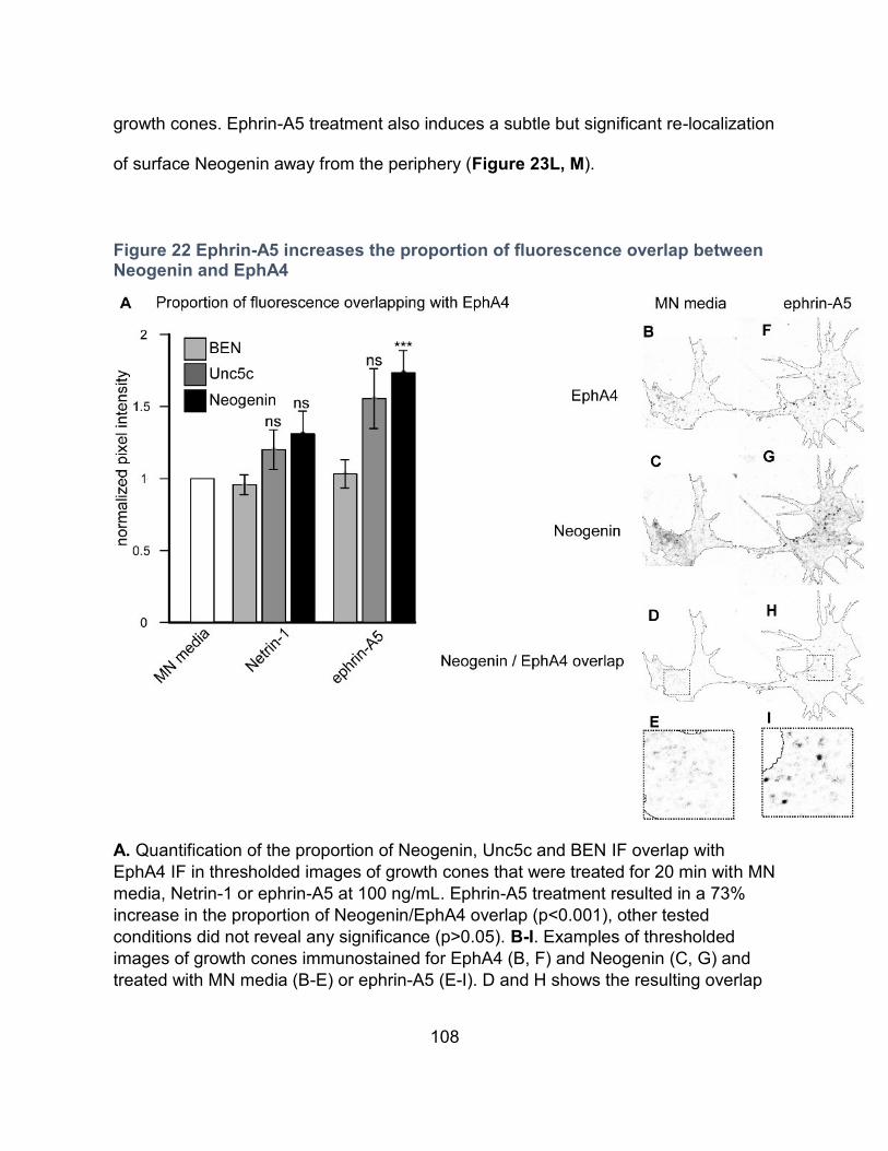
108
growth cones. Ephrin-A5 treatment also induces a subtle but significant re-localization
of surface Neogenin away from the periphery (Figure 23L, M).
Figure 22 Ephrin-A5 increases the proportion of fluorescence overlap between Neogenin and EphA4
A. Quantification of the proportion of Neogenin, Unc5c and BEN IF overlap with
EphA4 IF in thresholded images of growth cones that were treated for 20 min with MN
media, Netrin-1 or ephrin-A5 at 100 ng/mL. Ephrin-A5 treatment resulted in a 73%
increase in the proportion of Neogenin/EphA4 overlap (p<0.001), other tested
conditions did not reveal any significance (p>0.05). B-I. Examples of thresholded
images of growth cones immunostained for EphA4 (B, F) and Neogenin (C, G) and
treated with MN media (B-E) or ephrin-A5 (E-I). D and H shows the resulting overlap
109
in Neogenin/EphA4 signal in MN media and ephrin-A5 treated explants respectively,
panels E and I are magnified images of boxed regions in D and H.
Figure 23 Ephrin-A5 increases surface enriched Neogenin in growth cones
To enrich for surface Neogenin IF, a Neogenin antibody with an extracellular epitope
was added to LMC explant cultures during a 20 min treatment with either MN media
or ephrin-A5 at 50 ng/mL. A-H. To confirm the surface enrichment of Neogenin when
the antibody is added to live cultures, an antibody against the intracellular protein
beta III tubulin was included in fixed (A-D) and live (E-H) cultures, secondary
antibodies and phalloidin to detect F-actin were added post-fix (A-H). I, J. Detection of
surface enriched Neogenin in growth cones treated with MN media (I) or ephrin-A5 50
ng-mL (J). K. Quantification of surface enriched Neogenin IF in MN media or ephrin-
A5 50 ng/mL treated growth cones showing a 56% increase in surface enriched
Neogenin signal when treated with ephrin-A5 (p=0.012). L Quantification of the
relative distribution of surface enriched Neogenin fluorescence in MN media and
ephrin-A5 treated growth cones M. Same as in L but normalized to MN media
treatment values, bins with values significantly different between treatments are
shown in red.
110
Ephrin-A5 enhances Netrin-1 binding in growth cones
We next sought to determine whether increased LMC growth cone surface
Neogenin levels might result in increased Netrin-1 binding to LMC growth cones. LMC
explants were incubated with Netrin-1 and ephrin-A5 or Netrin-1 alone as control.
After fixation, an antibody against Netrin-1 was used to estimate relative Netrin-1
LMC growth cone binding (Figure 24). Compared to a Netrin-1 treatment, the addition
of ephrin-A5 resulted in increased levels of Netrin-1 IF in LMC growth cones (Figure
24A-C, 45% increase p< 0.001). The growth cones included in this analysis did not
show significant size differences between treatments (Supplementary Figure 1E). In
alignment with the enrichment of Neogenin in the periphery of Netrin-1 treated growth
cones (Figure 18), the distribution of Netrin-1 signal is also enriched at the periphery
(Figure 24D). Furthermore, the ephrin-A5 induced increase in Netrin-1 signal occurs
throughout the growth cone (Figure 24D).
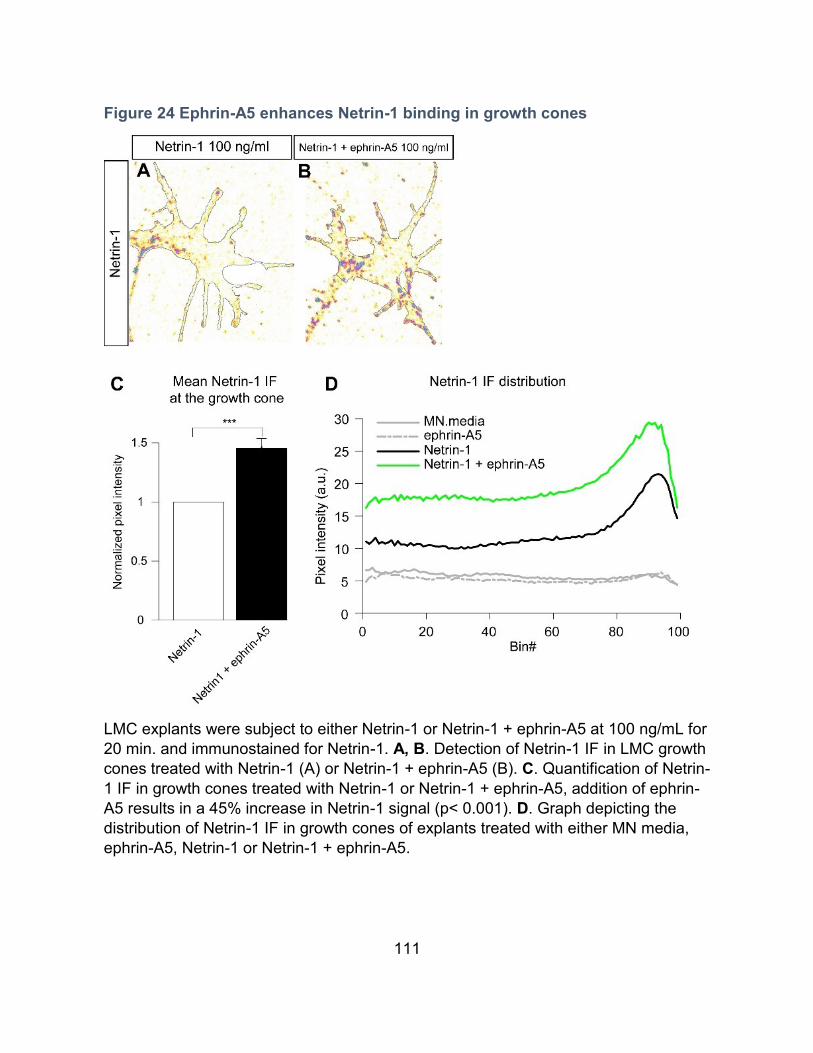
111
Figure 24 Ephrin-A5 enhances Netrin-1 binding in growth cones
LMC explants were subject to either Netrin-1 or Netrin-1 + ephrin-A5 at 100 ng/mL for
20 min. and immunostained for Netrin-1. A, B. Detection of Netrin-1 IF in LMC growth
cones treated with Netrin-1 (A) or Netrin-1 + ephrin-A5 (B). C. Quantification of Netrin-
1 IF in growth cones treated with Netrin-1 or Netrin-1 + ephrin-A5, addition of ephrin-
A5 results in a 45% increase in Netrin-1 signal (p< 0.001). D. Graph depicting the
distribution of Netrin-1 IF in growth cones of explants treated with either MN media,
ephrin-A5, Netrin-1 or Netrin-1 + ephrin-A5.
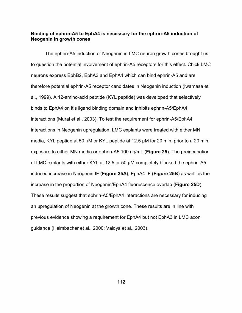
112
Binding of ephrin-A5 to EphA4 is necessary for the ephrin-A5 induction of Neogenin in growth cones
The ephrin-A5 induction of Neogenin in LMC neuron growth cones brought us
to question the potential involvement of ephrin-A5 receptors for this effect. Chick LMC
neurons express EphB2, EphA3 and EphA4 which can bind ephrin-A5 and are
therefore potential ephrin-A5 receptor candidates in Neogenin induction (Iwamasa et
al., 1999). A 12-amino-acid peptide (KYL peptide) was developed that selectively
binds to EphA4 on it’s ligand binding domain and inhibits ephrin-A5/EphA4
interactions (Murai et al., 2003). To test the requirement for ephrin-A5/EphA4
interactions in Neogenin upregulation, LMC explants were treated with either MN
media, KYL peptide at 50 µM or KYL peptide at 12.5 µM for 20 min. prior to a 20 min.
exposure to either MN media or ephrin-A5 100 ng/mL (Figure 25). The preincubation
of LMC explants with either KYL at 12.5 or 50 µM completely blocked the ephrin-A5
induced increase in Neogenin IF (Figure 25A), EphA4 IF (Figure 25B) as well as the
increase in the proportion of Neogenin/EphA4 fluorescence overlap (Figure 25D).
These results suggest that ephrin-A5/EphA4 interactions are necessary for inducing
an upregulation of Neogenin at the growth cone. These results are in line with
previous evidence showing a requirement for EphA4 but not EphA3 in LMC axon
guidance (Helmbacher et al., 2000; Vaidya et al., 2003).
113
Figure 25 Blocking ephrin-A5/EphA4 interactions inhibits the ephrin-A5 induced increase in Neogenin levels and overlap with EphA4
LMC explants were incubated with either MN media, KYL 12.5 µM or KYL 50 µM for 20 min. prior to being treated with either MN media or ephrin-A5 at 100 ng/mL with and without KYL peptide for 20 min. followed by Neogenin, EphA4 and F-actin immunostaining. A-C. Quantification of Neogenin (A), EphA4 (B) or F-actin (C) IF in growth cones normalized to MN media treatment D. Quantification of the proportion of Neogenin IF overlapping with EphA4 IF normalized to MN media treatment. A. ** p=0.003, B. ***p<0.001, D. **p=0.009.
114
Overexpression of EphA4 potentiates LMC growth cones to the ephrin-A5-dependent increase in Neogenin abundance
We reasoned that EphA4 could be a critical receptor mediating ephrin-A5-
Netrin-1 synergy since it is required for the normal pathfinding of LMC axons
(Helmbacher et al., 2000; Eberhart et al., 2002), and that ephrin-A5 exposure results
in increased Neogenin abundance and co-localization with EphA4 in LMC growth
cones. One prediction of this model is that increasing EphA4 expression levels in
LMC growth cones would result in exaggerated increases in Neogenin abundance in
response to ephrin-A5. To test this idea, GFP expression plasmids alone or together
with mouse EphA4 expression plasmids were electroporated into chick neural tubes
at HH st. 18-19 by in ovo electroporation (Kania and Jessell, 2003). GFP-expressing
LMC neurons were explanted as above, and following treatment with MN media,
Netrin-1 or ephrin-A5 (either at 100 mg/mL), Neogenin IF levels were quantified in
GFP+ growth cones (Figure 26A). Treatment with ephrin-A5 in EphA4
overexpressing LMC neurons led to a threefold increase in Neogenin signal
compared to LMC neurons overexpressing GFP alone (Figure 26A, D, G), arguing
that EphA4 can potentiate the ephrin-A5-induced increase in Neogenin abundance.
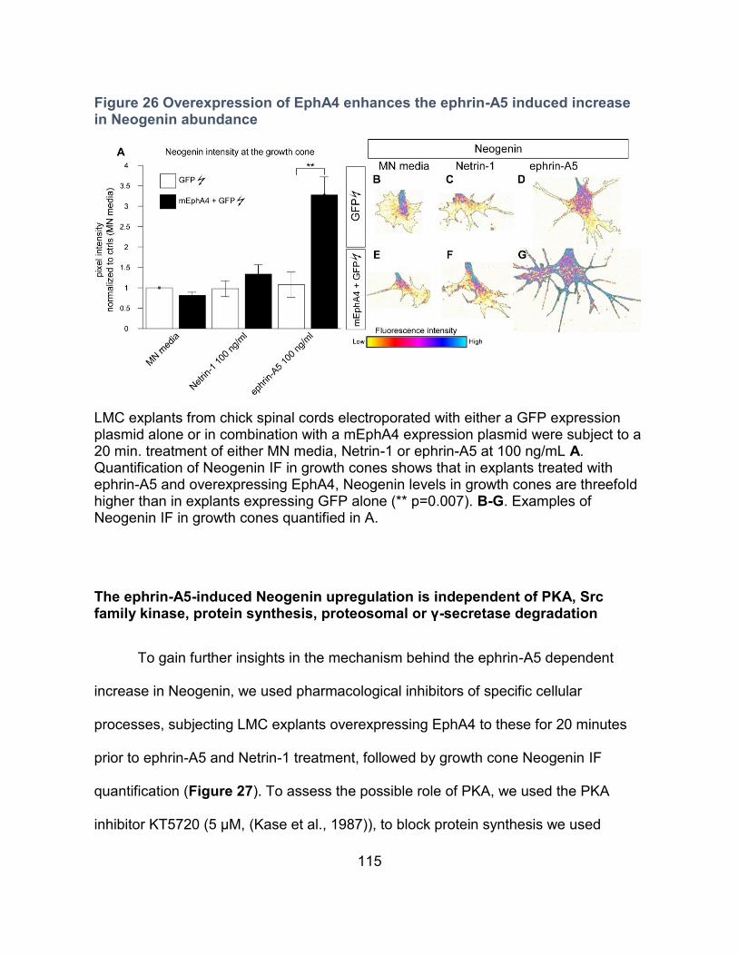
115
Figure 26 Overexpression of EphA4 enhances the ephrin-A5 induced increase in Neogenin abundance
LMC explants from chick spinal cords electroporated with either a GFP expression plasmid alone or in combination with a mEphA4 expression plasmid were subject to a 20 min. treatment of either MN media, Netrin-1 or ephrin-A5 at 100 ng/mL A. Quantification of Neogenin IF in growth cones shows that in explants treated with ephrin-A5 and overexpressing EphA4, Neogenin levels in growth cones are threefold higher than in explants expressing GFP alone (** p=0.007). B-G. Examples of Neogenin IF in growth cones quantified in A.
The ephrin-A5-induced Neogenin upregulation is independent of PKA, Src family kinase, protein synthesis, proteosomal or γ-secretase degradation
To gain further insights in the mechanism behind the ephrin-A5 dependent
increase in Neogenin, we used pharmacological inhibitors of specific cellular
processes, subjecting LMC explants overexpressing EphA4 to these for 20 minutes
prior to ephrin-A5 and Netrin-1 treatment, followed by growth cone Neogenin IF
quantification (Figure 27). To assess the possible role of PKA, we used the PKA
inhibitor KT5720 (5 µM, (Kase et al., 1987)), to block protein synthesis we used
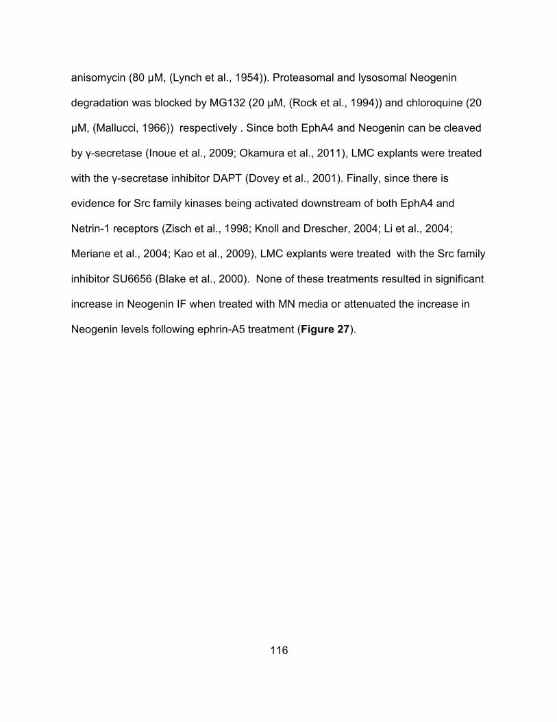
116
anisomycin (80 µM, (Lynch et al., 1954)). Proteasomal and lysosomal Neogenin
degradation was blocked by MG132 (20 µM, (Rock et al., 1994)) and chloroquine (20
µM, (Mallucci, 1966)) respectively . Since both EphA4 and Neogenin can be cleaved
by γ-secretase (Inoue et al., 2009; Okamura et al., 2011), LMC explants were treated
with the γ-secretase inhibitor DAPT (Dovey et al., 2001). Finally, since there is
evidence for Src family kinases being activated downstream of both EphA4 and
Netrin-1 receptors (Zisch et al., 1998; Knoll and Drescher, 2004; Li et al., 2004;
Meriane et al., 2004; Kao et al., 2009), LMC explants were treated with the Src family
inhibitor SU6656 (Blake et al., 2000). None of these treatments resulted in significant
increase in Neogenin IF when treated with MN media or attenuated the increase in
Neogenin levels following ephrin-A5 treatment (Figure 27).
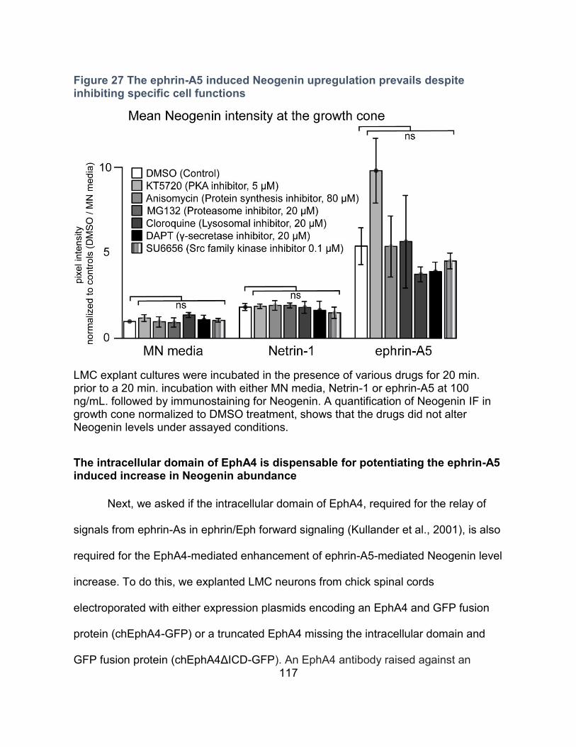
117
Figure 27 The ephrin-A5 induced Neogenin upregulation prevails despite inhibiting specific cell functions
LMC explant cultures were incubated in the presence of various drugs for 20 min. prior to a 20 min. incubation with either MN media, Netrin-1 or ephrin-A5 at 100 ng/mL. followed by immunostaining for Neogenin. A quantification of Neogenin IF in growth cone normalized to DMSO treatment, shows that the drugs did not alter Neogenin levels under assayed conditions.
The intracellular domain of EphA4 is dispensable for potentiating the ephrin-A5 induced increase in Neogenin abundance
Next, we asked if the intracellular domain of EphA4, required for the relay of
signals from ephrin-As in ephrin/Eph forward signaling (Kullander et al., 2001), is also
required for the EphA4-mediated enhancement of ephrin-A5-mediated Neogenin level
increase. To do this, we explanted LMC neurons from chick spinal cords
electroporated with either expression plasmids encoding an EphA4 and GFP fusion
protein (chEphA4-GFP) or a truncated EphA4 missing the intracellular domain and
GFP fusion protein (chEphA4ΔICD-GFP). An EphA4 antibody raised against an
118
intracellular epitope and GFP fluorescence was used to confirm fusion protein
expression levels (Figure 28C). Both sets of explants were treated with either MN
media or ephrin-A5, and Neogenin IF levels were quantified in individual growth
cones (Figure 28A). Surprisingly, chEphA4ΔICD-GFP expressing growth cones
showed a robust upregulation of Neogenin in response to ephrin-A5 when compared
to controls (Figure 28A, 6.56-fold induction; p<0.001), which was indistinguishable
from that observed in chEphA4-GFP expressing growth cones (Figure 28A, 5.14-fold
induction; p>0.05). We also noted that in chEphA4-GFP expressing growth cones, in
the presence of ephrin-A5, GFP fluorescence becomes punctate and co-localizes
with Neogenin (Figure 28 E-G). In growth cones expressing chEphA4ΔICD-GFP and
treated with ephrin-A5, the appearance of GFP is much less punctate than in
chEphA4-GFP expressing growth cones, suggesting a requirement for the
intracellular domain of EphA4 for ephrin-A5 induced EphA4 clustering (Figure 28 E,
K). In line with the pervasiveness of Neogenin induction in chEphA4ΔICD-GFP
expressing growth cones, overexpression of an EphA4 with an intracellular mutation
blocking the tyrosine kinase activity also enhanced the ephrin-A5-EphA4mediated
Neogenin upregulation (data not shown).
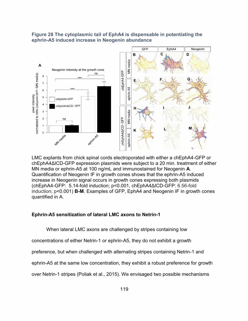
119
Figure 28 The cytoplasmic tail of EphA4 is dispensable in potentiating the ephrin-A5 induced increase in Neogenin abundance
LMC explants from chick spinal cords electroporated with either a chEphA4-GFP or chEphA4ΔICD-GFP expression plasmids were subject to a 20 min. treatment of either MN media or ephrin-A5 at 100 ng/mL and immunostained for Neogenin A. Quantification of Neogenin IF in growth cones shows that the ephrin-A5 induced increase in Neogenin signal occurs in growth cones expressing both plasmids (chEphA4-GFP: 5.14-fold induction; p<0.001, chEphA4ΔICD-GFP: 6.56-fold induction; p<0.001) B-M. Examples of GFP, EphA4 and Neogenin IF in growth cones quantified in A.
Ephrin-A5 sensitization of lateral LMC axons to Netrin-1
When lateral LMC axons are challenged by stripes containing low
concentrations of either Netrin-1 or ephrin-A5, they do not exhibit a growth
preference, but when challenged with alternating stripes containing Netrin-1 and
ephrin-A5 at the same low concentration, they exhibit a robust preference for growth
over Netrin-1 stripes (Poliak et al., 2015). We envisaged two possible mechanisms

120
explaining this behaviour: (1) Netrin-1 sensitizes lateral LMC axons to ephrin-A5
avoidance or (2) ephrin-A5 sensitizes lateral LMC axons to Netrin-1 attraction. To
distinguish between these, LMC explants were challenged with stripes containing
either ephrin-A5 or Netrin-1 and control stripes containing Fc, with bath application of
Netrin-1 or ephrin-A5, respectively (Figure 29). Lateral LMC axons were visualised
using EphA4 antibodies, and their stripe preference outgrowth was scored as
previously (Poliak et al., 2015). When lateral LMC axons were challenged with
alternating ephrin-A5 and Fc stripes (ephrin-A5 / Fc) with or without bath Netrin-1 (10
ng/mL), no avoidance of ephrin-A5 stripes was observed (Figure 29C, D, ephrin-A5
51.6 ± 5.0%, ephrin-A5 49.9 ± 8.4% respectively, P=0.5723). Lateral LMC axons
challenged with alternating Netrin-1 (10 ng/mL) and Fc stripes (Netrin-1/Fc) show no
growth preference (Figure 29A, Netrin-1 47.3 ± 6.7%, p>0.05). In contrast, in the
presence of bath applied ephrin-A5 (50 ng/mL), lateral LMC axons show preference
for Netrin-1 stripes (Figure 29B, Netrin-1 66.9 ± 7.8%, P<0.001). Although we cannot
rule out the possibility that higher Netrin-1 concentrations could sensitize LMCl axons
to ephrin-A5 avoidance, our results suggest that ephrin-A5 sensitizes LMCl axons to
Netrin-1 but Netrin-1 does not sensitize LMCl axons to ephrin-A5 avoidance.
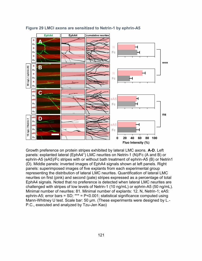
121
Figure 29 LMCl axons are sensitized to Netrin-1 by ephrin-A5
Growth preference on protein stripes exhibited by lateral LMC axons. A-D. Left panels: explanted lateral (EphA4+) LMC neurites on Netrin-1 (N)/Fc (A and B) or ephrin-A5 (eA5)/Fc stripes with or without bath treatment of ephrin-A5 (B) or Netrin1 (D). Middle panels: inverted images of EphA4 signals shown at left panels. Right panels: superimposed images of five explants from each experimental group representing the distribution of lateral LMC neurites. Quantification of lateral LMC neurites on first (pink) and second (pale) stripes expressed as a percentage of total EphA4 signals. Noted that no preference is detected when lateral LMC neurites are challenged with stripes of low levels of Netrin-1 (10 ng/mL) or ephrin-A5 (50 ng/mL). Minimal number of neurites: 81. Minimal number of explants: 12. N, Netrin-1; eA5: ephrin-A5; error bars = SD; *** = P<0.001; statistical significance computed using Mann-Whitney U test. Scale bar: 50 μm. (These experiments were designed by L.-P.C., executed and analyzed by Tzu-Jen Kao)
122
The cytoplasmic tail of EphA4 is dispensable for the sensitization of LMCl axons to Netrin-1 by ephrin-A5
Our results suggest that the ephrin-A5 dependent upregulation of Neogenin
occurs independently of the EphA4 cytoplasmic domain (Figure 28). To test whether
this also occurs in the context of guidance decisions made by LMC axon, we first
challenged LMC explants from chEphA4-GFP or chEphA4ΔICD-GFP electroporated
chickens with ephrin-A5/Fc stripes (Figure 30 C, D). As anticipated, since the
intracellular domain of EphA4 is required for its ability to elicit repulsion from ephrin-
A5, LMC axons expressing chEphA4ΔICD-GFP showed less avoidance from ephrin-
A5 in contrast to LMC axons expressing chEphA4-GFP (Figure 30 C, D; chEphA4-
GFP: ephrin-A5 stripe = 82.6 ± 9.4%, chEphA4ΔICD-GFP: ephrin-A5 stripe = 51.7 ±
7.4%; p=0.1581). However, expression of chEphA4-GFP and chEphA4ΔICD-GFP
resulted in the same enhancement of growth preference over Netrin-1 stripes in the
presence of bath-applied ephrin-A5 (Figure 30 A, B; chEphA4-GFP: Netrin-1 70.3 ±
10.2%, chEphA4ΔICD-GFP: Netrin-1 67.2 ± 7.7%; p=0.1581). Together, these data
argue that EphA4 can promote ephrin-A5-mediated sensitization of LMC axons to
Netrin-1 in the absence of its intracellular signal relay.
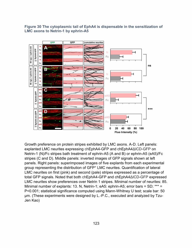
123
Figure 30 The cytoplasmic tail of EphA4 is dispensable in the sensitization of LMC axons to Netrin-1 by ephrin-A5
Growth preference on protein stripes exhibited by LMC axons. A-D. Left panels:
explanted LMC neurites expressing chEphA4-GFP and chEphA4ΔICD-GFP on
Netrin-1 (N)/Fc stripes bath treatment of ephrin-A5 (A and B) or ephrin-A5 (eA5)/Fc
stripes (C and D). Middle panels: inverted images of GFP signals shown at left
panels. Right panels: superimposed images of five explants from each experimental
group representing the distribution of GFP+ LMC neurites. Quantification of lateral
LMC neurites on first (pink) and second (pale) stripes expressed as a percentage of
total GFP signals. Noted that both chEphA4-GFP and chEphA4ΔICD-GFP expressed
LMC neurites show preferences over Netrin 1 stripes. Minimal number of neurites: 85.
Minimal number of explants: 13. N, Netrin-1; eA5: ephrin-A5; error bars = SD; *** =
P<0.001; statistical significance computed using Mann-Whitney U test; scale bar: 50
μm. (These experiments were designed by L.-P.C., executed and analyzed by Tzu-
Jen Kao)

124
CHAPTER IV - DISCUSSION
125
Neogenin in LMC neuron differentiation
Taken together, the experiments presented in this chapter suggest that
Neogenin is required in LMC neuron subtype segregation and differentiation in the
chick. The formulation of a strong conclusion regarding the requirement for Neogenin
in LMC development is hindered by the absence of non-siRNA based experiments.
For this reason, we can not rule out the possibility that the phenotype seen in
[Neo]siRNA embryos could be the result of siRNA off target effects. Some
experiments, such as the analysis of LMC neuron subtype differentiation in
[RGMb]siRNA embryos and in Neo-/- mice require additional experiments to be
conclusive. For these reasons, I have hesitated in including these results in this thesis
and had no intention in including work from this chapter in my thesis defence
presentation. I will leave it upon my thesis committee to decide whether certain
elements of this chapter should be excluded from my final submission. Nonetheless, I
will discuss insights in Neogenin function from recent publication that may shed light
on my experimental results.
Neogenin in LMC subtype segregation
In epithelia, cadherin based adherens junctions regulate cell-cell junctional
stability (Priya and Yap, 2015). Adherens junctions rely upon the interaction between
cadherins and closely opposed actin rings (Priya and Yap, 2015). Actin nucleation via
the Arp2/3 enzyme complex is necessary for maintaining epithelial cohesion
(Bernadskaya et al., 2011). Arp2/3 dependent actin nucleation is stimulated by the
126
WAVE regulatory complex (WRC) (Chen et al., 2014). Several surface molecules
including proteins involved in adhesion such as protocadherins and axon guidance
receptors such as Robo1, Dcc and Neogenin, have WRC binding domains (Chen et
al., 2014). In in vitro epithelial cell cultures, Neogenin and RGMa are required for
adherens junction stability and the spatial control of WRC/Arp2/3 recruitment to
adherens junctions (Lee et al., 2016). NeoGT/GT mice display a loss of adherens
junctions in radial glial cells of the embryonic cortex as well as in ependymal cells in
the postnatal ventricular zone (O'Leary et al., 2017). Interestingly, loss of cadherin
function through the genetic ablation of N-Cadherin or the combined loss of beta and
gamma-catenin in spinal motor neurons, results in the intermingling of LMCm and
LMCl neurons (Demireva et al., 2011). Although no change in the generation of LMCl
neurons have been reported in mice carrying these mutations, this phenotype is
reminiscent of the lack of LMCm/LMCl neuron segregation observed in [Neo]siRNA
embryos. Evidence of Neogenin in cadherin based cell adhesion and the requirement
for cadherins in LMC subtype segregation suggests that the perturbation in
LMCm/LMCl segregation observed in [Neo]siRNA embryos may be attributable to
impaired cell adhesion among LMC neurons.
127
Neogenin in LMC subtype differentiation
In [Neo]siRNA embryos, ectopic LMCm neurons more frequently occupy the
ventral portion of the LMC while the most lateral population of LMCl neurons remains
largely unaltered. These results suggest that the requirement for Neogenin in Lim1
expression may differ among LMCl neurons (Figure 13). During LMC neuron
development, retinoic acid is generated by the paraxial mesoderm immediately
adjacent to the spinal cord (Ji et al., 2006). In mice carrying a conditional mutation
that ablates Raldh2 in brachial motor neurons but maintains expression in the
paraxial mesoderm, 80% of LMCl neurons are generated (Vermot et al., 2005).
Conversely, mice hypomorphic for Raldh2 in the paraxial mesoderm also exhibit a
similar decrease in LMCl generation (Ji et al., 2006). This suggests that retinoic acid
provided by both LMC neurons and the paraxial mesoderm, contribute to the
generation of LMCl neurons (Ji et al., 2006). It is therefore likely that in [Neo]siRNA
embryos, the more lateral population of LMCl neurons may acquire sufficient retinoic
acid from the paraxial mesoderm whereas the more ventral LMCl neuron
subpopulation may be more reliant on retinoic acid derived from LMC neurons.
In the olfactory epithelium of mice, the proper regulation of progenitor cell cycle
progression and exit relies on the expression of Neogenin by progenitors and the
expression of RGMb by neighbouring immature olfactory receptor neurons (Kam et
al., 2016). A partially analogous mechanism may be occurring in LMC subtype
differentiation. Prospective LMCl neurons expressing Neogenin may bind RGMb on
128
earlier born LMCm neurons. These interactions may favor cell-cell contacts that favor
retinoic acid uptake by prospective LMCl neurons.
The absence of an obvious LMC subtype differentiation phenotype in Neo-/-
mice argues that the acquisition of Lim1 expression occurs independently from
Neogenin in the mouse (Figure 16). We can not rule out the possibility that Dcc
expression by LMC neurons could compensate for the loss of Neogenin in Lim1
expression in mice. This would entail a mechanism independent from RGMs since
Dcc is not known for binding RGMs. The requirement for Neogenin/RGM in LMC
development may differ between chick in mice. Whereas in chick, loss of RGMa
results in RGC axon guidance defects, RGC axons appear normal in RGMa-/- mice.
(Monnier et al., 2002; Niederkofler et al., 2004).
My results suggest that Neogenin is involved in multiple aspects of LMC
neuron development in the chick including the acquisition of Lim1 expression by
prospective LMCl neurons, the segregation of LMCm/LMCl neurons as well as the
guidance of LMC axons. To confirm the involvement of Neogenin in theses aspects of
LMC development would require additional experimentation such as the rescue of
[Neo]siRNA phenotype by the overexpression mouse Neogenin and or Dcc as well as
additional experiments regarding the potential involvement of RGMs
129
Sensitization of LMCl axons to Netrin-1 by ephrin-A5
Through the stripe assay, we have previously demonstrated that ephrin-A5 and
Netrin-1 act synergistically on LMCl axons in vitro (Poliak et al., 2015). To gain insight
in the molecular mechanism underling the synergistic activity of ephrin-A5 and Netrin-
1 in LMC neurons, an analysis of receptor dynamics in LMC growth cones subject to
Netrin-1 and ephrin-A5 was carried out. Our results demonstrate that ephrin-A5
induces the upregulation of Neogenin in growth cones leading to enhanced Netrin-1
binding. Furthermore, we show that the upregulation of Neogenin is dependent on the
interaction between ephrin-A5 and EphA4 and is enhanced by the overexpression of
full-length and truncated EphA4. Using stripe assays, we show that bath applied
ephrin-A5 sensitizes LMCl axons to Netrin-1 and demonstrate that sensitization
occurs independently from the cytoplasmic tail of EphA4. These results demonstrate
that ephrin-A5/ EphA4 forward signalling and ephrin-A5/EphA4 sensitization to Netrin-
1 occur in molecularly distinct pathways.
Netrin-1 increases and redistributes Neogenin to the growth cone periphery
We show that Netrin-1 induces the re-localization of Neogenin towards the
growth cone periphery at Netrin-1 concentrations as low as 10 ng/mL in a PKA
dependent fashion (Figure 18, 3). The observation that surface Neogenin is enriched
peripherally in the absence of Netrin-1 treatment (Figure 23G) suggests that the
Netrin-1- induced peripheral relocalization is likely accompanied by Neogenin
insertion into the plasma membrane. The dependence on PKA for membrane
130
insertion of Dcc has been documented in rat commissural neurons and is likely
analogous to the dependence on PKA for Neogenin relocalization (Bouchard et al.,
2004; Bouchard et al., 2008). The absence of Netrin-1 induced change in the levels of
EphA4 suggests that Netrin-1 unlikely sensitizes LMC growth cones to ephrin-A5 by
modulating EphA4. This is substantiated by the results showing that bath application
Netrin-1 does not increase the avoidance of LMCl axons to ephrin-A5 stripes (Figure
29C, D). Since the EphA4 antibody used to measure EphA4 IF levels recognizes an
intracellular epitope, we can not rule out the possibility that Netrin-1 could increase
the membrane insertion of EphA4 without changing total levels. The use of an
antibody against the extracellular portion of EphA4 in non-permeabilized, Netrin-1
treated LMC cultures could resolve this query.
Ephrin-A5 on receptor dynamics in LMC growth cones
Our results show that a 20 min. exposure to ephrin-A5 at 50 and 100 ng/mL
but not at higher concentrations, results in increased growth cone Neogenin levels. In
vitro, ephrin-A2 can be growth promoting at low concentrations and switch to growth
inhibition at higher concentrations in RGCs axons (Hansen et al., 2004). It is
proposed that at low ephrin-A2 concentrations, low levels of EphA receptor clustering
would favor adhesion and outgrowth whereas high levels of EphA clustering, resulting
from higher ephrin-A2 concentrations, would induce repulsive downstream signaling
and decreased adhesion through EphA receptor cleavage (Hansen et al., 2004). It
should also be considered is that in the experiments presented here, only growth
131
cones that were non-collapsed and contained lamellipodia were considered for
analysis. The higher ephrin-A5 concentrations tested here may have collapsed a
subset of LMC growth cones capable of ephrin-A5 induced Neogenin upregulation at
lower ephrin-A5 concentrations.
Ephrin-A5 treatment results in a re-localization of EphA4, Unc5c, BEN (Figure
21H-K) and surface enriched Neogenin (Figure 23M) from the periphery to the
growth cone center. This may be the consequence of an ephrin-A5 induced
membrane endocytosis in the peripheral domain of the growth cone (Irie et al., 2005;
Tojima et al., 2010; Yoo et al., 2011) . The ephrin-A5 induced increase in Neogenin is
accompanied by an increase in the average size of Neogenin IF aggregates (Figure
20 F, G) and an increase in the proportion of overlap between EphA4 and Neogenin
signals (Figure 21). Furthermore, when overexpressing chEphA4-GFP, ephrin-A5
induces the appearance of dense GFP+, EphA4+, Neogenin+ aggregates (Figure 27
B-G). These results suggest that ephrin-A5/EphA4 interactions increase Neogenin
locally. Binding of ephrin-A2 or ephrin-B3 to EphA4 induces EphA4 endocytosis
(Zimmer et al., 2003; Deininger et al., 2008). The ephrin-B1 induction of EphB2
endocytosis requires EphB2’s cytoplasmic tail and kinase activity (Zimmer et al.,
2003). This suggests that in growth cones expressing chEphA4ΔICD-GFP, the
ephrin-A5 induction of truncated EphA4 endocytosis is likely impaired. The high level
of ephrin-A5 dependent Neogenin induction in chEphA4ΔICD-GFP expressing growth
cones (Figure 27A) suggest that the increase in Neogenin occurs independently of
EphA4 endocytosis and is likely initiated at the membrane surface.
132
Ephrin-A5/EphA4 attenuation of Neogenin cleavage as a possible mechanism underlining the sensitization to Netrin-1
Several transmembrane proteins are regulated by ligand-dependent
ectodomain cleavage, followed by a secondary cleavage of the remaining intracellular
fragment by γ-secretase (Bai and Pfaff, 2011). The in vitro treatment of dorsal spinal
explants with metalloprotease inhibitors results in an increase in Dcc expression and
a potentiation towards Netrin-1 dependent axonal outgrowth (Galko and Tessier-
Lavigne, 2000). The ectodomain of Neogenin can be cleaved by the metalloprotease
ADAM17, and was proposed to modulate RGMa induced repulsion by regulating
surface Neogenin (Okamura et al., 2011). Furthermore, cleavage of Neogenin by
ADAM17 can be regulated by the transmembrane protein Lrig2 (van Erp et al., 2015).
Lrig2 is proposed to shield Neogenin from ADAM17 cleavage and binding of RGMa to
Neogenin inhibits Lrig2/Neogenin interactions and allows cleavage by ADAM17 (van
Erp et al., 2015). Dcc and Neogenin both undergo cleavage by γ-secretase, resulting
in the release of their intracellular domains (ICD) (Taniguchi et al., 2003;
Goldschneider et al., 2008). In mice mutant for Presenilin-1, an essential catalytic
component of the γ-secretase complex, a subset of spinal commissural axons fail to
cross the floor-plate and spinal motor neurons aberrantly extend axons to and across
the midline (Bai et al., 2011). These guidance defects are suggested to result from
altered sensitivity of spinal neuron axons towards Netrin-1(Bai et al., 2011). Thus, the
regulation of Dcc and Neogenin cleavage is a molecular strategy for modulating
cellular responses to external cues.
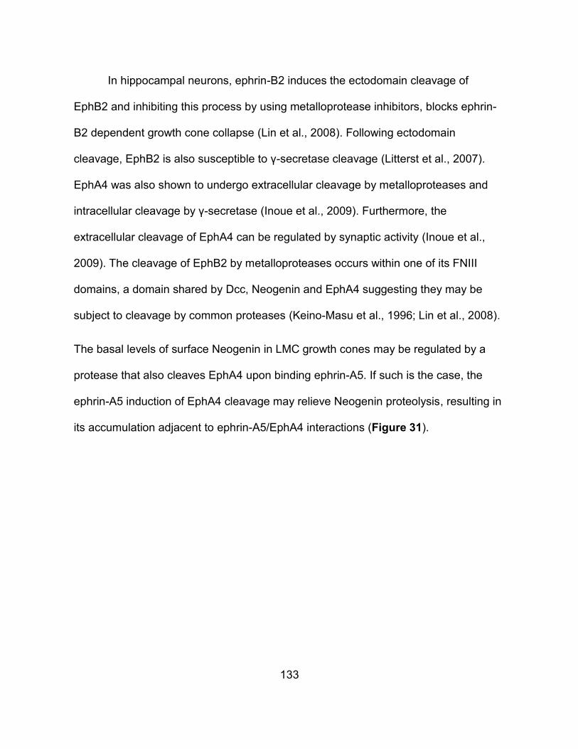
133
In hippocampal neurons, ephrin-B2 induces the ectodomain cleavage of
EphB2 and inhibiting this process by using metalloprotease inhibitors, blocks ephrin-
B2 dependent growth cone collapse (Lin et al., 2008). Following ectodomain
cleavage, EphB2 is also susceptible to γ-secretase cleavage (Litterst et al., 2007).
EphA4 was also shown to undergo extracellular cleavage by metalloproteases and
intracellular cleavage by γ-secretase (Inoue et al., 2009). Furthermore, the
extracellular cleavage of EphA4 can be regulated by synaptic activity (Inoue et al.,
2009). The cleavage of EphB2 by metalloproteases occurs within one of its FNIII
domains, a domain shared by Dcc, Neogenin and EphA4 suggesting they may be
subject to cleavage by common proteases (Keino-Masu et al., 1996; Lin et al., 2008).
The basal levels of surface Neogenin in LMC growth cones may be regulated by a
protease that also cleaves EphA4 upon binding ephrin-A5. If such is the case, the
ephrin-A5 induction of EphA4 cleavage may relieve Neogenin proteolysis, resulting in
its accumulation adjacent to ephrin-A5/EphA4 interactions (Figure 31).
134
Figure 31 Tentative model for the sensitization of LMCl axons to Netrin-1
A. The basal surface levels of Neogenin at the growth cone may be regulated through
the cleavage of its extracellular fragment by a protease. B. The binding of ephrin-A5
to EphA4 induces the cleavage of the extracellular fragment of EphA4. If Neogenin
and EphA4 are cleaved by a common protease, then the ephrin-A5 induced
proteolytic cleavage of EphA4 may attenuate the cleavage of Neogenin in the vicinity
of ephrin-A5/EphA4 interactions. This would result in an increase in Netrin-1 binding
and a potentiation in Netrin-1 downstream signalling (red arrows).
The increase in the proportion of Neogenin/EphA4 IF overlap upon ephrin-A5
treatment (Figure 22) and the co-localization of Neogenin/GFP puncta when
overexpressing chEphA4-GFP in the presence of ephrin-A5 (Figure 28E-G) suggests
that the upregulation of Neogenin is enriched where ephrin-A5/EphA4 interactions
occur. The re-localization of surface Neogenin towards the center of the growth cone,
where EphA4 is enriched when subject to ephrin-A5 (Figure 21J, Figure 23M) is also
135
in line with this possibility. We have recently obtained preliminary data showing that
unlike the overexpression of either full-length or truncated EphA4, the overexpression
of an non-cleavable EpA4 fails to enhance the increase in Neogenin levels at the
growth cone (data not shown). We are now in the process of adding additional
experiments implicating non-cleavable EphA4 as well as testing the requirement for
proteolytic cleavage in the ephrin-A5/EphA4 dependent upregulation of Neogenin
through the use of protease inhibitors. Interestingly, mice in which EphA4 cleavage is
abolished, a subset of LMCl axons are misguided into the ventral limb (Gatto et al.,
2014). If the ephrin-A5 upregulation of Neogenin requires cleavage of EphA4, the
LMCl axons guidance defects seen in the mouse harboring a cleavage resistant
EphA4 may be in part due to lower Neogenin/Dcc levels resulting in decreased axon
attraction to Netrin-1 in the dorsal limb (Poliak et al., 2015).
EphA4 has been implicated in various neurodegenerative diseases including
multiple sclerosis, amyotrophic lateral sclerosis and Alzheimer’s disease (Simon et
al., 2009; Shen et al., 2010; Van Hoecke et al., 2012; Munro et al., 2013).
Interestingly, ephrin-A/EphA4 interactions were shown to regulate the degradation of
the Alzheimer’s disease related protein amyloid precursor protein (APP) (Lai et al.,
2014). Thus, ephrin-A/EphA4 regulation of protein degradation may influence several
aspects of neuronal function during development as well as in neurodegenerative
diseases.
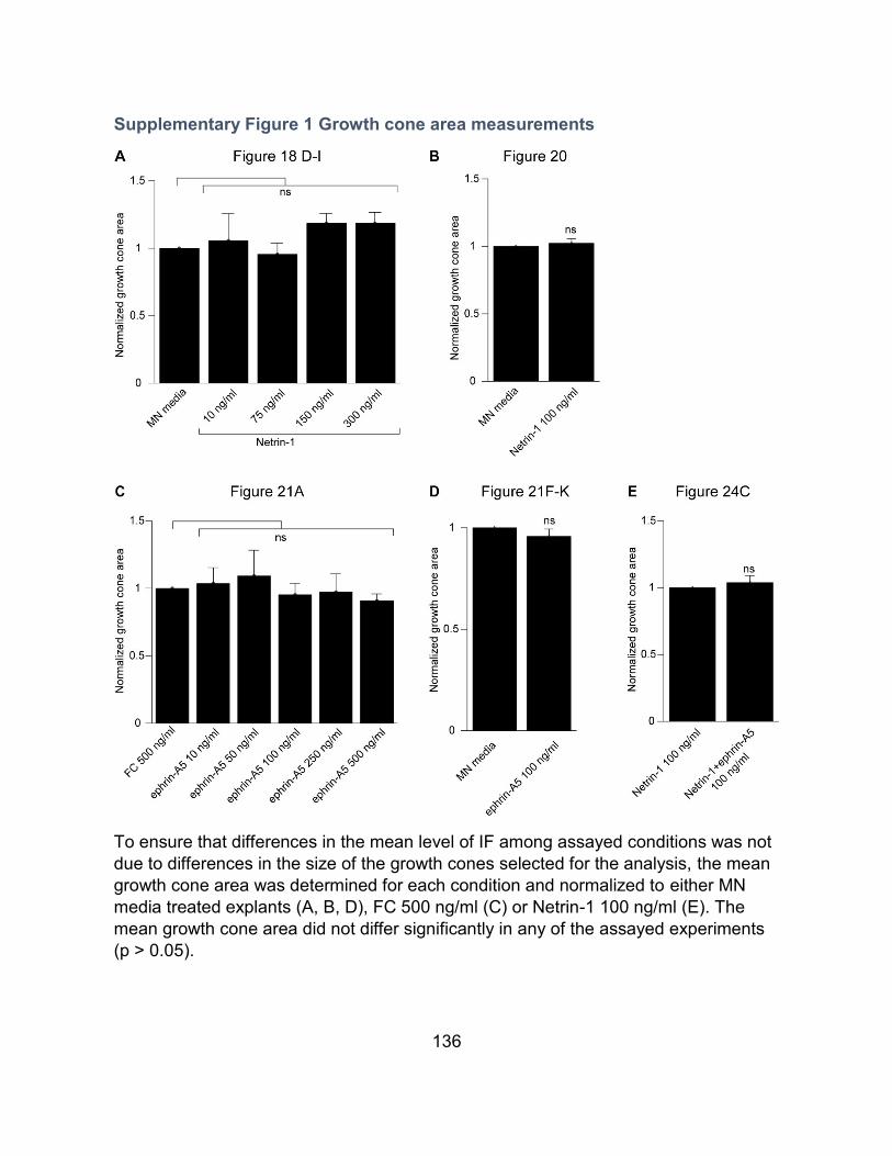
136
Supplementary Figure 1 Growth cone area measurements
To ensure that differences in the mean level of IF among assayed conditions was not
due to differences in the size of the growth cones selected for the analysis, the mean
growth cone area was determined for each condition and normalized to either MN
media treated explants (A, B, D), FC 500 ng/ml (C) or Netrin-1 100 ng/ml (E). The
mean growth cone area did not differ significantly in any of the assayed experiments
(p > 0.05).

137
CHAPTER V – CONTRIBUTIONS AND PERSPECTIVES
138
Neogenin as a mediator of attractive Netrin-1 signaling in LMC neurons
My thesis project began by investigating the role of chick Neogenin as a
Netrin-1 receptor in LMC neurons. Using the stripe assay and a Neogenin function
blocking antibody, I demonstrated that Neogenin is required for the responsiveness of
LMCl axons towards Netrin-1 in the chick. I also showed that this loss of
responsiveness can be rescued through the overexpression of it’s closely related
homologue Dcc. These results are in line with previous evidence suggesting that
chick Neogenin can functionally substitute for Dcc in mediating attraction towards
Netrin-1 and establishes chick LMCl neurons as a model system for studying
attractive Netrin-1 signaling (Phan et al., 2011; Poliak et al., 2015).
Speculations on the requirement for Neogenin dependent transcriptional regulation for the expression of Lim1 by LMC neurons
Neogenin was shown to interact directly with the transcription coactivator
LMO4 and the intracellular domain (ICD) of Neogenin can translocate to the nucleus
where it can presumably regulate transcription (Goldschneider et al., 2008; Banerjee
et al., 2016). RGMa induced outgrowth inhibition of RGC axons is lost when LMO4 is
knocked down, suggesting that NeoICD/LMO4 complexes may in part regulate RGMa
signaling by regulating transcription (Banerjee et al., 2016). Through transcriptional
regulation, LMO4 directs the balance between inhibitory and excitatory interneurons
in the spinal cord (Joshi et al., 2009). Interestingly, LMO4 is expressed by LMC
neurons and was shown to compete with Isl1 in binding LIM homeodomain
complexes. Furthermore, LMO4 mRNA is downregulated in cells exposed to retinoic
139
acid, suggesting a possible role for LMO4 in retinoic acid signalling (Duffy et al.,
2017). Taken together, the evidence suggests that Neogenin/RGM interactions in
prospective LMCl neurons may induce the nuclear translocation of NeoICD/LMO4
complexes which together with retinoic acid, may induce the switch from Isl1 to Lim1
expression. To assess a potential requirement for LMO4 in Lim1 expression by LMC
neurons, LMO4 could be overexpressed or Knocked down in LMC neurons. It would
also be interesting to attempt rescuing the LMC differentiation phenotype in
[Neo]siRNA embryos by overexpressing LM04.
Optimization of the chick in ovo electroporation protocol
In my initial in ovo electroporation experiments, I noticed that some
electroporated embryos displayed a loss in ventricular zone integrity accompanied by
ectopic LMC neurons adjacent to the disrupted ventricular zone. These
electroporation artifacts led us to optimize the standard protocol for in ovo
electroporation and to publish our findings (Croteau and Kania, 2011). The revised
protocol highlights the importance of the DNA dilution buffer and electrode placement
in maintaining the cellular integrity of electroporated spinal cords. We show that the
use of TE buffer in the electroporation solution can result in the loss of ventricular
zone integrity leading to neuron migration defects. This is likely due to the Ca2+
chelating properties of EDTA disrupting Cadherin based adherens junctions at the
ventricular zone (Takeichi, 1988; Croteau and Kania, 2011)
140
The sensitization of LMC neurons to Netrin-1 by ephrin-A5
My results demonstrate that low concentrations of ephrin-A5 increases the
levels of Neogenin and enhances Netrin-1 binding in LMC growth cones. We show
that the upregulation of Neogenin is dependent on the interaction between ephrin-A5 and
EphA4 and is enhanced by the overexpression of full-length and truncated EphA4. We
show that bath applied ephrin-A5 sensitizes LMCl axons to Netrin-1 and demonstrate that
sensitization occurs independently from the cytoplasmic tail of EphA4. These results
demonstrate that ephrin-A5/ EphA4 forward signalling and ephrin-A5/EphA4 sensitization
to Netrin-1 occur in molecularly distinct pathways.
A potential for ephrin-A5 induced upregulation of Neogenin in various cell types
Although at lower levels than in LMCl neurons, LMCm neurons express EphA4
and encounter ephrin-A5 in the ventral limb (Bonanomi et al., 2012; Gatto et al.,
2014). This raises the possibility that ephrin-A5 may also sensitize LMCm axons to
Netrin-1. There is evidence demonstrating that repulsion from Netrin-1 requires the
interaction between Unc5 receptors and Dcc (Hong et al., 1999). Although ephrin-A5
does not seem to increase Unc5c levels in LMC growth cones, it is possible that
higher levels of Neogenin at the plasma membrane may contribute to enhancing
Netrin-1/Unc5c signalling. There is also the possibility that other ephrins may induce
sensitization to Netrin-1, in mice lacking ephrin-A2, LMCl axons are misguided into
141
the ventral limb, a phenotype that is exacerbated in Efna2-/-;Efna5-/- double mutants
(Bonanomi et al., 2012).
We have yet to investigate the possibility that other neuronal populations may
be susceptible to ephrin-A5 induced sensitization to Netrin-1. In vitro, In the context of
EphA4 overexpression, ephrin-A5 treatment can increase Neogenin levels in cells
other than LMC neurons, suggesting that other cell types may be sensitized to Netrin-
1 by ephrin-A5 (data not shown). Netrin-1 is required for spinal commissural neurons
in the dorsal spinal cord to extend axons ventrally towards the floor plate (Kennedy et
al., 1994; Serafini et al., 1994; Serafini et al., 1996). EphA4 and ephrin-A5 are
expressed in the dorsal spinal cord as spinal commissural axons extend ventrally
(Iwamasa et al., 1999; Eberhart et al., 2004; Kramer et al., 2006). One intriguing
possibility is that commissural neurons may also be sensitized to Netrin-1 as growth
cones encounter ephrin-A5 expressed by radial glia.
Neogenin is implicated in multiple developmental processes including iron
homeostasis, myogenesis and bone formation (Bae et al., 2009; Lee et al., 2010;
Zhou et al., 2010). A requirement for Neogenin in adults has also been demonstrated
in acute inflammation as well as in adult neurogenesis (Konig et al., 2012; O'Leary et
al., 2015). Ephrin-A5 being widely expressed during development and adulthood (Yue
et al., 2014) raises the possibility that ephirn-A5 may enhance several aspects of
Neogenin function.
142
A potential requirement for ephrin-A/EphA induction of Neogenin for neural tube closure
Lack of either Neogenin, RGMa or Netrin-1 expression have previously been
shown to cause neural tube closure defects (Mawdsley et al., 2004; Niederkofler et
al., 2004; Kee et al., 2008; Kee et al., 2013). Interestingly, neural tube closure defects
are also seen in EphA7 and ephrin-A5 null embryos (Holmberg et al., 2000). During
neurulation in mice, three EphA7 splice variants and ephrin-A5 are expressed at the
dorsal edges of neural folds (Holmberg et al., 2000). Two EphA7 splice isoforms are
truncated and lack kinase domains. It has been proposed that the expression the
expression of truncated EphA7 in the neural folds inhibits ephrin-A5/EphA7 repulsion
and through an unknown mechanism, promotes adhesion (Holmberg et al., 2000). In
light of the evidence provided here whereby ephrin-A5/EphA4 interactions result in
Neogenin upregulation, one can imagine a scenario where ephrin-A5/EphA7
interactions in the neural folds would result in increased Neogenin levels thereby
increasing Neogenin/RGMa and Neogenin/Netrin-1 interactions required for proper
neural tube closure. The prevalence of Neogenin upregulation in the context of
overexpressing EphA4 lacking its cytoplasmic tail suggests that the full-length and
truncated isoforms of EphA7 could potentially up-regulate Neogenin when bound to
ephrin-A5. It would be interesting to determine if the levels of Neogenin expression
are diminished in the neural folds of EphA7 and ephrin-A5 null mice.
143
Speculations on the requirement for ephrin/EphA4 dependent upregulation of Dcc/Neogenin for LTP
Post-synaptic EphA4 and ephrin-A3 expressed by astrocytes are required for
LTP in the hippocampus (Filosa et al., 2009). The requirement for EphA4 in LTP
occurs independently from its cytoplasmic tail. LTP deficits seen in EphA4 null mice
are absent in mice expressing EphA4 lacking its cytoplasmic tail (Grunwald et al.,
2004). Loss of EphA4 increases the abundance of glial glutamate transporters and
LTP deficits can be rescued by blocking glial glutamate transporters (Filosa et al.,
2009). The mechanism underlining the requirement for EphA4 in LTP is unknown
(Filosa et al., 2009). Interestingly, Dcc null mice also show LTP deficits in the
hippocampus, proposed to originate from decreased levels of Dcc-dependent Src
activation of NMDA receptors (Horn et al., 2013). Post-synaptic Src activation is
necessary and sufficient for inducing LTP (Lu et al., 1998). The overlapping functions
between chicken Neogenin and mouse Dcc as well as the expression of Neogenin in
the hippocampus in mice raises the possibility that ephrin/EphA4 interactions may in
part promote LTP by increasing the abundance of post-synaptic Dcc/Neogenin. The
Ephrin-A5 induced increase in post-synaptic Dcc/Neogenin could result in higher
levels of Src dependent NMDA receptor phosphorylation an thereby favor LTP (Gad
et al., 1997; Phan et al., 2011; Poliak et al., 2015).
:
144
BIBLIOGRAPHY
. Ahmed G, Shinmyo Y, Ohta K, Islam SM, Hossain M, Naser IB, Riyadh MA, Su Y,
Zhang S, Tessier-Lavigne M, Tanaka H (2011) Draxin inhibits axonal outgrowth through the netrin receptor DCC. The Journal of neuroscience : the official journal of the Society for Neuroscience 31:14018-14023.
Ahn K, Mishina Y, Hanks MC, Behringer RR, Crenshaw EB, 3rd (2001) BMPR-IA signaling is required for the formation of the apical ectodermal ridge and dorsal-ventral patterning of the limb. Development (Cambridge, England) 128:4449-4461.
Airaksinen MS, Saarma M (2002) The GDNF family: signalling, biological functions and therapeutic value. Nature reviews Neuroscience 3:383-394.
Andrews GL, Tanglao S, Farmer WT, Morin S, Brotman S, Berberoglu MA, Price H, Fernandez GC, Mastick GS, Charron F, Kidd T (2008) Dscam guides embryonic axons by Netrin-dependent and -independent functions. Development (Cambridge, England) 135:3839-3848.
Babitt JL, Zhang Y, Samad TA, Xia Y, Tang J, Campagna JA, Schneyer AL, Woolf CJ, Lin HY (2005) Repulsive guidance molecule (RGMa), a DRAGON homologue, is a bone morphogenetic protein co-receptor. The Journal of biological chemistry 280:29820-29827.
Babitt JL, Huang FW, Wrighting DM, Xia Y, Sidis Y, Samad TA, Campagna JA, Chung RT, Schneyer AL, Woolf CJ, Andrews NC, Lin HY (2006) Bone morphogenetic protein signaling by hemojuvelin regulates hepcidin expression. Nature genetics 38:531-539.
Bae GU, Yang YJ, Jiang G, Hong M, Lee HJ, Tessier-Lavigne M, Kang JS, Krauss RS (2009) Neogenin regulates skeletal myofiber size and focal adhesion kinase and extracellular signal-regulated kinase activities in vivo and in vitro. Molecular biology of the cell 20:4920-4931.
Bai G, Pfaff SL (2011) Protease regulation: the Yin and Yang of neural development and disease. Neuron 72:9-21.
Bai G, Chivatakarn O, Bonanomi D, Lettieri K, Franco L, Xia C, Stein E, Ma L, Lewcock JW, Pfaff SL (2011) Presenilin-dependent receptor processing is required for axon guidance. Cell 144:106-118.
Banerjee P, Harada H, Tassew NG, Charish J, Goldschneider D, Wallace VA, Sugita S, Mehlen P, Monnier PP (2016) Upsilon-secretase and LARG mediate distinct RGMa activities to control appropriate layer targeting within the optic tectum. Cell death and differentiation 23:442-453.
Barallobre MJ, Pascual M, Del Rio JA, Soriano E (2005) The Netrin family of guidance factors: emphasis on Netrin-1 signalling. Brain research Brain research reviews 49:22-47.
Bell CH, Healey E, van Erp S, Bishop B, Tang C, Gilbert RJC, Aricescu AR, Pasterkamp RJ, Siebold C (2013) Structure of the repulsive guidance molecule (RGM)-neogenin signaling hub. Science (New York, NY) 341:77-80.
145
Bennett KL, Bradshaw J, Youngman T, Rodgers J, Greenfield B, Aruffo A, Linsley PS (1997) Deleted in colorectal carcinoma (DCC) binds heparin via its fifth fibronectin type III domain. The Journal of biological chemistry 272:26940-26946.
Bernadskaya YY, Patel FB, Hsu HT, Soto MC (2011) Arp2/3 promotes junction formation and maintenance in the Caenorhabditis elegans intestine by regulating membrane association of apical proteins. Molecular biology of the cell 22:2886-2899.
Bielle F, Marcos-Mondejar P, Leyva-Diaz E, Lokmane L, Mire E, Mailhes C, Keita M, Garcia N, Tessier-Lavigne M, Garel S, Lopez-Bendito G (2011) Emergent growth cone responses to combinations of Slit1 and Netrin 1 in thalamocortical axon topography. Current biology : CB 21:1748-1755.
Bin JM, Han D, Lai Wing Sun K, Croteau LP, Dumontier E, Cloutier JF, Kania A, Kennedy TE (2015) Complete Loss of Netrin-1 Results in Embryonic Lethality and Severe Axon Guidance Defects without Increased Neural Cell Death. Cell reports 12:1099-1106.
Blake RA, Broome MA, Liu X, Wu J, Gishizky M, Sun L, Courtneidge SA (2000) SU6656, a selective src family kinase inhibitor, used to probe growth factor signaling. Molecular and cellular biology 20:9018-9027.
Bonanomi D, Pfaff SL (2010) Motor axon pathfinding. Cold Spring Harb Perspect Biol 2:a001735.
Bonanomi D, Chivatakarn O, Bai G, Abdesselem H, Lettieri K, Marquardt T, Pierchala BA, Pfaff SL (2012) Ret is a multifunctional coreceptor that integrates diffusible- and contact-axon guidance signals. Cell 148:568-582.
Borgius L, Nishimaru H, Caldeira V, Kunugise Y, Low P, Reig R, Itohara S, Iwasato T, Kiehn O (2014) Spinal glutamatergic neurons defined by EphA4 signaling are essential components of normal locomotor circuits. The Journal of neuroscience : the official journal of the Society for Neuroscience 34:3841-3853.
Bottcher RT, Pollet N, Delius H, Niehrs C (2004) The transmembrane protein XFLRT3 forms a complex with FGF receptors and promotes FGF signalling. Nature cell biology 6:38-44.
Bouchard JF, Horn KE, Stroh T, Kennedy TE (2008) Depolarization recruits DCC to the plasma membrane of embryonic cortical neurons and enhances axon extension in response to netrin-1. Journal of neurochemistry 107:398-417.
Bouchard JF, Moore SW, Tritsch NX, Roux PP, Shekarabi M, Barker PA, Kennedy TE (2004) Protein kinase A activation promotes plasma membrane insertion of DCC from an intracellular pool: A novel mechanism regulating commissural axon extension. The Journal of neuroscience : the official journal of the Society for Neuroscience 24:3040-3050.
Brankatschk M, Dickson BJ (2006) Netrins guide Drosophila commissural axons at short range. Nature neuroscience 9:188-194.
Briancon-Marjollet A, Ghogha A, Nawabi H, Triki I, Auziol C, Fromont S, Piche C, Enslen H, Chebli K, Cloutier JF, Castellani V, Debant A, Lamarche-Vane N
146
(2008) Trio mediates netrin-1-induced Rac1 activation in axon outgrowth and guidance. Molecular and cellular biology 28:2314-2323.
Briscoe J, Ericson J (2001) Specification of neuronal fates in the ventral neural tube. Current opinion in neurobiology 11:43-49.
Briscoe J, Small S (2015) Morphogen rules: design principles of gradient-mediated embryo patterning. Development (Cambridge, England) 142:3996-4009.
Bron R, Vermeren M, Kokot N, Andrews W, Little GE, Mitchell KJ, Cohen J (2007) Boundary cap cells constrain spinal motor neuron somal migration at motor exit points by a semaphorin-plexin mechanism. Neural development 2:21.
Carter SB (1965) Principles of cell motility: the direction of cell movement and cancer invasion. Nature 208:1183-1187.
Carvalho RF, Beutler M, Marler KJ, Knoll B, Becker-Barroso E, Heintzmann R, Ng T, Drescher U (2006) Silencing of EphA3 through a cis interaction with ephrinA5. Nature neuroscience 9:322-330.
Castellani V, Yue Y, Gao PP, Zhou R, Bolz J (1998) Dual action of a ligand for Eph receptor tyrosine kinases on specific populations of axons during the development of cortical circuits. The Journal of neuroscience : the official journal of the Society for Neuroscience 18:4663-4672.
Castets M, Broutier L, Molin Y, Brevet M, Chazot G, Gadot N, Paquet A, Mazelin L, Jarrosson-Wuilleme L, Scoazec JY, Bernet A, Mehlen P (2011) DCC constrains tumour progression via its dependence receptor activity. Nature 482:534-537.
Chacon MR, Fazzari P (2011) FAK: dynamic integration of guidance signals at the growth cone. Cell adhesion & migration 5:52-55.
Chen B, Brinkmann K, Chen Z, Pak CW, Liao Y, Shi S, Henry L, Grishin NV, Bogdan S, Rosen MK (2014) The WAVE regulatory complex links diverse receptors to the actin cytoskeleton. Cell 156:195-207.
Chen H, Lun Y, Ovchinnikov D, Kokubo H, Oberg KC, Pepicelli CV, Gan L, Lee B, Johnson RL (1998) Limb and kidney defects in Lmx1b mutant mice suggest an involvement of LMX1B in human nail patella syndrome. Nature genetics 19:51-55.
Cirulli V, Yebra M (2007) Netrins: beyond the brain. Nature reviews Molecular cell biology 8:296-306.
Colamarino SA, Tessier-Lavigne M (1995) The axonal chemoattractant netrin-1 is also a chemorepellent for trochlear motor axons. Cell 81:621-629.
Conrad S, Genth H, Hofmann F, Just I, Skutella T (2007) Neogenin-RGMa signaling at the growth cone is bone morphogenetic protein-independent and involves RhoA, ROCK, and PKC. The Journal of biological chemistry 282:16423-16433.
Croteau LP, Kania A (2011) Optimisation of in ovo electroporation of the chick neural tube. Journal of neuroscience methods 201:381-384.
Dasen JS, Liu JP, Jessell TM (2003) Motor neuron columnar fate imposed by sequential phases of Hox-c activity. Nature 425:926-933.
Dasen JS, Tice BC, Brenner-Morton S, Jessell TM (2005) A Hox regulatory network establishes motor neuron pool identity and target-muscle connectivity. Cell 123:477-491.
147
Dasen JS, De Camilli A, Wang B, Tucker PW, Jessell TM (2008) Hox repertoires for motor neuron diversity and connectivity gated by a single accessory factor, FoxP1. Cell 134:304-316.
Davis S, Gale NW, Aldrich TH, Maisonpierre PC, Lhotak V, Pawson T, Goldfarb M, Yancopoulos GD (1994) Ligands for EPH-related receptor tyrosine kinases that require membrane attachment or clustering for activity. Science (New York, NY) 266:816-819.
De Strooper B, Saftig P, Craessaerts K, Vanderstichele H, Guhde G, Annaert W, Von Figura K, Van Leuven F (1998) Deficiency of presenilin-1 inhibits the normal cleavage of amyloid precursor protein. Nature 391:387-390.
De Strooper B, Annaert W, Cupers P, Saftig P, Craessaerts K, Mumm JS, Schroeter EH, Schrijvers V, Wolfe MS, Ray WJ, Goate A, Kopan R (1999) A presenilin-1-dependent gamma-secretase-like protease mediates release of Notch intracellular domain. Nature 398:518-522.
Deininger K, Eder M, Kramer ER, Zieglgansberger W, Dodt HU, Dornmair K, Colicelli J, Klein R (2008) The Rab5 guanylate exchange factor Rin1 regulates endocytosis of the EphA4 receptor in mature excitatory neurons. Proc Natl Acad Sci U S A 105:12539-12544.
Del Toro D, Ruff T, Cederfjall E, Villalba A, Seyit-Bremer G, Borrell V, Klein R (2017) Regulation of Cerebral Cortex Folding by Controlling Neuronal Migration via FLRT Adhesion Molecules. Cell 169:621-635.e616.
Demireva EY, Shapiro LS, Jessell TM, Zampieri N (2011) Motor neuron position and topographic order imposed by beta- and gamma-catenin activities. Cell 147:641-652.
Dominici C, Moreno-Bravo JA, Puiggros SR, Rappeneau Q, Rama N, Vieugue P, Bernet A, Mehlen P, Chedotal A (2017) Floor-plate-derived netrin-1 is dispensable for commissural axon guidance. Nature 545:350-354.
Dottori M, Hartley L, Galea M, Paxinos G, Polizzotto M, Kilpatrick T, Bartlett PF, Murphy M, Kontgen F, Boyd AW (1998) EphA4 (Sek1) receptor tyrosine kinase is required for the development of the corticospinal tract. Proc Natl Acad Sci U S A 95:13248-13253.
Dovey HF et al. (2001) Functional gamma-secretase inhibitors reduce beta-amyloid peptide levels in brain. Journal of neurochemistry 76:173-181.
Dudanova I, Gatto G, Klein R (2010) GDNF acts as a chemoattractant to support ephrinA-induced repulsion of limb motor axons. Current biology : CB 20:2150-2156.
Dudanova I, Kao TJ, Herrmann JE, Zheng B, Kania A, Klein R (2012) Genetic evidence for a contribution of EphA:ephrinA reverse signaling to motor axon guidance. The Journal of neuroscience : the official journal of the Society for Neuroscience 32:5209-5215.
Duffy DJ, Krstic A, Halasz M, Schwarzl T, Konietzny A, Iljin K, Higgins DG, Kolch W (2017) Retinoic acid and TGF-beta signalling cooperate to overcome MYCN-induced retinoid resistance. Genome medicine 9:15.
148
Dupin I, Lokmane L, Dahan M, Garel S, Studer V (2015) Subrepellent doses of Slit1 promote Netrin-1 chemotactic responses in subsets of axons. Neural development 10:5.
Ebens A, Brose K, Leonardo ED, Hanson MG, Jr., Bladt F, Birchmeier C, Barres BA, Tessier-Lavigne M (1996) Hepatocyte growth factor/scatter factor is an axonal chemoattractant and a neurotrophic factor for spinal motor neurons. Neuron 17:1157-1172.
Eberhart J, Swartz ME, Koblar SA, Pasquale EB, Krull CE (2002) EphA4 constitutes a population-specific guidance cue for motor neurons. Developmental biology 247:89-101.
Eberhart J, Swartz M, Koblar SA, Pasquale EB, Tanaka H, Krull CE (2000) Expression of EphA4, ephrin-A2 and ephrin-A5 during axon outgrowth to the hindlimb indicates potential roles in pathfinding. Developmental neuroscience 22:237-250.
Eberhart J, Barr J, O'Connell S, Flagg A, Swartz ME, Cramer KS, Tosney KW, Pasquale EB, Krull CE (2004) Ephrin-A5 exerts positive or inhibitory effects on distinct subsets of EphA4-positive motor neurons. The Journal of neuroscience : the official journal of the Society for Neuroscience 24:1070-1078.
Endo M, Yamashita T (2009) Inactivation of Ras by p120GAP via focal adhesion kinase dephosphorylation mediates RGMa-induced growth cone collapse. The Journal of neuroscience : the official journal of the Society for Neuroscience 29:6649-6662.
Esko JD, Lindahl U (2001) Molecular diversity of heparan sulfate. The Journal of clinical investigation 108:169-173.
Fazeli A, Dickinson SL, Hermiston ML, Tighe RV, Steen RG, Small CG, Stoeckli ET, Keino-Masu K, Masu M, Rayburn H, Simons J, Bronson RT, Gordon JI, Tessier-Lavigne M, Weinberg RA (1997) Phenotype of mice lacking functional Deleted in colorectal cancer (Dcc) gene. Nature 386:796-804.
Fearon ER, Cho KR, Nigro JM, Kern SE, Simons JW, Ruppert JM, Hamilton SR, Preisinger AC, Thomas G, Kinzler KW, et al. (1990) Identification of a chromosome 18q gene that is altered in colorectal cancers. Science (New York, NY) 247:49-56.
Ferguson BA (1983) Development of motor innervation of the chick following dorsal-ventral limb bud rotations. The Journal of neuroscience : the official journal of the Society for Neuroscience 3:1760-1772.
Filosa A, Paixao S, Honsek SD, Carmona MA, Becker L, Feddersen B, Gaitanos L, Rudhard Y, Schoepfer R, Klopstock T, Kullander K, Rose CR, Pasquale EB, Klein R (2009) Neuron-glia communication via EphA4/ephrin-A3 modulates LTP through glial glutamate transport. Nature neuroscience 12:1285-1292.
Fredette BJ, Ranscht B (1994) T-cadherin expression delineates specific regions of the developing motor axon-hindlimb projection pathway. The Journal of neuroscience : the official journal of the Society for Neuroscience 14:7331-7346.
149
Frisen J, Yates PA, McLaughlin T, Friedman GC, O'Leary DD, Barbacid M (1998) Ephrin-A5 (AL-1/RAGS) is essential for proper retinal axon guidance and topographic mapping in the mammalian visual system. Neuron 20:235-243.
Gad JM, Keeling SL, Wilks AF, Tan SS, Cooper HM (1997) The expression patterns of guidance receptors, DCC and Neogenin, are spatially and temporally distinct throughout mouse embryogenesis. Developmental biology 192:258-273.
Gale NW, Holland SJ, Valenzuela DM, Flenniken A, Pan L, Ryan TE, Henkemeyer M, Strebhardt K, Hirai H, Wilkinson DG, Pawson T, Davis S, Yancopoulos GD (1996) Eph receptors and ligands comprise two major specificity subclasses and are reciprocally compartmentalized during embryogenesis. Neuron 17:9-19.
Galko MJ, Tessier-Lavigne M (2000) Function of an axonal chemoattractant modulated by metalloprotease activity. Science (New York, NY) 289:1365-1367.
Gallarda BW, Bonanomi D, Muller D, Brown A, Alaynick WA, Andrews SE, Lemke G, Pfaff SL, Marquardt T (2008) Segregation of axial motor and sensory pathways via heterotypic trans-axonal signaling. Science (New York, NY) 320:233-236.
Gao X, Metzger U, Panza P, Mahalwar P, Alsheimer S, Geiger H, Maischein HM, Levesque MP, Templin M, Sollner C (2015) A Floor-Plate Extracellular Protein-Protein Interaction Screen Identifies Draxin as a Secreted Netrin-1 Antagonist. Cell reports 12:694-708.
Garrett AM, Jucius TJ, Sigaud LP, Tang FL, Xiong WC, Ackerman SL, Burgess RW (2016) Analysis of Expression Pattern and Genetic Deletion of Netrin5 in the Developing Mouse. Frontiers in molecular neuroscience 9:3.
Gatto G, Morales D, Kania A, Klein R (2014) EphA4 receptor shedding regulates spinal motor axon guidance. Current biology : CB 24:2355-2365.
Golding JP, Cohen J (1997) Border controls at the mammalian spinal cord: late-surviving neural crest boundary cap cells at dorsal root entry sites may regulate sensory afferent ingrowth and entry zone morphogenesis. Molecular and cellular neurosciences 9:381-396.
Goldschneider D, Rama N, Guix C, Mehlen P (2008) The neogenin intracellular domain regulates gene transcription via nuclear translocation. Molecular and cellular biology 28:4068-4079.
Gong J, Korner R, Gaitanos L, Klein R (2016) Exosomes mediate cell contact-independent ephrin-Eph signaling during axon guidance. The Journal of cell biology 214:35-44.
Grunwald IC, Korte M, Adelmann G, Plueck A, Kullander K, Adams RH, Frotscher M, Bonhoeffer T, Klein R (2004) Hippocampal plasticity requires postsynaptic ephrinBs. Nature neuroscience 7:33-40.
Gu Z, Imai F, Kim IJ, Fujita H, Katayama Ki, Mori K, Yoshihara Y, Yoshida Y (2015) Expression of the Immunoglobulin Superfamily Cell Adhesion Molecules in the Developing Spinal Cord and Dorsal Root Ganglion. PLOS ONE 10:e0121550.
Hamburger V, Hamilton HL (1951) A series of normal stages in the development of the chick embryo. 1951. Developmental dynamics : an official publication of the American Association of Anatomists 195:231-272.
150
Hansen MJ, Dallal GE, Flanagan JG (2004) Retinal axon response to ephrin-as shows a graded, concentration-dependent transition from growth promotion to inhibition. Neuron 42:717-730.
Hata K, Kaibuchi K, Inagaki S, Yamashita T (2009) Unc5B associates with LARG to mediate the action of repulsive guidance molecule. The Journal of cell biology 184:737-750.
Hata K, Fujitani M, Yasuda Y, Doya H, Saito T, Yamagishi S, Mueller BK, Yamashita T (2006) RGMa inhibition promotes axonal growth and recovery after spinal cord injury. The Journal of cell biology 173:47-58.
Hattori D, Chen Y, Matthews BJ, Salwinski L, Sabatti C, Grueber WB, Zipursky SL (2009) Robust discrimination between self and non-self neurites requires thousands of Dscam1 isoforms. Nature 461:644-648.
Hattori M, Osterfield M, Flanagan JG (2000) Regulated cleavage of a contact-mediated axon repellent. Science (New York, NY) 289:1360-1365.
Healey EG, Bishop B, Elegheert J, Bell CH, Padilla-Parra S, Siebold C (2015) Repulsive guidance molecule is a structural bridge between neogenin and bone morphogenetic protein. Nature structural & molecular biology 22:458-465.
Helmbacher F, Schneider-Maunoury S, Topilko P, Tiret L, Charnay P (2000) Targeting of the EphA4 tyrosine kinase receptor affects dorsal/ventral pathfinding of limb motor axons. Development (Cambridge, England) 127:3313-3324.
Himanen JP, Chumley MJ, Lackmann M, Li C, Barton WA, Jeffrey PD, Vearing C, Geleick D, Feldheim DA, Boyd AW, Henkemeyer M, Nikolov DB (2004) Repelling class discrimination: ephrin-A5 binds to and activates EphB2 receptor signaling. Nature neuroscience 7:501-509.
Hinck L (2004) The versatile roles of "axon guidance" cues in tissue morphogenesis. Developmental cell 7:783-793.
Hoffman-Kim D, Kerner JA, Chen A, Xu A, Wang TF, Jay DG (2002) pp60(c-src) is a negative regulator of laminin-1-mediated neurite outgrowth in chick sensory neurons. Molecular and cellular neurosciences 21:81-93.
Hollyday M, Hamburger V (1977) An autoradiographic study of the formation of the lateral motor column in the chick embryo. Brain research 132:197-208.
Holmberg J, Clarke DL, Frisen J (2000) Regulation of repulsion versus adhesion by different splice forms of an Eph receptor. Nature 408:203-206.
Hong K, Hinck L, Nishiyama M, Poo MM, Tessier-Lavigne M, Stein E (1999) A ligand-gated association between cytoplasmic domains of UNC5 and DCC family receptors converts netrin-induced growth cone attraction to repulsion. Cell 97:927-941.
Horn KE, Glasgow SD, Gobert D, Bull SJ, Luk T, Girgis J, Tremblay ME, McEachern D, Bouchard JF, Haber M, Hamel E, Krimpenfort P, Murai KK, Berns A, Doucet G, Chapman CA, Ruthazer ES, Kennedy TE (2013) DCC expression by neurons regulates synaptic plasticity in the adult brain. Cell reports 3:173-185.
Hornberger MR, Dutting D, Ciossek T, Yamada T, Handwerker C, Lang S, Weth F, Huf J, Wessel R, Logan C, Tanaka H, Drescher U (1999) Modulation of EphA
151
receptor function by coexpressed ephrinA ligands on retinal ganglion cell axons. Neuron 22:731-742.
Huang Z, Sun D, Hu JX, Tang FL, Lee DH, Wang Y, Hu G, Zhu XJ, Zhou J, Mei L, Xiong WC (2016) Neogenin Promotes BMP2 Activation of YAP and Smad1 and Enhances Astrocytic Differentiation in Developing Mouse Neocortex. The Journal of neuroscience : the official journal of the Society for Neuroscience 36:5833-5849.
Huber AB, Kania A, Tran TS, Gu C, De Marco Garcia N, Lieberam I, Johnson D, Jessell TM, Ginty DD, Kolodkin AL (2005) Distinct roles for secreted semaphorin signaling in spinal motor axon guidance. Neuron 48:949-964.
Ibanez CF, Andressoo JO (2017) Biology of GDNF and its receptors - Relevance for disorders of the central nervous system. Neurobiology of disease 97:80-89.
Inoue E, Deguchi-Tawarada M, Togawa A, Matsui C, Arita K, Katahira-Tayama S, Sato T, Yamauchi E, Oda Y, Takai Y (2009) Synaptic activity prompts gamma-secretase-mediated cleavage of EphA4 and dendritic spine formation. The Journal of cell biology 185:551-564.
Irie F, Okuno M, Pasquale EB, Yamaguchi Y (2005) EphrinB-EphB signalling regulates clathrin-mediated endocytosis through tyrosine phosphorylation of synaptojanin 1. Nature cell biology 7:501-509.
Islam SM, Shinmyo Y, Okafuji T, Su Y, Naser IB, Ahmed G, Zhang S, Chen S, Ohta K, Kiyonari H, Abe T, Tanaka S, Nishinakamura R, Terashima T, Kitamura T, Tanaka H (2009) Draxin, a repulsive guidance protein for spinal cord and forebrain commissures. Science (New York, NY) 323:388-393.
Iwamasa H, Ohta K, Yamada T, Ushijima K, Terasaki H, Tanaka H (1999) Expression of Eph receptor tyrosine kinases and their ligands in chick embryonic motor neurons and hindlimb muscles. Development, growth & differentiation 41:685-698.
Janes PW, Saha N, Barton WA, Kolev MV, Wimmer-Kleikamp SH, Nievergall E, Blobel CP, Himanen JP, Lackmann M, Nikolov DB (2005) Adam meets Eph: an ADAM substrate recognition module acts as a molecular switch for ephrin cleavage in trans. Cell 123:291-304.
Janes PW, Wimmer-Kleikamp SH, Frangakis AS, Treble K, Griesshaber B, Sabet O, Grabenbauer M, Ting AY, Saftig P, Bastiaens PI, Lackmann M (2009) Cytoplasmic relaxation of active Eph controls ephrin shedding by ADAM10. PLoS biology 7:e1000215.
Jessell TM (2000) Neuronal specification in the spinal cord: inductive signals and transcriptional codes. Nature reviews Genetics 1:20-29.
Ji SJ, Zhuang B, Falco C, Schneider A, Schuster-Gossler K, Gossler A, Sockanathan S (2006) Mesodermal and neuronal retinoids regulate the induction and maintenance of limb innervating spinal motor neurons. Developmental biology 297:249-261.
Joshi K, Lee S, Lee B, Lee JW, Lee SK (2009) LMO4 controls the balance between excitatory and inhibitory spinal V2 interneurons. Neuron 61:839-851.
Kam JW, Dumontier E, Baim C, Brignall AC, Mendes da Silva D, Cowan M, Kennedy TE, Cloutier JF (2016) RGMB and neogenin control cell differentiation in the
152
developing olfactory epithelium. Development (Cambridge, England) 143:1534-1546.
Kania A, Jessell TM (2003) Topographic motor projections in the limb imposed by LIM homeodomain protein regulation of ephrin-A:EphA interactions. Neuron 38:581-596.
Kania A, Klein R (2016) Mechanisms of ephrin-Eph signalling in development, physiology and disease. Nature reviews Molecular cell biology 17:240-256.
Kania A, Johnson RL, Jessell TM (2000) Coordinate roles for LIM homeobox genes in directing the dorsoventral trajectory of motor axons in the vertebrate limb. Cell 102:161-173.
Kao TJ, Kania A (2011) Ephrin-mediated cis-attenuation of Eph receptor signaling is essential for spinal motor axon guidance. Neuron 71:76-91.
Kao TJ, Palmesino E, Kania A (2009) SRC family kinases are required for limb trajectory selection by spinal motor axons. The Journal of neuroscience : the official journal of the Society for Neuroscience 29:5690-5700.
Karaulanov E, Bottcher RT, Stannek P, Wu W, Rau M, Ogata S, Cho KW, Niehrs C (2009) Unc5B interacts with FLRT3 and Rnd1 to modulate cell adhesion in Xenopus embryos. PLoS One 4:e5742.
Kase H, Iwahashi K, Nakanishi S, Matsuda Y, Yamada K, Takahashi M, Murakata C, Sato A, Kaneko M (1987) K-252 compounds, novel and potent inhibitors of protein kinase C and cyclic nucleotide-dependent protein kinases. Biochemical and biophysical research communications 142:436-440.
Kee N, Wilson N, Key B, Cooper HM (2013) Netrin-1 is required for efficient neural tube closure. Developmental neurobiology 73:176-187.
Kee N, Wilson N, De Vries M, Bradford D, Key B, Cooper HM (2008) Neogenin and RGMa control neural tube closure and neuroepithelial morphology by regulating cell polarity. The Journal of neuroscience : the official journal of the Society for Neuroscience 28:12643-12653.
Keino-Masu K, Masu M, Hinck L, Leonardo ED, Chan SS, Culotti JG, Tessier-Lavigne M (1996) Deleted in Colorectal Cancer (DCC) encodes a netrin receptor. Cell 87:175-185.
Kennedy TE, Serafini T, de la Torre JR, Tessier-Lavigne M (1994) Netrins are diffusible chemotropic factors for commissural axons in the embryonic spinal cord. Cell 78:425-435.
Keynes R, Tannahill D, Morgenstern DA, Johnson AR, Cook GM, Pini A (1997) Surround repulsion of spinal sensory axons in higher vertebrate embryos. Neuron 18:889-897.
Keynes RJ, Stern CD (1984) Segmentation in the vertebrate nervous system. Nature 310:786-789.
Klein R (2004) Eph/ephrin signaling in morphogenesis, neural development and plasticity. Current opinion in cell biology 16:580-589.
Knoll B, Drescher U (2004) Src family kinases are involved in EphA receptor-mediated retinal axon guidance. The Journal of neuroscience : the official journal of the Society for Neuroscience 24:6248-6257.
153
Koeberle PD, Tura A, Tassew NG, Schlichter LC, Monnier PP (2010) The repulsive guidance molecule, RGMa, promotes retinal ganglion cell survival in vitro and in vivo. Neuroscience 169:495-504.
Kolodkin AL, Tessier-Lavigne M (2011) Mechanisms and molecules of neuronal wiring: a primer. Cold Spring Harb Perspect Biol 3.
Konig K, Gatidou D, Granja T, Meier J, Rosenberger P, Mirakaj V (2012) The axonal guidance receptor neogenin promotes acute inflammation. PLoS One 7:e32145.
Kramer ER, Knott L, Su F, Dessaud E, Krull CE, Helmbacher F, Klein R (2006) Cooperation between GDNF/Ret and ephrinA/EphA4 signals for motor-axon pathway selection in the limb. Neuron 50:35-47.
Krawchuk D, Kania A (2008) Identification of genes controlled by LMX1B in the developing mouse limb bud. Developmental dynamics : an official publication of the American Association of Anatomists 237:1183-1192.
Krull CE, Collazo A, Fraser SE, Bronner-Fraser M (1995) Segmental migration of trunk neural crest: time-lapse analysis reveals a role for PNA-binding molecules. Development (Cambridge, England) 121:3733-3743.
Kullander K, Mather NK, Diella F, Dottori M, Boyd AW, Klein R (2001) Kinase-dependent and kinase-independent functions of EphA4 receptors in major axon tract formation in vivo. Neuron 29:73-84.
Kullander K, Butt SJ, Lebret JM, Lundfald L, Restrepo CE, Rydstrom A, Klein R, Kiehn O (2003) Role of EphA4 and EphrinB3 in local neuronal circuits that control walking. Science (New York, NY) 299:1889-1892.
Kuo G, Arnaud L, Kronstad-O'Brien P, Cooper JA (2005) Absence of Fyn and Src causes a reeler-like phenotype. The Journal of neuroscience : the official journal of the Society for Neuroscience 25:8578-8586.
Lacombe J, Hanley O, Jung H, Philippidou P, Surmeli G, Grinstein J, Dasen JS (2013) Genetic and functional modularity of Hox activities in the specification of limb-innervating motor neurons. PLoS genetics 9:e1003184.
Lai WB, Wang BJ, Hu MK, Hsu WM, Her GM, Liao YF (2014) Ligand-dependent activation of EphA4 signaling regulates the proteolysis of amyloid precursor protein through a Lyn-mediated pathway. Molecular neurobiology 49:1055-1068.
Lai Wing Sun K, Correia JP, Kennedy TE (2011) Netrins: versatile extracellular cues with diverse functions. Development (Cambridge, England) 138:2153-2169.
Lance-Jones C, Landmesser L (1981) Pathway selection by embryonic chick motoneurons in an experimentally altered environment. Proceedings of the Royal Society of London Series B, Biological sciences 214:19-52.
Landmesser L (1978a) The development of motor projection patterns in the chick hind limb. The Journal of physiology 284:391-414.
Landmesser L (1978b) The distribution of motoneurones supplying chick hind limb muscles. The Journal of physiology 284:371-389.
Landmesser L, Dahm L, Schultz K, Rutishauser U (1988) Distinct roles for adhesion molecules during innervation of embryonic chick muscle. Developmental biology 130:645-670.
154
Landmesser L, Dahm L, Tang JC, Rutishauser U (1990) Polysialic acid as a regulator of intramuscular nerve branching during embryonic development. Neuron 4:655-667.
Landmesser LT (2001) The acquisition of motoneuron subtype identity and motor circuit formation. International journal of developmental neuroscience : the official journal of the International Society for Developmental Neuroscience 19:175-182.
Law C, Schaan Profes M, Levesque M, Kaltschmidt JA, Verhage M, Kania A (2016) Normal Molecular Specification and Neurodegenerative Disease-Like Death of Spinal Neurons Lacking the SNARE-Associated Synaptic Protein Munc18-1. The Journal of neuroscience : the official journal of the Society for Neuroscience 36:561-576.
Lee DH, Zhou LJ, Zhou Z, Xie JX, Jung JU, Liu Y, Xi CX, Mei L, Xiong WC (2010) Neogenin inhibits HJV secretion and regulates BMP-induced hepcidin expression and iron homeostasis. Blood 115:3136-3145.
Lee JS, Chien CB (2004) When sugars guide axons: insights from heparan sulphate proteoglycan mutants. Nature reviews Genetics 5:923-935.
Lee NK, Fok KW, White A, Wilson NH, O'Leary CJ, Cox HL, Michael M, Yap AS, Cooper HM (2016) Neogenin recruitment of the WAVE regulatory complex maintains adherens junction stability and tension. Nature communications 7:11082.
Leighton PA, Mitchell KJ, Goodrich LV, Lu X, Pinson K, Scherz P, Skarnes WC, Tessier-Lavigne M (2001) Defining brain wiring patterns and mechanisms through gene trapping in mice. Nature 410:174-179.
Leonardo ED, Hinck L, Masu M, Keino-Masu K, Ackerman SL, Tessier-Lavigne M (1997) Vertebrate homologues of C. elegans UNC-5 are candidate netrin receptors. Nature 386:833-838.
Leshchyns'ka I, Sytnyk V (2016) Reciprocal Interactions between Cell Adhesion Molecules of the Immunoglobulin Superfamily and the Cytoskeleton in Neurons. Frontiers in cell and developmental biology 4:9.
Leyva-Diaz E, del Toro D, Menal MJ, Cambray S, Susin R, Tessier-Lavigne M, Klein R, Egea J, Lopez-Bendito G (2014) FLRT3 is a Robo1-interacting protein that determines Netrin-1 attraction in developing axons. Current biology : CB 24:494-508.
Li J, Ye L, Shi X, Chen J, Feng F, Chen Y, Xiao Y, Shen J, Li P, Jiang WG, He J (2016) Repulsive guidance molecule B inhibits metastasis and is associated with decreased mortality in non-small cell lung cancer. Oncotarget 7:15678-15689.
Li W, Lee J, Vikis HG, Lee SH, Liu G, Aurandt J, Shen TL, Fearon ER, Guan JL, Han M, Rao Y, Hong K, Guan KL (2004) Activation of FAK and Src are receptor-proximal events required for netrin signaling. Nature neuroscience 7:1213-1221.
Li X, Gao X, Liu G, Xiong W, Wu J, Rao Y (2008) Netrin signal transduction and the guanine nucleotide exchange factor DOCK180 in attractive signaling. Nature neuroscience 11:28-35.
155
Li X, Meriane M, Triki I, Shekarabi M, Kennedy TE, Larose L, Lamarche-Vane N (2002) The adaptor protein Nck-1 couples the netrin-1 receptor DCC (deleted in colorectal cancer) to the activation of the small GTPase Rac1 through an atypical mechanism. The Journal of biological chemistry 277:37788-37797.
Lim YS, McLaughlin T, Sung TC, Santiago A, Lee KF, O'Leary DD (2008) p75(NTR) mediates ephrin-A reverse signaling required for axon repulsion and mapping. Neuron 59:746-758.
Lin KT, Sloniowski S, Ethell DW, Ethell IM (2008) Ephrin-B2-induced cleavage of EphB2 receptor is mediated by matrix metalloproteinases to trigger cell repulsion. The Journal of biological chemistry 283:28969-28979.
Litterst C, Georgakopoulos A, Shioi J, Ghersi E, Wisniewski T, Wang R, Ludwig A, Robakis NK (2007) Ligand binding and calcium influx induce distinct ectodomain/gamma-secretase-processing pathways of EphB2 receptor. The Journal of biological chemistry 282:16155-16163.
Liu G, Li W, Wang L, Kar A, Guan KL, Rao Y, Wu JY (2009) DSCAM functions as a netrin receptor in commissural axon pathfinding. Proc Natl Acad Sci U S A 106:2951-2956.
Liu G, Beggs H, Jurgensen C, Park HT, Tang H, Gorski J, Jones KR, Reichardt LF, Wu J, Rao Y (2004) Netrin requires focal adhesion kinase and Src family kinases for axon outgrowth and attraction. Nature neuroscience 7:1222-1232.
Liu J, Yao F, Wu R, Morgan M, Thorburn A, Finley RL, Jr., Chen YQ (2002) Mediation of the DCC apoptotic signal by DIP13 alpha. The Journal of biological chemistry 277:26281-26285.
Long H, Sabatier C, Ma L, Plump A, Yuan W, Ornitz DM, Tamada A, Murakami F, Goodman CS, Tessier-Lavigne M (2004) Conserved roles for Slit and Robo proteins in midline commissural axon guidance. Neuron 42:213-223.
Lowery LA, Van Vactor D (2009) The trip of the tip: understanding the growth cone machinery. Nature reviews Molecular cell biology 10:332-343.
Lu YM, Roder JC, Davidow J, Salter MW (1998) Src activation in the induction of long-term potentiation in CA1 hippocampal neurons. Science (New York, NY) 279:1363-1367.
Luria V, Laufer E (2007) Lateral motor column axons execute a ternary trajectory choice between limb and body tissues. Neural development 2:13.
Luria V, Krawchuk D, Jessell TM, Laufer E, Kania A (2008) Specification of motor axon trajectory by ephrin-B:EphB signaling: symmetrical control of axonal patterning in the developing limb. Neuron 60:1039-1053.
Ly A, Nikolaev A, Suresh G, Zheng Y, Tessier-Lavigne M, Stein E (2008) DSCAM is a netrin receptor that collaborates with DCC in mediating turning responses to netrin-1. Cell 133:1241-1254.
Lynch JE, English AR, Bauck H, Deligianis H (1954) Studies on the in vitro activity of anisomycin. Antibiotics & chemotherapy (Northfield, Ill) 4:844-848.
Mallucci L (1966) Effect of chloroquine on lysosomes and on growth of mouse hepatitis virus (MHV-3). Virology 28:355-362.
Marambaud P, Shioi J, Serban G, Georgakopoulos A, Sarner S, Nagy V, Baki L, Wen P, Efthimiopoulos S, Shao Z, Wisniewski T, Robakis NK (2002) A presenilin-
156
1/gamma-secretase cleavage releases the E-cadherin intracellular domain and regulates disassembly of adherens junctions. The EMBO journal 21:1948-1956.
Marler KJ, Becker-Barroso E, Martinez A, Llovera M, Wentzel C, Poopalasundaram S, Hindges R, Soriano E, Comella J, Drescher U (2008) A TrkB/EphrinA interaction controls retinal axon branching and synaptogenesis. The Journal of neuroscience : the official journal of the Society for Neuroscience 28:12700-12712.
Marquardt T, Shirasaki R, Ghosh S, Andrews SE, Carter N, Hunter T, Pfaff SL (2005) Coexpressed EphA receptors and ephrin-A ligands mediate opposing actions on growth cone navigation from distinct membrane domains. Cell 121:127-139.
Marston DJ, Dickinson S, Nobes CD (2003) Rac-dependent trans-endocytosis of ephrinBs regulates Eph-ephrin contact repulsion. Nature cell biology 5:879-888.
Matsumoto Y, Irie F, Inatani M, Tessier-Lavigne M, Yamaguchi Y (2007) Netrin-1/DCC signaling in commissural axon guidance requires cell-autonomous expression of heparan sulfate. The Journal of neuroscience : the official journal of the Society for Neuroscience 27:4342-4350.
Matsunaga E, Nakamura H, Chedotal A (2006) Repulsive guidance molecule plays multiple roles in neuronal differentiation and axon guidance. The Journal of neuroscience : the official journal of the Society for Neuroscience 26:6082-6088.
Mawdsley DJ, Cooper HM, Hogan BM, Cody SH, Lieschke GJ, Heath JK (2004) The Netrin receptor Neogenin is required for neural tube formation and somitogenesis in zebrafish. Developmental biology 269:302-315.
McLaughlin T, Lim YS, Santiago A, O'Leary DD (2014) Multiple EphB receptors mediate dorsal-ventral retinotopic mapping via similar bi-functional responses to ephrin-B1. Molecular and cellular neurosciences 63:24-30.
Mehlen P, Rabizadeh S, Snipas SJ, Assa-Munt N, Salvesen GS, Bredesen DE (1998) The DCC gene product induces apoptosis by a mechanism requiring receptor proteolysis. Nature 395:801-804.
Meng W, Takeichi M (2009) Adherens junction: molecular architecture and regulation. Cold Spring Harb Perspect Biol 1:a002899.
Meriane M, Tcherkezian J, Webber CA, Danek EI, Triki I, McFarlane S, Bloch-Gallego E, Lamarche-Vane N (2004) Phosphorylation of DCC by Fyn mediates Netrin-1 signaling in growth cone guidance. The Journal of cell biology 167:687-698.
Metchnikoff E (1892) Leçons sur la Pathologie Comparée de l'Inflamation. Pasteur Institute.
Meyerhardt JA, Look AT, Bigner SH, Fearon ER (1997) Identification and characterization of neogenin, a DCC-related gene. Oncogene 14:1129-1136.
Mitra SK, Hanson DA, Schlaepfer DD (2005) Focal adhesion kinase: in command and control of cell motility. Nature reviews Molecular cell biology 6:56-68.
Monnier PP, Sierra A, Macchi P, Deitinghoff L, Andersen JS, Mann M, Flad M, Hornberger MR, Stahl B, Bonhoeffer F, Mueller BK (2002) RGM is a repulsive guidance molecule for retinal axons. Nature 419:392-395.
157
Moore SW, Zhang X, Lynch CD, Sheetz MP (2012) Netrin-1 attracts axons through FAK-dependent mechanotransduction. The Journal of neuroscience : the official journal of the Society for Neuroscience 32:11574-11585.
Moore SW, Correia JP, Lai Wing Sun K, Pool M, Fournier AE, Kennedy TE (2008) Rho inhibition recruits DCC to the neuronal plasma membrane and enhances axon chemoattraction to netrin 1. Development (Cambridge, England) 135:2855-2864.
Morales D, Kania A (2016) Cooperation and crosstalk in axon guidance cue integration: Additivity, synergy, and fine-tuning in combinatorial signaling. Developmental neurobiology.
Morse WR, Whitesides JG, 3rd, LaMantia AS, Maness PF (1998) p59fyn and pp60c-src modulate axonal guidance in the developing mouse olfactory pathway. Journal of neurobiology 36:53-63.
Munro KM, Dixon KJ, Gresle MM, Jonas A, Kemper D, Doherty W, Fabri LJ, Owczarek CM, Pearse M, Boyd AW, Kilpatrick TJ, Butzkueven H, Turnley AM (2013) EphA4 receptor tyrosine kinase is a modulator of onset and disease severity of experimental autoimmune encephalomyelitis (EAE). PLoS One 8:e55948.
Murai KK, Nguyen LN, Koolpe M, McLennan R, Krull CE, Pasquale EB (2003) Targeting the EphA4 receptor in the nervous system with biologically active peptides. Molecular and cellular neurosciences 24:1000-1011.
Naruse S, Thinakaran G, Luo JJ, Kusiak JW, Tomita T, Iwatsubo T, Qian X, Ginty DD, Price DL, Borchelt DR, Wong PC, Sisodia SS (1998) Effects of PS1 deficiency on membrane protein trafficking in neurons. Neuron 21:1213-1221.
Neuhaus-Follini A, Bashaw GJ (2015) The Intracellular Domain of the Frazzled/DCC Receptor Is a Transcription Factor Required for Commissural Axon Guidance. Neuron 87:751-763.
Niederkofler V, Salie R, Sigrist M, Arber S (2004) Repulsive guidance molecule (RGM) gene function is required for neural tube closure but not retinal topography in the mouse visual system. The Journal of neuroscience : the official journal of the Society for Neuroscience 24:808-818.
Niederlander C, Lumsden A (1996) Late emigrating neural crest cells migrate specifically to the exit points of cranial branchiomotor nerves. Development (Cambridge, England) 122:2367-2374.
O'Leary C, Cole SJ, Langford M, Hewage J, White A, Cooper HM (2013) RGMa regulates cortical interneuron migration and differentiation. PLoS One 8:e81711.
O'Leary CJ, Bradford D, Chen M, White A, Blackmore DG, Cooper HM (2015) The Netrin/RGM receptor, Neogenin, controls adult neurogenesis by promoting neuroblast migration and cell cycle exit. Stem cells (Dayton, Ohio) 33:503-514.
O'Leary CJ, Nourse CC, Lee NK, White A, Langford M, Sempert K, Cole SJ, Cooper HM (2017) Neogenin Recruitment of the WAVE Regulatory Complex to Ependymal and Radial Progenitor Adherens Junctions Prevents Hydrocephalus. Cell reports 20:370-383.
158
O'Sullivan ML, de Wit J, Savas JN, Comoletti D, Otto-Hitt S, Yates JR, 3rd, Ghosh A (2012) FLRT proteins are endogenous latrophilin ligands and regulate excitatory synapse development. Neuron 73:903-910.
Oakley RA, Tosney KW (1991) Peanut agglutinin and chondroitin-6-sulfate are molecular markers for tissues that act as barriers to axon advance in the avian embryo. Developmental biology 147:187-206.
Okamura Y, Kohmura E, Yamashita T (2011) TACE cleaves neogenin to desensitize cortical neurons to the repulsive guidance molecule. Neuroscience research 71:63-70.
Oldekamp J, Kramer N, Alvarez-Bolado G, Skutella T (2004) Expression pattern of the repulsive guidance molecules RGM A, B and C during mouse development. Gene expression patterns : GEP 4:283-288.
Paixao S, Balijepalli A, Serradj N, Niu J, Luo W, Martin JH, Klein R (2013) EphrinB3/EphA4-mediated guidance of ascending and descending spinal tracts. Neuron 80:1407-1420.
Palmesino E, Haddick PC, Tessier-Lavigne M, Kania A (2012) Genetic analysis of DSCAM's role as a Netrin-1 receptor in vertebrates. The Journal of neuroscience : the official journal of the Society for Neuroscience 32:411-416.
Petros TJ, Rebsam A, Mason CA (2008) Retinal axon growth at the optic chiasm: to cross or not to cross. Annual review of neuroscience 31:295-315.
Phan KD, Croteau LP, Kam JW, Kania A, Cloutier JF, Butler SJ (2011) Neogenin may functionally substitute for Dcc in chicken. PLoS One 6:e22072.
Poliak S, Morales D, Croteau LP, Krawchuk D, Palmesino E, Morton S, Cloutier JF, Charron F, Dalva MB, Ackerman SL, Kao TJ, Kania A (2015) Synergistic integration of Netrin and ephrin axon guidance signals by spinal motor neurons. eLife 4.
Pourquie O, Coltey M, Thomas JL, Le Douarin NM (1990) A widely distributed antigen developmentally regulated in the nervous system. Development (Cambridge, England) 109:743-752.
Priya R, Yap AS (2015) Active tension: the role of cadherin adhesion and signaling in generating junctional contractility. Current topics in developmental biology 112:65-102.
Rajagopalan S, Deitinghoff L, Davis D, Conrad S, Skutella T, Chedotal A, Mueller BK, Strittmatter SM (2004) Neogenin mediates the action of repulsive guidance molecule. Nature cell biology 6:756-762.
Ramon CY (1890) A quelle epoque apparaissent les expansions des cellules nerveuses de la moëlle épinière du poulet? Anat Anz 5:609-613, 631-639.
Ramon CY (1892) La rétine des vertébrés. La Ceflule 9:121-133. Ren XR, Ming GL, Xie Y, Hong Y, Sun DM, Zhao ZQ, Feng Z, Wang Q, Shim S,
Chen ZF, Song HJ, Mei L, Xiong WC (2004) Focal adhesion kinase in netrin-1 signaling. Nature neuroscience 7:1204-1212.
Robles E, Woo S, Gomez TM (2005) Src-dependent tyrosine phosphorylation at the tips of growth cone filopodia promotes extension. The Journal of neuroscience : the official journal of the Society for Neuroscience 25:7669-7681.
159
Rock KL, Gramm C, Rothstein L, Clark K, Stein R, Dick L, Hwang D, Goldberg AL (1994) Inhibitors of the proteasome block the degradation of most cell proteins and the generation of peptides presented on MHC class I molecules. Cell 78:761-771.
Roffers-Agarwal J, Gammill LS (2009) Neuropilin receptors guide distinct phases of sensory and motor neuronal segmentation. Development (Cambridge, England) 136:1879-1888.
Rousso DL, Gaber ZB, Wellik D, Morrisey EE, Novitch BG (2008) Coordinated actions of the forkhead protein Foxp1 and Hox proteins in the columnar organization of spinal motor neurons. Neuron 59:226-240.
Rousso DL, Pearson CA, Gaber ZB, Miquelajauregui A, Li S, Portera-Cailliau C, Morrisey EE, Novitch BG (2012) Foxp-mediated suppression of N-cadherin regulates neuroepithelial character and progenitor maintenance in the CNS. Neuron 74:314-330.
Samad TA, Rebbapragada A, Bell E, Zhang Y, Sidis Y, Jeong SJ, Campagna JA, Perusini S, Fabrizio DA, Schneyer AL, Lin HY, Brivanlou AH, Attisano L, Woolf CJ (2005) DRAGON, a bone morphogenetic protein co-receptor. The Journal of biological chemistry 280:14122-14129.
Schmucker D, Clemens JC, Shu H, Worby CA, Xiao J, Muda M, Dixon JE, Zipursky SL (2000) Drosophila Dscam is an axon guidance receptor exhibiting extraordinary molecular diversity. Cell 101:671-684.
Seiradake E, del Toro D, Nagel D, Cop F, Hartl R, Ruff T, Seyit-Bremer G, Harlos K, Border EC, Acker-Palmer A, Jones EY, Klein R (2014) FLRT structure: balancing repulsion and cell adhesion in cortical and vascular development. Neuron 84:370-385.
Serafini T, Kennedy TE, Galko MJ, Mirzayan C, Jessell TM, Tessier-Lavigne M (1994) The netrins define a family of axon outgrowth-promoting proteins homologous to C. elegans UNC-6. Cell 78:409-424.
Serafini T, Colamarino SA, Leonardo ED, Wang H, Beddington R, Skarnes WC, Tessier-Lavigne M (1996) Netrin-1 is required for commissural axon guidance in the developing vertebrate nervous system. Cell 87:1001-1014.
Shekarabi M, Kennedy TE (2002) The netrin-1 receptor DCC promotes filopodia formation and cell spreading by activating Cdc42 and Rac1. Molecular and cellular neurosciences 19:1-17.
Shen L, Kim S, Risacher SL, Nho K, Swaminathan S, West JD, Foroud T, Pankratz N, Moore JH, Sloan CD, Huentelman MJ, Craig DW, Dechairo BM, Potkin SG, Jack CR, Jr., Weiner MW, Saykin AJ (2010) Whole genome association study of brain-wide imaging phenotypes for identifying quantitative trait loci in MCI and AD: A study of the ADNI cohort. NeuroImage 53:1051-1063.
Shi Y, Chen GB, Huang XX, Xiao CX, Wang HH, Li YS, Zhang JF, Li S, Xia Y, Ren JL, Guleng B (2015) Dragon (repulsive guidance molecule b, RGMb) is a novel gene that promotes colorectal cancer growth. Oncotarget 6:20540-20554.
Shinmyo Y, Asrafuzzaman Riyadh M, Ahmed G, Bin Naser I, Hossain M, Takebayashi H, Kawasaki H, Ohta K, Tanaka H (2015) Draxin from neocortical
160
neurons controls the guidance of thalamocortical projections into the neocortex. Nature communications 6:10232.
Siebold C, Yamashita T, Monnier PP, Mueller BK, Pasterkamp RJ (2017) RGMs: Structural Insights, Molecular Regulation, and Downstream Signaling. Trends in cell biology 27:365-378.
Simon AM, de Maturana RL, Ricobaraza A, Escribano L, Schiapparelli L, Cuadrado-Tejedor M, Perez-Mediavilla A, Avila J, Del Rio J, Frechilla D (2009) Early changes in hippocampal Eph receptors precede the onset of memory decline in mouse models of Alzheimer's disease. Journal of Alzheimer's disease : JAD 17:773-786.
Smith DH (2009) Stretch growth of integrated axon tracts: extremes and exploitations. Progress in neurobiology 89:231-239.
Sockanathan S, Jessell TM (1998) Motor neuron-derived retinoid signaling specifies the subtype identity of spinal motor neurons. Cell 94:503-514.
Sockanathan S, Perlmann T, Jessell TM (2003) Retinoid receptor signaling in postmitotic motor neurons regulates rostrocaudal positional identity and axonal projection pattern. Neuron 40:97-111.
Sperry RW (1963) Chemoaffinity in the Orderly Growth of Nerve Fiber Patterns and Connections. Proc Natl Acad Sci U S A 50:703-710.
Srour M, Riviere JB, Pham JM, Dube MP, Girard S, Morin S, Dion PA, Asselin G, Rochefort D, Hince P, Diab S, Sharafaddinzadeh N, Chouinard S, Theoret H, Charron F, Rouleau GA (2010) Mutations in DCC cause congenital mirror movements. Science (New York, NY) 328:592.
Stankiewicz TR, Linseman DA (2014) Rho family GTPases: key players in neuronal development, neuronal survival, and neurodegeneration. Frontiers in Cellular Neuroscience 8.
Stein E, Tessier-Lavigne M (2001) Hierarchical organization of guidance receptors: silencing of netrin attraction by slit through a Robo/DCC receptor complex. Science (New York, NY) 291:1928-1938.
Stein E, Zou Y, Poo M, Tessier-Lavigne M (2001) Binding of DCC by netrin-1 to mediate axon guidance independent of adenosine A2B receptor activation. Science (New York, NY) 291:1976-1982.
Stifani N (2014) Motor neurons and the generation of spinal motor neuron diversity. Front Cell Neurosci 8:293.
Struhl G, Greenwald I (1999) Presenilin is required for activity and nuclear access of Notch in Drosophila. Nature 398:522-525.
Takeichi M (1988) The cadherins: cell-cell adhesion molecules controlling animal morphogenesis. Development (Cambridge, England) 102:639-655.
Tang J, Landmesser L, Rutishauser U (1992) Polysialic acid influences specific pathfinding by avian motoneurons. Neuron 8:1031-1044.
Tang J, Rutishauser U, Landmesser L (1994) Polysialic acid regulates growth cone behavior during sorting of motor axons in the plexus region. Neuron 13:405-414.
161
Taniguchi Y, Kim SH, Sisodia SS (2003) Presenilin-dependent "gamma-secretase" processing of deleted in colorectal cancer (DCC). The Journal of biological chemistry 278:30425-30428.
Taylor H, Campbell J, Nobes CD (2017) Ephs and ephrins. Current biology : CB 27:R90-r95.
Tessier-Lavigne M, Placzek M, Lumsden AG, Dodd J, Jessell TM (1988) Chemotropic guidance of developing axons in the mammalian central nervous system. Nature 336:775-778.
Thakar S, Chenaux G, Henkemeyer M (2011) Critical roles for EphB and ephrin-B bidirectional signalling in retinocollicular mapping. Nature communications 2:431.
Timofeev K, Joly W, Hadjieconomou D, Salecker I (2012) Localized netrins act as positional cues to control layer-specific targeting of photoreceptor axons in Drosophila. Neuron 75:80-93.
Tojima T, Itofusa R, Kamiguchi H (2010) Asymmetric clathrin-mediated endocytosis drives repulsive growth cone guidance. Neuron 66:370-377.
Tomas AR, Certal AC, Rodriguez-Leon J (2011) FLRT3 as a key player on chick limb development. Developmental biology 355:324-333.
Tosney KW, Landmesser LT (1985) Development of the major pathways for neurite outgrowth in the chick hindlimb. Developmental biology 109:193-214.
Triplett JW (2014) Molecular guidance of retinotopic map development in the midbrain. Current opinion in neurobiology 24:7-12.
Tsuchida T, Ensini M, Morton SB, Baldassare M, Edlund T, Jessell TM, Pfaff SL (1994) Topographic organization of embryonic motor neurons defined by expression of LIM homeobox genes. Cell 79:957-970.
Tzarfati-Majar V, Burstyn-Cohen T, Klar A (2001) F-spondin is a contact-repellent molecule for embryonic motor neurons. Proc Natl Acad Sci U S A 98:4722-4727.
Vaidya A, Pniak A, Lemke G, Brown A (2003) EphA3 null mutants do not demonstrate motor axon guidance defects. Molecular and cellular biology 23:8092-8098.
van Erp S, van den Heuvel DM, Fujita Y, Robinson RA, Hellemons AJ, Adolfs Y, Van Battum EY, Blokhuis AM, Kuijpers M, Demmers JA, Hedman H, Hoogenraad CC, Siebold C, Yamashita T, Pasterkamp RJ (2015) Lrig2 Negatively Regulates Ectodomain Shedding of Axon Guidance Receptors by ADAM Proteases. Developmental cell 35:537-552.
Van Hoecke A et al. (2012) EPHA4 is a disease modifier of amyotrophic lateral sclerosis in animal models and in humans. Nature medicine 18:1418-1422.
Varadarajan SG, Kong JH, Phan KD, Kao TJ, Panaitof SC, Cardin J, Eltzschig H, Kania A, Novitch BG, Butler SJ (2017) Netrin1 Produced by Neural Progenitors, Not Floor Plate Cells, Is Required for Axon Guidance in the Spinal Cord. Neuron 94:790-799.e793.
Vermeren M, Maro GS, Bron R, McGonnell IM, Charnay P, Topilko P, Cohen J (2003) Integrity of developing spinal motor columns is regulated by neural crest derivatives at motor exit points. Neuron 37:403-415.
162
Vermeren MM, Cook GM, Johnson AR, Keynes RJ, Tannahill D (2000) Spinal nerve segmentation in the chick embryo: analysis of distinct axon-repulsive systems. Developmental biology 225:241-252.
Vermot J, Schuhbaur B, Le Mouellic H, McCaffery P, Garnier JM, Hentsch D, Brulet P, Niederreither K, Chambon P, Dolle P, Le Roux I (2005) Retinaldehyde dehydrogenase 2 and Hoxc8 are required in the murine brachial spinal cord for the specification of Lim1+ motoneurons and the correct distribution of Islet1+ motoneurons. Development (Cambridge, England) 132:1611-1621.
Vielmetter J, Kayyem JF, Roman JM, Dreyer WJ (1994) Neogenin, an avian cell surface protein expressed during terminal neuronal differentiation, is closely related to the human tumor suppressor molecule deleted in colorectal cancer. The Journal of cell biology 127:2009-2020.
Vitriol EA, Zheng JQ (2012) Growth cone travel in space and time: the cellular ensemble of cytoskeleton, adhesion, and membrane. Neuron 73:1068-1081.
Wang HU, Anderson DJ (1997) Eph family transmembrane ligands can mediate repulsive guidance of trunk neural crest migration and motor axon outgrowth. Neuron 18:383-396.
Wang L, Klein R, Zheng B, Marquardt T (2011) Anatomical coupling of sensory and motor nerve trajectory via axon tracking. Neuron 71:263-277.
Wang L, Mongera A, Bonanomi D, Cyganek L, Pfaff SL, Nusslein-Volhard C, Marquardt T (2014) A conserved axon type hierarchy governing peripheral nerve assembly. Development (Cambridge, England) 141:1875-1883.
Weschenfelder M, Weth F, Knoll B, Bastmeyer M (2013) The stripe assay: studying growth preference and axon guidance on binary choice substrates in vitro. Methods in molecular biology (Clifton, NJ) 1018:229-246.
Williams ME, Lu X, McKenna WL, Washington R, Boyette A, Strickland P, Dillon A, Kaprielian Z, Tessier-Lavigne M, Hinck L (2006) UNC5A promotes neuronal apoptosis during spinal cord development independent of netrin-1. Nature neuroscience 9:996-998.
Williams SE, Mann F, Erskine L, Sakurai T, Wei S, Rossi DJ, Gale NW, Holt CE, Mason CA, Henkemeyer M (2003) Ephrin-B2 and EphB1 mediate retinal axon divergence at the optic chiasm. Neuron 39:919-935.
Wilson NH, Key B (2006) Neogenin interacts with RGMa and netrin-1 to guide axons within the embryonic vertebrate forebrain. Developmental biology 296:485-498.
Wojtowicz WM, Wu W, Andre I, Qian B, Baker D, Zipursky SL (2007) A vast repertoire of Dscam binding specificities arises from modular interactions of variable Ig domains. Cell 130:1134-1145.
Xu K, Wu Z, Renier N, Antipenko A, Tzvetkova-Robev D, Xu Y, Minchenko M, Nardi-Dei V, Rajashankar KR, Himanen J, Tessier-Lavigne M, Nikolov DB (2014) Neural migration. Structures of netrin-1 bound to two receptors provide insight into its axon guidance mechanism. Science (New York, NY) 344:1275-1279.
Yang F, West AP, Jr., Allendorph GP, Choe S, Bjorkman PJ (2008) Neogenin interacts with hemojuvelin through its two membrane-proximal fibronectin type III domains. Biochemistry 47:4237-4245.
163
Yoo S, Kim Y, Noh H, Lee H, Park E, Park S (2011) Endocytosis of EphA receptors is essential for the proper development of the retinocollicular topographic map. The EMBO journal 30:1593-1607.
Yue F et al. (2014) A comparative encyclopedia of DNA elements in the mouse genome. Nature 515:355-364.
Zhang J, Woodhead GJ, Swaminathan SK, Noles SR, McQuinn ER, Pisarek AJ, Stocker AM, Mutch CA, Funatsu N, Chenn A (2010) Cortical neural precursors inhibit their own differentiation via N-cadherin maintenance of beta-catenin signaling. Developmental cell 18:472-479.
Zhou Z, Xie J, Lee D, Liu Y, Jung J, Zhou L, Xiong S, Mei L, Xiong WC (2010) Neogenin regulation of BMP-induced canonical Smad signaling and endochondral bone formation. Developmental cell 19:90-102.
Zimmer M, Palmer A, Kohler J, Klein R (2003) EphB-ephrinB bi-directional endocytosis terminates adhesion allowing contact mediated repulsion. Nature cell biology 5:869-878.
Zisch AH, Kalo MS, Chong LD, Pasquale EB (1998) Complex formation between EphB2 and Src requires phosphorylation of tyrosine 611 in the EphB2 juxtamembrane region. Oncogene 16:2657-2670.
Zou Y, Stoeckli E, Chen H, Tessier-Lavigne M (2000) Squeezing axons out of the gray matter: a role for slit and semaphorin proteins from midline and ventral spinal cord. Cell 102:363-375.