Embed Size (px)
Citation preview
52. savetovanje Srpskog hemijskog društva, Novi Sad, 29. i 30. maj 2015. OH O 2
90
Organska hemija / Organic Chemistry
Interactions of 5-aryl-1H-pyrazole-3-carboxylic acids with four human carbonic anhydrase isoforms - a molecular modelling perspective
Ilija N. Cvijetić, Muhammet Tanç*, Ivan O. Juranić**, Tatjana Ž. Verbić***, Claudiu T. Supuran*, Branko J. Drakulić**
Innovation Center of the Faculty of Chemistry, University of Belgrade, Studentski Trg 12-16, 11000 Belgrade, Serbia
*NEUROFARBA Department, Sezione di Scienze Farmaceutiche e Nutraceutiche, Universita ̀degli Studi di Firenze, Via Ugo Schiff 6, 50019 Sesto Fiorentino, Florence, Italy
**Department of Chemistry - Institute of Chemistry, Technology and Metallurgy, University of Belgrade, Njegoševa 12, 11000 Belgrade, Serbia, ([email protected])
***Faculty of Chemistry, University of Belgrade, Studentski Trg 12-16, 11000 Belgrade, Serbia
Introduction
Carbonic anhydrases (CAs, EC 4.2.1.1) are metalloenzymes, present in prokaryotes and eukaryotes, encoded by the four evolutionary unrelated gene families. In mammals, sixteen isozymes that belong to α-CA family exist, having different tissue distribution, subcellular localization and catalytic activity. There are five cytosolic, five membrane-bound, two mitochondrial and one secreted isoform. CAs catalyze conversion of CO2 to HCO3
– ion and protons, and are involved in numerous physiological and pathological processes. Consequently, CA isozymes are an important therapeutic targets for treatment of widespread pathologies, spanning from glaucoma to cancer.1,2 At least 25 clinically used drugs have been reported to possess significant CA inhibitory properties. As the majority of other isoforms, α-class isoforms comprise catalytic Zn2+ ion in active site, and are inhibited with small organic molecules bearing zinc-binding groups (ZBG). The majority of reported small molecule CA inhibitors (CAIs) comprise a primary sulfonamide moiety, or its bioisosters (sulphamates and sulphamides) as ZBG. So far, in Protein Data Bank more than 300 unique sulfonamide-bearing small molecules co-crystallized with carbonic anhydrases were described. There are significantly lesser molecules comprising other functional groups that can interact with catalytic zinc ion of CAs, and which are described as CAs inhibitors. Such molecules are generally known as a non-sulfonamide CAIs. Recently we evaluated inhibitory activity of the congeneric set of twenty-three 5-phenyl-1H-pyrazole-3-carboxylic acids, with different substitution pattern on phenyl ring, toward four human CA isoforms I, II, IX and XII.3 CAs I and II are cytosolic, while CAs IX and XII are membrane-bound, having catalytic subunits out of the cell and are associated with malignancies. Many powerful CAIs exert similar inhibition activity toward cytosolic and toward membrane bound isoforms and, due to non-selectivity, can trigger serious side-effects if be used as therapeutics. Our compounds showed strong preferences toward membrane-bound over cytosolic isoforms. Active compounds inhibited hCA IX and XII in micromolar range of concentrations, comparable with the vast majority of so-far reported non-sulfonamide CAIs bearing carboxyl group. Furthermore, strong inhibitory preferences toward hCA IX or hCA XII of different subsets of compounds can be clearly observed. Such selectivity, both regarding strong hCA IX and XII preferences and regarding preferences of different subsets toward hCA IX and hCA XII can not be explained by simple structure-activity relationship. After extensive molecular modeling studies we elucidated structural requirements responsible for selectivity. Constraints imposed by subtle structural differences of four examined hCA isoforms active sites lead to interesting selectivity profile within the set of examined compounds. Those results are reported in this communication. Along with this, our study is a fair demonstration of Nature underlying principle of emergency of molecular properties due not only to increased complexity of molecular system under consideration, but also due to the difference of environments in which molecules can exert most favorable intermolecular interactions.4,5
Results and discussion
Title compounds (Scheme 1) were prepared from aryldiketo acids and hydrazine-hydrate in glacial acetic acid. All reaction products appeared as solids, which were further purified by recrystallization. Identity and purity of compounds was confirmed by 1H and 13C NMR spectroscopy and LC-HR/ESI-MS.
52. savetovanje Srpskog hemijskog društva, Novi Sad, 29. i 30. maj 2015. OH O 2
91
Inhibitory activity of compounds toward examined hCA isoforms was evaluated by a stopped-flow CO2 hydrase assay and potency of compounds are given as Ki values (inhibition constants).3 Part of calculations described in following lines were performed on multi-nodes Linux-based cluster equipped by Intel Xeon X5560 processors.6
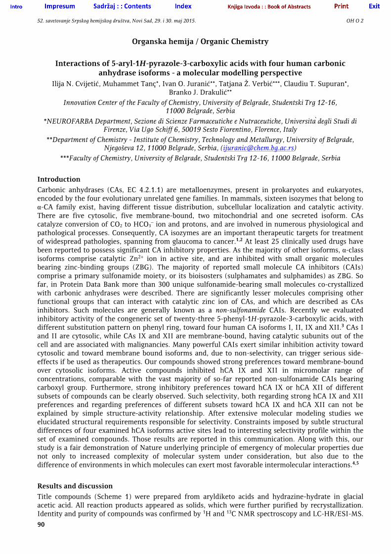
R = 4-Me; 4-Et; 4-i-Pr; 4-n-Bu; 4-t-Bu; 2,4-di-Me; 3,4-di-Me; 2,4,5-tri-Me; 2,3,5,6-tetra-Me; 2,4,6-tri-Et; 2,4-di-i-Pr;
2,4,6-tri-i-Pr; β-Tetralinyl; β-Naphtyl; 4-Ph; 4-Pyrrolidinyl; 4-F-; 4-Cl; 3-Br; 4-OH; 2-MeO; 4-MeO; 4-OMe-2,5-di-Me
Scheme 1. Structure of title compounds.
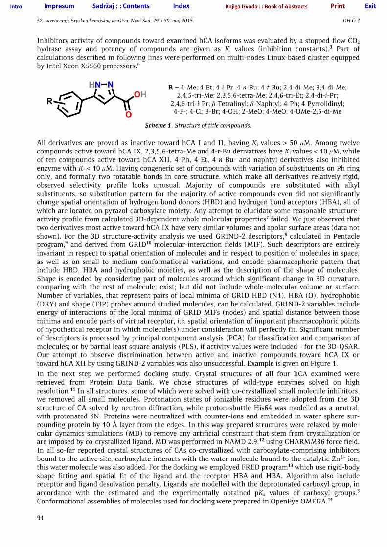
All derivatives are proved as inactive toward hCA I and II, having Ki values > 50 μM. Among twelve compounds active toward hCA IX, 2,3,5,6-tetra-Me and 4-t-Bu derivatives have Ki values < 10 μM, while of ten compounds active toward hCA XII, 4-Ph, 4-Et, 4-n-Bu- and naphtyl derivatives also inhibited enzyme with Ki < 10 μM. Having congeneric set of compounds with variation of substituents on Ph ring only, and formally two rotatable bonds in core structure, which make all derivatives relatively rigid, observed selectivity profile looks unusual. Majority of compounds are substituted with alkyl substituents, so substitution pattern for the majority of active compounds even did not significantly change spatial orientation of hydrogen bond donors (HBD) and hydrogen bond acceptors (HBA), all of which are located on pyrazol-carboxylate moiety. Any attempt to elucidate some reasonable structure-activity profile from calculated 3D-dependent whole molecular properties7 failed. We just observed that two derivatives most active toward hCA IX have very similar volumes and apolar surface areas (data not shown). For the 3D structure-activity analysis we used GRIND-2 descriptors,8 calculated in Pentacle program,9 and derived from GRID10 molecular-interaction fields (MIF). Such descriptors are entirely invariant in respect to spatial orientation of molecules and in respect to position of molecules in space, as well as on small to medium conformational variations, and encode pharmacophoric pattern that include HBD, HBA and hydrophobic moieties, as well as the description of the shape of molecules. Shape is encoded by considering part of molecules around which significant change in 3D curvature, comparing with the rest of molecule, exist; but did not include whole-molecular volume or surface. Number of variables, that represent pairs of local minima of GRID HBD (N1), HBA (O), hydrophobic (DRY) and shape (TIP) probes around studied molecules, can be calculated. GRIND-2 variables include energy of interactions of the local minima of GRID MIFs (nodes) and spatial distance between those minima and encode parts of virtual receptor, i.e. spatial orientation of important pharmacophoric points of hypothetical receptor in which molecule(s) under consideration will perfectly fit. Significant number of descriptors is processed by principal component analysis (PCA) for classification and comparison of molecules; or by partial least square analysis (PLS), if activity values were included - for the 3D-QSAR. Our attempt to observe discrimination between active and inactive compounds toward hCA IX or toward hCA XII by using GRIND-2 variables was also unsuccessful. Example is given on Figure 1.
In the next step we performed docking study. Crystal structures of all four hCA examined were retrieved from Protein Data Bank. We chose structures of wild-type enzymes solved on high resolution.11 In all structures, some of which were solved with co-crystallized small molecule inhibitors, we removed all small molecules. Protonation states of ionizable residues were adopted from the 3D structure of CA solved by neutron diffraction, while proton-shuttle His64 was modelled as a neutral, with protonated δN. Proteins were neutralized with counter-ions and embedded in water sphere sur-rounding protein by 10 Å layer from the edges. In this way prepared structures were relaxed by mole-cular dynamics simulations (MD) to remove any artificial constraint that stem from crystallization or are imposed by co-crystallized ligand. MD was performed in NAMD 2.9,12 using CHARMM36 force field. In all so-far reported crystal structures of CAs co-crystallized with carboxylate-comprising inhibitors bound to the active site, carboxylate interacts with the water molecule bound to the catalytic Zn2+ ion; this water molecule was also added. For the docking we employed FRED program13 which use rigid-body shape fitting and spatial fit of the ligand and the receptor HBA and HBA. Algorithm also include receptor and ligand desolvation penalty. Ligands are modelled with the deprotonated carboxyl group, in accordance with the estimated and the experimentally obtained pKa values of carboxyl groups.3 Conformational assemblies of molecules used for docking were prepared in OpenEye OMEGA.14
52. savetovanje Srpskog hemijskog društva, Novi Sad, 29. i 30. maj 2015. OH O 2
92
a b c
Figure 1. a) Position of compounds in the space of the first three principal components (PC), as obtained by PCA of GRIND-2 variables. Compounds active toward hCA XII was labeled by red spheres, while inactive compounds were labeled by blue spheres. b) Matrix-like representation of GRIND-2 variables. Compounds active toward hCA XII are
positioned above yellow line, inactive below line. c) Illustration of MIFs local minima (crosses) obtained by HBD (N1 - blue), HBA (O - red), hydrophobic (DRY - yellow) and shape (TIP - green) probes around 4-Ph-derivative.
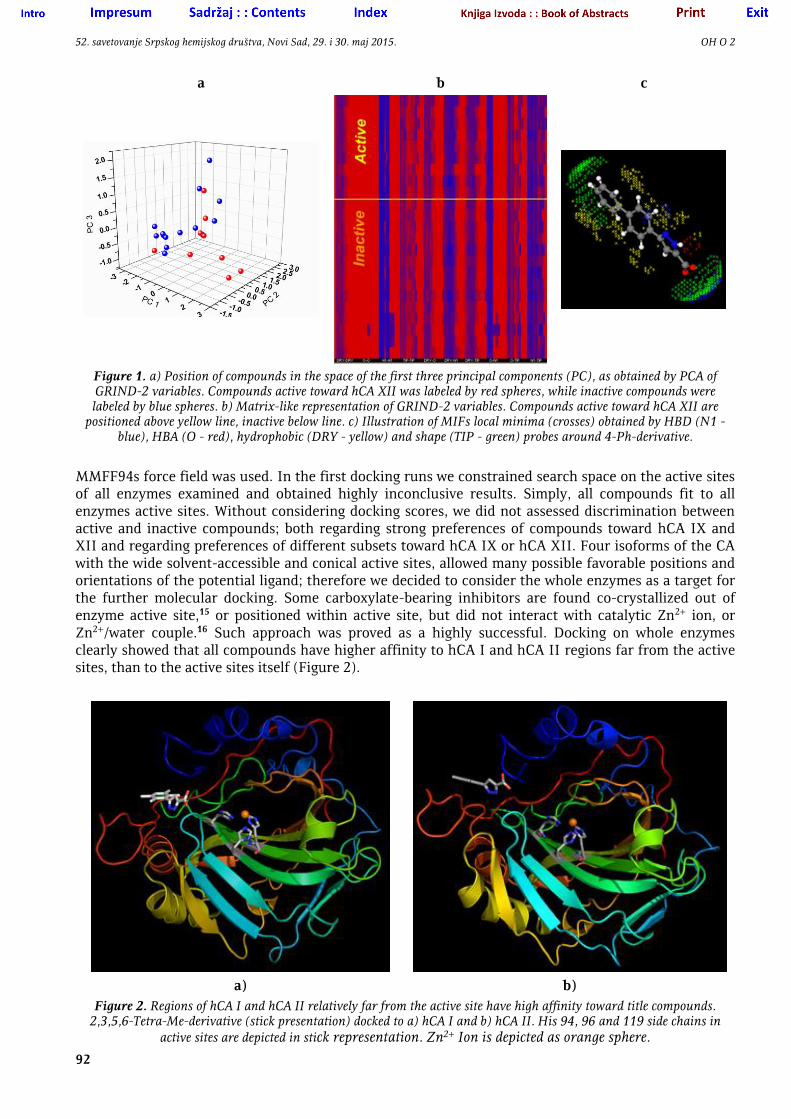
MMFF94s force field was used. In the first docking runs we constrained search space on the active sites of all enzymes examined and obtained highly inconclusive results. Simply, all compounds fit to all enzymes active sites. Without considering docking scores, we did not assessed discrimination between active and inactive compounds; both regarding strong preferences of compounds toward hCA IX and XII and regarding preferences of different subsets toward hCA IX or hCA XII. Four isoforms of the CA with the wide solvent-accessible and conical active sites, allowed many possible favorable positions and orientations of the potential ligand; therefore we decided to consider the whole enzymes as a target for the further molecular docking. Some carboxylate-bearing inhibitors are found co-crystallized out of enzyme active site,15 or positioned within active site, but did not interact with catalytic Zn2+ ion, or Zn2+/water couple.16 Such approach was proved as a highly successful. Docking on whole enzymes clearly showed that all compounds have higher affinity to hCA I and hCA II regions far from the active sites, than to the active sites itself (Figure 2).
a) b)
Figure 2. Regions of hCA I and hCA II relatively far from the active site have high affinity toward title compounds. 2,3,5,6-Tetra-Me-derivative (stick presentation) docked to a) hCA I and b) hCA II. His 94, 96 and 119 side chains in
active sites are depicted in stick representation. Zn2+ Ion is depicted as orange sphere.
52. savetovanje Srpskog hemijskog društva, Novi Sad, 29. i 30. maj 2015. OH O 2
93
Also, docking results revealed that when whole enzymes were used as targets, all compounds have higher affinity to hCA IX and hCA XII active sites than to other regions of examined enzymes. Although all compounds (including those with Ki > 50 μM) exert high affinity toward hCA IX and hCA XII active sites, as obtained from the results of our docking study, compounds are clustered in similar manner in the hCA IX or in hCA XII active sites, but position of such clusters in hCA IX is significantly different than in hCA XII. By careful inspection of interactions that compounds exert with the residues of active site, we observed clear distinction of derivatives active toward hCA IX or toward hCA XII from other, inactive compounds. Examples are given on Figures 3 and 4.
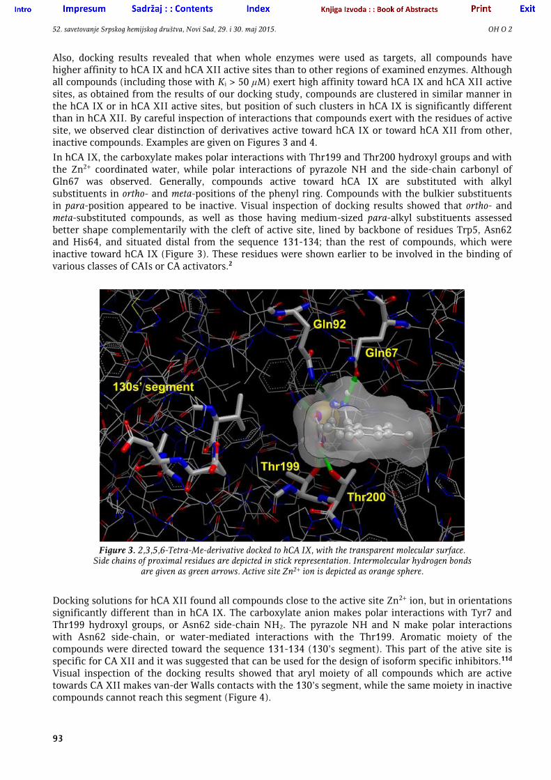
In hCA IX, the carboxylate makes polar interactions with Thr199 and Thr200 hydroxyl groups and with the Zn2+ coordinated water, while polar interactions of pyrazole NH and the side-chain carbonyl of Gln67 was observed. Generally, compounds active toward hCA IX are substituted with alkyl substituents in ortho- and meta-positions of the phenyl ring. Compounds with the bulkier substituents in para-position appeared to be inactive. Visual inspection of docking results showed that ortho- and meta-substituted compounds, as well as those having medium-sized para-alkyl substituents assessed better shape complementarily with the cleft of active site, lined by backbone of residues Trp5, Asn62 and His64, and situated distal from the sequence 131-134; than the rest of compounds, which were inactive toward hCA IX (Figure 3). These residues were shown earlier to be involved in the binding of various classes of CAIs or CA activators.2
Figure 3. 2,3,5,6-Tetra-Me-derivative docked to hCA IX, with the transparent molecular surface.
Side chains of proximal residues are depicted in stick representation. Intermolecular hydrogen bonds are given as green arrows. Active site Zn2+ ion is depicted as orange sphere.
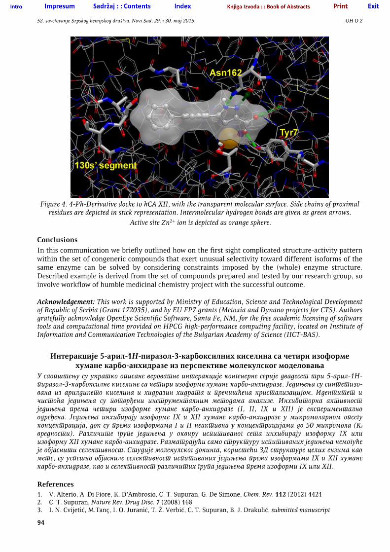
Docking solutions for hCA XII found all compounds close to the active site Zn2+ ion, but in orientations significantly different than in hCA IX. The carboxylate anion makes polar interactions with Tyr7 and Thr199 hydroxyl groups, or Asn62 side-chain NH2. The pyrazole NH and N make polar interactions with Asn62 side-chain, or water-mediated interactions with the Thr199. Aromatic moiety of the compounds were directed toward the sequence 131-134 (130’s segment). This part of the ative site is specific for CA XII and it was suggested that can be used for the design of isoform specific inhibitors.11d Visual inspection of the docking results showed that aryl moiety of all compounds which are active towards CA XII makes van-der Walls contacts with the 130’s segment, while the same moiety in inactive compounds cannot reach this segment (Figure 4).
52. savetovanje Srpskog hemijskog društva, Novi Sad, 29. i 30. maj 2015. OH O 2
94
Figure 4. 4-Ph-Derivative docke to hCA XII, with the transparent molecular surface. Side chains of proximal
residues are depicted in stick representation. Intermolecular hydrogen bonds are given as green arrows.
Active site Zn2+ ion is depicted as orange sphere.
Conclusions
In this communication we briefly outlined how on the first sight complicated structure-activity pattern within the set of congeneric compounds that exert unusual selectivity toward different isoforms of the same enzyme can be solved by considering constraints imposed by the (whole) enzyme structure. Described example is derived from the set of compounds prepared and tested by our research group, so involve workflow of humble medicinal chemistry project with the successful outcome.
Acknowledgement: This work is supported by Ministry of Education, Science and Technological Development of Republic of Serbia (Grant 172035), and by EU FP7 grants (Metoxia and Dynano projects for CTS). Authors gratefully acknowledge OpenEye Scientific Software, Santa Fe, NM, for the free academic licensing of software tools and computational time provided on HPCG high-performance computing facility, located on Institute of Information and Communication Technologies of the Bulgarian Academy of Science (IICT-BAS).
Интеракције 5-арил-1Н-пиразол-3-карбоксилних киселина са четири изоформе хумане карбо-анхидразе из перспективе молекулског моделовања
У саопштењу су укратко описане вероватне интеракције конгенерне серије двадесет три 5-арил-1Н-пиразол-3-карбоксилне киселине са четири изоформе хумане карбо-анхидразе. Једињења су синтетизо-вана из арилдикето киселина и хидразин хидрата и пречишћена кристализацијом. Идентитет и чистоћа једињења су потврђени инструменталним методама анализе. Инхибиторна активност једињења према четири изоформе хумане карбо-анхидразе (I, II, IX и XII) је експериментално одређена. Једињења инхибирају изоформе IX и XII хумане карбо-анхидразе у микромоларном опсегу концентрација, док су према изоформама I и II неактивна у концентрацијама до 50 микромола (Ki вредности). Различите групе једињења у оквиру испитиваног сета инхибирају изоформу IX или изоформу XII хумане карбо-анхидразе. Разматрајући само структуру испитиваних једињења немогуће је објаснити селективност. Студије молекулског докинга, користећи 3Д структуре целих ензима као мете, су успешно објасниле селективност испитиваних једињења према изоформама IX и XII хумане карбо-анхидразе, као и селективност различитих група једињења према изоформи IX или XII.
References 1. V. Alterio, A. Di Fiore, K. D’Ambrosio, C. T. Supuran, G. De Simone, Chem. Rev. 112 (2012) 4421 2. C. T. Supuran, Nature Rev. Drug Disc. 7 (2008) 168 3. I. N. Cvijetić, M.Tanç, I. O. Juranić, T. Ž. Verbić, C. T. Supuran, B. J. Drakulić, submitted manuscript
52. savetovanje Srpskog hemijskog društva, Novi Sad, 29. i 30. maj 2015. OH O 2
95
4. B. Testa, Entropy 11 (2009) 993 5. B. Testa, G. Vistoli, A. Pedretti, Chem. Biodiv. 11 (2014) 1309 6. E. Atanassov, T. Gurov, A. Karaivanova, Automatika and Informatika 2 (2011) 7 7. A. Pedretti, L. Villa, G. Vistoli, J. Comput. Aid. Mol. Des. 18 (2004) 167; http://ddl.unimi.it 8. Pentacle 1.06, http://www.moldiscovery.com/software/pentacle/ 9. A. Duran, G. Comesaña, M. Pastor, J. Chem. Inf. Model. 48 (2008) 1813 10. P. J. Goodford, J. Med. Chem. 28 (1985) 849; http://www.moldiscovery.com/software/grid/ 11. a) V. Kumar, K. K. Kannan, J. Mol. Biol. 241 (1994) 226; b) Ö. Güzel, C. Temperini, A. Innocenti, A.
Scozzafava, A. Salman, C.T. Supuran, Bioorg. Med. Chem. Lett. 18 (2008) 152; c) V. Alterio, M. Hilvo, A. Di Fiore, C. T. Supuran, P. Pan, S. Parkkila, A. Scaloni, J. Pastorek, S. Pastorekova, C. Pedone, A. Scozzafava, S. M. Monti, G. De Simone, Proc. Natl. Acad. Sci. U.S.A. 106 (2009) 16233; d) D. A. Whittington, A. Waheed, B. Ulmasov, G. N. Shah, J. H. Grubb,; W. S. Sly, D.W. Christianson, Proc. Natl. Acad. Sci. U.S.A. 98 (2001) 9545.
12. J. C. Phillips, R. Braun, W. Wang, J. Gumbart, E. Tajkhorshid, E. Villa, C. Chipot, R. D. Skeel, L. Kalé, K. S. Schulten, J. Comp. Chem. 26 (2005) 1781; http://www.ks.uiuc.edu/Research/namd/
13. a) M. R. McGann, H. R. Almond, A. Nicholls, J. A.Grant, F. K. Brown, Biopolymers 68 (2003) 76; b) M. McGann, J. Chem. Inf. Model. 51 (2011) 578; http://www.eyesopen.com/oedocking
14. P. C. D. Hawkins, A. G. Skillman, G. L. Warren, B. A. Ellingson, M. T. Stahl. J. Chem. Inf. Model. 50 (2010) 572; http://www.eyesopen.com/omega
15. K. D’Ambrosio, S. Carradori, S. M. Monti, M. Buonanno, D. Secci, D. Vullo, C. T. Supuran, G. De Simone, Chem. Commun. 51 (2015) 302
16. A. Maresca, C. Temperini, H. Vu, N. B. Pham, S.-A. Poulsen, A. Scozzafava, R.J. Quinn, C. T. Supuran, J. Am. Chem. Soc. 131 (2009) 3057