Embed Size (px)
Citation preview
Phosphorylation of SOS3-LIKE CALCIUM BINDING PROTEIN8by SOS2 Protein Kinase Stabilizes Their Protein Complex andRegulates Salt Tolerance in Arabidopsis W
HuixinLin,aYongqingYang,aRuidangQuan,a ImeldaMendoza,bYishengWu,aWenmingDu,aShuangshuangZhao,a
Karen S. Schumaker,c Jose M. Pardo,b and Yan Guoa,1
a National Institute of Biological Sciences, Beijing, Zhongguancun Life Science Park, Beijing 102206, P.R. Chinab Instituto de Recursos Naturales y Agrobiologıa, Consejo Superior de Investigaciones Cientıficas, Sevilla 41012, Spainc Department of Plant Sciences, University of Arizona, Tucson, Arizona 85721
The Salt Overly Sensitive (SOS) pathway plays an important role in the regulation of Na+/K+ ion homeostasis and salt
tolerance in Arabidopsis thaliana. Previously, we reported that the calcium binding proteins SOS3 and SOS3-LIKE CALCIUM
BINDING PROTEIN8 (SCaBP8) nonredundantly activate the protein kinase SOS2. Here, we show that SOS2 phosphorylates
SCaBP8 at its C terminus but does not phosphorylate SOS3. In vitro, SOS2 phosphorylation of SCaBP8 was enhanced by the
bimolecular interaction of SOS2 and SCaBP8 and did not require calcium ions. In vivo, this phosphorylation was induced by
salt stress, occurred at the membrane, stabilized the SCaBP8-SOS2 interaction, and enhanced plasma membrane Na+/H+
exchange activity. When a Ser at position 237 in the SCaBP8 protein (the SOS2 phosphorylation target) was mutated to Ala,
SCaBP8 was no longer phosphorylated by SOS2 and the mutant protein could not fully rescue the salt-sensitive phenotype
of the scabp8 mutant. By contrast, when Ser-237 was mutated to Asp to mimic the charge of a phosphorylated Ser residue,
the mutant protein rescued the scabp8 salt sensitivity. These data demonstrate that calcium sensor phosphorylation is a
critical component of SOS pathway regulation of salt tolerance in Arabidopsis.
INTRODUCTION
Maintenance of Na+/K+ ion homeostasis is essential for plant
growth and development in saline conditions where accumula-
tion of salts leads to significant reductions in plant growth. The
Salt Overly Sensitive (SOS) pathway inArabidopsis thaliana plays
an important role in regulating Na+/ K+ ion homeostasis during
plant growth in high salt conditions. SOS3 (Liu and Zhu, 1998), a
calcium binding protein, perceives an increase in cellular calcium
levels induced by salt stress. Upon sensing the calcium signal,
SOS3 interacts with, activates, and recruits SOS2, a protein
kinase, to the plasma membrane (Halfter et al., 2000; Quintero
et al., 2002). This complex further activates SOS1 (Shi et al.,
2000), a Na+/H+ exchanger (antiporter), to regulate Na+/K+ ion
homeostasis and prevent the accumulation of toxic levels of Na+
(Zhu et al., 1998; Zhu, 2002).
The Arabidopsis genome encodes nine additional SOS3-LIKE
CALCIUM BINDING PROTEINS (SCaBPs), also known as CAL-
CINEURIN B-LIKE (CBL) calcium binding proteins (Luan et al.,
2002; Gong et al., 2004; Kolukisaoglu et al., 2004). SOS3 is also
known as CBL4 (Kim et al., 2000; see Supplemental Figure
1 online for a protein alignment of the SCaBP/CBL family
members). Four SCaBP/CBL proteins have been shown to
regulate ion homeostasis. SCaBP1/CBL2 interacts with and
activates SOS2-LIKE PROTEIN KINASE5 (PKS5), and this
complex negatively regulates the activity of AHA2, a plasma
membrane H+-ATPase (Fuglsang et al., 2007). CBL1/SCaBP5
and CBL9/SCaBP7 interact with and activate CBL-INTERACT-
ING PROTEIN KINASE23 (CIPK23); this complex in turn acti-
vates the activity of AKT1, a plasma membrane K+ channel, to
regulate K+ homeostasis (Xu et al., 2006; Cheong et al., 2007).
Recently, we identified SCaBP8/CBL10 as another regulator of
SOS2 that, in contrast with SOS3, which mainly functions in
root tissue, preferentially protects shoot tissue from salt stress.
Like SOS3, SCaBP8 interacts with, activates, and recruits
SOS2 to the plasma membrane to activate SOS1 in the plant
plasma membrane or SOS1 expressed in yeast (Quan et al.,
2007).
A number of SCaBP/CBL proteins contain the four helix-loop-
helix structural domains (EF-hands) found in many calcium
binding proteins. These SCaBP proteins share significant se-
quence similarity with the B subunit of calcineurin in yeast and a
neuronal calcium sensor in animals, with sequences only differ-
ing at their N and C termini (Gong et al., 2004). Both the B subunit
of calcineurin and neuronal calcium sensor proteins are myris-
toylated in vivo; however, it is not known if this modification is
required for their function (Zhu et al., 1995). Similar protein
domains have been shown to regulate the localization or activ-
ities of several SCaBP proteins. Three out of 10 of the SCaBP/
CBL family proteins have the MGXXXS/T (K) myristoylation
signature sequence (Gong et al., 2004; Kolukisaoglu et al.,
1 Address correspondence to [email protected] author responsible for distribution of materials integral to thefindings presented in this article in accordance with the policy describedin the Instructions for Authors (www.plantcell.org) is: Yan Guo([email protected]).WOnline version contains Web-only data.www.plantcell.org/cgi/doi/10.1105/tpc.109.066217
The Plant Cell, Vol. 21: 1607–1619, May 2009, www.plantcell.org ã 2009 American Society of Plant Biologists
Dow
nloaded from https://academ
ic.oup.com/plcell/article/21/5/1607/6095343 by guest on 16 O
ctober 2021
2004; Batistic et al., 2008). This N-terminal myristoylation and
acylation were shown to be required for plasma membrane
localization of CBL1/SCaBP5 (Batistic et al., 2008), while a
myristoylation domain at the SOS3 N terminus was shown to
be important for SOS3 function in salt tolerance (Ishitani et al.,
2000). While SCaBP8 lacks an N-terminal myristoylation motif,
an N-terminal 25–amino acid hydrophobic domain is required for
SCaBP8 plasmamembrane localization (Quan et al., 2007). Both
SOS3 and SCaBP8 activate SOS2 and SOS1; however, they
cannot replace each other in reciprocalmutant complementation
assays, suggesting that each has a unique function(s) in the
response of Arabidopsis to growth in salt.
In this study, we report that SOS2 phosphorylates SCaBP8,
but not SOS3, and that this modification is important for SCaBP8
function in salt tolerance by stabilizing themembrane localization
of the SCaBP8-SOS2 complex.
RESULTS
SOS2 Phosphorylates SCaBP8 at Its C Terminus
The SOS3 and SCaBP8 calcium sensors have been shown to
specifically function in the salt stress response of Arabidopsis
(Liu and Zhu, 1998; Quan et al., 2007) but perform unique
functions in the regulation of SOS2. One possible mechanism
underlying this difference in regulation is that the SCaBP8 protein
may require an additional modification, such as phosphorylation
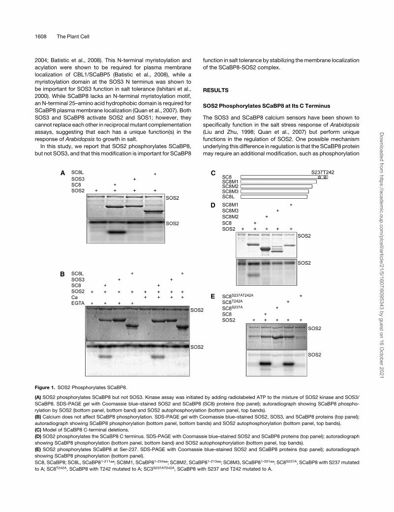
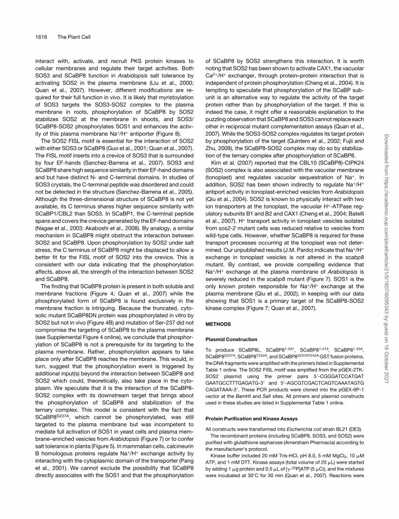
Figure 1. SOS2 Phosphorylates SCaBP8.
(A) SOS2 phosphorylates SCaBP8 but not SOS3. Kinase assay was initiated by adding radiolabeled ATP to the mixture of SOS2 kinase and SOS3/
SCaBP8. SDS-PAGE gel with Coomassie blue–stained SOS2 and SCaBP8 (SC8) proteins (top panel); autoradiograph showing SCaBP8 phospho-
rylation by SOS2 (bottom panel, bottom band) and SOS2 autophosphorylation (bottom panel, top bands).
(B) Calcium does not affect SCaBP8 phosphorylation. SDS-PAGE gel with Coomassie blue–stained SOS2, SOS3, and SCaBP8 proteins (top panel);
autoradiograph showing SCaBP8 phosphorylation (bottom panel, bottom bands) and SOS2 autophosphorylation (bottom panel, top bands).
(C) Model of SCaBP8 C-terminal deletions.
(D) SOS2 phosphorylates the SCaBP8 C terminus. SDS-PAGE with Coomassie blue–stained SOS2 and SCaBP8 proteins (top panel); autoradiograph
showing SCaBP8 phosphorylation (bottom panel, bottom band) and SOS2 autophosphorylation (bottom panel, top bands).
(E) SOS2 phosphorylates SCaBP8 at Ser-237. SDS-PAGE with Coomassie blue–stained SOS2 and SCaBP8 proteins (top panel); autoradiograph
showing SCaBP8 phosphorylation (bottom panel).
SC8, SCaBP8; SC8L, SCaBP81-211aa; SC8M1, SCaBP81-234aa; SC8M2, SCaBP81-213aa; SC8M3, SCaBP81-201aa; SC8S237A, SCaBP8 with S237 mutated
to A; SC8T242A, SCaBP8 with T242 mutated to A; SC3S237AT242A, SCaBP8 with S237 and T242 mutated to A.
1608 The Plant Cell
Dow
nloaded from https://academ
ic.oup.com/plcell/article/21/5/1607/6095343 by guest on 16 O
ctober 2021
by SOS2. To test this, we monitored SOS2 phosphorylation of
SOS3 or SCaBP8 in vitro and, surprisingly, found that SOS2
phosphorylated SCaBP8 but not SOS3 (Figure 1A). SCaBP8L, a
mis-spliced cDNA of SCaBP8 that retains the seventh intron and
encodes a 211–amino acid protein truncated at its C terminus
(Quan et al., 2007), was not phosphorylated by SOS2 (Figure 1A),
indicating that the phosphorylation site in SCaBP8 is located in
its C terminus. Because SCaBP8 is a calciumbinding protein that
is thought to sense calcium signals triggered by salt stress to
activate SOS2 (Harper et al., 2004; Quan et al., 2007), we
determined if calcium is required for SOS2 phosphorylation of
SCaBP8. To do this, 0.5 mM calcium or 5 mM EGTA were
included in the SOS2 kinase assays. The results indicate that
phosphorylation is not enhanced by the presence of calcium
(Figure 1B).
SOS2 Phosphorylates SCaBP8 at Ser-237
To determine the SOS2 phosphorylation site in SCaBP8, we
generated three additional SCaBP8 truncated proteins with
C-terminal deletions: glutathione S-transferase (GST)-SCaBP81-201,
GST-SCaBP81-213, and GST-SCaBP81-234 (Figure 1C). None of
these proteins were phosphorylated by SOS2 (Figure 1D). These
results indicated that the phosphorylation site must be in the last
12 amino acids of the SCaBP8 C terminus as the SCaBP81-234
construct had only these 12 amino acids removed. To map the
phosphorylation site, we mutated all of the Ser and Thr residues
in this region to Ala. When Ser-237 was changed to Ala
(SCaBP8S237A), the mutant protein was no longer phosphory-
lated by SOS2 (Figure 1E). As a control, Thr-242 was also
mutated to Ala. While SCaBP8T242A was still phosphorylated by
SOS2, the double mutant SCaBP8S237A/T242A (containing both
the T242A and S237Amutations) was not (Figure 1E). Our results
demonstrate that Ser-237 is the only amino acid phosphorylated
by SOS2 in vitro. When we compared the C-terminal sequences
of the SCaBP/CBL family members, Ser-237 was found to be
conserved in eight of the SCaBP proteins, including SOS3 (see
Supplemental Figure 1 online).
SOS2 Phosphorylation of SCaBP8 Is Enhanced by
SCaBP8-SOS2 Interaction
SCaBP8 has been shown to interact with SOS2 via a FISL motif
in the SOS2 kinase (Quan et al., 2007). Deletion of this motif or
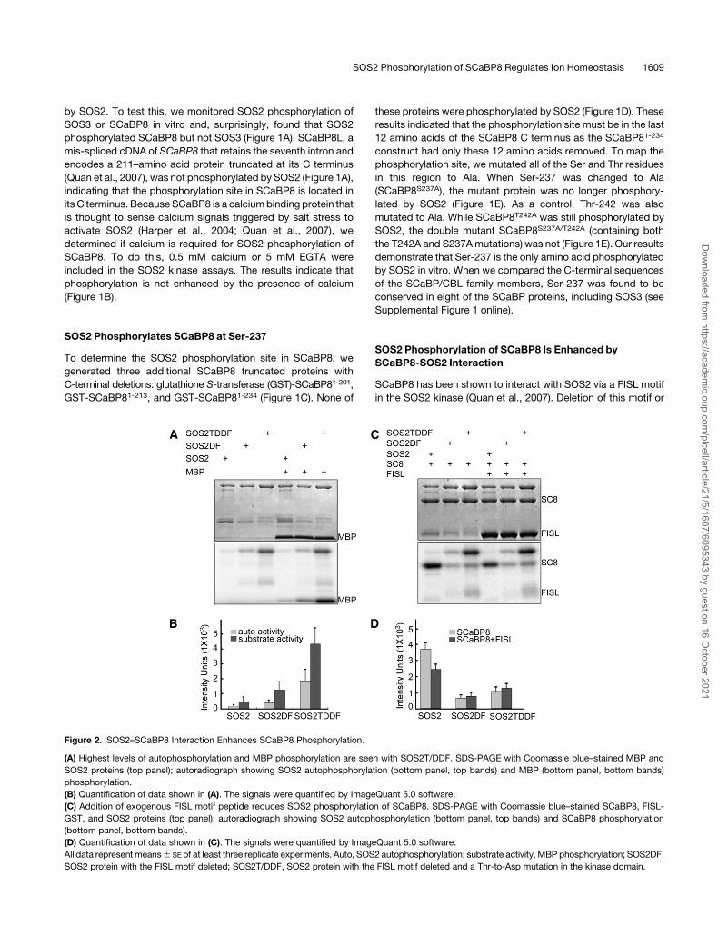
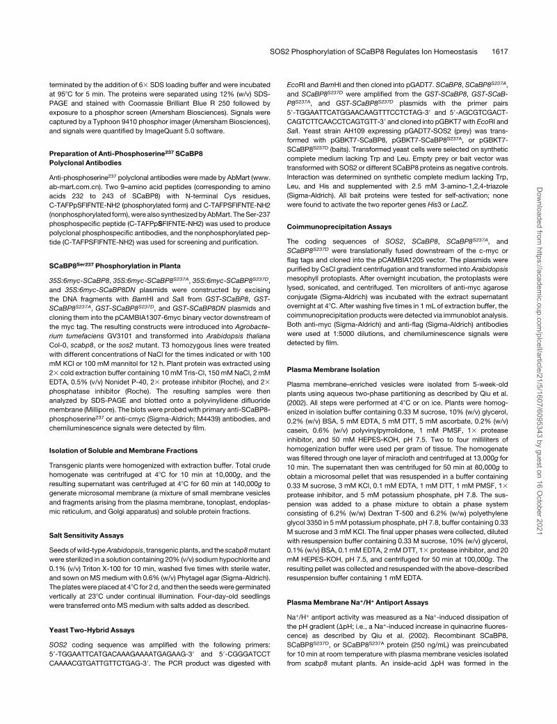
Figure 2. SOS2–SCaBP8 Interaction Enhances SCaBP8 Phosphorylation.
(A) Highest levels of autophosphorylation and MBP phosphorylation are seen with SOS2T/DDF. SDS-PAGE with Coomassie blue–stained MBP and
SOS2 proteins (top panel); autoradiograph showing SOS2 autophosphorylation (bottom panel, top bands) and MBP (bottom panel, bottom bands)
phosphorylation.
(B) Quantification of data shown in (A). The signals were quantified by ImageQuant 5.0 software.
(C) Addition of exogenous FISL motif peptide reduces SOS2 phosphorylation of SCaBP8. SDS-PAGE with Coomassie blue–stained SCaBP8, FISL-
GST, and SOS2 proteins (top panel); autoradiograph showing SOS2 autophosphorylation (bottom panel, top bands) and SCaBP8 phosphorylation
(bottom panel, bottom bands).
(D) Quantification of data shown in (C). The signals were quantified by ImageQuant 5.0 software.
All data represent means6 SE of at least three replicate experiments. Auto, SOS2 autophosphorylation; substrate activity, MBP phosphorylation; SOS2DF,
SOS2 protein with the FISL motif deleted; SOS2T/DDF, SOS2 protein with the FISL motif deleted and a Thr-to-Asp mutation in the kinase domain.
SOS2 Phosphorylation of SCaBP8 Regulates Ion Homeostasis 1609
Dow
nloaded from https://academ
ic.oup.com/plcell/article/21/5/1607/6095343 by guest on 16 O
ctober 2021
a change in a Thr in the SOS2 kinase loop to Asp (T/D) has
been shown to increase SOS2 activity (Guo et al., 2001). To test
if the interaction between SCaBP8 and SOS2 is required for
phosphorylation, recombinant SOS2 protein, SOS2 protein with
the FISL motif deleted (SOS2DF), and SOS2 protein with the
combined FISL deletion and T/D change (SOS2T/DDF) were
used in our in vitro kinase assays. SOS2T/DDF had the highest
activity both in terms of autophosphorylation and phosphory-
lation of the substrate Myelin Basic Protein (MBP), followed by
SOS2DF and SOS2 (Figures 2A and 2B). These results are
consistent with data described by Guo et al. (2001). However,
when SCaBP8 was used as a substrate, the SCaBP8 phos-
phorylation level was significantly higher in the assay with wild-
type SOS2 protein compared with the assays with SOS2DF or
SOS2T/DDF protein (Figures 2C and 2D). These data suggest
that, while SOS2-SCaBP8 interaction is not essential for SOS2
phosphorylation of SCaBP8, this interaction dramatically en-
hances the phosphorylation. Additional support for this con-
clusion came from experiments in which we added FISL
peptide to our SOS2-SCaBP8 kinase assays. The results
showed that blocking the SOS2–SCaBP8 interaction with
added FISL peptide significantly reduced SOS2 phosphoryla-
tion of SCaBP8 (Figures 2C and 2D).
SOS2 Phosphorylation of SCaBP8 Is Induced by NaCl
in Planta
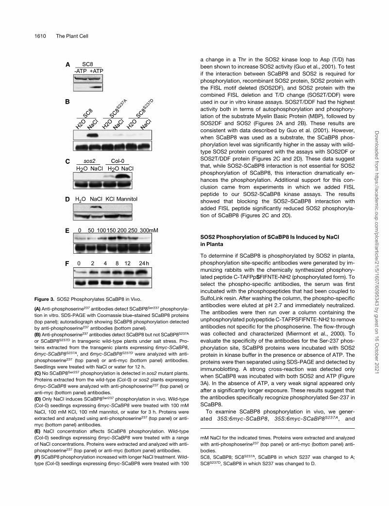
To determine if SCaBP8 is phosphorylated by SOS2 in planta,
phosphorylation site-specific antibodies were generated by im-
munizing rabbits with the chemically synthesized phosphory-
lated peptide C-TAFPpSFIFNTE-NH2 (phosphorylated form). To
select the phospho-specific antibodies, the serum was first
incubated with the phosphopeptides that had been coupled to
SulfoLink resin. After washing the column, the phospho-specific
antibodies were eluted at pH 2.7 and immediately neutralized.
The antibodies were then run over a column containing the
unphosphorylated polypeptide C-TAFPSFIFNTE-NH2 to remove
antibodies not specific for the phosphoserine. The flow-through
was collected and characterized (Miermont et al., 2000). To
evaluate the specificity of the antibodies for the Ser-237 phos-
phorylation site, SCaBP8 proteins were incubated with SOS2
protein in kinase buffer in the presence or absence of ATP. The
proteins were then separated using SDS-PAGE and detected by
immunoblotting. A strong cross-reaction was detected only
when SCaBP8 was incubated with both SOS2 and ATP (Figure
3A). In the absence of ATP, a very weak signal appeared only
after a significantly longer exposure. These results suggest that
the antibodies specifically recognize phosphorylated Ser-237 in
SCaBP8.
To examine SCaBP8 phosphorylation in vivo, we gener-
ated 35S:6myc-SCaBP8, 35S:6myc-SCaBP8S237A, and
Figure 3. SOS2 Phosphorylates SCaBP8 in Vivo.
(A) Anti-phosphoserine237 antibodies detect SCaBP8Ser237 phosphoryla-
tion in vitro. SDS-PAGE with Coomassie blue–stained SCaBP8 proteins
(top panel); autoradiograph showing SCaBP8 phosphorylation detected
by anti-phosphoserine237 antibodies (bottom panel).
(B) Anti-phosphoserine237 antibodies detect SCaBP8 but not SCaBP8S237A
or SCaBP8S237D in transgenic wild-type plants under salt stress. Pro-
teins extracted from the transgenic plants expressing 6myc-SCaBP8,
6myc-SCaBP8S237A, and 6myc-SCaBP8S237D were analyzed with anti-
phosphoserine237 (top panel) or anti-myc (bottom panel) antibodies.
Seedlings were treated with NaCl or water for 12 h.
(C) No SCaBP8Ser237 phosphorylation is detected in sos2 mutant plants.
Proteins extracted from the wild-type (Col-0) or sos2 plants expressing
6myc-SCaBP8 were analyzed with anti-phosphoserine237 (top panel) or
anti-myc (bottom panel) antibodies.
(D) Only NaCl induces SCaBP8Ser237 phosphorylation in vivo. Wild-type
(Col-0) seedlings expressing 6myc-SCaBP8 were treated with 100 mM
NaCl, 100 mM KCl, 100 mM mannitol, or water for 3 h. Proteins were
extracted and analyzed using anti-phosphoserine237 (top panel) or anti-
myc (bottom panel) antibodies.
(E) NaCl concentration affects SCaBP8 phosphorylation. Wild-type
(Col-0) seedlings expressing 6myc-SCaBP8 were treated with a range
of NaCl concentrations. Proteins were extracted and analyzed with anti-
phosphoserine237 (top panel) or anti-myc (bottom panel) antibodies.
(F) SCaBP8 phosphorylation increased with longer NaCl treatment. Wild-
type (Col-0) seedlings expressing 6myc-SCaBP8 were treated with 100
mM NaCl for the indicated times. Proteins were extracted and analyzed
with anti-phosphoserine237 (top panel) or anti-myc (bottom panel) anti-
bodies.
SC8, SCaBP8; SC8S237A, SCaBP8 in which S237 was changed to A;
SC8S237D, SCaBP8 in which S237 was changed to D.
1610 The Plant Cell
Dow
nloaded from https://academ
ic.oup.com/plcell/article/21/5/1607/6095343 by guest on 16 O
ctober 2021
35S:6myc-SCaBP8S237D transgenic lines in wild-type Arabi-
dopsis (Columbia-0 [Col-0] background) and 35S:6myc-SCaBP8
transgenic lines in the sos2 mutant background. Total proteins
were extracted from 10-d-old seedlings of these four transgenic
lines and subjected to immunoblots using either anti-myc or anti-
phosphoserine237 antibodies. Anti-cmyc (anti-myc) antibody
readily detected myc-labeled SCaBP8 in all transgenic plants,
but no signal was observed when anti-phosphoserine237 anti-
bodies were used. Because the SOS pathway specifically func-
tions in the response of Arabidopsis to growth in salt, we
reasoned that SOS2 phosphorylation of SCaBP8 might be in-
duced by salt stress. To test this, transgenic plants were treated
with 100 mMNaCl or water for 12 h, and proteins were extracted
and assayed by immunoblotting. The phosphoserine237 signal
was detected only in protein extracts from wild-type plants
expressingmyc-SCaBP8 treated with NaCl (Figures 3B and 3C).
No phosphoserine237 signal was seen in wild-type plants ex-
pressing either myc-SCaBP8S237D or myc-SCaBP8S237A (Figure
3B) or in sos2 mutant plants expressing myc-SCaBP8 (Figure
3C). Nearly equal amounts ofmyc-SCaBP8, myc-SCaBP8S237A,
or myc-SCaBP8S237D protein were present in the assays as
demonstrated by immunoblots with anti-myc antibodies (Figures
3B and 3C). Our data demonstrate that SOS2 specifically phos-
phorylates SCaBP8S237 and that this phosphorylation is induced
by salt stress.
To further determinewhether the phosphorylation is specific to
NaCl stress, the 35S:6myc-SCaBP8 transgenic plants in the
wild-type background were treated with NaCl, KCl, or mannitol.
Immunoblots showed that phosphorylation was detected only
after NaCl treatment evenwhen similar amounts ofmyc-SCaBP8
protein were present in each treatment (Figure 3D). To determine
if calcium affects this phosphorylation, transgenic plants were
treated with 5 or 50 mM CaCl2, and proteins were analyzed by
immunoblot assays. Calcium treatment did not significantly
enhance phosphorylation (see Supplemental Figure 2 online).
SOS2 phosphorylation of SCaBP8 reached a maximum level at
NaCl concentrations between 150 and 200 mM and declined at
higher NaCl concentrations (Figure 3E). The SCaBP8 phosphor-
ylation level increased gradually with longer NaCl treatments
(Figure 3F).
SOS2 Phosphorylates Membrane-Bound SCaBP8
SCaBP8 recruits SOS2 to the plasma membrane in yeast and
Arabidopsis (Quan et al., 2007). To determine if SOS2 phos-
phorylates membrane-bound SCaBP8, the 35S:6myc-SCaBP8
transgenic plants were treated with NaCl, and membrane and
soluble proteins were isolated and used in immunoblot assays
with anti-phosphoserine237 antibody. The phosphorylation of
SCaBP8S237 was detected in total protein extract and in the
membrane protein fraction, but not in the soluble protein
fraction (Figure 4A). Arabidopsis AHA2 and MAPK3 antibodies
were used as markers to label the membrane and soluble frac-
tions, respectively. Using anti-myc antibodies, myc-SCaBP8
protein was detected in all three fractions. These results sug-
gest that the phosphorylated SCaBP8 protein is membrane
bound.
Previously, we demonstrated that a 25–amino acid hydro-
phobic domain in the N terminus of SCaBP8 is required for
plasma membrane localization in yeast and onion cells (Quan
et al., 2007). To determine the importance of plasma membrane
localization for SCaBP8 function in salt tolerance, we removed
this peptide from SCaBP8 (SCaBP8DN) using PCR-based
mutagenesis. The truncated protein was fused to a myc-tag,
expressed under the control of the 35S promoter, and trans-
formed into wild type and the scabp8 mutant. T2 seed from >50
independent T1 transgenic lines in the scabp8 background
were tested for salt tolerance; none could rescue the mutant
phenotype, suggesting that this peptide is essential for
SCaBP8 function in salt tolerance (see Supplemental Figure 3
online). To further demonstrate that this peptide is essential for
SCaBP8 localization to the membrane in Arabidopsis, we
separated membrane and soluble protein fractions from the
transgenic plants expressing SCaBP8DN. The myc-SCaBP8DN
protein could be detected in the total protein extract and
soluble protein fraction but was below detection levels in the
Figure 4. SCaBP8 Phosphorylation Takes Place at the Membrane.
(A) SCaBP8Ser237 phosphorylation was detected only in membrane
fractions. Seedlings of wild-type (Col-0) plants expressing 6myc-
SCaBP8 or 6myc-SCaBP8DN, in which a 25–amino acid hydrophobic
domain in the N terminus of SCaBP8 that is required for SCaBP8 plasma
membrane localization was removed, were treated with 100 mMNaCl for
3 h. Membrane and soluble fractions were separated by centrifugation at
48C for 60 min at 140,000g. Equal aliquots of total (T), supernatant (S),
and pellet (P) proteins were separated by SDS-PAGE followed by
analysis with anti-myc (loading control), anti-phosphoserine237, anti-
MAPK3 (loading control for a soluble-protein fraction), or anti-AHA2
antibodies (loading control for a cell membrane fraction).
(B) SCaBP8Ser237 phosphorylation was not detected in 6myc-
SCaBP8DN transgenic plants. Seedlings of wild-type (Col-0) plants
expressing 6myc-SCaBP8 or 6myc-SCaBP8DN were treated with 100
mM NaCl or water for 3 h. Proteins were extracted and analyzed with
anti-phosphoserine237 (top panel) or anti-myc (bottom panel) antibodies.
(C) Anti-phosphoserine237 antibodies detect SCaBP8Ser237 phosphoryla-
tion of SCaBP8DN in vitro. SDS-PAGE with Coomassie blue–stained
SCaBP8 proteins (top panel); autoradiograph showing SCaBP8DN
phosphorylation detected by anti-phosphoserine237 antibodies (bottom
panel).
SC8DN, SCaBP8DN in which a 25–amino acid hydrophobic domain in
the N terminus has been deleted; AHA2, isoform of the plasma mem-
brane H+-ATPAse; MAPK3, mitogen-activated protein kinase3.
SOS2 Phosphorylation of SCaBP8 Regulates Ion Homeostasis 1611
Dow
nloaded from https://academ
ic.oup.com/plcell/article/21/5/1607/6095343 by guest on 16 O
ctober 2021
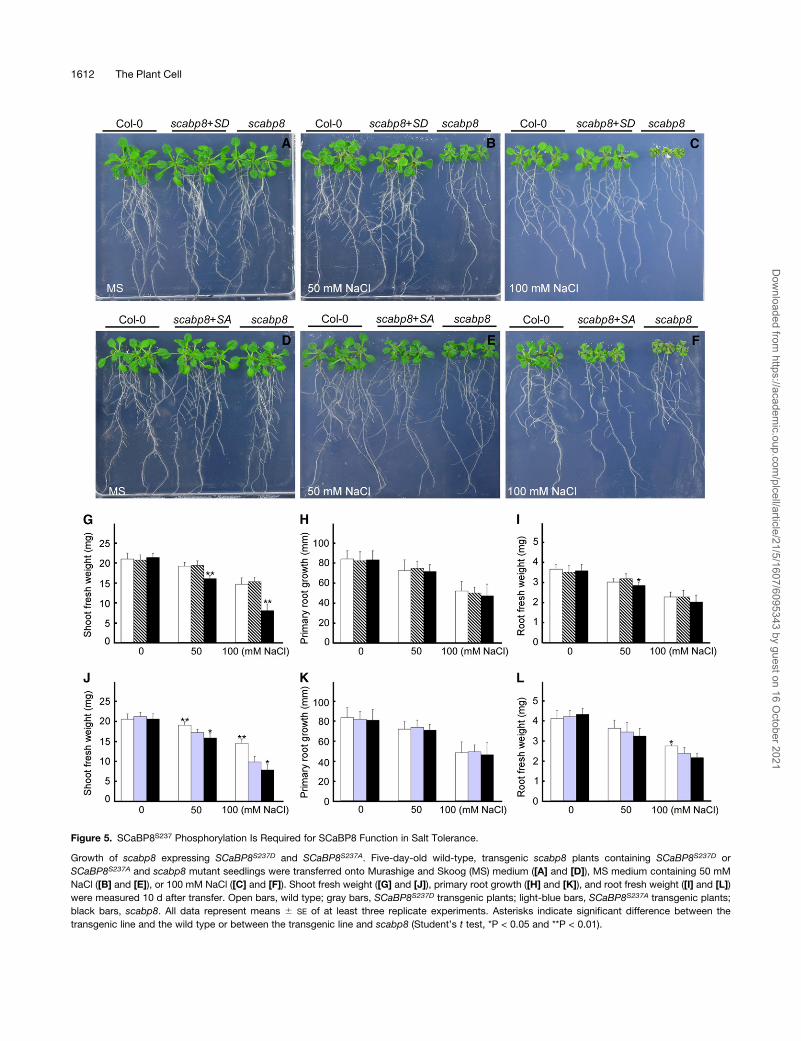
Figure 5. SCaBP8S237 Phosphorylation Is Required for SCaBP8 Function in Salt Tolerance.
Growth of scabp8 expressing SCaBP8S237D and SCaBP8S237A. Five-day-old wild-type, transgenic scabp8 plants containing SCaBP8S237D or
SCaBP8S237A and scabp8 mutant seedlings were transferred onto Murashige and Skoog (MS) medium ([A] and [D]), MS medium containing 50 mM
NaCl ([B] and [E]), or 100 mM NaCl ([C] and [F]). Shoot fresh weight ([G] and [J]), primary root growth ([H] and [K]), and root fresh weight ([I] and [L])
were measured 10 d after transfer. Open bars, wild type; gray bars, SCaBP8S237D transgenic plants; light-blue bars, SCaBP8S237A transgenic plants;
black bars, scabp8. All data represent means 6 SE of at least three replicate experiments. Asterisks indicate significant difference between the
transgenic line and the wild type or between the transgenic line and scabp8 (Student’s t test, *P < 0.05 and **P < 0.01).
1612 The Plant Cell
Dow
nloaded from https://academ
ic.oup.com/plcell/article/21/5/1607/6095343 by guest on 16 O
ctober 2021
membrane fraction (Figure 4A). When we examined the phos-
phorylation status of SCaBP8DN in transgenic scabp8 plants,
the phosphorylated protein was also below the level of detec-
tion (Figure 4B). As a control, we purified SCaBP8DN and SOS2
proteins from Escherichia coli, incubated them in kinase buffer
with or without ATP, and analyzed the phosphorylation using
our anti-phosphoserine237 antibodies. As shown in Figure 4C,
SOS2 phosphorylated SCaBP8DN in vitro. Together, these
results indicate that the phosphorylation event occurs after
SCaBP8 reaches the membrane.
Additional support for SCaBP8 phosphorylation after it
reaches the membrane came from studies in which we demon-
strated that phosphorylation of SCaBP8 is not required for
targeting to the plasma membrane. These studies were based
on analysis of the subcellular localization of SCaBP8S237A and
SCaBP8S237D mutant proteins in yeast using the Sos Recruit-
ment System (Quintero et al., 2002). This system canmonitor the
interaction between a plasma membrane–located protein and a
cytoplasmic protein fused to the human hSos protein, a func-
tional homolog of the yeast Ras GEF CDC25. When two proteins
interact at the plasma membrane, the human hSos protein
restores Ras activation and rescues the thermosensitive yeast
mutant cdc25-2 (Aronheim et al., 1997). We have shown previ-
ously that SCaBP8 recruits SOS2 to the plasma membrane of
yeast (Quan et al., 2007). Mutant proteins SCaBP8S237A and
SCaBP8S237D were coexpressed with the reporter fusion protein
SOS2:hSos to test whether recruitment of the protein kinase to
the plasma membrane was affected by mutation of Ser-237 in
SCaBP8. As shown in Supplemental Figure 4 online, the S237A
and S237D mutations did not significantly affect the targeting of
SCaBP8 to the plasma membrane or the recruitment of SOS2.
Moreover, when the targeting of SCaBP8 and SCaBP8S237A to
the plasma membrane was monitored directly in the presence of
the wild-type SOS2 protein or an inactive SOS2 kinase (with a
mutation in an essential Lys residue at position 40; SOS2K40N), no
difference in subcellular localization was found.
SCaBP8 Phosphorylation Is Required for Salt Tolerance
To determine if phosphorylation of SCaBP8 at Ser-237 is re-
quired for salt tolerance in Arabidopsis, we transformed
35S:6myc-SCaBP8S237A and 35S:6myc-SCaBP8S237D into the
scabp8 mutant. T2 seed from >100 independent T1 transgenic
lines from each transformation were tested for their growth in
salt. In 93 lines out of a total of 112 independent transgenic lines
harboring SCaBP8S237D, the salt-sensitive phenotype was com-
pletely suppressed. Partial rescue was seen in 14 lines, while
scapb8 mutant levels of sensitivity were seen in only five lines.
These numbers are comparable to the complementation rates
reported previously for the 35SP:SCaBP8 construct (Quan et al.,
2007). These results suggest that mimicking the phosphorylation
status of Ser-237 made SCaBP8 fully functional in Arabidopsis.
By contrast, no transgenic line was found among 121 T2 lines
harboring SCaBP8S237A in which the salt-sensitive phenotype of
scabp8was fully suppressed. Complementation was partial in 78
lines and negligible in the remaining 43 lines. These results
demonstrate that phosphorylation of SCaBP8 is essential for salt
tolerance (Figure 5). RT-PCR analysis showed that expression of
SCaBP8 was similar in the 35S:6myc-SCaBP8S237A, 35S:6myc-
SCaBP8S237D, and 35S:6myc-SCaBP8DN transgenic lines (see
Supplemental Figure 5 online), demonstrating that differences in
complementation were not due to differences in levels of trans-
gene expression.
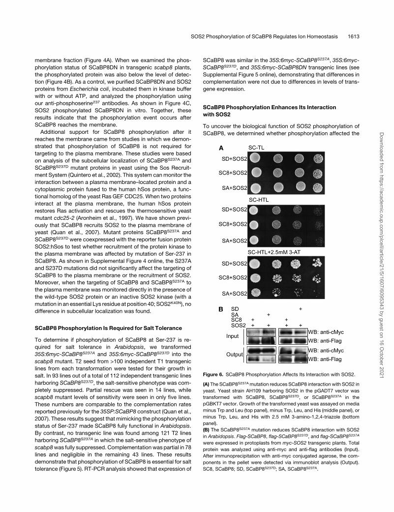
SCaBP8 Phosphorylation Enhances Its Interaction
with SOS2
To uncover the biological function of SOS2 phosphorylation of
SCaBP8, we determined whether phosphorylation affected the
Figure 6. SCaBP8 Phosphorylation Affects Its Interaction with SOS2.
(A) The SCaBP8S237Amutation reduces SCaBP8 interaction with SOS2 in
yeast. Yeast strain AH109 harboring SOS2 in the pGADT7 vector was
transformed with SCaBP8, SCaBP8S237D, or SCaBP8S237A in the
pGBKT7 vector. Growth of the transformed yeast was assayed on media
minus Trp and Leu (top panel), minus Trp, Leu, and His (middle panel), or
minus Trp, Leu, and His with 2.5 mM 3-amino-1,2,4-triazole (bottom
panel).
(B) The SCaBP8S237A mutation reduces SCaBP8 interaction with SOS2
in Arabidopsis. Flag-SCaBP8, flag-SCaBP8S237D, and flag-SCaBP8S237A
were expressed in protoplasts from myc-SOS2 transgenic plants. Total
protein was analyzed using anti-myc and anti-flag antibodies (Input).
After immunoprecipitation with anti-myc conjugated agarose, the com-
ponents in the pellet were detected via immunoblot analysis (Output).
SC8, SCaBP8; SD, SCaBP8S237D; SA, SCaBP8S237A.
SOS2 Phosphorylation of SCaBP8 Regulates Ion Homeostasis 1613
Dow
nloaded from https://academ
ic.oup.com/plcell/article/21/5/1607/6095343 by guest on 16 O
ctober 2021
ability of SCaBP8 to bind calcium and to interact with, activate, or
recruit SOS2 to the plasmamembrane. In vitro, we did not detect
a significant difference between SCaBP8 and SCaBP8S237A in
calcium binding assays or the activation of SOS2. In yeast, both
SCaBP8 and SCaBP8S237A recruited SOS2 to the plasma
membrane (see Supplemental Figure 4 online). However, in
yeast two-hybrid assays, mutation of Ser to Ala in SCaBP8S237A
significantly decreased the interaction between the mutant pro-
tein and SOS2 when compared with wild-type SCaBP8 (Figure
6A). By contrast, the SCaBP8S237D mutation, which mimics a
phosphorylated Ser residue, enhanced the interactionwith SOS2
(Figure 6A). These interaction differences were not due to differ-
ences in levels of SCaBP8, SCaBP8S237A, or SCaBP8S237D
expression (see Supplemental Figure 6 online). To examine this
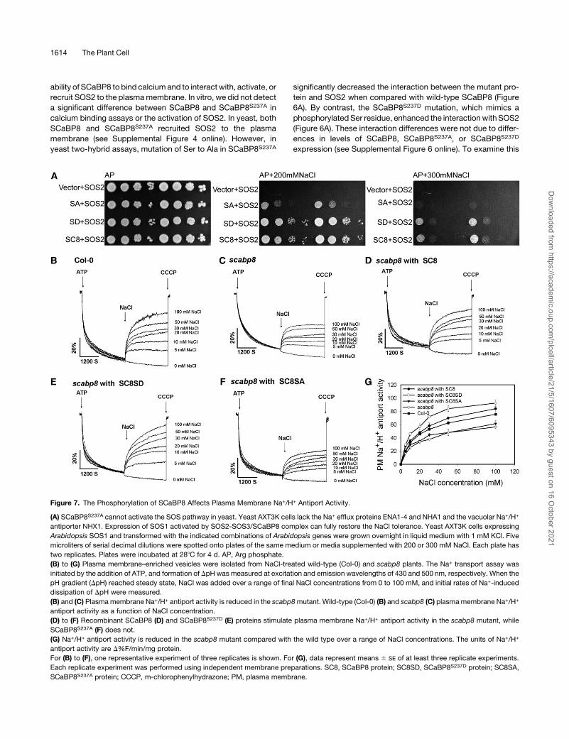
Figure 7. The Phosphorylation of SCaBP8 Affects Plasma Membrane Na+/H+ Antiport Activity.
(A) SCaBP8S237A cannot activate the SOS pathway in yeast. Yeast AXT3K cells lack the Na+ efflux proteins ENA1-4 and NHA1 and the vacuolar Na+/H+
antiporter NHX1. Expression of SOS1 activated by SOS2-SOS3/SCaBP8 complex can fully restore the NaCl tolerance. Yeast AXT3K cells expressing
Arabidopsis SOS1 and transformed with the indicated combinations of Arabidopsis genes were grown overnight in liquid medium with 1 mM KCl. Five
microliters of serial decimal dilutions were spotted onto plates of the same medium or media supplemented with 200 or 300 mM NaCl. Each plate has
two replicates. Plates were incubated at 288C for 4 d. AP, Arg phosphate.
(B) to (G) Plasma membrane–enriched vesicles were isolated from NaCl-treated wild-type (Col-0) and scabp8 plants. The Na+ transport assay was
initiated by the addition of ATP, and formation of DpH was measured at excitation and emission wavelengths of 430 and 500 nm, respectively. When the
pH gradient (DpH) reached steady state, NaCl was added over a range of final NaCl concentrations from 0 to 100 mM, and initial rates of Na+-induced
dissipation of DpH were measured.
(B) and (C) Plasma membrane Na+/H+ antiport activity is reduced in the scabp8mutant. Wild-type (Col-0) (B) and scabp8 (C) plasmamembrane Na+/H+
antiport activity as a function of NaCl concentration.
(D) to (F) Recombinant SCaBP8 (D) and SCaBP8S237D (E) proteins stimulate plasma membrane Na+/H+ antiport activity in the scabp8 mutant, while
SCaBP8S237A (F) does not.
(G) Na+/H+ antiport activity is reduced in the scabp8 mutant compared with the wild type over a range of NaCl concentrations. The units of Na+/H+
antiport activity are D%F/min/mg protein.
For (B) to (F), one representative experiment of three replicates is shown. For (G), data represent means 6 SE of at least three replicate experiments.
Each replicate experiment was performed using independent membrane preparations. SC8, SCaBP8 protein; SC8SD, SCaBP8S237D protein; SC8SA,
SCaBP8S237A protein; CCCP, m-chlorophenylhydrazone; PM, plasma membrane.
1614 The Plant Cell
Dow
nloaded from https://academ
ic.oup.com/plcell/article/21/5/1607/6095343 by guest on 16 O
ctober 2021
interaction in planta, we generated 35S:6myc-SOS2 transgenic
plants and transformed 35S:3flag-SCaBP8, 35S:3flag-SCaB-
P8S237A, or 35S:3flag-SCaBP8S237D into protoplasts of a T3
homozygous line expressing 35S:6myc-SOS2 (Sheen, 2001).
Protein extracts were sonicated, myc-SOS2 was immunopreci-
pitated, and flag-SCaBP8 was detected via immunoblot analy-
sis. In comparison to SCaBP8 and SCaBP8S237D, the amount of
SCaBP8S237A protein pulled down by SOS2 was substantially
reduced (Figure 6B).
SCaBP8S237 Phosphorylation Is Required for Activation of
SOS1, the PlasmaMembrane Na+/H+ Antiporter
SCaBP8 recruits SOS2 to the plasma membrane, and this
complex activates SOS1 in yeast (Quan et al., 2007). To deter-
mine if phosphorylation of SCaBP8 affects the capacity of
the SCaBP8-SOS2 complex to activate SOS1, yeast cells
expressing SOS1 andSOS2were transformedwith empty vector
or the SCaBP8, SCaBP8S237A, or SCaBP8S237D construct. The
transformed yeast strains were incubated on AP media contain-
ing 200 or 300 mM NaCl for 3 d (Figure 7A). SCaBP8S237D in
combination with SOS2 activated SOS1 to levels similar to what
has been seen for the wild-type SCaBP8-SOS2 complex. How-
ever, the activity of SOS1, measured as yeast growth in NaCl,
was significantly reduced when SCaBP8S237A mutant protein
was used (Figure 7A). These results suggest that full activation of
SOS1 by SCaBP8-SOS2 requires the phosphorylation of
SCaBP8.
To determine whether SCaBP8 affects the activity of SOS1 in
Arabidopsis, Na+-induced dissipation of a pH gradient (DpH),
which is monitored by a Na+-induced increase in quinacrine
fluorescence that is positively related to Na+/H+ exchange ac-
tivity, was examined. Plasma membrane vesicles were isolated
from 5-week-old plants of the wild type and the scabp8 mutant
treated with 250 mM NaCl for 3 d. As shown in Figures 7B and
7C, plasma membrane Na+/H+ antiport activity was reduced in
scabp8 plants compared with the wild type. The initial rate of
dissipation of DpH by 100mMNaCl was 75 units/min/mg protein
in wild-type plants compared with 55 units/min/mg protein in
scabp8. Over a range of NaCl concentrations between 0 and 100
mM, plasmamembraneNa+/H+ antiport activity was consistently
lower in scabp8 than in the wild type. Together with our yeast
data, reduced Na+/H+ transport activity in scabp8 plants demon-
strates that SCaBP8 is involved in the regulation of SOS1 activity.
Next, we analyzed whether the phosphorylation of SCaBP8
also affects SOS1 activity. Plasma membrane vesicles were
isolated from the scabp8 mutant, and purified SCaBP8,
SCaBP8S237D, or SCaBP8S237A protein was added to the Na+/H+
antiport assays. In the presence of 250 ng/mL SCaBP8, Na+/H+
antiport activity in scabp8 plants was slightly higher than activity
in wild-type plants (Figure 7). Stimulation of Na+/H+ antiport
activity in scabp8 mutant vesicles by SCaBP8S237D was greater
than of SCaBP8; however, little effect of SCaBP8S237A was
observed. Boiled recombinant SCaBP8, SCaBP8S237D, or
SCaBP8S237A proteins did not have any effect on antiport activ-
ity. These data demonstrate that the phosphorylation of SCaBP8
is critical for the regulation of plasma membrane Na+/H+ antiport
activity.
DISCUSSION
SCaBP calcium sensor activation of its interacting SOS2-like
protein kinase is critical for its function in regulating ion transport
in Arabidopsis (Qiu et al., 2002; Shi et al., 2002; Xu et al., 2006,
Fuglsang et al., 2007, Quan et al., 2007). While SCaBP proteins
do not have predicted transmembrane domains, some family
members locate at and recruit PKS kinases to cellular mem-
branes to perform their function. Lipid modifications of the
SCaBP proteins have been shown to be essential for their
function or membrane localization in planta (Ishitani et al.,
2000; Batistic et al., 2008). N-terminal myristoylation of SOS3
is important for its function in salt tolerance, and, in yeast, it is
required to recruit SOS2 to the plasma membrane (J.M. Pardo,
unpublished results). Whether myristoylation is required for
SOS3 plasma membrane localization in Arabidopsis remains to
be determined. Like SOS3, SCaBP8 activates SOS2 kinase
activity and recruits SOS2 to the plasmamembrane, but it lacks a
myristoylation signature sequence (MGXXXS/K). SCaBP8 pro-
tein associates with the plasma membrane due to an N-terminal
hydrophobic domain (Quan et al., 2007). This association is very
stable because high concentrations of KCl or Triton X-100 do not
remove it from the plasma membrane (Quan et al., 2007). In this
study, we determined that, in Arabidopsis, SCaBP8 is phos-
phorylated by SOS2 and that this modification stabilizes the
SOS2-SCaBP8 complex at the membrane by enhancing the
interaction of these two proteins. This finding identifies a post-
translational modification and an additional level of regulation
within the SCaBP family.
It is believed that once the calcium signal is induced by
stress stimuli, calcium binds to and activates SCaBP proteins
(Harper et al., 2004). Some of the SCaBP proteins in turn
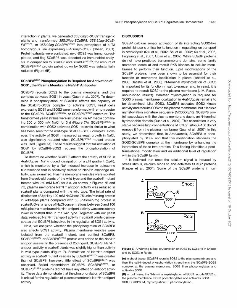
Figure 8. A Working Model of Activation of SOS2 by SCaBP8 in Shoots
and by SOS3 in Roots.
(A) In shoot tissue, SCaBP8 recruits SOS2 to the plasma membrane and
then the salt-induced phosphorylation strengthens the SCaBP8-SOS2
complex at the plasma membrane. SOS2 then phosphorylates and
activates SOS1.
(B) In root tissue, the N-terminal myristoylation of SOS3 recruits SOS2 to
the plasma membrane. SOS2 phosphorylates and activates SOS1.
SC8, SCaBP8; M, myristoylation; P, phosphorylation.
SOS2 Phosphorylation of SCaBP8 Regulates Ion Homeostasis 1615
Dow
nloaded from https://academ
ic.oup.com/plcell/article/21/5/1607/6095343 by guest on 16 O
ctober 2021
interact with, activate, and recruit PKS protein kinases to
cellular membranes and regulate their target activities. Both
SOS3 and SCaBP8 function in Arabidopsis salt tolerance by
activating SOS2 in the plasma membrane (Liu et al., 2000;
Quan et al., 2007). However, different modifications are re-
quired for their full function in vivo. It is likely that myristoylation
of SOS3 targets the SOS3-SOS2 complex to the plasma
membrane in roots, phosphorylation of SCaBP8 by SOS2
stabilizes SOS2 at the membrane in shoots, and SOS3/
SCaBP8-SOS2 phosphorylates SOS1 and enhances the activ-
ity of this plasma membrane Na+/H+ antiporter (Figure 8).
The SOS2 FISL motif is essential for the interaction of SOS2
with either SOS3 or SCaBP8 (Guo et al., 2001; Quan et al., 2007).
The FISL motif inserts into a crevice of SOS3 that is surrounded
by four EF-hands (Sanchez-Barrena et al., 2007). SOS3 and
SCaBP8 share high sequence similarity in their EF-hand domains
and but have distinct N- and C-terminal domains. In studies of
SOS3 crystals, the C-terminal peptide was disordered and could
not be detected in the structure (Sanchez-Barrena et al., 2005).
Although the three-dimensional structure of SCaBP8 is not yet
available, its C terminus shares higher sequence similarity with
SCaBP1/CBL2 than SOS3. In SCaBP1, the C-terminal peptide
spans and covers the crevice generated by the EF-hand domains
(Nagae et al., 2003; Akaboshi et al., 2008). By analogy, a similar
mechanism in SCaBP8 might obstruct the interaction between
SOS2 and SCaBP8. Upon phosphorylation by SOS2 under salt
stress, the C terminus of SCaBP8 might be displaced to allow a
better fit for the FISL motif of SOS2 into the crevice. This is
consistent with our data indicating that the phosphorylation
affects, above all, the strength of the interaction between SOS2
and SCaBP8.
The finding that SCaBP8 protein is present in both soluble and
membrane fractions (Figure 4; Quan et al., 2007) while the
phosphorylated form of SCaBP8 is found exclusively in the
membrane fraction is intriguing. Because the truncated, cyto-
solic mutant SCaBP8DN protein was phosphorylated in vitro by
SOS2 but not in vivo (Figure 4B) and mutation of Ser-237 did not
compromise the targeting of SCaBP8 to the plasma membrane
(see Supplemental Figure 4 online), we conclude that phosphor-
ylation of SCaBP8 is not a prerequisite for its targeting to the
plasma membrane. Rather, phosphorylation appears to take
place only after SCaBP8 reaches the membrane. This would, in
turn, suggest that the phosphorylation event is triggered by
additional input(s) beyond the interaction between SCaBP8 and
SOS2 which could, theoretically, also take place in the cyto-
plasm. We speculate that it is the interaction of the SCaBP8-
SOS2 complex with its downstream target that brings about
the phosphorylation of SCaBP8 and stabilization of the
ternary complex. This model is consistent with the fact that
SCaBP8S237A, which cannot be phosphorylated, was still
targeted to the plasma membrane but was incompetent to
mediate full activation of SOS1 in yeast cells and plasma mem-
brane–enriched vesicles from Arabidopsis (Figure 7) or to confer
salt tolerance in planta (Figure 5). Inmammalian cells, calcineurin
B homologous proteins regulate Na+/H+ exchange activity by
interacting with the cytoplasmic domain of the transporter (Pang
et al., 2001). We cannot exclude the possibility that SCaBP8
directly associates with the SOS1 and that the phosphorylation
of SCaBP8 by SOS2 strengthens this interaction. It is worth
noting that SOS2 has been shown to activate CAX1, the vacuolar
Ca2+/H+ exchanger, through protein–protein interaction that is
independent of protein phosphorylation (Cheng et al., 2004). It is
tempting to speculate that phosphorylation of the SCaBP sub-
unit is an alternative way to regulate the activity of the target
protein rather than by phosphorylation of the target. If this is
indeed the case, it might offer a reasonable explanation to the
puzzling observation thatSCaBP8andSOS3cannot replaceeach
other in reciprocal mutant complementation assays (Quan et al.,
2007). While the SOS3-SOS2 complex regulates its target protein
by phosphorylation of the target (Quintero et al., 2002; Fujii and
Zhu, 2009), the SCaBP8-SOS2 complex may do so by stabiliza-
tion of the ternary complex after phosphorylation of SCaBP8.
Kim et al. (2007) reported that the CBL10 (SCaBP8)-CIPK24
(SOS2) complex is also associated with the vacuolar membrane
(tonoplast) and regulates vacuolar sequestration of Na+. In
addition, SOS2 has been shown indirectly to regulate Na+/H+
antiport activity in tonoplast-enriched vesicles from Arabidopsis
(Qiu et al., 2004). SOS2 is known to physically interact with two
ion transporters at the tonoplast, the vacuolar H+-ATPase reg-
ulatory subunits B1 and B2 and CAX1 (Cheng et al., 2004; Batelli
et al., 2007). H+ transport activity in tonoplast vesicles isolated
from sos2-2 mutant cells was reduced relative to vesicles from
wild-type cells. However, whether SCaBP8 is required for these
transport processes occurring at the tonoplast was not deter-
mined. Our unpublished results (J.M. Pardo) indicate that Na+/H+
exchange in tonoplast vesicles is not altered in the scabp8
mutant. By contrast, we provide compelling evidence that
Na+/H+ exchange at the plasma membrane of Arabidopsis is
severely reduced in the scabp8 mutant (Figure 7). SOS1 is the
only known protein responsible for Na+/H+ exchange at the
plasma membrane (Qiu et al., 2002), in keeping with our data
showing that SOS1 is a primary target of the SCaBP8-SOS2
kinase complex (Figure 7; Quan et al., 2007).
METHODS
Plasmid Construction
To produce SCaBP8L, SCaBP81-201, SCaBP81-213, SCaBP81-234,
SCaBP8S237A, SCaBP8T242A, and SCaBP8S237AT242AGST fusion proteins,
the DNA fragmentswere amplifiedwith the primers listed in Supplemental
Table 1 online. The SOS2 FISL motif was amplified from the pGEX-2TK-
SOS2 plasmid using the primer pairs 59-CGGGATCCATGAT
GAATGCCTTTGAGATG-39 and 59-AGCGTCGACTCAGTCAAATAGTG
CAGATAAA-39. These PCR products were cloned into the pGEX-6P-1
vector at the BamHI and SalI sites. All primers and plasmid constructs
used in these studies are listed in Supplemental Table 1 online.
Protein Purification and Kinase Assays
All constructs were transformed into Escherichia coli strain BL21 (DE3).
The recombinant proteins (including SCaBP8, SOS3, and SOS2) were
purified with glutathione sepharose (Amersham Pharmacia) according to
the manufacturer’s protocol.
Kinase buffer included 20 mM Tris-HCl, pH 8.0, 5 mM MgCl2, 10 mM
ATP, and 1 mM DTT. Kinase assays (total volume of 20 mL) were started
by adding 1 mg protein and 0.5 mL of [g-32P]ATP (5 mCi), and the mixtures
were incubated at 308C for 30 min (Quan et al., 2007). Reactions were
1616 The Plant Cell
Dow
nloaded from https://academ
ic.oup.com/plcell/article/21/5/1607/6095343 by guest on 16 O
ctober 2021
terminated by the addition of 63 SDS loading buffer and were incubated
at 958C for 5 min. The proteins were separated using 12% (w/v) SDS-
PAGE and stained with Coomassie Brilliant Blue R 250 followed by
exposure to a phosphor screen (Amersham Biosciences). Signals were
captured by a Typhoon 9410 phosphor imager (Amersham Biosciences),
and signals were quantified by ImageQuant 5.0 software.
Preparation of Anti-Phosphoserine237 SCaBP8
Polyclonal Antibodies
Anti-phosphoserine237 polyclonal antibodies were made by AbMart (www.
ab-mart.com.cn). Two 9–amino acid peptides (corresponding to amino
acids 232 to 243 of SCaBP8) with N-terminal Cys residues,
C-TAFPpSFIFNTE-NH2 (phosphorylated form) and C-TAFPSFIFNTE-NH2
(nonphosphorylated form),were also synthesizedbyAbMart. TheSer-237
phosphospecific peptide (C-TAFPpSFIFNTE-NH2) was used to produce
polyclonal phosphospecific antibodies, and the nonphosphorylated pep-
tide (C-TAFPSFIFNTE-NH2) was used for screening and purification.
SCaBP8Ser237 Phosphorylation in Planta
35S:6myc-SCaBP8, 35S:6myc-SCaBP8S237A, 35S:6myc-SCaBP8S237D,
and 35S:6myc-SCaBP8DN plasmids were constructed by excising
the DNA fragments with BamHI and SalI from GST-SCaBP8, GST-
SCaBP8S237A, GST-SCaBP8S237D, and GST-SCaBP8DN plasmids and
cloning them into the pCAMBIA1307-6myc binary vector downstream of
the myc tag. The resulting constructs were introduced into Agrobacte-
rium tumefaciens GV3101 and transformed into Arabidopsis thaliana
Col-0, scabp8, or the sos2 mutant. T3 homozygous lines were treated
with different concentrations of NaCl for the times indicated or with 100
mM KCl or 100 mM mannitol for 12 h. Plant protein was extracted using
23 cold extraction buffer containing 10mM Tris-Cl, 150mMNaCl, 2 mM
EDTA, 0.5% (v/v) Nonidet P-40, 23 protease inhibitor (Roche), and 23
phosphatase inhibitor (Roche). The resulting samples were then
analyzed by SDS-PAGE and blotted onto a polyvinylidene difluoride
membrane (Millipore). The blots were probed with primary anti-SCaBP8-
phosphoserine237 or anti-cmyc (Sigma-Aldrich; M4439) antibodies, and
chemiluminescence signals were detected by film.
Isolation of Soluble and Membrane Fractions
Transgenic plants were homogenized with extraction buffer. Total crude
homogenate was centrifuged at 48C for 10 min at 10,000g, and the
resulting supernatant was centrifuged at 48C for 60 min at 140,000g to
generate microsomal membrane (a mixture of small membrane vesicles
and fragments arising from the plasma membrane, tonoplast, endoplas-
mic reticulum, and Golgi apparatus) and soluble protein fractions.
Salt Sensitivity Assays
Seeds ofwild-typeArabidopsis, transgenic plants, and the scabp8mutant
were sterilized in a solution containing 20% (v/v) sodium hypochlorite and
0.1% (v/v) Triton X-100 for 10 min, washed five times with sterile water,
and sown on MS medium with 0.6% (w/v) Phytagel agar (Sigma-Aldrich).
The plateswere placed at 48C for 2 d, and then the seedswere germinated
vertically at 238C under continual illumination. Four-day-old seedlings
were transferred onto MS medium with salts added as described.
Yeast Two-Hybrid Assays
SOS2 coding sequence was amplified with the following primers:
59-TGGAATTCATGACAAAGAAAATGAGAAG-39 and 59-CGGGATCCT
CAAAACGTGATTGTTCTGAG-39. The PCR product was digested with
EcoRI and BamHI and then cloned into pGADT7. SCaBP8, SCaBP8S237A,
and SCaBP8S237D were amplified from the GST-SCaBP8, GST-SCaB-
P8S237A, and GST-SCaBP8S237D plasmids with the primer pairs
59-TGGAATTCATGGAACAAGTTTCCTCTAG-39 and 59-AGCGTCGACT-
CAGTCTTCAACCTCAGTGTT-39 and cloned into pGBKT7with EcoRI and
SalI. Yeast strain AH109 expressing pGADT7-SOS2 (prey) was trans-
formed with pGBKT7-SCaBP8, pGBKT7-SCaBP8S237A, or pGBKT7-
SCaBP8S237D (baits). Transformed yeast cells were selected on synthetic
complete medium lacking Trp and Leu. Empty prey or bait vector was
transformedwith SOS2 or different SCaBP8 proteins as negative controls.
Interaction was determined on synthetic complete medium lacking Trp,
Leu, and His and supplemented with 2.5 mM 3-amino-1,2,4-triazole
(Sigma-Aldrich). All bait proteins were tested for self-activation; none
were found to activate the two reporter genes His3 or LacZ.
Coimmunoprecipitation Assays
The coding sequences of SOS2, SCaBP8, SCaBP8S237A, and
SCaBP8S237D were translationally fused downstream of the c-myc or
flag tags and cloned into the pCAMBIA1205 vector. The plasmids were
purified by CsCl gradient centrifugation and transformed intoArabidopsis
mesophyll protoplasts. After overnight incubation, the protoplasts were
lysed, sonicated, and centrifuged. Ten microliters of anti-myc agarose
conjugate (Sigma-Aldrich) was incubated with the extract supernatant
overnight at 48C. After washing five times in 1 mL of extraction buffer, the
coimmunoprecipitation products were detected via immunoblot analysis.
Both anti-myc (Sigma-Aldrich) and anti-flag (Sigma-Aldrich) antibodies
were used at 1:5000 dilutions, and chemiluminescence signals were
detected by film.
PlasmaMembrane Isolation
Plasma membrane–enriched vesicles were isolated from 5-week-old
plants using aqueous two-phase partitioning as described by Qiu et al.
(2002). All steps were performed at 48C or on ice. Plants were homog-
enized in isolation buffer containing 0.33 M sucrose, 10% (w/v) glycerol,
0.2% (w/v) BSA, 5 mM EDTA, 5 mM DTT, 5 mM ascorbate, 0.2% (w/v)
casein, 0.6% (w/v) polyvinylpyrrolidone, 1 mM PMSF, 13 protease
inhibitor, and 50 mM HEPES-KOH, pH 7.5. Two to four milliliters of
homogenization buffer were used per gram of tissue. The homogenate
was filtered through one layer of miracloth and centrifuged at 13,000g for
10 min. The supernatant then was centrifuged for 50 min at 80,000g to
obtain a microsomal pellet that was resuspended in a buffer containing
0.33 M sucrose, 3 mM KCl, 0.1 mM EDTA, 1 mM DTT, 1 mM PMSF, 13
protease inhibitor, and 5 mM potassium phosphate, pH 7.8. The sus-
pension was added to a phase mixture to obtain a phase system
consisting of 6.2% (w/w) Dextran T-500 and 6.2% (w/w) polyethylene
glycol 3350 in 5 mM potassium phosphate, pH 7.8, buffer containing 0.33
M sucrose and 3 mM KCl. The final upper phases were collected, diluted
with resuspension buffer containing 0.33 M sucrose, 10% (w/v) glycerol,
0.1% (w/v) BSA, 0.1 mM EDTA, 2 mM DTT, 13 protease inhibitor, and 20
mM HEPES-KOH, pH 7.5, and centrifuged for 50 min at 100,000g. The
resulting pellet was collected and resuspendedwith the above-described
resuspension buffer containing 1 mM EDTA.
PlasmaMembrane Na+/H+ Antiport Assays
Na+/H+ antiport activity was measured as a Na+-induced dissipation of
the pH gradient (DpH; i.e., a Na+-induced increase in quinacrine fluores-
cence) as described by Qiu et al. (2002). Recombinant SCaBP8,
SCaBP8S237D, or SCaBP8S237A protein (250 ng/mL) was preincubated
for 10 min at room temperature with plasma membrane vesicles isolated
from scabp8 mutant plants. An inside-acid DpH was formed in the
SOS2 Phosphorylation of SCaBP8 Regulates Ion Homeostasis 1617
Dow
nloaded from https://academ
ic.oup.com/plcell/article/21/5/1607/6095343 by guest on 16 O
ctober 2021
vesicles by the activity of the H+-ATPase and measured as a decrease
(quench) in the fluorescence of quinacrine (a pH-sensitive fluorescent
probe). Assays (2 mL) contained 5 mMquinacrine, 3 mMMgSO4, 100 mM
KCl, 25 mM 1,3-bis[Tris(hydroxylmethyl)methylamino]propane-HEPES,
pH 6.5, 250 mM mannitol, and 50 mg/mL of plasma membrane protein.
Reactions were mixed by inversion several times and then placed in a
dark chamber in a fluorescence spectrophotometer (Hitachi F-4500).
Reactions were equilibrated in the dark with stirring for 5 min before
beginning fluorescence readings. The assaywas initiated by the addition of
ATP to a final concentration of 3 mM, and formation of DpHwas measured
at excitation and emission wavelengths of 430 and 500 nm, respectively.
When the maximum DpH was formed (reached steady state), NaCl was
added to initiate Na+ transport. At the end of each reaction, 10 mM (final
concentration) of the protonophorem-chlorophenylhydrazone (CCCP) was
added to dissipate any remaining DpH. Specific activity was calculated by
dividing the initial rate by the mass of plasma membrane protein in the
reaction (D%F/min per mg of protein). Unless indicated, all data represent
means 6 SE of at least three replicate experiments.
Accession Numbers
Sequence data from this article can be found in the Arabidopsis Genome
Initiative or GenBank/EMBL databases under accession numbers
At5g24270 (SOS3) and At4g33000 (SCaBP8).
Supplemental Data
The following materials are available in the online version of this article.
Supplemental Figure 1. Comparison of the C-Terminal Sequence of
the SCaBP/CBL Family Members.
Supplemental Figure 2. Calcium Does Not Enhance the Phosphor-
ylation of SCaBP8.
Supplemental Figure 3. A 25–Amino Acid Hydrophobic Peptide in
the N Terminus of SCaBP8 Is Required for Salt Tolerance.
Supplemental Figure 4. Ser-237 Is Not Required for Targeting
SCaBP8 to the Plasma Membrane or for Recruitment of SOS2.
Supplemental Figure 5. Expression of SCaBP8 in Wild-Type and
scabp8 Mutant Plants.
Supplemental Figure 6. Expression of SCaBP8 in Yeast.
Supplemental Table 1. Primers Used for Plasmid Construction.
Supplemental Data Set 1. Text File Corresponding to Alignment in
Supplemental Figure 1.
ACKNOWLEDGMENTS
We thank Xing Wang Deng and Jianmin Zhou for critical reading of the
manuscript and stimulating discussions, Quansheng Qiu for help with the
measurement of Na+/H+ antiport activity, Anja T. Fuglsang andMichael G.
Palmgren for providing anti-AHA2 antibodies, and Cheng Zhan and Jun
Zhang for excellent technical assistance. This work was supported by
National Basic Research Program of China Grant 2006CB100100 and
National High Technology Research and Development Program of China
863 Grant 2003AA210100 to Y.G., by Spanish Ministry of Science and
Innovation Grant BFU2006-06968 to J.M.P., and by U.S. Department of
Energy/Energy Biosciences Grant DE-FG02-04ER15616 to K.S.S.
Received February 11, 2009; revised April 5, 2009; acceptedMay 1, 2009;
published May 15, 2009.
REFERENCES
Akaboshi, M., Hashimoto, H., Ishida, H., Saijo, S., Koizumi, N., Sato,
M., and Shimizu, T. (2008). The crystal structure of plant-specific
calcium-binding protein AtCBL2 in complex with the regulatory do-
main of AtCIPK14. J. Mol. Biol. 377: 246–257.
Aronheim, A., Zandi, E., Hennemann, H., Elledge, S.J., and Karin, M.
(1997). Isolation of an AP-1 repressor by a novel method for detecting
protein-protein interactions. Mol. Cell. Biol. 17: 3094–3102.
Batelli, G., Verslues, P.E., Agius, F., Qiu, Q., Fujii, H., Pan, S.,
Schumaker, K.S., Grillo, S., and Zhu, J.-K. (2007). SOS2 promotes
salt tolerance in part by interacting with the vacuolar H+-ATPase and
upregulating its transport activity. Mol. Cell. Biol. 27: 7781–7790.
Batistic, O., Sorek, N., Schultke, S., Yalovsky, S., and Kudla, J.
(2008). Dual fatty acyl modification determines the localization and
plasma membrane targeting of CBL/CIPK Ca2+ signaling complexes
in Arabidopsis. Plant Cell 20: 1346–1362.
Cheng, N.H., Pittman, J.K., Zhu, J.-K., and Hirschi, K.D. (2004). The
protein kinase SOS2 activates the Arabidopsis H+/Ca2+ antiporter
CAX1 to integrate calcium transport and salt tolerance. J. Biol. Chem.
279: 2922–2926.
Cheong, Y.H., Pandey, G.K., Grant, J.J., Batistic, O., Li, L., Kim,
B.-G., Lee, S.-C., Kudla, J., and Luan, S. (2007). Two calcineurin
B-like calcium sensors, interacting with protein kinase CIPK23, regulate
leaf transpiration and root potassium uptake in Arabidopsis. Plant J.
52: 223–239.
Fuglsang, A.T., Guo, Y., Cuin, T.A., Qiu, Q., Song, C.P., Kristiansen,
K.A., Bych, K., Schulz, A., Shabala, S., Schumaker, K.S.,
Palmgren, M.G., and Zhu, J.-K. (2007). Arabidopsis protein kinase
PKS5 inhibits the plasma membrane H+-ATPase by preventing inter-
action with 14-3-3 protein. Plant Cell 19: 1617–1634.
Fujii, H., and Zhu, J.-K. (2009). An autophosphorylation site of the
protein kinase SOS2 is important for salt tolerance in Arabidopsis.
Mol. Plant 2: 183–190.
Gong, D., Guo, Y., Schumaker, K.S., and Zhu, J.-K. (2004). The SOS3
family of calcium sensors and SOS2 family of protein kinases in
Arabidopsis. Plant Physiol. 134: 919–926.
Guo, Y., Halfter, U., Ishitani, M., and Zhu, J.-K. (2001). Molecular
characterization of functional domains in the protein kinase SOS2 that
is required for plant salt tolerance. Plant Cell 13: 1383–1400.
Halfter, U., Ishitani, M., and Zhu, J.-K. (2000). The Arabidopsis SOS2
protein kinase physically interacts with and is activated by the calcium-
binding protein SOS3. Proc. Natl. Acad. Sci. USA 97: 3735–3740.
Harper, J.F., Breton, G., and Harmon, A. (2004). DecodingCa2+ signals
through plant protein kinases. Annu. Rev. Plant Biol. 55: 263–288.
Ishitani, M., Liu, J., Halfter, U., Kim, C.S., Shi, W., and Zhu, J.-K.
(2000). SOS3 function in plant salt tolerance requires N-myristoylation
and calcium binding. Plant Cell 12: 1667–1678.
Kim, B.-G., Waadt, R., Cheong, Y.H., Pandey, G.K., Dominguez-
Solis, J.R., Schultke, S., Lee, S.C., Kudla, J., and Luan, S. (2007).
The calcium sensor CBL10 mediates salt tolerance by regulating ion
homeostasis in Arabidopsis. Plant J. 52: 473–484.
Kim, K.N., Cheong, Y.H., Gupta, R., and Luan, S. (2000). Interaction
specificity of Arabidopsis calcineurin B-like calcium sensors and their
target kinases. Plant Physiol. 124: 1844–1853.
Kolukisaoglu, U., Weinl, S., Blazevic, D., Batistic, O., and Kudla, J.
(2004). Calcium sensors and their interacting protein kinases: Ge-
nomics of the Arabidopsis and rice CBL-CIPK signaling networks
Plant Physiol. 134: 43–58.
Liu, J., and Zhu, J.-K. (1998). A calcium sensor homolog required for
plant salt tolerance. Science 280: 1943–1945.
Liu, J., Ishitani, M., Halfter, U., Kim, C.-S., and Zhu, J.-K. (2000). The
1618 The Plant Cell
Dow
nloaded from https://academ
ic.oup.com/plcell/article/21/5/1607/6095343 by guest on 16 O
ctober 2021
Arabidopsis thaliana SOS2 gene encodes a protein kinase that is
required for salt tolerance. Proc. Natl. Acad. Sci. USA 97: 3730–3734.
Luan, S., Kudla, J., Rodriguez-Concepcion, M., Yalovsky, S., and
Gruissem, W. (2002). Calmodulins and calcineurin B-like proteins:
Calcium sensors for specific signal response coupling in plants. Plant
Cell 14: S389–S400.
Miermont, A.M., Mohamed, A.S., and Swope, S.L. (2000). Generation
of phosphorylation state-specific SRC-class kinase antibodies for
analysis of kinase activation. J. Immunol. Methods 246: 203–215.
Nagae, M., Nozawa, A., Koizumi, N., Sano, H., Hashimoto, H., Sato,
M., and Shimizu, T. (2003). The crystal structure of the novel calcium-
binding protein AtCBL2 from Arabidopsis thaliana. J. Biol. Chem. 278:
42240–42246.
Pang, T., Su, X., Wakabayashi, S., and Shigekawa, M. (2001). Cal-
cineurin homologous protein as an essential cofactor for Na+/H+
exchangers. J. Biol. Chem. 276: 17367–17372.
Qiu, Q.S., Guo, Y., Dietrich, M.A., Schumaker, K.S., and Zhu, J.-K.
(2002). Regulation of SOS1, a plasma membrane Na+/H+ exchanger in
Arabidopsis thaliana, by SOS2 and SOS3. Proc. Natl. Acad. Sci. USA
99: 8436–8441.
Qiu, Q.S., Guo, Y., Quintero, F.J., Pardo, J.M., Schumaker, K.S., and
Zhu, J.-K. (2004). Regulation of vacuolar Na+/H+ exchange in Arabi-
dopsis thaliana by the salt-overly-sensitive (SOS) pathway. J. Biol.
Chem. 279: 207–215.
Quan, R., Lin, H., Mendoza, I., Zhang, Y., Cao, W., Yang, Y., Shang,
M., Chen, S., Pardo, J.M., and Guo, Y. (2007). SCaBP8/CBL10, a
putative calcium sensor, interacts with the protein kinase SOS2 to
protect Arabidopsis shoots from salt stress. Plant Cell 19: 1415–
1431.
Quintero, F.J., Ohta, M., Shi, H., Zhu, J.-K., and Pardo, J.M. (2002).
Reconstitution in yeast of the Arabidopsis SOS signaling pathway for
Na+ homeostasis. Proc. Natl. Acad. Sci. USA 99: 9061–9066.
Sanchez-Barrena, M.J., Fujii, H., Angulo, I., Martinez-Ripoll, M., Zhu,
J.-K., and Albert, A. (2007). The structure of the c-terminal domain of
the protein kinase AtSOS2 bound to the calcium sensor AtSOS3. Mol.
Cell 26: 427–435.
Sanchez-Barrena, M.J., Martinez-Ripoll, M., Zhu, J.-K., and Albert,
A. (2005). The structure of the Arabidopsis thaliana SOS3: Molecular
mechanism of sensing calcium for salt stress response. J. Mol. Biol.
345: 1253–1264.
Sheen, J. (2001). Signal transduction in maize and Arabidopsis meso-
phyll protoplasts. Plant Physiol. 127: 1466–1475.
Shi, H., Ishitani, M., Kim, C., and Zhu, J.-K. (2000). The Arabidopsis
thaliana salt tolerance gene SOS1 encodes a putative Na+/H+ anti-
porter. Proc. Natl. Acad. Sci. USA 97: 6896–6901.
Shi, H., Quintero, F.J., Pardo, J.M., and Zhu, J.-K. (2002). The putative
plasma membrane Na(+)/H(+) antiporter SOS1 controls long-distance
Na(+) transport in plants. Plant Cell 14: 465–477.
Xu, J., Li, H.D., Chen, L.Q., Wang, Y., Liu, L.L., He, L., and Wu, W.H.
(2006). A protein kinase, interacting with two calcineurin B-like proteins,
regulates K+ transporter AKT1 in Arabidopsis. Cell 125: 1347–1360.
Zhu, D., Cardenas, M.E., and Heitman, J. (1995). Myristoylation of
calcineurin B is not required for function or interaction with immuno-
philin–immunosuppressant complexes in the yeast Saccharomyces
cerevisiae. J. Biol. Chem. 270: 24831–24838.
Zhu, J.-K. (2002). Salt and drought stress signal transduction in plants.
Annu. Rev. Plant Biol. 53: 247–273.
Zhu, J.-K., Liu, J., and Xiong, L. (1998). Genetic analysis of salt
tolerance in Arabidopsis: Evidence for a critical role of potassium
nutrition. Plant Cell 10: 1181–1192.
SOS2 Phosphorylation of SCaBP8 Regulates Ion Homeostasis 1619
Dow
nloaded from https://academ
ic.oup.com/plcell/article/21/5/1607/6095343 by guest on 16 O
ctober 2021