Embed Size (px)
Citation preview
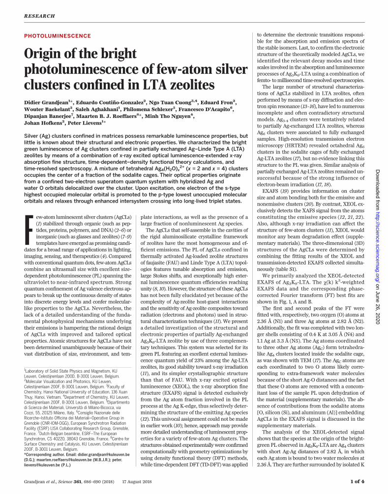
PHOTOLUMINESCENCE
Origin of the brightphotoluminescence of few-atom silverclusters confined in LTA zeolitesDidier Grandjean1*, Eduardo Coutiño-Gonzalez2, Ngo Tuan Cuong3,4, Eduard Fron2,Wouter Baekelant2, Saleh Aghakhani1, Philomena Schlexer5, Francesco D’Acapito6,Dipanjan Banerjee7, Maarten B. J. Roeffaers8*, Minh Tho Nguyen4,Johan Hofkens2, Peter Lievens1*
Silver (Ag) clusters confined in matrices possess remarkable luminescence properties, butlittle is known about their structural and electronic properties. We characterized the brightgreen luminescence of Ag clusters confined in partially exchanged Ag–Linde Type A (LTA)zeolites by means of a combination of x-ray excited optical luminescence-extended x-rayabsorption fine structure, time-dependent–density functional theory calculations, andtime-resolved spectroscopy. A mixture of tetrahedral Ag4(H2O)x
2+ (x = 2 and x = 4) clustersoccupies the center of a fraction of the sodalite cages. Their optical properties originatefrom a confined two-electron superatom quantum system with hybridized Ag andwater O orbitals delocalized over the cluster. Upon excitation, one electron of the s-typehighest occupied molecular orbital is promoted to the p-type lowest unoccupied molecularorbitals and relaxes through enhanced intersystem crossing into long-lived triplet states.
Few-atom luminescent silver clusters (AgCLs)(1) stabilized through organic (such as pep-tides, proteins, polymers, and DNA) (2–6) orinorganic (such as glasses and zeolites) (7–9)templates have emerged as promising candi-
dates for a broad range of applications in lighting,imaging, sensing, and therapeutics (4). Comparedwith conventional quantumdots, few-atomAgCLscombine an ultrasmall size with excellent size-dependent photoluminescence (PL) spanning theultraviolet to near-infrared spectrum. Strongquantum confinement of Ag valence electrons ap-pears to break up the continuous density of statesinto discrete energy levels and confer molecular-like properties to the AgCLs. Nevertheless, thelack of a detailed understanding of the funda-mental photophysical mechanisms underlyingtheir emissions is hampering the rational designof AgCLs with improved and tailored opticalproperties. Atomic structures for AgCLs have notbeen determined unambiguously because of theirvast distribution of size, environment, and tem-
plate interactions, as well as the presence of alarge fraction of nonluminescent Ag species.The AgCLs that self-assemble in the cavities of
the rigid aluminosilicate crystalline frameworkof zeolites have the most homogeneous and ef-ficient emissions. The PL of AgCLs confined inthermally activated Ag-loaded zeolite structuresof faujasite (FAU) and Linde Type A (LTA) topol-ogies features tunable absorption and emission,large Stokes shifts, and exceptionally high exter-nal luminescence quantum efficiencies reachingunity (8, 10). However, the structure of these AgCLshas not been fully elucidated yet because of thecomplexity of Ag-zeolite host-guest interactionsand the sensitivity of Ag-zeolite composites towardradiation (electrons and photons) used in struc-tural characterization techniques (11). We presenta detailed investigation of the structural andelectronic properties of partially Ag-exchangedAg3K9-LTA zeolite by use of three complemen-tary techniques. This system was selected for itsgreen PL featuring an excellent external lumines-cence quantum yield of 23% among the Ag-LTAzeolites, its good stability toward x-ray irradiation(11), and its simpler crystallographic structurethan that of FAU. With x-ray excited opticalluminescence (XEOL), the x-ray absorption finestructure (EXAFS) signal is detected exclusivelyfrom the Ag atom fraction involved in the PLprocess at the Ag K-edge, thus selectively deter-mining the structure of the emitting Ag species(12). This univocal assignment could not bemadein earlier work (10); hence, approachmay providemore detailed understanding of luminescent prop-erties for a variety of few-atom Ag clusters. Thestructures obtained experimentallywere confirmedcomputationally with geometry optimizations byusing density functional theory (DFT) methods,while time-dependent DFT (TD-DFT) was applied
to determine the electronic transitions responsi-ble for the absorption and emission spectra ofthe stable isomers. Last, to confirm the electronicstructure of the theoretically modeled AgCLs, weidentified the relevant decay modes and timescales involved in the absorption and luminescenceprocesses of Ag3K9-LTA using a combination offemto- tomillisecond time-resolved spectroscopies.The large number of structural characteriza-
tions of AgCLs stabilized in LTA zeolites, oftenperformed by means of x-ray diffraction and elec-tron spin resonance (13–16), have led to numerousincomplete and often contradictory structuralmodels. Ag3-4 clusters were tentatively relatedto partially Ag-exchanged LTA zeolites, whereasAg6 clusters were associated to fully exchangedsamples. High-resolution transmission electronmicroscopy (HRTEM) revealed octahedral Ag6clusters in the sodalite cages of fully exchangedAg-LTA zeolites (17), but no evidence linking thisstructure to the PL was given. Similar analysis ofpartially exchanged Ag-LTA zeolites remained un-successful because of the strong influence ofelectron-beam irradiation (17, 18).EXAFS (19) provides information on cluster
size and atom bonding both for the emissive andnonemissive clusters (20). By contrast, XEOL ex-clusively detects the XAFS signal from the atomsconstituting the emissive species (12, 21, 22).Also, although x-ray irradiation can affect thestructure of few-atom clusters (11), XEOL wouldmonitor any beam degradation effect (supple-mentary materials). The three-dimensional (3D)structures of the AgCLs were determined bycombining the fitting results of the XEOL andtransmission-detected EXAFS collected simulta-neously (table S1).We primarily analyzed the XEOL-detected
EXAFS of Ag3K9-LTA. The c(k) k3-weightedEXAFS data and the corresponding phase-corrected Fourier transform (FT) best fits areshown in Fig. 1, A and B.The first and second peaks of the FT were
fitted with, respectively, two oxygen (O) atoms at2.36 Å (N1) and three Ag atoms at 2.82 Å (N2).Additionally, the fit was completed with two lon-ger shells consisting of 0.4 K at 3.05 Å (N4) and1.1 Ag at 3.3 Å (N5). The Ag atoms coordinatedto three other Ag atoms (AgC) form tetrahedra-like Ag4 clusters located inside the sodalite cage,as was shown with TEM (17). The AgC atoms areeach coordinated to two O atoms likely corre-sponding to extra-framework water moleculesbecause of the short Ag-O distances and the factthat these O atoms are removed with a concom-itant loss of the sample PL upon dehydration ofthe material (supplementary materials). The ab-sence of contributions from the sodalite atoms[O, silicon (Si), and aluminum (Al)] embeddingAgCLs in the EXAFS signal is discussed in thesupplementary materials.The analysis of the XEOL-detected signal
shows that the species at the origin of the bright-green PL observed in Ag3K9-LTA are Ag4 clusterswith short Ag-Ag distances of 2.82 Å, in whicheach Ag atom is bound to twowatermolecules at2.36 Å. They are further surrounded by isolatedK
RESEARCH
Grandjean et al., Science 361, 686–690 (2018) 17 August 2018 1 of 4
1Laboratory of Solid State Physics and Magnetism, KULeuven, Celestijnenlaan 200D, B-3001 Leuven, Belgium.2Molecular Visualization and Photonics, KU Leuven,Celestijnenlaan 200F, B-3001 Leuven, Belgium. 3Faculty ofChemistry, Hanoi National University of Education, 136 XuanThuy, Hanoi, Vietnam. 4Department of Chemistry, KU Leuven,Celestijnenlaan 200F, B-3001 Leuven, Belgium. 5Dipartimentodi Scienza dei Materiali, Università di Milano-Bicocca, viaCozzi, 55, 20125 Milano, Italy. 6Consiglio Nazionale delleRicerche–Istituto Officina dei Materiali–Operative Group inGrenoble (CNR-IOM-OGG), European Synchrotron RadiationFacility (ESRF) LISA Collaborating Research Group, Grenoble,France. 7Dutch-Belgian beamline, ESRF–The EuropeanSynchrotron, CS 40220, 38043 Grenoble, France. 8Centre forSurface Chemistry and Catalysis, KU Leuven, Celestijnenlaan200F, B-3001 Leuven, Belgium.*Corresponding author. Email: [email protected](D.G.); [email protected] (M.B.J.R.); [email protected] (P.L.)
on June 26, 2020
http://science.sciencemag.org/
Dow
nloaded from
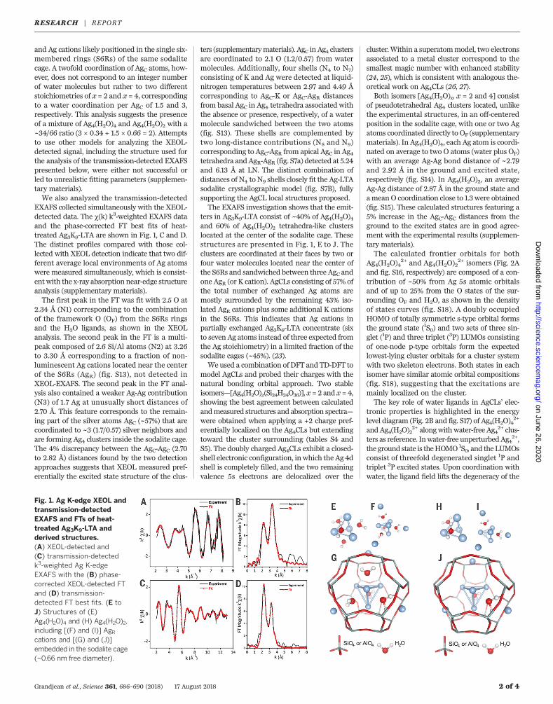
and Ag cations likely positioned in the single six-membered rings (S6Rs) of the same sodalitecage. A twofold coordination of AgC atoms, how-ever, does not correspond to an integer numberof water molecules but rather to two differentstoichiometries of x = 2 and x = 4, correspondingto a water coordination per AgC of 1.5 and 3,respectively. This analysis suggests the presenceof a mixture of Ag4(H2O)4 and Ag4(H2O)2 with a~34/66 ratio (3 × 0.34 + 1.5 × 0.66 = 2). Attemptsto use other models for analyzing the XEOL-detected signal, including the structure used forthe analysis of the transmission-detected EXAFSpresented below, were either not successful orled to unrealistic fitting parameters (supplemen-tary materials).We also analyzed the transmission-detected
EXAFS collected simultaneously with the XEOL-detected data. The c(k) k3-weighted EXAFS dataand the phase-corrected FT best fits of heat-treated Ag3K9-LTA are shown in Fig. 1, C and D.The distinct profiles compared with those col-lected with XEOL detection indicate that two dif-ferent average local environments of Ag atomswere measured simultaneously, which is consist-ent with the x-ray absorption near-edge structureanalysis (supplementary materials).The first peak in the FT was fit with 2.5 O at
2.34 Å (N1) corresponding to the combinationof the framework O (OF) from the S6Rs ringsand the H2O ligands, as shown in the XEOLanalysis. The second peak in the FT is a multi-peak composed of 2.6 Si/Al atoms (N2) at 3.26to 3.30 Å corresponding to a fraction of non-luminescent Ag cations located near the centerof the S6Rs (AgR) (fig. S13), not detected inXEOL-EXAFS. The second peak in the FT anal-ysis also contained a weaker Ag-Ag contribution(N3) of 1.7 Ag at unusually short distances of2.70 Å. This feature corresponds to the remain-ing part of the silver atoms AgC (~57%) that arecoordinated to ~3 (1.7/0.57) silver neighbors andare forming Ag4 clusters inside the sodalite cage.The 4% discrepancy between the AgC-AgC (2.70to 2.82 Å) distances found by the two detectionapproaches suggests that XEOL measured pref-erentially the excited state structure of the clus-
ters (supplementarymaterials). AgC inAg4 clustersare coordinated to 2.1 O (1.2/0.57) from watermolecules. Additionally, four shells (N4 to N7)consisting of K and Ag were detected at liquid-nitrogen temperatures between 2.97 and 4.49 Åcorresponding to AgC-K or AgC-AgR distancesfrom basal AgC in Ag4 tetrahedra associated withthe absence or presence, respectively, of a watermolecule sandwiched between the two atoms(fig. S13). These shells are complemented bytwo long-distance contributions (N8 and N9)corresponding to AgC-AgR from apical AgC in Ag4tetrahedra andAgR-AgR (fig. S7a) detected at 5.24and 6.13 Å at LN. The distinct combination ofdistances of N4 to N9 shells closely fit the Ag-LTAsodalite crystallographic model (fig. S7B), fullysupporting the AgCL local structures proposed.The EXAFS investigation shows that the emit-
ters in Ag3K9-LTA consist of ~40% of Ag4(H2O)4and 60% of Ag4(H2O)2 tetrahedra-like clusterslocated at the center of the sodalite cage. Thesestructures are presented in Fig. 1, E to J. Theclusters are coordinated at their faces by two orfour water molecules located near the center ofthe S6Rs and sandwiched between three AgC andone AgR (or K cation). AgCLs consisting of 57% ofthe total number of exchanged Ag atoms aremostly surrounded by the remaining 43% iso-lated AgR cations plus some additional K cationsin the S6Rs. This indicates that Ag cations inpartially exchanged Ag3K9-LTA concentrate (sixto seven Ag atoms instead of three expected fromthe Ag stoichiometry) in a limited fraction of thesodalite cages (~45%). (23).We used a combination of DFT and TD-DFT to
model AgCLs and probed their charges with thenatural bonding orbital approach. Two stableisomers—[Ag4(H2O)x(Si24H24O36)], x = 2 and x = 4,showing the best agreement between calculatedandmeasured structures and absorption spectra—were obtained when applying a +2 charge pref-erentially localized on the Ag4CLs but extendingtoward the cluster surrounding (tables S4 andS5). The doubly charged Ag4CLs exhibit a closed-shell electronic configuration, inwhich the Ag 4dshell is completely filled, and the two remainingvalence 5s electrons are delocalized over the
cluster.Within a superatommodel, two electronsassociated to a metal cluster correspond to thesmallest magic number with enhanced stability(24, 25), which is consistent with analogous the-oretical work on Ag4CLs (26, 27).Both isomers [Ag4(H2O)x, x = 2 and 4] consist
of pseudotetrahedral Ag4 clusters located, unlikethe experimental structures, in an off-centeredposition in the sodalite cage, with one or two Agatoms coordinated directly to OF (supplementarymaterials). In Ag4(H2O)4, each Ag atom is coordi-nated on average to two O atoms (water plus OF)with an average Ag-Ag bond distance of ~2.79and 2.92 Å in the ground and excited state,respectively (fig. S14). In Ag4(H2O)2, an averageAg-Ag distance of 2.87 Å in the ground state andameanO coordination close to 1.3 were obtained(fig. S15). These calculated structures featuring a5% increase in the AgC-AgC distances from theground to the excited states are in good agree-ment with the experimental results (supplemen-tary materials).The calculated frontier orbitals for both
Ag4(H2O)42+ and Ag4(H2O)2
2+ isomers (Fig. 2Aand fig. S16, respectively) are composed of a con-tribution of ~50% from Ag 5s atomic orbitalsand of up to 25% from the O states of the sur-rounding OF and H2O, as shown in the densityof states curves (fig. S18). A doubly occupiedHOMO of totally symmetric s-type orbital formsthe ground state (1S0) and two sets of three sin-glet (1P) and three triplet (3P) LUMOs consistingof one-node p-type orbitals form the expectedlowest-lying cluster orbitals for a cluster systemwith two skeleton electrons. Both states in eachisomer have similar atomic orbital compositions(fig. S18), suggesting that the excitations aremainly localized on the cluster.The key role of water ligands in AgCLs’ elec-
tronic properties is highlighted in the energylevel diagram (Fig. 2B and fig. S17) of Ag4(H2O)4
2+
andAg4(H2O)22+ alongwithwater-free Ag4
2+ clus-ters as reference. Inwater-free unperturbedAg4
2+,the ground state is theHOMO1S0, and theLUMOsconsist of threefold degenerated singlet 1P andtriplet 3P excited states. Upon coordination withwater, the ligand field lifts the degeneracy of the
Grandjean et al., Science 361, 686–690 (2018) 17 August 2018 2 of 4
Fig. 1. Ag K-edge XEOL andtransmission-detectedEXAFS and FTs of heat-treated Ag3K9-LTA andderived structures.(A) XEOL-detected and(C) transmission-detectedk3-weighted Ag K-edgeEXAFS with the (B) phase-corrected XEOL-detected FTand (D) transmission-detected FT best fits. (E toJ) Structures of (E)Ag4(H2O)4 and (H) Ag4(H2O)2,including [(F) and (I)] AgRcations and [(G) and (J)]embedded in the sodalite cage(~0.66 nm free diameter).
RESEARCH | REPORTon June 26, 2020
http://science.sciencemag.org/
Dow
nloaded from
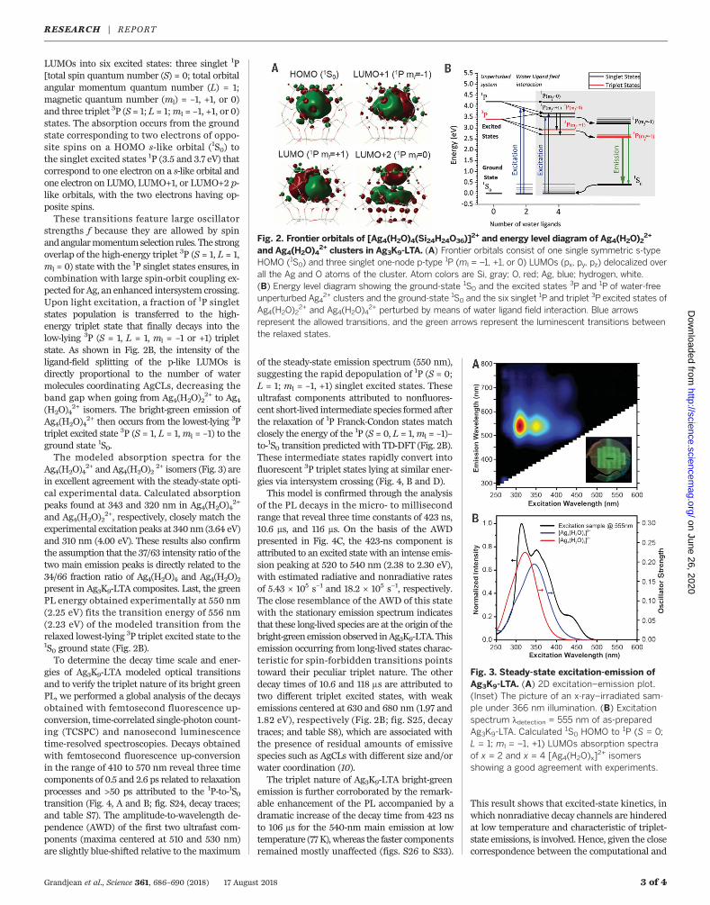
LUMOs into six excited states: three singlet 1P[total spin quantum number (S) = 0; total orbitalangular momentum quantum number (L) = 1;magnetic quantum number (ml) = –1, +1, or 0)and three triplet 3P (S= 1; L = 1;ml = –1, +1, or 0)states. The absorption occurs from the groundstate corresponding to two electrons of oppo-site spins on a HOMO s-like orbital (1S0) tothe singlet excited states 1P (3.5 and 3.7 eV) thatcorrespond to one electron on a s-like orbital andone electron on LUMO, LUMO+1, or LUMO+2 p-like orbitals, with the two electrons having op-posite spins.These transitions feature large oscillator
strengths f because they are allowed by spinandangularmomentumselection rules. The strongoverlap of the high-energy triplet 3P (S = 1, L = 1,ml = 0) state with the 1P singlet states ensures, incombination with large spin-orbit coupling ex-pected for Ag, an enhanced intersystem crossing.Upon light excitation, a fraction of 1P singletstates population is transferred to the high-energy triplet state that finally decays into thelow-lying 3P (S = 1, L = 1, ml = –1 or +1) tripletstate. As shown in Fig. 2B, the intensity of theligand-field splitting of the p-like LUMOs isdirectly proportional to the number of watermolecules coordinating AgCLs, decreasing theband gap when going from Ag4(H2O)2
2+ to Ag4(H2O)4
2+ isomers. The bright-green emission ofAg4(H2O)4
2+ then occurs from the lowest-lying 3Ptriplet excited state 3P (S = 1, L = 1,ml = –1) to theground state 1S0.The modeled absorption spectra for the
Ag4(H2O)42+ and Ag4(H2O)2
2+ isomers (Fig. 3) arein excellent agreement with the steady-state opti-cal experimental data. Calculated absorptionpeaks found at 343 and 320 nm in Ag4(H2O)4
2+
and Ag4(H2O)22+, respectively, closely match the
experimental excitation peaks at 340nm (3.64 eV)and 310 nm (4.00 eV). These results also confirmthe assumption that the 37/63 intensity ratio of thetwo main emission peaks is directly related to the34/66 fraction ratio of Ag4(H2O)4 and Ag4(H2O)2present in Ag3K9-LTA composites. Last, the greenPL energy obtained experimentally at 550 nm(2.25 eV) fits the transition energy of 556 nm(2.23 eV) of the modeled transition from therelaxed lowest-lying 3P triplet excited state to the1S0 ground state (Fig. 2B).To determine the decay time scale and ener-
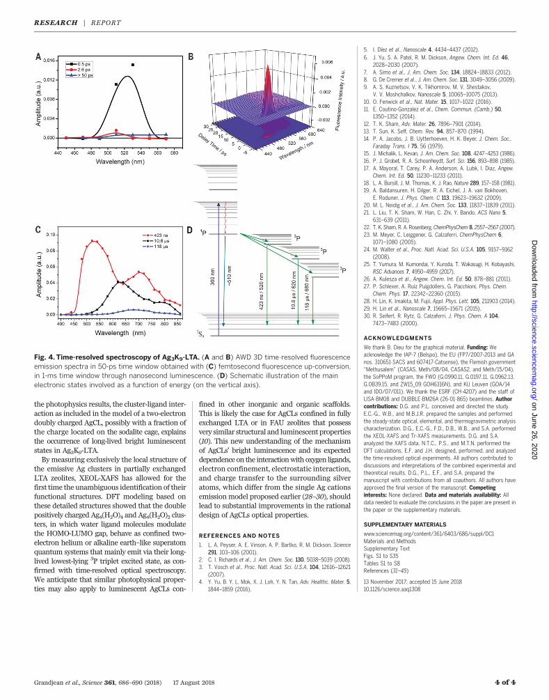
gies of Ag3K9-LTA modeled optical transitionsand to verify the triplet nature of its bright greenPL, we performed a global analysis of the decaysobtained with femtosecond fluorescence up-conversion, time-correlated single-photon count-ing (TCSPC) and nanosecond luminescencetime-resolved spectroscopies. Decays obtainedwith femtosecond fluorescence up-conversionin the range of 410 to 570 nm reveal three timecomponents of 0.5 and 2.6 ps related to relaxationprocesses and >50 ps attributed to the 1P-to-1S0transition (Fig. 4, A and B; fig. S24, decay traces;and table S7). The amplitude-to-wavelength de-pendence (AWD) of the first two ultrafast com-ponents (maxima centered at 510 and 530 nm)are slightly blue-shifted relative to the maximum
of the steady-state emission spectrum (550 nm),suggesting the rapid depopulation of 1P (S = 0;L = 1; ml = –1, +1) singlet excited states. Theseultrafast components attributed to nonfluores-cent short-lived intermediate species formed afterthe relaxation of 1P Franck-Condon states matchclosely the energy of the 1P (S = 0, L = 1,ml = –1)–to-1S0 transition predicted with TD-DFT (Fig. 2B).These intermediate states rapidly convert intofluorescent 3P triplet states lying at similar ener-gies via intersystem crossing (Fig. 4, B and D).This model is confirmed through the analysis
of the PL decays in the micro- to millisecondrange that reveal three time constants of 423 ns,10.6 ms, and 116 ms. On the basis of the AWDpresented in Fig. 4C, the 423-ns component isattributed to an excited state with an intense emis-sion peaking at 520 to 540 nm (2.38 to 2.30 eV),with estimated radiative and nonradiative ratesof 5.43 × 10⁵ s−1 and 18.2 × 10⁵ s−1, respectively.The close resemblance of the AWD of this statewith the stationary emission spectrum indicatesthat these long-lived species are at the origin of thebright-greenemissionobserved inAg3K9-LTA.Thisemission occurring from long-lived states charac-teristic for spin-forbidden transitions pointstoward their peculiar triplet nature. The otherdecay times of 10.6 and 118 ms are attributed totwo different triplet excited states, with weakemissions centered at 630 and 680 nm (1.97 and1.82 eV), respectively (Fig. 2B; fig. S25, decaytraces; and table S8), which are associated withthe presence of residual amounts of emissivespecies such as AgCLs with different size and/orwater coordination (10).The triplet nature of Ag3K9-LTA bright-green
emission is further corroborated by the remark-able enhancement of the PL accompanied by adramatic increase of the decay time from 423 nsto 106 ms for the 540-nm main emission at lowtemperature (77K), whereas the faster componentsremained mostly unaffected (figs. S26 to S33).
This result shows that excited-state kinetics, inwhich nonradiative decay channels are hinderedat low temperature and characteristic of triplet-state emissions, is involved. Hence, given the closecorrespondence between the computational and
Grandjean et al., Science 361, 686–690 (2018) 17 August 2018 3 of 4
Fig. 3. Steady-state excitation-emission ofAg3K9-LTA. (A) 2D excitation–emission plot.(Inset) The picture of an x-ray–irradiated sam-ple under 366 nm illumination. (B) Excitationspectrum ldetection = 555 nm of as-preparedAg3K9-LTA. Calculated
1S0 HOMO to 1P (S = 0;L = 1; ml = –1, +1) LUMOs absorption spectraof x = 2 and x = 4 [Ag4(H2O)x]
2+ isomersshowing a good agreement with experiments.
Fig. 2. Frontier orbitals of [Ag4(H2O)4(Si24H24O36)]2+ and energy level diagram of Ag4(H2O)2
2+
and Ag4(H2O)42+ clusters in Ag3K9-LTA. (A) Frontier orbitals consist of one single symmetric s-type
HOMO (1S0) and three singlet one-node p-type 1P (ml = –1, +1, or 0) LUMOs (px, py, pz) delocalized overall the Ag and O atoms of the cluster. Atom colors are Si, gray; O, red; Ag, blue; hydrogen, white.(B) Energy level diagram showing the ground-state 1S0 and the excited states 3P and 1P of water-freeunperturbed Ag4
2+ clusters and the ground-state 1S0 and the six singlet 1P and triplet 3P excited states ofAg4(H2O)2
2+ and Ag4(H2O)42+ perturbed by means of water ligand field interaction. Blue arrows
represent the allowed transitions, and the green arrows represent the luminescent transitions betweenthe relaxed states.
RESEARCH | REPORTon June 26, 2020
http://science.sciencemag.org/
Dow
nloaded from
the photophysics results, the cluster-ligand inter-action as included in the model of a two-electrondoubly charged AgCL, possibly with a fraction ofthe charge located on the sodalite cage, explainsthe occurrence of long-lived bright luminescentstates in Ag3K9-LTA.By measuring exclusively the local structure of
the emissive Ag clusters in partially exchangedLTA zeolites, XEOL-XAFS has allowed for thefirst time the unambiguous identification of theirfunctional structures. DFT modeling based onthese detailed structures showed that the doublepositively charged Ag4(H2O)4 and Ag4(H2O)2 clus-ters, in which water ligand molecules modulatethe HOMO-LUMO gap, behave as confined two-electron helium or alkaline earth–like superatomquantum systems that mainly emit via their long-lived lowest-lying 3P triplet excited state, as con-firmed with time-resolved optical spectroscopy.We anticipate that similar photophysical proper-ties may also apply to luminescent AgCLs con-
fined in other inorganic and organic scaffolds.This is likely the case for AgCLs confined in fullyexchanged LTA or in FAU zeolites that possessvery similar structural and luminescent properties(10). This new understanding of the mechanismof AgCLs’ bright luminescence and its expecteddependence on the interactionwith oxygen ligands,electron confinement, electrostatic interaction,and charge transfer to the surrounding silveratoms, which differ from the single Ag cationsemissionmodel proposed earlier (28–30), shouldlead to substantial improvements in the rationaldesign of AgCLs optical properties.
REFERENCES AND NOTES
1. L. A. Peyser, A. E. Vinson, A. P. Bartko, R. M. Dickson, Science291, 103–106 (2001).
2. C. I. Richards et al., J. Am. Chem. Soc. 130, 5038–5039 (2008).3. T. Vosch et al., Proc. Natl. Acad. Sci. U.S.A. 104, 12616–12621
(2007).4. Y. Yu, B. Y. L. Mok, X. J. Loh, Y. N. Tan, Adv. Healthc. Mater. 5,
1844–1859 (2016).
5. I. Díez et al., Nanoscale 4, 4434–4437 (2012).6. J. Yu, S. A. Patel, R. M. Dickson, Angew. Chem. Int. Ed. 46,
2028–2030 (2007).7. A. Simo et al., J. Am. Chem. Soc. 134, 18824–18833 (2012).8. G. De Cremer et al., J. Am. Chem. Soc. 131, 3049–3056 (2009).9. A. S. Kuznetsov, V. K. Tikhomirov, M. V. Shestakov,
V. V. Moshchalkov, Nanoscale 5, 10065–10075 (2013).10. O. Fenwick et al., Nat. Mater. 15, 1017–1022 (2016).11. E. Coutino-Gonzalez et al., Chem. Commun. (Camb.) 50,
1350–1352 (2014).12. T. K. Sham, Adv. Mater. 26, 7896–7901 (2014).13. T. Sun, K. Seff, Chem. Rev. 94, 857–870 (1994).14. P. A. Jacobs, J. B. Uytterhoeven, H. K. Beyer, J. Chem. Soc.,
Faraday Trans. I 75, 56 (1979).15. J. Michalik, L. Kevan, J. Am. Chem. Soc. 108, 4247–4253 (1986).16. P. J. Grobet, R. A. Schoonheydt, Surf. Sci. 156, 893–898 (1985).17. A. Mayoral, T. Carey, P. A. Anderson, A. Lubk, I. Diaz, Angew.
Chem. Int. Ed. 50, 11230–11233 (2011).18. L. A. Bursill, J. M. Thomas, K. J. Rao, Nature 289, 157–158 (1981).19. A. Baldansuren, H. Dilger, R. A. Eichel, J. A. van Bokhoven,
E. Roduner, J. Phys. Chem. C 113, 19623–19632 (2009).20. M. L. Neidig et al., J. Am. Chem. Soc. 133, 11837–11839 (2011).21. L. Liu, T. K. Sham, W. Han, C. Zhi, Y. Bando, ACS Nano 5,
631–639 (2011).22. T. K. Sham, R. A. Rosenberg, ChemPhysChem 8, 2557–2567 (2007).23. M. Meyer, C. Leiggener, G. Calzaferri, ChemPhysChem 6,
1071–1080 (2005).24. M. Walter et al., Proc. Natl. Acad. Sci. U.S.A. 105, 9157–9162
(2008).25. T. Yumura, M. Kumondai, Y. Kuroda, T. Wakasugi, H. Kobayashi,
RSC Advances 7, 4950–4959 (2017).26. A. Kulesza et al., Angew. Chem. Int. Ed. 50, 878–881 (2011).27. P. Schlexer, A. Ruiz Puigdollers, G. Pacchioni, Phys. Chem.
Chem. Phys. 17, 22342–22360 (2015).28. H. Lin, K. Imakita, M. Fujii, Appl. Phys. Lett. 105, 211903 (2014).29. H. Lin et al., Nanoscale 7, 15665–15671 (2015).30. R. Seifert, R. Rytz, G. Calzaferri, J. Phys. Chem. A 104,
7473–7483 (2000).
ACKNOWLEDGMENTS
We thank B. Dieu for the graphical material. Funding: Weacknowledge the IAP-7 (Belspo), the EU (FP7/2007-2013 and GAnos. 310651-SACS and 607417-Catsense), the Flemish government“Methusalem” (CASAS, Meth/08/04, CASAS2, and Meth/15/04),the SoPPoM program, the FWO (G.0990.11, G.0197.11, G.0962.13,G.0B39.15, and ZW15_09 GOH6316N), and KU Leuven (GOA/14and IDO/07/011). We thank the ESRF (CH-4207) and the staff ofLISA-BM08 and DUBBLE-BM26A (26-01-865) beamlines. Authorcontributions: D.G. and P.L. conceived and directed the study.E.C.-G., W.B., and M.B.J.R. prepared the samples and performedthe steady-state optical, elemental, and thermogravimetric analysischaracterization. D.G., E.C.-G., F.D., D.B., W.B., and S.A. performedthe XEOL-XAFS and Tr-XAFS measurements. D.G. and S.A.analyzed the XAFS data. N.T.C., P.S., and M.T.N. performed theDFT calculations. E.F. and J.H. designed, performed, and analyzedthe time-resolved optical experiments. All authors contributed todiscussions and interpretations of the combined experimental andtheoretical results. D.G., P.L., E.F., and S.A. prepared themanuscript with contributions from all coauthors. All authors haveapproved the final version of the manuscript. Competinginterests: None declared. Data and materials availability: Alldata needed to evaluate the conclusions in the paper are present inthe paper or the supplementary materials.
SUPPLEMENTARY MATERIALS
www.sciencemag.org/content/361/6403/686/suppl/DC1Materials and MethodsSupplementary TextFigs. S1 to S35Tables S1 to S8References (31–45)
13 November 2017; accepted 15 June 201810.1126/science.aaq1308
Grandjean et al., Science 361, 686–690 (2018) 17 August 2018 4 of 4
Fig. 4. Time-resolved spectroscopy of Ag3K9-LTA. (A and B) AWD 3D time-resolved fluorescenceemission spectra in 50-ps time window obtained with (C) femtosecond fluorescence up-conversion,in 1-ms time window through nanosecond luminescence. (D) Schematic illustration of the mainelectronic states involved as a function of energy (on the vertical axis).
RESEARCH | REPORTon June 26, 2020
http://science.sciencemag.org/
Dow
nloaded from

Origin of the bright photoluminescence of few-atom silver clusters confined in LTA zeolites
and Peter LievensPhilomena Schlexer, Francesco D'Acapito, Dipanjan Banerjee, Maarten B. J. Roeffaers, Minh Tho Nguyen, Johan Hofkens Didier Grandjean, Eduardo Coutiño-Gonzalez, Ngo Tuan Cuong, Eduard Fron, Wouter Baekelant, Saleh Aghakhani,
DOI: 10.1126/science.aaq1308 (6403), 686-690.361Science
, this issue p. 686; see also p. 645Sciencethus identifying candidate materials for application in lighting, imaging, and therapeutics.−−produce bright green emission
theoretical calculations, they identified the electronic states of four-atom silver clusters bound with water molecules that luminescence to monitor only the emissive structures (see the Perspective by Quintanilla and Liz-Marzán). Aided by
studied silver clusters in zeolites, using x-ray excited optical et al.many larger but inactive clusters. Grandjean but the origin of this effect has been difficult to establish, in part because the materials are heterogeneous and contain
Small silver clusters stabilized by organic materials or inorganic surfaces can exhibit bright photoluminescence,Unmasking the glow of silver clusters
ARTICLE TOOLS http://science.sciencemag.org/content/361/6403/686
MATERIALSSUPPLEMENTARY http://science.sciencemag.org/content/suppl/2018/08/15/361.6403.686.DC1
CONTENTRELATED http://science.sciencemag.org/content/sci/361/6403/645.full
REFERENCES
http://science.sciencemag.org/content/361/6403/686#BIBLThis article cites 39 articles, 3 of which you can access for free
PERMISSIONS http://www.sciencemag.org/help/reprints-and-permissions
Terms of ServiceUse of this article is subject to the
is a registered trademark of AAAS.ScienceScience, 1200 New York Avenue NW, Washington, DC 20005. The title (print ISSN 0036-8075; online ISSN 1095-9203) is published by the American Association for the Advancement ofScience
Science. No claim to original U.S. Government WorksCopyright © 2018 The Authors, some rights reserved; exclusive licensee American Association for the Advancement of
on June 26, 2020
http://science.sciencemag.org/
Dow
nloaded from


![cations Supporting information anions and organic · Supporting information Efficient modulation of photoluminescence by hydrogen bonding interactions among inorganic [MnBr4]2-anions](https://img.pdfslide.fr/doc/110x75/5f8c419e2fd7220f220cd9f4/cations-supporting-information-anions-and-supporting-information-efficient-modulation.jpg)