Embed Size (px)
Citation preview
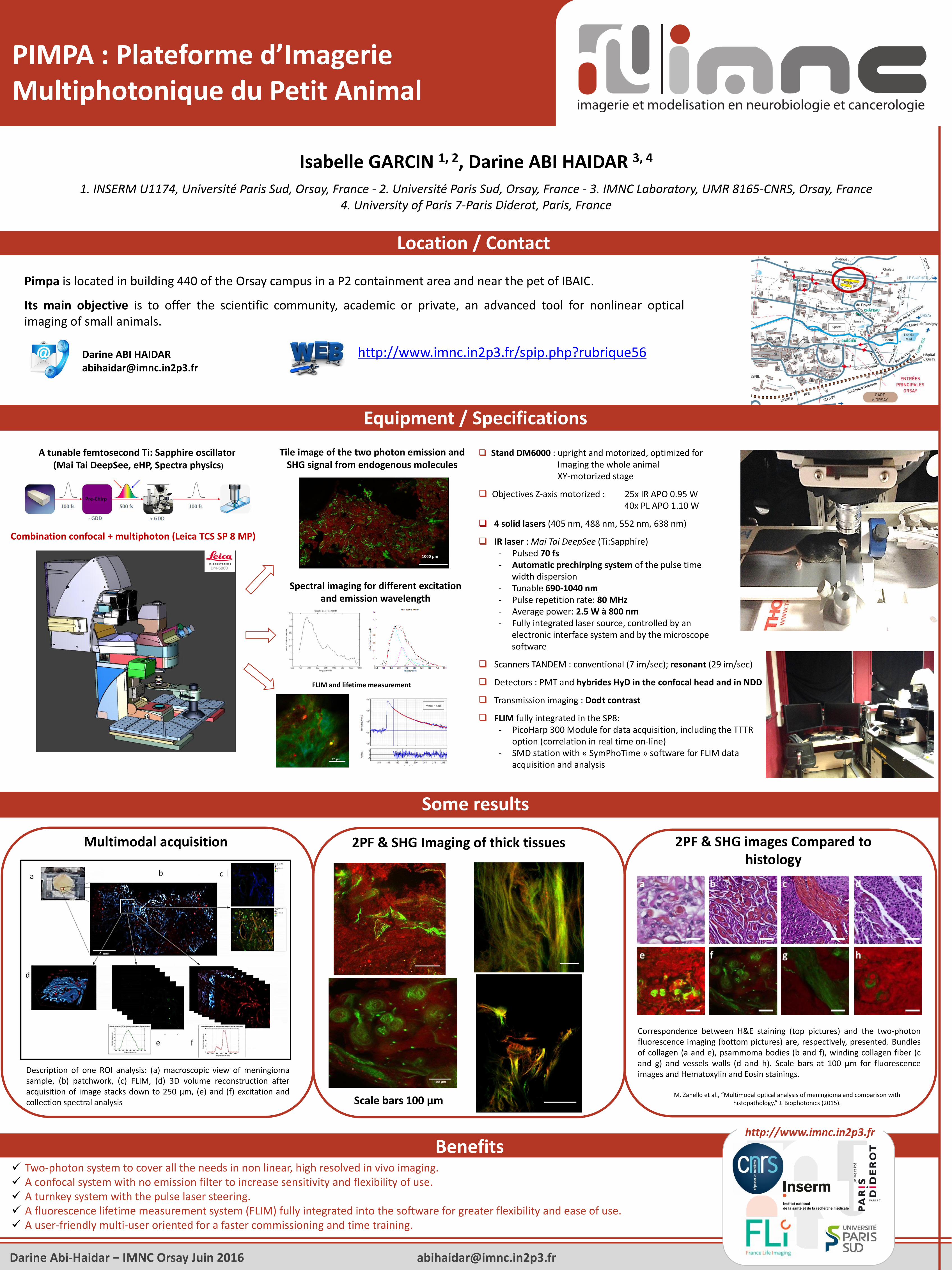
Location / Contact
Equipment / Specifications
Isabelle GARCIN 1, 2, Darine ABI HAIDAR 3, 4
1. INSERM U1174, Université Paris Sud, Orsay, France - 2. Université Paris Sud, Orsay, France - 3. IMNC Laboratory, UMR 8165-CNRS, Orsay, France 4. University of Paris 7-Paris Diderot, Paris, France
PIMPA : Plateforme d’Imagerie Multiphotonique du Petit Animal
Pimpa is located in building 440 of the Orsay campus in a P2 containment area and near the pet of IBAIC.
Its main objective is to offer the scientific community, academic or private, an advanced tool for nonlinear opticalimaging of small animals.
Darine ABI [email protected]
Stand DM6000 : upright and motorized, optimized for Imaging the whole animalXY-motorized stage
Objectives Z-axis motorized : 25x IR APO 0.95 W40x PL APO 1.10 W
4 solid lasers (405 nm, 488 nm, 552 nm, 638 nm)
IR laser : Mai Tai DeepSee (Ti:Sapphire) ‐ Pulsed 70 fs‐ Automatic prechirping system of the pulse time
width dispersion‐ Tunable 690-1040 nm‐ Pulse repetition rate: 80 MHz‐ Average power: 2.5 W à 800 nm‐ Fully integrated laser source, controlled by an
electronic interface system and by the microscope software
Scanners TANDEM : conventional (7 im/sec); resonant (29 im/sec)
Detectors : PMT and hybrides HyD in the confocal head and in NDD
Transmission imaging : Dodt contrast
FLIM fully integrated in the SP8: ‐ PicoHarp 300 Module for data acquisition, including the TTTR
option (correlation in real time on-line)‐ SMD station with « SymPhoTime » software for FLIM data
acquisition and analysis
Combination confocal + multiphoton (Leica TCS SP 8 MP)
Tile image of the two photon emission and SHG signal from endogenous molecules
FLIM and lifetime measurement
Spectral imaging for different excitation and emission wavelength
1000 μm
A tunable femtosecond Ti: Sapphire oscillator(Mai Tai DeepSee, eHP, Spectra physics)
25μm
M. Zanello et al., “Multimodal optical analysis of meningioma and comparison withhistopathology,” J. Biophotonics (2015).
Multimodal acquisition 2PF & SHG Imaging of thick tissues 2PF & SHG images Compared to histology
Benefits Two-photon system to cover all the needs in non linear, high resolved in vivo imaging. A confocal system with no emission filter to increase sensitivity and flexibility of use. A turnkey system with the pulse laser steering. A fluorescence lifetime measurement system (FLIM) fully integrated into the software for greater flexibility and ease of use. A user-friendly multi-user oriented for a faster commissioning and time training.
Correspondence between H&E staining (top pictures) and the two-photonfluorescence imaging (bottom pictures) are, respectively, presented. Bundlesof collagen (a and e), psammoma bodies (b and f), winding collagen fiber (cand g) and vessels walls (d and h). Scale bars at 100 μm for fluorescenceimages and Hematoxylin and Eosin stainings.
Scale bars 100 μm
Some results
Description of one ROI analysis: (a) macroscopic view of meningiomasample, (b) patchwork, (c) FLIM, (d) 3D volume reconstruction afteracquisition of image stacks down to 250 μm, (e) and (f) excitation andcollection spectral analysis
Darine Abi-Haidar − IMNC Orsay Juin 2016 [email protected]
http://www.imnc.in2p3.fr/spip.php?rubrique56
http://www.imnc.in2p3.fr