Embed Size (px)
Citation preview
Report about fish parasitic diseases
Alvarez-Pellitero P.
in
Alvarez-Pellitero P. (ed.), Barja J.L. (ed.), Basurco B. (ed.), Berthe F. (ed.), Toranzo A.E.(ed.). Mediterranean aquaculture diagnostic laboratories
Zaragoza : CIHEAMOptions Méditerranéennes : Série B. Etudes et Recherches; n. 49
2004pages 103-130
Article available on line / Article disponible en ligne à l’adresse :
--------------------------------------------------------------------------------------------------------------------------------------------------------------------------
http://om.ciheam.org/article.php?IDPDF=4600222
--------------------------------------------------------------------------------------------------------------------------------------------------------------------------
To cite th is article / Pour citer cet article
--------------------------------------------------------------------------------------------------------------------------------------------------------------------------
Alvarez-Pellitero P. Report about fish parasitic diseases. In : Alvarez-Pellitero P. (ed.), Barja J.L.
(ed.), Basurco B. (ed.), Berthe F. (ed.), Toranzo A.E. (ed.). Mediterranean aquaculture diagnostic
laboratories. Zaragoza : CIHEAM, 2004. p. 103-130 (Options Méditerranéennes : Série B. Etudes et
Recherches; n. 49)
--------------------------------------------------------------------------------------------------------------------------------------------------------------------------
http://www.ciheam.org/http://om.ciheam.org/
Report about fish parasitic diseases
P. Alvárez-Pellitero CSIC (Consejo Superior de Investigaciones Científicas)
Instituto de Acuicultura Torre de la Sal Ribera de Cabanes, 12595 Castellón, Spain
About diagnostic laboratories for parasitic diseases in the region
Most laboratories participating in the survey (48 out 54) from 13 Mediterranean countries stated that they perform diagnosis of fish parasitic diseases.
Of the 48 laboratories that declared to work on the diagnosis of fish parasitic diseases, 41 stated that they worked with seabream and seabass and 32 with trout. Other species they worked with were eels (13), carps (12), turbot (9) and salmon (9). Many (31 laboratories) stated to also work with other fish species, such as tilapia, mullets, other sparids (Dentex dentex, Puntazzo puntazzo, etc.) and other marine species.
This is coherent with available fish production information for the region, and as is the case for the diagnosis of viral and bacterial diseases, most diagnosis is performed on finfish species produced intensively in Mediterranean countries.
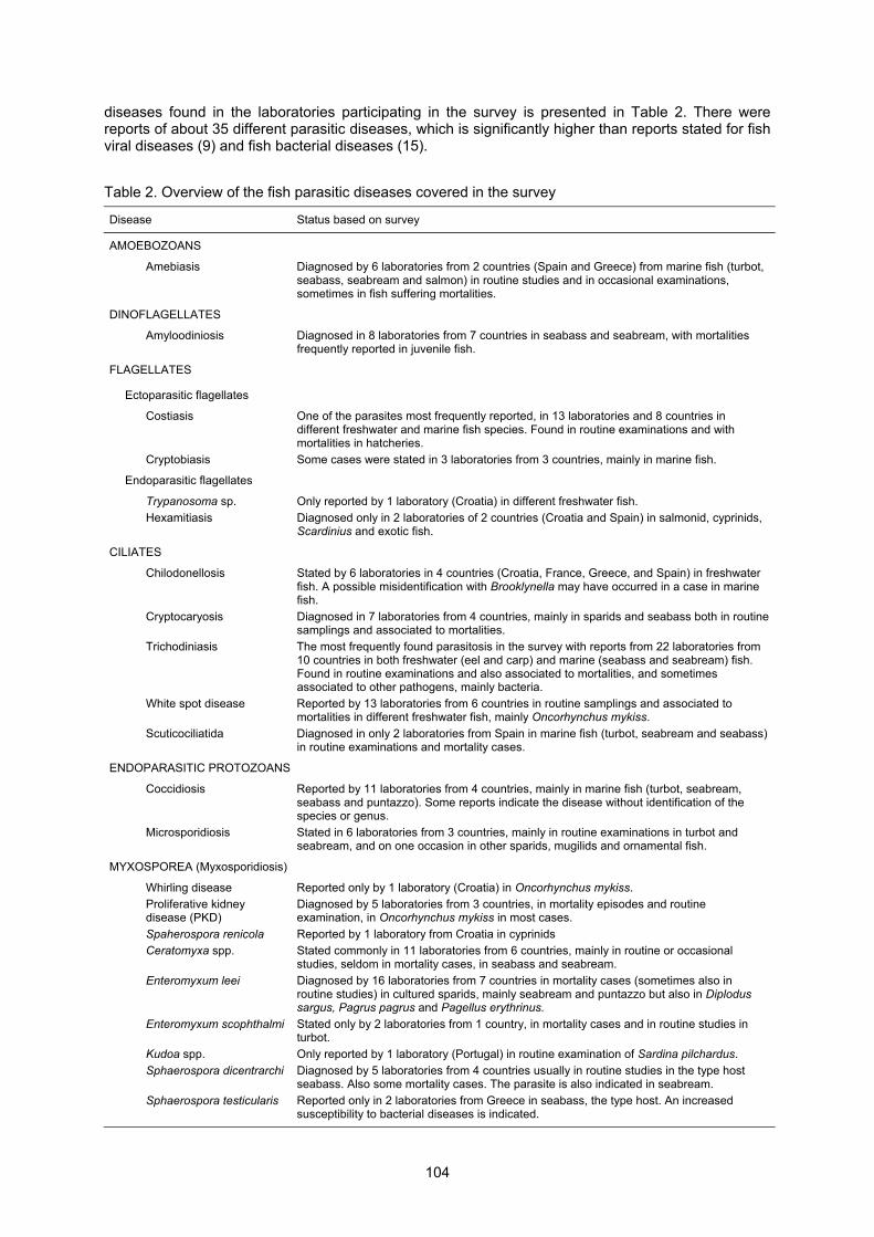
The different techniques available in the laboratories performing diagnosis of fish parasitic diseases (Table 1) show that most laboratories based their diagnosis partly or totally on simple techniques, such as observation of clinical signs, macroscopical examination of fish and examination of fresh samples (wet mounts, smears). Less laboratories (26 out 48) can perform histopathology studies, and even less (15 or 16) can perform more specialised techniques such as PCR or immunohistochemistry. Table 1. Available techniques in the laboratories performing diagnosis of fish parasitic diseases
Diagnostic methods Available
Clinical signs 48 Microscopical examination of fresh samples 47 Macroscopical examination 46 Histopathology 30 Haematological examination 26 Fluorescent antibody technique 15 PCR 16 Immunohistochemistry 15 Transmission electron microscopy 9 DNA probes 4
The above mentioned capability (available techniques) explains why, as indicated below, for most parasitic diseases the diagnosis is just based on simple techniques (clinical signs, macroscopical examination, examination of fresh samples) and also histopathology. Thus, the possibility of overlooking or misidentification of some parasites cannot be discarded. Efforts should be done to improve the technical level of diagnostic laboratories, in order to obtain more accurate diagnosis and the detection of asymptomatic carriers. Main reported parasites and diseases
A summary about the incidence (based on answers received) of the different parasites and
103
diseases found in the laboratories participating in the survey is presented in Table 2. There were reports of about 35 different parasitic diseases, which is significantly higher than reports stated for fish viral diseases (9) and fish bacterial diseases (15). Table 2. Overview of the fish parasitic diseases covered in the survey
Disease Status based on survey
AMOEBOZOANS
Amebiasis Diagnosed by 6 laboratories from 2 countries (Spain and Greece) from marine fish (turbot, seabass, seabream and salmon) in routine studies and in occasional examinations, sometimes in fish suffering mortalities.
DINOFLAGELLATES
Amyloodiniosis Diagnosed in 8 laboratories from 7 countries in seabass and seabream, with mortalities frequently reported in juvenile fish.
FLAGELLATES
Ectoparasitic flagellates
Costiasis One of the parasites most frequently reported, in 13 laboratories and 8 countries in different freshwater and marine fish species. Found in routine examinations and with mortalities in hatcheries.
Cryptobiasis Some cases were stated in 3 laboratories from 3 countries, mainly in marine fish.
Endoparasitic flagellates
Trypanosoma sp. Only reported by 1 laboratory (Croatia) in different freshwater fish.
Hexamitiasis Diagnosed only in 2 laboratories of 2 countries (Croatia and Spain) in salmonid, cyprinids, Scardinius and exotic fish.
CILIATES
Chilodonellosis Stated by 6 laboratories in 4 countries (Croatia, France, Greece, and Spain) in freshwater fish. A possible misidentification with Brooklynella may have occurred in a case in marine fish.
Cryptocaryosis Diagnosed in 7 laboratories from 4 countries, mainly in sparids and seabass both in routine samplings and associated to mortalities.
Trichodiniasis The most frequently found parasitosis in the survey with reports from 22 laboratories from 10 countries in both freshwater (eel and carp) and marine (seabass and seabream) fish. Found in routine examinations and also associated to mortalities, and sometimes associated to other pathogens, mainly bacteria.
White spot disease Reported by 13 laboratories from 6 countries in routine samplings and associated to mortalities in different freshwater fish, mainly Oncorhynchus mykiss.
Scuticociliatida Diagnosed in only 2 laboratories from Spain in marine fish (turbot, seabream and seabass) in routine examinations and mortality cases.
ENDOPARASITIC PROTOZOANS
Coccidiosis Reported by 11 laboratories from 4 countries, mainly in marine fish (turbot, seabream, seabass and puntazzo). Some reports indicate the disease without identification of the species or genus.
Microsporidiosis Stated in 6 laboratories from 3 countries, mainly in routine examinations in turbot and seabream, and on one occasion in other sparids, mugilids and ornamental fish.
MYXOSPOREA (Myxosporidiosis)
Whirling disease Reported only by 1 laboratory (Croatia) in Oncorhynchus mykiss.
Proliferative kidney disease (PKD)
Diagnosed by 5 laboratories from 3 countries, in mortality episodes and routine examination, in Oncorhynchus mykiss in most cases.
Spaherospora renicola Reported by 1 laboratory from Croatia in cyprinids
Ceratomyxa spp. Stated commonly in 11 laboratories from 6 countries, mainly in routine or occasional studies, seldom in mortality cases, in seabass and seabream.
Enteromyxum leei Diagnosed by 16 laboratories from 7 countries in mortality cases (sometimes also in routine studies) in cultured sparids, mainly seabream and puntazzo but also in Diplodus sargus, Pagrus pagrus and Pagellus erythrinus.
Enteromyxum scophthalmi Stated only by 2 laboratories from 1 country, in mortality cases and in routine studies in turbot.
Kudoa spp. Only reported by 1 laboratory (Portugal) in routine examination of Sardina pilchardus.
Sphaerospora dicentrarchi Diagnosed by 5 laboratories from 4 countries usually in routine studies in the type host seabass. Also some mortality cases. The parasite is also indicated in seabream.
Sphaerospora testicularis Reported only in 2 laboratories from Greece in seabass, the type host. An increased susceptibility to bacterial diseases is indicated.
104
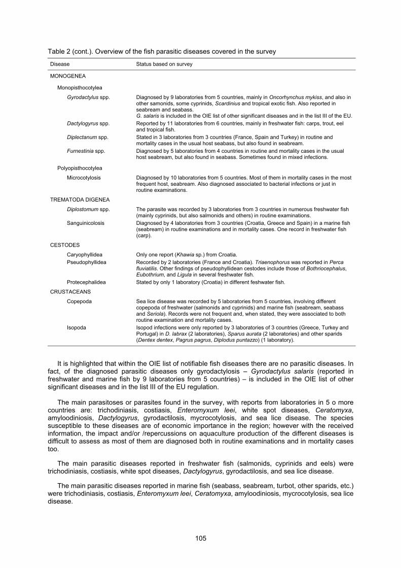
Table 2 (cont.). Overview of the fish parasitic diseases covered in the survey
Disease Status based on survey
MONOGENEA
Monopisthocotylea
Gyrodactylus spp. Diagnosed by 9 laboratories from 5 countries, mainly in Oncorhynchus mykiss, and also in other samonids, some cyprinids, Scardinius and tropical exotic fish. Also reported in seabream and seabass. G. salaris is included in the OIE list of other significant diseases and in the list III of the EU.
Dactylogyrus spp. Reported by 11 laboratories from 6 countries, mainly in freshwater fish: carps, trout, eel and tropical fish.
Diplectanum spp. Stated in 3 laboratories from 3 countries (France, Spain and Turkey) in routine and mortality cases in the usual host seabass, but also found in seabream.
Furnestinia spp.
Diagnosed by 5 laboratories from 4 countries in routine and mortality cases in the usual host seabream, but also found in seabass. Sometimes found in mixed infections.
Polyopisthocotylea
Microcotylosis Diagnosed by 10 laboratories from 5 countries. Most of them in mortality cases in the most frequent host, seabream. Also diagnosed associated to bacterial infections or just in routine examinations.
TREMATODA DIGENEA
Diplostomum spp. The parasite was recorded by 3 laboratories from 3 countries in numerous freshwater fish (mainly cyprinids, but also salmonids and others) in routine examinations.
Sanguinicolosis Diagnosed by 4 laboratories from 3 countries (Croatia, Greece and Spain) in a marine fish (seabream) in routine examinations and in mortality cases. One record in freshwater fish (carp).
CESTODES
Caryophyllidea Only one report (Khawia sp.) from Croatia.
Pseudophyllidea Recorded by 2 laboratories (France and Croatia). Triaenophorus was reported in Perca fluviatilis. Other findings of pseudophyllidean cestodes include those of Bothriocephalus, Eubothrium, and Ligula in several freshwater fish.
Protecephalidea Stated by only 1 laboratory (Croatia) in different feshwater fish.
CRUSTACEANS
Copepoda Sea lice disease was recorded by 5 laboratories from 5 countries, involving different copepoda of freshwater (salmonids and cyprinids) and marine fish (seabream, seabass and Seriola). Records were not frequent and, when stated, they were associated to both routine examination and mortality cases.
Isopoda Isopod infections were only reported by 3 laboratories of 3 countries (Greece, Turkey and Portugal) in D. labrax (2 laboratories), Sparus aurata (2 laboratories) and other sparids (Dentex dentex, Pagrus pagrus, Diplodus puntazzo) (1 laboratory).
It is highlighted that within the OIE list of notifiable fish diseases there are no parasitic diseases. In fact, of the diagnosed parasitic diseases only gyrodactylosis � Gyrodactylus salaris (reported in freshwater and marine fish by 9 laboratories from 5 countries) � is included in the OIE list of other significant diseases and in the list III of the EU regulation.
The main parasitoses or parasites found in the survey, with reports from laboratories in 5 o more countries are: trichodiniasis, costiasis, Enteromyxum leei, white spot diseases, Ceratomyxa, amyloodiniosis, Dactylogyrus, gyrodactilosis, mycrocotylosis, and sea lice disease. The species susceptible to these diseases are of economic importance in the region; however with the received information, the impact and/or /repercussions on aquaculture production of the different diseases is difficult to assess as most of them are diagnosed both in routine examinations and in mortality cases too.
The main parasitic diseases reported in freshwater fish (salmonids, cyprinids and eels) were trichodiniasis, costiasis, white spot diseases, Dactylogyrus, gyrodactilosis, and sea lice disease.
The main parasitic diseases reported in marine fish (seabass, seabream, turbot, other sparids, etc.) were trichodiniasis, costiasis, Enteromyxum leei, Ceratomyxa, amyloodiniosis, mycrocotylosis, sea lice disease.
105
Other diseases, with reports in 3 or 4 countries, with a significance that is difficult to establish, are: cryptobiasis, chilodonellosis, cryptocaryosis, coccidiosis, microsporidiosis, proliferative kidney disease, Sphaerospora dicentrarchi, Diplectanum spp., Furnestinia spp., Diplostomum, and sanguinicolosis.
Therefore, the survey has demonstrated the increasing concern of parasitic diseases in cultured
finfish in the Mediterranean area. Most of the obtained information deals with intensively cultured marine fish and several records were obtained from the expanding netpen cultures.
Some parasites can be considered a serious threat for mariculture, as Amyloodinium (Dinoflagellates), Scuticociliatida (Ciliates), Enteromyxum spp. (Myxosporea) or Mycrocotylidae (Monogenea). Other parasites are seldom reported in mortality episodes, or have been found only in routine examinations. However, their pathological concern should not be neglected, considering their increasing presence in the cultures and their direct or aside deleterious effects, even when they are not the direct cause of high mortality. General references about parasitic diseases of fish
Alvarez-Pellitero, P. (1988). Enfermedades producidas por parásitos en peces. In: Patología en
Acuicultura, Espinosa de los Monteros, J. and Labarta, U. (eds). CAICYT, Plan de Formación de Técnicos Superiores en Acuicultura, Madrid, pp. 215-326.
Alvarez-Pellitero, P., Palenzuela, O. and Sitjà-Bobadilla, A. (2001). Parásitos del rodaballo: Un nuevo reto para su cultivo. Available at: http://www.biopress.net/articulos/articulo0306.htm
Alvarez-Pellitero, P. and Sitjà-Bobadilla, A. (1993). Parasitosis de peces marinos cultivados. In: Acuicultura Marina: Fundamentos Biológicos y Tecnología de la Reproducción. Publicacions Universitat de Barcelona, pp. 515-533.
Alvarez-Pellitero, P., Sitjà-Bobadilla, A. and Franco-Sierra, A. (1993). Protozoan parasites of wild and cultured sea bass, Dicentrarchus labrax, from the Mediterranean area. Aquac. and Fish. Manag., 24: 101-108.
Alvarez-Pellitero, P., Sitjà-Bobadilla, A., Franco-Sierra, A. and Palenzuela, O. (1995). Protozoan parasites of gilthead sea bream (Sparus aurata) from different culture systems in Spain. J. Fish Dis., 18: 105-115.
Ariel, E. and Olesen, N.J. (2002). Finfish in aquaculture and their diseases � A retrospective view on the European Community. Bull. Eur. Ass. Fish Pathol., 22: 72-85.
Baptista, T., Costa, J. and Soares, F. (1999). Patologías más comunes en Dorada (Sparus aurata) y Lubina (Dicentrarchus labrax) registradas en las piscifactorías al sur del Río Tajo. Revista Aquatic, No. 7, http://www.revistaaquatic.com
Bruno, D.W., Alderman D.J. and Schlotfeldt, H.J. (1995). What Should I Do? A Practical Guide for the Marine Fish Farmer. Supplement to Bull. Eur. Ass. Fish Pathol., 15(4).
Company, R., Sitjà-Bobadilla, A., Pujalte, M.J., Garay, E., Alvarez-Pellitero, P. and Pérez-Sanchez, J. (1999). Bacterial and parasitic pathogens in cultured common dentex (Dentex dentex L.). J. Fish Dis., 22: 299-309.
Council of the European Communities (1991). Council Directive 91/67/EEC of 28 January of 1991 concerning the animal health conditions governing the placing on the market of aquaculture animals and products. Available at:
http://europa.eu.int/comm/fisheries/doc_et_publ/factsheets/legal_texts/aqua/aquaculture/animal_disease_en.html
Council of the European Communities (1993). Council Directive 93/53/EEC of 24 June 1993 introducing minimum Community measures for the control of certain fish diseases. Available at: http://europa.eu.int/comm/fisheries/doc_et_publ/factsheets/legal_texts/aqua/aquaculture/animal_disease_en.html
Le Breton, A.D. (1999). Mediterranean finfish pathologies: Present status and new developments in prophylactic methods. Bull. Eur. Ass. Fish Pathol., 19(6): 250-253.
Lom, J. and Dyková, I. (1992). Protozoan Parasites of Fish. Developments in Aquaculture and Fisheries Science, Vol. 26. Elsevier, Amsterdam.
Office International des Epizooties (OIE) (2000). Manual of Diagnostic Tests and Vaccines for Aquatic Animals, 3rd edn. OIE, Paris. Available at: http://www.oie.int
Office International des Epizooties (OIE) (2002). Aquatic Animal Health Code. 5th edn. OIE, Paris. Available at: http://www.oie.int
Paperna, I. (1984). Review of diseases affecting cultured Sparus aurata and Dicentrarchus labrax. In:
106
L'Aquaculture du Bar et des Sparidés, Barnabé, G. and Billard, R. (eds). INRA Publ. Paris, pp. 465-482.
Paperna, I. (1991). Diseases caused by parasites in the aquaculture of warm water fish. Annu. Rev. Fish Dis., 1: 155-194.
Rodgers, C.J. and Furones, M.D. (1998). Disease problems in cultured marine fish in the Mediterranean. Fish Pathol., 33(4): 157-164.
Santos, M.J. (1996). Observations on the parasitofauna of wild sea bass (Dicentrarchus labrax. L.) from Portugal. Bull. Eur. Ass. Fish Pathol., 16(3): 77-79.
Schlotfeldt, H.J. and Alderman D.J. (1995). What should I do? A Practical Guide for the Freshwater Fish Farmer. Supplement to Bull. Eur. Ass. Fish Pathol., 15(4).
Soares, M.B., Massarico, P.G., Pires, V., Costa, J. and Baptista, T.M. (1999). Occurrence of "winter disease" in reared sea bream in the south of Portugal. In: 9th International Conference Eur. Ass. Fish Pathol., Rhodes (Greece), 24-29 September 1999, p. 049 (Abstract).
Sterud, E. (2002). Parasites of wild sea bass Dicentrarchus labrax from Norway. Dis. Aquat. Org., 48: 209-212.
Sulgostowska, T., Szostakowska, B. and Myjak, P. (1998). Helminth fauna of flounder (Platichthyis flesus) and turbot (Scophthalmus maximus) from the Gulf of Gdansk. Acta Ichthyiol. Pisc., 28: 69-79.
Woo, P.T.K. (ed.) (1995). Fish Diseases and Disorders, Vol. 1: Protozoan and Metazoan Infections. CAB International, Wallingford, UK.
Zarza, C. and Aizpurua, J. (2001). Principales parasitosis de peces marinos cultivados en los países Mediterráneos. Trouvit Informa, Verano 2001: 18-24.
Amoebozoans
Amebiasis
Although different species of amoebas have been associated to amoebic diseases in fish, recent observations point out to Neoparamoeba spp. as agents of amebiasis. N. pemaquidensis may be of special pathological concern in salmon, in which it produces the amoebic gill disease (AGD). Amoebas may appear in small numbers, probably trapped in the gills, without damage. In heavy infections, the parasites elicit epithelial hyperplasia, resulting in complete fusion of secondary lamellae an subsequent gill disfunction. Fish become lethargic and thin. Diagnosis is mainly based on microscopical examination and histopathology, though immunodiagnosis and isolation in culture, followed by identification by morphology have also been used. Species specific PCR has been developed for N. pemaquidensis. The disease has been diagnosed mainly in Salmo salar, though recent findings include different sea fish, as Scophthalmus maximus, Sparus aurata and Dicentrarchus labrax. The most widely recommended treatment is the use of freshwater baths, though it is not fully effective in killing amoebae. Hydrogen peroxid and levamisol have also been assayed with variable results.
Current status based on answers received
In this survey of Mediterranean region, amebiasis has been diagnosed in 6 laboratories and 2 countries, 2 laboratories in Spain, 3 in Greece and another finding not located. There is an unique record in Salmo salar from Spain. The other findings include three marine species, S. maximus, S. aurata and D. labrax. It has been detected in some routine studies, but it was mostly found in occasional examinations, sometimes in fish suffering mortalities. Thus, prevalence is difficult to establish, but findings were rather occasional. Diagnosis in the survey was mainly based on microscopical examination and histopathology, rarely in electron microscopy or culture. No reference to control was made in the survey.
References about amebiasis Douglas-Helders, M., Saksida, S., Raverty, S., and Nowak, B.F. (2001). Temperature as a risk factor
for outbreaks of Amoebic Gill Disease in farmed Atlantic salmon (Salmo salar). Bull. Eur. Ass. Fish Pathol., 21: 114-116.
Douglas-Helders, M., Carson, J., Howard, T. and Nowak, B.F. (2001). Development and validation of a new dot blot test for the detection of Paramoeba pemaquidensis (Page) in fish. J. Fish. Dis., 24: 273-280.
107
Dykova, I., Figueras, A. and Novoa, B. (1995). Amoebic gill infection of turbot, Scophthalmus maximus. Folia Parasitol., 42: 91-96.
Dykova, I., Figueras, A. and Peric, Z. (2000). Neoparamoeba Page, 1987: Light and electron microscopic observations on six strains of different origin. Dis. Aquat. Org., 43: 217-223.
Dykova, I. and Novoa, B. (2001). Comments on diagnosis of amoebic gill disease (AGD) in turbot, Scophthalmus maximus. Bull. Eur. Ass. Fish Pathol., 21: 40-43.
Findlay, V.L., Zilberg, D. and Munday, B.L. (2000). Evaluation of levamisole as a treatment for amoebic gill disease of Atlantic salmon, Salmo salar L. J. Fish. Dis., 23: 193-198.
Leiro, J., Paniagua, E., Ortega, M., Parama, A., Fernández, J. and Sanmartín, M.L. (1998). An amoeba associated with gill disease in turbot, Scophthalmus maximus (L.). J. Fish. Dis., 21: 281-288.
Munday, B.L., Zilberg, D. and Findlay, V.L. (2001). Gill disease of marine fish caused by infection with Neoparamoeba pemaquidensis. J. Fish. Dis., 24: 497-507.
Nowak, B.F., Carson, J., Powell, M.D. and Dykova, I. (2002). Amoebic gill disease in the marine environment. Bull. Eur. Ass. Fish Pathol., 22: 144-147.
Parsons, H.J., Nowak, B.F., Fisk, D. and Powell, M.D. (2001). Effectiveness of commercial freshwater bathing as a treatment against amoebic gill disease in Atlantic salmon. Aquaculture, 195: 205-210.
Powell, M.D., Parsons, H.J. and Nowak, B.F. (2001). Physiological effects of freshwater bathing of Atlantic salmon (Salmo salar) as a treatment for amoebic gill disease. Aquaculture, 199: 259-266.
Tan, C.K.F., Nowak, B.F. and Hodson, S.L. (2002). Biofouling as a reservoir of Neoparamoeba pemaquidensis (Page, 1970), the causative agent of amoebic gill disease in Atlantic salmon. Aquaculture, 210: 49-58.
Zilberg, D., Nowak, B.F., Carson, J. and Wagner, T. (1999). Simple gill smear staining for diagnosis of amoebic gill disease. Bull. Eur. Ass. Fish Pathol., 19: 186-189.
Dinoflagellates
Amyloodiniosis
Also known as "velvet disease", the causative agent is Amyloodinium ocelatum, an ectoparasite on the skin and gills of different fish species. Appart from the velvet appearance, clinical signs consist of anorexia and scratching. Histopathological lesions include gill inflammation, haemorrhages and hyperplasy. Massive infections are frequently associated to mortalities, both in mariculture and sea aquaria, mainly at high temperatures. The infection is very common in Mediterranean fish, though other fish species are affected, including tropical and aquarium fish. Other dinoflagellates (as Piscinoodinium spp.) parasitize different freshwater fish. Diagnosis is mainly based on microscopic fresh and histological examination, though an ELISA test is available. No effective control measures are known for Mediterranean fish. Freshwater (2-4 minutes) or copper sulphate (0.75 mg/l, 12-14 days) baths have been suggested as an aid to control the trophonts or dinospores, respectively. In Pacific threadfin (Polydactylus sexfilis) recent findings suggest the suitability of hydrogen peroxide as treatment in juvenile fish. Some evidences suggest the development of immunity against re-infections, and specific antibodies have been demonstrated in the sera of infected fish.
Current status based on answers received
In the survey it was diagnosed in 8 laboratories from 7 countries, covering the main production
area of commercial Mediterranean fish. It was found in Dicentrarchus labrax and Sparus aurata in all the Mediterranean countries and Portugal. Less frequent in Diplodus puntazzo (Greece) and mullets (Italy). In addition, Oodinium was reported by 1 laboratory (Morocco) in seabream and puntazzo (we asume it is actually Amyloodinium). The results of the survey confirmed the pathological concern of this disease, as mortalities were frequently reported, mainly in juvenile seabass and seabream at temperatures over 20°C. Diagnosis was mainly based on microscopic fresh and histological examination. No reference to control measures was made in the survey.
References about amyloodiniosis Cecchini, S., Saroglia, M., Terova, G. and Albanesi, F. (2001). Detection of antibody response against
Amyloodinium ocellatum (Brown, 1931) in serum of naturally infected European sea bass by an enzyme-linked immunosorbent assay (ELISA). Bull. Eur. Ass. Fish Pathol., 21(3): 104-108.
108
Coats, D.W. (1999). Parasitic life styles of marine dinoflagellates. J. Eukaryot. Microbiol., 46: 402-409. Cobb, C.S., Levy, M.G. and Noga, E.J. (1998). Acquired immunity to amyloodiniosis is associated with
an antibody response. Dis. Aquat. Org., 34: 125-133. Ferraz, E. and Sommerville, C. (1998). Pathology of Piscinoodinium sp. (Protozoa: Dinoflagellida),
parasites of the ornamental freshwater catfishes Corydoras spp. and Brochis splendens (Pisces: Callichthyidae). Dis. Aquat. Org., 33: 43-49.
Montgomery-Brock, D., Sato, V.T., Brock, J.A. and Tamaru, C.S. (2001). The application of hydrogen peroxide as a treatment for the ectoparasite Amyloodinium ocellatum (Brown 1931) on the Pacific threadfin Polydactylus sexfilis. J. World Aquac. Soc., 32: 250-254.
Paperna, I. (1980). Amyloodinium ocellatum (Brown, 1931) (Dinoflagellida) infestations in cultured marine fish at Eilat, Red Sea: Epizootiology and pathology. J. Fish Dis., 3: 363-372.
Flagellates ECTOPARASITIC FLAGELLATES
Costiasis
Ichthyobodo spp. (also known as Costia) are the agents of this disease of the gills and skin. I. necator is the species parasitizing salmonids in freshwater, but a different species is considered to be present in marine fish. Affected fish appear thin and lethargic, and may show a grey-whitish pellicle on skin, epidermic erosion or even haemorhages or ulcers, as well as gill hyperplasy and edema. Costiasis is widely distributed in different fish species, mainly in larval and juvenile stages, and mortality can occur in fry or ornamental fish with moderate to severe infections. Besides direct mortalities, indirect damage due to decreased health condition and gill lesions must be considered. Diagnosis is based on microscopical examination and histopathology. Prevention relies on hygienic measures. Costiasis can be treated with formaline 1:4000 or 1:6000 in baths with a good aeration.
Current status based on answers received
It is one of the parasites most frequently found in the survey, both regarding the number of laboratories (13), countries (8) and the number fish species. Besides freshwater species (including Carassius auratus, Cyprinus carpio, Ictalurus punctatus, Oncorhynchus mykiss, Scardinius erythrophthalmus, Perca fluviatilis, Coregonus sp., Salmo trutta and exotic fish), it has been diagnosed in the marine fish Dicentrarchus labrax, Sparus aurata and Diplodus puntazzo. Some laboratories have identified marine costiasis as produced by I. necator. The findings by countries are representative of the importance of the culture of the different affected fish species. The parasite was reported in association with mortalities in several cases, mainly in hatcheries, though it was also found in routine examinations. Concomitant bacterial infections were found in some occasions. Diagnosis was again based on microscopical fresh and histological examination. No control measures were indicated in the survey. Cryptobiasis
The other ectoparasitic flagellates found in the survey are the gill Cryptobia spp., with a direct life cycle. Marine ectozoic species include C. branchialis, from different coastal fish and C. eilatica, described from the gills of Sparus aurata and Diplodus noct in the Red Sea. In heavy infections, the parasites produce gill hyperplasia and epithelial destruction, with subsequent respiratory impairment. External signs are anorexia and skin darkness. In the Mediterranean it is relatively common in cultured seabass and seabream. The infection can produce trickling but persistent mortalities, so loses can reach 10% after several weeks. Diagnosis is based on microscopic fresh and histological examination. Formalin baths (150 mg/l) can be effective for treatment.
Current status based on answers received
In the survey, Cryptobia spp. were only found in 3 laboratories from 3 countries. Affected fish
include the typical species more representative of the cultures, i.e. S. aurata in Spain and Greece, D.
109
labrax in Greece and Turkey and D. puntazzo in Greece. Occasionally associated to mortalities in Greece and Turkey, it was also diagnosed in routine examinations. It may appear in mixed infections, mainly with monogeneans. Diagnosis was again based on histopathology, sometimes also on fresh microscopical examination.
ENDOPARASITIC FLAGELLATES Cryptobia spp., Trypanoplasma spp., Trypanosoma spp.
Some species of these genera parasitize internal organs of fish. Cryptobia iubilans is the only pathogenic intestinal species, common in aquaria cichlid fish. Trypanoplasma spp. and Trypanosoma spp. include parasites of the bloodstream and of tissues, with indirect life cycles (leeches are the main vectors). The best known is Trypanoplasma salmositica (frequently referred as Cryptobia samositica) producing cryptobiasis of salmonids. Clinical signs consist of exophthalmia, splenomegaly, hepatomegaly, abdominal distension with ascites, anemia and anorexia. Mortality is dependent on fish stocks and species, but may be high in juveniles. The disease has severe impact in salmonid cultures in North America. An experimental protective vaccine has been developed. Other pathogenic species, Trypanoplasma borreli, parasitizes mainly cyprinids in Europe and North America. The genus Trypanosoma includes numerous species of both freshwater and marine fish. Some freshwater species are pathogenic for cyprinids.
Current status based on answers received
In the survey, only Trypanosoma sp. was reported by 1 laboratory (Croatia) in different freshwater fish (cyprinids, Esox lucius , Scardinius erythrophthalmus and Salmo trutta). Hexamitiasis
Hexamita spp. are parasites of the intestine and gall bladder of freshwater fish, mainly salmonids but also cyprinids and ornamental fish. Hexamitiasis, typical of weak fish, is frequent as a secondary infection. Affected fish can show nervous behaviour, and internally the intestine may appear pale. Mortalities can occur in fry and ornamental fish. Diagnosis is manly based on the direct observation of the flagellate in fresh intestinal scrapings or histopathology study.
Current status based on answers received
In the survey it was diagnosed only in 2 laboratories of 2 countries (Croatia and Spain), in salmonid, cyprinids, Scardinius and exotic fish. References about Flagellates Cruz, C. and Eiras, J.C. (1997). Prevalence of Trypanosoma granulosum in Anguilla anguilla in
Portugal. Bull. Eur. Ass. Fish Pathol., 17(3/4): 126-128. Diamant, A. (1990). Morphology and ultrastructure of Cryptobia eilatica n. sp. (Bodonidae:
Kinetoplastida), an ectoparasite from the gills of marine fish. J. Protozool., 37: 482-489. Poynton, S.L. and Sterud, E. (2001). Guidelines for species descriptions of diplomonad flagellates
from fish. J. Fish. Dis., 25: 15-31. Tojo, J.L. and Santamarina, M.T. (1998). Oral pharmacological treatments for parasitic diseases of
rainbow trout Oncorhynchus mykiss. III. Ichthyobodo necator. Dis. Aquat. Org., 33: 195-199. Tojo, J.L. and Santamarina, M.T. (1998). Oral pharmacological treatments for parasitic diseases of
rainbow trout Oncorhynchus mykiss. I. Hexamita salmonis. Dis. Aquat. Org., 33: 51-56. Urawa, S., Ueki, N. and Karlsbakk, E. (1998). A review of Ichthyobodo infection in marine fishes. Fish
Pathol., 33: 311-320. Woo, P.T.K. (2001). Cryptobiosis and its control in North American fishes. Int. J. Parasitol., 31: 566-
574. See also general references for fish parasites.
110
Ciliates Chilodonellosis
Most Chilodonella spp. are free living, but some of them are serious pathogens of freshwater fish, causing heavy loses in aquaria and in cultures. Under conditions favouring their proliferation they invade the gills and skin of affected fish. The gills suffer hyperplasia, degeneration and necrosis, and respiration is drastically impaired. On the skin they may virtually cover the body surface. Diagnosis is mainly based on the microscopical examination of gill or skin scrapings. Formalin baths have been suggested as treatment. Prevention relies on optimisation of fish maintenance and increasing of water quality.
Current status based on answers received
In the survey it was only reported by 6 laboratories (2 from Greece, 2 from Spain, 1 from Croatia and 1 from France) in several fish species, including ornamental freshwater species, barbels, rainbow trout or mullets (considered very sensitive in this study). A Greek laboratory reported it also from marine fish (Sparus aurata, Dentex dentex, Diplodus puntazzo and Dicentrarchus labrax). Considering that Chilodonella is typical of freshwater, a misidentification with Brooklynella, typical from marine fish and morphologically quite similar to Chilodonella could have occurred. Diagnosis was mainly based on fresh microscopical and/or histopathological examination. Cryptocaryosis
Cryptocaryon irritans, parasite of gills and skin, is the causative agent of this disease. External signs consist of white spots and mucous excess or ulcers on the skin and impairment of respiratory function. Gill histopathology consist of inflammation, haemorrhages, hyperplasia and lamellar destruction. This ciliate is a typical marine fish parasite affecting commercial and ornamental fish and producing high mortality in culture conditions. Outbreaks appeared mainly at high temperatures. Some treatment and control measures are similar to those recommended for ichthyophthiriasis, though quinine derivatives and low salinity baths have also been used. Diagnosis is based on macroscopical examination followed by microscopical examination for confirming the presence of the ciliate. The parasite can also be found in histopathological studies.
Current status based on answers received
In the survey it was diagnosed mainly in the Mediterranean cultures of sparids and seabass. Seven laboratories from 4 countries (all of them with the most representative Mediterranean maricultures) reported the disease, both in routine samplings and associated to mortalities. Diagnosis was mainly based on fresh microscopical and/or histopathological examination. Trichodiniasis
Fish trichodinids include mainly Trichodina spp., Trichodinella spp. and Tripartiella spp. These peritrichid ciliates are more commensals than genuine ectoparasites, but can produce different damages in massive infections. The fish show a grey-blue turbid layer on the skin. Respiratory function can be impaired in gill infections. Trichodinids parasitize a lot of freshwater and marine fish species. Diagnosis is mainly based on microscopical examination of fish or gill scraping preparations. Hygiene in hatcheries and quarantine for ornamental fish are recommended for prevention. This ciliatosis can be treated with formaldehyde in baths. In freshawter ornamental fish and fry, baths of salt solutions can be applied, with variable success.
Current status based on answers received
In the survey, trichodiniasis was the most frequently found parasitosis, as it was diagnosed in 22
laboratories from 10 countries. Considered mainly as opportunistic, it was sometimes associated to other pathogens, mainly bacteria. More frequent in routine examinations, it was also found associated to some mortality cases. Diagnosis was usually made to the genus level (Trichodina sp.) and was
111
mainly based on fresh microscopical or histopathological examination. Surprisingly, a diagnosis based on macroscopic examination is reported, apparently based on the presence of white spots on skin and gills. White spot disease
Ichthyophtirius multifiliis produces the well known white spot disease or ich. The most characteristic external sign is the presence of white spots on the skin and gills, due to parasite trophonts located under the upper layer of the skin. Affected fish can rub or flash and show breathing problems. In heavy infections the typical white spots are visible with the naked eye. Histopathological damage is more evident in heavy infections and in reinfections, including proliferative response and cell necrosis. The disease is widely distributed in many freshwater fish species, mainly in aquaria and culture conditions, in which it can produce epizootics. Mortality is mostly dependent on fish size and infection intensity. Diagnosis can be made macroscopically in heavy infections (white spots), followed by confirmation at microscope in fresh smears or scrapes. General preventive measures based on lower stocking densities or increased water flow and frequent routine examination, can aid to control the disease. Malachite green is quite effective in treatment of this ciliate, but presently its use is not allowed, and formalin is generally used. Free stages (tomites) are more susceptible but cysts (stages on plants or substrates) and trophonts (in the fish skin) are quite refractory to treatments. Evidence of resistance to reinfection has been observed in survivor fish, and some experimental vaccines have been assayed with different success.
Current status based on answers received
In the survey, it was found in 13 laboratories (of Croatia, France, Greece, Italy, Spain and Turkey) affecting numerous species of cultured fish (cyprinids, salmonids and exotic fish) and several wild fish. It was found both in routine samplings and associated to mortalities. Oncorhynchus mykiss was the fish most frequently parasitized. When the number of diagnosed cases is available, the values are similar for the 3 considered years. Diagnosis was mainly based on fresh microscopical or histopathological examination. No reference to control measures was made in the survey. Scuticociliatida
Several species of the genera Uronema, Phylasterides and Miamiensis have been recorded as facultative parasites of different fish. Clinical signs of scuticociliatosis depend on the parasite location. External signs include skin lesions or ulcers and pigmentation changes, but the parasite frequently invade the body muscle and the internal organs, which become destroyed by this histophagous parasite. Nervous system can also be colonised, which can be accompanied by erratic swimming, equilibrium loss or lethargy. The disease cause severe infections and outbreaks in some cultured fish and mortalities can reach 100% of some affected stocks. Different fish species can be affected, but the most severe cases reported deals with turbot, Mediterranean seabass, several marine aquarium fish, Australian tunnids and Japanese flounder.
Diagnosis is based on the finding of ciliates in ascitic fluid or scrapings of different organs. The typical morphology of Scuticociliatida can be easily observed in fresh preparations at light microscope, in stained smears or histological sections. However, the identification at the genus and species level requires specific staining evidencing the somatic and oral infraciliature and the scutica. There are no efficacious treatment for this parasitosis. Formalin baths have been assayed with certain success only in the initial phase of infection.
Current status based on answers received
In the survey it was diagnosed only in 2 laboratories from Spain, both in mortality cases and routine examination. Affected fish include turbot (both laboratories), seabream and seabass (1 laboratory). Diagnosis was based on fresh microscopical examination and histopathology.
112
References about ciliates
Ichthyophthirius
Burkart, M.A., Clark, T.G., and Dickerson, H.W. (1990). Immunization of channel catfish, Ictalurus
punctatus Rafinesque, against Ichthyophthirius multifiliis (Fouquet): Killed versus live vaccines. J. Fish. Dis., 13: 401-410.
He, J., Yin, Z., Xu, G., Gong, Z., Lam, T.J. and Sin, Y.M. (1997). Protection of goldfish against Ichthyophthirius multifiliis by immunization with a recombinant vaccine. Aquaculture, 158: 1-10.
Rahkonen, R. and Koski, P. (2002). Post malachite green: Alternative strategies for fungal infections and white spot disease. Bull. Eur. Ass. Fish Pathol., 22: 152-157.
Schlenk, D., Gollon, J.L. and Griffin, B.R. (1998). Efficacy of Cooper Sulfate for treatment of Ichthyophthiriasis in Channel Catfish. J. Aquat. Anim. Health, 10: 390-396.
Schmahl, G., Schmidt, H. and Ritter, G. (1996). The control of ichthyophthiriasis by a medicated food containing quinine: Efficacy tests and ultrastructure investigations. Parasitol. Res., 82, 697-705.
Straus, D.L. and Griffin, B.R. (2001). Prevention of an initial infestation of Ichthyophthirius multifiliis in channel catfish and blue tilapia by potassium permanganate treatment. North Am. J. Aquac., 63: 11-16.
Tieman, D.M. and Goodwin, A.E. (2001). Static and flow-through evaluation of treatments for Ich infestation in channel catfish. American Fisheries Society Fish Health Section, 2001 Annual Meeting / 42nd Fish Disease Workshop, Victoria, B.C. (Canada), 11 (abstract).
Tojo, J.L. and Santamarina, M.T. (2001). Attempts at oral pharmacological treatment of Ichthyophthirius multifiliis in rainbow trout, Oncorhynchus mykiss (Walbaum). J. Fish. Dis., 24: 249-252.
Wang, X., Clark, T.G., Noe, J. and Dickerson, H.W. (2002). Immunisation of channel catfish, Ictalurus punctatus, with Ichthyophthirius multifiliis immobilisation antigens elicits serotype-specific protection. Fish Shellfish Immunol., 13: 337-350.
Scuticociliatida
Bassleer, G. (1983). Uronema marinum, a new and common parasite on tropical salt-water fishes.
Freshw. Mar. Aquar., 6: 14 & 78-79. Cheung, P.J., Nigrelli, R.F. and Ruggieri, G.D. (1980). Studies on the morphology of Uronema
marinum Dujardin (Ciliatea: Uronematidae) with a description of the histopathology of the infection in marine fishes. J. Fish. Dis., 3: 295-303.
Dragesco, A., Dragesco, J., Coste, F., Gasc, C., Romestand, B., Raymond, J.C. and Bouix, G. (1995). Philasterides dicentrarchi, n. sp. (Ciliophora, Scuticociliata), a histophagous opportunistic parasite of Dicentrarchus labrax (Linnaeus, 1758), a reared marine fish. Eur. J. Protistol., 31: 327-340.
Dykova, I. and Figueras, A. (1994). Histopathological changes in turbot Scophthalmus maximus due to a histophagous ciliate. Dis. Aquat. Org., 18: 5-9.
Iglesias, R., Parama, A., Alvarez, M.F., Leiro, J., Fernandez, J. and Sanmartin, M.L. (2001). Philasterides dicentrarchi (Ciliophora, Scuticociliatida) as the causative agent of scuticociliatosis in farmed turbot Scophthalmus maximus in Galicia (NW Spain). Dis. Aquat. Org., 46: 47-55.
Iglesias, R., Paramá, A., Alvarez, M.F., Leiro, J., Ubeira, F.M. and Sanmartin, M.L. (2003). Philasterides dicentrarchi (Ciliophora: Scuticociliatida) expresses surface immobilization antigens that probably induce protective immune responses in turbot. Parasitology, 126: 125-134.
Jee, B.-Y., Kim, Y.-C. and Park, M. (2001). Morphology and biology of parasite responsible for scuticociliatosis of cultured olive flounder Paralichthys olivaceus. Dis. Aquat. Org., 47: 49-55.
Munday, B.L., O'Donoghue, P.J., Watts, M., Rough, K. and Hawkesford, T. (1997). Fatal encephalitis due to the scuticociliate Uronema nigricans in sea-caged, southern bluefin tuna Thunnus maccoyii. Dis. Aquat. Org., 30: 17-25.
Paramá, A., Iglesias, R., Alvarez, M.F., Leiro, J. and Sanmartin, M.L. (2003). Philasterides dicentrarchi (Cilophora, Scuticociliatida): experimental infection and possible routes of entry in farmed turbot (Scophthalmus maximus). Aquaculture, 217: 73-80.
Sterud, E., Hansen, M.K. and Mo, T.A. (2000). Systemic infection with Uronema-like ciliates in farmed turbot, Scophthalmus maximus (L.). J. Fish. Dis., 23: 33-37.
Thompson, J.C. and Moewus, L. (1964). Miamiensis avidus n. g., n. sp., a marine facultative parasite in the ciliate order Hymenostomatida. J. Protozool., 11: 378-381.
113
Other ciliates and general references for ciliates Bryant, M.S., Lee, R.P., Lester, R.J.G. and Whittington, R.J. (1999). Anti-immunoglobulin antisera
used in an ELISA to detect antibodies in barramundi Lates calcarifer to Cryptocaryon irritans. Dis. Aquat. Org., 36: 21-28.
Diamant, A. (1998). Brooklynella hostilis (Hartmannulidae), a pathogenic ciliate from the gills of maricultured sea bream. Bull. Eur. Ass. Fish Pathol., 18: 33-36.
Diamant, A., Issar, G., Colorni, A. and Paperna, I. (1991). A pathogenic Cryptocaryon-like cliate from the Mediterranean sea. Bull.Eur. Ass. Fish Pathol., 11: 122-124.
Dickerson, H.W. and Clark, T.G. (1996). Immune response of fishes to ciliates. Annu. Rev. Fish Dis., 6: 107-120.
Diggles, B.K. and Lester, R.J.G. (1996). Influence of temperature and host species on the development of Cryptocaryon irritans. J. Parasitol., 82: 45-51.
Hirazawa, N., Oshima, S., Hara, T., Mitsuboshi, T. and Hata, K. (2001). Antiparasitic effect of medium-chain fatty acids against the ciliate Cryptocaryon irritans infestation in the red bream Pagrus major. Aquaculture, 198: 219-228.
Madsen, H.C.K., Buchmann, K. and Mellergaard, S. (2000). Trichodina sp. (Ciliophora: Peritrichida) in eel Anguilla anguilla in recirculation systems in Denmark: Host-parasite relations. Dis. Aquat. Org., 42: 149-152.
Madsen, H.C.K., Buchmann, K. and Mellergaard, S. (2000). Treatment of trichodiniasis in eel (Anguilla anguilla) reared in recirculation systems in Denmark: Alternatives to formaldehyde. Aquaculture, 186: 221-231.
Rach, J.J., Gaikowski, M.P. and Ramsay, R.T. (2001). Efficacy of hydrogen peroxide to control parasitic infestations on hatchery-reared fish. J. Aquat. Anim. Health, 12: 267-273.
Rigos, G., Pavlidis, M., and Divanach, P. (2001). Host susceptibility to Cryptocaryon sp. infection of Mediterranean marine broodfish held under intensive culture conditions: A case report. Bull. Eur. Ass. Fish Pathol., 21: 33-36.
Wright, A. D.G. and Colorni, A. (2002). Taxonomic re-assignment of Cryptocaryon irritans, a marine fish parasite. Eur. J. Protistol., 37: 375-378.
Endoparasitic Protozoans
Coccidiosis
Different coccidia (Apicomplexa) are known from among freshwater and marine parasites, but their pathological significance for the cultures is very variable. The genera Eimeria, Goussia and Cryptosporidium include the species more frequently reported from cultured fish. In freshwater fish, G. carpelli parasitizes different cyprinids and E. anguillae is typical of eels. In marine fish, E. sparis and G. sparis have been reported from Sparus aurata and E. dicentrarchi and E. bouixi from Dicentrarchus labrax. Fish Cryptosporidium spp. include species from seabream, seabass, turbot, and aquarium fish, affecting mainly larvae and juvenile, with deleterious effects not always very evident, but resulting in poor condition. C. molnari is more frequent in seabream than in seabass. The species of turbot is probably a new one. Diagnosis of fish coccidia is mainly based on histopathology and/or on fresh examination at microscope. Immunodiagnostic methods are available for some human and animal species, but not for fish species. Control of animal coccidians is based on the use of different coccidiostatics or coccidiocides, but information regarding fish coccidia is very scarce. Furazolidone, amprolium chloride and furanace, among others, have been tried to treat different fish cocidia.
Current status based on answers received
In the survey, some reports indicate only coccidiosis without identification at the species or genus
level. In other cases, identification at the genus level is provided, including Cryptosporidium, Eimeria and Goussia. Considering all the coccidiosis, they have been reported by 11 laboratories of 4 countries, and another finding not located. The most frequent hosts are marine fish, mainly S. aurata and D. labrax, followed by Scophthalmus maximus and Diplodus puntazzo. Freshwater fish include only cyprinids and some exotic fish. Cryptosporidium spp. were found in seabream, seabass and turbot from Spain (2 laboratories), and seabream suffering mortality (1 laboratory, location not indicated). The involved species in cryptosporidiosis of seabass and seabream is probably the recently described C. molnari.
114
References about coccidiosis Alvarez-Pellitero, P., Redondo, M.J., Sitjà-Bobadilla, A., Macías, A., Riaza, A. and Padrós, F. (1999).
Epidemiological study of an intestinal coccidiosis of cultured turbot (Scophthalmus maximus L.). In: 5th Int. Symp. Fish Parasites, Ceské Budejovice (Czech Republic), 9-13 August 1999, p. 4.
Alvarez-Pellitero, P. and Sitjà-Bobadilla, A. (2002). Cryptosporidium molnari n. sp. (Apicomplexa: Cryptosporidiidae) infecting two marine fish species, Sparus aurata L. and Dicentrarchus labrax L. Int. J. Parasitol., 32: 1007-1021.
Lom, J. and Dykova, I. (1982). Some marine fish coccidia of the genera Eimeria Schneider, Epieimeria Dyková & Lom and Goussia Labbé. J. Fish. Dis., 5: 309-321.
Steinhagen, D., Hespe, K., Ellmer, B. and Korting, W. (1998). Goussia carpelli (Protozoa: Coccidia) infection in stressed and immunosuppressed common carp Cyprinus carpio. Dis. Aquat. Org., 34: 199-204.
Sitjà-Bobadilla, A., Palenzuela, O. and Alvarez-Pellitero, P. (1996). Light microscopic description of Eimeria sparis sp. nov. and Goussia sparis sp. nov. (Protozoa: Apicomplexa) from gilthead seabream (Sparus aurata L.) (Pisces: Teleostei). Parasitol. Res., 82: 323-332.
Microsporidiosis
Microsporea are represented in fish by different genera, mainly Enterocytozoon, Glugea, Loma, Pleistophora and Tetramicra. In freshwater fish, Pleistophora and Loma are relatively frequent. Among cultured marine fish, there have been several reports of Plesitophora senegalensis in gilthead seabream, whereas Glugea sp. and Tetramicra brevifillum have been found in turbot. Pathological concern of microsporidiosis in fish is dependent on location and infection intensity. Variable loses in turbot cultures have been related to Tetramicra infections. Diagnosis is based on the direct detection of the parasite at microscope, mainly the spores, but ultrastructural studies are necessary for identification at the specific (or even generic) level. A PCR based assay has been recently developed for T. brevifillum. Among chemicals tested for treatment, toltrazuril has apparently given better results than fumagillin and amprolium.
Current status based on answers received
In the survey, microsporidioses have been reported by 6 laboratories from 3 countries. Parasitized hosts include mainly Sparus aurata and Scophtahlmus maximus, though they were recorded in one occasion in other sparids (Dentex dentex and Pagrus pagrus), mugilids and ornamental fish. In one case, it was diagnosed in wild Pagellus acarne. Usually reported as microsporidiosis, the only case of generic identification is doubtful. Microsporeans were mainly found in routine examinations, only one case was associated to mortalities in cage juveniles. Prevalences (when indicated) are generally low. Diagnosis was mainly based on fresh microscopical examination and histopathology, only in one case in electron microscope. No references to control measures were made in the survey.
References about microsporidiosis Abela, M., Brinch-Iversen, J., Tanti, J. and Le Breton, A. (1996). Occurrence of a new histozoic
microsporidian (Protozoa, Microspora) in cultured gilthead sea bream, Sparus aurata L. Bull. Eur. Ass. Fish Pathol., 16: 196-200.
Athanassopoulou, F. (1998). A case report of Pleistophora sp. infection in cultured sea bream (Sparus aurata L.) in Greece. Bull. Eur. Ass. Fish Pathol., 18: 19-21.
Berrebi, P. and Bouix, G. (1980). Glugea atherinae, a microsporidian parasite de l'athérine Atherina boyeri Risso, 1810, des étangs côtières méditerrannéens. Évolution saisonnière et répartition géographique. Vie et Milieu, 30: 253-262.
Estévez J., Iglesias R., Leiro J., Ubeira M. and Sanmartín M.L. (1992). A unusual site of infection by a microsporean in the turbot Scophthalmus maximus. Dis. Aquat. Org., 13: 139-142.
Faye, N., Toguebaye, B.S. and Boiux, G. (1998). Ultrastucture and development of Pleistophora senegalensis sp. nov. (Protozoa, Microspora) from the gilt-head sea bream, Sparus aurata L. (Teleostei, Sparidae) from the coast of Senegal. J. Fish Dis., 13: 179-192.
Faye, N., Toguebaye, B.S. and Boiux, G. (1998). On the occurrence of microsporidian infections in the liver of four sparid fishes species from Senegal. Bull. Eur. Ass. Fish Pathol., 18: 84-86.
Figueras, A., Novoa, A., Santarem, B., Mártinez, E., Alvarez, J.M., Toranzo, A.E. and Dykova, I.
115
(1992). Tetramicra brevifilum, a potential threat to farmed turbot Scophthalmus maximus. Dis. Aquat. Org., 14: 127-135.
Leiro, J., Estévez, J., Santamarina, M.T., Sanmartín, M.L. and Ubeira, F.M. (1993). Humoral immune response of turbot, Scophthalmus maximus (L.), to antigens from Tetramicra brevifilum Matthews and Matthews, 1980 (Microspora). J. Fish Dis., 16: 577-584.
Leiro, J., Iglesias, R., Parama, A., Aragort, W. and Sanmartin, M.L. (2002). PCR detection of Tetramicra brevifilum (Microspora) infection in turbot (Scophthalmus maximus L.) musculature. Parasitology, 124: 145-151.
Mathieu-Daude, F., Faye, N., Coste, F., Monier, J. F., Marques, A. and Bouix, G. (1992). Occurrence of a microsporidiosis in marine cultured gilthead sea bream from the Languedoc Coast: a problem of specificity in the genus Glugea (Protozoa, Microspora). Bull. Eur. Ass. Fish Pathol., 12: 67-70.
Matthews, R.A., Matthews, B.F. (1980). Cells and tissue reactions of turbot Scophthalmus maximus (L.) to Tetramicra brevifilum gen. n., sp. n. (Microspora). J. Fish Dis., 3: 495-515.
Sánchez, J.G., Speare, D.J., Markham, R.J.F. and Jones, S.R.M. (2001). Isolation of a Loma salmonae variant: Biological characteristics and host range. J. Fish Biol., 59: 427-442.
Shaw, R.W. and Kent, M.L. (1999). Fish Microsporidia. In: The Microsporidia and Microsporidiosis, Wittner, M. and Weiss, L.M. (eds). American Society for Microbiology, Washington, D.C., pp. 418-446.
Speare, D.J., Daley, J., Markham, R.J.F., Sheppard, J., Beaman, H.J. and Sánchez, J.G. (1998). Loma salmonae-associated growth rate suppression in rainbow trout, Oncorhynchus mykiss (Walbaum), occurs during early onset xenoma dissolution as determined by in situ hybridization and immunohistochemistry. J. Fish. Dis., 21: 345-354.
Yokoyama, H., Kim, J.H., Sato, J., Sano, M. and Hirano, K. (1996). Fluorochrome Uvitex 2B Stain for Detection of the Microsporidian Causing Beko Disease of Yellowtail and Goldstriped Amberjack Juveniles. Fish Pathol., 31: 99-104.
Myxosporea (myxosporidiosis)
The class Myxosporea (phylum Myxozoa) include numerous genera and species, most of them parasites of fish. Some species are well know pathogens for freshwater fish. In the last years Myxosporea have been increasingly reported in cultured marine fish. They are characterised by a spore with one to several valves, one or more infective sporoplasms and one to several polar capsules with a coiled polar filament inside.
The most pathogenic species belong to the genera Ceratomyxa, Myxobolus, Myxidium,
Spaherospora, Enteromyxum, Kudoa, Tetracapsuloides and Sphaerospora.
In freshwater fish the most significant diseases are whirling disease, PKD, sphaerosporosis and ceratomyxosis (produced by Ceratomyxa shasta).
Myxosporea reported from cultured marine fish include species of the genera Ceratomyxa, Enteromyxum, Kudoa, Lepthoteca, Sphaerospora and Sinuolinea.
Other myxosporeans reported ocassionally in the survey are Leptotheca sp. and Polysporoplasma sparis from the kidney of S. aurata, and Sinoulinea sp. from the urinary bladder of turbot, the three reported only by 1 laboratory from Spain, mainly in routine or occasional samplings. The species of Leptotheca has been described as L. sparidarum from seabream an dentex (see references). Whirling disease
The causative agent is Myxobolus cerebralis (synonym Myxosoma cerebralis). Clinical signs include dark coloration of the posterior part of the body and abnormal swimming in spiral, followed by skull deformation and spinal curvature. Almost all salmonid species can be infected, but susceptibility is very variable according to the species. Oncorhynchus mykiss and other Oncorhynchus spp. are very susceptible, while Salmo trutta is rather resistant. The involvement of an intermediate oligochaete host in the life cycle of this myxosporean was demonstrated 18 years ago. This knowledge has facilitated preventive measures, consisting of the use of concrete or plastic ponds or tanks and their frequent cleaning for avoiding the presence of oligochaetes and thus the transmission of the disease.
116
This is very important, considering the limited efficacy of treatments assayed till now (fumagillin, toltrazuril) for this myxosporea and other species. In some European countries, the incidence of whirling disease has clearly decreased, whereas in USA whirling disease is widely distributed and is still an important pathological problem. Diagnosis is based on the histological examination of skull cartilage, or their enzymatic digestion followed by microscopical observation of the typical spores. A PCR assay has also been developed.
Current status based on answers received
In the survey it was diagnosed only in one laboratory of one country (Croatia) in Oncorhynchus
mykiss. Proliferative kidney disease (PKD)
The causative agent, formerly known as PKX, has been recently identified as Tetracapsuloides bryosalmonae (syn. Tetracapsula bryosalmonae, T. renicola). Extrasporogonic stages of this myxosporean are located in the kidney of different salmonid fish, but spores are formed in a bryoozoan host. The parasite is highly pathogenic and can produce a severe disease in rainbow trout, with 30-50% mortality. External clinical signs are abdominal swelling, darkening and exophthalmos. Internally, a kidney enlargement is observed, accompanied by ascites in advanced cases. In histopathology, the kidney shows interstitial hyperplasia, associated with chronic, granulomatous interstitital nephritis and tubular atrophy. A severe inflammatory reaction is also produced. Poor food conversion and immunodepression are also a consequence of this infection. Macroscopical diagnosis relies on the gross observation of kidney enlargement. Confirmation is achieved by observation of parasitic stages in histological sections or squash preparations (in this case by experienced observers).
Current status based on answers received
In the survey, PKD was diagnosed by 5 laboratories of 3 countries (Croatia, France and Italy)
mostly in mortality episodes but also in routine examination. Oncorhynchus mykiss was the fish host in most cases but there is one record in Salmo trutta in Italy. Diagnosis was mainly based on histopathology, but in two cases, electron microscopy or PCR were also used. Spaherospora renicola
Spaherospora renicola is widely distributed in intensive cultures of cyprinids, mainly Cyprinus carpio. Spores and sporognic stages are located in the renal tubules, but proliferative stages appear in the blood and can reach the swimmbladder, causing inflammation. S. renicola may be a serious pathogen. In the kidney tubules it produces dilatation, atrophy and necrosis of the epithelium, with subsequent impairment of renal function. The development of swimbladder stages elicits the swimbladder inflammation of juvenile carps. Fish can show some external clinical signs as locomotory disorders and swimming in circles.
Current status based on answers received
In the survey it was diagnosed only by 1 laboratory from Croatia, in one occasion. However, it must
be considered the scarcity of cyprinid cultures in the surveyed countries. Ceratomyxa shasta
Ceratomyxa shasta is an important pathogen, causing serious loses in cultured and wild populations of salmonids on the west coast of North America. Intestine is the target organ and parasites can be observed in the epithelium, eliciting lymphocytic infiltration, hyperplasia and necrosis. In advanced stages of the infection, parasites spread to other organs an fish become anorexic, lethargic, and show abdominal swelling, ascites and exophthalmia. Significant mortalities can occur, depending on fish species, as susceptibility is variable. An intermediate host (a polychaete) has been demonstrated in the life cycle of this Myxosporean. A PCR assay is available for diagnosis.
117
Ceratomyxa spp.
This genus include a lot of marine species, though they have been rarely associated with significant pathological problems. The main species recorded in cultured marine fish are C. diplodae, C. labracis and C. sparusaurati. C. diplodae (originally described from Diplodus annularis) and C. labracis are quite frequent in wild and cultured seabass. They are not usually associated with clinical disease, but they can induce several histopathological lesions in the gall bladder and neighboring tissues. C. sparusaurati is a very common parasite of the gall bladder of Sparus aurata. It generally causes limited histopathological damage, but in some massive infections it has been associated with clinical signs and mortality.
Current status based on answers received
In the survey Ceratomyxa spp. and/or ceratomyxosis have been reported by 11 laboratories of 6 countries (no indication of country is given by 1 laboratory). Records were very common in the 3 years of study. Host fish include Dicentrarchus labrax in 8 cases and S. aurata or other sparids in 10 cases. They have been mainly diagnosed in routine or occasional studies, though there are some records in mortality cases (though direct association with mortality is not stated). The results indicate the wide dispersion of Ceratomyxa spp. in Mediterranean cultured fish, though they are not considered significant pathogens. Diagnosis was mainly based on microscopical (fresh or histological) examination. Enteromyxum spp.
Two species of this genus have special pathological concern for marine fish of high commercial value, and both parasitize the digestive tract of infected fish.
Enteromyxum leei is the myxosporean previously known as Myxidium leei, which has been transferred to the new genus Enteromyxum after morphological and molecular studies (see references). It produces the myxidiosis of sparids, which now should be named enteromyxosis, the most significant myxosporidiosis of cultured sparids in the Mediterranean. Not only S. aurata, but also other sparids can be affected, including the new cultured species Diplodus puntazzo and Pagrus spp. The range of susceptible fish is extraordinarily wide, as seabass, mullets, Sciaenops ocellatus and different marine aquarium fish, belonging to 25 species of 4 separate orders have been found parasitized. The parasite invades the intestinal tract causing severe chronic enteritis, frequently followed by emaciation and dead. Thus, external clinical signs mainly consist of a extreme thinness ("knife-fish" for some farmers). Losses can reach 80% of some stocks, specially in D. puntazzo which seems to be specially susceptible. Horizontal direct fish-to-fish transmission has been demonstrated for this myxosporean, which enhances the pathological importance of the disease in the cultures. Diagnosis is mainly based on histopathological study of the intestine and detection of parasitic stages, though these stages can be also seen in fresh smears by a experienced observer. A PCR assay is being validated in the framework of an European project.
Current status based on answers received
In the survey, E. leei (reported as Myxidium leei or myxidiosis) was diagnosed by 16 laboratories of
7 countries, all of them important cultivators of sparids. S. aurata and D. puntazzo were the most frequent fish host, followed by D. sargus, Pagrus pagrus and Pagellus erythrinus. Diagnosis was based on clinical signs together with microscopical examination of fresh smears and/or histopathology. The survey has confirmed the pathological importance of the disease, as all laboratories have diagnosed it in mortality cases (sometimes also in routine studies) and some laboratories indicate high mortalities and important loses, mainly for D. puntazzo.
Enteromyxum scophthalmi is the other species of the recently erected genus Enteromyxum, reported up to now only from turbot, Scophthalmus maximus. E. scophtahlmi is an important pathogen of turbot cultures, as mortalities can reach 100 % of the affected tanks or stocks, with subsequent economical impact. Clinical external signs include anorexia, caquexia, sunken eyes and a typical prominent bony ridge on the skull. At necropsy, pallor of internal organs, intestinal haemorrhages and the presence of liquid in the intestine are also observed. Histopathological damage is specially evident
118
in the intestine, with severe enteritis, detachment of the epithelium, haemorrhages and inflammation. As in the case of E. leei, direct fish-to-fish transmission has been demonstrated, with subsequent influence on the disease dispersion and impact. Diagnosis is mainly based on microscopical examination of fresh smears and histopathology. A PCR assay is available (restricted use).
Current status based on answers received
In the survey, it is reported as enteric Myxosporea or myxidiosis, and only 2 laboratories from 1
country have diagnosed it in turbot, in mortality cases and in routine studies. Diagnosis was based on histopathology in 1 laboratory, and the other has also used macroscopic and microscope examination, immunohistochemistry, electron microscope and PCR. Kudoa spp.
These marine myxosporeans (Multivalvulida) infect the muscle of many marine fish forming plasmodial cysts. Heavy infections can cause unsightly white cysts or soft texture in filets, which gives the name to the disease (soft flesh condition), with subsequent lowering of market value. In aquaculture, Kudoa infections have been described in salmonids (mainly Atlantic and Pacific salmon), and Seriola. Reported findings in gilthead seabream are occasional. The soft texture of muscle fillets and the presence of white cysts can be observed with the naked eye. Diagnostic must be confirmed by microscopic detection of the parasite in fresh or stained squash preparations or in histological sections.
Current status based on answers received
In the survey, it was only reported by 1 laboratory in Portugal in routine examiantion of Sardina pilchardus. Sphaerospora dicentrarchi
Originally described from Dicentrarchus labrax, the type host, it can be ocassionaly found in some other fish species, as mullets. This histozoic parasite can be considered systemic, though it has speciall affinity for the connective tissue of gall bladder and intestine. It is usually found in chronic infections, without external clinical signs, though massive infections have been associated with extensive mortalities in juvenile fish. Diagnosis is mainly based on histopathology, though groups of spores can be seen in microscopical examination of fresh smears of gall bladder and intestine.
Current status based on answers received
In the survey it was diagnosed by 5 laboratories in 4 countries (1 laboratory without indication of country), usually in the type host D. labrax, but sphaerosporosis is indicated in S. aurata from Greece, and Sphaerospora sparusaurati, in the same host from Malta, which is a probable misidentificacion with Ceratomyxa sparusaurati. Sphaerosporosis was diagnosed more frequently in routine studies, but also in some mortality cases. Diagnosis was mainly based on histopathology and/or microscopical examination of fresh samples. Sphaerospora testicularis
The only known host is D. labrax. This myxosporean has a very specific location in testes, so only
male fish are affected. Heavy infections can result in parasitic castration of valuable broodstock fish. Affected testes can appear inflammed or necrotic at necropsy. In very heavy infections, the parasite invades the serosa and other organs, producing abdominal swelling and ascitis, though this situation is rather exceptional. In histopathological study parasite stages are observed filling the seminal tubules. In the reproductive period, the parasite can be easily diagnosed at microscope in fresh smears of seminal fluid.
119
Current status based on answers received
In the survey, it was only diagnosed in 2 laboratories from Greece. An increased susceptibility to bacterial diseases, but doubtfull direct association with mortality are indicated. References about Myxosporea
Myxosporea (general references) Alvarez-Pellitero, P. and Sitjà-Bobadilla, A. (1993). Pathology of Myxosporea in marine fish culture. Dis.
Aquat. Org., 17: 229-238. Athanassopoulou, F., Prapas, Th. and and Rodger, H. (1999). Diseases of Puntazzo puntazzo Cuvier
in marine aquaculture systems in Greece. J. Fish. Dis., 22: 215-218. Kent, M.L., Andree, K.B., Bartholomew, J.L., El-Matbouli, M., Desser, S.S., Devlin, R.H., Feist, S.W.,
Hedrick, R.P., Hoffmann, R.W., Khattra, J., Hallet, S.L., Lester, R.J.G., Longshaw, M., Palenzuela, O., Siddall, M.E. and Xiao, C. (2001). Recent advances in our knowledge of the Myxozoa. J. Eukaryot. Microbiol., 48: 395-413.
Le Breton, A., Brich Iversen, H., Spiteri, D. and Meyer, T. (1999). Endo-microparasitic infestation of cultured seabass Dicentrarchus labrax and seabream Sparus aurata: A case study on the economical impact in the Maltese Islands. 9th Intern. Conf. Eur. Ass. Fish Pathol., Rhodes (Greece), 19-24 September 1999, p. 186 (abstract).
Mladineo, I. (2003). Myxosporidean infections in Adriatic cage-reared fish. Bull. Eur. Ass. Fish Pathol., 23(3): 113-122.
Rigos, G., Christophilogannis, P., Yiagnisi, M., Andriopolulou, A., Koutsodimou, M., Nengas, I. and Alexis, M. (2000). Myxosporean infections in Greek mariculture. Aquac. Intern., 7: 361-364.
Ceratomyxa
Alvarez-Pellitero, P. and Sitjà-Bobadilla, A. (1993). Ceratomyxa spp. (Protozoa: Myxosporea) infections in
wild and cultured sea bass (Dicentrarchus labrax) from the Spanish Mediterranean area. J. Fish Biol., 42: 889-901.
Costa, G., Lom, J., Andrade, C. and Barradas, R. (1998). First report of Ceratomyxa sparusaurati (Protozoa: Myxosporea) and the occurrence of epitheliocystis in cultured sea bream, Sparus aurata L. from Madeira. Bull. Eur. Ass. Fish Pathol., 18: 165-167.
Palenzuela, O. and Bartholomew, J.L. (2002). Molecular tools for the diagnosis of Ceratomyxa shasta. In: Molecular Diagnosis of Salmonid Diseases, Reviews: Methods and Technology in Fish Biology and Fisheries, Cunningham, C.O. (ed.), Vol. 3. Kluwer Academic Publishers, The Netherlands, pp. 285-298.
Palenzuela, O., Sitjà-Bobadilla, A. and Alvarez-Pellitero, P. (1997). Ceratomyxa sparusaurati (Protozoa: Myxosporea) infections in cultured gilthead sea bream Sparus aurata (Pisces: Teleostei) from Spain: Aspects of the host-parasite relationship. Parasitol. Res., 83: 539-548.
Palenzuela, O., Trobridge, G. and Bartholomew, J.L. (1999). Development of a polymerase chain reaction diagnostic assay for Ceratomyxa shasta, a myxosporean parasite of salmonid fish. Dis. Aqua. Org., 36: 45-51.
Rigos, G., Grigorakis, K., Christophilogannis, P., Nengas, I. and Alexis, M. (1997). Ceratomyxa spp. (Myxosporea) infection in cultured common dentex from Greece. Bull. Eur. Ass. Fish Pathol.: 17, 174-176.
Sitjà-Bobadilla, A. and Alvarez-Pellitero, P (1993). Light and electron microscopic description of Ceratomyxa labracis n. sp. and redescription of C .diplodae (Myxosporea: Bivalvulida) from wild and cultured Mediterranean sea bass (Dicentrarchus labrax L.) (Teleostei: Serranidae). Syst. Parasitol., 26: 215-223.
Sitjá-Bobadilla, A., Palenzuela, O. and Alvarez-Pellitero, P. (1995). Ceratomyxa sparusaurati n. sp. (Myxosporea: Bivalvulida), a new parasite from cultured gilthead sea bream (Sparus aurata L.): Light and electron microscopic description. J. Eukaryot. Microbiol., 42: 529-539.
Enteromyxum spp., Myxidium leei
Branson, E., Riaza, A. and Alvarez-Pellitero, P. (1999). Myxosporean infection causing intestinal
disease in farmed turbot, Scophthalmus maximus (L.) (Teleostei: Scophthalmidae). J. Fish. Dis., 22: 395-399.
120
Diamant A. (1992). A new pathogenic histozoic Myxidium (Myxosporea) in cultured gilt-head sea bream Sparus aurata L. Bull. Eur. Ass. Fish Pathol., 12: 64-66.
Diamant A. (1997). Fish-to-fish transmission of a marine myxosporean. Dis. Aquat. Org. 30, 99-105. Diamant, A. (1998). Red drum Sciaenops ocellatus (Sciaenidae), a recent introduction to
Mediterranean mariculture, is susceptible to Myxidium leei (Myxosporea). Aquaculture, 162: 33-39. Diamant A., Lom J. and Dyková, I. (1994). Myxidium leei n. sp., a pathogenic myxosporean of cultured
sea bream Sparus aurata. Dis. Aquat. Org., 20: 137-141. Diamant, A. and Wajsbrot, N. (1997). Experimental transmission of Myxidium leei in gilthead sea
bream Sparus aurata. Bull. Eur. Ass. Fish Pathol., 17: 99-103. Le Breton, A. and Marques, A. (1995). Occurrence of an histozoic Myxidium infection in two marine
cultured species: Puntazzo puntazzo C. and Pagrus major. Bull. Eur. Ass. Fish Pathol., 15: 210-212.
Padrós, F., Palenzuela, O., Hispano, C., Tosas, O., Zarza, C., Crespo, S. and Alvarez-Pellitero, P. (2001). Myxidium leei (Myxozoa) infections in aquarium-reared Mediterranean fish species. Dis. Aquat. Org., 47: 57-62.
Palenzuela, O., Redondo, M.J. and Alvarez-Pellitero, P. (2002). Description of Enteromyxum scophthalmi gen. nov, sp. nov. (Myxozoa), an intestinal parasite of turbot (Scophthalmus maximus L.) using morphological and ribosomal RNA sequence data. Parasitology, 124: 369-380.
Redondo, M.J., Palenzuela, O. Riaza, A., Macías, A. and Alvarez Pellitero, P. (2002). Experimental transmission of Enteromyxum scophthalmi (Myxozoa), an enteric parasite of turbot (Scophthalmus maximus). J. Parasitol., 88: 482-488.
Sakiti, N., Tarer, V., Jacquemin, D. and Marques, A. (1996). Présence en Méditerranée occidentale d'une Mixosporidie histozoique pathogène dans les élevages du daurade, Sparus aurata. Ann. Sci. Natur., Zool., Paris, 17: 123-127.
Tarer, V., Sakiti, N.D., Le Breton, A. and Marques, A. (1996). Myxidium leei myxosporidie pathogène chez les Sparidés en Aquaculture en Méditerranée. Ichthyophysiol. Acta, 19: 127-139.
Zrncic, S., Oraic, D., Sostaric, B. and Filic, I. (1998). First Occurrence of Myxidium Infection in Cultivated Charp Snouted Sparus, Puntazzo puntazzo, in Croatia.Proc. In: Proc. of the 3rd Int. Symp. Aquat. Anim. Health, Baltimore (USA), 30 Aug. - 3 Sept. 1998, p. 248.
Kudoa
Moran, J.D.W., Whitaker, D.J. and Kent, M.L. (1999). A review of the myxosporean genus Kudoa
Meglitsch, 1947, and its impact on the international aquaculture industry and commercial fisheries. Aquaculture, 172: 163-196.
Paperna, I. (1982). Kudoa infection in the glomeruli, messentery and peritoneum of cultured Sparus aurata L. J. Fish Dis., 5: 539-543.
St-Hilaire, S., Ribble, C., Whitaker, D.J. and Kent, M.L. (1997). Evaluation of a nondestructive diagnostic test for Kudoa thyrsites in farmed Atlantic salmon (Salmo salar). Aquaculture, 156: 139-144.
PKD, PKX
Canning, E.U., Curry, A., Feist, S.W., Longshaw, M. and Okamura, B. (1999). Tetracapsula
bryosalmonae n. sp. for PKX organism, the cause of PKD in salmonid fish. Bull. Eur. Ass. Fish Pathol., 19: 203-206.
Canning, E.U., Tops, S., Curry, A., Wood, T.S. and Okamura, B. (2002). Ecology, development and pathogenicity of Buddenbrockia plumatellae Schröder, 1910 (Myxozoa, Malacosporea) (syn. Tetracapsula bryozoides) and establishment of Tetracapsuloides n. gen. for Tetracapsula bryosalmonae. J. Eukaryot. Microbiol., 49: 280-295.
Feist, S.W., Longshaw, M., Canning, E.U. and Okamura, B. (2001). Induction of proliferative kidney disease (PKD) in rainbow trout Oncorhynchus mykiss via the bryozoan Fredericella sultana infected with Tetracapsula bryosalmonae. Dis. Aquat. Org., 45: 61-68.
Higgins, M.J. and Kent, M.L. (1996). Field trials with fumagilin for the control of proliferative kidney disease in coho salmon. The Progressive Fish-Culturist, 58: 268-272.
Higgins, M.J. and Kent, M.L. (1998). TNP-470, the analogue of fumagillin-DCH, controls PKX in naturally infected sockeye salmon, Oncorhynchus nerka (Walbaum), underyearlings. J. Fish. Dis., 21: 455-457.
Morris, D., Morris, D.J. and Adams, A. (2002). Development of improved PCR to prevent false positives and false negatives in the detection of Tetracapsula bryosalmonae, the causative agent of proliferative kidney disease. J. Fish. Dis., 25: 483-490.
121
Peribáñez, M.A., Luco, D.F., García, L., and Castillo, J.A. (1997). The prevalence of proliferative kidney disease from the kidney and muscle of rainbow trout and brown trout in Aragón (Spain). Prev. Vet. Med., 32: 287-297.
Wahli, T., Knuesel, R., Bernet, D., Segner, H., Pugovkin, D., Burkhardt-Holm, P., Escher, M. and Schmidt-Posthaus, H. (2002). Proliferative kidney diseases in Switzerland: Current state of knowledge. J. Fish. Dis., 25: 491-500.
Sphaerospora spp.
Sitjà-Bobadilla, A. and Alvarez-Pellitero, P. (1992). Effect of Fumagillin treatment on sea bass
(Dicentrarchus labrax L.) parasitized by Sphaerospora testicularis (Myxosporea: Bivalvulida). Dis. Aquat. Org., 14: 171-178.
Sitjà-Bobadilla, A. and Alvarez-Pellitero, P. (1992). Light and electron microscopic description of Sphaerospora dicentrarchi n. sp. (Myxosporea: Sphaerosporidae) from wild and cultured sea bass, Dicentrarchus labrax L. J. Protozool., 39: 273-281.
Sitjà-Bobadilla, A. and Alvarez-Pellitero, P. (1993). Pathological effects of Sphaerospora dicentrarchi Sitjà-Bobadilla et Alvarez-Pellitero, 1992 and S. testicularis Sitjà-Bobadilla et Alvarez-Pellitero, 1990 parasitic of sea bass (Dicentrarchus labrax L.) (Teleostei: Serranidae), and cell mediated immune reaction: Light and electron microscopic study. Parasitol. Res., 79: 119-129.
Sitjà-Bobadilla, A. and Alvarez-Pellitero, P. (1993). Population dynamics of Sphaerospora dicentrarchi Sitjà-Bobadilla et Alvarez-Pellitero, 1992 and S. testicularis Sitjà-Bobadilla et Alvarez-Pellitero, 1990 (Myxosporea: Bivalvulida) infections in wild and cultured sea bass (Dicentrarchus labrax L.). Parasitology, 106: 39-45.
Sitjà-Bobadilla, A. and Alvarez-Pellitero, P. (1993). Light and electron microscopic description of Ceratomyxa labracis n. sp. and redescription of C. diplodae (Myxosporea: Bivalvulida) from wild and cultured Mediterranean sea bass (Dicentrarchus labrax L.) (Teleostei: Serranidae). Syst. Parasitol., 26: 215-223.
Whirling disease
Andree, K.B. and Antonio, D.B. (2002). Nucleic acid-based methods for detection of Myxobolus
cerebralis. In: Molecular Diagnosis of Salmonid Diseases, Cunningham, C.O. (ed.). Kluwer Acad. Publ., London, pp. 315-328.
Andree, K.B., Hedrick, R.P. and MacConnell, E. (2002). A review of the approaches to detect Myxobolus cerebralis, the cause of salmonid whirling disease. In: Whirling disease: Reviews and Current Topics, Batholomew, J.L. and Wilson, J.C. (eds). American Fisheries Society, Symposium 29, Bethesda, Maryland, 18-22 August 2002, pp. 197-212.
El-Matbouli, M., Hoffmann, R.W., Schoel, H., McDowell, T.S. and Hedrick, R.P. (1999). Whirling disease: Host specificity and interaction between the actinosporean stage of Myxobolus cerebralis and rainbow trout Oncorhynchus mykiss. Dis. Aquat. Org., 35: 1-12.
Hedrick, R.P. and El-Matbouli, M. (2002). Review: Recent advances with taxonomy, life cycle, and development of Myxobolus cerebralis in the fish and oligochaete host. In: Whirling Disease: Reviews and Current Topics, Batholomew, J.L. and Wilson, J.C. (eds). American Fisheries Society, Symposium 29, Bethesda, Maryland, 18-22 August 2002, pp. 45-54.
Hedrick, R.P., McDowell, T.S., Marty, G.D., Mukkatira, K., Antonio, D.B., Andree, K.B., Bukhari, Z. and Clancy, T. (2000). Ultraviolet irradiation inactivates the waterborne infective stages of Myxobolus cerebralis: A treatment for hatchery water supplies. Dis. Aquat. Org., 42: 53-59.
Hoffman, G.L. (1990). Myxobolus cerebralis, a worlwide cause of salmonid whirling disease. J. Aquat. An. Health, 2: 30-37.
Qureshi, T., White, M.R. and Santrich, C. (2002). Evaluation of nested polymerase chain reaction, pepsin/trypsin digest, and histology for the detection of Myxobolus cerebralis in salmonid fishes of Indiana and Michigan. J. Vet. Diagn. Invest., 14: 251-254.
Staton, L., Erdahl, D. and El-Matbouli, M. (2002). Efficacy of Fumagillin and TNP-470 to prevent experimentally induced whirling disease in rainbow trout Oncorhynchus mykiss. In: Whirling Disease: Reviews and Current Topics, Batholomew, J.L. and Wilson, J.C. (eds). American Fisheries Society, Symposium 29, Bethesda, Maryland, 18-22 August 2002, pp. 239-247.
Wagner, E.J. (2002). Whirling disease prevention, control and management: A review. In: Whirling Disease: Reviews and Current Topics, Batholomew, J.L. and Wilson, J.C. (eds). American Fisheries Society, Symposium 29, Bethesda, Maryland, 18-22 August 2002, pp. 217-226.
Wolf, K. and Markiw, M.E. (1984). Biology contravenes taxonomy in ten Myxozoa: New discoveries show alternation of invertebarte and vertebrate hosts. Science, 225: 1449-1452.
122
Other Myxosporea Palenzuela, O., Alvarez-Pellitero, P. and Sitjà-Bobadilla, A. (1999). Glomerular disease associated to
Polysporoplasma sparis (Myxosporea: Bivalvulida) infections in the gilthead sea bream, Sparus aurata, (Pisces: Teleostei): Aspects of the host-parasite relationship. Parasitology, 118: 245-256.
Sitjà-Bobadilla, A. and Alvarez-Pellitero, P. (1993). Zschokkella mugilis n. sp. (Myxosporea: Bivalvulida) from mullets (Teleostei: Mugilida) of Mediterranean waters: Light and electron microscopic study. J. Eukaryot. Microbiol., 40: 755-764.
Sitjà-Bobadilla, A. and Alvarez-Pellitero, P. (2001). Leptotheca sparidarum n. sp. (Myxosporea: Bivalvulida), a parasite from cultured common dentex (Dentex dentex L.) and gilthead sea bream (Sparus aurata L.) (Teleostei. Sparidae). J. Eukaryot. Microbiol., 48: 627-639.
Monogenea
Members of Monogenea, mostly ectoparasites of gills and skin, can cause different degrees of
damage in parasitized fish. Mortalities may appear in moderate or heavy infections, mainly in juvenile fish, increasing with water temperature. Clinical signs include lethargy, anoxia, loss of appetite and scratching. Mucus excess, opacity, and even ulcers or haemorrhages may appear. Gill histopathological signs include focal hyperplasia, lamellar fusion, haemorrhages and inflammatory infiltration. Monogeneans are usually very host specific, though in certain culture conditions some species can be found in unusual hosts. Diagnosis is mainly based on direct methods of fresh examination and histopathology. Identification to the species level usually needs the observation of fresh parasites at microscope and comparison of morphometric features. Different treatments have been assayed against Monogeneans, including organophosphates (though its use is restricted by legal regulations) or formaline (mainly for larval stages). However, some infections are very refractory to treatments, so general preventive measures are strongly recommended, as well as periodic examination of fish.
Monogenea include 2 main groups, Monopisthocotylea (with a simple adhesive disc) and
Polyopisthocotylea (with a complex adhesive disc including clumps and hooks).
Among Monopisthocotylea the most significant species for cultured fish are Gyrodactylus spp., Dactylogyrus spp., Diplectanum spp. and Furnestinia spp.
Polyopisthocotylea include several species of pathological concern for fish cultures, most of them belonging to the family Microcotylidae, and some to Heteraxinidae. Gyrodactylus spp.
Gyrodactylus spp., causing gyrodactylosis, are mainly typical from freshwater fish. Some species are very pathogenic for salmonid fish, specially G. salaris, considered the most pathogenic species, and included in the list III of the EEC policies.
Current status based on answers received
In the survey, gyrodactylosis has been diagnosed in 9 laboratories from 5 countries. Several
freshwater fish are affected, mainly Oncorhynchus mykiss, but also other salmonids, some cyprinids, Scardinius, and tropical exotic fish. However, gyrodactylosis has also been reported occasionally from marine fish, such as Sparidae (mainly Sparus aurata) and Dicentrarchus labrax. Dactylogyrus spp.
This genus include numerous species, mostly parasites of freshwater fish. Cyprinids are the main hosts of these monogeneans, but fish of many other families are also affected. The eel parasites Pseudodactylogyrus spp. are included in the same superfamily Dactylogyroidea. The pathologic significance is very dependent on the species and intensity of infection.
123
Current status based on answers received
In the survey, dactylogyrosis was diagnosed in 11 laboratories of 6 countries. Reported fish hosts include Cyprinus carpio, O. mykiss, Anguilla anguilla (probably Pseudodactylogyrus) and tropical fish from fresh waters. Among marine fish, Dactylogyrus has been found in S. aurata and D. labrax. A misidentification with the respective specific monogeneans, Furnestinia or Diplectanum could have occurred, specially for juvenile stages. Diplectanum spp.
The best known species of these genus are D. aequans and D. laubieri, parasites of D labrax. Their dispersion is very wide in the Mediterranean and Atlantic areas, mostly coinciding with seabass distribution. D. aequans is considered more pathogenic, mainly for juveniles and brood stocks.
Current status based on answers received
In the survey, Diplectanum was reported by 3 laboratories, from 3 countries (France, Turkey and Spain), both in mortality cases and routine examination. Surprisingly, on one occasion it was recorded from S. aurata, and not from the usual host D. labrax. Furnestinia spp.
This genus belongs also to the family Diplectanidae. F. echeneis is quite frequent in S. aurata in different Mediterranean countries. It is not considered very pathogenic, though certain damage can occur in heavy infections.
Current status based on answers received
In the survey, it was diagnosed in 5 laboratories of 4 countries (Greece, Israel, Spain and Turkey), usually in the specific host, S. aurata, but in one case in D. labrax. Findings are reported in mortality cases, routine and occasional samplings, sometimes found in mixed infections with other monogeneans or with protozoans. Microcotylosis
In the last years, microcotylosis has affected severely the sparid cultures, causing important loses. Besides the gill dysfunction usually associated with monogenosis, microcotylids produce increased damage due to their haematophagus condition, causing anemia and poor fish condition. These monogeneans are very frequent in Mediterranean cultures of sparids and D. labrax. The involved species have been identified as Sparicotyle chrysophry parasitizing Sparidae, and Serranicotyle labracis, parasite of seabass. In the Heteraxinidae, Heteraxine spp. important parasites for Japanese amberjack (Seriola spp.) cultures, have been also recorded in Mediterranean amberjack.
Current status based on answers received
In the survey, microcotylidis were diagnosed by 10 laboratories, belonging to 5 countries (Cyprus,
France, Greece, Portugal and Spain). S. aurata is the most frequent host, though there were some records in other sparids, and one in Seriola. The number of cases per year was not generally very high. Most of them were associated to mortalities, though they were also diagnosed in routine examinations and occasional samplings. Some laboratories reported heavy infections and significant mortality, caused by the associated anaemia and anorexia. The condition may be related to bad husbandry, fouled nets or high stock density. Secondary bacterial infections were also reported. Diagnosis was always based on fresh microscopical or histological examination, though macroscopical examination was also indicated. No control measures were indicated for monogenosis in the survey.
124
References about Monogenea
General references Buchmann, K. and Lindenstrom, T. (2002). Interactions between monogenean parasites and their fish
hosts. Intern. J. Parasitol., 32: 309-319. Papoutsoglou, S., Costello, M.J., Stamou, E. and Tziha, G. (1996) Environmental conditions at sea-
cages, and ectoparasites on farmed European sea-bass, Dicentrarchus labrax (L.), and gilt-head sea-bream, Sparus aurata L., at two farms in Greece. Aquacult. Res., 27: 25-34.
Rach, J.J., Gaikowski, M.P. and Ramsay, R.T. (2001). Efficacy of hydrogen peroxide to control parasitic infestations on hatchery-reared fish. J. Aquat. Anim. Health, 12: 267-273.
Roubal, F.R. (1995). Changes in monogenean and copepod infestation on captive Acanthopagrus australis (Sparidae). J. Fish Biol., 46: 423-431.
Sterud, E. (2002). Parasites of wild sea bass Dicentrarchus labrax from Norway. Dis. Aquat. Org., 48: 209-212.
Waller, P.J. and Buchmann, K. (2001). Anthelmintic resistance and parasite control in commercial eel farms: Consequences for producers. Vet. Rec., 148: 783-784.
Diplectanum spp. Cecchini, S., Saroglia, M., Berni, P. and Cognetti-Varriale, A.M. (1998). Influence of temperature on
the life cycle of Diplectanum aequans (Monogena, Diplectanidae), parasitic on sea bass, Dicentrarchus labrax (L.). J. Fish. Dis., 21: 73-75.
González-Lanza, C., Alvarez-Pellitero, P. and Sitjà-Bobadilla, A. (1991). Diplectanidae (Monogenea) infestations of sea bass, Dicentrarchus labrax (L.), from the Spanish Mediterranean area. Histopathology and population dynamics in culture conditions. Parasitol. Res., 77: 307-314.
Lambert, A. and Maillard, C. (1974). Parasitisme branchial simultané de deux éspèces de Diplectanum Diesing, 1885 (Monogenea, Monopisthocotylea) chez Dicentrarchus labrax (L., 1758) (Téléostéen). C.R. Acad. Sci., Paris, 279: 1345-1347.
Silan, P., Birgi, E., Louis, C., Clota, F., Mathieu, A. and Giral, L. (1996). Aquaculture et ichtyoparasitologie: Action in vitro du nitroxinil (anthelmintique) sur Diplectanum aequans, Monogène ectoparasite branchial du bar Dicentrarchus labrax. Rec. Méd. Vét., 172: 401-407.
Gyrodactylus, Dactylogyrus, Pseudodactylogyrus
Cable, J. and Harris, P.D. (2002). Gyrodactylid developmental biology: Historical review, current status
and future trends. Int. J. Parasitol., 32: 255-280. Mazzanti, C., Monni, G. and Cognetti Varriale, A.M. (1999). Observations on antigenic activity of
Pseudodactylogyrus anguillae (Monogenea) on the European eel (Anguilla anguilla). Bull. Eur. Ass. Fish Pathol., 19(2): 57-59.
McHugh, E.S., Shinn, A.P. and Kay, J.W. (2000). Discrimination of the notifiable pathogen Gyrodactylus salaris from G. thymalli (Monogenea) using statistical classifiers applied to morphometric data. Parasitology, 121: 315-323.
Monni, G., Orru, P. and Cognetti-Varriale, A.M. (1999). Distribution of Pseudodactylogyrus anguillae (Monogenea) on the gills of european glass eels (Anguilla anguilla,L.) in closed-circuit, intensive breeding farms. Bull. Eur. Ass. Fish Pathol., 19: 28-30.
Sterud, E., Mo, T.A., Collins, C.M. and Cunningham, C. (2002). The use of host specificity, pathogenicity, and molecular markers to differentiate between Gyrodactylus salaris Malmberg, 1957 and G. thymalli Zitnan, 1960 (Monogenea: Gyrodactylidae). Parasitology, 124: 203-213.
Tojo, J.L. and Santamarina, M.T. (1998). Oral pharmacological treatments for parasitic diseases of rainbow trout Oncorhynchus mykiss. II. Gyrodactylus sp. Dis. Aquat. Org., 33: 187-193.
Heteraxinidae
Grau, A., Crespo, S., Pastor, E., González, P. and Carbonell, E. (2003). High infection by Zeuxapta
seriolae (Monogenea: Heteraxinidae) associated with mass mortalities of amberjack Seriola dumerili Risso reared in sea cages in the Balearic Islands (western Mediterranean). Bull. Eur. Ass. Fish Pathol., 23: 139-142.
125
Microcotylidae Cruz e Silva, M.P., Freitas, M.S. and Orge, M.L. (1997). Co-infection by Monogenetic Trematodes of
the Genus Microcotyle Beneden & Hesse 1863, Lamellodiscus ignoratus Palombi, 1943, the Protozoan Trichodina sp. Ehrenberg, 1838 and the presence of epitheliocystis, Vibrio alginolyticus and V. vulnificus in cultured sea bream (Sparus aurata L.) in Portugal. Bull. Eur. Ass. Fish Pathol., 17: 40-42.
Faisal, M. and Iman, E.A. (1990). Microcotyle chrysophrii (Monogenea: Polyopisthocotylea), a pathogen of cultured and wild gilthead sea bream, Sparus aurata. In: Pathology in Marine Science, Perkins, F.O. and Cheng, T.C. (eds). Academic Press, San Diego, CA, pp. 283-290.
Kim, K.H. and Cho, J.B. (2000). Treatment of Microcotyle sebastis (Monogenea: Polyopisthocotylea) infestation with praziquantel in an experimental cage simulating commercial rockfish Sebastes schlegeli culture conditions. Dis. Aquat. Org., 40: 229-231.
Kim, K.H. and Choi, E.S. (1998). Treatment of Microcotyle sebatis (Monogenea) on the gills of cultured rockfish (Sebastes schlegeli) with oral administration of mebendazole and bithionol. Aquaculture, 167: 115-121.
Kim, K.H. and Kim, C.S. (2002). Cimetidine enhances the plasma praziquantel concentration and treatment afficacy against Microcotyle sebastis in cultured rockfish Sebastes schlegeli. Dis. Aquat. Org., 49: 45-49.
Maillard, C., Euzet, L. and Silan, P. (1988). Création du genre Serranicotyle (Monogenea, Microcotylidae): Serranicotyle labracis (Van Beneden et Hesse, 1863) n. comb., ectoparasite de Dicentrarchus labrax (Teleostei). Ann. Parasitol. Hum. Comp., 63: 33-36.
Oliver, G. (1984). Microcotyle chrysophrii van Beneden et Hesse, 1863 (Monogena, Polyopisthocotylea, Microcotylidae) parasite de Sparus aurata Linnaeus, 1758 (Teleostei, sparidae) dans les étangs litoraux du Languedoc-Roussillon (France). Bull. Soc. Zool. France, 109: 113-118.
Sanz, F.(1992). Mortality of cultured sea bream (Sparus aurata) caused by an infection with a trematode of the genus Microcotyle. Bull. Eur. Ass. Fish Pathol., 12: 186-188.
Silan, P. and Maillard, C. (1989). Biology de Serranicotyle labracis, ectoparasite of Dicentrarchus labrax (Teleostei): Contribution to the study of its populations. Mar. Biol., 103: 481-487.
Trematoda Digenea
Diplostomum spp. The metacercarial phase of these digeneans parasitizes the eye of fish, though it can be
occasionally found in other organs, including the brain. Numerous fish species are susceptible to metacercaria of Diplostomatidae. Clinical signs consist of cloudness of eye lens, leading to cristalline opacity and blindness. Dark body coloration can be also observed. Acute mortality is very rare, but poor condition is frequent, probably due to the problems with locating food. Diagnosis is based on macroscopical or estereomicroscopical observation of eye opacity, followed by microscopical detection of metacercariae in squash preparations at low magnification. No efective treatment is known, so prevention based on breaking of transmission chain, avoiding the presence of snails intermediate hosts, is recommended.
Current status based on answers received
In the survey, Diplostomum was recorded by 3 laboratories from 3 countries (Croatia, Italy and
Turkey) in numerous freshwater fish (mainly cyprinids, but also salmonids and others) and in exotic fish. Records were quite frequent and, when indicated, correspond to routine examinations. Diagnosis (if stated) was based on histopathology and on macroscopical and microscopical examination.
References about Diplostomum spp. Buchmann, K., Moller, S.H., Uldal, A. and Bresciani, J. (1997). Different seasonal infection dynamics
of two species of eye-flukes (Diplostomum spathaceum and Tylodelphys clavata) in rainbow trout (Oncorhynchus mykiss). Bull. Eur. Ass. Fish Pathol., 17: 165-170.
Midtlyng, P.J., Ahrend, M., Bjerkas, E., Waagbo, R. and Wall, T. (1999). Current research on cataracts in fish. Bull. Eur. Ass. Fish Pathol., 19: 299-301.
126
Sanguinicolosis
Members of the family Sanguinicolidae are parasites of the circulatory system of fish. Adults are located in the heart or kidney. Eggs can accumulate in blood vessels, and are quite frequent in the gills, producing vascular obstruction. Haemorrhages can also occur at miracidial eclosion. Moderate inflammatory response can be produced and the lesions are not extremely severe in moderate infections. Some species are parasites of freshwater fish, particularly salmonids and cyprinids, but there are also Sanguinicolidae of marine fish, as Sparus aurata and Seriola spp.
Current status based on answers received
In the survey, sanguinicolosis was diagnosed by 4 laboratories of 3 countries (Croatia, Greece and
Spain). Records of 1 laboratory (Croatia) corresponded to freshwater fish (cyprinids) and the reamining 3 laboratories found it in the marine fish S. aurata, both in routine examination and in mortality cases. In Greece, a serious affection of broodstock and subadults in cages was reported. Diagnosis was mainly based on histopathology.
References about sanguinicolosis Crespo S., Grau A. and Padrós F. (1992). Sanguinicoliasis in the cultured amberjack Seriola dumerili
Risso, from the Spanish Mediterranenan area. Bull. Eur. Ass. Fish Pathol., 12: 157-159. Montero, F.E., García, A. and Raga, J.A. (1999). First record of Paradeontacylix sp. Mcintosh, 1934
(Digenea: Sanguinicolidae) in Mediterranean Amberjack, Seriola dumerili (Risso, 1810) culture. Bull. Eur. Ass. Fish Pathol., 19: 36-38.
Montero, F.E., Aznar, F.J., Fernandez, M. and Raga, J.A. (2003). Girdles as the main infection site for Paradeontacylix kampachi (Sanguinicolidae) in the greater amberjack Seriola dumerili. Dis. Aquat. Org., 53: 271-272.
Padrós, F., Zarza, C. and Crespo, S. (2001). Histopathology of cultured sea bream Sparus aurata infected with sanguinicolid trematodes. Dis. Aquat. Org., 44: 47-52.
Cestodes Difilobotriosis, bothriocephalosis, eubotriosis, ligulosis, trienoforosis
These platyhelminthes may parasitize fishes in larval or adult stages, sometimes causing diseases
in cultured fish with variable economic impact. The life cycle involves at least one intermediate host. Most species causing disease in fish of economic importance fall within three orders: Caryophyllidea (Caryophyllaeus and Khawia), Pseudophyllidea (Bothriocephalus, Diphillobothrium, Ligula and Triaenophorus) and Proteocephalidea (Proteocephalus).
Cestodes in fish usually do not cause mortality, though poor condition is frequently observed, mainly in heavy infections. Caryophyllidea
Adult cestodes of the genera Caryophyllaeus and Khawia parasitize the digestive tract of cyprinids and salmonids, producing different degrees of damage and economic impact in the cultures. In heavy infections, abdominal swelling and poor condition can be observed, mainly in small fish. After necropsy, cestodes can be seen with the naked eye in the intestine.
Current status based on answers received
In the survey, only one member of Caryophyllidea (Khawia sp.) was reported by 1 laboratory (Croatia) in cyprinid fish and Salmo trutta, though the number of cases diagnosed was low in the 3 years. Pseudophyllidea
Bothriocephalus spp. and Eubothrium spp. are parasites of freshwater and marine fish. B. scorpius
127
has been found in turbot. Some Eubothrium spp. are frequent in salmonids. Triaenophorus spp. can cause severe pathology in some fish, as pike and trout. Fish are usually the final hosts, but some small fish may act as reservoirs of certain species.
Larval stages (plerocercoids) of Diphyllobothrium spp. parasitize the muscle and visceral organs of different freshwater fish, mainly salmonids, esocids, percids and coregonids. Mammals are the final hosts of these tapewormes, which can also infect humans. Thus, diphyllobothriasis in fish must be controlled due to its zoonotic character. Plerocercoids of Ligula spp. parasitize the visceral cavity of different fish, mainly some freshwater fish, as cyprinids and catostomids.
Current status based on answers received
In the survey, Triaenophorus was reported by 1 laboratory of 1 country (France) in Perca fluviatilis.
Other findings of pseudophyllidean cestodes include those of Bothriocephalus, Eubothrium, and Ligula reported by 1 laboratory (Croatia) in several freshwater fish. Proteocephalidea
Fish can act as definitive and intermediate hosts of different Proteocephalus spp. Affected fish include salmonids and coregonids, among others.
The only finding of Proteocephalidea in the survey was reported by 1 laboratory (Croatia) in
different feshwater fish. References about cestodes Barber, I. (1997). A non-invasive morphometric technique for estimating cestode plerocercoid burden
in small freshwater fish. J. Fish Biol., 51: 654-658. Dick, T.A. and Choudhury, A. (1999). Cestoidea (Phylum Platyhelminthes). In: Fish Diseases and
Disorders, Vol. I, Protozoan and Metazoan Infections, Woo, P.T.K. (ed.). CABI Publishing, New York, pp. 391-414.
Karasev, A.B., Mitenev, V.K. and Kalinina, N.R. (1997). Parasite fauna of cage-reared rainbow trout Oncorhynchus mykiss (Walbaum), research in freshwater farms (Kola Peninsula, Russia). Bull. Eur. Ass. Fish Pathol., 17: 177-179.
Pulkkinen, K. and Valtonen, E.T. (1999). Accumulation of plerocercoids of Triaenophorus crassus in the second intermediate host Coregonus lavaretus and their effect on growth of the host. J. Fish Biol., 55: 115-126.
Rahkonen, R. and Valtonen, E.T. (1998). Role of water temperature on the size, migration activity and pathogenity of Diphyllobothrium dendriticum (Cestoda) plerocercoids in brown trout Salmo trutta m. lacustris (L.). Ann. Zool. Fennici, 35: 107-113.
Scholz, T. (1999). Life cycles of species of Proteocephalus, parasites of fishes in the Paleartic Region: a review. J. Helminthol., 73: 1-19.
Sanmartín Durán, M.L., Caamano-García, F., Fernández Casal, J., Leiro, J. and Ubeira, F. (1989). Anthelminthic activity of praziquantel, niclosamide, netobimin and mebendazole against Bothriocephalus scorpii naturally infecting turbot (Scophthalmus maximus). Aquaculture, 76: 99-201.
Crustaceans
The main parasitic crustaceans of commercial fish belong to the groups Copepoda and Isopoda.
Among Copepoda, species of the genera Argulus, Caligus, Ergasilus, Lernanthropus, Lernaea, Lerneaocera and Lepeophtheirus (sea lice) parasitize different freshwater and marine fish. They are located on the gills, bucal cavity and skin and produce different degrees of damage, even mortality, depending on the fish species and degree of invasion. Clinical signs include occasional rubbing, decrease of condition and gill damage leading to respiration problems. Inflammed wounds, ulcers and mucous excess can be produced as a consequence of crustacean bites, even affecting muscle. Salmons affected by sea lice may show small white-grey spots. In addition, crustaceans can carry or facilitate other infections.
128
Different Isopoda have been reported, mainly in sea fish, including Nerocyla orbygnyi, Anilocra physodes, Gnathia and Paragnathia, parasitizing seabass, seabream and other sparids, Seriola spp., or mugilids. Clinical signs include lethargy, anorexia and respiratory difficulties, as a consequence of gill damage and necrosis. The picture is frequently complicated by secondary bacterial infections. Mortality can appear mainly in juvenile fish.
Some parasitic Crustacea are visible with the naked eye, but generic and specific identification require the observation at microscope or estereomicrocope. Routine examination is recommended as preventive measure. The use of organophosphates for treatment has legal restrictions. Current status based on answers received
In the survey, sea lice disease was recorded by 5 laboratories of 5 countries, involving different copepoda of freshwater (S. trutta, O. mykiss, C. carpio and other cyprinids, Esox and Scardinius) and marine fish (D. labrax, S. aurata and Seriola dumerili). Records were not frequent and, when stated, they were associated to both routine examination and mortality cases. One laboratory indicated low to moderate mortalities in heavy infections of D. labrax and S. dumerili.
Diagnosis in the survey was mainly based on macroscopical observations, less frequently on fresh
microscopical examination. References about Crustaceans
Athanassopoulou, F., Bouboulis, D. and Martinsen, B. (2001). In vitro treatments of deltamethrin
against the isopod parasite Ceratothoa oestroides, a pathogen of seabass Dicentrarchus labrax L. Bull. Eur. Ass. Fish Pathol., 21(1): 26-29.
Athanassopoulou, F., Ragias, V., Tavla, J., Christophilogannis, P. and Liberis, N. (2001). Preliminary trials on the efficacy and toxicity of ivermectin against Lernathropus kroyeri Van Beneden, 1851 in cultured sea bass Dicentrarchus labrax L. Aquac. Res., 32: 77-79.
Bjorn, P.A., Finstad, B. and Kristoffersen, R. (2001). Salmon lice infection of wild sea trout and Arctic char in marine and freshwaters: The effects of salmon farms. Aquac. Res., 32: 947-962.
Boxaspen, K. and Holm, J.C. (2001). The development of pyrethrum-based treatments against the ectoparasitic salmon lice Lepeophtheirus salmonis in sea cage rearing of atlantic salmon Salmo salar L. Aquac. Res., 32: 701-707.
Chairfi-Cheikhrouha, F., Zghidi, W., Yarba, L.O. and Trilles, J.P. (2000). Les Cymothoidae (Isopodes parasites de poissons) des côtes tunisiennes: écologie et indices parasitologiques. Syst. Parasitol., 46: 143-150.
Davies, I.M. and Rodger, G.K. (2000). A review of the use of ivermectin as a treatment for sea lice [Lepeophtheirus salmonis (Kroyer) and Caligus elongatus Nordmann] infestation in farmed Atlantic salmon (Salmo salar L.). Aquac. Res., 31: 869-883.
Davies, I.M., Rodger, G.K., Redshaw, J. and Stagg, R.M. (2001). Targeted environmental monitoring for the effects of medicines used to treat sea-lice infestation on farmed fish. ICES J. Mar. Sci., 58: 477-485.
Heuch, P.A. and Mo, T.A. (2001). A model of salmon louse production in Norway: Effects of increasing salmon production and public management measures. Dis. Aquat. Org., 45: 145-152.
Horton, T. and Okamura, B. (2001). Cymothoid isopod parasites in aquaculture: A review and case study of a Turkish sea bass (Dicentrarchus labrax) and sea bream (Sparus aurata) farm. Dis. Aquat. Org., 46: 181-188.
McAndrew, K.J., Sommerville, C., Wootten, R. and Bron, J.E. (1998). The effects of hydrogen peroxide treatment on different life-cycle stages of the salmon louse, Lepeophtheirus salmonis (Kroyer, 1837). J. Fish. Dis., 21: 221-228.
Merella, P. and Garippa, G. (2001). Metazoan parasites of grey mullets (Teleostea: Mugilidae) from the Mistras Lagoon (Sardinia, Western Mediterranean). Sci. Mar., 65: 201-206.
Mustafa, A. and MacKinnon, B.M. (1999). Atlantic salmon, Salmo salar L., and Arctic char, Salvelinus alpinus (L.): Comparative correlation between iodine-iodide supplementation, thyroid hormone levels, plasma cortisol levels, and infection intensity with the sea louse Caligus elongatus. Can. J. Zool., 77: 1092-1101.
Nolan, D.T., van der Salm, A.L. and Wendelaar Bonga, S.E. (2000). The host-parasite relationship
129
between the rainbow trout (Oncorhynchus mykiss) and the ectoparasite Argulus foliaceus (Crustacea: Branchiura): Epithelial mucous cell response, cortisol and factors which may influence parasite establishment. Contrib. Zool., 69: 57-63.
Papapanagiotou, E.P. and Trilles, J.P. (2001). Cymothoid parasite Ceratothoa parallela inflicts great losses on cultured gilthead sea bream Sparus aurata in Greece. Dis. Aquat. Org., 45: 237-239.
Pike, A.W. and Wadsworth, S.L. (1999). Sealice on salmonids: Their biology and control. Adv. Parasitol., 44: 233-337.
Raibaut, A., Combes, C. and Benoit, F. (1998). Analysis of the parasitic copepod species richness among Mediterranean fish. J. Mar. Syst., 15: 185-206.
Revie, C.W., Gettinby, G., Treasurer, J.W. and Rae, G.H. (2002). The epidemiology of the sea lice, Caligus elongatus Nordmann, in marine aquaculture of Atlantic salmon, Salmo salar L., in Scotland. J. Fish. Dis., 25: 391-399.
Ritchie, G., Ronsberg, S.S., Hoff, K.A. and Branson, E.J. (2002). Clinical efficacy of teflubenzuron (Calicide) for the treatment of Lepeophtheirus salmonis infestations of farmed Atlantic salmon Salmo salar at low water temperatures. Dis. Aquat. Org., 51: 101-106.
Saraiva, A. (1996). Ergasilus gibbus Nordmann, 1832 (Copepoda: Ergasilidae) on the gills of the european eel Anguilla anguilla L. from Portugal. Res. Rev. Parasitol., 56: 21-24.
Sarusic, G. (1999). Preliminary report of infestation by isopod Ceratothoa oestroides (Risso, 1826), in marine cultured fish. Bull. Eur. Ass. Fish Pathol., 19: 110-112.
Stone, J., Sutherland, I.H., Sommerville, C.S., Richards, R.H. and Varma, K.J. (1999). The efficacy of emamectin benzoate as an oral treatment of sea lice, Lepeophtheirus salmonis (Kroyer), infestations in Atlantic salmon, Salmo salar L. J. Fish. Dis., 22: 261-270.
Stone, J., Sutherland, I.H., Sommerville, C., Richards, R.H. and Varma, K.J. (2000). Field trials to evaluate the efficacy of emamectin benzoate in the control of sea lice, Lepeophtheirus salmonis (Krøyer) and Caligus elongatus Nordmann, infestations in Atlantic salmon Salmo salar L. Aquaculture, 186: 205-219.
Tillett, R.D., Bull, C.R., and Lines, J.A. (1999). An optical method for the detection of sea lice, Lepeophtheirus salmonis. Aquac. Eng., 21: 33-48.
Toovey, J.P.G. and Lyndo, A.R. (2000). Effects of hydrogen peroxide, dichlorvos and cypermethrin on subsequent fecundity of sea lice, Lepeophtheirus salmonis, under fish farm conditions. Bull. Eur. Ass. Fish Pathol., 20: 224-228.
130