Embed Size (px)
Citation preview
Arora et al. Cell Communication and Signaling 2012, 10:19http://www.biosignaling.com/content/10/1/19
RESEARCH Open Access
Expression of protein-tyrosine phosphatases inAcute Myeloid Leukemia cells: FLT3 ITD sustainshigh levels of DUSP6 expressionDeepika Arora1†, Susanne Köthe1†, Monique van den Eijnden2, Rob Hooft van Huijsduijnen2, Florian Heidel3,Thomas Fischer3, Sebastian Scholl4, Benjamin Tölle1, Sylvia-Annette Böhmer1, Johan Lennartsson5, Fabienne Isken6,Carsten Müller-Tidow6 and Frank-D Böhmer1*
Abstract
Protein-tyrosine phosphatases (PTPs) are important regulators of cellular signaling and changes in PTP activity cancontribute to cell transformation. Little is known about the role of PTPs in Acute Myeloid Leukemia (AML). The aimof this study was therefore to establish a PTP expression profile in AML cells and to explore the possible role ofFLT3 ITD (Fms-like tyrosine kinase 3 with internal tandem duplication), an important oncoprotein in AML for PTPgene expression. PTP mRNA expression was analyzed in AML cells from patients and in cell lines using a RT-qPCRplatform for detection of transcripts of 92 PTP genes. PTP mRNA expression was also analyzed based on a publicmicroarray data set for AML patients. Highly expressed PTPs in AML belong to all PTP subfamilies. Very abundantlyexpressed PTP genes include PTPRC, PTPN2, PTPN6, PTPN22, DUSP1, DUSP6, DUSP10, PTP4A1, PTP4A2, PTEN, and ACP1.PTP expression was further correlated with the presence of FLT3 ITD, focusing on a set of highly expresseddual-specificity phosphatases (DUSPs). Elevated expression of DUSP6 in patients harboring FLT3 ITD was detected inthis analysis. The mechanism and functional role of FLT3 ITD-mediated upregulation of DUSP6 was then exploredusing pharmacological inhibitors of FLT3 ITD signal transduction and si/shRNA technology in human and murinecell lines. High DUSP6 expression was causally associated with the presence of FLT3 ITD and dependent on FLT3ITD kinase activity and ERK signaling. DUSP6 depletion moderately increased ERK1/2 activity but attenuated FLT3ITD-dependent cell proliferation of 32D cells. In conclusion, DUSP6 may play a contributing role to FLT3ITD-mediated cell transformation.
Keywords: Acute myeloid leukemia, Protein-tyrosine phosphatases, Dual-specificity phosphatases (DUSP), mRNAexpression, Fms-like tyrosine kinase (FLT3) with internal tandem duplication (ITD), DUSP6, ERK signaling
Lay abstractIn Acute Myeloid Leukemia (AML), cells in the bonemarrow, which normally give rise to functioning bloodcells like monocytes, have stopped their differentiationat an early immature state. Moreover, the cells dividerapidly and are largely autonomous, i.e. independentform extracellular signals, in their capacity to proliferate.The molecular reasons for these defects are only par-tially understood. An important oncoprotein, which
* Correspondence: [email protected]†Equal contributors1Institute of Molecular Cell Biology, Center for Molecular Biomedicine, JenaUniversity Hospital, Jena, GermanyFull list of author information is available at the end of the article
© 2012 Arora et al.; licensee BioMed Central LCommons Attribution License (http://creativecreproduction in any medium, provided the or
drives proliferation of leukemic cells in a subset of AMLpatients, is named FLT3 ITD. ITD (internal tandem du-plication) stands for a molecular alteration which makesthis molecule, an enzyme catalyzing the transfer of phos-phate from ATP to tyrosine residues of proteins (aprotein-tyrosine kinase), highly and constitutively active.This leads to many alterations in the affected cells, in-cluding the re-programming of gene expression. In thisstudy we have analyzed most members of an enzymefamily designated protein-tyrosine phosphatases (PTPs,enzymes which revert the action of protein-tyrosinekinases by removing phosphate residues from phos-phorylated tyrosines) for their abundance in AML cells.Highly expressed PTPs may play a contributing role for
td. This is an Open Access article distributed under the terms of the Creativeommons.org/licenses/by/2.0), which permits unrestricted use, distribution, andiginal work is properly cited.
Arora et al. Cell Communication and Signaling 2012, 10:19 Page 2 of 15http://www.biosignaling.com/content/10/1/19
leukemic cell proliferation, lowly expressed membersmay be required for regulation of normal cell prolifera-tion. We observed that among the analyzed 92 genes,one particular PTP designated DUSP6 is selectivelyhighly expressed in such AML cells which alsoharbor the oncoprotein FLT3 ITD. Functional studiessuggest that this PTP seems to contribute to theundesired cell proliferation driven by FLT3 ITD.It may therefore be an interesting candidate as drugtarget.
BackgroundProtein-tyrosine phosphatases (PTP) are important regu-lators of cellular signal transduction [1]. Several types ofalterations of specific PTP functions have been reportedin cancer cells, including gene deletion, allele loss,reduced expression by promoter methylation, or inacti-vation by point mutation or oxidation, all leading toloss-of function [1,2]. Upregulation of expression, orgain-of-function mutations have also been found formembers of the PTP family, which have been shown orpresumed to promote oncogenesis [1-4].Acute myeloid leukemia (AML) is the most frequent
leukemia in adults. Treatment options for AML are stillvery limited [5], and the identification of suitable targetsfor novel therapies is highly warranted. The analysis ofalterations in signal transduction in AML cells may re-veal novel potential targets for therapy. Little is knownabout the role PTPs may play in deregulated signaltransduction in AML.Fms-like tyrosine kinase 3 (FLT3) is a class-III
receptor-tyrosine kinase, which is frequently mutated inAML cells. Mutations resulting in internal tandemduplications (ITD) of sequences of different length inthe juxtamembrane domain or the first part of the kin-ase domain of FLT3 cause strong FLT3 activation, asso-ciated with elevated and altered signal transduction.Notably, in addition to activation of ERK1/2 and AKT,FLT3 ITD can potently activate STAT5 [6,7]. We havepreviously characterized PTPs involved in FLT3signaling: SHP-1/PTPN6 and PTP1B/PTPN1, but notSHP-2/PTPN11 can dephosphorylate FLT3 upon transi-ent coexpression [8]. Consistent with findings of Heisset al. [9], SHP-2/PTPN11 was found to mediate ERK1/2activation and proliferation by wildtype FLT3, butappeared dispensable for transformation by FLT3 ITD[10]. Using an shRNA-based screen, we identified DEP-1/PTPRJ as a negative regulator of FLT3 autophosphoryla-tion and signaling [11]. Very recently, PRL-3/ PTP4A3has been identified as a downstream mediator of FLT3signaling [12].Gene expression analysis has been applied to AML
cells to identify signatures for AML subtypes and poten-tial predictors for prognosis and treatment [13-16]. Little
has, however, been reported about PTP gene expressionin AML cells. Alterations in PTP expression in AMLcells may lead to silencing of PTPs with tumor-suppressing functions, or enhance abundance of pro-oncogenic PTPs. Along these lines, downregulation ofmRNA expression of SHP-1/PTPN6, a potential negativeregulator of FLT3 signaling, has been reported in pres-ence of FLT3 ITD [17]. To obtain a more comprehensivepicture of PTPs, which may be relevant in the context ofAML, we have analyzed PTP mRNA expression in AMLpatient cells and cultured AML cell lines. We employeda RT-qPCR platform established for 92 members of thePTP family [18], and compared these results with datafrom published expression mRNA arrays. Among therelatively highly expressed PTPs were several dual-specificity PTPs (DUSPs). We correlated expression ofseveral DUSPs with the FLT3 status, and found a robustupregulation of DUSP6 mRNA as well as DUSP6 proteinassociated with FLT3 ITD expression. DUSP6 is an im-portant negative regulator of the RAS-ERK pathway,based on its capacity to potently dephosphorylate thepThr-X-pTyr motif in ERK1/2 [19,20]. We could recap-itulate a negative regulation of ERK1/2 by DUSP6 inFLT3 ITD-expressing cells. Surprisingly, reduction ofDUSP6 protein by shRNA did not enhance but appearedto diminish cell proliferation in this system, indicating acontributing role for DUSP6 in sustaining FLT3 ITD-dependent cell proliferation.
ResultsExpression of PTP genes in AML cellsWe first intended to obtain an overview of PTP expres-sion in AML cells. mRNA expression of 92 PTP geneswas analyzed by RT-qPCR in primary AML cells (n = 9)and compared with expression assessed in primary AMLcells by Affymetrix gene chips (n = 206) [14]. We alsoperformed PTP expression analysis by RT-qPCR in theAML cell lines THP-1, EOL-1, MV4-11, and RS4-11(Table 1). These cells were chosen since they representdifferent AML subtypes. Moreover, they are widely usedto assess signaling of AML-related oncoproteins. Not-ably, MV4-11 cells harbor the oncogenic version ofFLT3, FLT3 ITD, whereas the other cell lines expresswildtype FLT3. Affymetrix and RT-qPCR analysis resultswere to a large extent in agreement with respect to iden-tification of abundantly expressed PTP genes, with someexceptions discussed later. Highly expressed PTPs werefound in all PTP classes. The most abundantly expressedtransmembrane PTPs were PTPRC (common proteinname CD45), and PTPRA (RPTPα) in all samples. Othertransmembrane PTPs showed relatively low-level expres-sion. PTPRJ (DEP-1, CD148), and PTPRR were, however,still clearly detectable by RT-qPCR in the patient sam-ples. Only PTPRJ was well detectable also in all the cell
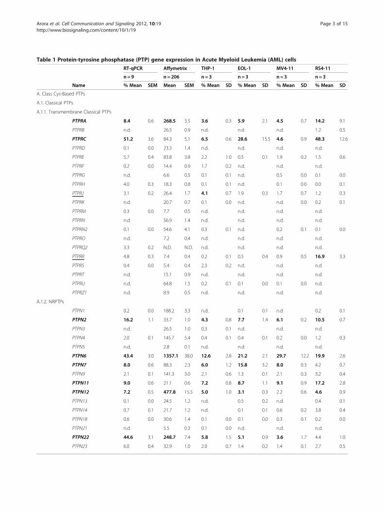
Table 1 Protein-tyrosine phosphatase (PTP) gene expression in Acute Myeloid Leukemia (AML) cells
RT-qPCR Affymetrix THP-1 EOL-1 MV4-11 RS4-11
n= 9 n=206 n= 3 n=3 n=3 n=3
Name % Mean SEM Mean SEM % Mean SD % Mean SD % Mean SD % Mean SD
A. Class Cys-Based PTPs
A.1. Classical PTPs
A.1.1. Transmembrane Classical PTPs
PTPRA 8.4 0.6 268.5 3.5 3.6 0.3 5.9 2.1 4.5 0.7 14.2 9.1
PTPRB n.d. 26.5 0.9 n.d. n.d. n.d. 1.2 0.5
PTPRC 51.2 3.6 84.3 5.1 6.5 0.6 28.6 15.5 4.6 0.9 48.3 12.6
PTPRD 0.1 0.0 23.3 1.4 n.d. n.d. n.d. n.d.
PTPRE 5.7 0.4 83.8 3.8 2.2 1.0 0.5 0.1 1.9 0.2 1.5 0.6
PTPRF 0.2 0.0 14.4 0.9 1.7 0.2 n.d. n.d. n.d.
PTPRG n.d. 6.6 0.5 0.1 0.1 n.d. 0.5 0.0 0.1 0.0
PTPRH 4.0 0.3 18.3 0.8 0.1 0.1 n.d. 0.1 0.0 0.0 0.1
PTPRJ 3.1 0.2 26.4 1.7 4.1 0.7 1.9 0.3 1.7 0.7 1.2 0.3
PTPRK n.d. 20.7 0.7 0.1 0.0 n.d. n.d. 0.0 0.2 0.1
PTPRM 0.3 0.0 7.7 0.5 n.d. n.d. n.d. n.d.
PTPRN n.d. 56.9 1.4 n.d. n.d. n.d. n.d.
PTPRN2 0.1 0.0 54.6 4.1 0.3 0.1 n.d. 0.2 0.1 0.1 0.0
PTPRO n.d. 7.2 0.4 n.d. n.d. n.d. n.d.
PTPRQ2 3.3 0.2 N.D. N.D. n.d. n.d. n.d. n.d.
PTPRR 4.8 0.3 7.4 0.4 0.2 0.1 0.5 0.4 0.9 0.5 16.9 3.3
PTPRS 0.4 0.0 5.4 0.4 2.3 0.2 n.d. n.d. n.d.
PTPRT n.d. 15.1 0.9 n.d. n.d. n.d. n.d.
PTPRU n.d. 64.8 1.5 0.2 0.1 0.1 0.0 0.1 0.0 n.d.
PTPRZ1 n.d. 8.9 0.5 n.d. n.d. n.d. n.d.
A.1.2. NRPTPs
PTPN1 0.2 0.0 188.2 3.3 n.d. 0.1 0.1 n.d. 0.2 0.1
PTPN2 16.2 1.1 33.7 1.0 4.3 0.8 7.7 1.4 6.1 0.2 10.5 0.7
PTPN3 n.d. 26.5 1.0 0.3 0.1 n.d. n.d. n.d.
PTPN4 2.0 0.1 145.7 5.4 0.4 0.1 0.4 0.1 0.2 0.0 1.2 0.3
PTPN5 n.d. 2.8 0.1 n.d. n.d. n.d. n.d.
PTPN6 43.4 3.0 1357.1 38.0 12.6 2.8 21.2 2.1 29.7 12.2 19.9 2.6
PTPN7 8.0 0.6 88.3 2.3 6.0 1.2 15.8 3.2 8.0 0.3 4.2 0.7
PTPN9 2.1 0.1 141.3 3.0 2.1 0.6 1.3 0.1 2.1 0.3 3.2 0.4
PTPN11 9.0 0.6 21.1 0.6 7.2 0.8 8.7 1.1 9.1 0.9 17.2 2.8
PTPN12 7.2 0.5 477.8 15.5 5.0 1.0 3.1 0.3 2.2 0.6 4.6 0.9
PTPN13 0.1 0.0 24.5 1.2 n.d. 0.5 0.2 n.d. 0.4 0.1
PTPN14 0.7 0.1 21.7 1.2 n.d. 0.1 0.1 0.6 0.2 3.8 0.4
PTPN18 0.6 0.0 30.6 1.4 0.1 0.0 0.1 0.0 0.3 0.1 0.2 0.0
PTPN21 n.d. 5.5 0.3 0.1 0.0 n.d. n.d. n.d.
PTPN22 44.6 3.1 248.7 7.4 5.8 1.5 5.1 0.9 3.6 1.7 4.4 1.0
PTPN23 6.0 0.4 32.9 1.0 2.0 0.7 1.4 0.2 1.4 0.1 2.7 0.5
Arora et al. Cell Communication and Signaling 2012, 10:19 Page 3 of 15http://www.biosignaling.com/content/10/1/19
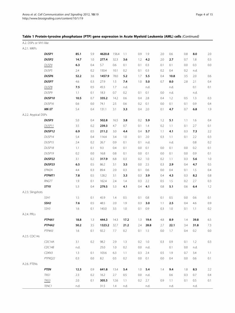
Table 1 Protein-tyrosine phosphatase (PTP) gene expression in Acute Myeloid Leukemia (AML) cells (Continued)
A.2. DSPs or VH1-like
A.2.1. MKPs
DUSP1 85.1 5.9 4620.8 156.4 1.1 0.9 1.9 2.0 0.6 0.8 8.0 2.0
DUSP2 14.7 1.0 277.4 32.3 3.6 1.2 4.2 2.0 2.7 0.7 1.8 0.3
DUSP4 6.3 0.4 5.7 0.6 0.1 0.1 0.3 0.1 0.1 0.0 0.3 0.0
DUSP5 2.4 0.2 150.4 10.1 0.2 0.1 0.3 0.2 0.4 0.2 n.d.
DUSP6 52.2 3.6 1457.9 78.0 5.2 1.7 5.5 0.4 10.8 3.5 2.0 0.6
DUSP7 4.6 0.3 27.9 1.5 7.4 1.0 5.0 0.7 8.0 2.8 2.1 0.4
DUSP8 7.5 0.5 45.5 1.7 n.d. n.d. n.d. 0.1 0.1
DUSP9 1.1 0.1 18.3 0.7 0.2 0.1 0.1 0.0 n.d. n.d.
DUSP10 10.5 0.7 335.2 14.2 0.6 0.4 2.8 0.4 1.2 0.5 1.3 0.3
DUSP16 0.6 0.0 74.1 2.5 0.6 0.2 0.1 0.0 0.1 0.1 0.9 0.4
MK-ST 5.4 0.4 131.1 3.1 3.3 0.4 2.0 0.1 4.7 0.7 6.8 1.9
A.2.2. Atypical DSPs
DUSP3 5.0 0.4 502.8 16.5 3.8 0.2 5.9 1.2 5.1 1.1 1.6 0.4
DUSP11 3.5 0.2 239.3 4.7 0.7 0.1 1.4 0.2 1.1 0.1 2.7 0.1
DUSP12 6.9 0.5 211.2 3.0 4.4 0.4 5.7 1.1 4.1 0.3 7.3 2.2
DUSP14 5.4 0.4 114.4 3.4 1.0 0.1 2.0 0.3 1.1 0.1 2.2 0.3
DUSP15 2.4 0.2 26.7 0.9 0.1 0.1 n.d. n.d. 0.8 0.2
DUSP18 1.1 0.1 9.3 0.4 0.1 0.0 0.1 0.0 0.1 0.0 0.2 0.1
DUSP19 0.2 0.0 16.8 0.8 0.1 0.0 0.1 0.0 0.1 0.0 0.9 0.1
DUSP22 3.1 0.2 317.9 6.8 0.3 0.2 1.0 0.2 1.1 0.3 5.6 1.0
DUSP23 6.5 0.5 86.2 3.1 3.5 0.0 2.5 0.3 2.9 0.4 4.7 0.5
EPM2A 4.4 0.3 89.4 2.0 0.3 0.1 0.6 0.0 0.4 0.1 1.5 0.4
PTPMT1 7.8 0.5 128.2 3.1 3.3 0.3 3.9 0.4 4.3 0.3 8.2 0.8
RNGTT 1.9 0.1 162.4 2.4 1.4 0.3 2.2 0.5 1.5 0.2 2.7 0.3
STYX 5.3 0.4 279.5 5.3 4.1 0.4 4.1 0.8 5.1 0.6 6.4 1.2
A.2.3. Slingshots
SSH1 1.5 0.1 45.9 1.4 0.5 0.1 0.8 0.1 0.5 0.0 0.6 0.1
SSH2 7.6 0.5 48.5 2.0 1.9 0.3 3.0 1.1 2.5 0.4 4.6 0.9
SSH3 1.6 0.1 143.0 3.5 1.0 0.1 0.9 0.3 1.0 0.1 1.1 0.2
A.2.4. PRLs
PTP4A1 18.8 1.3 444.3 14.3 17.2 1.3 19.4 4.8 8.9 1.4 39.8 6.5
PTP4A2 50.2 3.5 1323.2 32.7 21.2 2.4 28.8 2.7 22.1 3.4 31.8 7.3
PTP4A3 1.6 0.1 92.2 7.7 0.2 0.1 1.5 0.0 1.7 0.4 0.2 0.0
A.2.5. CDC14s
CDC14A 3.1 0.2 98.2 2.9 1.3 0.2 1.0 0.3 0.9 0.1 1.2 0.3
CDC14B n.d. 25.0 1.0 0.2 0.0 n.d. 0.1 0.0 n.d.
CDKN3 1.3 0.1 103.6 6.3 1.1 0.3 2.4 0.5 1.9 0.7 3.4 1.1
PTP9Q22 0.3 0.0 8.2 0.5 0.2 0.0 0.1 0.0 0.4 0.0 0.6 0.1
A.2.6. PTENs
PTEN 12.3 0.9 641.8 13.4 5.4 1.0 5.4 1.4 9.4 1.8 8.5 2.2
TNS1 2.3 0.2 16.2 2.7 0.5 0.0 n.d. 0.6 0.3 0.7 0.4
TNS3 2.0 0.1 305.5 12.6 1.1 0.2 2.7 0.9 1.1 0.1 0.5 0.1
TENC1 n.d. 31.5 1.4 n.d. n.d. n.d. n.d.
Arora et al. Cell Communication and Signaling 2012, 10:19 Page 4 of 15http://www.biosignaling.com/content/10/1/19
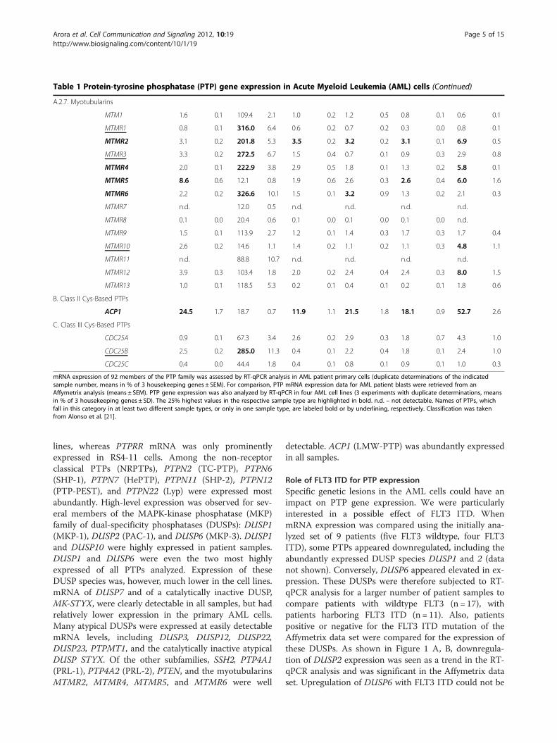
Table 1 Protein-tyrosine phosphatase (PTP) gene expression in Acute Myeloid Leukemia (AML) cells (Continued)
A.2.7. Myotubularins
MTM1 1.6 0.1 109.4 2.1 1.0 0.2 1.2 0.5 0.8 0.1 0.6 0.1
MTMR1 0.8 0.1 316.0 6.4 0.6 0.2 0.7 0.2 0.3 0.0 0.8 0.1
MTMR2 3.1 0.2 201.8 5.3 3.5 0.2 3.2 0.2 3.1 0.1 6.9 0.5
MTMR3 3.3 0.2 272.5 6.7 1.5 0.4 0.7 0.1 0.9 0.3 2.9 0.8
MTMR4 2.0 0.1 222.9 3.8 2.9 0.5 1.8 0.1 1.3 0.2 5.8 0.1
MTMR5 8.6 0.6 12.1 0.8 1.9 0.6 2.6 0.3 2.6 0.4 6.0 1.6
MTMR6 2.2 0.2 326.6 10.1 1.5 0.1 3.2 0.9 1.3 0.2 2.1 0.3
MTMR7 n.d. 12.0 0.5 n.d. n.d. n.d. n.d.
MTMR8 0.1 0.0 20.4 0.6 0.1 0.0 0.1 0.0 0.1 0.0 n.d.
MTMR9 1.5 0.1 113.9 2.7 1.2 0.1 1.4 0.3 1.7 0.3 1.7 0.4
MTMR10 2.6 0.2 14.6 1.1 1.4 0.2 1.1 0.2 1.1 0.3 4.8 1.1
MTMR11 n.d. 88.8 10.7 n.d. n.d. n.d. n.d.
MTMR12 3.9 0.3 103.4 1.8 2.0 0.2 2.4 0.4 2.4 0.3 8.0 1.5
MTMR13 1.0 0.1 118.5 5.3 0.2 0.1 0.4 0.1 0.2 0.1 1.8 0.6
B. Class II Cys-Based PTPs
ACP1 24.5 1.7 18.7 0.7 11.9 1.1 21.5 1.8 18.1 0.9 52.7 2.6
C. Class III Cys-Based PTPs
CDC25A 0.9 0.1 67.3 3.4 2.6 0.2 2.9 0.3 1.8 0.7 4.3 1.0
CDC25B 2.5 0.2 285.0 11.3 0.4 0.1 2.2 0.4 1.8 0.1 2.4 1.0
CDC25C 0.4 0.0 44.4 1.8 0.4 0.1 0.8 0.1 0.9 0.1 1.0 0.3
mRNA expression of 92 members of the PTP family was assessed by RT-qPCR analysis in AML patient primary cells (duplicate determinations of the indicatedsample number, means in % of 3 housekeeping genes ± SEM). For comparison, PTP mRNA expression data for AML patient blasts were retrieved from anAffymetrix analysis (means ± SEM). PTP gene expression was also analyzed by RT-qPCR in four AML cell lines (3 experiments with duplicate determinations, meansin % of 3 housekeeping genes ± SD). The 25% highest values in the respective sample type are highlighted in bold. n.d. – not detectable. Names of PTPs, whichfall in this category in at least two different sample types, or only in one sample type, are labeled bold or by underlining, respectively. Classification was takenfrom Alonso et al. [21].
Arora et al. Cell Communication and Signaling 2012, 10:19 Page 5 of 15http://www.biosignaling.com/content/10/1/19
lines, whereas PTPRR mRNA was only prominentlyexpressed in RS4-11 cells. Among the non-receptorclassical PTPs (NRPTPs), PTPN2 (TC-PTP), PTPN6(SHP-1), PTPN7 (HePTP), PTPN11 (SHP-2), PTPN12(PTP-PEST), and PTPN22 (Lyp) were expressed mostabundantly. High-level expression was observed for sev-eral members of the MAPK-kinase phosphatase (MKP)family of dual-specificity phosphatases (DUSPs): DUSP1(MKP-1), DUSP2 (PAC-1), and DUSP6 (MKP-3). DUSP1and DUSP10 were highly expressed in patient samples.DUSP1 and DUSP6 were even the two most highlyexpressed of all PTPs analyzed. Expression of theseDUSP species was, however, much lower in the cell lines.mRNA of DUSP7 and of a catalytically inactive DUSP,MK-STYX, were clearly detectable in all samples, but hadrelatively lower expression in the primary AML cells.Many atypical DUSPs were expressed at easily detectablemRNA levels, including DUSP3, DUSP12, DUSP22,DUSP23, PTPMT1, and the catalytically inactive atypicalDUSP STYX. Of the other subfamilies, SSH2, PTP4A1(PRL-1), PTP4A2 (PRL-2), PTEN, and the myotubularinsMTMR2, MTMR4, MTMR5, and MTMR6 were well
detectable. ACP1 (LMW-PTP) was abundantly expressedin all samples.
Role of FLT3 ITD for PTP expressionSpecific genetic lesions in the AML cells could have animpact on PTP gene expression. We were particularlyinterested in a possible effect of FLT3 ITD. WhenmRNA expression was compared using the initially ana-lyzed set of 9 patients (five FLT3 wildtype, four FLT3ITD), some PTPs appeared downregulated, including theabundantly expressed DUSP species DUSP1 and 2 (datanot shown). Conversely, DUSP6 appeared elevated in ex-pression. These DUSPs were therefore subjected to RT-qPCR analysis for a larger number of patient samples tocompare patients with wildtype FLT3 (n = 17), withpatients harboring FLT3 ITD (n = 11). Also, patientspositive or negative for the FLT3 ITD mutation of theAffymetrix data set were compared for the expression ofthese DUSPs. As shown in Figure 1 A, B, downregula-tion of DUSP2 expression was seen as a trend in the RT-qPCR analysis and was significant in the Affymetrix dataset. Upregulation of DUSP6 with FLT3 ITD could not be
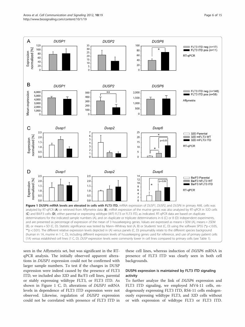
Figure 1 DUSP6 mRNA levels are elevated in cells with FLT3 ITD. mRNA expression of DUSP1, DUSP2, and DUSP6 in primary AML cells wasanalyzed by RT-qPCR (A) or retrieved from Affymetrix data (B). mRNA expression of the murine genes was also analyzed by RT-qPCR in 32D cells(C) and BA/F3 cells (D), either parental or expressing wildtype (WT) FLT3 or FLT3 ITD, as indicated. RT-qPCR data are based on duplicatedeterminations for the indicated sample numbers (A), and on duplicate or triplicate determinations in 6 (C) or 8 (D) independent experiments,and are presented as percentage of expression of the mean of 3 housekeeping genes. Values are expressed as means + SEM (A), means + 2SEM(B), or means + SD (C, D). Statistic significance was tested by Mann–Whitney test (A, B) or Students’ test (C, D) using the software SPSS (*p< 0.05,**p< 0.01). The different relative expression levels depicted in (A) versus panels (C, D) presumably relate to the different species background(human in 1A, murine in 1 C, D), including different expression levels of housekeeping genes used for reference, and use of primary patient cells(1A) versus established cell lines (1 C, D). DUSP expression levels were commonly lower in cell lines compared to primary cells (see Table 1).
Arora et al. Cell Communication and Signaling 2012, 10:19 Page 6 of 15http://www.biosignaling.com/content/10/1/19
seen in the Affymetrix set, but was significant in the RT-qPCR analysis. The initially observed apparent altera-tions in DUSP1 expression could not be confirmed withlarger sample numbers. To test if the changes in DUSPexpression were indeed caused by the presence of FLT3ITD, we included also 32D and Ba/F3 cell lines, parentalor stably expressing wildtype FLT3, or FLT3 ITD. Asshown in Figure 1 C, D, alterations of DUSP1 mRNAlevels in dependence of FLT3 ITD expression were notobserved. Likewise, regulation of DUSP2 expressioncould not be correlated with presence of FLT3 ITD in
these cell lines, whereas induction of DUSP6 mRNA inpresence of FLT3 ITD was clearly seen in both cellbackgrounds.
DUSP6 expression is maintained by FLT3 ITD signalingactivityTo further analyze the link of DUSP6 expression andFLT3 ITD signaling, we employed MV4-11 cells, en-dogenously expressing FLT3 ITD, RS4-11 cells endogen-ously expressing wildtype FLT3, and 32D cells withoutor with expression of wildtype FLT3 or FLT3 ITD.
Arora et al. Cell Communication and Signaling 2012, 10:19 Page 7 of 15http://www.biosignaling.com/content/10/1/19
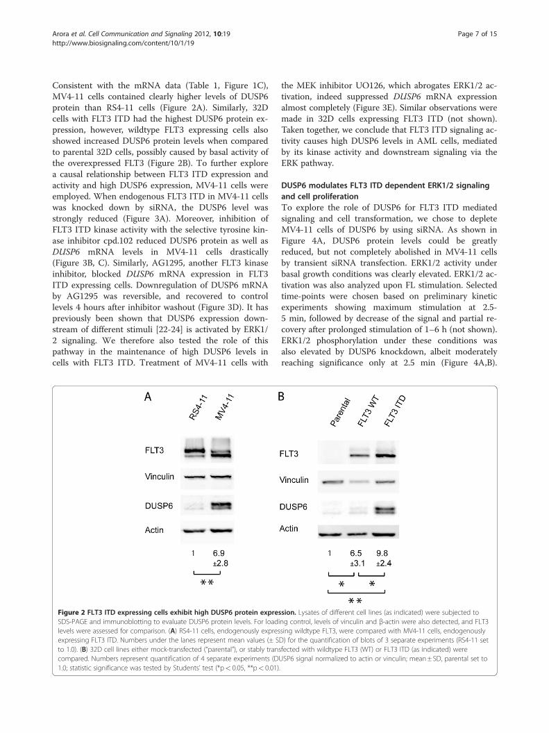
Consistent with the mRNA data (Table 1, Figure 1C),MV4-11 cells contained clearly higher levels of DUSP6protein than RS4-11 cells (Figure 2A). Similarly, 32Dcells with FLT3 ITD had the highest DUSP6 protein ex-pression, however, wildtype FLT3 expressing cells alsoshowed increased DUSP6 protein levels when comparedto parental 32D cells, possibly caused by basal activity ofthe overexpressed FLT3 (Figure 2B). To further explorea causal relationship between FLT3 ITD expression andactivity and high DUSP6 expression, MV4-11 cells wereemployed. When endogenous FLT3 ITD in MV4-11 cellswas knocked down by siRNA, the DUSP6 level wasstrongly reduced (Figure 3A). Moreover, inhibition ofFLT3 ITD kinase activity with the selective tyrosine kin-ase inhibitor cpd.102 reduced DUSP6 protein as well asDUSP6 mRNA levels in MV4-11 cells drastically(Figure 3B, C). Similarly, AG1295, another FLT3 kinaseinhibitor, blocked DUSP6 mRNA expression in FLT3ITD expressing cells. Downregulation of DUSP6 mRNAby AG1295 was reversible, and recovered to controllevels 4 hours after inhibitor washout (Figure 3D). It haspreviously been shown that DUSP6 expression down-stream of different stimuli [22-24] is activated by ERK1/2 signaling. We therefore also tested the role of thispathway in the maintenance of high DUSP6 levels incells with FLT3 ITD. Treatment of MV4-11 cells with
Figure 2 FLT3 ITD expressing cells exhibit high DUSP6 protein expresSDS-PAGE and immunoblotting to evaluate DUSP6 protein levels. For loadilevels were assessed for comparison. (A) RS4-11 cells, endogenously expresexpressing FLT3 ITD. Numbers under the lanes represent mean values (± Sto 1.0). (B) 32D cell lines either mock-transfected (“parental”), or stably transcompared. Numbers represent quantification of 4 separate experiments (DU1.0; statistic significance was tested by Students’ test (*p< 0.05, **p< 0.01)
the MEK inhibitor UO126, which abrogates ERK1/2 ac-tivation, indeed suppressed DUSP6 mRNA expressionalmost completely (Figure 3E). Similar observations weremade in 32D cells expressing FLT3 ITD (not shown).Taken together, we conclude that FLT3 ITD signaling ac-tivity causes high DUSP6 levels in AML cells, mediatedby its kinase activity and downstream signaling via theERK pathway.
DUSP6 modulates FLT3 ITD dependent ERK1/2 signalingand cell proliferationTo explore the role of DUSP6 for FLT3 ITD mediatedsignaling and cell transformation, we chose to depleteMV4-11 cells of DUSP6 by using siRNA. As shown inFigure 4A, DUSP6 protein levels could be greatlyreduced, but not completely abolished in MV4-11 cellsby transient siRNA transfection. ERK1/2 activity underbasal growth conditions was clearly elevated. ERK1/2 ac-tivation was also analyzed upon FL stimulation. Selectedtime-points were chosen based on preliminary kineticexperiments showing maximum stimulation at 2.5-5 min, followed by decrease of the signal and partial re-covery after prolonged stimulation of 1–6 h (not shown).ERK1/2 phosphorylation under these conditions wasalso elevated by DUSP6 knockdown, albeit moderatelyreaching significance only at 2.5 min (Figure 4A,B).
sion. Lysates of different cell lines (as indicated) were subjected tong control, levels of vinculin and β-actin were also detected, and FLT3sing wildtype FLT3, were compared with MV4-11 cells, endogenouslyD) for the quantification of blots of 3 separate experiments (RS4-11 setfected with wildtype FLT3 (WT) or FLT3 ITD (as indicated) wereSP6 signal normalized to actin or vinculin; mean± SD, parental set to
.
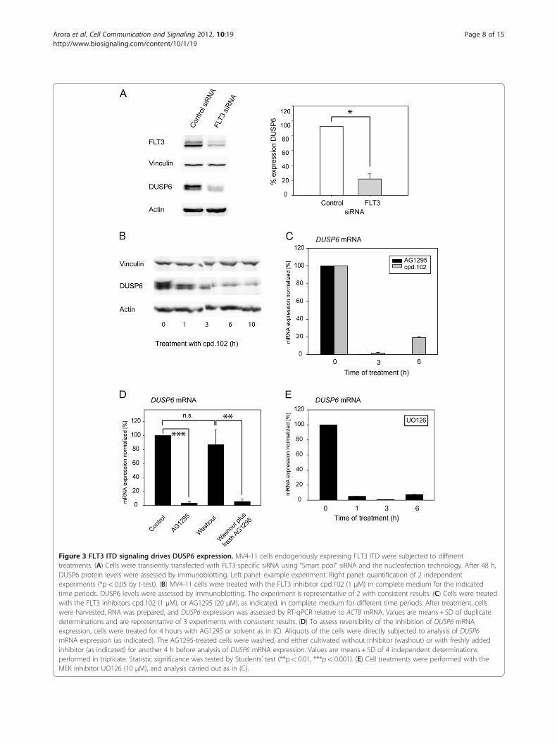
Figure 3 FLT3 ITD signaling drives DUSP6 expression. MV4-11 cells endogenously expressing FLT3 ITD were subjected to differenttreatments. (A) Cells were transiently transfected with FLT3-specific siRNA using “Smart pool” siRNA and the nucleofection technology. After 48 h,DUSP6 protein levels were assessed by immunoblotting. Left panel: example experiment. Right panel: quantification of 2 independentexperiments (*p< 0.05 by t-test). (B) MV4-11 cells were treated with the FLT3 inhibitor cpd.102 (1 μM) in complete medium for the indicatedtime periods. DUSP6 levels were assessed by immunoblotting. The experiment is representative of 2 with consistent results. (C) Cells were treatedwith the FLT3 inhibitors cpd.102 (1 μM), or AG1295 (20 μM), as indicated, in complete medium for different time periods. After treatment, cellswere harvested, RNA was prepared, and DUSP6 expression was assessed by RT-qPCR relative to ACTB mRNA. Values are means + SD of duplicatedeterminations and are representative of 3 experiments with consistent results. (D) To assess reversibility of the inhibition of DUSP6 mRNAexpression, cells were treated for 4 hours with AG1295 or solvent as in (C). Aliquots of the cells were directly subjected to analysis of DUSP6mRNA expression (as indicated). The AG1295-treated cells were washed, and either cultivated without inhibitor (washout) or with freshly addedinhibitor (as indicated) for another 4 h before analysis of DUSP6 mRNA expression. Values are means + SD of 4 independent determinationsperformed in triplicate. Statistic significance was tested by Students’ test (**p< 0.01, ***p< 0.001). (E) Cell treatments were performed with theMEK inhibitor UO126 (10 μM), and analysis carried out as in (C).
Arora et al. Cell Communication and Signaling 2012, 10:19 Page 8 of 15http://www.biosignaling.com/content/10/1/19
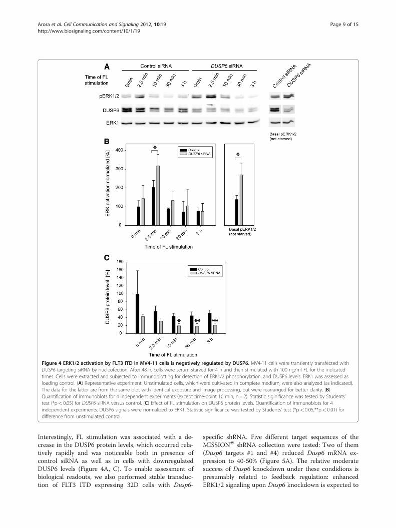
Figure 4 ERK1/2 activation by FLT3 ITD in MV4-11 cells is negatively regulated by DUSP6. MV4-11 cells were transiently transfected withDUSP6-targeting siRNA by nucleofection. After 48 h, cells were serum-starved for 4 h and then stimulated with 100 ng/ml FL for the indicatedtimes. Cells were extracted and subjected to immunoblotting for detection of ERK1/2 phosphorylation, and DUSP6 levels. ERK1 was assessed asloading control. (A) Representative experiment. Unstimulated cells, which were cultivated in complete medium, were also analyzed (as indicated).The data for the latter are from the same blot with identical exposure and image processing, but were rearranged for better clarity. (B)Quantification of immunoblots for 4 independent experiments (except time-point 10 min, n = 2). Statistic siginificance was tested by Students’test (*p< 0.05) for DUSP6 siRNA versus control. (C) Effect of FL stimulation on DUSP6 protein levels. Quantification of immunoblots for 4independent experiments. DUSP6 signals were normalized to ERK1. Statistic significance was tested by Students’ test (*p< 0.05,**p< 0.01) fordifference from unstimulated control.
Arora et al. Cell Communication and Signaling 2012, 10:19 Page 9 of 15http://www.biosignaling.com/content/10/1/19
Interestingly, FL stimulation was associated with a de-crease in the DUSP6 protein levels, which occurred rela-tively rapidly and was noticeable both in presence ofcontrol siRNA as well as in cells with downregulatedDUSP6 levels (Figure 4A, C). To enable assessment ofbiological readouts, we also performed stable transduc-tion of FLT3 ITD expressing 32D cells with Dusp6-
specific shRNA. Five different target sequences of theMISSIONW shRNA collection were tested: Two of them(Dusp6 targets #1 and #4) reduced Dusp6 mRNA ex-pression to 40-50% (Figure 5A). The relative moderatesuccess of Dusp6 knockdown under these condidions ispresumably related to feedback regulation: enhancedERK1/2 signaling upon Dusp6 knockdown is expected to
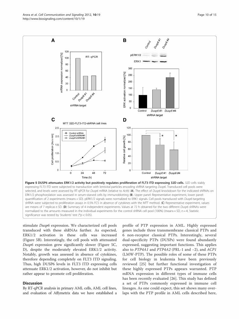
Figure 5 DUSP6 attenuates ERK1/2 activity but positively regulates proliferation of FLT3 ITD expressing 32D cells. 32D cells stablyexpressing FLT3 ITD were subjected to transduction with lentiviral particles encoding shRNA targeting Dusp6. Transduced cell pools wereselected, and levels were assessed by RT-qPCR for Dusp6 mRNA (relative to Actb) (A). The effect of Dusp6 knockdown for the indicated shRNAs onERK1/2 phosphorylation was assessed in serum-starved cells by immunoblotting (B). Upper panel: Representative experiment, lower panel:quantification of 2 experiments (means + SD). pERK1/2 signals were normalized to ERK1 signals. Cell pools transduced with Dusp6-targetingshRNA were subjected to proliferation assays in 0.5% FCS in absence of cytokines with the MTT method. (C) Representative experiment, valuesare means of 7 replicas ± SD. (D) Summary of 4 independent experiments. Values at 72 h obtained for the two different Dusp6 shRNAs werenormalized to the amounts measured in the individual experiments for the control shRNA cell pool (100%) (means + SD, n = 4, Statisticsignificance was tested by Students’ test (*p< 0.05).
Arora et al. Cell Communication and Signaling 2012, 10:19 Page 10 of 15http://www.biosignaling.com/content/10/1/19
stimulate Dusp6 expression. We characterized cell poolstransduced with these shRNAs further. As expected,ERK1/2 activation in these cells was increased(Figure 5B). Interestingly, the cell pools with attenuatedDusp6 expression grew significantly slower (Figure 5C,D), despite the moderately elevated ERK1/2 activity.Notably, growth was assessed in absence of cytokines,therefore depending completely on FLT3 ITD signaling.Thus, high DUSP6 levels in FLT3 ITD expressing cellsattenuate ERK1/2 activation, however, do not inhibit butrather appear to promote cell proliferation.
DiscussionBy RT-qPCR analysis in primary AML cells, AML cell lines,and evaluation of Affymetrix data we have established a
profile of PTP expression in AML. Highly expressedgenes include three transmembrane classical PTPs and6 non-receptor classical PTPs. Interestingly, severaldual-specificity PTPs (DUSPs) were found abundantlyexpressed, suggesting important functions. This appliesalso to PTP4A1 and PTP4A2 (PRL-1 and −2), and ACP1(LMW-PTP). The possible roles of some of these PTPsfor cell biology in leukemia have been previouslyreviewed [25] but further functional investigation ofthese highly expressed PTPs appears warranted. PTPmRNA expression in different types of immune cellshas been recently evaluated [26]. This study has defineda set of PTPs commonly expressed in immune celllineages. As one could expect, this set shows many over-laps with the PTP profile in AML cells described here,
Arora et al. Cell Communication and Signaling 2012, 10:19 Page 11 of 15http://www.biosignaling.com/content/10/1/19
for example PTPRC/CD45 and PTPN6/SHP-1 areamong the highly expressed genes in both analyses.However, certain of these “commonly expressed” PTPgenes in immune cells were only weakly or not detectedin our analysis, including for example PTPRF, PTPRS,PTPN3, PTPN9, or DUSP4. These PTPs may not beexpressed in the early differentiation stages representedby the leukemic blasts, or may potentially be downregu-lated, another interesting topic for further investigation.Some obvious discrepancies in the expression levels ofPTP genes in the patient samples either detected by RT-qPCR or by Affymetrix analysis were apparent. Theymay mostly relate to the different detection of spliceversions by the two methods, and in part to the differentsample sets. Still, of the 23 PTP genes detected amongthe upper 25% by expression level, 12 match for bothdetection methods. For the case of DUSP6, RT-qPCRanalysis was clearly advantageous over the microarraytechnique to detect relevant and further validated changes.We have focused our interest on the possible role of
FLT3 ITD, a common oncogenic lesion in AML, on PTPexpression. It appeared possible that changed PTP ex-pression would contribute to FLT3 ITD mediated trans-formation. Only relatively few alterations of PTPexpression were, however, observed. Notably, PTPN6(SHP-1), a highly expressed PTP with a proven negativeregulatory role for cytokine signaling and possible rolein regulation of FLT3 was not altered in mRNA expres-sion in our data sets, contrary to what has previouslybeen proposed [17]. Also, PTPRJ, a negative regulator ofFLT3 autophosphorylation [11], was not altered in ex-pression by FLT3 ITD (data not shown). As we havefound recently, FLT3 ITD appears, however, to inactivatePTPRJ by production of high levels of reactive oxygenspecies [27]. DUSP2 mRNA was downregulated in pri-mary AML cells with FLT3 ITD, but this phenomenoncould not be recapitulated in 32D or Ba/F3 cells. It ispossible that further lesions in the primary AML cellscontribute to DUSP2 regulation. Still, reduced DUSP2levels may play a role in FLT3 ITD-transformed cells,which we have not yet explored.Interestingly, DUSP6 was elevated in expression down-
stream of FLT3 ITD signaling, as found in our RT-qPCRanalysis of primary AML cells, as well as in differentmodel cell systems. DUSP6 induction at mRNA and pro-tein level could be causally linked to FLT3 ITD signalingactivity and ERK1/2 pathway activation. Upregulation ofDUSP6 mRNA was, however, not detectable in the Affy-metrix data set. It is possible that this method is not suf-ficiently sensitive to detect the FLT3 ITD-mediatedalteration in DUSP6 expression. In patients with activat-ing N-RAS mutations, an increase in DUSP6 expressioncould, however, be seen in the Affymetrix data set (notshown), possibly indicating that N-RAS mutations are
more potent than FLT3 ITD in driving DUSP6 expres-sion. Previous gene expression analyzes (also using Affy-metrix expression arrays) in myeloma cells haveidentified DUSP6 as one of only three genes which wereuniquely and strongly elevated in cells harboring acti-vated N-RAS [28]. Interestingly, other stimuli whichonly transiently activated ERK signaling such asinterleukin-6 stimulation did not induce a sufficientlyrobust DUSP6 response to allow detection with thistechnique. Consistent with our findings, DUSP6 has pre-viously been identified as one of the genes which aremost effectively downregulated in FLT3 ITD expressingAML cells treated with the FLT3/broad spectrum kinaseinhibitor CEP701 (Lestaurtinib) [29]. Based on its cap-acity for dephosphorylating the pThr-X-pTyr motif inERK1/2 [19], DUSP6 can negatively regulate pERK1/2levels in multiple cell types. Since the ERK1/2 pathwaymediates mitogenic signaling, among other responses,DUSP6 has been proposed as a tumor suppressor [23].Notably, in pancreatic cancer, DUSP6 levels are downre-gulated by gene deletion or promoter hypermethylation,consistent with such a function [30-32]. Recently,DUSP6 was shown to inhibit growth, migration andepithelial-to-mesenchymal transition (EMT) of esopha-gal squamous cell carcinoma and nasopharyngeal carcin-oma cells [33]. In non-small cell lung cancer, however,high DUSP6 levels have been found to predict poorprognosis, and evidence for a tumor-promoting role ofDUSP6 in human glioblastoma has recently been pro-vided [34,35]. Moreover, DUSP6 has recently been foundas a predictor of invasiveness in papillary thyroid cancer.Silencing of DUSP6 expression decreased the cell viabil-ity and migration rate of a corresponding cell line [36].As shown in the present study, elevated DUSP6 levelscorrelate with the presence of FLT3 ITD, a negative pre-dictor of prognosis [5] in AML cells. This observation,taken together with the functional findings discussedbelow, indicates that DUSP6 may play a pro-oncogenicrole in FLT3 ITD-positive AML. Obviouosly, dependingon the context, DUSP6 affects tumor biology in very dif-ferent ways.The function of high DUSP6 levels in FLT3 ITD
expressing cells was addressed by RNAi experiments.Consistent with its function as ERK1/2-PTP, downregu-lation of DUSP6 caused elevated constitutive ERK1/2 ac-tivation in FLT3 ITD expressing cells. Moreover, ERK1/2phosphorylation was also elevated in FL-stimulated cells,albeit only moderately. We observed that FL-stimulationcaused downregulation of DUSP6 protein levels, similaras it has been described earlier for the stimulation ofcells with other mitogens. In these studies the authorslinked the reduction of DUSP6 levels to proteasomaldegradation, which was prompted by phosphorylation atserine residues [22,37]. Surprisingly, 32D cell pools with
Arora et al. Cell Communication and Signaling 2012, 10:19 Page 12 of 15http://www.biosignaling.com/content/10/1/19
attenuated Dusp6 expression were, however, moderatelyimpaired in proliferation, indicating that high DUSP6levels may not play a negative but rather some positiverole in FLT3 ITD dependent cell growth. Forced overex-pression of exogenous DUSP6 in stable transfectantsreduced ERK1/2 phosphorylation, but did not inhibit cellproliferation (data not shown), further supporting thenotion that ERK1/2 activity and cell growth in FLT3ITD-expressing cells are not simply positively correlated.It is well known that the biological outcome of ERK1/2activation depends both on its magnitude and on itskinetics, which are determined by several feedbackmechanisms [38]. For example, sustained high ERK1/2activation by the phorbol ester TPA in MCF7 cellscauses growth arrest [24]. Similarly, nerve growth factor(NGF) stimulation of PC-12 pheochromocytoma cells asopposed to epidermal growth factor (EGF) stimulationleads to sustained high-level ERK1/2 activation and nota proliferative, but a differentiation response [39,40].The alternate cellular responses to different kinetics andmagnitude of ERK1/2 activation seem based on differ-ential transcriptional sensing [41], which is likely to de-pend on the specific cell type and the simultaneousactivation of other signaling pathways. In the context ofFLT3 ITD, high constitutive expression of DUSP6appears associated with a relatively low but constitutivelevel of ERK1/2 activity, which is compatible with effi-cient cell growth. ShRNA-mediated DUSP6 downregu-lation causes a higher level of constitutive ERK1/2activation (see Figure 4), which, similar as in the case ofMCF7 or PC12 cells, may ultimately result in dimin-ished cell proliferation by yet unclear downstreammechanisms. Alternatively or in addition, potentialother substrates of DUSP6 may play a role. These possi-bilities need to be further explored.
ConclusionsElevated expression of the dual-specificity phosphataseDUSP6 is driven by the oncoprotein FLT3 ITD in AML.Despite the known role of DUSP6 as negative regulatorof ERK1/2 activation, which was also observed in FLT3ITD-expressing leukemia cells, DUSP6 was identified asa positive component in the FLT3 ITD-driven prolifera-tion. This finding prompts its further characterization asa possible drug target.
MethodsCell linesThe human AML cell lines MV4-11, THP1, EOL1, RS4-11 were obtained from the German Collection of Micro-organisms and Cell Cultures (DSMZ, Braunschweig,Germany) and maintained in RPMI 1640 medium withglutamine (PAA, Cölbe, Germany) supplemented with10% heat inactivated fetal calf serum (FCS, BioWest,
Berlin, Germany). Murine 32D cells stably expressing ex-ogenous wild type mFLT3 or mFLT3 ITD were kindlyprovided by Drs. Justus Duyster and Rebekka Grundler(Klinikum Rechts der Isar, Munich, Germany), and wereroutinely cultured in RPMI 1640 medium with HEPES(Biochrome Berlin, Germany) supplemented with 10%heat inactivated FCS, 1 mM sodium pyruvate, and2.5 ng/ml murine recombinant IL3. Murine Ba/F3 cellsstably expressing exogenous wildtype hFLT3 or hFLT3ITD were kindly provided by Dr. Lars Rönnstrand (Ex-perimental Clinical Chemistry, University HospitalLund/Malmö, Sweden) and were cultured under thesame conditions as 32D cells. To assess Dusp6 mRNAor DUSP6 protein expression (experiments shown inFigures 1C,D, and 2B, respectively) or FLT3 ITD-dependent growth (e.g. experiments shown in Figure 5C,D), cultivation was done in absence of cytokines.
Antibodies and reagentsThe polyclonal DUSP6 antibody used in this study hasbeen described earlier [22]. Mouse monoclonal anti-β-actin antibody (clone AC15) was from Sigma Aldrich(A1978, Deisenhofen, Germany), mouse monoclonalanti-ERK1 antibody was from Transduction Laboratories(M12320/L1), mouse monoclonal anti-phospho-ERK1/2antibody from Cell Signaling (Frankfurt, Germany),FLT3 Polyclonal anti-FLT3 antibody (C-20, sc-479) wasfrom Santa Cruz Biotechnology (Heidelberg, Germany)and mouse monoclonal anti-Vinculin (BZL03106) anti-body was from Biozol Diagnostic (Eching, Germany).Horseradish peroxidase-conjugated secondary antibodieswere from KPL (Wedel, Germany). AG1295 was pur-chased from Alexis Biochemicals (Grünberg, Germany).FLT3 ligand (FL, human), and murine IL-3 were fromPeprotech (London, UK). The MEK inhibitor UO126was from Tocris Bioscience (Bristol, UK). The bisindolyl-methanone FLT3 inhibitor cpd. 102 has been describedearlier [42].
Cell treatments and preparation of RNA and proteinsamplesTo evaluate basal PTP expression in human AML celllines, 1–2 x 10⁶ cells were harvested from well-growingcultures by centrifugation at 300 x g for 5 minutes. Thesupernatant was discarded and the cell pellets were usedfor total RNA preparation using the RNeasy kit (Qiagen,Hilden, Germany). Biological replicas were preparedwith independently cultured cell batches. 32D cells andBa/F3 cells were starved from cytokines by washingthem twice and subsequently incubating them withRPMI medium containing 0.5% FCS for 4 h before iso-lating RNA. RS4-11 and MV4-11 cells were starved withserum-free RPMI medium for 4 h before FL stimulation.For protein extraction, cells were centrifuged (300 x g,
Arora et al. Cell Communication and Signaling 2012, 10:19 Page 13 of 15http://www.biosignaling.com/content/10/1/19
5 min). The pellets were washed once with ice cold PBS,and then extracted with 60–80 μl lysis buffer. containing50 mM HEPES (pH 7.4), 150 mM NaCl, 1 mM EDTA,25 mM NaF, 1% (v/v) NP-40 and (freshly added) aproti-nin, 65 KIU/ml, 1 μg/ml leupeptin, 1 μg/ml pepstatin,1 mM PMSF, 0.2 mg/ml Pefabloc, and 1 mM sodiumorthovanadate.
Human AML samplesAML patients included in this study were diagnosed andtreated either at the Jena University Hospital (Jena,Germany) or at the Otto-von-Guericke UniversityMagdeburg (Magdeburg, Germany). The retrospectivemolecular analyses of leukemic blasts were approvedby the institutional review boards of each university hos-pital. AML cells were isolated from peripheral blood orbone marrow from AML patients at diagnosis afterinformed consent. In part, bone marrow was frozen inFCS supplemented with 10% IMDM and 10% DMSOuntil workup. Erythrocytes were lysed using erythrocytelysis buffer (QIAGEN, Hilden, Germany) according tothe manufacturer´s instructions, and blasts were purifiedby Ficoll (Biochrom, Berlin, Germany) density gradientseparation. RNA isolation was carried out with theRNeasy Mini Kit (QIAGEN, Hilden, Germany). FLT3mutation status was determined by RT-PCR using stand-ard diagnostic primers ITD1 (5´- GCAATTTAGGTATGAAAGCCAGC-3´) and ITD2 (5´- CTTTCAGCATTTTGACGGCAACC-3´) or FLT3-ITD-fw 5-GCAATTTAGGTATGAAAGCCAGC-3 and FLT3-ITD-rev 5-CTTTCAGCATT TTGACGGCAACC-3.
Expression analysis by RT-qPCRThe platform for PTP mRNA expression analysis by RT-qPCR has been described recently [18]. RNA was quan-tified by UV measurement, and integrity was verifiedusing an Agilent 2100 Bioanalyzer. Samples withconcentrations< 100 ng/μl, A260:A280 ratios <2.0, orpartially degraded RNA were excluded from further pro-cessing. 1 μg RNA was used directly for preparation ofcDNA using a BioRad iScript cDNA synthesis kit(BioRad, Munich, Germany) (for human AML samplesand cell lines) or a Fermentas cDNA synthesis kit (Fer-mentas, St. Leon-Rot, Germany) (for all other samples)according to the manufacturer’s instructions. cDNA cor-responding to 40 ng RNA input was used for the qPCR.The primers for amplification of most of the transcriptswere purchased from Qiagen (QuantiTectW PrimerAssay, Qiagen, Hilden, Germany), except primer sets forhuDUSP2, huDUSP4, huSBF1, huPTPN2, huPTPN22,and huPTPRR, which were bought from SABiosciences(Hilden, Germany). The real-time PCR reactions werecarried out using the FastStart Universal SYBR GreenMaster (ROX) from Roche (Mannheim, Germany) and
an Applied Biosystems 7900HT Fast Real-Time PCR sys-tem (Merck Serono facility, Geneva, Switzerland), or theRTPCR, Maxima™ SYBR green from Fermentas (St.Leon-Rot, Germany, Cat. No. K0221) and an EppendorfRealplexW Mastercycler. The conditions for PCRincluded 95°C for 15 min (to activate the hotstart Taqpolymerase), followed by 40 cycles of 94°C for 15 sec,55°C for 30 sec, 72°C for 30 sec. Threshold Cycles (Ct)were determined after the completion of PCR and calcu-lations of the relative expression of PTP genes weredone based on ΔCt values and the mean Ct of the threehousekeeping genes proteasome subunit beta type-3(PSMB3/Psmb3), calnexin (CANX/Canx), and β-actin(ACTB/Actb). For analyses of drug effects of DUSP6/Dusp6 expression in cell lines, ΔCt were calculated rela-tive to β-actin (ACTB/Actb) expression.
Analysis of array dataA published microarray dataset (GSE1159) was analyzedfor PTP expression levels in FLT3 WT and FLT3 ITDsamples [43]. Data were analyzed as described previously[44].
siRNA and shRNA transfectionsFor siRNA-mediated knockdown of human FLT3 theON-TARGETplus SMARTpool from Dharmacon(ThermoFisher Scientific, Schwerte, Germany, Cat. No.L-003137-00-0005) was used. As control, the ON-TARGETplus non-targeting pool (Cat. No. D-001810-10-20) was used. MV4-11 cells were transfected usingthe Lonza (Cologne, Germany) Cell Line NucleofectorKit V according to the instructions of the manufac-turer. In brief, 2 x 10⁶ cells were taken up in 100 μlof transfection solution, 2 μg siRNA were added, andthe suspension was transferred to an electroporationcuvette and pulsed once using the program A30 of theAMAXA Nucleofector. Thereafter, cells were diluted inculture medium and maintained for 48 hours beforefurther analysis. For shRNA-mediated knockdown ofDUSP6 in 32D cells, pLKO.1-based constructs wereobtained from the Sigma-Aldrich (Deisenhofen, Ger-many) MISSIONW shRNA collection: Dusp6 #1 TRCID number TRCN0000055038 (5’CCGGCGATGCTTACGACATTGTTAACTCGAGTTAACAATGTCGTAAGCATCGTTTTTG 3’); Dusp6 #4 TRCN0000055041 5’CCGGCCTGAGGCCATTTCTTTCATACTCGAGTATGAAAGAAATGGCCTCAGGTTTTTG3’. Production oflentiviral particles and transduction and selection of32D cell pools was done as described before [11].
DUSP6 overexpressionA construct encoding ratDUSP6 has been described earl-ier [45]. It was subcloned into the vector LeGO-iC ([46],kindly provided by Dr. C. Stocking, Heinrich Pette
Arora et al. Cell Communication and Signaling 2012, 10:19 Page 14 of 15http://www.biosignaling.com/content/10/1/19
Institute, Hamburg, Germany) by standard techniques.Stable transfection of 32D cell pools was performed asdescribed previously [47].
Other assaysImmunoblotting and assessment of cell proliferation using3-(4,5-dimethylthiazol-2-yl)-2,5-diphenyltetrazolium brom-ide (MTT) were performed as described earlier [47].Immunoblots were developed using enhanced chemilumi-niscence and detection with a LAS4000 Imager (FujifilmEurope GmbH, Düsseldorf, Germany). Signals were quan-tified using Multi Gauge V3.0 Software (Fujifilm EuropeGmbH, Düsseldorf, Germany).
Competing interestsThe authors declare that they have no competing interests.
Authors’ contributionsDA, SK, MvdE and RHvH performed the PTP expression analysis and analyzedthe data, FH, TF, and SS provided and worked up clinical samples, BT, andSAB performed functional studies, JL developed a critical reagent andsupported the study with experimental protocols and advice, FI and CMTperformed bioinformatic analysis of expression data, FDB planned andsupervised the study and wrote the manuscript. All authors read andapproved the final manuscript.
AcknowledgmentsWe greatly acknowledge support from the European Community (“PTP-NET”Marie Curie Network MRTN-CT-2006-035830; D.A., M.v.d.E., R.H.v.H., F.D.B.),Merck Serono Geneva, Switzerland (M.v.d.E., R.H.v.H.), Deutsche Krebshilfe(Collaborative grant 108401, T.F., C.M.T., F.D.B) and DeutscheForschungsgemeinschaft (Grant BO1043/7-1 to F.D.B.). We thank Dr. C.Stocking for provision of a reagent, and C. Weißleder for help with some ofthe experiments.
Author details1Institute of Molecular Cell Biology, Center for Molecular Biomedicine, JenaUniversity Hospital, Jena, Germany. 2Merck Serono, Geneva 1202, Switzerland.3Department of Hematology/Oncology, Otto-von Guericke-University,Magdeburg, Germany. 4Department of Hematology/Oncology, Clinic forInternal Medicine II, Jena University Hospital, Jena, Germany. 5LudwigInstitute for Cancer Research, Uppsala Branch, Uppsala, Sweden. 6Departmentof Medicine A, Hematology and Oncology, University of Münster, Münster,Germany.
Received: 31 January 2012 Accepted: 24 June 2012Published: 11 July 2012
References1. Östman A, Hellberg C, Böhmer FD: Protein-tyrosine phosphatases and
cancer. Nat Rev Cancer 2006, 6:307–320.2. Julien SG, Dube N, Hardy S, Tremblay ML: Inside the human cancer
tyrosine phosphatome. Nat Rev Cancer 2011, 11:35–49.3. Mohi MG, Neel BG: The role of Shp2 (PTPN11) in cancer. Curr Opin Genet
Dev 2007, 17:23–30.4. Berman-Golan D, Granot-Attas S, Elson A: Protein tyrosine phosphatase
epsilon and Neu-induced mammary tumorigenesis. Cancer Metastasis Rev2008, 27:193–203.
5. Döhner H, Estey EH, Amadori S, Appelbaum FR, Buchner T, Burnett AK,Dombret H, Fenaux P, Grimwade D, Larson RA, et al: Diagnosis andmanagement of acute myeloid leukemia in adults: recommendationsfrom an international expert panel, on behalf of the EuropeanLeukemiaNet. Blood 2010, 115:453–474.
6. Fröhling S, Scholl C, Gilliland DG, Levine RL: Genetics of myeloidmalignancies: pathogenetic and clinical implications. J Clin Oncol 2005,23:6285–6295.
7. Choudhary C, Müller-Tidow C, Berdel WE, Serve H: Signal transduction ofoncogenic Flt3. Int J Hematol 2005, 82:93–99.
8. Schmidt-Arras DE, Böhmer A, Markova B, Choudhary C, Serve H, Böhmer FD:Tyrosine phosphorylation regulates maturation of receptor tyrosinekinases. Mol Cell Biol 2005, 25:3690–3703.
9. Heiss E, Masson K, Sundberg C, Pedersen M, Sun J, Bengtsson S, RönnstrandL: Identification of Y589 and Y599 in the juxtamembrane domain of Flt3as ligand-induced autophosphorylation sites involved in binding of Srcfamily kinases and the protein tyrosine phosphatase SHP2. Blood 2006,108:1542–1550.
10. Müller JP, Schönherr C, Markova B, Bauer R, Stocking C, Böhmer FD: Role ofSHP2 for FLT3-dependent proliferation and transformation in 32D cells.Leukemia 2008, 22:1945–1948.
11. Arora D, Stopp S, Böhmer SA, Schons J, Godfrey R, Masson K, RazumovskayaE, Rönnstrand L, Tänzer S, Bauer R, et al: Protein-tyrosine phosphataseDEP-1 controls receptor tyrosine kinase FLT3 signaling. J Biol Chem 2011,286:10918–10929.
12. Zhou J, Bi C, Chng WJ, Cheong LL, Liu SC, Mahara S, Tay KG, Zeng Q, Li J,Guo K, et al: PRL-3, a metastasis associated tyrosine phosphatase, isinvolved in FLT3-ITD signaling and implicated in anti-AML therapy. PLoSOne 2011, 6:e19798.
13. Bullinger L, Döhner K, Bair E, Fröhling S, Schlenk RF, Tibshirani R, Döhner H,Pollack JR: Use of gene-expression profiling to identify prognosticsubclasses in adult acute myeloid leukemia. N Engl J Med 2004,350:1605–1616.
14. Verhaak RG, Wouters BJ, Erpelinck CA, Abbas S, Beverloo HB, Lugthart S,Lowenberg B, Delwel R, Valk PJ: Prediction of molecular subtypes in acutemyeloid leukemia based on gene expression profiling. Haematologica2009, 94:131–134.
15. Kohlmann A, Bullinger L, Thiede C, Schaich M, Schnittger S, Döhner K,Dugas M, Klein HU, Döhner H, Ehninger G, Haferlach T: Gene expressionprofiling in AML with normal karyotype can predict mutations formolecular markers and allows novel insights into perturbed biologicalpathways. Leukemia 2011, 24:1216–1220.
16. Marcucci G, Haferlach T, Döhner H: Molecular genetics of adult acutemyeloid leukemia: prognostic and therapeutic implications. J Clin Oncol2011, 29:475–486.
17. Chen P, Levis M, Brown P, Kim KT, Allebach J, Small D: FLT3/ITD mutationsignaling includes suppression of SHP-1. J Biol Chem 2005, 280:5361–5369.
18. Schmidt F, van den Eijnden M, Gobert RP, Saborio G, Carboni S, Alliod C,Pouly S, Staugaitis SM, Dutta R, Trapp B, Hooft Van, Huijsduijnen R:Identification of VHY/Dusp15 as a regulator of oligodendrocytedifferentiation through a systematic genomics approach. PLoS One 2012,7:e40457.
19. Camps M, Nichols A, Gillieron C, Antonsson B, Muda M, Chabert C, BoschertU, Arkinstall S: Catalytic activation of the phosphatase MKP-3 by ERK2mitogen-activated protein kinase. Science 1998, 280:1262–1265.
20. Dickinson RJ, Keyse SM: Diverse physiological functions for dual-specificity MAP kinase phosphatases. J Cell Sci 2006, 119:4607–4615.
21. Alonso A, Sasin J, Bottini N, Friedberg I, Osterman A, Godzik A, Hunter T,Dixon J, Mustelin T: Protein tyrosine phosphatases in the humangenome. Cell 2004, 117:699–711.
22. Jurek A, Amagasaki K, Gembarska A, Heldin CH, Lennartsson J: Negativeand positive regulation of MAPK phosphatase 3 controls platelet-derivedgrowth factor-induced Erk activation. J Biol Chem 2009, 284:4626–4634.
23. Pulido R, Hooft van Huijsduijnen R: Protein tyrosine phosphatases: dual-specificity phosphatases in health and disease. FEBS J 2008, 275:848–866.
24. Nunes-Xavier CE, Tarrega C, Cejudo-Marin R, Frijhoff J, Sandin A, Östman A,Pulido R: Differential up-regulation of MAP kinase phosphatases MKP3/DUSP6 and DUSP5 by Ets2 and c-Jun converge in the control of thegrowth arrest versus proliferation response of MCF-7 breast cancer cellsto phorbol ester. J Biol Chem 2010, 285:26417–26430.
25. Ruela-de-Sousa RR, Queiroz KC, Peppelenbosch MP, Fuhler GM: Reversiblephosphorylation in haematological malignancies: potential role forprotein tyrosine phosphatases in treatment? Biochim Biophys Acta 2010,1806:287–303.
26. Arimura Y, Yagi J: Comprehensive expression profiles of genes for proteintyrosine phosphatases in immune cells. Sci Signal 2010, 3:rs1.
27. Godfrey R, Arora D, Bauer R, Stopp S, Müller JP, Heinrich T, Böhmer SA,Dagnell M, Schnetzke U, Scholl S, et al: Cell transformation by FLT3 ITD inacute myeloid leukemia involves oxidative inactivation of the tumor
Arora et al. Cell Communication and Signaling 2012, 10:19 Page 15 of 15http://www.biosignaling.com/content/10/1/19
suppressor protein-tyrosine phosphatase DEP-1/ PTPRJ. Blood 2012,119:4499–4511.
28. Croonquist PA, Linden MA, Zhao F, Van Ness BG: Gene profiling of amyeloma cell line reveals similarities and unique signatures among IL-6response, N-ras-activating mutations, and coculture with bone marrowstromal cells. Blood 2003, 102:2581–2592.
29. Kim KT, Baird K, Ahn JY, Meltzer P, Lilly M, Levis M, Small D: Pim-1 isup-regulated by constitutively activated FLT3 and plays a role inFLT3-mediated cell survival. Blood 2005, 105:1759–1767.
30. Xu S, Furukawa T, Kanai N, Sunamura M, Horii A: Abrogation of DUSP6 byhypermethylation in human pancreatic cancer. J Hum Genet 2005,50:159–167.
31. Furukawa T, Sunamura M, Motoi F, Matsuno S, Horii A: Potential tumorsuppressive pathway involving DUSP6/MKP-3 in pancreatic cancer. Am JPathol 2003, 162:1807–1815.
32. Furukawa T, Yatsuoka T, Youssef EM, Abe T, Yokoyama T, Fukushige S, SoedaE, Hoshi M, Hayashi Y, Sunamura M, et al: Genomic analysis of DUSP6, adual specificity MAP kinase phosphatase, in pancreatic cancer. CytogenetCell Genet 1998, 82:156–159.
33. Wong VC, Chen H, Ko JM, Chan KW, Chan YP, Law S, Chua D, Kwong DL,Lung HL, Srivastava G, et al: Tumor suppressor dual-specificityphosphatase 6 (DUSP6) impairs cell invasion and epithelial-mesenchymal transition (EMT)-associated phenotype. Int J Cancer 2012,130:83–95.
34. Chen HY, Yu SL, Chen CH, Chang GC, Chen CY, Yuan A, Cheng CL, WangCH, Terng HJ, Kao SF, et al: A five-gene signature and clinical outcome innon-small-cell lung cancer. N Engl J Med 2007, 356:11–20.
35. Messina S, Frati L, Leonetti C, Zuchegna C, Di Zazzo E, Calogero A, PorcelliniA: Dual-specificity phosphatase DUSP6 has tumor-promoting propertiesin human glioblastomas. Oncogene 2011, 30:3813–3820.
36. Lee JU, Huang S, Lee MH, Lee SE, Ryu MJ, Kim SJ, Kim YK, Kim SY, Joung KH,Kim JM, et al: Dual specificity phosphatase 6 (DUSP6) as a predictor ofinvasiveness in papillary thyroid cancer. Eur J Endocrinol 2012, 167:93-101.
37. Marchetti S, Gimond C, Chambard JC, Touboul T, Roux D, Pouyssegur J,Pages G: Extracellular signal-regulated kinases phosphorylate mitogen-activated protein kinase phosphatase 3/DUSP6 at serines 159 and 197,two sites critical for its proteasomal degradation. Mol Cell Biol 2005,25:854–864.
38. Kholodenko BN, Hancock JF, Kolch W: Signalling ballet in space and time.Nat Rev Mol Cell Biol 2010, 11:414–426.
39. Marshall CJ: Specificity of receptor tyrosine kinase signaling: transientversus sustained extracellular signal-regulated kinase activation. Cell1995, 80:179–185.
40. Santos SD, Verveer PJ, Bastiaens PI: Growth factor-induced MAPK networktopology shapes Erk response determining PC-12 cell fate. Nat Cell Biol2007, 9:324–330.
41. Murphy LO, Smith S, Chen RH, Fingar DC, Blenis J: Molecular interpretationof ERK signal duration by immediate early gene products. Nat Cell Biol2002, 4:556–564.
42. Mahboobi S, Uecker A, Sellmer A, Cenac C, Hocher H, Pongratz H, EichhornE, Hufsky H, Trumpler A, Sicker M, et al: Novel bis(1 H-indol-2-yl)methanones as potent inhibitors of FLT3 and platelet-derived growthfactor receptor tyrosine kinase. J Med Chem 2006, 49:3101–3115.
43. Valk PJ, Verhaak RG, Beijen MA, Erpelinck CA, Barjesteh van Waalwijk vanDoorn-Khosrovani S, Boer JM, Beverloo HB, Moorhouse MJ, van der Spek PJ,Lowenberg B, Delwel R: Prognostically useful gene-expression profiles inacute myeloid leukemia. N Engl J Med 2004, 350:1617–1628.
44. Agrawal-Singh S, Koschmieder S, Gelsing S, Stocking C, Stehling M, ThiedeC, Thoennissen NH, Kohler G, Valk PJ, Delwel R, et al: Pim2 cooperates withPML-RARalpha to induce acute myeloid leukemia in a bone marrowtransplantation model. Blood 2010, 115:4507–4516.
45. Wälchli S, Curchod ML, Gobert RP, Arkinstall S, Hooft van Huijsduijnen R:Identification of tyrosine phosphatases that dephosphorylate the insulinreceptor. A brute force approach based on "substrate-trapping" mutants.J Biol Chem 2000, 275:9792–9796.
46. Weber K, Bartsch U, Stocking C, Fehse B: A multicolor panel of novellentiviral "gene ontology" (LeGO) vectors for functional gene analysis.Mol Ther 2008, 16:698–706.
47. Schmidt-Arras D, Böhmer SA, Koch S, Muller JP, Blei L, Cornils H, Bauer R,Korasikha S, Thiede C, Böhmer FD: Anchoring of FLT3 in the endoplasmicreticulum alters signaling quality. Blood 2009, 113:3568–3576.
doi:10.1186/1478-811X-10-19Cite this article as: Arora et al.: Expression of protein-tyrosinephosphatases in Acute Myeloid Leukemia cells: FLT3 ITD sustains highlevels of DUSP6 expression. Cell Communication and Signaling 2012 10:19.
Submit your next manuscript to BioMed Centraland take full advantage of:
• Convenient online submission
• Thorough peer review
• No space constraints or color figure charges
• Immediate publication on acceptance
• Inclusion in PubMed, CAS, Scopus and Google Scholar
• Research which is freely available for redistribution
Submit your manuscript at www.biomedcentral.com/submit