Embed Size (px)
Citation preview
Cell, Vol. 121, 991–1004, July 1, 2005, Copyright ©2005 by Elsevier Inc. DOI 10.1016/j.cell.2005.04.015
Structural Basis for the Functionof the Ribosomal L7/12 Stalkin Factor Binding and GTPase Activation
Mihaela Diaconu,1,6 Ute Kothe,3,6 Frank Schlünzen,4,7
Niels Fischer,2 Jörg M. Harms,4,7
Alexander G. Tonevitsky,5 Holger Stark,2
Marina V. Rodnina,3 and Markus C. Wahl1,*1Röntgenkristallographie23D Kryo-ElektronenmikroskopieMax-Planck-Institut für Biophysikalische ChemieAm Faßberg 11D-37077 GöttingenGermany3 Institute of Physical BiochemistryUniversity of Witten/HerdeckeStockumer Straße 10D-58448 WittenGermany4Max-Planck-Arbeitsgruppen für Strukturelle
MolekularbiologieDESYNotkestraße 85D-22607 HamburgGermany5Biological DepartmentMV Lomonosov Moscow State UniversityVorobyevy GoryMoscow 119899Russia
Summary
The L7/12 stalk of the large subunit of bacterial ribo-somes encompasses protein L10 and multiple copiesof L7/12. We present crystal structures of Thermotogamaritima L10 in complex with three L7/12 N-terminal-domain dimers, refine the structure of an archaealL10E N-terminal domain on the 50S subunit, and iden-tify these elements in cryo-electron-microscopic re-constructions of Escherichia coli ribosomes. The mo-bile C-terminal helix �8 of L10 carries three L7/12dimers in T. maritima and two in E. coli, in concor-dance with the different length of helix �8 of L10 inthese organisms. The stalk is organized into threeelements (stalk base, L10 helix �8-L7/12 N-terminal-domain complex, and L7/12 C-terminal domains) linkedby flexible connections. Highly mobile L7/12 C-ter-minal domains promote recruitment of translationfactors to the ribosome and stimulate GTP hydrolysisby the ribosome bound factors through stabilizationof their active GTPase conformation.
Introduction
Protein synthesis on the ribosome is promoted by anumber of translation factors. Several of them, such as
*Correspondence: [email protected]
6 These authors contributed equally to this work. 7 Present address: MPI für Molekulare Genetik, Ihnestraße 63/73,D-14195 Berlin, Germany.initiation factor 2 (IF2), elongation factors Tu (EF-Tu)and G (EF-G), and release factor 3 (RF3), are GTPases(Bourne et al., 1991). Cryo-electron microscopy (EM)studies have revealed that the G domains of these fac-tors interact with a region delineated by the sarcin-ricinloop (SRL) of 23S ribosomal RNA (rRNA) and a neigh-boring lateral protrusion, the L7/12 stalk (referred to asthe stalk hereafter) (Agrawal et al., 1998; Klaholz et al.,2004; Stark et al., 2000; Stark et al., 2002; Valle et al.,2003). The stalk region encompasses ribosomal proteinL11, the region of 23S rRNA that binds proteins L11 andL10 (nucleotides 1030–1124 in E. coli), and a complexformed by L10 and multiple copies of L7/12. (L7 isequivalent to L12 except for an acetylated N terminus.We refer to the proteins from hereon collectively asL12.)
L12 is composed of an N-terminal dimerization mod-ule and a globular C-terminal domain (CTD) connectedby a flexible hinge region (Liljas and Gudkov, 1987). OnEscherichia coli (eco) ribosomes, four copies of L12 arebound as two dimers via their N-terminal domains(NTD) to L10, while L10 is attached to the rRNA. Extrac-tion/complementation experiments have demonstratedthe requirement of L12 for binding of EF-Tu, EF-G, IF2,and RF3 to the ribosome and for ribosome-stimulatedfactor-dependent GTP hydrolysis (Wahl and Moller,2002). Subsequent studies have shown that EF-G hasincreased GTPase activity in the presence of isolatedL12 (Savelsbergh et al., 2000) and that specific muta-tions in the L12 CTD affect the binding of the EF-Tu-GTP-aminoacyl-tRNA ternary complex to the ribosome(Kothe et al., 2004). Based on these data and a compar-ison of the L12 CTD structure to that of EF-Ts, a modelof direct, transient binding of the L12 CTD to the EF-TuG domain has been proposed (Kothe et al., 2004; Wie-den et al., 2001).
Orthologs of L10 and L12 have been identified in allbiological kingdoms. L10 is usually designated L10E inarchaea and P0 in eukaryotes. The eukaryotic L12 or-thologs belong to two families, P1 and P2, which insome organisms form further subfamilies. Although theproteins diverged in sequence during evolution (Figure1A), it is believed that they preserved the overall one(L10 orthologs) to four (L12 orthologs) stoichiometryand their roles in factor-related functions (Gonzalo andReboud, 2003; Wahl and Moller, 2002).
L10 and L12 are disordered or absent from recentcrystal structures of 50S ribosomal subunits (Ban et al.,2000; Harms et al., 2001) and 70S ribosomes (Yusupovet al., 2001). On the other hand, cryo-EM reconstruc-tions disclose density features in the stalk region (e.g.,Agrawal et al., 1998; Agrawal et al., 1999) that cannotbe accounted for by the elements seen in the 50S sub-unit crystal structures and cannot be reliably interpre-ted without an atomic structure of the L10-L12 com-plex. Here we describe crystal structures of a complexbetween L10 and the NTD of L12 from Thermotogamaritima (tma) and refine the structure of the NTD ofL10E from Haloarcula marismortui (hma) on the 50S ri-bosomal subunit. In conjunction with structural features
Cell992
Figure 1. Phylogenetic Comparison
(A) Sequence alignment of bacterial L10 proteins and of hmaL10E, yeast P0, and human P0 proteins. Secondary-structure elements of tmaL10and hmaL10E, as revealed in the present crystal structures, are indicated below each block (black and gray, respectively). Sequence number-ings below the blocks correspond to tmaL10 and hmaL10E, respectively. Within the bacterial L10 sequences, highly conserved amino acids
Structure and Function of the L7/12 Ribosomal Stalk993
for the 50S subunit from H. marismortui (PDB ID codetwo kinks of helix α8. The interdimer interactions sup-
are colored red, intermediately conserved positions yellow. In the L10E/P0 block, identical residues are shown in dark blue, conservedresidues in orange. Residues of hmaL10E that interact directly with 23S rRNA are labeled with a magenta triangle. Residues that contactprotein L11 are labeled with a brown triangle. The three segments of helix α8 in tmaL10 that associate with L12NTD dimers are indicatedby different shades of green. Above this element, arrows indicate hydrophobic residues of L10 that stack with the F29 side chains fromthe L12NTD.(B) Ribbon plots of tmaL10 (left) and hmaL10E (right) NTDs in a similar orientation. The proteins are colored blue to red from N to C terminus.Secondary-structure elements are labeled. All structure figures were prepared with PyMol (http://pymol.sourceforge.net/).
of the stalk seen in cryo-EM reconstructions and withfunctional studies, we propose a structural model ofthe stalk that explains its roles in translation.
Results
Structure of the L10-L12NTD ComplexSince the flexible hinge region in L12 may hamper theproduction of high-quality crystals, we coexpressedfull-length tmaL10 and the NTD of tmaL12 (residuesM1–G30; L12NTD). The proteins were copurified, andstructures at 1.9, 2.1, and 2.3 Å resolution from threedifferent crystal forms were obtained using the multipleanomalous diffraction (MAD) strategy (see Table S1 andFigure S1 in the Supplemental Data available with thisarticle online). In all three crystal structures, one mole-cule of L10 was complexed with six copies of L12NTD,the latter in the form of three dimers.
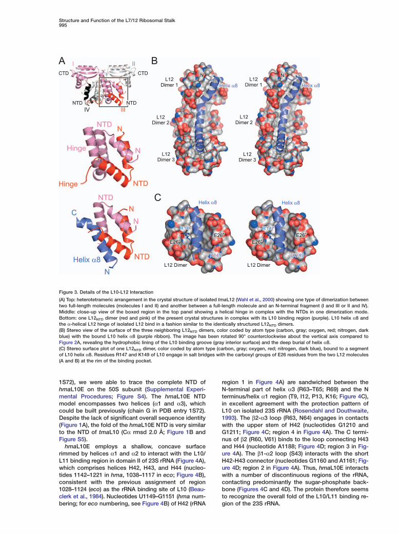
L10 comprises an α/β domain at the N terminus (Fig-ures 1B and 2A). A long C-terminal helix (α8, K137–K174) protruding from this domain is kinked twice, atresidues P151 and G161, dividing it into three ten-resi-due segments (Figure 1A). Each segment associateswith one L12NTD dimer through a five-helix bundle (Fig-ures 2A and 3A). Thus, the L10-L12 interaction regionis characterized by repetition of three almost identicalhelix α8-L12NTD dimer elements.
Within each dimer, the two L12NTD molecules are en-tangled in an antiparallel fashion by extensive hy-drophobic contacts. An identical arrangement of L12molecules has previously been observed in the crystalstructure of isolated tmaL12 (Wahl et al., 2000) and sub-sequently in ecoL12 in solution (Bocharov et al., 2004)(Figure 3A and Figure S2). In the tmaL12 crystal struc-ture (Wahl et al., 2000) the hinge region of one L12molecule folds back as an α helix onto the two entan-gled NTDs. In the L10-(L12NTD)6 complex, this hinge he-lix is replaced by the ten-residue segments of L10 helixα8 (Figure 3A).
L10-L12 InteractionThe interfaces of the L12NTD dimers with L10 buryabout 1500 Å2 of combined surface area each. About80% of the interface residues are hydrophobic (Figure3B). Shape complementarity and electrostatic interac-tions at the periphery, such as salt bridges, hydrogenbonds, and bridging water molecules, register theL12NTD dimers on L10 helix α8 (Figure 3C). Loops ofadjacent L12NTD dimers face each other and engage infour backbone-to-backbone hydrogen bonds via resi-dues E11, L13, V15, and S16. Turns of L10 helix α8,which fall at the border of two adjacent L12NTD dimers,are pried apart by interdimer contacts, leading to the
port a rigid arrangement of the three L12NTD dimers onhelix α8 independent of the crystal environment.
In the three crystal structures, helix α8-(L12NTD)6 ispositioned differently relative to the L10 NTD (Figure2B), which can be described as rotations around a pivotpoint at the beginning of an unstructured loop connect-ing the L10 NTD and helix α8 (Figure 2C). The structuresseem to be stabilized by the formation of different setsof salt bridges between helix α8 and the L10 NTD andbetween the L12NTD dimers and the L10 NTD (Figure2D). These salt bridges surround hydrophobic interac-tions, by which a convex surface area on the first helixα8-L12NTD dimer element is inserted into a concavesurface area of the L10 NTD (Figure 2E). These resultsindicate that the C-terminal helix of L10 bearing L12 isflexibly connected to the L10 NTD.
Stoichiometry of Stalk ProteinsThe 6:1 (L12:L10) stoichiometry was unexpected be-cause a 4:1 ratio has been found in E. coli (Subraman-ian, 1975). However, sequence comparisons show thathelix α8 in ecoL10 is missing one of the ten-residue L12binding sections compared to T. maritima (Figure 1A),consistent with the notion that it can only accommo-date two L12 dimers. In contrast, other bacteria exhibita length and partitioning in L10 helix α8 similar to that inT. maritima and are expected to maintain a L10-(L12)6complex (Figure 1A). In order to confirm the L12 copynumber in T. maritima, we produced a recombinant full-length tmaL10-L12 complex. Multiangle laser light scat-tering indicated a molecular mass of 101 ± 2 kDa forthis complex, in excellent agreement with the predictedmass of 102.6 kDa for a L10-(L12)6 complex (Table S2).A recombinant full-length ecoL10-L12 complex showeda mass of 68 ± 2 kDa, as compared to 66.8 kDa pre-dicted for a L10-(L12)4 composition. We also quantifiedthe amounts of L12 on the ribosomes from T. maritimaand E. coli by immunoblots. As expected, E. coli ribo-somes contained four copies of L12. In contrast, T. mar-itima ribosomes contained six copies of the protein (Ta-ble S2; Figure S3). Thus, the length and sequence ofL10 helix α8 determines the number of L12 copies perribosome. Except for the deletion of one of the threerepetitive elements in helix α8-L12NTD, the high degreeof overall sequence conservation (Figure 1A) suggeststhat the L10-L12 complex of E. coli ribosomes closelyresembles the L10-L12 complex of T. maritima.
Conserved Mode of L10 Ortholog Bindingto rRNA and L11An important question is how the L10-L12 complex issituated on the 50S ribosomal subunit. Using a densitymodification procedure and published structure factors

Cell994
Figure 2. Structure of the Bacterial L10-(L12NTD)6 Complex
(A) Stereo view of the overall structure of the tmaL10-(L12NTD)6 complex showing the L10 NTD at the top and three L12NTD dimers (protomerscolored pink/red, light green/dark green, or yellow/orange, respectively) bound to the C-terminal helix α8 of L10 (purple) at the bottom.(B) Superposition of L10 from the three crystal structures aligned on the NTDs showing the flexible attachment of helix α8 to the L10 NTD.L12NTD dimers have been omitted for clarity. Different L10 molecules are shown in purple (depicted also in [A]), dark gray, and light gray. Theview corresponds to a 90° clockwise rotation about the vertical axis compared to (A).(C) Close-up view of the flexible connector between the L10 NTD and helix α8 with the three L10 molecules superimposed and color codedas in (B). The figure is rotated 60° clockwise about the vertical axis compared to (B). The white button identifies a pivot point around whichhelix α8 rotates relative to the NTD.(D) The same view of the three individual L10 molecules as in (C), with the proximal L12NTD molecule shown as a pink tube. Coloring: carbon,same as the L10 molecules; oxygen, red; nitrogen, dark blue. Dashed lines indicate salt bridges between the L10 NTD and the flexibleconnector or helix α8 and between the L10 NTD and the proximal L12NTD molecule, which stabilize the different conformations.(E) A convex surface area of the proximal L12NTD dimer and the N-terminal part of L10 helix α8 (semitransparent pink surface) inserted into aconcave surface area on the L10 NTD (semitransparent purple surface). The view is identical to (C) and (D). Color coding for ribbons is as in (A).
Structure and Function of the L7/12 Ribosomal Stalk995
Figure 3. Details of the L10-L12 Interaction
(A) Top: heterotetrameric arrangement in the crystal structure of isolated tmaL12 (Wahl et al., 2000) showing one type of dimerization betweentwo full-length molecules (molecules I and II) and another between a full-length molecule and an N-terminal fragment (I and III or II and IV).Middle: close-up view of the boxed region in the top panel showing a helical hinge in complex with the NTDs in one dimerization mode.Bottom: one L12NTD dimer (red and pink) of the present crystal structures in complex with its L10 binding region (purple). L10 helix α8 andthe α-helical L12 hinge of isolated L12 bind in a fashion similar to the identically structured L12NTD dimers.(B) Stereo view of the surface of the three neighboring L12NTD dimers, color coded by atom type (carbon, gray; oxygen, red; nitrogen, darkblue) with the bound L10 helix α8 (purple ribbon). The image has been rotated 90° counterclockwise about the vertical axis compared toFigure 2A, revealing the hydrophobic lining of the L10 binding groove (gray interior surface) and the deep burial of helix α8.(C) Stereo surface plot of one L12NTD dimer, color coded by atom type (carbon, gray; oxygen, red; nitrogen, dark blue), bound to a segmentof L10 helix α8. Residues R147 and K149 of L10 engage in salt bridges with the carboxyl groups of E26 residues from the two L12 molecules(A and B) at the rim of the binding pocket.
1S72), we were able to trace the complete NTD ofhmaL10E on the 50S subunit (Supplemental Experi-mental Procedures; Figure S4). The hmaL10E NTDmodel encompasses two helices (α1 and α3), whichcould be built previously (chain G in PDB entry 1S72).Despite the lack of significant overall sequence identity(Figure 1A), the fold of the hmaL10E NTD is very similarto the NTD of tmaL10 (Cα rmsd 2.0 Å; Figure 1B andFigure S5).
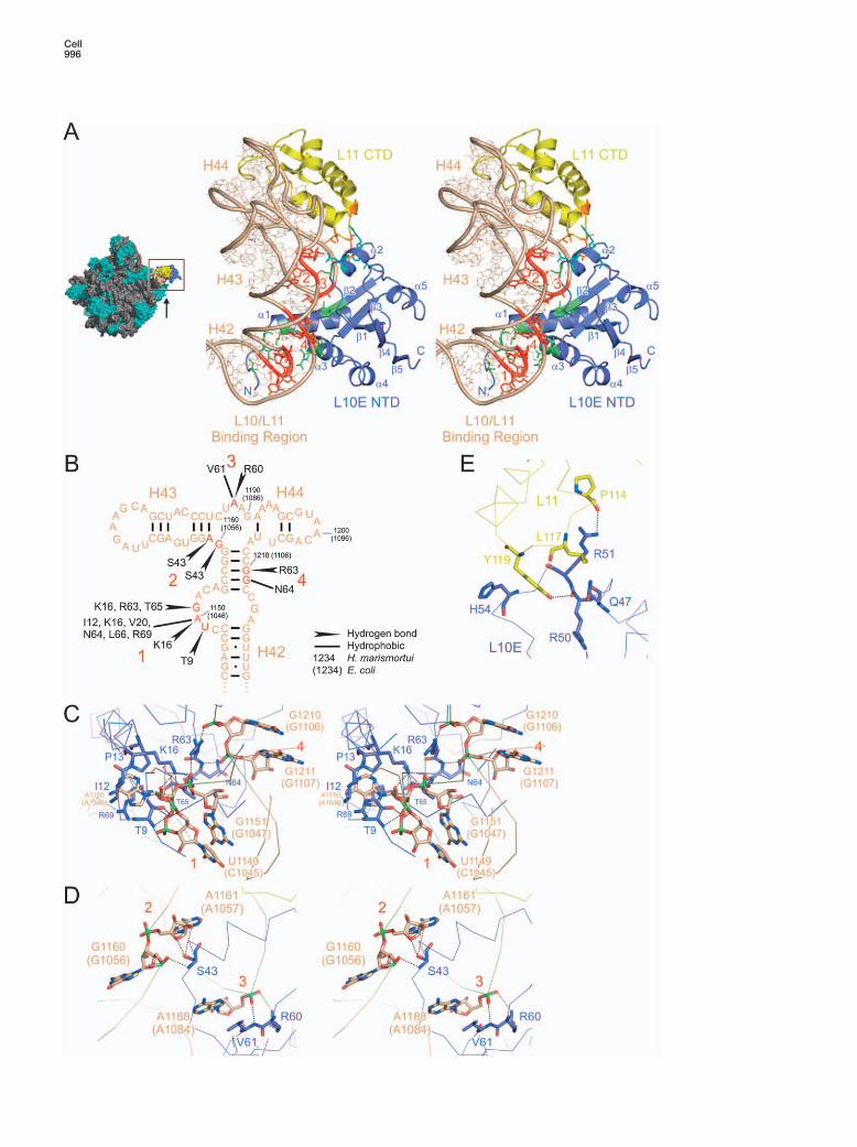
hmaL10E employs a shallow, concave surfacerimmed by helices α1 and α2 to interact with the L10/L11 binding region in domain II of 23S rRNA (Figure 4A),which comprises helices H42, H43, and H44 (nucleo-tides 1142–1221 in hma, 1038–1117 in eco; Figure 4B),consistent with the previous assignment of region1028–1124 (eco) as the rRNA binding site of L10 (Beau-clerk et al., 1984). Nucleotides U1149–G1151 (hma num-bering; for eco numbering, see Figure 4B) of H42 (rRNA
region 1 in Figure 4A) are sandwiched between theN-terminal part of helix α3 (R63–T65; R69) and the Nterminus/helix α1 region (T9, I12, P13, K16; Figure 4C),in excellent agreement with the protection pattern ofL10 on isolated 23S rRNA (Rosendahl and Douthwaite,1993). The β2-α3 loop (R63, N64) engages in contactswith the upper stem of H42 (nucleotides G1210 andG1211; Figure 4C; region 4 in Figure 4A). The C termi-nus of β2 (R60, V61) binds to the loop connecting H43and H44 (nucleotide A1188; Figure 4D; region 3 in Fig-ure 4A). The β1-α2 loop (S43) interacts with the shortH42-H43 connector (nucleotides G1160 and A1161; Fig-ure 4D; region 2 in Figure 4A). Thus, hmaL10E interactswith a number of discontinuous regions of the rRNA,contacting predominantly the sugar-phosphate back-bone (Figures 4C and 4D). The protein therefore seemsto recognize the overall fold of the L10/L11 binding re-gion of the 23S rRNA.
Cell996

Structure and Function of the L7/12 Ribosomal Stalk997
By superpositioning the tmaL10 NTD onto thehmaL10E NTD (Figure S5), the tmaL10-(L12NTD)6 com-plex can be placed on the 50S subunit. Because thestructures of the L10/L11 binding region of 23S rRNAsfrom Haloarcula and Thermotoga are highly conserved(Figure S5) and the protection pattern of bacterial L10matches the contact sites seen for archaeal L10E, bac-terial L10 and archaeal L10E are likely to bind in a sim-ilar fashion to rRNA, although the β2-α3 region is theonly interaction site in the proteins whose sequence isconserved across the kingdoms (Figure 1A).
On the hma50S subunit, the L11 CTD is bound to therRNA in the direct vicinity of the L10E NTD (Figure 4A).The loop between the two C-terminal L11 helices con-tacts the region between helix α2 and strand β2 ofL10E. There are only a few hydrophobic interactionsand two hydrogen bonds connecting the proteins(Y119L11-R50L10E and P114L11-R51L10E; Figure 4E). TheL11 CTD structure is also highly conserved betweenbacteria and archaea (Figures S4 and S5), suggestingsimilar L10-L11 interactions.
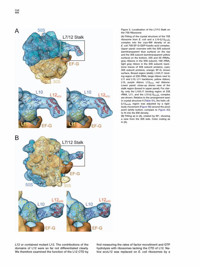
Cryo-EM Reconstructions of the StalkTo visualize the stalk in complete 70S ribosomes, weattempted to localize an L10-L12NTD complex in cryo-EM reconstructions of E. coli ribosomes. For fitting intothe EM maps, we used the crystal structure of the 70Sribosome from E. coli (Vila-Sanjurjo et al., 2003) and amodel of an E. coli-like L10-(L12NTD)4 complex that wasgenerated by shortening helix α8 of tmaL10 and omit-ting the peripheral L12NTD dimer. The 70S crystal struc-ture was docked into the cryo-EM densities and theL10-(L12NTD)4 model was aligned with the crystal struc-ture according to the orientation of the hmaL10E NTDon the hma50S subunit.
Cryo-EM reconstructions of ribosomes at variousstages of translation showed a well-defined densityneighboring the L10/L11 binding region of the 23SrRNA and L11, which is consistent with the crystalstructure of the tmaL10 NTD (Figure 5 and Figures S6and S7A–S7E). In the ribosome-EF-G-GDP complexstalled by fusidic acid (determined herein and in Agra-wal et al., 1998) an elongated protuberance is seen thatextends from the L10 NTD (Figure 5 and Figure S6).
studies used ribosomes that were either depleted ofFigure 4. Binding of the L10 NTD to the 23S rRNA and to Protein L11
(A) Left: crown view of the hma50S subunit—L10E NTD, purple; L11 CTD, yellow; L10/L11 binding region of 23S rRNA, beige. The arrowindicates the view of the boxed area in the right panel. Right: close-up stereo view of the L10/L11 binding region of hma23S rRNA in complexwith the hmaL10E NTD and the hmaL11 CTD. Sections of the L10/L11 binding region, which are contacted by hmaL10E, are highlighted inred (denoted by red numbers 1–4). L10E residues interacting with the rRNA are in green, L10E residues interacting with L11 in cyan. L11residues interacting with L10E are in orange.(B) Secondary-structure diagram of the L10/L11 binding region of 23S rRNA, in which specific contacts to hmaL10E residues are indicated.Nucleotide numbers are for H. marismortui, in parentheses for E. coli. Red numbers and residues denote the regions 1–4 (from [A]) contactedby L10E.(C) Stereo diagram depicting the recognition of nucleotides U1149–G1151 in H42 (region 1 in [A]) by amino acids R63–T65 from the N-terminalpart of helix α3 and by the N terminus of L10E (T9–K16). E. coli nucleotide numbers are in parentheses. R63–T65 are also in contact with theupper stem of H42 (residues G1210–G1211; region 4 in [A]). Relevant residues are shown as sticks (rRNA carbon, beige; L10E carbon, purple;nitrogen, dark blue; oxygen, red; phosphorus, green). Rotated 90° clockwise about the vertical axis relative to (A).(D) Stereo diagram showing R60 and V61 from the C terminus of β2 of L10E interacting with the loop between H43 and H44 (nucleotideA1188; region 3 in [A]). In addition, the β1-α2 loop (S43) is depicted, which contacts the H42–H43 connecting region (nucleotides G1160–A1161; region 2 in [A]). Rotated 50° counterclockwise about the vertical axis relative to (A).(E) Details of the L10E-L11 interaction. Amino acids involved in direct contacts between the proteins are highlighted as sticks (L10 carbon,purple; L11 carbon, yellow). Rotated 90° clockwise about the vertical axis relative to (A).
The L10 helix α8-(L12NTD)4 portion can be fitted to thisprotuberance by a rigid-body movement of helix α8-(L12NTD)4 around the pivot point in the flexible connec-tion to the L10 NTD (Figure 5 and Figure S6) that wasseen in the crystal structures (Figure 2C). The virtualaxis between the pivot point and the C terminus of helixα8 of L10 in the fitted structure is rotated by w25° com-pared to an equivalent axis in crystal structure II (TableS1). Protrusions of similar shape are visible in cryo-EMreconstructions of ribosome-RF2 complexes (Rawat etal., 2003). They can be interpreted in the same way,but with a different orientation of the helix α8-(L12NTD)4portion (w10° or 20° rotations around the pivot point,respectively; Figures S7D and 7E). The differences inthe orientation of the helix α8-(L12NTD)4 extension in thecomplexes with EF-G and RF2 suggest that its positionchanges during translation.
In various other EM structures, little or no density be-yond the L10 NTD can be discerned. The density van-ishes at the flexible connection between the L10 NTDand the helix α8-(L12NTD)4 part (Figures S7A–S7C), sug-gesting that here the helix α8-(L12NTD)4 part adoptsmultiple orientations with respect to the L10 NTD.
Role of L12 CTDs in Factor FunctionTo address the role of L12 for the function of translationfactors on the ribosome, we have used translation com-ponents from E. coli. This system is well characterizedbiochemically and is known to be representative forbacterial systems in general, in keeping with the highdegree of conservation of both sequence and structureas well as the exchangeability of ribosomal compo-nents and factors (see Supplemental Data). The E. coliL7/12 stalk encompasses two L12 dimers that may beconsidered the minimal stalk structure functional inbacterial ribosomes. Early experiments suggested thatL12 is involved in interactions with translation factorsand GTPase stimulation (Wahl and Moller, 2002). Re-cent rapid kinetics and mutagenesis studies showedthat the interaction with L12 promotes factor binding tothe ribosome (Kothe et al., 2004; Mohr et al., 2002) andis involved in the GTPase activation of EF-Tu and EF-G(Mohr et al., 2000; Savelsbergh et al., 2000). These
Cell998
Figure 5. Localization of the L7/12 Stalk onthe 70S Ribosome
(A) Fitting of the crystal structure of the 70Sribosome from E. coli and a L10-(L12NTD)4complex into the cryo-EM density of anE. coli 70S-EF-G-GDP-fusidic-acid complex.Upper panel: overview with the 50S subunit(semitransparent blue surface) on the topand the 30S subunit (semitransparent yellowsurface) on the bottom. 23S and 5S rRNAs,gray ribbons in the 50S subunit; 16S rRNA,light gray ribbon in the 30S subunit; back-bone traces of 50S subunit proteins, cyan;30S subunit proteins, orange; EF-G, brownsurface. Boxed region (stalk): L10/L11 bind-ing region of 23S rRNA, beige ribbon next toL11 and L10; L11 backbone, yellow ribbon;L10, purple ribbon; L12NTD, red ribbons.Lower panel: close-up stereo view of thestalk region (boxed in upper panel). For clar-ity, only the L10/L11 binding region of 23SrRNA, L11, and the L10-(L12NTD)4 complexare shown. Relative to the arrangement seenin crystal structure II (Table S1), the helix α8-(L12NTD)4 region was adjusted by a rigid-body movement (Figure S6) around the pivotpoint (white button; compare to Figure 2C)to fit into the EM density.(B) Fitting as in (A), rotated by 90°, showinga view from the 30S side. Color coding asin (A).
L12 or contained mutant L12. The contributions of the fhdomains of L12 were so far not differentiated clearly.
We therefore examined the function of the L12 CTD by t
irst measuring the rates of factor recruitment and GTPydrolysis with ribosomes lacking the CTD of L12. Na-ive ecoL12 was replaced on E. coli ribosomes by a
Structure and Function of the L7/12 Ribosomal Stalk999
CTD deletion mutant of L12 (L12NTD/hinge). Rate con-stants of the association of the EF-Tu-GTP-Phe-tRNAPhe
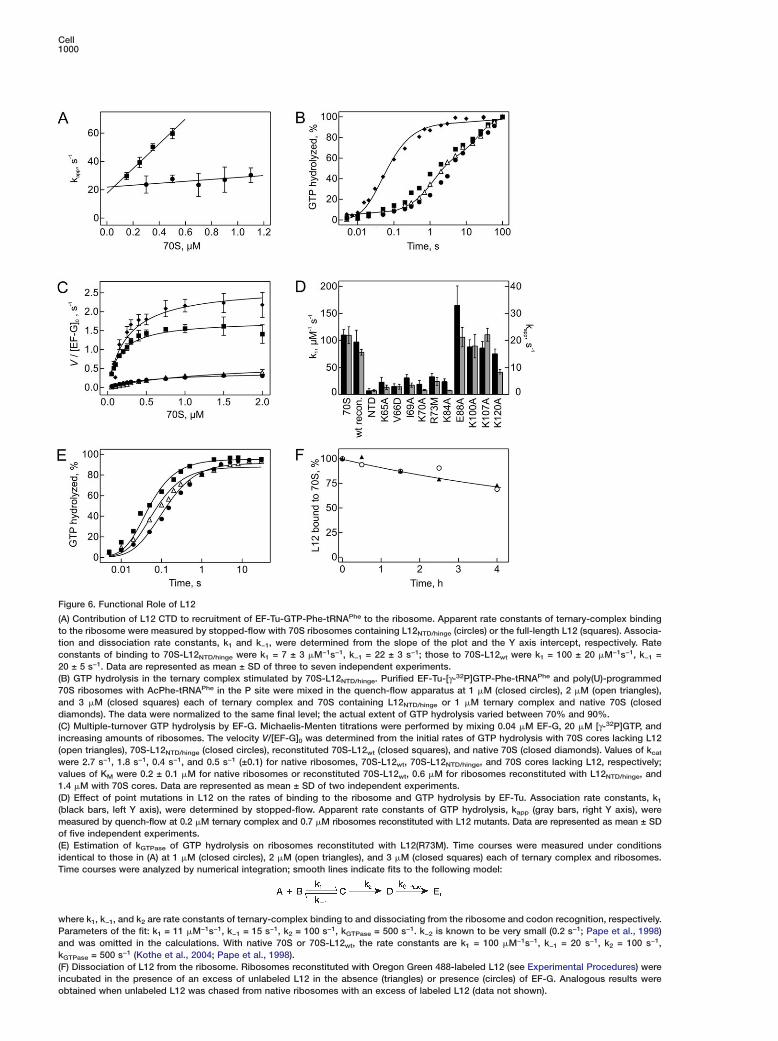
complex with ribosomes were measured by fluores-cence stopped-flow (Kothe et al., 2004; Mohr et al.,2002) (Figure 6A). The rate constant of EF-Tu-GTP-Phe-tRNAPhe binding to the ribosomes lacking the L12 CTDwas 7 �M−1s−1, >10 times smaller than that observedwith wild-type ribosomes, suggesting a significant con-tribution of the L12 CTD to factor binding. A number ofsingle amino acid exchanges in the L12 CTD had aneffect similar to removal of the whole domain (Fig-ure 6D).
Ribosomes lacking L12 CTDs were also strongly im-paired in EF-Tu-promoted GTP hydrolysis compared towild-type ribosomes (Figure 6B). The low rate of GTPhydrolysis, about 0.4 s−1, did not increase with concen-tration and was not limited by the binding step (ex-pected to be 7–21 s−1 at 1–3 �M concentrations usedin Figure 6B). The rate of GTP hydrolysis was about thesame as that of ribosome cores depleted of L12 (0.2s−1; Mohr et al., 2000) and >1000-fold slower than thatof intact ribosomes (>500 s−1; Pape et al., 1998). Therate of GTP hydrolysis by EF-G was reduced about 600-fold by removal of the L12 CTDs (to 0.4 s−1; Figure 6C)compared to intact ribosomes (250 s−1; Savelsbergh etal., 2003).
To identify amino acids responsible for GTPase acti-vation, point mutations of all conserved residues at thesurface of the L12 CTD were analyzed. Several mutantsshowed reduced rates of ternary-complex associationwith the ribosome and subsequent GTP hydrolysis byEF-Tu (Figure 6D). However, the detailed analysis of theconcentration dependence of GTP hydrolysis with ribo-somes containing L12(R73M) (Figure 6E) suggestedthat the decreased rate of GTP hydrolysis is solely dueto the 10-fold slower binding of the factor, as no signifi-cant effect on the rate constant of GTP hydrolysis wasfound. Two other mutations in the CTD, K70A andK84A, showed the same effects, i.e., slower bindingand no effect on GTP hydrolysis itself (data not shown).Likewise, mutations of these conserved residues in theCTD did not affect GTP hydrolysis by EF-G (data notshown). Thus, although the CTD of L12 is required forrapid GTP hydrolysis by EF-Tu and EF-G, none of themutated amino acid side chains of L12 is directly in-volved in catalysis.
Stalk Function in Factor RecruitmentEukaryotic orthologs of L12 appear to exchange readilybetween the ribosome bound and free cytoplasmicpools during translation, providing a potential regula-tory mechanism (Gonzalo and Reboud, 2003). Wetherefore asked whether such an exchange may takeplace on bacterial ribosomes and influence factor re-cruitment. To examine whether preformed L12-factorcomplexes could rapidly attach to the ribosome, theexchange of ribosome bound fluorescence-labeled L12with excess unlabeled L12, or vice versa, was studied.Only very slow (less than 10% per hour) exchange be-tween free and ribosome bound L12 was observed, in-dependent of the absence or presence of EF-G (Figure6F). In addition, the ratio of L12 to translation factors inthe cell (Figure S3) suggests that only about 10% of the
factors would have a chance to bind to free L12 off ofthe ribosome. These findings disfavor models suggest-ing the recruitment of translation factors to the ribo-some through the free L12 pool in bacteria.
Discussion
Structural Organization of the StalkIn the present study, we have determined crystal struc-tures of a bacterial L10-(L12NTD)6 complex and tracedan archaeal L10E NTD on the 50S ribosomal subunit.We have localized the L10-(L12NTD)4 element in cryo-EM reconstructions of E. coli 70S ribosomes and eluci-dated the functional role of the stalk by biochemicalassays. Using the crystal structures as well as pub-lished structures of a tmaL11-rRNA complex (Wimberlyet al., 1999) and ecoL12 in solution (Bocharov et al.,2004), we have built a model of the 50S ribosomal sub-unit encompassing a complete bacterial-type stalk witheither four or six L12 molecules (Figure 7). The struc-tures of all components in the model have been experi-mentally determined, and the components exhibitstructurally conserved overlaps with neighboring parts,which guide the model building (Figure S5).
The stalk can be divided into three structural andfunctional segments. The first segment is formed by theentire L10/L11 binding region of 23S rRNA, L11, andthe L10 NTD and is usually referred to as the stalk base.It serves as the attachment site for the peripheral com-ponents of the stalk, positioning them in the neighbor-hood of the ribosomal factor binding site. The secondsegment is composed of L10 helix α8 in complex withthe L12NTD dimers. The L10 helix α8-L12NTD part is flexi-bly attached to the stalk base, as seen in the three dif-ferent crystal structures of tmaL10-(L12NTD)6 and in theEM analysis. It can therefore be regarded as a move-able platform that carries the L12 hinges and CTDs. Thethird segment consists of the L12 CTDs, which are at-tached to the L10 helix α8-L12NTD platform through theL12 hinge regions. Most likely, the L12 hinges predomi-nantly adopt random-coil structures as in isolated L12(Bocharov et al., 2004) because they are displaced fromthe L12NTD dimers by L10 helix α8, in agreement withrecent NMR data for 70S ribosomes (Mulder et al.,2004). Thus, the flexible connection of the L10 NTD tohelix α8 and the flexible L12 hinge regions connectingthe NTDs and CTDs of L12 separate the three segmentsof the stalk and provide high mobility for the L12 CTDs.As shown by kinetic analysis, the functional interac-tions with the factors are performed by the CTDs of L12that constitute the “active sites” of the stalk. Restrictingthe motion of L12 CTDs by hinge deletions inactivatesthe ribosome (Oleinikov et al., 1993), indicating that themobility of the L12 CTDs is crucial for the activity ofthe stalk. E. coli ribosomes used herein for functionalanalyses comprise the minimal set of stalk componentspresent in all bacteria and thus should encompass thefundamental and ubiquitous stalk activities, corrobo-rated by the high degree of conservation of both L10and L12 throughout the bacteria. The additional repeti-tive stalk element, consisting of a segment of L10 helixα8 and a L12 dimer, such as found in T. maritima, mayaugment the activities of L12 in certain environments
Cell1000
Figure 6. Functional Role of L12
(A) Contribution of L12 CTD to recruitment of EF-Tu-GTP-Phe-tRNAPhe to the ribosome. Apparent rate constants of ternary-complex bindingto the ribosome were measured by stopped-flow with 70S ribosomes containing L12NTD/hinge (circles) or the full-length L12 (squares). Associa-tion and dissociation rate constants, k1 and k−1, were determined from the slope of the plot and the Y axis intercept, respectively. Rateconstants of binding to 70S-L12NTD/hinge were k1 = 7 ± 3 �M−1s−1, k−1 = 22 ± 3 s−1; those to 70S-L12wt were k1 = 100 ± 20 �M−1s−1, k−1 =20 ± 5 s−1. Data are represented as mean ± SD of three to seven independent experiments.(B) GTP hydrolysis in the ternary complex stimulated by 70S-L12NTD/hinge. Purified EF-Tu-[γ-32P]GTP-Phe-tRNAPhe and poly(U)-programmed70S ribosomes with AcPhe-tRNAPhe in the P site were mixed in the quench-flow apparatus at 1 �M (closed circles), 2 �M (open triangles),and 3 �M (closed squares) each of ternary complex and 70S containing L12NTD/hinge or 1 �M ternary complex and native 70S (closeddiamonds). The data were normalized to the same final level; the actual extent of GTP hydrolysis varied between 70% and 90%.(C) Multiple-turnover GTP hydrolysis by EF-G. Michaelis-Menten titrations were performed by mixing 0.04 �M EF-G, 20 �M [γ-32P]GTP, andincreasing amounts of ribosomes. The velocity V/[EF-G]0 was determined from the initial rates of GTP hydrolysis with 70S cores lacking L12(open triangles), 70S-L12NTD/hinge (closed circles), reconstituted 70S-L12wt (closed squares), and native 70S (closed diamonds). Values of kcat
were 2.7 s−1, 1.8 s−1, 0.4 s−1, and 0.5 s−1 (±0.1) for native ribosomes, 70S-L12wt, 70S-L12NTD/hinge, and 70S cores lacking L12, respectively;values of KM were 0.2 ± 0.1 �M for native ribosomes or reconstituted 70S-L12wt, 0.6 �M for ribosomes reconstituted with L12NTD/hinge, and1.4 �M with 70S cores. Data are represented as mean ± SD of two independent experiments.(D) Effect of point mutations in L12 on the rates of binding to the ribosome and GTP hydrolysis by EF-Tu. Association rate constants, k1
(black bars, left Y axis), were determined by stopped-flow. Apparent rate constants of GTP hydrolysis, kapp (gray bars, right Y axis), weremeasured by quench-flow at 0.2 �M ternary complex and 0.7 �M ribosomes reconstituted with L12 mutants. Data are represented as mean ± SDof five independent experiments.(E) Estimation of kGTPase of GTP hydrolysis on ribosomes reconstituted with L12(R73M). Time courses were measured under conditionsidentical to those in (A) at 1 �M (closed circles), 2 �M (open triangles), and 3 �M (closed squares) each of ternary complex and ribosomes.Time courses were analyzed by numerical integration; smooth lines indicate fits to the following model:
where k1, k−1, and k2 are rate constants of ternary-complex binding to and dissociating from the ribosome and codon recognition, respectively.Parameters of the fit: k1 = 11 �M−1s−1, k−1 = 15 s−1, k2 = 100 s−1, kGTPase = 500 s−1. k−2 is known to be very small (0.2 s−1; Pape et al., 1998)and was omitted in the calculations. With native 70S or 70S-L12wt, the rate constants are k1 = 100 �M−1s−1, k−1 = 20 s−1, k2 = 100 s−1,kGTPase = 500 s−1 (Kothe et al., 2004; Pape et al., 1998).(F) Dissociation of L12 from the ribosome. Ribosomes reconstituted with Oregon Green 488-labeled L12 (see Experimental Procedures) wereincubated in the presence of an excess of unlabeled L12 in the absence (triangles) or presence (circles) of EF-G. Analogous results wereobtained when unlabeled L12 was chased from native ribosomes with an excess of labeled L12 (data not shown).
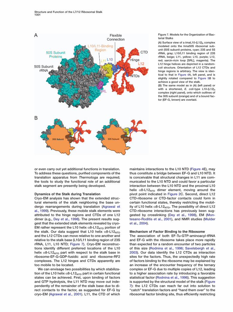
Structure and Function of the L7/12 Ribosomal Stalk1001
Figure 7. Models for the Organization of Bac-terial Stalks
(A) Surface view of a tmaL10-(L12)6 complexmodeled onto the hma50S ribosomal sub-unit (50S subunit proteins, cyan; 23S and 5SrRNA, gray; L10/L11 binding region of 23SrRNA, beige; L11, yellow; L10, purple; L12,red; sarcin-ricin loop [SRL], magenta). TheL12 hinge helices are depicted in a random-coil structure. Orientation of L12 CTDs andhinge regions is arbitrary. The view is iden-tical to that in Figure 4A, left panel, and isslightly rotated compared to Figure 5B toachieve a good view of the stalk.(B) The same model as in (A) (left panel) orwith a shortened, E. coli-type L10-(L12)4complex (right panel), onto which outlines ofthe 30S subunit (orange) and of a bound fac-tor (EF-G, brown) are overlaid.
or even carry out yet additional functions in translation.To address these questions, purified components of thetranslation apparatus from Thermotoga are required;the tools to study the functional role of an additionalstalk segment are presently being developed.
Dynamics of the Stalk during TranslationCryo-EM analysis has shown that the extended struc-tural elements of the stalk neighboring the base un-dergo rearrangements during translation (Agrawal etal., 1999). Previously, these mobile stalk elements wereattributed to the hinge regions and CTDs of one L12dimer (e.g., Dey et al., 1998). The present results sug-gest that the extended stalk elements revealed by cryo-EM rather represent the L10 helix α8-L12NTD portion ofthe stalk. Our data suggest that L10 helix α8-L12NTD
and the L12 CTDs can move relative to one another andrelative to the stalk base (L10/L11 binding region of 23SrRNA, L11, L10 NTD; Figure 7). Cryo-EM reconstruc-tions identify different preferred locations of the L10helix α8-L12NTD part with respect to the stalk base inribosome-EF-G-GDP-fusidic acid and ribosome-RF2complexes. The L12 hinges and CTDs apparently aretoo mobile to be located.
We can envisage two possibilities by which stabiliza-tion of the L10 helix α8-L12NTD part in certain functionalstates can be achieved. First, upon binding of factorsand GTP hydrolysis, the L11 NTD may move out inde-pendently of the remainder of the stalk base due to di-rect contacts to the factor, as suggested for EF-G bycryo-EM (Agrawal et al., 2001). L11, the CTD of which
maintains interactions to the L10 NTD (Figure 4E), maythus constitute a bridge between EF-G and L10 NTD. Itis conceivable that structural changes in L11 are com-municated to the L10 NTD and could favor a particularinteraction between the L10 NTD and the proximal L10helix α8-L12NTD dimer element, moving around thepivot point indicated in Figure 2C. Second, direct L12CTD-ribosome or CTD-factor contacts could form incertain functional states, thereby restricting the mobil-ity of L10 helix α8-L12NTD. The possibility of direct L12CTD-ribosome interactions has previously been sug-gested by crosslinking (Dey et al., 1998), EM (Mon-tesano-Roditis et al., 2001), and NMR studies (Mulderet al., 2004).
Mechanism of Factor Binding to the RibosomeThe association of both EF-Tu-GTP-aminoacyl-tRNAand EF-G with the ribosome takes place more rapidlythan expected for a random encounter of two particlesof this size (Rodnina et al., 1996; Savelsbergh et al.,2003). Our data identify the L12 CTDs as interactionsites for the factors. Thus, the unexpectedly high rateof factors binding to the ribosome may be explained byan increase of the encounter frequency of the ternarycomplex or EF-G due to multiple copies of L12, leadingto a higher association rate by introducing a favorablestatistical factor (Rodnina et al., 1996). This suggestionis supported by the structural model of the stalk (Figure7): the L12 CTDs can reach far out into solution to“catch” translation factors and “hand them over” to theribosomal factor binding site, thus efficiently restricting
Cell1002
tfactor diffusion and leading to rapid recruitment. Thenlong, unstructured L12 hinge regions and the flexiblehconnection of the L10 helix α8-L12NTD portion to thes
stalk base could allow the interaction of the translation mfactors with their ribosome binding site while being
wbound to the L12 CTDs. The presence of six copies ofaL12 on Thermotoga and some other ribosomes, com-cpared to four on E. coli-like ribosomes, may reflect theeoptimization of factor binding to the particular needs ofa
these organisms. d
Mechanism of GTPase Stimulation CThe L12 CTDs are responsible for an about 1000-fold N
pstimulation of GTP hydrolysis by EF-Tu and EF-G.cGTPase activation can be achieved by either promotingBconformational rearrangements within the G domainsr
of the factors that correctly position their own catalytic egroups in the active site or donating additional catalytic qgroups in trans. The unique, highly conserved arginine a
wresidue in the CTD of L12 is not essential for the activa-otion, excluding an “arginine finger”-type mechanismo(present data and Savelsbergh et al., 2000). Similarly,d
none of the other conserved, surface-exposed amino sacid residues in the CTD alone is responsible for the factivation. These findings suggest that L12 facilitates dGTP hydrolysis by stabilizing the GTPase transition
Cstate of the factors rather than by providing residuesTinvolved in catalysis. This mechanism of activation re-(sembles that of the regulators of G protein signalingd
(RGS) that stimulate GTP hydrolysis in Gα proteins (for (review, see Vetter and Wittinghofer, 1999). Given thehigh degree of sequence homology of L12 CTDs, EF- 1
oTu, and EF-G from different bacterial species, it is likelytthat the RGS-type mechanism is evolutionarily con-fserved, at least among bacteria.e
Cryo-EM reconstructions showed extensive interac- ttions of the G domains of both EF-Tu and EF-G with 2the SRL of 23S rRNA (Agrawal et al., 1998; Stark et al., u
o2002; Valle et al., 2003), indicating that the SRL mayLstabilize the transition-state conformation of the fac-(tors. Single-molecule fluorescence measurements indi-b
cated that cleavage of the SRL blocks EF-Tu in a state ubefore GTP hydrolysis (Blanchard et al., 2004). Other ccontacts that may contribute to GTP hydrolysis include f
Dribosomal protein L11 and the L11 binding region of23S rRNA (Agrawal et al., 2001). L12 represents a third
Bribosomal element important for stimulation of GTP hy-Rdrolysis. Through its CTD it may both facilitate position-ning of the factors relative to other ribosomal compo-w
nents, thereby contributing to catalysis, and stabilize cthe active conformation of the factors. We therefore en- f
rvisage that the L12 CTDs use their high freedom of mo-ation to reach back toward the ribosome bound factorsito stimulate their GTPase activity. The requirement forfadditional signals for full stimulation of the GTPase ac-t
tivity, such as the interaction with the SRL or L11, may dhelp to avoid premature GTP hydrolysis during initial
Nfactor binding.2(Experimental ProceduresmEProtein Production
tmaL10 and tmaL12NTD were coexpressed from two vectors in a3E. coli and purified by affinity chromatography on Ni2+-nitrilotriace-
ate (NTA) (Qiagen) through a His6 tag attached to the L10 N termi-us. After cleavage of the tag, the complex was further purified byeat treatment (20 min, 80°C) and additional chromatographicteps. Details of these and other protocols are given in the Supple-ental Data.Full-length ecoL12, ecoL12 point mutants, and ecoL12NTD/hinge
ere expressed from plasmid pGEX-5×-3-L12 in E. coli BL21 DE3s glutathione S-transferase fusion proteins and purified by affinityhromatography on glutathione-Sepharose 4B as described (Kothet al., 2004; Mohr et al., 2002; Savelsbergh et al., 2000). The cleav-ge of the fusion protein by factor Xa (Novagen) was carried outirectly on the affinity matrix.
rystallographic Analysesative and selenomethionine-derivatized tmaL10-L12NTD com-lexes yielded two orthorhombic crystal forms and one monoclinicrystal form (Table S1). Diffraction data were collected at beamlineW6 of DESY (Hamburg, Germany). The structure of one ortho-
hombic crystal form could be solved by a four-wavelength MADxperiment. The structures of the other crystal forms were subse-uently solved by molecular replacement. Partial models were builtutomatically and completed manually. Refinement of all structuresas carried out by established strategies (Table S1). The structuref the hmaL10E NTD could be built into the electron density mapf the hma50S subunit (PDB ID code 1S72) after application of aensity-modification procedure, which resembled established den-ity-modification protocols but employed a median filter knownrom 2D image processing to define the molecular boundaries (foretails, see Supplemental Data).
ryo-EM Reconstructions and Fitting of X-Ray Structureshe 3D structure of an E. coli 70S-EF-G-GDP-fusidic-acid complex
Stark et al., 2000) was refined at 18 Å resolution using recentlyeveloped software for improved alignment and CTF correction
see Supplemental Experimental Procedures).E. coli 30S and 50S atomic models (PDB ID codes 1PNX and
PNY, respectively) were docked into the EM density. Excellentverall fits were obtained by adjustment of the L1 stalk (w20° rota-ion toward the 30S subunit) and the L11 NTD (w30° rotation awayrom the rRNA). The crystal structure of the hma50S subunit (PDBntry 1S72) including the NTD of hmaL10E was then aligned withhe eco50S subunit with respect to the L10/L11 binding region of3S rRNA. The NTD of hmaL10E in this structure was subsequentlysed to sequentially align L10-(L12NTD)4 structures (omitting theutermost L12NTD dimer and its ten-residue binding segment on10) derived from crystal structures I, II, and III of tmaL10-(L12NTD)6
Table S1). The fit for the helix α8-(L12NTD)4 portion was optimizedy rigid-body movement relative to the L10 NTD (Figure 5 and Fig-re S6) around the pivot point identified in the tmaL10-(L12NTD)6rystal structures (Figure 2C). Similar fitting was also performedor the set of cryo-EM ribosome structures available from the EBIatabase (Table S3; Figure S7).
iochemical Proceduresibosomes from E. coli MRE 600 were prepared as described (Rod-ina and Wintermeyer, 1995). Ribosomes from T. maritima MSB8ere prepared by the same procedure, except for opening of theells with a French press (T. maritima MSB8 cells were a kind giftrom K.O. Stetter, Regensburg). ecoL12 was removed from eco70Sibosomes by NH4Cl/ethanol treatment (Kothe et al., 2004; Mohr etl., 2002). For reconstitution, ribosome cores depleted of L12 were
ncubated with a 20-fold excess of purified wild-type or mutant L12or 30 min at 37°C. AcPhe-tRNAPhe, [14C]Phe-tRNAPhe, Phe-RNAPhe(Prf16/17), EF-Tu, and EF-G were prepared and purified asescribed (Kothe et al., 2004; Mohr et al., 2002).A-site binding was studied in 50 mM Tris-HCl (pH 7.5), 70 mM
H4Cl, 30 mM KCl, 10 mM MgCl2, and 2 mM DTT (Kothe et al.,004). Rapid kinetic experiments were carried out as described
Kothe et al., 2004 and Supplemental Experimental Procedures). Toeasure ribosome-stimulated multiple-turnover GTP hydrolysis byF-G, ribosomes (0.05–2.0 �M) were mixed with EF-G (0.04 �M)nd [γ-32P]GTP (20 �M) in 50 mM Tris-HCl (pH 7.5), 70 mM NH4Cl,0 mM KCl, 7 mM MgCl , and 2 mM DTT at 37°C. Steady-state
2
Structure and Function of the L7/12 Ribosomal Stalk1003
kinetic parameters were determined under conditions of initialvelocity. Exchange of L12 on the ribosomes, the copy number ofL12 on eco and tma ribosomes, the ratio of ribosome bound andcytoplasmic L12, and the ratio between L12 and translation factorswere determined as described in Supplemental Experimental Pro-cedures.
Model BuildingFor visualization of a ribosome model (Figure 7), we used the struc-ture of the hma50S subunit. Full-length tmaL11 (PDB ID code1MMS) was modeled by superimposing the CTDs of tmaL11 andhmaL11. A tmaL10-(L12NTD)4 complex or a tmaL10-(L12NTD)6 com-plex was positioned as described above for cryo-EM. We then su-perimposed the NMR structure of an isolated ecoL12 dimer (PDBID code 1RQU) on the L12NTD dimers of the L10-L12NTD complexes.Torsion angles in the unstructured hinges were adjusted to showall L12 molecules in the crown view of the 50S subunit. To indicatethe location of the 30S subunit and a translation factor, we super-imposed the 30S subunit from E. coli as seen in an E. coli 70Sribosome structure (PDB ID codes 1PNX and 1PNY) and EF-G ac-cording to a cryo-EM structure (PDB ID code 1JQM).
Supplemental DataSupplemental Data include Supplemental Results and Discussion,Supplemental Experimental Procedures, Supplemental References,three tables, and seven figures and are available with this articleonline at http://www.cell.com/cgi/content/full/121/7/991/DC1/.
Acknowledgments
This work was supported by the Max-Planck-Society, the DeutscheForschungsgemeinschaft, the European Union, the Alfried Kruppvon Bohlen und Halbach-Stiftung, and the Fonds der ChemischenIndustrie. U.K. was supported by a fellowship of the Studienstiftungdes deutschen Volkes. N.F. was supported by a fellowship of theBoehringer Ingelheim Fonds. We thank G. Bourenkov for help indiffraction data acquisition, G. Stier for pETM cloning vectors, andU. Reidt for help in cloning.
Received: November 30, 2004Revised: March 4, 2005Accepted: April 14, 2005Published: June 30, 2005
References
Agrawal, R.K., Penczek, P., Grassucci, R.A., and Frank, J. (1998).Visualization of elongation factor G on the Escherichia coli 70S ri-bosome: the mechanism of translocation. Proc. Natl. Acad. Sci.USA 95, 6134–6138.
Agrawal, R.K., Heagle, A.B., Penczek, P., Grassucci, R.A., andFrank, J. (1999). EF-G-dependent GTP hydrolysis induces translo-cation accompanied by large conformational changes in the 70Sribosome. Nat. Struct. Biol. 6, 643–647.
Agrawal, R.K., Linde, J., Sengupta, J., Nierhaus, K.H., and Frank, J.(2001). Localization of L11 protein on the ribosome and elucidationof its involvement in EF-G-dependent translocation. J. Mol. Biol.311, 777–787.
Ban, N., Nissen, P., Hansen, J., Moore, P.B., and Steitz, T.A. (2000).The complete atomic structure of the large ribosomal subunit at2.4 Å resolution. Science 289, 905–920.
Beauclerk, A.A., Cundliffe, E., and Dijk, J. (1984). The binding sitefor ribosomal protein complex L8 within 23S ribosomal RNA ofEscherichia coli. J. Biol. Chem. 259, 6559–6563.
Blanchard, S.C., Gonzalez, R.L., Kim, H.D., Chu, S., and Puglisi,J.D. (2004). tRNA selection and kinetic proofreading in translation.Nat. Struct. Mol. Biol. 11, 1008–1014.
Bocharov, E.V., Sobol, A.G., Pavlov, K.V., Korzhnev, D.M., Jaravine,V.A., Gudkov, A.T., and Arseniev, A.S. (2004). From structure and
dynamics of protein L7/L12 to molecular switching in ribosome. J.Biol. Chem. 279, 17697–17706.
Bourne, H.R., Sanders, D.A., and McCormick, F. (1991). TheGTPase superfamily: conserved structure and molecular mecha-nism. Nature 349, 117–127.
Dey, D., Bochkariov, D.E., Jokhadze, G.G., and Traut, R.R. (1998).Cross-linking of selected residues in the N- and C-terminal do-mains of Escherichia coli protein L7/L12 to other ribosomal pro-teins and the effect of elongation factor Tu. J. Biol. Chem. 273,1670–1676.
Gonzalo, P., and Reboud, J.P. (2003). The puzzling lateral flexiblestalk of the ribosome. Biol. Cell. 95, 179–193.
Harms, J., Schluenzen, F., Zarivach, R., Bashan, A., Gat, S., Agmon,I., Bartels, H., Franceschi, F., and Yonath, A. (2001). High resolutionstructure of the large ribosomal subunit from a mesophilic eubacte-rium. Cell 107, 679–688.
Klaholz, B.P., Myasnikov, A.G., and Van Heel, M. (2004). Visualiza-tion of release factor 3 on the ribosome during termination of pro-tein synthesis. Nature 427, 862–865.
Kothe, U., Wieden, H.J., Mohr, D., and Rodnina, M.V. (2004). In-teraction of helix D of elongation factor Tu with helices 4 and 5 ofprotein L7/12 on the ribosome. J. Mol. Biol. 336, 1011–1021.
Liljas, A., and Gudkov, A.T. (1987). The structure and dynamics ofribosomal protein L12. Biochimie 69, 1043–1047.
Mohr, D., Wintermeyer, W., and Rodnina, M.V. (2000). Arginines 29and 59 of elongation factor G are important for GTP hydrolysis ortranslocation on the ribosome. EMBO J. 19, 3458–3464.
Mohr, D., Wintermeyer, W., and Rodnina, M.V. (2002). GTPase acti-vation of elongation factors Tu and G on the ribosome. Biochemis-try 41, 12520–12528.
Montesano-Roditis, L., Glitz, D.G., Traut, R.R., and Stewart, P.L.(2001). Cryo-electron microscopic localization of protein L7/L12within the Escherichia coli 70 S ribosome by difference mappingand Nanogold labeling. J. Biol. Chem. 276, 14117–14123.
Mulder, F.A.A., Bouakaz, L., Lundell, A., Venkataramana, M., Liljas,A., Akke, M., and Sanyal, S. (2004). Conformation and dynamicsof ribosomal stalk protein L12 in solution and on the ribosome.Biochemistry 43, 5930–5936.
Oleinikov, A.V., Perroud, B., Wang, B., and Traut, R.R. (1993). Struc-tural and functional domains of Escherichia coli ribosomal proteinL7/L12. The hinge region is required for activity. J. Biol. Chem. 268,917–922.
Pape, T., Wintermeyer, W., and Rodnina, M.V. (1998). Complete ki-netic mechanism of elongation factor Tu-dependent binding ofaminoacyl-tRNA to the A site of the E. coli ribosome. EMBO J. 17,7490–7497.
Rawat, U.B., Zavialov, A.V., Sengupta, J., Valle, M., Grassucci, R.A.,Linde, J., Vestergaard, B., Ehrenberg, M., and Frank, J. (2003). Acryo-electron microscopic study of ribosome-bound terminationfactor RF2. Nature 421, 87–90.
Rodnina, M.V., and Wintermeyer, W. (1995). GTP consumption ofelongation factor Tu during translation of heteropolymeric mRNAs.Proc. Natl. Acad. Sci. USA 92, 1945–1949.
Rodnina, M.V., Pape, T., Fricke, R., Kuhn, L., and Wintermeyer, W.(1996). Initial binding of the elongation factor Tu·GTP·aminoacyl-tRNA complex preceding codon recognition on the ribosome. J.Biol. Chem. 271, 646–652.
Rosendahl, G., and Douthwaite, S. (1993). Ribosomal proteins L11and L10.(L12)4 and the antibiotic thiostrepton interact with overlap-ping regions of the 23 S rRNA backbone in the ribosomal GTPasecentre. J. Mol. Biol. 234, 1013–1020.
Savelsbergh, A., Mohr, D., Wilden, B., Wintermeyer, W., and Rod-nina, M.V. (2000). Stimulation of the GTPase activity of translationelongation factor G by ribosomal protein L7/12. J. Biol. Chem. 275,890–894.
Savelsbergh, A., Katunin, V.I., Mohr, D., Peske, F., Rodnina, M.V.,and Wintermeyer, W. (2003). An elongation factor G-induced ribo-some rearrangement precedes tRNA-mRNA translocation. Mol.Cell 11, 1517–1523.
Cell1004
Stark, H., Rodnina, M.V., Wieden, H.J., van Heel, M., and Winter-meyer, W. (2000). Large-scale movement of elongation factor G andextensive conformational change of the ribosome during transloca-tion. Cell 100, 301–309.
Stark, H., Rodnina, M.V., Wieden, H.J., Zemlin, F., Wintermeyer, W.,and van Heel, M. (2002). Ribosome interactions of aminoacyl-tRNAand elongation factor Tu in the codon-recognition complex. Nat.Struct. Biol. 9, 849–854.
Subramanian, A.R. (1975). Copies of proteins L7 and L12 andheterogeneity of the large subunit of Escherichia coli ribosome. J.Mol. Biol. 95, 1–8.
Valle, M., Zavialov, A., Li, W., Stagg, S.M., Sengupta, J., Nielsen,R.C., Nissen, P., Harvey, S.C., Ehrenberg, M., and Frank, J. (2003).Incorporation of aminoacyl-tRNA into the ribosome as seen bycryo-electron microscopy. Nat. Struct. Biol. 10, 899–906.
Vetter, I.R., and Wittinghofer, A. (1999). Nucleoside triphosphate-binding proteins: different scaffolds to achieve phosphoryl transfer.Q. Rev. Biophys. 32, 1–56.
Vila-Sanjurjo, A., Ridgeway, W.K., Seymaner, V., Zhang, W., San-toso, S., Yu, K., and Cate, J.H. (2003). X-ray crystal structures ofthe WT and a hyper-accurate ribosome from Escherichia coli. Proc.Natl. Acad. Sci. USA 100, 8682–8687.
Wahl, M.C., and Moller, W. (2002). Structure and function of theacidic ribosomal stalk proteins. Curr. Protein Pept. Sci. 3, 93–106.
Wahl, M.C., Bourenkov, G.P., Bartunik, H.D., and Huber, R. (2000).Flexibility, conformational diversity and two dimerization modes incomplexes of ribosomal protein L12. EMBO J. 19, 174–186.
Wieden, H.-J., Wintermeyer, W., and Rodnina, M.V. (2001). A com-mon structural motif in elongation factor Ts and ribosomal proteinL7/12 may be involved in the interaction with elongation factor Tu.J. Mol. Evol. 52, 129–136.
Wimberly, B.T., Guymon, R., McCutcheon, J.P., White, S.W., andRamakrishnan, V. (1999). A detailed view of a ribosomal active site:The structure of the L11-RNA complex. Cell 97, 491–502.
Yusupov, M.M., Yusupova, G.Z., Baucom, A., Lieberman, K., Ear-nest, T.N., Cate, J.H.D., and Noller, H.F. (2001). Crystal structure ofthe ribosome at 5.5 angstrom resolution. Science 292, 883–896.
Accession Numbers
Coordinates and structure factors have been deposited in the Pro-tein Data Bank with ID codes 1ZAV, 1ZAW, and 1ZAX (tmaL10-(L12NTD)6) and 1ZB4 (hmaL10E NTD). The cryo-EM 3D map hasbeen deposited in the Electron Microscopy Database at the EMBLEuropean Bioinformatics Institute (http://www.ebi.ac.uk/msd-srv/emsearch/index.html; accession code EMD-1110).