Embed Size (px)
Citation preview
THESE
présentée pour obtenir le grade de
Docteur de l’Université de Strasbourg
Discipline: Science du Vivant
Spécialité: Aspects Moléculaire et Cellulaire de la Biologie
Par
Akiko TAKEUCHI
RNA-protein interaction in the selenoprotein synthesis machinery
Soutenue publiquement le 1 juillet 2009
Membres du jury
Rapporteur externe: Mme Christiane BRANLANT, Directeur de Recherche du CNRS, Nancy
Rapporteur externe: M Jean-Pierre ROUSSET, Professeur à l’Université Paris-Sud, Orsay
Rapporteur interne: M Mario KELLER, Professeur à l’Université de Strasbourg
Examinateur: M Rémy BORDONNE, Directeur de Recherche du CNRS, Montpellier
Directeur de thèse: M Alain KROL, Directeur de Recherche du CNRS, Strasbourg
Directeur de thèse: Mme Christine ALLMANG-CURA, Chargée de Recherche du CNRS, Strasbourg


Acknowledgements
I wish to express my appreciation to Dr. Christiane Branlant (ARN, RNP, Structure-Fonction-
Maturation Enzymologie Moléculaire et Structurale, Nancy), Pr. Jean-Pierre Rousset (Institut
de Génétique et Microbiologie, Orsay) and Pr. Mario Keller (Institit de Biologie Moléculaire
des Plantes, Strasbourg) for having accepted to evaluate my PhD studies. I am also grateful to
Dr. Rémy Bordonné (Institut de Génétique Moléculaire de Montpellier, Montpellier) for
having examined my studies.
I would like to express profound gratitude to my advisor, Dr. Alain Krol for having accepted
me as a laboratory member and for his supervision.
I am also highly thankful to Christine Allmang-Cura for her supervision and many advices on
science and the daily life in France.
I would also like to thank everyone in the laboratory for their kindness. I will never forget my
three years in Strasbourg.


Table of contents
TABLE OF CONTENTS .............................................................................................................................................1
TABLE OF FIGURES..................................................................................................................................................3
LIST OF ABBREVIATIONS......................................................................................................................................5
RESUME DE LA THESE EN FRANÇAIS ..............................................................................................................7
PART 1. INTRODUCTION ......................................................................................................................................13
1. SELENIUM AND ITS BIOLOGICAL FUNCTION .........................................................................................................15 1.1. Selenium .......................................................................................................................................................15 1.2. Selenocysteine ..............................................................................................................................................15 1.3. Selenoproteins ..............................................................................................................................................16
2. SELENOPROTEIN SYNTHESIS.................................................................................................................................20 2.1. Selenocysteine biosynthesis.........................................................................................................................20
2.1.1. tRNASec
................................................................................................................................................................... 20 2.1.2. From serine to phosphoserine (O-phosphoseryl-tRNA
Sec kinase / PSTK)......................................................... 22
2.1.3. From phosphoserine to selenocysteine.................................................................................................................. 24 2.1.3.1. Generation of the selenium donor (SPS1/2) ................................................................................................ 24 2.1.3.2. From Sep-tRNA
Sec to Sec-tRNA
Sec (SecS) .................................................................................................. 24
2.1.4. SECp43 ................................................................................................................................................................... 25 2.2. Sec incorporation.........................................................................................................................................25
2.2.1. Cis-acting factors.................................................................................................................................................... 26 2.2.1.1. SElenoCysteine Incorporation Sequence (SECIS) ...................................................................................... 26
2.2.1.1.a. Location in mRNA ................................................................................................................................ 26 2.2.1.1.b Secondary structure................................................................................................................................ 27
2.2.1.2. SRE................................................................................................................................................................. 31 2.2.2. Trans-acting factors................................................................................................................................................ 32
2.2.2.1. EFSec.............................................................................................................................................................. 32 2.2.2.2. SBP2 ............................................................................................................................................................... 33
2.2.2.2.a Domain structure of SBP2 ..................................................................................................................... 34 2.2.2.2.b. SECIS binding....................................................................................................................................... 35 2.2.2.2.c. EFSec-SBP2 interaction........................................................................................................................ 38 2.2.2.2.d. Ribosomal binding ................................................................................................................................ 38 2.2.2.2.e. Expression and localization .................................................................................................................. 39
2.2.2.3. L30.................................................................................................................................................................. 41 2.2.2.4. Other proteins................................................................................................................................................. 41
2.3. Sec incorporation model..............................................................................................................................42 3. SELENOPROTEIN MRNP ASSEMBLY .....................................................................................................................43
3.1. Nuclear assembly .........................................................................................................................................43 3.2. Assembly of selenoprotein mRNAs - similarities with sn/snoRNP assembly ...........................................44
4. OBJECTIVES AND OUTLINE OF THIS THESIS ..........................................................................................................47
PART 2. RESULTS.....................................................................................................................................................49
1. FUNCTIONAL CHARACTERIZATION OF DROSOPHILA MELANOGASTER SBP2 .......................................................51 1.1. Selenoproteome in Drosophila....................................................................................................................51 1.2. Objective.......................................................................................................................................................53 1.3. Summary of Article 1 ...................................................................................................................................56 1.4. Article 1 ........................................................................................................................................................58 1.5. Additional results and discussion ...............................................................................................................75
SBP2 and the selenoprotein synthesis machinery in Drosophila willistoni.................................................................. 75 2. TOWARD CRYSTALLIZATION OF THE SBP2/SECIS COMPLEX ............................................................................81
2.1. Objective.......................................................................................................................................................81 2.2. Results...........................................................................................................................................................82
2.2.1. cDNA cloning of SBP2 from various organisms ................................................................................................. 82 2.2.2. Expression of various SBP2 cDNAs using the Baculovirus expression system................................................ 83 2.2.3. Biophysical analysis of SBP2................................................................................................................................ 87
2.3. Article 2 (in press) .......................................................................................................................................89 2.4. SBP2 is an Intrinsically Disordered Protein..............................................................................................99
3. TOWARD IDENTIFICATION OF SBP2 PARTNERS................................................................................................ 101
-1-
3.1. Objective.................................................................................................................................................... 101 3.2. Results........................................................................................................................................................ 103 3.3. Discussion ................................................................................................................................................. 105
PART 3. GENERAL CONCLUSION................................................................................................................... 109
PART 4. ANNEX/ METHODS .............................................................................................................................. 117
1. CDNA CLONING USING THE GATEWAY TECHNOLOGY ................................................................................ 117 2. BACULOVIRUS EXPRESSION SYSTEM................................................................................................................. 119
2.1. Bacmid preparation .................................................................................................................................. 121 2.2. Mini expression test .................................................................................................................................. 121 2.3. Titration of viral particles and insect cell culture .................................................................................. 121
REFERENCES ......................................................................................................................................................... 125
-2-
Table of figures
Table 1. Selenoproteins identified in eukaryotes and their functions. ....................................19
Table 2. Selenoproteins identified in representative eukaryotic organisms. ...........................53
Table 3. List of primers used for PCR amplification. ..........................................................119
Figure 1. Chemical structures of cysteine and selenocysteine................................................16
Figure 2. Secondary structure models of canonical tRNAs and tRNAsSec
. ............................22
Figure 3. The selenocysteine biosynthesis pathway...............................................................23
Figure 4. Secondary structure models of form 1 and 2 SECIS...............................................28
Figure 5. The secondary structure of SECIS RNA and various K-turn RNAs........................29
Figure 6. The SECIS and SRE elements of SEPN1 mRNAs .................................................30
Figure 7. Schematic representations of the selenocysteine specialized translation elongation
factors compared to general elongation factors..............................................................32
Figure 8. Schematic representation of protein factors involved in selenoprotein synthesis.....35
Figure 9. RNA-protein interfaces at various L7Ae protein-K turn RNA complexes. .............36
Figure 10. SECIS RNA determinants for SBP2 binding........................................................37
Figure 11. Proposed model for the regulation of SBP2 subcellular localization and function
after oxidative stress. ....................................................................................................40
Figure 12. Selenocysteine incorporation models ...................................................................42
Figure 13. Nuclear assembly of the selenoprotein synthesis machinery.................................44
Figure 14. Selenoprotein mRNP assembly model. ................................................................45
Figure 15. Eukaryotic selenoproteomes ................................................................................52
Figure 16. Selenoproteins in D. melanogaster SBP2.............................................................54
Figure 17. Shematic representation of the human and D. melanogaster SBP2 proteins. ........55
Figure 18. One amino acid insertion in D. willistoni SBP2....................................................76
Figure 19. Aspargine insertion into the Glu 679-Glu 699 conserved spacing in human SBP2.
.....................................................................................................................................77
Figure 20. Disrupting the conserved Glu 679-Glu 699 spacing in hSBP2 by one amino acid
insertion had no significant effect on SECIS-binding. ...................................................78
Figure 21. Schematic drawings of SBP2 proteins designed and generated for crystallization
purposes. ......................................................................................................................83
Figure 22. Mini expression assays in baculovirus infected insect cells. .................................85
Figure 23. Expression tests from large-scale culture of baculovirus infected insect cells. ......86
Figure 24. Examples of X-ray structures of IDPs bound to their targets. .............................100
Figure 25. Composition and organization of C/D sno(s)RNPs and U4 snRNP ....................102
Figure 26. GST pull-down experiments. .............................................................................104
Figure 27. Proposed model for SBP2 functions during the selenoprotein mRNP formation.111
Figure 28. Principle of the GATEWAY cloning .................................................................118
Figure 29. PCR amplification strategy to generate the SBP2 cDNA fragment harboring the
AttB recombination sites.............................................................................................119
Figure 30. Generation of recombinant baculoviruses and gene expression. .........................120
-3-

-4-
List of abbreviations
A, C, G, T, U adenine, cytosine, guanine, thymine, uracil
ATP adenosine 5’-triphosphate
bp base pair
CD circular dichroism
C. elegans Caenorhabditis elegans
C-terminal carboxy-terminal
DIO iodothyronine deiodinase
DLS dynamic light scattering
D. melanogaster Drosophila melanogaster
DNA deoxyribonucleic acid
cDNA complementary deoxyribonucleic acid
E. coli Escherichia coli
EFSec selenocysteine-specific elongation factor
EST expressed sequence tag
GFP green fluorescent protein
GPx glutathione peroxidase
GST glutathione S-transferase
His hexahistidine tag
Hsp heat shock protein
IDP intrinsically disordered protein
kD kilo dalton
Msr methionine sulfoxide reductase
NES nuclear export signal
NLS nuclear localization signal
NMD nonsense-mediated decay
NMR nuclear magnetic resonance
nt nucleotide
N-terninal amino-terminal
ORF open reading frame
PCR polymerase chain reaction
PSTK phosphoseryl-tRNA kinase
RNA ribonucleic acid
-5-
mRNA messenger ribonucleic acid
sRNA small ribonucleic acid
snRNA small nuclear ribonucleic acid
snoRNA small nucleolar ribonucleic acid
tRNA transfer ribonucleic acid
RNP ribonucleoprotein
mRNP messenger ribonucleoprotein
snRNP small nuclear ribonucleoprotein
snoRNP small nuclear ribonucleoprotein
SAXS small-angle-X-ray scattering
SBP2 SECIS binding protein 2
Sec selenocysteine
SECIS selenocysteine insertion sequence
SecS selenocysteine synthase
Sel selenoprotein
SELEX systematic evolution of ligands by exponential enrichment
SEPN1 selenoprotein N gene
Ser serine
SerRS seryl-tRNA synthetase
SPS selenophosphate synthetase
SRE selenocysteine codon redefinition element
TR thioredoxin reductase
UTR untranslated region
! pseudouridine
-6-

Résumé de la thèse en français
-7-

-8-
Interactions ARN-protéines dans le mécanisme de biosynthèse des
sélénoprotéines
Le sélénium est un oligo-élément essentiel. Sa forme biologique majeure est l’acide
aminé sélénocystéine (Sec) que l’on retrouve essentiellement dans le site actif des
sélénoprotéines. La sélénocystéine est incorporée dans les sélénoprotéines de façon co-
traductionnelle en réponse à un codon UGA habituellement reconnu comme l’un des trois
codons de terminaison. Chez les eucaryotes, la biosynthèse et l’incorporation de
sélénocystéine requièrent la participation d’une machinerie moléculaire complexe qui
implique, entre autres, une structure en tige-boucle située dans la région 3’UTR de l’ARNm
des sélénoprotéines (élément SECIS), l’ARNtSec
spécifique, le facteur d’élongation spécialisé
EFSec ainsi que la protéine SBP2 (SECIS-binding protein). SBP2 joue un rôle majeur dans le
mécanisme de synthèse des sélénoprotéines.
Chez les mammifères, le domaine de liaison à l’ARN de SBP2 est situé dans la région C-
terminale de la protéine. Celui-ci comprend un module conservé, présent chez d’autres
protéines de liaison à l’ARN mais possédant d’autres fonctions, appelé motif L7Ae dans les
banques de données. La région N-terminale est dépourvue de toute similitude avec des
protéines connues et n’est pas nécessaire à la synthèse des sélénoprotéines in vitro. De façon
intéressante, une recherche bioinformatique dans les génomes de drosophile nous a permis
d’identifier des séquences potentielles portant toutes les signatures d’une vraie protéine SBP2
mais plus courte et ne possédant pas de domaine N-terminal homologue à celui des
mammifères. Avant que je n’entreprenne ce travail, SBP2 n’avait été caractérisée
fonctionnellement que chez le rat et l’homme. Au cours de cette thèse, j’ai cloné l’ADNc et
caractérisé fonctionnellement la protéine SBP2 de Drosophila melanogaster (dSBP2) à l’aide
de tests de liaison à l’ARN et d’expression de sélénoprotéines dans des lysats de réticulocytes
de lapin. Malgré sa taille plus courte, dSBP2 a montré la même capacité à promouvoir la
synthèse de sélénoprotéines que son homologue mammifère. Il n’en va pas de même en ce qui
concerne la liaison à l’ARN SECIS : en effet, alors que la protéine SBP2 humaine (hSBP2)
est capable de lier deux formes distinctes d’ARN SECIS (appelées type 1 et 2) avec des
-9-
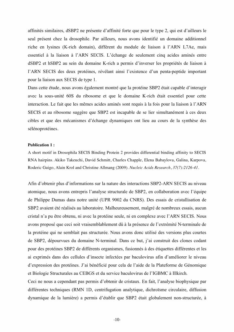
affinités similaires, dSBP2 ne présente d’affinité forte que pour le type 2, qui est d’ailleurs le
seul présent chez la drosophile. Par ailleurs, nous avons identifié un domaine additionnel
riche en lysines (K-rich domain), différent du module de liaison à l’ARN L7Ae, mais
essentiel à la liaison à l’ARN SECIS. L’échange de seulement cinq acides aminés entre
dSBP2 et hSBP2 au sein du domaine K-rich a permis d’inverser les propriétés de liaison à
l’ARN SECIS des deux protéines, révélant ainsi l’existence d’un penta-peptide important
pour la liaison aux SECIS de type 1.
Dans cette étude, nous avons également montré que la protéine SBP2 était capable d’interagir
avec la sous-unité 60S du ribosome et que le domaine K-rich était essentiel pour cette
interaction. Le fait que les mêmes acides aminés sont requis à la fois pour la liaison à l’ARN
SECIS et au ribosome suggère que SBP2 est incapable de se lier simultanément à ces deux
cibles et que des mécanismes d’échange dynamiques ont lieu au cours de la synthèse des
sélénoprotéines.
Publication 1 :
A short motif in Drosophila SECIS Binding Protein 2 provides differential binding affinity to SECIS
RNA hairpins. Akiko Takeuchi, David Schmitt, Charles Chapple, Elena Babaylova, Galina, Karpova,
Roderic Guigo, Alain Krol and Christine Allmang (2009). Nucleic Acids Research, 37(7):2126-41.
Afin d’obtenir plus d’informations sur la nature des interactions SBP2-ARN SECIS au niveau
atomique, nous avons entrepris l’analyse structurale de SBP2, en collaboration avec l’équipe
de Philippe Dumas dans notre unité (UPR 9002 du CNRS). Des essais de cristallisation de
SBP2 avaient été réalisés au laboratoire. Malheureusement, malgré de nombreux essais, aucun
cristal n’a pu être obtenu, ni avec la protéine seule, ni en complexe avec l’ARN SECIS. Nous
avons proposé que ceci soit vraisemblablement dû à la présence de l’extrémité N-terminale de
la protéine qui ne semblait pas structurée. Nous avons donc utilisé des versions plus courtes
de SBP2, dépourvues du domaine N-terminal. Dans ce but, j’ai construit des clones codant
pour des protéines SBP2 de différents organismes, fusionnés à des étiquettes différentes et les
ai exprimés dans des cellules d’insecte infectées par baculovirus afin d’améliorer le niveau
d’expression des protéines. J’ai bénéficié pour cela de l’aide de la Plateforme de Génomique
et Biologie Structurales au CEBGS et du service baculovirus de l’IGBMC à Illkirch.
Ceci ne nous a cependant pas permis d’obtenir de cristaux. En fait, l’analyse biophysique par
différentes techniques (RMN 1D, centrifugation analytique, dichroïsme circulaire, diffusion
dynamique de la lumière) a permis d’établir que SBP2 était globalement non-structurée, à
-10-
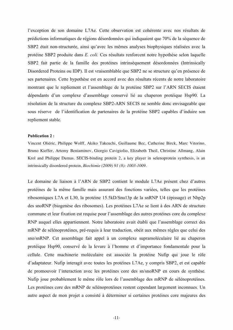
l’exception de son domaine L7Ae. Cette observation est cohérente avec nos résultats de
prédictions informatiques de régions désordonnées qui indiquaient que 70% de la séquence de
SBP2 était non-structurée, ainsi qu’avec les mêmes analyses biophysiques réalisées avec la
protéine SBP2 produite dans E. coli. Ces résultats renforcent notre hypothèse selon laquelle
SBP2 fait partie de la famille des protéines intrinsèquement désordonnées (Intrinsically
Disordered Proteins ou IDP). Il est vraisemblable que SBP2 ne se structure qu’en présence de
ses partenaires. Cette hypothèse est en accord avec des résultats récents de notre laboratoire
montrant que le repliement et l’assemblage de la protéine SBP2 sur l’ARN SECIS étaient
dépendants d’un complexe d’assemblage conservé lié au chaperon protéique Hsp90. La
résolution de la structure du complexe SBP2-ARN SECIS ne semble donc envisageable que
sous réserve de l’identification de partenaires de la protéine SBP2 capables d’induire son
repliement stable.
Publication 2 :
Vincent Oliéric, Philippe Wolff, Akiko Takeuchi, Guillaume Bec, Catherine Birck, Marc Vitorino,
Bruno Kieffer, Artemy Beniaminov, Giorgio Cavigiolio, Elizabeth Theil, Christine Allmang, Alain
Krol and Philippe Dumas. SECIS-binding protein 2, a key player in selenoprotein synthesis, is an
intrinsically disordered protein, Biochimie (2009) 91 (8): 1003-1009.
Le domaine de liaison à l’ARN de SBP2 contient le module L7Ae présent chez d’autres
protéines de la même famille mais assurant des fonctions variées, telles que les protéines
ribosomiques L7A et L30, la protéine 15.5kD/Snu13p de la snRNP U4 (épissage) et Nhp2p
des snoRNP (biogenèse des ribosomes). Les protéines L7Ae se lient à des ARN de structure
commune et leur fixation est requise pour l’assemblage des autres protéines core du complexe
RNP auquel elles appartiennent. Notre laboratoire avait établi que l’assemblage correct des
mRNP de sélénoprotéines, pré-requis à leur traduction, obéit aux mêmes règles que celui des
sno/snRNP. Cet assemblage fait appel à un complexe supramoléculaire lié au chaperon
protéique Hsp90, conservé de la levure à l’homme et d’importance fondamentale pour la
cellule. Cette machinerie moléculaire est associée la protéine Nufip qui joue le rôle
d’adaptateur. Nufip interagit avec toutes les protéines L7Ae, y compris SBP2, et est capable
de promouvoir l’interaction avec les protéines core des sn/snoRNP en cours de synthèse.
Nufip joue probablement le même rôle lors de l’assemblage des mRNP de sélénoprotéines.
Les protéines core des mRNP de sélénoprotéines restent cependant largement inconnues. Un
autre aspect de mon projet a consisté à déterminer si certaines protéines core majeures des
-11-
complexes sn/snoRNP pouvaient être des partenaires potentiels de SBP2. Cette hypothèse se
confirme puisque mon travail a permis de montrer que SBP2 interagissait in vitro avec au
moins l’une des protéines core des sn(o)RNP à boîte C/D, la protéine Nop58, et que cette
interaction est directe. A notre grande surprise, ces résultats révèlent que l’assemblage de la
catégorie particulière des ARNm de sélénoprotéines présente de nouvelles similitudes avec
celui des sn- et snoRNP.
L’ensemble de ces résultats a permis de mieux comprendre comment se forme le complexe
SBP2-ARN SECIS lors de la synthèse des sélénoprotéines, un processus au cœur du
mécanisme de recodage du codon UGA.
-12-

Part 1. Introduction
-13-

-14-
Introduction
1. Selenium and its biological function
1.1. Selenium
The non-metal element selenium was discovered by the Swedish chemist Jacob Berzelius in
1817. It was named after Sêlenê, the Greek goddess of the moon, in reference to the
previously discovered and chemically related chalcogen element tellurium (tellus, earth in
Latin). Selenium was considered a poison for a long time, especially to livestock eating
selenium accumulator plants of the genus Astragalus during periods of drought in western
USA and China. Later, selenium was defined as an essential micronutrient that exerts
significant health benefits. In the 1970’s, its biological activity could be attributed to the
newly identified amino acid selenocysteine (Sec). In humans, selenium deficiency has been
implicated as a factor for the emergence of the Keshan disease, an endemic cardiomyopathy
in certain regions of eastern China, where dietary selenium is very low because the soil is
deprived of this element. Selenium has also been implicated in the prevention of viral
infections, cancer, infertility; it has been shown as an important factor for thyroid hormone
maturation, the immune system as well as muscle development and function. However,
molecular evidence is missing for most of these pathologies with the exception of infertility,
thyroid maturation and muscle development. (See 1.3. Selenoproteins)
Selenium may also have a protective effect against inflammatory diseases (reviewed in
Hatfield & Gladyshev, 2002; Hatfield et al, 2009; Lescure et al, 2009; Rederstorff et al, 2006).
Selenium is mostly found at the catalytic site of most of the selenium-containing proteins
which are called selenoproteins.
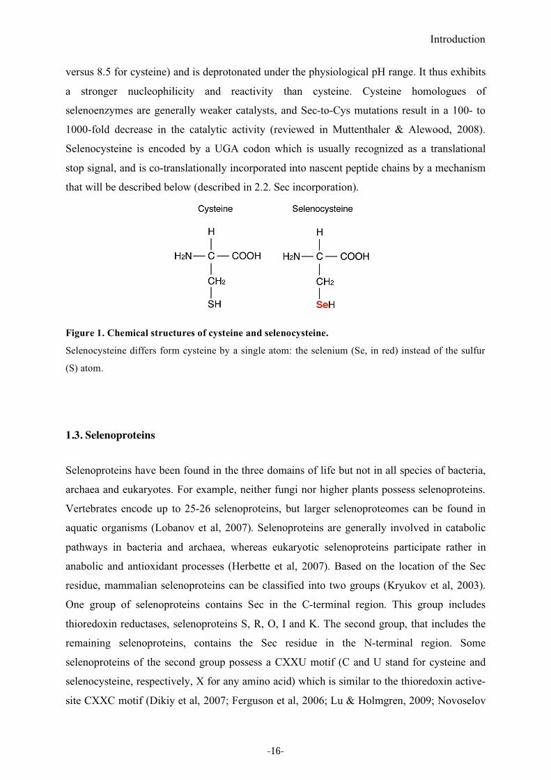
1.2. Selenocysteine
Selenocysteine is the major biological form of selenium in eukaryotes and is mostly found in
the active site of selenoproteins. Selenocysteine is called the 21st amino acid. Its chemical
structure differs from cysteine only by the presence of selenium in place of the sulfur atom
(see Figure 1). Even though selenium and sulfur belong to the same family, selenocysteine
exhibits distinct chemical properties versus cysteine. Selenocysteine has a lower pKa (5.2
-15-
Introduction
versus 8.5 for cysteine) and is deprotonated under the physiological pH range. It thus exhibits
a stronger nucleophilicity and reactivity than cysteine. Cysteine homologues of
selenoenzymes are generally weaker catalysts, and Sec-to-Cys mutations result in a 100- to
1000-fold decrease in the catalytic activity (reviewed in Muttenthaler & Alewood, 2008).
Selenocysteine is encoded by a UGA codon which is usually recognized as a translational
stop signal, and is co-translationally incorporated into nascent peptide chains by a mechanism
that will be described below (described in 2.2. Sec incorporation).
Figure 1. Chemical structures of cysteine and selenocysteine.
Selenocysteine differs form cysteine by a single atom: the selenium (Se, in red) instead of the sulfur
(S) atom.
1.3. Selenoproteins
Selenoproteins have been found in the three domains of life but not in all species of bacteria,
archaea and eukaryotes. For example, neither fungi nor higher plants possess selenoproteins.
Vertebrates encode up to 25-26 selenoproteins, but larger selenoproteomes can be found in
aquatic organisms (Lobanov et al, 2007). Selenoproteins are generally involved in catabolic
pathways in bacteria and archaea, whereas eukaryotic selenoproteins participate rather in
anabolic and antioxidant processes (Herbette et al, 2007). Based on the location of the Sec
residue, mammalian selenoproteins can be classified into two groups (Kryukov et al, 2003).
One group of selenoproteins contains Sec in the C-terminal region. This group includes
thioredoxin reductases, selenoproteins S, R, O, I and K. The second group, that includes the
remaining selenoproteins, contains the Sec residue in the N-terminal region. Some
selenoproteins of the second group possess a CXXU motif (C and U stand for cysteine and
selenocysteine, respectively, X for any amino acid) which is similar to the thioredoxin active-
site CXXC motif (Dikiy et al, 2007; Ferguson et al, 2006; Lu & Holmgren, 2009; Novoselov
-16-
Introduction
et al, 2007b). Such sequence signatures suggest that selenoproteins have redox-related
functions. Indeed, some of the selenoproteins are involved in oxidation-reduction reactions to
protect cells from oxidative damage; there is good reason to believe that the majority of the
still functionally uncharacterized selenoproteins participate in such mechanisms as well.
Selenoproteins with identified redox activity include five glutathione peroxidases (GPx), three
thioredoxin reductases (TR), three iodothyronine deiodinases (DIO) and selenophosphate
synthetase 2 (SPS2). Selenoproteins participate in thyroid hormone metabolism, muscle
formation, selenocysteine synthesis and in sperm maturation (Rederstorff et al, 2006).
Eukaryotic selenoproteins and their functions are summarized in Table 1.
Thioredoxin reductases regulate the thioredoxin system that participates in many cellular
signaling pathways by controlling the activity of transcription factors. Therefore, thioredoxin
reductases are involved in various cellular functions such as cell proliferation, antioxidant
defense and redox-regulated signaling cascades (reviewed in Arner, 2009; Lu & Holmgren,
2009).
Glutathione peroxidase (GPx, Enzyme Commission number 1.11.1.9; now GPx1) was the
first mammalian selenoprotein identified in 1973 (Flohe, 2009). There are seven isoenzymes
identified in humans, and five of them are selenoproteins (GPx1, 2, 3, 4 and 6). GPxs reduce
hydrogen peroxide and organic hydroperoxides, thus protecting cells from oxidative damage.
GPx1 is a cytosolic enzyme that is abundant in liver and erythrocytes. Its major function is the
detoxification of hydroxyperoxides to protect cells from oxidative stress that could result in
DNA damage. The GPx1 polymorphisms are also associated with cancer risk (reviewed in
Flohe, 2009; Gromer et al, 2005; Zhuo & Diamond, 2009).
Glutathione peroxidase 4 (GPx4; Enzyme Commission number 1.11.1.12) is also known as
phospholipid hydroperoxide GPx (PHGPx) because of its role in detoxification of lipid
peroxides. GPx4 transforms into a structural component of the midpiece of mature
spermatozoa by using hydroperoxides (Ursini et al, 1999). GPx4 is therefore involved in
sperm maturation and male fertility (reviewed in Flohe, 2009; Lu & Holmgren, 2009).
Iodothyronine deiodinases (DIOs) cleave specific iodine carbon bonds in the thyroid
hormones thyroxin (T4), bioactive 3,5,3’-tri-iodothyronine (T3) and 3’3’5’ reverse tri-
iodothyronine (rT4) which is less active than T3. Thereby DIOs regulate the hormonal
activity of the thyroid. DIO 1 and 2 convert T4 to T3, and DOI 3 converts T4 to rT3. DIO 1
-17-
Introduction
can also convert T4 to rT3 (Reviewed in Gromer et al, 2005; Lu & Holmgren, 2009; Pappas
et al, 2008).
SPS2 is the selenophosphate synthetase which is involved in selenocysteine biosynthesis.
This selenoprotein will be further described in 2.1.3.1.
Selenoprotein N (SelN) was the first selenoprotein shown to be involved in a genetic disorder
(Moghadaszadeh et al, 2001). SelN was discovered in the laboratory using a computational
screen based on the search of a conserved RNA structural motif that acts as a signature for
selenoprotein mRNAs, the selenocysteine insertion sequence (SECIS) (Lescure et al, 1999).
The pathology was known before SelN was identified. A large number of mutations in the
coding region of the SelN gene (SEPN1) are associated with a wide range of early-onset
muscular disorders now referred to as SEPN1-related myopathies. However, its catalytic
function still remains unknown. SelN was characterized as a glycosylated transmembrane
protein of the endoplasmic reticulum (ER). In addition to the transmembrane domain, SelN
contains a predicted domain consisting in a calcium binding EF-hand motif which may
contribute to the overall structure of the protein, and a SCUG catalytic site, reminiscent of a
thioredoxin reductase motif, suggesting a reductase activity (reviewed in Lescure et al, 2009).
-18-
Introduction
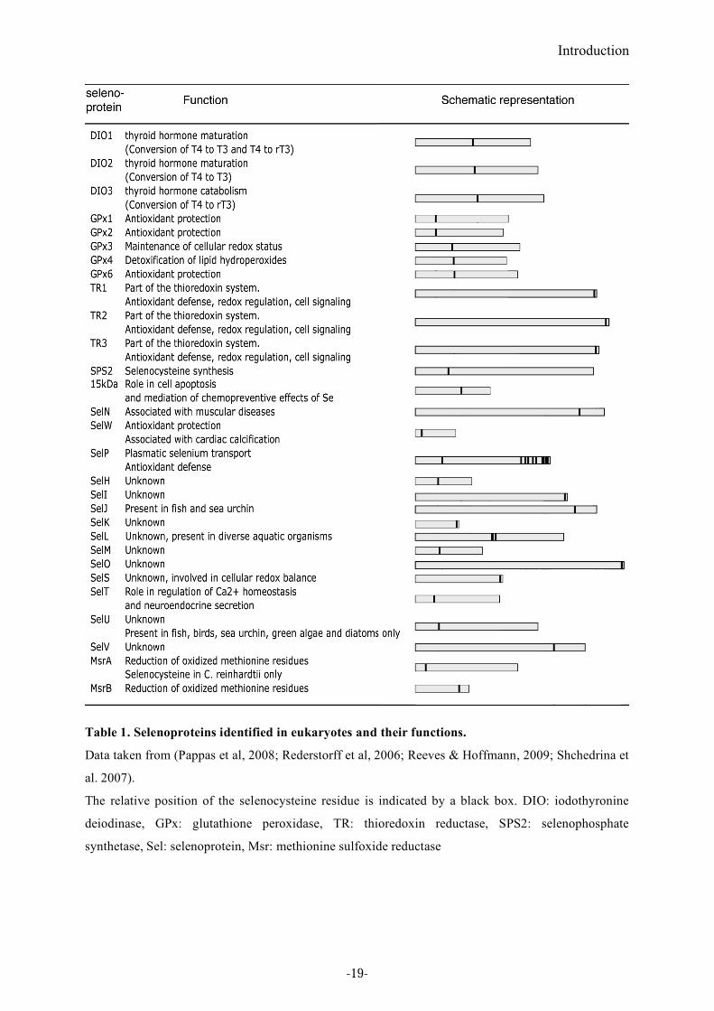
Table 1. Selenoproteins identified in eukaryotes and their functions.
Data taken from (Pappas et al, 2008; Rederstorff et al, 2006; Reeves & Hoffmann, 2009; Shchedrina et
al. 2007).
The relative position of the selenocysteine residue is indicated by a black box. DIO: iodothyronine
deiodinase, GPx: glutathione peroxidase, TR: thioredoxin reductase, SPS2: selenophosphate
synthetase, Sel: selenoprotein, Msr: methionine sulfoxide reductase
-19-
Introduction
2. Selenoprotein synthesis
Because selenocysteine is encoded by a UGA codon, one of the translational termination
signals in the universal genetic code, discriminating UGA Sec from the stop codon requires a
specialized translational machinery. This reprogramming mechanism is called UGA recoding.
The bacterial selenoprotein synthesis mechanism has been extensively studied and well
established by the Böck’s group (reviewed in Böck, 2006). Recent important progresses have
been made toward the elucidation of this mechanism in eukaryotes. This will be described
here in more details (reviewed in Allmang et al, 2009; Papp et al, 2007; Squires & Berry,
2008). Selenoprotein synthesis comprises two steps, selenocysteine biosynthesis and its co-
translational incorporation.
2.1. Selenocysteine biosynthesis
Selenocysteine does not occur as a free amino acid. Its biosynthesis occurs in two steps by
conversion of serine to selenocysteine directly on the selenocysteine tRNA.
2.1.1. tRNASec
tRNASec
is the selenocysteine specialized tRNA harboring anticodon complementary to UGA.
Although tRNASec
species in bacteria differ in sequence from eukaryal and archaeal homologs,
structure probing and computer modeling proposed similarities at the three-dimentional
structures (Baron 1993; Sturchler 1993). They also show functional conservation since both
eukaryotic and archaeal tRNASec
can function in bacterial systems (Baron et al, 1994; Lee et
al, 1989; Rother et al, 2000).
Eukaryotic tRNASec
was initially discovered as a serine acceptor suppressing the UGA opal
codon (Hatfield & Portugal, 1970). Later it was shown that this tRNA exists in the form of
selenocysteyl-tRNASec
(Lee et al, 1989; Mizutani, 1989). Heterozygous knockout mice retain
selenoprotein synthesis ability despite the reduced level of tRNASec
, implicating that it is not
limiting for selenoprotein synthesis. Homozygous knockout mice are embryonic lethal
demonstrating that selenoprotein synthesis is essential to mammalian development (Bosl et al,
1997).
tRNASec
has characteristic features in its secondary structure and a post-transcriptional
modification pattern that distinguish it from canonical tRNAs (reviewed in Allmang & Krol,
-20-
Introduction
2006b). tRNASec
is the longest tRNA, with 95 nucleotides in E.coli and 90 nucleotides in
eukaryotes (Amberg et al, 1993; Böck, 2006; Diamond et al, 1981; Diamond et al, 1993;
Hatfield et al, 1982).
Secondary structure models for the eukaryotic tRNASec
were proposed based on enzymatic
and chemical probing and structure-based sequence alignments (Hubert et al, 1998; Sturchler
et al, 1993). Compared to canonical tRNAs, tRNASec
has a longer D-stem and an extended
amino acid acceptor arm (consisting of the A and T-stems). The length of the D-stem is 6 bp,
whereas it only has 3-4 bp in other tRNAs. While the amino acid acceptor arm of canonical
tRNAs is 12 bp long, comprising a 7 bp A-stem and a 5 bp T-stem, that of tRNASec
is 13 bp.
Archaea and eukaryotes have a 9 bp A-stem and 4 bp T-stem, called ‘the 9/4 model’, and
bacteria have an 8 bp A-stem and a 5 bp T-stem, called ‘the 8/5 model’ (Figure 2). In bacteria,
the extra length of the acceptor arm is the determinant for binding to the specific elongation
factor SelB. It is required for the serine to selenocysteine conversion in eukaryotes (Baron &
Bock, 1991; Sturchler-Pierrat et al, 1995), which does not exclude the possibility that it also
participates in recognition of the homologous factor in eukaryotes. The long variable arm and
the discriminatory base G73 are the major identity elements for the serylation of tRNASec
and
tRNASer
(Wu & Gross, 1993, Figure 2).
Post-transcriptional modification of the Xenopus tRNASec
has been investigated (Diamond et
al, 1993; Sturchler et al, 1994). Compared to canonical tRNAs which contain 15-17 modified
bases, eukaryotic tRNASec
contains only 4 post-transcriptionally modified nucleotides:
pseudo-U55 (pseudouridine) and m1A58 (1-methyladenosine) in the T-loop, i
6A37 (6-
isopentenyladenosine) and mcm5Um34 (5-methylcarboxymethyluridine-2’-O-methylribose)
in the anticodon loop. There are two major isoforms of tRNASec
differing by the methylation
state of the ribose at U34, mcm5U34 and mcm
5Um34. The relative amounts and distribution
of these two isoforms vary in different cells and tissues. Efficient methylation of the U34
ribose to yield mcm5Um34 requires the prior modification of other bases and an intact tertiary
structure (Kim et al, 2000). Furthermore, methylation of the U34 ribose appears to be
enhanced in the presence of selenium (Diamond et al, 1993). Transgenic mice, overexpressing
a mutant tRNASec
gene lacking i6A (consequently also lacking Um34), display reduced
expression of several stress-related selenoproteins such as GPx1, GPx3 SelR and SelT
(Carlson et al, 2005). These results suggest that the isoforms may have different functions. In
addition, the Um34 modification appears to have a greater influence than that of i6A37 in
regulating the expression of various mammalian selenoproteins.
-21-
Introduction
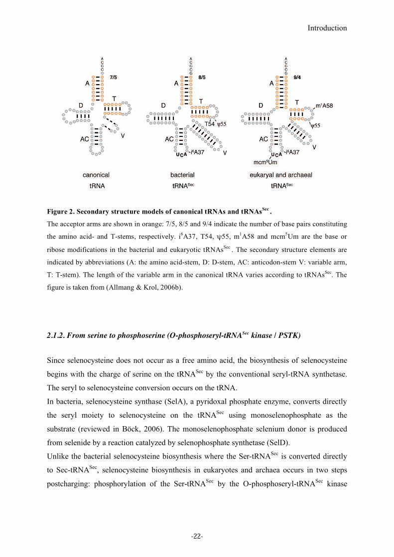
Figure 2. Secondary structure models of canonical tRNAs and tRNAsSec
.
The acceptor arms are shown in orange: 7/5, 8/5 and 9/4 indicate the number of base pairs constituting
the amino acid- and T-stems, respectively. i6A37, T54, !55, m
1A58 and mcm
5Um are the base or
ribose modifications in the bacterial and eukaryotic tRNAsSec
. The secondary structure elements are
indicated by abbreviations (A: the amino acid-stem, D: D-stem, AC: anticodon-stem V: variable arm,
T: T-stem). The length of the variable arm in the canonical tRNA varies according to tRNAsSec
. The
figure is taken from (Allmang & Krol, 2006b).
2.1.2. From serine to phosphoserine (O-phosphoseryl-tRNASec kinase / PSTK)
Since selenocysteine does not occur as a free amino acid, the biosynthesis of selenocysteine
begins with the charge of serine on the tRNASec
by the conventional seryl-tRNA synthetase.
The seryl to selenocysteine conversion occurs on the tRNA.
In bacteria, selenocysteine synthase (SelA), a pyridoxal phosphate enzyme, converts directly
the seryl moiety to selenocysteine on the tRNASec
using monoselenophosphate as the
substrate (reviewed in Böck, 2006). The monoselenophosphate selenium donor is produced
from selenide by a reaction catalyzed by selenophosphate synthetase (SelD).
Unlike the bacterial selenocysteine biosynthesis where the Ser-tRNASec
is converted directly
to Sec-tRNASec
, selenocysteine biosynthesis in eukaryotes and archaea occurs in two steps
postcharging: phosphorylation of the Ser-tRNASec
by the O-phosphoseryl-tRNASec
kinase
-22-
Introduction
(PSTK) and conversion of the phosphoseryl-tRNASec
(Sep-tRNASec
) to Sec-tRNASec
by
Selenocysteine synthase.
The presence of a kinase activity to convert the Ser-tRNASec
to Sep-tRNASec
was reported in
1970 (Maenpaa & Bernfield, 1970), but the O-phosphoseryl-tRNASec
kinase (PSTK) enzyme
was identified only recently by using a comparative genomics approach (Carlson et al, 2004).
This enzyme phosphorylates the serine moiety of Ser-tRNASec
to yield Sep-tRNASec
by using
ATP. In contrast to SerRS that recognizes both the tRNASer
and tRNASec
, PSTK discriminates
Ser-tRNASec
from Ser-tRNASer
. In eukaryotes, the length and secondary structure of the D-
stem of tRNASec
are the major determinants for phosphorylation (Wu & Gross, 1994),
whereas the archaeal enzyme recognizes the acceptor stem of the tRNASec
(Sherrer et al,
2008). Interestingly, the archaeal PSTK can efficiently phosphorylate a chimeric Thr-tRNASec
,
providing evidence that this enzyme does not recognize the amino acid (Figure 3).
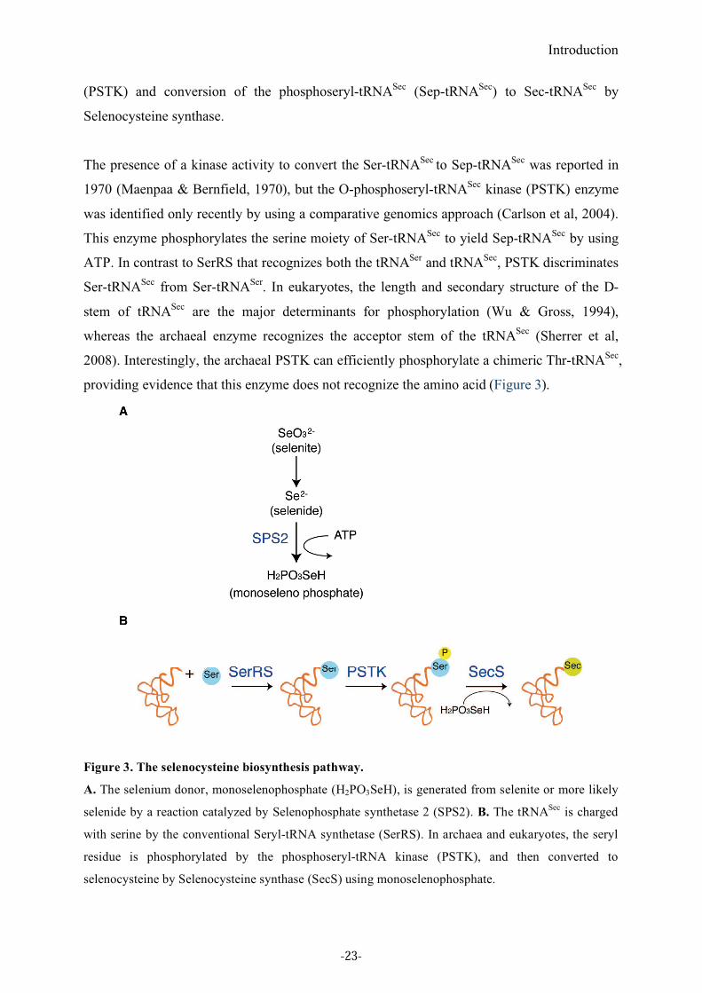
Figure 3. The selenocysteine biosynthesis pathway.
A. The selenium donor, monoselenophosphate (H2PO3SeH), is generated from selenite or more likely
selenide by a reaction catalyzed by Selenophosphate synthetase 2 (SPS2). B. The tRNASec
is charged
with serine by the conventional Seryl-tRNA synthetase (SerRS). In archaea and eukaryotes, the seryl
residue is phosphorylated by the phosphoseryl-tRNA kinase (PSTK), and then converted to
selenocysteine by Selenocysteine synthase (SecS) using monoselenophosphate.
-23-
Introduction
2.1.3. From phosphoserine to selenocysteine
2.1.3.1. Generation of the selenium donor (SPS1/2)
Selenophosphate synthetase (SelD) in bacteria generates monoselenophosphate which is the
selenium donor for selenocysteine biosynthesis. Selenophosphate synthetase 1 (SPS1) and
later selenophosphate synthetase 2 (SPS2) were identified as the eukaryotic homologues
(Guimaraes et al, 1996; Low et al, 1995). SPS2 is itself a selenoprotein in most organisms
(Guimaraes et al, 1996). Recent studies demonstrated that SPS2 but not SPS1 can synthesize
monoselenophosphate in vitro, and only SPS2 is essential for selenoprotein synthesis in vivo
(Xu et al, 2007a; Xu et al, 2007b). In addition, SPS1 is present in insects that have lost
selenoproteins, indicating that one of its major role is unrelated to selenoprotein synthesis
(Chapple & Guigo, 2008).
2.1.3.2. From Sep-tRNASec
to Sec-tRNASec
(SecS)
Soluble Liver Antigen/Liver Pancreas (SLA/LP) was initially identified as a 48kDa protein
co-immunoprecipitated with tRNASec
by autoantibodies from a subgroup of patients with a
severe form of autoimmune chronic active hepatitis, and implicated in the selenocysteine
pathway (Costa et al, 2000; Gelpi et al, 1992; Kernebeck et al, 2001). Later, two research
teams identified independently SLA/LP as the eukaryotic and archaeal selenocysteine
synthetase (Xu et al, 2007a; Yuan et al, 2006). The human and archaeal (Methanococcus.
maripaludis) enzymes were named SepSecS (Yuan et al, 2006), whereas the mouse homolog
was called mSecS (Xu et al, 2007a) according to the authors (‘SecS’ will be used in this thesis
for reason of convenience.). Human and archaeal SecS were shown to complement in vivo an
E.coli SelA null-strain and to convert the Sep-tRNASec
to Sec-tRNASec
in the presence of
sodium selenite and recombinant Escherichia.coli SelD in vitro (Yuan et al, 2006). In
addition, SecS exhibits higher affinity for the Sep-tRNASec
than for the tRNASec
and Ser-
tRNASec
(Xu et al, 2007b). These studies provided evidence that, in contrast to bacterial SelA,
eukaryotic and archaeal selenocysteine biosynthesis has an intermediate step where Sec-
tRNASec
is generated, using Sep-tRNASec
and monoselenophosphate as substrates.
-24-
Introduction
The crystal structures of the M.maripaludis and mouse SecS were solved and showed that
both enzymes are members of the fold Type 1 pyridoxal phosphate (PLP)-dependent enzyme
family, as is bacterial SelA (Araiso et al, 2008; Ganichkin et al, 2008).
2.1.4. SECp43
SECp43 was reported to interact with the tRNASec
and to be involved in selenocysteine
incorporation mechanism (Ding & Grabowski, 1999). SECp43 is predominantly present in the
nucleus (Xu et al, 2005) and can interact with Sec-tRNASec
-EFSec complex in vitro (Small-
Howard et al, 2006). SECp43 interacts with SecS and SPS1 in vivo, and redistributes these
proteins to the nucleus (Small-Howard et al, 2006). Knockdown of SECp43 by siRNA
demonstrated that SECp43 is required for ribose methylation at Um34 of tRNASec
, and
increases selenoprotein expression at both mRNA and protein levels. A role for SECp43 has
also been proposed in the orchestration of the interactions and localization of other
selenoprotein synthesis factors (Small-Howard et al, 2006; Xu et al, 2005).
2.2. Sec incorporation
The general Sec incorporation mechanism is different in bacteria and eukaryotes. In bacteria,
bSECIS (bacterial SElenoCysteine Insertion Sequence, a stem-loop structure immediately
downstream of the in-frame UGA codon in selenoprotein mRNAs) and SelB, a translation
elongation factor specialized for selenocysteine incorporation, play central roles for Sec
incorporation. The N-terminal region of SelB is highly-sequence similar and functionally
homologous to EF-Tu, the general translation elongation factor. Its C-teminal domain binds to
bSECIS. SelB binds specifically and uniquely Sec-tRNASec
. The Sec-tRNASec
harbored by
SelB, is brought directly to the UGA Sec codon through the bSECIS-SelB interaction,
allowing the incorporation of selenocysteine into the nascent polypeptide chain.
In eukaryotes, the SECIS element is located in the 3’UTR of selenoprotein mRNAs.
Eukaryotic SECIS elements have conserved helix-loop structures and differ from the bSECIS
structure. Sec incorporation requires the SECIS Binding Protein 2 (SBP2) and the specialized
translation elongation factor EFSec (reviewed in Allmang & Krol, 2006b; Allmang et al,
-25-
Introduction
2009). Ribosomal protein L30 has also been implicated in this mechanism (Chavatte et al,
2005).
2.2.1. Cis-acting factors
2.2.1.1. SElenoCysteine Incorporation Sequence (SECIS)
The SECIS is an RNA stem-loop structure that is mandatory for selenocysteine incorporation.
Depending on the kingdom, it varies in sequence, structure and localization in the mRNA.
2.2.1.1.a. Location in mRNA
In bacteria, the SECIS RNA is located in the coding region immediately downstream of the
in-frame UGA codon of selenoprotein mRNAs (reviewed in Böck, 2006). Unlike in bacteria,
the SECIS is found in the 3’UTR of selenoprotein mRNAs in eukaryotes and archaea,
suggesting similarities in the selenocysteine incorporation mechanism between archaea and
eukaryotes. The advantage of having the SECIS element in the 3’UTR rather than in the
coding region is that the RNA sequence is not constrained to maintain both the coding
capacity and the base-pairing ability of the SECIS element. The localization of the SECIS
element in the 3’UTR introduces flexibility by looping-out the intervening sequence between
the UGA codon. It can thus interact with distant UGA Sec codons. In addition, its residence in
the 3’UTR also enables selenoprotein mRNAs to harbor more than one UGA Sec codon.
Indeed, the SECIS element in the 3’UTR relieves the necessity for stem-loop structures in the
coding region, therefore providing complete flexibility in UGA codon position (Berry et al,
1993). Also, the SECIS element in the 3’UTR provides eukaryotes with a different Sec
incorporation mechanism than in bacteria, for example, it enables rapid and efficient
exchange of empty Sec-specific elongation factors (EFSec, see 2.2.2.1. EFSec) for Sec-
tRNASec
/EFSec complexes, which seems to be essential in the case of multiple UGA codons
(Tujebajeva et al, 2000). This is examplified for selenoprotein P (SelP). While most
selenoprotein mRNAs contain a single UGA codon and a single SECIS element, SelP
contains 10 to 18 UGA Sec codons, depending on animals, and 2 SECIS elements. In addition
to the full-length protein, rat SelP has three isoforms resulting from termination at the second,
third and seventh UGAs (Ma et al, 2002). However, it is possible that the isoforms of various
-26-
Introduction
length could arise from experimental conditions and not from abortive synthesis. It was
shown that the first UGA Sec is decoded by the second SECIS, and the first SECIS is
required for decoding the downstream UGA Sec codons (Stoytcheva et al, 2006). Another
surprising exception was found in the Fowlpox virus. The Fowlpox virus GPx4 mRNA
contains a SECIS element at the 3’end of the coding region and not in the 3’UTR.
Surprisingly also, this in-frame SECIS is able to support selenoprotein synthesis when the
virus GPx4 is expressed in mammalian cells (Mix et al, 2007).
2.2.1.1.b Secondary structure
Although there is little sequence similarity between SECIS RNAs, the SECIS 2D structure is
well conserved within each kingdom.
Bacterial SECIS is an approximately 40 nucleotide-long stem-loop structure. Although SECIS
sequence vary depending on species, the structure is grossly conserved in different organisms
and the apical loop is important for binding to the specialized translational factor SelB (Böck,
2006).
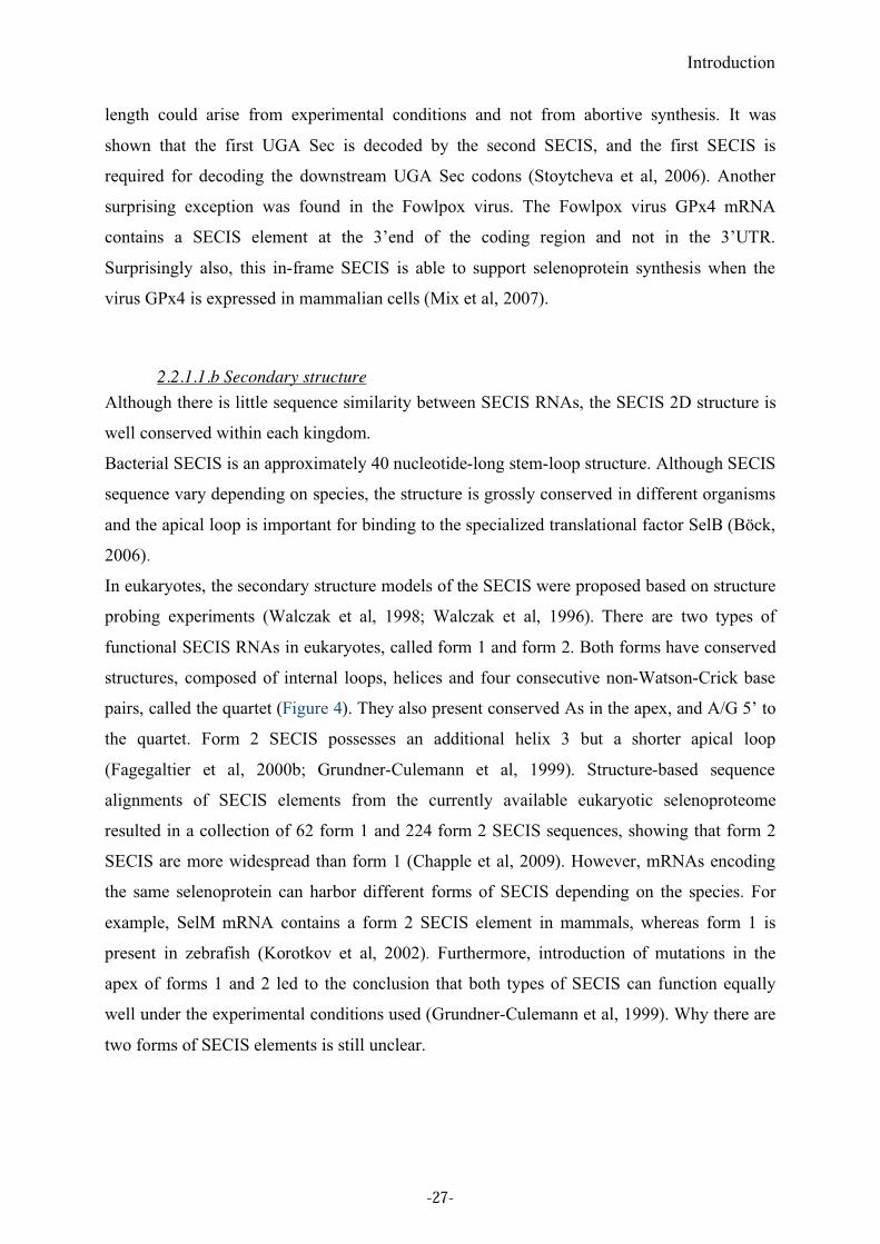
In eukaryotes, the secondary structure models of the SECIS were proposed based on structure
probing experiments (Walczak et al, 1998; Walczak et al, 1996). There are two types of
functional SECIS RNAs in eukaryotes, called form 1 and form 2. Both forms have conserved
structures, composed of internal loops, helices and four consecutive non-Watson-Crick base
pairs, called the quartet (Figure 4). They also present conserved As in the apex, and A/G 5’ to
the quartet. Form 2 SECIS possesses an additional helix 3 but a shorter apical loop
(Fagegaltier et al, 2000b; Grundner-Culemann et al, 1999). Structure-based sequence
alignments of SECIS elements from the currently available eukaryotic selenoproteome
resulted in a collection of 62 form 1 and 224 form 2 SECIS sequences, showing that form 2
SECIS are more widespread than form 1 (Chapple et al, 2009). However, mRNAs encoding
the same selenoprotein can harbor different forms of SECIS depending on the species. For
example, SelM mRNA contains a form 2 SECIS element in mammals, whereas form 1 is
present in zebrafish (Korotkov et al, 2002). Furthermore, introduction of mutations in the
apex of forms 1 and 2 led to the conclusion that both types of SECIS can function equally
well under the experimental conditions used (Grundner-Culemann et al, 1999). Why there are
two forms of SECIS elements is still unclear.
-27-
Introduction
Figure 4. Secondary structure models of form 1 and 2 SECIS.
The conserved sequence and structural features are indicated. Novel conserved residues found by
SECISaln are shown in red (Chapple et al, 2009). The positions of the conserved nucleotides are
indicated in blue. abcd/a’b’c’d’ indicates base-pairs forming the non-Watson-Crick quartet.
Numberings (-4, 1, 2, 9, 10, 1’ and 2’) show the distance from the quartet. Position “z” is the first
nucleotide after the conserved A/Cs, positions 2H3/2’H3 are the second base pair of the Helix III abd
1ap is the first nucleotide of the apical loop. The structures are from (Fagegaltier et al, 2000b;
Grundner-Culemann et al, 1999; Walczak et al, 1998; Walczak et al, 1996).
The non-Watson-Crick quartet is essential to selenocysteine incorporation in vivo, and
constitutes the binding site for SECIS Binding Protein 2 (the function of this key protein will
be detailed in paragraph 2.2.2.2.). This motif contains a central tandem of sheared G.A/A.G
base pairs (Fagegaltier et al, 2000b; Walczak et al, 1998; Walczak et al, 1996). Such a tandem
of G.A/A.G base pairs is also found in other RNAs such as ribosomal RNAs, snRNAs and
snoRNAs, and it constitutes a conserved structure, called the K (kink)-turn motif (Klein et al,
2001). The K-turn is an RNA structural motif that binds proteins in most of the cases and
mediates RNA tertiary structure interactions. The K-turn is a two-stranded, helix-internal
loop-helix motif comprising about 15 nucleotides, characterized by base stacking, the
presence of a tandem of G-A sheared base pairs, and a protruding residue accommodated by a
protein pocket. As a result, the structure has a kink of 120° in the phosphodiester backbone
that causes a sharp turn in the RNA helix (Klein et al, 2001). A K-turn was also found in the
-28-
Introduction
crystal structure of U4 snRNA-15.5kD, L30e RNA-L30e and sRNA-L7Ae complexes (Chao
& Williamson, 2004; Moore et al, 2004; Vidovic et al, 2000)(Figure 5).
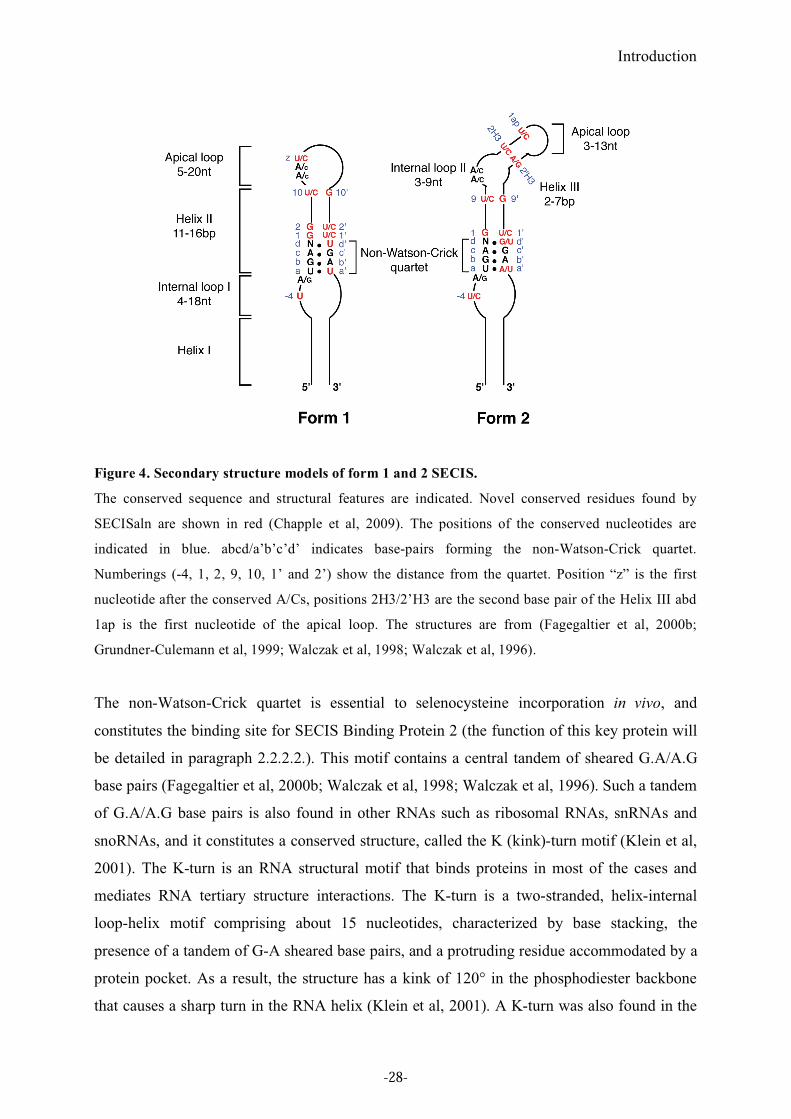
Figure 5. The secondary structure of SECIS RNA and various K-turn RNAs.
The secondary structures of the U4 snRNA, L30e pre-mRNA, L7Ae rRNA, L7Ae box C/D sRNA
were taken from the crystal structures of the corresponding RNA-protein complexes (Chao &
Williamson, 2004; Moore et al, 2004; Vidovic et al, 2000). Those of SECIS RNA and U3 Box B/C
snoRNA were determined by structure probing analyses (Fagegaltier et al, 2000b; Marmier-Gourrier
et al, 2003; Walczak et al, 1998; Walczak et al, 1996). The sheared G.A/A.G base pairs are indicated
in bold. The figure is taken from (Allmang & Krol, 2006a).
Because of these secondary structure similarities, we have proposed that the SECIS RNA is a
K-turn like motif (Allmang & Krol, 2006a). Furthermore, this is supported by previous
findings where structure probing and mutagenesis data allowed a 3D model for the SECIS
RNA to be proposed by computer modeling. In this model the phosphodiester backbone is
-29-
Introduction
indeed bent at the internal loop (Walczak et al, 1996). Compared to canonical K-turn RNAs,
SECIS elements have larger internal loops. This larger internal loop and a long helix 2
provide specificity for SBP2 binding to the SECIS (Cléry et al, 2007 ). The nucleotide 5’ to
the quartet is A in most of the cases, but G can be found, and the replacement by G does not
affect SECIS activity in vivo (Buettner et al, 1999; Fagegaltier et al, 2000b; Taskov et al,
2005). Interestingly, in a patient suffering from a SEPN1-related myopathy, a mutation in the
non-Watson-Crick quartet of the SEPN1 SECIS element that prevents the interaction with
SBP2, was found to be responsible for the pathology (Allamand et al, 2006)(Figure 6 A).
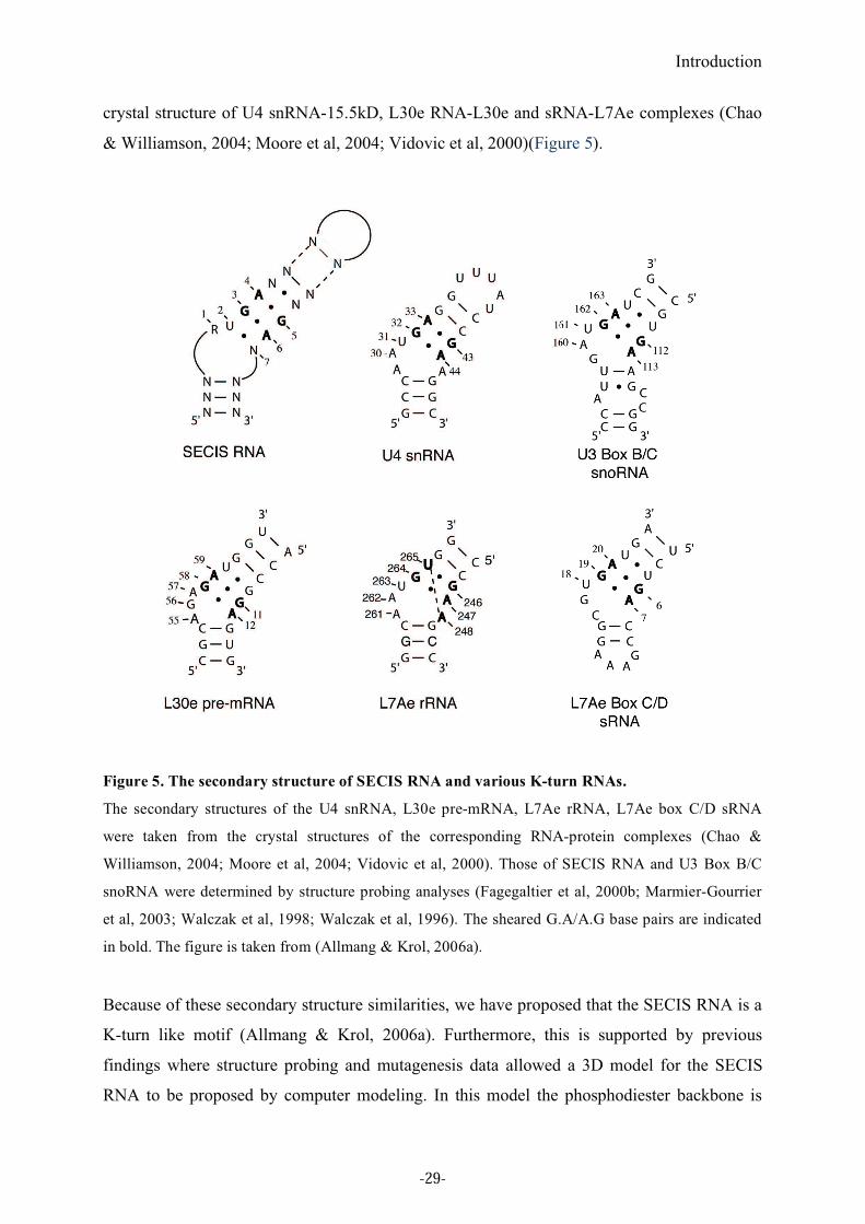
Figure 6. The SECIS and SRE elements of SEPN1 mRNAs
A. Secondary structure of the SEPN1 SECIS RNA. The conserved U in the non-Watson-Crick quartet
is essential for the recognition by SBP2 and the U to C mutation abolishes SBP2 binding (arrow). This
mutation was initially found in Selenoprotein N (SEPN) SECIS element from a patient with a SEPN1-
related myopathy (Maiti et al, 2008). B. Secondary structure model of the SRE RNA. The SRE hairpin
structure is located within the open reading frame (ORF) of certain selenoprotein mRNAs, here the
selenoprotein N (SEPN1) (Howard et al, 2005). The G to A mutation was found in a patient with
SEPN1-related myopathy (Allamand et al, 2006).
RNA structure probing experiments indicated that the conserved As in the apical loop (form
1) or the internal loop 2 (form 2) are single stranded and well accessible (Fagegaltier et al,
2000b). However, some exceptions to the invariant presence of As were reported. For
example, mammalian SelM SECIS and some of Chlamydomonas form 2 SECIS contain Cs
without altering Sec incorporation activity (Korotkov et al, 2002; Novoselov et al, 2002).
Other examples of Cs or a combination of As and Cs or even Gs were later found in
-30-
Introduction
eukaryotes (Chapple et al, 2009; Lobanov et al, 2006a; Lobanov et al, 2007; Lobanov et al,
2006b ). Although this apical A/C rich loop is not necessary for SBP2 binding, site-directed
mutagenesis showed that the unpaired As/Cs are important for selenoprotein synthesis in vivo
(Berry et al, 1993).
Such detailed knowledge of the secondary structure of SECIS element was used in
computational analysis to identify novel selenoprotein mRNAs (Kryukov et al, 1999; Lescure
et al, 1999) and to establish the whole mammalian selenoproteome with the help of
SECISearch, a computer program for analyzing structural and thermodynamic features of
SECIS elements (Kryukov et al, 2003). Recently, the well-defined secondary structure of the
SECIS RNA and the increased size of the eukaryotic selenoproteome allowed the
establishment of a web-based tool, SECISaln, providing extensive structure-based sequence
alignments of SECIS elements (Chapple et al, 2009). Analyzing the structural alignments
produced by SECISaln highlighted a few previously undetected conserved residues (see
Figure 4). There is an overrepresentation of G at position 1 (3’ to the quartet) and a
corresponding overrepresentation of C or U at position 1’ (see Figure 4). SECISaln also found
differences between form1 and form2 SECISes. The most striking one is a well-conserved U
4 nucleotides upstream of the quartet (at position -4 in Figure 4) in form1 SECIS, whereas C
can also be found in the form 2 SECIS (Chapple et al, 2009).
2.2.1.2. SRE
Another cis-acting element reported recently is the Selenocysteine codon Redefinition
Element (SRE). SRE is a phylogenetically conserved stem-loop structure located within the
coding region of selenoprotein mRNAs, adjacent to the UGA Sec codon. This element is
sufficient to stimulate readthrough of the UGA Sec codon in the absence of a SECIS element
in the 3’UTR in a synthetic mRNA, although higher readthough efficiency is observed in its
presence. SelN SRE inserted in a dual-luciferase system had a stimulatory effect on the UGA
Sec decoding in vitro (Howard et al, 2005; Howard et al, 2007). SRE was experimentally
analyzed in SelN mRNA, but bioinformatic approaches predicted found SREs in a few other
selenoprotein mRNAs such as SPS2, SelH, SelO and SelT (Howard et al, 2005; Pedersen et
al, 2006). Their 2D structure, however, is not conserved. The presence of an SRE in some but
not all selenoprotein mRNAs implies a differential role in regulating selenoprotein expression
at the translation level. Four point mutations leading to the SEPN1-related myopathy were
-31-
Introduction
found in the SelN SRE element. One of them weakens the secondary structure of SRE by
abolishing a G-C base pair, leading to a decrease in Sec incorporation and SelN levels (Maiti
et al, 2008). This data supports the importance of the SRE structure for selenoprotein
synthesis (Figure 6B).
2.2.2. Trans-acting factors
2.2.2.1. EFSec
In bacteria, SelB is the translation elongation factor specialized for selenocysteine
incorporation. The N-terminal domain of SelB is highly-sequence similar and functionally
homologous to EF-Tu (see Figure 7), the general translation elongation factor, and the C-
teminal domain binds to SECIS. SelB binds specifically and uniquely the Sec-tRNASec
(Böck,
2006).
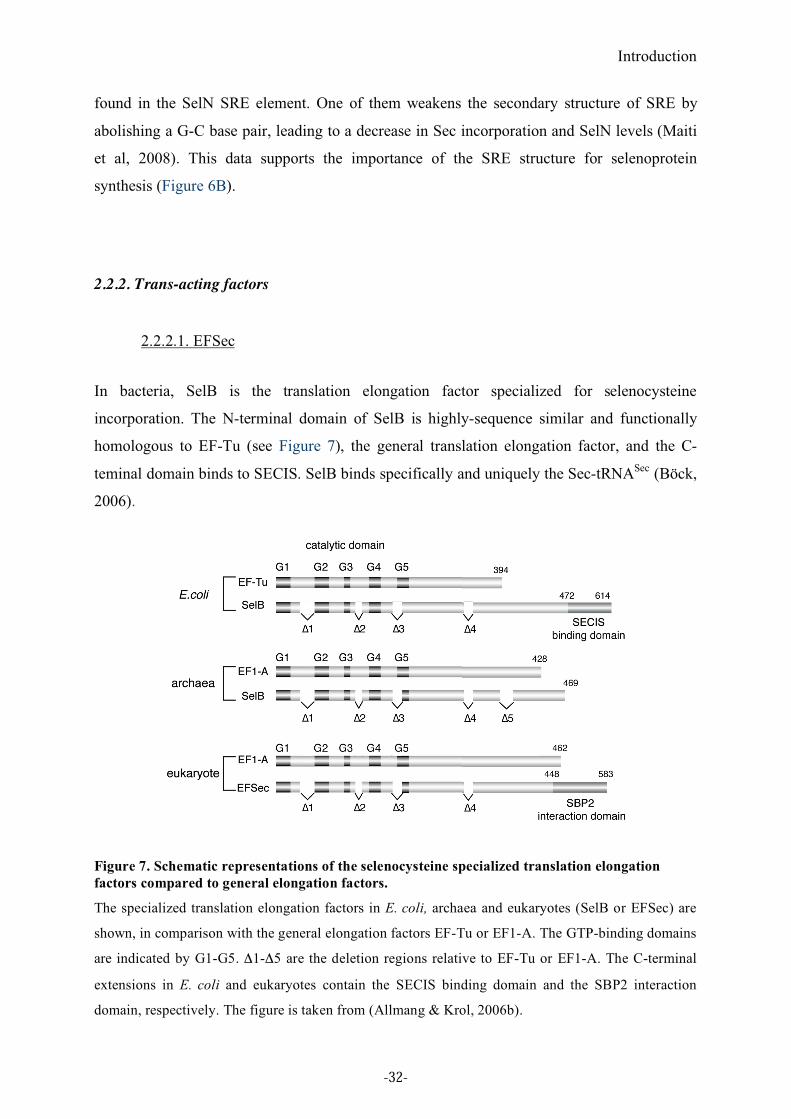
Figure 7. Schematic representations of the selenocysteine specialized translation elongation
factors compared to general elongation factors.
The specialized translation elongation factors in E. coli, archaea and eukaryotes (SelB or EFSec) are
shown, in comparison with the general elongation factors EF-Tu or EF1-A. The GTP-binding domains
are indicated by G1-G5. "1-"5 are the deletion regions relative to EF-Tu or EF1-A. The C-terminal
extensions in E. coli and eukaryotes contain the SECIS binding domain and the SBP2 interaction
domain, respectively. The figure is taken from (Allmang & Krol, 2006b).
-32-
Introduction
EFSec is the mammalian homolog of SelB. It was independently characterized in mouse by
our laboratory and by Berry’s group (Fagegaltier et al, 2000a; Tujebajeva et al, 2000). EFSec
binds specifically to the Sec-tRNASec
but not to Ser-tRNASec
. Like for bacterial SelB, the N-
terminal domain of EFSec has sequence similarities with the general elongation factor EF1A
and contains homologies to the G1-G4 GTP-domain (Fagegaltier et al, 2000a). The length of
the C-terminal extension varies in different organisms. In contrast to SelB, EFSec cannot bind
specifically the SECIS RNA, indicating another role than in bacteria. EFSec co-
immunoprecipitates SBP2 from mammalian cell extracts, and the SBP2 interaction domain
resides in the C-terminal extension (Tujebajeva et al, 2000). Thus, it is likely that EFSec is
recruited to the selenocysteine incorporation machinery by SBP2.
EFSec contains putative nuclear export and nuclear localization signals, in the N-terminal
domain and the C-terminal SBP2 interaction domain, respectively. The EFSec subcellular
localization varies depending on the cell line and may be influenced by SBP2 levels and
localization (de Jesus et al, 2006).
Archaeal EFSec (called SelB) was identified in Methanococcus jannaschii (Rother et al,
2000), and it possesses sequence features characteristic of bacterial SelB and EFSec
(Fagegaltier et al, 2000a; Rother et al, 2000). Furthermore, crystal structure of the
Methanococcus maripaludis EFSec revealed that its overall shape resembles a ‘chalice’
observed so far in translational initiation factor IF2/eIF5B (Leibundgut et al, 2005). This
raises the interesting question of whether mechanistic similarities could exist between Sec
incorporation and translational initiation.
2.2.2.2. SBP2
SBP2 (SECIS Binding Protein 2) is a trans-acting factor that plays a central role in eukaryotic
Sec incorporation. SBP2 was isolated and functionally characterized in rat and humans
(Copeland & Driscoll, 1999; Lescure et al, 2002). Its known functions are SECIS binding,
ribosomal binding and Sec incorporation. The importance of SBP2 for selenoprotein synthesis
was shown by SBP2 depletion which results in decreased Sec incorporation in cells and in
vitro (Copeland et al, 2000; Papp et al, 2006). Additionally, patients carrying mutations in
SBP2 display abnormal thyroid hormone metabolism leading to reduction of DIO2 activity
(Dumitrescu et al, 2005).
-33-
Introduction
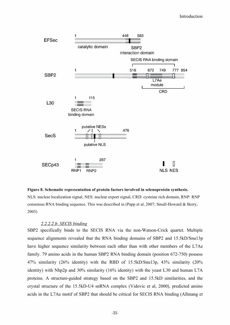
2.2.2.2.a Domain structure of SBP2
Mammalian SBP2 is about 850 amino acid long. The domain structure of SBP2 can be
roughly divided into two parts, the N-terminal and C-terminal domains. The N-terminal
domain is not essential for selenoprotein synthesis in vitro (Copeland et al, 2000). This
domain has no sequence similarity with any other known protein, thus its function still
remains unknown except for the presence of an NLS (Nuclear Localization Signal) (Papp et
al, 2006).
The C-terminal domain is essential and sufficient for Sec incorporation in vitro. It contains
the RNA-binding domain in a region lying between positions 516 and 777 in rat SBP2
(Copeland & Driscoll, 2001, see also Figure 8). This RNA-binding domain includes a
conserved motif, called the L7Ae motif. The L7Ae motif was originally identified as a
putative RNA binding motif by a computational study (Koonin et al, 1994). It is shared by
other functionally unrelated proteins such as 15.5kD/Snu13, Nhp2 and ribosomal proteins
L7Ae and L30, all of which bind to a K-turn motif and trigger RNP complex formation. Later,
point mutation analysis showed that the L7Ae motif in SBP2 is essential for SECIS RNA
binding (Allmang et al, 2002). In addition to the L7Ae motif, SBP2 specific sequences
upstream from the L7Ae motif also play an important role for the interaction with the SECIS
RNA. The RNA binding domain of SBP2 is thus bipartite (Bubenik & Driscoll, 2007;
Donovan et al, 2008; Takeuchi et al, 2009). The characterization of the additional RNA
binding module represents an important contribution to my thesis and will be detailed in
Chapter1 of Part 2. The C-terminal domain contains two functional NES (Nuclear Export
Signal) (Papp et al, 2006).
-34-
Introduction
Figure 8. Schematic representation of protein factors involved in selenoprotein synthesis.
NLS: nuclear localization signal, NES: nuclear export signal, CRD: cysteine rich domain, RNP: RNP
consensus RNA binding sequence. This was described in (Papp et al, 2007; Small-Howard & Berry,
2005)
2.2.2.2.b. SECIS binding
SBP2 specifically binds to the SECIS RNA via the non-Watson-Crick quartet. Multiple
sequence alignments revealed that the RNA binding domains of SBP2 and 15.5kD/Snu13p
have higher sequence similarity between each other than with other members of the L7Ae
family. 79 amino acids in the human SBP2 RNA binding domain (position 672-750) possess
47% similarity (26% identity) with the RBD of 15.5kD/Snu13p, 43% similarity (20%
identity) with Nhp2p and 30% similarity (16% identity) with the yeast L30 and human L7A
proteins. A structure-guided strategy based on the SBP2 and 15.5kD similarities, and the
crystal structure of the 15.5kD-U4 snRNA complex (Vidovic et al, 2000), predicted amino
acids in the L7Ae motif of SBP2 that should be critical for SECIS RNA binding (Allmang et
-35-
Introduction
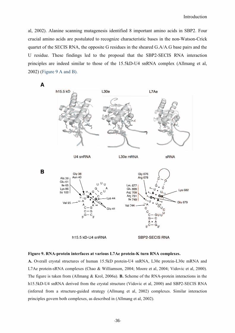
al, 2002). Alanine scanning mutagenesis identified 8 important amino acids in SBP2. Four
crucial amino acids are postulated to recognize characteristic bases in the non-Watson-Crick
quartet of the SECIS RNA, the opposite G residues in the sheared G.A/A.G base pairs and the
U residue. These findings led to the proposal that the SBP2-SECIS RNA interaction
principles are indeed similar to those of the 15.5kD-U4 snRNA complex (Allmang et al,
2002) (Figure 9 A and B).
Figure 9. RNA-protein interfaces at various L7Ae protein-K turn RNA complexes.
A. Overall crystal structures of human 15.5kD protein-U4 snRNA, L30e protein-L30e mRNA and
L7Ae protein-sRNA complexes (Chao & Williamson, 2004; Moore et al, 2004; Vidovic et al, 2000).
The figure is taken from (Allmang & Krol, 2006a). B. Scheme of the RNA-protein interactions in the
h15.5kD-U4 snRNA derived from the crystal structure (Vidovic et al, 2000) and SBP2-SECIS RNA
(inferred from a structure-guided strategy (Allmang et al, 2002) complexes. Similar interaction
principles govern both complexes, as described in (Allmang et al, 2002).
-36-
Introduction
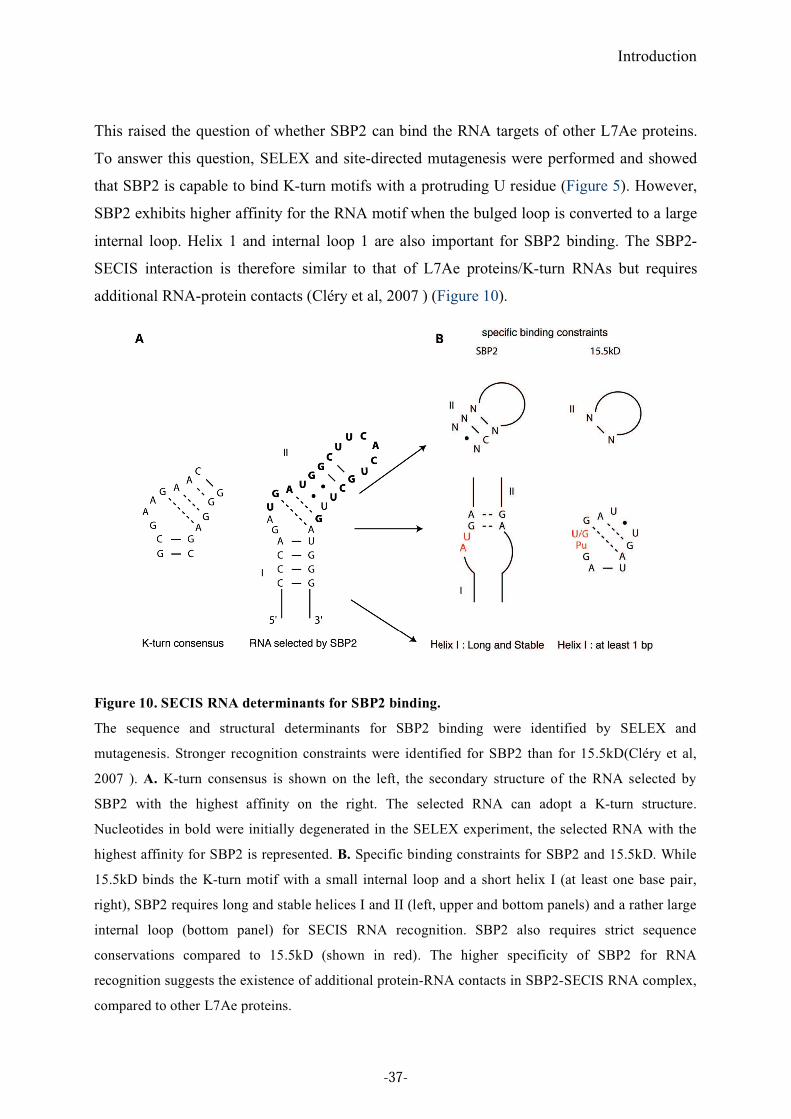
This raised the question of whether SBP2 can bind the RNA targets of other L7Ae proteins.
To answer this question, SELEX and site-directed mutagenesis were performed and showed
that SBP2 is capable to bind K-turn motifs with a protruding U residue (Figure 5). However,
SBP2 exhibits higher affinity for the RNA motif when the bulged loop is converted to a large
internal loop. Helix 1 and internal loop 1 are also important for SBP2 binding. The SBP2-
SECIS interaction is therefore similar to that of L7Ae proteins/K-turn RNAs but requires
additional RNA-protein contacts (Cléry et al, 2007 ) (Figure 10).
Figure 10. SECIS RNA determinants for SBP2 binding.
The sequence and structural determinants for SBP2 binding were identified by SELEX and
mutagenesis. Stronger recognition constraints were identified for SBP2 than for 15.5kD(Cléry et al,
2007 ). A. K-turn consensus is shown on the left, the secondary structure of the RNA selected by
SBP2 with the highest affinity on the right. The selected RNA can adopt a K-turn structure.
Nucleotides in bold were initially degenerated in the SELEX experiment, the selected RNA with the
highest affinity for SBP2 is represented. B. Specific binding constraints for SBP2 and 15.5kD. While
15.5kD binds the K-turn motif with a small internal loop and a short helix I (at least one base pair,
right), SBP2 requires long and stable helices I and II (left, upper and bottom panels) and a rather large
internal loop (bottom panel) for SECIS RNA recognition. SBP2 also requires strict sequence
conservations compared to 15.5kD (shown in red). The higher specificity of SBP2 for RNA
recognition suggests the existence of additional protein-RNA contacts in SBP2-SECIS RNA complex,
compared to other L7Ae proteins.
-37-
Introduction
Furthermore, the SECIS-binding affinity of SBP2 differs between different selenoprotein
mRNAs and is suggested to play a major role in determining the differential selenoprotein
mRNA translation and sensitivity to nonsense-mediated decay (Squires et al, 2007).
In addition to mutations in the coding frame of the SelN protein causing SEPN1-related
myopathies, a single homozygous point mutation in the SelN mRNA SECIS element was also
shown to be responsible for the pathology. This mutation was found in the non-Watson-Crick
quartet, preventing the interaction with SBP2 (Allamand et al, 2006)(Figure 6A).
2.2.2.2.c. EFSec-SBP2 interaction
An interaction between SBP2 and EFSec was observed in co-immunoprecipitation assays
using mammalian cell extracts (Tujebajeva et al, 2000), requiring the tRNASec
(Zavacki et al,
2003). This interaction occurred via the C-terminal 64 amino acid sequence of EFSec and the
C-terminal domain of SBP2 (Donovan et al, 2008; Zavacki et al, 2003). However, no
interaction could be detected in rabbit reticulocyte lysate. The interaction could not be
reconstituted in vitro unless a masking region of EFSec was removed (Zavacki et al, 2003).
Surprisingly, however, a recent study reported the EFSec-SBP2 interaction in vitro in the sole
presence of the SECIS RNA in the reaction mixture (Donovan et al, 2008). The discrepancies
observed by the various investigators may be caused by differences in the experimental
conditions, a co-immunoprecipitation assay using cell extracts on the one hand, EMSA using
recombinant proteins on the other. A 6xHis tagged EFSec may also be detrimental to the
interaction (Donovan et al, 2008). Furthermore, co-expression of SECp43 was shown to
promote the interaction between EFSec and SBP2 (Small-Howard et al, 2006), explaining
why it might be difficult to observe it in vitro.
2.2.2.2.d. Ribosomal binding
Since eukaryotic selenoprotein mRNAs contain the SECIS element in the 3’UTR,
selenocysteine incorporation requires factor(s) that connect the ribosome with the SECIS
element to tell not to stop at the UGA Sec codon. Indeed, SBP2 plays an important role in this
process. Glycerol gradient centrifugation established that SBP2 quantitatively associates with
ribosomes through its RNA binding domain (Kinzy et al, 2005). SECIS RNA can compete
with the ribosome for SBP2 binding, indicating that SBP2 is not able to simultaneously
interact with the ribosome and the SECIS RNA (Kinzy et al, 2005). Like SECIS binding, the
-38-
Introduction
ribosome binding activity of SBP2 is essential for Sec incorporation (Caban et al, 2007;
Donovan et al, 2008; Kinzy et al, 2005). We have analyzed ribosomal binding in more detail
using purified ribosomal subunits. The results will be described in Chapter 1 of Part 2.
2.2.2.2.e. Expression and localization
SBP2 mRNA is expressed in most tissues as revealed by Northern blotting analyses, with
higher levels in testis (Copeland et al, 2000; Lescure et al, 2002). SBP2 was detected
predominantly in the cytoplasm, in stable association with ribosomes (Copeland et al, 2001;
Kinzy et al, 2005; Papp et al, 2006). SECp43, which promotes the interaction between EFSec
and SBP2, interacts in vivo with the Sec-tRNASec
/EFSec in a high molecular weight complex
(Small-Howard et al, 2006), implying that SBP2 is also present in the high molecular weight
complex comprising EFSec and SECp43. However, recent studies showed that SBP2 can
undergo nucleocytoplasmic shuttling via intrinsic nuclear localization (NLS) and nuclear
export signals (NES) that are located in the N-terminal part and the C-terminal cysteine-rich
domain (CRD), respectively (Papp et al, 2006). The nuclear export of SBP2 is dependent on
the CRM1 pathway. Indeed, the best characterized pathway for nuclear export of proteins
from nucleus to cytoplasm involves the nuclear export receptor CRM1, which binds to NES.
Inhibition of CRM1 induces nuclear sequestration of SBP2 and decreases selenoprotein
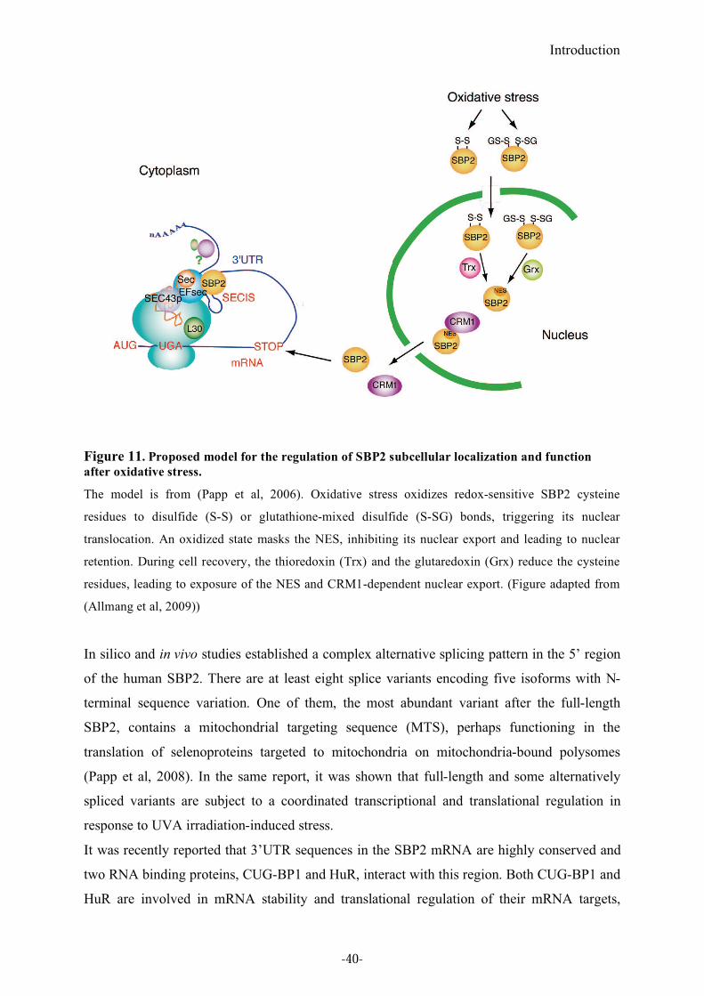
synthesis. Interestingly, oxidative stress induces nuclear accumulation of SBP2 through the
formation of disulfide (S-S) and/or glutathione-mixed disulfide (S-SG) bonds in the redox
sensitive cysteines of the CRD, which masks the NES. These modifications are efficiently
reversed in vitro by thioredoxins and glutaredoxins. These antioxidant systems might regulate
the redox state of SBP2. The nuclear retention of SBP2 after oxidative stress reduces Sec
incorporation, suggesting a mechanism to regulate selenoprotein expression (Papp et al,
2006)(Figure 11).
-39-
Introduction
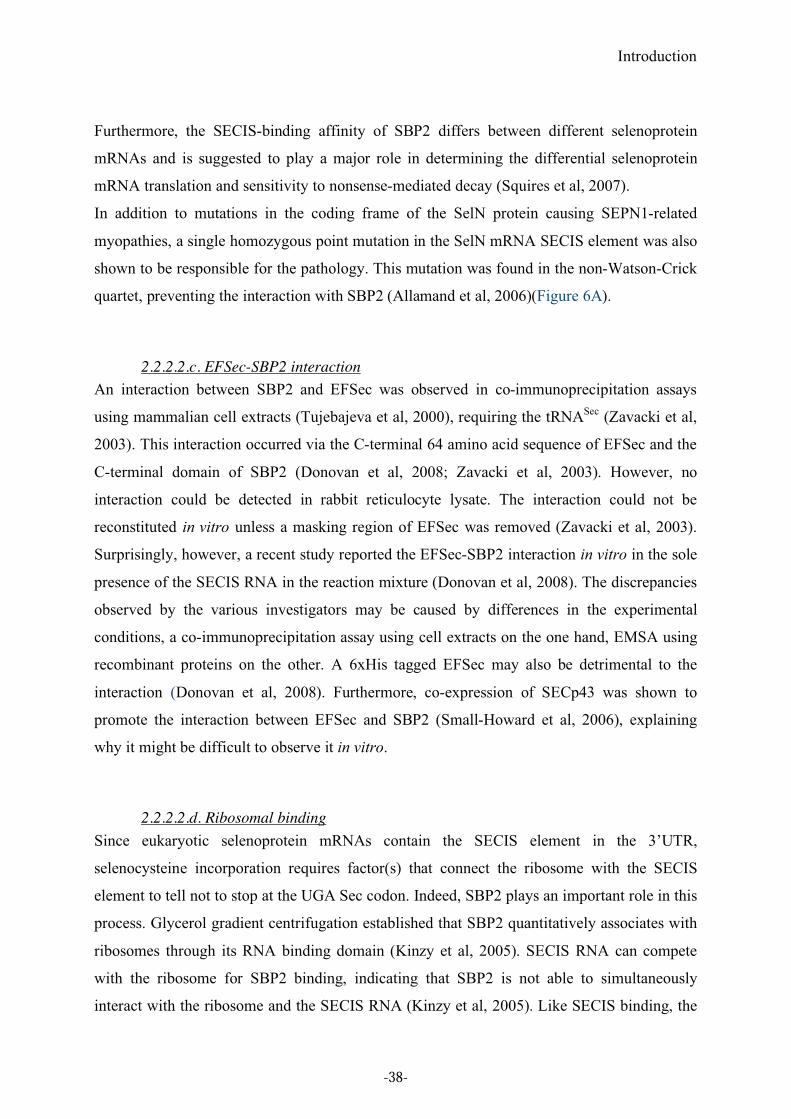
Figure 11. Proposed model for the regulation of SBP2 subcellular localization and function
after oxidative stress.
The model is from (Papp et al, 2006). Oxidative stress oxidizes redox-sensitive SBP2 cysteine
residues to disulfide (S-S) or glutathione-mixed disulfide (S-SG) bonds, triggering its nuclear
translocation. An oxidized state masks the NES, inhibiting its nuclear export and leading to nuclear
retention. During cell recovery, the thioredoxin (Trx) and the glutaredoxin (Grx) reduce the cysteine
residues, leading to exposure of the NES and CRM1-dependent nuclear export. (Figure adapted from
(Allmang et al, 2009))
In silico and in vivo studies established a complex alternative splicing pattern in the 5’ region
of the human SBP2. There are at least eight splice variants encoding five isoforms with N-
terminal sequence variation. One of them, the most abundant variant after the full-length
SBP2, contains a mitochondrial targeting sequence (MTS), perhaps functioning in the
translation of selenoproteins targeted to mitochondria on mitochondria-bound polysomes
(Papp et al, 2008). In the same report, it was shown that full-length and some alternatively
spliced variants are subject to a coordinated transcriptional and translational regulation in
response to UVA irradiation-induced stress.
It was recently reported that 3’UTR sequences in the SBP2 mRNA are highly conserved and
two RNA binding proteins, CUG-BP1 and HuR, interact with this region. Both CUG-BP1 and
HuR are involved in mRNA stability and translational regulation of their mRNA targets,
-40-
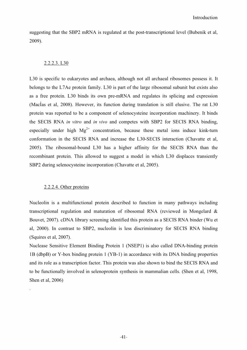
Introduction
suggesting that the SBP2 mRNA is regulated at the post-transcriptional level (Bubenik et al,
2009).
2.2.2.3. L30
L30 is specific to eukaryotes and archaea, although not all archaeal ribosomes possess it. It
belongs to the L7Ae protein family. L30 is part of the large ribosomal subunit but exists also
as a free protein. L30 binds its own pre-mRNA and regulates its splicing and expression
(MacÌas et al, 2008). However, its function during translation is still elusive. The rat L30
protein was reported to be a component of selenocysteine incorporation machinery. It binds
the SECIS RNA in vitro and in vivo and competes with SBP2 for SECIS RNA binding,
especially under high Mg2+
concentration, because these metal ions induce kink-turn
conformation in the SECIS RNA and increase the L30-SECIS interaction (Chavatte et al,
2005). The ribosomal-bound L30 has a higher affinity for the SECIS RNA than the
recombinant protein. This allowed to suggest a model in which L30 displaces transiently
SBP2 during selenocysteine incorporation (Chavatte et al, 2005).
2.2.2.4. Other proteins
Nucleolin is a multifunctional protein described to function in many pathways including
transcriptional regulation and maturation of ribosomal RNA (reviewed in Mongelard &
Bouvet, 2007). cDNA library screening identified this protein as a SECIS RNA binder (Wu et
al, 2000). In contrast to SBP2, nucleolin is less discriminatory for SECIS RNA binding
(Squires et al, 2007).
Nuclease Sensitive Element Binding Protein 1 (NSEP1) is also called DNA-binding protein
1B (dbpB) or Y-box binding protein 1 (YB-1) in accordance with its DNA binding properties
and its role as a transcription factor. This protein was also shown to bind the SECIS RNA and
to be functionally involved in selenoprotein synthesis in mammalian cells. (Shen et al, 1998,
Shen et al, 2006)
.
-41-
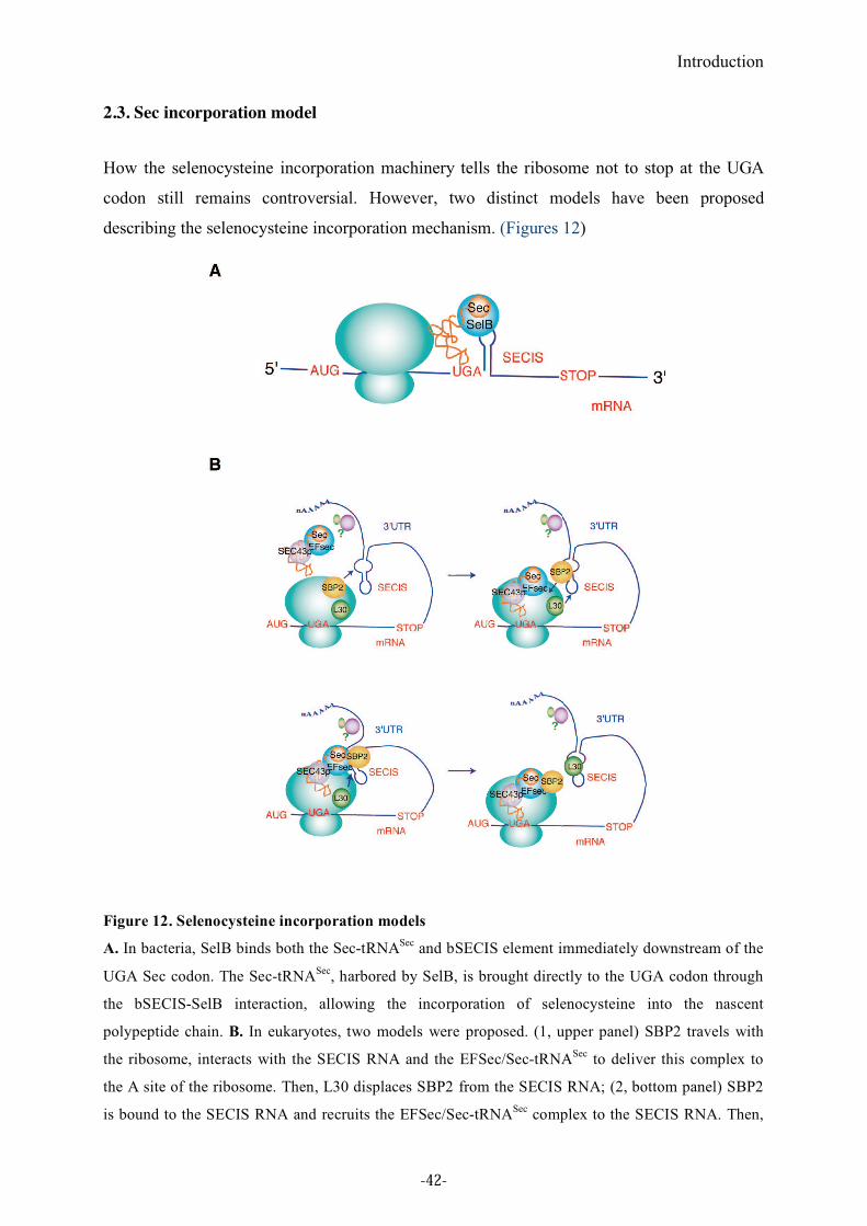
Introduction
2.3. Sec incorporation model
How the selenocysteine incorporation machinery tells the ribosome not to stop at the UGA
codon still remains controversial. However, two distinct models have been proposed
describing the selenocysteine incorporation mechanism. (Figures 12)
Figure 12. Selenocysteine incorporation models
A. In bacteria, SelB binds both the Sec-tRNASec
and bSECIS element immediately downstream of the
UGA Sec codon. The Sec-tRNASec
, harbored by SelB, is brought directly to the UGA codon through
the bSECIS-SelB interaction, allowing the incorporation of selenocysteine into the nascent
polypeptide chain. B. In eukaryotes, two models were proposed. (1, upper panel) SBP2 travels with
the ribosome, interacts with the SECIS RNA and the EFSec/Sec-tRNASec
to deliver this complex to
the A site of the ribosome. Then, L30 displaces SBP2 from the SECIS RNA; (2, bottom panel) SBP2
is bound to the SECIS RNA and recruits the EFSec/Sec-tRNASec
complex to the SECIS RNA. Then,
-42-
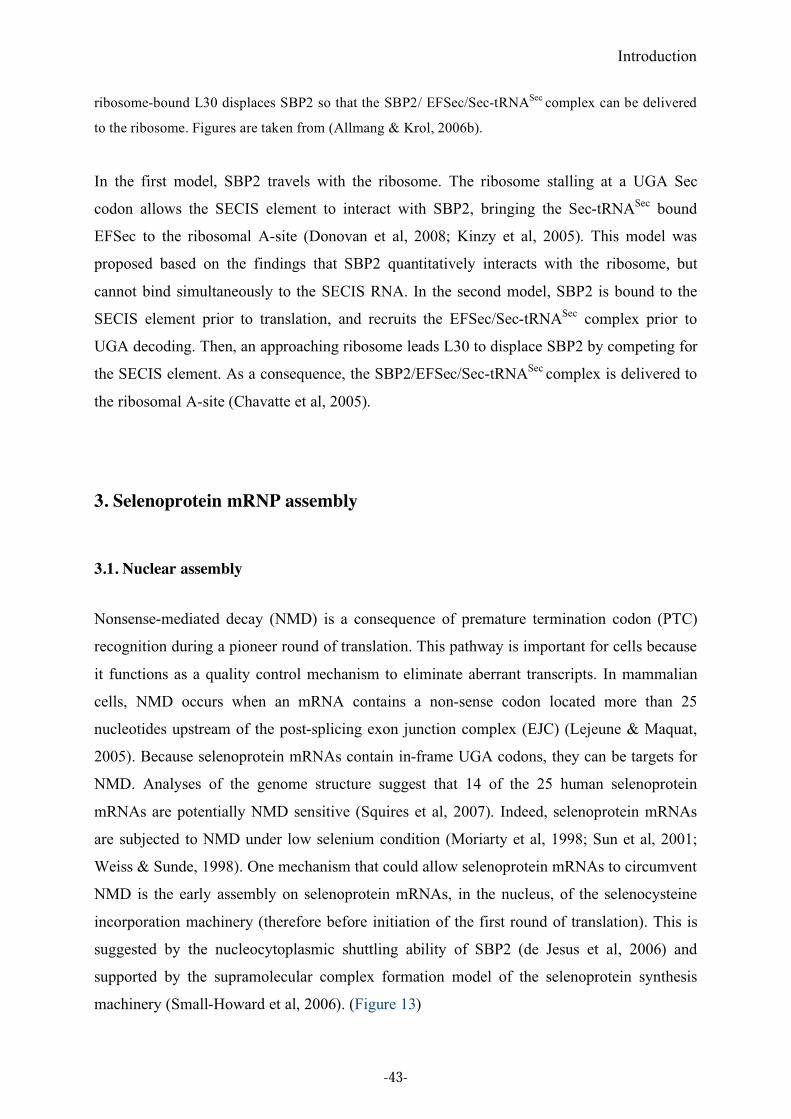
Introduction
ribosome-bound L30 displaces SBP2 so that the SBP2/ EFSec/Sec-tRNASec
complex can be delivered
to the ribosome. Figures are taken from (Allmang & Krol, 2006b).
In the first model, SBP2 travels with the ribosome. The ribosome stalling at a UGA Sec
codon allows the SECIS element to interact with SBP2, bringing the Sec-tRNASec
bound
EFSec to the ribosomal A-site (Donovan et al, 2008; Kinzy et al, 2005). This model was
proposed based on the findings that SBP2 quantitatively interacts with the ribosome, but
cannot bind simultaneously to the SECIS RNA. In the second model, SBP2 is bound to the
SECIS element prior to translation, and recruits the EFSec/Sec-tRNASec
complex prior to
UGA decoding. Then, an approaching ribosome leads L30 to displace SBP2 by competing for
the SECIS element. As a consequence, the SBP2/EFSec/Sec-tRNASec
complex is delivered to
the ribosomal A-site (Chavatte et al, 2005).
3. Selenoprotein mRNP assembly
3.1. Nuclear assembly
Nonsense-mediated decay (NMD) is a consequence of premature termination codon (PTC)
recognition during a pioneer round of translation. This pathway is important for cells because
it functions as a quality control mechanism to eliminate aberrant transcripts. In mammalian
cells, NMD occurs when an mRNA contains a non-sense codon located more than 25
nucleotides upstream of the post-splicing exon junction complex (EJC) (Lejeune & Maquat,
2005). Because selenoprotein mRNAs contain in-frame UGA codons, they can be targets for
NMD. Analyses of the genome structure suggest that 14 of the 25 human selenoprotein
mRNAs are potentially NMD sensitive (Squires et al, 2007). Indeed, selenoprotein mRNAs
are subjected to NMD under low selenium condition (Moriarty et al, 1998; Sun et al, 2001;
Weiss & Sunde, 1998). One mechanism that could allow selenoprotein mRNAs to circumvent
NMD is the early assembly on selenoprotein mRNAs, in the nucleus, of the selenocysteine
incorporation machinery (therefore before initiation of the first round of translation). This is
suggested by the nucleocytoplasmic shuttling ability of SBP2 (de Jesus et al, 2006) and
supported by the supramolecular complex formation model of the selenoprotein synthesis
machinery (Small-Howard et al, 2006). (Figure 13)
-43-
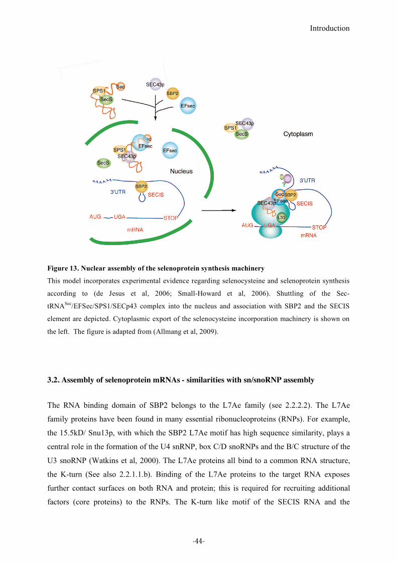
Introduction
Figure 13. Nuclear assembly of the selenoprotein synthesis machinery
This model incorporates experimental evidence regarding selenocysteine and selenoprotein synthesis
according to (de Jesus et al, 2006; Small-Howard et al, 2006). Shuttling of the Sec-
tRNASec
/EFSec/SPS1/SECp43 complex into the nucleus and association with SBP2 and the SECIS
element are depicted. Cytoplasmic export of the selenocysteine incorporation machinery is shown on
the left. The figure is adapted from (Allmang et al, 2009).
3.2. Assembly of selenoprotein mRNAs - similarities with sn/snoRNP assembly
The RNA binding domain of SBP2 belongs to the L7Ae family (see 2.2.2.2). The L7Ae
family proteins have been found in many essential ribonucleoproteins (RNPs). For example,
the 15.5kD/ Snu13p, with which the SBP2 L7Ae motif has high sequence similarity, plays a
central role in the formation of the U4 snRNP, box C/D snoRNPs and the B/C structure of the
U3 snoRNP (Watkins et al, 2000). The L7Ae proteins all bind to a common RNA structure,
the K-turn (See also 2.2.1.1.b). Binding of the L7Ae proteins to the target RNA exposes
further contact surfaces on both RNA and protein; this is required for recruiting additional
factors (core proteins) to the RNPs. The K-turn like motif of the SECIS RNA and the
-44-
Introduction
nucleocytoplasmic shuttling ability of SBP2 are consistent with a possible nuclear assembly
of SECIS mRNPs obeying to the same rules as the sn/snoRNP assembly. Indeed, a recent
study in the laboratory identified a complex assembly machinery linked to the protein
chaperone Hsp90 and that assembles RNPs of the L7Ae family, such as box C/D and H/ACA
snoRNPs, telomerase, U4 snRNP and selenoprotein mRNPs (Boulon et al, 2008; Zhao et al,
2008). This machinery is composed of a co-chaperone complex comprising Rvb1-Rvb2,
Spagh and Pih1 (R2TP complex) associated to the Hsp90 chaperone, and the adaptor protein
Nufip. Nufip binds the L7Ae family proteins including SBP2 and can tether them to other
core proteins of immature RNPs. It also links them to the Hsp90 chaperone complex. These
associations are necessary for the stability of SBP2 and its assembly on the SECIS RNA
(Boulon et al, 2008). Altogether, these results implied that SBP2 has functional similarities
with other L7Ae proteins during RNP assembly, and that the interaction with Nufip leads to
recruit additional core protein(s) yet to be identified (Figure 14).
Figure 14. Selenoprotein mRNP assembly model.
The Hsp90 co-chaperone complex is composed of the AAA+ ATPases Rvb1 and Rvb2 (Rvb1/2),
Spagh and Pih1. Hsp90 is required for the stability of SBP2. It may control the folding of SBP2 during
SECIS RNP formation. Nufip acts as an adaptor between the chaperone complex and SBP2. The
interaction between Pih1 and SBP2 has been experimentally demonstrated. The curved arrow points to
interactions with putative core proteins yet to be discovered. The figure is taken from (Allmang et al,
2009).
-45-

Introduction
-46-
Introduction
4. Objectives and outline of this thesis
SBP2 plays a central role in the eukaryotic Sec incorporation mechanism. However, SBP2
was characterized only in rat and humans when I started my PhD work. We were therefore
interested in SBP2 from other species for evolutionary and crystallographic purposes. During
my PhD studies, I cloned the Drosophila melanogaster SBP2 and analyzed its function. We
identified a new domain which is important for SECIS RNA binding and exhibits selective
affinities toward SECIS RNAs. This new domain, found in all SBP2s, is also responsible for
the binding of SBP2 to the ribosome. This work will be described in Chapter 1 of Part 2.
The laboratory recently identified a common molecular machinery for the assembly of
selenoprotein mRNPs with sn/snoRNPs. Since most of the core proteins of selenoprotein
mRNPs are still unknown, I investigated whether major core proteins of the sn/snoRNP
complex could also be potential partners of SBP2. This wok will be described in Chapter 2 of
Part 3
Finally, I also participated in the structural analysis of SBP2, in collaboration with the group
of Philippe Dumas in the same UPR. This will be described in Chapter 2 of Part 2.
Altogether, these studies provided important insight into how the SBP2-SECIS RNA complex
is formed during selenoprotein synthesis, a process which is at the heart of the UGA
reprogramming mechanism.
-47-

-48-

Part 2. Results
-49-

Results-Chapter 1
-50-
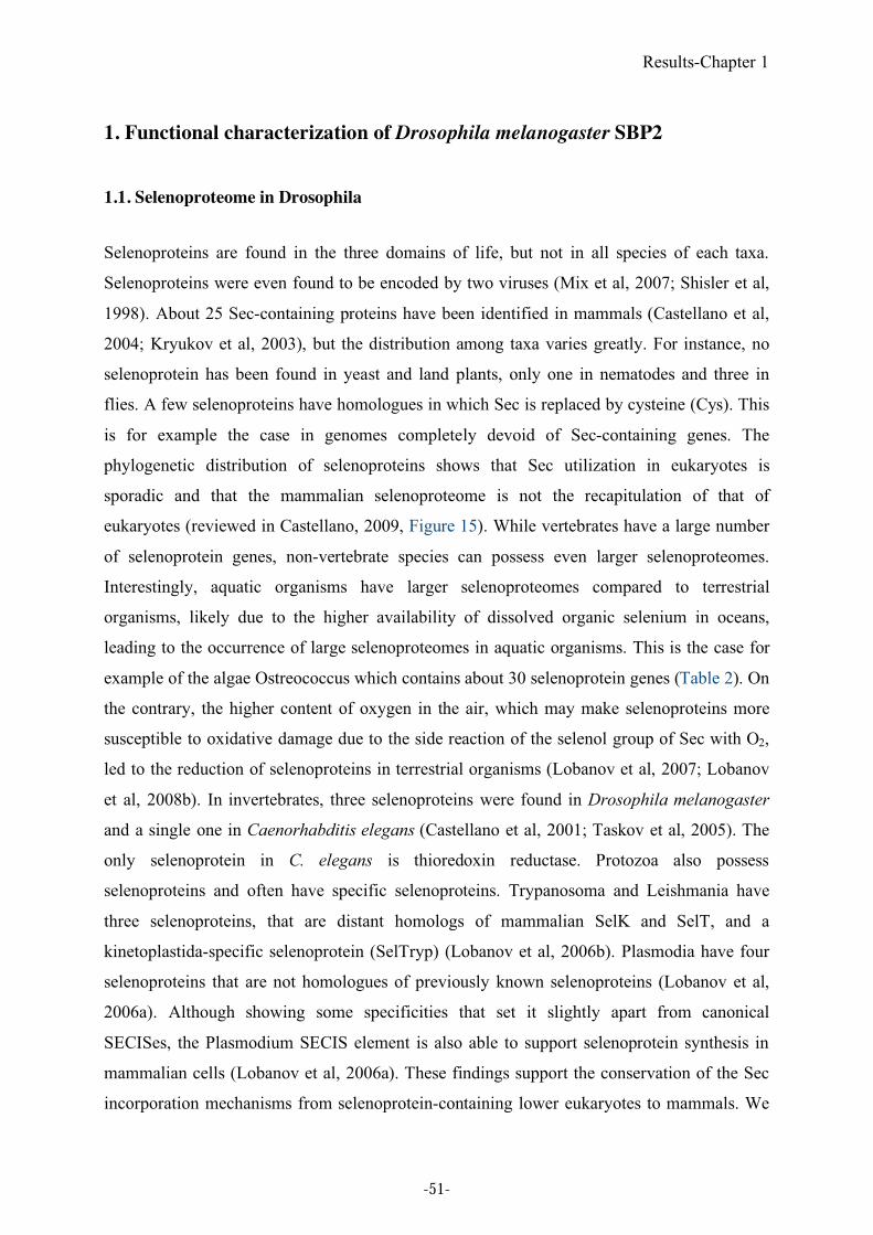
Results-Chapter 1
1. Functional characterization of Drosophila melanogaster SBP2
1.1. Selenoproteome in Drosophila
Selenoproteins are found in the three domains of life, but not in all species of each taxa.
Selenoproteins were even found to be encoded by two viruses (Mix et al, 2007; Shisler et al,
1998). About 25 Sec-containing proteins have been identified in mammals (Castellano et al,
2004; Kryukov et al, 2003), but the distribution among taxa varies greatly. For instance, no
selenoprotein has been found in yeast and land plants, only one in nematodes and three in
flies. A few selenoproteins have homologues in which Sec is replaced by cysteine (Cys). This
is for example the case in genomes completely devoid of Sec-containing genes. The
phylogenetic distribution of selenoproteins shows that Sec utilization in eukaryotes is
sporadic and that the mammalian selenoproteome is not the recapitulation of that of
eukaryotes (reviewed in Castellano, 2009, Figure 15). While vertebrates have a large number
of selenoprotein genes, non-vertebrate species can possess even larger selenoproteomes.
Interestingly, aquatic organisms have larger selenoproteomes compared to terrestrial
organisms, likely due to the higher availability of dissolved organic selenium in oceans,
leading to the occurrence of large selenoproteomes in aquatic organisms. This is the case for
example of the algae Ostreococcus which contains about 30 selenoprotein genes (Table 2). On
the contrary, the higher content of oxygen in the air, which may make selenoproteins more
susceptible to oxidative damage due to the side reaction of the selenol group of Sec with O2,
led to the reduction of selenoproteins in terrestrial organisms (Lobanov et al, 2007; Lobanov
et al, 2008b). In invertebrates, three selenoproteins were found in Drosophila melanogaster
and a single one in Caenorhabditis elegans (Castellano et al, 2001; Taskov et al, 2005). The
only selenoprotein in C. elegans is thioredoxin reductase. Protozoa also possess
selenoproteins and often have specific selenoproteins. Trypanosoma and Leishmania have
three selenoproteins, that are distant homologs of mammalian SelK and SelT, and a
kinetoplastida-specific selenoprotein (SelTryp) (Lobanov et al, 2006b). Plasmodia have four
selenoproteins that are not homologues of previously known selenoproteins (Lobanov et al,
2006a). Although showing some specificities that set it slightly apart from canonical
SECISes, the Plasmodium SECIS element is also able to support selenoprotein synthesis in
mammalian cells (Lobanov et al, 2006a). These findings support the conservation of the Sec
incorporation mechanisms from selenoprotein-containing lower eukaryotes to mammals. We
-51-
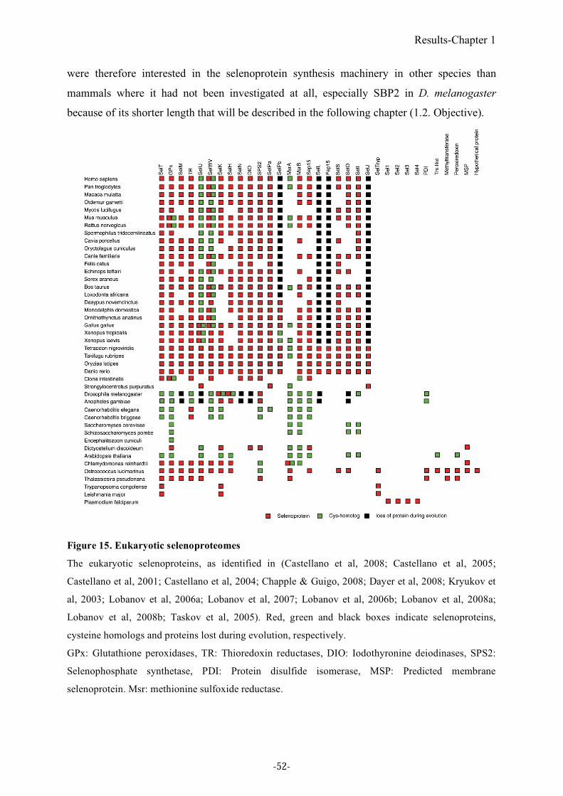
Results-Chapter 1
were therefore interested in the selenoprotein synthesis machinery in other species than
mammals where it had not been investigated at all, especially SBP2 in D. melanogaster
because of its shorter length that will be described in the following chapter (1.2. Objective).
Figure 15. Eukaryotic selenoproteomes
The eukaryotic selenoproteins, as identified in (Castellano et al, 2008; Castellano et al, 2005;
Castellano et al, 2001; Castellano et al, 2004; Chapple & Guigo, 2008; Dayer et al, 2008; Kryukov et
al, 2003; Lobanov et al, 2006a; Lobanov et al, 2007; Lobanov et al, 2006b; Lobanov et al, 2008a;
Lobanov et al, 2008b; Taskov et al, 2005). Red, green and black boxes indicate selenoproteins,
cysteine homologs and proteins lost during evolution, respectively.
GPx: Glutathione peroxidases, TR: Thioredoxin reductases, DIO: Iodothyronine deiodinases, SPS2:
Selenophosphate synthetase, PDI: Protein disulfide isomerase, MSP: Predicted membrane
selenoprotein. Msr: methionine sulfoxide reductase.
-52-
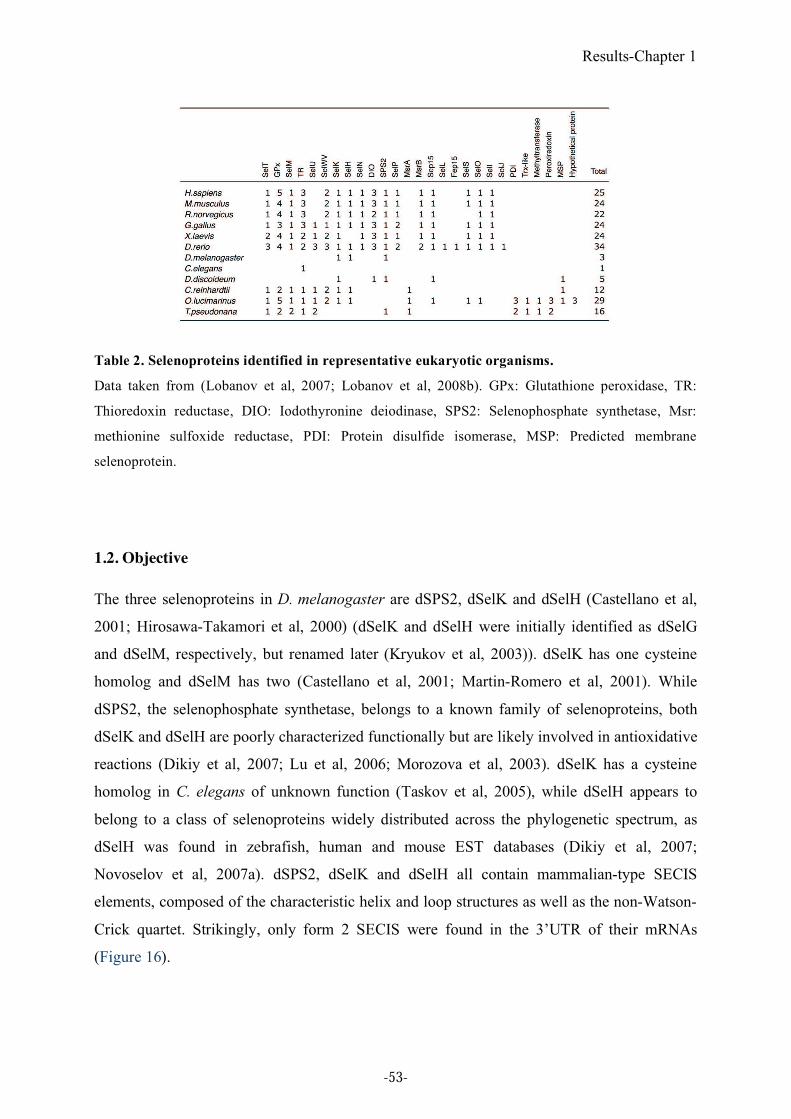
Results-Chapter 1
Table 2. Selenoproteins identified in representative eukaryotic organisms.
Data taken from (Lobanov et al, 2007; Lobanov et al, 2008b). GPx: Glutathione peroxidase, TR:
Thioredoxin reductase, DIO: Iodothyronine deiodinase, SPS2: Selenophosphate synthetase, Msr:
methionine sulfoxide reductase, PDI: Protein disulfide isomerase, MSP: Predicted membrane
selenoprotein.
1.2. Objective
The three selenoproteins in D. melanogaster are dSPS2, dSelK and dSelH (Castellano et al,
2001; Hirosawa-Takamori et al, 2000) (dSelK and dSelH were initially identified as dSelG
and dSelM, respectively, but renamed later (Kryukov et al, 2003)). dSelK has one cysteine
homolog and dSelM has two (Castellano et al, 2001; Martin-Romero et al, 2001). While
dSPS2, the selenophosphate synthetase, belongs to a known family of selenoproteins, both
dSelK and dSelH are poorly characterized functionally but are likely involved in antioxidative
reactions (Dikiy et al, 2007; Lu et al, 2006; Morozova et al, 2003). dSelK has a cysteine
homolog in C. elegans of unknown function (Taskov et al, 2005), while dSelH appears to
belong to a class of selenoproteins widely distributed across the phylogenetic spectrum, as
dSelH was found in zebrafish, human and mouse EST databases (Dikiy et al, 2007;
Novoselov et al, 2007a). dSPS2, dSelK and dSelH all contain mammalian-type SECIS
elements, composed of the characteristic helix and loop structures as well as the non-Watson-
Crick quartet. Strikingly, only form 2 SECIS were found in the 3’UTR of their mRNAs
(Figure 16).
-53-
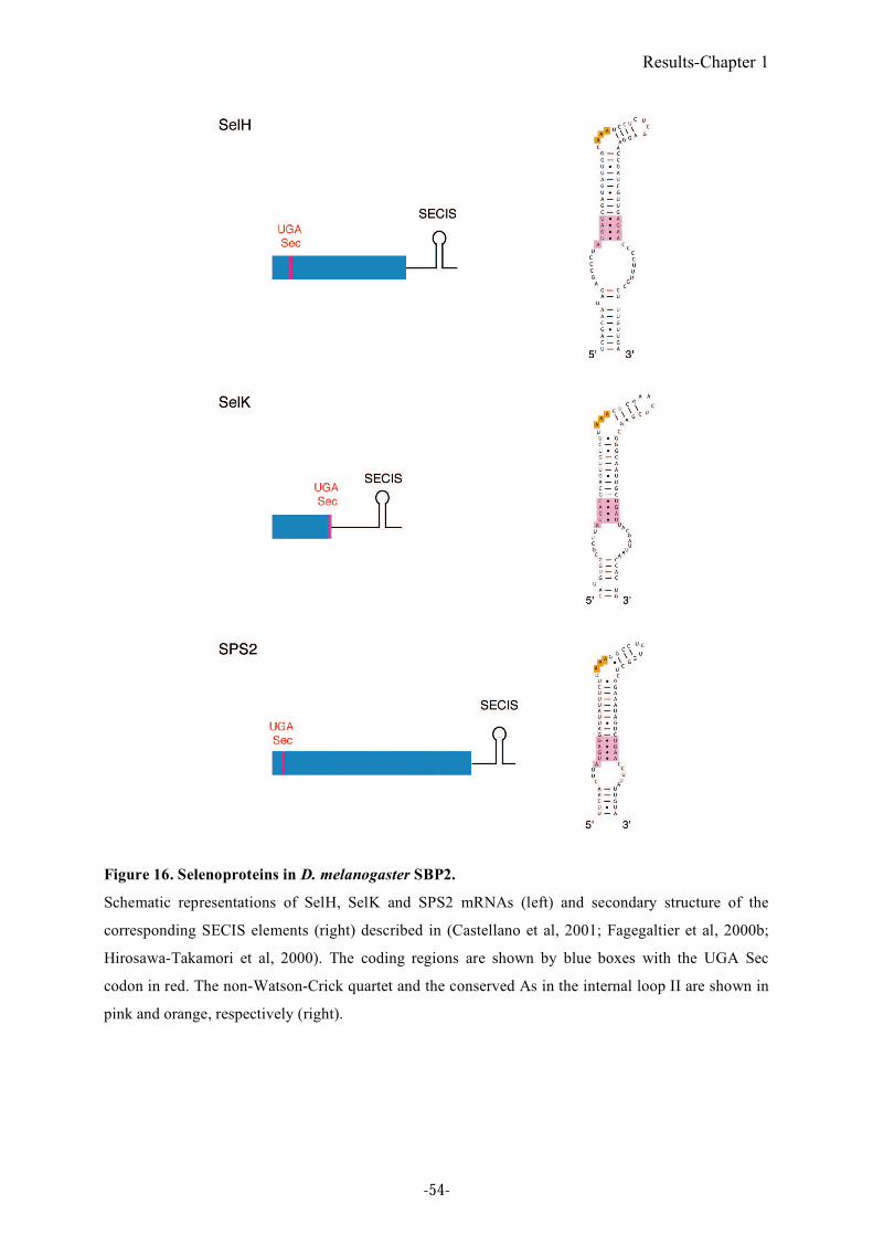
Results-Chapter 1
Figure 16. Selenoproteins in D. melanogaster SBP2.
Schematic representations of SelH, SelK and SPS2 mRNAs (left) and secondary structure of the
corresponding SECIS elements (right) described in (Castellano et al, 2001; Fagegaltier et al, 2000b;
Hirosawa-Takamori et al, 2000). The coding regions are shown by blue boxes with the UGA Sec
codon in red. The non-Watson-Crick quartet and the conserved As in the internal loop II are shown in
pink and orange, respectively (right).
-54-
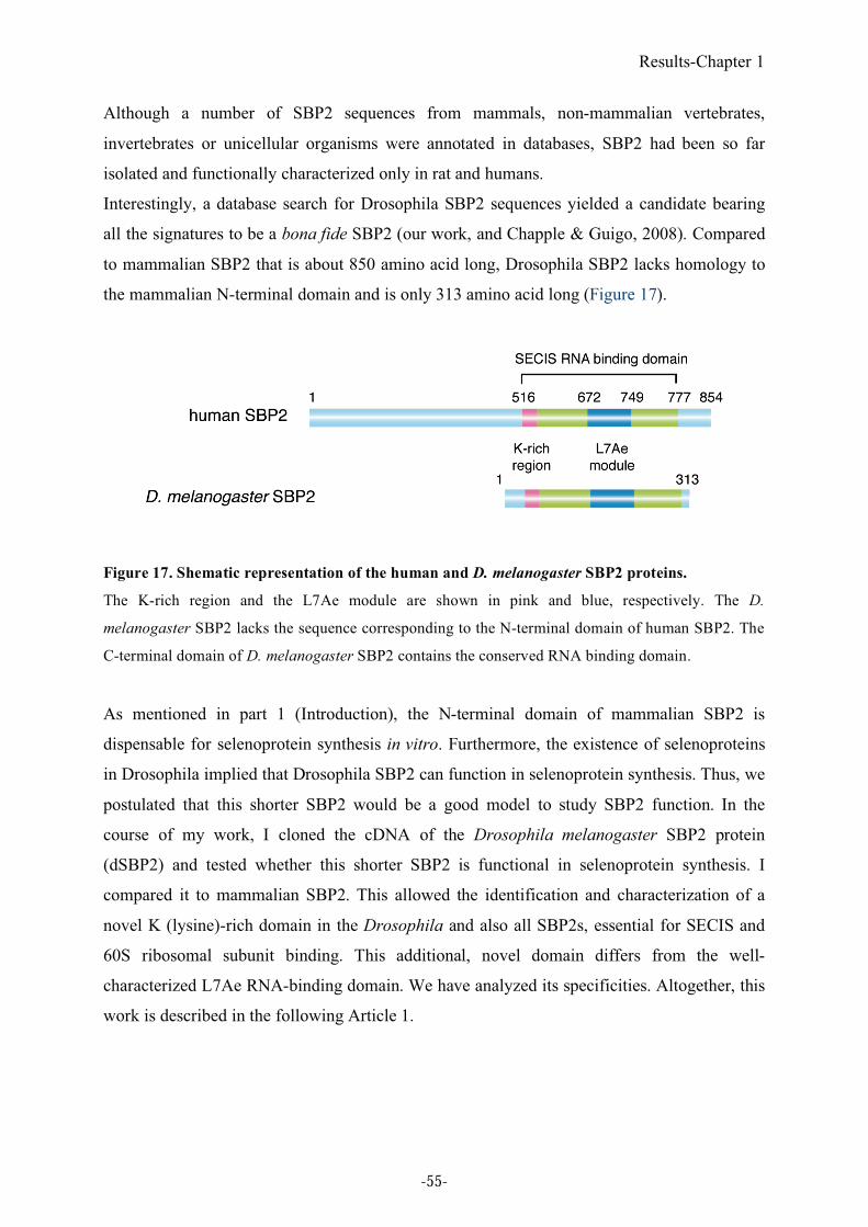
Results-Chapter 1
Although a number of SBP2 sequences from mammals, non-mammalian vertebrates,
invertebrates or unicellular organisms were annotated in databases, SBP2 had been so far
isolated and functionally characterized only in rat and humans.
Interestingly, a database search for Drosophila SBP2 sequences yielded a candidate bearing
all the signatures to be a bona fide SBP2 (our work, and Chapple & Guigo, 2008). Compared
to mammalian SBP2 that is about 850 amino acid long, Drosophila SBP2 lacks homology to
the mammalian N-terminal domain and is only 313 amino acid long (Figure 17).
Figure 17. Shematic representation of the human and D. melanogaster SBP2 proteins.
The K-rich region and the L7Ae module are shown in pink and blue, respectively. The D.
melanogaster SBP2 lacks the sequence corresponding to the N-terminal domain of human SBP2. The
C-terminal domain of D. melanogaster SBP2 contains the conserved RNA binding domain.
As mentioned in part 1 (Introduction), the N-terminal domain of mammalian SBP2 is
dispensable for selenoprotein synthesis in vitro. Furthermore, the existence of selenoproteins
in Drosophila implied that Drosophila SBP2 can function in selenoprotein synthesis. Thus, we
postulated that this shorter SBP2 would be a good model to study SBP2 function. In the
course of my work, I cloned the cDNA of the Drosophila melanogaster SBP2 protein
(dSBP2) and tested whether this shorter SBP2 is functional in selenoprotein synthesis. I
compared it to mammalian SBP2. This allowed the identification and characterization of a
novel K (lysine)-rich domain in the Drosophila and also all SBP2s, essential for SECIS and
60S ribosomal subunit binding. This additional, novel domain differs from the well-
characterized L7Ae RNA-binding domain. We have analyzed its specificities. Altogether, this
work is described in the following Article 1.
-55-
Results-Chapter 1
1.3. Summary of Article 1
In this work, I have isolated and functionally characterized a bona fide D. melanogaster SBP2
that specifically lacks the region homologous to the N-terminus of vertebrate SBP2. While
human SBP2 (hSBP2) binds both form 1 and 2 SECIS RNAs with similar affinities, dSBP2
exhibits high affinity toward form 2 only. In addition, with a homology search, we identified
a lysine-rich (K-rich) domain in human SBP2 that is essential for SECIS binding. Both
domains differ by their lysine content. They read SVRVY in Drosophila and IILKE in
humans. We showed, by introducing point mutations into hSBP2, that the differential binding
affinities to SECIS RNAs are attributed to a short amino acid sequence in the K-rich domain.
Exchanging the sequence of dSBP2 (SVRVY) with that of hSBP2 (IILKE) enabled dSBP2 to
bind form 1 SECIS RNAs but impaired binding of hSBP2. We therefore concluded that this
five amino acid sequence (IILKE) in the hSBP2 K-rich domain confers the ability to bind
form 1 SECIS RNAs.
It was previously shown that SBP2 binds the 80S ribosome (Copeland & Driscoll, 2001). In
this study, we showed that SBP2 binds in fact to the 60S subunit and that the K-rich domain is
essential for binding. This is consistent with the proposal that this association with the
ribosome may occur through binding to one of the K-turn motifs in the 28S ribosomal RNA
(Caban et al, 2007; Copeland & Driscoll, 2001; Kinzy et al, 2005). Furthermore, our finding
that the same amino acids in the K-rich region that are crucial to SECIS recognition are also
involved in ribosomal binding supports the notion that SBP2 cannot bind the SECIS element
and the ribosome simultaneously (Copeland & Driscoll, 2001).
Although SBP2 belongs to the L7Ae family, it differs from other L7Ae family proteins.
While the L7Ae family requires the single L7Ae domain to bind specifically the cognate
RNA (Chao & Williamson, 2004; Marmier-Gourrier et al, 2003; Moore et al, 2004; Nottrott
et al, 2002; Vidovic et al, 2000), SBP2 needs both the L7Ae and the K-rich domain for SECIS
specific binding. These findings were in fact already suspected from the following
experiments. While the 15.5kD and L7Ae ribosomal proteins have a rather broad specificity
and can recognize the SECIS RNA, SBP2 displayed a higher selectivity since it could not
bind either the U3 snoRNA or the archaea sRNA (Cléry et al, 2007 ). This specificity is also
reflected by the presence of nucleotide determinants in the SECIS RNA that are unique to
SBP2 recognition (See Figure 10). Indeed, helix I and internal loop 1 which are required for
SBP2 recognition, are found only in the SECIS RNAs and not in other K-turn RNAs (Cléry et
al, 2007 ).
-56-
Results-Chapter 1
These observations led us to propose the following model rationalizing the necessity of two
domains in SBP2 for complex formation with the SECIS RNA. The L7Ae motif recognizes
the guanine bases of the G.A/A.G base pairs in a similar manner to other L7Ae proteins, and
the conserved U in the SECIS non-Watson-Crick quartet. The positively charged lysines in
the K-rich domain likely increase the affinity of SBP2 for SECIS RNAs through electrostatic
interactions with the phosphates of the SECIS-specific structural features. Indeed, earlier
experiments showed that SBP2 required the phosphates of helix 1 for binding (Fletcher et al,
2001). In the K-rich domain, IILKE sequence could mediate form 1 SECIS recognition by
either a direct effect on SECIS binding or indirectly by inducing a conformational change of
the L7Ae motif. Validation of these models has to await resolution of the crystal structure of
the SBP2-SECIS RNA complex.
-57-
Results-Chapter 1
1.4. Article 1
A short motif in Drosophila SECIS Binding Protein 2 provides differential binding
affinity to SECIS RNA hairpins,
Akiko Takeuchi, David Schmitt, Charles Chapple, Elena Babaylova, Galina, Karpova,
Roderic Guigo, Alain Krol and Christine Allmang (2009) Nucleic Acids Research,
37(7):2126-41.
-58-
Signalement bibliographique ajouté par le :
UNIVERSITÉ DE STRASBOURG Service Commun de Documentation
A short motif in Drosophila SECIS Binding Protein 2 provides differential binding affinity to SECIS RNA hairpins
Akiko TAKEUCHI, David SCHMITT, Charles CHAPPLE, Elena BABAYLOVA, Galina
KARPOVA, Roderic GUIGO, Alain KROL, and Christine ALLMANG
Nucleic Acids Research, 2009, vol. 37, n° 7, pages 2126-2141
Article 1 : p 59-74 :
La publication présentée ici dans la thèse est soumise à des droits détenus par un éditeur
commercial.
Les utilisateurs de l’UdS peuvent consulter cette publication sur le site de l’éditeur :
http://dx.doi.org/10.1093/nar/gkp078
La version imprimée de cette thèse peut être consultée à la bibliothèque ou dans un autre
établissement via une demande de prêt entre bibliothèques (PEB) auprès de nos services :
http://www-sicd.u-strasbg.fr/services/peb/
Results-Chapter 1
1.5. Additional results and discussion
SBP2 and the selenoprotein synthesis machinery in Drosophila willistoni
Although dietary selenium has been thought to be involved in normalizing the life span and
fertility in Drosophila (Martin-Romero et al, 2001), selenoproteins do not seem to be essential
for viability in this fly. Mutant flies that lack EFSec failed to decode the UGA Sec codon, but
were viable and fertile (Hirosawa-Takamori et al, 2004). Furthermore, in silico comparative
genomic approaches including 12 Drosophila genomes revealed that several insect species
such as Drosophila willistoni, honey bee (Apis mellifera), wasp (Nasonia vitripennis), beetle
(Tribolium castaneum) and silkworm (Bombyx mori), do not possess selenoproteins and have
lost some of the factors involved in selenoprotein synthesis (Chapple & Guigo, 2008). In D.
willistoni, SelK and SelH are cysteine orthologs. Moreover and very interestingly, the SelK
mRNA contains a SECIS relic. EFSec is also absent in D. willistoni, and the sequence of the
tRNASec
is degenerated. An SBP2 homolog can be found, but it contains an amino acid
variation in the conserved RNA binding region (Chapple & Guigo, 2008). SPS1 and Secp43
are present and highly conserved in D. willistoni but also in other Drosophila and insect
species irrespective of their ability to encode selenoproteins. This could either mean an
ongoing genetic drift or imply that these proteins play additional function(s) unrelated to
selenoprotein synthesis (Chapple & Guigo, 2008). These data suggest the dispensability of
selenoproteins in insects, and selenoprotein may therefore harbor important but non vital
functions in insects, at least under the experimental conditions tested. We have mentioned the
existence of an SBP2 homolog in D. willistoni. Its C-terminal region, containing the SECIS
RNA-binding domain, is conserved as in the other Drosophila species (Chapple & Guigo,
2008) (Figure 18A). However, D. willistoni SBP2 contains one amino acid insertion in a
critical region of the L7Ae SECIS RNA binding domain (Figure 18B). Indeed, a conserved 19
amino acid spacing is always observed in this domain of SBP2 between two acidic amino acid
residues (Glu 679 and Glu 699 Figure 18 B and C). Position 679 is always occupied by a
glutamic acid while position 699 is sometimes replaced by an aspartic acid. Surprisingly, the
conserved spacing is not maintained in D. willistoni where insertion of an asparagine was
observed (Figure 18B). The two glutamic acids are essential for SECIS recognition: it has
been proposed that the first glutamic acid (Glu 679 in Figure 18C) contacts the conserved
guanines of the sheared base-pairs and the second one (Glu 699 in Figure 18C) recognizes the
conserved U in the non-Watson-Crick quartet (Allmang et al, 2002).
-75-
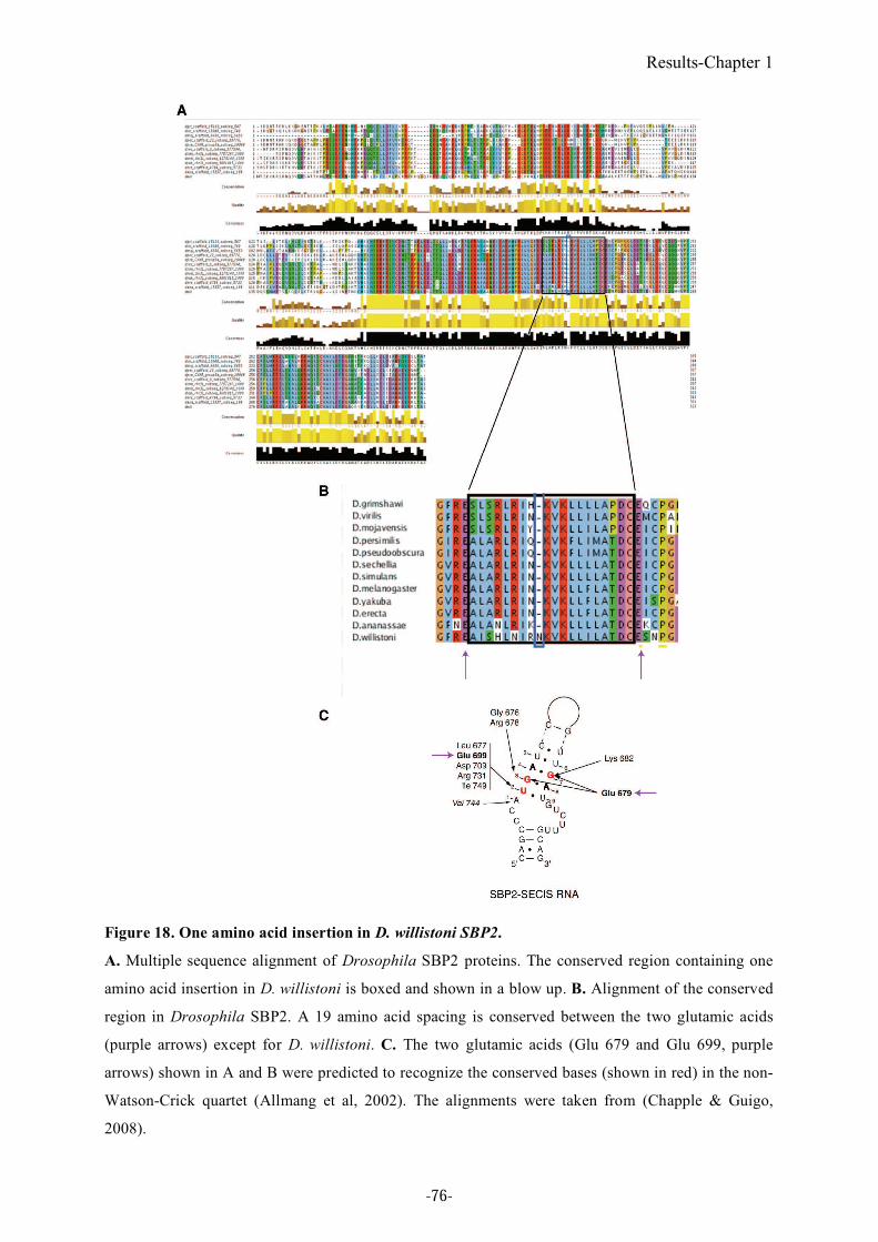
Results-Chapter 1
Figure 18. One amino acid insertion in D. willistoni SBP2.
A. Multiple sequence alignment of Drosophila SBP2 proteins. The conserved region containing one
amino acid insertion in D. willistoni is boxed and shown in a blow up. B. Alignment of the conserved
region in Drosophila SBP2. A 19 amino acid spacing is conserved between the two glutamic acids
(purple arrows) except for D. willistoni. C. The two glutamic acids (Glu 679 and Glu 699, purple
arrows) shown in A and B were predicted to recognize the conserved bases (shown in red) in the non-
Watson-Crick quartet (Allmang et al, 2002). The alignments were taken from (Chapple & Guigo,
2008).
-76-
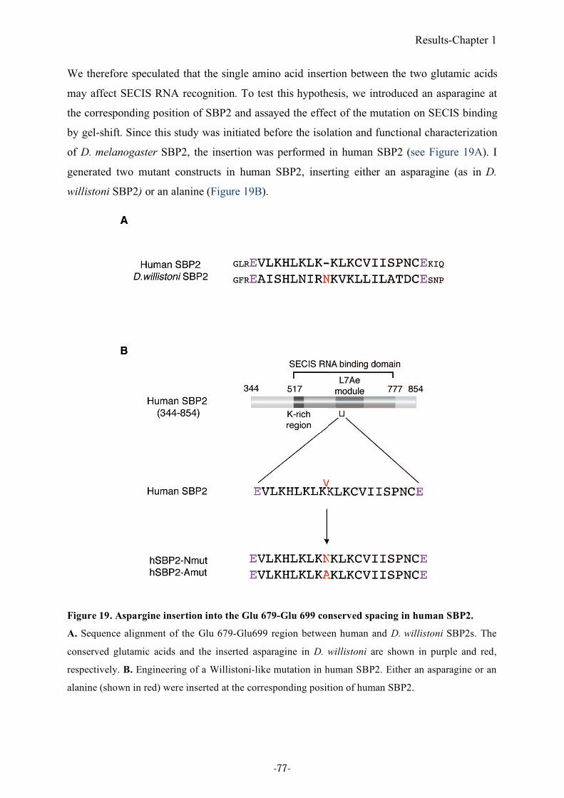
Results-Chapter 1
We therefore speculated that the single amino acid insertion between the two glutamic acids
may affect SECIS RNA recognition. To test this hypothesis, we introduced an asparagine at
the corresponding position of SBP2 and assayed the effect of the mutation on SECIS binding
by gel-shift. Since this study was initiated before the isolation and functional characterization
of D. melanogaster SBP2, the insertion was performed in human SBP2 (see Figure 19A). I
generated two mutant constructs in human SBP2, inserting either an asparagine (as in D.
willistoni SBP2) or an alanine (Figure 19B).
Figure 19. Aspargine insertion into the Glu 679-Glu 699 conserved spacing in human SBP2.
A. Sequence alignment of the Glu 679-Glu699 region between human and D. willistoni SBP2s. The
conserved glutamic acids and the inserted asparagine in D. willistoni are shown in purple and red,
respectively. B. Engineering of a Willistoni-like mutation in human SBP2. Either an asparagine or an
alanine (shown in red) were inserted at the corresponding position of human SBP2.
-77-
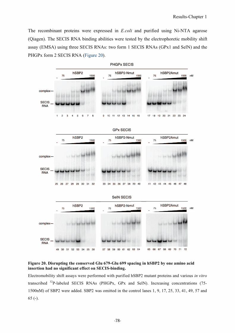
Results-Chapter 1
The recombinant proteins were expressed in E.coli and purified using Ni-NTA agarose
(Qiagen). The SECIS RNA binding abilities were tested by the electrophoretic mobility shift
assay (EMSA) using three SECIS RNAs: two form 1 SECIS RNAs (GPx1 and SelN) and the
PHGPx form 2 SECIS RNA (Figure 20).
Figure 20. Disrupting the conserved Glu 679-Glu 699 spacing in hSBP2 by one amino acid
insertion had no significant effect on SECIS-binding.
Electromobility shift assays were performed with purified hSBP2 mutant proteins and various in vitro
transcribed 32
P-labeled SECIS RNAs (PHGPx, GPx and SelN). Increasing concentrations (75-
1500nM) of SBP2 were added. SBP2 was omitted in the control lanes 1, 9, 17, 25, 33, 41, 49, 57 and
65 (-).
-78-
Results-Chapter 1
Surprisingly, both mutant proteins were able to bind all the SECIS RNAs tested, and no
significant difference was observed with the wild-type SBP2. This result indicates that in
contrast to our expectations, alteration of the spacing between the two glutamic acids has no
major effect on hSBP2 SECIS RNA binding, under our experimental conditions. As shown in
Article 1, however, the sequence of the K-rich domain differs substantially between
Drosophila and all the other eukaryotes. It is thus possible that the K-rich domain of hSBP2
could compensate for the decreased affinity for SECIS RNAs caused by the one amino acid
insertion. Other experiments will be required to determine whether the one amino acid
insertion has a real impact on SECIS binding. The most straightforward would be to perform
the insertion in the D.melanogaster SBP2 protein which displays a K-rich domain similar to
that of D. willistoni, instead of the human SBP2. However, one cannot exclude the possibility
that the insertion only reflects the genetic variety of the SBP2 gene in Drosophila without
affecting its RNA binding ability in vivo. Also, the asparagine insertion could arise from an
ongoing genetic drift concomitant to the selenoprotein gene disappearance in this Drosophila
species. Interestingly, D.willistoni lives in a specific ecological niche in Brazil where the
selenium content of the soil might be low. This could be an example of an adaptative
evolution.
Except for this amino acid insertion, the C-terminal region of SBP2 is highly conserved
across Drosophila species, independently of the selenoprotein coding capacity (Chapple &
Guigo, 2008). Furthermore, the SBP2 gene was also found in the genomes of other
selenoprotein-lacking insects, such as A. mellifera and N. vitripennis (Chapple & Guigo,
2008). SBP2 might also have other roles than selenoprotein synthesis so that SBP2 was
retained in the genome of selenoprotein-lacking species (Chapple & Guigo, 2008). However,
the conservation might also just reflect an ongoing genetic drift.
-79-

-80-
Results-Chapter 2
2. Toward crystallization of the SBP2/SECIS complex
2.1. Objective
The SBP2-SECIS interaction is at the heart of the selenocysteine incorporation mechanism.
To get more insight into this mechanism, it is important to understand the specific features of
this interaction at the atomic level. To this end, we started a crystallographic study of the
SBP2-SECIS RNA complex in collaboration with the group of Philippe Dumas in the same
research unit, UPR9002 of CNRS.
As discussed in Chapter 1 (in 1.4.), SBP2 is set apart from other L7Ae proteins: besides the
L7Ae motif, SBP2 requires the additional domain (K-rich region) for SECIS recognition
(Takeuchi et al, 2009). The SECIS element also presents unique functional and structural
features that are different from canonical K-turn RNAs such as the long and stable helices 1
and 2 and the large internal loop (Cléry et al, 2007 ). Therefore, the SBP2-SECIS interaction
principle has been proposed to be different from that of other L7Ae proteins/K-turn RNA
complexes (Cléry et al, 2007 and unpublished results from the laboratory). Resolution of the
crystal structure of the SBP2-SECIS complex is required for the validation of our model
proposing that additional protein-RNA contacts must exist in the SBP2-SECIS complex, with
respect to other complexes involving similar proteins and RNAs. Resolution of the structure
would also be invaluable to understand the role of the K-rich region of SBP2 in SECIS RNA
recognition as well as to establish whether the SECIS RNA adopts a K-turn like structure
(Allmang et al, 2002).
Crystallization attempts were previously carried out in the laboratory and were the subject of
the thesis of Vincent Olieric (team of Philippe Dumas). However, despite many trials, no
crystal could be obtained, either of the protein alone or of the complex with the SECIS RNA.
All the purification and crystallization attempts were performed using SBP2 proteins
expressed in E coli. However, several problems were observed during protein expression and
purification, such as a low level of protein production and sensitivity to proteolysis. We
speculated that the unavailability of sufficient amounts of SBP2 may rely on the absence of
post-translational modification. We therefore decided to clone the cDNA of other SBP2
proteins, fused with different tags and to express them in baculovirus-infected insect cells.
This would hopefully improve protein expression and enable the obtention of proteins
produced in a eukaryotic system for further crystallization assays. My participation in this
-81-
Results-Chapter 2
study was to generate various SBP2 constructs compatible with expression in insect cells
using the baculovirus expression system. I also determined the best conditions for protein
expression with the help of the Plateforme de Génomique et Biologie Structurales at the
Centre Européen de Biologie et Génomique Structurales (CEBGS) and the baculovirus
service at IGBMC in Illkirch. In collaboration with the group of Philippe Dumas, various
biophysical techniques as well as sequence analysis were used in addition to the
crystallographic attempts to better understand the structure of SBP2.
2.2. Results
2.2.1. cDNA cloning of SBP2 from various organisms
Because only human SBP2 had been used so far for crystallographic attempts, we set out to
clone and express SBP2 from rat and Drosophila (see Figure 21). In addition to rat SBP2, that
has been used by others for experimental studies (Bubenik & Driscoll, 2007; Caban et al,
2007; Chavatte et al, 2005; Copeland et al, 2000; Copeland et al, 2001; de Jesus et al, 2006;
Donovan et al, 2008; Gupta & Copeland, 2007; Kinzy et al, 2005; Small-Howard et al, 2006)
and that is highly similar to human SBP2 in both length and sequence (Lescure et al, 2002),
we also selected the newly identified Drosophila SBP2 (Takeuchi et al, 2009). Because of its
shorter length, we postulated that it would be produced more efficiently, and that it would be
especially more soluble than the longer mammalian SBP2s, both criteria being important for
the crystallographic study.
A previous work in the laboratory, using different constructs of human SBP2 bearing a
thrombin cleavage site between the tag and the actual SBP2 sequence, revealed the
occurrence of a sensitive cleavage site after Lys525 during the purification steps. Although
this purification procedure included the treatment with thrombin to separate SBP2 from the
tag, the cleavage after Lys525 did not seem to be provoked by thrombin because of the
following observations. The sequence EIPK525AKK does not match the consensus thrombin
cleavage site, and increasing concentrations of thrombin did not modify the
cleaved/uncleaved ratio. In addition, the constructs used in the previous work led to low
solubility. To avoid proteolysis, we resorted to a new construct starting at Ala526 (hereafter
-82-
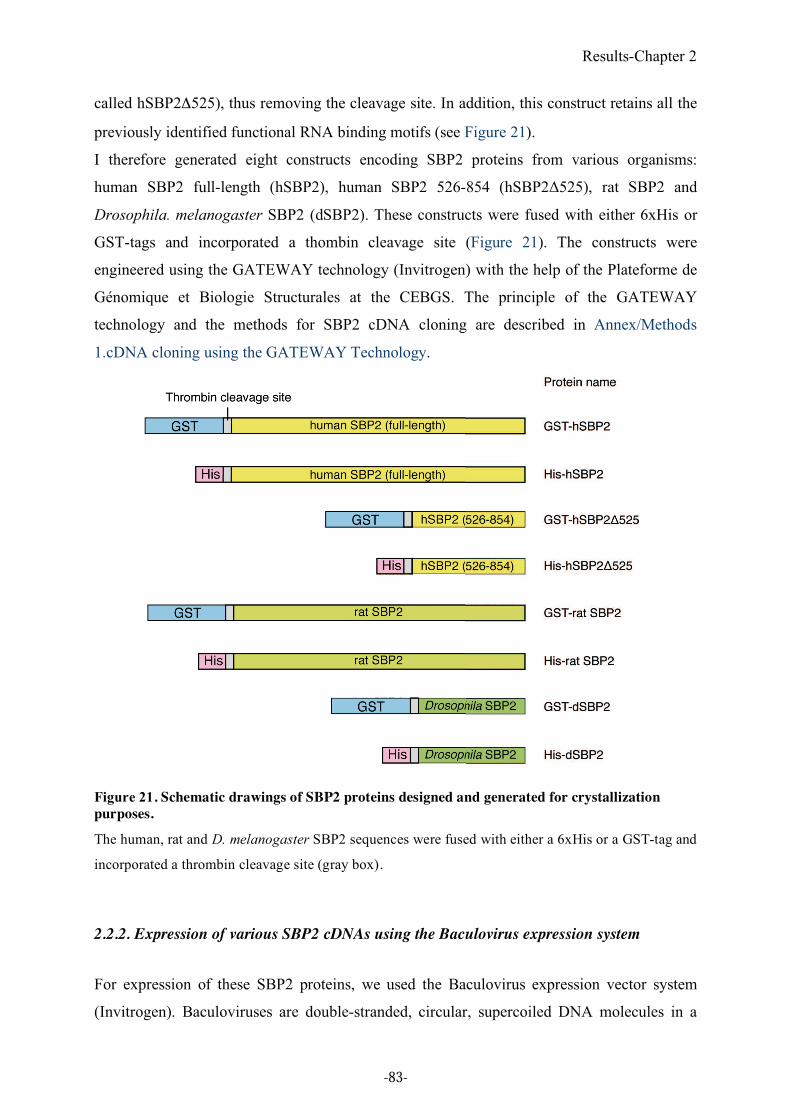
Results-Chapter 2
called hSBP2"525), thus removing the cleavage site. In addition, this construct retains all the
previously identified functional RNA binding motifs (see Figure 21).
I therefore generated eight constructs encoding SBP2 proteins from various organisms:
human SBP2 full-length (hSBP2), human SBP2 526-854 (hSBP2!525), rat SBP2 and
Drosophila. melanogaster SBP2 (dSBP2). These constructs were fused with either 6xHis or
GST-tags and incorporated a thombin cleavage site (Figure 21). The constructs were
engineered using the GATEWAY technology (Invitrogen) with the help of the Plateforme de
Génomique et Biologie Structurales at the CEBGS. The principle of the GATEWAY
technology and the methods for SBP2 cDNA cloning are described in Annex/Methods
1.cDNA cloning using the GATEWAY Technology.
Figure 21. Schematic drawings of SBP2 proteins designed and generated for crystallization
purposes.
The human, rat and D. melanogaster SBP2 sequences were fused with either a 6xHis or a GST-tag and
incorporated a thrombin cleavage site (gray box).
2.2.2. Expression of various SBP2 cDNAs using the Baculovirus expression system
For expression of these SBP2 proteins, we used the Baculovirus expression vector system
(Invitrogen). Baculoviruses are double-stranded, circular, supercoiled DNA molecules in a
-83-
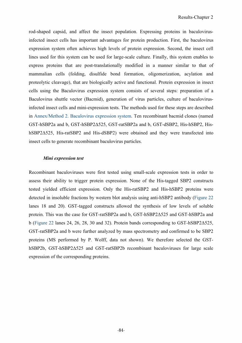
Results-Chapter 2
rod-shaped capsid, and affect the insect population. Expressing proteins in baculovirus-
infected insect cells has important advantages for protein production. First, the baculovirus
expression system often achieves high levels of protein expression. Second, the insect cell
lines used for this system can be used for large-scale culture. Finally, this system enables to
express proteins that are post-translationally modified in a manner similar to that of
mammalian cells (folding, disulfide bond formation, oligomerization, acylation and
proteolytic cleavage), that are biologically active and functional. Protein expression in insect
cells using the Baculovirus expression system consists of several steps: preparation of a
Baculovirus shuttle vector (Bacmid), generation of virus particles, culture of baculovirus-
infected insect cells and mini-expression tests. The methods used for these steps are described
in Annex/Method 2. Baculovirus expression system. Ten recombinant bacmid clones (named
GST-hSBP2a and b, GST-hSBP2!525, GST-ratSBP2a and b, GST-dSBP2, His-hSBP2, His-
hSBP2!525, His-ratSBP2 and His-dSBP2) were obtained and they were transfected into
insect cells to generate recombinant baculovirus particles.
Mini expression test
Recombinant baculoviruses were first tested using small-scale expression tests in order to
assess their ability to trigger protein expression. None of the His-tagged SBP2 constructs
tested yielded efficient expression. Only the His-ratSBP2 and His-hSBP2 proteins were
detected in insoluble fractions by western blot analysis using anti-hSBP2 antibody (Figure 22
lanes 18 and 20). GST-tagged constructs allowed the synthesis of low levels of soluble
protein. This was the case for GST-ratSBP2a and b, GST-hSBP2!525 and GST-hSBP2a and
b (Figure 22 lanes 24, 26, 28, 30 and 32). Protein bands corresponding to GST-hSBP2!525,
GST-ratSBP2a and b were further analyzed by mass spectrometry and confirmed to be SBP2
proteins (MS performed by P. Wolff, data not shown). We therefore selected the GST-
hSBP2b, GST-hSBP2!525 and GST-ratSBP2b recombinant baculoviruses for large scale
expression of the corresponding proteins.
-84-
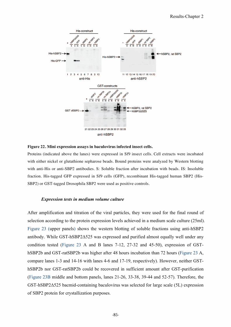
Results-Chapter 2
Figure 22. Mini expression assays in baculovirus infected insect cells.
Proteins (indicated above the lanes) were expressed in Sf9 insect cells. Cell extracts were incubated
with either nickel or glutathione sepharose beads. Bound proteins were analyzed by Western blotting
with anti-His or anti-SBP2 antibodies. S: Soluble fraction after incubation with beads. IS: Insoluble
fraction. His-tagged GFP expressed in Sf9 cells (GFP), recombinant His-tagged human SBP2 (His-
SBP2) or GST-tagged Drosophila SBP2 were used as positive controls.
Expression tests in medium volume culture
After amplification and titration of the viral particles, they were used for the final round of
selection according to the protein expression levels achieved in a medium scale culture (25ml).
Figure 23 (upper panels) shows the western blotting of soluble fractions using anti-hSBP2
antibody. While GST-hSBP2!525 was expressed and purified almost equally well under any
condition tested (Figure 23 A and B lanes 7-12, 27-32 and 45-50), expression of GST-
hSBP2b and GST-ratSBP2b was higher after 48 hours incubation than 72 hours (Figure 23 A,
compare lanes 1-3 and 14-16 with lanes 4-6 and 17-19, respectively). However, neither GST-
hSBP2b nor GST-ratSBP2b could be recovered in sufficient amount after GST-purification
(Figure 23B middle and bottom panels, lanes 21-26, 33-38, 39-44 and 52-57). Therefore, the
GST-hSBP2!525 bacmid-containing baculovirus was selected for large scale (5L) expression
of SBP2 protein for crystallization purposes.
-85-
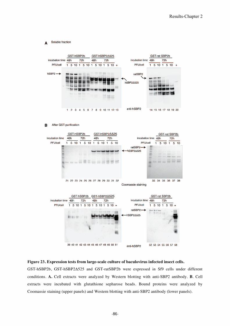
Results-Chapter 2
Figure 23. Expression tests from large-scale culture of baculovirus infected insect cells.
GST-hSBP2b, GST-hSBP2"525 and GST-ratSBP2b were expressed in Sf9 cells under different
conditions. A. Cell extracts were analyzed by Western blotting with anti-SBP2 antibody. B. Cell
extracts were incubated with glutathione sepharose beads. Bound proteins were analyzed by
Coomassie staining (upper panels) and Western blotting with anti-SBP2 antibody (lower panels).
-86-
Results-Chapter 2
2.2.3. Biophysical analysis of SBP2
Our initial aim, when producing SBP2 in baculovirus-infected insect cells, was to improve
protein expression and stability and therefore the ability of the protein to crystallize.
However, no crystal could be obtained even with the protein produced under the conditions
described in this chapter.
Interestingly, the use of structure prediction algorithms suggested the existence of disordered
areas in SBP2. It appears now that a growing number of eukaryotic genes encode proteins that
lack three-dimensional folding but become folded upon binding to their targets. Such proteins
are called Intrinsically Disordered Proteins (IDPs).
Unstructured protein domains differ from structured proteins in many properties, such as
amino acid compositional bias, high apparent molecular mass, heat resistance, acid resistance
and extreme proteolytic sensitivity. For amino acid composition, unstructured domains are
enriched in disorder-promoting amino acids (A, R, G, Q, S, P, E and K) and comprise less
order-promoting amino acids (W, C, F, I, Y, V, L and N) (reviewed in Dyson & Wright,
2005; Rajkowitsch et al, 2007; Tompa, 2002; Wright & Dyson, 1999). The analysis of folded
and unfolded proteins based on the normalized net charge and mean hydrophobicity revealed
that IDPs are usually localized within a unique region of the charge-hydrophobicity phase
space. Indeed, a combination of high mean net charge and low overall hydrophobicity
represents a unique structural feature of IDPs (Uversky et al, 2000). A number of computer
programs such as PONDR, DISOPRED2, DisEMBL GlobProt2 and IUPred allow the
prediction of disordered regions based on the amino acid sequence (reviewed in Dyson &
Wright, 2005; Rajkowitsch et al, 2007).
We therefore decided to test whether SBP2 is an IDP. Indeed, SBP2 does exhibit several
features often observed with unfolded proteins. For example, the SBP2 sequence is rich in
proline residues, one of the disorder promoting residues, and the percentages of proline
contents are 7.3% for rat SBP2 and 8.6% for human SBP2, figures that are higher than for the
average of the Swiss-Prot proteins (4.9%) (Linding et al, 2003; Tompa, 2002). This is
consistent with the early experimental results in the laboratory that SBP2 exhibited unusually
higher molecular mass in gel filtration experiments and SDS-PAGE than those measured by
mass spectrometry. Computer predictions were performed using the various forms of SBP2
proteins that I described in this chapter. Interestingly, the disorder-prediction methods showed
that ca. 70 % of the SBP2 sequence is disordered, whereas the L7Ae RNA binding domain
-87-
Results-Chapter 2
appears to be folded. We therefore decided to perform biophysical analyses to test this
hypothesis. The results are described in the following article (2.3. Article 2)
Since no high-resolution X-ray structures of intrinsically disordered proteins are available due
to the lack of stable three-dimensional folds, a number of other experimental methods are
used to elucidate disordered domains. Nuclear magnetic resonance (NMR), circular dichroism
(CD), differential scanning calorimetry and fluorescence anisotropy that are based on
structure determination in solution, distinguish between structured and unstructured domains
(reviewed in Rajkowitsch et al, 2007). Several hydrodynamic techniques can also detect
unfolded conformations. Gel filtration (size-exclusion chromatography), Small-Angle-X-ray
Scattering (SAXS), sedimentation analysis and dynamic light scattering provide information
in hydrodynamic parameters that can detect characteristic behaviors to intrinsically disordered
proteins (reviewed in Tompa, 2002).
We have used several of these biophysical techniques such as 1H 1D-NMR, analytical
centrifugation and dynamic light scattering (DLS). Results are described In Article 2.
They reveal that SBP2 shows an unusually high molecular weight in dynamic light scattering
(DLS) experiments, supporting the non-globular character of the rat SBP2 and human
SBP2"525. Ultracentrifugation analysis showed that a monomeric SBP2 protein is in part
unstructured. 1H 1D-NMR, a technique that can differentiate a folded from a non-folded
protein in the resonance range characteristic of peptide bonds, showed the lack of peptide
bonds and methyl resonances of the rat SBP2 and human SBP2"525 expressed in the
bacterial and the eukaryotic systems. All these results show that SBP2 is largely unfolded,
and is indeed a member of the family of Intrinsically Disordered Proteins. We postulated that
the reasons for the previous unsuccessful crystallization may rely on the presence of disorder
in the protein.
-88-
Results-Chapter 2
2.3. Article 2 (in press)
SECIS-binding protein 2, a key player in selenoprotein synthesis, is an intrinsically
disordered protein
Vincent Oliéric, Philippe Wolff, Akiko Takeuchi, Guillaume Bec, Catherine Birck, Marc
Vitorino, Bruno Kieffer, Artemy Beniaminov, Giorgio Cavigiolio, Elizabeth Theil,
Christine Allmang, Alain Krol and Philippe Dumas (2009) Biochimie 91(8): 1003-1009.
-89-

Results-Chapter 2
-90-
Signalement bibliographique ajouté par le :
UNIVERSITÉ DE STRASBOURG Service Commun de Documentation
SECIS-binding protein 2, a key player in selenoprotein synthesis, is an intrinsically disordered protein
Vincent OLIERIC, Philippe WOLFF, Akiko TAKEUCHI, Guillaume BEC, Catherine BIRCK, Marc
VITORINO, Bruno KIEFFER, Artemy BENIAMINOV, Giorgio CAVIGIOLIO, Elizabeth THEIL,
Christine ALLMANG, Alain KROL and Philippe DUMAS
Biochimie, 2009, vol. 91, n° 8, pages 1003-1009
Article 2 : p 91-97 :
La publication présentée ici dans la thèse est soumise à des droits détenus par un éditeur
commercial.
Les utilisateurs de l’UdS peuvent consulter cette publication sur le site de l’éditeur :
http://dx.doi.org/10.1016/j.biochi.2009.05.004
La version imprimée de cette thèse peut être consultée à la bibliothèque ou dans un autre
établissement via une demande de prêt entre bibliothèques (PEB) auprès de nos services :
http://www-sicd.u-strasbg.fr/services/peb/

Results-Chapter 2
-98-
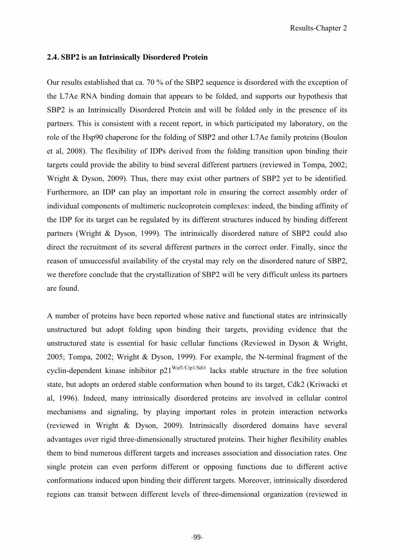
Results-Chapter 2
2.4. SBP2 is an Intrinsically Disordered Protein
Our results established that ca. 70 % of the SBP2 sequence is disordered with the exception of
the L7Ae RNA binding domain that appears to be folded, and supports our hypothesis that
SBP2 is an Intrinsically Disordered Protein and will be folded only in the presence of its
partners. This is consistent with a recent report, in which participated my laboratory, on the
role of the Hsp90 chaperone for the folding of SBP2 and other L7Ae family proteins (Boulon
et al, 2008). The flexibility of IDPs derived from the folding transition upon binding their
targets could provide the ability to bind several different partners (reviewed in Tompa, 2002;
Wright & Dyson, 2009). Thus, there may exist other partners of SBP2 yet to be identified.
Furthermore, an IDP can play an important role in ensuring the correct assembly order of
individual components of multimeric nucleoprotein complexes: indeed, the binding affinity of
the IDP for its target can be regulated by its different structures induced by binding different
partners (Wright & Dyson, 1999). The intrinsically disordered nature of SBP2 could also
direct the recruitment of its several different partners in the correct order. Finally, since the
reason of unsuccessful availability of the crystal may rely on the disordered nature of SBP2,
we therefore conclude that the crystallization of SBP2 will be very difficult unless its partners
are found.
A number of proteins have been reported whose native and functional states are intrinsically
unstructured but adopt folding upon binding their targets, providing evidence that the
unstructured state is essential for basic cellular functions (Reviewed in Dyson & Wright,
2005; Tompa, 2002; Wright & Dyson, 1999). For example, the N-terminal fragment of the
cyclin-dependent kinase inhibitor p21Waf1/Cip1/Sdi1
lacks stable structure in the free solution
state, but adopts an ordered stable conformation when bound to its target, Cdk2 (Kriwacki et
al, 1996). Indeed, many intrinsically disordered proteins are involved in cellular control
mechanisms and signaling, by playing important roles in protein interaction networks
(reviewed in Wright & Dyson, 2009). Intrinsically disordered domains have several
advantages over rigid three-dimensionally structured proteins. Their higher flexibility enables
them to bind numerous different targets and increases association and dissociation rates. One
single protein can even perform different or opposing functions due to different active
conformations induced upon binding their different targets. Moreover, intrinsically disordered
regions can transit between different levels of three-dimensional organization (reviewed in
-99-
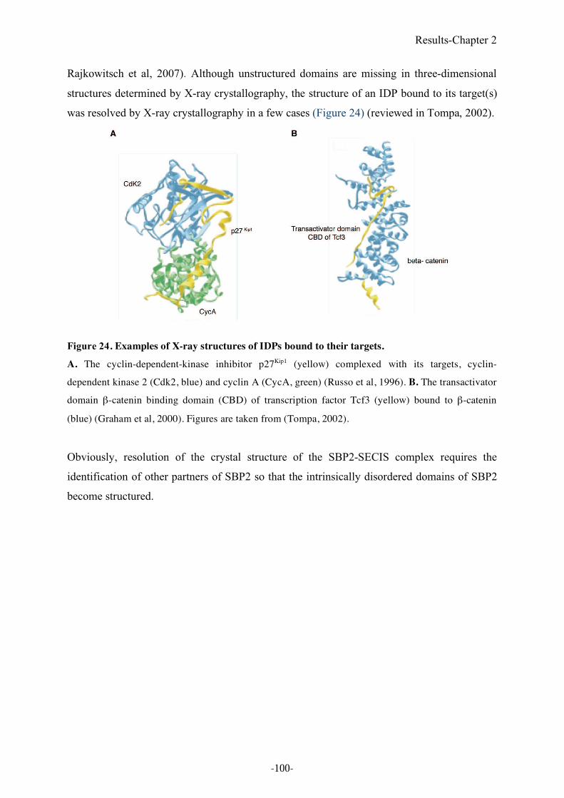
Results-Chapter 2
Rajkowitsch et al, 2007). Although unstructured domains are missing in three-dimensional
structures determined by X-ray crystallography, the structure of an IDP bound to its target(s)
was resolved by X-ray crystallography in a few cases (Figure 24) (reviewed in Tompa, 2002).
Figure 24. Examples of X-ray structures of IDPs bound to their targets.
A. The cyclin-dependent-kinase inhibitor p27Kip1 (yellow) complexed with its targets, cyclin-
dependent kinase 2 (Cdk2, blue) and cyclin A (CycA, green) (Russo et al, 1996). B. The transactivator
domain #-catenin binding domain (CBD) of transcription factor Tcf3 (yellow) bound to #-catenin
(blue) (Graham et al, 2000). Figures are taken from (Tompa, 2002).
Obviously, resolution of the crystal structure of the SBP2-SECIS complex requires the
identification of other partners of SBP2 so that the intrinsically disordered domains of SBP2
become structured.
-100-
Results-Chapter 3
3. Toward identification of SBP2 partners
3.1. Objective
The expression mechanism of selenoprotein mRNAs involves a number of factors (see Part 1.
Introduction). Among these, SBP2 plays an important role in the assembly of selenoprotein
mRNPs. It notably recruits the Sec-tRNASec
/EFSec complex to the SECIS element during
selenoprotein synthesis (Tujebajeva et al, 2000) but it also associates with a complex
machinery linked to the Hsp90 chaperone that triggers proper SBP2 folding and subsequent
SECIS RNP assembly (Boulon et al, 2008). This assembly machinery is conserved and
involved in the assembly of several L7Ae RNPs and implies that SBP2 has functional
similarities with other L7Ae proteins during RNP assembly (Boulon et al, 2008).
Interestingly, the L7Ae proteins are primary binding proteins that participate in several RNP
complexes by binding directly to the K-turn RNA motifs. This binding is a prerequisite to the
assembly of the other core proteins of the RNP complexes to which they belong.
In archaea, formation of the L7Ae initiation complex enables the recruitment of Nop5 to the
assembling archeal RNP, which in turn facilitates the association of fibrillarin to the
catalytically active sRNP in vitro (Omer et al, 2002). In eukaryotes, 15.5kD (the L7Ae protein
that exhibits the highest similarity with SBP2 (Allmang et al, 2002)) is at the heart of different
sn/snoRNPs (Watkins et al, 2000) such as box C/D snoRNPs, U3 snoRNP and U4 snRNP. In
box C/D snoRNPs, Snu13p/15.5kD (the eukaryotic homolog of archaeal L7Ae) is part of an
RNP complex similar to the archaeal one containing Nop56, Nop58 (the eukaryotic orthologs
of archaeal Nop5) and fibrillarin (Figure 25 A and B, Kuhn et al, 2002; Nottrott et al, 1999,
for a review on sn(o)RNP structure, see Reichow et al, 2007). In the U3 box C/D snoRNPs,
that plays essential roles in ribose 2’-O-methylation and pre-rRNA processing, 15.5kD is also
present together with the methyltransferase fibrillarin, Nop56 and Nop58 (Watkins et al,
2002). In the case of the U4 snRNPs, one of the major components of the spliceosome, the
interaction between 15.5kD and the U4 snRNA is required for the association of a different
set of core proteins, PRP31 and the cyclophilin H-hPRP4-hPRP3 complex (Nottrott et al,
2002) (Figure 25 C).
-101-
Results-Chapter 3
Figure 25. Composition and organization of C/D sno(s)RNPs and U4 snRNP
A. Core proteins of the archaeal C/D sRNP are L7Ae, fibrillarin and Nop5. Nop5 interacts with
fibrillarin, and Nop5-fibrillarin complex dimerizes. B. The eukaryotic C/D snoRNP contains
15.5kD/Snu13 (L7Ae homolog), fibrillarin, Nop56 and Nop58 (Nop5 paralogs). In contrast to the
archaeal L7Ae, 15.5kD appears to bind only at the C/D sites of the snoRNA. C. The U4 snRNP
contains 15.5kD/Snu13, hPRP31, and the cyclophilin H-hPRP4-hPRP3 complex as well as seven
common Sm proteins. hPRP31 contains the Nop domain that mediates binding to the 15.5kD-U4
snRNA complex (Liu et al, 2007).
We therefore speculated that, by analogy with 15.5kD, SBP2 may be able to recruit to
selenoprotein mRNP core proteins that are common to those of sn/snoRNPs. As a starting
hypothesis, we tested whether Nop56 and Nop58, the most common interactants of 15.5 kD,
could also interact with SBP2. This work was initiated together with Laurence Wurth, another
PhD student in the laboratory whose main project is to understand the assembly pathway of
the selenoprotein mRNPs and identify new protein partners for SBP2. In this work, based on
the encouraging preliminary results of her in vivo studies, I further analyzed the interaction
between SBP2 and Nop56 and Nop58 in vitro, in order to determine whether these proteins
could be potential partners of SBP2.
-102-
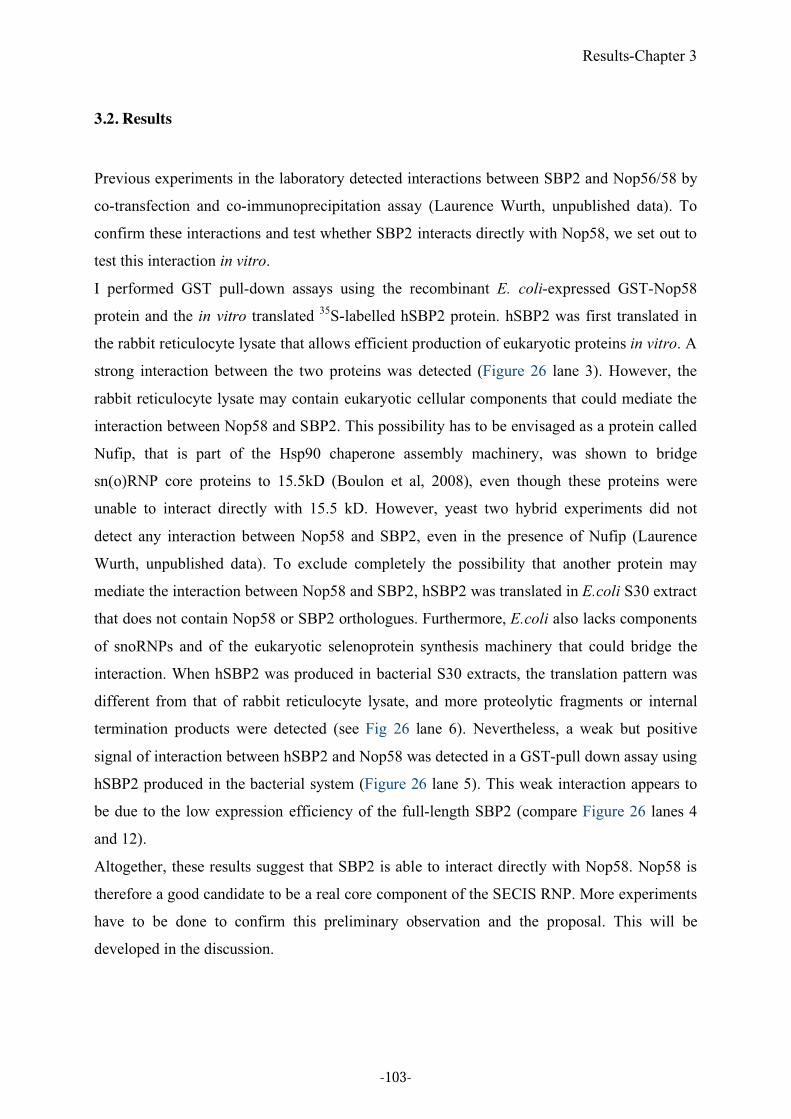
Results-Chapter 3
3.2. Results
Previous experiments in the laboratory detected interactions between SBP2 and Nop56/58 by
co-transfection and co-immunoprecipitation assay (Laurence Wurth, unpublished data). To
confirm these interactions and test whether SBP2 interacts directly with Nop58, we set out to
test this interaction in vitro.
I performed GST pull-down assays using the recombinant E. coli-expressed GST-Nop58
protein and the in vitro translated 35
S-labelled hSBP2 protein. hSBP2 was first translated in
the rabbit reticulocyte lysate that allows efficient production of eukaryotic proteins in vitro. A
strong interaction between the two proteins was detected (Figure 26 lane 3). However, the
rabbit reticulocyte lysate may contain eukaryotic cellular components that could mediate the
interaction between Nop58 and SBP2. This possibility has to be envisaged as a protein called
Nufip, that is part of the Hsp90 chaperone assembly machinery, was shown to bridge
sn(o)RNP core proteins to 15.5kD (Boulon et al, 2008), even though these proteins were
unable to interact directly with 15.5 kD. However, yeast two hybrid experiments did not
detect any interaction between Nop58 and SBP2, even in the presence of Nufip (Laurence
Wurth, unpublished data). To exclude completely the possibility that another protein may
mediate the interaction between Nop58 and SBP2, hSBP2 was translated in E.coli S30 extract
that does not contain Nop58 or SBP2 orthologues. Furthermore, E.coli also lacks components
of snoRNPs and of the eukaryotic selenoprotein synthesis machinery that could bridge the
interaction. When hSBP2 was produced in bacterial S30 extracts, the translation pattern was
different from that of rabbit reticulocyte lysate, and more proteolytic fragments or internal
termination products were detected (see Fig 26 lane 6). Nevertheless, a weak but positive
signal of interaction between hSBP2 and Nop58 was detected in a GST-pull down assay using
hSBP2 produced in the bacterial system (Figure 26 lane 5). This weak interaction appears to
be due to the low expression efficiency of the full-length SBP2 (compare Figure 26 lanes 4
and 12).
Altogether, these results suggest that SBP2 is able to interact directly with Nop58. Nop58 is
therefore a good candidate to be a real core component of the SECIS RNP. More experiments
have to be done to confirm this preliminary observation and the proposal. This will be
developed in the discussion.
-103-
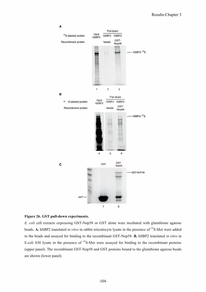
Results-Chapter 3
Figure 26. GST pull-down experiments.
E. coli cell extracts expressing GST-Nop58 or GST alone were incubated with glutathione agarose
beads. A. hSBP2 translated in vitro in rabbit reticulocyte lysate in the presence of 35
S-Met were added
to the beads and assayed for binding to the recombinant GST-Nop58. B. hSBP2 translated in vitro in
E.coli S30 lysate in the presence of 35
S-Met were assayed for binding to the recombinant proteins
(upper panel). The recombinant GST-Nop58 and GST proteins bound to the glutathione agarose beads
are shown (lower panel).
-104-
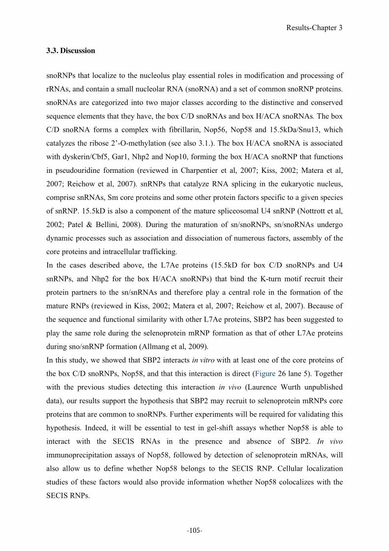
Results-Chapter 3
3.3. Discussion
snoRNPs that localize to the nucleolus play essential roles in modification and processing of
rRNAs, and contain a small nucleolar RNA (snoRNA) and a set of common snoRNP proteins.
snoRNAs are categorized into two major classes according to the distinctive and conserved
sequence elements that they have, the box C/D snoRNAs and box H/ACA snoRNAs. The box
C/D snoRNA forms a complex with fibrillarin, Nop56, Nop58 and 15.5kDa/Snu13, which
catalyzes the ribose 2’-O-methylation (see also 3.1.). The box H/ACA snoRNA is associated
with dyskerin/Cbf5, Gar1, Nhp2 and Nop10, forming the box H/ACA snoRNP that functions
in pseudouridine formation (reviewed in Charpentier et al, 2007; Kiss, 2002; Matera et al,
2007; Reichow et al, 2007). snRNPs that catalyze RNA splicing in the eukaryotic nucleus,
comprise snRNAs, Sm core proteins and some other protein factors specific to a given species
of snRNP. 15.5kD is also a component of the mature spliceosomal U4 snRNP (Nottrott et al,
2002; Patel & Bellini, 2008). During the maturation of sn/snoRNPs, sn/snoRNAs undergo
dynamic processes such as association and dissociation of numerous factors, assembly of the
core proteins and intracellular trafficking.
In the cases described above, the L7Ae proteins (15.5kD for box C/D snoRNPs and U4
snRNPs, and Nhp2 for the box H/ACA snoRNPs) that bind the K-turn motif recruit their
protein partners to the sn/snRNAs and therefore play a central role in the formation of the
mature RNPs (reviewed in Kiss, 2002; Matera et al, 2007; Reichow et al, 2007). Because of
the sequence and functional similarity with other L7Ae proteins, SBP2 has been suggested to
play the same role during the selenoprotein mRNP formation as that of other L7Ae proteins
during sno/snRNP formation (Allmang et al, 2009).
In this study, we showed that SBP2 interacts in vitro with at least one of the core proteins of
the box C/D snoRNPs, Nop58, and that this interaction is direct (Figure 26 lane 5). Together
with the previous studies detecting this interaction in vivo (Laurence Wurth unpublished
data), our results support the hypothesis that SBP2 may recruit to selenoprotein mRNPs core
proteins that are common to snoRNPs. Further experiments will be required for validating this
hypothesis. Indeed, it will be essential to test in gel-shift assays whether Nop58 is able to
interact with the SECIS RNAs in the presence and absence of SBP2. In vivo
immunoprecipitation assays of Nop58, followed by detection of selenoprotein mRNAs, will
also allow us to define whether Nop58 belongs to the SECIS RNP. Cellular localization
studies of these factors would also provide information whether Nop58 colocalizes with the
SECIS RNPs.
-105-
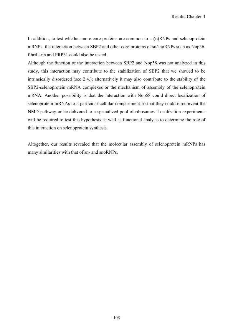
Results-Chapter 3
In addition, to test whether more core proteins are common to sn(o)RNPs and selenoprotein
mRNPs, the interaction between SBP2 and other core proteins of sn/snoRNPs such as Nop56,
fibrillarin and PRP31 could also be tested.
Although the function of the interaction between SBP2 and Nop58 was not analyzed in this
study, this interaction may contribute to the stabilization of SBP2 that we showed to be
intrinsically disordered (see 2.4.); alternatively it may also contribute to the stability of the
SBP2-selenoprotein mRNA complexes or the mechanism of assembly of the selenoprotein
mRNA. Another possibility is that the interaction with Nop58 could direct localization of
selenoprotein mRNAs to a particular cellular compartment so that they could circumvent the
NMD pathway or be delivered to a specialized pool of ribosomes. Localization experiments
will be required to test this hypothesis as well as functional analysis to determine the role of
this interaction on selenoprotein synthesis.
Altogether, our results revealed that the molecular assembly of selenoprotein mRNPs has
many similarities with that of sn- and snoRNPs.
-106-

Part 3. General conclusion
-107-

-108-
General conclusion
Part 3. General conclusion
Since selenocysteine is co-translationally incorporated into a growing peptide chain in
response to a UGA Sec codon, otherwise read as a translational termination signal, the correct
recoding of UGA stop to UGA Sec requires a specialized translational machinery. Recent
important progresses have been made toward the identification of the involved factors and the
elucidation of the UGA recoding mechanism in eukaryotes. Among the cis- and trans-acting
factors, SBP2 plays a central role in the eukaryotic selenocysteine incorporation machinery.
During my PhD studies, our results provided important insight into how the SBP2-SECIS
RNA complex is formed during selenoprotein synthesis, a process that is at the heart of this
recoding mechanism.
The first part of my PhD studies concerned the identification and functional characterization
of the Drosophila melanogaster SBP2 protein (dSBP2) that lacks the region homologous to
the N-terminus of vertebrate SBP2 (Chapple & Guigo, 2008; Takeuchi et al, 2009). Despite
its shorter length, dSBP2 retained functional properties similar to the mammalian counterpart.
However and interestingly, it exhibited differential SECIS recognition that was not observed
in human SBP2 (hSBP2) and furthermore not expected at all. While hSBP2 binds both form 1
and 2 SECIS RNAs with similar affinities, dSBP2 exhibits high affinity toward form 2 only,
the only form present in Drosophila selenoprotein mRNAs. In addition, we identified in SBP2
a K (lysine) rich-domain that is essential for SECIS and 60S ribosomal subunit binding (Fig
27). This domain thus constitutes an additional but different RNA binding domain from the
L7Ae RNA binding module. Swapping five amino acids between dSBP2 (SVRVY) and
hSBP2 (IILKE) in the K-rich domain conferred reversed SECIS binding properties to the
proteins, thus unveiling a pentapeptide sequence important for form 1 binding.
Another part of my project consisted in the structural analysis of SBP2, in collaboration with
the group of Philippe Dumas. Our results established that SBP2 is globally unstructured, with
the exception of the L7Ae RNA binding domain (Oliéric et al, in press). This is consistent
with recent results in the laboratory that showed that the stability (and most likely the folding)
of SBP2 is dependent on the protein chaperone Hsp90. In that work, SBP2 has been shown to
interact with the adaptor protein Nufip but also directly with some co-factors of Hsp90
(Figure 27, Boulon et al, 2008). Unlike other Heat Shock Proteins that function in folding
-109-
General conclusion
non-native proteins, Hsp90 acts as a chaperone interacting with target proteins (reviewed in
Zhao et al, 2005). Therefore, Nufip could mediate the interaction between SBP2 and Hsp90 to
ensure efficient RNP formation. This assembly machinery was shown to be conserved and
involved in other L7Ae RNP complexes such as sn(o)RNPs. In these cases, the adaptor Nufip
also played in important role in stimulating the interaction between the L7Ae proteins and
other core proteins of the RNP during the assembly process. A similar mechanism may occur
in the case of selenoprotein RNP assembly. The intrinsically disordered nature of SBP2 also
implies the presence of several different partners. However, protein partners of SBP2 are
poorly characterized and largely unknown apart from EFSec. An interaction between SBP2
and EFSec was detected by in vivo co-immunoprecipitation assays (Tujebajeva et al, 2000,
see also 2.2.2.2.c in Part 1.). This interaction was later reported to be tRNA dependent unless
a masking region of EFSec was removed (Zavacki et al, 2003), but more recently, it was
shown that SBP2 and EFSec can form a stable complex in vitro that is SECIS dependent
(Donovan et al, 2008). However, RNA binding may not be sufficient for the SBP2-EFSec
interaction, and other protein factors may be required to reinforce this interaction. This is
consistent with SECp43 being shown to promote the SBP2-EFSec interaction in vivo (Small-
Howard et al, 2006). It appears therefore that many factors are involved in linking SBP2 and
the Sec incorporation machinery.
The last part of my PhD studies was precisely to look for potential partners of SBP2. Previous
studies in the laboratory established the functional similarities between SBP2 and other L7Ae
proteins during RNP assembly (Allmang et al, 2002; Boulon et al, 2008). We therefore
speculated that, by analogy with 15.5kD (the L7Ae protein that exhibits the highest similarity
with SBP2 (Allmang et al, 2002)), SBP2 may be able to recruit to selenoprotein mRNPs core
proteins that are common to those of sn/snoRNPs. Interestingly, I could show that SBP2
interacts directly with at least one of the core proteins of box C/D snoRNPs, Nop58. This
corroborated in vivo results by Laurence Wurth. This result points to another similarity
between the selenoprotein mRNP and sn/snoRNP assembly process. Future work will aim at
clarifying and understanding the role of this interaction.
-110-
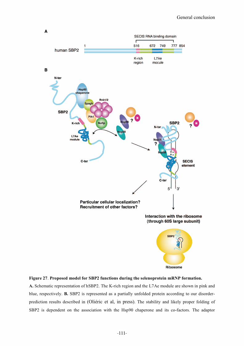
General conclusion
Figure 27. Proposed model for SBP2 functions during the selenoprotein mRNP formation.
A. Schematic representation of hSBP2. The K-rich region and the L7Ae module are shown in pink and
blue, respectively. B. SBP2 is represented as a partially unfolded protein according to our disorder-
prediction results described in (Oliéric et al, in press). The stability and likely proper folding of
SBP2 is dependent on the association with the Hsp90 chaperone and its co-factors. The adaptor
-111-
General conclusion
protein Nufip stimulates the interaction between SBP2 and the chaperone complex but probably also
core proteins. It also triggers SECIS RNP formation. Nop58 is likely to be one of the SECIS RNP core
proteins. The cellular localization of the SECIS RNA-SBP2-Nop58/56 complex is unknown. Other
core proteins may be recruited to the SECIS RNP. SBP2 also interacts with the 60S ribosomal subunit,
very likely through binding to a stem-loop structure in the 28S rRNA. When associating with the
ribosome, SBP2 cannot bind simultaneously SECIS RNA. The question mark represents potential
interactions and interactants yet to be discovered. The stem-loop structure in the 60S ribosomal
subunit is a possible SBP2 binding site in the 28S rRNA, yet to be identified.
Altogether, our observations allowed us to propose the following model for the role of SBP2
during selenoprotein mRNP formation (Figure 27). SBP2, that is globally unfolded except for
the L7Ae domain, associates with the Hsp90 chaperone complex. This association contributes
to the folding and therefore the stabilization of SBP2 (Boulon et al, 2008). This probably
triggers SBP2 binding to SECIS RNA through the L7Ae module that establishes direct
contacts with the non-Watson-Crick quartet. The K-rich domain in SBP2 may directly contact
helix 1 of the SECIS RNA to increase the affinity of SBP2 for the SECIS RNA (Allmang et
al, 2002; Takeuchi et al, 2009). The chaperone complex and the adaptor protein Nufip are also
likely to mediate the recruitment of Nop58 to SBP2. Formation of the SECIS RNA-SBP2-
Nop58 complex possibly induces further folding of SBP2, which could stabilize the complex
and/or expose different interacting surfaces for other targets. Whether other core proteins
common to sn/snoRNPs such as Nop56, fibrillarin and PRP31 are recruited to selenoprotein
mRNPs remains to be tested.
The assembly of Nop58 to the selenoprotein mRNP may also help direct selenoprotein
mRNAs to the nucleus to escape the NMD pathway or to deliver selenoprotein mRNPs to a
specialized pool of ribosomes.
We showed that SBP2 interacts with the ribosome through binding the 60S subunit (Takeuchi
et al, 2009). Very recently, SBP2 was shown to crosslink to the 28S rRNA (Olga Kossinova,
PhD student in co-tutelle with our group and Galina Karpova, Novosibirsk). The exact SBP2-
binding site in the 28S rRNA is still unknown but this result shows that there must be a helix
in the 28S rRNA sharing a structure similar to the SECIS element. Our results revealed that
the same region of SBP2 (K-rich domain) is essential for both SECIS and ribosomal binding.
Therefore SBP2 cannot bind both targets simultaneously (see Chapter 1). This is consistent
with previous models suggesting that SBP2 exchanges between the SECIS element and the
ribosome during the recoding event and Sec incorporation (Allmang & Krol, 2006a; Caban &
-112-
General conclusion
Copeland, 2006; Chavatte et al, 2005; Kinzy et al, 2005). However, our results do not allow to
discriminate whether SBP2 is pre-bound to the ribosome or the SECIS RNA prior to
translation (see also 2.3. in Part 1.). They also do not explain if the complex formed at the
SECIS in the 3’UTR could help to tether translation factors to the UGA Sec located in the
coding region. More detailed information about the interaction between SBP2 and the
ribosome will provide important insight into how the ribosome is told by the Sec
incorporation machinery not to stop at the UGA Sec codon.
Altogether, our results revealed that the molecular assembly of selenoprotein synthesis
machinery, that bears many similarities with that of sn- and snoRNPs, undergoes more
dynamic processes than anticipated. Surprisingly, SBP2 plays important roles in regulating
these events.
-113-

-114-

Part 4. Annex/ Methods
-115-

-116-
Annex/ Methods
Part 4. Annex/ Methods
1. cDNA cloning using the GATEWAY Technology
The principle of the GATEWAY Technology is based on the conservative site-specific
recombination of the $ phage between a DNA fragment harbouring specific recombination
sites and GATEWAY-adapted vectors. Two recombination reactions are performed to create:
(i) an entry clone; (ii) a destination vector. The first reaction is a recombination between an
attB (the Bacterial attachment site) DNA segment and an attP (the Phage attachment site)
donor vector to create an entry clone (also called the BP reaction because of the utilization of
the attB and the attP sites, hence the names B and P), and the second recombination reaction
occurs between an attL (the Left prophage attachment site) entry clone and an attR (the Right
prophage attachment site) destination vector, hence the LR reaction (Figure 28).
The reading frame and orientation of the DNA fragment is maintained during recombination;
attB1, attB2, attL1 and attL2 interact only with attP1, attP2, attR1 and attR2, respectively. To
clone SBP2 cDNAs using this technology, I first added the recombination sequences at both
ends of each SBP2 sequence. A two-step PCR was performed to generate SBP2 fragments
harboring attB1 and attB2 recombination sites (Figure 29). The first PCR added the thrombin
cleavage site at the 5’ and the attB2 sequence at the 3’ ends, the second one incorporating the
attB1 sequence at the 5’ end (Figure 29). Primers used for the PCR amplifications are listed in
Table 3. The PCR products were then incubated with the pDONR207 (Invitrogen) vector in
the presence of BP clonase mix (Invitrogen) containing the bacteriophage $ recombination
protein Integrase (Int) and the E.coli-encoded protein Integration Host Factor (IHF), to
generate entry clones (the BP reaction). The entry clones were next incubated with either the
pDEST10 or pDEST20 (Invitrogen) destination vectors in the presence of LR clonase mix
(Invitrogen) containing Int, IHF and Excisionase (Xis) (the LR reaction). pDEST10 and
pDEST20 contain 6xHis tag and GST sequences, respectively, upstream of the attR1
recombination site.
-117-
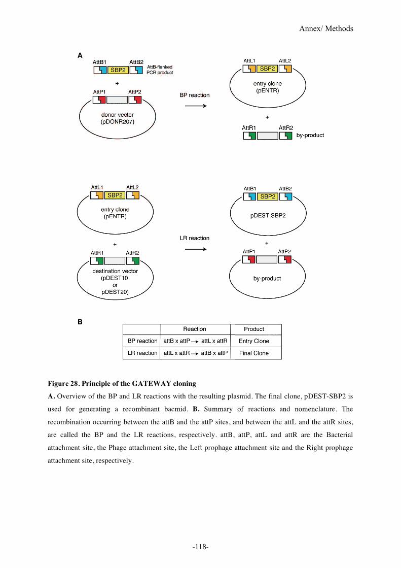
Annex/ Methods
Figure 28. Principle of the GATEWAY cloning
A. Overview of the BP and LR reactions with the resulting plasmid. The final clone, pDEST-SBP2 is
used for generating a recombinant bacmid. B. Summary of reactions and nomenclature. The
recombination occurring between the attB and the attP sites, and between the attL and the attR sites,
are called the BP and the LR reactions, respectively. attB, attP, attL and attR are the Bacterial
attachment site, the Phage attachment site, the Left prophage attachment site and the Right prophage
attachment site, respectively.
-118-
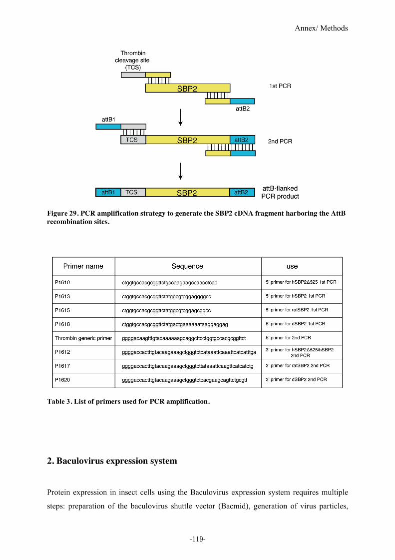
Annex/ Methods
Figure 29. PCR amplification strategy to generate the SBP2 cDNA fragment harboring the AttB
recombination sites.
Table 3. List of primers used for PCR amplification.
2. Baculovirus expression system
Protein expression in insect cells using the Baculovirus expression system requires multiple
steps: preparation of the baculovirus shuttle vector (Bacmid), generation of virus particles,
-119-
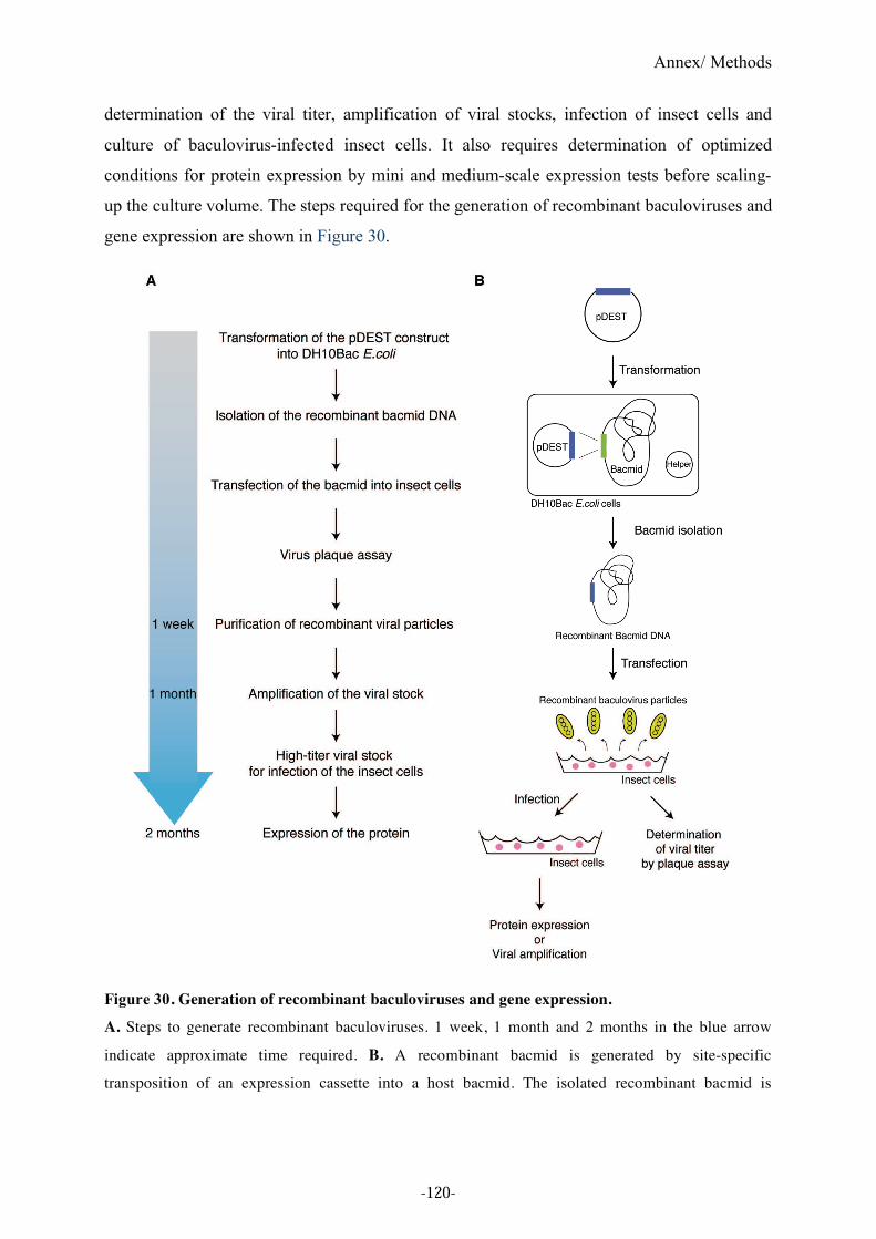
Annex/ Methods
determination of the viral titer, amplification of viral stocks, infection of insect cells and
culture of baculovirus-infected insect cells. It also requires determination of optimized
conditions for protein expression by mini and medium-scale expression tests before scaling-
up the culture volume. The steps required for the generation of recombinant baculoviruses and
gene expression are shown in Figure 30.
Figure 30. Generation of recombinant baculoviruses and gene expression.
A. Steps to generate recombinant baculoviruses. 1 week, 1 month and 2 months in the blue arrow
indicate approximate time required. B. A recombinant bacmid is generated by site-specific
transposition of an expression cassette into a host bacmid. The isolated recombinant bacmid is
-120-
Annex/ Methods
transfected into insect cells to produce recombinant baculovirus particles. The viral particles are used
for plaque assay or infection of the insect cells.
2.1. Bacmid preparation
The pDEST destination vectors cannot be used for protein expression in insect cells. The
preparation of recombinant baculovirus shuttle vectors (Bacmid) is required. To generate
recombinant bacmid DNAs, the pDEST destination constructs were transformed into the
E.coli DH10Bac strain that contains a host bacmid and a helper plasmid encoding a
transposase (Figure 30 B). This reaction is based on site-specific transposition of an
expression cassette into a bacmid. After bacmid purification and PCR analysis to confirm
insertion of the expression cassettes, ten bacmid clones were obtained (pGST-hSBP2a and b,
pGST-hSBP2"525, pGST-ratSBP2a and b, pGST-dSBP2, pHis-hSBP2, pHis-hSBP2"525,
pHis-ratSBP2 and pHis-dSBP2). They were used for generation of recombinant baculovirus
particles by transfecting the recombinant bacmid DNA into insect cells (virus particles were
generated at the baculovirus service at IGBMC). The recombinant viruses were used for
expression tests.
2.2. Mini expression test
For initial screening of the recombinant baculovirus (before viral amplification and titration),
the expression of each construct was tested. The insect cells (Spodotera frugiperda: Sf9 cells)
from 2ml culture were collected and lysed in the T20N250 buffer containing 20mM of Tris-HCl
pH8.0, 250mM NaCl. After sonication and centrifugation, soluble fractions were incubated
with either Glutathione Sepharose (Amersham) or Ni-NTA (Qiagen) beads according to the
tags. The purified proteins retained on the beads and the insoluble fractions were then loaded
onto SDS-PAGE gels, followed by Western blotting. For the His-tagged constructs, Western
blotting was performed using anti-His (SantaCruz) and anti-hSBP2 antibodies (NeoMPS).
2.3. Titration of viral particles and insect cell culture
The recombinant baculoviruses corresponding to the GST-tagged constructs (pGST-hSBP2b,
pGST-hSBP2"525 and pGST-ratSBP2b) selected by the mini expression tests were amplified
-121-
Annex/ Methods
and titrated. These titrated viral particles were used for infection of the insect cells. Sf9 cells
in 25ml cell culture (20 x 106 cells), infected with different viral titers (from 1 to 10 PFU/cell:
Plaque-forming unit per cell) were used for protein production The baculovirus infected-cells
were incubated at 27 °C for either 48 or 72 hours and then collected. The cell extract was
prepared as described in 2.2.2. in the paragraph of mini expression test, and the soluble
fraction was incubated with Glutathione Sepharose (Amersham). The purified proteins were
loaded onto SDS-PAGE gels, analyzed by Coomasie staining or Western blotting.
-122-

References
-123-

-124-
References
References
Allamand V, Richard P, Lescure A, Ledeuil C, Desjardin D, Petit N, Gartioux C, Ferreiro A, Krol A,
Pellegrini N, Urtizberea JA, Guicheney P (2006) A single homozygous point mutation in a
3'untranslated region motif of selenoprotein N mRNA causes SEPN1-related myopathy. EMBO Rep
7(4): 450-454
Allmang C, Carbon P, Krol A (2002) The SBP2 and 15.5 kD/Snu13p proteins share the same RNA
binding domain: identification of SBP2 amino acids important to SECIS RNA binding. RNA 8(10):
1308-1318
Allmang C, Krol A (2006a) SECIS RNAs and K-turn binding proteins. A survey of evolutionary
conserved RNA and protein motifs. Selenium, its molecular Biology and role in human Health 2nd
edition DL Hatfield (ed) Kluwer Academic Publishers 5: 51-61
Allmang C, Krol A (2006b) Selenoprotein synthesis: UGA does not end the story. Biochimie 88(11):
1561-1571
Allmang C, Wurth L, Krol A (2009) The selenium to selenoprotein pathway in eukaryotes: more
molecular partners than anticipated. Biochim Biophys Acta, doi:10.1016/j.bbagen.2009.03.003.
Amberg R, Urban C, Reuner B, Scharff P, Pomerantz SC, McCloskey JA, Gross HJ (1993) Editing
does not exist for mammalian selenocysteine tRNAs. Nucleic Acids Res 21(24): 5583-5588
Araiso Y, Palioura S, Ishitani R, Sherrer RL, O'Donoghue P, Yuan J, Oshikane H, Domae N, Defranco
J, Soll D, Nureki O (2008) Structural insights into RNA-dependent eukaryal and archaeal
selenocysteine formation. Nucleic Acids Res 36(4): 1187-1199
Arner ES (2009) Focus on mammalian thioredoxin reductases - Important selenoproteins with
versatile functions. Biochimica et Biophysica Acta (BBA) - General Subjects,
doi:10.1016/j.bbagen.2009.01.014
Baron C, Bock A (1991) The length of the aminoacyl-acceptor stem of the selenocysteine-specific
tRNA(Sec) of Escherichia coli is the determinant for binding to elongation factors SELB or Tu. J Biol
Chem 266(30): 20375-20379
Baron C, Sturchler C, Wu XQ, Gross HJ, Krol A, Bock A (1994) Eukaryotic selenocysteine inserting
tRNA species support selenoprotein synthesis in Escherichia coli. Nucleic Acids Res 22(12): 2228-
2233
Berry MJ, Banu L, Harney JW, Larsen PR (1993) Functional characterization of the eukaryotic SECIS
elements which direct selenocysteine insertion at UGA codons. EMBO J 12(8): 3315-3322
Böck A (2006) Selenium metabolism in prokaryotes. D L Hatfield (Ed), Selenium, its molecular
biology and role in human health Second edition Springer Science + Business Media: 9-28
Bosl MR, Takaku K, Oshima M, Nishimura S, Taketo MM (1997) Early embryonic lethality caused
by targeted disruption of the mouse selenocysteine tRNA gene (Trsp). Proc Natl Acad Sci U S A
94(11): 5531-5534
-125-
References
Boulon S, Marmier-Gourrier N, Pradet-Balade B, Wurth L, Verheggen C, Jady BE, Rothe B, Pescia C,
Robert MC, Kiss T, Bardoni B, Krol A, Branlant C, Allmang C, Bertrand E, Charpentier B (2008) The
Hsp90 chaperone controls the biogenesis of L7Ae RNPs through conserved machinery. J Cell Biol
180(3): 579-595
Bubenik JL, Driscoll DM (2007) Altered RNA binding activity underlies abnormal thyroid hormone
metabolism linked to a mutation in selenocysteine insertion sequence-binding protein 2. J Biol Chem
282(48): 34653-34662
Bubenik JL, Ladd AN, Gerber CA, Budiman ME, Driscoll DM (2009) Known turnover and
translation regulatory RNA-binding proteins interact with the 3' UTR of SECIS-binding protein 2.
RNA Biol 6(1): 73-83
Buettner C, Harney JW, Berry MJ (1999) The Caenorhabditis elegans homologue of thioredoxin
reductase contains a selenocysteine insertion sequence (SECIS) element that differs from mammalian
SECIS elements but directs selenocysteine incorporation. J Biol Chem 274(31): 21598-21602
Caban K, Copeland PR (2006) Size matters: a view of selenocysteine incorporation from the
ribosome. Cell Mol Life Sci 63(1): 73-81
Caban K, Kinzy SA, Copeland PR (2007) The L7Ae RNA binding motif is a multifunctional domain
required for the ribosome-dependent Sec incorporation activity of Sec insertion sequence binding
protein 2. Mol Cell Biol 27(18): 6350-6360
Carlson BA, Xu X-M, Kryukov GV, Rao M, Berry MJ, Gladyshev VN, Hatfield DL (2004)
Identification and characterization of phosphoseryl-tRNA[Ser]Sec kinase. PNAS 101(35): 12848-
12853
Carlson BA, Xu XM, Gladyshev VN, Hatfield DL (2005) Selective rescue of selenoprotein expression
in mice lacking a highly specialized methyl group in selenocysteine tRNA. J Biol Chem 280(7): 5542-
5548
Castellano S (2009) On the unique function of selenocysteine - insights from the evolution of
selenoproteins. Biochim Biophys Acta
Castellano S, Gladyshev VN, Guigo R, Berry MJ (2008) SelenoDB 1.0 : a database of selenoprotein
genes, proteins and SECIS elements. Nucleic Acids Res 36(Database issue): D332-338
Castellano S, Lobanov AV, Chapple C, Novoselov SV, Albrecht M, Hua D, Lescure A, Lengauer T,
Krol A, Gladyshev VN, Guigo R (2005) Diversity and functional plasticity of eukaryotic
selenoproteins: identification and characterization of the SelJ family. Proc Natl Acad Sci U S A
102(45): 16188-16193
Castellano S, Morozova N, Morey M, Berry MJ, Serras F, Corominas M, Guigo R (2001) In silico
identification of novel selenoproteins in the Drosophila melanogaster genome. EMBO Rep 2(8): 697-
702.
Castellano S, Novoselov S, Kryukov G, Lescure A, Blanco E, Krol A, Gladyshev V, Guigó R (2004)
Reconsidering the evolution of eukaryotic selenoproteins: a novel nonmammalian family with
scattered phylogenetic distribution. EMBO Rep 5(1): 71-77
Chao JA, Williamson JR (2004) Joint X-ray and NMR refinement of the yeast L30e-mRNA complex.
Structure (Camb) 12(7): 1165-1176
-126-
References
Chapple CE, Guigo R (2008) Relaxation of selective constraints causes independent selenoprotein
extinction in insect genomes. PLoS ONE 3(8): e2968
Chapple CE, Guigo R, Krol A (2009) SECISaln, a web-based tool for the creation of structure-based
alignments of eukaryotic SECIS elements. Bioinformatics 25(5): 674-675
Charpentier B, Fourmann JB, Branlant C (2007) Reconstitution of archaeal H/ACA sRNPs and test of
their activity. Methods Enzymol 425: 389-405
Chavatte L, Brown BA, Driscoll DM (2005) Ribosomal protein L30 is a component of the UGA-
selenocysteine recoding machinery in eukaryotes. Nat Struct Mol Biol
Cléry A, Bourguignon-Igel V, Allmang C, Krol A, Branlant C (2007 ) An improved definition of the
RNA-binding specificity of SECIS-binding protein 2, an essential component of the selenocysteine
incorporation machinery. Nucleic Acids Res 35(6): 1868–1884
Copeland PR, Driscoll DM (1999) Purification, redox sensitivity, and RNA binding properties of
SECIS- binding protein 2, a protein involved in selenoprotein biosynthesis. J Biol Chem 274(36):
25447-25454.
Copeland PR, Driscoll DM (2001) RNA binding proteins and selenocysteine. Biofactors 14(1-4): 11-
16
Copeland PR, Fletcher JE, Carlson BA, Hatfield DL, Driscoll DM (2000) A novel RNA binding
protein, SBP2, is required for the translation of mammalian selenoprotein mRNAs. Embo J 19(2):
306-314.
Copeland PR, Stepanik VA, Driscoll DM (2001) Insight into mammalian selenocysteine insertion:
domain structure and ribosome binding properties of Sec insertion sequence binding protein 2. Mol
Cell Biol 21(5): 1491-1498.
Costa M, Rodríguez-Sánchez J, Czaja A, Gelpí C (2000) Isolation and characterization of cDNA
encoding the antigenic protein of the human tRNP(Ser)Sec complex recognized by autoantibodies
from patients withtype-1 autoimmune hepatitis. Clin Exp Immunol 121(2): 364-374
Dayer R, Fischer BB, Eggen RI, Lemaire SD (2008) The peroxiredoxin and glutathione peroxidase
families in Chlamydomonas reinhardtii. Genetics 179(1): 41-57
de Jesus LA, Hoffmann PR, Michaud T, Forry EP, Small-Howard A, Stillwell RJ, Morozova N,
Harney JW, Berry MJ (2006) Nuclear Assembly of UGA Decoding Complexes on Selenoprotein
mRNAs: a Mechanism for Eluding Nonsense-Mediated Decay? Mol Cell Biol 26(5): 1795-1805
Diamond A, Dudock B, Hatfield D (1981) Structure and properties of a bovine liver UGA suppressor
serine tRNA with a tryptophan anticodon. Cell 25(2): 497-506
Diamond AM, Choi IS, Crain PF, Hashizume T, Pomerantz SC, Cruz R, Steer CJ, Hill KE, Burk RF,
McCloskey JA, et al. (1993) Dietary selenium affects methylation of the wobble nucleoside in the
anticodon of selenocysteine tRNA([Ser]Sec). J Biol Chem 268(19): 14215-14223
Dikiy A, Novoselov SV, Fomenko DE, Sengupta A, Carlson BA, Cerny RL, Ginalski K, Grishin NV,
Hatfield DL, Gladyshev VN (2007) SelT, SelW, SelH, and Rdx12: genomics and molecular insights
into the functions of selenoproteins of a novel thioredoxin-like family. Biochemistry 46(23): 6871-
6882
-127-
References
Ding F, Grabowski PJ (1999) Identification of a protein component of a mammalian tRNA(Sec)
complex implicated in the decoding of UGA as selenocysteine. RNA 5(12): 1561-1569
Donovan J, Caban K, Ranaweera R, Gonzalez-Flores JN, Copeland PR (2008) A novel protein domain
induces high affinity selenocysteine insertion sequence binding and elongation factor recruitment. J
Biol Chem 283(50): 35129-35139
Dumitrescu AM, Liao XH, Abdullah MS, Lado-Abeal J, Majed FA, Moeller LC, Boran G, Schomburg
L, Weiss RE, Refetoff S (2005) Mutations in SECISBP2 result in abnormal thyroid hormone
metabolism. Nat Genet 37(11): 1247-1252
Dyson HJ, Wright PE (2005) Intrinsically unstructured proteins and their functions. Nat Rev Mol Cell
Biol 6(3): 197-208
Fagegaltier D, Hubert N, Yamada K, Mizutani T, Carbon P, Krol A (2000a) Characterization of
mSelB, a novel mammalian elongation factor for selenoprotein translation. Embo J 19(17): 4796-
4805.
Fagegaltier D, Lescure A, Walczak R, Carbon P, Krol A (2000b) Structural analysis of new local
features in SECIS RNA hairpins. Nucleic Acids Res 28(14): 2679-2689.
Ferguson AD, Labunskyy VM, Fomenko DE, Arac D, Chelliah Y, Amezcua CA, Rizo J, Gladyshev
VN, Deisenhofer J (2006) NMR Structures of the Selenoproteins Sep15 and SelM Reveal Redox
Activity of a New Thioredoxin-like Family. J Biol Chem 281(6): 3536-3543
Fletcher JE, Copeland PR, Driscoll DM, Krol A (2001) The selenocysteine incorporation machinery:
interactions between the SECIS RNA and the SECIS-binding protein SBP2. RNA 7(10): 1442-1453.
Flohe L (2009) The labour pains of biochemical selenology: The history of selenoprotein biosynthesis.
Biochim Biophys Acta
Ganichkin OM, Xu XM, Carlson BA, Mix H, Hatfield DL, Gladyshev VN, Wahl MC (2008) Structure
and catalytic mechanism of eukaryotic selenocysteine synthase. J Biol Chem 283(9): 5849-5865
Gelpi C, Sontheimer EJ, Rodriguez-Sanchez JL (1992) Autoantibodies against a serine tRNA-protein
complex implicated in cotranslational selenocysteine insertion. Proc Natl Acad Sci U S A 89(20):
9739-9743
Graham TA, Weaver C, Mao F, Kimelman D, Xu W (2000) Crystal structure of a beta-catenin/Tcf
complex. Cell 103(6): 885-896
Gromer S, Eubel JK, Lee BL, Jacob J (2005) Human selenoproteins at a glance. Cell Mol Life Sci
62(21): 2414-2437
Grundner-Culemann E, Martin GW, 3rd, Harney JW, Berry MJ (1999) Two distinct SECIS structures
capable of directing selenocysteine incorporation in eukaryotes. RNA 5(5): 625-635
Guimaraes MJ, Peterson D, Vicari A, Cocks BG, Copeland NG, Gilbert DJ, Jenkins NA, Ferrick DA,
Kastelein RA, Bazan JF, Zlotnik A (1996) Identification of a novel selD homolog from eukaryotes,
bacteria, and archaea: is there an autoregulatory mechanism in selenocysteine metabolism? Proc Natl
Acad Sci U S A 93(26): 15086-15091
Gupta M, Copeland PR (2007) Functional analysis of the interplay between translation termination,
selenocysteine codon context, and selenocysteine insertion sequence-binding protein 2. J Biol Chem
282(51): 36797-36807
-128-
References
Hatfield D, Diamond A, Dudock B (1982) Opal suppressor serine tRNAs from bovine liver form
phosphoseryl-tRNA. Proc Natl Acad Sci U S A 79(20): 6215-6219
Hatfield D, Portugal FH (1970) Seryl-tRNA in mammalian tissues: chromatographic differences in
brain and liver and a specific response to the codon, UGA. Proc Natl Acad Sci U S A 67(3): 1200-
1206
Hatfield DL, Gladyshev VN (2002) How selenium has altered our understanding of the genetic code.
Mol Cell Biol 22(11): 3565-3576
Hatfield DL, Yoo MH, Carlson BA, Gladyshev VN (2009) Selenoproteins that function in cancer
prevention and promotion. Biochim Biophys Acta
Herbette S, Roeckel-Drevet P, Drevet JR (2007) Seleno-independent glutathione peroxidases. More
than simple antioxidant scavengers. FEBS J 274(9): 2163-2180
Hirosawa-Takamori M, Chung HR, Jackle H (2004) Conserved selenoprotein synthesis is not critical
for oxidative stress defence and the lifespan of Drosophila. EMBO Rep 5(3): 317-322
Hirosawa-Takamori M, Jackle H, Vorbruggen G (2000) The class 2 selenophosphate synthetase gene
of Drosophila contains a functional mammalian-type SECIS. EMBO Rep 1(5): 441-446
Howard MT, Aggarwal G, Anderson CB, Khatri S, Flanigan KM, Atkins JF (2005) Recoding
elements located adjacent to a subset of eukaryal selenocysteine-specifying UGA codons. EMBO J
24(8): 1596-1607
Howard MT, Moyle MW, Aggarwal G, Carlson BA, Anderson CB (2007) A recoding element that
stimulates decoding of UGA codons by Sec tRNA[Ser]Sec. RNA 13(6): 912-920
Hubert N, Sturchler C, Westhof E, Carbon P, Krol A (1998) The 9/4 secondary structure of eukaryotic
selenocysteine tRNA: more pieces of evidence. RNA 4(9): 1029-1033.
Kernebeck T, Lohse A, Grötzinger J (2001) A bioinformatical approach suggests the function of the
autoimmune hepatitis target antigen soluble liver antigen/liver pancreas. Hepatology 34(2): 230-233
Kim LK, Matsufuji T, Matsufuji S, Carlson BA, Kim SS, Hatfield DL, Lee BJ (2000) Methylation of
the ribosyl moiety at position 34 of selenocysteine tRNA[Ser]Sec is governed by both primary and
tertiary structure. RNA 6(9): 1306-1315
Kinzy SA, Caban K, Copeland PR (2005) Characterization of the SECIS binding protein 2 complex
required for the co-translational insertion of selenocysteine in mammals. Nucleic Acids Res 33(16):
5172-5180
Kiss T (2002) Small nucleolar RNAs: an abundant group of noncoding RNAs with diverse cellular
functions. Cell 109(2): 145-148
Klein DJ, Schmeing TM, Moore PB, Steitz TA (2001) The kink-turn: a new RNA secondary structure
motif. EMBO J 20(15): 4214-4221
Koonin EV, Bork P, Sander C (1994) A novel RNA-binding motif in omnipotent suppressors of
translation termination, ribosomal proteins and a ribosome modification enzyme? Nucleic Acids Res
22(11): 2166-2167.
-129-
References
Korotkov KV, Novoselov SV, Hatfield DL, Gladyshev VN (2002) Mammalian selenoprotein in which
selenocysteine (Sec) incorporation is supported by a new form of Sec insertion sequence element. Mol
Cell Biol 22(5): 1402-1411.
Kriwacki RW, Hengst L, Tennant L, Reed SI, Wright PE (1996) Structural studies of
p21Waf1/Cip1/Sdi1 in the free and Cdk2-bound state: conformational disorder mediates binding
diversity. Proc Natl Acad Sci U S A 93(21): 11504-11509
Kryukov GV, Castellano S, Novoselov SV, Lobanov AV, Zehtab O, Guigo R, Gladyshev VN (2003)
Characterization of mammalian selenoproteomes. Science 300(5624): 1439-1443.
Kryukov GV, Kryukov VM, Gladyshev VN (1999) New mammalian selenocysteine-containing
proteins identified with an algorithm that searches for selenocysteine insertion sequence elements. J
Biol Chem 274(48): 33888-33897
Kuhn JF, Tran EJ, Maxwell ES (2002) Archaeal ribosomal protein L7 is a functional homolog of the
eukaryotic 15.5kD/Snu13p snoRNP core protein. Nucleic Acids Res 30(4): 931-941
Lee BJ, Worland PJ, Davis JN, Stadtman TC, Hatfield DL (1989) Identification of a selenocysteyl-
tRNA(Ser) in mammalian cells that recognizes the nonsense codon, UGA. J Biol Chem 264(17):
9724-9727
Leibundgut M, Frick C, Thanbichler M, Bock A, Ban N (2005) Selenocysteine tRNA-specific
elongation factor SelB is a structural chimaera of elongation and initiation factors. Embo J 24(1): 11-
22
Lejeune F, Maquat LE (2005) Mechanistic links between nonsense-mediated mRNA decay and pre-
mRNA splicing in mammalian cells. Curr Opin Cell Biol 17(3): 309-315
Lescure A, Allmang C, Yamada K, Carbon P, Krol A (2002) cDNA cloning, expression pattern and
RNA binding analysis of human selenocysteine insertion sequence (SECIS) binding protein 2. Gene
291(1-2): 279-285
Lescure A, Gautheret D, Carbon P, Krol A (1999) Novel selenoproteins identified in silico and in vivo
by using a conserved RNA structural motif. J Biol Chem 274(53): 38147-38154.
Lescure A, Rederstorff M, Krol A, Guicheney P, Allamand V (2009) Selenoprotein function and
muscle disease. Biochimica et Biophysica Acta (BBA) - General Subjects,
doi:10.1016/j.bbagen.2009.03.002
Linding R, Jensen LJ, Diella F, Bork P, Gibson TJ, Russell RB (2003) Protein disorder prediction:
implications for structural proteomics. Structure 11(11): 1453-1459
Liu S, Li P, Dybkov O, Nottrott S, Hartmuth K, Luhrmann R, Carlomagno T, Wahl MC (2007)
Binding of the human Prp31 Nop domain to a composite RNA-protein platform in U4 snRNP. Science
316(5821): 115-120
Lobanov AV, Delgado C, Rahlfs S, Novoselov SV, Kryukov GV, Gromer S, Hatfield DL, Becker K,
Gladyshev VN (2006a) The Plasmodium selenoproteome. Nucleic Acids Res 34(2): 496-505
Lobanov AV, Fomenko DE, Zhang Y, Sengupta A, Hatfield DL, Gladyshev VN (2007) Evolutionary
dynamics of eukaryotic selenoproteomes: large selenoproteomes may associate with aquatic life and
small with terrestrial life. Genome Biol 8(9): R198
-130-
References
Lobanov AV, Gromer S, Salinas G, Gladyshev VN (2006b) Selenium metabolism in Trypanosoma:
characterization of selenoproteomes and identification of a Kinetoplastida-specific selenoprotein.
Nucleic Acids Res 34(14): 4012-4024
Lobanov AV, Hatfield DL, Gladyshev VN (2008a) Reduced reliance on the trace element selenium
during evolution of mammals. Genome Biol 9(3): R62
Lobanov AV, Hatfield DL, Gladyshev VN (2008b) Selenoproteinless animals: selenophosphate
synthetase SPS1 functions in a pathway unrelated to selenocysteine biosynthesis. Protein Sci 17(1):
176-182
Low SC, Harney JW, Berry MJ (1995) Cloning and Functional Characterization of Human
Selenophosphate Synthetase, an Essential Component of Selenoprotein Synthesis. J Biol Chem
270(37): 21659-21664
Lu C, Qiu F, Zhou H, Peng Y, Hao W, Xu J, Yuan J, Wang S, Qiang B, Xu C, Peng X (2006)
Identification and characterization of selenoprotein K: an antioxidant in cardiomyocytes. FEBS Lett
580(22): 5189-5197
Lu J, Holmgren A (2009) Selenoproteins. J Biol Chem 284(2): 723-727
Ma S, Hill KE, Caprioli RM, Burk RF (2002) Mass spectrometric characterization of full-length rat
selenoprotein P and three isoforms shortened at the C terminus. Evidence that three UGA codons in
the mRNA open reading frame have alternative functions of specifying selenocysteine insertion or
translation termination. J Biol Chem 277(15): 12749-12754.
MacÌas S, Bragulat M, Tardiff DF, Vilardell J (2008) L30 Binds the Nascent RPL30 Transcript to
Repress U2 snRNP Recruitment. Molecular Cell 30(6): 732-742
Maenpaa PH, Bernfield MR (1970) A specific hepatic transfer RNA for phosphoserine. Proc Natl
Acad Sci U S A 67(2): 688-695
Maiti B, Arbogast S, Allamand V, Moyle MW, Anderson CB, Richard P, Guicheney P, Ferreiro A,
Flanigan KM, Howard MT (2008) A mutation in the SEPN1selenocysteine redefinition element (SRE)
reduces selenocysteine incorporation and leads to SEPN1 related myopathy. Human Mutation doi:
10.1002/humu.20879
Marmier-Gourrier N, Clery A, Senty-Segault V, Charpentier B, Schlotter F, Leclerc F, Fournier R,
Branlant C (2003) A structural, phylogenetic, and functional study of 15.5-kD/Snu13 protein binding
on U3 small nucleolar RNA. RNA 9(7): 821-838
Martin-Romero FJ, Kryukov GV, Lobanov AV, Carlson BA, Lee BJ, Gladyshev VN, Hatfield DL
(2001) Selenium metabolism in Drosophila: selenoproteins, selenoprotein mRNA expression, fertility,
and mortality. J Biol Chem 276(32): 29798-29804
Matera AG, Terns RM, Terns MP (2007) Non-coding RNAs: lessons from the small nuclear and small
nucleolar RNAs. Nat Rev Mol Cell Biol 8(3): 209-220
Mix H, Lobanov AV, Gladyshev VN (2007) SECIS elements in the coding regions of selenoprotein
transcripts are functional in higher eukaryotes. Nucleic Acids Res 35(2): 414-423
Mizutani T (1989) Some evidence of the enzymatic conversion of bovine suppressor phosphoseryl-
tRNA to selenocysteyl-tRNA. FEBS Lett 250(2): 142-146
-131-
References
Moghadaszadeh B, Petit N, Jaillard C, Brockington M, Roy SQ, Merlini L, Romero N, Estournet B,
Desguerre I, Chaigne D, Muntoni F, Topaloglu H, Guicheney P (2001) Mutations in SEPN1 cause
congenital muscular dystrophy with spinal rigidity and restrictive respiratory syndrome. Nat Genet
29(1): 17-18.
Mongelard F, Bouvet P (2007) Nucleolin: a multiFACeTed protein. Trends Cell Biol 17(2): 80-86
Moore T, Zhang Y, Fenley MO, Li H (2004) Molecular basis of box C/D RNA-protein interactions;
cocrystal structure of archaeal L7Ae and a box C/D RNA. Structure (Camb) 12(5): 807-818
Moriarty PM, Reddy CC, Maquat LE (1998) Selenium deficiency reduces the abundance of mRNA
for Se-dependent glutathione peroxidase 1 by a UGA-dependent mechanism likely to be nonsense
codon-mediated decay of cytoplasmic mRNA. Mol Cell Biol 18(5): 2932-2939
Morozova N, Forry EP, Shahid E, Zavacki AM, Harney JW, Kraytsberg Y, Berry MJ (2003)
Antioxidant function of a novel selenoprotein in Drosophila melanogaster. Genes Cells 8(12): 963-971
Muttenthaler M, Alewood PF (2008) Selenopeptide chemistry. J Pept Sci 14(12): 1223-1239
Nottrott S, Hartmuth K, Fabrizio P, Urlaub H, Vidovic I, Ficner R, Luhrmann R (1999) Functional
interaction of a novel 15.5kD [U4/U6.U5] tri-snRNP protein with the 5' stem-loop of U4 snRNA.
EMBO J 18(21): 6119-6133
Nottrott S, Urlaub H, Luhrmann R (2002) Hierarchical, clustered protein interactions with U4/U6
snRNA: a biochemical role for U4/U6 proteins. Embo J 21(20): 5527-5538
Novoselov SV, Kryukov GV, Xu XM, Carlson BA, Hatfield DL, Gladyshev VN (2007a)
Selenoprotein H is a nucleolar thioredoxin-like protein with a unique expression pattern. J Biol Chem
282(16): 11960-11968
Novoselov SV, Lobanov AV, Hua D, Kasaikina MV, Hatfield DL, Gladyshev VN (2007b) A highly
efficient form of the selenocysteine insertion sequence element in protozoan parasites and its use in
mammalian cells. Proc Natl Acad Sci U S A 104(19): 7857-7862
Novoselov SV, Rao M, Onoshko NV, Zhi H, Kryukov GV, Xiang Y, Weeks DP, Hatfield DL,
Gladyshev VN (2002) Selenoproteins and selenocysteine insertion system in the model plant cell
system, Chlamydomonas reinhardtii. EMBO J 21(14): 3681-3693
Olieric V, Wolff P, Takeuchi A, Bec G, Birck C, Vitorino M, Kieffer B, Beniaminov A, Cavigiolio G,
Theil E, Allmang C, Krol A, Dumas P (2009) SECIS-binding protein 2, a key player in selenoprotein
synthesis, is an intrinsically disordered protein. biochimie
Omer AD, Ziesche S, Ebhardt H, Dennis PP (2002) In vitro reconstitution and activity of a C/D box
methylation guide ribonucleoprotein complex. Proc Natl Acad Sci U S A 99(8): 5289-5294
Papp LV, Lu J, Holmgren A, Khanna KK (2007) From selenium to selenoproteins: synthesis, identity,
and their role in human health. Antioxid Redox Signal 9(7): 775-806
Papp LV, Lu J, Striebel F, Kennedy D, Holmgren A, Khanna KK (2006) The redox state of SECIS
binding protein 2 controls its localization and selenocysteine incorporation function. Mol Cell Biol
26(13): 4895-4910
Papp LV, Wang J, Kennedy D, Boucher D, Zhang Y, Gladyshev VN, Singh RN, Khanna KK (2008)
Functional characterization of alternatively spliced human SECISBP2 transcript variants. Nucleic
Acids Res 36(22): 7192-7206
-132-
References
Pappas AC, Zoidis E, Surai PF, Zervas G (2008) Selenoproteins and maternal nutrition. Comp
Biochem Physiol B Biochem Mol Biol 151(4): 361-372
Patel SB, Bellini M (2008) The assembly of a spliceosomal small nuclear ribonucleoprotein particle.
Nucleic Acids Res 36(20): 6482-6493
Pedersen JS, Bejerano G, Siepel A, Rosenbloom K, Lindblad-Toh K, Lander ES, Kent J, Miller W,
Haussler D (2006) Identification and classification of conserved RNA secondary structures in the
human genome. PLoS Comput Biol 2(4): e33
Rajkowitsch L, Chen D, Stampfl S, Semrad K, Waldsich C, Mayer O, Jantsch MF, Konrat R, Blasi U,
Schroeder R (2007) RNA chaperones, RNA annealers and RNA helicases. RNA Biol 4(3): 118-130
Rederstorff M, Krol A, Lescure A (2006) Understanding the importance of selenium and
selenoproteins in muscle function. Cell Mol Life Sci 63(1): 52-59
Reeves MA, Hoffmann PR (2009) The human selenoproteome: recent insights into functions and
regulation. Cell Mol Life Sci
Reichow SL, Hamma T, Ferre-D'Amare AR, Varani G (2007) The structure and function of small
nucleolar ribonucleoproteins. Nucleic Acids Res 35(5): 1452-1464
Rother M, Wilting R, Commans S, Bock A (2000) Identification and characterisation of the
selenocysteine-specific translation factor SelB from the archaeon Methanococcus jannaschii. J Mol
Biol 299(2): 351-358
Russo AA, Jeffrey PD, Patten AK, Massague J, Pavletich NP (1996) Crystal structure of the p27Kip1
cyclin-dependent-kinase inhibitor bound to the cyclin A-Cdk2 complex. Nature 382(6589): 325-331
Shchedrina VA, Novoselov SV, Malinouski MY, Gladyshev VN (2007) Identification and
characterization of a selenoprotein family containing a diselenide bond in a redox motif. Proc Natl
Acad Sci U S A 104(35): 13919-13924
Shen Q, Wu R, Leonard JL, Newburger PE (1998) Identification and molecular cloning of a human
selenocysteine insertion sequence-binding protein. A bifunctional role for DNA-binding protein B. J
Biol Chem 273(10): 5443-5446
Shen Q, Fan L, Newburger PE (2006) Nuclease sensitive element binding protein 1 associates with the
selenocysteine insertion sequence and functions in mammalian selenoprotein translation. J Cell
Physiol 207(3): 775-783
Sherrer RL, Ho JM, Soll D (2008) Divergence of selenocysteine tRNA recognition by archaeal and
eukaryotic O-phosphoseryl-tRNASec kinase. Nucleic Acids Res 36(6): 1871-1880
Shisler JL, Senkevich TG, Berry MJ, Moss B (1998) Ultraviolet-induced cell death blocked by a
selenoprotein from a human dermatotropic poxvirus. Science 279(5347): 102-105
Small-Howard A, Morozova N, Stoytcheva Z, Forry EP, Mansell JB, Harney JW, Carlson BA, Xu
XM, Hatfield DL, Berry MJ (2006) Supramolecular complexes mediate selenocysteine incorporation
in vivo. Mol Cell Biol 26(6): 2337-2346
Small-Howard AL, Berry MJ (2005) Unique features of selenocysteine incorporation function within
the context of general eukaryotic translational processes. Biochem Soc Trans 33(Pt 6): 1493-1497
-133-
References
Squires JE, Berry MJ (2008) Eukaryotic selenoprotein synthesis: mechanistic insight incorporating
new factors and new functions for old factors. IUBMB Life 60(4): 232-235
Squires JE, Stoytchev I, Forry EP, Berry MJ (2007) SBP2 binding affinity is a major determinant in
differential selenoprotein mRNA translation and sensitivity to nonsense-mediated decay. Mol Cell
Biol 27(22): 7848-7855
Stoytcheva Z, Tujebajeva RM, Harney JW, Berry MJ (2006) Efficient incorporation of multiple
selenocysteines involves an inefficient decoding step serving as a potential translational checkpoint
and ribosome bottleneck. Mol Cell Biol 26(24): 9177-9184
Sturchler C, Lescure A, Keith G, Carbon P, Krol A, Ibba M, Soll D (1994) Base modification pattern
at the wobble position of Xenopus selenocysteine tRNA(Sec). Nucleic Acids Res 22(8): 1354-1358.
co-translationally inserted during protein synthesis, probably by a mechanism analogous to that
operating during selenocysteine incorporation.
Sturchler C, Westhof E, Carbon P, Krol A (1993) Unique secondary and tertiary structural features of
the eucaryotic selenocysteine tRNA(Sec). Nucleic Acids Res 21(5): 1073-1079
Sturchler-Pierrat C, Hubert N, Totsuka T, Mizutani T, Carbon P, Krol A (1995) Selenocysteylation in
eukaryotes necessitates the uniquely long aminoacyl acceptor stem of selenocysteine tRNA(Sec). J
Biol Chem 270(31): 18570-18574.
Sun X, Li X, Moriarty PM, Henics T, LaDuca JP, Maquat LE (2001) Nonsense-mediated decay of
mRNA for the selenoprotein phospholipid hydroperoxide glutathione peroxidase is detectable in
cultured cells but masked or inhibited in rat tissues. Mol Biol Cell 12(4): 1009-1017.
Takeuchi A, Schmitt D, Chapple C, Babaylova E, Karpova G, Guigo R, Krol A, Allmang C (2009) A
short motif in Drosophila SECIS Binding Protein 2 provides differential binding affinity to SECIS
RNA hairpins. Nucleic Acids Res 37(7): 2126-2141
Taskov K, Chapple C, Kryukov GV, Castellano S, Lobanov AV, Korotkov KV, Guigo R, Gladyshev
VN (2005) Nematode selenoproteome: the use of the selenocysteine insertion system to decode one
codon in an animal genome? Nucleic Acids Res 33(7): 2227-2238
Tompa P (2002) Intrinsically unstructured proteins. Trends Biochem Sci 27(10): 527-533
Tujebajeva RM, Copeland PR, Xu XM, Carlson BA, Harney JW, Driscoll DM, Hatfield DL, Berry MJ
(2000) Decoding apparatus for eukaryotic selenocysteine insertion. EMBO Rep 1(2): 158-163.
Ursini F, Heim S, Kiess M, Maiorino M, Roveri A, Wissing J, Flohe L (1999) Dual function of the
selenoprotein PHGPx during sperm maturation. Science 285(5432): 1393-1396.
Uversky VN, Gillespie JR, Fink AL (2000) Why are "natively unfolded" proteins unstructured under
physiologic conditions? Proteins 41(3): 415-427
Vidovic I, Nottrott S, Hartmuth K, Luhrmann R, Ficner R (2000) Crystal structure of the spliceosomal
15.5kD protein bound to a U4 snRNA fragment. Mol Cell 6(6): 1331-1342
Walczak R, Carbon P, Krol A (1998) An essential non-Watson-Crick base pair motif in 3'UTR to
mediate selenoprotein translation. RNA 4(1): 74-84
Walczak R, Westhof E, Carbon P, Krol A (1996) A novel RNA structural motif in the selenocysteine
insertion element of eukaryotic selenoprotein mRNAs. RNA 2(4): 367-379
-134-
References
Watkins NJ, Dickmanns A, Luhrmann R (2002) Conserved stem II of the box C/D motif is essential
for nucleolar localization and is required, along with the 15.5K protein, for the hierarchical assembly
of the box C/D snoRNP. Mol Cell Biol 22(23): 8342-8352
Watkins NJ, Segault V, Charpentier B, Nottrott S, Fabrizio P, Bachi A, Wilm M, Rosbash M, Branlant
C, Luhrmann R (2000) A common core RNP structure shared between the small nucleoar box C/D
RNPs and the spliceosomal U4 snRNP. Cell 103(3): 457-466
Weiss SL, Sunde RA (1998) Cis-acting elements are required for selenium regulation of glutathione
peroxidase-1 mRNA levels. RNA 4(7): 816-827
Wright PE, Dyson HJ (1999) Intrinsically unstructured proteins: re-assessing the protein structure-
function paradigm. J Mol Biol 293(2): 321-331
Wright PE, Dyson HJ (2009) Linking folding and binding. Curr Opin Struct Biol 19(1): 31-38
Wu R, Shen Q, Newburger PE (2000) Recognition and binding of the human selenocysteine insertion
sequence by nucleolin. J Cell Biochem 77(3): 507-516
Wu XQ, Gross HJ (1993) The long extra arms of human tRNA((Ser)Sec) and tRNA(Ser) function as
major identify elements for serylation in an orientation-dependent, but not sequence-specific manner.
Nucleic Acids Res 21(24): 5589-5594
Wu XQ, Gross HJ (1994) The length and the secondary structure of the D-stem of human
selenocysteine tRNA are the major identity determinants for serine phosphorylation. EMBO J 13(1):
241-248
Xu X, Carlson B, Irons R, Mix H, Zhong N, Gladyshev V, DL H (2007a) Selenophosphate synthetase
2 is essential for selenoprotein biosynthesis. Biochem J 404(1): 115-120
Xu X-M, Carlson BA, Mix H, Zhang Y, Saira K, Glass RS, Berry MJ, Gladyshev VN, Hatfield DL
(2007b) Biosynthesis of Selenocysteine on Its tRNA in Eukaryotes. PLoS Biology 5(1): e4
Xu X-M, Mix H, Carlson BA, Grabowski PJ, Gladyshev VN, Berry MJ, Hatfield DL (2005) Evidence
for Direct Roles of Two Additional Factors, SECp43 and Soluble Liver Antigen, in the Selenoprotein
Synthesis Machinery. J Biol Chem 280(50): 41568-41575
Yuan J, Palioura S, Salazar JC, Su D, O'Donoghue P, Hohn MJ, Cardoso AM, Whitman WB, Soll D
(2006) RNA-dependent conversion of phosphoserine forms selenocysteine in eukaryotes and archaea.
PNAS 103(50): 18923-18927
Zavacki AM, Mansell JB, Chung M, Klimovitsky B, Harney JW, Berry MJ (2003) Coupled
tRNA(Sec)-dependent assembly of the selenocysteine decoding apparatus. Mol Cell 11(3): 773-781.
Zhao R, Davey M, Hsu Y-C, Kaplanek P, Tong A, Parsons AB, Krogan N, Cagney G, Mai D,
Greenblatt J, Boone C, Emili A, Houry WA (2005) Navigating the Chaperone Network: An
Integrative Map of Physical and Genetic Interactions Mediated by the Hsp90 Chaperone. Cell 120(5):
715-727
Zhao R, Kakihara Y, Gribun A, Huen J, Yang G, Khanna M, Costanzo M, Brost RL, Boone C,
Hughes TR, Yip CM, Houry WA (2008) Molecular chaperone Hsp90 stabilizes Pih1/Nop17 to
maintain R2TP complex activity that regulates snoRNA accumulation. J Cell Biol 180(3): 563-578
Zhuo P, Diamond AM (2009) Molecular mechanisms by which selenoproteins affect cancer risk and
progression. Biochim Biophys Acta, doi:10.1016/j.bbagen.2009.03.004
-135-