Embed Size (px)
Citation preview
Volume 22 July 15, 2011 2541
Turnover of branched actin filament networks by stochastic fragmentation with ADF/cofilinAnne-Cécile Reymanna,*, Cristian Suareza,*, Christophe Guérina, Jean-Louis Martiela, Christopher J. Staigerb,c, Laurent Blanchoina, and Rajaa Boujemaa-PaterskiaaLaboratoire de Physiologie Cellulaire & Végétale, Institut de Recherches en Technologies et Sciences pour le Vivant (iRTSV), Centre National de la Recherche Scientifique/Commissariat à l’énergie atomique et aux énergies alternatives/Institut National de la Recherche Agronomique/Université Joseph Fourier (CNRS/CEA/INRA/UJF), Grenoble 38054, France; bDepartment of Biological Sciences and cThe Bindley Bioscience Center, Purdue University, West Lafayette, IN 47907-2064
This article was published online ahead of print in MBoC in Press (http://www .molbiolcell.org/cgi/doi/10.1091/mbc.E11-01-0052) on May 25, 2011.*These authors contributed equally to this work.Address correspondence to: Laurent Blanchoin ([email protected]) or Rajaa Boujemaa-Paterski ([email protected]).Abbreviations used: ABP, actin-binding protein; ADF, actin-depolymerizing fac-tor; BSA, bovine serum albumin; CP, capping protein; DTT, dithiothreitol; Ena/VASP, Enabled/vasodilator-stimulated phosphoprotein; FRAP, fluorescence re-covery after photobleaching; GST-pWA, glutathione-S-transferase–polyproline verproline homology acidic C-terminus regions of human Wiskott–Aldrich syn-drome protein (WASp); NPF, nucleation-promoting factor; TCEP, Tris(2-carboxy-ethyl)phosphine.© 2011 Reymann et al. This article is distributed by The American Society for Cell Biology under license from the author(s). Two months after publication it is avail-able to the public under an Attribution–Noncommercial–Share Alike 3.0 Unported Creative Commons License (http://creativecommons.org/licenses/by-nc-sa/3.0).“ASCB®,“ “The American Society for Cell Biology®,” and “Molecular Biology of the Cell®” are registered trademarks of The American Society of Cell Biology.
ABSTRACT Cell motility depends on the rapid assembly, aging, severing, and disassembly of actin filaments in spatially distinct zones. How a set of actin regulatory proteins that sustains actin-based force generation during motility work together in space and time remains poorly understood. We present our study of the distribution and dynamics of Arp2/3 complex, cap-ping protein (CP), and actin-depolymerizing factor (ADF)/cofilin in actin “comet tails,” using a minimal reconstituted system with nucleation-promoting factor (NPF)-coated beads. The Arp2/3 complex concentrates at nucleation sites near the beads as well as in the first actin shell. CP colocalizes with actin and is homogeneously distributed throughout the comet tail; it serves to constrain the spatial distribution of ATP/ADP-Pi filament zones to areas near the bead. The association of ADF/cofilin with the actin network is therefore governed by kinetics of actin assembly, actin nucleotide state, and CP binding. A kinetic simulation accurately vali-dates these observations. Following its binding to the actin networks, ADF/cofilin is able to break up the dense actin filament array of a comet tail. Stochastic severing by ADF/cofilin loosens the tight entanglement of actin filaments inside the comet tail and facilitates turn-over through the macroscopic release of large portions of the aged actin network.
INTRODUCTIONCellular motility is a fundamental process essential for embryonic development, immune responses, wound healing, and neuronal growth cone migration (Pollard et al., 2000). During motility, the rapid assembly and disassembly of actin networks provide the driving
force that deforms the plasma membrane (Pollard and Cooper, 2009). Upon signaling, site-directed actin assembly at the leading edge of migrating cells depends primarily on the activation of the Arp2/3 complex, one of the three known actin nucleators in eukary-otes (Welch et al., 1998; Machesky et al., 1999; Rohatgi et al., 1999; Svitkina and Borisy, 1999; Pollard and Borisy, 2003; Goley and Welch, 2006; Rouiller et al., 2008). The activated complex triggers the rapid de novo assembly of a rigid and highly cross-linked actin filament array called the “dendritic network” (Mullins et al., 1998; Machesky et al., 1999; Blanchoin et al., 2000a; Achard et al., 2010). To effi-ciently convert the chemical energy provided by ATP hydrolysis within an actin filament into mechanical forces, rapid assembly of the actin network must be 1) localized underneath the plasma mem-brane (Rohatgi et al., 1999) and 2) supplied with a large reservoir of actin monomers (Pantaloni et al., 2000; Pollard et al., 2000). Selective recruitment of nucleation-promoting factors (NPFs) to the sites of signaling (Chhabra and Higgs, 2007; Pollard and Cooper, 2009), fol-lowed by the rapid arrest of fast-growing barbed ends of actin fila-ments by capping protein (CP; Schafer et al., 1996; Yamashita et al., 2003; Kim et al., 2004), prevents the expansion of Arp2/3-branched
Monitoring EditorFred ChangColumbia University
Received: Jan 20, 2011Revised: May 12, 2011Accepted: May 17, 2011
MBoC | ARTICLE
2542 | A.-C. Reymann et al. Molecular Biology of the Cell
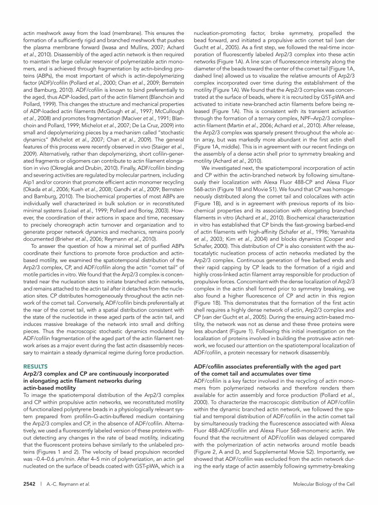
nucleation-promoting factor, broke symmetry, propelled the bead forward, and initiated a propulsive actin comet tail (van der Gucht et al., 2005). As a first step, we followed the real-time incor-poration of fluorescently labeled Arp2/3 complex into these actin networks (Figure 1A). A line scan of fluorescence intensity along the diameter of the beads toward the center of the comet tail (Figure 1A, dashed line) allowed us to visualize the relative amounts of Arp2/3 complex incorporated over time during the establishment of the motility (Figure 1A). We found that the Arp2/3 complex was concen-trated at the surface of beads, where it is recruited by GST-pWA and activated to initiate new-branched actin filaments before being re-leased (Figure 1A). This is consistent with its transient activation through the formation of a ternary complex, NPF–Arp2/3 complex–actin filament (Martin et al., 2006; Achard et al., 2010). After release, the Arp2/3 complex was sparsely present throughout the whole ac-tin array, but was markedly more abundant in the first actin shell (Figure 1A, middle). This is in agreement with our recent findings on the assembly of a dense actin shell prior to symmetry breaking and motility (Achard et al., 2010).
We investigated next, the spatiotemporal incorporation of actin and CP within the actin-branched network by following simultane-ously their localization with Alexa Fluor 488-CP and Alexa Fluor 568-actin (Figure 1B and Movie S1). We found that CP was homoge-neously distributed along the comet tail and colocalizes with actin (Figure 1B), and is in agreement with previous reports of its bio-chemical properties and its association with elongating branched filaments in vitro (Achard et al., 2010). Biochemical characterization in vitro has established that CP binds the fast-growing barbed-end of actin filaments with high-affinity (Schafer et al., 1996; Yamashita et al., 2003; Kim et al., 2004) and blocks dynamics (Cooper and Schafer, 2000). This distribution of CP is also consistent with the au-tocatalytic nucleation process of actin networks mediated by the Arp2/3 complex. Continuous generation of free barbed ends and their rapid capping by CP leads to the formation of a rigid and highly cross-linked actin filament array responsible for production of propulsive forces. Concomitant with the dense localization of Arp2/3 complex in the actin shell formed prior to symmetry breaking, we also found a higher fluorescence of CP and actin in this region (Figure 1B). This demonstrates that the formation of the first actin shell requires a highly dense network of actin, Arp2/3 complex and CP (van der Gucht et al., 2005). During the ensuing actin-based mo-tility, the network was not as dense and these three proteins were less abundant (Figure 1). Following this initial investigation on the localization of proteins involved in building the protrusive actin net-work, we focused our attention on the spatiotemporal localization of ADF/cofilin, a protein necessary for network disassembly.
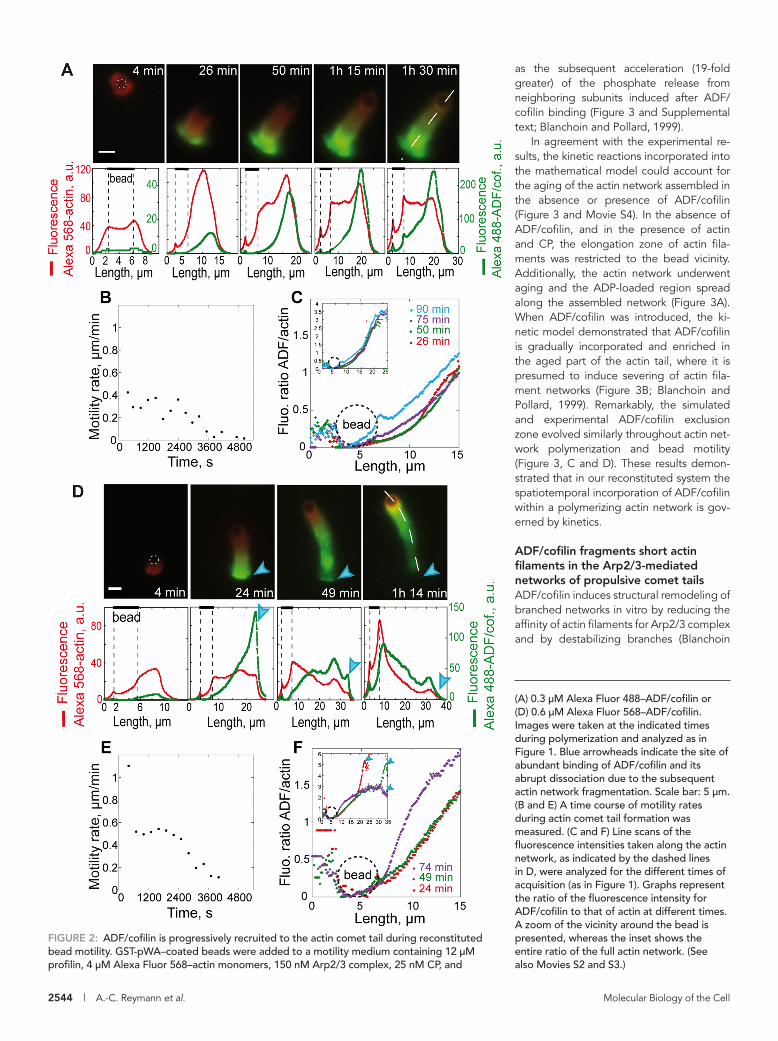
ADF/cofilin associates preferentially with the aged part of the comet tail and accumulates over timeADF/cofilin is a key factor involved in the recycling of actin mono-mers from polymerized networks and therefore renders them available for actin assembly and force production (Pollard et al., 2000). To characterize the macroscopic distribution of ADF/cofilin within the dynamic branched actin network, we followed the spa-tial and temporal distribution of ADF/cofilin in the actin comet tail by simultaneously tracking the fluorescence associated with Alexa Fluor 488-ADF/cofilin and Alexa Fluor 568-monomeric actin. We found that the recruitment of ADF/cofilin was delayed compared with the polymerization of actin networks around motile beads (Figure 2, A and D, and Supplemental Movie S2). Importantly, we showed that ADF/cofilin was excluded from the actin network dur-ing the early stage of actin assembly following symmetry-breaking
actin meshwork away from the load (membrane). This ensures the formation of a sufficiently rigid and branched meshwork that pushes the plasma membrane forward (Iwasa and Mullins, 2007; Achard et al., 2010). Disassembly of the aged actin network is then required to maintain the large cellular reservoir of polymerizable actin mono-mers, and is achieved through fragmentation by actin-binding pro-teins (ABPs), the most important of which is actin-depolymerizing factor (ADF)/cofilin (Pollard et al., 2000; Chan et al., 2009; Bernstein and Bamburg, 2010). ADF/cofilin is known to bind preferentially to the aged, thus ADP-loaded, part of the actin filament (Blanchoin and Pollard, 1999). This changes the structure and mechanical properties of ADP-loaded actin filaments (McGough et al., 1997; McCullough et al., 2008) and promotes fragmentation (Maciver et al., 1991; Blan-choin and Pollard, 1999; Michelot et al., 2007; De La Cruz, 2009) into small and depolymerizing pieces by a mechanism called “stochastic dynamics” (Michelot et al., 2007; Chan et al., 2009). The general features of this process were recently observed in vivo (Staiger et al., 2009). Alternatively, rather than depolymerizing, short cofilin-gener-ated fragments or oligomers can contribute to actin filament elonga-tion in vivo (Okreglak and Drubin, 2010). Finally, ADF/cofilin binding and severing activities are regulated by molecular partners, including Aip1 and/or coronin that promote efficient actin monomer recycling (Okada et al., 2006; Kueh et al., 2008; Gandhi et al., 2009; Bernstein and Bamburg, 2010). The biochemical properties of most ABPs are individually well characterized in bulk solution or in reconstituted minimal systems (Loisel et al., 1999; Pollard and Borisy, 2003). How-ever, the coordination of their actions in space and time, necessary to precisely choreograph actin turnover and organization and to generate proper network dynamics and mechanics, remains poorly documented (Brieher et al., 2006; Reymann et al., 2010).
To answer the question of how a minimal set of purified ABPs coordinate their functions to promote force production and actin-based motility, we examined the spatiotemporal distribution of the Arp2/3 complex, CP, and ADF/cofilin along the actin “comet tail” of motile particles in vitro. We found that the Arp2/3 complex is concen-trated near the nucleation sites to initiate branched actin networks, and remains attached to the actin tail after it detaches from the nucle-ation sites. CP distributes homogeneously throughout the actin net-work of the comet tail. Conversely, ADF/cofilin binds preferentially at the rear of the comet tail, with a spatial distribution consistent with the state of the nucleotide in these aged parts of the actin tail, and induces massive breakage of the network into small and drifting pieces. Thus the macroscopic stochastic dynamics modulated by ADF/cofilin fragmentation of the aged part of the actin filament net-work arises as a major event during the fast actin disassembly neces-sary to maintain a steady dynamical regime during force production.
RESULTSArp2/3 complex and CP are continuously incorporated in elongating actin filament networks during actin-based motilityTo image the spatiotemporal distribution of the Arp2/3 complex and CP within propulsive actin networks, we reconstituted motility of functionalized polystyrene beads in a physiologically relevant sys-tem prepared from profilin–G-actin-buffered medium containing the Arp2/3 complex and CP, in the absence of ADF/cofilin. Alterna-tively, we used a fluorescently labeled version of these proteins with-out detecting any changes in the rate of bead motility, indicating that the fluorescent proteins behave similarly to the unlabeled pro-teins (Figures 1 and 2). The velocity of bead propulsion recorded was ∼0.4–0.6 μm/min. After 4–5 min of polymerization, an actin gel nucleated on the surface of beads coated with GST-pWA, which is a
Volume 22 July 15, 2011 Actin dynamics during motility | 2543
similarly governed by the biochemical state of actin-bound nucleotides of reconstituted parallel networks (Supplemental Figure S1 and Movie S3).
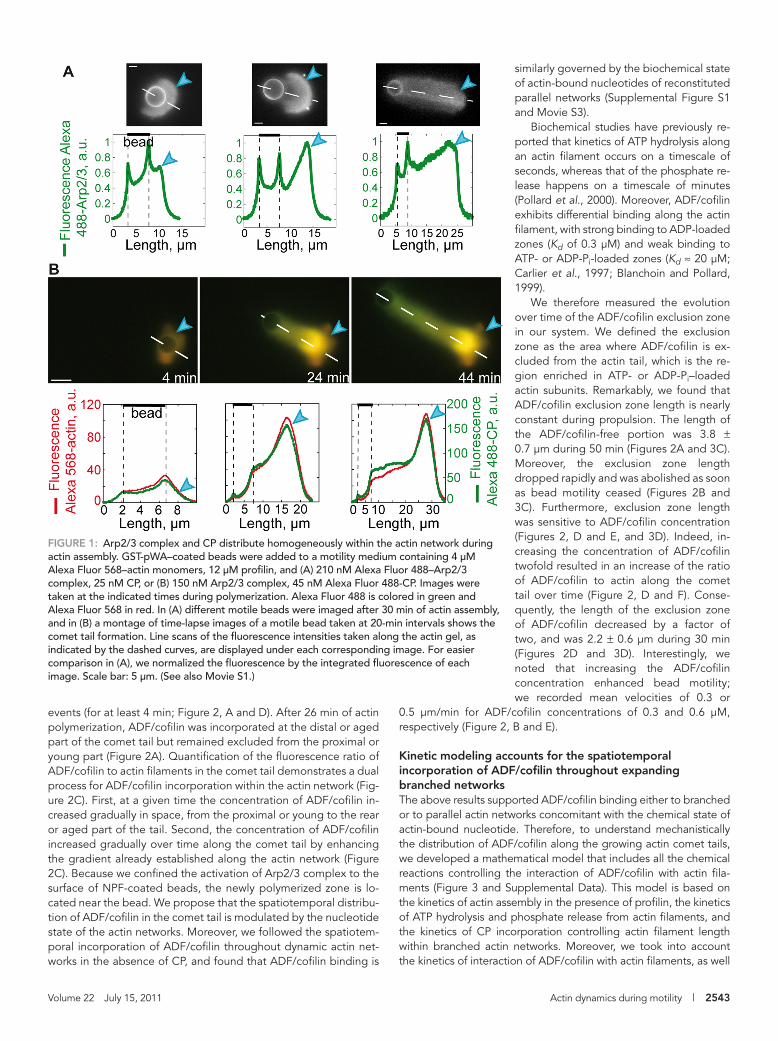
Biochemical studies have previously re-ported that kinetics of ATP hydrolysis along an actin filament occurs on a timescale of seconds, whereas that of the phosphate re-lease happens on a timescale of minutes (Pollard et al., 2000). Moreover, ADF/cofilin exhibits differential binding along the actin filament, with strong binding to ADP-loaded zones (Kd of 0.3 μM) and weak binding to ATP- or ADP-Pi-loaded zones (Kd ≈ 20 μM; Carlier et al., 1997; Blanchoin and Pollard, 1999).
We therefore measured the evolution over time of the ADF/cofilin exclusion zone in our system. We defined the exclusion zone as the area where ADF/cofilin is ex-cluded from the actin tail, which is the re-gion enriched in ATP- or ADP-Pi–loaded actin subunits. Remarkably, we found that ADF/cofilin exclusion zone length is nearly constant during propulsion. The length of the ADF/cofilin-free portion was 3.8 ± 0.7 μm during 50 min (Figures 2A and 3C). Moreover, the exclusion zone length dropped rapidly and was abolished as soon as bead motility ceased (Figures 2B and 3C). Furthermore, exclusion zone length was sensitive to ADF/cofilin concentration (Figures 2, D and E, and 3D). Indeed, in-creasing the concentration of ADF/cofilin twofold resulted in an increase of the ratio of ADF/cofilin to actin along the comet tail over time (Figure 2, D and F). Conse-quently, the length of the exclusion zone of ADF/cofilin decreased by a factor of two, and was 2.2 ± 0.6 μm during 30 min (Figures 2D and 3D). Interestingly, we noted that increasing the ADF/cofilin concentration enhanced bead motility; we recorded mean velocities of 0.3 or
0.5 μm/min for ADF/cofilin concentrations of 0.3 and 0.6 μM, respectively (Figure 2, B and E).
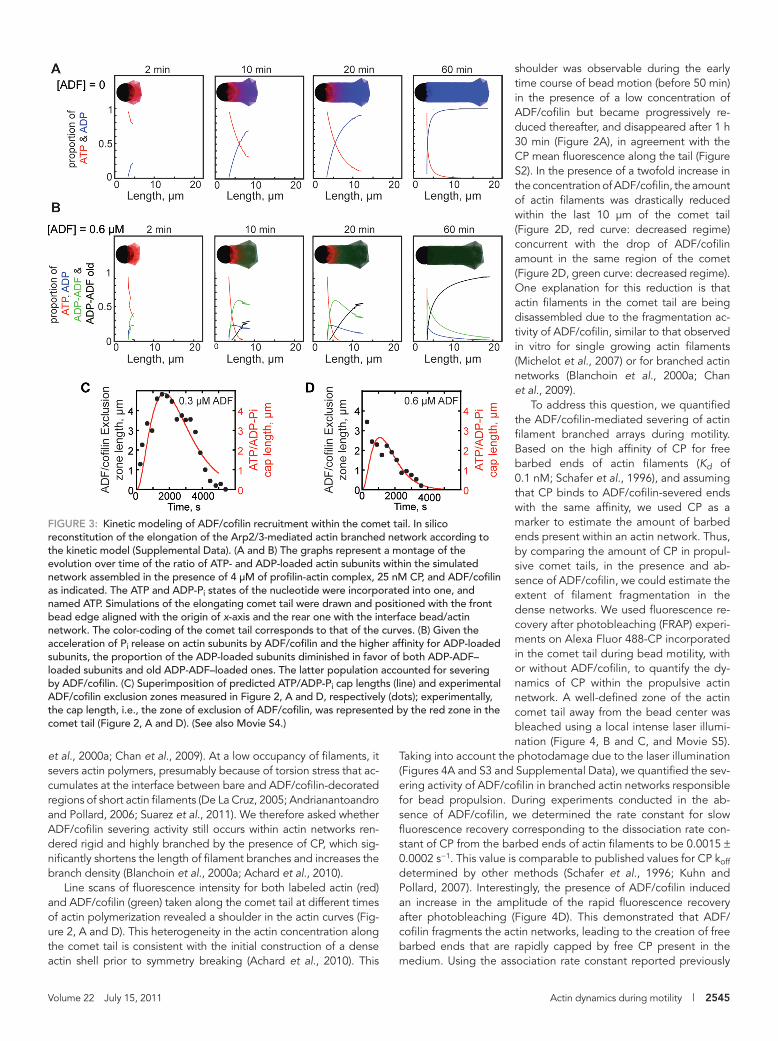
Kinetic modeling accounts for the spatiotemporal incorporation of ADF/cofilin throughout expanding branched networksThe above results supported ADF/cofilin binding either to branched or to parallel actin networks concomitant with the chemical state of actin-bound nucleotide. Therefore, to understand mechanistically the distribution of ADF/cofilin along the growing actin comet tails, we developed a mathematical model that includes all the chemical reactions controlling the interaction of ADF/cofilin with actin fila-ments (Figure 3 and Supplemental Data). This model is based on the kinetics of actin assembly in the presence of profilin, the kinetics of ATP hydrolysis and phosphate release from actin filaments, and the kinetics of CP incorporation controlling actin filament length within branched actin networks. Moreover, we took into account the kinetics of interaction of ADF/cofilin with actin filaments, as well
events (for at least 4 min; Figure 2, A and D). After 26 min of actin polymerization, ADF/cofilin was incorporated at the distal or aged part of the comet tail but remained excluded from the proximal or young part (Figure 2A). Quantification of the fluorescence ratio of ADF/cofilin to actin filaments in the comet tail demonstrates a dual process for ADF/cofilin incorporation within the actin network (Fig-ure 2C). First, at a given time the concentration of ADF/cofilin in-creased gradually in space, from the proximal or young to the rear or aged part of the tail. Second, the concentration of ADF/cofilin increased gradually over time along the comet tail by enhancing the gradient already established along the actin network (Figure 2C). Because we confined the activation of Arp2/3 complex to the surface of NPF-coated beads, the newly polymerized zone is lo-cated near the bead. We propose that the spatiotemporal distribu-tion of ADF/cofilin in the comet tail is modulated by the nucleotide state of the actin networks. Moreover, we followed the spatiotem-poral incorporation of ADF/cofilin throughout dynamic actin net-works in the absence of CP, and found that ADF/cofilin binding is
FIGURE 1: Arp2/3 complex and CP distribute homogeneously within the actin network during actin assembly. GST-pWA–coated beads were added to a motility medium containing 4 μM Alexa Fluor 568–actin monomers, 12 μM profilin, and (A) 210 nM Alexa Fluor 488–Arp2/3 complex, 25 nM CP, or (B) 150 nM Arp2/3 complex, 45 nM Alexa Fluor 488-CP. Images were taken at the indicated times during polymerization. Alexa Fluor 488 is colored in green and Alexa Fluor 568 in red. In (A) different motile beads were imaged after 30 min of actin assembly, and in (B) a montage of time-lapse images of a motile bead taken at 20-min intervals shows the comet tail formation. Line scans of the fluorescence intensities taken along the actin gel, as indicated by the dashed curves, are displayed under each corresponding image. For easier comparison in (A), we normalized the fluorescence by the integrated fluorescence of each image. Scale bar: 5 μm. (See also Movie S1.)
2544 | A.-C. Reymann et al. Molecular Biology of the Cell
as the subsequent acceleration (19-fold greater) of the phosphate release from neighboring subunits induced after ADF/cofilin binding (Figure 3 and Supplemental text; Blanchoin and Pollard, 1999).
In agreement with the experimental re-sults, the kinetic reactions incorporated into the mathematical model could account for the aging of the actin network assembled in the absence or presence of ADF/cofilin (Figure 3 and Movie S4). In the absence of ADF/cofilin, and in the presence of actin and CP, the elongation zone of actin fila-ments was restricted to the bead vicinity. Additionally, the actin network underwent aging and the ADP-loaded region spread along the assembled network (Figure 3A). When ADF/cofilin was introduced, the ki-netic model demonstrated that ADF/cofilin is gradually incorporated and enriched in the aged part of the actin tail, where it is presumed to induce severing of actin fila-ment networks (Figure 3B; Blanchoin and Pollard, 1999). Remarkably, the simulated and experimental ADF/cofilin exclusion zone evolved similarly throughout actin net-work polymerization and bead motility (Figure 3, C and D). These results demon-strated that in our reconstituted system the spatiotemporal incorporation of ADF/cofilin within a polymerizing actin network is gov-erned by kinetics.
ADF/cofilin fragments short actin filaments in the Arp2/3-mediated networks of propulsive comet tailsADF/cofilin induces structural remodeling of branched networks in vitro by reducing the affinity of actin filaments for Arp2/3 complex and by destabilizing branches (Blanchoin
FIGURE 2: ADF/cofilin is progressively recruited to the actin comet tail during reconstituted bead motility. GST-pWA–coated beads were added to a motility medium containing 12 μM profilin, 4 μM Alexa Fluor 568–actin monomers, 150 nM Arp2/3 complex, 25 nM CP, and
(A) 0.3 μM Alexa Fluor 488–ADF/cofilin or (D) 0.6 μM Alexa Fluor 568–ADF/cofilin. Images were taken at the indicated times during polymerization and analyzed as in Figure 1. Blue arrowheads indicate the site of abundant binding of ADF/cofilin and its abrupt dissociation due to the subsequent actin network fragmentation. Scale bar: 5 μm. (B and E) A time course of motility rates during actin comet tail formation was measured. (C and F) Line scans of the fluorescence intensities taken along the actin network, as indicated by the dashed lines in D, were analyzed for the different times of acquisition (as in Figure 1). Graphs represent the ratio of the fluorescence intensity for ADF/cofilin to that of actin at different times. A zoom of the vicinity around the bead is presented, whereas the inset shows the entire ratio of the full actin network. (See also Movies S2 and S3.)
Volume 22 July 15, 2011 Actin dynamics during motility | 2545
shoulder was observable during the early time course of bead motion (before 50 min) in the presence of a low concentration of ADF/cofilin but became progressively re-duced thereafter, and disappeared after 1 h 30 min (Figure 2A), in agreement with the CP mean fluorescence along the tail (Figure S2). In the presence of a twofold increase in the concentration of ADF/cofilin, the amount of actin filaments was drastically reduced within the last 10 μm of the comet tail (Figure 2D, red curve: decreased regime) concurrent with the drop of ADF/cofilin amount in the same region of the comet (Figure 2D, green curve: decreased regime). One explanation for this reduction is that actin filaments in the comet tail are being disassembled due to the fragmentation ac-tivity of ADF/cofilin, similar to that observed in vitro for single growing actin filaments (Michelot et al., 2007) or for branched actin networks (Blanchoin et al., 2000a; Chan et al., 2009).
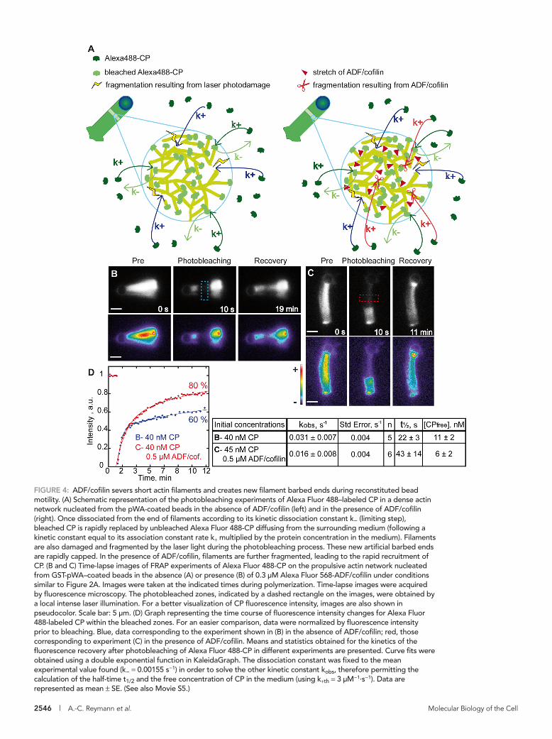
To address this question, we quantified the ADF/cofilin-mediated severing of actin filament branched arrays during motility. Based on the high affinity of CP for free barbed ends of actin filaments (Kd of 0.1 nM; Schafer et al., 1996), and assuming that CP binds to ADF/cofilin-severed ends with the same affinity, we used CP as a marker to estimate the amount of barbed ends present within an actin network. Thus, by comparing the amount of CP in propul-sive comet tails, in the presence and ab-sence of ADF/cofilin, we could estimate the extent of filament fragmentation in the dense networks. We used fluorescence re-covery after photobleaching (FRAP) experi-ments on Alexa Fluor 488-CP incorporated in the comet tail during bead motility, with or without ADF/cofilin, to quantify the dy-namics of CP within the propulsive actin network. A well-defined zone of the actin comet tail away from the bead center was bleached using a local intense laser illumi-nation (Figure 4, B and C, and Movie S5).
Taking into account the photodamage due to the laser illumination (Figures 4A and S3 and Supplemental Data), we quantified the sev-ering activity of ADF/cofilin in branched actin networks responsible for bead propulsion. During experiments conducted in the ab-sence of ADF/cofilin, we determined the rate constant for slow fluorescence recovery corresponding to the dissociation rate con-stant of CP from the barbed ends of actin filaments to be 0.0015 ± 0.0002 s−1. This value is comparable to published values for CP koff determined by other methods (Schafer et al., 1996; Kuhn and Pollard, 2007). Interestingly, the presence of ADF/cofilin induced an increase in the amplitude of the rapid fluorescence recovery after photobleaching (Figure 4D). This demonstrated that ADF/cofilin fragments the actin networks, leading to the creation of free barbed ends that are rapidly capped by free CP present in the medium. Using the association rate constant reported previously
et al., 2000a; Chan et al., 2009). At a low occupancy of filaments, it severs actin polymers, presumably because of torsion stress that ac-cumulates at the interface between bare and ADF/cofilin-decorated regions of short actin filaments (De La Cruz, 2005; Andrianantoandro and Pollard, 2006; Suarez et al., 2011). We therefore asked whether ADF/cofilin severing activity still occurs within actin networks ren-dered rigid and highly branched by the presence of CP, which sig-nificantly shortens the length of filament branches and increases the branch density (Blanchoin et al., 2000a; Achard et al., 2010).
Line scans of fluorescence intensity for both labeled actin (red) and ADF/cofilin (green) taken along the comet tail at different times of actin polymerization revealed a shoulder in the actin curves (Fig-ure 2, A and D). This heterogeneity in the actin concentration along the comet tail is consistent with the initial construction of a dense actin shell prior to symmetry breaking (Achard et al., 2010). This
FIGURE 3: Kinetic modeling of ADF/cofilin recruitment within the comet tail. In silico reconstitution of the elongation of the Arp2/3-mediated actin branched network according to the kinetic model (Supplemental Data). (A and B) The graphs represent a montage of the evolution over time of the ratio of ATP- and ADP-loaded actin subunits within the simulated network assembled in the presence of 4 μM of profilin-actin complex, 25 nM CP, and ADF/cofilin as indicated. The ATP and ADP-Pi states of the nucleotide were incorporated into one, and named ATP. Simulations of the elongating comet tail were drawn and positioned with the front bead edge aligned with the origin of x-axis and the rear one with the interface bead/actin network. The color-coding of the comet tail corresponds to that of the curves. (B) Given the acceleration of Pi release on actin subunits by ADF/cofilin and the higher affinity for ADP-loaded subunits, the proportion of the ADP-loaded subunits diminished in favor of both ADP-ADF–loaded subunits and old ADP-ADF–loaded ones. The latter population accounted for severing by ADF/cofilin. (C) Superimposition of predicted ATP/ADP-Pi cap lengths (line) and experimental ADF/cofilin exclusion zones measured in Figure 2, A and D, respectively (dots); experimentally, the cap length, i.e., the zone of exclusion of ADF/cofilin, was represented by the red zone in the comet tail (Figure 2, A and D). (See also Movie S4.)
2546 | A.-C. Reymann et al. Molecular Biology of the Cell
FIGURE 4: ADF/cofilin severs short actin filaments and creates new filament barbed ends during reconstituted bead motility. (A) Schematic representation of the photobleaching experiments of Alexa Fluor 488–labeled CP in a dense actin network nucleated from the pWA-coated beads in the absence of ADF/cofilin (left) and in the presence of ADF/cofilin (right). Once dissociated from the end of filaments according to its kinetic dissociation constant k− (limiting step), bleached CP is rapidly replaced by unbleached Alexa Fluor 488-CP diffusing from the surrounding medium (following a kinetic constant equal to its association constant rate k+ multiplied by the protein concentration in the medium). Filaments are also damaged and fragmented by the laser light during the photobleaching process. These new artificial barbed ends are rapidly capped. In the presence of ADF/cofilin, filaments are further fragmented, leading to the rapid recruitment of CP. (B and C) Time-lapse images of FRAP experiments of Alexa Fluor 488-CP on the propulsive actin network nucleated from GST-pWA–coated beads in the absence (A) or presence (B) of 0.3 μM Alexa Fluor 568-ADF/cofilin under conditions similar to Figure 2A. Images were taken at the indicated times during polymerization. Time-lapse images were acquired by fluorescence microscopy. The photobleached zones, indicated by a dashed rectangle on the images, were obtained by a local intense laser illumination. For a better visualization of CP fluorescence intensity, images are also shown in pseudocolor. Scale bar: 5 μm. (D) Graph representing the time course of fluorescence intensity changes for Alexa Fluor 488-labeled CP within the bleached zones. For an easier comparison, data were normalized by fluorescence intensity prior to bleaching. Blue, data corresponding to the experiment shown in (B) in the absence of ADF/cofilin; red, those corresponding to experiment (C) in the presence of ADF/cofilin. Means and statistics obtained for the kinetics of the fluorescence recovery after photobleaching of Alexa Fluor 488-CP in different experiments are presented. Curve fits were obtained using a double exponential function in KaleidaGraph. The dissociation constant was fixed to the mean experimental value found (k− = 0.00155 s−1) in order to solve the other kinetic constant kobs, therefore permitting the calculation of the half-time t1/2 and the free concentration of CP in the medium (using k+th = 3 μM−1·s−1). Data are represented as mean ± SE. (See also Movie S5.)
Volume 22 July 15, 2011 Actin dynamics during motility | 2547
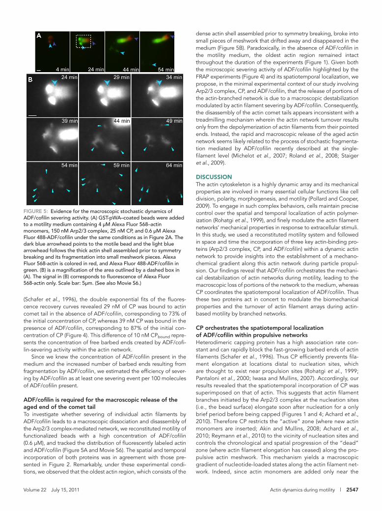
dense actin shell assembled prior to symmetry breaking, broke into small pieces of meshwork that drifted away and disappeared in the medium (Figure 5B). Paradoxically, in the absence of ADF/cofilin in the motility medium, the oldest actin region remained intact throughout the duration of the experiments (Figure 1). Given both the microscopic severing activity of ADF/cofilin highlighted by the FRAP experiments (Figure 4) and its spatiotemporal localization, we propose, in the minimal experimental context of our study involving Arp2/3 complex, CP, and ADF/cofilin, that the release of portions of the actin-branched network is due to a macroscopic destabilization modulated by actin filament severing by ADF/cofilin. Consequently, the disassembly of the actin comet tails appears inconsistent with a treadmilling mechanism wherein the actin network turnover results only from the depolymerization of actin filaments from their pointed ends. Instead, the rapid and macroscopic release of the aged actin network seems likely related to the process of stochastic fragmenta-tion mediated by ADF/cofilin recently described at the single-filament level (Michelot et al., 2007; Roland et al., 2008; Staiger et al., 2009).
DISCUSSIONThe actin cytoskeleton is a highly dynamic array and its mechanical properties are involved in many essential cellular functions like cell division, polarity, morphogenesis, and motility (Pollard and Cooper, 2009). To engage in such complex behaviors, cells maintain precise control over the spatial and temporal localization of actin polymer-ization (Rohatgi et al., 1999), and finely modulate the actin filament networks’ mechanical properties in response to extracellular stimuli. In this study, we used a reconstituted motility system and followed in space and time the incorporation of three key actin-binding pro-teins (Arp2/3 complex, CP, and ADF/cofilin) within a dynamic actin network to provide insights into the establishment of a mechano-chemical gradient along this actin network during particle propul-sion. Our findings reveal that ADF/cofilin orchestrates the mechani-cal destabilization of actin networks during motility, leading to the macroscopic loss of portions of the network to the medium, whereas CP coordinates the spatiotemporal localization of ADF/cofilin. Thus these two proteins act in concert to modulate the biomechanical properties and the turnover of actin filament arrays during actin-based motility by branched networks.
CP orchestrates the spatiotemporal localization of ADF/cofilin within propulsive networksHeterodimeric capping protein has a high association rate con-stant and can rapidly block the fast-growing barbed ends of actin filaments (Schafer et al., 1996). Thus CP efficiently prevents fila-ment elongation at locations distal to nucleation sites, which are thought to exist near propulsion sites (Rohatgi et al., 1999; Pantaloni et al., 2000; Iwasa and Mullins, 2007). Accordingly, our results revealed that the spatiotemporal incorporation of CP was superimposed on that of actin. This suggests that actin filament branches initiated by the Arp2/3 complex at the nucleation sites (i.e., the bead surface) elongate soon after nucleation for a only brief period before being capped (Figures 1 and 4; Achard et al., 2010). Therefore CP restricts the “active” zone (where new actin monomers are inserted; Akin and Mullins, 2008; Achard et al., 2010; Reymann et al., 2010) to the vicinity of nucleation sites and controls the chronological and spatial progression of the “dead” zone (where actin filament elongation has ceased) along the pro-pulsive actin meshwork. This mechanism yields a macroscopic gradient of nucleotide-loaded states along the actin filament net-work. Indeed, since actin monomers are added only near the
(Schafer et al., 1996), the double exponential fits of the fluores-cence recovery curves revealed 29 nM of CP was bound to actin comet tail in the absence of ADF/cofilin, corresponding to 73% of the initial concentration of CP, whereas 39 nM CP was bound in the presence of ADF/cofilin, corresponding to 87% of the initial con-centration of CP (Figure 4). This difference of 10 nM CPbound repre-sents the concentration of free barbed ends created by ADF/cofi-lin-severing activity within the actin network.
Since we knew the concentration of ADF/cofilin present in the medium and the increased number of barbed ends resulting from fragmentation by ADF/cofilin, we estimated the efficiency of sever-ing by ADF/cofilin as at least one severing event per 100 molecules of ADF/cofilin present.
ADF/cofilin is required for the macroscopic release of the aged end of the comet tailTo investigate whether severing of individual actin filaments by ADF/cofilin leads to a macroscopic dissociation and disassembly of the Arp2/3 complex-mediated network, we reconstituted motility of functionalized beads with a high concentration of ADF/cofilin (0.6 μM), and tracked the distribution of fluorescently labeled actin and ADF/cofilin (Figure 5A and Movie S6). The spatial and temporal incorporation of both proteins was in agreement with those pre-sented in Figure 2. Remarkably, under these experimental condi-tions, we observed that the oldest actin region, which consists of the
FIGURE 5: Evidence for the macroscopic stochastic dynamics of ADF/cofilin severing activity. (A) GST-pWA–coated beads were added to a motility medium containing 4 μM Alexa Fluor 568–actin monomers, 150 nM Arp2/3 complex, 25 nM CP, and 0.6 μM Alexa Fluor 488-ADF/cofilin under the same conditions as in Figure 2A. The dark blue arrowhead points to the motile bead and the light blue arrowhead follows the thick actin shell assembled prior to symmetry breaking and its fragmentation into small meshwork pieces. Alexa Fluor 568-actin is colored in red, and Alexa Fluor 488-ADF/cofilin in green. (B) is a magnification of the area outlined by a dashed box in (A). The signal in (B) corresponds to fluorescence of Alexa Fluor 568-actin only. Scale bar: 5μm. (See also Movie S6.)
2548 | A.-C. Reymann et al. Molecular Biology of the Cell
Concluding remarksThe rapid and continuous recruitment of CP throughout the Arp2/3-mediated actin assembly imposes mechanical properties on the ac-tin network conformation, which is a rigid array necessary for the production of protrusive force. CP also directly controls the spa-tiotemporal recruitment of ADF/cofilin, which in turn affects the ac-tin network mechanics and CP turnover. Consequently, we propose that the synergy between CP and ADF/cofilin leads to a tight cou-pling between both the biochemical and mechanical properties of the actin networks, which explains the turnover of actin filaments at the leading edge of motile cells (Svitkina and Borisy, 1999).
It must be kept in mind that cells display an infinite complexity and the spatiotemporal distribution of actin networks’ aging zones imposed by CP, together with the subsequent actin monomer recy-cling by ADF/cofilin severing activity, are modulated by a diverse bat-tery of regulating proteins, including Aip1 (Kueh et al., 2008; Okreglak and Drubin, 2010), coronin (Gandhi et al., 2009), tropomyosin (Blanchoin et al., 2001; Bugyi et al., 2010), and myosin II (Hotulainen and Lappalainen, 2006). In addition, processive actin polymerases such as formin (Hotulainen and Lappalainen, 2006; Lee et al., 2010) or Enabled/vasodilator-stimulated phosphoprotein (Ena/VASP) fam-ily of proteins (Bear, 2002; Trichet et al., 2007; Hansen and Mullins, 2010) protect actin filament barbed ends against CP and generate parallel actin networks with specific mechanical properties. Further understanding of the interplay between the control of actin dynamics and the actin network’s mechanical properties should come from studies where the minimal reconstituted motility system is expanded to include the presence of additional actin-regulating factors.
MATERIALS AND METHODSProtein production and labelingActin was purified from rabbit skeletal muscle acetone powder (Spudich and Watt, 1971). Monomeric Ca–ATP–actin was purified by gel-filtration chromatography on Sephacryl S-300 (McLean-Fletcher and Pollard, 1980) at 4°C in G buffer (5 mM Tris-HCl, pH 8.0, 0.2 mM ATP, 0.1 mM CaCl2, and 0.5 mM dithiothreitol [DTT]). Actin was la-beled on lysines with Alexa Fluor 568 or Alexa Fluor 488 as de-scribed previously (Isambert et al., 1995; Egile et al., 1999). The Arp2/3 complex was purified from bovine brain extracts according to Egile et al. (1999). GST-WA, GST-pWA, human profilin, mouse CP, and ADF/cofilin were expressed and purified as described previ-ously (Almo et al., 1994; Machesky et al., 1999; Blanchoin et al., 2000b; Falck et al., 2004). The Arp2/3 complex, CP, and ADF/cofilin were labeled on cysteines with Alexa Fluor 568 or Alexa Fluor 488. D34C, C62A mutations were introduced on COF1 Saccharomyces cerevisiae cofilin gene, as previously described (Lappalainen et al., 1997). We referred to this double mutant protein as “D34C-ADF/cofilin” and labeled it on C34 (Suarez et al., 2011). The purified pro-tein solution was dialyzed overnight against two changes of dialysis buffer (10 mM Tris-HCl, pH 7.5, 150 mM NaCl, 2 mM EDTA, 2 mM Tris(2-carboxyethyl)phosphine hydrochloride [TCEP]). The protein concentration was then adjusted to 100 μM for a total volume of 1 ml and labeled overnight with sixfold excess of Alexa 488 or Alexa 568 C5–maleimide (Invitrogen, Carlsbad, CA). Alexa Fluor 488- or Alexa Fluor 568–protein was purified by gel-filtration chromatogra-phy on G25 (Sigma-Aldrich, St. Louis, MO) in elution buffer (10 mM Tris-HCl, pH 7.5, 150 mM NaCl, 2 mM EDTA, 2 mM DTT) and stored at −80°C.
Coating of beadsCarboxylate polystyrene microspheres (4.5 μm diameter, 2.6% sol-ids-latex suspension; Polysciences, Eppelheim, Germany) were
bead surface, ATP-loaded subunits are temporarily constrained to this region of the comet tail before ATP hydrolysis and phos-phate dissociation occur in the whole network. Because ADF/co-filin has a marked preference for ADP-loaded actin (Blanchoin and Pollard, 1999), the establishment of this gradient controls the gradual incorporation of ADF/cofilin along the actin network (Figures 2A and 3). In addition, by increasing the rate of phos-phate release, ADF/cofilin limits the size of the actin filament net-work resistant to its interaction (Blanchoin and Pollard, 1999; Suarez et al., 2011). Consistent with this scheme, as soon as bead motility stops (Figure 2B), due to a lack of actin monomers in so-lution, the size of the ADF/cofilin exclusion zone around the bead rapidly decreases, and ultimately disappears after 1 h. The spa-tiotemporal localization of CP and ADF/cofilin described here is fully consistent with their mutual distribution in vivo (Svitkina and Borisy, 1999; Okreglak and Drubin, 2007). Indeed, CP localizes to the lamellipodium in the 1 μm immediately adjacent to the plasma membrane (Iwasa and Mullins, 2007), whereas ADF/cofilin is ex-cluded from 0.2–0.7 μm of the lamellipodial network (Svitkina and Borisy, 1999). Our present results demonstrate that the gradient of ADF/cofilin along the actin network is controlled by kinetics, that is, by its higher affinity for ADP-loaded subunits and its abil-ity to accelerate Pi release (Figures 2 and 3).
ADF/cofilin catalyzes the microscopic severing and macroscopic destabilization of dense and rigid actin networksIn terms of its mechanical properties, the propulsive actin fila-ment network is made of a dense and rigid actin filament array with an elongation zone restricted spatially and temporally to the vicinity of the nucleation particle. The FRAP experiments provide evidence that ADF/cofilin was able to sever actin filaments within this stiff and highly cross-linked actin network, as the actin-based propulsion of particles was achieved in the presence of a critical amount of CP. This also emphasized that actin filament severing by ADF/cofilin, followed by the rapid recruitment of CP to newly created barbed ends, introduces a mechanical heterogeneity throughout branched networks. The microscopic fragmentation of actin filaments by ADF/cofilin within the comet tail is consis-tent with the fact that its spatial and temporal incorporation cor-related with the decrease of actin filament density along the ag-ing regions of the tail (Figure 2, A and D, line scans). In addition, studies have established that ADF/cofilin also dissociates Arp2/3-generated actin filament branches (Blanchoin et al., 2000a; Chan et al., 2009) and only slightly affects the rate of depolymerization of actin filaments (Andrianantoandro and Pollard, 2006; Suarez et al., 2011). Thus a global process that relies on filament deb-ranching and severing by ADF/cofilin, rather than enhanced pointed-end depolymerization, is an attractive explanation for the loss of actin mass from older regions of an actin array that is dense and rigid (Figures 2A, line scans, and 3C). We propose that loss of actin filaments in the comet tail due to fragmentation by ADF/cofilin occurs in two steps: 1) it happens microscopically at the single actin filament level, and 2) it propagates macro-scopically to release stochastically not single filaments but large portions of the aged actin network. This model is in agreement with the fast disassembly of actin filaments within dense branched networks in endocytic patches (Berro et al., 2010). Furthermore, it reconciles the paradox for the apparent dissociation rate of CP being three orders of magnitude faster in vivo compared with in vitro in the absence of any severing protein (Miyoshi et al., 2006).
Volume 22 July 15, 2011 Actin dynamics during motility | 2549
mixed with 2 μM GST-pWA in X buffer (10 mM HEPES, pH 7.5, 0.1 M KCl, 1 mM MgCl2, 1 mM ATP, and 0.1 mM CaCl2) for 15 min at 20°C on a thermoshaker. The beads coated with pWA were then washed in X buffer containing 1% bovine serum albumin (BSA) and stored on ice for 48 h in X buffer-0.1% BSA. GST-pWA surface density on the beads was quantified on SDS–PAGE gel: 2.4 × 10⁴ pWA/μm2.
Motility assayGST-pWA–coated beads were mixed with a motility medium con-taining 4 μM actin monomers, 12 μM profilin, and the indicated con-centration of Arp2/3, CP, and ADF/cofilin, labeled or not, in X buffer (10 mM HEPES, pH 7.0, 0.1 M KCl, 1 mM MgCl2, 1 mM ATP, and 0.1 mM CaCl2) supplemented with 1% BSA, 0.2% methylcellulose, 3 mM DTT, 0.13 mM DABCO, 1.8 mM ATP. Image acquisition was performed on a Zeiss axioplan microscope (Jena, Germany) equipped with a 63×, 1.5 numerical aperture Plan-APOCHROMAT objective lens, and images were collected with a Hamamatsu ORCA CCD camera (Hamamatsu, Herrsching am Ammersee, Germany) with Metavue version 6.2r6 (Universal Imaging, Media, PA). Two sets of filters were used: FITC filter (BP 450–490, FT 510, BP 515–568, Zeiss) adapted to the Alexa Fluor 488 and rhodamine filter (BP 546/12, FT580, LP 590, Zeiss) adapted to the Alexa Fluor 568. A journal was created in Metavue in order to acquire three images quasi-simultaneously (phase contrast, Alexa Fluor 568 acquisition, Alexa Fluor 488 acquisition).
For bead motility, time-lapse images of motile beads were ac-quired quasi-simultaneously by fluorescence microscopy with two sets of filters adapted to Alexa Fluor 488 and Alexa Fluor 568, per-mitting the tracking of two proteins.
Data analysis of FRAP experimentsImages were then combined using Metamorph (Molecular Devices, Sunnyvale, CA). Images corresponding to the different fluorophores were color combined and analyzed (line scan, threshold area) using Metamorph version 7.5. Data were then analyzed (normalization, curve fits) and plotted with KaleidaGraph version 4.01 (Synergy Soft-ware, Reading, PA). For the fluorescence recoveries after bleaching, double exponential curve fits were first achieved on experiments in the absence of ADF/cofilin in order to obtain a mean value of the k− (0.00155 s−1 ± 0.00016; n = 5). This value was then fixed in all double exponential fits possible to obtain the other kinetic constant (kobs = k+ * [CPfree]). Using the theoretical k+ (3 μM−1 s−1) published by Schafer et al. (1996), we obtained an estimation of the concentra-tion of free CP in the medium.
ACKNOWLEDGMENTSThis work was supported by grants to L.B. from Agence Nationale de la Recherche (ANR-08-BLAN-0012 and ANR-08-SYSC-013) and a fellowship to A.C.R. from CEA. C.J.S. was supported by the Physical Biosciences Program of the Office of Basic Energy Sciences, U. S. Department of Energy, under contract number DE-FG02–04ER15526.
REFERENCESAchard V, Martiel JL, Michelot A, Guerin C, Reymann AC, Blanchoin L,
Boujemaa-Paterski R (2010). A “primer”-based mechanism underlies branched actin filament network formation and motility. Curr Biol 20, 423–428.
Akin O, Mullins RD (2008). Capping protein increases the rate of actin-based motility by promoting filament nucleation by the Arp2/3 complex. Cell 133, 841–851.
Almo SC, Pollard TD, Way M, Lattman EE (1994). Purification, characteriza-tion and crystallization of Acanthamoeba profilin expressed in Escheri-chia coli. J Mol Biol 236, 950–952.
Andrianantoandro E, Pollard TD (2006). Mechanism of actin filament turnover by severing and nucleation at different concentrations of ADF/cofilin. Mol Cell 24, 13–23.
Bear JE (2002). Formins: taking a ride on the barbed end. Dev Cell 3, 149–150.
Bernstein BW, Bamburg JR (2010). ADF/cofilin: a functional node in cell biology. Trends Cell Biol 20, 187–195.
Berro J, Sirotkin V, Pollard TD (2010). Mathematical modeling of endocytic actin patch kinetics in fission yeast: disassembly requires release of actin filament fragments. Mol Biol Cell 21, 2905–2915.
Blanchoin L, Pollard TD (1999). Mechanism of interaction of Acanthamoeba actophorin (ADF/cofilin) with actin filaments. J Biol Chem 274, 15538–15546.
Blanchoin L, Pollard TD, Hitchcock-DeGregori SE (2001). Inhibition of the Arp2/3 complex-nucleated actin polymerization and branch formation by tropomyosin. Curr Biol 11, 1300–1304.
Blanchoin L, Pollard TD, Mullins RD (2000a). Interaction of ADF/cofi-lin, Arp2/3 complex, capping protein and profilin in remodeling of branched actin filament networks. Curr Biol 10, 1273–1282.
Blanchoin L, Robinson RC, Choe S, Pollard TD (2000b). Phosphorylation of Acanthamoeba actophorin (ADF/cofilin) blocks interaction with actin without a change in atomic structure. J Mol Biol 295, 203–211.
Brieher WM, Kueh HY, Ballif BA, Mitchison TJ (2006). Rapid actin monomer-insensitive depolymerization of Listeria actin comet tails by cofilin, coronin, and Aip1. J Cell Biol 175, 315–324.
Bugyi B, Didry D, Carlier MF (2010). How tropomyosin regulates lamel-lipodial actin-based motility: a combined biochemical and reconstituted motility approach. EMBO J 29, 14–26.
Carlier MF, Laurent V, Santolini J, Melki R, Didry D, Xia GX, Hong Y, Chua NH, Pantaloni D (1997). Actin depolymerizing factor (ADF/cofilin) en-hances the rate of filament turnover: implication in actin-based motility. J Cell Biol 136, 1307–1322.
Chan C, Beltzner CC, Pollard TD (2009). Cofilin dissociates Arp2/3 complex and branches from actin filaments. Curr Biol 19, 537–545.
Chhabra ES, Higgs HN (2007). The many faces of actin: matching assembly factors with cellular structures. Nat Cell Biol 9, 1110–1121.
Cooper JA, Schafer DA (2000). Control of actin assembly and disassembly at filament ends. Curr Opin Cell Biol 12, 97–103.
De La Cruz EM (2005). Cofilin binding to muscle and non-muscle actin filaments: isoform-dependent cooperative interactions. J Mol Biol 346, 557–564.
De La Cruz EM (2009). How cofilin severs an actin filament. Biophys Rev 1, 51–59.
Egile C, Loisel TP, Laurent V, Li R, Pantaloni D, Sansonetti PJ, Carlier MF (1999). Activation of the CDC42 effector N-WASP by the Shigella flexneri IcsA protein promotes actin nucleation by Arp2/3 complex and bacterial actin-based motility. J Cell Biol 146, 1319–1332.
Falck S, Paavilainen VO, Wear MA, Grossmann JG, Cooper JA, Lappalainen P (2004). Biological role and structural mechanism of twinfilin-capping protein interaction. EMBO J 23, 3010–3019.
Gandhi M, Achard V, Blanchoin L, Goode BL (2009). Coronin switches roles in actin disassembly depending on the nucleotide state of actin. Mol Cell 34, 364–374.
Goley ED, Welch MD (2006). The ARP2/3 complex: an actin nucleator comes of age. Nat Rev Mol Cell Biol 7, 713–726.
Hansen SD, Mullins RD (2010). VASP is a processive actin polymerase that requires monomeric actin for barbed end association. J Cell Biol 191, 571–584.
Hotulainen P, Lappalainen P (2006). Stress fibers are generated by two dis-tinct actin assembly mechanisms in motile cells. J Cell Biol 173, 383–394.
Isambert H, Venier P, Maggs AC, Fattoum A, Kassab R, Pantaloni D, Carlier MF (1995). Flexibility of actin filaments derived from thermal fluctua-tions. Effect of bound nucleotide, phalloidin, and muscle regulatory proteins. J Biol Chem 270, 11437–11444.
Iwasa JH, Mullins RD (2007). Spatial and temporal relationships between actin-filament nucleation, capping, and disassembly. Curr Biol 17, 395–406.
Kim K, Yamashita A, Wear MA, Maeda Y, Cooper JA (2004). Capping pro-tein binding to actin in yeast: biochemical mechanism and physiological relevance. J Cell Biol 164, 567–580.
Kueh HY, Charras GT, Mitchison TJ, Brieher WM (2008). Actin disassembly by cofilin, coronin, and Aip1 occurs in bursts and is inhibited by barbed-end cappers. J Cell Biol 182, 341–353.
Kuhn JR, Pollard TD (2007). Single molecule kinetic analysis of actin filament capping. Polyphosphoinositides do not dissociate capping proteins. J Biol Chem 282, 28014–28024.
2550 | A.-C. Reymann et al. Molecular Biology of the Cell
Pollard TD, Blanchoin L, Mullins RD (2000). Molecular mechanisms control-ling actin filament dynamics in nonmuscle cells. Annu Rev Biophys 29, 545–576.
Pollard TD, Borisy GG (2003). Cellular motility driven by assembly and disas-sembly of actin filaments. Cell 112, 453–465.
Pollard TD, Cooper JA (2009). Actin, a central player in cell shape and movement. Science 326, 1208–1212.
Reymann A-C, Martiel J-L, Cambier T, Blanchoin L, Boujemaa-Paterski R, Théry M (2010). Nucleation geometry governs ordered actin networks structures. Nat Mat 9, 827–832.
Rohatgi R, Ma L, Miki H, Lopez M, Kirchhausen T, Takenawa T, Kirsch-ner MW (1999). The interaction between N-WASP and the Arp2/3 complex links Cdc42-dependent signals to actin assembly. Cell 97, 221–231.
Roland J, Berro J, Michelot A, Blanchoin L, Martiel JL (2008). Stochas-tic severing of actin filaments by actin depolymerizing factor/cofilin controls the emergence of a steady dynamical regime. Biophys J 94, 2082–2094.
Rouiller I, Xu XP, Amann KJ, Egile C, Nickell S, Nicastro D, Li R, Pollard TD, Volkmann N, Hanein D (2008). The structural basis of actin filament branching by the Arp2/3 complex. J Cell Biol 180, 887–895.
Schafer DA, Jennings PB, Cooper JA (1996). Dynamics of capping protein and actin assembly in vitro: uncapping barbed ends by polyphospho-inositides. J Cell Biol 135, 169–179.
Spudich JA, Watt S (1971). The regulation of rabbit skeletal muscle contrac-tion. I. Biochemical studies of the interaction of the tropomyosin-tro-ponin complex with actin and the proteolytic fragments of myosin. J Biol Chem 246, 4866–4871.
Staiger CJ, Sheahan MB, Khurana P, Wang X, McCurdy DW, Blanchoin L (2009). Actin filament dynamics are dominated by rapid growth and severing activity in the Arabidopsis cortical array. J Cell Biol 184, 269–280.
Suarez C, Roland J, Boujemaa-Paterski R, Kang H, McCullough BR, Reymann AC, Guérin C, Martiel JL, De La Cruz E, Blanchoin L (2011). Cofilin tunes the nucleotide state of actin filaments and severs at bare and decorated segment boundaries. Curr Biol 21, 862–868.
Svitkina TM, Borisy GG (1999). Arp2/3 complex and actin depolymerizing factor/cofilin in dendritic organization and treadmilling of actin filament array in lamellipodia. J Cell Biol 145, 1009–1026.
Trichet L, Campas O, Sykes C, Plastino J (2007). VASP governs actin dynam-ics by modulating filament anchoring. Biophys J 92, 1081–1089.
van der Gucht J, Paluch E, Plastino J, Sykes C (2005). Stress release drives symmetry breaking for actin-based movement. Proc Natl Acad Sci USA 102, 7847–7852.
Welch MD, Rosenblatt J, Skoble J, Portnoy DA, Mitchison TJ (1998). Inter-action of human Arp2/3 complex and the Listeria monocytogenes ActA protein in actin filament nucleation. Science 281, 105–108.
Yamashita A, Maeda K, Maeda Y (2003). Crystal structure of CapZ: structural basis for actin filament barbed end capping. EMBO J 22, 1529–1538.
Lappalainen P, Fedorov EV, Fedorov AA, Almo SC, Drubin DG (1997). Es-sential functions and actin-binding surfaces of yeast cofilin revealed by systematic mutagenesis. EMBO J 16, 5520–5530.
Lee K, Gallop JL, Rambani K, Kirschner MW (2010). Self-assembly of filopo-dia-like structures on supported lipid bilayers. Science 329, 1341–1345.
Loisel TP, Boujemaa R, Pantaloni D, Carlier MF (1999). Reconstitution of actin-based motility of Listeria and Shigella using pure proteins. Nature 401, 613–616.
Machesky LM, Mullins DM, Higgs HN, Kaiser DA, Blanchoin L, May RC, Hall ME, Pollard TD (1999). Scar, a WASp-related protein, activates nucleation of actin filaments by the Arp2/3 complex. Proc Natl Acad Sci USA 96, 3739–3744.
Maciver SK, Zot HG, Pollard TD (1991). Characterization of actin filament severing by actophorin from Acanthamoeba castellanii. J Cell Biol 115, 1611–1620.
Martin AC, Welch MD, Drubin DG (2006). Arp2/3 ATP hydrolysis-catalysed branch dissociation is critical for endocytic force generation. Nat Cell Biol 8, 826–833.
McCullough BR, Blanchoin L, Martiel JL, De la Cruz EM (2008). Cofilin increases the bending flexibility of actin filaments: implications for sever-ing and cell mechanics. J Mol Biol 381, 550–558.
McGough A, Pope B, Chiu W, Weeds A (1997). Cofilin changes the twist of F-actin: implications for actin filament dynamics and cellular function. J Cell Biol 138, 771–781.
McLean-Fletcher S, Pollard TD (1980). Identification of a factor in conven-tional muscle actin preparations which inhibits actin filament self-associ-ation. Biochem Biophys Res Commun 96, 18–27.
Michelot A, Berro J, Guerin C, Boujemaa-Paterski R, Staiger CJ, Martiel JL, Blanchoin L (2007). Actin-filament stochastic dynamics mediated by ADF/cofilin. Curr Biol 17, 825–833.
Miyoshi T, Tsuji T, Higashida C, Hertzog M, Fujita A, Narumiya S, Scita G, Watanabe N (2006). Actin turnover-dependent fast dissociation of capping protein in the dendritic nucleation actin network: evidence of frequent filament severing. J Cell Biol 175, 947–955.
Mullins RD, Heuser JA, Pollard TD (1998). The interaction of Arp2/3 complex with actin: nucleation, high-affinity pointed end capping, and formation of branching networks of filaments. Proc Natl Acad Sci USA 95, 6181–6186.
Okada K, Ravi H, Smith EM, Goode BL (2006). Aip1 and cofilin promote rapid turnover of yeast actin patches and cables: a coordinated mecha-nism for severing and capping filaments. Mol Biol Cell 17, 2855–2868.
Okreglak V, Drubin DG (2007). Cofilin recruitment and function during actin-mediated endocytosis dictated by actin nucleotide state. J Cell Biol 178, 1251–1264.
Okreglak V, Drubin DG (2010). Loss of Aip1 reveals a role in maintaining the actin monomer pool and an in vivo oligomer assembly pathway. J Cell Biol 188, 769–777.
Pantaloni D, Boujemaa R, Didry D, Gounon P, Carlier M-F (2000). The Arp2/3 complex branches filament barbed ends: functional antagonism with capping proteins. Nat Cell Biol 2, 385–391.

![A unique profilin-actin interface is important for malaria ...jultika.oulu.fi/files/nbnfi-fe201708158115.pdf · mission [19, 20]. Parasites can exert forces on various substrates](https://img.pdfslide.fr/doc/110x75/5f3ffa52317435472c17507b/a-unique-profilin-actin-interface-is-important-for-malaria-mission-19-20.jpg)