Embed Size (px)
Citation preview
Duy et al. BMC Infectious Diseases (2017) 17:333 DOI 10.1186/s12879-017-2427-4
RESEARCH ARTICLE Open Access
Valine/isoleucine variants drive selectivepressure in the VP1 sequence of EV-A71enteroviruses
Nghia Ngu Duy1,2,3*, Le Thi Thanh Huong1, Patrice Ravel4, Le Thi Song Huong5, Ankit Dwivedi6,October Michael Sessions7, Yan’An Hou7, Robert Chua7, Guilhem Kister8, Aneta Afelt9, Catherine Moulia2,Duane J. Gubler7, Vu Dinh Thiem1, Nguyen Thi Hien Thanh1, Christian Devaux10, Tran Nhu Duong1,Nguyen Tran Hien1, Emmanuel Cornillot4,6, Laurent Gavotte2 and Roger Frutos3,11*Abstract
Background: In 2011–2012, Northern Vietnam experienced its first large scale hand foot and mouth disease (HFMD)epidemic. In 2011, a major HFMD epidemic was also reported in South Vietnam with fatal cases. This 2011–2012 outbreakwas the first one to occur in North Vietnam providing grounds to study the etiology, origin and dynamic of the disease.We report here the analysis of the VP1 gene of strains isolated throughout North Vietnam during the 2011–2012 outbreakand before.
Methods: The VP1 gene of 106 EV-A71 isolates from North Vietnam and 2 from Central Vietnam were sequenced.Sequence alignments were analyzed at the nucleic acid and protein level. Gene polymorphism was also analyzed.A Factorial Correspondence Analysis was performed to correlate amino acid mutations with clinical parameters.
Results: The sequences were distributed into four phylogenetic clusters. Three clusters corresponded to thesubgenogroup C4 and the last one corresponded to the subgenogroup C5. Each cluster displayed differentpolymorphism characteristics. Proteins were highly conserved but three sites bearing only Isoleucine (I) or Valine (V)were characterized. The isoleucine/valine variability matched the clusters. Spatiotemporal analysis of the I/V variantsshowed that all variants which emerged in 2011 and then in 2012 were not the same but were all present in theregion prior to the 2011–2012 outbreak. Some correlation was found between certain I/V variants and ethnicity andseverity.
Conclusions: The 2011–2012 outbreak was not caused by an exogenous strain coming from South Vietnam orelsewhere but by strains already present and circulating at low level in North Vietnam. However, what triggered theoutbreak remains unclear. A selective pressure is applied on I/V variants which matches the genetic clusters. I/V variantswere shown on other viruses to correlate with pathogenicity. This should be investigated in EV-A71. I/V variants are aneasy and efficient way to survey and identify circulating EV-A71 strains.
Keywords: VP1, HFMD, Enterovirus, EV-A71, Vietnam
* Correspondence: [email protected]; [email protected];[email protected] Institute of Hygiene and Epidemiology, 1 Pho Yersin Street, Hanoi10000, Vietnam3Cirad, UMR 17, Intertryp, TA-A17/G, Campus International de Baillarguet,34398 Montpellier Cedex 5, FranceFull list of author information is available at the end of the article
© The Author(s). 2017 Open Access This article is distributed under the terms of the Creative Commons Attribution 4.0International License (http://creativecommons.org/licenses/by/4.0/), which permits unrestricted use, distribution, andreproduction in any medium, provided you give appropriate credit to the original author(s) and the source, provide a link tothe Creative Commons license, and indicate if changes were made. The Creative Commons Public Domain Dedication waiver(http://creativecommons.org/publicdomain/zero/1.0/) applies to the data made available in this article, unless otherwise stated.
Duy et al. BMC Infectious Diseases (2017) 17:333 Page 2 of 12
BackgroundHand, foot and mouth disease (HFMD) is an acutefebrile illness in children with a papulovesicular skinrash at the palms or soles of the feet, or both. Presenta-tion can be with or without inclusion of mouth ulcers.Although the disease is usually mild and self-limiting, insome cases HFMD can result in severe complicationssuch as encephalitis, aseptic meningitis, pulmonaryedema, myocarditis, and death [29]. HFMD is caused bymembers of Human Enterovirus A, a family of picorna-viridae which includes Coxsackievirus A (CV-A) andHuman Enterovirus 71 (EV-A71) [3, 7]. The EV-A71viruses are genetically related to CV-A; indeed, it hasbeen suggested that these viruses may have diverged asrecently as the 1940s [27]. Both EV-A71 and CV-A infec-tions have been associated with severe HFMD in youngchildren, sometimes resulting in death [2, 21, 33].Enteroviruses are characterized by the presence of 4
structural proteins, VP4 being the internal capsid proteinand VP1, VP2 and VP3 making the three external capsidproteins [6]. VP1 is the most external and is the maincomponent of the canyon on the surface of picornavi-ruses. VP1 is involved in viral pathogenicity, receptorbinding and immune modulation of EV-A71 [13, 31].Differences in EV-A71 strains might contribute to thedifferent severity of the disease [18, 24] and virulencedeterminants have been identified in the VP1 protein suchas residues G/Q/R at position VP1–145, E at VP1–164[5, 12, 16]. VP1 is used to classify enteroviruses. Based onthe VP1 gene, EV-A71 is classified into three independentgenogroups: A, B, and C. The EV-A71 B and C gen-ogroups are each further subdivided into five subge-nogoups, B1 to B5 and C1 to C5 [4].Although EV-A71 was isolated for the first time in
Vietnam in 2003, the first outbreak of HFMD was notreported in the southern provinces until 2005. The 2005outbreak was associated with EV-A71 C1, C4 and C5genotypes and Coxsackievirus A16 [14, 28]. In 2011, amajor HFMD epidemic was reported in South Vietnamwith fatal cases reported [19]. This 2011–2012 outbreakwas the first one to occur in North Vietnam providinggrounds to study the etiology, origin and dynamic of thedisease. We report here the analysis of the VP1 gene ofstrains isolated throughout North Vietnam during the2011–2012 outbreak.
MethodsEpidemiological information and source of specimensAll HFMD cases in Northern provinces were reported tothe National Institute of Hygiene and Epidemiology(NIHE) through the national communicable disease sur-veillance system since 2011. HFMD patients that re-ported to health centers or hospitals were diagnosed andclassified in 4 severity levels (Additional file 1: Table S1).
The evaluation of the disease was performed accordingto the guidelines specifically published by the Vietnam-ese Ministry of Health which are based on WHO andTaiwanese guidelines [11, 29]. A hundred and eight EV-A71 throat swabs from North Vietnam and 2 fromCentral Vietnam were collected from 2003 to 2012.
SamplingNinety four samples were obtained from 94 differenthospitalized patients diagnosed with EV A71 HFMD in19 out of 28 provinces in North Vietnam in 2011 and2012 and stored at −80 °C. Fourteen reference samplesobtained from previous cases of EV A71 HFMD between2003 and 2010 in seven provinces in North Vietnam andtwo provinces in Central Vietnam were included in thestudy (Table 1). All samples were sent to the EnterovirusLaboratory of NIHE for etiological assays. Enterovirus-positive and EV-A71-positive samples were identifiedaccording to Nix et al. [20] using SO (SO224/SO222),AN (AN88/AN89) and MAS (MAS01S/MAS02A) [22]primer sets. Viral RNA was directly extracted fromthroat swab using QIAamp® Viral RNA Mini Kit (Qia-gen, Valencia, USA). The cDNA was prepared using theGoScript™ Reverse Transcriptase kit from Promega.Seminested RT-PCR was conducted as described by Nixet al. [20]. The cDNA was first synthesized from theRNA for 10 min at 25 °C and followed by synthesis ofthe second strand at 42 °C for 50 min, 72 °C for 15 minusing primers AN32, AN33, AN34 and AN35 [20]. PCRwas done as described by Nix et al. [20] with 40 cyclesof amplification (95 °C for 30 s, 42 °C for 30 s and, 60 °Cfor 45 s). One microliter of the first PCR was used a sec-ond seminested amplification for 40 amplification cyclesof 95 °C for 30 s, 60 °C for 20 s, and 72 °C for 15 s.Sequencing was performed with the Sanger methodusing the is Bigdye Terminator V3.1 cycle sequencing kitfrom Applied Biosystems in an ABI sequencer 3130.
Sequence analysesSequences were deposited in GenBank and accessionnumbers are provided in Table 1. The VP1 genetic se-quences were aligned in Seaview 4.6 [10] using Musclealgorithm [9]. Best-fitting evolutionary models were de-termined by JModelTest 2.1 [8] or by ProtTest 2.4 [1]using the corrected version of the Akaike InformationCriterion (AICc). The phylogeny of VP1 was performedby Maximum Likelihood (ML) inference using themodel GTR + G + F with Seaview 4.6 [10]. The robust-ness of nodes was assessed with 500 bootstrap replicates.ML analysis of the amino acid sequences was performedusing the model JTT + I + G with Seaview 4.6 [10]. Therobustness of nodes was assessed with 500 bootstrapreplicates. Sequence polymorphism was investigatedusing the DnaSP 5.10.01 package [17]. Amino acid
Table 1 Characteristics of the isolated EV-A71 strains
Strain Age (month) Gender Province Ethnicity Time from onsetto collection (days)
Date Severityf V/I Type Subgenogroup Accession number
2003019 12 NAa Hà Nội NA 4 03/2003 NA VIV C4 KX906272
2005184 0 NA Quảng Nam NA 2 05/2005 NA VVI C5 KX906332
2006023 0 NA Phú Yên NA 3 04/2006 NA VIV C4 KX906278
2007015 24 NA Hà Nam NA 3 01/2007 NA IVI C5 KX906365
2007037 24 NA Yên Bái NA 1 03/2007 NA IVI C5 KX906264
2007041 144 NA Cao Bằng NA 2 03/2007 NA VII C4 KX906262
2007053 30 NA Hải Phòng NA 3 04/2007 NA VII C4 KX906261
2008014 24 1b Nam Định NA 2 05/2008 NA VII C4 KX906263
2008017 30 1 Ninh Bình NA 3 05/2008 NA VII C4 KX906267
2008021 30 2c Ninh Bình NA 2 05/2008 NA IVI C5 KX906299
2008022 24 2 Ninh Bình NA 0g 05/2008 NA IIV C4 KX906273
2008044 30 1 Ninh Bình NA 2 06/2008 NA IVI C5 KX906300
2008065 30 1 Hải Phòng 1d 3 06/2008 1 IVI C5 KX906345
2010002 19 2 Bắc Kạn NA 3 05/2010 NA IVI C5 KX906358
2011011 20 2 Hòa Bình 2e 1 06/2011 1 VII C4 KX906301
2011020 24 2 Hòa Bình 2 1 06/2011 1 VII C4 KX906289
2011022 28 1 Hòa Bình 2 1 06/2011 1 VII C4 KX906315
2011031 72 2 Hà Nội 1 3 06/2011 2a VIV C4 KX906338
2011033 26 1 Hòa Bình 2 1 08/2011 1 VII C4 KX906274
2011034 48 2 Hòa Bình 1 1 06/2011 1 VIV C4 KX906292
2011047 21 1 Sơn La NA 4 07/2011 1 VII C4 KX906269
2011048 22 1 Sơn La NA 4 07/2011 2a VIV C4 KX906352
2011060 21 2 Thanh Hóa 1 3 06/2011 2a VVV C4 KX906265
2011063 24 1 Hòa Bình 1 2 07/2011 1 VII C4 KX906290
2011095 19 1 Hòa Bình 2 1 07/2011 1 VII C4 KX906337
2011096 12 2 Hòa Bình NA 3 07/2011 1 VII C4 KX906308
2011097 7 1 Hòa Bình 1 6 09/2011 1 VII C4 KX906335
2011117 42 1 Thanh Hóa 1 3 07/2011 2a IIV C4 KX906350
2011123 12 2 Hòa Bình 2 0 07/2011 1 VIV C4 KX906281
2011124 9 1 Hòa Bình 2 1 07/2011 1 VII C4 KX906329
2011125 13 1 Hòa Bình NA 1 07/2011 1 VII C4 KX906368
2011158 11 2 Hòa Bình NA 2 08/2011 1 IVI C5 KX906309
2011161 21 1 Hòa Bình 2 0 07/2011 1 VII C4 KX906296
2011165 22 1 Hòa Bình 1 1 08/2011 1 VII C4 KX906349
2011278 21 1 Nam Định 1 2 08/2011 1 VVV C4 KX906310
2011282 23 1 Nam Định 1 0 08/2011 1 IIV C4 KX906343
2011340 2 1 Lào Cai 2 1 08/2011 2a VII C4 KX906293
2011488 60 2 Hòa Bình NA 2 09/2011 1 VII C4 KX906319
2011490 38 1 Hòa Bình 2 1 09/2011 1 VII C4 KX906276
2011521 43 2 Hà Nội 1 3 09/2011 1 VVV C4 KX906305
2011571 32 1 Hòa Bình 2 1 09/2011 1 VIV C4 KX906331
2011573 12 1 Hòa Bình NA 1 09/2011 1 VII C4 KX906307
2011575 12 1 Hòa Bình 2 4 09/2011 1 VIV C4 KX906341
Duy et al. BMC Infectious Diseases (2017) 17:333 Page 3 of 12
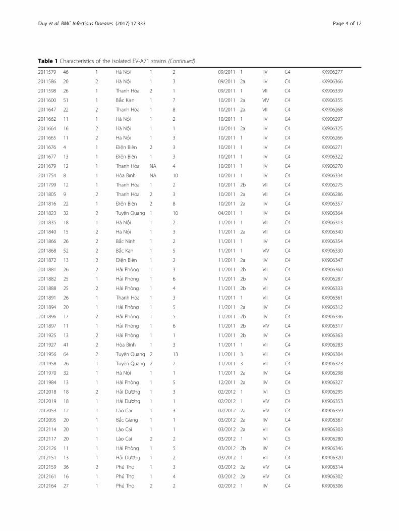
Table 1 Characteristics of the isolated EV-A71 strains (Continued)
2011579 46 1 Hà Nội 1 2 09/2011 1 IIV C4 KX906277
2011586 20 2 Hà Nội 1 3 09/2011 2a IIV C4 KX906366
2011598 26 1 Thanh Hóa 2 1 09/2011 1 VII C4 KX906339
2011600 51 1 Bắc Kạn 1 7 10/2011 2a VIV C4 KX906355
2011647 22 2 Thanh Hóa 1 8 10/2011 2a VII C4 KX906268
2011662 11 1 Hà Nội 1 2 10/2011 1 IIV C4 KX906297
2011664 16 2 Hà Nội 1 1 10/2011 2a IIV C4 KX906325
2011665 11 2 Hà Nội 1 3 10/2011 1 IIV C4 KX906266
2011676 4 1 Điện Biên 2 3 10/2011 1 IIV C4 KX906271
2011677 13 1 Điện Biên 1 3 10/2011 1 IIV C4 KX906322
2011679 12 1 Thanh Hóa NA 4 10/2011 1 IIV C4 KX906270
2011754 8 1 Hòa Bình NA 10 10/2011 1 IIV C4 KX906334
2011799 12 1 Thanh Hóa 1 2 10/2011 2b VII C4 KX906275
2011805 9 2 Thanh Hóa 2 3 10/2011 2a VII C4 KX906286
2011816 22 1 Điện Biên 2 8 10/2011 2a IIV C4 KX906357
2011823 32 2 Tuyên Quang 1 10 04/2011 1 IIV C4 KX906364
2011835 18 1 Hà Nội 1 2 11/2011 1 VII C4 KX906313
2011840 15 2 Hà Nội 1 3 11/2011 2a VII C4 KX906340
2011866 26 2 Bắc Ninh 1 2 11/2011 1 IIV C4 KX906354
2011868 52 2 Bắc Kạn 1 5 11/2011 1 VIV C4 KX906330
2011872 13 2 Điện Biên 1 2 11/2011 2a IIV C4 KX906347
2011881 26 2 Hải Phòng 1 3 11/2011 2b VII C4 KX906360
2011882 25 1 Hải Phòng 1 6 11/2011 2b IIV C4 KX906287
2011888 25 2 Hải Phòng 1 4 11/2011 2b VII C4 KX906333
2011891 26 1 Thanh Hóa 1 3 11/2011 1 VII C4 KX906361
2011894 20 1 Hải Phòng 1 5 11/2011 2a IIV C4 KX906312
2011896 17 2 Hải Phòng 1 5 11/2011 2b IIV C4 KX906336
2011897 11 1 Hải Phòng 1 6 11/2011 2b VIV C4 KX906317
2011925 13 2 Hải Phòng 1 1 11/2011 2b IIV C4 KX906363
2011927 41 2 Hòa Bình 1 3 11/2011 1 VII C4 KX906283
2011956 64 2 Tuyên Quang 2 13 11/2011 3 VII C4 KX906304
2011958 26 1 Tuyên Quang 2 7 11/2011 3 VII C4 KX906323
2011970 32 1 Hà Nội 1 1 11/2011 2a IIV C4 KX906298
2011984 13 1 Hải Phòng 1 5 12/2011 2a IIV C4 KX906327
2012018 18 2 Hải Dương 1 3 02/2012 1 IVI C5 KX906295
2012019 18 1 Hải Dương 1 1 02/2012 1 VIV C4 KX906353
2012053 12 1 Lào Cai 1 3 02/2012 2a VIV C4 KX906359
2012095 20 1 Bắc Giang 1 1 03/2012 2a IIV C4 KX906367
2012114 20 1 Lào Cai 1 1 03/2012 2a VII C4 KX906303
2012117 20 1 Lào Cai 2 2 03/2012 1 IVI C5 KX906280
2012126 11 1 Hải Phòng 1 5 03/2012 2b IIV C4 KX906346
2012151 13 1 Hải Dương 1 2 03/2012 1 VII C4 KX906320
2012159 36 2 Phú Thọ 1 3 03/2012 2a VIV C4 KX906314
2012161 16 1 Phú Thọ 1 4 03/2012 2a VIV C4 KX906302
2012164 27 1 Phú Thọ 2 2 02/2012 1 IIV C4 KX906306
Duy et al. BMC Infectious Diseases (2017) 17:333 Page 4 of 12
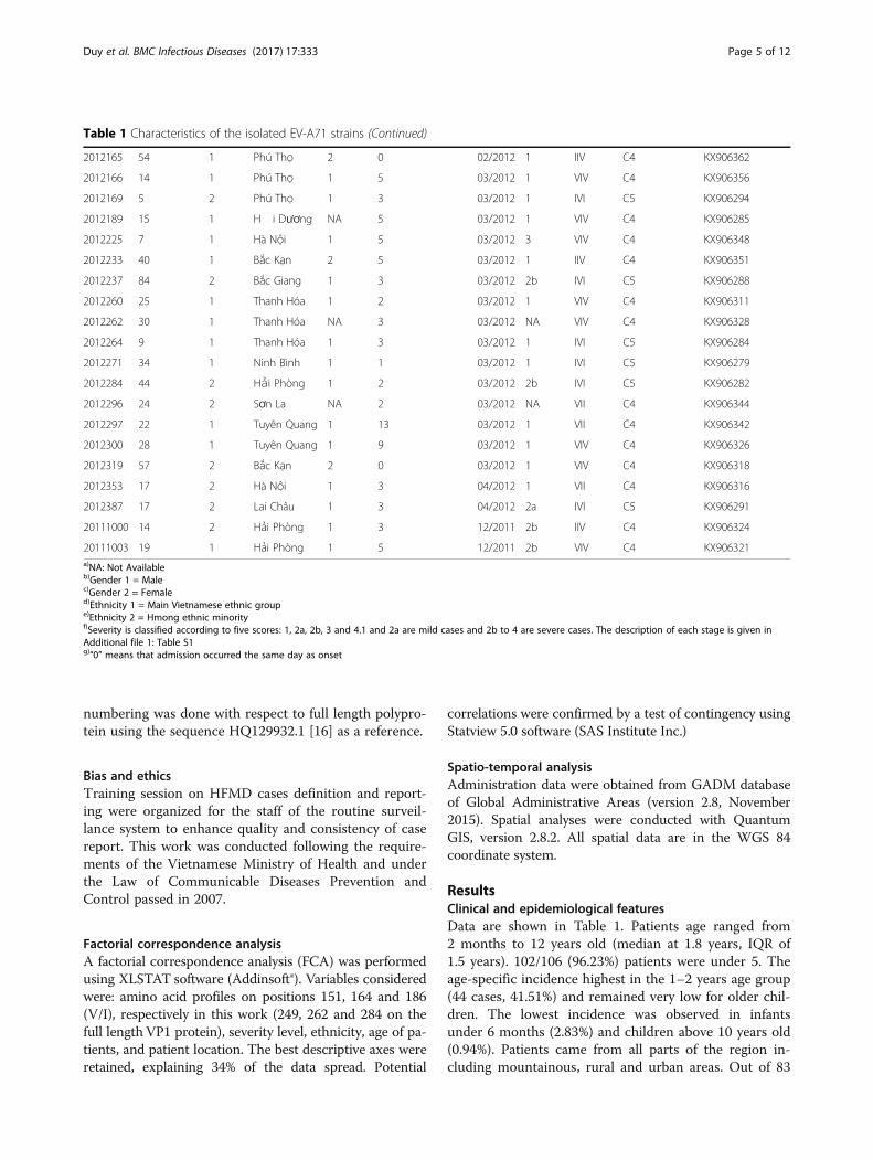
Table 1 Characteristics of the isolated EV-A71 strains (Continued)
2012165 54 1 Phú Thọ 2 0 02/2012 1 IIV C4 KX906362
2012166 14 1 Phú Thọ 1 5 03/2012 1 VIV C4 KX906356
2012169 5 2 Phú Thọ 1 3 03/2012 1 IVI C5 KX906294
2012189 15 1 H i Dương NA 5 03/2012 1 VIV C4 KX906285
2012225 7 1 Hà Nội 1 5 03/2012 3 VIV C4 KX906348
2012233 40 1 Bắc Kạn 2 5 03/2012 1 IIV C4 KX906351
2012237 84 2 Bắc Giang 1 3 03/2012 2b IVI C5 KX906288
2012260 25 1 Thanh Hóa 1 2 03/2012 1 VIV C4 KX906311
2012262 30 1 Thanh Hóa NA 3 03/2012 NA VIV C4 KX906328
2012264 9 1 Thanh Hóa 1 3 03/2012 1 IVI C5 KX906284
2012271 34 1 Ninh Bình 1 1 03/2012 1 IVI C5 KX906279
2012284 44 2 Hải Phòng 1 2 03/2012 2b IVI C5 KX906282
2012296 24 2 Sơn La NA 2 03/2012 NA VII C4 KX906344
2012297 22 1 Tuyên Quang 1 13 03/2012 1 VII C4 KX906342
2012300 28 1 Tuyên Quang 1 9 03/2012 1 VIV C4 KX906326
2012319 57 2 Bắc Kạn 2 0 03/2012 1 VIV C4 KX906318
2012353 17 2 Hà Nội 1 3 04/2012 1 VII C4 KX906316
2012387 17 2 Lai Châu 1 3 04/2012 2a IVI C5 KX906291
20111000 14 2 Hải Phòng 1 3 12/2011 2b IIV C4 KX906324
20111003 19 1 Hải Phòng 1 5 12/2011 2b VIV C4 KX906321a)NA: Not Availableb)Gender 1 = Malec)Gender 2 = Femaled)Ethnicity 1 = Main Vietnamese ethnic groupe)Ethnicity 2 = Hmong ethnic minorityf)Severity is classified according to five scores: 1, 2a, 2b, 3 and 4.1 and 2a are mild cases and 2b to 4 are severe cases. The description of each stage is given inAdditional file 1: Table S1g)“0” means that admission occurred the same day as onset
Duy et al. BMC Infectious Diseases (2017) 17:333 Page 5 of 12
numbering was done with respect to full length polypro-tein using the sequence HQ129932.1 [16] as a reference.
Bias and ethicsTraining session on HFMD cases definition and report-ing were organized for the staff of the routine surveil-lance system to enhance quality and consistency of casereport. This work was conducted following the require-ments of the Vietnamese Ministry of Health and underthe Law of Communicable Diseases Prevention andControl passed in 2007.
Factorial correspondence analysisA factorial correspondence analysis (FCA) was performedusing XLSTAT software (Addinsoft®). Variables consideredwere: amino acid profiles on positions 151, 164 and 186(V/I), respectively in this work (249, 262 and 284 on thefull length VP1 protein), severity level, ethnicity, age of pa-tients, and patient location. The best descriptive axes wereretained, explaining 34% of the data spread. Potential
correlations were confirmed by a test of contingency usingStatview 5.0 software (SAS Institute Inc.)
Spatio-temporal analysisAdministration data were obtained from GADM databaseof Global Administrative Areas (version 2.8, November2015). Spatial analyses were conducted with QuantumGIS, version 2.8.2. All spatial data are in the WGS 84coordinate system.
ResultsClinical and epidemiological featuresData are shown in Table 1. Patients age ranged from2 months to 12 years old (median at 1.8 years, IQR of1.5 years). 102/106 (96.23%) patients were under 5. Theage-specific incidence highest in the 1–2 years age group(44 cases, 41.51%) and remained very low for older chil-dren. The lowest incidence was observed in infantsunder 6 months (2.83%) and children above 10 years old(0.94%). Patients came from all parts of the region in-cluding mountainous, rural and urban areas. Out of 83
Duy et al. BMC Infectious Diseases (2017) 17:333 Page 6 of 12
cases, 59 (71.08%) belonged to main Vietnamese ethni-city (Ethnicity 1) while the rest of patients belonged tothe minority Hmong ethnicity (Ethnicity 2) (Table 1). Allseverity levels were reported for the patients. Mild forms(severity level 1) made the majority of cases (57 cases,61.29%) while 15 patients displayed severe symptoms(16.13%). Among this group, 3 patients displayed aseverity score of 3. No case with the highest level of4 was recorded. Moderate forms of HFMD werefound in 21 patients (22.58). (Table 1).
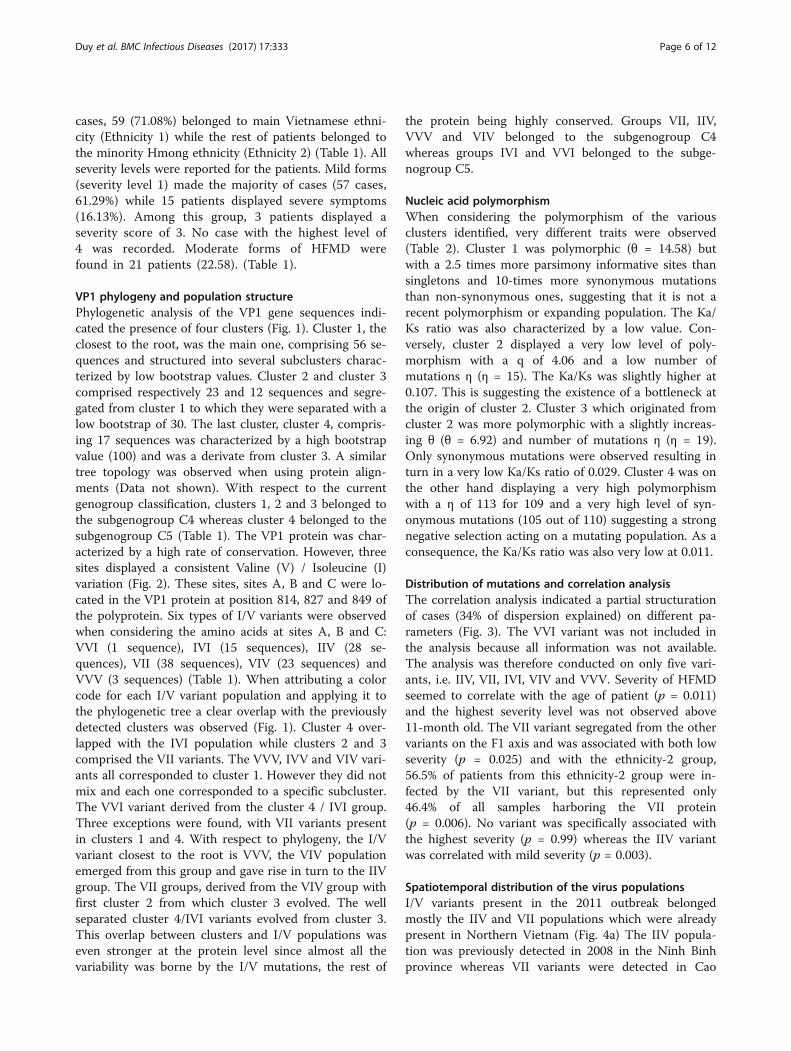
VP1 phylogeny and population structurePhylogenetic analysis of the VP1 gene sequences indi-cated the presence of four clusters (Fig. 1). Cluster 1, theclosest to the root, was the main one, comprising 56 se-quences and structured into several subclusters charac-terized by low bootstrap values. Cluster 2 and cluster 3comprised respectively 23 and 12 sequences and segre-gated from cluster 1 to which they were separated with alow bootstrap of 30. The last cluster, cluster 4, compris-ing 17 sequences was characterized by a high bootstrapvalue (100) and was a derivate from cluster 3. A similartree topology was observed when using protein align-ments (Data not shown). With respect to the currentgenogroup classification, clusters 1, 2 and 3 belonged tothe subgenogroup C4 whereas cluster 4 belonged to thesubgenogroup C5 (Table 1). The VP1 protein was char-acterized by a high rate of conservation. However, threesites displayed a consistent Valine (V) / Isoleucine (I)variation (Fig. 2). These sites, sites A, B and C were lo-cated in the VP1 protein at position 814, 827 and 849 ofthe polyprotein. Six types of I/V variants were observedwhen considering the amino acids at sites A, B and C:VVI (1 sequence), IVI (15 sequences), IIV (28 se-quences), VII (38 sequences), VIV (23 sequences) andVVV (3 sequences) (Table 1). When attributing a colorcode for each I/V variant population and applying it tothe phylogenetic tree a clear overlap with the previouslydetected clusters was observed (Fig. 1). Cluster 4 over-lapped with the IVI population while clusters 2 and 3comprised the VII variants. The VVV, IVV and VIV vari-ants all corresponded to cluster 1. However they did notmix and each one corresponded to a specific subcluster.The VVI variant derived from the cluster 4 / IVI group.Three exceptions were found, with VII variants presentin clusters 1 and 4. With respect to phylogeny, the I/Vvariant closest to the root is VVV, the VIV populationemerged from this group and gave rise in turn to the IIVgroup. The VII groups, derived from the VIV group withfirst cluster 2 from which cluster 3 evolved. The wellseparated cluster 4/IVI variants evolved from cluster 3.This overlap between clusters and I/V populations waseven stronger at the protein level since almost all thevariability was borne by the I/V mutations, the rest of
the protein being highly conserved. Groups VII, IIV,VVV and VIV belonged to the subgenogroup C4whereas groups IVI and VVI belonged to the subge-nogroup C5.
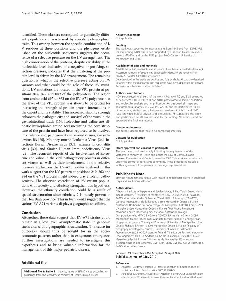
Nucleic acid polymorphismWhen considering the polymorphism of the variousclusters identified, very different traits were observed(Table 2). Cluster 1 was polymorphic (θ = 14.58) butwith a 2.5 times more parsimony informative sites thansingletons and 10-times more synonymous mutationsthan non-synonymous ones, suggesting that it is not arecent polymorphism or expanding population. The Ka/Ks ratio was also characterized by a low value. Con-versely, cluster 2 displayed a very low level of poly-morphism with a q of 4.06 and a low number ofmutations η (η = 15). The Ka/Ks was slightly higher at0.107. This is suggesting the existence of a bottleneck atthe origin of cluster 2. Cluster 3 which originated fromcluster 2 was more polymorphic with a slightly increas-ing θ (θ = 6.92) and number of mutations η (η = 19).Only synonymous mutations were observed resulting inturn in a very low Ka/Ks ratio of 0.029. Cluster 4 was onthe other hand displaying a very high polymorphismwith a η of 113 for 109 and a very high level of syn-onymous mutations (105 out of 110) suggesting a strongnegative selection acting on a mutating population. As aconsequence, the Ka/Ks ratio was also very low at 0.011.
Distribution of mutations and correlation analysisThe correlation analysis indicated a partial structurationof cases (34% of dispersion explained) on different pa-rameters (Fig. 3). The VVI variant was not included inthe analysis because all information was not available.The analysis was therefore conducted on only five vari-ants, i.e. IIV, VII, IVI, VIV and VVV. Severity of HFMDseemed to correlate with the age of patient (p = 0.011)and the highest severity level was not observed above11-month old. The VII variant segregated from the othervariants on the F1 axis and was associated with both lowseverity (p = 0.025) and with the ethnicity-2 group,56.5% of patients from this ethnicity-2 group were in-fected by the VII variant, but this represented only46.4% of all samples harboring the VII protein(p = 0.006). No variant was specifically associated withthe highest severity (p = 0.99) whereas the IIV variantwas correlated with mild severity (p = 0.003).
Spatiotemporal distribution of the virus populationsI/V variants present in the 2011 outbreak belongedmostly the IIV and VII populations which were alreadypresent in Northern Vietnam (Fig. 4a) The IIV popula-tion was previously detected in 2008 in the Ninh Binhprovince whereas VII variants were detected in Cao
Fig. 1 Phylogenetic analysis of partial VP1 sequences. a Phylogenetic analysis of the nucleic acid sequences.Tree was designed using MaximumLikelihood. Color code: Black: VVV; Light blue: VII; Yellow: IIV; Purple: VIV; Dark blue: VVI
Duy et al. BMC Infectious Diseases (2017) 17:333 Page 7 of 12
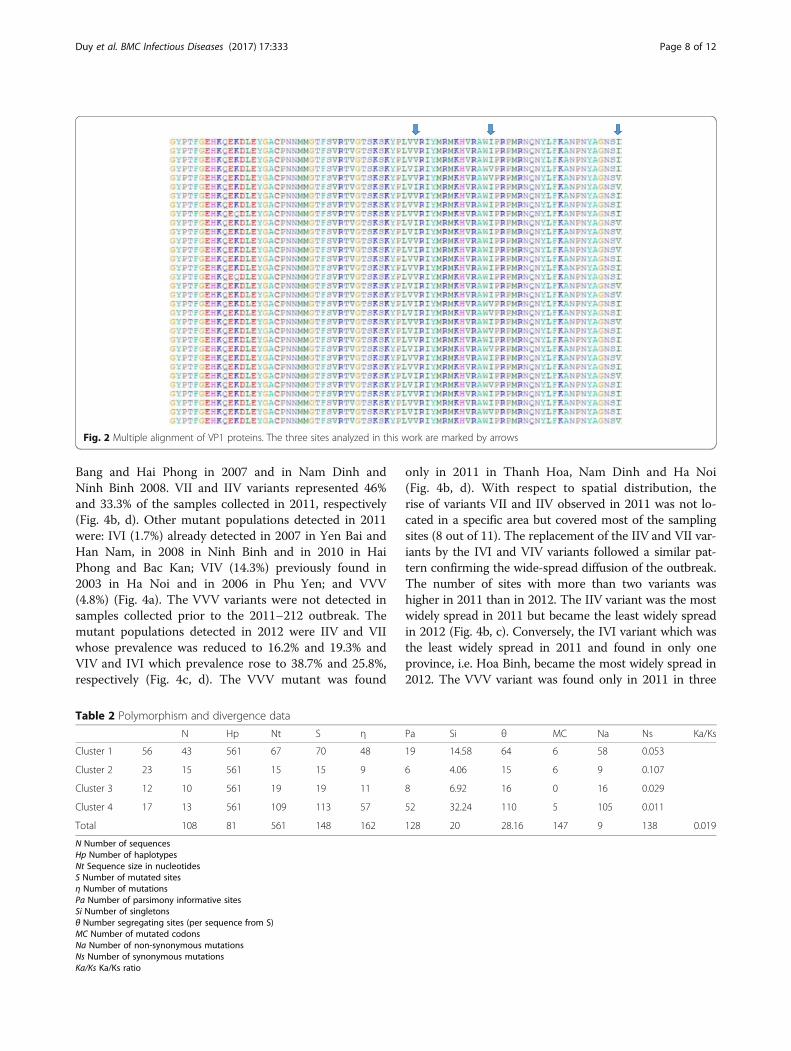
Fig. 2 Multiple alignment of VP1 proteins. The three sites analyzed in this work are marked by arrows
Duy et al. BMC Infectious Diseases (2017) 17:333 Page 8 of 12
Bang and Hai Phong in 2007 and in Nam Dinh andNinh Binh 2008. VII and IIV variants represented 46%and 33.3% of the samples collected in 2011, respectively(Fig. 4b, d). Other mutant populations detected in 2011were: IVI (1.7%) already detected in 2007 in Yen Bai andHan Nam, in 2008 in Ninh Binh and in 2010 in HaiPhong and Bac Kan; VIV (14.3%) previously found in2003 in Ha Noi and in 2006 in Phu Yen; and VVV(4.8%) (Fig. 4a). The VVV variants were not detected insamples collected prior to the 2011–212 outbreak. Themutant populations detected in 2012 were IIV and VIIwhose prevalence was reduced to 16.2% and 19.3% andVIV and IVI which prevalence rose to 38.7% and 25.8%,respectively (Fig. 4c, d). The VVV mutant was found
Table 2 Polymorphism and divergence data
N Hp Nt S η
Cluster 1 56 43 561 67 70 48
Cluster 2 23 15 561 15 15 9
Cluster 3 12 10 561 19 19 11
Cluster 4 17 13 561 109 113 57
Total 108 81 561 148 162
N Number of sequencesHp Number of haplotypesNt Sequence size in nucleotidesS Number of mutated sitesη Number of mutationsPa Number of parsimony informative sitesSi Number of singletonsθ Number segregating sites (per sequence from S)MC Number of mutated codonsNa Number of non-synonymous mutationsNs Number of synonymous mutationsKa/Ks Ka/Ks ratio
only in 2011 in Thanh Hoa, Nam Dinh and Ha Noi(Fig. 4b, d). With respect to spatial distribution, therise of variants VII and IIV observed in 2011 was not lo-cated in a specific area but covered most of the samplingsites (8 out of 11). The replacement of the IIV and VII var-iants by the IVI and VIV variants followed a similar pat-tern confirming the wide-spread diffusion of the outbreak.The number of sites with more than two variants washigher in 2011 than in 2012. The IIV variant was the mostwidely spread in 2011 but became the least widely spreadin 2012 (Fig. 4b, c). Conversely, the IVI variant which wasthe least widely spread in 2011 and found in only oneprovince, i.e. Hoa Binh, became the most widely spread in2012. The VVV variant was found only in 2011 in three
Pa Si θ MC Na Ns Ka/Ks
19 14.58 64 6 58 0.053
6 4.06 15 6 9 0.107
8 6.92 16 0 16 0.029
52 32.24 110 5 105 0.011
128 20 28.16 147 9 138 0.019
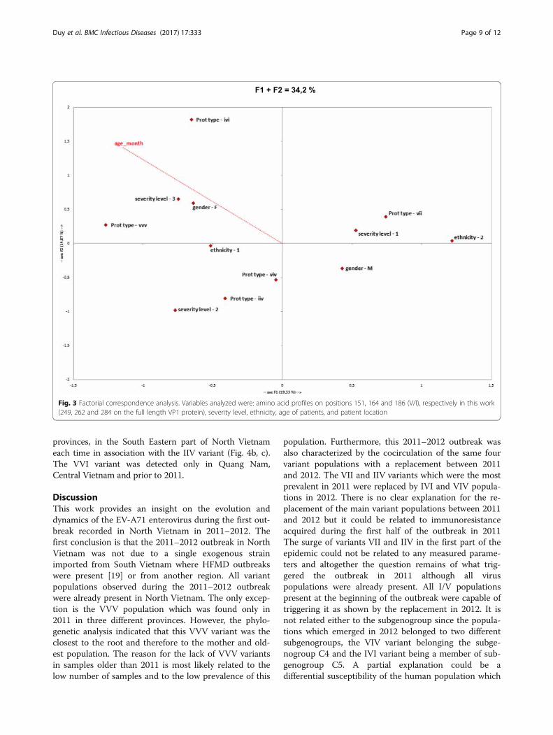
Fig. 3 Factorial correspondence analysis. Variables analyzed were: amino acid profiles on positions 151, 164 and 186 (V/I), respectively in this work(249, 262 and 284 on the full length VP1 protein), severity level, ethnicity, age of patients, and patient location
Duy et al. BMC Infectious Diseases (2017) 17:333 Page 9 of 12
provinces, in the South Eastern part of North Vietnameach time in association with the IIV variant (Fig. 4b, c).The VVI variant was detected only in Quang Nam,Central Vietnam and prior to 2011.
DiscussionThis work provides an insight on the evolution anddynamics of the EV-A71 enterovirus during the first out-break recorded in North Vietnam in 2011–2012. Thefirst conclusion is that the 2011–2012 outbreak in NorthVietnam was not due to a single exogenous strainimported from South Vietnam where HFMD outbreakswere present [19] or from another region. All variantpopulations observed during the 2011–2012 outbreakwere already present in North Vietnam. The only excep-tion is the VVV population which was found only in2011 in three different provinces. However, the phylo-genetic analysis indicated that this VVV variant was theclosest to the root and therefore to the mother and old-est population. The reason for the lack of VVV variantsin samples older than 2011 is most likely related to thelow number of samples and to the low prevalence of this
population. Furthermore, this 2011–2012 outbreak wasalso characterized by the cocirculation of the same fourvariant populations with a replacement between 2011and 2012. The VII and IIV variants which were the mostprevalent in 2011 were replaced by IVI and VIV popula-tions in 2012. There is no clear explanation for the re-placement of the main variant populations between 2011and 2012 but it could be related to immunoresistanceacquired during the first half of the outbreak in 2011The surge of variants VII and IIV in the first part of theepidemic could not be related to any measured parame-ters and altogether the question remains of what trig-gered the outbreak in 2011 although all viruspopulations were already present. All I/V populationspresent at the beginning of the outbreak were capable oftriggering it as shown by the replacement in 2012. It isnot related either to the subgenogroup since the popula-tions which emerged in 2012 belonged to two differentsubgenogroups, the VIV variant belonging the subge-nogroup C4 and the IVI variant being a member of sub-genogroup C5. A partial explanation could be adifferential susceptibility of the human population which
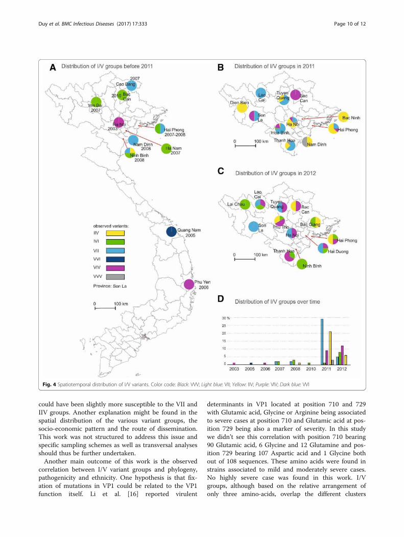
Fig. 4 Spatiotemporal distribution of I/V variants. Color code: Black: VVV; Light blue: VII; Yellow: IIV; Purple: VIV; Dark blue: VVI
Duy et al. BMC Infectious Diseases (2017) 17:333 Page 10 of 12
could have been slightly more susceptible to the VII andIIV groups. Another explanation might be found in thespatial distribution of the various variant groups, thesocio-economic pattern and the route of dissemination.This work was not structured to address this issue andspecific sampling schemes as well as transversal analysesshould thus be further undertaken.Another main outcome of this work is the observed
correlation between I/V variant groups and phylogeny,pathogenicity and ethnicity. One hypothesis is that fix-ation of mutations in VP1 could be related to the VP1function itself. Li et al. [16] reported virulent
determinants in VP1 located at position 710 and 729with Glutamic acid, Glycine or Arginine being associatedto severe cases at position 710 and Glutamic acid at pos-ition 729 being also a marker of severity. In this studywe didn’t see this correlation with position 710 bearing90 Glutamic acid, 6 Glycine and 12 Glutamine and pos-ition 729 bearing 107 Aspartic acid and 1 Glycine bothout of 108 sequences. These amino acids were found instrains associated to mild and moderately severe cases.No highly severe case was found in this work. I/Vgroups, although based on the relative arrangement ofonly three amino-acids, overlap the different clusters
Duy et al. BMC Infectious Diseases (2017) 17:333 Page 11 of 12
identified. These clusters correspond to genetically differ-ent populations characterized by specific polymorphismtraits. This overlap between the specific combination of I/V residues at three positions and the phylogeny estab-lished on the nucleotide sequences suggests the occur-rence of a selective pressure on the I/V arrangement. Thehigh conservation of the proteins, despite variability at thenucleotide level, indicative of a negative, or purifying, se-lection pressure, indicates that the clustering at the pro-tein level is driven by the I/V arrangement. The remainingquestion is what is the selective pressure acting on I/Vvariants and what could be the role of these I/V muta-tions. I/V mutations are located in the VP1 protein at po-sitions 814, 827 and 849 of the polyprotein. The regionfrom amino acid 697 to 862 on the EV-A71 polyprotein atthe level of the VP1 protein was shown to be crucial forincreasing the strength of protein-protein interactions inthe capsid and its stability. This increased stability stronglyenhances the pathogenicity and survival of the virus in thegastrointestinal track [15]. Isoleucine and valine are ali-phatic hydrophobic amino acid mediating the core struc-ture of the protein and have been reported to be involvedin virulence and pathogenicity in several viruses, coxsack-ievirus B3 [25], Moloney murne Leukemia Virus [26], In-fectious Bursal Disease virus [32], Japanese Encephalitisvirus [30], and Simian-Human Immunodeficiency Virus[23]. The recurrent reports of the involvement of isoleu-cine and valine in the viral pathogenicity process in differ-ent viruses as well as their involvement in the selectivepressure applied on the EV-A71 isolates analyzed in thiswork suggest that the I/V pattern at positions 249, 262 and284 on the VP1 protein might indeed play a role in patho-genicity. The observed correlation of I/V variant popula-tions with severity and ethnicity strengthen this hypothesis.However, the ethnicity correlation could be a result ofspatial structuration since ethnicity-2 is mostly present inthe Hòa Bình province. This in turn would suggest that thevarious EV-A71 variants display a geographic specificity.
ConclusionAltogether, these data suggest that EV-A71 strains couldremain in a low level, asymptomatic state, in genomicstasis and with a geographic structuration. The cause foroutbreaks should thus be sought for in the socio-economic patterns rather than in exogenous emergence.Further investigations are needed to investigate thishypothesis and to bring valuable information for themanagement of this major pediatric disease.
Additional file
Additional file 1: Table S1. Severity levels of HFMD cases according toguidelines from the Vietnamese Ministry of Health. (DOCX 15 kb)
AcknowledgementsNot applicable.
FundingThe work was supported by internal grants from NIHE and from DUKE/NUSfor sequencing. NDN was in part supported by European Erasmus Mundusproject MAHEVA and by the PEPS project MoDyCa from University ofMontpellier and CNRS.
Availability of data and materialsAll data are publicly available and sequences have been deposited in Genbank.Accession numbers of sequences deposited in Genbank are ranging FromKX906261 to KX906368 (108 sequences).Data described in this article are publicly and fully available. All data are describedin tables within the manuscript and sequences have been deposited in Genbank.Accession numbers are provided in Table 1.
Authors’ contributionsNDN participated to all parts of the work. OMS, YAH, RC and DJG generatedall sequences. LTTH, LTSH, VDT and NTHT participated to sample collectionand molecular analysis and amplification. AA designed all maps andspatiotemporal analysis. LG, CM, PR, GK, EC and RF participated to allbioinformatic, statistic and phylogenetic analyses. CD, NTH and TNDhave provided fruitful advises and discussions. RF supervised the workand participated to all analyses and to the writing. All authors read andapproved the final manuscript.
Competing interestsThe authors declare that there is no competing interests.
Consent for publicationNot Applicable.
Ethics approval and consent to participateThis work was conducted strictly following the requirements of theVietnamese Ministry of Health and under the Law of CommunicableDiseases Prevention and Control passed in 2007. This work was conductedunder the control of NIHE Ethic committee. These procedures include awritten agreement from parents or their legal representatives.
Publisher’s NoteSpringer Nature remains neutral with regard to jurisdictional claims in publishedmaps and institutional affiliations.
Author details1National Institute of Hygiene and Epidemiology, 1 Pho Yersin Street, Hanoi10000, Vietnam. 2University of Montpellier, ISEM, CC063, Place E. Bataillon,34095 Montpellier Cedex 5, France. 3Cirad, UMR 17, Intertryp, TA-A17/G,Campus International de Baillarguet, 34398 Montpellier Cedex 5, France.4Institut de Recherche en Cancérologie de Montpellier (U1194), Campus Vald’Aurelle, 34298 Montpellier Cedex 5, France. 5Hai Phong PreventiveMedicine Center, Hai Phong city, Vietnam. 6Institut de BiologieComputationnelle, MMVE, La Galera, CC6005, 95 rue de la Galera, 34095Montpellier, France. 7DUKE-NUS Graduate Medical School, 8 College Road,Singapore, Singapore. 8Faculty of Pharmacy, University of Montpellier, 15 avCharles Flahault, BP14491, 34093 Montpellier Cedex 5, France. 9Faculty ofGeography and Regional Studies, University of Warsaw, KrakowskiePrzedmiescie 26/28, 00-927 Warsaw, Poland. 10Institut de Recherche pour leDéveloppement (IRD), Le Sextant, 44, bd de Dunkerque, CS 90009, 13572Marseille cedex 02, France. 11Université de Montpellier, IES – Institutd’Electronique et des Systèmes, UMR 5214, CNRS-UM, 860 rue St. Priest, Bt. 5,34095 Montpellier, France.
Received: 19 November 2016 Accepted: 27 April 2017
References1. Abascal F, Zardoya R, Posada D. ProtTest: selection of best-fit models of
protein evolution. Bioinformatics. 2005;21:2104–5.2. Abu Bakar S, Chee HY, Al-Kobaisi MF, Xiaoshan J, Bing CK, Kit LS. Identification
of enterovirus 71 isolates from an outbreak of hand, foot and mouth disease
Duy et al. BMC Infectious Diseases (2017) 17:333 Page 12 of 12
(HFMD) with fatal cases of encephalomyelitis in Malaysia. Virus Res.1999;61:1–9.
3. Ang LW, Koh BK, Chan KP, Chua LT, James L, Goh KT. Epidemiology andcontrol of hand, foot and mouth disease in Singapore. Ann Acad Med Singap.2009;38:106–12.
4. Brown BA, Oberste MS, Alexander JP Jr, Kennett ML, Pallansch MA. Molecularepidemiology and evolution of enterovirus 71 strains isolated from 1970 to1998. J Virol. 1999;73:9969–75.
5. Caine EA, Moncla LH, Ronderos MD, Friedrich TC, Osorio JE. A singlemutation in the VP1 of Enterovirus 71 is responsible for increased virulenceand Neurotropism in adult interferon-deficient mice. J Virol. 2016;90:8592–604.
6. Carter J, Saunders VA. Virology: principles and applications. Hoboken:John Wiley & Sons; 2007.
7. Chen KT, Chang HL, Wang ST, Cheng YT, Yang JY. Epidemiologic features ofhand-foot-mouth disease and herpangina caused by enterovirus 71 inTaiwan, 1998–2005. Pediatrics. 2007;120:e244–52.
8. Darriba D, Taboada GL, Doallo R, Posada D. jModelTest 2: more models,new heuristics and parallel computing. Nat Methods. 2012;9(8):772.
9. Edgar RC. MUSCLE: a multiple sequence alignment method with reducedtime and space complexity. BMC bioinformatics. 2004;5:113.
10. Gouy M, Guindon S, Gascuel O. SeaView version 4: a multiplatform graphicaluser interface for sequence alignment and phylogenetic tree building. MolBiol Evol. 2010;27:221–4.
11. Huang CC, Liu CC, Chang YC, Chen CY, Wang ST, Yeh TF. Neurologiccomplications in children with Enterovirus 71 infection. N Engl J Med. 1999;341:936–42.
12. Huang SW, Tai CH, Fonville JM, Lin CH., Wang SM, Liu CC, .Su IJ, Smith DJ,Wang JR. Mapping enterovirus A71 antigenic determinants from viral evolution.J Virol 2005; 89:11500-11506.
13. Kataoka C, Suzuki T, Kotani O, Iwata-Yoshikawa O, Nagata N, Ami Y, WakitaT, Nishimura Y, Shimizu H. The role of VP1 amino acid residue 145 ofEnterovirus 71 in viral fitness and pathogenesis in a Cynomolgus monkeymodel. PLoS Pathog. 2015;11(7):e1005033.
14. Khanh TH, Sabanathan S, Thanh TT, Thoa le PK, Thuong TC, Hang VT, FarrarJ, Hien TT, Chau NV, van Doorn HR. Enterovirus 71-associated hand, foot,and mouth disease, southern Vietnam, 2011. Emerging Infect Dis. 2012;18:2002–5.
15. Lal SK, Kumar P, Yeo WM, Kar-Roy A, Chow VT. The VP1 protein of humanenterovirus 71 self-associates via an interaction domain spanning aminoacids 66–297. J Med Virol. 2006;78:582–90.
16. Li R, Zou Q, Chen L, Zhang H, Wang Y. Molecular analysis of virulentdeterminants of enterovirus 71. PLoS One. 2011;6(10):e26237.
17. Librado P, Rozas J. DnaSP v5: a software for comprehensive analysis of DNApolymorphism data. Bioinformatics. 2009;25:1451–2.
18. McMinn P, Lindsay K, Perera D, Chan HM, Chan KP, Cardosa MJ. Phylogeneticanalysis of enterovirus 71 strains isolated during linked epidemics in Malaysia,Singapore, and Western Australia. J Virol. 2001;75:7732–8.
19. Nguyen NTB, Pham H, Hoang CQ, Nguyen TM, Nguyen LT, Phan HC, PhanLT, Vu LN, Minh NNT. Epidemiological and clinical characteristics of childrenwho died from hand, foot and mouth disease in Vietnam, 2011. BMC InfectDis. 2014;14:341.
20. Nix WA, Oberste MS, Pallansch MA. Sensitive, seminested PCR amplificationof VP1 sequences for direct identification of all enterovirus serotypes fromoriginal clinical specimens. J Clin Microbiol. 2006;44:2698–704.
21. Ooi MH, Wong SC, Lewthwaite P, Cardosa MJ, Solomon T. Clinical features,diagnosis, and management of enterovirus 71. The Lancet Neurology. 2010;9:1097–105.
22. Perera D, Podin Y, Akin W, Tan CS, Cardosa MJ. Incorrect identification ofrecent Asian strains of Coxsackievirus A16 as human enterovirus 71: improvedprimers for the specific detection of human enterovirus 71 by RT PCR. BMCInfect Dis. 2004;4:11.
23. Peyerl FW, Barouch DH, Yeh WW, Bazick HS, Kunstman J, Kunstman KJ,Letvin NL. Simian-human immunodeficiency virus escape from cytotoxic T-lymphocyte recognition at a structurally constrained epitope. J Virol. 2003;77:12572–8.
24. Sanders S, Herrero L, McPhie K, Chow S, Craig M, Dwyer D, Rawlinson W,McMinn PC. Molecular epidemiology of enterovirus 71 over two decades inan Australian urban community. Arch Virol. 2006;151:1003–13.
25. Schmidtke M, Hammerschmidt E, Schüler S, Zell R, Birch-Hirschfeld E, Makarov VA,Wutzler P. Susceptibility of coxsackievirus B3 laboratory strains and clinical isolatesto the capsid function inhibitor pleconaril: antiviral studies with virus chimeras
demonstrate the crucial role of amino acid 1092 in treatment. J AntimicrobChemother. 2005;56:648–56.
26. Szurek PF, Yuen PH, Ball JK, Wong PK. A Val-25-to-Ile substitution in theenvelope precursor polyprotein, gPr80env, is responsible for the temperaturesensitivity, inefficient processing of gPr80env, and neurovirulence of ts1, amutant of Moloney murine leukemia virus TB. J Virol. 1990;64:467–75.
27. Tee KK, Lam TTY, Chan YF, Bible JM, Kamarulzaman A, Tong C, Takebe Y,Pybus OG. Evolutionary genetics of human enterovirus 71: origin, populationdynamics, natural selection, and seasonal periodicity of the VP1 gene. J Virol.2010;84:3339–50.
28. Tu PV, Thao NTT, Perera D, Huu TK, Tien NTK, Thuong TC, How OM, CardosaMJ, McMinn PC. Epidemiologic and virologic investigation of hand, foot,and mouth disease, southern Vietnam, 2005. Emerging Infect Dis. 2007;13:1733–41.
29. WHO. A Guide to Clinical Management and Public Health Response forHand, Foot and Mouth Disease (HFMD). WHO WPRO; 2011.
30. Yamaguchi Y, Nukui Y, Tajima S, Nerome R, Kato F, Watanabe H, Kurane I.An amino acid substitution (V3I) in the Japanese encephalitis virus NS4Aprotein increases its virulence in mice, but not its growth rate in vitro. J GenVirol. 2011;92:1601–6.
31. Yang SL, Chou YT, Wu CN, Ho MS. Annexin II binds to capsid protein VP1 ofenterovirus 71 and enhances viral infectivity. J Virol. 2011;85:11809–20.
32. Yu F, Ren X, Wang Y, Qi X, Song J, Gao Y, Wang X. A single amino acid V4Isubstitution in VP1 attenuates virulence of very virulent infectious bursaldisease virus (vvIBDV) in SPF chickens and increases replication in CEF cells.Virology. 2013;440:204–9.
33. Zeng M, Li YF, Wang XH, Lu GP, Shen HG, Yu H, Zhu QR. Epidemiology ofhand, foot, and mouth disease in children in shanghai 2007–2010. EpidemiolInfect. 2012;140:1122–30.
• We accept pre-submission inquiries
• Our selector tool helps you to find the most relevant journal
• We provide round the clock customer support
• Convenient online submission
• Thorough peer review
• Inclusion in PubMed and all major indexing services
• Maximum visibility for your research
Submit your manuscript atwww.biomedcentral.com/submit
Submit your next manuscript to BioMed Central and we will help you at every step: