AVERTISSEMENT
Ce document est le fruit d'un long travail approuvé par le jury de soutenance et mis à disposition de l'ensemble de la communauté universitaire élargie. Il est soumis à la propriété intellectuelle de l'auteur. Ceci implique une obligation de citation et de référencement lors de l’utilisation de ce document. D'autre part, toute contrefaçon, plagiat, reproduction illicite encourt une poursuite pénale. Contact : [email protected]
LIENS Code de la Propriété Intellectuelle. articles L 122. 4 Code de la Propriété Intellectuelle. articles L 335.2- L 335.10 http://www.cfcopies.com/V2/leg/leg_droi.php http://www.culture.gouv.fr/culture/infos-pratiques/droits/protection.htm
Ecole Doctorale : Synthèse, Simulations, Applications: de la Molécule aux Edifices Supramoléculaires (SESAMES), ED 412
Soutenance publique prévue le 15 December 2016 devant le jury composé de :
Rapporteurs: Prof. Philip N Bartlett, University of Southampton, United Kingdom
Dr. Elisabeth Lojou, BIP, CNRS, Marseille
Invités: Dr. Alain Walcarius, LCPME, CNRS, Nancy
Prof. François Lapicque, LRGP, CNRS, Nancy
Directeur : Dr. Mathieu Etienne, LCPME, CNRS, Nancy
Examinateur : Dr. Neus Vila, Université de Lorraine, Nancy
Laboratoire de Chimie Physique et Microbiologie pour l’Environnement (LCPME) Unité mixte de recherche – UMR 756
405 Rue de Vandoeuvre, 54600, Villers-lès-Nancy, France
Sujet de thèse :
Co-immobilisation du complexe (2,2'-bipyridyl) (pentaméthylcyclopentadiényl)-rhodium et de
déshydrogénases NAD-dépendantes pour l’électrosynthèse enzymatique énantiosélective
Par
Lin ZHANG
Pour l‟obtention de titre de Docteur

Acknowlegements
First of all, I would like to give my greatest thanks to my supervisors Mathieu ETIENNE and
Neus VILA for their guiding and supporting me during these past three years. Without their
constant advice and efforts, this thesis would not have been completed. I feel very lucky to be
a student of them. I could still remember at the beginning of my Ph.D when I just enter the
field of electrochemistry how they encourage me to go out of frustration when I meet
difficulties. Mathieu teach me how to carry out research independently and systematically,
giving me the confidence to continue my career in science. Neus not only help me in work,
showing me the proper ways to carry out different experiments, but also help me in life when
I meet difficulties. There are so many things they have teach me and help me that I‟m not able
to list all here, but all the details are kept in my mind, which will become part of my great
memories in my life.
I would like to acknowledge Alain WALCARIUS who is the director of the lab and our
ELAN group for his scientific supports and useful discussions. I would also like to
acknowledge Prof. Gert-Wieland KOHRING from Saarland University for his support in
biological part of this thesis. I am also very grateful to my friend Ievgen MAZURENKO, who
is the former post-doc in ELAN group, he give me detailed guidance at the beginning of my
Ph.D, leading me to the field of bioelectrochemistry.
I would also like to thank all the members in ELAN Group, present and past, for their selfless
support and friendships. They made my life more colorful during the three years. Special
thanks to: Cheryl KARMAN, for her generous help when I was sick; Maciej MIERZWA, for
being a good partner to have nice lunch time during two years; Mohana AFSHARIAN, for her
help when I was looking for an apartment; Martha COLLINS, for her help in some documents
preparation; Lukasz POLOTORAK, for his warm-hearted assistance.
There are also many people in LCPME who have help me from time to time, I‟m not able to
thank them one by one here, I will always remember their kindness to me.
Here I also want to thank my Master supervisor Eric GRELET in Bordeaux, he teach me the
right attitude toward science, which has influenced me on way of research.

I would like to thank my family, especially my parents for their constant support since I go
abroad, I could always get sense of security from them no matter how difficult situation I was
in. I would also like to thank my father in law for his advices for my scientific career.
At last, I would like to express my thanks to my husband Zhanming for his love, it‟s very
important that we hold each other‟s hand, sharing the happiness and facing the difficulties
together.
Table of content
Abstract .................................................................................................................................................. 1
Résumé ................................................................................................................................................... 2
General introduction ............................................................................................................................. 3
Chapter 1. Literature survey ................................................................................................................ 7
1.1 Introduction ........................................................................................................................... 8
1.2 Enzymatic systems, cofactor recycling and target reactions ........................................... 12
1.2.1 NAD(P)-dependent-redox enzymes ............................................................................ 12
1.2.2 Flavoenzymes ............................................................................................................... 16
1.2.3 Metalloproteins ............................................................................... 错误!未定义书签。
1.2.4 Electrode materials ...................................................................................................... 19
1.2.4.1 Metals ....................................................................................................................... 19
1.2.4.2 Metal oxides ............................................................................................................. 20
1.2.4.3 Carbon materials ..................................................................................................... 20
1.3 Immobilization of enzymes on electrodes .......................................................................... 21
1.3.1 Membrane confinement with membranes ................................................................. 22
1.3.2 Chemical bonding ........................................................................................................ 25
1.3.2.1 Carbodiimide activation ......................................................................................... 25
1.3.2.2 Glutaraldehyde crosslinking................................................................................... 27
1.3.2.3 Self-assembled monolayer (SAM) .......................................................................... 27
1.3.3 Encapsulation ............................................................................................................... 29
1.3.3.1 Nafion coating .......................................................................................................... 29
1.3.3.2 Surfactant and lipidic layers................................................................................... 30
1.3.3.3 Alginate-based gel and sol-gel silica ....................................................................... 31
1.3.3.4 Conducting polymers .............................................................................................. 33
1.4 Immobilization of mediators .............................................................................................. 34
1.4.1 Methyl-viologen ........................................................................................................... 35
1.4.2 [Cp*Rh(bpy)Cl]+ .......................................................................................................... 35
1.5 Conclusions .......................................................................................................................... 37
Chapter 2. Immobilization of cysteine-tagged proteins on electrode surfaces by thiol-ene click chemistry .............................................................................................................................................. 47
2.1 Introduction ......................................................................................................................... 48
2.2 Experimental ........................................................................................................................ 50
2.3 Results and discussions ....................................................................................................... 52
2.3.1 Immobilization of ferrocene moieties on glassy carbon electrode ........................... 52
2.3.2 Immobilization of cysteine-tagged D-sorbitol dehydrogenase on glassy carbon ... 57
2.3.3 Immobilization of cysteine-tagged D-sorbitol dehydrogenase on carbon felt electrode. ...................................................................................................................................... 61
2.3.4 Application to the electroenzymatic reduction of D-fructose in the presence of [Cp*Rh(bpy)Cl]+.......................................................................................................................... 66
2.4 Conclusions .......................................................................................................................... 71
Chapter 3. Covalent immobilization of rhodium complex on porous carbon electrode for NADH regeneration ......................................................................................................................................... 72
3.1 Introduction ......................................................................................................................... 73
3.2 Experimental ........................................................................................................................ 76
3.3 Results and discussions ....................................................................................................... 79
3.3.1 Characterization of the alkynyl functionalized rhodium complex in solution ...... 79
3.3.2 Covalent immobilization of rhodium complex on the electrode ............................ 82
3.3.2.1 Diazonium electrografting ...................................................................................... 82
3.3.2.2 Huisgen cycloaddition ............................................................................................. 84
3.3.2.3 Rhodium complexation ........................................................................................... 86
3.3.3 NADH regeneration .................................................................................................... 87
3.3.4 Bioelectrocatalytic amperometric responses of co-immobilized electrodes ........... 92
3.4 Conclusion ............................................................................................................................ 97
Chapter 4. Functionalization of carbon felt supported carbon nanotubes by combining diazonium chemistry and a sequential dual click chemistry approach .......................................... 98
4.1 Introduction ......................................................................................................................... 99
4.2 Experimental ...................................................................................................................... 101
4.3 Results and discussions ..................................................................................................... 105
4.3.1 Electrochemical behavior of 4-azidobenzene and 4-vinylbenzene diazonium salts individually and its corresponding mixture. ........................................................................... 105
4.3.2 Bi-functionalization of the electrode by diazonium electrografting ...................... 106
4.3.3 Evaluation of click chemistry reactions on the bi-functionalized electrode ......... 108
4.3.4 Co-immobilization of [Cp*Rh(bpy)Cl]+and enzymes on the electrode ................ 111
4.4 Conclusion .......................................................................................................................... 113
Chapter 5. Co-immobilization of rhodium complex and NAD-dependent dehydrogenase in an electrochemical bioreactor for enantioselective bioconversion ..................................................... 115
5.1 Introduction ....................................................................................................................... 116
5.2 Experimental ...................................................................................................................... 119
5.3 Results and discussions ..................................................................................................... 123
5.3.1 Covalent immobilization of rhodium complex on CF-CNT electrodes by bipyridyl diazonium electrografting ......................................................................................................... 123
5.3.2 Immobilization of rhodium complex on ‘bucky paper’ electrode by bipyridyl diazonium electrografting ......................................................................................................... 126
5.3.3 The combination of BP-Bpy-Rh electrode with enzymes ...................................... 129
5.3.3.1 Co-immobilization with enzymes gel on the top ................................................. 129
5.3.3.2 Co-immobilization with replaceable enzyme layer ............................................. 130
5.4 Conclusions ........................................................................................................................ 133
Conclusions and perspectives ........................................................................................................... 134
References .......................................................................................................................................... 140
Appendix 1 ......................................................................................................................................... 154
Appendix 2 ......................................................................................................................................... 160
1
Abstract
In this work we developed methods for co-immobilization of NAD-dependent
dehydrogenase and the (2,2'-bipyridyl)(pentamethylcyclopentadienyl)-rhodium complex
([Cp*Rh(bpy)Cl]+) on porous carbon electrodes for applications on electroenzymatic
synthesis of chiral alcohols and sugars. The goal was to avoid degradation of the enzymatic
activity originated from the interaction between enzyme surface functional groups (eg.-SH, -
NH2) and [Cp*Rh(bpy)Cl]+ and to allow the recyclability for catalysts. Diazonium
electrografting was used to introduce either alkene or azide groups on a carbon surface (flat
glassy carbon, porous carbon felt or carbon nanotubes layers). Thiol-ene click chemistry was
applied to bind D-sorbitol dehydrogenases with cysteine tag (either 1 or 2 cysteine moieties at
the N terminus of the polypeptide chain) onto carbon electrodes. Azide-alkyne Huisgen
cycloaddition reaction was used to bind an alkyne modified [Cp*Rh(bpy)Cl]+. Then co-
immobilization of the redox enzymes (D-sorbitol and Galactitol dehydrogenase) with
[Cp*Rh(bpy)Cl]+ was tested by encapsulation of the proteins in a silica gel layer, inside a
rhodium-functionalized porous carbon felt. The immobilized [Cp*Rh(bpy)Cl]+ was stable
over weeks for NADH regeneration, but this electrode architecture led to the inhibition of the
enzymatic activity, possibly because of local microenvironment (increase of pH and product
concentration). The combination of „thiol−ene‟ and Huisgen cycloaddition was then
investigated for sequential immobilization of [Cp*Rh(bpy)Cl]+ and cysteine-tagged D-
sorbitol dehydrogenase on an azide-alkene bifunctionalized electrodes. Finally, considering
the different lifetime of enzymes and [Cp*Rh(bpy)Cl]+ catalyst, and the need for better
separation of this element from the bioelectrochemical system, the best configuration was
achieved by overlaying a porous silica layer with the immobilized enzyme on the top of
[Cp*Rh(bpy)Cl]+ functionalized bucky paper. The reusability of this rhodium complex
functionalized bucky paper electrode was proved and the designed bioelectrode was
successfully applied to bioelectrochemical conversion of D-fructose to D-sorbitol.
2
Résumé
Dans ce travail, nous avons développé différentes méthodes pour la co-immobilisation
sur des électrodes poreuses de carbone de déshydrogénases NAD-dépendantes avec le
complexe (2,2'-bipyridyle)(pentaméthylcyclopentadiényl)-rhodium ([Cp*Rh(bpy)Cl]+) pour
des applications de synthèse électroenzymatique d‟alcools et de sucres chiraux. L'objectif était
d'éviter la dégradation de l'activité enzymatique provenant de l'interaction entre les groupes
fonctionnels de surface de l'enzyme (-SH, -NH2) et le complexe [Cp*Rh(bpy)Cl]+, et
également de permettre le recyclage des catalyseurs. L‟électrogreffage de diazonium a été
utilisé pour introduire des fonctions alcène et/ou azoture sur une surface de carbone (carbone
vitreux plan, feutre de carbone poreux ou couches de nanotubes de carbone). La chimie click
« thiol-ène » a été utilisée pour lier de manière covalente une D-sorbitol déshydrogénases
modifiée par un tag cystéine (soit 1 ou 2 fragments cystéine à l'extrémité N-terminale de la
chaîne polypeptidique) à des électrodes de carbone. Ensuite, la réaction de cycloaddition de
Huisgen alcyne-azoture a été utilisé pour lier le complexe [Cp*Rh(bpy)Cl]+ à l‟électrode.
Ensuite la co-immobilisation des enzymes redox (D-sorbitol et galactitol déshydrogénases)
avec le complexe [Cp*Rh(bpy)Cl]+ a été testée par l'encapsulation des protéines dans une
couche de gel de silice, à l'intérieur d'un feutre de carbone poreux préalablement
fonctionnalisé par le complexe de rhodium. Le catalyseur est alors stable pendant plusieurs
semaines pour la réaction de régénération de NADH, mais cette architecture d'électrode
conduit à l'inhibition de l'activité enzymatique, probablement causé par un
microenvironnement local (augmentation du pH et de la concentration du produit). La
combinaison des chimies clicks « thiol-ène » et cyclo-addition de Huisgen a ensuite été étudié
pour l'immobilisation séquentielle de [Cp*Rh(bpy)Cl]+ et d‟une D-sorbitol déshydrogénase
porteuse d‟un tag cystéine, sur une électrode poreuse bi-fonctionnalisée par les groupes
azoture et alcène. Enfin, compte tenu de la différence de durée de vie des enzymes et du
complexe [Cp*Rh(bpy)Cl]+ et de la nécessité d'améliorer la séparation de ces éléments du
système bioélectrochimique, l‟assemblage optimal a été obtenu en associant une couche
poreuse de silice dans laquelle est immobilisée l‟enzyme avec un papier de nanotubes de
carbone fonctionnalisé par le complexe de rhodium. Le catalyseur [Cp*Rh(bpy)Cl]+ pour la
régénération de NADH peut être réutilisé successivement avec plusieurs couches de protéines.
Ce système optimal a finalement été appliqué à la conversion bioélectrochimique du D-
fructose en D-sorbitol.
3
General introduction
Enzymatic synthesis is an alternative and a very promising field of research for
addressing some challenges in organic synthesis since it offers facile routes to selectively
prepare some valuable products without suffering from side-reactions as in conventional
organic synthesis methods [1]. Among the different enzymatic catalytic systems,
dehydrogenase is an important class of oxidoreductase that catalyze redox reactions to
produce valuable products from broad substrates taking advantage of their enantioselectivity,
showing great potential in industrial applications [2]. For nicotinamide adenine dinucleotide
(phosphate) (NAD(P))-dependent enzymes whose cofactor is freely diffusing in the solution,
the expensive cofactor needs to be regenerated in long-term biosynthesis. Electrochemical
cofactor regeneration is a continuous process based on electron transfer reactions which
would largely simplify the purification step, showing thus great potential in enzymatic
synthesis [3,4].
Because the direct electrochemical regeneration of NAD(P)H results in inactivation of
the cofactor, indirect electrochemical regeneration methods have been developed from the last
decades [5]. Up to now, the (2,2'-bipyridyl)(pentamethylcyclopentadienyl)-rhodium complex
([Cp*Rh(bpy)Cl]+) family was found to be the most efficient non-enzymatic NAD(P)H
regeneration catalyst, however, it suffers from degradation in the presence of proteins due to
the interaction between rhodium center and surface functional groups of proteins. This
interaction can also inactivate the proteins in some conditions [6,7]. So it is important to
separate the rhodium complex from enzymes if we want to achieve a long-term stability of
bioenzymatic catalysis and electrocatalysis. Moreover, immobilization of enzymes and
rhodium complex separately can not only avoid the unfavorable interaction, but also simplify
the product purification process and increase the reusability of catalyst or enzymes.
There are several methods for immobilization of the enzymes on the electrodes.
Among these approaches, covalent bonding and encapsulation provide stable immobilization
which ensured the long-term stability. Usually, covalent reaction occurred between the
functional groups on the electrode surface and amino acid residues of the enzyme, but the
disadvantage is that the orientation of the enzyme was not ensured upon grafting and essential
amino acid residues close to the enzyme active site could be involved in the chemical reaction,
leading to a reduction of enzyme activity [8]. To overcome the disadvantages of conventional
4
covalent reactions, „tag‟ residues can be introduced onto the specific positions of the enzymes.
This has been reported in the past with histidine tags [9,10]. With this method, the orientation
of enzymes can be controlled and the activity of the enzymes was kept. However, the
attachment of proteins with this tag does not allow long term stability of the immobilization.
This stability can only be achieved with chemical bonding or encapsulation. There are already
several surface functionalization processes that been developed by means of covalent bonds.
Among them the „click chemistry‟ approaches are attracting increasing attention being an
easy and powerful mean for joining molecular species to each other in rather mild conditions
[11–13]. In this thesis we have explored the immobilization of a cysteine-tagged
dehydrogenase via „thiol-ene‟ click reaction. Silica gel encapsulation developed in previous
studies was also applied in this thesis to electroenzymatic reduction reactions.
Covalent immobilization of [Cp*Rh(bpy)Cl]+ was carried out in two steps to avoid
unfavorable interaction. A bipyridine ligand was first bound on an electrode surface followed
by a metal complexation step. This bipyridine was immobilized by either a two step process
involving surface functionalization and azide-alkyne Huisgen cycloaddition click chemistry,
or a direct bipyridyl diazonium electrografting process. The efficiency of NADH regeneration
with these rhodium functionalized electrodes was evaluated as well as the stability of the
catalytic reactions.
Based on the methodology developed above, electrodes with co-immobilized
dehydrogenases and rhodium complex have been fabricated. One strategy was to encapsulate
enzymes inside silica gel deposited inside a porous rhodium complex functionalized electrode.
Another approach was a covalent functionalization of these two components through two
different kinds of „click‟ reactions on a bi-functionalized surface. Taking into account some
difficulties met in the electrosynthesis process and the different lifetime scale of enzymes and
rhodium complex, we have finally considered this reversible co-immobilization. Bioelectrode
with co-immobilized rhodium complex and NAD-dependent dehydrogenase were
systematically tested in bioconversion experiment.
5
Introduction générale
L‟utilisation de la catalyse enzymatique est une alternative prometteuse à certaines
voies de synthèse organique car cela offre la possibilité de préparer sélectivement des produits
à haute valeur sans être limité par les réactions secondaires rencontrées avec les méthodes
usuelles de synthèse organique [1]. Parmi les différents systèmes enzymatiques, les
déshydrogénases sont une classe importante des protéines redox permettant la production
énantiosélective de molécules d‟intérêt industriel [2]. La plus grande partie des
déshydrogénases dépendent du cofacteur NAD(P), nicotinamide adénine di-nucléotide
(phosphate). Celui-ci a la particularité de diffuser librement au sein de la solution. En raison
du coût de ce cofacteur il est nécessaire de considérer sa régénération dans un procédé
biotechnologique impliquant des déshydrogénases. La régénération électrochimique de ce
cofacteur est un processus continu basé des réactions de transfert d‟électron, permettant une
simplification des étapes de purification des molécules synthétisées, présentant donc un grand
intérêt pour les processus de synthèse enzymatiques [3,4].
La régénération électrochimique directe du cofacteur NAD(P)H conduit cependant à
son inactivation. Des approches permettant cette régénération indirecte ont donc été
développées au cours des dernières décennies [5]. Aujourd‟hui, le complexe 2,2'-
bipyridyl)(pentamethylcyclopentadienyl)-rhodium ([Cp*Rh(bpy)Cl]+) est un des plus
efficaces pour cette régénération non-enzymatique du cofacteur NAD(P)H. Malheureusement,
certains auteurs ont observé une dégradation de ce complexe en présence de protéines causée
par l‟interaction entre le centre rhodium et les groupes fonctionnels de surface de la protéine.
De plus, cette interaction peut aussi inactiver l‟enzyme dans certaines conditions [6,7]. Pour
ces raisons, il est important de séparer le complexe de rhodium de l‟enzyme si l‟on souhaite
atteindre une grande stabilité du système bioélectrochimique. Enfin, Cette séparation peut
aussi simplifier la purification des produits synthétisés et augmenter la réutilisation du
catalyseur et de l‟enzyme.
Il existe plusieurs méthodes pour immobiliser des enzymes à la surface d‟une
électrode. Parmi ces méthodes, la création de liaisons covalentes et l‟encapsulation dans une
matrice permettent d‟immobiliser durablement l‟enzyme. Les liaisons covalentes impliquent
généralement des groupes fonctionnels à la surface de l‟électrode et des acides aminés de
l‟enzyme. Un désavantage est que l‟orientation de la protéine n‟est pas nécessairement
contrôlée. Par ailleurs, des acides aminés impliqués dans le site actif de l‟enzyme peuvent être
6
impliqués et conduire à une modification de l‟activité enzymatique [8]. Pour résoudre ces
problèmes, des acides aminés « tag » peuvent être introduits à des positions spécifiques de
l‟enzyme. Ainsi, des tags histidines ont été produits [9,10] qui permettent l‟orientation de
l‟enzyme lors de son immobilisation. Cependant, cet accrochage est irréversible (c‟est en fait
son principal intérêt pour la séparation de protéines) et ne peut pas permettre cette stabilité à
long-terme que nous recherchons, cette stabilité pouvant seulement être obtenue par une
liaison covalent irréversible dans les conditions d‟utilisation du bioréacteur. Il y a déjà de
nombreux procédés de fonctionnalisation de surface par le moyen d‟une liaison covalente.
Parmi celles-ci, la chimie click est particulièrement attrayante car elle permet de faire des
jonctions moléculaires dans des conditions relativement douces [11–13].
Dans cette thèse, nous avons exploré l‟immobilisation de déshydrogénases porteuses
d‟un ou deux tags cystéines via la chimie click « thiol-ene ». L‟encapsulation sol-gel
optimisée dans des études précédentes a également été appliquée dans cette thèse à
l‟immobilisation des déshydrogénases pour l‟électrosynthèse enzymatique cathodique.
L‟immobilisation covalente du complexe [Cp*Rh(bpy)Cl]+ a ensuite été menée en
deux étapes pour éviter les interactions défavorables observées dans de précédents travaux.
Tout d‟abord, le ligand bipyridine a été lié à la surface de l‟électrode afin de faire la
complexation. Ce ligand bipyridine a ainsi été fixé en suivant deux voies de synthèse, en deux
étapes impliquant la fonctionnalisation de surface par un sel de diazonium et une cyclo-
addition de Huisgen azoture-alcène, ou l‟électrogreffage direct d‟une aminopyridine.
L‟efficacité de la régénération de NADH par ces électrodes fonctionnalisées par le complexe
de rhodium a alors été étudiée ainsi que la stabilité de cette réaction électrocatalytique.
Sur la base des méthodes développées dans ce travail, nous avons ensuite fabriqué des
électrodes sur lesquelles étaient co-immobilisées une déshydrogénase et le complexe de
rhodium. Une première stratégie fut d‟encapsuler l‟enzyme dans une matrice sol-gel déposée
au sein d‟une électrode poreuse fonctionnalisée par le complexe de rhodium. Une autre
stratégie impliquait la combinaison de deux voies de chimies de manière séquentielle à la
surface d‟une électrode bi-fonctionnalisée. Enfin, prenant en compte certains écueils
rencontrés lors des expériences d‟électrosynthèse enzymatique nous avons finalement
considéré une co-immobilisation réversible. Toutes les bioélectrodes qui ont été élaborées
dans cette étude ont été évaluées dans des expériences de bioconversion.
7
Chapter 1. Literature survey
The general principle of enzymatic electrosynthesis (also named electroenzymatic
synthesis) is described in this chapter for different classes of proteins, i.e. NAD-dependent
dehydrogenases, flavoenzymes and metalloproteins. Typical target reactions are given.
Electrode materials used for cofactor regeneration and for the immobilization of enzymes
and/or mediators are described. Immobilization of proteins has been achieved by membrane
confinement, encapsulation in various materials (gel, polymers, surfactant molecules) or
chemical bonding to the electrode surface. An important requirement for electroenzymatic
synthesis is the stability of the bioelectrochemical reaction over a long period of time to allow
quantitative conversion of the enzymatic substrates to the targeted products and recycling of
the bioelectrode for successive bioconversion experiments.

8
1.1 Introduction
Enzymatic synthesis is an alternative and a very promising field of research for
addressing some challenges in organic synthesis since it offers facile routes to selectively
prepare some valuable products without suffering from side-reactions as in conventional
organic synthesis methods [1]. Different classes of enzymes such as acylases, amidases,
hydrolases or cellulases are already used in the industrial production of antibiotics, herbicides,
fuel alcohols and pharmaceutical intermediates. In industrial processes, some enzymes like
lipases or nitrilases have been produced in large scale or have been used for production of
valuable products like enantiopure alcohols or R-mandelic acid [14,15].
Proteins can be broadly categorized as either soluble or membrane-associated. Soluble
proteins, which include enzymes, antibodies, regulatory proteins and many others, reside in an
aqueous environment, and thus are generally amenable to aqueous processing methods for
immobilization. The solubility of these proteins arises owing to the presence of polar or
charged amino acid residues on the exterior surface. Therefore, immobilization techniques for
soluble proteins must provide a hydrated environment at a pH that does not alter the
membrane of proteins and does not have significant polarity differences relative to water. On
the other hand, membrane-associated proteins, contain either fully (intrinsic membrane
proteins) or partially (extrinsic membrane proteins) embedded within cellular lipid
membranes [16]. Therefore, successful immobilization of membrane proteins must address
two important issues. Firstly, the method must allow retention of the tertiary folded structure
of the protein as is the case for soluble proteins. Secondly, it must accommodate the
phospholipid membrane structure, which is held together mainly through hydrophobic
interactions [16] or replace these lipids by surfactant molecules. These issues are necessary to
accommodate both the hydrophilic and hydrophobic parts of the protein.
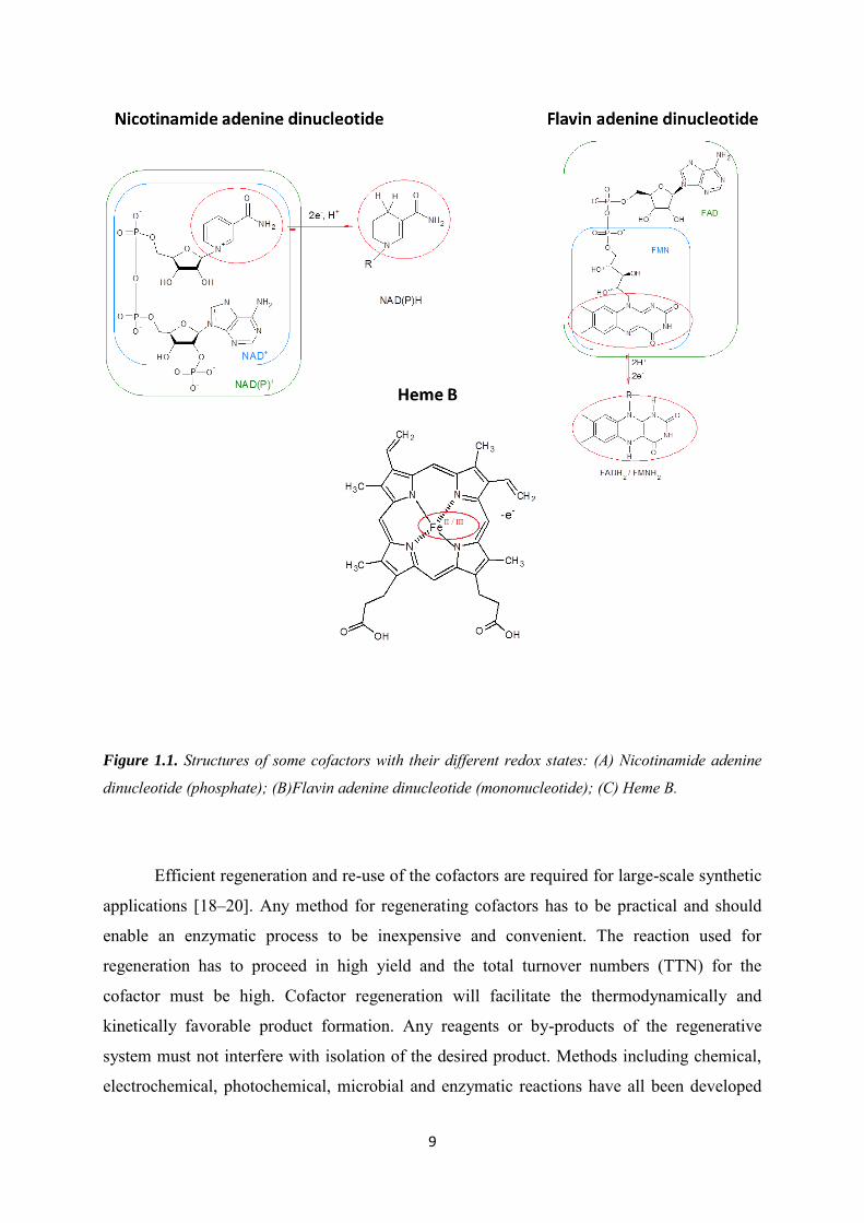
Among the different enzymatic catalytic systems, redox enzymes are an important
class of proteins that catalyze redox reactions. Enzymatic cofactors which are involved in
redox reactions are used for transferring both electrons and protons. The structures of some of
these cofactors are illustrated in Figure 1.1 including their different redox states. Flavin
adenine dinucleotide (FAD), flavin mononucleotide (FMN), heme or transition metals are
associated with the enzymes. Nicotinamide adenine dinucleotide (NAD(P)+/NAD(P)H are
dissociated from the enzymes [4,17].
9
Figure 1.1. Structures of some cofactors with their different redox states: (A) Nicotinamide adenine
dinucleotide (phosphate); (B)Flavin adenine dinucleotide (mononucleotide); (C) Heme B.
Efficient regeneration and re-use of the cofactors are required for large-scale synthetic
applications [18–20]. Any method for regenerating cofactors has to be practical and should
enable an enzymatic process to be inexpensive and convenient. The reaction used for
regeneration has to proceed in high yield and the total turnover numbers (TTN) for the
cofactor must be high. Cofactor regeneration will facilitate the thermodynamically and
kinetically favorable product formation. Any reagents or by-products of the regenerative
system must not interfere with isolation of the desired product. Methods including chemical,
electrochemical, photochemical, microbial and enzymatic reactions have all been developed
10
for cofactor regeneration [18–20]. Chemical regeneration of cofactor can be realized by
introducing reducing agents (e.g. sodium dithionite, sodium borohydride, H2, etc.) or
chemical oxidants (e.g. O2) into the system. However, this method suffers from low TTN and
low selectivity, along with the purification difficulties [21]. Enzymatic approaches are
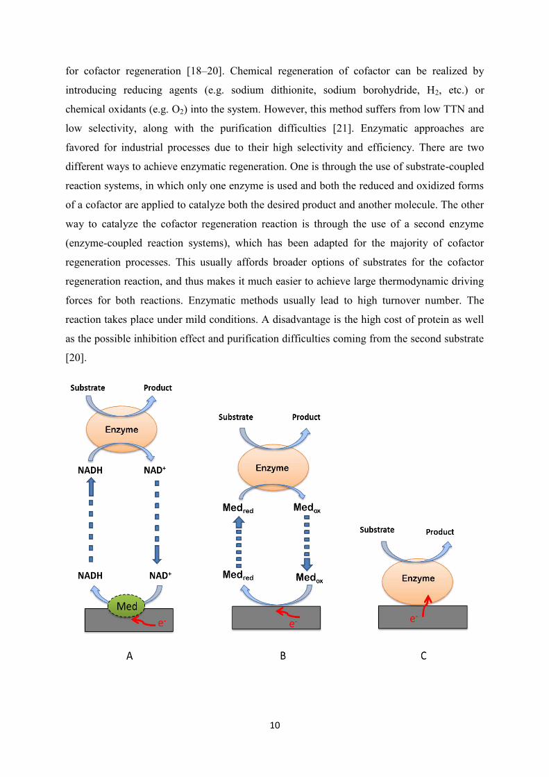
favored for industrial processes due to their high selectivity and efficiency. There are two
different ways to achieve enzymatic regeneration. One is through the use of substrate-coupled
reaction systems, in which only one enzyme is used and both the reduced and oxidized forms
of a cofactor are applied to catalyze both the desired product and another molecule. The other
way to catalyze the cofactor regeneration reaction is through the use of a second enzyme
(enzyme-coupled reaction systems), which has been adapted for the majority of cofactor
regeneration processes. This usually affords broader options of substrates for the cofactor
regeneration reaction, and thus makes it much easier to achieve large thermodynamic driving
forces for both reactions. Enzymatic methods usually lead to high turnover number. The
reaction takes place under mild conditions. A disadvantage is the high cost of protein as well
as the possible inhibition effect and purification difficulties coming from the second substrate
[20].
11
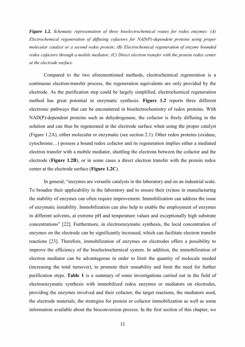
Figure 1.2. Schematic representation of three bioelectrochemical routes for redox enzymes: (A)
Electrochemical regeneration of diffusing cofactors for NAD(P)-dependent proteins using proper
molecular catalyst or a second redox protein; (B) Electrochemical regeneration of enzyme bounded
redox cofactors through a mobile mediator; (C) Direct electron transfer with the protein redox center
at the electrode surface.
Compared to the two aforementioned methods, electrochemical regeneration is a
continuous electron-transfer process, the regeneration equivalents are only provided by the
electrode. As the purification step could be largely simplified, electrochemical regeneration
method has great potential in enzymatic synthesis. Figure 1.2 reports three different
electronic pathways that can be encountered in bioelectrochemistry of redox proteins. With
NAD(P)-dependent proteins such as dehydrogenase, the cofactor is freely diffusing in the
solution and can thus be regenerated at the electrode surface when using the proper catalyst
(Figure 1.2A), either molecular or enzymatic (see section 2.1). Other redox proteins (oxidase,
cytochrome…) possess a bound redox cofactor and its regeneration implies either a mediated
electron transfer with a mobile mediator, shuttling the electrons between the cofactor and the
electrode (Figure 1.2B), or in some cases a direct electron transfer with the protein redox
center at the electrode surface (Figure 1.2C).
In general, “enzymes are versatile catalysts in the laboratory and on an industrial scale.
To broaden their applicability in the laboratory and to ensure their (re)use in manufacturing
the stability of enzymes can often require improvement. Immobilization can address the issue
of enzymatic instability. Immobilization can also help to enable the employment of enzymes
in different solvents, at extreme pH and temperature values and exceptionally high substrate
concentrations” [22]. Furthermore, in electroenzymatic synthesis, the local concentration of
enzymes on the electrode can be significantly increased, which can facilitate electron transfer
reactions [23]. Therefore, immobilization of enzymes on electrodes offers a possibility to
improve the efficiency of the bioelectrochemical system. In addition, the immobilization of
electron mediator can be advantageous in order to limit the quantity of molecule needed
(increasing the total turnover), to promote their reusability and limit the need for further
purification steps. Table 1 is a summary of some investigations carried out in the field of
electroenzymatic synthesis with immobilized redox enzymes or mediators on electrodes,
providing the enzymes involved and their cofactor, the target reactions, the mediators used,
the electrode materials, the strategies for protein or cofactor immobilization as well as some
information available about the bioconversion process. In the first section of this chapter, we

12
will discuss the most typical target reactions that have been used in electroenzymatic
synthesis for different classes of redox enzymes according to their redox cofactor: NAD(P)-
dependent-redox enzymes, flavoenzymes and metal complex-based cofactors from
cytochromes or tyrosinase (heme, copper, …). The purpose is not to provide an exhaustive
view of the bioelectrochemistry of these redox proteins that have been covered by specific
reviews in others fields such as enzymatic biosensor [24] and enzymatic biofuel cell [25], but
to provide a detailed description of their application in electroenzymatic synthesis, focusing
on the strategies used to immobilize the enzyme and/or the mediator.
1.2 Enzymatic systems, cofactor recycling and target reactions
1.2.1 NAD(P)-dependent-redox enzymes
Both nicotinamide adenine dinucleotide phosphate (NADP+/NADPH) and
nicotinamide adenine dinucleotide (NAD+/NADH) have been used in electroenzymatic
synthesis. These different forms of the nicotinamide adenine dinucleotide cofactor display
similar electrochemical properties. For this reason, the discussion will be focused on the
regeneration of NAD+ or NADH and all considerations and conclusions could be, in principle,
applied to NADP+ and NADPH.
The electrochemistry of NAD+/NADH is highly irreversible, and the oxidation of
NADH or the reduction of NAD+ at a bare electrode only occurs at high overpotential.
Several efficient methods have been developed for cofactor regeneration. Synthesis where the
cofactor NAD+/NADH has to be regenerated to its oxidized/reduced state can be carried out
with direct, indirect and enzyme-coupled electrochemical cofactor regeneration [4]. Direct
regeneration means that the species to be regenerated itself reacts at the electrode, however, it
can result in inactivation of the cofactor [5]. Indirect regeneration means that a mediator
catalyst acts as an electron shuttle between the electrode and the cofactor and decreases the
overpotential of the electrochemical reaction. Different molecules are involved to catalyze
either the oxidation of NADH or the reduction of NAD+. If a second enzyme is used, eg.
diaphorase, the process is called enzyme-coupled electrochemical cofactor regeneration. With
few exceptions that will be discussed in the section related to electrode materials (section 3),
direct regeneration is not used in practice, so the discussion will be mainly focused on indirect
13
regeneration and enzyme-coupled electrochemical cofactor regeneration. Both NADH and
NAD+ regeneration will be discussed in the following sections for electrosynthesis
applications.
1.2.1.1 NADH regeneration by NAD+ electrochemical reduction
In NADH regeneration process, NAD+ reduction occurs through two discrete 1e- steps
with a radical protonation process concerted with the second electron transfer [5]. The first
electron transfer to NAD+ is a reversible process. However, the dimerization of the NAD
radical is more favorable than the second electron transfer in a direct electrochemical
reduction [26] and the enzymatically inactive NAD+-dimers can be formed, resulting in the
degradation of the electro-regeneration system [3,4,27]. In consequence, it is critical to avoid
the formation of electrochemically inactive NAD-dimers, and indirect NADH regeneration
methods have been developed.
The non-enzymatic indirect regeneration of NADH consists of using a mediator
catalyst who could transfer two electrons in one step [3]. As the formal potential of the
NAD+/NADH redox couple is −0.56 V (vs. SCE), which means catalytic reduction of NAD+
into NADH is thermodynamically favorable when the potential is more negative than -0.56V
[5], to avoid direct electrochemical regeneration, the operating window of the mediator should
be in the potential window between −0.56 V and -0.9 V. Many efforts have been made to find
the appropriate mediator.
Rh(bpy)3 was the first kind of rhodium-based mediator used for indirect
electrochemical regeneration of NADH. The synthesis of cyclohexanol from cyclohexanone
catalyzed by alcohol dehydrogenase in the presence of this mediator was carried out [28] and
its electropolymerized form on the electrode was also tested [29]. Later on, the mediator
selectivity has been highly improved up to 99% when the pentamethyl cyclopentadienyl (Cp*)
ligand was incorporated. After the first electroenzymatic synthesis from pyruvate to lactate in
the presence of [Cp*Rh(bpy)Cl]+ catalyzed by D-Lactate dehydrogenase, it was later
combined with alcohol dehydrogenase to synthesize different products (e.g. cyclohexanol
[30,31], (S)-4-phenyl-2-butanol [32], (R)-phenylethanol [33], L-malate [34]). Besides,
[Cp*Rh(phen)Cl]+ with a 1,4-phenanthroline ligand instead of 2‟2-bipyridine ligand was also
proved to be efficient in electroenzymatic synthesis of L-glutamate [35]. Moreover,
14
polymerized neutral red (NR) film (at a potential of -0.6V vs. SCE) was also successfully
applied to NADH regeneration [36].
The enzymatic electrochemical regeneration of NADH consists on the combination of
a NAD-dependent dehydrogenase with a second enzyme (usually a flavoenzyme) in the
presence of a mediator to prevent the dimer formation. If one refers to Figure 1.2, the electron
transfer pathway of the dehydrogenase is described by Figure 1.2A, while the electron
transfer pathway of the second enzyme is described by Figure 1.2B. The most commonly
used regeneration enzymes include diaphorase [37–39], lipoamide dehydrogenase [31,39,40],
ferredoxin NAD(P)+ reductase [40,41], and AMAPORS (artificial mediator accepting
oxidoreductases) [42]. Then the electron mediators like viologen derivatives [31,37–42] and
flavin [43,44] or quinone [45] have been used to mediate the electron transfer reaction from
the electrode to the flavin adenine cofactor of the enzymatic catalyst. Electrons were then
transferred to the NAD+ cofactor in the enzyme catalytic site. Some redox enzymes can also
directly communicate with electrode supports (as schematically drawn in Figure 1.2C) and
thus stimulate the regeneration of the NADH cofactor. Hydrogenases (from Rhodococcus
opacus and Atcaligenes eutrophus H16) have been successfully applied for the
bioelectrocatalytic regeneration of NADH without the use of a redox-mediator [46–49] and
have been applied to the bioconversion of alpha-ketoglutarate into L-glutamate catalyzed by
an L-glutamate dehydrogenase [50]. However, the absence of protein confinement at the
electrode surface was pointed out at that time as a limit of the experiment. More recently,
isolated diaphorase fragment has been described as a catalyst for regeneration of NADH in
the presence of H2 and hydrogenase on pyrolytic graphite [51,52] and direct electron transfer
reaction starts to be explored for NAD+ - NADH interconversion by Escherichia coli
flavohemoglobin [53].
D-lactate is by far the most studied target. It is an important product in industrial
manufacture (e.g. cosmetic, food), that can be synthesized from pyruvate in the presence of
D-lactate dehydrogenase [54–64]. The conversion of cyclohexanone to cyclohexanol
[30,31,65], acetophenone to (R)-phenylethanol [7,33], and 2-methyl-cyclohexanone to
(1S,2S)-(+)-2- methylcyclohexanol [37] have also been reported. The synthesis of (L)/(S)-
glutamate which is a compound used as flavor enhancer and applied in pharmacology have
been carried out with the redox enzyme glutamate dehydrogenase [35,40,42,50,66].
15
1.2.1.2 NAD+ Regeneration by NADH electrochemical oxidation
The potential of direct NADH oxidation is close to 0.9 V (vs. SCE) [4]. The
electroenzymatic synthesis using direct NAD+ oxidation has been applied in the conversion of
alcohol to aldehyde in the very early stages of electroenzymatic synthesis [67]. The
production of gluconic acid from glucose catalyzed by glucose dehydrogenase using this
direct electrochemical oxidation of NADH was also reported [68]. But, the overpotential for
this reaction can be dramatically decreased in the presence of a mediator electrocatalyst, as
commonly used in biosensors [24]. It prevents the risk of side reaction that could occur when
the electrode surface is poised at a high potential. If a mediator is introduced in NAD+
regeneration, the mechanism of the mediated electrochemical regeneration of NAD+ can be
described in the following steps: first, the mediator is oxidized by applying a suitable
potential; then the reduced NADH is oxidized by the oxidized mediator [69], the mediator
itself will go back to its reduced form; finally, the reduced mediator will be oxidized again on
the electrode, in the meantime, the oxidized NAD+ will participate in the enzymatic reaction.
An additional possibility to regenerate cofactors is through photoelectrochemical methods
[70,71], but this approach has not been applied in the field of electroenzymatic synthesis yet.
Different mediators have been used for indirect electrochemical regeneration of NAD+
in electroenzymatic synthesis [5,21,72]. Two-electron mediators are the most commonly
found, like bipyridine and phenanthroline metal complexes [73,74], azine dyes like methylene
green [75] or phenazine methosulfate [76], 4-carboxy-2,5,7-trinitro fluorenyliden-malonnitrile
[77], 3,4- dihydroxybenzaldehyde [78]. Another kind of mediators for NAD+ regeneration are
based on single-electron transfer processes like 2,2‟-azinobis(3-ethylbenzothiazoline-6-
sulfonate) (ABTS). Compared to two-electron based mediators, one-electron mediator was
reported to be more stable under basic conditions, which could be suitable for some large
scale synthesis [79]. It is also possible to combine indirect regeneration of NAD+ with an
enzymatic regeneration step. For example, diaphorase has been applied to oxidize NADH
using a variety of quinone compounds [45], ferrocene derivatives [80,81], or osmium redox
polymers [82].
By using different mediators as electron carriers, a variety of substrates catalyzed by
different NAD-dependent enzymes are obtained. The production of gluconic acid catalyzed
by glucose dehydrogenase [78] or glucose-6-phosphate dehydrogenase [76] were reported.
Alcohol dehydrogenase has been applied on different substrates to regio-selectively oxidize
16
alcohols to corresponding carbonyl group products (e.g. cyclohexanone [73,80,83], 2-butanon
[84]). Glycerol dehydrogenase was applied to synthesize (S)-Phenylethane-1,2-diol [85]. D-
sorbotol dehydrogenase was used for D-fructose production from D-sorbitol [75]. Moreover,
there are examples suggesting the electrosynthesis of D-sorbose catalyzed by ribitol
dehydrogenase [86] and hydroxyacetone with galactitol dehydrogenase [77].
1.2.2 Flavoenzymes
Flavoenzymes contain a flavin nucleotide (FMN or FAD) as a prosthetic group.
Glucose oxidase is a typical flavoenzyme which has been used for gluconic acid production in
the presence of oxygen, however, hydrogen peroxide which is consequently formed could
deactivate the redox enzymes. In some system, the conversion rate of glucose was
significantly improved (from 4% to 30% [87]) by eliminating the H2O2 through
electrochemical method, by which the H2O2 was oxidized back to O2 [31,88,89]. However, for
the majority of flavoenzymes applied in electrosynthesis, the bioprocess was accomplished by
electrochemical regeneration of cofactor FAD or FMN using an electron mediator (see Figure
1.2B). For example, instead of using oxygen, when benzoquinone was introduced into the
system, the formed hydroquinone was electrochemically regenerated back to benzoquinone.
The operational stability for gluconic acid production was increased in this case by a factor 50
[90]. p-Cresol methylenehydroxylase and 4-ethylphenol methylenehydroxylase have been
applied for synthesis of 4-hydroxybenzaldehyde or 1-(4-hydroxy phenyl)ethanol in the
presence of ferrocene as electron mediator, a high TTN up to 400000 and a high conversion
rate up to 100 % were achieved in the electrochemical bioreactor [91]. Ferrocenedimethanol
was used for wiring membrane-bounded (S)-mandelate dehydrogenase (S-MDH) which is a
FMN associated enzyme and applied to the catalytic conversion of (S)-mandelic acid to
phenylglyoxylic acid in order to separate (S)-mandelic and (R)-mandelic [92]. In
electroenzymatic reduction, l-aminopropyl-1 methyl-4,4'-dipyridine (ADPy) amino acid
oxidase (AOx) was used in the production of D-alanine from pyruvic acid. The conversion
was almost total and a TTN more than 36000 was achieved for immobilized ADPy [93].
17
1.2.3 Metalloproteins
Most of the metalloproteins used in electroenzymatic synthesis contain a heme
prosthetic group with a central iron atom (see an illustration in Figure 1.1C). Cytochrome
P450 (CYP) is a representative class of heme-containing monooxygenase enzymes
responsible for oxidative metabolism of most drugs and xenobiotics [94]. For most
applications, CYP needs to be immobilized on the electrode to benefit from direct electron
transfer in electrochemical regeneration as shown in Figure 1.2C. The immobilization is not
only necessary for stabilizing or recycling the enzyme (as for dehydrogenases and
flavoenzymes), it is also a requirement for electrochemical recycling of the electroactive
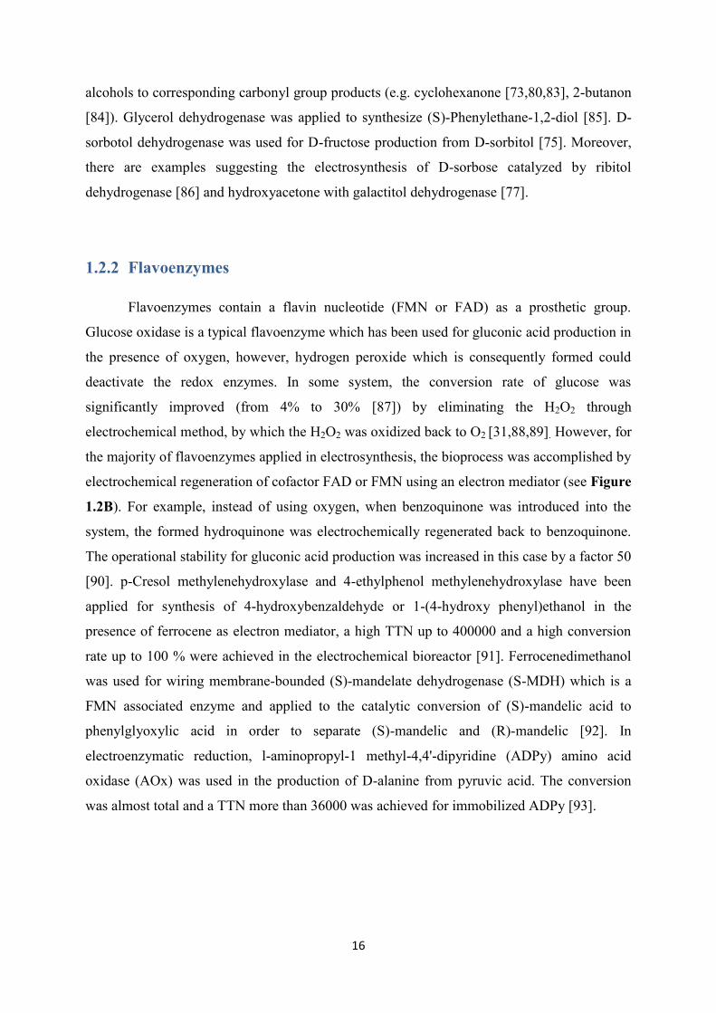
redox cofactor. CYP catalysis route is a two electrons transfer process, a catalytic cycle using
electrochemistry as driving force is illustrated in Figure 1.3 [95]. The first electron is used for
heme reduction, then the reduced iron group is associated with the co-substrate O2. The
second electron is further used for reducing the oxygenated heme, forming the target product
in the presence of protons.
Figure 1.3. Illustration of two-electron transfer steps in electrochemical catalytic cycle of P450
enzymes [95].
18
CYP 119 was used for electrocatalytic reduction of nitrite to ammonia. This is a two
steps electrocatalytic process with two redox peaks corresponding to the reduction of nitric
oxide and production of ammonia [96]. Human CYP 2E1 enzyme was immobilized on gold
electrode surface for synthesizing p-nitrocatechol by applying a potential of -500 mV (vs.
Ag/AgCl) for 30 min [97]. Hydroxylation of 3-phenoxytoluene was catalyzed by immobilized
CYP BM-3 by applying a potential of -0.7V (vs. SCE), showing the possibility of using
electrochemical method as electron donor [98]. The same protein was also used for synthesis
of p-nitrophenolate by hydroxylation of p-nitrophenoxycarboxylic acid [99]. Not only single
enzymes have been considered in electrosynthesis but also bi-enzymatic systems have been
studied. Recently, a work combining two species of cytochrome P450, CYP1A2 and CYP3A4,
on an electrode was described. Two redox peaks of the bi-enzyme complex were observed at -
0.531V and -0.474V respectively. Clopidogrel carboxylic acid was then obtained as
metabolite [100]. Instead of using the direct electrochemical reduction of the P450 redox
center, Rusling et al. reported the combination of a human cytochrome P450 / cytochrome
P450 reductase (CYP/CPR) in a bioelectronic film on pyrolytic graphite electrode. They
developed the idea that electrons could be delivered from cytochrome P450 through CPR
[101].
Chloro-peroxidase is another family of enzymes containing heme cofactor that
catalyzes the chlorination of organic chemicals. In a CPO-containing composite film
functionalized electrode, H2O2 which was used for catalytic oxidation cinnamyl alcohol to
produce aldehydes was constantly generated by the electrochemical reduction of O2, giving a
high yield of 52 % and TTN of 80500 [102]. H2O2 can also be provided for this enzyme by a
gas diffusion electrode (GDE) [103,104]. The role of electrochemistry was not to provide
electrons to the redox center of the enzyme, but to regenerate a co-substrate of the enzymatic
reaction, production that could be carefully tuned by electrochemistry. GDE with
chloroperoxidase (CPO) catalyzed system has also been employed for monochlorodimedone
chlorination [105], halogenation of the phenolic monoterpenes thymol and carvacrol [106].
A final example of redox enzyme is tyrosinase which contains a coupled binuclear
copper active site that has been used as catalyst for hydroxylation of L-tyrosine to L-3,4-
dihydroxyphenylalanine (L-DOPA). Tyrosinase catalyzed the ortho-hydroxylation of l-
tyrosine to l-DOPA by its cresolase activity, and subsequently also catalyzed the
oxidoreduction of l-DOPA to DOPAquinone by its catecholase activity. The by-product
DOPAquinone was then electrochemically reduced to L- DOPA [107,108]. According to the
19
authors, this electroenzymatic system, showed the highest conversion rate and a highly
enhanced productivity if compared to other approaches for l-DOPA synthesis reported
previously.
1.2.4 Electrode materials
Several factors could limit the efficiency of a biosystem to be applied for a bioreactor
[109], among them the electrode materials is an important factor. Metal, metal oxides and
carboceous materials were used, as a flat or as a porous electrode.
1.2.4.1 Metals
The use of platinum electrodes is mostly encountered in membrane electroenzymatic
reactors in which the enzymes stay in solution [31,54,65,87]. It has been reported that the
adsorption of some hydrogenases on a platinum electrode would yield efficient catalysis for
NAD+ reduction [47,48]. The usage of platinum is expected to decrease the regeneration
potential in the presence of Alcaligenes eutrophus H16 hydrogenase. However, the working
potential window of platinum is quite limited since hydrogen evolution often occurs in the
range of NAD+ reduction. Another kind of applicable metal electrode is gold, even though it
suffers from the similar hydrogen evolution problem in reduction reactions, it offers a
possibility to modify the electrode surface by means of the formation of a self-assembled
monolayer with thiol containing molecules [9,77,97]. The use of macroporous gold electrodes
was also suggested in order to take advantage of the high porosity which would facilitate the
mass transport through the electrode macrostructure to favor electrochemical bioconversion
processes [110–112]. Finally, platinum and nickel nanoparticles deposited on a glassy carbon
electrode were recently evaluated for NAD+ reduction. The role of Pt and Ni was to provide
'active' adsorbed hydrogen (Pt-H ads or Ni-H ads) for the fast protonation of electrochemically
formed NAD-radical. The authors could demonstrate that up to a 100% recovery of 1,4-
NADH from NAD+ was possible on the produced electrodes at relatively low potentials,
making them excellent candidates for regeneration of 1,4-NADH in industrial bioreactors
[113]. The same research group also tested other electrode materials (Ti, Ni, Co and Cd) [114].
20
1.2.4.2 Metal oxides
Tin (IV) oxide electrode was prepared using an anodization and annealing method and
was used for mediator free electrochemical cofactor regeneration. Both oxidation of NADH
and reduction of NAD+ were studied. The electrochemical oxidation of NADH and NAD(P)H
were performed at -0.5 V versus a Ag/AgCl reference electrode. The NAD+ reduction was
performed at -0.95 V. Only the NAD+ regeneration was however applied to electroenzymatic
reaction. The authors suggested that the method can be applied to various cofactor-dependent
enzyme reactions for chemical production, biosensor or enzyme fuel cell construction [115].
A thermally prepared iridium/ruthenium-oxide coating (Ir0.8Ru0.2-oxide) formed on a
titanium substrate was also proposed as a possible electrode for direct electrochemical
regeneration of enzymatically-active 1,4-NADH from its oxidized form, NAD+. The coating
surface was characterized by „cracked mud‟ morphology, yielding a high surface roughness.
The percentage of enzymatically-active 1,4-NADH present in the product mixture was
strongly dependent on the electrode potential, reaching a maximum (88%) at -1.70 V versus a
mercury/mercurous sulfate reference electrode [116]. The use of conductive vanadia-silica
gels with encapsulated GDH was also reported for NADH regeneration, allowing complete
conversion of alpha-ketoglutarate to l-glutamate [66].
1.2.4.3 Carbon materials
Carbon materials are widely used due to their low cost, large potential window,
relatively inert surface, and active electrochemical properties for redox reactions [117].
Compared to metal electrodes, different carbon materials exhibit different microstructure as
well as surface functional groups, leading to different electrochemical behaviors. There are
already reviews exhaustively explaining the interfacial as well as the electroanalytical
properties of carbon materials [117,118]. Here, we only focus the discussions on the
application of carbon materials as electrodes in the electroenzymatic synthesis with
immobilized proteins.
Carbon paste electrodes (CPE) have also been used after its introduction in 1958.
Basically, it is made from a mixture of carbon powder with a liquid non-electroactive binder.
The carbon paste could be „modified‟ by adding one or more components (e.g. enzymes or
mediators) [119]. The CPEs are popular because they are easily obtained at minimal costs and
21
certain electrode properties can be pre-determined by adding additional admixtures [120]. It
can be applied for encapsulation of enzymes on electrode [121]. However, its properties differ
from one preparation to another and limit their applications.
The most conventional carbon electrode is glassy carbon (GC), its use is convenient
since its surface is renewable by polishing. As it is a flat electrode with uniform functional
groups on the surface, it is a good candidate to be covalently modified for bounding enzymes
or molecules [63,93], or deposited with uniform membranes [57], nanoparticles [113] or films
[86,98,99]. If the GC is produced as a foam, it is then called reticulated vitreous carbon
(RVC), which also been used in electrochemical bioreactors [55,58].
Porous carbon felt (CF) electrodes have been largely used electrode in the field of
electroenzymatic synthesis, taking benefit from their low-cost and large surface area, which
lead to high current density and high conversion efficiency. CF electrodes have been used as
working electrodes in electroenzymatic synthesis reactors with dialysis [30,31,65] or
ultrafiltration membranes [91]. From another point of view, the functional groups on the
surface make it an appropriate substrate for surface modification, which could be further used
for different covalent reactions. CF was used for example as electrode support for
immobilization of enzymes with carbodiimide or glutaraldehyde as linking agent [33,64,90].
Furthermore, to increase the surface area as well as the biocatalytic property of the
electrodes, carbon nanoparticles [107,108], carbon nanotubes [75], graphene nanosheets [122]
and graphene oxide nanocomposite [100], allowed to improve significantly the efficiency of
the electroenzymatic synthesis system by increasing the enzyme loading and/or by tuning
some electrochemical properties.
1.3 Immobilization of enzymes on electrodes
In the past years, different strategies have been developed to build enzyme-
immobilized electrodes [23,123]. There are several conditions that need to be fulfilled in
enzymes immobilization. First, the activity of enzymes needs to be kept upon immobilization.
Second, the stability of enzymes towards microenvironments on the electrode needs to be
ensured. Usually, adsorption is the easiest method for enzyme immobilization, but it is
inappropriate for bioconversion, because the electrosynthesis process requires long-term
stability especially when the solution is under continuous stirring, the adsorbed enzymes
would be rapidly leached out into the solution. In the field of electro-enzymatic synthesis, the
22
immobilization methods that have been typically used are membrane confinement in a small
volume at close distance from the electrode (Figure 1.4A), surface immobilization with
chemical bonding (Figure 1.4B&C) and encapsulation (Figure 1.4D-F). The next sections
will provide a discussion of these different strategies.
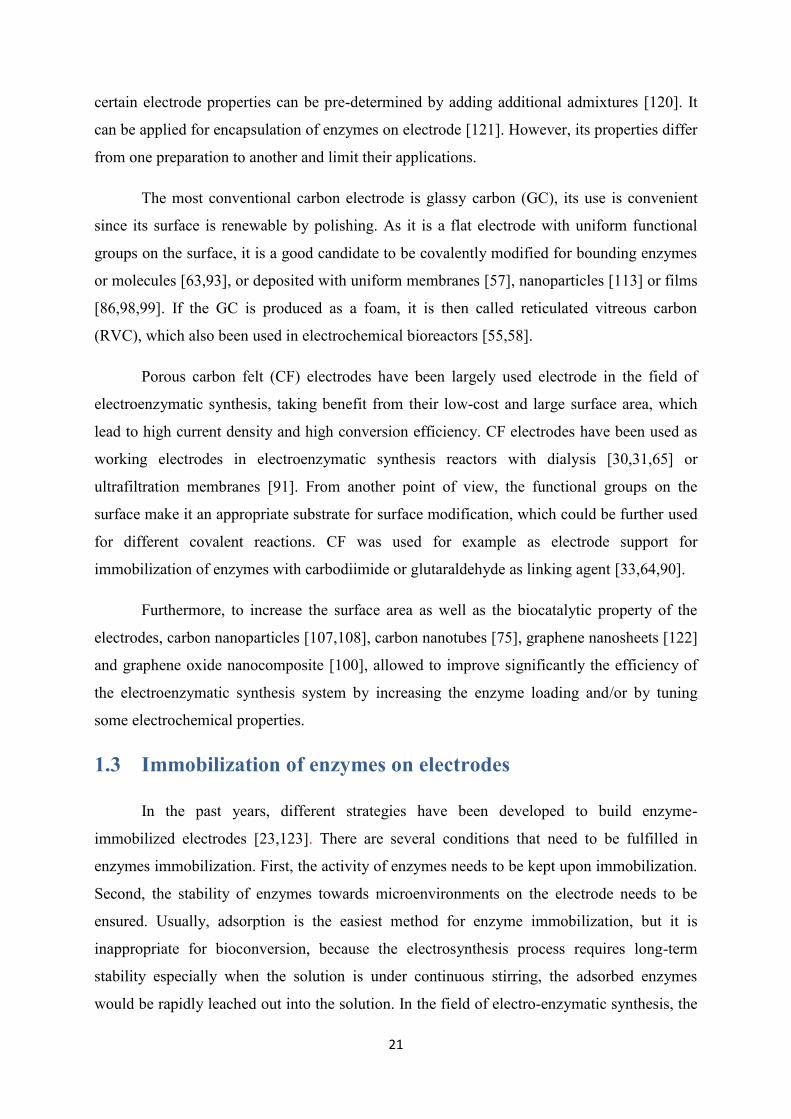
Figure 1.4. Typical enzyme immobilization methods on electrodes (A - F): (A) Membrane confinement;
(B) Covalent bonding; (C) ‘Thiol-gold’ self-assembly monolayer; (D) Nafion membrane coating; (E)
Surfactant film embedding; (F) Encapsulation in solid matrix.
1.3.1 Membrane confinement with membranes
Membrane confinement is a simple way to immobilize enzymes in an electrosynthesis
bioreactor. The enzymes are confined in solution near the electrode without any physical or
chemical modification, but only soluble redox enzymes can be immobilized by this method.
Generally, two kinds of membrane configuration are used for immobilization: dialysis or
23
ultra-filtration membrane. The formation of a Nafion coating on an electrode surface can be
considered as a surface confinement comparable to encapsulation and will be discussed in
another section.
Commercial dialysis or ultra-filtration membranes have been used inside a bioreactor
[30,31,54,56,59–61,65,67,68,87,91]. These membranes are made of different kinds of
materials, e.g. cellulose [30,31,54,65,87], hollow fiber [67], Nafion 117 [55,56,60,61,68].
Generally speaking, the ability to immobilize the enzyme is dependant on the pore size of the
membrane. Enzymes larger than the membrane cutoff value are confined in the membrane
near electrode and small molecules can freely pass through the membrane.
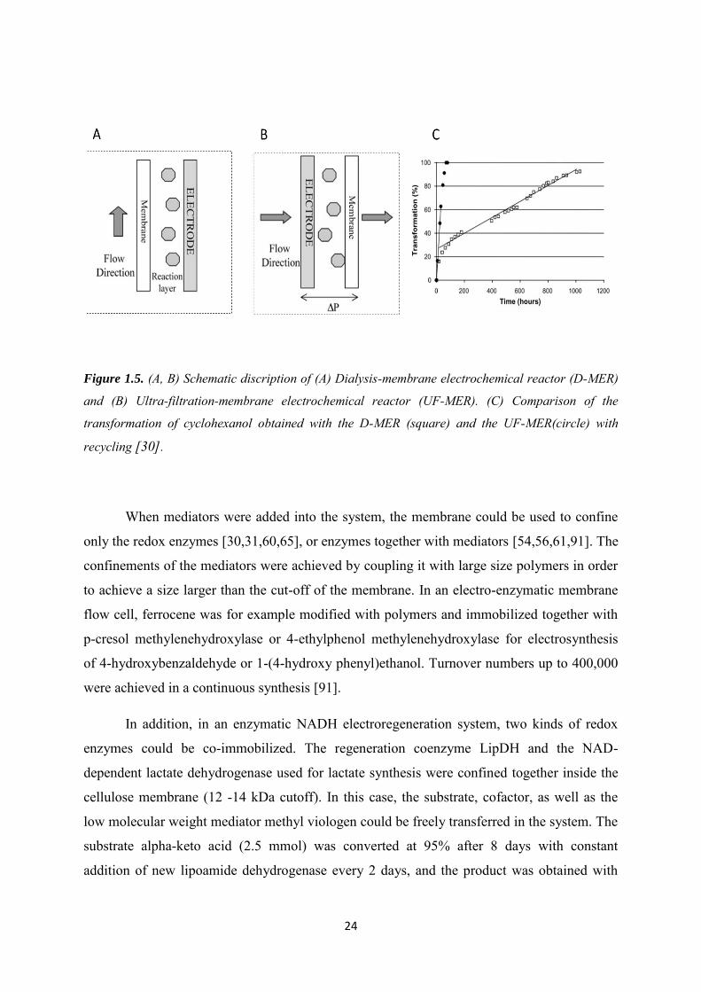
In a dialysis membrane reactor (D-MER), the transport of compounds through the
membrane was mainly due to the gradient of concentration, while in ultra-filtration membrane
UF-MER, solution passing through the membrane in a direction perpendicular to the electrode
was driven by a pressure gradient. The influence of flowing mode on the transformation rate
in a bioreactor has been studied. An ultrafiltration-membrane electrochemical reactor (UF-
MER) (Figure 1.5A) was compared with D-MER (Figure 1.5B) in synthesis of cyclohexanol
by alcohol dehydrogenase with [Cp(Me)5Rh(bpy)Cl] as mediator. The transformations rate in
these two configurations are compared in Figure 1.5C, leading to 92% in 45 days for D-MER
and 100% in 3 days for UF-MER. UF-MER was the more efficient reactor configuration, and
a limiting step in D-MER was observed: the inhibition of the enzyme by increasing
concentration of the products at the end of the electrolysis [30].
In an early stage, membrane confinement of redox enzymes has been used in the
bioreactor with direct electrochemical regeneration of NAD+. Coughlin et al. use a
continuous-flow reactor with ultra-filtration membrane to confine NAD-dependent alcohol
dehydrogenase for direct electrochemical regeneration of NAD+ in electro-synthesis of
aldehyde from alcohol. Compared with dialysis, the ultra-filtration membrane was able to
keep the cofactors and enzymes persistently in the continuous-flow process, while the
products left the system through the membrane [67]. Later on, the confinement of glucose
dehydrogenase in a dialysis Nafion membrane reactor for synthesis of gluconic acid with
direct regeneration of NAD+ was also achieved [68]. To avoid the large overpotential which
may cause inactivation of the cofactor, diffusing mediators were added in the system
afterwards to facilitate the electron transfer between electrodes and cofactors [68].
24
Figure 1.5. (A, B) Schematic discription of (A) Dialysis-membrane electrochemical reactor (D-MER)
and (B) Ultra-filtration-membrane electrochemical reactor (UF-MER). (C) Comparison of the
transformation of cyclohexanol obtained with the D-MER (square) and the UF-MER(circle) with
recycling [30].
When mediators were added into the system, the membrane could be used to confine
only the redox enzymes [30,31,60,65], or enzymes together with mediators [54,56,61,91]. The
confinements of the mediators were achieved by coupling it with large size polymers in order
to achieve a size larger than the cut-off of the membrane. In an electro-enzymatic membrane
flow cell, ferrocene was for example modified with polymers and immobilized together with
p-cresol methylenehydroxylase or 4-ethylphenol methylenehydroxylase for electrosynthesis
of 4-hydroxybenzaldehyde or 1-(4-hydroxy phenyl)ethanol. Turnover numbers up to 400,000
were achieved in a continuous synthesis [91].
In addition, in an enzymatic NADH electroregeneration system, two kinds of redox
enzymes could be co-immobilized. The regeneration coenzyme LipDH and the NAD-
dependent lactate dehydrogenase used for lactate synthesis were confined together inside the
cellulose membrane (12 -14 kDa cutoff). In this case, the substrate, cofactor, as well as the
low molecular weight mediator methyl viologen could be freely transferred in the system. The
substrate alpha-keto acid (2.5 mmol) was converted at 95% after 8 days with constant
addition of new lipoamide dehydrogenase every 2 days, and the product was obtained with
25
98% enantiomeric excess which was comparable to that obtained in a non-membrane system
[54].
1.3.2 Chemical bonding
Compared to adsorption, the leaching of enzymes from electrode was minimized with
covalent bonding and long-term stability was ensured. Generally, the chemical reaction
occurred between the functional groups on the electrode surface and active amino acid
residues of the enzyme. But an obvious disadvantage of covalent binding is that enzyme itself
is chemically modified when immobilized, if essential amino acid residues close to the
enzyme active site are involved in the chemical reaction, enzyme activity might be reduced.
So when covalently immobilizing the enzymes on electrodes, physical and chemical nature of
the electrode, the nature of the linkage or binding chemistry, and even enzyme orientation
need to be considered [8].
Different kinds of carbon materials (carbon felt [33,64,90], graphene felt [78],
graphene oxide nanocomposite (RGO) [100], carbon nanopodwer [107,108], rotating disc
graphite [76], glassy carbon [63,93]) can be used for covalent immobilization of enzymes in
the field of electroenzymatic synthesis. These carbon materials have advantages such as
chemical and physical stability, large surface area, and rich surface functional groups. It is
already reported that several chemical methods could be applied for covalent immobilization
of enzymes [124,125], but the chemical reactions that have been used in the electroenzymatic
synthesis are quite limited. The generally used methods include carbodiimide [64,90,107,108],
and glutardialdehyde crosslinking [33,63,93]. Sometimes the carbodiimide and
glutardialdehyde are used together in two successive steps [76,78,100]. On gold electrode,
self-assemble monolayers could be also involved [77,97].
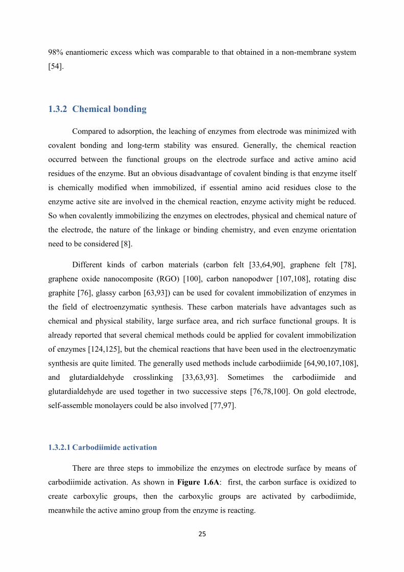
1.3.2.1 Carbodiimide activation
There are three steps to immobilize the enzymes on electrode surface by means of
carbodiimide activation. As shown in Figure 1.6A: first, the carbon surface is oxidized to
create carboxylic groups, then the carboxylic groups are activated by carbodiimide,
meanwhile the active amino group from the enzyme is reacting.
26
For the production of gluconic acid from glucose oxidase in the presense of
benzoquinone in the solution as mediator, the enzyme was covalently immobilized on the
carbon felt electrode by carbodiimide [90]. Tyrosinase was immobilized to functionalized
carbon nanoparticles via (1-ethyl-3-(3-dimethylaminopropyl) carbodiimide, before being
integrated in a 3-D bioelectrode [107,108]. In some systems, both mediator and enzymes
could be covalently co-immobilized on the electrode by using carbodiimide. For the
electrosynthesis of lactate, the coenzyme viologen-accepting pyridine nucleotide
oxidoreductases (VAPORS) combined with viologen directives (DAPV) was co-immobilized
on an electrode for NADH regeneration. Different immobilization order of both components
were tested, either first covalent binding DAPV to VAPORS then followed by attachment of
the DAPV-VAPORS to a functionalized carbon electrode, or covalent binding of the enzyme
to viologen functionalized carbon electrode. These different covalent bonding strategies
allowed optimization of the enzyme orientation. A relationship was drawn between electron
transfer reactions and bioconversion efficiencies [64].
Figure 1.6. Chemical reactions applied on covalent immobilization of enzymes on electrodes: (A)
Carbodiimide; (B) Glutardialdehyde.

27
1.3.2.2 Glutaraldehyde crosslinking
Another popular linking agent for covalent immobilization of enzymes is
glutaraldehyde. There are two types of possible reactions as shown in Figure 1.6B. The first
possible process carried out on the amino functionalized electrodes, they can be activated by
glutaraldehyde, before the enzymes are covalently attached on the glutaraldehyde
functionalized electrode by reacting with surface amino groups. The second possible process
is to use glutaraldehyde as cross-linking agent in a polymerization process by reacting with
enzyme surface amino groups. Cross-linking provides a facile route to achieve co-
immobilization. Amino acid oxidase (AOx) as well as the mediator ADPy were co-
immobilized on electrode through glutaraldehyde crosslinking [93]. Co-immobilization of a
polymerized viologen and LipDH on electrode by glutaraldehyde crosslinking was also
applied in lactate production [63]. In an aqueous–organic two-phase system, alcohol
dehydrogenase was covalently bound to porous amino-epoxy sepabeads support (reaction of
amine groups from the protein with epoxy groups) and a subsequent cross-linking with
glutaraldehyde was used. The enzyme-support preparation was applied to the production of
(R)-phenylethanol from acetophenone in the presence of Cp*Rh(bpy) as mediator for NADH
regeneration [33]. The stability of the enzyme in a bioreactor was strongly increased after
glutaraldehyde treatment, resulting in a half-life time of over 1200 h when stored at 30 °C.
This was a 60-fold increase in stability compared to the soluble enzyme [126].
1.3.2.3 Self-assembled monolayer (SAM)
In some applications, enzymes can be bound to a gold electrode surface with a self-
assembled monolayer (SAM) due to the interaction between thiol groups and gold. Mak et al.
reported the immobilization of CYP2E1 and its mutants on gold electrodes that were first
modified with dithio-bismaleimidoethane (DTME) forming a SAM. Then the functionalized
DTME electrode was allowed to react with the exposed cysteine group on the enzyme surface.
The orientation of the protein was well controlled and better catalytic activity was kept in the
presence of the molecular spacer provided by the SAM [97].
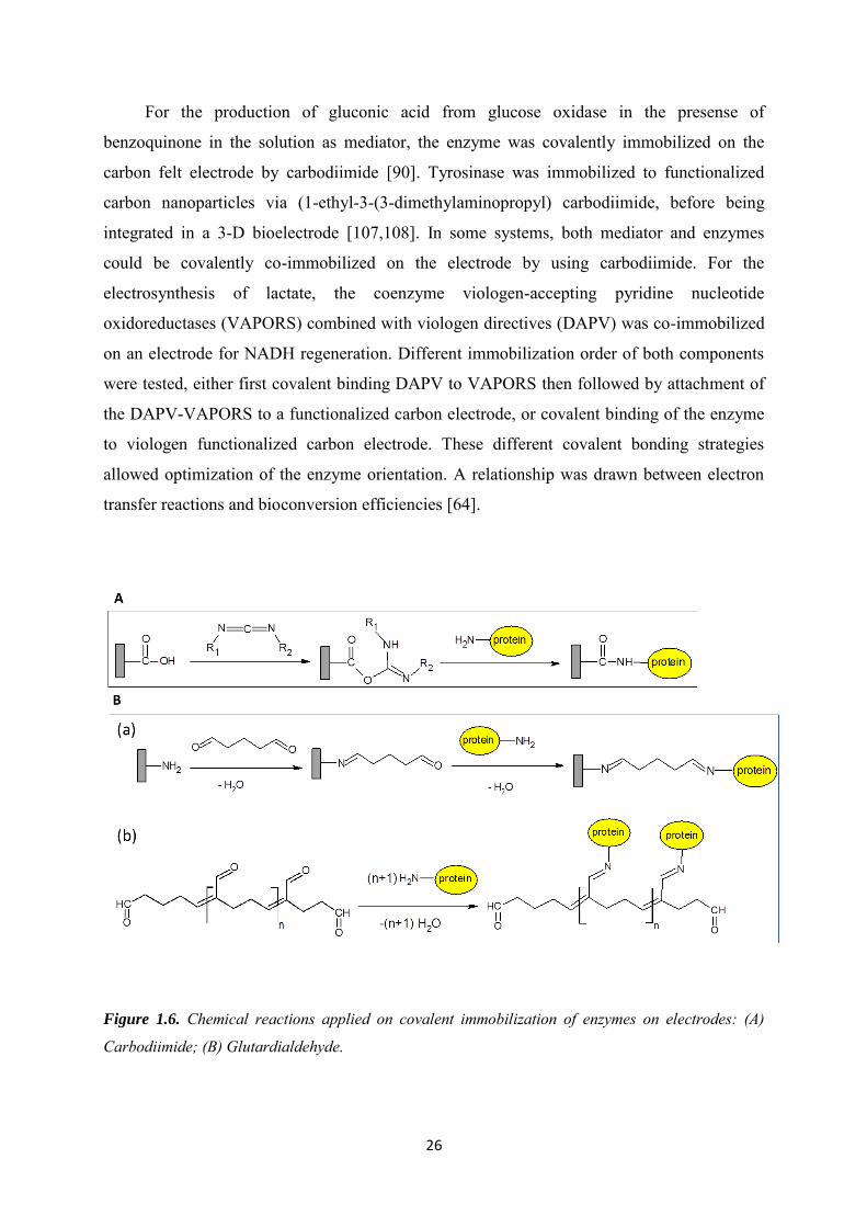
A successful immobilization by direct reaction between the thiol group of a cysteine
tag on the protein and a gold electrode was also reported. The schematic representation of this
electrochemical system is shown in Figure 1.7A. Galactitol dehydrogenase modified with a
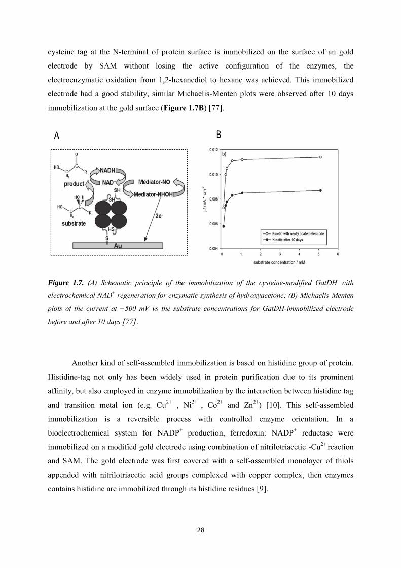
28
cysteine tag at the N-terminal of protein surface is immobilized on the surface of an gold
electrode by SAM without losing the active configuration of the enzymes, the
electroenzymatic oxidation from 1,2-hexanediol to hexane was achieved. This immobilized
electrode had a good stability, similar Michaelis-Menten plots were observed after 10 days
immobilization at the gold surface (Figure 1.7B) [77].
Figure 1.7. (A) Schematic principle of the immobilization of the cysteine-modified GatDH with
electrochemical NAD+ regeneration for enzymatic synthesis of hydroxyacetone; (B) Michaelis-Menten
plots of the current at +500 mV vs the substrate concentrations for GatDH-immobilized electrode
before and after 10 days [77].
Another kind of self-assembled immobilization is based on histidine group of protein.
Histidine-tag not only has been widely used in protein purification due to its prominent
affinity, but also employed in enzyme immobilization by the interaction between histidine tag
and transition metal ion (e.g. Cu2+ , Ni2+ , Co2+ and Zn2+) [10]. This self-assembled
immobilization is a reversible process with controlled enzyme orientation. In a
bioelectrochemical system for NADP+ production, ferredoxin: NADP+ reductase were
immobilized on a modified gold electrode using combination of nitrilotriacetic -Cu2+ reaction
and SAM. The gold electrode was first covered with a self-assembled monolayer of thiols
appended with nitrilotriacetic acid groups complexed with copper complex, then enzymes
contains histidine are immobilized through its histidine residues [9].

29
1.3.3 Encapsulation
The immobilization of proteins by encapsulation can be done either with conductive
electrode materials (conducting polymers) or more often with insulating materials such as
Nafion, alginate, silica gel or hybrid materials.
1.3.3.1 Nafion coating
Nafion layer from a Nafion solution can be deposited on the top of enzyme modified
electrodes [55,57–59,62] as shown in Figure 1.4C. This method was generally applied for co-
immobilization of enzymes and mediators in electrosynthesis of lactate. The Nafion coating
electrode was prepared by first dipping an electrode into the solution containing the enzymes
and mediators. After drying, the modified electrode was dipped into a polymer Nafion
solution to get a Nafion membrane coating electrode.
In an electroenzymatic synthesis of lactate, MV and LipDH have been confined
together on the cathode by a Nafion coating membrane, allowing the extension of LipDH
lifetime [58]. Compared to dialysis or ultrafiltration membrane reactors, this kind of coating
membrane was easier to prepare, but it had the disadvantage of slow leakage of MV into the
solution. As MV species are very toxic which may bring undesirable contamination, a
viologen derivative was first chemically attached to LipDH before immobilization on the
electrode with a Nafion coating [55,62].
Alternatively, the toxic viologen species could be replaced by a natural redox cofactor
such as flavin adenine dinucleotide (FAD). Both electropolymerized FAD or monomeric FAD
were immobilized together with LipDH on the electrode by a Nafion coating, a relatively
slower conversion rate of lactate was observed when using FAD in place of viologen, but the
long-term stability of the enzyme was improved with electropolymerized FAD immobilized
electrode, LipDH kept 80% of the initial activity after 30 days with constant performance [57].
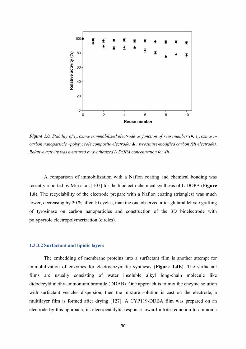
30
Figure 1.8. Stability of tyrosinase-immobilized electrode as function of reusenumber (●, tyrosinase–
carbon nanoparticle –polypyrrole composite electrode; ▲, tyrosinase-modified carbon felt electrode).
Relative activity was measured by synthesized l- DOPA concentration for 4h.
A comparison of immobilization with a Nafion coating and chemical bonding was
recently reported by Min et al. [107] for the bioelectrochemical synthesis of L-DOPA (Figure
1.8). The recyclability of the electrode prepare with a Nafion coating (triangles) was much
lower, decreasing by 20 % after 10 cycles, than the one observed after glutaraldehyde grafting
of tyrosinase on carbon nanoparticles and construction of the 3D bioelectrode with
polypyrrole electropolymerization (circles).
1.3.3.2 Surfactant and lipidic layers
The embedding of membrane proteins into a surfactant film is another attempt for
immobilization of enzymes for electroenzymatic synthesis (Figure 1.4E). The surfactant
films are usually consisting of water insoluble alkyl long-chain molecule like
didodecyldimethylammonium bromide (DDAB). One approach is to mix the enzyme solution
with surfactant vesicles dispersion, then the mixture solution is cast on the electrode, a
multilayer film is formed after drying [127]. A CYP119-DDBA film was prepared on an
electrode by this approach, its electrocatalytic response toward nitrite reduction to ammonia
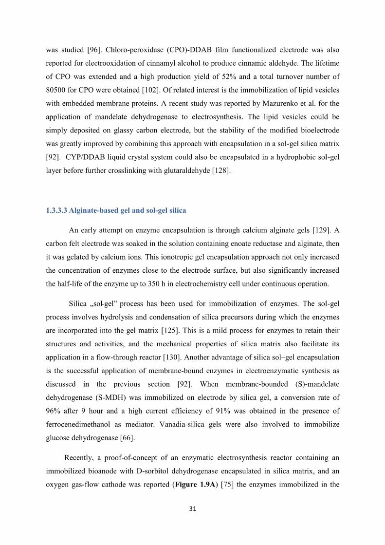
31
was studied [96]. Chloro-peroxidase (CPO)-DDAB film functionalized electrode was also
reported for electrooxidation of cinnamyl alcohol to produce cinnamic aldehyde. The lifetime
of CPO was extended and a high production yield of 52% and a total turnover number of
80500 for CPO were obtained [102]. Of related interest is the immobilization of lipid vesicles
with embedded membrane proteins. A recent study was reported by Mazurenko et al. for the
application of mandelate dehydrogenase to electrosynthesis. The lipid vesicles could be
simply deposited on glassy carbon electrode, but the stability of the modified bioelectrode
was greatly improved by combining this approach with encapsulation in a sol-gel silica matrix
[92]. CYP/DDAB liquid crystal system could also be encapsulated in a hydrophobic sol-gel
layer before further crosslinking with glutaraldehyde [128].
1.3.3.3 Alginate-based gel and sol-gel silica
An early attempt on enzyme encapsulation is through calcium alginate gels [129]. A
carbon felt electrode was soaked in the solution containing enoate reductase and alginate, then
it was gelated by calcium ions. This ionotropic gel encapsulation approach not only increased
the concentration of enzymes close to the electrode surface, but also significantly increased
the half-life of the enzyme up to 350 h in electrochemistry cell under continuous operation.
Silica „sol-gel‟ process has been used for immobilization of enzymes. The sol-gel
process involves hydrolysis and condensation of silica precursors during which the enzymes
are incorporated into the gel matrix [125]. This is a mild process for enzymes to retain their
structures and activities, and the mechanical properties of silica matrix also facilitate its
application in a flow-through reactor [130]. Another advantage of silica sol–gel encapsulation
is the successful application of membrane-bound enzymes in electroenzymatic synthesis as
discussed in the previous section [92]. When membrane-bounded (S)-mandelate
dehydrogenase (S-MDH) was immobilized on electrode by silica gel, a conversion rate of
96% after 9 hour and a high current efficiency of 91% was obtained in the presence of
ferrocenedimethanol as mediator. Vanadia-silica gels were also involved to immobilize
glucose dehydrogenase [66].
Recently, a proof-of-concept of an enzymatic electrosynthesis reactor containing an
immobilized bioanode with D-sorbitol dehydrogenase encapsulated in silica matrix, and an
oxygen gas-flow cathode was reported (Figure 1.9A) [75] the enzymes immobilized in the
32
silica film show very good operational stability for two weeks, which was much better than
that in solution (half- life period of 48h at 28 ℃). In simultaneous energy production mode, as
shown in Figure 1.9B, 60% D-sorbitol was converted to D-fructose. Gluconic acid
production from glucose was also achieved in a membraneless, flow-through
electroenzymatic reactor operating in the mode of co-generating chemicals and electrical
energy [131].
Hybrid gel can be used for immobilization of redox proteins. Recently, a bioelectrode
was prepared with alcohol dehydrogenases embedded in an alginate–silicate hybrid gel. This
study is very intriguing as the authors argue for a direct electron transfer reaction into the
biocatalysts for reduction of butyraldehyde to butanol [132], however the reaction should
occur in the presence of a NAD+ cofactor that is not mentioned in the bioelectrochemical
reaction.
Figure 1.9. (A) Scheme of the flow bioreactor using D-sorbitol dehydrogenase (DSDH) immobilized
bioelectrode as anode while a gas diffusion electrode (GDE) as cathode. (B) Conversion of 1 mM D-
sorbitol to D-Fructose in the GDE bioreactor with simultaneous energy production [131].
33
1.3.3.4 Conducting polymers
As electrochemistry is involved in enzymatic electrosynthesis, conducting polymers
could provide unique advantages allowing both the protein immobilization and cofactor
regeneration and wiring. Conducting biocompatible polymer materials (e.g. polypyrrole,
poly(3,4-ethylenedioxythiophene)) have indeed been applied to this field. In a work with
enzymes encapsulated in polypyrrole matrix, the film was electrografted on a platinum
electrode from pyrrole solution in the presence of glucose oxidase, resulting in a uniform
polymer structure and enzyme distribution. The modified electrode was adaptable to a
bioelectrochemical process for gluconic acid production [88]. The electrochemical process
benefited from the oxygen regeneration at the electrode and from the enzyme protection effect
from the polymer matrix, leading to an increase in the transformation ratios by a factor of two
[89].
The P450 enzymes were immobilized in conducting polymers like polypyrrole films
[99] and poly(3,4-ethylenedioxythiophene) doped with polystyrene sulfonic acid
(PEDOT/PSS) [98] on electrode in an active form, the conductive polymer was
electrochemically wiring the enzymes for direct electron transfer reaction (see scheme 2C).
Figure 1.10 reports a comparison of enzymatic production in the presence of the natural
cofactor NADPH (black symbols) with the electrochemically-driven biocatalysis (white
symbols). Both experiments have been performed with the cytochrome P450 CYP102A1
immobilized in a polypyrrole matrix. Two different kinds of electrode material were used,
either Pt (squares) or glassy carbon (circles). This immobilization did not hinder the
enzymatic activity of the proteins on glassy carbon electrode. The electrochemically-driven
biocatalysis was as efficient as the one fed by NADPH on this latter electrode material. The
enzymatic activity determined on Pt electrodes show at the contrary a more complex behavior,
largely unfavorable to electrochemically-driven reactions [99].

34
Figure 1.10. Product (p-nitrophenolate, detected at 410 nm) formation kinetics of P450 BM-3 QM
embedded in polypyrrole on platinum (■) or glassy carbon (●) electrodes with the natural cofactor
NADPH (□) or by electrochemistry (○). The insert is the scheme of corresponding reaction [99].
1.4 Immobilization of mediators
In practical electrosynthesis, the immobilization of mediators is also necessary from
economic and ecological considerations. The immobilization of mediators would not only
increase their reusability and simplify the product purification step, but also limit the
undesirable interactions between enzymes and mediators. A huge effort has been done for
immobilization of redox mediator for flavoenzymes (especially for glucose oxidase) or NAD-
dependent dehydrogenases in the field of biosensors [24,72], or enzymatic fuel cells [133–
135]. This topic was more rarely studied in the field of electrosynthesis and essentially for
NADH regenerations which concerns two molecules methyl-viologen and (2,2'-
bipyridyl)(pentamethylcyclopentadienyl)-rhodium family ([Cp*Rh(bpy)Cl]+). Here we will
briefly review the immobilization of methyl-viologen and rhodium complex in relation with
electrosynthesis.
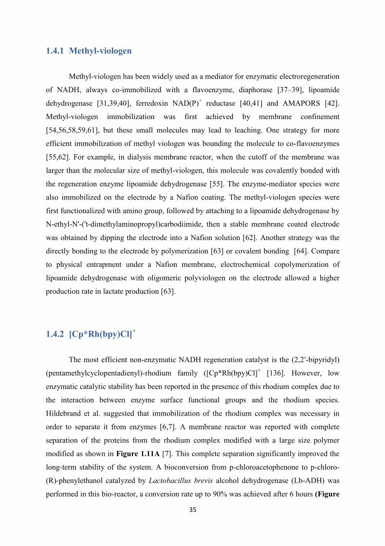
35
1.4.1 Methyl-viologen
Methyl-viologen has been widely used as a mediator for enzymatic electroregeneration
of NADH, always co-immobilized with a flavoenzyme, diaphorase [37–39], lipoamide
dehydrogenase [31,39,40], ferredoxin NAD(P)+ reductase [40,41] and AMAPORS [42].
Methyl-viologen immobilization was first achieved by membrane confinement
[54,56,58,59,61], but these small molecules may lead to leaching. One strategy for more
efficient immobilization of methyl viologen was bounding the molecule to co-flavoenzymes
[55,62]. For example, in dialysis membrane reactor, when the cutoff of the membrane was
larger than the molecular size of methyl-viologen, this molecule was covalently bonded with
the regeneration enzyme lipoamide dehydrogenase [55]. The enzyme-mediator species were
also immobilized on the electrode by a Nafion coating. The methyl-viologen species were
first functionalized with amino group, followed by attaching to a lipoamide dehydrogenase by
N-ethyl-N'-('t-dimethylaminopropyl)carbodiimide, then a stable membrane coated electrode
was obtained by dipping the electrode into a Nafion solution [62]. Another strategy was the
directly bonding to the electrode by polymerization [63] or covalent bonding [64]. Compare
to physical entrapment under a Nafion membrane, electrochemical copolymerization of
lipoamide dehydrogenase with oligomeric polyviologen on the electrode allowed a higher
production rate in lactate production [63].
1.4.2 [Cp*Rh(bpy)Cl]+
The most efficient non-enzymatic NADH regeneration catalyst is the (2,2′-bipyridyl)
(pentamethylcyclopentadienyl)-rhodium family ([Cp*Rh(bpy)Cl]+ [136]. However, low
enzymatic catalytic stability has been reported in the presence of this rhodium complex due to
the interaction between enzyme surface functional groups and the rhodium species.
Hildebrand et al. suggested that immobilization of the rhodium complex was necessary in
order to separate it from enzymes [6,7]. A membrane reactor was reported with complete
separation of the proteins from the rhodium complex modified with a large size polymer
modified as shown in Figure 1.11A [7]. This complete separation significantly improved the
long-term stability of the system. A bioconversion from p-chloroacetophenone to p-chloro-
(R)-phenylethanol catalyzed by Lactobacillus brevis alcohol dehydrogenase (Lb-ADH) was
performed in this bio-reactor, a conversion rate up to 90% was achieved after 6 hours (Figure
36
1.11B), the activity of immobilized enzymes was completely kept (5.5±1 U mg-1 before and
6±1 U mg-1 after). In addition, 86% of the rhodium complex could be recovered.
Figure 1.11. (A) Setup of a membrane reactor with complete separation of the proteins from the large
size polymer modified rhodium complex with setup: 1) pump, 2) membrane reactor, 3) electrochemical
cell, 4) immobilized enzyme. (B) Conversion from 4 mM p-chloroacetophenone to p-chloro-(R)-
phenylethanol by immobilized Lb-ADH with electrochemical cofactor regeneration [7].
Some efforts have been made to immobilize the rhodium complex on the electrode.
Generally, the immobilization methods can be classified into two categories. The first
category is based on polymerization. Polypyrrolic rhodium (III) complex (2,2'-bipyridine or
1,10-phenanthroline ligand) films was deposited on electrodes by anodic
electropolymerization of pyrrole monomers, its electrocatalytic interest for hydrogen
evolution was demonstrated [137]. Rhodium(III) bis-terpyridinepyrrole complex on a
reticulated vitreous carbon electrode by electropolymerization was characterized by reduction
of NAD+ into 1,4-NADH, then its enzymatic catalytic response was tested for reduction of
cyclohexanone to cyclohexanol in the presence of alcohol dehydrogenase [29]. The catalytic
activity of the rhodium complexes incorporated into the bilayers of vesicles formed from a
polymerizable ammonium surfactant was investigated, the catalytic response was proved to
depend on its position in the bilayer [138]. Despite electropolymerization, ɤ-irradiation cross-
linking can also be used to obtain [RhIII(C5Me5)(L)C1]+ complex polymer-modified
electrodes. Even through polymerization would provide a stable bonding, it suffered from the
electron shielding effect coming from the thick porous polymer film. When the amount of
immobilized polymer was doubled on the electrode, the charge calculated from the reduction
37
peak was only increased by a factor of less than 2. This suggested that there was some
limitation in charge transfer reaction caused by the polymer layer [139].
The second immobilization strategy was based in π-π stacking effect between
carbonaceous materials (graphene or carbon nanotubes) and aromatic moieties from the
rhodium complex. Park and coworkers reported that a phenanthroline modified Rh complex
was strongly immobilized on the graphene surface through π-π interaction and π-cation
interaction the repeated usability for electrochemical NADH regeneration up to 7 cycles (each
cycle lasts 2 h) was demonstrated [35]. Minteer and coworkers immobilize a rhodium
complex with a pyrene-substituted phenanthroline ligand onto MWCNT via π-π stacking,
good catalytic response for the regeneration of NADH and repeated usability up to 10 cycles
(each cycle lasted 30 min) were achieved [34]. This route eliminated the electron shielding
effect and significantly increased the usability of the catalyst. However, it could be only
applied on CNT or 3D-structured graphene which has aromatic rings, as the electrode. In
addition, π–π stacking which has a bond energy 0-10 kcal/mol is a kind of reversible non-
covalent bond, was not as stable as covalent binding whose bond energy is 80-100 kcal/mol
when applied for continuous long-term bioconversion [140,141]. Thiol-functionalized
rhodium complex was also covalently immobilized by self-assembled monolayers (SAMs) on
gold electrodes. The immobilized rhodium complex was electrochemically active, however,
no electrocatalytic activity to addition of NAD+ was observed. The hypothesis of the author
was that the presence of thiol disturbed the complex as it was reported for the interaction of
rhodium with proteins [142].
1.5 Conclusions
Enzymatic catalysis has become a central area for the synthesis/transformation of
industrially important products and intermediates. They allow high regio- and
stereoselectivity for the desired reactions under mild conditions. Inside this large field of
research and biotechnological applications, electrochemistry is providing the possibility to
regenerate efficiently the electroactive cofactor associated with redox proteins. For the
community of electrochemists, electroenzymatic synthesis has a lot of similarities with
enzymatic biofuel cells or biosensors. If one compares today the quantity of publications in
these different domains, it appears that electrosynthesis is much less popular than other
38
mentioned fields. The reason is probably related to major challenges associated with
bioconversion experiments, i.e., the need for long term stability of the biocatalyst, its
reusability and, somehow related to the previous items, the economic competitiveness.
In general, enzymes need to be immobilized in order to increase their operational
stability and eventually their recycling. In electrosynthesis, they need to be immobilized at
close distance to the electrode surface in order to favor efficient electron transfer reactions
between the electrode and the redox cofactors. Membrane confinement has been successfully
proposed in the past for bioelectrochemical conversion but this approach has the major
drawback that enzymes cannot be recycled after experiments. Surface grafting or
encapsulation is probably the more suitable approaches that need to be investigated for further
improvement of this biotechnology.
Electrochemical regeneration of the enzymatic cofactor implies either a mediator to
shuttle electron from the redox center inside the protein to the electrode surface or a catalyst
to facilitate the electrochemical conversion of nicotinamide adenine dinucleotide cofactors
(NAD+, NADH, NADP+, NADPH). One of the most popular reaction in electroenzymatic
synthesis is the electrochemical reduction of NAD+ to NADH that involves either methyl-
viologen as mediator in the presence of a co-enzyme, or [Cp*Rh(bpy)Cl]+ as catalyst. Methyl-
viologen has the disadvantage to be highly toxic and should either be strongly immobilized on
the electrode surface or simply replaced, e.g. by FAD, or removed from the
bioelectrochemical pathway by exploring direct electron transfer reactions with proteins
allowing regeneration of NADH, as reported for diaphorase fragments. [Cp*Rh(bpy)Cl]+ is
one of the best catalyst for this reaction. Most applications in electrosynthesis with this
catalyst have been performed when using it in solution. Immobilization and separation from
the proteins have been proposed and this is surely one interesting area to be investigated.
Recyclability of both the electrochemical catalyst and proteins should be the major objective
to achieve.
The goal of this thesis was to develop a co-immobilization electrode with a stable
covalent bonding to the catalyst, but a reversible bond with enzymes. As soon as the enzymes
lost their enzymatic activity, they can be replaced by a new batch of active enzymes. Some
preliminary work has been carried out on the immobilization of enzymes inside mesoporous
silica films which has mediators or catalyst covalently immobilized on the inner surface
(Figure 1.12). either for enzymatic assisted regeneration of nicotinamide cofactor using

39
mediators (eg. ferrocene or viologen) in the presence of diaphorase (Figure 1.12A) or non
enzymatic regeneration of NADH with rhodium complex (Figure 1.12B).
Indeed these porous materials are good for protein immobilization for
bioelectrochemistry (see Appendix 2). The reversible adsorption or desorption of enzymes
were supposed to be controlled by pH , ionic strength or taking advantage of a histidine tag.
The functionalization or dual functionalization of the silica film would have been achieved
with click chemistry [143,144]. However, some difficulties we met: (1) silica film was
biocompatible but its stability in aqueous solution was quite limited, making it inappropriate
for long-term experiments; (2) for the materials we prepared, there was no direct evidence to
prove that the enzymes were adsorbed inside the pore [145], this point is critical as only an
immobilization into the pores would avoid rapid leaching with time. Moreover,
immobilization of a rhodium complex in mesoporous silica film was achieved but no
electrocatalytic response for NADH regeneration was observed. These preliminary results
showed that it was not obvious to achieve a stable biosystem inside a mesoporous silica film
(see Appendix 1).
Figure 1.12 Electrochemical process of redox proteins immobilized in mesoporous silica with (A)
redox mediators and (B)catalyst covalently connected to the inner surface.
40
Porous carbon electrodes were finally used instead of porous silica films. Proteins with
histidine tag were also considered initially to immobilize the proteins on the electrode surface.
But for improving the stability of the attachment, cysteine tag was preferred and a protocal for
selective attachment of this tag on electrode was studied. [Cp*Rh(bpy)Cl]+ was covalently
immobilized on the electrode surface in order to improve the recyclability and stability of
NADH regeneration, and different co-immobilized electrodes for electroenzymatic synthesis
were developed, keeping in mind the need for the recyclability of the rhodium catalyst and
the reversible immobilization of the proteins.
This thesis is composed of 5 chapters and 2 appendices. In Chapter 1, we introduced
the general principle of electroenzymatic synthesis for different classes of proteins and
different immobilization methods for enzymes and catalysts which have been applied in the
field of electroenzymatic synthesis. In Chapter 2, cysteine-tagged dehydrogenases were
covalently immobilized onto carbon electrodes via „thiol−ene‟ click chemistry. In Chapter 3,
we covalently immobilized rhodium complex catalyst on porous carbon electrode used for
nicotinamide adenine dinucleotide reduced form (NADH) regeneration. In Chapter 4, a
generic approach for heterogeneous surface functionalization of CF-CNT electrode with
rhodium complex and enzymes respectively based on the azide-alkyne cycloaddition Huisgen
reaction and alkene-thiol coupling was developed. In Chapter 5, a reversible packing co-
immobilized electrode with rhodium complex functionalized layer and enzyme gel layer
applicable to bioconversion was fabricated. Finally, the appendices include the preliminary
results about mesoporous silica film and a review article describing the application of
mesoporous materials in bioelectrochemistry.

41
Table 1: Electroenzymatic synthesis involving immobilized enzyme and/or mediator
Enzyme Cofactor Substrate Product Mediator Co-enzymes
Electrode Immobilization method
Bio-conversion Ref
GOx FAD Glucose Gluconic acid O2 Pt grid Dialysis membrane Transformation ratio up to 30% after 3h with membrane cut off 12–14 kDa
[87]
GOx FAD Glucose Gluconic acid O2 - Pt / CF Dialysis membrane Conversion 30% after 3h
[31]
ADH NAD Cyclohexanone
Cyclohexanol MV / rhodium complex
LipDH Pt / CF Dialysis membrane and Ultra-filtration membranes
MV: up to 72% in continuous mode; Rhodium complex: 75% over 36 h in continuous mode 100%over70h in recycling mode
[31] [65]
ADH NAD Alcohol Aldehyde - Ultra-filtration membrane (ADH-Al2O3 particles)
Continuous-flow type [67]
ADH NAD Cyclohexanone Cyclohexanol ([Rh (III) (C2Me5)(bpy)Cl] +
CF Dialysis membrane and Ultra- filtration membrane
92% in 45 days for D-MER and 100% in 3 days for UF-MER
[30]
ADH NAD p-chloroaceto- phenone
p-chloro-(R)-phenylethanol
Polymer-bound Rh(bpy)
ADH covalent on amino-epoxy carrier supports, glutaraldehyde, Rh by ultra-filtration membrane
Conversion reach 90%, Rh recovery rate 86%, TTN =214, ee =97.3%
[7]
GDH NAD Glucose Gluconic acid - - GF Nafion membrane half-life deactivation time of the enzyme t1/2 = 24.5 h; Substrate consumption rates rs = 59 µmol min-1
[68]
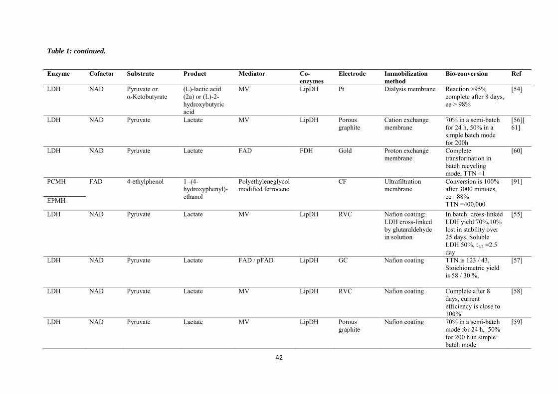
42
Table 1: continued.
Enzyme Cofactor Substrate Product Mediator Co-enzymes
Electrode Immobilization method
Bio-conversion Ref
LDH NAD Pyruvate or α-Ketobutyrate
(L)-lactic acid (2a) or (L)-2- hydroxybutyric acid
MV LipDH Pt Dialysis membrane Reaction >95% complete after 8 days, ee > 98%
[54]
LDH NAD Pyruvate Lactate MV LipDH Porous graphite
Cation exchange membrane
70% in a semi-batch for 24 h, 50% in a simple batch mode for 200h
[56][61]
LDH NAD Pyruvate Lactate FAD FDH Gold Proton exchange membrane
Complete transformation in batch recycling mode, TTN =1
[60]
PCMH FAD 4-ethylphenol
1 -(4-hydroxyphenyl)-ethanol
Polyethyleneglycol modified ferrocene
CF Ultrafiltration membrane
Conversion is 100% after 3000 minutes, ee =88% TTN =400,000
[91]
EPMH
LDH NAD Pyruvate Lactate MV LipDH RVC Nafion coating; LDH cross-linked by glutaraldehyde in solution
In batch: cross-linked LDH yield 70%,10% lost in stability over 25 days. Soluble LDH 50%, t1/2 =2.5 day
[55]
LDH NAD Pyruvate Lactate FAD / pFAD LipDH GC Nafion coating TTN is 123 / 43, Stoichiometric yield is 58 / 30 %,
[57]
LDH NAD Pyruvate Lactate MV LipDH RVC Nafion coating Complete after 8 days, current efficiency is close to 100%
[58]
LDH NAD Pyruvate
Lactate MV LipDH Porous graphite
Nafion coating 70% in a semi-batch mode for 24 h, 50% for 200 h in simple batch mode
[59]
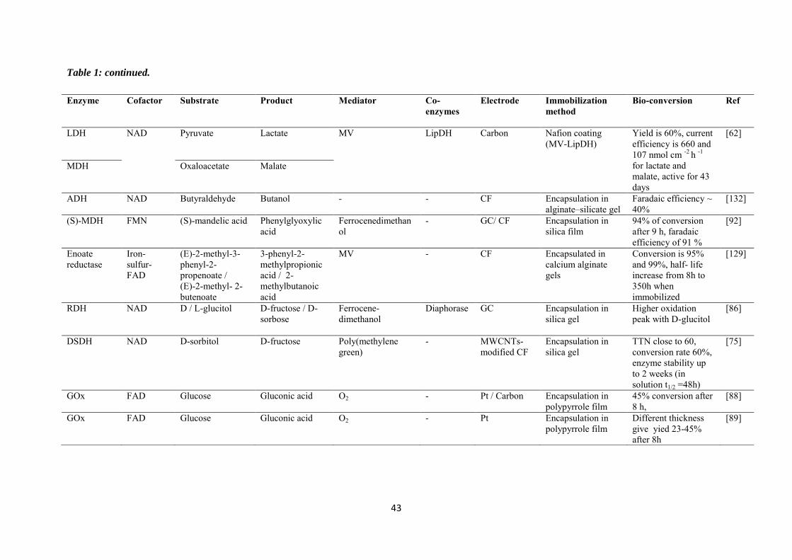
43
Table 1: continued.
Enzyme Cofactor Substrate Product Mediator Co-enzymes
Electrode Immobilization method
Bio-conversion Ref
LDH NAD Pyruvate Lactate MV LipDH Carbon Nafion coating (MV-LipDH)
Yield is 60%, current efficiency is 660 and 107 nmol cm -2 h -1 for lactate and malate, active for 43 days
[62]
MDH Oxaloacetate Malate
ADH NAD Butyraldehyde Butanol - - CF Encapsulation in alginate–silicate gel
Faradaic efficiency ~ 40%
[132]
(S)-MDH FMN (S)-mandelic acid Phenylglyoxylic acid
Ferrocenedimethanol
- GC/ CF Encapsulation in silica film
94% of conversion after 9 h, faradaic efficiency of 91 %
[92]
Enoate reductase
Iron-sulfur-FAD
(E)-2-methyl-3-phenyl-2-propenoate / (E)-2-methyl- 2-butenoate
3-phenyl-2-methylpropionic acid / 2-methylbutanoic acid
MV - CF Encapsulated in calcium alginate gels
Conversion is 95% and 99%, half- life increase from 8h to 350h when immobilized
[129]
RDH NAD D / L-glucitol D-fructose / D-sorbose
Ferrocene-dimethanol
Diaphorase GC Encapsulation in silica gel
Higher oxidation peak with D-glucitol
[86]
DSDH NAD D-sorbitol D-fructose Poly(methylene green)
- MWCNTs-modified CF
Encapsulation in silica gel
TTN close to 60, conversion rate 60%, enzyme stability up to 2 weeks (in solution t1/2 =48h)
[75]
GOx FAD Glucose Gluconic acid O2 - Pt / Carbon Encapsulation in polypyrrole film
45% conversion after 8 h,
[88]
GOx FAD Glucose Gluconic acid O2 - Pt Encapsulation in polypyrrole film
Different thickness give yied 23-45% after 8h
[89]
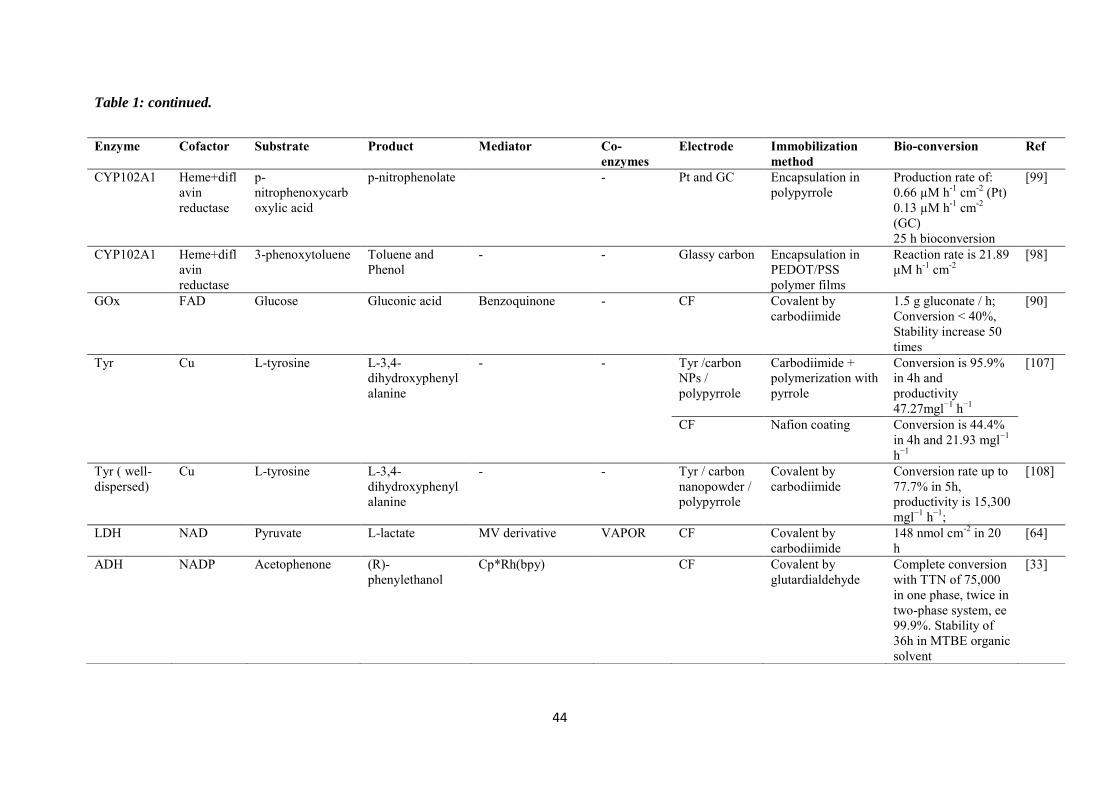
44
Table 1: continued.
Enzyme Cofactor Substrate Product Mediator Co-enzymes
Electrode Immobilization method
Bio-conversion Ref
CYP102A1
Heme+diflavin reductase
p-nitrophenoxycarboxylic acid
p-nitrophenolate - Pt and GC Encapsulation in polypyrrole
Production rate of: 0.66 µM h-1 cm-2 (Pt) 0.13 µM h-1 cm-2 (GC) 25 h bioconversion
[99]
CYP102A1 Heme+diflavin reductase
3-phenoxytoluene Toluene and Phenol
- - Glassy carbon Encapsulation in PEDOT/PSS polymer films
Reaction rate is 21.89 μM h-1 cm-2
[98]
GOx FAD Glucose Gluconic acid Benzoquinone - CF Covalent by carbodiimide
1.5 g gluconate / h; Conversion ˂ 40%, Stability increase 50 times
[90]
Tyr Cu L-tyrosine L-3,4- dihydroxyphenylalanine
- - Tyr /carbon NPs / polypyrrole
Carbodiimide + polymerization with pyrrole
Conversion is 95.9% in 4h and productivity 47.27mgl−1 h−1
[107]
CF Nafion coating Conversion is 44.4% in 4h and 21.93 mgl−1 h−1
Tyr ( well-dispersed)
Cu L-tyrosine L-3,4- dihydroxyphenylalanine
- - Tyr / carbon nanopowder / polypyrrole
Covalent by carbodiimide
Conversion rate up to 77.7% in 5h, productivity is 15,300 mgl−1 h−1;
[108]
LDH NAD Pyruvate L-lactate MV derivative VAPOR CF Covalent by carbodiimide
148 nmol cm-2 in 20 h
[64]
ADH
NADP Acetophenone (R)-phenylethanol
Cp*Rh(bpy) CF Covalent by glutardialdehyde
Complete conversion with TTN of 75,000 in one phase, twice in two-phase system, ee 99.9%. Stability of 36h in MTBE organic solvent
[33]

45
Table 1: continued.
Enzyme Cofactor Substrate Product Mediator Co-enzymes
Electrode Immobilization method
Bio-conversion Ref
AOx FAD Pyruvic acid D-alanine ADPy GC Covalent by glutaraldehyde
ee~100%, current efficiency 97% in 10h, 8.9 mmol dm-3, TTN more than 36000
[93]
CYP1A2 + CYP3A4
Heme Clopidogrel Clopidogrel carboxylic acid
- - Au / chitosan / RGO nanosheets
Covalent, glutaraldehyde+ carbonyldiimide
7.2% after 1h [100]
GDH NAD Glucose Gluconic acid 3,4-DHB - GF Covalent by glutaraldehyde+ carbonyldiimide
TTNmax is 100 after 4h
[78]
G6PDH NAD Glucose 6- phosphate
6-phosphogluconate
PMS - Rotating disc graphite
Covalent by glutaraldehyde or carbonyldiimide
0.016 mol / day, current efficiency is 100%
[76]
LDH NAD Pyruvate Lactate PV LipDH GC Covalent by glutaraldehyde
35% in 62 h, TTN is 38
[63]
ADH NAD 2- and 3-methylcyclohexanone
(1 S, 2S)-( +)-2- and (lS, 3S)-( -)-3-methylcyclohexanol
Methyl viologen Diaphorase GF Polyacrylic acid cross-linking
Yield (49.8 and 50.2%), current efficiency (98.6 and 96.5%) and ee % (100 and 93.1%) in 8 h
[37]
Cys-DSDH NAD D-fructose D-sorbitol [Cp*Rh(bpy)Cl]+ - CF Covalent by „thiol-ene‟ click chemistry
Conversion rate is 72%, faradaic efficiency is 77 %
[146]
Cys- GatDH NAD 1,2-hexanediol Hydroxyacetone 4-carboxy-2,5,7-trinitrofluorenyliden- malonnitrile
- Gold Self-assembled monolayers
Proof of concept with cyclic voltammetry
[77]
CYP2E1, MUT261, MUT268
Heme p-nitrophenol
p-nitrocatechol
- - Gold Self-assembled monolayers+ covalent by (DTME)
1.77 ± 0.64 (P450 2E1); 4.53 ± 0.44 (MUT261); 3.66 ± 0.35 (MUT268) µM
[97]
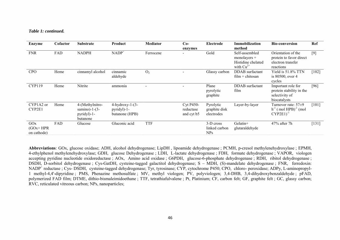
46
Table 1: continued.
Abbreviations: GOx, glucose oxidase; ADH, alcohol dehydrogenase; LipDH , lipoamide dehydrogenase ; PCMH, p-cresol methylenehydroxylase ; EPMH, 4-ethylphenol methylenehydroxylase; GDH, glucose Dehydrogenase ; LDH, L-lactate dehydrogenase ; FDH, formate dehydrogenase ; VAPOR, viologen accepting pyridine nucleotide oxidoreductase ; AOx, Amino acid oxidase ; G6PDH, glucose-6-phosphate dehydrogenase ; RDH, ribitol dehydrogenase ; DSDH, D-sorbitol dehydrogenase ; Cys-GatDH, cysteine-tagged galactitol dehydrogenase; S – MDH, (S)-mandelate dehydrogenase ; FNR, ferredoxin: NADP+ reductase ; Cys- DSDH, cysteine-tagged dehydrogenase; Tyr, tyrosinase; CYP, cytochrome P450; CPO, chloro- peroxidase; ADPy, L-aminopropyl-1 methyl-4,4'-dipyridine ; PMS, Phenazine methosulfate ; MV, methyl viologen; PV, polyviologen; 3,4-DHB, 3,4-dihydroxybenzaldehyde ; pFAD, polymerized FAD film; DTME, dithio-bismaleimidoethane ; TTF, tetrathiafulvalene ; Pt, Platinium; CF, carbon felt; GF, graphite felt ; GC, glassy carbon; RVC, reticulated vitreous carbon; NPs, nanoparticles;
Enzyme Cofactor Substrate Product Mediator Co-enzymes
Electrode Immobilization method
Bio-conversion Ref
FNR
FAD NADPH NADP+ Ferrocene - Gold Self-assembled monolayers + Histidine chelated with Cu2+
Orientation of the protein to favor direct electron transfer reactions
[9]
CPO Heme cinnamyl alcohol cinnamic aldehyde
O2 - Glassy carbon DDAB surfactant film + chitosan
Yield is 51.8% TTN is 80500, over 4 cycles
[102]
CYP119 Heme Nitrite ammonia - - Plane pyrolytic graphite
DDAB surfactant film
Important role for protein stability in the selectivity of biocatalysts
[96]
CYP1A2 or CYP2E1
Heme 4-(Methylnitro- samino)-1-(3-pyridyl)-1-butanone
4-hydroxy-1-(3-pyridyl)-1- butanone (HPB)
Cyt P450-reductase and cyt b5
Pyrolytic graphite disk electrodes
Layer-by-layer Turnover rate: 57±9 h-1 ( mol HPB)-1 (mol CYP2E1)-1
[101]
GOx (GOx+ HPR on cathode)
FAD Glucose Gluconic acid TTF 3-D cross linked carbon NPs
Gelatin+ glutaraldehyde
47% after 7h [131]
47
Chapter 2. Immobilization of cysteine-tagged proteins on electrode surfaces by thiol-ene click
chemistry
Thiol-ene click chemistry can be exploited for the immobilization of cysteine-tagged
dehydrogenases in an active form onto carbon electrodes (glassy carbon and carbon felt). The
electrode surfaces have been first modified with vinylphenyl groups by electrochemical
reduction of the corresponding diazonium salts generated in situ from 4-vinylaniline. The
grafting process has been optimized in order to not hinder the electrochemical regeneration of
NAD+/NADH cofactor and soluble mediators such as ferrocenedimethanol and
[Cp*Rh(bpy)Cl]+. Having demonstrated the feasibility of thiol-ene click chemistry for
attaching ferrocene moieties onto those carbon surfaces, the same approach was then applied
to the immobilization of D-sorbitol dehydrogenases with cysteine tag. These proteins can be
effectively immobilized (as pointed out by XPS), and the cysteine tag (either 1 or 2 cysteine
moieties at the N terminus of the polypeptide chain) was proven to maintain the enzymatic
activity of the dehydrogenase upon grafting. The bioelectrode was applied to
electroenzymatic enantioselective reduction of D-fructose to D- sorbitol, as a case study.
48
2.1 Introduction
In the field of sustainable chemistry, the exploitation of enzymes, which are likely to
catalyze chemical reactions with high regio- and stereoselectivity, appears to be a powerful
strategy to develop bioprocesses targeted to the synthesis of molecules for food industry,
pharmacy, and cosmetics, etc [147,148]. For instance, the NADH-dependent redox proteins
belonging to the dehydrogenases family can be used for the enantioselective reduction of
ketone to alcohol [4], providing that regeneration of the enzymatic cofactor is ensured (e.g.,
by NAD+ reduction to NADH) [149]. The effectiveness of such bioelectrochemical processes
implies a durable immobilization of the protein in an active form to guarantee its long-term
operational stability in the bioreactor [150], as well as efficient electrochemical regeneration
of the catalyst, either by direct or mediated electron transfer [17]. Among the various methods
reported for protein immobilization [150], a recent approach is based on the specific
interaction between histidine-tagged proteins engineered by molecular biology [151] and
porous materials holding divalent metal complexes [152]. Examples are available for enzyme
immobilization on mesoporous silicates [153–155] or electrode surfaces [152], including
controlled orientation [156], while keeping their catalytic activity [152,155]. The histidine
tag-metal complex being reversible [157], a significant portion of the proteins can be lost
upon prolonged use in solution. Attempting to circumvent this limitation, some authors
suggested strengthening the enzyme-support linkage, e.g., by exploiting the thiol-gold
interaction for attaching dehydrogenases or laccase to gold electrodes via their cysteine
residues [77,158]. If such immobilized proteins operate in a satisfactory way in oxidation,
they can, however, suffer from poor long-term stability when used for reduction reactions
because of possible reductive desorption of species chemisorbed on gold via thiol moieties
[159,160]. This is particularly critical when considering electroenzymatic synthesis with
NAD-dependent dehydrogenases in the reduction mode, which requires the regeneration of
the enzymatic catalyst at a potential more cathodic than the standard potential of the cofactor
redox couple, i.e., -0.59 V vs SCE for NAD+/NADH [161]. Going one step further to tackle
these drawbacks would be the development of a method for covalent binding of cysteine-
tagged proteins to other electrode materials, such as carbon surfaces, and this is what we plan
to perform here by combining electrografting and click chemistry. Several click reactions are
now well-established in chemical synthesis [162], materials science [163], and for the
preparation of peptide conjugates [164]. The modification of proteins with azide groups was,
for example, used to immobilize horseradish peroxidase on gold [165] or screen-printed
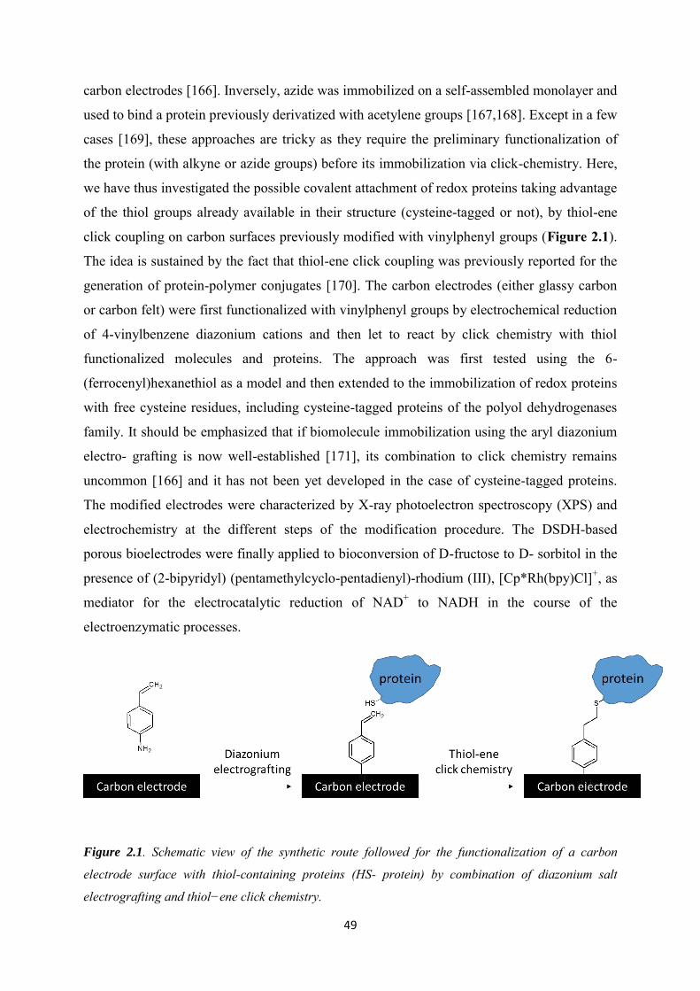
49
carbon electrodes [166]. Inversely, azide was immobilized on a self-assembled monolayer and
used to bind a protein previously derivatized with acetylene groups [167,168]. Except in a few
cases [169], these approaches are tricky as they require the preliminary functionalization of
the protein (with alkyne or azide groups) before its immobilization via click-chemistry. Here,
we have thus investigated the possible covalent attachment of redox proteins taking advantage
of the thiol groups already available in their structure (cysteine-tagged or not), by thiol-ene
click coupling on carbon surfaces previously modified with vinylphenyl groups (Figure 2.1).
The idea is sustained by the fact that thiol-ene click coupling was previously reported for the
generation of protein-polymer conjugates [170]. The carbon electrodes (either glassy carbon
or carbon felt) were first functionalized with vinylphenyl groups by electrochemical reduction
of 4-vinylbenzene diazonium cations and then let to react by click chemistry with thiol
functionalized molecules and proteins. The approach was first tested using the 6-
(ferrocenyl)hexanethiol as a model and then extended to the immobilization of redox proteins
with free cysteine residues, including cysteine-tagged proteins of the polyol dehydrogenases
family. It should be emphasized that if biomolecule immobilization using the aryl diazonium
electro- grafting is now well-established [171], its combination to click chemistry remains
uncommon [166] and it has not been yet developed in the case of cysteine-tagged proteins.
The modified electrodes were characterized by X-ray photoelectron spectroscopy (XPS) and
electrochemistry at the different steps of the modification procedure. The DSDH-based
porous bioelectrodes were finally applied to bioconversion of D-fructose to D- sorbitol in the
presence of (2-bipyridyl) (pentamethylcyclo-pentadienyl)-rhodium (III), [Cp*Rh(bpy)Cl]+, as
mediator for the electrocatalytic reduction of NAD+ to NADH in the course of the
electroenzymatic processes.
Figure 2.1. Schematic view of the synthetic route followed for the functionalization of a carbon
electrode surface with thiol-containing proteins (HS- protein) by combination of diazonium salt
electrografting and thiol−ene click chemistry.
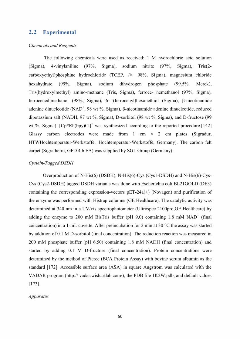
50
2.2 Experimental
Chemicals and Reagents
The following chemicals were used as received: 1 M hydrochloric acid solution
(Sigma), 4-vinylaniline (97%, Sigma), sodium nitrite (97%, Sigma), Tris(2-
carboxyethyl)phosphine hydrochloride (TCEP, ≥ 98%, Sigma), magnesium chloride
hexahydrate (99%, Sigma), sodium dihydrogen phosphate (99.5%, Merck),
Tris(hydroxylmethyl) amino-methane (Tris, Sigma), ferroce- nemethanol (97%, Sigma),
ferrocenedimethanol (98%, Sigma), 6- (ferrocenyl)hexanethiol (Sigma), β-nicotinamide
adenine dinucleotide (NAD+, 98 wt %, Sigma), β-nicotinamide adenine dinucleotide, reduced
dipotassium salt (NADH, 97 wt %, Sigma), D-sorbitol (98 wt %, Sigma), and D-fructose (99
wt %, Sigma). [Cp*Rh(bpy)Cl]+ was synthesized according to the reported procedure.[142]
Glassy carbon electrodes were made from 1 cm × 2 cm plates (Sigradur,
HTWHochtemperatur-Werkstoffe, Hochtemperatur-Werkstoffe, Germany). The carbon felt
carpet (Sigratherm, GFD 4.6 EA) was supplied by SGL Group (Germany).
Cystein-Tagged DSDH
Overproduction of N-His(6) (DSDH), N-His(6)-Cys (Cys1-DSDH) and N-His(6)-Cys-
Cys (Cys2-DSDH) tagged DSDH variants was done with Escherichia coli BL21GOLD (DE3)
containing the corresponding expression-vectors pET-24a(+) (Novagen) and purification of
the enzyme was performed with Histrap columns (GE Healthcare). The catalytic activity was
determined at 340 nm in a UV/vis spectrophotometer (Ultrospec 2100pro,GE Healthcare) by
adding the enzyme to 200 mM BisTris buffer (pH 9.0) containing 1.8 mM NAD+ (final
concentration) in a 1-mL cuvette. After preincubation for 2 min at 30 °C the assay was started
by addition of 0.1 M D-sorbitol (final concentration). The reduction reaction was measured in
200 mM phosphate buffer (pH 6.50) containing 1.8 mM NADH (final concentration) and
started by adding 0.1 M D-fructose (final concentration). Protein concentrations were
determined by the method of Pierce (BCA Protein Assay) with bovine serum albumin as the
standard [172]. Accessible surface area (ASA) in square Angstrom was calculated with the
VADAR program (http:// vadar.wishartlab.com/), the PDB file 1K2W.pdb, and default values
[173].
Apparatus

51
All electrochemical experiments were carried out using an Autolab PGSTAT-12
potentiostat, with a three-electrode configuration cell including an Ag/AgCl reference
electrode (3 M KCl), a steel auxiliary electrode, and a carbon working electrode (glassy
carbon plate or porous carbon felt). A pencil core (0.5 mm diameter) glued with silver paint to
a copper wire was used for connecting the carbon felt working electrode. XPS measurements
were performed using the Kratos AxisUltra DLD apparatus equipped with a monochromatic
Al Kα source, aiming to prove the occurrence of the thiol-ene click reaction. Three glassy
carbon electrodes were modified by 4-vinylphenyl diazonium cations by applying -0.6 V for 2,
300, and 1200 s, respectively, in order to get increasing surface density of vinylphenyl groups
for reacting with 6-(ferrocenyl)hexanethiol. Meanwhile, two glassy carbon electrodes
modified by 4-vinylphenyl diazonium cations by applying -0.6 V for 600 s were prepared
forreacting with Cys1-DSDH or DSDH. Samples of the conversion were taken after different
periods and analyzed by HPLC using a Ca2+-column (Rezex RCM Mono- saccharide Ca2+
300 × 7.8 mm, Phenomenex) at 80 °C, with distilled water as the mobile phase and a flow rate
of 0.5 mL min-1. Detection was done with a refractive index-detector at 35 °C and calibrations
were performed with authentic standard substances.
Preparation and Pretreatment of Carbon Electrodes
Glassy carbon (GC) plates were wet-polished with SiC grinding paper (#4000, Struers,
Denmark) for 1 min, then cleaned with ethanol and distilled water under ultrasonic treatment.
Carbon felt (CF) electrodes were cut with size of 5 mm × 5 mm from the carbon felt carpet.
The surface of CF was then activated via cycling potentials over the range between -0.7 and
+2.5 V (20 times) in 0.1 M H2SO4, in order to modify the surface state from hydrophobic to
hydrophilic. After this, the carbon felt was rinsed with water, followed by heat treatment at
200 °C for 1 h to remove the residual H2SO4.
Functionalization of Electrodes with Vinylphenyl Moieties by Electrografting
The diazonium cations were generated in situ by mixing 5 mM 4-vinylaniline and 10
mM sodium nitrite in 0.5 M HCl water solution under stirring at room temperature for 5 min,
yielding the 4-vinylbenzene diazonium salt quantitatively. The electrografting on GC was
carried out by cyclic voltammetry (consecutive reduction scan with the potential varying from
0 to -0.45 V). However, the electrografting on CF electrode was realized by applying -0.6 V
for 1 s. After electrografting, the electrodes were rinsed with distilled water to remove the
residual ungrafted compounds.

52
Functionalization of Electrodes with Ferrocene Moieties
Vinylphenyl functionalized electrodes were immersed into 1 mM of 6-
(ferrocenyl)hexanethiol in H2O/methanol (3/7) solution in the presence 2 mM TCEP used as
catalyst for the reaction between alkene moieties and terminal thiol functionalities on
ferrocene via thiol-ene “click chemistry”. After reaction overnight, the electrode was
thoroughly rinsed in THF and then in water to remove the possible residual 6-
(ferrocenyl)hexanethiol remaining on the electrode, and subsequently dried.
Functionalization of Electrodes with Proteins
Vinylphenyl functionalized electrodes were immersed into 1 mL of aqueous PBS
buffer (250 mM, pH = 7) containing 2.5 mg of proteins, 1 mM MgCl2, and 0.3 mM TCEP.
After reaction overnight at 4 °C, the electrodes were rinsed with water and buffer solution.
2.3 Results and discussions
2.3.1 Immobilization of ferrocene moieties on glassy carbon electrode
A two-step protocol was employed to get the modified carbon surfaces, i.e., the
electrografting of vinylphenyl groups and then the thiol-ene click coupling with proteins
bearing thiol groups. The feasibility of the sequential process was first demonstrated on a
glassy carbon electrode with 6-(ferrocenyl)hexanethiol moieties (see scheme in Figure 2.2).
Electrochemical reduction of vinylaniline diazonium salts allowed the formation of
arylvinyl radicals able to form a covalent bond on the electrode surface that will be available
for the thiol-ene coupling with ferrocene moieties employed as a model probe. 4-Vinylaniline
was let to react with two molar equivalents of sodium nitrite in 0.5 M HCl to form the
corresponding diazonium cations in situ as reported in previous work [174]. The solution was
subsequently used without any treatment to modify the GC plate by recording two
consecutive cathodic scans in a potential range varying from 0 to -0.45 V at 100 mV s-1
(Figure 2.3A).
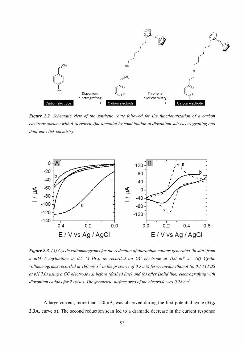
53
Figure 2.2. Schematic view of the synthetic route followed for the functionalization of a carbon
electrode surface with 6-(ferrocenyl)hexanethiol by combination of diazonium salt electrografting and
thiol-ene click chemistry.
Figure 2.3. (A) Cyclic voltammograms for the reduction of diazonium cations generated ‘in situ’ from
5 mM 4-vinylaniline in 0.5 M HCl, as recorded on GC electrode at 100 mV s-1. (B) Cyclic
voltammograms recorded at 100 mV s-1 in the presence of 0.5 mM ferrocenedimethanol (in 0.1 M PBS
at pH 7.0) using a GC electrode (a) before (dashed line) and (b) after (solid line) electrografting with
diazonium cations for 2 cycles. The geometric surface area of the electrode was 0.28 cm2.
A large current, more than 120 μA, was observed during the first potential cycle (Fig.
2.3A, curve a). The second reduction scan led to a dramatic decrease in the current response

54
(Fig. 2.3A, curve b). This decrease can be explained by the partial blocking of the surface
after grafting of vinylphenyl moieties on the carbon surface. The presence of the organic layer
on the carbon electrode was further evidenced by the comparison of the cyclic voltammogram
of 0.5 mM ferrocenedimethanol recorded on the mentioned GC electrode before (curve a) and
after electrografting (curve b) of the arylvinyl radical species. The blocking effect of the
organic layer was demonstrated by an increase of the difference between anodic and cathodic
peaks (∆Ep varying from 91 to 300 mV) indicative of much slower electron transfer rates on
the modified electrodes (Fig. 2.3B). In addition, a significant decrease of the current peaks
was also observed. The electrochemical reduction of the diazonium cations generated
vinylphenyl radicals that can be immobilized on the glassy carbon surface by means of a
covalent bond.
Figure 2.4. (A) Cyclic voltammograms recorded at a potential scan rate of 100 mV s−1 in 0.1 M Tris-
HCl buffer (pH 9.0) using vinylphenyl functionalized GC electrodes after reaction with (a) 6-
(ferrocenyl)- hexanethiol (dashed line) and (b) ferrocenemethanol (solid line). (B) The 2nd, 20th, 60th,
and 80th curves recorded in the same conditions as above in the course of 80 successive voltammetric
cycles for ferrocene functionalized glassy carbon electrode (prepared from clicking 6-
(ferrocenyl)hexanethiol). The geometric surface area of the electrode was 0.28 cm2.
This vinylphenyl-functionalized surface was then used to evaluate the possibility to
attach ferrocene moieties by thiol-ene reaction. The electrode was let to react overnight with
6-(hexanethiol)ferrocene in the presence of TCEP and Mg2+ ions as reported in the literature
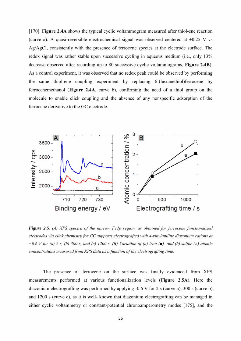
55
[170]. Figure 2.4A shows the typical cyclic voltammogram measured after thiol-ene reaction
(curve a). A quasi-reversible electrochemical signal was observed centered at +0.25 V vs
Ag/AgCl, consistently with the presence of ferrocene species at the electrode surface. The
redox signal was rather stable upon successive cycling in aqueous medium (i.e., only 13%
decrease observed after recording up to 80 successive cyclic voltammograms, Figure 2.4B).
As a control experiment, it was observed that no redox peak could be observed by performing
the same thiol-ene coupling experiment by replacing 6-(hexanethiol)ferrocene by
ferrocenemethanol (Figure 2.4A, curve b), confirming the need of a thiol group on the
molecule to enable click coupling and the absence of any nonspecific adsorption of the
ferrocene derivative to the GC electrode.
Figure 2.5. (A) XPS spectra of the narrow Fe2p region, as obtained for ferrocene functionalized
electrodes via click chemistry for GC supports electrografted with 4-vinylaniline diazonium cations at
−0.6 V for (a) 2 s, (b) 300 s, and (c) 1200 s. (B) Variation of (a) iron (■) and (b) sulfur (○) atomic
concentrations measured from XPS data as a function of the electrografting time.
The presence of ferrocene on the surface was finally evidenced from XPS
measurements performed at various functionalization levels (Figure 2.5A). Here the
diazonium electrografting was performed by applying -0.6 V for 2 s (curve a), 300 s (curve b),
and 1200 s (curve c), as it is well- known that diazonium electrografting can be managed in
either cyclic voltammetry or constant-potential chronoamperometry modes [175], and the
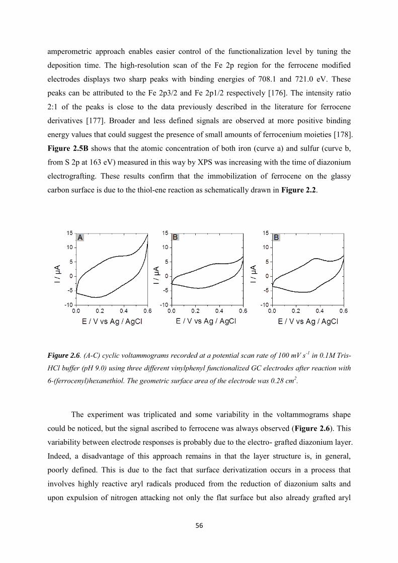
56
amperometric approach enables easier control of the functionalization level by tuning the
deposition time. The high-resolution scan of the Fe 2p region for the ferrocene modified
electrodes displays two sharp peaks with binding energies of 708.1 and 721.0 eV. These
peaks can be attributed to the Fe 2p3/2 and Fe 2p1/2 respectively [176]. The intensity ratio
2:1 of the peaks is close to the data previously described in the literature for ferrocene
derivatives [177]. Broader and less defined signals are observed at more positive binding
energy values that could suggest the presence of small amounts of ferrocenium moieties [178].
Figure 2.5B shows that the atomic concentration of both iron (curve a) and sulfur (curve b,
from S 2p at 163 eV) measured in this way by XPS was increasing with the time of diazonium
electrografting. These results confirm that the immobilization of ferrocene on the glassy
carbon surface is due to the thiol-ene reaction as schematically drawn in Figure 2.2.
Figure 2.6. (A-C) cyclic voltammograms recorded at a potential scan rate of 100 mV s-1 in 0.1M Tris-
HCl buffer (pH 9.0) using three different vinylphenyl functionalized GC electrodes after reaction with
6-(ferrocenyl)hexanethiol. The geometric surface area of the electrode was 0.28 cm2.
The experiment was triplicated and some variability in the voltammograms shape
could be noticed, but the signal ascribed to ferrocene was always observed (Figure 2.6). This
variability between electrode responses is probably due to the electro- grafted diazonium layer.
Indeed, a disadvantage of this approach remains in that the layer structure is, in general,
poorly defined. This is due to the fact that surface derivatization occurs in a process that
involves highly reactive aryl radicals produced from the reduction of diazonium salts and
upon expulsion of nitrogen attacking not only the flat surface but also already grafted aryl
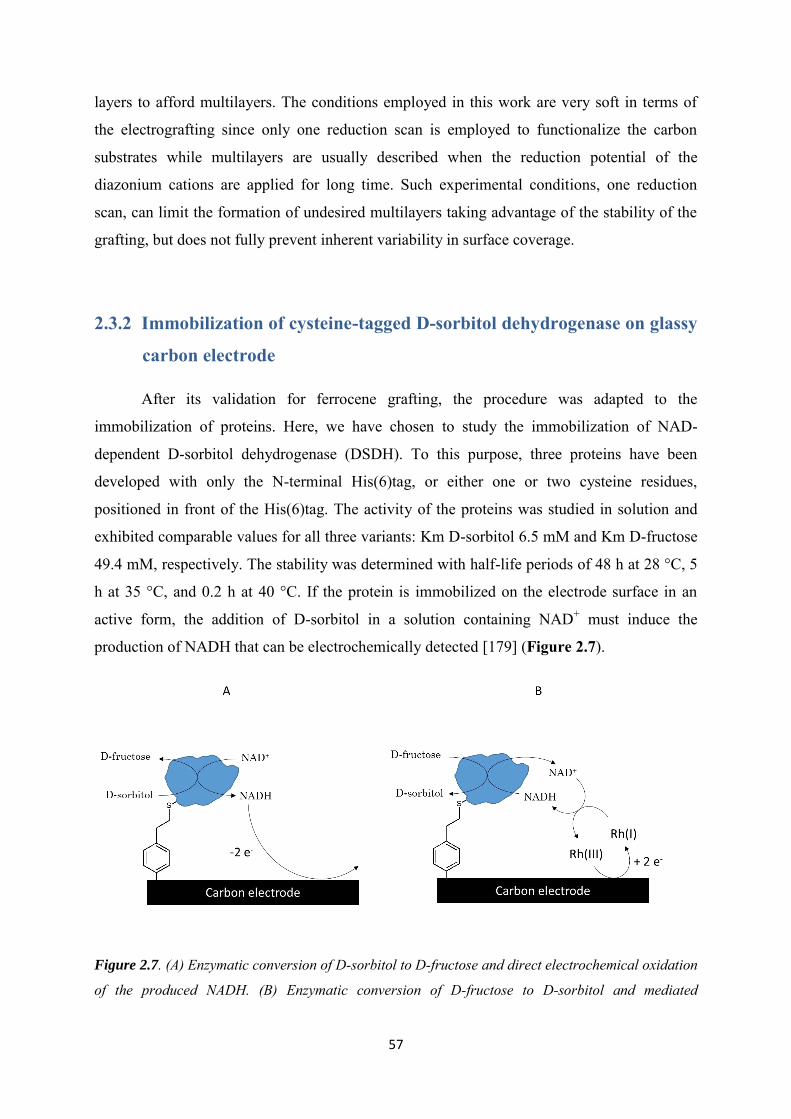
57
layers to afford multilayers. The conditions employed in this work are very soft in terms of
the electrografting since only one reduction scan is employed to functionalize the carbon
substrates while multilayers are usually described when the reduction potential of the
diazonium cations are applied for long time. Such experimental conditions, one reduction
scan, can limit the formation of undesired multilayers taking advantage of the stability of the
grafting, but does not fully prevent inherent variability in surface coverage.
2.3.2 Immobilization of cysteine-tagged D-sorbitol dehydrogenase on glassy
carbon electrode
After its validation for ferrocene grafting, the procedure was adapted to the
immobilization of proteins. Here, we have chosen to study the immobilization of NAD-
dependent D-sorbitol dehydrogenase (DSDH). To this purpose, three proteins have been
developed with only the N-terminal His(6)tag, or either one or two cysteine residues,
positioned in front of the His(6)tag. The activity of the proteins was studied in solution and
exhibited comparable values for all three variants: Km D-sorbitol 6.5 mM and Km D-fructose
49.4 mM, respectively. The stability was determined with half-life periods of 48 h at 28 °C, 5
h at 35 °C, and 0.2 h at 40 °C. If the protein is immobilized on the electrode surface in an
active form, the addition of D-sorbitol in a solution containing NAD+ must induce the
production of NADH that can be electrochemically detected [179] (Figure 2.7).
Figure 2.7. (A) Enzymatic conversion of D-sorbitol to D-fructose and direct electrochemical oxidation
of the produced NADH. (B) Enzymatic conversion of D-fructose to D-sorbitol and mediated
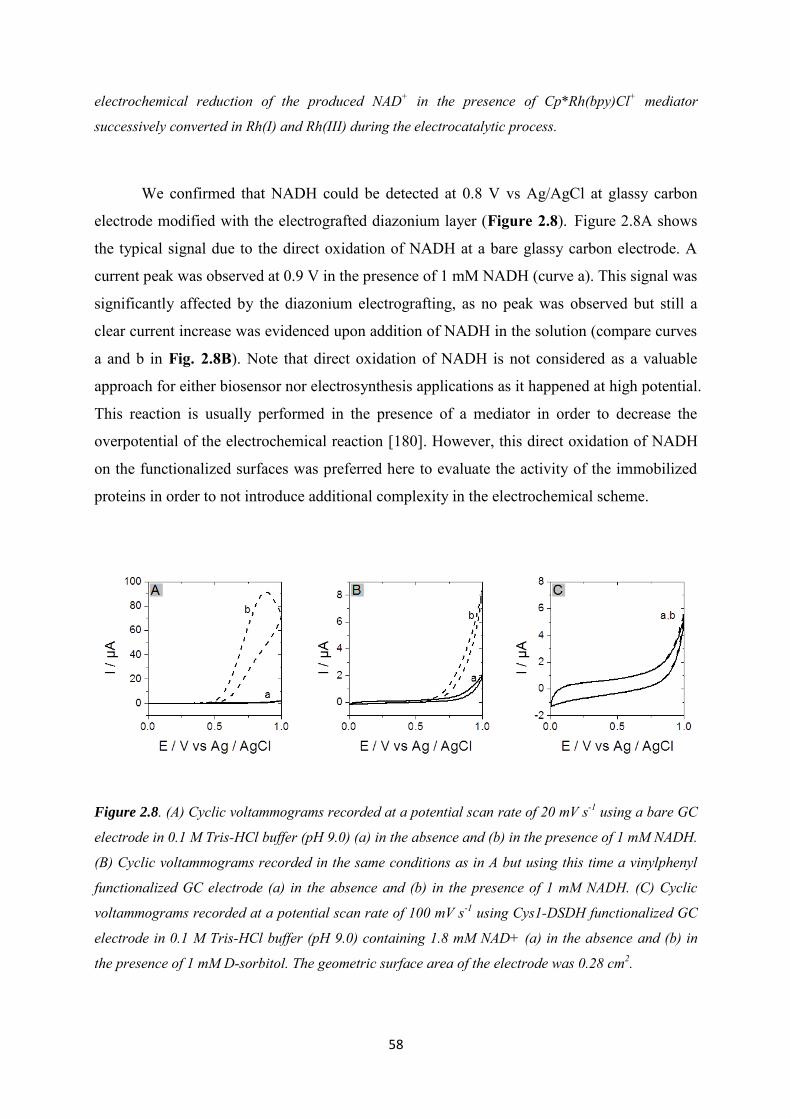
58
electrochemical reduction of the produced NAD+ in the presence of Cp*Rh(bpy)Cl+ mediator
successively converted in Rh(I) and Rh(III) during the electrocatalytic process.
We confirmed that NADH could be detected at 0.8 V vs Ag/AgCl at glassy carbon
electrode modified with the electrografted diazonium layer (Figure 2.8). Figure 2.8A shows
the typical signal due to the direct oxidation of NADH at a bare glassy carbon electrode. A
current peak was observed at 0.9 V in the presence of 1 mM NADH (curve a). This signal was
significantly affected by the diazonium electrografting, as no peak was observed but still a
clear current increase was evidenced upon addition of NADH in the solution (compare curves
a and b in Fig. 2.8B). Note that direct oxidation of NADH is not considered as a valuable
approach for either biosensor nor electrosynthesis applications as it happened at high potential.
This reaction is usually performed in the presence of a mediator in order to decrease the
overpotential of the electrochemical reaction [180]. However, this direct oxidation of NADH
on the functionalized surfaces was preferred here to evaluate the activity of the immobilized
proteins in order to not introduce additional complexity in the electrochemical scheme.
Figure 2.8. (A) Cyclic voltammograms recorded at a potential scan rate of 20 mV s-1 using a bare GC
electrode in 0.1 M Tris-HCl buffer (pH 9.0) (a) in the absence and (b) in the presence of 1 mM NADH.
(B) Cyclic voltammograms recorded in the same conditions as in A but using this time a vinylphenyl
functionalized GC electrode (a) in the absence and (b) in the presence of 1 mM NADH. (C) Cyclic
voltammograms recorded at a potential scan rate of 100 mV s-1 using Cys1-DSDH functionalized GC
electrode in 0.1 M Tris-HCl buffer (pH 9.0) containing 1.8 mM NAD+ (a) in the absence and (b) in
the presence of 1 mM D-sorbitol. The geometric surface area of the electrode was 0.28 cm2.
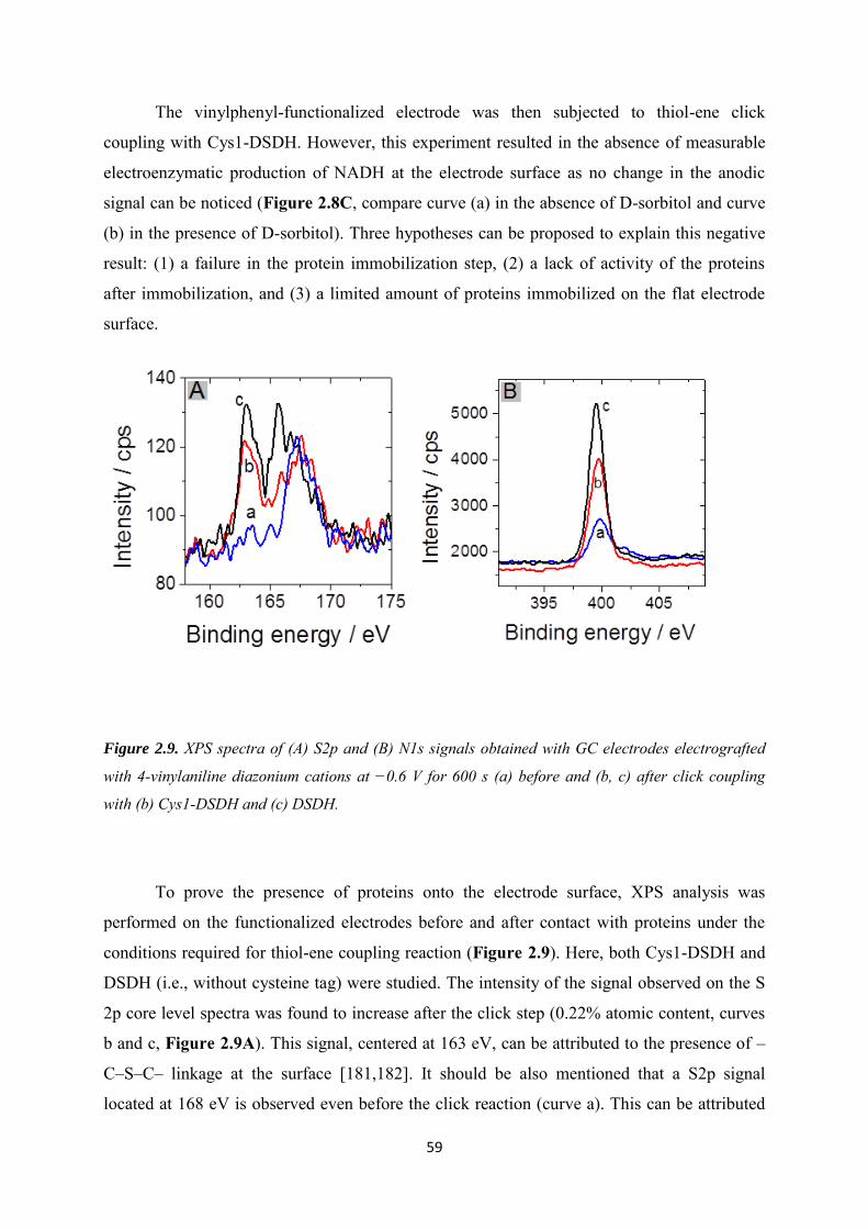
59
The vinylphenyl-functionalized electrode was then subjected to thiol-ene click
coupling with Cys1-DSDH. However, this experiment resulted in the absence of measurable
electroenzymatic production of NADH at the electrode surface as no change in the anodic
signal can be noticed (Figure 2.8C, compare curve (a) in the absence of D-sorbitol and curve
(b) in the presence of D-sorbitol). Three hypotheses can be proposed to explain this negative
result: (1) a failure in the protein immobilization step, (2) a lack of activity of the proteins
after immobilization, and (3) a limited amount of proteins immobilized on the flat electrode
surface.
Figure 2.9. XPS spectra of (A) S2p and (B) N1s signals obtained with GC electrodes electrografted
with 4-vinylaniline diazonium cations at −0.6 V for 600 s (a) before and (b, c) after click coupling
with (b) Cys1-DSDH and (c) DSDH.
To prove the presence of proteins onto the electrode surface, XPS analysis was
performed on the functionalized electrodes before and after contact with proteins under the
conditions required for thiol-ene coupling reaction (Figure 2.9). Here, both Cys1-DSDH and
DSDH (i.e., without cysteine tag) were studied. The intensity of the signal observed on the S
2p core level spectra was found to increase after the click step (0.22% atomic content, curves
b and c, Figure 2.9A). This signal, centered at 163 eV, can be attributed to the presence of –
C–S–C– linkage at the surface [181,182]. It should be also mentioned that a S2p signal
located at 168 eV is observed even before the click reaction (curve a). This can be attributed
60
to the presence of SO42- traces (coming from the sodium nitrite used to generate the
diazonium cations from the 4-vinylaniline since the electrografting step is carried out using
and excess of the nitrite source, in order to ensure quantitative conversion into the diazonium
salt, and without separation of the final product). In addition, analysis of the high-resolution
core level spectra for N 1s shows a peak located at 400 eV after electrografting of the
diazonium salt attributed to the possible presence of –C-N=N-C- linkage (Figure 2.9B). The
atomic content of nitrogen on the surface detected at 399.6 eV was found to increase by
reaction with the two proteins, from 4% before (curve a) to 9% (Cys1-DSDH) or 11% (DSDH)
after contact with the proteins. Overall, the clear increase of the nitrogen and sulfur contents
after reaction with the proteins must result from their adsorption/attachment onto the electrode
surface. These results confirm that proteins are really present on the surface of glassy carbon,
but there is no clear evidence of enzyme attachment via the cysteine tag. Indeed, in the
absence of cysteine tag, any cysteine residues from the protein [183,184] could react with the
vinyl moieties in a comparable way as reported for cytochrome c with maleimide [185]. The
structure of DSDH is shown in Figure 2.10 and the positions of cysteine residues are pointed
out. The only cysteine, which might have access to vinylphenyl, would be Cys54 with
accessible surface areas (ASA) of 37.9 or 43.5 square Angstrom, respectively. Because there
is no crystal structure of the protein with the His(6)-Cys(2)tag, ASA values for these cysteines
cannot be given. However, the ASA of the N-terminal methionine is calculated for both
chains as 106.1 or 108.5, respectively, assuming that the surface areas of the free accessible
cysteines in the tag are probably even larger and therefore, click-reaction with these thiol
groups should be highly preferred. We will show in the next section that there exists indeed
an advantage of the cysteine tag for the immobilization of proteins on the electrode surface.
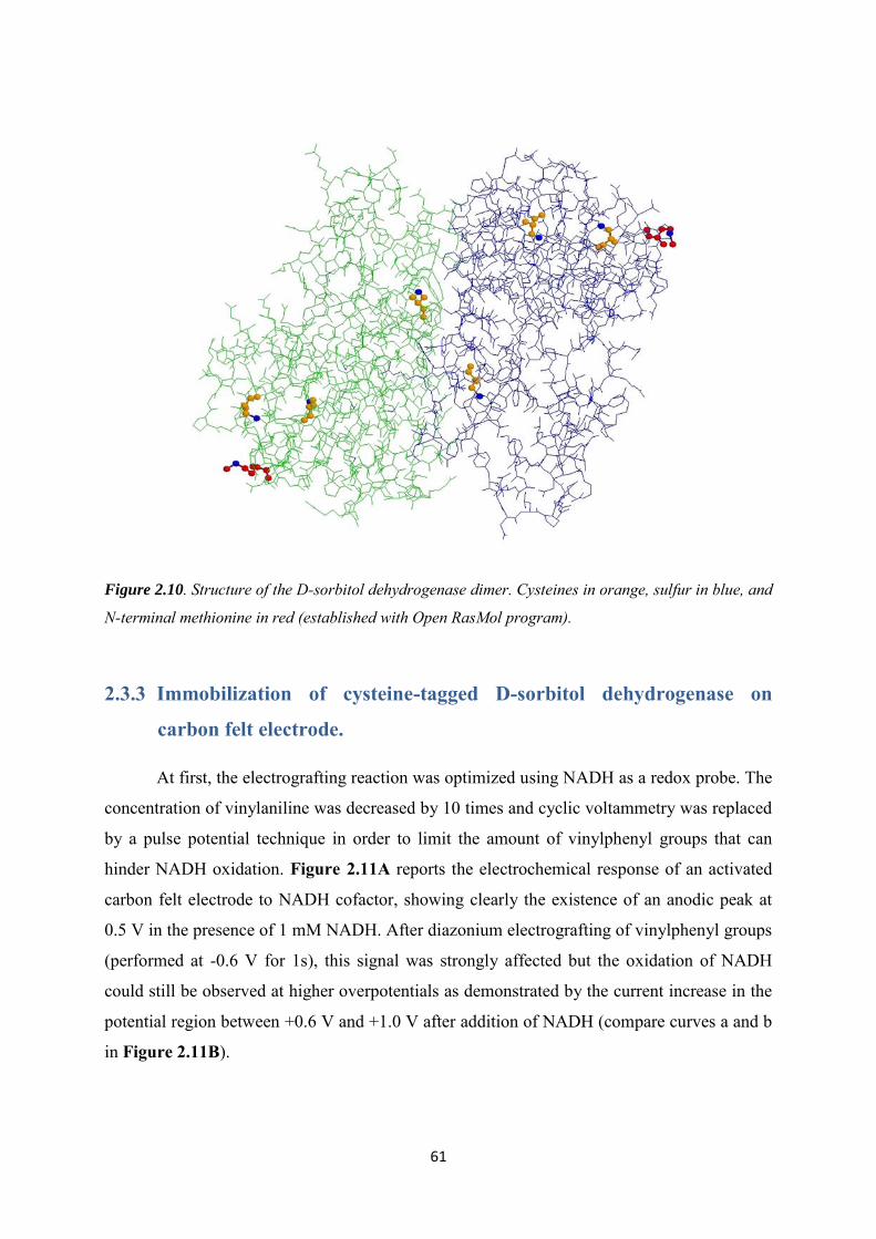
61
Figure 2.10. Structure of the D-sorbitol dehydrogenase dimer. Cysteines in orange, sulfur in blue, and
N-terminal methionine in red (established with Open RasMol program).
2.3.3 Immobilization of cysteine-tagged D-sorbitol dehydrogenase on
carbon felt electrode.
At first, the electrografting reaction was optimized using NADH as a redox probe. The
concentration of vinylaniline was decreased by 10 times and cyclic voltammetry was replaced
by a pulse potential technique in order to limit the amount of vinylphenyl groups that can
hinder NADH oxidation. Figure 2.11A reports the electrochemical response of an activated
carbon felt electrode to NADH cofactor, showing clearly the existence of an anodic peak at
0.5 V in the presence of 1 mM NADH. After diazonium electrografting of vinylphenyl groups
(performed at -0.6 V for 1s), this signal was strongly affected but the oxidation of NADH
could still be observed at higher overpotentials as demonstrated by the current increase in the
potential region between +0.6 V and +1.0 V after addition of NADH (compare curves a and b
in Figure 2.11B).
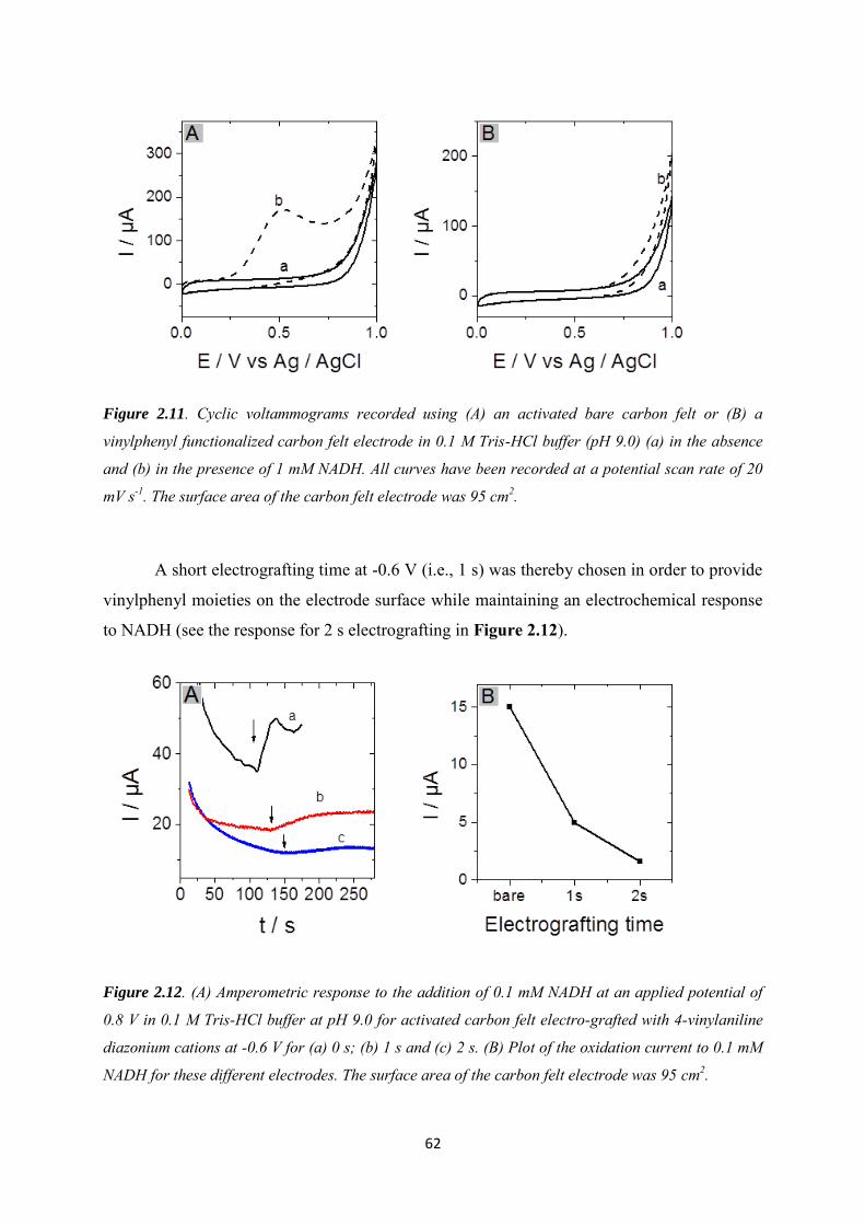
62
Figure 2.11. Cyclic voltammograms recorded using (A) an activated bare carbon felt or (B) a
vinylphenyl functionalized carbon felt electrode in 0.1 M Tris-HCl buffer (pH 9.0) (a) in the absence
and (b) in the presence of 1 mM NADH. All curves have been recorded at a potential scan rate of 20
mV s-1. The surface area of the carbon felt electrode was 95 cm2.
A short electrografting time at -0.6 V (i.e., 1 s) was thereby chosen in order to provide
vinylphenyl moieties on the electrode surface while maintaining an electrochemical response
to NADH (see the response for 2 s electrografting in Figure 2.12).
Figure 2.12. (A) Amperometric response to the addition of 0.1 mM NADH at an applied potential of
0.8 V in 0.1 M Tris-HCl buffer at pH 9.0 for activated carbon felt electro-grafted with 4-vinylaniline
diazonium cations at -0.6 V for (a) 0 s; (b) 1 s and (c) 2 s. (B) Plot of the oxidation current to 0.1 mM
NADH for these different electrodes. The surface area of the carbon felt electrode was 95 cm2.
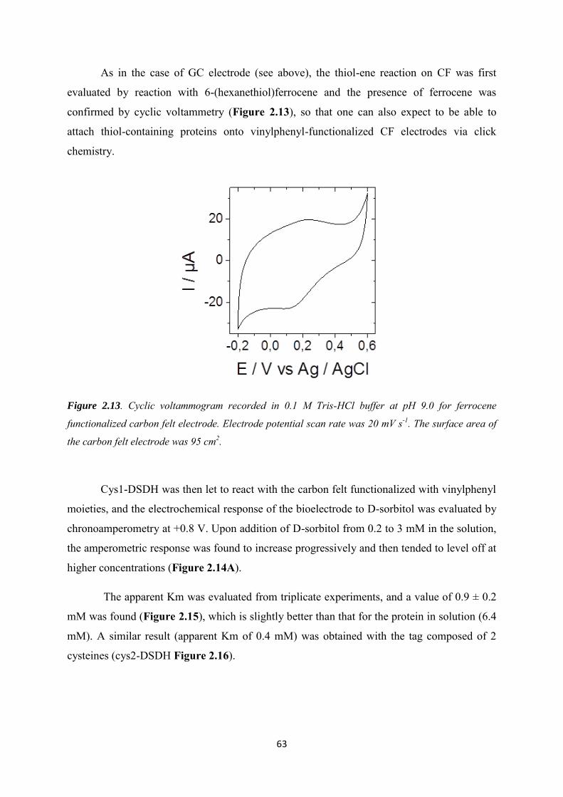
63
As in the case of GC electrode (see above), the thiol-ene reaction on CF was first
evaluated by reaction with 6-(hexanethiol)ferrocene and the presence of ferrocene was
confirmed by cyclic voltammetry (Figure 2.13), so that one can also expect to be able to
attach thiol-containing proteins onto vinylphenyl-functionalized CF electrodes via click
chemistry.
Figure 2.13. Cyclic voltammogram recorded in 0.1 M Tris-HCl buffer at pH 9.0 for ferrocene
functionalized carbon felt electrode. Electrode potential scan rate was 20 mV s-1. The surface area of
the carbon felt electrode was 95 cm2.
Cys1-DSDH was then let to react with the carbon felt functionalized with vinylphenyl
moieties, and the electrochemical response of the bioelectrode to D-sorbitol was evaluated by
chronoamperometry at +0.8 V. Upon addition of D-sorbitol from 0.2 to 3 mM in the solution,
the amperometric response was found to increase progressively and then tended to level off at
higher concentrations (Figure 2.14A).
The apparent Km was evaluated from triplicate experiments, and a value of 0.9 ± 0.2
mM was found (Figure 2.15), which is slightly better than that for the protein in solution (6.4
mM). A similar result (apparent Km of 0.4 mM) was obtained with the tag composed of 2
cysteines (cys2-DSDH Figure 2.16).
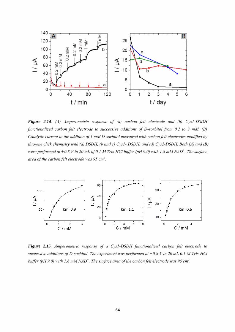
64
Figure 2.14. (A) Amperometric response of (a) carbon felt electrode and (b) Cys1-DSDH
functionalized carbon felt electrode to successive additions of D-sorbitol from 0.2 to 3 mM. (B)
Catalytic current to the addition of 1 mM D-sorbitol measured with carbon felt electrodes modified by
thio-ene click chemistry with (a) DSDH, (b and c) Cys1- DSDH, and (d) Cys2-DSDH. Both (A) and (B)
were performed at +0.8 V in 20 mL of 0.1 M Tris-HCl buffer (pH 9.0) with 1.8 mM NAD+. The surface
area of the carbon felt electrode was 95 cm2.
Figure 2.15. Amperometric response of a Cys1-DSDH functionalized carbon felt electrode to
successive additions of D-sorbitol. The experiment was performed at +0.8 V in 20 mL 0.1 M Tris-HCl
buffer (pH 9.0) with 1.8 mM NAD+. The surface area of the carbon felt electrode was 95 cm2.
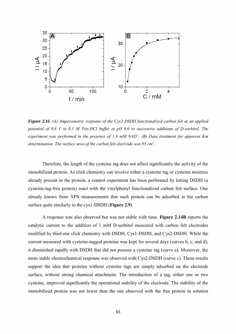
65
Figure 2.16. (A) Amperometric response of the Cys2-DSDH functionalized carbon felt at an applied
potential of 0.8 V in 0.1 M Tris-HCl buffer at pH 9.0 to successive additions of D-sorbitol. The
experiment was performed in the presence of 1.8 mM NAD+. (B) Data treatment for apparent Km
determination. The surface area of the carbon felt electrode was 95 cm2.
Therefore, the length of the cysteine tag does not affect significantly the activity of the
immobilized protein. As click chemistry can involve either a cysteine tag or cysteine moieties
already present in the protein, a control experiment has been performed by letting DSDH (a
cysteine-tag-free protein) react with the vinylphenyl functionalized carbon felt surface. One
already knows from XPS measurements that such protein can be adsorbed at the carbon
surface quite similarly to the cys1-DSDH (Figure 2.9).
A response was also observed but was not stable with time. Figure 2.14B reports the
catalytic current to the addition of 1 mM D-sorbitol measured with carbon felt electrodes
modified by thiol-ene click chemistry with DSDH, Cys1-DSDH, and Cys2-DSDH. While the
current measured with cysteine-tagged proteins was kept for several days (curves b, c, and d),
it diminished rapidly with DSDH that did not possess a cysteine tag (curve a). Moreover, the
more stable electrochemical response was observed with Cys2-DSDH (curve c). These results
support the idea that proteins without cysteine tags are simply adsorbed on the electrode
surface, without strong chemical attachment. The introduction of a tag, either one or two
cysteine, improved significantly the operational stability of the electrode. The stability of the
immobilized protein was not lower than the one observed with the free protein in solution

66
(half-life period of 48 h at 28 °C) and could be considered for application in electroenzymatic
synthesis.
2.3.4 Application to the electroenzymatic reduction of D-fructose in the
presence of [Cp*Rh(bpy)Cl]+
The two major fields of application of dehydrogenases are biosensing and
electrosynthesis [180]. In biosensing, the oxidation reaction is most often exploited,
considering different targets such as glucose, lactate, or ethanol. Detection via NADH
oxidation is usually performed in the presence of an electrochemical mediator that permits
decreasing significantly the anodic overpotentials in order to limit interferences. In
electrosynthesis, reduction reactions are most often investigated, for example the
enantioselective reduction of ketone to alcohol. In this case, NADH needs to be regenerated
by reduction of NAD+. The direct reduction of NAD+ occurs at high overpotential and leads
to the degradation of the enzymatic cofactor [17], so that long-term use of the
enzyme/cofactor system and high turnover numbers require the use of a mediator. One of the
most appropriate mediators for efficient NADH regeneration (Figure 2.7B) is the complex
(2,2‟-bipyridyl)(pentamethylcyclopentadienyl)-rhodium(III), [Cp*Rh(bpy)Cl]+ [32], which
will be evaluated hereafter with Cys1-DSDH proteins immobilized on carbon felt.
Figure 2.17. (A) Cyclic voltammograms recorded at a potential scan rate of 20 mV s−1 using a Cys1-
DSDH functionalized carbon felt electrode (a) in the absence and (b) in the presence of 1 mM D-
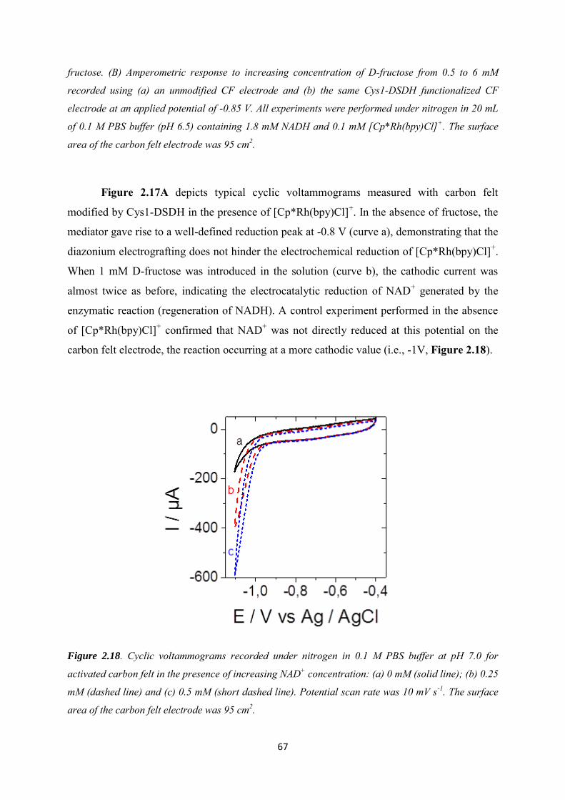
67
fructose. (B) Amperometric response to increasing concentration of D-fructose from 0.5 to 6 mM
recorded using (a) an unmodified CF electrode and (b) the same Cys1-DSDH functionalized CF
electrode at an applied potential of -0.85 V. All experiments were performed under nitrogen in 20 mL
of 0.1 M PBS buffer (pH 6.5) containing 1.8 mM NADH and 0.1 mM [Cp*Rh(bpy)Cl]+. The surface
area of the carbon felt electrode was 95 cm2.
Figure 2.17A depicts typical cyclic voltammograms measured with carbon felt
modified by Cys1-DSDH in the presence of [Cp*Rh(bpy)Cl]+. In the absence of fructose, the
mediator gave rise to a well-defined reduction peak at -0.8 V (curve a), demonstrating that the
diazonium electrografting does not hinder the electrochemical reduction of [Cp*Rh(bpy)Cl]+.
When 1 mM D-fructose was introduced in the solution (curve b), the cathodic current was
almost twice as before, indicating the electrocatalytic reduction of NAD+ generated by the
enzymatic reaction (regeneration of NADH). A control experiment performed in the absence
of [Cp*Rh(bpy)Cl]+ confirmed that NAD+ was not directly reduced at this potential on the
carbon felt electrode, the reaction occurring at a more cathodic value (i.e., -1V, Figure 2.18).
Figure 2.18. Cyclic voltammograms recorded under nitrogen in 0.1 M PBS buffer at pH 7.0 for
activated carbon felt in the presence of increasing NAD+ concentration: (a) 0 mM (solid line); (b) 0.25
mM (dashed line) and (c) 0.5 mM (short dashed line). Potential scan rate was 10 mV s-1. The surface
area of the carbon felt electrode was 95 cm2.
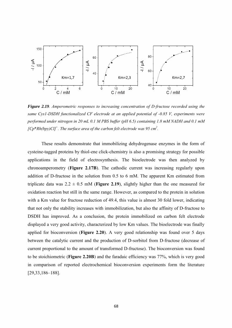
68
Figure 2.19. Amperometric responses to increasing concentration of D-fructose recorded using the
same Cys1-DSDH functionalized CF electrode at an applied potential of -0.85 V, experiments were
performed under nitrogen in 20 mL 0.1 M PBS buffer (pH 6.5) containing 1.8 mM NADH and 0.1 mM
[Cp*Rh(bpy)Cl]+. The surface area of the carbon felt electrode was 95 cm2.
These results demonstrate that immobilizing dehydrogenase enzymes in the form of
cysteine-tagged proteins by thiol-ene click-chemistry is also a promising strategy for possible
applications in the field of electrosynthesis. The bioelectrode was then analyzed by
chronoamperometry (Figure 2.17B). The cathodic current was increasing regularly upon
addition of D-fructose in the solution from 0.5 to 6 mM. The apparent Km estimated from
triplicate data was 2.2 ± 0.5 mM (Figure 2.19), slightly higher than the one measured for
oxidation reaction but still in the same range. However, as compared to the protein in solution
with a Km value for fructose reduction of 49.4, this value is almost 30 fold lower, indicating
that not only the stability increases with immobilization, but also the affinity of D-fructose to
DSDH has improved. As a conclusion, the protein immobilized on carbon felt electrode
displayed a very good activity, characterized by low Km values. The bioelectrode was finally
applied for bioconversion (Figure 2.20). A very good relationship was found over 5 days
between the catalytic current and the production of D-sorbitol from D-fructose (decrease of
current proportional to the amount of transformed D-fructose). The bioconversion was found
to be stoichiometric (Figure 2.20B) and the faradaic efficiency was 77%, which is very good
in comparison of reported electrochemical bioconversion experiments form the literature
[29,33,186–188].
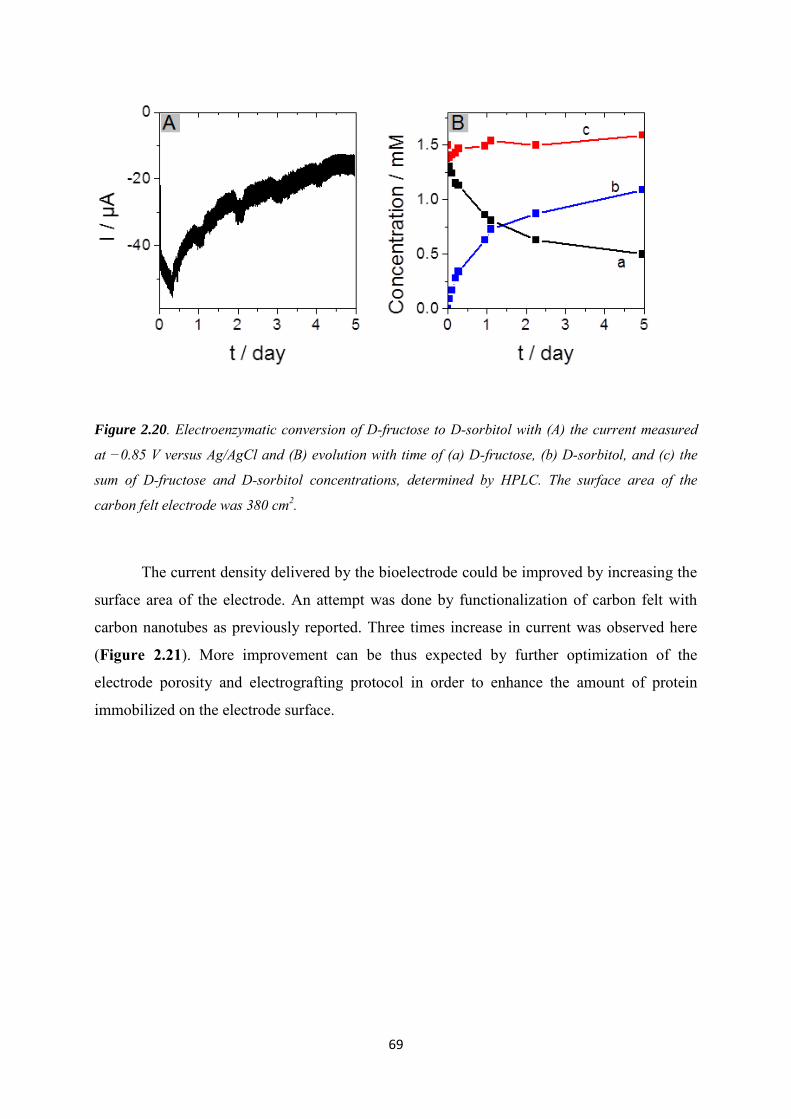
69
Figure 2.20. Electroenzymatic conversion of D-fructose to D-sorbitol with (A) the current measured
at −0.85 V versus Ag/AgCl and (B) evolution with time of (a) D-fructose, (b) D-sorbitol, and (c) the
sum of D-fructose and D-sorbitol concentrations, determined by HPLC. The surface area of the
carbon felt electrode was 380 cm2.
The current density delivered by the bioelectrode could be improved by increasing the
surface area of the electrode. An attempt was done by functionalization of carbon felt with
carbon nanotubes as previously reported. Three times increase in current was observed here
(Figure 2.21). More improvement can be thus expected by further optimization of the
electrode porosity and electrografting protocol in order to enhance the amount of protein
immobilized on the electrode surface.

70
Figure 2.21. Amperometric responses of the cys1-DSDH modified (a) MWCNT-carbon felt
(0.5*0.5*0.5cm3), (b) bare carbon felt to successive additions of D-sorbitol. The experiment was
performed at +0.8 V in 20 mL 0.1 M Tris-HCl buffer (pH 9.0) with 1.8 mM NAD+. The porous
electrode was prepared by dipping CF into a 1 mg mL-1 suspension of multi-walled carbon nanotubes
functionalized by carboxylic groups (MWCNT, 95%, ; 15 ± 5 nm, L 1–5 mm, Nanolab) and dried at
130°C for 30 min. This process was repeated 10 times. The modifications with cysteine-tagged DSDH
were then performed following the same protocol as reported in the manuscript.
71
2.4 Conclusions
Thiol-ene click chemistry was successfully applied to the immobilization of
dehydrogenase-based proteins on carbon electrode surfaces for application in
bioelectrochemical reactions. The combination of this reaction with diazonium
electrochemistry allowed the fabrication of electrodes that did not prevent the electrochemical
detection of redox probes in solution (ferrocenedimethanol, [Cp*Rh(bpy)Cl]+,and NADH).
The activity of D-sorbitol dehydrogenase was kept when click coupling was made on proteins
bearing a cysteine tag in front of a His(6)tag (either composed of 1 or 2 cysteine moieties). It
appears very convenient to take advantage of histidine-cysteine-tagged proteins as they can
easily be purified via the His(6)Tag and IMAC before immobilization on electrode. The
electrocatalytic reduction of D-fructose to D-sorbitol by the immobilized DSDH was
evidenced with using [Cp*Rh(bpy)Cl]+ as electron mediator. The system was applied
successfully to electroenzymatic enantioselective synthesis of D-sorbitol from D-fructose.
The combination of cysteine-tagged proteins with thiol-ene click reaction opens great
perspectives for covalent immobilization of redox proteins in an active form on electrode
surfaces for biosensing, fuel cell, and electrosynthesis applications. Moreover, positioning a
cysteine tag at different locations of the polypeptide chain should allow, in principle, a
controlled orientation of the protein in order to optimize direct electron transfer reactions with
bound cofactor of redox proteins.
72
Chapter 3. Covalent immobilization of rhodium complex on porous carbon electrode for NADH
regeneration
Here we report a new synthetic route for covalent immobilization of rhodium complex
on porous carbon electrode in three steps as a catalyst for nicotinamide adenine dinucleotide
reduced form (NADH) regeneration. The carbon electrode (carbon felt modified with multi-
wall carbon nanotube, CF-CNT) was first modified with azidophenyl groups groups by
diazonium electrografting generated from 4-azidoaniline. The bipyridine ligand was modified
with an alkynyl group, the covalent bond between the alkynyl group from bipyridine and the
azide group from the electrode was formed via azide-alkyne Huisgen cycloaddition reaction
(CF-CNT-4‟4-Bpy). After a final step complexation with pentamethylcyclopentadienyl
rhodium(III) chloride dimer ((RhCp*Cl2)2), rhodium complex was functionalized on the
electrode in an active form (CF-CNT-4‟4-Bpy-Rh). Different steps were confirmed by x-ray
photoelectron spectroscopy. The eletrocatalytic response of CF-CNT-4‟4-Bpy-Rh to NADH
regeneration from NAD+ was evaluated and a high faradic efficiency of 87% was obtained in
a 18 h chronoamperometry experiment. In comparison with the the CF-CNT electrode with
simple adsorption of [Cp*Rh(bpy)Cl]+, a better long-term stability up to 14 days in solution
with stirring was validated. Finally, the rhodium complex functionalized electrode was
applied in electroenzymatic synthesis by depositing a silica gel layer contains NAD-
dependent D-sorbitol dehydrogenase on the top, the transformation from D-fructose to D-
sorbitol was achieved.
73
3.1 Introduction
Nowadays, NAD-dependent enzymes, especially dehydrogenases are of increasing
interest for enantioselective bioconversion of a variety of compounds, eg. alcohols, acids, and
sugars [4] . However, their practical applications are limited by the high price of the
nicotinamide cofactors, so the regeneration of the cofactor is critical when considering to
build a long-term synthesis system [18,189–192].
The regeneration of nicotinamide cofactor could be realized by enzymatic [191,193],
chemical [6,194], or electrochemical methods [2,195]. Compared to chemical or enzyme-
coupled cofactor regeneration, which needs a second enzyme as well as a second substrate,
electrochemical regeneration methods are getting rid of the side product formation produced
from the coenzyme as well as the co-substrate separation. The main advantage of mass-free
electrochemical regeneration method is to offer the possibility of simplifying the processes in
bioconversion. In enzymatic reduction reactions, the easiest way for electrochemical
regeneration of NADH is by applying a negative potential on the electrode surface to reduce
NAD+ back to NADH. However, the negative potential needed is very high (-0.9 V, as
reported in the literature [3]), at this potential, NAD+ will be dimerized to NAD2 instead of
NADH, making the molecule unusable by the enzyme [4,27]. Therefore, to avoid the
dimerization of cofactors at high potential, a catalyst who has a redox potential slightly more
negative than the redox potential of cofactor NAD+ / NADH (-0.56 V) would be favored [5].
Up to now, rhodium-based complexes [Cp*RhIII(L)Cl]+ proposed by Steckhan et al.
[27,28,186,196,197] was proved to be the most successful non-enzymatic regeneration
catalyst for NADH regeneration [136].
However, some reports illustrate that low enzymatic catalytic stability is associated
with the presence of a rhodium complex since the surface functional groups of enzymes can
interact with rhodium complex, leading to degradation of rhodium complexes. The separation
of the enzymes from the rhodium complex by immobilizing them separately would provide a
system without losing the long-term stability [6,7,198]. Furthermore, immobilization of
rhodium complex on electrodes will not only avoid the degradation problem, but also make
easier the isolation of the reaction products and cofactors from the rhodium complex. Many
efforts have been done to immobilize rhodium complexes onto different electrodes in order to
74
improve their reusability as well as their enzymatic catalytic stability in a bio-electrochemical
system,
Generally, two strategies have been successfully developed to immobilize rhodium
complexes on an electrode surface in an active form. The first category is based on polymers
deposition onto the electrodes. Chardon-Noblat and coworkers prepared polypyrrolic rhodium
(III) complex (2,2'-bipyridine or 1,10-phenanthroline ligand) films on electrode by anodic
electropolymerization of pyrrole monomers. The electrocatalytic interest of this modified
electrode for hydrogen evolution was demonstrated [137]. Beley and coworkers bound a
rhodium (III) bis-terpyridinepyrrole complex by electropolymerization on a reticulated
vitreous carbon electrode, the long-term stability of the modified electrode was characterized
by reduction of NAD+ into 1,4-NADH, and it was then applied for reduction of
cyclohexanone to cyclohexanol in the presence of alcohol dehydrogenase [29]. In a report
from Nolte and coworkers, rhodium complexes were incorporated into the bilayers of vesicles
formed from a polymerizable ammonium surfactant by copolymerization reaction. The
catalytic activity of the rhodium complexes was depending on its position in the bilayer [138].
In above cases, the porous polymerized electrode was modified by electropolymerization.
Despite electropolymerization, another polymerization route by ɤ-irradiation cross-linking
was also reported to obtain [RhIII(C5Me5)(L)C1]+ complex polymer-modified electrodes. Even
through this method can provide a stable bonding, it suffered from the electron shielding
effect caused by the thick porous polymer film. This charge transfer limitation effect caused
by polymer layer was proved by the fact that when the amount of immobilized polymer was
doubled on the electrode, the charge calculated from the reduction peak was only increased by
a factor of less than 2 [139].
The second strategy is to take advantage of the π-π stacking effect between
carbonaceous materials (graphene and carbon nanotubes) and aromatic moieties from
rhodium complex. This interaction would favor a strong adsorption of the rhodium complex
on the carbon support. In this route, the electrode surface was only covered with a monolayer
rather than thick porous polymer films, the electron shielding effect was in principle
eliminated and the electron transfer between the rhodium complex and the electrode was
facilitated, a surface-confined electrochemical behavior was observed. Park and coworkers
reported the synthesis of a 3D-structured graphene–Rh-complex hydrogel, on which the
phenanthroline introduced Rh complex was strongly immobilized on the graphene surface
through π-π interaction and π-cation interaction, the repeated usability for electrochemical
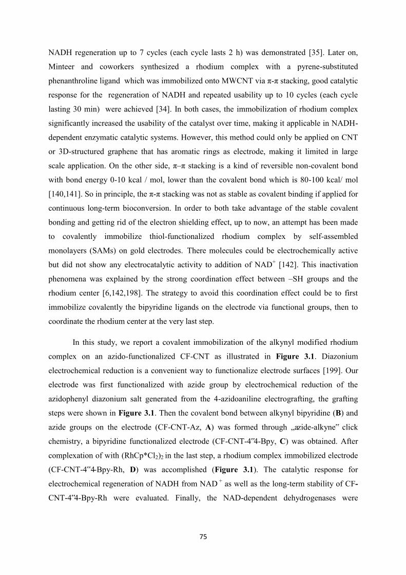
75
NADH regeneration up to 7 cycles (each cycle lasts 2 h) was demonstrated [35]. Later on,
Minteer and coworkers synthesized a rhodium complex with a pyrene-substituted
phenanthroline ligand which was immobilized onto MWCNT via π-π stacking, good catalytic
response for the regeneration of NADH and repeated usability up to 10 cycles (each cycle
lasting 30 min) were achieved [34]. In both cases, the immobilization of rhodium complex
significantly increased the usability of the catalyst over time, making it applicable in NADH-
dependent enzymatic catalytic systems. However, this method could only be applied on CNT
or 3D-structured graphene that has aromatic rings as electrode, making it limited in large
scale application. On the other side, π–π stacking is a kind of reversible non-covalent bond
with bond energy 0-10 kcal / mol, lower than the covalent bond which is 80-100 kcal/ mol
[140,141]. So in principle, the π-π stacking was not as stable as covalent binding if applied for
continuous long-term bioconversion. In order to both take advantage of the stable covalent
bonding and getting rid of the electron shielding effect, up to now, an attempt has been made
to covalently immobilize thiol-functionalized rhodium complex by self-assembled
monolayers (SAMs) on gold electrodes. There molecules could be electrochemically active
but did not show any electrocatalytic activity to addition of NAD+ [142]. This inactivation
phenomena was explained by the strong coordination effect between –SH groups and the
rhodium center [6,142,198]. The strategy to avoid this coordination effect could be to first
immobilize covalently the bipyridine ligands on the electrode via functional groups, then to
coordinate the rhodium center at the very last step.
In this study, we report a covalent immobilization of the alkynyl modified rhodium
complex on an azido-functionalized CF-CNT as illustrated in Figure 3.1. Diazonium
electrochemical reduction is a convenient way to functionalize electrode surfaces [199]. Our
electrode was first functionalized with azide group by electrochemical reduction of the
azidophenyl diazonium salt generated from the 4-azidoaniline electrografting, the grafting
steps were shown in Figure 3.1. Then the covalent bond between alkynyl bipyridine (B) and
azide groups on the electrode (CF-CNT-Az, A) was formed through „azide-alkyne‟ click
chemistry, a bipyridine functionalized electrode (CF-CNT-4‟4-Bpy, C) was obtained. After
complexation of with (RhCp*Cl2)2 in the last step, a rhodium complex immobilized electrode
(CF-CNT-4‟4-Bpy-Rh, D) was accomplished (Figure 3.1). The catalytic response for
electrochemical regeneration of NADH from NAD + as well as the long-term stability of CF-
CNT-4‟4-Bpy-Rh were evaluated. Finally, the NAD-dependent dehydrogenases were
76
immobilized in a sol-gel matrix on the top of the rhodium complex modified electrode in
order to study the electroenzymatic response to addition of substrate.
Figure 3.1. Synthetic route followed for the functionalization of a carbon electrode surface with
rhodium complex.
3.2 Experimental
Chemicals and Enzymes
The following chemicals were used as received: tetraethoxysilane (TEOS, 98%, Alfa
Aesar), 3-glycidoxypropyltrimethoxysilane (GPS, 98%, Sigma-Aldrich), β-nicotinamide
adenine dinucleotide hydrate (NAD+, ≥ 96.5%, Sigma-Aldrich ), β-nicotinamide adenine
dinucleotide, reduced disodium salt hydrate (NADH, ≥97%, Sigma-Aldrich), 4-azidoaniline
hydrochloride (97%, Sigma-Aldrich), sodium nitrite (≥97.0% , Sigma-Aldrich), copper actate
(Prolab), ascorbic acid (Prolab), pentamethylcyclopentadienylrhodium(III) chloride dimer
((RhCp*Cl2)2, Sigma-Aldrich), D-fructose (99 wt%, Sigma), sodium dihydrogen phosphate
(99.5%, Merck), hydroxyacetone (95%, Alfa Aesar), carbon felt (GFD 4.6 EA, density 0.09 g
cm-3, electrical resistivity 3-6 mm, Sigratherm®), poly(ethylene imine) (PEI, 50% w/v in
water, Mn = 60000, Fluka), carboxylic-functionalized multi-walled carbon nanotubes
(MWCNT, 95%, Φ 15±5 nm, L 1-5 µm, Nanolab). Overproduction of N-His(6) D-sorbitol
dehydrogenase (800U mL-1 ) and N-His(6)-Cys-galactitol dehydrogenase (60U mL-1) were
done with Escherichia coli BL21GOLD (DE3) containing the corresponding expression-
77
vectors pET-24a(+) (Novagen) and purification of the enzyme was performed with Histrap
columns (GE Healthcare).
Sythesis of 4-[(2-propyn-1-yloxy)methyl]-4’-methyl-2,2’-bipyridine (compound B)
The protocol for synthesis is following the reported literature in three steps [142].
a. 4-(carboxyaldehyde)-4‟-methyl-2,2‟-bipyridine MS (CI, CH4) m/z: 213 (100) [M+1]. 1H
NMR (400 MHz, CDCl3) : 2.48 (s, 3H), 7.23 (d, J=5.0 Hz, 1H), 7.74-7.78 (m, 1H), 8.32 (s,
1H), 8.62 (d, J= 4.9 Hz, 1H), 8.88 (s, 1H), 8.95 (d, J=4.9 Hz, 1H), 10.23 (s, 1H).
b. 4-(hydroxymethyl)-4‟-methyl-2,2‟-bipyridine MS (CI, CH4) m/z: 201 (100) [M+1]. 1H
NMR (400 MHz, CDCl3) : 2.49 (s, 3H), 5.03 (s, 2H), 7.18-7.25 (m, 1H), 7.41-7.48 (m, 1H),
8. 30-8.35 (m, 1H), 8.40-8.42 (m, 1H), 8.59 (d, J=4.9 Hz, 1H), 8.72 (d, J=5.0 Hz, 1H,)
c. 4-[(2-propyn-1-yloxy)methyl]-4‟-methyl-2,2‟-bipyridine MS (CI, CH4) m/z: 239 (100)
[M+1]. 1H NMR (400 MHz, CDCl3) : 2.36 (s, 3H), 2.40 (t, J=2.4 Hz, 2H), 4.23 (d, J=2.4 Hz,
2H), 4.67 (s, 2H), 7.15-7.16 (m, 1H), 7.18-7.20 (m, 1H), 8.24-8.28 (m, 1H), 8.30-8.33 (m,
1H), 8.49 (d, J=5.0 Hz, 1H), 8.68 (d, J=5.0 Hz, 1H).
MWCNT-functionalized carbon felt electrode
In order to change the surface from hydrophobic to hydrophilic, the surface of carbon
felt was activated by recording up to 20 consecutive cyclic voltammetry scans over the range
potential from -0.7 V to 2.5 V in 0.1M H2SO4. After such process of electrochemical
conditioning, the carbon felt was first rinsed with water and subsequently heated up to at
200℃ for 1h to remove the remaining H2SO4. Afterwards, the carbon felt was cut into pieces
with dimension 0.5 cm* 0.5 cm. MWCNT-functionalized carbon felt was prepared by dipping
the carbon felt into 0.5 mg/mL MWCNT suspension which is dispersed in an ultrasonic bath,
then let it dry in the oven at 130 ℃. The dipping/drying cycles were repeated for 10 times in
order to ensure that plenty of CNT were immobilized on the carbon felt substrate.
Azido-functionalized CF-CNT electrode
The diazonium cations were generated “in situ” by mixing 1 mM 4-azidoaniline and 2
mM sodium nitrite in 0.5 M HCl water solution with stirring for 5 min. The isolation of the
diazonium salt is not required and its grafting process is carried out using the same solution
they were generated. The grafting process was carried out by electrochemical reduction
performed by recording two consecutive cyclic voltammetry potential scans over the potential
78
range from 0.4 V to -0.6 V on a CF-MWCNT electrode. After electro-grafting, the carbon
electrode was rinsed with distilled water to remove the remaining ungrafted compounds.
Bipyridine derivative covalent immobilization on CF-CNT-Az electrode via Huisgen
cycloaddition reaction.
To a solution of molecule B (2.4 mg) dissolved in dimethilformamide (7 mL), a
mixture of copper actate (1 mg) and ascorbic acid (2 mg) dissolved in an aqueous solution (3
mL) was added. The CF-CNT-Az electrode was immersed in this solution at room
temperature for 24h in darkness. After this period the electrode was rinsed carefully with
water and DMF.
Complexation reaction of rhodium with the CF-CNT-4’4-Bpy electrode.
The ability of the bipyridine ligands to coordinate transition metal was used in order to
immobilize a rhodium complex. The bipyridine-functionalized CF-MWCNT electrode was
then put in a dichloromethane (20 mL) solution containing (RhCp*Cl2)2 (2 mg) with
continuous stirring for 3h.
Co-immobilization of enzymes and rhodium complex on the electrode
For co-immobilization of enzymes on the Rh complex functionalized electrode, an
enzyme silica sol was prepared according to a protocol that was previously described [200].
Briefly, a mixture of 0.18 g TEOS, 0.13 g GPS, together with 0.5 mL water and 0.625 mL
0.01 M HCl was pre-hydrolyzed by stirring overnight. Then, this sol was diluted 3 times and a
200 µL aliquot was mixed with 100 µL of PEI (20%), 100 µL of water and 150µL DSDH (or
GatDH) stock solution. This mixture was spread over the CF-CNT-4‟4-Bpy-Rh electrode and
let it completely dry in the fridge for use. The CF-CNT-4‟4-Bpy-Rh-gel electrode was
prepared by following the same protocol but replacing the enzyme solution with same volume
of water. The CF-CNT-ads-Rh-gel electrode was prepared by dipping CF-MWCNT electrode
in 1mM [Cp*Rh(bpy)Cl]+ DMF / H2O (7 : 3) solution overnight, then following the same
protocol as for the CF-CNT-4‟4-Bpy-Rh-gel electrode.
Procedures
All electrochemical measurements were carried out using an Autolab PGSTAT-12
potentiostat, and carried out in a sealed glass cell. A pencil core (0.5 mm diameter) connected
with silver wire glued by carbon black was used for connecting the carbon felt working
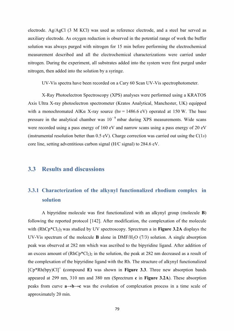
79
electrode. Ag/AgCl (3 M KCl) was used as reference electrode, and a steel bar served as
auxiliary electrode. As oxygen reduction is observed in the potential range of work the buffer
solution was always purged with nitrogen for 15 min before performing the electrochemical
measurement described and all the electrochemical characterizations were carried under
nitrogen. During the experiment, all substrates added into the system were first purged under
nitrogen, then added into the solution by a syringe.
UV-Vis spectra have been recorded on a Cary 60 Scan UV-Vis spectrophotometer.
X-Ray Photoelectron Spectroscopy (XPS) analyses were performed using a KRATOS
Axis Ultra X-ray photoelectron spectrometer (Kratos Analytical, Manchester, UK) equipped
with a monochromated AlKα X-ray source (hν = 1486.6 eV) operated at 150 W. The base
pressure in the analytical chamber was 10− 9 mbar during XPS measurements. Wide scans
were recorded using a pass energy of 160 eV and narrow scans using a pass energy of 20 eV
(instrumental resolution better than 0.5 eV). Charge correction was carried out using the C(1s)
core line, setting adventitious carbon signal (H/C signal) to 284.6 eV.
3.3 Results and discussions
3.3.1 Characterization of the alkynyl functionalized rhodium complex in
solution
A bipyridine molecule was first functionalized with an alkynyl group (molecule B)
following the reported protocol [142]. After modification, the complexation of the molecule
with (RhCp*Cl2)2 was studied by UV spectroscopy. Sprectrum a in Figure 3.2A displays the
UV-Vis spectrum of the molecule B alone in DMF/H2O (7/3) solution. A single absorption
peak was observed at 282 nm which was ascribed to the bipyridine ligand. After addition of
an excess amount of (RhCp*Cl2)2 in the solution, the peak at 282 nm decreased as a result of
the complexation of the bipyridine ligand with the Rh. The structure of alkynyl functionalized
[Cp*Rh(bpy)Cl]+ (compound E) was shown in Figure 3.3. Three new absorption bands
appeared at 299 nm, 310 nm and 380 nm (Sprectrum c in Figure 3.2A). These absorption
peaks from curve a→b→c was the evolution of complexation process in a time scale of
approximately 20 min.
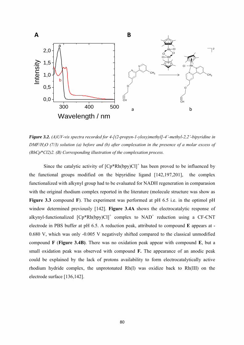
80
300 400 500
0,0
0,5
1,0
1,5
2,0
In
ten
sity
Wavelength / nm
a
b
RhN
N
Cl
CH3
O
CH
+
Cl
ClRh
Cl
Cl Rh-
N
N
CH3
O
CH
a b
A B
Figure 3.2. (A)UV-vis spectra recorded for 4-[(2-propyn-1-yloxy)methyl]-4’-methyl-2,2’-bipyridine in
DMF/H2O (7/3) solution (a) before and (b) after complexation in the presence of a molar excess of
(RhCp*Cl2)2. (B) Corresponding illustration of the complexation process.
Since the catalytic activity of [Cp*Rh(bpy)Cl]+ has been proved to be influenced by
the functional groups modified on the bipyridine ligand [142,197,201], the complex
functionalized with alkynyl group had to be evaluated for NADH regeneration in comparasion
with the original rhodium complex reported in the literature (molecule structure was show as
Figure 3.3 compound F). The experiment was performed at pH 6.5 i.e. in the optimol pH
window determined previously [142]. Figure 3.4A shows the electrocatalytic response of
alkynyl-functionalized [Cp*Rh(bpy)Cl]+ complex to NAD+ reduction using a CF-CNT
electrode in PBS buffer at pH 6.5. A reduction peak, attributed to compound E appears at -
0.680 V, which was only -0.005 V negatively shifted compared to the classical unmodified
compound F (Figure 3.4B). There was no oxidation peak appear with compound E, but a
small oxidation peak was observed with compound F. The appearance of an anodic peak
could be explained by the lack of protons availability to form electrocatalytically active
rhodium hydride complex, the unprotonated Rh(I) was oxidize back to Rh(III) on the
electrode surface [136,142].
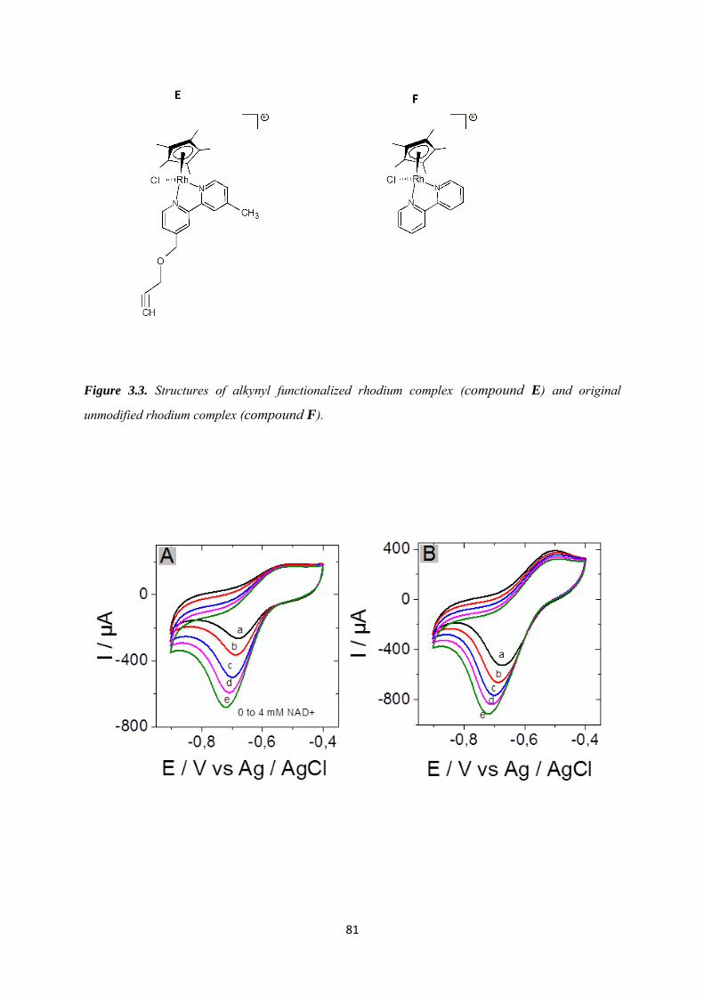
81
Figure 3.3. Structures of alkynyl functionalized rhodium complex (compound E) and original
unmodified rhodium complex (compound F).
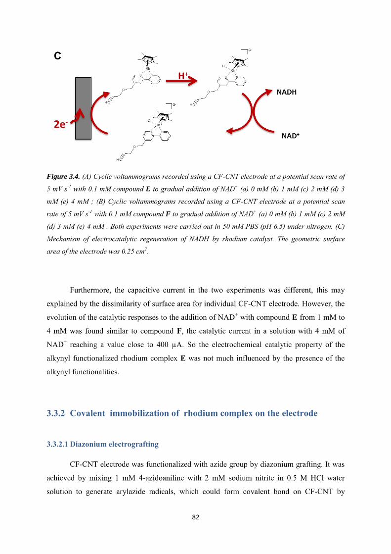
82
Figure 3.4. (A) Cyclic voltammograms recorded using a CF-CNT electrode at a potential scan rate of
5 mV s-1 with 0.1 mM compound E to gradual addition of NAD+ (a) 0 mM (b) 1 mM (c) 2 mM (d) 3
mM (e) 4 mM ; (B) Cyclic voltammograms recorded using a CF-CNT electrode at a potential scan
rate of 5 mV s-1 with 0.1 mM compound F to gradual addition of NAD+ (a) 0 mM (b) 1 mM (c) 2 mM
(d) 3 mM (e) 4 mM . Both experiments were carried out in 50 mM PBS (pH 6.5) under nitrogen. (C)
Mechanism of electrocatalytic regeneration of NADH by rhodium catalyst. The geometric surface
area of the electrode was 0.25 cm2.
Furthermore, the capacitive current in the two experiments was different, this may
explained by the dissimilarity of surface area for individual CF-CNT electrode. However, the
evolution of the catalytic responses to the addition of NAD+ with compound E from 1 mM to
4 mM was found similar to compound F, the catalytic current in a solution with 4 mM of
NAD+ reaching a value close to 400 µA. So the electrochemical catalytic property of the
alkynyl functionalized rhodium complex E was not much influenced by the presence of the
alkynyl functionalities.
3.3.2 Covalent immobilization of rhodium complex on the electrode
3.3.2.1 Diazonium electrografting
CF-CNT electrode was functionalized with azide group by diazonium grafting. It was
achieved by mixing 1 mM 4-azidoaniline with 2 mM sodium nitrite in 0.5 M HCl water
solution to generate arylazide radicals, which could form covalent bond on CF-CNT by
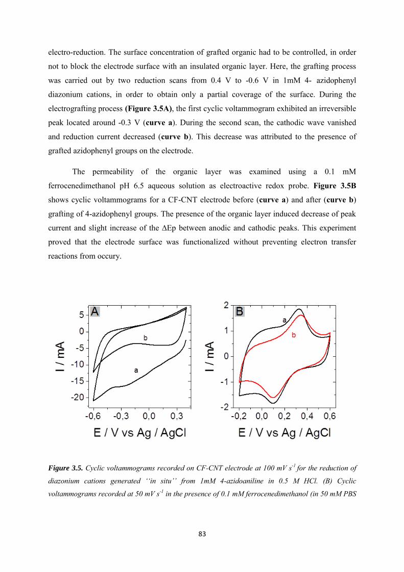
83
electro-reduction. The surface concentration of grafted organic had to be controlled, in order
not to block the electrode surface with an insulated organic layer. Here, the grafting process
was carried out by two reduction scans from 0.4 V to -0.6 V in 1mM 4- azidophenyl
diazonium cations, in order to obtain only a partial coverage of the surface. During the
electrografting process (Figure 3.5A), the first cyclic voltammogram exhibited an irreversible
peak located around -0.3 V (curve a). During the second scan, the cathodic wave vanished
and reduction current decreased (curve b). This decrease was attributed to the presence of
grafted azidophenyl groups on the electrode.
The permeability of the organic layer was examined using a 0.1 mM
ferrocenedimethanol pH 6.5 aqueous solution as electroactive redox probe. Figure 3.5B
shows cyclic voltammograms for a CF-CNT electrode before (curve a) and after (curve b)
grafting of 4-azidophenyl groups. The presence of the organic layer induced decrease of peak
current and slight increase of the ∆Ep between anodic and cathodic peaks. This experiment
proved that the electrode surface was functionalized without preventing electron transfer
reactions from occury.
Figure 3.5. Cyclic voltammograms recorded on CF-CNT electrode at 100 mV s-1 for the reduction of
diazonium cations generated ‘‘in situ’’ from 1mM 4-azidoaniline in 0.5 M HCl. (B) Cyclic
voltammograms recorded at 50 mV s-1 in the presence of 0.1 mM ferrocenedimethanol (in 50 mM PBS
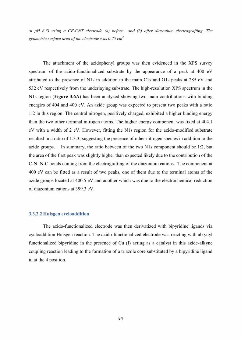
84
at pH 6.5) using a CF-CNT electrode (a) before and (b) after diazonium electrografting. The
geometric surface area of the electrode was 0.25 cm2.
The attachment of the azidophenyl groups was then evidenced in the XPS survey
spectrum of the azido-functionalized substrate by the appearance of a peak at 400 eV
attributed to the presence of N1s in addition to the main C1s and O1s peaks at 285 eV and
532 eV respectively from the underlaying substrate. The high-resolution XPS spectrum in the
N1s region (Figure 3.6A) has been analyzed showing two main contributions with binding
energies of 404 and 400 eV. An azide group was expected to present two peaks with a ratio
1:2 in this region. The central nitrogen, positively charged, exhibited a higher binding energy
than the two other terminal nitrogen atoms. The higher energy component was fixed at 404.1
eV with a width of 2 eV. However, fitting the N1s region for the azido-modified substrate
resulted in a ratio of 1:3.3, suggesting the presence of other nitrogen species in addition to the
azide groups. In summary, the ratio between of the two N1s component should be 1:2, but
the area of the first peak was slightly higher than expected likely due to the contribution of the
C-N=N-C bonds coming from the electrografting of the diazonium cations. The component at
400 eV can be fitted as a result of two peaks, one of them due to the terminal atoms of the
azide groups located at 400.5 eV and another which was due to the electrochemical reduction
of diazonium cations at 399.3 eV.
3.3.2.2 Huisgen cycloaddition
The azido-functionalized electrode was then derivatized with bipyridine ligands via
cycloaddition Huisgen reaction. The azido-functionalized electrode was reacting with alkynyl
functionalized bipyridine in the presence of Cu (I) acting as a catalyst in this azide-alkyne
coupling reaction leading to the formation of a triazole core substituted by a bipyridine ligand
in at the 4 position.
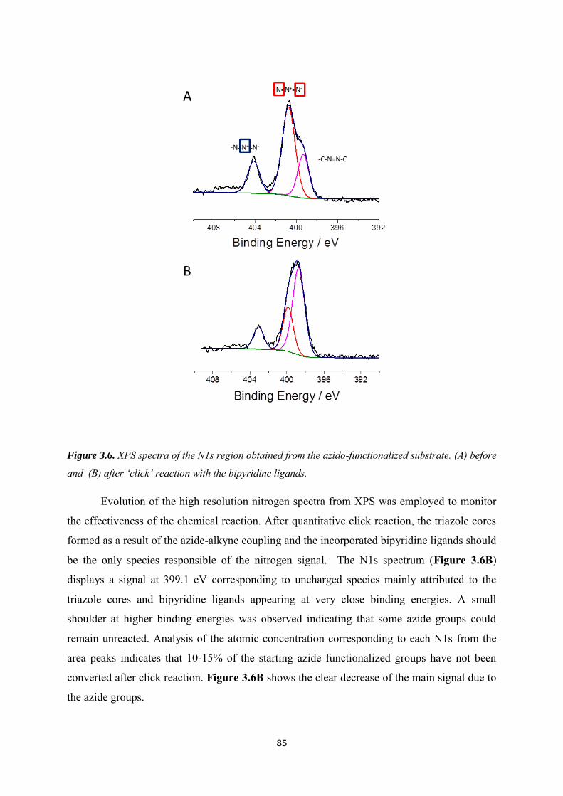
85
Figure 3.6. XPS spectra of the N1s region obtained from the azido-functionalized substrate. (A) before
and (B) after ‘click’ reaction with the bipyridine ligands.
Evolution of the high resolution nitrogen spectra from XPS was employed to monitor
the effectiveness of the chemical reaction. After quantitative click reaction, the triazole cores
formed as a result of the azide-alkyne coupling and the incorporated bipyridine ligands should
be the only species responsible of the nitrogen signal. The N1s spectrum (Figure 3.6B)
displays a signal at 399.1 eV corresponding to uncharged species mainly attributed to the
triazole cores and bipyridine ligands appearing at very close binding energies. A small
shoulder at higher binding energies was observed indicating that some azide groups could
remain unreacted. Analysis of the atomic concentration corresponding to each N1s from the
area peaks indicates that 10-15% of the starting azide functionalized groups have not been
converted after click reaction. Figure 3.6B shows the clear decrease of the main signal due to
the azide groups.
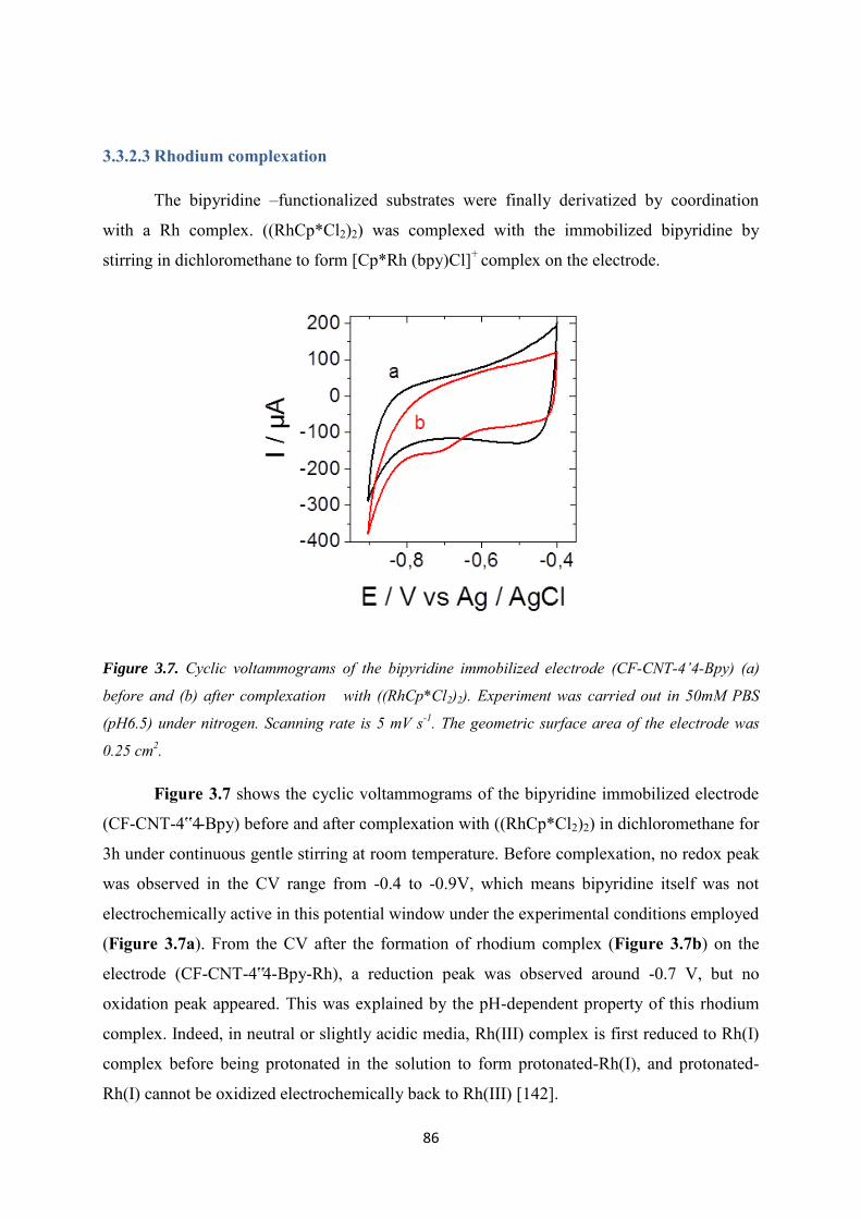
86
3.3.2.3 Rhodium complexation
The bipyridine –functionalized substrates were finally derivatized by coordination
with a Rh complex. ((RhCp*Cl2)2) was complexed with the immobilized bipyridine by
stirring in dichloromethane to form [Cp*Rh (bpy)Cl]+ complex on the electrode.
Figure 3.7. Cyclic voltammograms of the bipyridine immobilized electrode (CF-CNT-4’4-Bpy) (a)
before and (b) after complexation with ((RhCp*Cl2)2). Experiment was carried out in 50mM PBS
(pH6.5) under nitrogen. Scanning rate is 5 mV s-1. The geometric surface area of the electrode was
0.25 cm2.
Figure 3.7 shows the cyclic voltammograms of the bipyridine immobilized electrode
(CF-CNT-4‟4-Bpy) before and after complexation with ((RhCp*Cl2)2) in dichloromethane for
3h under continuous gentle stirring at room temperature. Before complexation, no redox peak
was observed in the CV range from -0.4 to -0.9V, which means bipyridine itself was not
electrochemically active in this potential window under the experimental conditions employed
(Figure 3.7a). From the CV after the formation of rhodium complex (Figure 3.7b) on the
electrode (CF-CNT-4‟4-Bpy-Rh), a reduction peak was observed around -0.7 V, but no
oxidation peak appeared. This was explained by the pH-dependent property of this rhodium
complex. Indeed, in neutral or slightly acidic media, Rh(III) complex is first reduced to Rh(I)
complex before being protonated in the solution to form protonated-Rh(I), and protonated-
Rh(I) cannot be oxidized electrochemically back to Rh(III) [142].
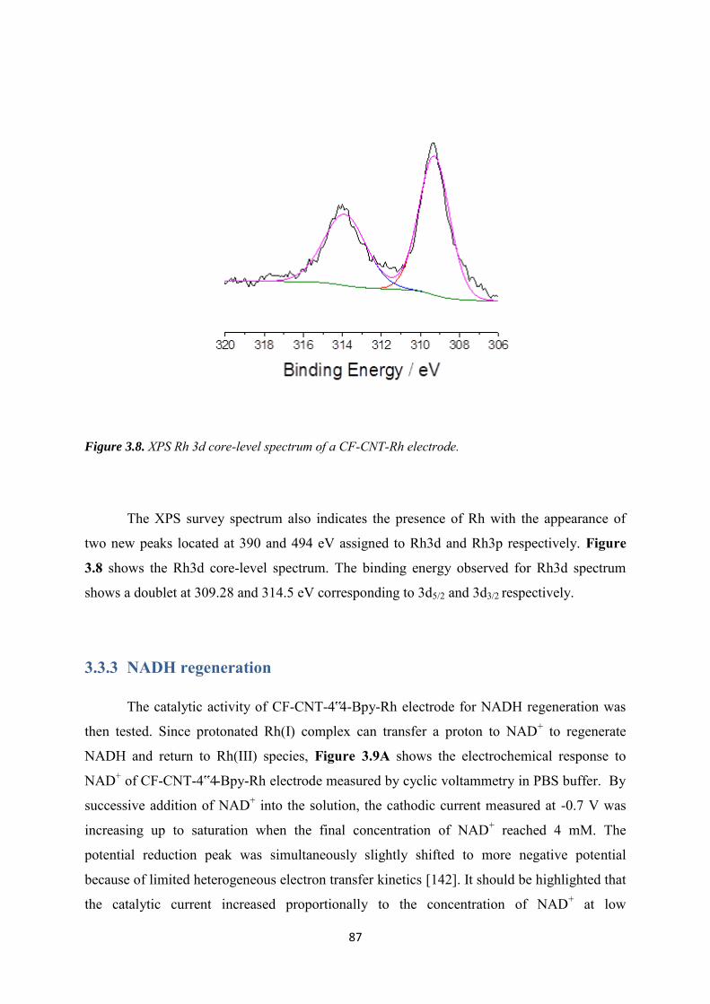
87
Figure 3.8. XPS Rh 3d core-level spectrum of a CF-CNT-Rh electrode.
The XPS survey spectrum also indicates the presence of Rh with the appearance of
two new peaks located at 390 and 494 eV assigned to Rh3d and Rh3p respectively. Figure
3.8 shows the Rh3d core-level spectrum. The binding energy observed for Rh3d spectrum
shows a doublet at 309.28 and 314.5 eV corresponding to 3d5/2 and 3d3/2 respectively.
3.3.3 NADH regeneration
The catalytic activity of CF-CNT-4‟4-Bpy-Rh electrode for NADH regeneration was
then tested. Since protonated Rh(I) complex can transfer a proton to NAD+ to regenerate
NADH and return to Rh(III) species, Figure 3.9A shows the electrochemical response to
NAD+ of CF-CNT-4‟4-Bpy-Rh electrode measured by cyclic voltammetry in PBS buffer. By
successive addition of NAD+ into the solution, the cathodic current measured at -0.7 V was
increasing up to saturation when the final concentration of NAD+ reached 4 mM. The
potential reduction peak was simultaneously slightly shifted to more negative potential
because of limited heterogeneous electron transfer kinetics [142]. It should be highlighted that
the catalytic current increased proportionally to the concentration of NAD+ at low
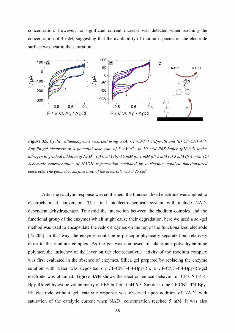
88
concentration. However, no significant current increase was detected when reaching the
concentration of 4 mM, suggesting that the availability of rhodium species on the electrode
surface was near to the saturation.
Figure 3.9. Cyclic voltammograms recorded using a (A) CF-CNT-4’4-Bpy-Rh and (B) CF-CNT-4’4-
Bpy-Rh-gel electrode at a potential scan rate of 5 mV s-1 in 50 mM PBS buffer (pH 6.5) under
nitrogen to gradual addition of NAD+ (a) 0 mM (b) 0.5 mM (c) 1 mM (d) 2 mM (e) 3 mM (f) 4 mM. (C)
Schematic representation of NADH regeneration mediated by a rhodium catalyst functionalized
electrode. The geometric surface area of the electrode was 0.25 cm2.
After the catalytic response was confirmed, the functionalized electrode was applied to
electrochemical conversion. The final bioelectrochemical system will include NAD-
dependent dehydrogenase. To avoid the interaction between the rhodium complex and the
functional group of the enzymes which might cause their degradation, here we used a sol-gel
method was used to encapsulate the redox enzymes on the top of the functionalized electrode
[75,202]. In that way, the enzymes could be in principle physically separated but relatively
close to the rhodium complex. As the gel was composed of silane and polyethylenimine
polymer, the influence of the layer on the electrocatalytic activity of the rhodium complex
was first evaluated in the absence of enzymes. Silica gel prepared by replacing the enzyme
solution with water was deposited on CF-CNT-4‟4-Bpy-Rh, a CF-CNT-4‟4-Bpy-Rh-gel
electrode was obtained. Figure 3.9B shows the electrochemical behavior of CF-CNT-4‟4-
Bpy-Rh-gel by cyclic voltammetry in PBS buffer at pH 6.5. Similar to the CF-CNT-4‟4-Bpy-
Rh electrode without gel, catalytic response was observed upon addition of NAD+ with
saturation of the catalytic current when NAD+ concentration reached 5 mM. It was also
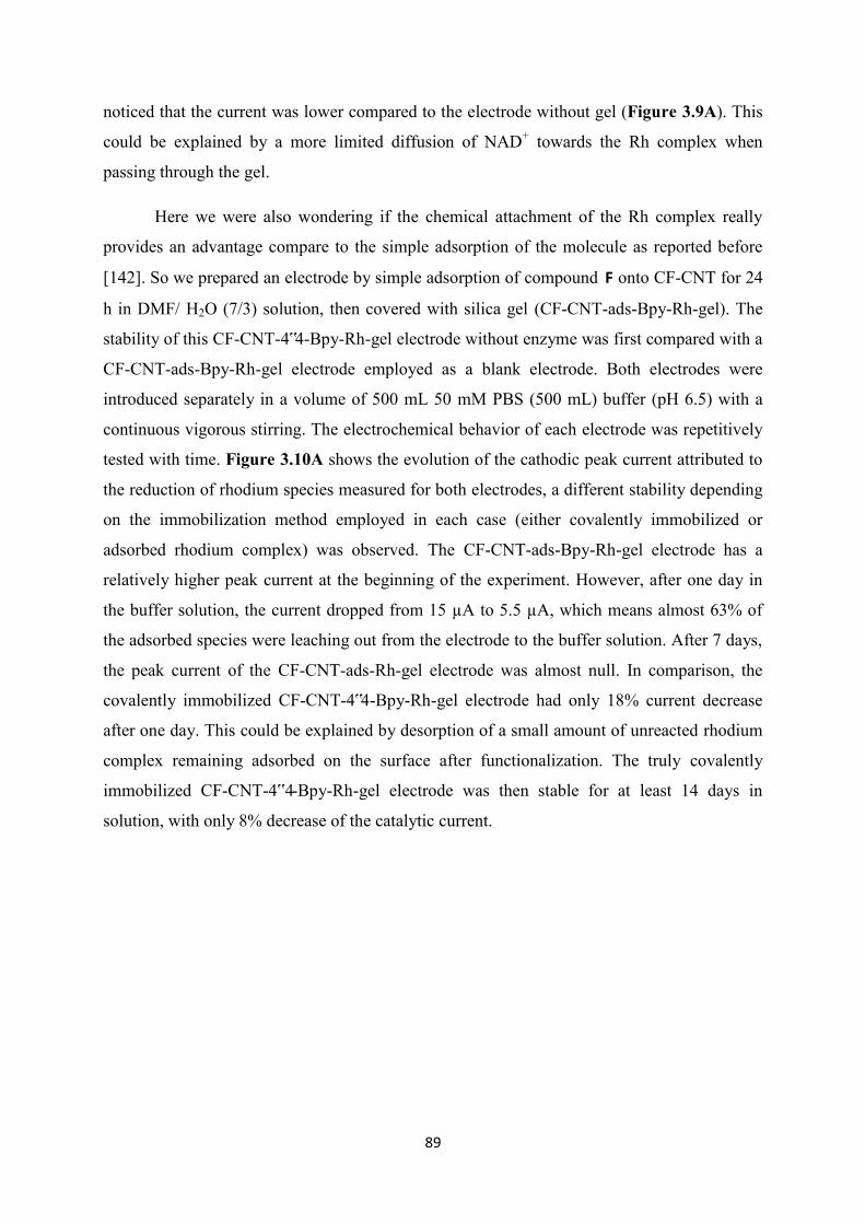
89
noticed that the current was lower compared to the electrode without gel (Figure 3.9A). This
could be explained by a more limited diffusion of NAD+ towards the Rh complex when
passing through the gel.
Here we were also wondering if the chemical attachment of the Rh complex really
provides an advantage compare to the simple adsorption of the molecule as reported before
[142]. So we prepared an electrode by simple adsorption of compound F onto CF-CNT for 24
h in DMF/ H2O (7/3) solution, then covered with silica gel (CF-CNT-ads-Bpy-Rh-gel). The
stability of this CF-CNT-4‟4-Bpy-Rh-gel electrode without enzyme was first compared with a
CF-CNT-ads-Bpy-Rh-gel electrode employed as a blank electrode. Both electrodes were
introduced separately in a volume of 500 mL 50 mM PBS (500 mL) buffer (pH 6.5) with a
continuous vigorous stirring. The electrochemical behavior of each electrode was repetitively
tested with time. Figure 3.10A shows the evolution of the cathodic peak current attributed to
the reduction of rhodium species measured for both electrodes, a different stability depending
on the immobilization method employed in each case (either covalently immobilized or
adsorbed rhodium complex) was observed. The CF-CNT-ads-Bpy-Rh-gel electrode has a
relatively higher peak current at the beginning of the experiment. However, after one day in
the buffer solution, the current dropped from 15 µA to 5.5 µA, which means almost 63% of
the adsorbed species were leaching out from the electrode to the buffer solution. After 7 days,
the peak current of the CF-CNT-ads-Rh-gel electrode was almost null. In comparison, the
covalently immobilized CF-CNT-4‟4-Bpy-Rh-gel electrode had only 18% current decrease
after one day. This could be explained by desorption of a small amount of unreacted rhodium
complex remaining adsorbed on the surface after functionalization. The truly covalently
immobilized CF-CNT-4‟4-Bpy-Rh-gel electrode was then stable for at least 14 days in
solution, with only 8% decrease of the catalytic current.
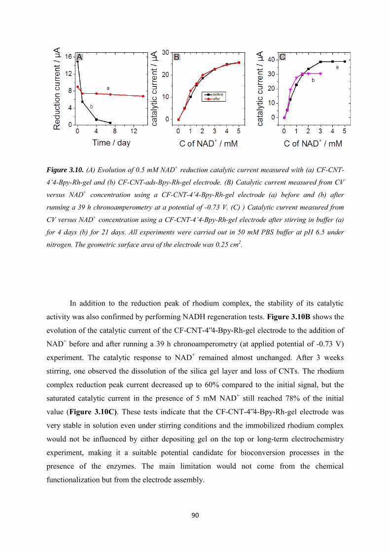
90
Figure 3.10. (A) Evolution of 0.5 mM NAD+ reduction catalytic current measured with (a) CF-CNT-
4’4-Bpy-Rh-gel and (b) CF-CNT-ads-Bpy-Rh-gel electrode. (B) Catalytic current measured from CV
versus NAD+ concentration using a CF-CNT-4’4-Bpy-Rh-gel electrode (a) before and (b) after
running a 39 h chronoamperometry at a potential of -0.73 V. (C) ) Catalytic current measured from
CV versus NAD+ concentration using a CF-CNT-4’4-Bpy-Rh-gel electrode after stirring in buffer (a)
for 4 days (b) for 21 days. All experiments were carried out in 50 mM PBS buffer at pH 6.5 under
nitrogen. The geometric surface area of the electrode was 0.25 cm2.
In addition to the reduction peak of rhodium complex, the stability of its catalytic
activity was also confirmed by performing NADH regeneration tests. Figure 3.10B shows the
evolution of the catalytic current of the CF-CNT-4‟4-Bpy-Rh-gel electrode to the addition of
NAD+ before and after running a 39 h chronoamperometry (at applied potential of -0.73 V)
experiment. The catalytic response to NAD+ remained almost unchanged. After 3 weeks
stirring, one observed the dissolution of the silica gel layer and loss of CNTs. The rhodium
complex reduction peak current decreased up to 60% compared to the initial signal, but the
saturated catalytic current in the presence of 5 mM NAD+ still reached 78% of the initial
value (Figure 3.10C). These tests indicate that the CF-CNT-4‟4-Bpy-Rh-gel electrode was
very stable in solution even under stirring conditions and the immobilized rhodium complex
would not be influenced by either depositing gel on the top or long-term electrochemistry
experiment, making it a suitable potential candidate for bioconversion processes in the
presence of the enzymes. The main limitation would not come from the chemical
functionalization but from the electrode assembly.
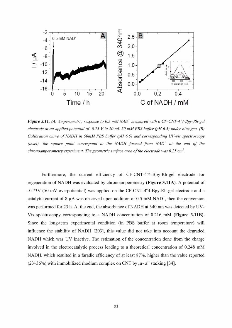
91
Figure 3.11. (A) Amperometric response to 0.5 mM NAD+ measured with a CF-CNT-4’4-Bpy-Rh-gel
electrode at an applied potential of -0.73 V in 20 mL 50 mM PBS buffer (pH 6.5) under nitrogen. (B)
Calibration curve of NADH in 50mM PBS buffer (pH 6.5) and corresponding UV-vis spectroscopy
(inset), the square point correspond to the NADH formed from NAD+ at the end of the
chronoamperometry experiment. The geometric surface area of the electrode was 0.25 cm2.
Furthermore, the current efficiency of CF-CNT-4‟4-Bpy-Rh-gel electrode for
regeneration of NADH was evaluated by chronoamperometry (Figure 3.11A). A potential of
-0.73V (50 mV overpotiential) was applied on the CF-CNT-4‟4-Bpy-Rh-gel electrode and a
catalytic current of 8 µA was observed upon addition of 0.5 mM NAD+, then the conversion
was performed for 23 h. At the end, the absorbance of NADH at 340 nm was detected by UV-
Vis spectroscopy corresponding to a NADH concentration of 0.216 mM (Figure 3.11B).
Since the long-term experimental condition (in PBS buffer at room temperature) will
influence the stability of NADH [203], this value did not take into account the degraded
NADH which was UV inactive. The estimation of the concentration done from the charge
involved in the electrocatalytic process leading to a theoretical concentration of 0.248 mM
NADH, which resulted in a faradic efficiency of at least 87%, higher than the value reported
(23–36%) with immobilized rhodium complex on CNT by „π- π‟ stacking [34].
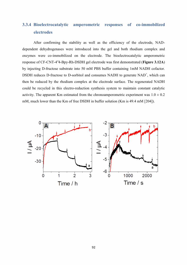
92
3.3.4 Bioelectrocatalytic amperometric responses of co-immobilized
electrodes
After confirming the stability as well as the efficiency of the electrode, NAD-
dependent dehydrogenases were introduced into the gel and both rhodium complex and
enzymes were co-immobilized on the electrode. The bioelectrocatalytic amperometric
response of CF-CNT-4‟4-Bpy-Rh-DSDH gel electrode was first demonstrated (Figure 3.12A)
by injecting D-fructose substrate into 50 mM PBS buffer containing 1mM NADH cofactor.
DSDH reduces D-fructose to D-sorbitol and consumes NADH to generate NAD+, which can
then be reduced by the rhodium complex at the electrode surface. The regenerated NADH
could be recycled in this electro-reduction synthesis system to maintain constant catalytic
activity. The apparent Km estimated from the chronoamperometric experiment was 1.0 ± 0.2
mM, much lower than the Km of free DSDH in buffer solution (Km is 49.4 mM [204]).

93
C
Figure 3.12. (A) Amperometric response to increasing concentration of D-fructose from 0.5 to 3mM
recorded using (a) CF-CNT-4’4-Bpy-Rh-DSDH-gel electrode (b) CF-CNT-4’4-Bpy-Rh-gel electrode
measured in 50 mM PBS (pH 6.5) contains 1 mM NADH at an applied potential of -0.74 V. (B)
Amperometric response to increasing concentration of hydroxyacetone from 1 to 4 mM recorded using
(a) CF-CNT-4’4-Bpy-Rh-GatDH-gel (b) CF-CNT-4’4-Bpy-Rh-gel electrode measured in 50 mM PBS
(pH 6.5) contains 1 mM NADH and 1mM MgCl2 at an applied potential of -0.72 V. Both experiments
were carried out under nitrogen. (C) Schematic representation of the electroenzymatic synthetic
process for CF-CNT-4’4-Bpy-Rh-DSDH-gel electrode. The geometric surface area of the electrode
was 0.25 cm2.

94
To test the applicability of CF-CNT-4‟4-Bpy-Rh electrode with other NAD-dependent
redox enzymes, Galactitol dehydrogenase (GatDH) was also encapsulated into the gel layer
instead of DSDH. GatDH is a multifunctional enzyme that can catalyze broad substrates [205],
here we choose hydroxyacetone as the substrate to investigate its electroenzymatic property.
Figure 3.12B shows the amperometric response against the addition hydroxyacetone
substrate in 50 mM PBS buffer containing 1 mM NADH cofactor and 1 mM MgCl2 on a
CNT-Rh-GatDH gel electrode which contained GatDH instead of DSDH. Even through the
response was lower than with DSDH electrode due to its low enzymatic activity, it indicated
that this co-immobilized electrode was able to be applied on different kinds of NAD-
dependent enzymes which is promising for bio-electrochemical synthesis of a variety of
valuable products.
Figure 3.13. (A) Amperometric response recorded using CF-CNT-4’4-Bpy-Rh-DSDH gel electrode to
addition of 4 mM of D-fructose in the beginning, 1 mM of D-fructose and 1 mM NADH in the end after
18 h in 50 mM PBS (pH 6.5) contains 1 mM NADH. (B) Amperometric response to addition of 4 mM
of D-fructose using the same CF-CNT-4’4-Bpy-Rh-DSDH gel electrode in fresh 50 mM PBS (pH 6.5)
contains 1 mM NADH. Applied potential was -0.73 V. The geometric surface area of the electrode was
0.25 cm2.
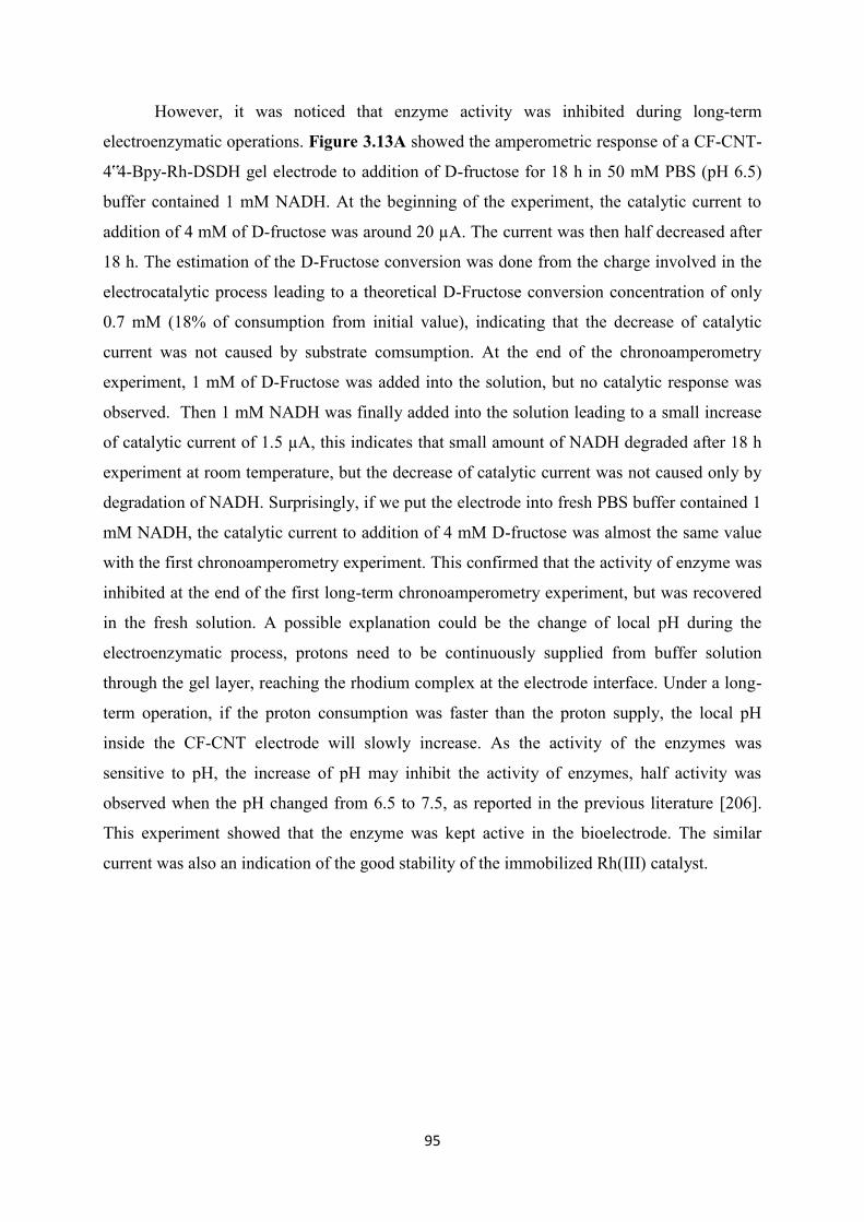
95
However, it was noticed that enzyme activity was inhibited during long-term
electroenzymatic operations. Figure 3.13A showed the amperometric response of a CF-CNT-
4‟4-Bpy-Rh-DSDH gel electrode to addition of D-fructose for 18 h in 50 mM PBS (pH 6.5)
buffer contained 1 mM NADH. At the beginning of the experiment, the catalytic current to
addition of 4 mM of D-fructose was around 20 µA. The current was then half decreased after
18 h. The estimation of the D-Fructose conversion was done from the charge involved in the
electrocatalytic process leading to a theoretical D-Fructose conversion concentration of only
0.7 mM (18% of consumption from initial value), indicating that the decrease of catalytic
current was not caused by substrate comsumption. At the end of the chronoamperometry
experiment, 1 mM of D-Fructose was added into the solution, but no catalytic response was
observed. Then 1 mM NADH was finally added into the solution leading to a small increase
of catalytic current of 1.5 µA, this indicates that small amount of NADH degraded after 18 h
experiment at room temperature, but the decrease of catalytic current was not caused only by
degradation of NADH. Surprisingly, if we put the electrode into fresh PBS buffer contained 1
mM NADH, the catalytic current to addition of 4 mM D-fructose was almost the same value
with the first chronoamperometry experiment. This confirmed that the activity of enzyme was
inhibited at the end of the first long-term chronoamperometry experiment, but was recovered
in the fresh solution. A possible explanation could be the change of local pH during the
electroenzymatic process, protons need to be continuously supplied from buffer solution
through the gel layer, reaching the rhodium complex at the electrode interface. Under a long-
term operation, if the proton consumption was faster than the proton supply, the local pH
inside the CF-CNT electrode will slowly increase. As the activity of the enzymes was
sensitive to pH, the increase of pH may inhibit the activity of enzymes, half activity was
observed when the pH changed from 6.5 to 7.5, as reported in the previous literature [206].
This experiment showed that the enzyme was kept active in the bioelectrode. The similar
current was also an indication of the good stability of the immobilized Rh(III) catalyst.
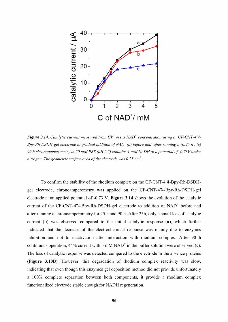
96
Figure 3.14. Catalytic current measured from CV versus NAD+ concentration using a CF-CNT-4’4-
Bpy-Rh-DSDH-gel electrode to gradual addition of NAD+ (a) before and after running a (b)25 h , (c)
90 h chronoamperometry in 50 mM PBS (pH 6.5) contains 1 mM NADH at a potential of -0.73V under
nitrogen. The geometric surface area of the electrode was 0.25 cm2.
To confirm the stability of the rhodium complex on the CF-CNT-4‟4-Bpy-Rh-DSDH-
gel electrode, chronoamperometry was applied on the CF-CNT-4‟4-Bpy-Rh-DSDH-gel
electrode at an applied potential of -0.73 V. Figure 3.14 shows the evolution of the catalytic
current of the CF-CNT-4‟4-Bpy-Rh-DSDH-gel electrode to addition of NAD+ before and
after running a chronoamperometry for 25 h and 90 h. After 25h, only a small loss of catalytic
current (b) was observed compared to the initial catalytic response (a), which further
indicated that the decrease of the electrochemical response was mainly due to enzymes
inhibition and not to inactivation after interaction with rhodium complex. After 90 h
continuous operation, 44% current with 5 mM NAD+ in the buffer solution were observed (c).
The loss of catalytic response was detected compared to the electrode in the absence proteins
(Figure 3.10B). However, this degradation of rhodium complex reactivity was slow,
indicating that even though this enzymes gel deposition method did not provide unfortunately
a 100% complete separation between both components, it provide a rhodium complex
functionalized electrode stable enough for NADH regeneration.
97
3.4 Conclusion
[Cp*Rh(bpy)Cl]+ was successfully immobilized on a CF-CNT electrode by covalent
bonding. Azido-functionalized electrodes were prepared by electrochemical reduction of 4-
azidophenyl diazonium cations that could be used later on to incorporate bipyridine ligands
via cycloaddition Huisgen reaction. A final post-functionalization step was based on the
ability of bipyridine ligands to coordinate with rhodium centers such as (RhCp*Cl2)2 dimer
under mild conditions yielding CF-CNT-4‟4-Bpy-Rh electrode. The catalytic response of this
CF-CNT-4‟4-Bpy-Rh electrode towards transformation from NAD+ to NADH was evaluated,
a faradic efficiency of 87% was measured. The application of the CF-CNT-4‟4-Bpy-Rh
electrode was demonstrated by combining NAD-dependent enzymes encapsulated in silica gel
on the top of the electrode (D-sorbitol dehydrogenase and galactitol dehydrogenase).
However, enzyme inhabitation was observed that was probably caused by the local
environment of the enzymes (pH, sorbitol accumulation). Moreover, a slow degradation of
rhodium complex was still observed.
98
Chapter 4. Functionalization of carbon felt supported carbon nanotubes by combining
diazonium chemistry and a sequential dual click chemistry approach
Concerning the enzyme inhibition effect in Chapter 3, a more opened functionalized
surface of electrode was built in this chapter. A generic approach has been developed for
heterogeneous surface modification of carbon felt/carbon nanotubes (CF-CNT) electrode
composites. Such strategy, applicable for a wide range of functional groups, provides a
versatile methodology to yield multifunctionalized carbon surfaces that can employed in a
variety of applications. The sequential method is consisting of two main steps. In the first step,
azide-alkene bifunctionalized CF-CNT electrodes (CF-CNT-AzVi) are achieved by
electrochemical reduction of a mixture of the corresponding diazonium salts generated in situ
from 4-azidoaniline and a 4-vinylaniline. Reactive sites available for further functionalization,
azide and alkene moieties, are incorporated in the carbon surfaces during the first step. Two
consecutive „click reactions‟ can be subsequently used to immobilize the targeted molecules.
In that way, we provide reactive sites that are available for further functionalization in a
sequential process based on the azide-alkyne cycloaddition Huisgen reaction and alkene-thiol
coupling. The concept by sequentially combining the diazonium chemistry and robust
chemical reactions („click chemistry‟) allows versatile, simple, and environmentally friendly
heterogeneous modification of CF-CNT substrates. The methodology has been employed first
to co-immobilize cobaltocenium and ferrocene moieties on the CF-CNT-AzVi which can be
characterized by cyclic voltammetry and X-ray photoelectron spectroscopy measurements.
This pair of compounds has been used as a model to demonstrate the feasibility of the generic
approach developed. After proving the possibility to use this method to introduce various
functional groups in a selective way, the work has been extended to the co-immobilization of
a molecule catalyst [Cp*Rh(bpy)Cl]+ and a NAD-dependent dehydrogenase that were proved
to act simultaneously in electroenzymatic conversion of D-fructose to D-sorbitol.
99
4.1 Introduction
In the early 90s, the most widely used system for modifying surfaces with monolayers
containing multiple components was initially the alkanethiol chemistry, where thiol groups
spontaneously adsorb onto gold. However, a number of disadvantages arising from the low
strength of the interaction between the thiol and the substrates [207], such as thermal
instability [208], evidence of the changing structures over time [209] and limited potential
window (typically -0.8 to +0.8 V versus Ag/AgCl) [207] justified the growing interest in
alternative processes overcoming some of the mentioned disadvantages. Later on, several
methods have been reported in the literature describing the modification of surfaces by means
of covalent bonding (electrochemical reduction of aryl diazonium salts [210–246],
electrochemical oxidation of alkyl amines [247], for instance). Among them, the
electrochemical reduction of aryl diazonium salts, firstly reported by Pinson and co-workers
[210], has been extensively used to prepare modified electrodes. Such electrografting method
was based on the electrochemical generation of highly reactive aryl radicals concerted with
the loss of N2. These aryl radicals are able to attack the electrode surface resulting on the
formation of an organic monolayer covalently attached to the substrate. The method can be
applied to a wide range of substrates such as carbon, silicon and metal surfaces [210–254].
The generation of organic monolayers by means of a covalent bond provides functionalized
surfaces which are highly stable, resistant to heat, ultrasonication and chemical degradation.
However, depending on the application considered, the high reactivity of these aryl
radicals could be less convenient since they can further react with phenyl groups already
grafted on the electrode surface and multilayers can be obtained. The formation of organic
multilayers can result in the formation of insulating coatings that usually are not favorable for
electrochemical applications. In this sense, the control of the compacting degree of the
organic layer is mandatory.
Since the introduction of several organic functions at the electrode surface would be
advantageous in a large variety of applications, including surface wetting, cell biology,
chemical sensing, biosensing and photosynthesis, multifunctionalized surfaces based on
covalent bonds have been already prepared by electrochemical reduction of mixtures of aryl
diazonium cations [246,248–252]. The main drawback of such modification approach to
obtain multiple organic functions on the substrate is the low degree of selectivity due to the
high reactivity of the aryl radicals generated, making difficult their use to obtain mixed
100
organic layers with a controlled composition. Different experimental approaches have been
described in the literature in order to get a precise control over the composition of binary
monolayers deposited at the electrode surface. Applying a constant cathodic potential
negative enough to reduce all the different diazonium cations present in solution was a first
strategy employed. The results obtained when working in solutions of different composition
suggested that changing the composition of the starting mixture in solution do not make a
significant difference in the final composition of the organic layers deposited on the electrode
surface. This fact could be rationalized taking into account that, even in the most optimal
situations, where the peak potential separation for the reduction of two different diazonium
salts can be up to 1V, the efficiency of the grafting is not the same for all the species of
different nature [253–255]. Another factor influencing the final composition of the deposited
organic layer is the potential required to reduce the aryldiazonium cations involved in the
grafting which leads to a most important surface concentration for the species which are easy
to reduce even if its concentration in the starting solution is lower.
The demand/requirement of more selective modification processes that could address
the difficulties encountered in the control of the composition of binary layers prepared by
electrochemical reduction of diazonium cations has opened the door to the development of
novel methods to incorporate organic functions in a variety of substrates by means of covalent
bonds. In this sense, more recently new indirect and versatile methods have been developed
in order to get functionalized substrates providing new functional platforms which allow the
selective incorporation of a variety of organic groups. Among them, “click chemistry”
approaches are attracting increasing attention in the field of materials science [11–13], being
an easy and powerful mean for joining molecular species to each other in rather mild
conditions as first reported in 2001 [256]. The mild reaction conditions of the copper (I)-
catalyzed azide-alkyne cycloaddition (CuAAC), producing 1,2,3-triazoles and its very high
yield makes this reaction particularly suitable for the functionalization of a large variety of
substrates[257]. Lately, other reactions are classified as click-type reactions since they
accomplished the requirements established by Kolb et al. [256]. (that is modular, wide in
scope, gives close to quantitative yields, generates inoffensive (if any) byproducts that are
easily removed by non-chromatographic methods and stereospecific.) The required processes
use to take place under simple and mild experimental reaction conditions (insensitive to
oxygen and water), readily accessible starting materials and reagents, the use of no solvent or
an environmentally friendly solvent (such as water) which should be easily removed and
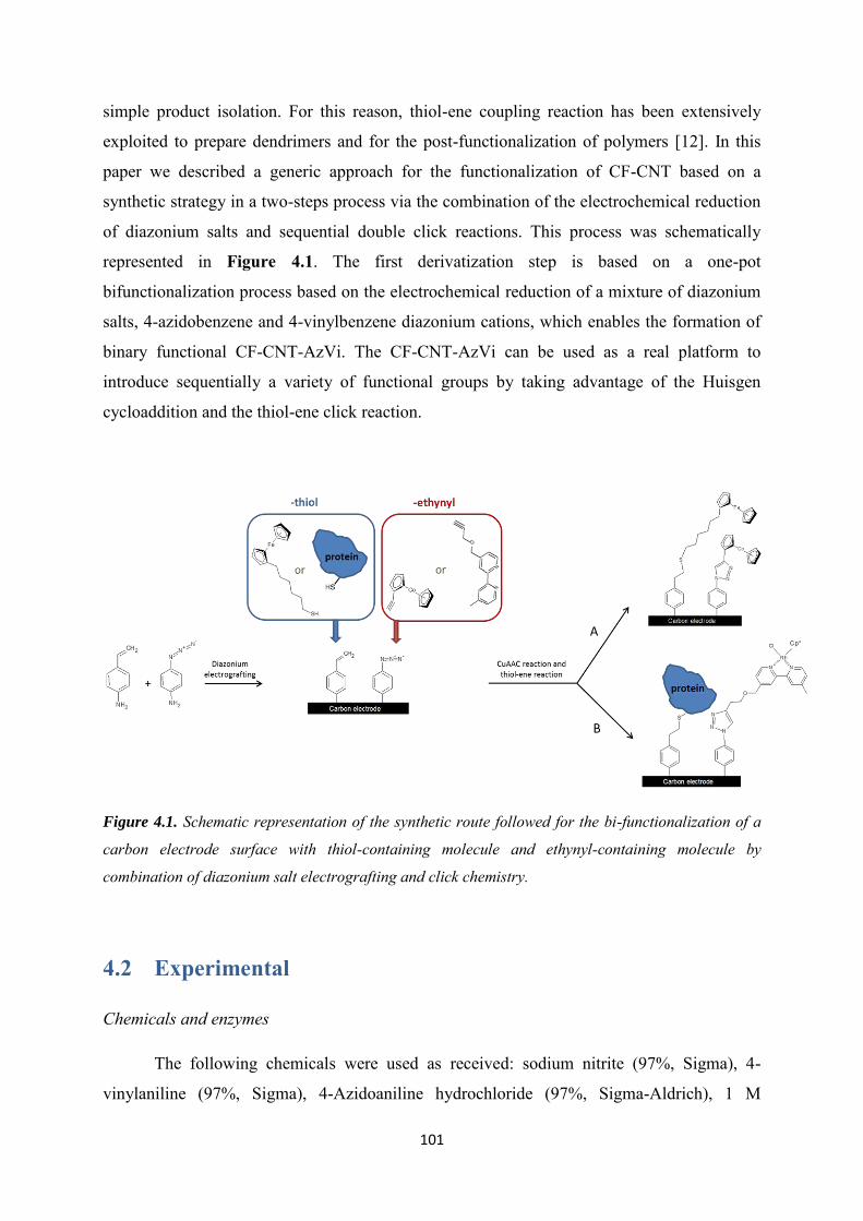
101
simple product isolation. For this reason, thiol-ene coupling reaction has been extensively
exploited to prepare dendrimers and for the post-functionalization of polymers [12]. In this
paper we described a generic approach for the functionalization of CF-CNT based on a
synthetic strategy in a two-steps process via the combination of the electrochemical reduction
of diazonium salts and sequential double click reactions. This process was schematically
represented in Figure 4.1. The first derivatization step is based on a one-pot
bifunctionalization process based on the electrochemical reduction of a mixture of diazonium
salts, 4-azidobenzene and 4-vinylbenzene diazonium cations, which enables the formation of
binary functional CF-CNT-AzVi. The CF-CNT-AzVi can be used as a real platform to
introduce sequentially a variety of functional groups by taking advantage of the Huisgen
cycloaddition and the thiol-ene click reaction.
Figure 4.1. Schematic representation of the synthetic route followed for the bi-functionalization of a
carbon electrode surface with thiol-containing molecule and ethynyl-containing molecule by
combination of diazonium salt electrografting and click chemistry.
4.2 Experimental
Chemicals and enzymes
The following chemicals were used as received: sodium nitrite (97%, Sigma), 4-
vinylaniline (97%, Sigma), 4-Azidoaniline hydrochloride (97%, Sigma-Aldrich), 1 M

102
hydrochloric acid solution (Sigma), β-Nicotinamide adenine dinucleotide hydrate (NAD+, ≥
96.5%, Sigma-Aldrich ), β-Nicotinamide adenine dinucleotide, reduced disodium salt hydrate
(NADH, ≥97%, Sigma-Aldrich), 6-(ferrocenyl)hexanethiol (Sigma), D-fructose (99 wt%,
Sigma), sodium dihydrogen phosphate (99.5%, Merck), Tris(2-carboxyethyl)phosphine
hydrochloride (TCEP, 98%, Sigma ), Carboxylic-functionalized multi-walled carbon
nanotubes (MWCNT, 95%, Φ 15±5 nm, L 1-5 µm, Nanolab), the carbon felt carpet
(Sigratherm®, GFD 4.6 EA) was supplied by SGL Group (Germany), Overproduction of N-
His(6)-Cys (Cys-DSDH) tagged DSDH variants was done with Escherichia coli BL21GOLD
(DE3) containing the corresponding expression-vectors pET-24a(+) (Novagen) and
purification of the enzyme was performed with Histrap columns (GE Healthcare)
Synthesis of ethynylcobaltocenium (Cc+)
Ethynylcobaltocenium was synthesized by following an experimental procedure
slightly modified from the literature. The synthetic procedure is based on the introduction of a
suitable carbanion and subsequent hydride abstraction by nucleophilic attack into a cationic
sandwich as cobaltocenium. In a first step of the synthetic procedure, to a suspension of
cobaltocenium hexafluorophosphate in THF at 0°C was added lithium ethynylide complex.
The reaction mixture was stirred for 30 min at 4-5°C and the solvent was evaporated under
vacuum. The solid residue was dissolved in cyclohexane. The solution was filtered through
Celite and evaporated and the compound obtained was purified.
In the second step of the synthesis, the compound previously obtained was dissolved
in dichloromethane and trityliumhexafluorophosphate was added. The mixture was stirred at
room temperature for 10 minutes. Afterwards the solvent was removed under vacuum and the
compound was purified by chromatography.
Procedures
All electrochemical experiments were performed using an Autolab PGSTAT-12
potentiostat, with a three-electrode configuration cell including an Ag/AgCl reference
electrode (3 M KCl), a steel auxiliary electrode, and a working electrode. A pencil core (0.5
mm diameter) glued with a copper wire was used for connecting the carbon felt as working
electrode. All the electrochemical measurements performed in negative potential window are
under nitrogen flow, and substrates were purged under nitrogen for 15 min before adding into
the electrochemical cell, in order to eliminate the influence of oxygen.
103
X-ray Photoelectron Spectroscopy (XPS) measurements.
The surface chemical composition of the samples was evaluated by using X-Ray
Photoelectron Spectroscopy (XPS). The measurements were performed using a KRATOS
Axis Ultra X-Ray Photoelectron spectrometer (Kratos Analytical, Manchester, UK) equipped
with a monochromated AlK X-Ray source (h = 1486.6 eV) operated at 150 W. The low
pressure in the analytical chamber was 10-9 mbar during XPS measurements. Wide scans were
recorded using an analyzer pass energy of 160 eV and narrow scans using a pass energy of 20
eV (instrumental resolution better than 0.5 eV). Charge correction was carried out using the
C1s core line, setting adventitious carbon signal (H/C signal) to 284.6 eV.
Preparation of multi-wall carbon nanotube functionalized electrode
The surface of carbon felt was first activated by running 20 cyclic voltammetry scans
over the range from -0.7 V to 2.5 V in 0.1M H2SO4 solution. Then the carbon felt was rinsed
carefully with water followed by heat treatment at 200 ℃ for 1h to eliminate the H2SO4
residues. Subsequently, the MWCNT-functionalized carbon felt was prepared by repeating
the dipping/drying process for 10 cycles. The dipping step was carried out in 0.5 mg/mL
MWCNT suspension, and the drying step is at 130℃ in the oven.
Azido and vinyl groups functionalized electrode (CF-CNT-AzVi)
Bi-functionalized electrodes CF-CNT-AzVi were prepared from solution containing a
mixture of the corresponding diazonium cations. To a solution containing 0.3 mM 4-
vinylaniline and 0.7 mM 4-azidoaniline in 0.5 M HCl was added 2 mM sodium nitrite with
continuous stirring for 5 min at room temperature. Then the grafting process was applied
immediately on the CF-MWCNT electrode by running two successive scans from 0.4 V to -
0.6 V, after rinsing with water, the vinyl and azido groups functionalized electrode was
obtained.
Ferrocene and cobaltocenium co-functionalized electrode (CF-CNT-Cc+Fc)
A sequential dual click chemistry approach was used in order to carry out the post-
functionalization of the CF-CNT-AzVi composite electrodes. Such sequential process has
been performed primarily using a couple of electroactive molecules, ethynylcobaltocenium
and 6-(ferrocenyl)hexanethiol to exemplify the feasibility of such strategy. The advantage of
using the ethynylcobaltocenium and 6-(ferrocenyl)hexanethiol, having active sites allowing
104
the sequential post-functionalization is based on their characteristic electrochemical behavior
that can be studied by cyclic voltammetry. The post-functionalization process consists of the
combination of two different consecutive click reactions, copper (I)-catalyzed „alkyne-azide‟
cycloaddition (CuAAC) and „thiol-ene‟ click coupling, for which it has been previously
proves that they can be accomplished in a stepwise procedure without interaction between the
reactants. The reaction sequence employed was as follows: the electrode was first dipped into
a mixture of DMF/H2O (70/30) solution containing 0.1 mM copper acetate, 0.3 mM ascorbic
acid and 1mM ethynylcobaltocenium. The reaction mixture was left for 24 h at room
temperature in the darkness. Afterwards, the electrode was thoroughly rinsed with DMF and
water to remove residual species that could remain adsorbed on the substrate. After drying,
the sequence was completed in a second step by a thiol-ene click reaction. The electrode was
dipped in methanol/H2O (70/30) mixture in the presence of 1 mM TCEP and 1 mM 6-
(ferrocenyl)hexanethiol overnight. After completion of reaction, the ferrocene /cobaltocenium
functionalized electrodes were washed with water and methanol before use.
DSDH and rhodium complex co-functionalized electrode (CF-CNT-Rh/DSDH)
After proving the possibility to perform a double post-functionalization on the CF-
CNT-AzVi via the sequential dual click chemistry process, this generic and versatile approach
has been used to co-immobilize DSDH and rhodium complex. The co-functionalization
process involves two steps. The CF-CNT-AzVi electrode was first put into a DMF/H2O
(70/30) solution consisting of 4-[(2-propyn-1-yloxy)methyl]-4‟-methyl-2,2‟-bipyridine (1
mM)( synthetic procedure was following the reported literature [142]), and copper acetate
(0.3 mM) and ascorbic acid (0.9 mM) for 24 h in the darkness. The modified electrode was
thoroughly rinsed with water, DMF and followed by a last rinsing in dichloromethane (DCM).
A complexation step of the bipyridine ligand to immobilize a rhodium complex was then
carried out. The electrode was then immersed in a DCM (20 mL) solution containing
(RhCp*Cl2)2 (2 mg) with continuous stirring for 3h and the CF-CNT-RhVi electrode was
obtained. Thiol-ene click reaction was subsequently used to co-functionalize the electrode
with cysteine-tagged DSDH taking advantage of the availability of the alkene functional
groups present on the substrate. The CF-CNT-RhVi electrode was immersed into 1 mL of
aqueous PBS buffer (250 mM, pH=7) containing 2.5 mg protein, 0.3 mM TCEP and 1 mM
MgCl2. The mixture was allowed to react at 4 ℃ overnight. The resulting CF-CNT-Rh/DSDH
electrode was rinsed carefully with water and buffer solutions before use.
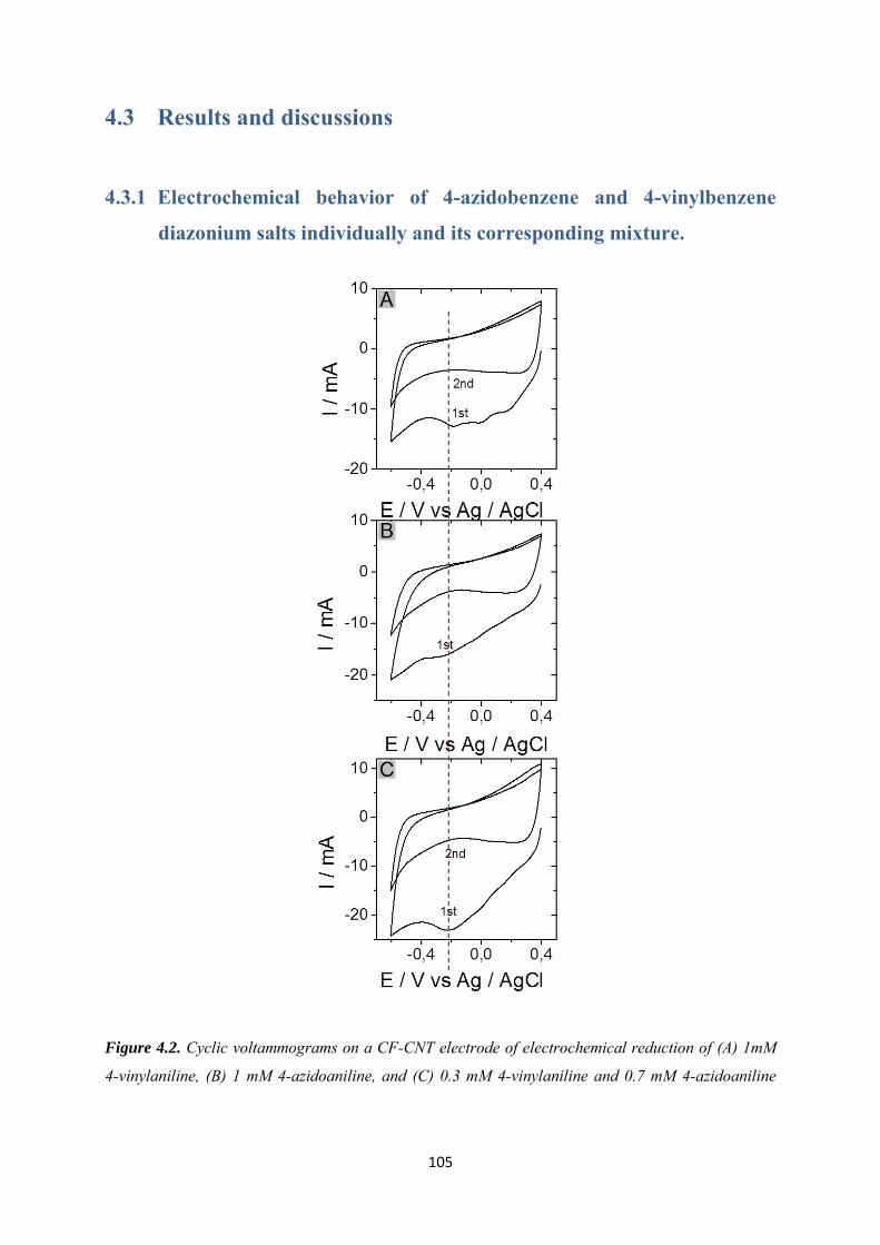
105
4.3 Results and discussions
4.3.1 Electrochemical behavior of 4-azidobenzene and 4-vinylbenzene
diazonium salts individually and its corresponding mixture.
Figure 4.2. Cyclic voltammograms on a CF-CNT electrode of electrochemical reduction of (A) 1mM
4-vinylaniline, (B) 1 mM 4-azidoaniline, and (C) 0.3 mM 4-vinylaniline and 0.7 mM 4-azidoaniline
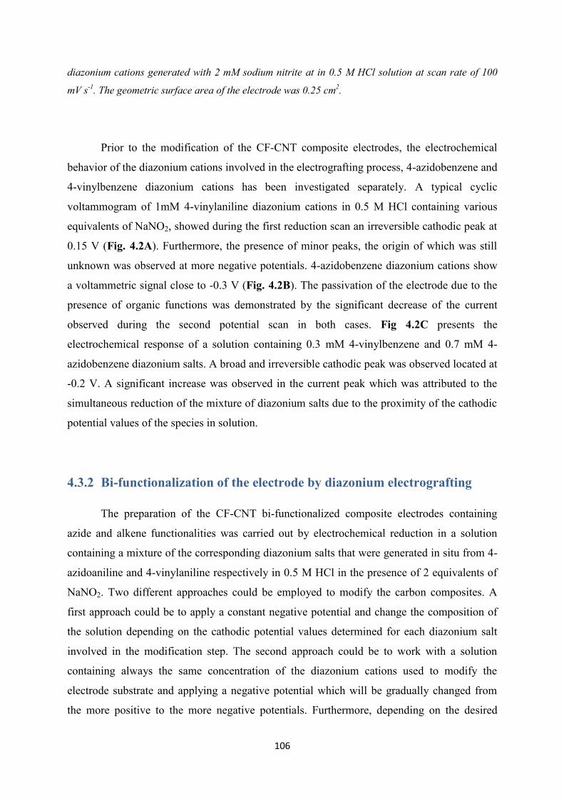
106
diazonium cations generated with 2 mM sodium nitrite at in 0.5 M HCl solution at scan rate of 100
mV s-1. The geometric surface area of the electrode was 0.25 cm2.
Prior to the modification of the CF-CNT composite electrodes, the electrochemical
behavior of the diazonium cations involved in the electrografting process, 4-azidobenzene and
4-vinylbenzene diazonium cations has been investigated separately. A typical cyclic
voltammogram of 1mM 4-vinylaniline diazonium cations in 0.5 M HCl containing various
equivalents of NaNO2, showed during the first reduction scan an irreversible cathodic peak at
0.15 V (Fig. 4.2A). Furthermore, the presence of minor peaks, the origin of which was still
unknown was observed at more negative potentials. 4-azidobenzene diazonium cations show
a voltammetric signal close to -0.3 V (Fig. 4.2B). The passivation of the electrode due to the
presence of organic functions was demonstrated by the significant decrease of the current
observed during the second potential scan in both cases. Fig 4.2C presents the
electrochemical response of a solution containing 0.3 mM 4-vinylbenzene and 0.7 mM 4-
azidobenzene diazonium salts. A broad and irreversible cathodic peak was observed located at
-0.2 V. A significant increase was observed in the current peak which was attributed to the
simultaneous reduction of the mixture of diazonium salts due to the proximity of the cathodic
potential values of the species in solution.
4.3.2 Bi-functionalization of the electrode by diazonium electrografting
The preparation of the CF-CNT bi-functionalized composite electrodes containing
azide and alkene functionalities was carried out by electrochemical reduction in a solution
containing a mixture of the corresponding diazonium salts that were generated in situ from 4-
azidoaniline and 4-vinylaniline respectively in 0.5 M HCl in the presence of 2 equivalents of
NaNO2. Two different approaches could be employed to modify the carbon composites. A
first approach could be to apply a constant negative potential and change the composition of
the solution depending on the cathodic potential values determined for each diazonium salt
involved in the modification step. The second approach could be to work with a solution
containing always the same concentration of the diazonium cations used to modify the
electrode substrate and applying a negative potential which will be gradually changed from
the more positive to the more negative potentials. Furthermore, depending on the desired
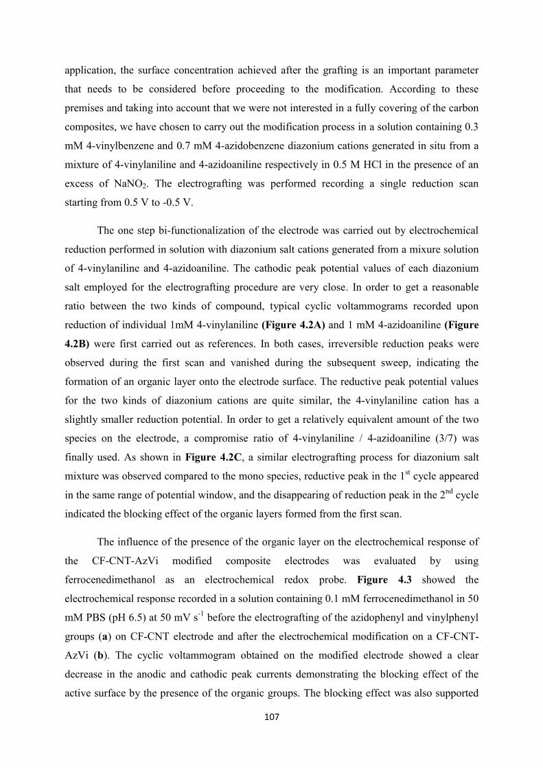
107
application, the surface concentration achieved after the grafting is an important parameter
that needs to be considered before proceeding to the modification. According to these
premises and taking into account that we were not interested in a fully covering of the carbon
composites, we have chosen to carry out the modification process in a solution containing 0.3
mM 4-vinylbenzene and 0.7 mM 4-azidobenzene diazonium cations generated in situ from a
mixture of 4-vinylaniline and 4-azidoaniline respectively in 0.5 M HCl in the presence of an
excess of NaNO2. The electrografting was performed recording a single reduction scan
starting from 0.5 V to -0.5 V.
The one step bi-functionalization of the electrode was carried out by electrochemical
reduction performed in solution with diazonium salt cations generated from a mixure solution
of 4-vinylaniline and 4-azidoaniline. The cathodic peak potential values of each diazonium
salt employed for the electrografting procedure are very close. In order to get a reasonable
ratio between the two kinds of compound, typical cyclic voltammograms recorded upon
reduction of individual 1mM 4-vinylaniline (Figure 4.2A) and 1 mM 4-azidoaniline (Figure
4.2B) were first carried out as references. In both cases, irreversible reduction peaks were
observed during the first scan and vanished during the subsequent sweep, indicating the
formation of an organic layer onto the electrode surface. The reductive peak potential values
for the two kinds of diazonium cations are quite similar, the 4-vinylaniline cation has a
slightly smaller reduction potential. In order to get a relatively equivalent amount of the two
species on the electrode, a compromise ratio of 4-vinylaniline / 4-azidoaniline (3/7) was
finally used. As shown in Figure 4.2C, a similar electrografting process for diazonium salt
mixture was observed compared to the mono species, reductive peak in the 1st cycle appeared
in the same range of potential window, and the disappearing of reduction peak in the 2nd cycle
indicated the blocking effect of the organic layers formed from the first scan.
The influence of the presence of the organic layer on the electrochemical response of
the CF-CNT-AzVi modified composite electrodes was evaluated by using
ferrocenedimethanol as an electrochemical redox probe. Figure 4.3 showed the
electrochemical response recorded in a solution containing 0.1 mM ferrocenedimethanol in 50
mM PBS (pH 6.5) at 50 mV s-1 before the electrografting of the azidophenyl and vinylphenyl
groups (a) on CF-CNT electrode and after the electrochemical modification on a CF-CNT-
AzVi (b). The cyclic voltammogram obtained on the modified electrode showed a clear
decrease in the anodic and cathodic peak currents demonstrating the blocking effect of the
active surface by the presence of the organic groups. The blocking effect was also supported
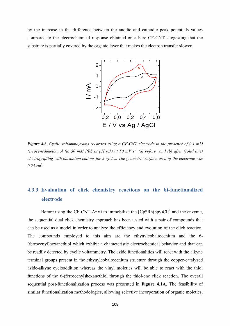
108
by the increase in the difference between the anodic and cathodic peak potentials values
compared to the electrochemical response obtained on a bare CF-CNT suggesting that the
substrate is partially covered by the organic layer that makes the electron transfer slower.
Figure 4.3. Cyclic voltammograms recorded using a CF-CNT electrode in the presence of 0.1 mM
ferrocenedimethanol (in 50 mM PBS at pH 6.5) at 50 mV s-1 (a) before and (b) after (solid line)
electrografting with diazonium cations for 2 cycles. The geometric surface area of the electrode was
0.25 cm2.
4.3.3 Evaluation of click chemistry reactions on the bi-functionalized
electrode
Before using the CF-CNT-AzVi to immobilize the [Cp*Rh(bpy)Cl]+ and the enzyme,
the sequential dual click chemistry approach has been tested with a pair of compounds that
can be used as a model in order to analyze the efficiency and evolution of the click reaction.
The compounds employed to this aim are the ethynylcobaltocenium and the 6-
(ferrocenyl)hexanethiol which exhibit a characteristic electrochemical behavior and that can
be readily detected by cyclic voltammetry. The azide functionalities will react with the alkyne
terminal groups present in the ethynylcobaltocenium structure through the copper-catalyzed
azide-alkyne cycloaddition whereas the vinyl moieties will be able to react with the thiol
functions of the 6-(ferrocenyl)hexanethiol through the thiol-ene click reaction. The overall
sequential post-functionalization process was presented in Figure 4.1A. The feasibility of
similar functionalization methodologies, allowing selective incorporation of organic moieties,
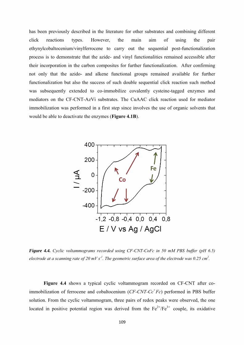
109
has been previously described in the literature for other substrates and combining different
click reactions types. However, the main aim of using the pair
ethynylcobaltocenium/vinylferrocene to carry out the sequential post-functionalization
process is to demonstrate that the azide- and vinyl functionalities remained accessible after
their incorporation in the carbon composites for further functionalization. After confirming
not only that the azido- and alkene functional groups remained available for further
functionalization but also the success of such double sequential click reaction such method
was subsequently extended to co-immobilize covalently cysteine-tagged enzymes and
mediators on the CF-CNT-AzVi substrates. The CuAAC click reaction used for mediator
immobilization was performed in a first step since involves the use of organic solvents that
would be able to deactivate the enzymes (Figure 4.1B).
Figure 4.4. Cyclic voltammograms recorded using CF-CNT-CoFc in 50 mM PBS buffer (pH 6.5)
electrode at a scanning rate of 20 mV s-1. The geometric surface area of the electrode was 0.25 cm2.
Figure 4.4 shows a typical cyclic voltammogram recorded on CF-CNT after co-
immobilization of ferrocene and cobaltocenium (CF-CNT-Cc+Fc) performed in PBS buffer
solution. From the cyclic voltammogram, three pairs of redox peaks were observed, the one
located in positive potential region was derived from the Fe2+/Fe3+ couple, its oxidative

110
voltammetric signal was located at +0.46 V. Another two pairs of redox peaks in the negative
potential window was due to cobaltocenium reduction, corresponding to Co3+/Co2+ and
Co2+/Co+ redox pairs. Their reductive voltammetric signal was located around -0.45 V and
-0.9 V, respectively. The electroactive behavior of the electrode not only indicates the
successful coupling of the two reactions with vinyl and azido groups, but also proves the
facility in electron transfer from molecular to the electrode after organic layer grafting.
Figure 4.5. XPS spectra of the (A) Co 2p and (B) Fe 2p core level spectrum from the cobaltocenium
and ferrocene functionalized sunbstrate.
X-Ray Photoelectron Spectroscopy measurements were performed to confirm the
uptake of cobaltocenium and ferrocene moieties on ITO modified electrodes. The
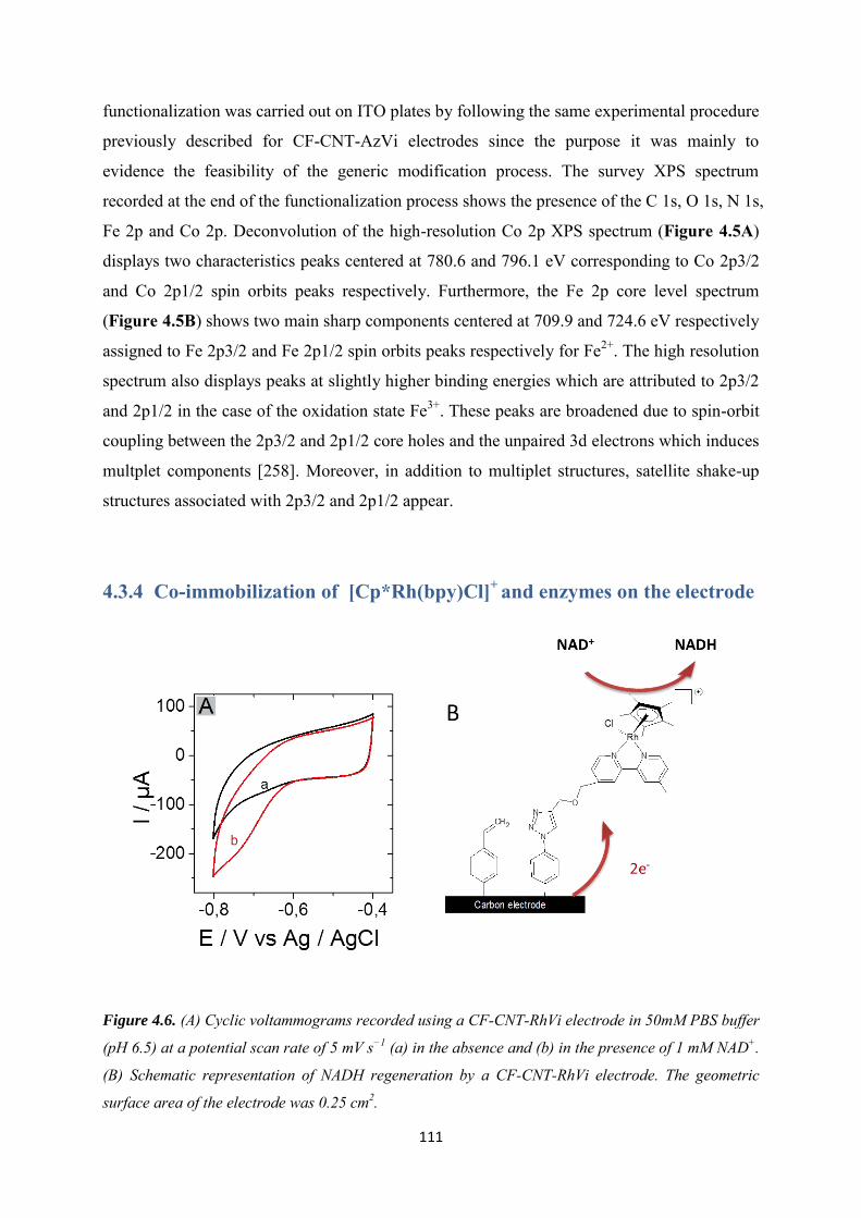
111
functionalization was carried out on ITO plates by following the same experimental procedure
previously described for CF-CNT-AzVi electrodes since the purpose it was mainly to
evidence the feasibility of the generic modification process. The survey XPS spectrum
recorded at the end of the functionalization process shows the presence of the C 1s, O 1s, N 1s,
Fe 2p and Co 2p. Deconvolution of the high-resolution Co 2p XPS spectrum (Figure 4.5A)
displays two characteristics peaks centered at 780.6 and 796.1 eV corresponding to Co 2p3/2
and Co 2p1/2 spin orbits peaks respectively. Furthermore, the Fe 2p core level spectrum
(Figure 4.5B) shows two main sharp components centered at 709.9 and 724.6 eV respectively
assigned to Fe 2p3/2 and Fe 2p1/2 spin orbits peaks respectively for Fe2+. The high resolution
spectrum also displays peaks at slightly higher binding energies which are attributed to 2p3/2
and 2p1/2 in the case of the oxidation state Fe3+. These peaks are broadened due to spin-orbit
coupling between the 2p3/2 and 2p1/2 core holes and the unpaired 3d electrons which induces
multplet components [258]. Moreover, in addition to multiplet structures, satellite shake-up
structures associated with 2p3/2 and 2p1/2 appear.
4.3.4 Co-immobilization of [Cp*Rh(bpy)Cl]+ and enzymes on the electrode
Figure 4.6. (A) Cyclic voltammograms recorded using a CF-CNT-RhVi electrode in 50mM PBS buffer
(pH 6.5) at a potential scan rate of 5 mV s−1 (a) in the absence and (b) in the presence of 1 mM NAD+.
(B) Schematic representation of NADH regeneration by a CF-CNT-RhVi electrode. The geometric
surface area of the electrode was 0.25 cm2.
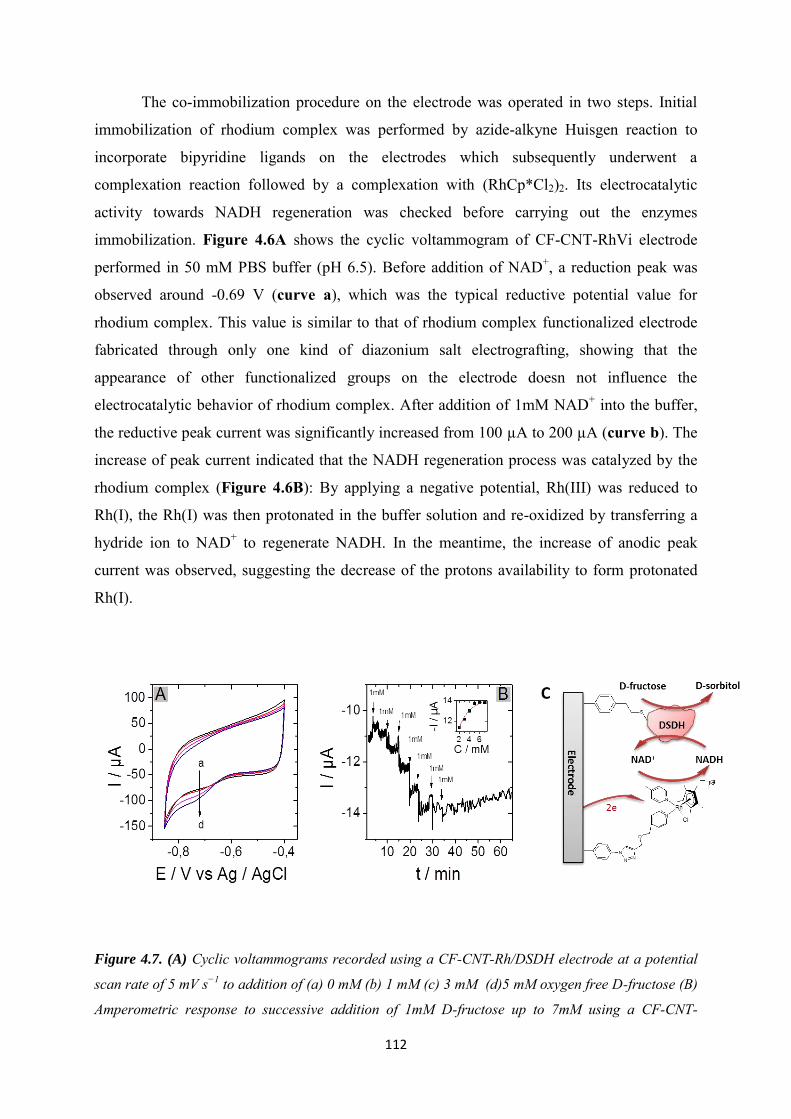
112
The co-immobilization procedure on the electrode was operated in two steps. Initial
immobilization of rhodium complex was performed by azide-alkyne Huisgen reaction to
incorporate bipyridine ligands on the electrodes which subsequently underwent a
complexation reaction followed by a complexation with (RhCp*Cl2)2. Its electrocatalytic
activity towards NADH regeneration was checked before carrying out the enzymes
immobilization. Figure 4.6A shows the cyclic voltammogram of CF-CNT-RhVi electrode
performed in 50 mM PBS buffer (pH 6.5). Before addition of NAD+, a reduction peak was
observed around -0.69 V (curve a), which was the typical reductive potential value for
rhodium complex. This value is similar to that of rhodium complex functionalized electrode
fabricated through only one kind of diazonium salt electrografting, showing that the
appearance of other functionalized groups on the electrode doesn not influence the
electrocatalytic behavior of rhodium complex. After addition of 1mM NAD+ into the buffer,
the reductive peak current was significantly increased from 100 µA to 200 µA (curve b). The
increase of peak current indicated that the NADH regeneration process was catalyzed by the
rhodium complex (Figure 4.6B): By applying a negative potential, Rh(III) was reduced to
Rh(I), the Rh(I) was then protonated in the buffer solution and re-oxidized by transferring a
hydride ion to NAD+ to regenerate NADH. In the meantime, the increase of anodic peak
current was observed, suggesting the decrease of the protons availability to form protonated
Rh(I).
Figure 4.7. (A) Cyclic voltammograms recorded using a CF-CNT-Rh/DSDH electrode at a potential
scan rate of 5 mV s−1 to addition of (a) 0 mM (b) 1 mM (c) 3 mM (d)5 mM oxygen free D-fructose (B)
Amperometric response to successive addition of 1mM D-fructose up to 7mM using a CF-CNT-
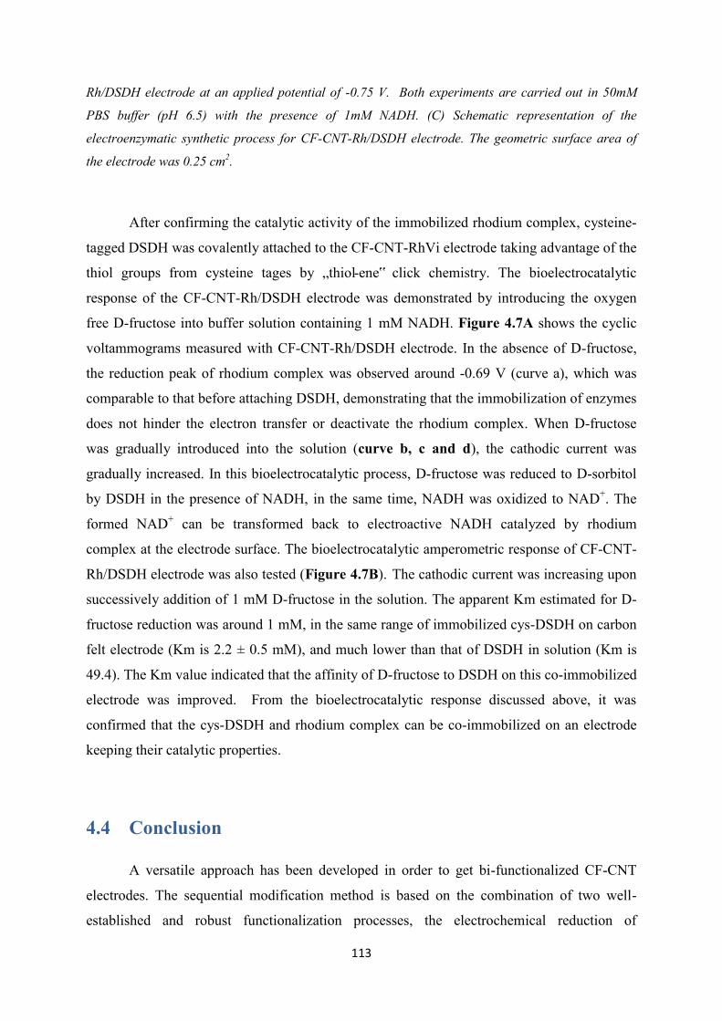
113
Rh/DSDH electrode at an applied potential of -0.75 V. Both experiments are carried out in 50mM
PBS buffer (pH 6.5) with the presence of 1mM NADH. (C) Schematic representation of the
electroenzymatic synthetic process for CF-CNT-Rh/DSDH electrode. The geometric surface area of
the electrode was 0.25 cm2.
After confirming the catalytic activity of the immobilized rhodium complex, cysteine-
tagged DSDH was covalently attached to the CF-CNT-RhVi electrode taking advantage of the
thiol groups from cysteine tages by „thiol-ene‟ click chemistry. The bioelectrocatalytic
response of the CF-CNT-Rh/DSDH electrode was demonstrated by introducing the oxygen
free D-fructose into buffer solution containing 1 mM NADH. Figure 4.7A shows the cyclic
voltammograms measured with CF-CNT-Rh/DSDH electrode. In the absence of D-fructose,
the reduction peak of rhodium complex was observed around -0.69 V (curve a), which was
comparable to that before attaching DSDH, demonstrating that the immobilization of enzymes
does not hinder the electron transfer or deactivate the rhodium complex. When D-fructose
was gradually introduced into the solution (curve b, c and d), the cathodic current was
gradually increased. In this bioelectrocatalytic process, D-fructose was reduced to D-sorbitol
by DSDH in the presence of NADH, in the same time, NADH was oxidized to NAD+. The
formed NAD+ can be transformed back to electroactive NADH catalyzed by rhodium
complex at the electrode surface. The bioelectrocatalytic amperometric response of CF-CNT-
Rh/DSDH electrode was also tested (Figure 4.7B). The cathodic current was increasing upon
successively addition of 1 mM D-fructose in the solution. The apparent Km estimated for D-
fructose reduction was around 1 mM, in the same range of immobilized cys-DSDH on carbon
felt electrode (Km is 2.2 ± 0.5 mM), and much lower than that of DSDH in solution (Km is
49.4). The Km value indicated that the affinity of D-fructose to DSDH on this co-immobilized
electrode was improved. From the bioelectrocatalytic response discussed above, it was
confirmed that the cys-DSDH and rhodium complex can be co-immobilized on an electrode
keeping their catalytic properties.
4.4 Conclusion
A versatile approach has been developed in order to get bi-functionalized CF-CNT
electrodes. The sequential modification method is based on the combination of two well-
established and robust functionalization processes, the electrochemical reduction of
114
diazonium cations and a double consecutive click reactions process. In situ electrografting of
a mixture of diazonium salts (4-azidobenzene and 4-vinylbenzene diazonium salts). The
cathodic peak potentials of the species involved in the functionalization step are very close
and the modification of the CF-CNT electrodes was achieved by recording a single scan
towards negative potentials by cyclic voltammetry. The co-functionalized electrodes allow
selective covalent bonding of alkyne and thiol organic moieties, via two „click‟
immobilization steps. The combination of these reactions with diazonium electrochemistry in
fabrication of electrodes did not hinder the electron transfer between redox probes and
electrode. The electron mediator [Cp*Rh(bpy)Cl]+ was immobilized by first coupling vinyl
2,2‟-bipyridine on the electrode via CuAAC reaction, followed a complexation step with
(RhCp*Cl2)2. Taking advantage of the thiol groups from cysteine tags, the cys-DSDH was
then coupled to vinyl groups on the electrode via thiol-ene click reaction. The activity of both
rhodium complex and D-sorbitol dehydrogenase were kept after click couplings were made.
The electrocatalytic reduction of D-fructose to D-sorbitol by the immobilized DSDH using
rhodium complex as mediator was evidenced by both cyclic voltammetry and
chronoamperometry. This facile co-immobilization route could be further used for other bio-
systems with different redox enzymes and mediators in the field of biosynthesis, biosensing
and fuel cell applications.

115
Chapter 5. Co-immobilization of rhodium complex and NAD-dependent dehydrogenase in an
electrochemical bioreactor for enantioselective bioconversion
Here we report a simple immobilization method for covalent bounding of rhodium
complex ([Cp*Rh(bpy)Cl]+) onto carbon electrode (CF-CNT, carbon nanotube functionalized
carbon felt or BP, „ bucky paper‟). The bipyridine ligand was first grafted on the electrode by
electro-reduction of bipyridyl diazonium cations generated from 4-amino-2,2‟-bipyridine
compound. After a mild complexation step with rhodium dimer (((RhCp*Cl2)2), the rhodium
complex functionalized electrode was obtained. Their electrocatalytic property towards
NADH regeneration was investigated for rhodium functionalized CF-CNT electrode. Taking
advantage of the thin layer structure of BP electrode, a separated glassy filter layer with D-
sorbitol dehydrogenase in the gel was overlaid on the top of the rhodium functionalized
electrode, the enzyme layers could be easily changed, the reusability of BP-Bpy-Rh electrode
was significantly improved. Finally, a bioconversion experiment for electroenzymatic
enantioselective reduction of D-fructose to D- sorbitol was carried out, a conversion rate of
87%, faradic effciency up to 83% and a high total turnover number (TTN) up to 12500 for
rhodium complex was achieved.
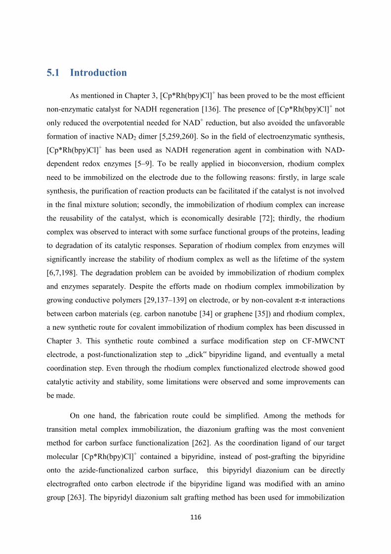
116
5.1 Introduction
As mentioned in Chapter 3, [Cp*Rh(bpy)Cl]+ has been proved to be the most efficient
non-enzymatic catalyst for NADH regeneration [136]. The presence of [Cp*Rh(bpy)Cl]+ not
only reduced the overpotential needed for NAD+ reduction, but also avoided the unfavorable
formation of inactive NAD2 dimer [5,259,260]. So in the field of electroenzymatic synthesis,
[Cp*Rh(bpy)Cl]+ has been used as NADH regeneration agent in combination with NAD-
dependent redox enzymes [5–9]. To be really applied in bioconversion, rhodium complex
need to be immobilized on the electrode due to the following reasons: firstly, in large scale
synthesis, the purification of reaction products can be facilitated if the catalyst is not involved
in the final mixture solution; secondly, the immobilization of rhodium complex can increase
the reusability of the catalyst, which is economically desirable [72]; thirdly, the rhodium
complex was observed to interact with some surface functional groups of the proteins, leading
to degradation of its catalytic responses. Separation of rhodium complex from enzymes will
significantly increase the stability of rhodium complex as well as the lifetime of the system
[6,7,198]. The degradation problem can be avoided by immobilization of rhodium complex
and enzymes separately. Despite the efforts made on rhodium complex immobilization by
growing conductive polymers [29,137–139] on electrode, or by non-covalent π-π interactions
between carbon materials (eg. carbon nanotube [34] or graphene [35]) and rhodium complex,
a new synthetic route for covalent immobilization of rhodium complex has been discussed in
Chapter 3. This synthetic route combined a surface modification step on CF-MWCNT
electrode, a post-functionalization step to „click‟ bipyridine ligand, and eventually a metal
coordination step. Even through the rhodium complex functionalized electrode showed good
catalytic activity and stability, some limitations were observed and some improvements can
be made.
On one hand, the fabrication route could be simplified. Among the methods for
transition metal complex immobilization, the diazonium grafting was the most convenient
method for carbon surface functionalization [262]. As the coordination ligand of our target
molecular [Cp*Rh(bpy)Cl]+ contained a bipyridine, instead of post-grafting the bipyridine
onto the azide-functionalized carbon surface, this bipyridyl diazonium can be directly
electrografted onto carbon electrode if the bipyridine ligand was modified with an amino
group [263]. The bipyridyl diazonium salt grafting method has been used for immobilization
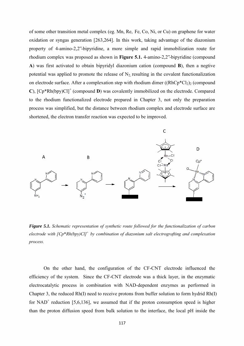
117
of some other transition metal complex (eg. Mn, Re, Fe, Co, Ni, or Cu) on graphene for water
oxidation or syngas generation [263,264]. In this work, taking advantage of the diazonium
property of 4-amino-2,2‟-bipyridine, a more simple and rapid immobilization route for
rhodium complex was proposed as shown in Figure 5.1. 4-amino-2,2‟-bipyridine (compound
A) was first activated to obtain bipyridyl diazonium cation (compound B), then a negtive
potential was applied to promote the release of N2, resulting in the covalent functionalization
on electrode surface. After a complexation step with rhodium dimer ((RhCp*Cl2)2 (compound
C), [Cp*Rh(bpy)Cl]+ (compound D) was covalently immobilized on the electrode. Compared
to the rhodium functionalized electrode prepared in Chapter 3, not only the preparation
process was simplified, but the distance between rhodium complex and electrode surface are
shortened, the electron transfer reaction was expected to be improved.
Figure 5.1. Schematic representation of synthetic route followed for the functionalization of carbon
electrode with [Cp*Rh(bpy)Cl]+ by combination of diazonium salt electrografting and complexation
process.
On the other hand, the configuration of the CF-CNT electrode influenced the
efficiency of the system. Since the CF-CNT electrode was a thick layer, in the enzymatic
electrocatalytic process in combination with NAD-dependent enzymes as performed in
Chapter 3, the reduced Rh(I) need to receive protons from buffer solution to form hydrid Rh(I)
for NAD+ reduction [5,6,136], we assumed that if the proton consumption speed is higher
than the proton diffusion speed from bulk solution to the interface, the local pH inside the
118
porous electrode can increase, significantly influencing the activity of enzymes [206], leading
to decrease of conversion efficiency of the system [142]. To get rid of the potential local pH
inhibition, thinner electrodes would be preferred, so another kind of thinner layered „bucky
paper‟ electrode could be employed. It was fabricated from stable suspensions of carbon
nanotubes (CNT), by controlling the volume of the suspension in the filtration process, the
thickness was controlled within hundred micrometers, this highly mesoporous layer was
mechanically robust, flexible, stable and electrically conductive [265]. „Bucky paper‟ has
been widely applied as working electrode in the field of bio-sensing [266] or bio-fuel cells
[265,267], it will also be a good candidate in electroenzymatic synthesis.
Figure 5.2. Configuration and electroenzymatic process involved of the packing electrode with
separated D-sorbitol dehydrogenase gel layer and rhodium functionalized BP layer. BP can be
prepared in the thickness range of 1~200 µm according to the reported literature [265].
119
Furthermore, the rhodium complex functionalized CF-CNT electrode with enzyme gel
layer deposited on the top showed good electroenzymatic catalytic response, however, a long-
term stability experiment show 40% decrease of catalytic current to NAD+ reduction at their
saturated concentrations over 90h continuous electrochemical operation (Chapter 3),
indicating that this enzymes immobilization method did not provide complete prevention of
interactions between the immobilized rhodium complex and enzyme in the gel. On the other
hand, the enzymes and the rhodium complex have different range of lifetime, the enzyme has
a quite limited one (eg. DSDH has a half-life period of 48 h at 28°C, 5 h at 35°C and 0.2 h at
40°C in solution) [204], while the [Cp*Rh(bpy)Cl]+ immobilized on CF-CNT was stable at
least for 2 weeks under continuous stirring in solution (Chapter 3). As soon as the enzymes
lost their activity, the gel deposited co-immobilized electrode has to be discarded. The
objective of the work in this chapter was to build an electrode which could be applied in
bioconversion without suffering from the above problems. If we physically separated the
long-term stable BP-Bpy-Rh layer with the lifetime-limited enzyme layer, we could build the
co-immobilized electrode by simple packing, in this way, the BP-Bpy-Rh electrode can be
reused with new enzyme gel layers, practically and economically improved the life-time of
the BP-Bpy-Rh electrode in system. In this work, as shown in Figure 5.2, we prepared an
individual enzyme gel layer by depositing enzyme gel on a piece of microfiber filter, then it
was overlaid on the top of BP-Bpy-Rh layer. The distance between enzymes and rhodium
complexes was close but physically restricted in two layers, the interaction between rhodium
complex and enzymes should be completely prevented, and the pH inhibition should also be
avoided due to their thin layer structures. The reusability of this rhodium functionalized BP
electrode will be studied, and the packed co-immobilized electrode will be applied to
bioconversion of D-fructose to D-sorbitol.
5.2 Experimental
Chemicals and Enzymes
β-Nicotinamide adenine dinucleotide hydrate (NAD+, ≥ 96.5%, Sigma-Aldrich ) , β-
nicotinamide adenine dinucleotide, reduced disodium salt hydrate (NADH, ≥97%, Sigma-
Aldrich), 4-azidoaniline hydrochloride (97%, Sigma-Aldrich), sodium nitrite (≥97.0% ,
Sigma-Aldrich), 1,1‟-ferrocenedimethanol (97%, Sigma-Aldrich), 97% D-fructose (99 wt%,
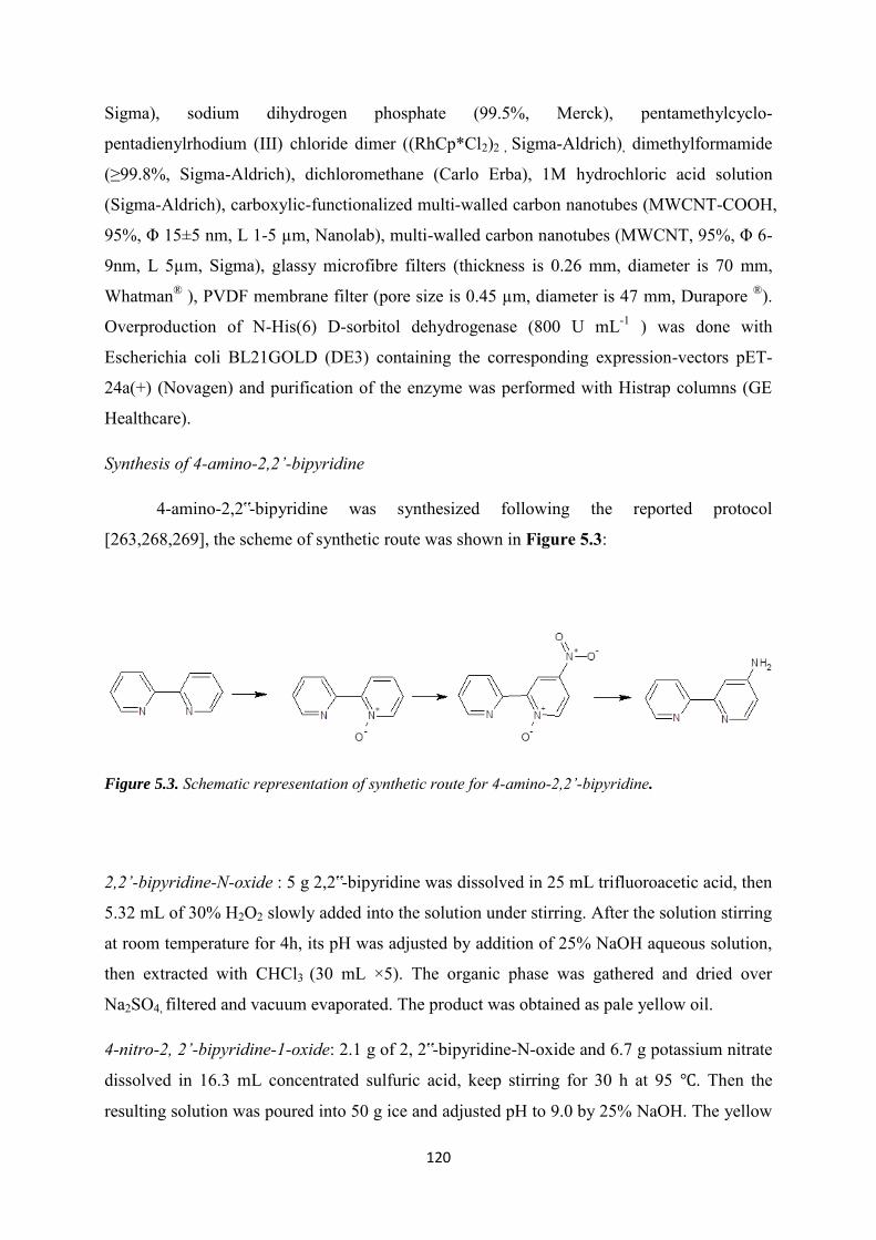
120
Sigma), sodium dihydrogen phosphate (99.5%, Merck), pentamethylcyclo-
pentadienylrhodium (III) chloride dimer ((RhCp*Cl2)2 , Sigma-Aldrich), dimethylformamide
(≥99.8%, Sigma-Aldrich), dichloromethane (Carlo Erba), 1M hydrochloric acid solution
(Sigma-Aldrich), carboxylic-functionalized multi-walled carbon nanotubes (MWCNT-COOH,
95%, Φ 15±5 nm, L 1-5 µm, Nanolab), multi-walled carbon nanotubes (MWCNT, 95%, Φ 6-
9nm, L 5µm, Sigma), glassy microfibre filters (thickness is 0.26 mm, diameter is 70 mm,
Whatman® ), PVDF membrane filter (pore size is 0.45 µm, diameter is 47 mm, Durapore ®).
Overproduction of N-His(6) D-sorbitol dehydrogenase (800 U mL-1 ) was done with
Escherichia coli BL21GOLD (DE3) containing the corresponding expression-vectors pET-
24a(+) (Novagen) and purification of the enzyme was performed with Histrap columns (GE
Healthcare).
Synthesis of 4-amino-2,2’-bipyridine
4-amino-2,2‟-bipyridine was synthesized following the reported protocol
[263,268,269], the scheme of synthetic route was shown in Figure 5.3:
Figure 5.3. Schematic representation of synthetic route for 4-amino-2,2’-bipyridine.
2,2’-bipyridine-N-oxide : 5 g 2,2‟-bipyridine was dissolved in 25 mL trifluoroacetic acid, then
5.32 mL of 30% H2O2 slowly added into the solution under stirring. After the solution stirring
at room temperature for 4h, its pH was adjusted by addition of 25% NaOH aqueous solution,
then extracted with CHCl3 (30 mL ×5). The organic phase was gathered and dried over
Na2SO4, filtered and vacuum evaporated. The product was obtained as pale yellow oil.
4-nitro-2, 2’-bipyridine-1-oxide: 2.1 g of 2, 2‟-bipyridine-N-oxide and 6.7 g potassium nitrate
dissolved in 16.3 mL concentrated sulfuric acid, keep stirring for 30 h at 95 ℃. Then the
resulting solution was poured into 50 g ice and adjusted pH to 9.0 by 25% NaOH. The yellow

121
precipitate formed was collected by vacuum filtration and washed with plenty of cold water.
After drying the solid in the air, it was dissolved in DCM, filtered off and the then DCM was
removed by vacuum evaporation, the product was obtained as yellow solid.
4-amino-2, 2’-bipyridine: 500 mg of 4-nitro-2, 2‟-bipyridine-1-oxide was dissolved in 100
mL methanol, 115 mg 10% Pd/C was added under nitrogen atmosphere with vigorously
stirring. The solution was incubated in the ice bath, 1.25 g sodium borohydride was slowly
added. Stirring was continued at 4 ℃ overnight until the gas evolution ceased. The catalyst
was removed by Gravity filtration, and the methanol was removed from the filtrate by
vacuum evaporation. 30 mL water was added to dissolve the solid, extracted with DCM (50
mL×5), the organic phase was dried with NaSO4 and then removed the solvent to get product
as white solid.
MWCNT-functionalized electrode (CF-CNT)
The surface of carbon felt was activated by cyclic voltammetry scans from -0.7 V to
2.5 V for 20 cycles in 0.1M H2SO4, in order to change the surface from hydrophobic to
hydrophilic. After treatment, the carbon felt was rinsed with water, followed by heat treatment
at 200 ℃ for 1 h to remove the residue H2SO4. Then the carbon felt was cut into pieces with
dimension of 0.5 cm*0.5 cm. The carbon felt was dipped into 0.5 mg mL-1 MWCNT-COOH
suspension which was dispersed by ultrasonic for 1 h, then let it dry in the oven at 130 ℃. The
dipping and drying process was repeated for 10 times in order to get enough CNT on the
carbon felt.
‘Bucky paper’ electrode (BP)
The electrode was prepared following the reported literature[265]. 10 mg MWCNT
was dispersed in 50 mL ethanol by ultrasonication for 5 h. Afterwards, the MWCNTs solution
were decanted and vacuum filtered using a PVDF membrane filter (Durapore ®, pore size is
0.45 µm, diameter is 47 mm), then it was dried at 50 ℃ overnight. Finally, the electrode was
cut into suitable size for use (1 cm* 1 cm or 2 cm* 2 cm).
2, 2’-bipyridine functionalized electrode
1 mM 4-amino-2, 2‟-bipyridine was mixed with 2 mM sodium nitrite in 0.5 M HCl
aqueous solution with stirring for 5 min to generate 2, 2‟-bipyridine cations, the grafting was
achieved by running 2 cycles of cyclic voltammograms from 0.4 V to -0.8 V on the electrode
122
inside the 2, 2‟-bipyridine diazonium cations solution. After grafting, the electrode was rinsed
carefully with water and left dry for use.
[Cp*Rh(bpy)Cl]+ functionalized electrode
The 2,2‟-bipyridine functionalized electrode was put into 0.15 mM ((RhCp*Cl2)2
dichloromethane solution with gentle stirring for 4 h. Then the electrode was first rinsed with
DCM and then immersed inside clean DCM for 5 min to remove the unreacted residue.
DSDH- [Cp*Rh(bpy)Cl]+ co-immobilized CF-CNT-gel electrode
The sol was prepared following the reported protocol[75]. 0.18 g TEOS, 0.13 g GPS,
together with 0.5 mL water and 0.625 mL 0.01 M HCl was pre-hydrolyzed by stirring
overnight. Then, this sol was diluted 3 times and a 40 µL aliquot was mixed with 20 µL PEI
(20%), 20 µL of water and 30 µL DSDH stock solutions. This mixture was spread over the
[Cp*Rh(bpy)Cl]+ functionalized electrode and let it completely dry at 4 ℃ for use.
Preparation of DSDH gel layer packed electrode
A piece of glassy microfibre filter was cut into certain size and then spread over by the
same DSDH sol as mentioned above, keep it suspend in a bottle and dried at 4 ℃ over night
for use. For electroenzymatic experiment, this DSDH gel layer was overload on the top of the
[Cp*Rh(bpy)Cl]+ functionalized bucky paper electrode, then four corners were fixed by
cotton thread.
Apparatus
All electrochemical experiments were conducted with an Autolab PGSTAT-12
potentiostat. The three-electrode configuration cell included an Ag/AgCl reference electrode
(3 M KCl), a steel auxiliary electrode, and a carbon working electrode (CF-CNT or BP). A
pencil core (0.5 mm diameter) glued by carbon black to a copper wire was used for
connecting the CF-CNT working electrode. A steel clip was used for connecting with the BP
electrode.
UV-Vis spectra have been recorded on a Cary 60 Scan UV-Vis spectrophotometer
X-Ray Photoelectron Spectroscopy (XPS) analyses were performed using a KRATOS
Axis Ultra X-ray photoelectron spectrometer (Kratos Analytical, Manchester, UK) equipped
with a monochromated AlKα X-ray source (hν = 1486.6 eV) operated at 150 W. The base
123
pressure in the analytical chamber was 10− 9 mbar during XPS measurements. Wide scans
were recorded using a pass energy of 160 eV and narrow scans using a pass energy of 20 eV
(instrumental resolution better than 0.5 eV). Charge correction was carried out using the C(1s)
core line, setting adventitious carbon signal (H/C signal) to 284.6 eV.
5.3 Results and discussions
In order to evaluate the electrochemical properties of the immobilized rhodium
complex between new synthetic route and the old one in Chapter 3, rhodium complex was
first immobilized on CF-MWCNT electrode, then applied to „bucky paper‟ electrode with the
same protocol.
5.3.1 Covalent immobilization of rhodium complex on CF-CNT electrodes
by bipyridyl diazonium electrografting
A two-step protocol was applied to covalently immobilize the rhodium complex on the
CF-CNT electrode. The bipyridine ligand was first immobilized on electrode surface by
electrografting of bipyridyl diazonium cations generated from 1 mM 4-amino-2,2‟-bipyridine
activated with the presence of NaNO2 in 0.5 mM HCl aqueous solution (CF-CNT-Bpy). The
covalent attachment to the electrode surface was realized by running 2 successive cyclic
voltammograms in a potential range from 0.4 V to -0.8 V (Figure 5.4A). During the first scan,
an irreversible peak located around -0.7 V was observed (curve a), in which process the
grafting of bipyridine radical occurred along with the release of N2. When carrying out the
second scan, the cathodic reduction current was significantly decreased (curve b). This
decrease was attributed to the presence of grafted bipyridyl groups on the electrode, partially
blocking the surface. The presence of the organic layer on the CF-CNT electrode was further
evidenced by the comparison of the cyclic voltammograms of 0.1 mM ferrocenedimethanol
recorded on the CF-CNT electrode before and after electrografting of the bipyridine layer
(Figure 5.4B). We observed a slight decrease in peaks current in both oxidation and reduction
after electrografting of bipyridine groups demonstrating the blocking effect of the organic
layer, but the remained peak current showed that the electrode after functionalization still
kept in a good condition for electron transfer. The bipyridine functionalized electrode was
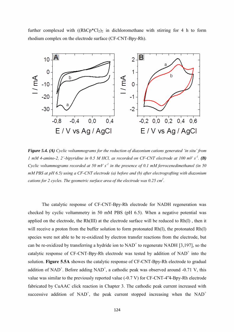
124
further complexed with ((RhCp*Cl2)2 in dichloromethane with stirring for 4 h to form
rhodium complex on the electrode surface (CF-CNT-Bpy-Rh).
Figure 5.4. (A) Cyclic voltammograms for the reduction of diazonium cations generated ‘in situ’ from
1 mM 4-amino-2, 2’-bipyridine in 0.5 M HCl, as recorded on CF-CNT electrode at 100 mV s-1. (B)
Cyclic voltammograms recorded at 50 mV s-1 in the presence of 0.1 mM ferrocenedimethanol (in 50
mM PBS at pH 6.5) using a CF-CNT electrode (a) before and (b) after electrografting with diazonium
cations for 2 cycles. The geometric surface area of the electrode was 0.25 cm2.
The catalytic response of CF-CNT-Bpy-Rh electrode for NADH regeneration was
checked by cyclic voltammetry in 50 mM PBS (pH 6.5). When a negative potential was
applied on the electrode, the Rh(III) at the electrode surface will be reduced to Rh(I) , then it
will receive a proton from the buffer solution to form protonated Rh(I), the protonated Rh(I)
species were not able to be re-oxidized by electron transfer reactions from the electrode, but
can be re-oxidized by transferring a hydride ion to NAD+ to regenerate NADH [3,197], so the
catalytic response of CF-CNT-Bpy-Rh electrode was tested by addition of NAD+ into the
solution. Figure 5.5A showes the catalytic response of CF-CNT-Bpy-Rh electrode to gradual
addition of NAD+. Before adding NAD+, a cathodic peak was observed around -0.71 V, this
value was similar to the previously reported value (-0.7 V) for CF-CNT-4‟4-Bpy-Rh electrode
fabricated by CuAAC click reaction in Chapter 3. The cathodic peak current increased with
successive addition of NAD+, the peak current stopped increasing when the NAD+
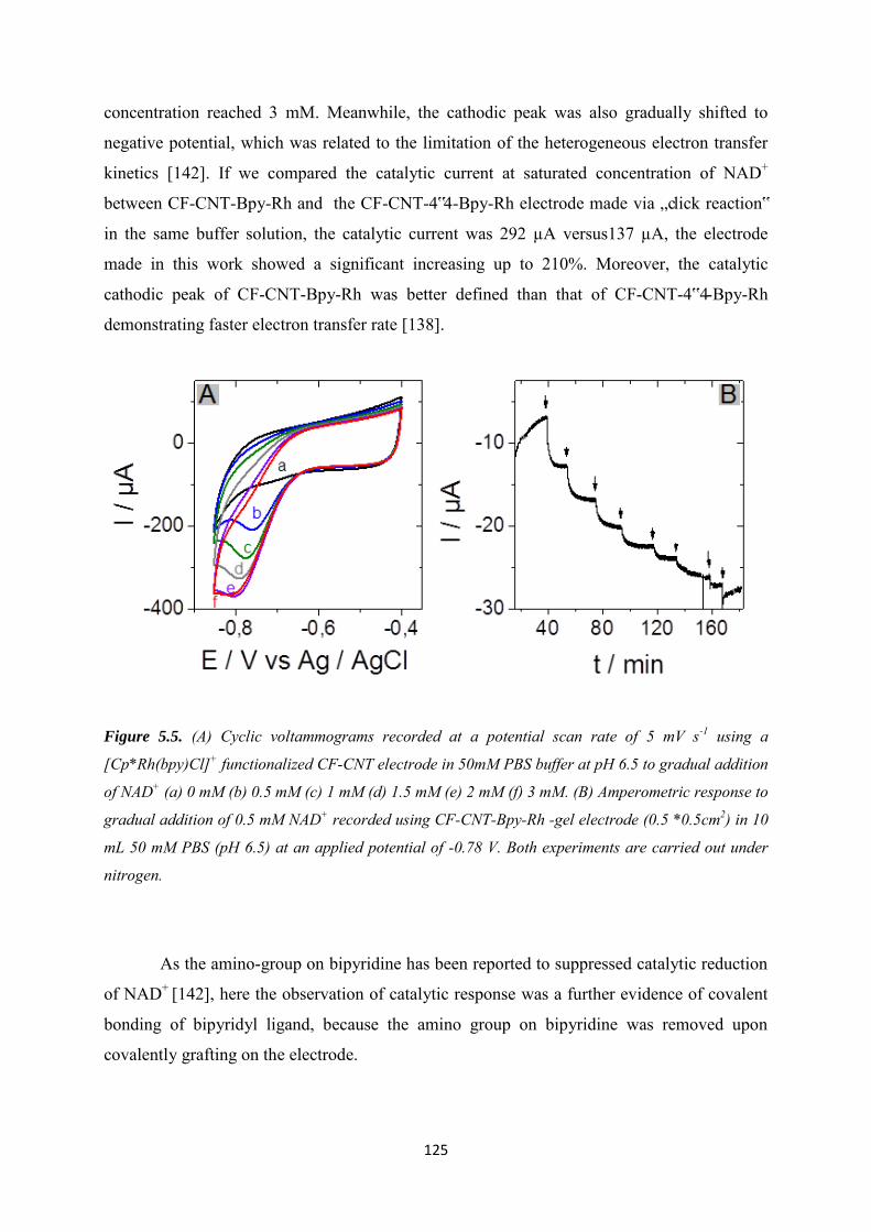
125
concentration reached 3 mM. Meanwhile, the cathodic peak was also gradually shifted to
negative potential, which was related to the limitation of the heterogeneous electron transfer
kinetics [142]. If we compared the catalytic current at saturated concentration of NAD+
between CF-CNT-Bpy-Rh and the CF-CNT-4‟4-Bpy-Rh electrode made via „click reaction‟
in the same buffer solution, the catalytic current was 292 µA versus137 µA, the electrode
made in this work showed a significant increasing up to 210%. Moreover, the catalytic
cathodic peak of CF-CNT-Bpy-Rh was better defined than that of CF-CNT-4‟4-Bpy-Rh
demonstrating faster electron transfer rate [138].
Figure 5.5. (A) Cyclic voltammograms recorded at a potential scan rate of 5 mV s-1 using a
[Cp*Rh(bpy)Cl]+ functionalized CF-CNT electrode in 50mM PBS buffer at pH 6.5 to gradual addition
of NAD+ (a) 0 mM (b) 0.5 mM (c) 1 mM (d) 1.5 mM (e) 2 mM (f) 3 mM. (B) Amperometric response to
gradual addition of 0.5 mM NAD+ recorded using CF-CNT-Bpy-Rh -gel electrode (0.5 *0.5cm2) in 10
mL 50 mM PBS (pH 6.5) at an applied potential of -0.78 V. Both experiments are carried out under
nitrogen.
As the amino-group on bipyridine has been reported to suppressed catalytic reduction
of NAD+ [142], here the observation of catalytic response was a further evidence of covalent
bonding of bipyridyl ligand, because the amino group on bipyridine was removed upon
covalently grafting on the electrode.
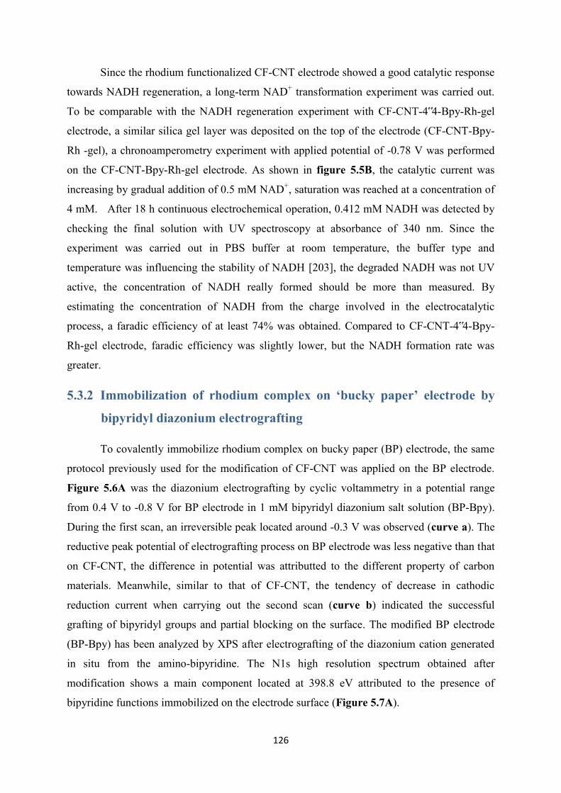
126
Since the rhodium functionalized CF-CNT electrode showed a good catalytic response
towards NADH regeneration, a long-term NAD+ transformation experiment was carried out.
To be comparable with the NADH regeneration experiment with CF-CNT-4‟4-Bpy-Rh-gel
electrode, a similar silica gel layer was deposited on the top of the electrode (CF-CNT-Bpy-
Rh -gel), a chronoamperometry experiment with applied potential of -0.78 V was performed
on the CF-CNT-Bpy-Rh-gel electrode. As shown in figure 5.5B, the catalytic current was
increasing by gradual addition of 0.5 mM NAD+, saturation was reached at a concentration of
4 mM. After 18 h continuous electrochemical operation, 0.412 mM NADH was detected by
checking the final solution with UV spectroscopy at absorbance of 340 nm. Since the
experiment was carried out in PBS buffer at room temperature, the buffer type and
temperature was influencing the stability of NADH [203], the degraded NADH was not UV
active, the concentration of NADH really formed should be more than measured. By
estimating the concentration of NADH from the charge involved in the electrocatalytic
process, a faradic efficiency of at least 74% was obtained. Compared to CF-CNT-4‟4-Bpy-
Rh-gel electrode, faradic efficiency was slightly lower, but the NADH formation rate was
greater.
5.3.2 Immobilization of rhodium complex on ‘bucky paper’ electrode by
bipyridyl diazonium electrografting
To covalently immobilize rhodium complex on bucky paper (BP) electrode, the same
protocol previously used for the modification of CF-CNT was applied on the BP electrode.
Figure 5.6A was the diazonium electrografting by cyclic voltammetry in a potential range
from 0.4 V to -0.8 V for BP electrode in 1 mM bipyridyl diazonium salt solution (BP-Bpy).
During the first scan, an irreversible peak located around -0.3 V was observed (curve a). The
reductive peak potential of electrografting process on BP electrode was less negative than that
on CF-CNT, the difference in potential was attributted to the different property of carbon
materials. Meanwhile, similar to that of CF-CNT, the tendency of decrease in cathodic
reduction current when carrying out the second scan (curve b) indicated the successful
grafting of bipyridyl groups and partial blocking on the surface. The modified BP electrode
(BP-Bpy) has been analyzed by XPS after electrografting of the diazonium cation generated
in situ from the amino-bipyridine. The N1s high resolution spectrum obtained after
modification shows a main component located at 398.8 eV attributed to the presence of
bipyridine functions immobilized on the electrode surface (Figure 5.7A).
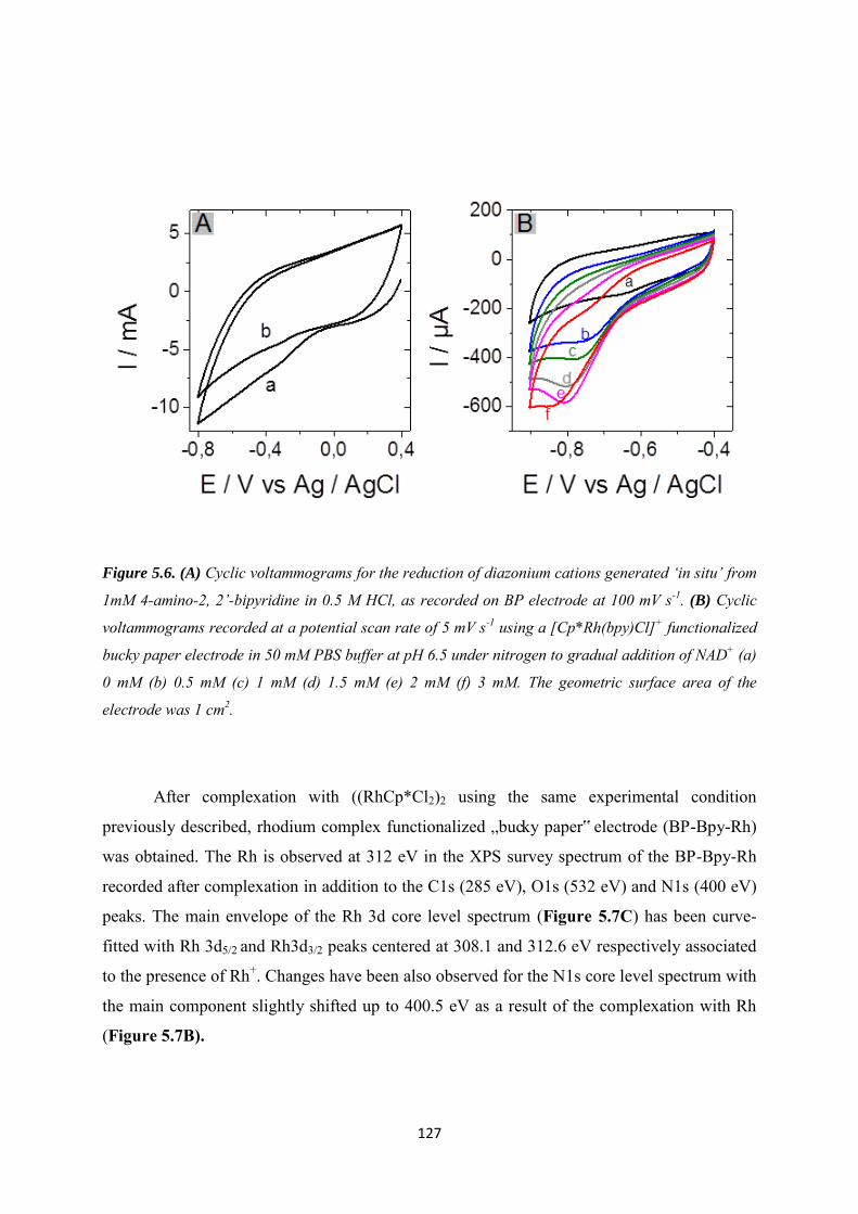
127
Figure 5.6. (A) Cyclic voltammograms for the reduction of diazonium cations generated ‘in situ’ from
1mM 4-amino-2, 2’-bipyridine in 0.5 M HCl, as recorded on BP electrode at 100 mV s-1. (B) Cyclic
voltammograms recorded at a potential scan rate of 5 mV s-1 using a [Cp*Rh(bpy)Cl]+ functionalized
bucky paper electrode in 50 mM PBS buffer at pH 6.5 under nitrogen to gradual addition of NAD+ (a)
0 mM (b) 0.5 mM (c) 1 mM (d) 1.5 mM (e) 2 mM (f) 3 mM. The geometric surface area of the
electrode was 1 cm2.
After complexation with ((RhCp*Cl2)2 using the same experimental condition
previously described, rhodium complex functionalized „bucky paper‟ electrode (BP-Bpy-Rh)
was obtained. The Rh is observed at 312 eV in the XPS survey spectrum of the BP-Bpy-Rh
recorded after complexation in addition to the C1s (285 eV), O1s (532 eV) and N1s (400 eV)
peaks. The main envelope of the Rh 3d core level spectrum (Figure 5.7C) has been curve-
fitted with Rh 3d5/2 and Rh3d3/2 peaks centered at 308.1 and 312.6 eV respectively associated
to the presence of Rh+. Changes have been also observed for the N1s core level spectrum with
the main component slightly shifted up to 400.5 eV as a result of the complexation with Rh
(Figure 5.7B).

128
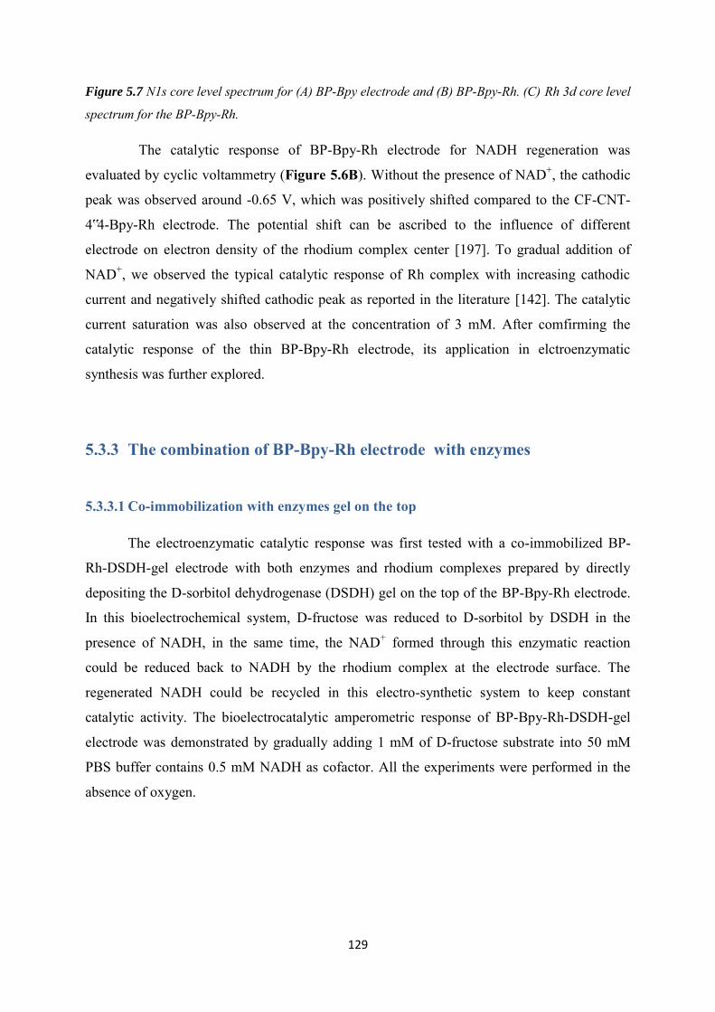
129
Figure 5.7 N1s core level spectrum for (A) BP-Bpy electrode and (B) BP-Bpy-Rh. (C) Rh 3d core level
spectrum for the BP-Bpy-Rh.
The catalytic response of BP-Bpy-Rh electrode for NADH regeneration was
evaluated by cyclic voltammetry (Figure 5.6B). Without the presence of NAD+, the cathodic
peak was observed around -0.65 V, which was positively shifted compared to the CF-CNT-
4‟4-Bpy-Rh electrode. The potential shift can be ascribed to the influence of different
electrode on electron density of the rhodium complex center [197]. To gradual addition of
NAD+, we observed the typical catalytic response of Rh complex with increasing cathodic
current and negatively shifted cathodic peak as reported in the literature [142]. The catalytic
current saturation was also observed at the concentration of 3 mM. After comfirming the
catalytic response of the thin BP-Bpy-Rh electrode, its application in elctroenzymatic
synthesis was further explored.
5.3.3 The combination of BP-Bpy-Rh electrode with enzymes
5.3.3.1 Co-immobilization with enzymes gel on the top
The electroenzymatic catalytic response was first tested with a co-immobilized BP-
Rh-DSDH-gel electrode with both enzymes and rhodium complexes prepared by directly
depositing the D-sorbitol dehydrogenase (DSDH) gel on the top of the BP-Bpy-Rh electrode.
In this bioelectrochemical system, D-fructose was reduced to D-sorbitol by DSDH in the
presence of NADH, in the same time, the NAD+ formed through this enzymatic reaction
could be reduced back to NADH by the rhodium complex at the electrode surface. The
regenerated NADH could be recycled in this electro-synthetic system to keep constant
catalytic activity. The bioelectrocatalytic amperometric response of BP-Bpy-Rh-DSDH-gel
electrode was demonstrated by gradually adding 1 mM of D-fructose substrate into 50 mM
PBS buffer contains 0.5 mM NADH as cofactor. All the experiments were performed in the
absence of oxygen.
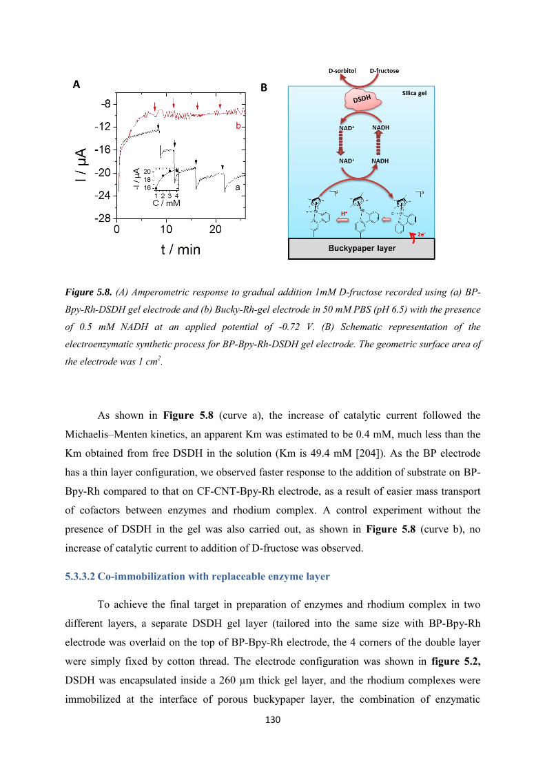
130
Figure 5.8. (A) Amperometric response to gradual addition 1mM D-fructose recorded using (a) BP-
Bpy-Rh-DSDH gel electrode and (b) Bucky-Rh-gel electrode in 50 mM PBS (pH 6.5) with the presence
of 0.5 mM NADH at an applied potential of -0.72 V. (B) Schematic representation of the
electroenzymatic synthetic process for BP-Bpy-Rh-DSDH gel electrode. The geometric surface area of
the electrode was 1 cm2.
As shown in Figure 5.8 (curve a), the increase of catalytic current followed the
Michaelis–Menten kinetics, an apparent Km was estimated to be 0.4 mM, much less than the
Km obtained from free DSDH in the solution (Km is 49.4 mM [204]). As the BP electrode
has a thin layer configuration, we observed faster response to the addition of substrate on BP-
Bpy-Rh compared to that on CF-CNT-Bpy-Rh electrode, as a result of easier mass transport
of cofactors between enzymes and rhodium complex. A control experiment without the
presence of DSDH in the gel was also carried out, as shown in Figure 5.8 (curve b), no
increase of catalytic current to addition of D-fructose was observed.
5.3.3.2 Co-immobilization with replaceable enzyme layer
To achieve the final target in preparation of enzymes and rhodium complex in two
different layers, a separate DSDH gel layer (tailored into the same size with BP-Bpy-Rh
electrode was overlaid on the top of BP-Bpy-Rh electrode, the 4 corners of the double layer
were simply fixed by cotton thread. The electrode configuration was shown in figure 5.2,
DSDH was encapsulated inside a 260 µm thick gel layer, and the rhodium complexes were
immobilized at the interface of porous buckypaper layer, the combination of enzymatic
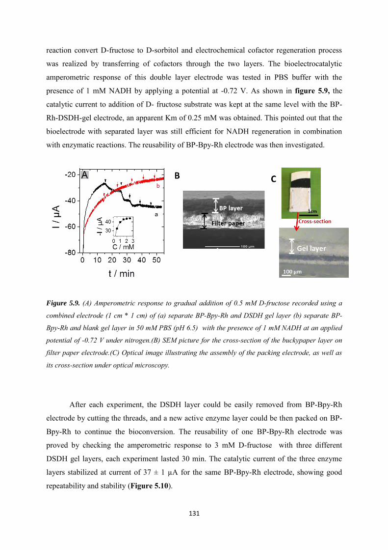
131
reaction convert D-fructose to D-sorbitol and electrochemical cofactor regeneration process
was realized by transferring of cofactors through the two layers. The bioelectrocatalytic
amperometric response of this double layer electrode was tested in PBS buffer with the
presence of 1 mM NADH by applying a potential at -0.72 V. As shown in figure 5.9, the
catalytic current to addition of D- fructose substrate was kept at the same level with the BP-
Rh-DSDH-gel electrode, an apparent Km of 0.25 mM was obtained. This pointed out that the
bioelectrode with separated layer was still efficient for NADH regeneration in combination
with enzymatic reactions. The reusability of BP-Bpy-Rh electrode was then investigated.
Figure 5.9. (A) Amperometric response to gradual addition of 0.5 mM D-fructose recorded using a
combined electrode (1 cm * 1 cm) of (a) separate BP-Bpy-Rh and DSDH gel layer (b) separate BP-
Bpy-Rh and blank gel layer in 50 mM PBS (pH 6.5) with the presence of 1 mM NADH at an applied
potential of -0.72 V under nitrogen.(B) SEM picture for the cross-section of the buckypaper layer on
filter paper electrode.(C) Optical image illustrating the assembly of the packing electrode, as well as
its cross-section under optical microscopy.
After each experiment, the DSDH layer could be easily removed from BP-Bpy-Rh
electrode by cutting the threads, and a new active enzyme layer could be then packed on BP-
Bpy-Rh to continue the bioconversion. The reusability of one BP-Bpy-Rh electrode was
proved by checking the amperometric response to 3 mM D-fructose with three different
DSDH gel layers, each experiment lasted 30 min. The catalytic current of the three enzyme
layers stabilized at current of 37 ± 1 µA for the same BP-Bpy-Rh electrode, showing good
repeatability and stability (Figure 5.10).
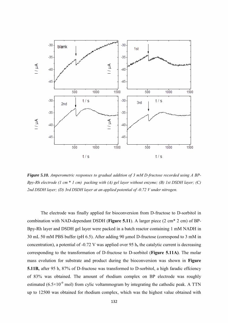
132
Figure 5.10. Amperometric responses to gradual addition of 3 mM D-fructose recorded using A BP-
Bpy-Rh electrode (1 cm * 1 cm) packing with (A) gel layer without enzyme; (B) 1st DSDH layer; (C)
2nd DSDH layer; (D) 3rd DSDH layer at an applied potential of -0.72 V under nitrogen.
The electrode was finally applied for bioconversion from D-fructose to D-sorbitol in
combination with NAD-dependant DSDH (Figure 5.11). A larger piece (2 cm* 2 cm) of BP-
Bpy-Rh layer and DSDH gel layer were packed in a batch reactor containing 1 mM NADH in
30 mL 50 mM PBS buffer (pH 6.5). After adding 90 µmol D-fructose (correspond to 3 mM in
concentration), a potential of -0.72 V was applied over 95 h, the catalytic current is decreasing
corresponding to the transformation of D-fructose to D-sorbitol (Figure 5.11A). The molar
mass evolution for substrate and product during the bioconversion was shown in Figure
5.11B, after 95 h, 87% of D-fructose was transformed to D-sorbitol, a high faradic effciency
of 83% was obtained. The amount of rhodium complex on BP electrode was roughly
estimated (6.5×10⁻⁹ mol) from cylic voltammogram by integrating the cathodic peak. A TTN
up to 12500 was obtained for rhodium complex, which was the highest value obtained with
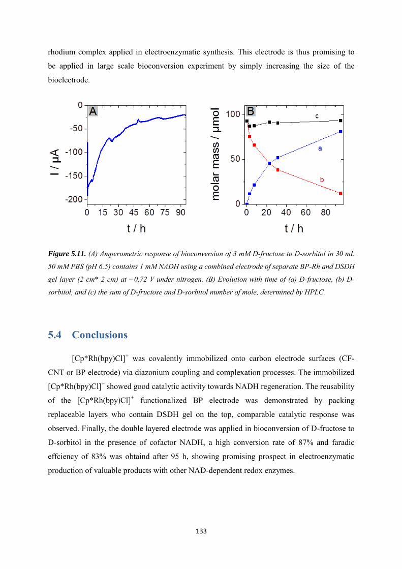
133
rhodium complex applied in electroenzymatic synthesis. This electrode is thus promising to
be applied in large scale bioconversion experiment by simply increasing the size of the
bioelectrode.
Figure 5.11. (A) Amperometric response of bioconversion of 3 mM D-fructose to D-sorbitol in 30 mL
50 mM PBS (pH 6.5) contains 1 mM NADH using a combined electrode of separate BP-Rh and DSDH
gel layer (2 cm* 2 cm) at −0.72 V under nitrogen. (B) Evolution with time of (a) D-fructose, (b) D-
sorbitol, and (c) the sum of D-fructose and D-sorbitol number of mole, determined by HPLC.
5.4 Conclusions
[Cp*Rh(bpy)Cl]+ was covalently immobilized onto carbon electrode surfaces (CF-
CNT or BP electrode) via diazonium coupling and complexation processes. The immobilized
[Cp*Rh(bpy)Cl]+ showed good catalytic activity towards NADH regeneration. The reusability
of the [Cp*Rh(bpy)Cl]+ functionalized BP electrode was demonstrated by packing
replaceable layers who contain DSDH gel on the top, comparable catalytic response was
observed. Finally, the double layered electrode was applied in bioconversion of D-fructose to
D-sorbitol in the presence of cofactor NADH, a high conversion rate of 87% and faradic
effciency of 83% was obtaind after 95 h, showing promising prospect in electroenzymatic
production of valuable products with other NAD-dependent redox enzymes.
134
Conclusions and perspectives
In this thesis, different chemical routes have been employed to immobilize enzymes
and rhodium complex catalyst in order to apply them to enzymatic electrosynthesis.
Experiments have been performed with D-sorbitol dehydrogenase (DSDH) catalyzing the
reduction of D-fructose to D-sorbitol. Some experiments were also done with galactitol
dehydrogenase (GatDH) having a wider range of substrate.
The electrode functionalization was mainly performed via electrochemical reduction
based on the generation of highly reactive aryl radical species from diazonium salts. A variety
of functional groups have been grafted depending on the final usage. 4-vinylaniline was used
to obtain a vinly functionalized surface, 4-azidoaniline was used to obtain an azido
functionalized surface, and 4-amino-2,2‟-bipyridine was used to obtain bipyridyl
functionalized surface. In addition, the simultaneous introduction of two functions at the
electrode surface was achieved by electrochemical reduction of mixtures of diazonium salts to
create selectively mixed monolayers with a controlled chemical composition. These
functional groups offered the possibility for further reactions steps including „click‟ chemistry
or metal-ligand complexation. In all above cases, the coverage of the organic layer on the
electrode was controlled, in order to allow electron transfer reactions.
„Click‟ chemistry was applied in covalent immobilization of proteins or catalysts due
to their simple and mild experimental reaction conditions. There were two types of „click‟
reactions employed to these works. „Thiol-ene‟ click chemistry was applied to the
immobilization of dehydrogenase-based proteins on carbon electrode surfaces, the thiol group
from cysteine residue of enzymes was reacting with the vinyl groups on the electrode. In this
way, the activity of DSDH was kept when click coupling was made on proteins bearing a
cysteine tag. This method was also applied to the immobilization of GatDH. Copper (I)-
catalyzed azide-alkyne cycloaddition reaction (Huisgen coupling) was then used to
immobilize covalently (2,2'-bipyridyl)(pentamethylcyclopentadienyl)-rhodium as catalyst on
carbon electrode surface, a better long-term stability for NADH regeneration was achieved by
using this covalent bond compared to the molecule only adsorbed. The combination of the
two click types was finally evaluated. The co-functionalization involved electrochemical
reduction of diazonium cations and a double consecutive click reactions process, after
135
diazonium electrografting process, the co-functionalized electrodes allow selective
incorporation of rhodium complex and DSDH, via two „click‟ immobilization steps. Co-
immobilization was also achieved by combining click chemistry and sol-gel immobilization
of molecules.
Several problems were met when fabricating the bioelectrode for electroenzymatic
synthesis. Firstly, the enzymes were inhibited during long-term experiments, but the activity
was recovered after replacing the used solution by a fresh buffer. We assumed that this
inhibition may due to the change of local environment eg. increase of local pH at the electrode
interface or product accumulation. Secondly, a slow degradation of rhodium complex was
also observed when depositing silica gel containing enzyme directly on the top of the rhodium
complex functionalized electrode. Since no degradation was observed in the same condition
in the absence of enzyme, we can conclude that the slow degradation originated from possible
interaction between enzyme and rhodium complex after long-term experimental operation.
In order to fulfill the demand for long-term stability, the enzyme and the rhodium
complex had to be better separated while still keeping close distance for NADH regeneration.
This was achieved by using a bioelectrode consisting of two separated layers. A rhodium
complex functionalized „buckypaper‟ layer on one side and a glassy fiber layer with gel
immobilized DSDH on the other side. The bioelectrode was obtained by assembling these two
materials. The enzyme gel glassy fiber layer could be easily replaced when losing their
activity. Good reusability with three different enzyme layers and stable electrocatalytic
response were observed with a rhodium complex functionalized electrode. Not only the
degradation of rhodium complex was avoided benefiting from the complete separation, but
also the enzymatic inhibition effect observed previousely was avoided. This co-immobilized
bioelectrode must simplify the purification steps and show promising prospect in
electroenzymatic production of chiral valuable products with NAD-dependent redox enzymes.
Actually this system could be applied to regeneration of NADPH for other enzymes such as
P450 cytochrome.
If considering larger size of bioelectrodes that could be applied in large-scale
electroenzymatic synthesis, the proton consumption at the electrode interface has to be
considered. We assume that the local pH control will be a key parameter. For long-terms
operation with layer structured flow-cell system, the pH of buffer solution may increase due
to the proton consumption. Since in some tests, we observed that the elecrocatalytic response
136
was not observed in high buffer concentration (0.5 M), the proton supplement cannot be
provided simply by increasing the concentration of buffer. The production of protons from
water oxidation at the counter electrode would also produce O2 in the solution which should
be avoided. A solution to keep constant pH would be the combination of the bioelectrode with
a gas diffusion electrode to continuously produce H+ from H2 without production of O2.
In a more general thinking, when we started these PhD studies, the rhodium complex
was, and is still, the most efficient non-enzymatic NADH regeneration catalyst. However the
metal is rather rare, expensive and toxic. Direct electron transfer with flavoenzymes allowing
NADH regeneration is certainly one topic to be further investigated in the coming years for
electroenzymatic synthesis. Developing stable and recyclable bioelectrodes leading to long-
term enzymatic activity and direct electron transfer reactions, would allow significant steps
forward for major industrial developments with lower environmental impact.
Finally, another direction to improve the electrochemical regeneration in this
electroenzymatic synthesis would be the application of NAD analogs, which would avoid the
limited stability of the original biomolecule [270] that could eventually be immobilized on the
electrode surface as suggested before [202] with the goal to build optimal and stable
electronic pathway usable in bioelectrochemical conversions.
137
Conclusion générale et perspectives
Dans cette thèse, différentes voies chimiques ont été employées pour immobiliser
ensemble des enzymes et un complexe de rhodium afin de les appliquer en électrosynthèse
enzymatique. Les expériences ont été menées avec la D-sorbitol déshydrogénase (DSDH)
catalysant la réduction du D-fructose en D-sorbitol. Certaines expériences ont également été
menées avec la galactitol déshydrogénase (GatDH) ayant un spectre de substrat plus large.
La fonctionnalisation des électrodes a principalement été faîte par génération
électrochimique de radicaux aryle hautement réactifs à partir de sels de diazonium. Une
grande variété de groupes fonctionnels a été utilisée pour différents usages. La 4-vinylaniline
a été utilisée pour obtenir une surface fonctionnalisée par des groupements vinyles. La 4-
azidoaniline a été utilisée pour obtenir une surface fonctionnalisée par des groupements
azoture. Enfin, la 4-amino-2,2‟-bipyridine a été utilisée pour préparer une surface
fonctionnalisée par des groupements pyridine. Finalement, l‟introduction simultanée de deux
groupements fonctionnels a été obtenue par électrogreffage d‟un mélange de sels de
diazonium. Ces groupements fonctionnels ont ensuite été utilisés pour des réactions de chimie
click ou de complexation métal-ligand. Dans tous les cas, le dépôt organique a été optimisé
afin d‟autoriser des réactions de transfert d‟électron avec des espèces en solution ou greffées
en surface.
La chimie “click” a été appliquée à l‟immobilisation covalente de protéines ou de
catalyseurs en raison des conditions simples et douces qu‟elle permet. Deux types de chimie
click ont été utilisés dans ce travail. Le couplage « thiol-ène » a été appliqué à
l‟immobilisation de déshydrogénases à la surface d‟électrodes de carbone. Dans ce cas, le
groupement thiol de résidus cystéine pouvait réagir avec le groupe vinyle greffé à la surface
de l‟électrode. De cette manière, l‟activité de la DSDH était conservée plus longtemps à la
surface de l‟électrode lorsque le couplage était fait avec des protéines portant un tag cystéine.
La réaction de cyclo-addition azoture-alcène catalysée par le cuivre(I) (couplage de Huisgen)
a ensuite été utilisée pour immobiliser de manière covalente le complexe (2,2'-
bipyridyl)(pentamethylcyclopentadienyl)-rhodium à la surface d‟électrodes carbonées. Une
meilleure stabilité pour la régénération de NADH a été observée avec le complexe
immobilisée qu‟avec ce complexe simplement adsorbé à la surface de l‟électrode. La
combinaison de ces deux chimies click a ensuite été étudiée. La co-fonctionnalisation
138
impliquait la réduction électrochimique de cations diazonium et deux réactions de chimie
click consécutives permettant l‟incorporation sélective du complexe de rhodium et de la
DSDH. La co-fonctionnalisation a également obtenue par combinaison d‟une première étape
de couplage par chimie click pour l‟immobilisation du complexe de rhodium et une seconde
étape d‟encapsulation des protéines dans une matrice de silice par voie sol-gel.
Plusieurs problèmes ont été rencontrés lorsque nous avons testés ces électrodes en
électrosynthèse enzymatique. Tout d‟abord, une inhibition de l‟activité enzymatique a été
observée au cours de l‟expérience, mais cette activité était retrouvée lorsque la solution était
remplacée par une solution fraîche. Nous supposons que cette inhibition pouvait être causée
un environnement local dans l‟électrode poreuse, par exemple une augmentation de pH ou
une accumulation du produit de la réaction enzymatique. Ensuite, la dégradation lente de
l‟activité du complexe de rhodium a été observée lorsque le gel de silice contenant les
enzymes était déposé au contact de l‟électrode fonctionnalisée par le complexe de rhodium.
Cette dégradation n‟a pas été observée en absence d‟enzymes et serait causée par l‟interaction
entre les enzymes et les complexes de rhodium après des temps de réactions longs qui
favorise une dissolution partielle de la silice.
Afin d‟améliorer la stabilité de la bioélectrode dans le temps, les enzymes et les
complexes métalliques doivent être mieux séparés tout en conservant une faible distance pour
permettre la régénération de NADH. Ceci a été obtenu en combinant deux couches séparées,
une première sur laquelle était greffée le complexe de rhodium et une seconde sur la quelle
était immobilisée l‟enzyme. La bioélectrode était alors obtenue par l‟assemblage de ces deux
matériaux. Le matériau (un filtre en fibres de verre) permettant l‟immobilisation de l‟enzyme
pouvait être facilement remplacée. Ainsi, il fut possible de réutiliser le complexe de rhodium
avec trois couches enzymatiques différentes. De plus, la dégradation du complexe de rhodium
n‟a pas été observée. Enfin, cette configuration de bioélectrode a permis de supprimer
l‟inhibition enzymatique observée précédemment. Cette voie de co-immobilisation devrait
donc simplifier la purification des produits de synthèse et pourrait conduire à de nouvelles
applications en électrosynthèse enzymatique. Par exemple, ce système pourrait être appliqué à
la régénération du cofacteur NADPH pour d‟autres enzymes redox comme les cytochromes
P450.
Si nous considérons l‟utilisation d‟électrodes de grande taille dans des dispositifs
d‟électrosynthèse en continu, la consommation des protons à l‟électrode de travail risque
139
d‟être un paramètre limitant de la réaction. Nous supposons que le pH local à proximité de
l‟enzyme est un paramètre essentiel permettant de favoriser la plus grande activité
enzymatique. Ces protons pourraient être apportés par l‟oxydation de l‟eau, mais cette
réaction conduit également à la production d‟oxygène, qui devrait être évité pour conserver
une bonne efficacité pour la réaction de régénération de NADH. Une solution technologique
pourrait être l‟utilisation d‟une électrode à diffusion de gaz pour produire en continu des
protons à partir de l‟oxydation de H2, sans production d‟O2.
D‟un point de vue plus général, lorsque cette thèse a commencé, le complexe de
rhodium que nous avons de rhodium que nous avons immobilisé était, et est encore, le
catalyseur le plus efficace pour la régénération non-enzymatique de NADH. Cependant ce
métal est relativement rare, cher et toxique. Les réactions de transfert d‟électron directes avec
une flavoenzyme permettant la régénération de NADH est certainement un sujet à explorer
encore dans les années à venir pour application en électrosynthèse enzymatique. Le
développement de bioélectrodes stables et recyclables permettant de conserver l‟activité
enzymatique sur des temps longs tout en autorisant des réactions de transfert d‟électron serait
un grand pas vers un développement industriel ayant un impact environnemental limité.
Finalement, une autre direction de recherche pour améliorer la régénération
électrochimique dans cette réaction d‟électrosynthèse serait l‟étude des analogues de NAD qui
permettent de dépasser les limites en stabilité de la molécule naturelle [270] qui pourrait alors
être immobilisée à la surface de l‟électrode comme cela a été suggéré auparavant [202] pour
construire un chemin de transfert électronique optimal et stable utilisable en dans des
réactions de conversion bioélectrochimique.
140
References [1] K. Drauz, H. Waldmann, eds., Enzyme Catalysis in Organic Synthesis: A
Comprehensive Handbook, Second Edition, Wiley-VCH, Veinheim, 2002.
[2] P. Kornberger, F. Giffhorn, G.-W. Kohring, Encycl. Ind. Biotechnol. 2009, 1.
[3] E. Steckhan, Top. Curr. Chem. 1994, 170, 83.
[4] C. Kohlmann, W. Märkle, S. Lütz, J. Mol. Catal. B: Enzym. 2008, 51, 57.
[5] L. Gorton, E. Dominguez, Encycl. Electrochem. 2002, 9, 67.
[6] M. Poizat, I.W.C.E. Arends, F. Hollmann, J. Mol. Catal. B: Enzym. 2010, 63, 149.
[7] F. Hildebrand, S. Lütz, Chem. Eur. J. 2009, 15, 4998.
[8] L. Cao, in Carrier-bound Immobilized Enzymes, Wiley-VCH Verlag GmbH & Co. KGaA, Weinheim, 2006, pp. 169–316.
[9] J. Madoz-gurpide, J.M. Abad, J. Fernandez-Recio, M. Velez, J. Am. Chem. Soc. 2000, 122, 9808.
[10] C. Ley, D. Holtmann, K.-M. Mangold, J. Schrader, Colloids Surf. B 2011, 88, 539.
[11] J. Hodac, L. Raehm, J.D. Michel, W. Chi, J. Sol-Gel Sci. Technol. 2014, 70, 245.
[12] P.M.G. Finn, V. Fokin, Chem.Soc.Rev. 2010, 39, 1355.
[13] C.E. Hoyle, T.A.I.Y. Lee, T. Roper, J. Polym. Sci. Part A Polym. Chem. 2004, 42, 5301.
[14] A. Schmid, J.S. Dordick, B. Hauer, A. Kiener, M. Wubbolts, B. Witholt, Nature 2001, 409, 258.
[15] V. Uppada, S. Bhaduri, S.B. Noronha, Curr. Sci. 2014, 106, 946.
[16] T.R. Besanger, J.D. Brennan, J. Sol-Gel Sci. Technol. 2006, 40, 209.
[17] F. Hollmann, I.W.C.E. Arends, K. Buehler, ChemCatChem 2010, 2, 762.
[18] R. Wichmann, D. Vasic-Racki, Adv Biochem Engin/Biotechnol 2005, 92, 225.
[19] W. Andrea, G. Harald, H. Werner, Adv Biochem Eng Biotechnol. 2010, 120, 195.
[20] V. Urbanova, G.-W. Kohring, T. Klein, Z. Wang, O. Mert, M. Emrullahoglu, K. Buran, A.S. Demir, M. Etienne, A. Walcarius, Z. Phys. Chem. 2013, 227, 667.
[21] H. Wu, C. Tian, X. Song, C. Liu, D. Yang, Z. Jiang, Green Chem. 2013, 15, 1773.
141
[22] U. Hanefeld, L. Gardossi, E. Magner, Chem. Soc. Rev. 2009, 38, 453.
[23] Xochitl, S. Srikanth, Y. Satyawali, K. Vanbroekhoven, D. Pant, J Microb. Biochem Technol 2013, S6, 007.
[24] L. Gorton, P.N. Bartlett, in Bioelectrochemistry, P.N. Bartlett (Ed.), John Wiley & Sons, Ltd, Chichester, 2008, pp. 157–198.
[25] M. Rasmussen, S. Abdellaoui, S.D. Minteer, Biosens. Bioelectron. 2016, 76, 91.
[26] P.J. Elving, C.O. Schmakel, K.S. V Santhanam, P. Zuman, C R C Crit. Rev. Anal. Chem. 1976, 6, 1.
[27] B.E. Steckhan, S. Herrmann, R. Ruppert, Angew. Chem. Int. Ed. 1990, 29, 388.
[28] R. Wienkamp, E. Steckhan, Angew. Chem. Int. Ed. 1982, 21, 782.
[29] M. Beley, J.-P. Collin, J. Mol. Catal. 1993, 79, 133.
[30] K. Délécouls-Servat, R. Basséguy, A. Bergel, Chem. Eng. Sci. 2002, 57, 4633.
[31] K. Délécouls-Servat, R. Basséguy, A. Bergel, Bioelectrochemistry 2002, 55, 93.
[32] E. Steckhan, Top. Curr. Chem. 1994, 170, 83.
[33] F. Hildebrand, S. Lu, Tetrahedron: Asymmetry 2007, 18, 1187.
[34] B. Tan, D.P. Hickey, R.D. Milton, F. Giroud, S.D. Minteer, J. Electrochem. Soc. 2015, 162, H102.
[35] J.S. Lee, S.H. Lee, J. Kim, C.B. Park, J. Mater. Chem. A 2013, 1, 1040.
[36] A.A. Karyakin, O.A. Bobrova, E.E. Karyakina, J. Electroanal. Chem. 1995, 399, 179.
[37] Y. Kashiwagi, Y. Yanagisawa, N. Shibayama, K. Nakahara, F. Kurashima, J. Anzai, T. Osa, Electrochim. Acta 1997, 42, 2267.
[38] Y.W. Kang, C. Kang, J.S. Hong, S.E. Yun, Biotechnol. Lett. 2001, 23, 599.
[39] M.H. Kim, S.E. Yun, Biotechnol. Lett. 2004, 26, 21.
[40] R. DiCosimo, C.-H. Wong, L. Daniels, G.M. Whitesides, J. Org. Chem. 1981, 46, 4622.
[41] R. Yuan, S. Watanabe, S. Kuwabata, H. Yoneyama, J. Org. Chem. 1997, 3263, 2494.
[42] M. Schulz, H. Leichmann, H. Günther, H. Simon, Appl. Microbiol. Biotechnol. 1995, 42, 916.
[43] A. Bergel, M. Comtat, J. Electroanal. Chem. 1991, 302, 219.
142
[44] T. Tzedakis, K. Cheikhou, R. Jerome, G.S. Karine, R. Olivier, Electrochim. Acta 2010, 55, 2286.
[45] Y. Ogino, K. Takagi, K. Kano, T. Ikeda, J. Electroanal. Chem. 1995, 396, 517.
[46] P. Gros, C. Zaborosch, H.G. Schlegel, A. Bergel, J. Electroanal. Chem. 1996, 405, 189.
[47] K. Délécouls, P. Saint-Aguet, C. Zaborosch, a. Bergel, J. Electroanal. Chem. 1999, 468, 139.
[48] J. Cantet, A. Bergel, M. Comtat, J. Mol. Catal. 1992, 73, 371.
[49] J. Cantet, A. Bergel, M. Comtat, J. Electroanal. Chem. 1992, 342, 475.
[50] J. Cantet, A. Bergel, M. Comtat, Enzyme Microb. Technol. 1996, 18, 72.
[51] H.A. Reeve, L. Lauterbach, P.A. Ash, O. Lenz, K.A. Vincent, Chem. Commun. 2012, 48, 1589.
[52] H.A. Reeve, L. Lauterbach, O. Lenz, K.A. Vincent, ChemCatChem 2015, 7, 3480.
[53] S. Shipovskov, A. Bonamore, A. Boffi, E.E. Ferapontova, Chem. Commun. 2015, 51, 16096.
[54] M.T. Grimes, D.G. Drueckhammer, J. Org. Chem. 1993, 58, 6148.
[55] S.B. Sobolov, M.D. Leonida, A. Bartoszko-Malik, K.I. Voivodov, F. McKinney, J. Kim, A.J. Fry, J. Org. Chem. 1996, 61, 2125.
[56] M.E. Zawalich, R.J. Fisher, in Bioengineering Conference, 1998. Proceedings of the IEEE 24th Annual Northeast, 1998, pp. 85–87.
[57] M.D. Leonida, S.B. Sobolov, A.J. Fry, Bioorganic Med. Chem. Lett. 1998, 8, 2819.
[58] A.J. Fry, S.B. Sobolov, M.D. Leonida, K.I. Voivodov, Tetrahedron Lett. 1994, 35, 5607.
[59] R.J. Fisher, J.M. Fenton, J. Iranmahboob, J. Memb. Sci. 2000, 177, 17.
[60] C. Kane, T. Theodore, AIChE J. 2008, 54, 1365.
[61] X. Chen, J.M. Fenton, R.J. Fisher, R.A. Peattie, J. Electrochem. Soc. 2004, 151, 56.
[62] K.I. Voivodov, S.B. Sobolov, M.D. Leonida, A.J. Fry, Bioorg. Med. Chem. Lett. 1995, 5, 681.
[63] M.D. Leonida, A.J. Fry, S.B. Sobolov, V.K. 1, Bioorg. Med. Chem. Lett. 1996, 6, 1663.
[64] H. Günther, A.S. Paxinos, M. Schulz, C. van Dijk, H. Simon, Angew. Chemie Int. Ed. 1990, 29, 1053.
143
[65] K. Delecouls-Servat, A. Bergel, R. Basseguy, Bioprocess Biosyst. Eng. 2004, 26, 205.
[66] E. Siu, K. Won, B.P. Chan, Biotechnol. Prog. 2007, 23, 293.
[67] R.W. Coughlin, M. Aizawa, F. Bruce, M. Charles, Biotechnol. Bioeng. 1975, XVII, 515.
[68] J.M. Obón, P. Casanova, A. Manjón, V.M. Fernández, J.L. Iborra, Biotechnol. Prog. 1997, 13, 557.
[69] A. Radoi, D. Compagnone, Bioelectrochemistry 2009, 76, 126.
[70] Y. Dilgin, L. Gorton, G. Nisli, Electroanalysis 2007, 19, 286.
[71] D.G. Dilgin, D. Gligor, H.I. Gökçel, Z. Dursun, Y. Dilgin, Microchim. Acta 2011, 173, 469.
[72] S. Kochius, A.O. Magnusson, F. Hollmann, J. Schrader, D. Holtmann, Appl. Microbiol. Biotechnol. 2012, 93, 2251.
[73] G. Hilt, E. Steckhan, J. Chem. Soc 1993, 1706.
[74] J. Komoschinski, E. Steckhan, Tetrahedron Lett. 1988, 29, 3299.
[75] I. Mazurenko, M. Etienne, G. Kohring, F. Lapicque, A. Walcarius, Electrochim. Acta 2016, 199, 342.
[76] O. Miyawaki, T. Yano, Enzyme Microb. Technol. 1993, 15, 525.
[77] P. Kornberger, J. Gajdzik, H. Natter, G. Wenz, F. Giffhorn, G.W. Kohring, R. Hempelmann, Langmuir 2009, 25, 12380.
[78] A. Manjón, J.M. Obón, P. Casanova, V.M. Fernández, J.L. Ilborra, Biotechnol. Lett. 2002, 24, 1227.
[79] I. Schröder, E. Steckhan, A. Liese, J. Electroanal. Chem. 2003, 541, 109.
[80] Y. Kashiwagi, T. Osa, Chem. Lett. 1993, 4, 677.
[81] T. Osa, Y. Kashiwagi, Y. Yanagisawa, Chem. Lett. 1994, 23, 367.
[82] Z. Wang, M. Etienne, S. Pöller, W. Schuhmann, G.-W. Kohring, V. Mamane, A. Walcarius, Electroanalysis 2012, 24, 376.
[83] S. Itoh, H. Fukushima, M. Komatsu, Y. Ohshiro, Chem. Lett. 1992, 8, 1583.
[84] E. Steckhan, T. Arns, W.R. Heineman, G. Hilt, D. Hoormann, J. Jörissen, L. Kröner, B. Lewall, H. Pütter, Chemosphere 2001, 43, 63.
144
[85] D. Degenring, I. Schroder, C. Wandrey, A. Liese, L. Greiner, Org. Process Res. Dev. 2004, 8, 213.
[86] S. Gauer, Z. Wang, H. Otten, M. Etienne, M.J. Bjerrum, L. Lo Leggio, A. Walcarius, F. Giffhorn, G.-W. Kohring, Appl. Microbiol. Biotechnol. 2014, 98, 3023.
[87] R. Basseguy, K. Delecouls-Servat, A. Bergel, Bioprocess Biosyst. Eng. 2004, 26, 165.
[88] R. Devaux-Basseguy, G. Pierre, A. Bergel, J. Chem. Tech. Biotechnol 1997, 68, 389.
[89] P. Gros, A. Bergel, AIChE J. 2005, 51, 989.
[90] C. Bourdillon, R. Lortie, J.M. Laval, Biotechnol. Bioeng. 1988, 31, 553.
[91] B. Brielbeck, M. Frede, E. Steckhan, Biocatalysis 1994, 10, 49.
[92] I. Mazurenko, W. Ghach, G. Kohring, C. Despas, Bioelectrochemistry 2015, 104, 65.
[93] S. Kawabata, N. Iwata, H. Yoneyama, Chem. Lett. 2000, 29, 110.
[94] S. Krishnan, J.B. Schenkman, J.F. Rusling, J. Phys. Chem. B 2011, 115, 8371.
[95] B.D. Fleming, D.L. Johnson, A.M. Bond, L.L. Martin, Expert Opin. Drug Metab. Toxicol. 2006, 2, 581.
[96] C.E. Immoos, J. Chou, M. Bayachou, E. Blair, J. Greaves, P.J. Farmer, J. Am. Chem. Soc. 2004, 126, 4934.
[97] L.H. Mak, S.J. Sadeghi, A. Fantuzzi, G. Gilardi, Anal. Chem. 2010, 82, 5357.
[98] G. Güven, ECS Trans. 2011, 35, 9.
[99] D. Holtmann, K.M. Mangold, J. Schrader, Biotechnol. Lett. 2009, 31, 765.
[100] J. Lu, D. Cui, H. Li, Y. Zhang, S. Liu, Electrochim. Acta 2015, 165, 36.
[101] S. Krishnan, D. Wasalathanthri, L. Zhao, J.B. Schenkman, J.F. Rusling, J. Am. Chem. Soc. 2011, 133, 1459.
[102] H. Tian, S. Mu, H. Li, X. Wu, Z. Lu, ChemCatChem 2012, 4, 1850.
[103] T. Krieg, S. Hüttmann, K.-M. Mangold, J. Schrader, D. Holtmann, Green Chem. 2011, 13, 2686.
[104] A.E.W. Horst, K.M. Mangold, D. Holtmann, Biotechnol. Bioeng. 2015, 113, 260.
[105] D. Holtmann, T. Krieg, L. Getrey, J. Schrader, Catal. Commun. 2014, 51, 82.
[106] L. Getrey, T. Krieg, F. Hollmann, J. Schrader, D. Holtmann, Green Chem. 2014, 16, 1104.

145
[107] K. Min, D. Park, Y. Je, J. Biotechnol. 2010, 146, 40.
[108] K. Min, T. Kathavarayan, K. Park, Y. Je, J. Mol. Catal. B. Enzym. 2013, 90, 87.
[109] T. Krieg, A. Sydow, U. Schröder, J. Schrader, D. Holtmann, Trends Biotechnol. 2014, 32, 645.
[110] R. Szamocki, S. Reculusa, S. Ravaine, P.N. Bartlett, A. Kuhn, R. Hempelmann, Angew. Chem. Int. Ed. 2006, 45, 1317.
[111] Y.B. Saint Côme, H. Lalo, Z. Wang, G.-W. Kohring, R. Hempelmann, M. Etienne, A. Walcarius, A. Kuhn, Electroanalysis 2013, 25, 621.
[112] F. Qu, R. Nasraoui, M. Etienne, Y.B. Saint Côme, A. Kuhn, J. Lenz, J. Gajdzik, R. Hempelmann, A. Walcarius, Electrochem. Commun. 2011, 13, 138.
[113] I. Ali, A. Gill, S. Omanovic, Chem. Eng. J. 2012, 188, 173.
[114] I. Ali, T. Khan, S. Omanovic, J. Mol. Catal. A Chem. 2014, 387, 86.
[115] Y.H. Kim, Y.J. Yoo, Enzyme Microb. Technol. 2009, 44, 129.
[116] N. Ullah, I. Ali, S. Omanovic, Mater. Chem. Phys. 2015, 149, 413.
[117] R.L. Mccreery, Chem. Rev. 2008, 108, 2646.
[118] W. Zhang, S. Zhu, R. Luque, S. Han, L. Hu, G. Xu, Chem. Soc. Rev. 2016, 45, 715.
[119] P. Taylor, I. Švancara, K. Vyt, I. Svancara, Crit. Rev. Anal. Chem. 2010, 31, 311.
[120] B.J. Sanghavi, A.K. Srivastava, Electrochim. Acta 2010, 55, 8638.
[121] M. Merino, L.J. Nunez-Vergara, J.A. Squella, Electroanalysis 1999, 11, 1285.
[122] S.-H. Shuo-Han, J. Zhang, X. Gao, H. Ying, J. Mater. Chem. B 2015, 3, 6626.
[123] P. Zucca, E. Sanjust, Molecules 2014, 19, 14139.
[124] S.Y.R. Pugh, in Enzymes as Catalysts in Organic Synthesis, M.P. Schneider (Ed.), Springer Netherlands, Cranfield, 1986, pp. 217–232.
[125] J. Krenková, F. Frantisek, Electrophoresis 2004, 25, 3550.
[126] F. Hildebrand, S. Lütz, Tetrahedron Asymmetry 2006, 17, 3219.
[127] J.G. Vos, R.J. Forster, T.E. Keyes, in Interfacial Supramolecular Assemblies, John Wiley & Sons, Ltd, Chichester, 2003, pp. 87–152.
[128] E. Nouri-Nigjeh, R. Bischoff, A.P. Bruins, H.P. Permentier, Curr. Drug Metab. 2011, 12, 359.
146
[129] I.C.G. Thanos, H. Simon, J. Biotechnol. 1987, 6, 13.
[130] H.R. Luckarift, J.C. Spain, R.R. Naik, M.O. Stone, Nat. Biotechnol. 2004, 22, 211.
[131] M. Varni, T. Vidakovi c-Koch, K. Sundmacher, Electrochim. Acta 2015, 174, 480.
[132] S. Schlager, H. Neugebauer, M. Haberbauer, G. Hinterberger, N.S. Sariciftci, ChemCatChem 2015, 7, 967.
[133] D. Membreno, L. Smith, B. Dunn, J. Sol-Gel Sci. Technol. 2014, 70, 203.
[134] M.J. Moehlenbrock, S.D. Minteer, Chem. Soc. Rev. 2008, 37, 1188.
[135] E. Lojou, Electrochim. Acta 2011, 56, 10385.
[136] F. Hollmann, B. Witholt, a. Schmid, J. Mol. Catal. B: Enzym. 2002, 19-20, 167.
[137] S. Chardon-Noblat, S. Cosnier, A. Deronzier, N. Vlachopoulos, J. Electroanal. Chem. 1993, 352, 213.
[138] J.H. van Esch, M. a M. Hoffmann, R.J.M. Nolte, J. Org. Chem. 1995, 60, 1599.
[139] H. Eva, E. Steckhan, R. Brigitte, W.R. Heineman, J. Electroanal. Chem. 1996, 402, 115.
[140] L. Qin, K. Lv, Z. Shen, M. Liu, in Soft Matter Nanotechnology, X. Chen, H. Fuchs (Eds.), Wiley-VCH Verlag GmbH & Co. KGaA, Weinheim, 2015, pp. 21–94.
[141] T. Schlick, MolecularModeling and Simulation An Interdisciplinary Guide, Springer-Verlag New York, 2010.
[142] A. Walcarius, R. Nasraoui, Z. Wang, F. Qu, V. Urbanova, M. Etienne, M. Göllü, A.S. Demir, J. Gajdzik, R. Hempelmann, Bioelectrochemistry 2011, 82, 46.
[143] N. Vilà, J. Ghanbaja, E. Aubert, A. Walcarius, Angew. Chem. Int. Ed. Engl. 2014, 53, 2945.
[144] N. Vilà, J. Ghanbaja, A. Walcarius, Adv. Mater. Interfaces 2016, 3, 1500440.
[145] L. Bayne, R. V Ulijn, P.J. Halling, Chem. Soc. Rev. 2013, 42, 9000.
[146] L. Zhang, N. Vilà, T. Klein, I. Kohring, Gert-Wieland Mazurenko, A. Walcarius, M. Etienne, ACS Appl. Mater. Interfaces 2016, 8, 17591.
[147] M.C.R. Franssen, P. Steunenberg, E.L. Scott, H. Zuilhof, J.P.M. Sanders, Chem. Soc. Rev. 2013, 42, 6491.
[148] T. Hudlicky, J.W. Reed, Chem. Soc. Rev. 2009, 38, 3117.
147
[149] P. Kornberger, F. Giffhorn, G.W. Kohring, in Encyclopedia of Industrial Biotechnology: Bioprocess, Bioseparation, and Cell Technology, M.C. Flickinger (Ed.), John Wiley & Sons, Inc, Hoboken, NJ, USA, 2010, pp. 1888–1898.
[150] R.A. Sheldon, S. van Pelt, Chem. Soc. Rev. 2013, 42, 6223.
[151] E. Hochuli, W. Bannwarth, H. Döbeli, R. Gentz, D. Stüber, Nat. Biotechnol. 1988, 6, 1321.
[152] C. Ley, D. Holtmann, K.-M. Mangold, J. Schrader, Colloids Surf. B 2011, 88, 539.
[153] D. Gaffney, J. Cooney, E. Magner, Top. Catal. 2012, 55, 1101.
[154] D.A. Gaffney, S. O‟Neill, M.C. O‟Loughlin, U. Hanefeld, J.C. Cooney, E. Magner, Chem. Commun. 2010, 46, 1124.
[155] Y.-C. Lin, M.-R. Liang, Y.-C. Lin, C.-T. Chen, Chem. Eur. J. 2011, 17, 13059.
[156] M. Mielecki, J. Wojtasik, M. Zborowska, K. Kurzątkowska, K. Grzelak, W. Dehaen, J. Radecki, H. Radecka, Electrochim. Acta 2013, 96, 147.
[157] D.S. Waugh, Trends Biotechnol. 2005, 23, 316.
[158] Y. Li, J. Zhang, X. Huang, T. Wang, Biochem. Biophys. Res. Commun. 2014, 446, 201.
[159] A.C. Widrig, C. Chung, M.D. Porter, J. Electroanal. Chem. 1991, 310, 335.
[160] M.W.J. Beulen, M.I. Kastenberg, F.C.J.M. van Veggel, D.N. Reinhoudt, Langmuir 1998, 14, 7463.
[161] A.A. Karyakin, Electrochem. Commun. 2003, 5, 677.
[162] J.E. Moses, A.D. Moorhouse, Chem. Soc. Rev. 2007, 36, 1249.
[163] D.P. Nair, M. Podgórski, S. Chatani, T. Gong, W. Xi, C.R. Fenoli, C.N. Bowman, Chem. Mater. 2014, 26, 724.
[164] W. Tang, M.L. Becker, Chem. Soc. Rev. 2014, 43, 7013.
[165] Q. Ran, R. Peng, C. Liang, S. Ye, Y. Xian, W. Zhang, L. Jin, Anal. Chim. Acta 2011, 697, 27.
[166] A. Hayat, J.-L. Marty, A.-E. Radi, Electroanalysis 2012, 24, 1446.
[167] L. Wang, Q. Ran, Y. Tian, J. Xu, Y. Xian, R. Peng, L. Jin, J. Colloid Interface Sci. 2010, 350, 544.
[168] G. Bayramoglu, O. Celikbicak, M.Y. Arica, B. Salih, Ind. Eng. Chem. Res. 2014, 53, 4554.
148
[169] M.H. Stenzel, ACS Macro Lett. 2013, 2, 14.
[170] M.W. Jones, G. Mantovani, S.M. Ryan, X. Wang, D.J. Brayden, D.M. Haddleton, Chem. Commun. 2009, 5272.
[171] S. Abdellaoui, B.C. Corgier, C. a. Mandon, B. Doumèche, C. a. Marquette, L.J. Blum, Electroanalysis 2013, 25, 671.
[172] P.K. Smith, R.I. Krohn, G.T. Hermanson, A.K. Mallia, F.H. Gartner, M.D. Provenzano, E.K. Fujimoto, N.M. Goeke, B.J. Olson, D.C. Klenk, Anal. Biochem. 1985, 150, 76.
[173] L. Willard, A. Ranjan, H. Zhang, H. Monzavi, R.F. Boyko, B.D. Sykes, D.S. Wishart, Nucleic Acids Res. 2003, 31, 3316.
[174] N. Vilà, M. Van Brussel, M. D‟Amours, J. Marwan, C. Buess-Herman, D. Bélanger, J. Electroanal. Chem. 2007, 609, 85.
[175] D. Bélanger, J. Pinson, Chem. Soc. Rev. 2011, 40, 3995.
[176] J.F. Moulder, W.F. Stickle, P.E. Sobol, K.D. Bomben, Handbook of X-Ray Photoelectron Spectroscopy: A Reference Book of Standard Spectra for Identification and Interpretation of XPS Data, Physical Electronics, Eden Prairie, 1995.
[177] M.R. Das, M. Wang, S. Szunerits, L. Gengembre, R. Boukherroub, Chem. Commun. (Camb). 2009, 2753.
[178] A.W. Taylor, P. Licence, ChemPhysChem 2012, 13, 1917.
[179] Z. Wang, M. Etienne, G.-W. Kohring, A. Walcarius, Electroanalysis 2010, 22, 2092.
[180] L. Gorton, P.N. Bartlett, in Bioelectrochemistry: fundamentals, experimental techniques and applications, P.N. Bartlett (Ed.), John Wiley & Sons, Ltd, Chippenham, 2008, pp. 157–198.
[181] Z. Yang, Z. Yao, G. Li, G. Fang, H. Nie, Z. Liu, X. Zhou, X. Chen, S. Huang, ACS Nano 2012, 6, 205.
[182] C.D. Bain, H.A. Biebuyck, G.M. Whitesides, Langmuir 1989, 5, 723.
[183] J. Johansson, L. Fleetwood, H. Jesrnvall, FEBS Lett. 1992, 303, 5.
[184] J. Jeffery, L. Cummins, M. Carlquist, H. Jornvall, Eur. J. Biochem. 1981, 120, 229.
[185] E.J. Wright, M. Sosna, S. Bloodworth, J.D. Kilburn, P.N. Bartlett, Chem. Eur. J. 2014, 2014, 20, 5550.
[186] R. Ruppert, S. Herrmann, E. Steckhan, Tetrahedron Lett. 1987, 28, 6583.
[187] C. Caix, S. Chardon-noblat, A. Deronzier, J. Electroanal. Chem. 1997, 434, 163.
149
[188] S. Kim, G.Y. Lee, J. Lee, E. Rajkumar, J.-O. Baeg, J. Kim, Electrochim. Acta 2013, 96, 141.
[189] T. Quinto, V. Köhler, T.R. Ward, Top. Catal. 2014, 57, 321.
[190] W. Liu, P. Wang, Biotechnol. Adv. 2007, 25, 369.
[191] H. Zhao, W. a. Van Der Donk, Curr. Opin. Biotechnol. 2003, 14, 583.
[192] C. Rodriguez, I. Lavandera, V. Gotor, Curr. Org. Chem. 2012, 16, 2525.
[193] H.K. Chenault, G.M. Whitesides, Appl. Biochem. Biotechnol. 2007, 14, 147.
[194] M.M. Grau, M. Poizat, I.W.C.E. Arends, F. Hollmann, Appl. Organomet. Chem. 2010, 24, 380.
[195] L. Campanella, T. Ferri, M.P. Sammartino, W. Marconi, A. Nidola, J. Mol. Catal. 1987, 43, 153.
[196] F. Hollmann, A. Schmid, E. Steckhan, Angew. Chem. Int. Ed. 2001, 40, 169.
[197] E. Steckhan, S. Herrmann, R. Ruppert, E. Dietz, M. Frede, Organometallics 1991, 10, 1568.
[198] J. Lutz, F. Hollmann, T.V. Ho, A. Schnyder, R.H. Fish, A. Schmid, J. Organomet. Chem. 2004, 689, 4783.
[199] N. Vilà, M. Van Brussel, M. D‟Amours, J. Marwan, C. Buess-Herman, D. Bélanger, J. Electroanal. Chem. 2007, 609, 85.
[200] Z. Wang, M. Etienne, V. Urbanova, G.-W. Kohring, A. Walcarius, Anal. Bioanal. Chem. 2013, 405, 3899.
[201] D. Sivanesan, S. Yoon, Polyhedron 2013, 57, 52.
[202] Z. Wang, M. Etienne, F. Quilès, G.-W. Kohring, A. Walcarius, Biosens. Bioelectron. 2012, 32, 111.
[203] L. Rover Júnior, J.C. Fernandes, G. de Oliveira Neto, L.T. Kubota, E. Katekawa, S.H. Serrano, Anal. Biochem. 1998, 260, 50.
[204] S. Schauder, K.H. Schneider, F. Giffhorn, Microbiology 1995, 141, 1857.
[205] P. Kornberger, J. Gajdzik, H. Natter, G. Wenz, F. Giffhorn, G.W. Kohring, R. Hempelmann, Langmuir 2009, 25, 12380.
[206] Z. Wang, M. Etienne, G.-W. Kohring, Y. Bon-Saint-Côme, A. Kuhn, A. Walcarius, Electrochim. Acta 2011, 56, 9032.
150
[207] R.L. Garrell, J.E. Chadwick, D.L. Severance, N.A. Mcdonald, D.C. Myles, J. Am. Chem. Soc. 1995, 117, 11563.
[208] E. Delamarche, B. Michel, H. Kang, C. Gerber, Langmuir 1994, 10, 4103.
[209] M.H. Schoenfisch, J.E. Pemberton, J. Am. Chem. Soc. 1998, 120, 4502.
[210] M. Deiamar, R. Hitmi, J. Pinson, J.M. Savbnt, J. Am. Chem. Soc. 1992, 114, 5883.
[211] P. Allongue, M. Delamar, B. Desbat, O. Fagebaume, R. Hitmi, J. Pinson, J. Save, J. Am. Chem. Soc. 1997, 119, 201.
[212] D. Matcriaux, Carbon N. Y. 1997, 35, 801.
[213] A. Adenier, M. Bernard, M.M. Chehimi, E. Cabet-deliry, B. Desbat, O. Fagebaume, J. Pinson, F. Podvorica, J. Am. Chem. Soc. 2001, 123, 4541.
[214] A. Chausse, M.M. Chehimi, N. Karsi, J. Pinson, F. Podvorica, C. Vautrin-ul, Chem. Mater. 2002, 14, 392.
[215] A. Adenier, E. Cabet-deliry, T. Lalot, J. Pinson, Chem. Mater. 2002, 14, 4576.
[216] J. Pinson, F. Podvorica, J. Pinson, Chem. Soc. Rev. 2005, 34, 429.
[217] D. Belanger, J. Pinson, Chem. Soc. Rev. 2011, 40, 3995.
[218] A.J. Downard, D.J. Garrett, E.S.Q. Tan, Langmuir 2006, 22, 10739.
[219] A.C. Cruickshank, E.S.Q. Tan, P.A. Brooksby, A.J. Downard, Electrochem. Commun. 2007, 9, 1456.
[220] Y. Liu, R.L. Mccreery, J. Am. Chem. Soc. 1995, 117, 11254.
[221] H. Yang, R.L. Mccreery, Anal. Chem. 1999, 71, 4081.
[222] B.L. Hurley, R.L. Mccreery, J. Electrochem. Soc. 2004, 151, B252.
[223] F. Anariba, U. Viswanathan, D.F. Bocian, R.L. Mccreery, Anal. Chem. 2006, 78, 3104.
[224] C. Saby, B. Ortiz, G.Y. Champagne, D. Be, Langmuir 1997, 13, 6805.
[225] B. Ortiz, C. Saby, G.Y. Champagne, D. Be, J. Electroanal. Chem. 1998, 455, 75.
[226] D. Be, J. Phys. Chem. B 2003, 107, 4811.
[227] D. Cations, J. Phys. Chem. B 2005, 109, 24401.
[228] A. Laforgue, T. Addou, D. Be, Langmuir 2005, 21, 6855.
[229] J. Lyskawa, D. Belanger, Chem. Mater. 2006, 18, 4755.
151
[230] J.L. Bahr, J.M. Tour, Chem. Mater. 2001, 13, 3823.
[231] M.P. Stewart, F. Maya, D. V Kosynkin, S.M. Dirk, J.J. Stapleton, C.L. Mcguiness, D.L. Allara, J.M. Tour, V. Pennsyl, S. Uni, U. V Park, V. Pennsyl, J. Am. Chem. Soc. 2004, 126, 370.
[232] J.L. Hudson, H. Jian, A.D. Leonard, J.J. Stephenson, J.M. Tour, N. Science, R.U. V, M. Street, R. V January, V. Re, M. Recei, V. March, Chem. Mater. 2006, 18, 2766.
[233] N. Nair, W. Kim, M.L. Usrey, M.S. Strano, J. Am. Chem. Soc. 2007, 129, 3946.
[234] K. Roodenko, M. Gensch, J. Rappich, K. Hinrichs, N. Esser, R. Hunger, D. Elektronen-synchrotron, H. Berlin, J. Phys. Chem. B 2007, 111, 7541.
[235] D. Pandey, D.Y. Zemlyanov, K. Bevan, R.G. Reifenberger, S.M. Dirk, S.W. Howell, D.R. Wheeler, Langmuir 2007, 23, 4700.
[236] C.E. Banks, T.J. Davies, G.G. Wildgoose, R.G. Compton, Chem. Commun. 2005, 829.
[237] G.G. Wildgoose, C.E. Banks, R.G. Compton, Small 2006, 2, 182.
[238] W. Yang, S.E. Baker, J.E. Butler, C. Lee, J.N. Russell, L. Shang, B. Sun, R.J. Hamers, Chem. Mater. 2005, 17, 938.
[239] B.P. Corgier, C.A. Marquette, J. Blum, C.B. Lyon, J. Am. Chem. Soc. 2005, 127, 18328.
[240] B.P. Corgier, F. Li, J. Blum, C.A. Marquette, L. Icbms, Langmuir 2007, 23, 8619.
[241] J.C. Harper, R. Polsky, D.R. Wheeler, S.M. Dirk, S.M. Brozik, Langmuir 2007, 23, 5883.
[242] A. Hermans, A.T. Seipel, C.E. Miller, R.M. Wightman, C. Hill, N. Carolina, Langmuir 2006, 22, 11254.
[243] J. Wang, J.A. Carlisle, Diam. Relat. Mater. 2006, 15, 279.
[244] D. Guo, H. Li, Electroanalysis 2005, 17, 869.
[245] J. Marwan, T. Addou, D. Be, C. Postale, C. Hc, Chem. Mater. 2005, 17, 2395.
[246] M. Van Brussel, M.D. Amours, J. Marwan, C. Buess-Herman, D. Be, J. Electroanal. Chem. 2007, 609, 85.
[247] A. Adenier, M.M. Chehimi, I. Gallardo, J. Pinson, Langmuir 2004, 20, 8243.
[248] N. Vilà, D. Bélanger, Electrochim. Acta 2012, 85, 538.
[249] L. Santos, A. Mattiuzzi, I. Jabin, N. Vandencasteele, O. Reinaud, P. Hapiot, Y. Leroux, C. Lagrost, J. Phys. Chem. C 2014, 118, 15919.

152
[250] A. Mattiuzzi, I. Jabin, C. Mangeney, C. Roux, O. Reinaud, L. Santos, J. Bergamini, P. Hapiot, C. Lagrost, Nat. Commun. 2012, 3, 1130.
[251] Y.R. Leroux, F. Hui, J. No, A.J. Downard, P. Hapiot, D. Rennes, C. De Beaulieu, Langmuir 2011, 27, 11222.
[252] A.L. Gui, H.M. Yau, D.S. Thomas, M. Chockalingam, J.B. Harper, J.J. Gooding, Langmuir 2013, 29, 4772.
[253] S. Mei, G. Liu, C. Fairman, S.G. Iyengar, J.J. Gooding, Biosens. Bioelectron. 2011, 26, 2038.
[254] G. Liu, M. Chockalingham, S.M. Khor, A.L. Gui, J.J. Gooding, Electroanalysis 2010, 22, 918.
[255] C. Louault, M.D. Amours, D. Bølanger, ChemPhysChem 2008, 9, 1164.
[256] H.C. Kolb, M.G. Finn, K.B. Sharpless, Angew. Chem. Int. Ed. 2001, 40, 2004.
[257] P.M.G. Finn, V. Fokin, M.G. Finn, V. V Fokin, Chem.Soc.Rev. 2010, 39, 1231.
[258] P. Re, Phys. Rev. B 1974, 10, 71.
[259] F. Hildebrand, C. Kohlmann, A. Franz, S. Lütz, Adv. Synth. Catal. 2008, 350, 909.
[260] J. Gajdzik, J. Lenz, H. Natter, A. Walcarius, G.W. Kohring, F. Giffhorn, M. G ll , A.S. Demir, R. Hempelmann, J. Electrochem. Soc. 2012, 159, F10.
[261] K. Vuorilehto, S. Lütz, C. Wandrey, Bioelectrochemistry 2004, 65, 1.
[262] M.R. Axet, O. Dechy-Cabaret, J. Durand, M. Gouygou, P. Serp, Coord. Chem. Rev. 2016, 308, 236.
[263] X. Zhou, T. Zhang, C.W. Abney, Z. Li, W. Lin, ACS Appl. Mater. Interfaces 2014, 6, 18475.
[264] X. Zhou, D. Micheroni, Z. Lin, C. Poon, Z. Li, W. Lin, ACS Appl. Mater. Interfaces 2016, 8, 4192.
[265] L. Hussein, G. Urban, M. Krüger, Phys. Chem. Chem. Phys. 2011, 13, 5831.
[266] A. Le Goff, F. Moggia, N. Debou, P. Jegou, V. Artero, M. Fontecave, B. Jousselme, S. Palacin, J. Electroanal. Chem. 2010, 641, 57.
[267] C.W. Narváez Villarrubia, R. a. Rincón, V.K. Radhakrishnan, V. Davis, P. Atanassov, ACS Appl. Mater. Interfaces 2011, 3, 2402.
[268] M. Zalas, M. Ceglowski, G. Schroeder, Chem. Pap. 2012, 66, 733.
[269] M. Sprecher, R. Breslow, O. Uziel, T.M. Link, Org. Prep. Proced. Int. 1994, 26, 696.
153
[270] H.J. Lee, S.H. Lee, C.B. Park, K. Won, Chem. Commun. 2011, 47, 12538.
[271] F. Krohm, H. Didzoleit, M. Schulze, C. Dietz, R.W. Stark, C. Hess, B. Stühn, A. Brunsen, Langmuir 2013, 30, 369.
[272] N. Vilà, J. Ghanbaja, E. Aubert, A. Walcarius, Angew. Chem. Int. Ed. Engl. 2014, 53, 2945.
154
Appendix 1
Section 1. Immobilization of diaphorase in mesoropous silica film
The target of this section was to immobilize diaphorase inside a mediator
functionalized mesopore by adsorption which could be used for NAD cofactor regeneration.
Since the dimension of diaphorase was estimated to be (4~5)3 nm3, the size of mesopore should
be larger than the size of enzyme, but shouldn‟t be too large in order to avoid the rapid
leaching.
A mesoporous silica film who has 15% (3-aminopropyl)triethoxysilane (APTES) and
85% TEOS in the starting sol give good pore size for adsorption of diaphorase [271]. In
addition, pI of diaphrase is 4.7, when we work at neutral buffer solution, the enzyme was
negtively charged. The pure silica film was also negatively charge, the introduction of amino
group was expected to change the silica surface more positive, favourable for the interaction
between diaphorase and silica film.
The adsorption of diaphorase was first tested with mesoporous silica film obtained by
calcination (amino functional groups was removed during the calcination process) (Figure A1).
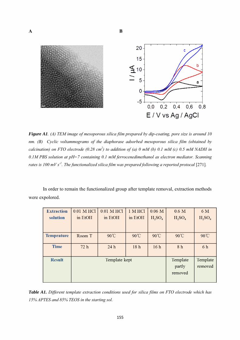
155
A B
Figure A1. (A) TEM image of mesoporous silica film prepared by dip-coating, pore size is around 10
nm. (B) Cyclic voltammograms of the diaphorase adsorbed mesoporous silica film (obtained by
calcination) on FTO electrode (0.28 cm2) to addition of (a) 0 mM (b) 0.1 mM (c) 0.5 mM NADH in
0.1M PBS solution at pH=7 containing 0.1 mM ferrocenedimethanol as electron mediator. Scanning
rates is 100 mV s-1. The functionalized silica film was prepared following a reported protocal [271].
In order to remain the functionalized group after template removal, extraction methods
were expolored.
Table A1. Different template extraction conditions used for silica films on FTO electrode which has
15% APTES and 85% TEOS in the starting sol.
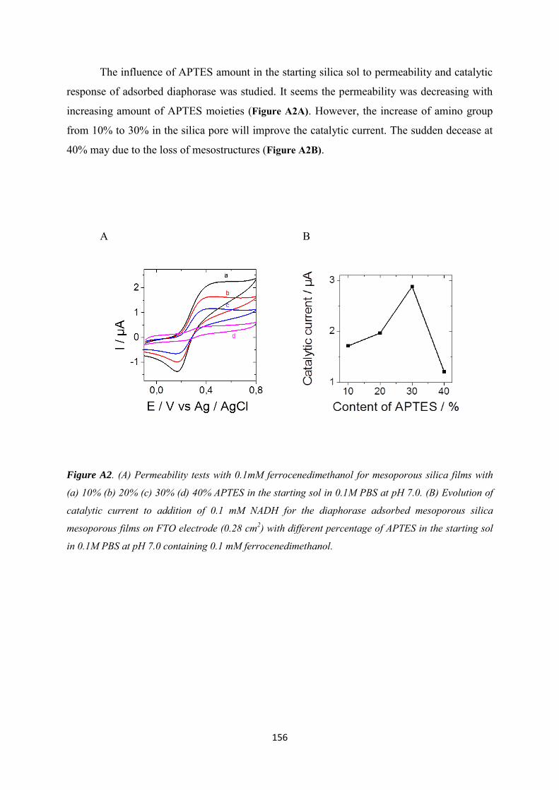
156
The influence of APTES amount in the starting silica sol to permeability and catalytic
response of adsorbed diaphorase was studied. It seems the permeability was decreasing with
increasing amount of APTES moieties (Figure A2A). However, the increase of amino group
from 10% to 30% in the silica pore will improve the catalytic current. The sudden decease at
40% may due to the loss of mesostructures (Figure A2B).
A B
Figure A2. (A) Permeability tests with 0.1mM ferrocenedimethanol for mesoporous silica films with
(a) 10% (b) 20% (c) 30% (d) 40% APTES in the starting sol in 0.1M PBS at pH 7.0. (B) Evolution of
catalytic current to addition of 0.1 mM NADH for the diaphorase adsorbed mesoporous silica
mesoporous films on FTO electrode (0.28 cm2) with different percentage of APTES in the starting sol
in 0.1M PBS at pH 7.0 containing 0.1 mM ferrocenedimethanol.
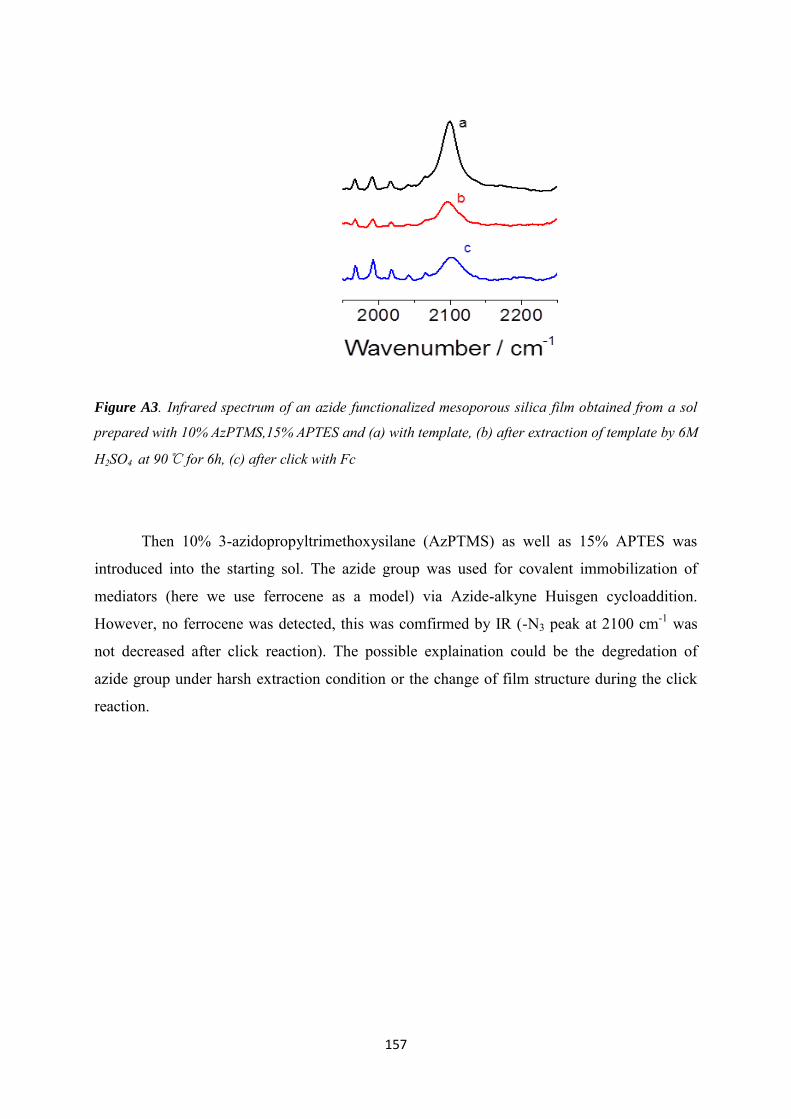
157
Figure A3. Infrared spectrum of an azide functionalized mesoporous silica film obtained from a sol
prepared with 10% AzPTMS,15% APTES and (a) with template, (b) after extraction of template by 6M
H2SO4 at 90℃ for 6h, (c) after click with Fc
Then 10% 3-azidopropyltrimethoxysilane (AzPTMS) as well as 15% APTES was
introduced into the starting sol. The azide group was used for covalent immobilization of
mediators (here we use ferrocene as a model) via Azide-alkyne Huisgen cycloaddition.
However, no ferrocene was detected, this was comfirmed by IR (-N3 peak at 2100 cm-1 was
not decreased after click reaction). The possible explaination could be the degredation of
azide group under harsh extraction condition or the change of film structure during the click
reaction.
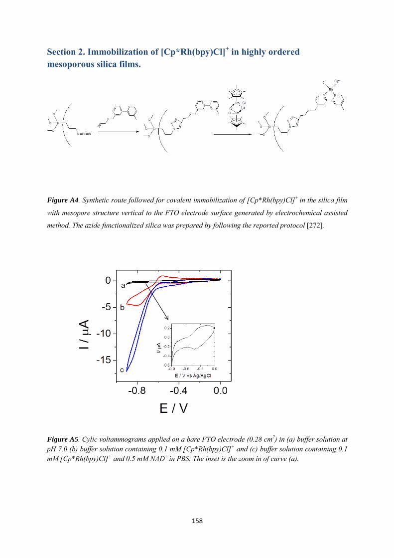
158
Section 2. Immobilization of [Cp*Rh(bpy)Cl]+ in highly ordered mesoporous silica films.
Figure A4. Synthetic route followed for covalent immobilization of [Cp*Rh(bpy)Cl]+ in the silica film
with mesopore structure vertical to the FTO electrode surface generated by electrochemical assisted
method. The azide functionalized silica was prepared by following the reported protocol [272].
Figure A5. Cylic voltammograms applied on a bare FTO electrode (0.28 cm2) in (a) buffer solution at pH 7.0 (b) buffer solution containing 0.1 mM [Cp*Rh(bpy)Cl]+ and (c) buffer solution containing 0.1 mM [Cp*Rh(bpy)Cl]+ and 0.5 mM NAD+ in PBS. The inset is the zoom in of curve (a).
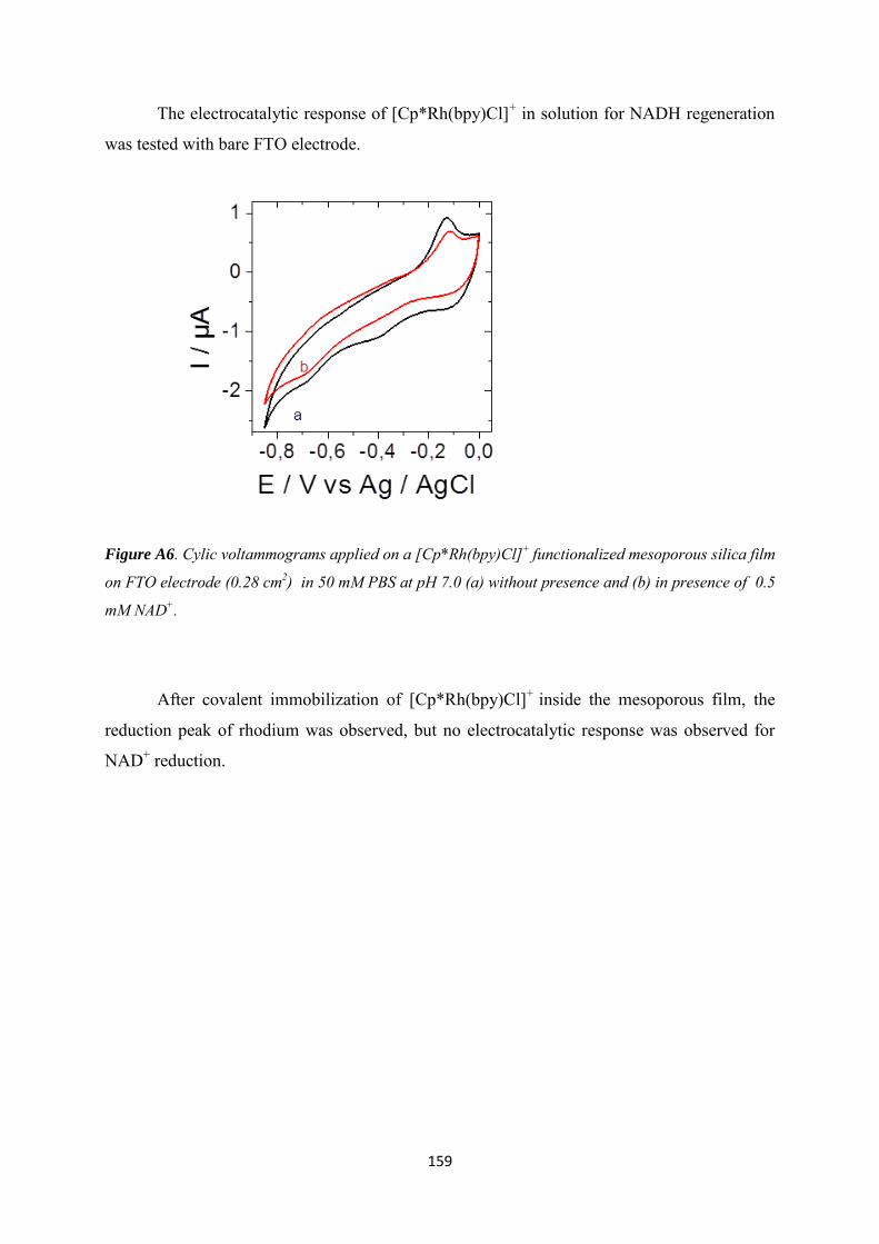
159
The electrocatalytic response of [Cp*Rh(bpy)Cl]+ in solution for NADH regeneration
was tested with bare FTO electrode.
Figure A6. Cylic voltammograms applied on a [Cp*Rh(bpy)Cl]+ functionalized mesoporous silica film
on FTO electrode (0.28 cm2) in 50 mM PBS at pH 7.0 (a) without presence and (b) in presence of 0.5
mM NAD+.
After covalent immobilization of [Cp*Rh(bpy)Cl]+ inside the mesoporous film, the
reduction peak of rhodium was observed, but no electrocatalytic response was observed for
NAD+ reduction.

160
Appendix 2
DOI: 10.1002/elan.201500172
Mesoporous Materials-Based Electrochemical EnzymaticBiosensorsMathieu Etienne,[a] Lin Zhang,[a] Neus Vil�,[a] and Alain Walcarius*[a]
1 IntroductionOrdered mesoporous materials exhibiting a regular, peri-odic organization made of mesopores of uniform size dis-tribution, and characterized by very high specific surfaceareas and pore volumes, as well as possible tuning of re-activity via suitable modification, have inspired promi-nent research interest in the past two decades due totheir unique properties and functionalities [1–27]. Theyhave indeed potential applications in adsorption [28–31],separation [32–34], catalysis [35–37], electrochemistry[38,39], sensors [39–46] and biosensors [44, 47–53], drugdelivery and other biomedical fields [54–58], immobiliza-tion of biomolecules and biocatalysis [59–64], environ-mental processes [28,30, 31, 65, 66], energy conversion andstorage [39,66–69], and so on [70,71]. Nowadays, effec-tive synthesis procedures have been developed to gener-ate various types of ordered mesoporous materials, suchas silica and silica-based organic-inorganic hybrid materi-als [1–8, 72], metal oxides other than silica [1, 9–12,73–75], mesoporous non-oxide materials [13,14, 76], orderedporous metals [1, 11, 15,16], ordered mesoporous carbons[1, 17–26,77, 78], or mesostructured organic polymers[22,26, 27]. Many of them are particularly attractive forbeing used in electrochemical sensing and biosensing de-vices, in which one can take advantage of their support/hosting properties (i.e. , for immobilization of biomole-cules, catalysts, or charge transfer mediators), their intrin-sic (electro)catalytic and/or conductivity properties(mainly mesoporous metal and carbon), their widelyopen, highly ordered and mechanically stable inorganicmesostructure (ensuring fast transport of reactantsthroughout highly porous and accessible spaces), andtheir ease of functionalization with huge amounts of di-verse reactive moieties that can be attached to mesoporewalls over wide surface areas (mainly on mesoporoussilica), for instance.
Here, we provide a comprehensive overview of electro-des modified with mesoporous materials bearing enzymesor other redox biomolecules applied in biosensing appli-cations, as a follow-up of a very recent review on the(non-enzymatic) electrochemical sensors based on or-dered mesoporous materials [47]. In the field of enzymat-ic electrochemical biosensors, there are several crucialpoints that could be addressed with the aid of orderedmesoporous materials. An electrochemical biosensor is anintegrated device combining a biological recognition ele-ment (e.g. antibodies, enzymes or whole cells), and anelectronic signal transducer (i.e. , potentiometry, amper-ometry, conductometric or surface charge using fieldeffect transistors) [79]. To be effective, the biomolecule(s)should be immobilized under an active form, in sufficientamounts, and in a durable way. This can be achieved byencapsulation in the course of the sol-gel process [80] orthrough the precipitation of 2D-inorganic materials, i.e.clays [81], LDH [82]. Following a different pathway, thehosting properties of preformed mesoporous matrices, as-sociated to their extremely large surface areas, are likelyto provide promising approaches for protein immobiliza-tion [59–64].
Electronic signal transduction of the biological recogni-tion event is usually made by the electrochemical detec-tion of one product of the enzymatic reaction (first gener-ation biosensors), or via the use of a redox mediatoracting as an electron shuttle between the active site ofthe biomolecule and the electrode surface (second gener-
[a] M. Etienne, L. Zhang, N. Vil�, A. WalcariusLaboratoire de Chimie Physique et Microbiologie pourlÏEnvironnement, UMR 7564, CNRS – Universit¦ deLorraine405 rue de Vandoeuvre, F-54600 Villers-lÀs-Nancy, France*e-mail: [email protected]
Abstract : This review (239 references) is dealing with theuse of mesoporous materials in the field of electrochemi-cal enzymatic biosensors. After a brief discussion on theinterest of mesoporous materials for electrochemical bio-sensing, a presentation of the various types of inorganicand organic-inorganic hybrid mesostructures used to datein the elaboration of enzymatic bioelectrodes is given and
the main bioelectrode configurations are described. Thevarious biosensor applications are then summarized onthe basis of a comprehensive table and some representa-tive illustrations. The main detection schemes developedin the field are based on amperometry and mediated elec-trocatalysis, as well as some on spectroelectrochemistry.
Keywords: Ordered mesoporous materials · Mesoporous silica · Organic-inorganic hybrids · Mesoporous carbon · Mesoporousmetals · Mesoporous metal oxides · Self-assembly · Surfactant template · Mediator · Thin films · Composite electrodes · Biomoleculeimmobilization · Electrochemical detection · Enzymatic biosensors · Heme proteins · Oxidases · Dehydrogenases · Tyrosinase
Specia
lIssu
eA
DVA
NC
ED
MAT
ER
IALS
www.electroanalysis.wiley-vch.de Ó 2015 Wiley-VCH Verlag GmbH & Co. KGaA, Weinheim Electroanalysis 2015, 27, 2028 – 2054 2028
Review
ation, which is often used to decrease the high overpoten-tials usually observed for various bioelectrochemical sys-tems), or by direct electronic wiring of redox enzymes(third generation biosensors) [83–88]. In this respect, itbecame recently evident that nanomaterials (such ascarbon nanotubes or graphene, metal nanoparticles, ornanostructured solids, for instance) are likely to improvethe bioelectrocatalytic performance of electrochemicalbiosensing devices [89–96]. The ordered mesoporous ma-terials belong to this category and offer attractive fea-tures, such as intrinsic catalytic and/or conductivity prop-erties or large surface area to support huge amounts ofredox mediators, which can be exploited in the field ofelectrochemical biosensors.
Actually, some reviews are already available, dealingentirely or in part with the use of mesoporous materialsfor electrochemical biosensing. They include the applica-tion of mesoporous silica-based materials for sensors andbiosensors [46,49] or immunosensors [51], or the use of
mesoporous silica for enzyme immobilization and applica-tion in biosensor technology [48,50], or the interest ofmesoporous carbon for electrocatalysis and electrochemi-cal sensing [43,44]. A general overview on bioanalysisbased on nanoporous materials is also available [52]. Todate, however, no comprehensive review including allkinds of ordered mesoporous materials (silica-based, non-siliceous metal oxide, metal, carbon, etc.) being used indesigning enzyme-based electrochemical biosensors, hasappeared. This is what we describe in this paper, on thebasis of an extensive table, after having discussed brieflythe interest of mesoporous materials for electrochemicalbiosensing and mentioned the various types of mesopo-rous materials used in enzymatic bioelectrodes and theirmost usual configurations. This constitutes a follow-upand actually a complement of our previous report onmesoporous materials-based electrochemical sensors (nonbio) [47].
Specia
lIssu
eA
DVA
NC
ED
MAT
ER
IALS
Mathieu Etienne received his PhD inElectroanalytical Chemistry fromUniversity Henri Poincar¦ Nancy I(France) in 2001 before to workwith Prof. Wolfgang Schuhmann atRuhr-Universit�t Bochum (Germa-ny). In 2004, he joined the Labora-tory of Physical Chemistry and Mi-crobiology for the Environment(France) as CNRS researcher wherehe is currently developing research-es on textured and functionalizedmaterials for bioconversion andenergy storage. He is also involvedin the development of shearforce-regulated scanning electrochemicalmicroscopy. He is the co-author ofabout 90 papers.
Lin Zhang received her B.Sc. inChemistry from Henan University(China) in 2010. Then, in theframe of an ÐErasmus MundusÏprogram, she received two M.Sc.,one in ÐAdvanced Functional Ma-terialsÏ from Augsburg University(Germany) and one in ÐChemistryand Physical Chemistry of Materi-alsÏ from Bordeaux University(France) in 2013. She is curentlyPhD student at Lorraine Universi-ty, working in the Electrochemistryand Analytical Chemistry groupheaded by Dr. Alain Walcarius in the Laboratory of PhysicalChemistry and Microbiology for the Environment (Nancy) since2013, under the supervision of Dr. Mathieu Etienne and Dr.Neus Vil�. Her research is focusing on functionalization of elec-trodes and immobilization of redox proteins on porous electro-des.
Neus Vil� received her PhD degree inchemistry in 2005 at the UniversitatAutonoma de Barcelona under theguidance of Pr. Iluminada Gallardo.Between 2005 and 2012, she held sev-eral postdoctoral positions in differ-ent research groups (Pr. Daniel B¦-langer, Universit¦ du Quebec, Mon-tr¦al; Pr. H¦ctor D. AbruÇa, CornellUniversity, New York; Dr. Alain Der-onzier, Universit¦ Joseph Fourier,Grenoble; Dr. IsraÜl Mbomekall¦, In-stitut Lavoisier, Versailles). Since2012, she is a lecturer at the Universit¦ de Lorraine working inthe Electrochemistry and Analytical Chemistry group headed byDr. Alain Walcarius in the Laboratory of Physical Chemistry andMicrobiology for the Environment (Nancy). She is currentlyworking on the functionalization and characterization of mesopo-rous silica-based materials.
Alain Walcarius received his Ph.D.from the University of Namur (Bel-gium) in 1994. After a postdoctoralstay in the Joe WangÏs group in NewMexico State University, he joinedthe CNRS (France) as a ResearchAssociate. He is currently CNRS Re-search Director and Head of the Lab-oratory of Physical Chemistry andMicrobiology for the Environment(Nancy). His analytical and electroa-nalytical chemistry group works inthe area of reactions at solid/liquidinterfaces. Among his actual research interests is the intersectionbetween the chemistry of silica-based organic-inorganic hybridmaterials and electroanalysis, nanostructuration of electrode sur-faces with mesoporous thin films, and bioelectrocatalysis. He haspublished over 200 peer-reviewed manuscripts, 8 book chapters,and one book.
www.electroanalysis.wiley-vch.de Ó 2015 Wiley-VCH Verlag GmbH & Co. KGaA, Weinheim Electroanalysis 2015, 27, 2028 – 2054 2029
Review
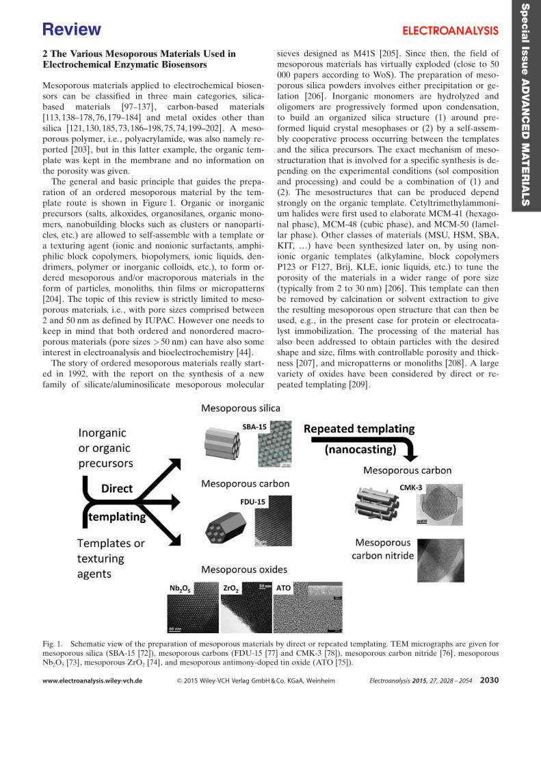
2 The Various Mesoporous Materials Used inElectrochemical Enzymatic Biosensors
Mesoporous materials applied to electrochemical biosen-sors can be classified in three main categories, silica-based materials [97–137], carbon-based materials[113, 138–178,76, 179–184] and metal oxides other thansilica [121, 130, 185,73, 186–198, 75,74, 199–202]. A meso-porous polymer, i.e. , polyacrylamide, was also namely re-ported [203], but in this latter example, the organic tem-plate was kept in the membrane and no information onthe porosity was given.
The general and basic principle that guides the prepa-ration of an ordered mesoporous material by the tem-plate route is shown in Figure 1. Organic or inorganicprecursors (salts, alkoxides, organosilanes, organic mono-mers, nanobuilding blocks such as clusters or nanoparti-cles, etc.) are allowed to self-assemble with a template ora texturing agent (ionic and nonionic surfactants, amphi-philic block copolymers, biopolymers, ionic liquids, den-drimers, polymer or inorganic colloids, etc.), to form or-dered mesoporous and/or macroporous materials in theform of particles, monoliths, thin films or micropatterns[204]. The topic of this review is strictly limited to meso-porous materials, i.e., with pore sizes comprised between2 and 50 nm as defined by IUPAC. However one needs tokeep in mind that both ordered and nonordered macro-porous materials (pore sizes >50 nm) can have also someinterest in electroanalysis and bioelectrochemistry [44].
The story of ordered mesoporous materials really start-ed in 1992, with the report on the synthesis of a newfamily of silicate/aluminosilicate mesoporous molecular
sieves designed as M41S [205]. Since then, the field ofmesoporous materials has virtually exploded (close to 50000 papers according to WoS). The preparation of meso-porous silica powders involves either precipitation or ge-lation [206]. Inorganic monomers are hydrolyzed andoligomers are progressively formed upon condensation,to build an organized silica structure (1) around pre-formed liquid crystal mesophases or (2) by a self-assem-bly cooperative process occurring between the templatesand the silica precursors. The exact mechanism of meso-structuration that is involved for a specific synthesis is de-pending on the experimental conditions (sol compositionand processing) and could be a combination of (1) and(2). The mesostructures that can be produced dependstrongly on the organic template. Cetyltrimethylammoni-um halides were first used to elaborate MCM-41 (hexago-nal phase), MCM-48 (cubic phase), and MCM-50 (lamel-lar phase). Other classes of materials (MSU, HSM, SBA,KIT, …) have been synthesized later on, by using non-ionic organic templates (alkylamine, block copolymersP123 or F127, Brij, KLE, ionic liquids, etc.) to tune theporosity of the materials in a wider range of pore size(typically from 2 to 30 nm) [206]. This template can thenbe removed by calcination or solvent extraction to givethe resulting mesoporous open structure that can then beused, e.g., in the present case for protein or electrocata-lyst immobilization. The processing of the material hasalso been addressed to obtain particles with the desiredshape and size, films with controllable porosity and thick-ness [207], and micropatterns or monoliths [208]. A largevariety of oxides have been considered by direct or re-peated templating [209].
Fig. 1. Schematic view of the preparation of mesoporous materials by direct or repeated templating. TEM micrographs are given formesoporous silica (SBA-15 [72]), mesoporous carbons (FDU-15 [77] and CMK-3 [78]), mesoporous carbon nitride [76], mesoporousNb2O5 [73], mesoporous ZrO2 [74], and mesoporous antimony-doped tin oxide (ATO [75]).
Specia
lIssu
eA
DVA
NC
ED
MAT
ER
IALS
www.electroanalysis.wiley-vch.de Ó 2015 Wiley-VCH Verlag GmbH & Co. KGaA, Weinheim Electroanalysis 2015, 27, 2028 – 2054 2030
Review
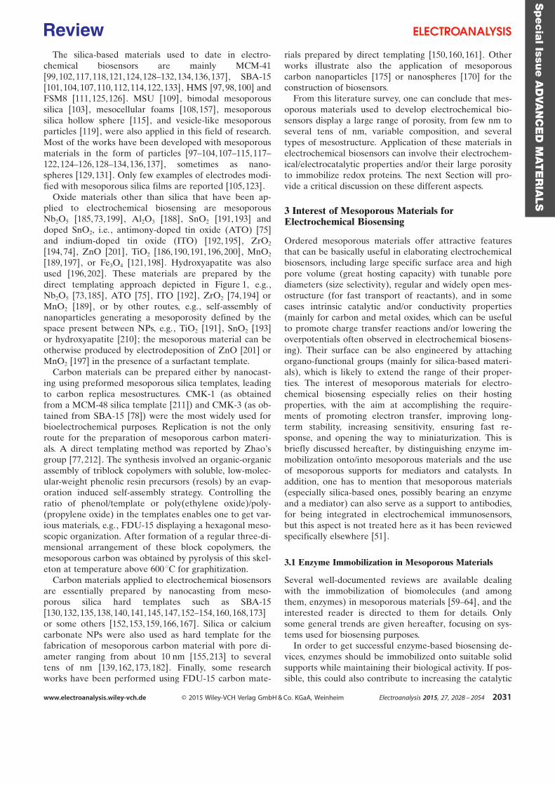
The silica-based materials used to date in electro-chemical biosensors are mainly MCM-41[99,102, 117,118,121, 124,128–132, 134, 136,137], SBA-15[101, 104,107, 110,112,114, 122,133], HMS [97,98, 100] andFSM8 [111, 125,126]. MSU [109], bimodal mesoporoussilica [103], mesocellular foams [108, 157], mesoporoussilica hollow sphere [115], and vesicle-like mesoporousparticles [119], were also applied in this field of research.Most of the works have been developed with mesoporousmaterials in the form of particles [97–104,107–115, 117–122,124–126, 128–134,136, 137], sometimes as nano-spheres [129, 131]. Only few examples of electrodes modi-fied with mesoporous silica films are reported [105, 123].
Oxide materials other than silica that have been ap-plied to electrochemical biosensing are mesoporousNb2O5 [185, 73, 199], Al2O3 [188], SnO2 [191, 193] anddoped SnO2, i.e. , antimony-doped tin oxide (ATO) [75]and indium-doped tin oxide (ITO) [192, 195], ZrO2
[194, 74], ZnO [201], TiO2 [186, 190,191, 196,200], MnO2
[189, 197], or Fe3O4 [121, 198]. Hydroxyapatite was alsoused [196, 202]. These materials are prepared by thedirect templating approach depicted in Figure 1, e.g.,Nb2O5 [73,185], ATO [75], ITO [192], ZrO2 [74,194] orMnO2 [189], or by other routes, e.g., self-assembly ofnanoparticles generating a mesoporosity defined by thespace present between NPs, e.g., TiO2 [191], SnO2 [193]or hydroxyapatite [210]; the mesoporous material can beotherwise produced by electrodeposition of ZnO [201] orMnO2 [197] in the presence of a surfactant template.
Carbon materials can be prepared either by nanocast-ing using preformed mesoporous silica templates, leadingto carbon replica mesostructures. CMK-1 (as obtainedfrom a MCM-48 silica template [211]) and CMK-3 (as ob-tained from SBA-15 [78]) were the most widely used forbioelectrochemical purposes. Replication is not the onlyroute for the preparation of mesoporous carbon materi-als. A direct templating method was reported by ZhaoÏsgroup [77,212]. The synthesis involved an organic-organicassembly of triblock copolymers with soluble, low-molec-ular-weight phenolic resin precursors (resols) by an evap-oration induced self-assembly strategy. Controlling theratio of phenol/template or poly(ethylene oxide)/poly-(propylene oxide) in the templates enables one to get var-ious materials, e.g., FDU-15 displaying a hexagonal meso-scopic organization. After formation of a regular three-di-mensional arrangement of these block copolymers, themesoporous carbon was obtained by pyrolysis of this skel-eton at temperature above 600 8C for graphitization.
Carbon materials applied to electrochemical biosensorsare essentially prepared by nanocasting from meso-porous silica hard templates such as SBA-15[130, 132,135, 138,140,141, 145,147, 152–154,160,168,173]or some others [152,153, 159,166, 167]. Silica or calciumcarbonate NPs were also used as hard template for thefabrication of mesoporous carbon material with pore di-ameter ranging from about 10 nm [155, 213] to severaltens of nm [139, 162, 173,182]. Finally, some researchworks have been performed using FDU-15 carbon mate-
rials prepared by direct templating [150,160, 161]. Otherworks illustrate also the application of mesoporouscarbon nanoparticles [175] or nanospheres [170] for theconstruction of biosensors.
From this literature survey, one can conclude that mes-oporous materials used to develop electrochemical bio-sensors display a large range of porosity, from few nm toseveral tens of nm, variable composition, and severaltypes of mesostructure. Application of these materials inelectrochemical biosensors can involve their electrochem-ical/electrocatalytic properties and/or their large porosityto immobilize redox proteins. The next Section will pro-vide a critical discussion on these different aspects.
3 Interest of Mesoporous Materials forElectrochemical Biosensing
Ordered mesoporous materials offer attractive featuresthat can be basically useful in elaborating electrochemicalbiosensors, including large specific surface area and highpore volume (great hosting capacity) with tunable porediameters (size selectivity), regular and widely open mes-ostructure (for fast transport of reactants), and in somecases intrinsic catalytic and/or conductivity properties(mainly for carbon and metal oxides, which can be usefulto promote charge transfer reactions and/or lowering theoverpotentials often observed in electrochemical biosens-ing). Their surface can be also engineered by attachingorgano-functional groups (mainly for silica-based materi-als), which is likely to extend the range of their proper-ties. The interest of mesoporous materials for electro-chemical biosensing especially relies on their hostingproperties, with the aim at accomplishing the require-ments of promoting electron transfer, improving long-term stability, increasing sensitivity, ensuring fast re-sponse, and opening the way to miniaturization. This isbriefly discussed hereafter, by distinguishing enzyme im-mobilization onto/into mesoporous materials and the useof mesoporous supports for mediators and catalysts. Inaddition, one has to mention that mesoporous materials(especially silica-based ones, possibly bearing an enzymeand a mediator) can also serve as a support to antibodies,for being integrated in electrochemical immunosensors,but this aspect is not treated here as it has been reviewedspecifically elsewhere [51].
3.1 Enzyme Immobilization in Mesoporous Materials
Several well-documented reviews are available dealingwith the immobilization of biomolecules (and amongthem, enzymes) in mesoporous materials [59–64], and theinterested reader is directed to them for details. Onlysome general trends are given hereafter, focusing on sys-tems used for biosensing purposes.
In order to get successful enzyme-based biosensing de-vices, enzymes should be immobilized onto suitable solidsupports while maintaining their biological activity. If pos-sible, this could also contribute to increasing the catalytic
Specia
lIssu
eA
DVA
NC
ED
MAT
ER
IALS
www.electroanalysis.wiley-vch.de Ó 2015 Wiley-VCH Verlag GmbH & Co. KGaA, Weinheim Electroanalysis 2015, 27, 2028 – 2054 2031
Review

activity and the recyclability of the enzyme towards harshconditions. Ordered mesoporous materials are good can-didates for hosting enzymes thanks to their unique advan-tages such as large pore volume and high specific surfacearea, uniform pore size distribution and tunable pore size,as well as defined pore geometry [60, 214,215], whichcould also contribute to enable fast transport of sub-strates and products to and from the biologically-activesites in the material.
To improve the biocompatibility of materials with pro-teins, the surface hydrophilicity, surface charge and meso-porous structure become main factors to manipulate[216]. To reach this goal, the surface of mesoporous silicawas modified by selected functional groups to increasethe interaction between the enzymes and the silica walls.This was realized either by post-grafting of pure silica[119, 133] or from one-step synthesis by co-condensationof organosilanes and tetraalkoxysilane [105]. For exam-ple, the introduction of amino groups into a mesoporoussilica film allowed increasing its interaction with cyto-chrome c, which was claimed to be higher on amino-func-tionalized films than on the pure silica films [105]. Formesoporous carbon materials, their surface can be modi-fied by hydrophilic ¢COOH groups through sulfuric acidtreatment [141]. These carboxylic groups not only im-prove the hydrophilicity of the carbon material, but alsointroduce negative charges on the carbon surface, favor-ing thereby the immobilization of the positively-chargedhemoglobin in the conditions of the experiment [147].
Basically, protein immobilization can be achieved byphysical adsorption or covalent binding. Zhou et al. com-pared the performance of glucose biosensors with glucoseoxidase immobilized on mesoporous silica by physical ad-sorption and by covalent binding, showing that the latterapproach resulted in higher enzyme loading but lower de-tection sensitivity than for physical adsorption [119].
Besides the interfacial properties of the mesoporousmaterial, pore characteristics also influenced the enzymeloadings. Bayne et al. have summarized some trends thatcan be drawn between pore characteristics and proteinloadings on the basis of 182 experiments in total, involv-ing many porous materials classified according to threecategories of pore diameters [217]. They report a decreasein protein loadings for mesopores with diameters lessthan 10 nm, as explained by physical restrictions limitingthe protein molecules to access the internal volume/sur-face of so small mesopores. For pore sizes larger than100 nm, the protein loading declines due to the decreaseof available surface area, but this belongs no more tomesoporous materials. Meanwhile, pore sized in therange of 40–70 nm seems to be the optimum for gettingthe best protein loadings [217]. However, there is still notenough work to systematically certify the definite conclu-sion within whole ranges and this is expected to bestrongly dependent on the nature and structure of thehost and the type and size of proteins.
The relative size of the protein compared to the meso-pore aperture affects necessarily the immobilization be-
havior. Different mesoporous materials have been ap-plied to immobilize glucose oxidase, not only because thisenzyme is particularly stable and can thus be used asa model protein but also regarding the great importanceof electrochemical glucose biosensors [74]. However,among these experiments, some mesoporous materials ex-hibited a pore diameter smaller than the size of glucoseoxidase (7.0 × 5.5× 8.0 nm3), meaning that it should not bepossible for the protein to enter the internal pore volumeof the hosting material. In such case, the adsorption/im-mobilization should mainly occur at the outer surface ofthe mesoporous particles, which would basically not pre-vent the biosensor application, but without taking advant-age of the mesostructure of the material. Full exploitationof the pore volume and high specific surface areas of thehosting solid requires the use of larger pore mesoporousmaterials. For example, immobilization of glucose oxidasein SBA-15 functionalized with amino groups (11.4 nmpore diameter) gave a higher normalized absolute activitythan for the same protein adsorbed onto the outer surfaceof microporous silica particles (pore diameter <2 nm), anadvantage that was also evidenced in their application forglucose biosensing in terms of extended linear concentra-tion range (0.4–13.0 mM instead of 0.4–3.1 mM) [133].
Comparative studies revealed also that a material withmesopore diameter similar to the size of the protein (i.e. ,6 nm, Figure 2A) could have significant advantages overa material with larger pore size (i.e., 13 nm, Figure 2B) infacilitating the direct electron transfer of entrapped bio-molecule (as pointed out with hemoglobin [155]) and im-proving thereby the performance of the correspondingbiosensor. It has been suggested that the “entrapment”immobilization mode and “interspace confinementeffect”, which would restrain the unfolding or conforma-tional change of enzyme molecules from inactivation,could result in pore-size-dependent enzymatic stabilityand bioactivity, which might be a crucial factor for the su-perior biosensing performance. On the other hand, onehas to mention that large pores can be beneficial for thepreparation of nanoscale enzyme reactors (NERs), as ex-emplified for mesoporous carbon with a bottleneck porestructure (mesocellular pores of 26 nm connected withwindow mesopores of 17 nm), which was applied to pre-vent the cross-linked glucose oxidase enzymes fromleaching through the smaller window mesopores (Fig-ure 2C). This NER approach improved the enzyme stabil-ity and favored electron transfer reaction between theenzyme and the conductive mesoporous carbon by main-taining a short distance between them. The comparisonwith redox proteins simply adsorbed without cross-link-ing, allowed to prove that the NER approach was effec-tively improving the sensitivity of glucose biosensors[182, 218].
Mesoporous conductive or semi-conductive and trans-parent matrices constitute another attractive family ofsolids useful to enzyme immobilization and offering theadvantage to allow the spectroelectrochemical investiga-tion of redox proteins and the elaboration of spectroelec-
Specia
lIssu
eA
DVA
NC
ED
MAT
ER
IALS
www.electroanalysis.wiley-vch.de Ó 2015 Wiley-VCH Verlag GmbH & Co. KGaA, Weinheim Electroanalysis 2015, 27, 2028 – 2054 2032
Review
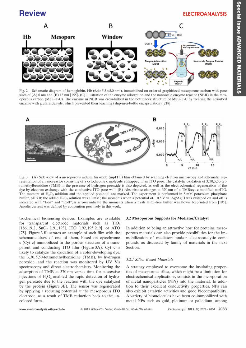
trochemical biosensing devices. Examples are availablefor transparent electrode materials such as TiO2
[186, 191], SnO2 [191, 193], ITO [192, 195,219], or ATO[75]. Figure 3 illustrates an example of such film with theschematic draw of one of them, based on cytochromec (Cyt c) immobilized in the porous structure of a trans-parent and conducting ITO film (Figure 3A). Cyt c islikely to catalyze the oxidation of a color-developing dye,the 3, 30,5,50-tetramethylbenzidine (TMB), by hydrogenperoxide, and the reaction was monitored by UV¢Visspectroscopy and direct electrochemistry. Monitoring theadsorption of TMB at 370 nm versus time for successiveinjections of H2O2 enabled the rapid detection of hydro-gen peroxide due to the reaction with the dye catalyzedby the protein (Figure 3B). The sensor was regeneratedby applying a reducing potential at the mesoporous ITOelectrode, as a result of TMB reduction back to the un-colored form.
3.2 Mesoporous Supports for Mediator/Catalyst
In addition to being an attractive host for proteins, meso-porous materials can also provide possibilities for the im-mobilization of mediators and/or electrocatalytic com-pounds, as discussed by family of materials in the nextSection.
3.2.1 Silica-Based Materials
A strategy employed to overcome the insulating proper-ties of mesoporous silica, which might be a limitation forelectrochemical applications, consists in the incorporationof metal nanoparticles (NPs) into the material. In addi-tion to their excellent conductivity properties, NPs canalso exhibit catalytic activities and good biocompatibility.A variety of biomolecules have been co-immobilized withmetal NPs such as gold, platinum or palladium, among
Fig. 2. Schematic diagram of hemoglobin, Hb (6.4 × 5.5 ×5.0 nm3), immobilized on ordered graphitized mesoporous carbon with poresizes of (A) 6 nm and (B) 13 nm [155]. (C) Illustration of the enzyme adsorption and the nanoscale enzyme reactor (NER) in the mes-oporous carbon (MSU-F-C). The enzyme in NER was cross-linked in the bottleneck structure of MSU-F-C by treating the adsorbedenzyme with glutaraldehyde, which prevented their leaching (ship-in-a-bottle encapsulation) [218].
Fig. 3. (A) Side-view of a mesoporous indium tin oxide (mpITO) film obtained by scanning electron microscopy and schematic rep-resentation of a nanoreactor consisting of a cytochrome c molecule entrapped in an ITO pore. The catalytic oxidation of 3, 30,5,50-tet-ramethylbenzidine (TMB) in the presence of hydrogen peroxide is also depicted, as well as the electrochemical regeneration of thedye by electron exchange with the conductive ITO pore wall. (B) Absorbance changes at 370 nm of a TMB/cyt c-modified mpITO.The moment of H2O2 addition and the applied potential are marked. The experiment is performed in 5 mM potassium phosphatebuffer, pH 7.0; the added H2O2 solution was 10 mM; the moments when a potential of ¢0.5 V vs. Ag/AgCl was switched on and off isindicated with “Eon” and “Eoff”; b arrows indicate the moments when a fresh H2O2-free buffer was flown. Reprinted from [195].Anodic current was defined by convention positively in this work.
Specia
lIssu
eA
DVA
NC
ED
MAT
ER
IALS
www.electroanalysis.wiley-vch.de Ó 2015 Wiley-VCH Verlag GmbH & Co. KGaA, Weinheim Electroanalysis 2015, 27, 2028 – 2054 2033
Review
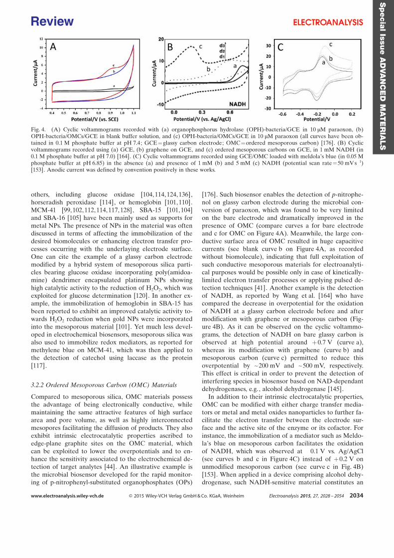
others, including glucose oxidase [104, 114, 124,136],horseradish peroxidase [114], or hemoglobin [101, 110].MCM-41 [99,102,112, 114,117, 128], SBA-15 [101,104]and SBA-16 [105] have been mainly used as supports formetal NPs. The presence of NPs in the material was oftendiscussed in terms of affecting the immobilization of thedesired biomolecules or enhancing electron transfer pro-cesses occurring with the underlaying electrode surface.One can cite the example of a glassy carbon electrodemodified by a hybrid system of mesoporous silica parti-cles bearing glucose oxidase incorporating poly(amidoa-mine) dendrimer encapsulated platinum NPs showinghigh catalytic activity to the reduction of H2O2, which wasexploited for glucose determination [120]. In another ex-ample, the immobilization of hemoglobin in SBA-15 hasbeen reported to exhibit an improved catalytic activity to-wards H2O2 reduction when gold NPs were incorporatedinto the mesoporous material [101]. Yet much less devel-oped in electrochemical biosensors, mesoporous silica wasalso used to immobilize redox mediators, as reported formethylene blue on MCM-41, which was then applied tothe detection of catechol using laccase as the protein[117].
3.2.2 Ordered Mesoporous Carbon (OMC) Materials
Compared to mesoporous silica, OMC materials possessthe advantage of being electronically conductive, whilemaintaining the same attractive features of high surfacearea and pore volume, as well as highly interconnectedmesopores facilitating the diffusion of products. They alsoexhibit intrinsic electrocatalytic properties ascribed toedge-plane graphite sites on the OMC material, whichcan be exploited to lower the overpotentials and to en-hance the sensitivity associated to the electrochemical de-tection of target analytes [44]. An illustrative example isthe microbial biosensor developed for the rapid monitor-ing of p-nitrophenyl-substituted organophosphates (OPs)
[176]. Such biosensor enables the detection of p-nitrophe-nol on glassy carbon electrode during the microbial con-version of paraoxon, which was found to be very limitedon the bare electrode and dramatically improved in thepresence of OMC (compare curves a for bare electrodeand c for OMC on Figure 4A). Meanwhile, the large con-ductive surface area of OMC resulted in huge capacitivecurrents (see blank curve b on Figure 4A, as recordedwithout biomolecule), indicating that full exploitation ofsuch conductive mesoporous materials for electroanalyti-cal purposes would be possible only in case of kinetically-limited electron transfer processes or applying pulsed de-tection techniques [41]. Another example is the detectionof NADH, as reported by Wang et al. [164] who havecompared the decrease in overpotential for the oxidationof NADH at a glassy carbon electrode before and aftermodification with graphene or mesoporous carbon (Fig-ure 4B). As it can be observed on the cyclic voltammo-grams, the detection of NADH on bare glassy carbon isobserved at high potential around +0.7 V (curve a),whereas its modification with graphene (curve b) andmesoporous carbon (curve c) permitted to reduce thisoverpotential by ~200 mV and ~500 mV, respectively.This effect is critical in order to prevent the detection ofinterfering species in biosensor based on NAD-dependantdehydrogenases, e.g., alcohol dehydrogenase [145].
In addition to their intrinsic electrocatalytic properties,OMC can be modified with either charge transfer media-tors or metal and metal oxides nanoparticles to further fa-cilitate the electron transfer between the electrode sur-face and the active site of the enzyme or its cofactor. Forinstance, the immobilization of a mediator such as Meldo-laÏs blue on mesoporous carbon facilitates the oxidationof NADH, which was observed at ¢0.1 V vs. Ag/AgCl(see curves b and c in Figure 4C) instead of +0.2 V onunmodified mesoporous carbon (see curve c in Fig. 4B)[153]. When applied in a device comprising alcohol dehy-drogenase, such NADH-sensitive material constitutes an
Fig. 4. (A) Cyclic voltammograms recorded with (a) organophosphorus hydrolase (OPH)-bacteria/GCE in 10 mM paraoxon, (b)OPH-bacteria/OMCs/GCE in blank buffer solution, and (c) OPH-bacteria/OMCs/GCE in 10 mM paraoxon (all curves have been ob-tained in 0.1 M phosphate buffer at pH 7.4; GCE=glassy carbon electrode; OMC=ordered mesoporous carbon) [176]. (B) Cyclicvoltammograms recorded using (a) GCE, (b) graphene on GCE, and (c) ordered mesoporous carbons on GCE, in 1 mM NADH (in0.1 M phosphate buffer at pH 7.0) [164]. (C) Cyclic voltammograms recorded using GCE/OMC loaded with meldolaÏs blue (in 0.05 Mphosphate buffer at pH 6.85) in the absence (a) and presence of 1 mM (b) and 5 mM (c) NADH (potential scan rate=50 mV s¢1)[153]. Anodic current was defined by convention positively in these works.
Specia
lIssu
eA
DVA
NC
ED
MAT
ER
IALS
www.electroanalysis.wiley-vch.de Ó 2015 Wiley-VCH Verlag GmbH & Co. KGaA, Weinheim Electroanalysis 2015, 27, 2028 – 2054 2034
Review
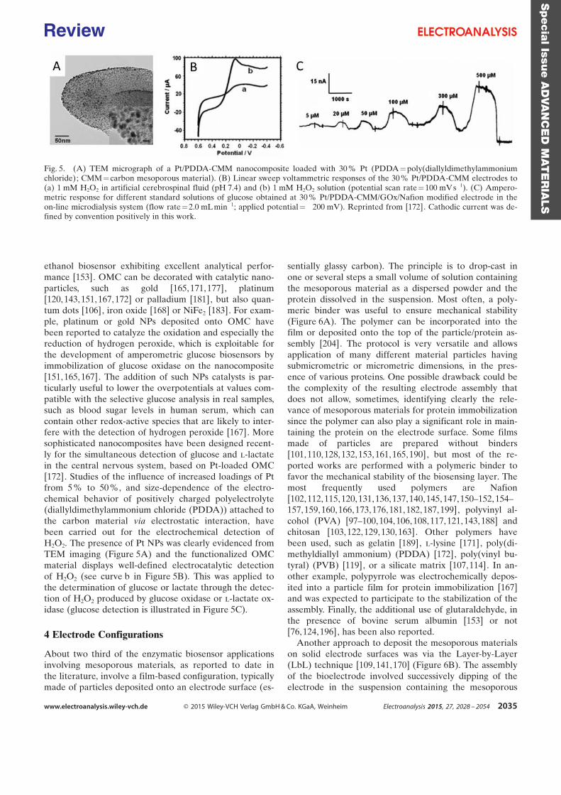
ethanol biosensor exhibiting excellent analytical perfor-mance [153]. OMC can be decorated with catalytic nano-particles, such as gold [165,171, 177], platinum[120, 143,151, 167,172] or palladium [181], but also quan-tum dots [106], iron oxide [168] or NiFe2 [183]. For exam-ple, platinum or gold NPs deposited onto OMC havebeen reported to catalyze the oxidation and especially thereduction of hydrogen peroxide, which is exploitable forthe development of amperometric glucose biosensors byimmobilization of glucose oxidase on the nanocomposite[151, 165,167]. The addition of such NPs catalysts is par-ticularly useful to lower the overpotentials at values com-patible with the selective glucose analysis in real samples,such as blood sugar levels in human serum, which cancontain other redox-active species that are likely to inter-fere with the detection of hydrogen peroxide [167]. Moresophisticated nanocomposites have been designed recent-ly for the simultaneous detection of glucose and l-lactatein the central nervous system, based on Pt-loaded OMC[172]. Studies of the influence of increased loadings of Ptfrom 5 % to 50 %, and size-dependence of the electro-chemical behavior of positively charged polyelectrolyte(diallyldimethylammonium chloride (PDDA)) attached tothe carbon material via electrostatic interaction, havebeen carried out for the electrochemical detection ofH2O2. The presence of Pt NPs was clearly evidenced fromTEM imaging (Figure 5A) and the functionalized OMCmaterial displays well-defined electrocatalytic detectionof H2O2 (see curve b in Figure 5B). This was applied tothe determination of glucose or lactate through the detec-tion of H2O2 produced by glucose oxidase or l-lactate ox-idase (glucose detection is illustrated in Figure 5C).
4 Electrode Configurations
About two third of the enzymatic biosensor applicationsinvolving mesoporous materials, as reported to date inthe literature, involve a film-based configuration, typicallymade of particles deposited onto an electrode surface (es-
sentially glassy carbon). The principle is to drop-cast inone or several steps a small volume of solution containingthe mesoporous material as a dispersed powder and theprotein dissolved in the suspension. Most often, a poly-meric binder was useful to ensure mechanical stability(Figure 6A). The polymer can be incorporated into thefilm or deposited onto the top of the particle/protein as-sembly [204]. The protocol is very versatile and allowsapplication of many different material particles havingsubmicrometric or micrometric dimensions, in the pres-ence of various proteins. One possible drawback could bethe complexity of the resulting electrode assembly thatdoes not allow, sometimes, identifying clearly the rele-vance of mesoporous materials for protein immobilizationsince the polymer can also play a significant role in main-taining the protein on the electrode surface. Some filmsmade of particles are prepared without binders[101, 110,128, 132,153,161, 165,190], but most of the re-ported works are performed with a polymeric binder tofavor the mechanical stability of the biosensing layer. Themost frequently used polymers are Nafion[102, 112,115, 120,131,136, 137,140, 145,147,150–152, 154–157,159, 160,166, 173,176,181, 182,187, 199], polyvinyl al-cohol (PVA) [97–100,104, 106,108,117, 121,143, 188] andchitosan [103, 122, 129,130,163]. Other polymers havebeen used, such as gelatin [189], l-lysine [171], poly(di-methyldiallyl ammonium) (PDDA) [172], poly(vinyl bu-tyral) (PVB) [119], or a silicate matrix [107,114]. In an-other example, polypyrrole was electrochemically depos-ited into a particle film for protein immobilization [167]and was expected to participate to the stabilization of theassembly. Finally, the additional use of glutaraldehyde, inthe presence of bovine serum albumin [153] or not[76,124, 196], has been also reported.
Another approach to deposit the mesoporous materialson solid electrode surfaces was via the Layer-by-Layer(LbL) technique [109, 141,170] (Figure 6B). The assemblyof the bioelectrode involved successively dipping of theelectrode in the suspension containing the mesoporous
Fig. 5. (A) TEM micrograph of a Pt/PDDA-CMM nanocomposite loaded with 30 % Pt (PDDA=poly(diallyldimethylammoniumchloride); CMM=carbon mesoporous material). (B) Linear sweep voltammetric responses of the 30% Pt/PDDA-CMM electrodes to(a) 1 mM H2O2 in artificial cerebrospinal fluid (pH 7.4) and (b) 1 mM H2O2 solution (potential scan rate=100 mVs¢1). (C) Ampero-metric response for different standard solutions of glucose obtained at 30% Pt/PDDA-CMM/GOx/Nafion modified electrode in theon-line microdialysis system (flow rate=2.0 mLmin¢1; applied potential=¢200 mV). Reprinted from [172]. Cathodic current was de-fined by convention positively in this work.
Specia
lIssu
eA
DVA
NC
ED
MAT
ER
IALS
www.electroanalysis.wiley-vch.de Ó 2015 Wiley-VCH Verlag GmbH & Co. KGaA, Weinheim Electroanalysis 2015, 27, 2028 – 2054 2035
Review
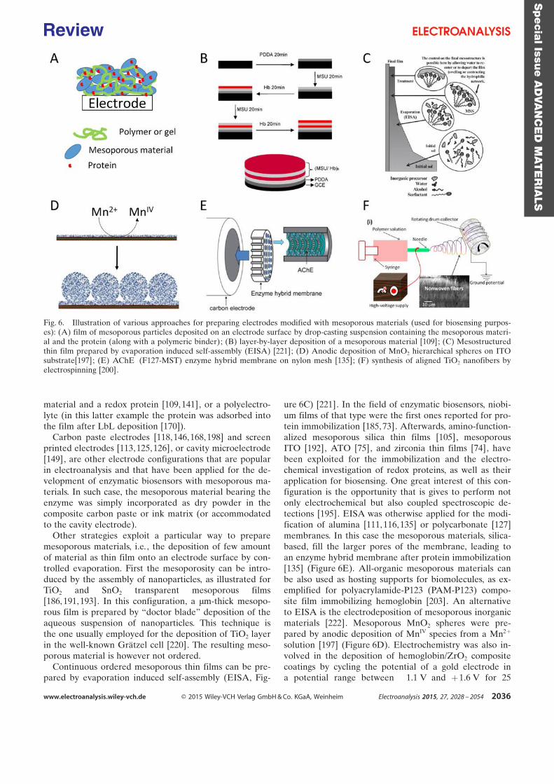
material and a redox protein [109, 141], or a polyelectro-lyte (in this latter example the protein was adsorbed intothe film after LbL deposition [170]).
Carbon paste electrodes [118, 146,168, 198] and screenprinted electrodes [113, 125, 126], or cavity microelectrode[149], are other electrode configurations that are popularin electroanalysis and that have been applied for the de-velopment of enzymatic biosensors with mesoporous ma-terials. In such case, the mesoporous material bearing theenzyme was simply incorporated as dry powder in thecomposite carbon paste or ink matrix (or accommodatedto the cavity electrode).
Other strategies exploit a particular way to preparemesoporous materials, i.e. , the deposition of few amountof material as thin film onto an electrode surface by con-trolled evaporation. First the mesoporosity can be intro-duced by the assembly of nanoparticles, as illustrated forTiO2 and SnO2 transparent mesoporous films[186, 191,193]. In this configuration, a mm-thick mesopo-rous film is prepared by “doctor blade” deposition of theaqueous suspension of nanoparticles. This technique isthe one usually employed for the deposition of TiO2 layerin the well-known Gr�tzel cell [220]. The resulting meso-porous material is however not ordered.
Continuous ordered mesoporous thin films can be pre-pared by evaporation induced self-assembly (EISA, Fig-
ure 6C) [221]. In the field of enzymatic biosensors, niobi-um films of that type were the first ones reported for pro-tein immobilization [185, 73]. Afterwards, amino-function-alized mesoporous silica thin films [105], mesoporousITO [192], ATO [75], and zirconia thin films [74], havebeen exploited for the immobilization and the electro-chemical investigation of redox proteins, as well as theirapplication for biosensing. One great interest of this con-figuration is the opportunity that is gives to perform notonly electrochemical but also coupled spectroscopic de-tections [195]. EISA was otherwise applied for the modi-fication of alumina [111, 116,135] or polycarbonate [127]membranes. In this case the mesoporous materials, silica-based, fill the larger pores of the membrane, leading toan enzyme hybrid membrane after protein immobilization[135] (Figure 6E). All-organic mesoporous materials canbe also used as hosting supports for biomolecules, as ex-emplified for polyacrylamide-P123 (PAM-P123) compo-site film immobilizing hemoglobin [203]. An alternativeto EISA is the electrodeposition of mesoporous inorganicmaterials [222]. Mesoporous MnO2 spheres were pre-pared by anodic deposition of MnIV species from a Mn2+
solution [197] (Figure 6D). Electrochemistry was also in-volved in the deposition of hemoglobin/ZrO2 compositecoatings by cycling the potential of a gold electrode ina potential range between ¢1.1 V and +1.6 V for 25
Fig. 6. Illustration of various approaches for preparing electrodes modified with mesoporous materials (used for biosensing purpos-es): (A) film of mesoporous particles deposited on an electrode surface by drop-casting suspension containing the mesoporous materi-al and the protein (along with a polymeric binder); (B) layer-by-layer deposition of a mesoporous material [109]; (C) Mesostructuredthin film prepared by evaporation induced self-assembly (EISA) [221]; (D) Anodic deposition of MnO2 hierarchical spheres on ITOsubstrate[197]; (E) AChE¢(F127-MST) enzyme hybrid membrane on nylon mesh [135]; (F) synthesis of aligned TiO2 nanofibers byelectrospinning [200].
Specia
lIssu
eA
DVA
NC
ED
MAT
ER
IALS
www.electroanalysis.wiley-vch.de Ó 2015 Wiley-VCH Verlag GmbH & Co. KGaA, Weinheim Electroanalysis 2015, 27, 2028 – 2054 2036
Review

sweeps [194], and for the deposition of enzyme-integratedphospholipid-templated mesoporous ZnO films [201]. Fi-nally, an alternative approach for the preparation of tex-tured materials is the electrospinning method (Figure 6F).The high voltage applied between a metallic needle ofa syringe (filled with the titania starting sol) and an elec-trode collector allows the formation of mesoporous elec-trospun TiO2 nanofiber suitable for bio-functionalization[200].
5 Applications: Biosensors and Analytes
Table is providing an overview of the various enzyme bio-sensors developed with mesoporous materials. About 25analytes are summarized there by alphabetical order,from the ascorbic acid to the uric acid. The porous mate-rials, the immobilized biomolecules, the electrode config-uration and the analytical performance of the biosensorsare reported.
Two target analytes were massively studied, i.e., H2O2
[97,98, 100, 101, 103, 105–110,113, 115, 118, 122, 127, 130,141, 144, 147, 154, 155, 157, 160, 161, 170, 180, 183, 185,73, 188, 190, 191, 193, 194, 199, 203] and glucose [91, 93,101, 103, 108, 109, 113, 117, 120, 122, 125, 129, 132, 134,135, 138–141, 145, 154–157, 161, 162, 164, 170–173, 180,187, 188, 191, 194, 195], H2O2 being also in some casesthe electroactive species detected in glucose biosensors[103, 108, 109, 113, 117, 122, 125, 132, 135, 140, 141, 156,157, 161, 162, 170, 172, 173, 180, 191], as well as a seriesof other analytes (see Table 1).
The detection of these targets involves proteins withdistinct characteristics. First, are the heme proteins, i.e. ,hemoglobin [86, 90, 92, 96–99, 107, 130, 136, 143, 144,149, 150, 170, 177, 179, 181, 185, 189, 192, 196], myoglo-bin [98, 106, 113, 157], cytochrome c (Cyt c) [94, 133, 159,175, 176, 183], microperoxidase from Cyt c [193] andhorseradish peroxidase (HRP) [99, 100, 114, 115, 122,127, 128, 130, 137, 73, 191, 223]. They contain an iron por-phyrin complex allowing in many cases a direct electrontransfer to occur between this cofactor and the electrodesurface. Second, are the oxidases, including the glucoseoxidase, that are flavoproteins containing a flavin adeninedinucleotide (FAD) or a flavin adenine mononlucleotide(FMN) cofactor. For the specific example of glucose ox-idase, FAD cofactor is located inside the protein anddirect electron transfer reactions are difficult to achieve[224]. Other target analytes of concern in this reviewusing oxidases are lactate [125, 126, 172], choline [116]and cholesterol [132, 200]. Other proteins have also beenimmobilized on electrode surface taking advantage ofmesoporous materials, i.e., acethylcholine [135] andcholesterol esterases [200], alcohol [145, 153, 187, 201]and formaldehyde dehydrogenases [111], tyrosinase [99,121, 171, 76, 213], laccase [117, 163], human sulfite ox-idase [75], uricase [137] superoxide dismutase [129] andorganophosphorus hydrolase [159, 176].
The interest of mesoporous supports in biosensing iseither related to the stabilization of the enzyme in its
active form or to the capability of the hosting material tofavor the electrochemical detection of redox species in-volved in the enzymatic reaction or the direct electrontransfer with the redox proteins. If looking carefully atthe information on stability for the various biosensors re-ported in Table 1, it appears clearly that better stabilitywas achieved with mesoporous silica materials [98, 99,102, 111, 116, 125, 126, 168] than with others, at leastwhen the information was provided. This quite cleartrend was observed with various proteins, such as lactateoxidase [125, 126], myoglobin [98], tyrosinase and horse-radish peroxidase [99], glucose oxidase [102, 168], formal-dehyde dehydrogenase [111], choline oxidase [116], oracethylcholine esterase [135]. Concerning the analyticalcharacteristics of the biosensors, the interest of one mate-rial versus another one is more difficult to analyze sinceno clear trend is appearing from the data collected inTable 1. Beside the detection limit or the concentrationrange that can be reached, the advantage of some materi-al, e.g., mesoporous carbon (either functionalized with or-ganic groups or metal nanoparticle, or not) is to decreasethe overpotential for detection of some analytes or tohelp in the observation of direct transfer reactions withthe redox proteins (see Section 3.2). The consequence ishere more related to the ability to detect the target ana-lyte in the presence of potential interfering species, e.g.,by decreasing the overpotential of NADH oxidation forethanol analysis [145, 153].
The next sections will provide a discussion on the basisof chosen illustrations of the various biosensors that havebeen reported in the literature with the goal to take ad-vantage of the attractive/unique properties of mesoporousmaterials to improve the electrochemical detection of thetarget analyte. The discussion is organized by reference tothe proteins immobilized in the biosensor platform, firstlythe heme enzymes, secondly the oxidases, and other pro-teins in a third section.
5.1 Heme Proteins
5.1.1 Cytochrome c and Related Proteins
Cytochrome c (Cyt c, 12.5 kDa) is a quite small size pro-tein (2.6×3.2 × 3.0 nm3 [225]), thus being a good candidatefor immobilization in mesopores, including those ofrather small dimensions. The first report on the immobili-zation of a redox protein in mesoporous materials for bio-sensing application was implemented with a niobiumoxide film deposited on a conducting glass (Figure 7)[73,185]. The film was deposited by dip-coating (see an il-lustration of this method in Figure 6C) using triblock co-polymer Pluronic P123 (EO20PO70EO20) as structuringagent and Nb(Cl5)/Nb(OC2H5)5 as the inorganic precur-sors. The material displayed a uniform pore size centredat about 6.0 nm and a relatively high specific surface area(190 m2 g¢1) favorable for the adsorption of Cyt c. Cyt c issufficiently small for allowing direct electron transfer be-tween the electroactive heme and the electrode surface.Figure 7B displays the electrochemical characterization of
Specia
lIssu
eA
DVA
NC
ED
MAT
ER
IALS
www.electroanalysis.wiley-vch.de Ó 2015 Wiley-VCH Verlag GmbH & Co. KGaA, Weinheim Electroanalysis 2015, 27, 2028 – 2054 2037
Review
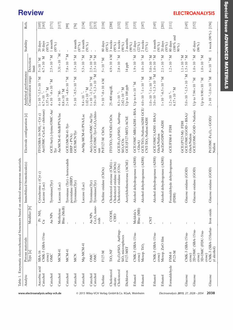
Tabl
e1.
Enz
ymat
icel
ectr
oche
mic
albi
osen
sors
base
don
orde
red
mes
opor
ous
mat
eria
ls.
Ana
lyte
Poro
usm
ater
ials
Imm
obili
zed
biom
olec
ule(
s)E
lect
rode
conf
igur
atio
n[c
]A
naly
tica
lpe
rfor
man
ceSt
abili
tyR
efs.
Type
[a]
Mod
ifie
r[b
]C
once
ntra
tion
rang
eD
etec
tion
limit
Asc
orbi
cac
idSB
A-1
6¢P
r¢N
H2
Cyt
ochr
ome
c(C
ytc)
ITO
/(SB
A-1
6-N
H2+
Cyt
c)1
×10¢6
–1.5
×10¢3
M5
×10¢7
M20
days
[105
]C
atec
hol
CM
K-3
(SB
A-1
5/su
-cr
ose)
Cu
Lac
case
(Lac
)A
u/(O
MC
-Cu/
Lac
+ch
itos
an)
6.7
×10¢7
–1.
57×
1 0¢5
M6.
7×
1 0¢
M30
days
(95
%)
[163
]
Cat
echo
lO
MC
Au
NP
sTy
rosi
nase
(Tyr
)G
CE
/Au/
(l)-
lysi
ne/O
MC
-Au/
Tyr
4×
10¢7
–8×
10¢5
M2.
5×
10¢8
M1
mon
th(8
5%
)[1
71]
Cat
echo
lM
CM
-41
Met
hyle
neB
lue
(MeB
)L
acca
se(L
ac)
Au/
MC
M-4
1-M
eB/P
VA
/lac
4×
10¢6
M–
8.8
×1 0¢5
M3.
31×
1 0¢7
M–
[117
]
Cat
echo
lM
CM
-41
–Ty
rosi
nase
(Tyr
)+ho
rser
adis
hpe
roxi
dase
(HR
P)
GC
E/(
MC
M-4
1-Ty
r-H
RP
+P
VA
)2
×10¢7
–4.8
×1 0¢4
M7.
8×
10¢1
0M
–[9
9]
Cat
echo
lM
CN
–Ty
rosi
nase
(Tyr
)G
CE
/MC
N/T
yr5
×10¢7
–9.5
×10¢6
M1.
5×
10¢8
M1
mon
th(8
7%
)[7
6]
Cat
echo
lM
g-M
CM
-41
–L
acca
se(L
ac)
Au/
Mg-
MC
M-4
1/P
VA
/Lac
9.4
×10¢7
–1.
02×
1 0¢5
M5.
3×
1 0¢9
M1
mon
th(9
1%
)[1
34]
Cat
echo
lO
MC
Au
NP
sTy
rosi
nase
(Tyr
)A
u/(l
)-ly
sine
/OM
C-A
u/Ty
r1
×10¢7
–1.7
×1 0¢4
M2.
5×
10¢8
M–
[177
]C
atec
hol
OM
CC
o 3O
4na
no-
rods
Tyro
sina
se(T
yr)
GC
E/O
MC
-Tyr
-Co 3
O4/
chit
o-sa
n5.
0×
10¢8
–1.3
×10¢5
M2.
5×
10¢8
M–
[213
]
Cho
line
F12
7-M
–C
holin
eox
idas
e(C
hOx)
Pt/
F12
7-M¢C
hOx
5×
10¢6
–8×
10¢ 4
M5
×10¢6
M80
days
(85
%)
[116
]
Cho
lest
erol
olea
teT
iO2-
NF
¢CO
OH
,¢C
HO
Cho
lest
erol
este
rase
(ChE
t)+
Cho
lest
erol
oxid
ase
(ChO
x)IT
O/T
iO2-
NF
/ChE
t-C
hOx
25–4
00m
g/dL
4.9
×10¢ 4
M3
wee
ks(9
5%
)[2
00]
Cho
lest
erol
Fe 3
O4@
SiO
2¢A
u@m
p-Si
O2
mic
rosp
here
s–
Cho
lest
erol
oxid
ase
(CO
x)G
CE
/Fe 3
O4@
SiO
2¢A
u@m
p-Si
O2/
CO
x8.
3×
10¢4
–2.
62×
10¢3
M2.
8×
1 0¢7
M1
wee
ks(9
0%
)[1
32]
Dic
hlor
vos
F12
7-M
ST–
Ace
tylc
holin
este
rase
(AC
hE)
GC
E/(
F12
7-M
ST)-
AC
hEU
pto
100
ppb
14pp
b2
mon
ths
(90
%)
[135
]
Eth
anol
CM
K-3
(SB
A-1
5/su
-cr
ose)
Mel
dola
ÏsB
lue
(MB
)A
lcoh
olde
hydr
ogen
ase
(AD
H)
GC
E/O
MC
-MB
/(A
DH
+B
SA+
Glu
A)
Up
to6
×10¢3
M1.
9×
1 0¢5
M15
days
(22
%)
[153
]
Eth
anol
Mes
op.T
iO2
–A
lcoh
olde
hydr
ogen
ase
(AD
H)
GC
E/T
iO2-
Naf
ion/
AD
HG
CE
/C
NT
-TiO
2-N
afio
n/A
DH
4.0
×1 0¢5
–1×
10¢1
M1.
0×
10¢5
M2
wee
ks(7
5%
)[1
87]
CN
T1.
0×
10¢5
–1×
1 0¢1
M5.
0×
10¢6
ME
than
olC
MK
-3(S
BA
-15/
su-
cros
e)-
Alc
ohol
dehy
drog
enas
e(A
DH
)G
CE
/OM
C/(
AD
H+
BSA
)/G
luA
/Naf
ion
3.0
×10¢4
–1.3
×1 0¢2
M1.
0×
10¢4
M1
mon
th(9
1%
)[1
45]
Eth
anol
Mes
op.Z
nOfi
lm–
Alc
ohol
dehy
drog
enas
e(A
DH
)A
u/Z
nO/D
PG
P-A
DH
1×
10¢5
–6.5
×10¢4
M2.
1×
10¢6
M20
days
(93
%)
[201
]
Form
alde
hyde
FSM
-8P
123-
M–
Form
alde
hyde
dehy
drog
enas
e(F
DH
)G
CE
/FSM
-8¢F
DH
1.2
×10¢6
–6.
17×
1 0¢4
M1.
2×
1 0¢6
M80
days
(100
%an
d90
%)
[111
]
GC
E/(
P12
3-M
)¢F
DH
1.1
×10¢6
MG
luco
seC
MK
-3(S
BA
-15/
su-
cros
e)–
Glu
cose
oxid
ase
(GO
D)
GC
E/O
MC
/(G
OD
+B
SA)/
Glu
A/N
afio
n5.
0×
10¢4
–1.5
×1 0¢2
M1.
5×
10¢4
M–
[145
]
Glu
cose
2D-O
MC
(SB
A-1
5/su
-cr
ose)
–G
luco
seox
idas
e(G
OD
)G
CE
/(O
MC
-GO
D+
Naf
ion)
Up
to7.
94×
10¢3
M1.
0×
1 0¢5
M45
days
(86
%)
[152
]
3D-O
MC
(FD
U-5
/su-
cros
e)U
pto
9.90
×10¢3
M1.
0×
1 0¢5
M
Glu
cose
CM
K-3
(SB
A-1
5/fu
rfur
-yl
alco
hol)
Iron
oxid
eG
luco
seox
idas
e(G
OD
)P
t/(O
MC
-Fe 3
O4+
GO
D)/
Naf
ion
2×
10¢4
–1.0
×10¢2
M8
×10¢5
M1
wee
k(9
0%
)[1
56]
Specia
lIssu
eA
DVA
NC
ED
MAT
ER
IALS
www.electroanalysis.wiley-vch.de Ó 2015 Wiley-VCH Verlag GmbH & Co. KGaA, Weinheim Electroanalysis 2015, 27, 2028 – 2054 2038
Review
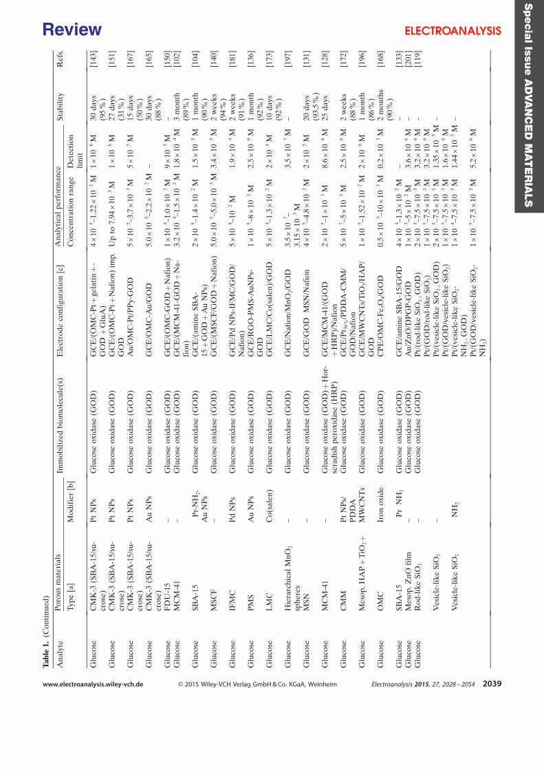
Tabl
e1.
(Con
tinu
ed)
Ana
lyte
Poro
usm
ater
ials
Imm
obili
zed
biom
olec
ule(
s)E
lect
rode
conf
igur
atio
n[c
]A
naly
tica
lpe
rfor
man
ceSt
abili
tyR
efs.
Type
[a]
Mod
ifie
r[b
]C
once
ntra
tion
rang
eD
etec
tion
limit
Glu
cose
CM
K-3
(SB
A-1
5/su
-cr
ose)
Pt
NP
sG
luco
seox
idas
e(G
OD
)G
CE
/(O
MC
-Pt+
gela
tin
+-
GO
D+
Glu
A)
4×
10¢5
–1.2
2×
1 0¢2
M1
×10¢6
M30
days
(95
%)
[143
]
Glu
cose
CM
K-3
(SB
A-1
5/su
-cr
ose)
Pt
NP
sG
luco
seox
idas
e(G
OD
)G
CE
/(O
MC
-Pt+
Naf
ion)
imp.
GO
DU
pto
7.94
×10¢3
M1
×1 0¢6
M27
days
(31
%)
[151
]
Glu
cose
CM
K-3
(SB
A-1
5/su
-cr
ose)
Pt
NP
sG
luco
seox
idas
e(G
OD
)A
u/O
MC
-Pt/
PP
y-G
OD
5×
10¢5
–3.7
×10¢3
M5
×10¢5
M15
days
(50
%)
[167
]
Glu
cose
CM
K-3
(SB
A-1
5/su
-cr
ose)
Au
NP
sG
luco
seox
idas
e(G
OD
)G
CE
/OM
C-A
u/G
OD
5.0
×10¢5
–2.2
×1 0¢2
M–
30da
ys(8
8%
)[1
65]
Glu
cose
FD
U-1
5–
Glu
cose
oxid
ase
(GO
D)
GC
E/(
OM
C-G
OD
+N
afio
n)1
×10¢4
–1.0
×1 0¢3
M9
×10¢5
M–
[150
]G
luco
seM
CM
-41
–G
luco
seox
idas
e(G
OD
)G
CE
/(M
CM
-41-
GO
D+
Na-
fion
)3.
2×
10¢4
–1.5
×1 0¢2
M1.
8×
10¢4
M3
mon
th(8
9%
)[1
02]
Glu
cose
SBA
-15
¢Pr-
NH
2,A
uN
Ps
Glu
cose
oxid
ase
(GO
D)
GC
E/(
amin
eSB
A-
15+
GO
D+
Au
NP
s)2
×10¢5
–1.4
×10¢2
M1.
5×
10¢5
M1
mon
th(9
0%
)[1
04]
Glu
cose
MSC
F–
Glu
cose
oxid
ase
(GO
D)
GC
E/(
MSC
F/G
OD
+N
afio
n)5.
0×
10¢5
–5.0
×1 0¢3
M3.
4×
10¢5
M2
wee
ks(9
4%
)[1
40]
Glu
cose
IFM
CP
dN
Ps
Glu
cose
oxid
ase
(GO
D)
GC
E/P
dN
Ps-
IFM
C/G
OD
/N
afio
n)5
×10¢4
–10¢
2M
1.9
×1 0¢4
M2
wee
ks(9
1%
)[1
81]
Glu
cose
PM
SA
uN
Ps
Glu
cose
oxid
ase
(GO
D)
GC
E/R
GO
-PM
S-A
uNP
s-G
OD
1×
10¢5
–8×
1 0¢3
M2.
5×
10¢6
M1
mon
th(9
2%
)[1
36]
Glu
cose
LM
CC
o(sa
len)
Glu
cose
oxid
ase
(GO
D)
GC
E/L
MC
/Co(
sale
n)/G
OD
5×
10¢4
–1.3
×1 0¢2
M2
×10¢4
M10
days
(92
%)
[173
]
Glu
cose
Hie
rarc
hica
lM
nO2
sphe
res
–G
luco
seox
idas
e(G
OD
)G
CE
/Naf
ion/
MnO
2/G
OD
3.5
×10¢7
–3.
15×
10¢3
M3.
5×
1 0¢7
M–
[197
]
Glu
cose
MSN
–G
luco
seox
idas
e(G
OD
)G
CE
/GO
D¢M
SN/N
afio
n4
×10¢5
–4.8
×1 0¢3
M2
×10¢5
M20
days
(93.
5%
)[1
31]
Glu
cose
MC
M-4
1–
Glu
cose
oxid
ase
(GO
D)+
Hor
-se
radi
shpe
roxi
dase
(HR
P)
GC
E/M
CM
-41/
(GO
D+
HR
P)/
Naf
ion
2×
10¢5
–1×
1 0¢3
M8.
6×
10¢6
M25
days
[128
]
Glu
cose
CM
MP
tN
Ps/
PD
DA
Glu
cose
oxid
ase
(GO
D)
GC
E/P
t 30
%/P
DD
A-C
MM
/G
OD
/Naf
ion
5×
10¢7
–5×
10¢5
M2.
5×
10¢6
M2
wee
ks(8
8%
)[1
72]
Glu
cose
Mes
op.H
AP
+T
iO2+
MW
CN
Ts
Glu
cose
oxid
ase
(GO
D)
GC
E/M
WC
NT
s/T
iO2/
HA
P/
GO
D1
×10¢5
–1.5
2×
10¢2
M2
×10¢6
M1
mon
th(8
6%
)[1
96]
Glu
cose
OM
CIr
onox
ide
Glu
cose
oxid
ase
(GO
D)
CP
E/O
MC
-Fe 3
O4/
GO
D0.
5×
10¢3
–10
×10¢3
M0.
2×
10¢3
M2
mon
ths
(90
%)
[168
]
Glu
cose
SBA
-15
¢Pr¢
NH
2G
luco
seox
idas
e(G
OD
)G
CE
/am
ine
SBA
-15/
GO
D4
×10¢4
–1.3
×10¢2
M–
–[1
33]
Glu
cose
Mes
op.Z
nOfi
lm–
Glu
cose
oxid
ase
(GO
D)
Au/
ZnO
/DP
GP
-GO
D1
×10¢3
–5×
1 0¢2
M3.
6×
10¢6
M–
[201
]G
luco
seR
od-l
ike
SiO
2–
Glu
cose
oxid
ase
(GO
D)
Pt/
(rod
-lik
eSi
O2
+G
OD
)2
×10¢5
–7.5
×10¢3
M3.
2×
10¢6
M–
[119
]P
t/(G
OD
/rod
-lik
eSi
O2)
1×
1 0¢5
–7.5
×10¢3
M3.
2×
10¢6
MV
esic
le-l
ike
SiO
2–
Pt/
(ves
icle
-lik
eSi
O2
+G
OD
)2
×10¢5
–7.5
×10¢3
M1.
35×
10¢5
M–
Pt/
(GO
D/v
esic
le-l
ike
SiO
2)1
×1 0¢5
–7.5
×10¢3
M1.
6×
10¢6
MV
esic
le-l
ike
SiO
2¢N
H2
Pt/
(ves
icle
-lik
eSi
O2-
NH
2+
GO
D)
1×
1 0¢4
–7.5
×10¢3
M1.
44×
10¢5
M–
Pt/
(GO
D/v
esic
le-l
ike
SiO
2-N
H2)
1×
10¢5
–7.5
×10¢3
M5.
2×
10¢6
M
Specia
lIssu
eA
DVA
NC
ED
MAT
ER
IALS
www.electroanalysis.wiley-vch.de Ó 2015 Wiley-VCH Verlag GmbH & Co. KGaA, Weinheim Electroanalysis 2015, 27, 2028 – 2054 2039
Review
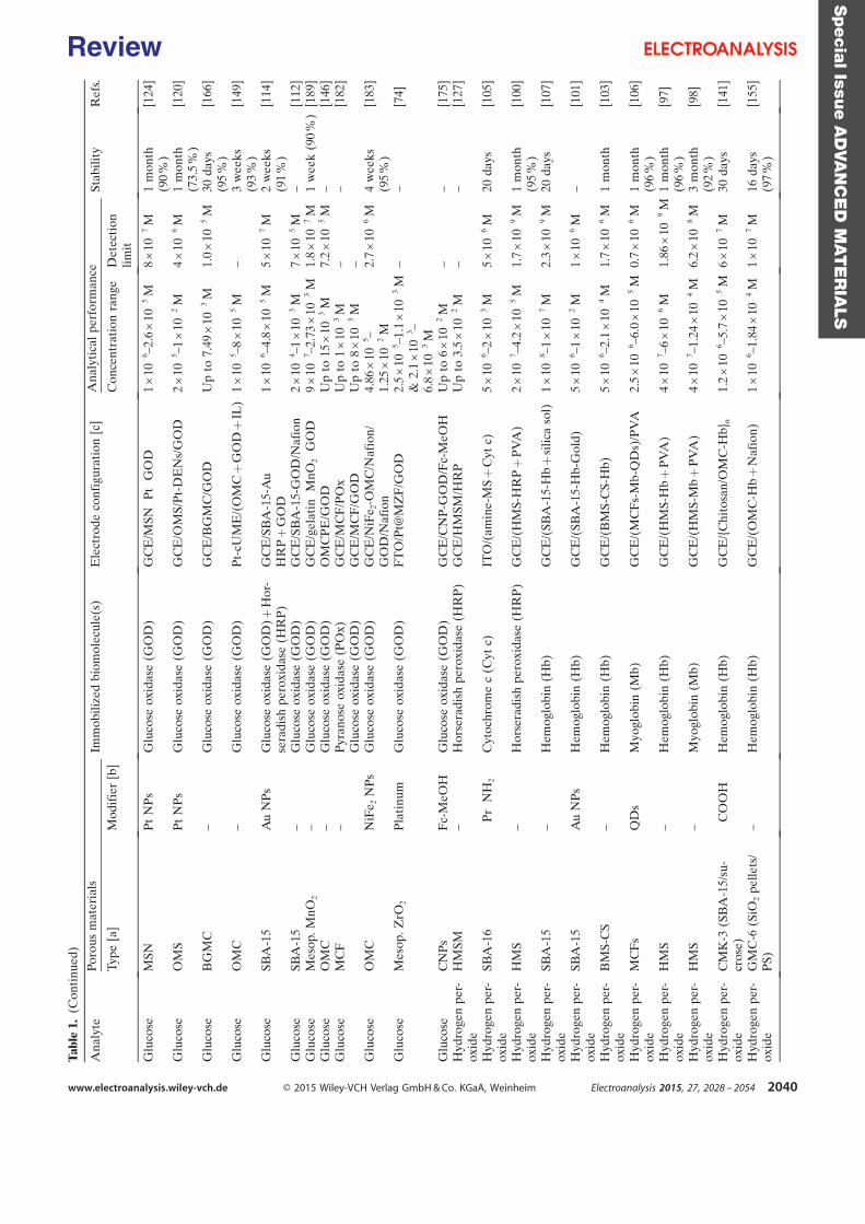
Tabl
e1.
(Con
tinu
ed)
Ana
lyte
Poro
usm
ater
ials
Imm
obili
zed
biom
olec
ule(
s)E
lect
rode
conf
igur
atio
n[c
]A
naly
tica
lpe
rfor
man
ceSt
abili
tyR
efs.
Type
[a]
Mod
ifie
r[b
]C
once
ntra
tion
rang
eD
etec
tion
limit
Glu
cose
MSN
Pt
NP
sG
luco
seox
idas
e(G
OD
)G
CE
/MSN¢P
t¢G
OD
1×
10¢6
–2.6
×1 0¢5
M8
×10¢7
M1
mon
th(9
0%
)[1
24]
Glu
cose
OM
SP
tN
Ps
Glu
cose
oxid
ase
(GO
D)
GC
E/O
MS/
Pt-
DE
Ns/
GO
D2
×10¢5
–1×
1 0¢2
M4
×10¢6
M1
mon
th(7
3.5
%)
[120
]
Glu
cose
BG
MC
–G
luco
seox
idas
e(G
OD
)G
CE
/BG
MC
/GO
DU
pto
7.49
×10¢3
M1.
0×
10¢5
M30
days
(95
%)
[166
]
Glu
cose
OM
C–
Glu
cose
oxid
ase
(GO
D)
Pt-
cUM
E/(
OM
C+
GO
D+
IL)
1×
10¢5
–8×
1 0¢5
M–
3w
eeks
(93
%)
[149
]
Glu
cose
SBA
-15
Au
NP
sG
luco
seox
idas
e(G
OD
)+H
or-
sera
dish
pero
xida
se(H
RP
)G
CE
/SB
A-1
5-A
u¢H
RP
+G
OD
1×
10¢6
–4.8
×1 0¢5
M5
×10¢7
M2
wee
ks(9
1%
)[1
14]
Glu
cose
SBA
-15
–G
luco
seox
idas
e(G
OD
)G
CE
/SB
A-1
5-G
OD
/Naf
ion
2×
10¢4
–1×
1 0¢3
M7
×10¢5
M–
[112
]G
luco
seM
esop
.MnO
2–
Glu
cose
oxid
ase
(GO
D)
GC
E/g
elat
in¢M
nO2¢
GO
D9
×10¢7
–2.7
3×
10¢3
M1.
8×
10¢7
M1
wee
k(9
0%
)[1
89]
Glu
cose
OM
C–
Glu
cose
oxid
ase
(GO
D)
OM
CP
E/G
OD
Up
to15
×10¢3
M7.
2×
1 0¢5
M–
[146
]G
luco
seM
CF
–P
yran
ose
oxid
ase
(PO
x)G
CE
/MC
F/P
Ox
Up
to1
×10¢3
M–
–[1
82]
Glu
cose
oxid
ase
(GO
D)
GC
E/M
CF
/GO
DU
pto
8×
10¢3
M–
Glu
cose
OM
CN
iFe 2
NP
sG
luco
seox
idas
e(G
OD
)G
CE
/NiF
e 2-O
MC
/Naf
ion/
GO
D/N
afio
n4.
86×
1 0¢5
–1.
25×
10¢2
M2.
7×
1 0¢6
M4
wee
ks(9
5%
)[1
83]
Glu
cose
Mes
op.Z
rO2
Pla
tinu
mG
luco
seox
idas
e(G
OD
)F
TO
/Pt@
MZ
F/G
OD
2.5
×10¢5
–1.1
×10¢3
M–
–[7
4]&
2.1
×10¢3
–6.
8×
1 0¢3
MG
luco
seC
NP
sF
c-M
eOH
Glu
cose
oxid
ase
(GO
D)
GC
E/C
NP
-GO
D/F
c-M
eOH
Up
to6
×10¢2
M–
–[1
75]
Hyd
roge
npe
r-ox
ide
HM
SM–
Hor
sera
dish
pero
xida
se(H
RP
)G
CE
/HM
SM/H
RP
Up
to3.
5×
10¢2
M–
–[1
27]
Hyd
roge
npe
r-ox
ide
SBA
-16
¢Pr¢
NH
2C
ytoc
hrom
ec
(Cyt
c)IT
O/(
amin
e-M
S+
Cyt
c)5
×10¢6
–2×
10¢3
M5
×10¢6
M20
days
[105
]
Hyd
roge
npe
r-ox
ide
HM
S–
Hor
sera
dish
pero
xida
se(H
RP
)G
CE
/(H
MS-
HR
P+
PV
A)
2×
10¢7
–4.2
×10¢5
M1.
7×
10¢9
M1
mon
th(9
5%
)[1
00]
Hyd
roge
npe
r-ox
ide
SBA
-15
–H
emog
lobi
n(H
b)G
CE
/(SB
A-1
5-H
b+
silic
aso
l)1
×10¢8
–1×
1 0¢7
M2.
3×
10¢9
M20
days
[107
]
Hyd
roge
npe
r-ox
ide
SBA
-15
Au
NP
sH
emog
lobi
n(H
b)G
CE
/(SB
A-1
5-H
b-G
old)
5×
10¢6
–1×
1 0¢2
M1
×10¢6
M–
[101
]
Hyd
roge
npe
r-ox
ide
BM
S-C
S–
Hem
oglo
bin
(Hb)
GC
E/(
BM
S-C
S-H
b)5
×10¢6
–2.1
×1 0¢4
M1.
7×
10¢6
M1
mon
th[1
03]
Hyd
roge
npe
r-ox
ide
MC
Fs
QD
sM
yogl
obin
(Mb)
GC
E/(
MC
Fs-
Mb-
QD
s)/P
VA
2.5
×10¢6
–6.0
×1 0¢5
M0.
7×
10¢6
M1
mon
th(9
6%
)[1
06]
Hyd
roge
npe
r-ox
ide
HM
S–
Hem
oglo
bin
(Hb)
GC
E/(
HM
S-H
b+
PV
A)
4×
10¢7
–6×
10¢6
M1.
86×
10¢9
M1
mon
th(9
6%
)[9
7]
Hyd
roge
npe
r-ox
ide
HM
S–
Myo
glob
in(M
b)G
CE
/(H
MS-
Mb
+P
VA
)4
×10¢7
–1.2
4×
1 0¢4
M6.
2×
10¢8
M3
mon
th(9
2%
)[9
8]
Hyd
roge
npe
r-ox
ide
CM
K-3
(SB
A-1
5/su
-cr
ose)
¢CO
OH
Hem
oglo
bin
(Hb)
GC
E/{
Chi
tosa
n/O
MC
-Hb}
n1.
2×
10¢6
–5.7
×10¢5
M6
×10¢7
M30
days
[141
]
Hyd
roge
npe
r-ox
ide
GM
C-6
(SiO
2pe
llets
/P
S)–
Hem
oglo
bin
(Hb)
GC
E/(
OM
C-H
b+
Naf
ion)
1×
10¢6
–1.8
4×
10¢4
M1
×10¢7
M16
days
(97
%)
[155
]
Specia
lIssu
eA
DVA
NC
ED
MAT
ER
IALS
www.electroanalysis.wiley-vch.de Ó 2015 Wiley-VCH Verlag GmbH & Co. KGaA, Weinheim Electroanalysis 2015, 27, 2028 – 2054 2040
Review
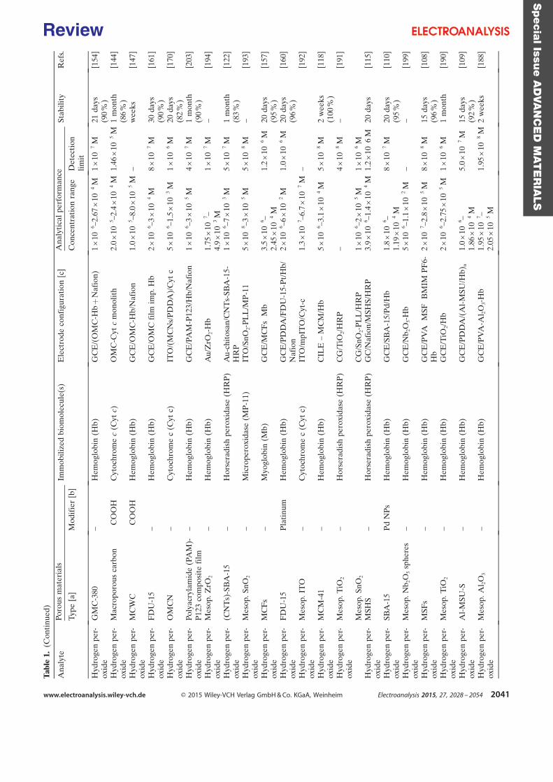
Tabl
e1.
(Con
tinu
ed)
Ana
lyte
Poro
usm
ater
ials
Imm
obili
zed
biom
olec
ule(
s)E
lect
rode
conf
igur
atio
n[c
]A
naly
tica
lpe
rfor
man
ceSt
abili
tyR
efs.
Type
[a]
Mod
ifie
r[b
]C
once
ntra
tion
rang
eD
etec
tion
limit
Hyd
roge
npe
r-ox
ide
GM
C-3
80–
Hem
oglo
bin
(Hb)
GC
E/(
OM
C-H
b+
Naf
ion)
1×
10¢6
–2.6
7×
1 0¢4
M1
×10¢7
M21
days
(90
%)
[154
]
Hyd
roge
npe
r-ox
ide
Mac
ropo
rous
carb
on¢C
OO
HC
ytoc
hrom
ec
(Cyt
c)O
MC
-Cyt
cm
onol
ith
2.0
×10¢5
–2.4
×1 0¢4
M1.
46×
10¢5
M1
mon
th(8
6%
)[1
44]
Hyd
roge
npe
r-ox
ide
MC
WC
¢CO
OH
Hem
oglo
bin
(Hb)
GC
E/O
MC
-Hb/
Naf
ion
1.0
×10¢5
–8.0
×10¢5
M–
wee
ks[1
47]
Hyd
roge
npe
r-ox
ide
FD
U-1
5–
Hem
oglo
bin
(Hb)
GC
E/O
MC
film
imp.
Hb
2×
10¢6
–3×
1 0¢4
M8
×10¢7
M30
days
(90
%)
[161
]
Hyd
roge
npe
r-ox
ide
OM
CN
–C
ytoc
hrom
ec
(Cyt
c)IT
O/(
MC
Ns/
PD
DA
)/C
ytc
5×
10¢6
–1.5
×1 0¢3
M1
×10¢6
M20
days
(82
%)
[170
]
Hyd
roge
npe
r-ox
ide
Poly
acry
lam
ide
(PA
M)-
P12
3co
mpo
site
film
–H
emog
lobi
n(H
b)G
CE
/PA
M-P
123/
Hb/
Naf
ion
1×
10¢6
–3×
1 0¢5
M4
×10¢7
M1
mon
th(9
0%
)[2
03]
Hyd
roge
npe
r-ox
ide
Mes
op.Z
rO2
–H
emog
lobi
n(H
b)A
u/Z
rO2-
Hb
1.75
×10¢7
–4.
9×
10¢3
M1
×1 0¢7
M–
[194
]
Hyd
roge
npe
r-ox
ide
(CN
Ts)
-SB
A-1
5–
Hor
sera
dish
pero
xida
se(H
RP
)A
u-ch
itos
an/C
NT
s-SB
A-1
5-H
RP
1×
10¢6
–7×
1 0¢3
M5
×10¢7
M1
mon
th(8
3%
)[1
22]
Hyd
roge
npe
r-ox
ide
Mes
op.S
nO2
–M
icro
pero
xida
se(M
P-1
1)IT
O/S
nO2-
PL
L/M
P-1
15
×10¢8
–3×
10¢5
M5
×10¢8
M–
[193
]
Hyd
roge
npe
r-ox
ide
MC
Fs
–M
yogl
obin
(Mb)
GC
E/M
CF
s¢M
b3.
5×
10¢6
–2.
45×
10¢4
M1.
2×
10¢6
M20
days
(95
%)
[157
]
Hyd
roge
npe
r-ox
ide
FD
U-1
5P
lati
num
Hem
oglo
bin
(Hb)
GC
E/P
DD
A/F
DU
-15-
Pt/
Hb/
Naf
ion
2×
10¢6
–6×
1 0¢2
M1.
0×
10¢6
M20
days
(96
%)
[160
]
Hyd
roge
npe
r-ox
ide
Mes
op.I
TO
–C
ytoc
hrom
ec
(Cyt
c)IT
O/m
pIT
O/C
yt-c
1.3
×10¢7
–6.7
×1 0¢7
M–
[192
]
Hyd
roge
npe
r-ox
ide
MC
M-4
1–
Hem
oglo
bin
(Hb)
CIL
E–
MC
M/H
b5
×10¢6
–3.1
×1 0¢4
M5
×10¢8
M2
wee
ks(1
00%
)[1
18]
Hyd
roge
npe
r-ox
ide
Mes
op.T
iO2
–H
orse
radi
shpe
roxi
dase
(HR
P)
CG
/TiO
2/H
RP
–4
×10¢8
M–
[191
]
Mes
op.S
nO2
CG
/SnO
2-P
LL
/HR
P1
×10¢6
–2×
10¢5
M1
×10¢6
MH
ydro
gen
per-
oxid
eM
SHS
–H
orse
radi
shpe
roxi
dase
(HR
P)
GC
/Naf
ion/
MSH
S/H
RP
3.9
×10¢6
–1.4
×1 0¢4
M1.
2×
10¢ 6
M20
days
[115
]
Hyd
roge
npe
r-ox
ide
SBA
-15
Pd
NP
sH
emog
lobi
n(H
b)G
CE
/SB
A-1
5/P
d/H
b1.
8×
10¢6
–1.
19×
1 0¢4
M8
×10¢7
M20
days
(95
%)
[110
]
Hyd
roge
npe
r-ox
ide
Mes
op.N
b 2O
5sp
here
s–
Hem
oglo
bin
(Hb)
GC
E/N
b 2O
5-H
b5
×10¢6
–1.1
×10¢2
M–
–[1
99]
Hyd
roge
npe
r-ox
ide
MSF
s–
Hem
oglo
bin
(Hb)
GC
E/P
VA¢M
SF¢B
MIM
PF
6-H
b2
×10¢7
–2.8
×10¢5
M8
×10¢8
M15
days
(96
%)
[108
]
Hyd
roge
npe
r-ox
ide
Mes
op.T
iO2
–H
emog
lobi
n(H
b)G
CE
/TiO
2/H
b2
×10¢6
–2.7
5×
10¢5
M1
×10¢6
M1
mon
th[1
90]
Hyd
roge
npe
r-ox
ide
Al-
MSU
-S–
Hem
oglo
bin
(Hb)
GC
E/P
DD
A/(
Al-
MSU
/Hb)
n1.
0×
10¢6
–1.
86×
10¢4
M5.
0×
1 0¢7
M15
days
(92
%)
[109
]
Hyd
roge
npe
r-ox
ide
Mes
op.A
l 2O
3–
Hem
oglo
bin
(Hb)
GC
E/P
VA
-Al 2
O3-
Hb
1.95
×10¢7
–2.
05×
10¢5
M1.
95×
1 0¢8
M2
wee
ks[1
88]
Specia
lIssu
eA
DVA
NC
ED
MAT
ER
IALS
www.electroanalysis.wiley-vch.de Ó 2015 Wiley-VCH Verlag GmbH & Co. KGaA, Weinheim Electroanalysis 2015, 27, 2028 – 2054 2041
Review
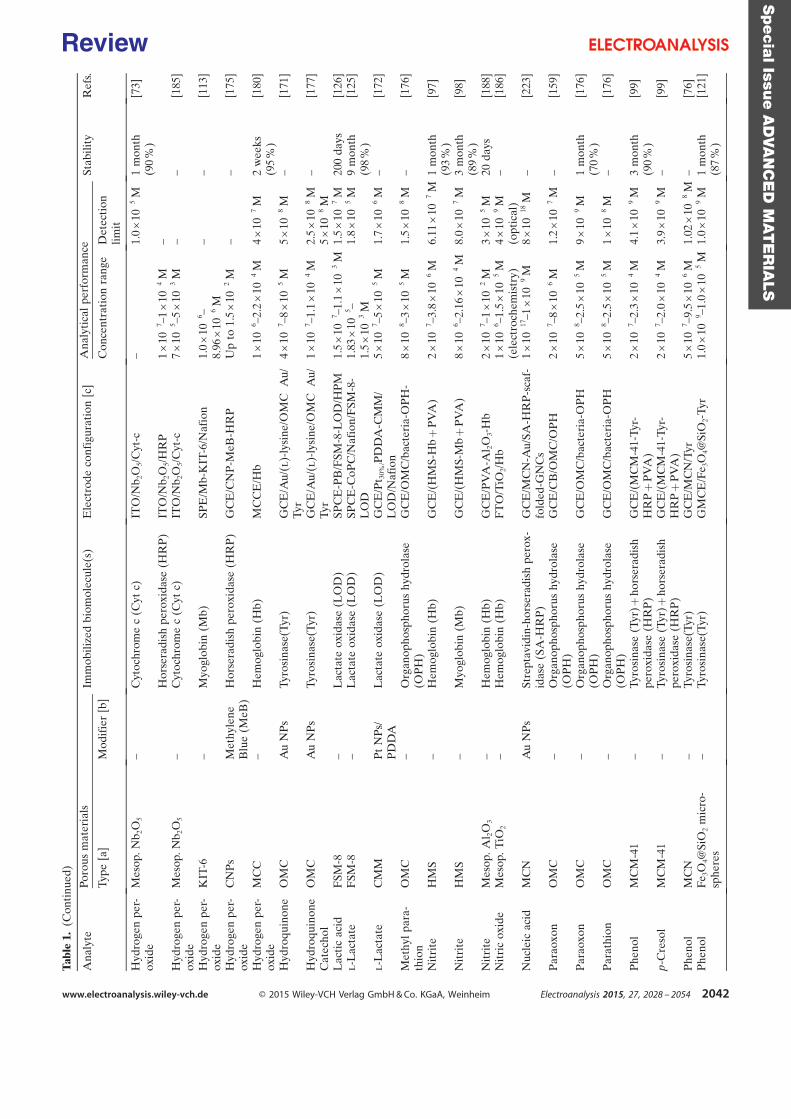
Tabl
e1.
(Con
tinu
ed)
Ana
lyte
Poro
usm
ater
ials
Imm
obili
zed
biom
olec
ule(
s)E
lect
rode
conf
igur
atio
n[c
]A
naly
tica
lpe
rfor
man
ceSt
abili
tyR
efs.
Type
[a]
Mod
ifie
r[b
]C
once
ntra
tion
rang
eD
etec
tion
limit
Hyd
roge
npe
r-ox
ide
Mes
op.N
b 2O
5–
Cyt
ochr
ome
c(C
ytc)
ITO
/Nb 2
O5/
Cyt
-c–
1.0
×10¢5
M1
mon
th(9
0%
)[7
3]
Hor
sera
dish
pero
xida
se(H
RP
)IT
O/N
b 2O
5/H
RP
1×
10¢7
–1×
10¢4
M–
Hyd
roge
npe
r-ox
ide
Mes
op.N
b 2O
5–
Cyt
ochr
ome
c(C
ytc)
ITO
/Nb 2
O5/
Cyt
-c7
×10¢5
–5×
10¢3
M–
–[1
85]
Hyd
roge
npe
r-ox
ide
KIT
-6–
Myo
glob
in(M
b)SP
E/M
b-K
IT-6
/Naf
ion
1.0
×10¢6
–8.
96×
1 0¢6
M–
–[1
13]
Hyd
roge
npe
r-ox
ide
CN
Ps
Met
hyle
neB
lue
(MeB
)H
orse
radi
shpe
roxi
dase
(HR
P)
GC
E/C
NP
-MeB
-HR
PU
pto
1.5
×10¢2
M–
–[1
75]
Hyd
roge
npe
r-ox
ide
MC
C–
Hem
oglo
bin
(Hb)
MC
CE
/Hb
1×
10¢6
–2.2
×1 0¢4
M4
×10¢7
M2
wee
ks(9
5%
)[1
80]
Hyd
roqu
inon
eO
MC
Au
NP
sTy
rosi
nase
(Tyr
)G
CE
/Au/
(l)-
lysi
ne/O
MC¢A
u/Ty
r4
×10¢7
–8×
10¢5
M5
×10¢8
M–
[171
]
Hyd
roqu
inon
eC
atec
hol
OM
CA
uN
Ps
Tyro
sina
se(T
yr)
GC
E/A
u/(l
)-ly
sine
/OM
C¢A
u/Ty
r1
×10¢7
–1.1
×1 0¢4
M2.
5×
10¢8
M5
×10¢8
M–
[177
]
Lac
tic
acid
FSM
-8–
Lac
tate
oxid
ase
(LO
D)
SPC
E-P
B/F
SM-8
-LO
D/H
PM
1.5
×10¢7
–1.1
×1 0¢3
M1.
5×
10¢7
M20
0da
ys[1
26]
l-L
acta
teF
SM-8
–L
acta
teox
idas
e(L
OD
)SP
CE
-CoP
C/N
afio
n/F
SM-8
-L
OD
1.83
×10¢5
–1.
5×
1 0¢3
M1.
8×
1 0¢5
M9
mon
th(9
8%
)[1
25]
l-L
acta
teC
MM
Pt
NP
s/P
DD
AL
acta
teox
idas
e(L
OD
)G
CE
/Pt 3
0%/P
DD
A-C
MM
/L
OD
/Naf
ion
5×
10¢7
–5×
10¢5
M1.
7×
10¢6
M–
[172
]
Met
hyl
para
-th
ion
OM
C–
Org
anop
hosp
horu
shy
drol
ase
(OP
H)
GC
E/O
MC
/bac
teri
a-O
PH
-8
×10¢8
–3×
10¢5
M1.
5×
10¢8
M–
[176
]
Nit
rite
HM
S–
Hem
oglo
bin
(Hb)
GC
E/(
HM
S-H
b+
PV
A)
2×
10¢7
–3.8
×10¢6
M6.
11×
10¢7
M1
mon
th(9
3%
)[9
7]
Nit
rite
HM
S–
Myo
glob
in(M
b)G
CE
/(H
MS-
Mb
+P
VA
)8
×10¢6
–2.1
6×
1 0¢4
M8.
0×
10¢7
M3
mon
th(8
9%
)[9
8]
Nit
rite
Mes
op.A
l 2O
3–
Hem
oglo
bin
(Hb)
GC
E/P
VA
-Al 2
O3-
Hb
2×
10¢7
–1×
10¢2
M3
×10¢5
M20
days
[188
]N
itri
cox
ide
Mes
op.T
iO2
–H
emog
lobi
n(H
b)F
TO
/TiO
2/H
b1
×10¢6
–1.5
×10¢5
M(e
lect
roch
emis
try)
4×
10¢9
M(o
ptic
al)
–[1
86]
Nuc
leic
acid
MC
NA
uN
Ps
Stre
ptav
idin
-hor
sera
dish
pero
x-id
ase
(SA
-HR
P)
GC
E/M
CN
-Au/
SA-H
RP
-sca
f-fo
lded
-GN
Cs
1×
10¢1
7 –1×
10¢9
M8
×10¢1
8M
–[2
23]
Par
aoxo
nO
MC
–O
rgan
opho
spho
rus
hydr
olas
e(O
PH
)G
CE
/CB
/OM
C/O
PH
2×
10¢7
–8×
10¢6
M1.
2×
10¢7
M–
[159
]
Par
aoxo
nO
MC
–O
rgan
opho
spho
rus
hydr
olas
e(O
PH
)G
CE
/OM
C/b
acte
ria-
OP
H5
×10¢8
–2.5
×1 0¢5
M9
×10¢9
M1
mon
th(7
0%
)[1
76]
Par
athi
onO
MC
–O
rgan
opho
spho
rus
hydr
olas
e(O
PH
)G
CE
/OM
C/b
acte
ria-
OP
H5
×10¢8
–2.5
×1 0¢5
M1
×10¢8
M–
[176
]
Phe
nol
MC
M-4
1–
Tyro
sina
se(T
yr)+
hors
erad
ish
pero
xida
se(H
RP
)G
CE
/(M
CM
-41-
Tyr-
HR
P+
PV
A)
2×
10¢7
–2.3
×1 0¢4
M4.
1×
10¢9
M3
mon
th(9
0%
)[9
9]
p-C
reso
lM
CM
-41
–Ty
rosi
nase
(Tyr
)+ho
rser
adis
hpe
roxi
dase
(HR
P)
GC
E/(
MC
M-4
1-Ty
r-H
RP
+P
VA
)2
×10¢7
–2.0
×1 0¢4
M3.
9×
10¢9
M–
[99]
Phe
nol
MC
N–
Tyro
sina
se(T
yr)
GC
E/M
CN
/Tyr
5×
10¢7
–9.5
×1 0¢6
M1.
02×
10¢8
M–
[76]
Phe
nol
Fe 3
O4@
SiO
2m
icro
-sp
here
s–
Tyro
sina
se(T
yr)
GM
CE
/Fe 3
O4@
SiO
2-Ty
r1.
0×
10¢9
–1.0
×10¢5
M1.
0×
10¢9
M1
mon
th(8
7%
)[1
21]
Specia
lIssu
eA
DVA
NC
ED
MAT
ER
IALS
www.electroanalysis.wiley-vch.de Ó 2015 Wiley-VCH Verlag GmbH & Co. KGaA, Weinheim Electroanalysis 2015, 27, 2028 – 2054 2042
Review
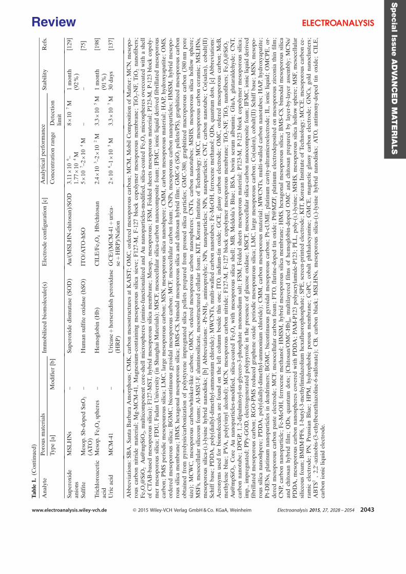
Tab
le1.
(Con
tinu
ed)
Ana
lyte
Poro
usm
ater
ials
Imm
obili
zed
biom
olec
ule(
s)E
lect
rode
conf
igur
atio
n[c
]A
naly
tica
lpe
rfor
man
ceSt
abili
tyR
efs.
Type
[a]
Mod
ifie
r[b
]C
once
ntra
tion
rang
eD
etec
tion
limit
Supe
roxi
dean
ions
MSL
HN
s–
Supe
roxi
dedi
smut
ase
(SO
D)
Au/
(MSL
HN
-chi
tosa
n)/S
OD
3.11
×10¢6
–1.
77×
1 0¢4
M8
×1 0¢7
M1
mon
th(9
2%
)[1
29]
Sulf
ite
Mes
op.S
b-do
ped
SnO
2
(AT
O)
–H
uman
sulf
ite
oxid
ase
(hSO
)IT
O/A
TO
-hSO
5×
10¢7
–2×
10¢4
M–
–[7
5]
Tric
hlor
oace
tic
acid
Mes
op.F
e 3O
4sp
here
s–
Hem
oglo
bin
(Hb)
CIL
E/F
e 3O
4¢H
b/ch
itos
an2.
4×
10¢3
–2×
10¢2
M3.
3×
10¢5
M1
mon
th(9
1%
)[1
98]
Uri
cac
idM
CM
-41
–U
rica
se+
hors
erad
ish
pero
xida
se(H
RP
)G
CE
/(M
CM
-41
+ur
ica-
se+
HR
P)/
Naf
ion
2×
10¢6
–1×
1 0¢5
M3.
3×
10¢7
M30
days
[137
]
Abb
revi
atio
ns:S
BA
,San
taB
arba
raA
mor
phou
s;C
MK
,car
bon
mes
ostr
uctu
res
atK
AIS
T;O
MC
,ord
ered
mes
opor
ous
carb
on;M
CM
,Mob
ilC
ompo
siti
onof
Mat
ter;
MC
N,m
esop
o-ro
usca
rbon
nitr
ide
mat
eria
l;M
g-M
CM
-41,
Mag
nesi
um-c
onta
inin
gm
esop
orou
ssi
lica
siev
e;
F12
7-M
,F
-127
bloc
kco
poly
mer
mes
opor
ous
mem
bran
e;
TiO
2-N
F,T
iO2
nano
fibe
rs;
Fe 3
O4@
SiO
2¢A
u@m
pSiO
2,m
ulti
com
pone
ntco
re-s
hell
mic
rosp
here
s(a
min
o-fu
ncti
onal
ized
and
Au
nano
part
icle
s-m
odif
ied,
silic
a-co
ated
Fe 3
O4
mic
rosp
here
sov
erco
ated
wit
ha
shel
lof
CT
AB
-bas
edm
esop
orou
ssi
lica)
;F
127-
MST
,hy
brid
mes
opor
ous
silic
am
embr
ane
;M
esop
.,m
esop
orou
s;F
SM,
Fold
edsh
eets
mes
opor
ous
mat
eria
l;P
123-
M,
P-1
23bl
ock
copo
ly-
mer
mes
opor
ous
silic
a;
FD
U,
Fud
anU
nive
rsit
y(i
nSh
angh
aiM
ater
ials
);M
SCF,
mes
ocel
lula
rsi
lica-
carb
onna
noco
mpo
site
foam
;IF
MC
,io
nic
liqui
dde
rive
dfi
brill
ated
mes
opor
ous
carb
on;
PM
Spe
riod
icm
esop
ouro
ussi
lica
;L
MC
,la
rge
mes
opor
ous
carb
on;
MSN
,m
esop
orou
ssi
lica
nano
shpe
re;
CM
M,
carb
onm
esop
orou
sm
ater
ial;
HA
P,hy
drox
yapa
tite
;O
MS,
orde
red
mes
opor
ous
silic
a;
BG
MC
,bi
cont
inuo
usgy
roid
alm
esop
orou
sca
rbon
;M
CF,
mes
ocel
lula
rca
rbon
foam
;C
NP
s,m
esop
orou
sca
rbon
nano
part
icle
s;H
MSM
,hy
brid
mes
opo-
rous
silic
am
embr
ane
;H
MS,
hexa
gona
lm
esop
orou
ssi
lica
;B
MS-
CS,
bim
odal
mes
opor
ous
silic
aan
dch
itos
anhy
brid
film
;G
MC
-6(S
iO2
pelle
ts/P
S),
grap
hiti
zed
mes
opor
ous
carb
onob
tain
edfr
ompy
roly
sis/
carb
oniz
atio
nof
poly
styr
ene
impr
egna
ted
silic
ape
llets
prep
ared
from
pres
sed
silic
apa
rtic
les;
GM
C-3
80,
grap
hiti
zed
mac
ropo
rous
carb
on(3
80nm
pore
size
);M
CW
C,
mes
opor
ous
carb
on/w
hisk
er-l
ike
carb
on;
OM
CN
,or
dere
dm
esop
orou
sca
rbon
nano
sphe
res;
CN
Ts,
carb
onna
notu
bes;
MSH
S,m
esop
orou
ssi
lica
hollo
wsp
here
;M
SFs,
mes
ocel
lula
rsi
liceo
usfo
ams;
Al-
MSU
-F,
alum
inos
ilica
te,
mes
ostr
uctu
red
cellu
lar
foam
;K
IT,
Kor
ean
Inst
itut
eof
Tech
nolo
gy;
MC
C,
mes
opor
ous
carb
once
ram
ic;
MSL
HN
s,m
esop
orou
ssi
lica-
(l)-
lysi
nehy
brid
nano
disk
s;[b
]A
bbre
viat
ions
:-P
r-N
H2,
amin
opro
lyle
;N
Ps,
nano
part
icle
s;N
Ps,
nano
part
icle
s;C
NT
,ca
rbon
nano
tube
;C
o(sa
len)
,co
balt
(II)
Schi
ffba
se;
PD
DA
,po
ly(d
ially
l-di
met
hyl-
amm
oniu
mch
lori
de);
MW
CN
Ts,
mul
ti-w
alle
dca
rbon
nano
tube
s;F
c-M
eOH
,fe
rroc
ene
met
hano
l;Q
Ds,
quan
tum
dots
.[c
]A
bbre
viat
ions
:A
cron
yms
used
for
biom
olec
ules
are
foun
don
the
left
colu
mn
besi
deth
ison
e;IT
O,
indi
um-t
inox
ide
;G
CE
,gl
assy
carb
onel
ectr
ode
;O
MC
,or
dere
dm
esop
orou
sca
rbon
;M
eB,
met
hyle
nebl
ue;
PV
A,
poly
(vin
ylal
coho
l);
MC
N,
mes
opor
ous
carb
onni
trid
e;
F12
7-M
,F
-127
bloc
kco
poly
mer
mes
opor
ous
mem
bran
e;
TiO
2-N
F,T
iO2
nano
fibe
rs;
Fe 3
O4@
SiO
2¢A
u@m
pSiO
2,C
ore
Au
nano
part
icle
s-m
odif
ied,
silic
a-co
ated
Fe 3
O4
wit
hm
esop
orou
ssi
lica
shel
l;M
B,
Mel
dola
ÏsB
lue
;B
SA,
bovi
nse
rum
albu
min
;G
luA
,gl
utar
alde
hyde
;C
NT
,ca
rbon
nano
tube
;D
PG
P,1,
2-di
palm
itoy
l-sn
-gly
cero
-3-p
hosp
hate
mon
osod
ium
salt
;F
SM,
Fold
edsh
eets
mes
opor
ous
mat
eria
l;P
123-
M,
P-1
23bl
ock
copo
lym
erm
esop
orou
ssi
lica
;im
p.,
impr
egna
ted
;P
Py-
GO
D,
elec
trog
ener
ated
poly
pyrr
ole
inth
epr
esen
ceof
gluc
ose
oxid
ase
;M
SCF,
mes
ocel
lula
rsi
lica-
carb
onna
noco
mpo
site
foam
;IF
MC
,io
nic
liqui
dde
rive
dfi
brill
ated
mes
opor
ous
carb
on;
RG
O-P
MS
redu
ced
grap
hene
oxid
e¢pe
riod
icm
esop
ouro
ussi
lica
;L
MC
,la
rge
mes
opor
ous
carb
on;
Co(
sale
n),
coba
lt(I
I)Sc
hiff
base
;M
SN,
mes
opo-
rous
silic
ana
nosh
pere
;P
DD
A,
poly
(dia
llyl-
dim
ethy
l-am
mon
ium
chlo
ride
);C
MM
,ca
rbon
mes
opor
ous
mat
eria
l;M
WC
NT
s,m
ulti
-wal
led
carb
onna
notu
bes;
HA
P,hy
drox
yapa
tite
;P
t-D
EN
s,pl
atin
umna
nopa
rtic
les
inde
ndri
mer
s;B
GM
C,
bico
ntin
uous
gyro
idal
mes
opor
ous
carb
on;
Pt-
cUM
E,
plat
inum
cavi
ty-u
ltra
mic
roel
ectr
ode
;IL
,io
nic
liqui
d;
OM
CP
E,
or-
dere
dm
esop
orou
sca
rbon
past
eel
ectr
ode
;M
CF,
mes
ocel
lula
rca
rbon
foam
;F
TO
,fl
urin
e-do
ped
tin
oxid
e;
Pt@
MZ
F,pl
atin
umel
ectr
odep
osit
edon
mes
opor
ous
zirc
onia
thin
film
;C
NP,
carb
onna
nopa
rtic
le;
Fc-
MeO
H,
ferr
ocen
em
etha
nol;
HM
SM,
hybr
idm
esop
orou
ssi
lica
mem
bran
e;
HM
S,he
xago
nal
mes
opor
ous
silic
a;
BM
S-C
S,bi
mod
alm
esop
orou
ssi
lica
and
chit
osan
hybr
idfi
lm;
QD
s,qu
antu
mdo
ts;
{Chi
tosa
n/O
MC
-Hb}
n,m
ulti
laye
red
film
sof
hem
oglo
bin-
dope
dO
MC
and
chit
osan
prep
ared
byla
yer-
by-l
ayer
asse
mbl
y;
MC
Ns/
PD
DA
,m
esop
orou
sca
rbon
nano
sphe
res
cove
red
wit
hP
DD
A;
PAM
-P12
3po
lyac
ryla
mid
e-P
123
;P
LL
,po
ly-(
l)-l
ysin
e;
MSH
S,m
esop
orou
ssi
lica
hollo
wsp
here
;M
SF,
mes
ocel
lula
rsi
liceo
usfo
am;B
MIM
·PF
6,1-
buty
l-3-
met
hylim
idaz
oliu
mhe
xafl
uoro
phos
phat
e;S
PE
,scr
een-
prin
ted
elec
trod
e;K
IT,K
orea
nIn
stit
ute
ofTe
chno
logy
;MC
CE
,mes
opor
ous
carb
once
-ra
mic
elec
trod
e;
PN
,P
russ
ian
Blu
e;
HP
M,
hydr
opho
bic
poro
usm
embr
ane
;C
oPC
,co
balt
phth
aloc
yani
ne;
GM
CE
,m
agne
tic
glas
syca
rbon
elec
trod
e;
GN
Cs,
gold
nano
clus
ters
;A
BT
S2¢,
2,2 ’
-azi
nobi
s-(3
-eth
ylbe
nzth
iazo
line-
6-su
lfon
ate)
;C
B,
carb
onbl
ack
;M
SLH
Ns,
mes
opor
ous
silic
a-(l
)-ly
sine
hybr
idna
nodi
sks;
AT
O,
anti
mon
y-do
ped
tin
oxid
e;
CIL
E,
carb
onio
nic
liqui
del
ectr
ode.
Specia
lIssu
eA
DVA
NC
ED
MAT
ER
IALS
www.electroanalysis.wiley-vch.de Ó 2015 Wiley-VCH Verlag GmbH & Co. KGaA, Weinheim Electroanalysis 2015, 27, 2028 – 2054 2043
Review
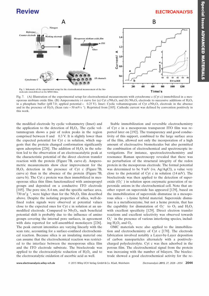
the modified electrode by cyclic voltammetry (Inset) andthe application to the detection of H2O2. The cyclic vol-tammogram shows a pair of redox peaks in the regioncomprised between 0 and ¢0.3 V. It is slightly lower thanthe expected potential for Cyt c in solution, which sug-gests that the protein changed conformation significantlyupon adsorption [226]. The addition of H2O2 in the solu-tion led to the observation of an electroacatalytic peak atthe characteristic potential of the direct electron transferreaction with the protein (Figure 7B, curve d). Ampero-metric measurements show clear improvement for theH2O2 detection in the presence of Cyt c (Figure 7B,curve a) than in the absence of the protein (Figure 7B,curve b). The Cyt c protein was then immobilized in mes-oporous silica thin films functionalized with aminopropylgroups and deposited on a conductive ITO electrode[105]. The pore size, 8.4 nm, and the specific surface area,730 m2 g¢1, were higher than for the Nb2O5 film describedabove. Despite the isolating properties of silica, well-de-fined redox signals were observed at potential valuesclose to the expected ones for Cyt c in solution at an un-modified electrode. Compared to Nb2O5, such beneficialpotential shift is probably due to the influence of aminogroups covering the internal pore surfaces, in agreementwith data reported for self-assembled monolayers [226].The peak current intensities are varying linearly with thescan rate, accounting for a surface-confined electrochemi-cal reaction. Because silica is an insulating material onecan assume that the electrochemical reaction was restrict-ed to the interface between the mesoporous silica filmand the ITO electrode substrate. The bioelectrode wasapplied to the electrocatalytic reduction of H2O2, and tothe electrocatalytic oxidation of ascorbic acid as well.
Stable immobilization and reversible electrochemistryof Cyt c in a mesoporous transparent ITO film was re-ported later on [192]. The transparency and good conduc-tivity of this support, combined to the large surface areaof the film, allowed not only the incorporation of a highamount of electroactive biomolecules but also permittedthe combination of electrochemical and spectroscopic in-vestigations. For instance, spectroelectrochemistry andresonance Raman spectroscopy revealed that there wasno perturbation of the structural integrity of the redoxprotein in the mesoporous electrode. The formal potentialwas determined to be 9 mV (vs. Ag/AgCl), a value veryclose to the potential of Cyt c in solution (14 mV). Thebioelectrode was then applied to the detection of super-oxide (O2C¢) in solution upon enzymatic generation of su-peroxide anions in the electrochemical cell. Note that an-other report on superoxide has appeared [129], based onthe immobilization of superoxide dismutase in a mesopo-rous silica – l-lysine hybrid material. Superoxide dismu-tase is a metalloenzyme, but not a heme protein, that hasthe capability for dismutation of O2C¢ to O2 and H2O2
with excellent specificity [129]. Direct electron transferreactions and excellent selectivity was observed towardsO2C¢ in the presence of various interfering species, includ-ing H2O2 and O2.
OMC materials were also applied to the immobiliza-tion and electrochemistry of Cyt c [170]. The electrodefabrication involved notably a Layer-by-Layer depositionof carbon nanoparticles alternately with a positively-charged polyelectrolyte. Cyt c was then adsorbed in theporous film. The electrochemical signal from the proteinwas increasing with the number of bilayers. The bioelec-trode showed a good electrochemical activity for the re-
Fig. 7. (A) Illustration of the experimental setup for electrochemical measurements with cytochrome c (Cyt c) immobilized in a mes-oporous niobium oxide film. (B) Amperometric i-t curve for (a) Cyt c/Nb2O5 and (b) Nb2O5 electrode to successive additions of H2O2
in a phosphate buffer (pH 7.0; applied potential=¢0.25 V). Inset: Cyclic voltammograms of Cyt c/Nb2O5 electrode in the absenceand in the presence of H2O2 (Scan rate=50 mV s¢1). Reprinted from [185]. Cathodic current was defined by convention positively inthis work.
Specia
lIssu
eA
DVA
NC
ED
MAT
ER
IALS
www.electroanalysis.wiley-vch.de Ó 2015 Wiley-VCH Verlag GmbH & Co. KGaA, Weinheim Electroanalysis 2015, 27, 2028 – 2054 2044
Review
duction of H2O2 and, according to the authors, exhibitedfast amperometric response, wide linear range, low detec-tion limit and good reproducibility. Even if slightly out ofthe scope of the review, it is noteworthy that macropo-rous carbon electrodes can be also applied to the immobi-lization of Cyt c for the electrocatalytic detection of H2O2
[144].Of related interest one can cite here the immobilization
microperoxidase 11 (MP11) in mesoporous SnO2 films[193]. MP-11 is a small size redox enzyme obtained byproteolytic digestion of Cyt c. It is an oligopeptide con-sisting of eleven amino acids and a covalently linkedFeIII¢protoporphyrin IX heme [193]. The catalytic activityof the immobilized MP-11 in mesoporous SnO2 electrodeswas found to proceed via the Fenton reaction, yieldingOH* radical intermediates. A biosensor for H2O2 detec-tion was demonstrated on that basis.
5.1.2 Hemoglobin and Myoglobin
Hemoglobin (Hb, 66 kDa) is nearly spherical with molec-ular size of 5.0 ×5.5 × 6.5 nm3 [199], i.e., bigger than Cyt c.Myoglobin (Mb, 17.6 kDa) is smaller but biologically re-lated to hemoglobin. First developments involving Hb orMb and mesoporous materials have been performed withhexagonal mesoporous silica displaying ca. 4 nm pore di-ameter [97,98]. Evidence for direct electron transfer be-tween the redox proteins and the electrode surface, aswell as illustrative applications to the detection of H2O2
and NO2¢ , have been provided in parallel for these two
proteins. However, considering the inadequacy betweenthe pore diameter and the protein size of hemoglobin,one can suspect that the protein was essentially adsorbedon the outer surface of the silica material, and for thisreason the interest of the mesoporous structure could belimited in the case of hemoglobin.
Not only mesoporous silica [97,98, 101,103,106–110,113, 118,157] but also mesoporous carbon[141, 147,154, 155,160,180] and mesoporous polymers[203], and other metal oxides, i.e. , TiO2 [186, 190], Al2O3
[188], ZrO2 [194], Fe3O4 [198] and Nb2O5 [199], havebeen applied for the construction of hemoglobin/myoglo-bin biosensors. The target analyte was essentially H2O2
[97, 101, 103, 107–110, 118, 141, 147, 154, 155, 160, 161,180, 188, 190, 194, 199, 203]. Few other analytes havebeen analyzed, such as NO2
¢ [97, 188], NO [186] and tri-choloroacetic acid [198].
Several authors have developed a combination of mes-oporous materials, either silica [101, 106,110] or carbon[160], with metal nanoparticles in order to improve theefficiency of direct electron transfer reactions with hemo-globin or myoglobin. These particles were made of Pt[160], Pd [110], gold [101] or even CdTe quantum dots[106]. Hemoglobin was used twice for the Layer-by-Layerassembly of the bioelectrode, in combination with CMK-3[141] or carbon Al-MSU-S particles [109]. The immobili-zation of the protein was also made in the presence ofionic liquids, into a mesocellular silica foam [108], or in
a carbon ionic liquid electrode [118]. Direct electrochem-istry of hemoglobin was otherwise observed in a renewa-ble mesoporous carbon ceramic electrode [180] and withmesoporous silica KIT-6 modified screen-printed electro-des [113].
An interesting investigation has been conducted on theoptimization of the porosity of graphitized carbon elec-trodes, with the objective to improve the efficiency of theprotein immobilization and the biosensor characteristics[154, 155]; this work has been discussed in more detail inSection 3.1. Other mesoporous carbon materials havebeen used for the immobilization of hemoglobin, e.g.,FDU-15 [160,161].
5.1.3 Horseradish Peroxidase
Horseradish peroxidase (HRP, ~44 kDa, 4.0 ×6.7 ×11.6 nm3) is bigger than the other heme proteins discussedabove. The hosting material likely to accommodate HRPthus needs to provide sufficiently large pore volume. Firstinvestigations have been performed by Xu et al. usingmesoporous Nb2O5 [73]. The direct electrochemistry ofHRP was studied in a series of experiments similar as forCyt c. The influence of both the film thickness (respec-tively 20 nm, 500 nm and 1 mm) and pore size (2, 6 and10 nm) on the sensor response was studied there. Optimalresponses were observed with the thickest film and for6 nm pores. However, the effect of film thickness was notso dramatic and it is interesting to note that the materialswith 2 nm pores, i.e., a size that does not allow the pro-tein to enter into the mesopores, still displayed an impor-tant response.
A mesoporous SnO2 film was applied for direct spec-troelectrochemical investigation of immobilized cyto-chrome c peroxidase (CcP) and HRP [191]. The immobi-lized HRP was found to have less favorable interfacialelectron transfer than that of CcP. A carbohydrate chainon the surface of native HRP may hinder the interfacialelectron transfer through at least three ways: (1) insulat-ing effect, (2) increasing the electron-transfer distance be-tween the heme edge and the electrode, and (3) orientat-ing the redox centre away from the surface. The authorshave demonstrated the feasibility of using peroxidasemodified mesoporous metal oxide film electrodes asH2O2 biosensors via both optical and electrochemical de-tection schemes. Employing TiO2 electrodes, a linear in-crease of the absorbance measured at 418 nm is observedas a function of H2O2 concentration, within a range 0.04–0.8 mM, while the current response of the HRP modifiedelectrode was linear within a range 1–20 mM.
Several works have been performed with HRP-loadedsilica-based mesoporous silicas with different pore geom-etry and size, i.e., HMS [100], mesoporous silica hollowspheres [115], SBA-15 [122], and a hybrid mesoporoussilica membrane [127]. Direct electrochemistry of HRPwas investigated and the observed direct electron transferreactions were applied to the detection of H2O2. Severalauthors have described the co-immobilization of HRP
Specia
lIssu
eA
DVA
NC
ED
MAT
ER
IALS
www.electroanalysis.wiley-vch.de Ó 2015 Wiley-VCH Verlag GmbH & Co. KGaA, Weinheim Electroanalysis 2015, 27, 2028 – 2054 2045
Review
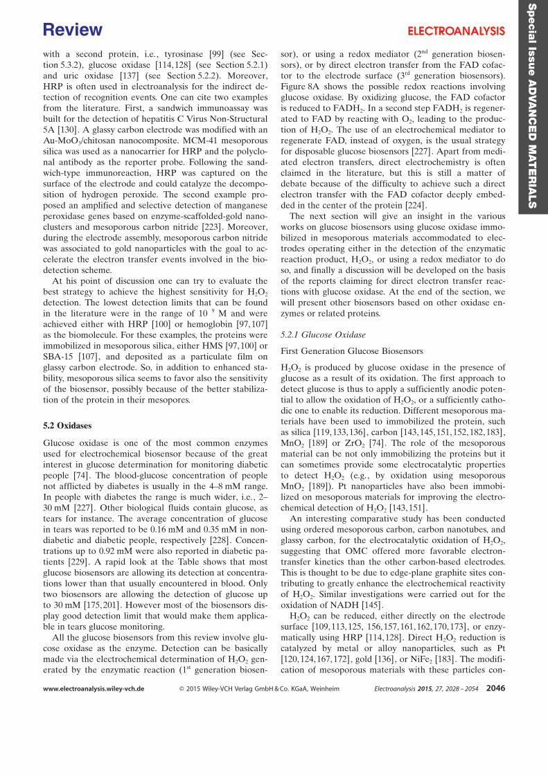
with a second protein, i.e., tyrosinase [99] (see Sec-tion 5.3.2), glucose oxidase [114, 128] (see Section 5.2.1)and uric oxidase [137] (see Section 5.2.2). Moreover,HRP is often used in electroanalysis for the indirect de-tection of recognition events. One can cite two examplesfrom the literature. First, a sandwich immunoassay wasbuilt for the detection of hepatitis C Virus Non-Structural5A [130]. A glassy carbon electrode was modified with anAu-MoO3/chitosan nanocomposite. MCM-41 mesoporoussilica was used as a nanocarrier for HRP and the polyclo-nal antibody as the reporter probe. Following the sand-wich-type immunoreaction, HRP was captured on thesurface of the electrode and could catalyze the decompo-sition of hydrogen peroxide. The second example pro-posed an amplified and selective detection of manganeseperoxidase genes based on enzyme-scaffolded-gold nano-clusters and mesoporous carbon nitride [223]. Moreover,during the electrode assembly, mesoporous carbon nitridewas associated to gold nanoparticles with the goal to ac-celerate the electron transfer events involved in the bio-detection scheme.
At his point of discussion one can try to evaluate thebest strategy to achieve the highest sensitivity for H2O2
detection. The lowest detection limits that can be foundin the literature were in the range of 10¢9 M and wereachieved either with HRP [100] or hemoglobin [97, 107]as the biomolecule. For these examples, the proteins wereimmobilized in mesoporous silica, either HMS [97,100] orSBA-15 [107], and deposited as a particulate film onglassy carbon electrode. So, in addition to enhanced sta-bility, mesoporous silica seems to favor also the sensitivityof the biosensor, possibly because of the better stabiliza-tion of the protein in their mesopores.
5.2 Oxidases
Glucose oxidase is one of the most common enzymesused for electrochemical biosensor because of the greatinterest in glucose determination for monitoring diabeticpeople [74]. The blood-glucose concentration of peoplenot afflicted by diabetes is usually in the 4–8 mM range.In people with diabetes the range is much wider, i.e., 2–30 mM [227]. Other biological fluids contain glucose, astears for instance. The average concentration of glucosein tears was reported to be 0.16 mM and 0.35 mM in non-diabetic and diabetic people, respectively [228]. Concen-trations up to 0.92 mM were also reported in diabetic pa-tients [229]. A rapid look at the Table shows that mostglucose biosensors are allowing its detection at concentra-tions lower than that usually encountered in blood. Onlytwo biosensors are allowing the detection of glucose upto 30 mM [175,201]. However most of the biosensors dis-play good detection limit that would make them applica-ble in tears glucose monitoring.
All the glucose biosensors from this review involve glu-cose oxidase as the enzyme. Detection can be basicallymade via the electrochemical determination of H2O2 gen-erated by the enzymatic reaction (1st generation biosen-
sor), or using a redox mediator (2nd generation biosen-sors), or by direct electron transfer from the FAD cofac-tor to the electrode surface (3rd generation biosensors).Figure 8A shows the possible redox reactions involvingglucose oxidase. By oxidizing glucose, the FAD cofactoris reduced to FADH2. In a second step FADH2 is regener-ated to FAD by reacting with O2, leading to the produc-tion of H2O2. The use of an electrochemical mediator toregenerate FAD, instead of oxygen, is the usual strategyfor disposable glucose biosensors [227]. Apart from medi-ated electron transfers, direct electrochemistry is oftenclaimed in the literature, but this is still a matter ofdebate because of the difficulty to achieve such a directelectron transfer with the FAD cofactor deeply embed-ded in the center of the protein [224].
The next section will give an insight in the variousworks on glucose biosensors using glucose oxidase immo-bilized in mesoporous materials accommodated to elec-trodes operating either in the detection of the enzymaticreaction product, H2O2, or using a redox mediator to doso, and finally a discussion will be developed on the basisof the reports claiming for direct electron transfer reac-tions with glucose oxidase. At the end of the section, wewill present other biosensors based on other oxidase en-zymes or related proteins.
5.2.1 Glucose Oxidase
First Generation Glucose Biosensors
H2O2 is produced by glucose oxidase in the presence ofglucose as a result of its oxidation. The first approach todetect glucose is thus to apply a sufficiently anodic poten-tial to allow the oxidation of H2O2, or a sufficiently catho-dic one to enable its reduction. Different mesoporous ma-terials have been used to immobilized the protein, suchas silica [119, 133,136], carbon [143,145, 151,152, 182,183],MnO2 [189] or ZrO2 [74]. The role of the mesoporousmaterial can be not only immobilizing the proteins but itcan sometimes provide some electrocatalytic propertiesto detect H2O2 (e.g., by oxidation using mesoporousMnO2 [189]). Pt nanoparticles have also been immobi-lized on mesoporous materials for improving the electro-chemical detection of H2O2 [143, 151].
An interesting comparative study has been conductedusing ordered mesoporous carbon, carbon nanotubes, andglassy carbon, for the electrocatalytic oxidation of H2O2,suggesting that OMC offered more favorable electron-transfer kinetics than the other carbon-based electrodes.This is thought to be due to edge-plane graphite sites con-tributing to greatly enhance the electrochemical reactivityof H2O2. Similar investigations were carried out for theoxidation of NADH [145].
H2O2 can be reduced, either directly on the electrodesurface [109, 113, 125, 156,157,161, 162,170, 173], or enzy-matically using HRP [114, 128]. Direct H2O2 reduction iscatalyzed by metal or alloy nanoparticles, such as Pt[120, 124,167, 172], gold [136], or NiFe2 [183]. The modifi-cation of mesoporous materials with these particles con-
Specia
lIssu
eA
DVA
NC
ED
MAT
ER
IALS
www.electroanalysis.wiley-vch.de Ó 2015 Wiley-VCH Verlag GmbH & Co. KGaA, Weinheim Electroanalysis 2015, 27, 2028 – 2054 2046
Review
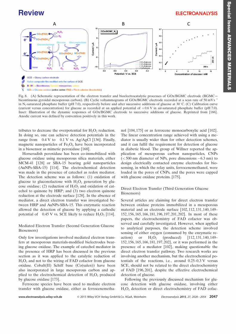
tributes to decrease the overpotential for H2O2 reduction.In doing so, one can achieve detection potentials in therange from ¢0.4 V to ¢0.1 V vs. Ag/AgCl [136]. Finally,magnetic nanoparticles of Fe3O4 have been incorporatedin a biosensor as mimetic peroxidase [168].
Horseradish peroxidase has been co-immobilized withglucose oxidase using mesoporous silica materials, eitherMCM-41 [128] or SBA-15 bearing gold nanoparticles(AuNPs-SBA-15) [114]. The electrochemical detectionwas made in the presence of catechol as redox mediator.The detection scheme was as follows: (1) oxidation ofglucose to gluconolactone with H2O2 generation by glu-cose oxidase; (2) reduction of H2O2 and oxidation of cat-echol to quinone by HRP; and (3) two electron quinonereduction at the electrode surface [128]. In the absence ofmediator, a direct electron transfer was investigated be-tween HRP and AuNPs-SBA-15. This enzymatic reactionallowed the detection of glucose by applying a cathodicpotential of ¢0.45 V vs. SCE likely to reduce H2O2 [114].
Mediated Electron Transfer (Second Generation GlucoseBiosensors)
Only few investigations involved mediated electron trans-fers at mesoporous materials-modified bielectrodes bear-ing glucose oxidase. The example of catechol mediator inthe presence of HRP has been discussed in the previoussection as it was applied to the catalytic reduction ofH2O2 and not to the wiring of FAD cofactor from glucoseoxidase. Cobalt(II) Schiff base (Co(salen)) have beenalso incorporated in large mesoporous carbon and ap-plied to the electrochemical detection of H2O2 producedby glucose oxidase [173].
Ferrocene species have been used to mediate electrontransfer with glucose oxidase, either as ferrocenemetha-
nol [104,175] or as ferrocene monocarboxylic acid [102].The linear concentration range achieved with using a me-diator is usually wider than for other detection schemes,and it can fulfil the requirement for detection of glucosein diabetic blood. The group of Willner reported the ap-plication of mesoporous carbon nanoparticles, CNPs(<500 nm diameter of NPs, pore dimensions ~6.3 nm) todesign electrically contacted enzyme electrodes for bio-sensing, in which the relay units, ferrocenemethanol, wereloaded in the pores of CNPs, and the pores were cappedwith glucose oxidase proteins. [175].
Direct Electron Transfer (Third Generation GlucoseBiosensors)
Several articles are claiming for direct electron transferbetween oxidase proteins immobilized in a mesoporousmaterial and an electrode surface [102, 112, 131,140,149–152,156, 165,166, 181,196,197, 201,202]. In most of thesepapers, the electrochemistry of FAD cofactor was ob-served and carefully investigated. However, when appliedto analytical purposes, the detection scheme involvedsensing of either oxygen (consumed by the enzymatic re-action) or H2O2 (produced) [112,131, 140,149–152,156, 165,166, 181,197,202], or it was performed in thepresence of a mediator [102], making questionable thedirect electron transfer pathway. Two research works areinvolving another mechanism, but the electrochemical po-tentials of the reactions, i.e. , around 0.25–0.3 V versusSCE, should not be related to the direct electrochemistryof FAD [196,201], despite the effective electrochemicaldetection of glucose.
Following the previously discussed mechanism for glu-cose detection with glucose oxidase, involving eitherH2O2 detection or direct electrochemistry of FAD cofac-
Fig. 8. (A) Schematic representation of the electron transfer and bioelectrocatalytic processes of GOx/BGMC electrode (BGMC=bicontinuous gyroidal mesoporous carbon). (B) Cyclic voltammograms of GOx/BGMC electrode recorded at a scan rate of 50 mV s¢1
in N2-saturated phosphate buffer (pH 7.0), respectively before and after successive additions of glucose at 30 8C. (C) Calibration curve(current versus concentration) for glucose as recorded at an applied potential of +0.6 V in air-saturated phosphate buffer (pH 7.0).Inset: Illustration of the dynamic responses of GOx/BGMC electrode to successive additions of glucose. Reprinted from [166].Anodic current was defined by convention positively in this work.
Specia
lIssu
eA
DVA
NC
ED
MAT
ER
IALS
www.electroanalysis.wiley-vch.de Ó 2015 Wiley-VCH Verlag GmbH & Co. KGaA, Weinheim Electroanalysis 2015, 27, 2028 – 2054 2047
Review
tor (Figure 8A), the use of a bicontinuous gyroidal meso-porous carbon was claimed to favor the latter process[166]. The typical cyclic voltammogram that can be ob-served exhibits important capacitive currents as a resultof the large electroactive surface area developed by thematerial, but also a noticeable pair of peaks observed ata potential close to ¢0.45 V vs. SCE (Figure 8B). This po-tential is compatible with the electrochemistry of FADcoming from glucose oxidase. The addition of glucose inthe solution, in the absence of oxygen, does not allow ob-serving a significant modification of the voltammetric sig-nals. This experiment supports the argument that despitethe effective electrochemistry of FAD cofactor on meso-porous carbon materials, its application to the electro-chemical biosensing of glucose is not obvious. The au-thors have then applied the bioelectrode to the detectionof glucose by amperometry and the current was indeedgrowing by increasing the concentration of glucose in so-lution (Figure 8C). The protein has thus maintained itsenzymatic activity after immobilization on the electrode.But note that the amperometric measurement was per-formed at +0.6 V vs. SCE, a potential at which H2O2 canbe detected onto the electrode surface, meaning that thebiosensor was indeed likely to detect glucose but not viaa direct electron transfer scheme (it was actually a 1st gen-eration biosensor, not a 3rd generation one).
All in all, the best detection limits for glucose, usingmesoporous materials-based electrochemical biosensors,have been reached with mesoporous MnO2 [189, 197], op-erating via the detection of H2O2 in either the anodic[189] or cathodic [197] mode. The concentration rangeachieved with these two biosensors is however not suita-ble for the detection of glucose in blood and should findbetter application in the analysis of glucose in other fluidcontaining lower concentrations of analyte, e.g., tears.
5.2.2 Other Oxidase-Type Enzymes
Pyranose Oxidase
Pyranose oxidase has been recently applied to the elec-trochemical detection of glucose. Pyranose oxidase hasbroader substrate specificity than glucose oxidase and anoption of large scale production using recombinant E.coli, which provides it with a great potential for biofuelcell application but not necessarily for biosensing [182].
Lactate Oxidase
Lactate oxidase was applied to l-lactate determination[125, 126,172]. The detection scheme was similar as forglucose oxidase. The oxidation of l-lactate by FMN co-factor in the presence of oxygen led to the production ofH2O2 that was detected by electrochemical oxidation[125, 126] or reduction [172]. The oxidation reaction wascatalyzed by cobalt phthalocyanine [125] or Prussian blue[126] immobilized on the mesoporous materials, while thereduction reaction was catalyzed by Pt nanoparticles (seeFigure 5 for an illustration) [172].
Cholesterol Oxidase
The detection of cholesterol involved cholesterol oxidase.Two examples are available, involving a different detec-tion scheme. In the first one, cholesterol oxidase was im-mobilized in high amount on core-shell structured silicamicrospheres. The reaction of cholesterol with the proteinin the presence of oxygen led to the production of H2O2
that was then detected by electrochemiluminescence inthe presence of 0.67 mM luminol [132]. In the second ex-ample, cholesterol oxidase was immobilized along withcholesterol esterase on the surface of electrospun TiO2
nanofibers (see Figure 6F for the way the electrospunfibers are obtained). Then, the electrochemical detectionwas performed in the presence of Fe(CN)6
3¢ as electronmediator between the enzymatic FAD cofactor of cholineoxidase and the electrode surface [200].
Choline Oxidase
Choline determination was achieved through the oxida-tion of this substrate by choline oxidase into betaine andproduction of H2O2. The biosensor was developed by im-mobilization of the protein in a hybrid mesoporous mem-brane (see Figure 6E for illustration), giving rise toa hybrid material with 12 nm pore diameter, which waslikely to detect H2O2 detection via its oxidation on a plati-num electrode [116]. The determination range was 5.0–800 mM. The sensor was stable compared to the nativeenzyme sensor and 85% of the initial response was main-tained even after storage for 80 days. More recently, chol-ine oxidase was encapsulated into a silica matrix electro-deposited onto screen-printed electrodes [230]. The depo-sition was performed in the presence of CTAB that canact as both protein stabilizer and template for pore gener-ation. The sensor was applied successfully to the determi-nation of choline in child milk.
Human Sulfite Oxidase
Human sulfate oxidase catalyzes the conversion of sulfiteinto sulfate. The protein is a homodimer and each mono-mer contains a b-type heme (Heme domain, HD) linkedby a 10 amino acid flexible loop to a second domain con-taining a molybdenum cofactor (Moco domain, MD) anda dimerization domain [231]. The catalytic sulfite oxida-tion takes place at the molybdenum cofactor (Moco). Toregenerate the catalytic center, two electrons are thentransferred from the Moco (MoIV) via the HD to thephysiological external electron acceptor cytochromec [232]. The protein was immobilized in mesoporousATO electrode [75]. Catalytically-active enzyme ina direct electron transfer configuration was ensured with-out further chemical modification of the ATO surfaces.Interestingly, the binding of hSO onto the ATO surfaceseems to be not only of electrostatic nature, but also orig-inates from a strong interaction between the histidine-tagof the enzyme and the supporting material. If confirmed
Specia
lIssu
eA
DVA
NC
ED
MAT
ER
IALS
www.electroanalysis.wiley-vch.de Ó 2015 Wiley-VCH Verlag GmbH & Co. KGaA, Weinheim Electroanalysis 2015, 27, 2028 – 2054 2048
Review
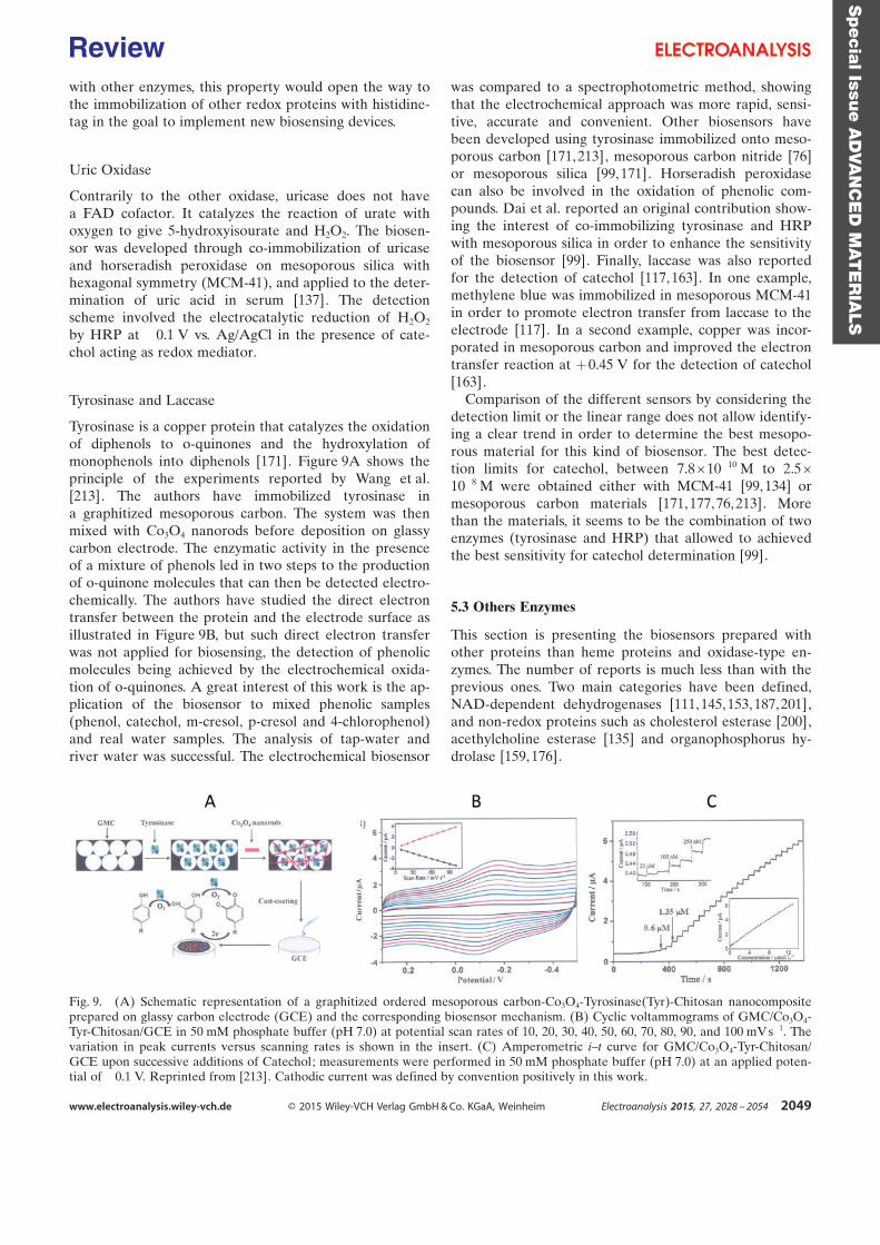
with other enzymes, this property would open the way tothe immobilization of other redox proteins with histidine-tag in the goal to implement new biosensing devices.
Uric Oxidase
Contrarily to the other oxidase, uricase does not havea FAD cofactor. It catalyzes the reaction of urate withoxygen to give 5-hydroxyisourate and H2O2. The biosen-sor was developed through co-immobilization of uricaseand horseradish peroxidase on mesoporous silica withhexagonal symmetry (MCM-41), and applied to the deter-mination of uric acid in serum [137]. The detectionscheme involved the electrocatalytic reduction of H2O2
by HRP at ¢0.1 V vs. Ag/AgCl in the presence of cate-chol acting as redox mediator.
Tyrosinase and Laccase
Tyrosinase is a copper protein that catalyzes the oxidationof diphenols to o-quinones and the hydroxylation ofmonophenols into diphenols [171]. Figure 9A shows theprinciple of the experiments reported by Wang et al.[213]. The authors have immobilized tyrosinase ina graphitized mesoporous carbon. The system was thenmixed with Co3O4 nanorods before deposition on glassycarbon electrode. The enzymatic activity in the presenceof a mixture of phenols led in two steps to the productionof o-quinone molecules that can then be detected electro-chemically. The authors have studied the direct electrontransfer between the protein and the electrode surface asillustrated in Figure 9B, but such direct electron transferwas not applied for biosensing, the detection of phenolicmolecules being achieved by the electrochemical oxida-tion of o-quinones. A great interest of this work is the ap-plication of the biosensor to mixed phenolic samples(phenol, catechol, m-cresol, p-cresol and 4-chlorophenol)and real water samples. The analysis of tap-water andriver water was successful. The electrochemical biosensor
was compared to a spectrophotometric method, showingthat the electrochemical approach was more rapid, sensi-tive, accurate and convenient. Other biosensors havebeen developed using tyrosinase immobilized onto meso-porous carbon [171, 213], mesoporous carbon nitride [76]or mesoporous silica [99,171]. Horseradish peroxidasecan also be involved in the oxidation of phenolic com-pounds. Dai et al. reported an original contribution show-ing the interest of co-immobilizing tyrosinase and HRPwith mesoporous silica in order to enhance the sensitivityof the biosensor [99]. Finally, laccase was also reportedfor the detection of catechol [117, 163]. In one example,methylene blue was immobilized in mesoporous MCM-41in order to promote electron transfer from laccase to theelectrode [117]. In a second example, copper was incor-porated in mesoporous carbon and improved the electrontransfer reaction at +0.45 V for the detection of catechol[163].
Comparison of the different sensors by considering thedetection limit or the linear range does not allow identify-ing a clear trend in order to determine the best mesopo-rous material for this kind of biosensor. The best detec-tion limits for catechol, between 7.8 ×10¢10 M to 2.5 ×10¢8 M were obtained either with MCM-41 [99,134] ormesoporous carbon materials [171, 177,76, 213]. Morethan the materials, it seems to be the combination of twoenzymes (tyrosinase and HRP) that allowed to achievedthe best sensitivity for catechol determination [99].
5.3 Others Enzymes
This section is presenting the biosensors prepared withother proteins than heme proteins and oxidase-type en-zymes. The number of reports is much less than with theprevious ones. Two main categories have been defined,NAD-dependent dehydrogenases [111, 145,153, 187,201],and non-redox proteins such as cholesterol esterase [200],acethylcholine esterase [135] and organophosphorus hy-drolase [159, 176].
Fig. 9. (A) Schematic representation of a graphitized ordered mesoporous carbon-Co3O4-Tyrosinase(Tyr)-Chitosan nanocompositeprepared on glassy carbon electrode (GCE) and the corresponding biosensor mechanism. (B) Cyclic voltammograms of GMC/Co3O4-Tyr-Chitosan/GCE in 50 mM phosphate buffer (pH 7.0) at potential scan rates of 10, 20, 30, 40, 50, 60, 70, 80, 90, and 100 mV s¢1. Thevariation in peak currents versus scanning rates is shown in the insert. (C) Amperometric i–t curve for GMC/Co3O4-Tyr-Chitosan/GCE upon successive additions of Catechol; measurements were performed in 50 mM phosphate buffer (pH 7.0) at an applied poten-tial of ¢0.1 V. Reprinted from [213]. Cathodic current was defined by convention positively in this work.
Specia
lIssu
eA
DVA
NC
ED
MAT
ER
IALS
www.electroanalysis.wiley-vch.de Ó 2015 Wiley-VCH Verlag GmbH & Co. KGaA, Weinheim Electroanalysis 2015, 27, 2028 – 2054 2049
Review
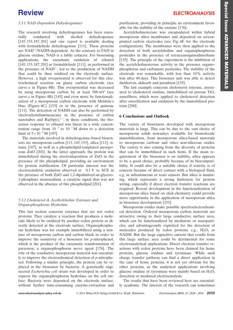
5.3.1 NAD-Dependent Dehydrogenases
The research involving dehydrogenases has been essen-tially conducted with alcohol dehydrogenase[145, 153,187, 201] and one report is available dealingwith formaldehyde dehydrogenase [111]. These proteinsare NAD+/NADH-dependent. At the contrary to FAD inglucose oxidase, NAD is a labile cofactor. For biosensingapplications, the enzymatic oxidation of ethanol[145, 153,187, 201] or formaldehyde [111], as performed inthe presence of NAD+, led to the production of NADHthat could be then oxidized on the electrode surface.However, a high overpotential is observed for this elec-trochemical reaction on glassy carbon electrode (seecurve a in Figure 4B). This overpotential was decreasedby using mesoporous carbon by at least 500 mV (seecurve c in Figure 4B) [145] and even more by functionali-zation of a mesoporous carbon electrode with MeldolaÏsblue (Figure 4C) [153] or in the presence of quinone[111]. The detection of NADH can also be performed byelectrochemiluminescence in the presence of carbonnanotubes and Ru(bpy)3
2 +; in these conditions, the bio-sensor response to ethanol was linear in a wide concen-tration range from 10¢5 to 10¢1 M down to a detectionlimit of 5 ×10¢6 M [187].
The materials involved in dehydrogenase-based biosen-sors are mesoporous carbon [111,145, 153], silica [111], ti-tania [187], as well as a phospholipid-templated mesopo-rous ZnO [201]. In the latter approach, the protein wasimmobilized during the electrodeposition of ZnO in thepresence of the phospholipid, providing an environmentfor protein stabilization. Of particular interest was theelectrocatalytic oxidation observed at ¢0.1 V vs. SCE inthe presence of both ZnO and 1,2-dipalmitoyl-sn-glycero-3-phosphate monosodium, a catalytic signal that was notobserved in the absence of this phospholipid [201].
5.3.2 Cholesterol & Acethylcholine Esterases andOrganophosphorus Hydrolase
This last section concerns enzymes that are not redoxproteins. They catalyze a reaction that produces a mole-cule likely to be oxidized by another redox protein or di-rectly detected at the electrode surface. Organophospho-rus hydrolase was for example immobilized using a mix-ture of mesoporous carbon and carbon black in order toimprove the sensitivity of a biosensor for p-nitrophenol,which is the product of the enzymatic transformation ofparaoxon, a organophosphorus nerve agent [176]. Therole of the conductive mesoporous material was essential-ly to improve the electrochemical detection of p-nitrophe-nol. Following a similar principle, the protein can be re-placed in the biosensor by bacteria. A genetically engi-neered Escherichia coli strain was developed in order toexpress the organophosphorus hydrolase on the cell sur-face. Bacteria were deposited on the electrode surface,without further time-consuming enzyme-extraction and
purification, providing in principle an environment favor-able for the stability of the enzyme [176].
Acetylcholinesterase was encapsulated within hybridmesoporous silica membranes and deposited on screen-printed carbon electrodes (see Fig. 6E for the electrodeconfiguration). The membranes were then applied to thedetection of both acetylcholine and organophosphoruspesticides in the presence of tetracyanoquinodimethane[135]. The principle of the experiment is the inhibition ofthe acetylcholinesterase activity in the presence organo-phosphorus and carbamate pesticides. The stability of theelectrode was remarkable, with less than 10 % activityloss after 60 days. This biosensor unit was able to detectdichlorvos, aldicarb and parathion [135].
The last example concerns cholesterol esterase, associ-ated to cholesterol oxidase, immobilized on porous TiO2
nanofibers, which were applied to cholesterol detectionafter esterification and oxidation by the immobilized pro-teins [200].
6 Conclusions and Outlook
The variety of biosensors developed with mesoporousmaterials is large. This can be due to the vast choice ofmesoporous solids nowadays available for biomoleculeimmobilization, from mesoporous silica-based materialsto mesoporous carbons and other non-siliceous oxides.The variety is also coming from the diversity of proteinsthat can be immobilized in such hosts. If the main re-quirement of the biosensor is on stability, silica appearsto be a good choice, probably because of its biocompati-bility. It could also be a suitable choice if toxicity is ofconcern because of direct contact with a biological fluid,e.g. in subcutaneous or tears sensors. But silica is insulat-ing, which represents a strong limitation for proteinwiring, especially if direct electron transfer reactions arerequired. Recent development in the functionalization ofmesoporous silica based on click-chemistry could providemore opportunity in the application of mesoporous silicain biosensor development [233].
Mesoporous oxides make possible spectroelectrochemi-cal detection. Ordered mesoporous carbon materials areattractive owing to their large conductive surface area,which can be functionalized by mediators or nanoparti-cles, and advantageously exploited for the detection ofmolecules produced by redox proteins, e.g., H2O2 orNADH. But the large capacitive current that results fromthis large surface area could be detrimental for someelectroanalytical applications. Direct electron transfer re-actions with redox proteins have been claimed for hemeproteins, glucose oxidase and tyrosinase. While suchcharge transfer pathway can find a direct application inthe case of heme proteins, it is not yet obvious for theother proteins, as the analytical applications involvingglucose oxidase or tyrosinase were mainly based on H2O2
detection or mediated electrocatalysis.The results that have been reviewed here are essential-
ly academic. The interest of the research can sometimes
Specia
lIssu
eA
DVA
NC
ED
MAT
ER
IALS
www.electroanalysis.wiley-vch.de Ó 2015 Wiley-VCH Verlag GmbH & Co. KGaA, Weinheim Electroanalysis 2015, 27, 2028 – 2054 2050
Review
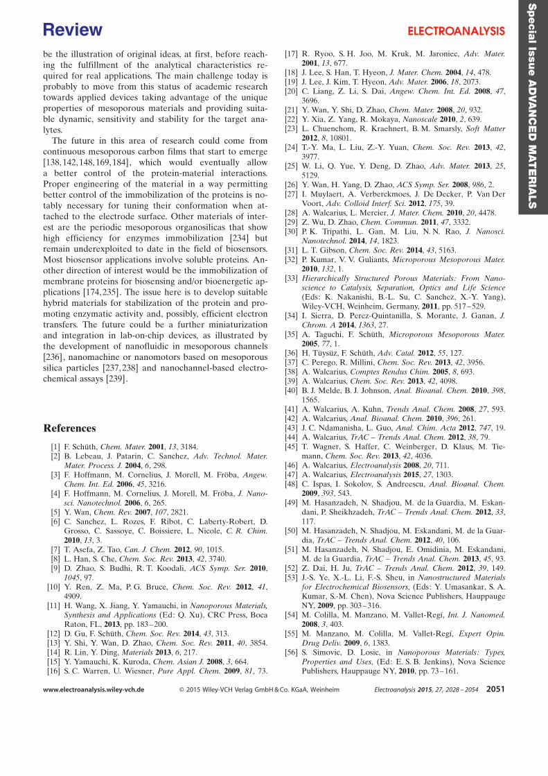
be the illustration of original ideas, at first, before reach-ing the fulfillment of the analytical characteristics re-quired for real applications. The main challenge today isprobably to move from this status of academic researchtowards applied devices taking advantage of the uniqueproperties of mesoporous materials and providing suita-ble dynamic, sensitivity and stability for the target ana-lytes.
The future in this area of research could come fromcontinuous mesoporous carbon films that start to emerge[138, 142,148, 169,184], which would eventually allowa better control of the protein-material interactions.Proper engineering of the material in a way permittingbetter control of the immobilization of the proteins is no-tably necessary for tuning their conformation when at-tached to the electrode surface. Other materials of inter-est are the periodic mesoporous organosilicas that showhigh efficiency for enzymes immobilization [234] butremain underexploited to date in the field of biosensors.Most biosensor applications involve soluble proteins. An-other direction of interest would be the immobilization ofmembrane proteins for biosensing and/or bioenergetic ap-plications [174,235]. The issue here is to develop suitablehybrid materials for stabilization of the protein and pro-moting enzymatic activity and, possibly, efficient electrontransfers. The future could be a further miniaturizationand integration in lab-on-chip devices, as illustrated bythe development of nanofluidic in mesoporous channels[236], nanomachine or nanomotors based on mesoporoussilica particles [237, 238] and nanochannel-based electro-chemical assays [239].
References
[1] F. Schîth, Chem. Mater. 2001, 13, 3184.[2] B. Lebeau, J. Patarin, C. Sanchez, Adv. Technol. Mater.
Mater. Process. J. 2004, 6, 298.[3] F. Hoffmann, M. Cornelius, J. Morell, M. Frçba, Angew.
Chem. Int. Ed. 2006, 45, 3216.[4] F. Hoffmann, M. Cornelius, J. Morell, M. Frçba, J. Nano-
sci. Nanotechnol. 2006, 6, 265.[5] Y. Wan, Chem. Rev. 2007, 107, 2821.[6] C. Sanchez, L. Rozes, F. Ribot, C. Laberty-Robert, D.
Grosso, C. Sassoye, C. Boissiere, L. Nicole, C. R. Chim.2010, 13, 3.
[7] T. Asefa, Z. Tao, Can. J. Chem. 2012, 90, 1015.[8] L. Han, S. Che, Chem. Soc. Rev. 2013, 42, 3740.[9] D. Zhao, S. Budhi, R. T. Koodali, ACS Symp. Ser. 2010,
1045, 97.[10] Y. Ren, Z. Ma, P. G. Bruce, Chem. Soc. Rev. 2012, 41,
4909.[11] H. Wang, X. Jiang, Y. Yamauchi, in Nanoporous Materials,
Synthesis and Applications (Ed: Q. Xu), CRC Press, BocaRaton, FL, 2013, pp. 183–200.
[12] D. Gu, F. Schîth, Chem. Soc. Rev. 2014, 43, 313.[13] Y. Shi, Y. Wan, D. Zhao, Chem. Soc. Rev. 2011, 40, 3854.[14] R. Lin, Y. Ding, Materials 2013, 6, 217.[15] Y. Yamauchi, K. Kuroda, Chem. Asian J. 2008, 3, 664.[16] S. C. Warren, U. Wiesner, Pure Appl. Chem. 2009, 81, 73.
[17] R. Ryoo, S. H. Joo, M. Kruk, M. Jaroniec, Adv. Mater.2001, 13, 677.
[18] J. Lee, S. Han, T. Hyeon, J. Mater. Chem. 2004, 14, 478.[19] J. Lee, J. Kim, T. Hyeon, Adv. Mater. 2006, 18, 2073.[20] C. Liang, Z. Li, S. Dai, Angew. Chem. Int. Ed. 2008, 47,
3696.[21] Y. Wan, Y. Shi, D. Zhao, Chem. Mater. 2008, 20, 932.[22] Y. Xia, Z. Yang, R. Mokaya, Nanoscale 2010, 2, 639.[23] L. Chuenchom, R. Kraehnert, B. M. Smarsly, Soft Matter
2012, 8, 10801.[24] T.-Y. Ma, L. Liu, Z.-Y. Yuan, Chem. Soc. Rev. 2013, 42,
3977.[25] W. Li, Q. Yue, Y. Deng, D. Zhao, Adv. Mater. 2013, 25,
5129.[26] Y. Wan, H. Yang, D. Zhao, ACS Symp. Ser. 2008, 986, 2.[27] I. Muylaert, A. Verberckmoes, J. De Decker, P. Van Der
Voort, Adv. Colloid Interf. Sci. 2012, 175, 39.[28] A. Walcarius, L. Mercier, J. Mater. Chem. 2010, 20, 4478.[29] Z. Wu, D. Zhao, Chem. Commun. 2011, 47, 3332.[30] P. K. Tripathi, L. Gan, M. Liu, N. N. Rao, J. Nanosci.
Nanotechnol. 2014, 14, 1823.[31] L. T. Gibson, Chem. Soc. Rev. 2014, 43, 5163.[32] P. Kumar, V. V. Guliants, Microporous Mesoporous Mater.
2010, 132, 1.[33] Hierarchically Structured Porous Materials: From Nano-
science to Catalysis, Separation, Optics and Life Science(Eds: K. Nakanishi, B.-L. Su, C. Sanchez, X.-Y. Yang),Wiley-VCH, Weinheim, Germany, 2011, pp. 517–529.
[34] I. Sierra, D. Perez-Quintanilla, S. Morante, J. Ganan, J.Chrom. A 2014, 1363, 27.
[35] A. Taguchi, F. Schîth, Microporous Mesoporous Mater.2005, 77, 1.
[36] H. Tîysîz, F. Schîth, Adv. Catal. 2012, 55, 127.[37] C. Perego, R. Millini, Chem. Soc. Rev. 2013, 42, 3956.[38] A. Walcarius, Comptes Rendus Chim. 2005, 8, 693.[39] A. Walcarius, Chem. Soc. Rev. 2013, 42, 4098.[40] B. J. Melde, B. J. Johnson, Anal. Bioanal. Chem. 2010, 398,
1565.[41] A. Walcarius, A. Kuhn, Trends Anal. Chem. 2008, 27, 593.[42] A. Walcarius, Anal. Bioanal. Chem. 2010, 396, 261.[43] J. C. Ndamanisha, L. Guo, Anal. Chim. Acta 2012, 747, 19.[44] A. Walcarius, TrAC – Trends Anal. Chem. 2012, 38, 79.[45] T. Wagner, S. Haffer, C. Weinberger, D. Klaus, M. Tie-
mann, Chem. Soc. Rev. 2013, 42, 4036.[46] A. Walcarius, Electroanalysis 2008, 20, 711.[47] A. Walcarius, Electroanalysis 2015, 27, 1303.[48] C. Ispas, I. Sokolov, S. Andreescu, Anal. Bioanal. Chem.
2009, 393, 543.[49] M. Hasanzadeh, N. Shadjou, M. de la Guardia, M. Eskan-
dani, P. Sheikhzadeh, TrAC – Trends Anal. Chem. 2012, 33,117.
[50] M. Hasanzadeh, N. Shadjou, M. Eskandani, M. de la Guar-dia, TrAC – Trends Anal. Chem. 2012, 40, 106.
[51] M. Hasanzadeh, N. Shadjou, E. Omidinia, M. Eskandani,M. de la Guardia, TrAC – Trends Anal. Chem. 2013, 45, 93.
[52] Z. Dai, H. Ju, TrAC – Trends Anal. Chem. 2012, 39, 149.[53] J.-S. Ye, X.-L. Li, F.-S. Sheu, in Nanostructured Materials
for Electrochemical Biosensors, (Eds: Y. Umasankar, S. A.Kumar, S.-M. Chen), Nova Science Publishers, HauppaugeNY, 2009, pp. 303–316.
[54] M. Colilla, M. Manzano, M. Vallet-Reg�, Int. J. Nanomed.2008, 3, 403.
[55] M. Manzano, M. Colilla, M. Vallet-Reg�, Expert Opin.Drug Deliv. 2009, 6, 1383.
[56] S. Simovic, D. Losic, in Nanoporous Materials: Types,Properties and Uses, (Ed: E. S. B. Jenkins), Nova SciencePublishers, Hauppauge NY, 2010, pp. 73–161.
Specia
lIssu
eA
DVA
NC
ED
MAT
ER
IALS
www.electroanalysis.wiley-vch.de Ó 2015 Wiley-VCH Verlag GmbH & Co. KGaA, Weinheim Electroanalysis 2015, 27, 2028 – 2054 2051
Review
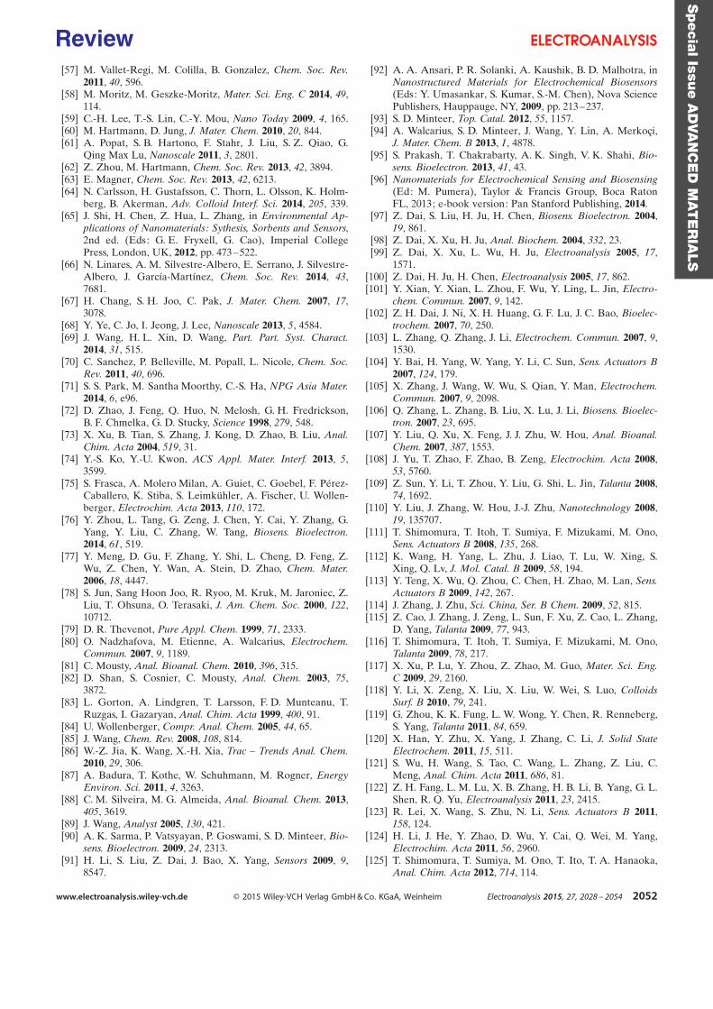
[57] M. Vallet-Regi, M. Colilla, B. Gonzalez, Chem. Soc. Rev.2011, 40, 596.
[58] M. Moritz, M. Geszke-Moritz, Mater. Sci. Eng. C 2014, 49,114.
[59] C.-H. Lee, T.-S. Lin, C.-Y. Mou, Nano Today 2009, 4, 165.[60] M. Hartmann, D. Jung, J. Mater. Chem. 2010, 20, 844.[61] A. Popat, S. B. Hartono, F. Stahr, J. Liu, S. Z. Qiao, G.
Qing Max Lu, Nanoscale 2011, 3, 2801.[62] Z. Zhou, M. Hartmann, Chem. Soc. Rev. 2013, 42, 3894.[63] E. Magner, Chem. Soc. Rev. 2013, 42, 6213.[64] N. Carlsson, H. Gustafsson, C. Thorn, L. Olsson, K. Holm-
berg, B. Akerman, Adv. Colloid Interf. Sci. 2014, 205, 339.[65] J. Shi, H. Chen, Z. Hua, L. Zhang, in Environmental Ap-
plications of Nanomaterials: Sythesis, Sorbents and Sensors,2nd ed. (Eds: G. E. Fryxell, G. Cao), Imperial CollegePress, London, UK, 2012, pp. 473–522.
[66] N. Linares, A. M. Silvestre-Albero, E. Serrano, J. Silvestre-Albero, J. Garc�a-Mart�nez, Chem. Soc. Rev. 2014, 43,7681.
[67] H. Chang, S. H. Joo, C. Pak, J. Mater. Chem. 2007, 17,3078.
[68] Y. Ye, C. Jo, I. Jeong, J. Lee, Nanoscale 2013, 5, 4584.[69] J. Wang, H. L. Xin, D. Wang, Part. Part. Syst. Charact.
2014, 31, 515.[70] C. Sanchez, P. Belleville, M. Popall, L. Nicole, Chem. Soc.
Rev. 2011, 40, 696.[71] S. S. Park, M. Santha Moorthy, C.-S. Ha, NPG Asia Mater.
2014, 6, e96.[72] D. Zhao, J. Feng, Q. Huo, N. Melosh, G. H. Fredrickson,
B. F. Chmelka, G. D. Stucky, Science 1998, 279, 548.[73] X. Xu, B. Tian, S. Zhang, J. Kong, D. Zhao, B. Liu, Anal.
Chim. Acta 2004, 519, 31.[74] Y.-S. Ko, Y.-U. Kwon, ACS Appl. Mater. Interf. 2013, 5,
3599.[75] S. Frasca, A. Molero Milan, A. Guiet, C. Goebel, F. P¦rez-
Caballero, K. Stiba, S. Leimkîhler, A. Fischer, U. Wollen-berger, Electrochim. Acta 2013, 110, 172.
[76] Y. Zhou, L. Tang, G. Zeng, J. Chen, Y. Cai, Y. Zhang, G.Yang, Y. Liu, C. Zhang, W. Tang, Biosens. Bioelectron.2014, 61, 519.
[77] Y. Meng, D. Gu, F. Zhang, Y. Shi, L. Cheng, D. Feng, Z.Wu, Z. Chen, Y. Wan, A. Stein, D. Zhao, Chem. Mater.2006, 18, 4447.
[78] S. Jun, Sang Hoon Joo, R. Ryoo, M. Kruk, M. Jaroniec, Z.Liu, T. Ohsuna, O. Terasaki, J. Am. Chem. Soc. 2000, 122,10712.
[79] D. R. Thevenot, Pure Appl. Chem. 1999, 71, 2333.[80] O. Nadzhafova, M. Etienne, A. Walcarius, Electrochem.
Commun. 2007, 9, 1189.[81] C. Mousty, Anal. Bioanal. Chem. 2010, 396, 315.[82] D. Shan, S. Cosnier, C. Mousty, Anal. Chem. 2003, 75,
3872.[83] L. Gorton, A. Lindgren, T. Larsson, F. D. Munteanu, T.
Ruzgas, I. Gazaryan, Anal. Chim. Acta 1999, 400, 91.[84] U. Wollenberger, Compr. Anal. Chem. 2005, 44, 65.[85] J. Wang, Chem. Rev. 2008, 108, 814.[86] W.-Z. Jia, K. Wang, X.-H. Xia, Trac – Trends Anal. Chem.
2010, 29, 306.[87] A. Badura, T. Kothe, W. Schuhmann, M. Rogner, Energy
Environ. Sci. 2011, 4, 3263.[88] C. M. Silveira, M. G. Almeida, Anal. Bioanal. Chem. 2013,
405, 3619.[89] J. Wang, Analyst 2005, 130, 421.[90] A. K. Sarma, P. Vatsyayan, P. Goswami, S. D. Minteer, Bio-
sens. Bioelectron. 2009, 24, 2313.[91] H. Li, S. Liu, Z. Dai, J. Bao, X. Yang, Sensors 2009, 9,
8547.
[92] A. A. Ansari, P. R. Solanki, A. Kaushik, B. D. Malhotra, inNanostructured Materials for Electrochemical Biosensors(Eds: Y. Umasankar, S. Kumar, S.-M. Chen), Nova SciencePublishers, Hauppauge, NY, 2009, pp. 213–237.
[93] S. D. Minteer, Top. Catal. 2012, 55, 1157.[94] A. Walcarius, S. D. Minteer, J. Wang, Y. Lin, A. MerkoÅi,
J. Mater. Chem. B 2013, 1, 4878.[95] S. Prakash, T. Chakrabarty, A. K. Singh, V. K. Shahi, Bio-
sens. Bioelectron. 2013, 41, 43.[96] Nanomaterials for Electrochemical Sensing and Biosensing
(Ed: M. Pumera), Taylor & Francis Group, Boca RatonFL, 2013; e-book version: Pan Stanford Publishing, 2014.
[97] Z. Dai, S. Liu, H. Ju, H. Chen, Biosens. Bioelectron. 2004,19, 861.
[98] Z. Dai, X. Xu, H. Ju, Anal. Biochem. 2004, 332, 23.[99] Z. Dai, X. Xu, L. Wu, H. Ju, Electroanalysis 2005, 17,
1571.[100] Z. Dai, H. Ju, H. Chen, Electroanalysis 2005, 17, 862.[101] Y. Xian, Y. Xian, L. Zhou, F. Wu, Y. Ling, L. Jin, Electro-
chem. Commun. 2007, 9, 142.[102] Z. H. Dai, J. Ni, X. H. Huang, G. F. Lu, J. C. Bao, Bioelec-
trochem. 2007, 70, 250.[103] L. Zhang, Q. Zhang, J. Li, Electrochem. Commun. 2007, 9,
1530.[104] Y. Bai, H. Yang, W. Yang, Y. Li, C. Sun, Sens. Actuators B
2007, 124, 179.[105] X. Zhang, J. Wang, W. Wu, S. Qian, Y. Man, Electrochem.
Commun. 2007, 9, 2098.[106] Q. Zhang, L. Zhang, B. Liu, X. Lu, J. Li, Biosens. Bioelec-
tron. 2007, 23, 695.[107] Y. Liu, Q. Xu, X. Feng, J. J. Zhu, W. Hou, Anal. Bioanal.
Chem. 2007, 387, 1553.[108] J. Yu, T. Zhao, F. Zhao, B. Zeng, Electrochim. Acta 2008,
53, 5760.[109] Z. Sun, Y. Li, T. Zhou, Y. Liu, G. Shi, L. Jin, Talanta 2008,
74, 1692.[110] Y. Liu, J. Zhang, W. Hou, J.-J. Zhu, Nanotechnology 2008,
19, 135707.[111] T. Shimomura, T. Itoh, T. Sumiya, F. Mizukami, M. Ono,
Sens. Actuators B 2008, 135, 268.[112] K. Wang, H. Yang, L. Zhu, J. Liao, T. Lu, W. Xing, S.
Xing, Q. Lv, J. Mol. Catal. B 2009, 58, 194.[113] Y. Teng, X. Wu, Q. Zhou, C. Chen, H. Zhao, M. Lan, Sens.
Actuators B 2009, 142, 267.[114] J. Zhang, J. Zhu, Sci. China, Ser. B Chem. 2009, 52, 815.[115] Z. Cao, J. Zhang, J. Zeng, L. Sun, F. Xu, Z. Cao, L. Zhang,
D. Yang, Talanta 2009, 77, 943.[116] T. Shimomura, T. Itoh, T. Sumiya, F. Mizukami, M. Ono,
Talanta 2009, 78, 217.[117] X. Xu, P. Lu, Y. Zhou, Z. Zhao, M. Guo, Mater. Sci. Eng.
C 2009, 29, 2160.[118] Y. Li, X. Zeng, X. Liu, X. Liu, W. Wei, S. Luo, Colloids
Surf. B 2010, 79, 241.[119] G. Zhou, K. K. Fung, L. W. Wong, Y. Chen, R. Renneberg,
S. Yang, Talanta 2011, 84, 659.[120] X. Han, Y. Zhu, X. Yang, J. Zhang, C. Li, J. Solid State
Electrochem. 2011, 15, 511.[121] S. Wu, H. Wang, S. Tao, C. Wang, L. Zhang, Z. Liu, C.
Meng, Anal. Chim. Acta 2011, 686, 81.[122] Z. H. Fang, L. M. Lu, X. B. Zhang, H. B. Li, B. Yang, G. L.
Shen, R. Q. Yu, Electroanalysis 2011, 23, 2415.[123] R. Lei, X. Wang, S. Zhu, N. Li, Sens. Actuators B 2011,
158, 124.[124] H. Li, J. He, Y. Zhao, D. Wu, Y. Cai, Q. Wei, M. Yang,
Electrochim. Acta 2011, 56, 2960.[125] T. Shimomura, T. Sumiya, M. Ono, T. Ito, T. A. Hanaoka,
Anal. Chim. Acta 2012, 714, 114.
Specia
lIssu
eA
DVA
NC
ED
MAT
ER
IALS
www.electroanalysis.wiley-vch.de Ó 2015 Wiley-VCH Verlag GmbH & Co. KGaA, Weinheim Electroanalysis 2015, 27, 2028 – 2054 2052
Review
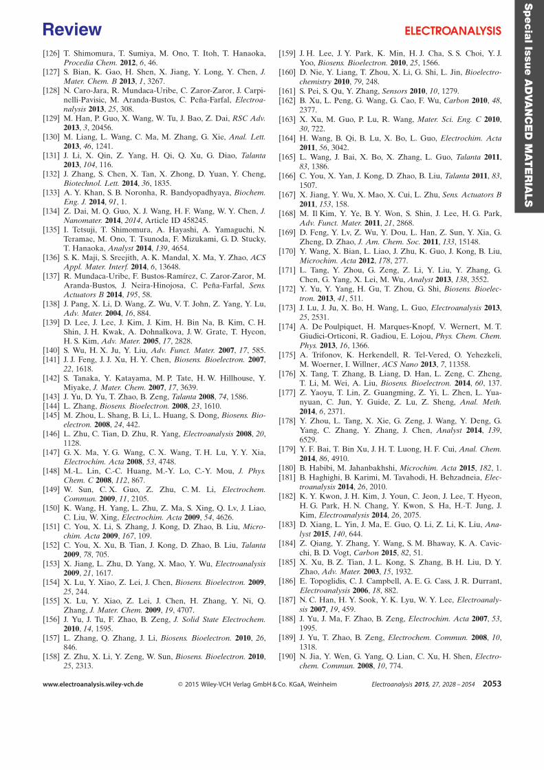
[126] T. Shimomura, T. Sumiya, M. Ono, T. Itoh, T. Hanaoka,Procedia Chem. 2012, 6, 46.
[127] S. Bian, K. Gao, H. Shen, X. Jiang, Y. Long, Y. Chen, J.Mater. Chem. B 2013, 1, 3267.
[128] N. Caro-Jara, R. Mundaca-Uribe, C. Zaror-Zaror, J. Carpi-nelli-Pavisic, M. Aranda-Bustos, C. PeÇa-Farfal, Electroa-nalysis 2013, 25, 308.
[129] M. Han, P. Guo, X. Wang, W. Tu, J. Bao, Z. Dai, RSC Adv.2013, 3, 20456.
[130] M. Liang, L. Wang, C. Ma, M. Zhang, G. Xie, Anal. Lett.2013, 46, 1241.
[131] J. Li, X. Qin, Z. Yang, H. Qi, Q. Xu, G. Diao, Talanta2013, 104, 116.
[132] J. Zhang, S. Chen, X. Tan, X. Zhong, D. Yuan, Y. Cheng,Biotechnol. Lett. 2014, 36, 1835.
[133] A. Y. Khan, S. B. Noronha, R. Bandyopadhyaya, Biochem.Eng. J. 2014, 91, 1.
[134] Z. Dai, M. Q. Guo, X. J. Wang, H. F. Wang, W. Y. Chen, J.Nanomater. 2014, 2014, Article ID 458245.
[135] I. Tetsuji, T. Shimomura, A. Hayashi, A. Yamaguchi, N.Teramae, M. Ono, T. Tsunoda, F. Mizukami, G. D. Stucky,T. Hanaoka, Analyst 2014, 139, 4654.
[136] S. K. Maji, S. Sreejith, A. K. Mandal, X. Ma, Y. Zhao, ACSAppl. Mater. Interf. 2014, 6, 13648.
[137] R. Mundaca-Uribe, F. Bustos-Ram�rez, C. Zaror-Zaror, M.Aranda-Bustos, J. Neira-Hinojosa, C. PeÇa-Farfal, Sens.Actuators B 2014, 195, 58.
[138] J. Pang, X. Li, D. Wang, Z. Wu, V. T. John, Z. Yang, Y. Lu,Adv. Mater. 2004, 16, 884.
[139] D. Lee, J. Lee, J. Kim, J. Kim, H. Bin Na, B. Kim, C. H.Shin, J. H. Kwak, A. Dohnalkova, J. W. Grate, T. Hyeon,H. S. Kim, Adv. Mater. 2005, 17, 2828.
[140] S. Wu, H. X. Ju, Y. Liu, Adv. Funct. Mater. 2007, 17, 585.[141] J. J. Feng, J. J. Xu, H. Y. Chen, Biosens. Bioelectron. 2007,
22, 1618.[142] S. Tanaka, Y. Katayama, M. P. Tate, H. W. Hillhouse, Y.
Miyake, J. Mater. Chem. 2007, 17, 3639.[143] J. Yu, D. Yu, T. Zhao, B. Zeng, Talanta 2008, 74, 1586.[144] L. Zhang, Biosens. Bioelectron. 2008, 23, 1610.[145] M. Zhou, L. Shang, B. Li, L. Huang, S. Dong, Biosens. Bio-
electron. 2008, 24, 442.[146] L. Zhu, C. Tian, D. Zhu, R. Yang, Electroanalysis 2008, 20,
1128.[147] G. X. Ma, Y. G. Wang, C. X. Wang, T. H. Lu, Y. Y. Xia,
Electrochim. Acta 2008, 53, 4748.[148] M.-L. Lin, C.-C. Huang, M.-Y. Lo, C.-Y. Mou, J. Phys.
Chem. C 2008, 112, 867.[149] W. Sun, C. X. Guo, Z. Zhu, C. M. Li, Electrochem.
Commun. 2009, 11, 2105.[150] K. Wang, H. Yang, L. Zhu, Z. Ma, S. Xing, Q. Lv, J. Liao,
C. Liu, W. Xing, Electrochim. Acta 2009, 54, 4626.[151] C. You, X. Li, S. Zhang, J. Kong, D. Zhao, B. Liu, Micro-
chim. Acta 2009, 167, 109.[152] C. You, X. Xu, B. Tian, J. Kong, D. Zhao, B. Liu, Talanta
2009, 78, 705.[153] X. Jiang, L. Zhu, D. Yang, X. Mao, Y. Wu, Electroanalysis
2009, 21, 1617.[154] X. Lu, Y. Xiao, Z. Lei, J. Chen, Biosens. Bioelectron. 2009,
25, 244.[155] X. Lu, Y. Xiao, Z. Lei, J. Chen, H. Zhang, Y. Ni, Q.
Zhang, J. Mater. Chem. 2009, 19, 4707.[156] J. Yu, J. Tu, F. Zhao, B. Zeng, J. Solid State Electrochem.
2010, 14, 1595.[157] L. Zhang, Q. Zhang, J. Li, Biosens. Bioelectron. 2010, 26,
846.[158] Z. Zhu, X. Li, Y. Zeng, W. Sun, Biosens. Bioelectron. 2010,
25, 2313.
[159] J. H. Lee, J. Y. Park, K. Min, H. J. Cha, S. S. Choi, Y. J.Yoo, Biosens. Bioelectron. 2010, 25, 1566.
[160] D. Nie, Y. Liang, T. Zhou, X. Li, G. Shi, L. Jin, Bioelectro-chemistry 2010, 79, 248.
[161] S. Pei, S. Qu, Y. Zhang, Sensors 2010, 10, 1279.[162] B. Xu, L. Peng, G. Wang, G. Cao, F. Wu, Carbon 2010, 48,
2377.[163] X. Xu, M. Guo, P. Lu, R. Wang, Mater. Sci. Eng. C 2010,
30, 722.[164] H. Wang, B. Qi, B. Lu, X. Bo, L. Guo, Electrochim. Acta
2011, 56, 3042.[165] L. Wang, J. Bai, X. Bo, X. Zhang, L. Guo, Talanta 2011,
83, 1386.[166] C. You, X. Yan, J. Kong, D. Zhao, B. Liu, Talanta 2011, 83,
1507.[167] X. Jiang, Y. Wu, X. Mao, X. Cui, L. Zhu, Sens. Actuators B
2011, 153, 158.[168] M. Il Kim, Y. Ye, B. Y. Won, S. Shin, J. Lee, H. G. Park,
Adv. Funct. Mater. 2011, 21, 2868.[169] D. Feng, Y. Lv, Z. Wu, Y. Dou, L. Han, Z. Sun, Y. Xia, G.
Zheng, D. Zhao, J. Am. Chem. Soc. 2011, 133, 15148.[170] Y. Wang, X. Bian, L. Liao, J. Zhu, K. Guo, J. Kong, B. Liu,
Microchim. Acta 2012, 178, 277.[171] L. Tang, Y. Zhou, G. Zeng, Z. Li, Y. Liu, Y. Zhang, G.
Chen, G. Yang, X. Lei, M. Wu, Analyst 2013, 138, 3552.[172] Y. Yu, Y. Yang, H. Gu, T. Zhou, G. Shi, Biosens. Bioelec-
tron. 2013, 41, 511.[173] J. Lu, J. Ju, X. Bo, H. Wang, L. Guo, Electroanalysis 2013,
25, 2531.[174] A. De Poulpiquet, H. Marques-Knopf, V. Wernert, M. T.
Giudici-Orticoni, R. Gadiou, E. Lojou, Phys. Chem. Chem.Phys. 2013, 16, 1366.
[175] A. Trifonov, K. Herkendell, R. Tel-Vered, O. Yehezkeli,M. Woerner, I. Willner, ACS Nano 2013, 7, 11358.
[176] X. Tang, T. Zhang, B. Liang, D. Han, L. Zeng, C. Zheng,T. Li, M. Wei, A. Liu, Biosens. Bioelectron. 2014, 60, 137.
[177] Z. Yaoyu, T. Lin, Z. Guangming, Z. Yi, L. Zhen, L. Yua-nyuan, C. Jun, Y. Guide, Z. Lu, Z. Sheng, Anal. Meth.2014, 6, 2371.
[178] Y. Zhou, L. Tang, X. Xie, G. Zeng, J. Wang, Y. Deng, G.Yang, C. Zhang, Y. Zhang, J. Chen, Analyst 2014, 139,6529.
[179] Y. F. Bai, T. Bin Xu, J. H. T. Luong, H. F. Cui, Anal. Chem.2014, 86, 4910.
[180] B. Habibi, M. Jahanbakhshi, Microchim. Acta 2015, 182, 1.[181] B. Haghighi, B. Karimi, M. Tavahodi, H. Behzadneia, Elec-
troanalysis 2014, 26, 2010.[182] K. Y. Kwon, J. H. Kim, J. Youn, C. Jeon, J. Lee, T. Hyeon,
H. G. Park, H. N. Chang, Y. Kwon, S. Ha, H.-T. Jung, J.Kim, Electroanalysis 2014, 26, 2075.
[183] D. Xiang, L. Yin, J. Ma, E. Guo, Q. Li, Z. Li, K. Liu, Ana-lyst 2015, 140, 644.
[184] Z. Qiang, Y. Zhang, Y. Wang, S. M. Bhaway, K. A. Cavic-chi, B. D. Vogt, Carbon 2015, 82, 51.
[185] X. Xu, B. Z. Tian, J. L. Kong, S. Zhang, B. H. Liu, D. Y.Zhao, Adv. Mater. 2003, 15, 1932.
[186] E. Topoglidis, C. J. Campbell, A. E. G. Cass, J. R. Durrant,Electroanalysis 2006, 18, 882.
[187] N. C. Han, H. Y. Sook, Y. K. Lyu, W. Y. Lee, Electroanaly-sis 2007, 19, 459.
[188] J. Yu, J. Ma, F. Zhao, B. Zeng, Electrochim. Acta 2007, 53,1995.
[189] J. Yu, T. Zhao, B. Zeng, Electrochem. Commun. 2008, 10,1318.
[190] N. Jia, Y. Wen, G. Yang, Q. Lian, C. Xu, H. Shen, Electro-chem. Commun. 2008, 10, 774.
Specia
lIssu
eA
DVA
NC
ED
MAT
ER
IALS
www.electroanalysis.wiley-vch.de Ó 2015 Wiley-VCH Verlag GmbH & Co. KGaA, Weinheim Electroanalysis 2015, 27, 2028 – 2054 2053
Review

[191] Y. Astuti, E. Topoglidis, A. G. Cass, J. R. Durrant, Anal.Chim. Acta 2009, 648, 2.
[192] S. Frasca, T. von Graberg, J. J. Feng, A. Thomas, B. M.Smarsly, I. M. Weidinger, F. W. Scheller, P. Hildebrandt, U.Wollenberger, ChemCatChem 2010, 2, 839.
[193] Y. Astuti, E. Topoglidis, J. R. Durrant, Anal. Chim. Acta2011, 686, 126.
[194] C. Liu, Y. Teng, J. Xu, Y. Yang, Z. Wu, Int. J. Environ.Anal. Chem. 2011, 91, 1367.
[195] S. Frasca, C. Richter, T. von Graberg, B. M. Smarsly, U.Wollenberger, Eng. Life Sci. 2011, 11, 554.
[196] J. Li, D. Kuang, Y. Feng, F. Zhang, M. Liu, Microchim.Acta 2012, 176, 73.
[197] P. Si, P. Chen, D.-H. Kim, J. Mater. Chem. B 2013, 1, 2696.[198] W. Sun, Z. Sun, L. Zhang, X. Qi, G. Li, J. Wu, M. Wang,
Colloids Surf. B 2013, 101, 177.[199] W. Luo, Y. Li, J. Dong, J. Wei, J. Xu, Y. Deng, D. Zhao,
Angew. Chem. Int. Ed. 2013, 52, 10505.[200] K. Mondal, M. A. Ali, V. V. Agrawal, B. D. Malhotra, A.
Sharma, ACS Appl. Mater. Interf. 2014, 6, 2516.[201] D. Wang, Y. Tan, Electrochim. Acta 2014, 116, 495.[202] G. Bharath, R. Madhu, S.-M. Chen, V. Veeramani, A. Ba-
lamurugan, D. Mangalaraj, C. Viswanathan, N. Ponpandi-an, J. Mater. Chem. B 2015, 3, 1360.
[203] J. Li, J. Tang, L. Zhou, X. Han, H. Liu, Bioelectrochemistry2012, 86, 60.
[204] M. Etienne, A. Walcarius, in Electrochemistry, Vol. 11,Nanosystems Electrochemistry, The Royal Society ofChemistry, London, 2013, pp. 124 –197.
[205] J. S. Beck, J. C. Vartuli, W. J. Roth, M. E. Leonowicz, C. T.Kresge, K. D. Schmitt, C. T. W. Chu, D. H. Olson, E. W.Sheppard, J. Am. Chem. Soc. 1992, 114, 10834.
[206] G. J. de A. A. Soler-Illia, C. Sanchez, B. Lebeau, J. Patarin,Chem. Rev. 2002, 102, 4093.
[207] C. Sanchez, C. BoissiÀre, D. Grosso, C. Laberty, L. Nicole,Chem. Mater. 2008, 20, 682.
[208] Z.-Y. Yuan, B.-L. Su, J. Mater. Chem. 2006, 16, 663.[209] M. Tiemann, Chem. Mater. 2008, 20, 961.[210] H. Wang, L. Zhai, Y. Li, T. Shi, Mater. Res. Bull. 2008, 43,
1607.[211] R. Ryoo, S. H. Joo, S. Jun, J. Phys. Chem. B 1999, 103,
7743.[212] Y. Meng, D. Gu, F. Zhang, Y. Shi, H. Yang, Z. Li, C. Yu,
B. Tu, D. Zhao, Angew. Chem. Int. Ed. 2005, 44, 7053.[213] X. Wang, X. Lu, L. Wu, J. Chen, ChemElectroChem 2014,
1, 808.[214] Z. Zhou, M. Hartmann, Top. Catal. 2012, 55, 1081.[215] S. Hudson, J. Cooney, E. Magner, Angew. Chem. Int. Ed.
2008, 47, 8582.[216] J. N. Talbert, J. M. Goddard, Colloids Surf. B. 2012, 93, 8.[217] L. Bayne, R. V. Ulijn, P. J. Halling, Chem. Soc. Rev. 2013,
42, 9000.
[218] K. Y. Kwon, J. Youn, J. H. Kim, Y. Park, C. Jeon, B. C.Kim, Y. Kwon, X. Zhao, P. Wang, B. I. Sang, J. Lee, H. G.Park, H. N. Chang, T. Hyeon, S. Ha, H.-T. Jung, J. Kim,Biosens. Bioelectron. 2010, 26, 655.
[219] C. Renault, K. D. Harris, M. J. Brett, V. Balland, B. Limog-es, Chem. Commun. 2011, 47, 1863.
[220] A. Hagfeldt, M. Gr�tzel, Acc. Chem. Res. 2000, 33, 269.[221] D. Grosso, F. Cagnol, G. J. de A. A. Soler-Illia, E. L. Cre-
paldi, H. Amenitsch, A. Brunet-Bruneau, A. Bourgeois, C.Sanchez, Adv. Funct. Mater. 2004, 14, 309.
[222] A. Walcarius, E. Sibottier, M. Etienne, J. Ghanbaja, Nat.Mater. 2007, 6, 602.
[223] Y. Zhou, L. Tang, G. Zeng, J. Chen, J. Wang, C. Fan, G.Yang, Y. Zhang, X. Xie, Biosens. Bioelectron. 2015, 65,382.
[224] S. Borgmann, A. Schulte, S. Neugebauer, W. Schuhmann,in Bioelectrochemistry (Eds: R. C. Alkire, D. M. Kolb, J.Lipkowski), Wiley-VCH, Weinheim, 2011, pp. 1–83.
[225] L. H. Guo, H. Allen, O. Hill, Adv. Inorg. Chem. 1991, 36,341.
[226] X. Chen, R. Ferrigno, J. Yang, G. M. Whitesides, Langmuir2002, 18, 7009.
[227] A. Heller, B. Feldman, Chem. Rev. 2008, 108, 2482.[228] J. D. Lane, D. M. Krumholz, R. A. Sack, C. Morris, Curr.
Eye Res. 2006, 31, 895.[229] D. K. Sen, G. S. Sarin, Br. J. Ophthalmol. 1980, 64, 693.[230] I. Mazurenko, O. Tananaiko, O. Biloivan, M. Zhybak, I.
Pelyak, V. Zaitsev, M. Etienne, A. Walcarius, Electroanaly-sis 2015, in press.
[231] C. Kisker, H. Schindelin, A. Pacheco, W. A. Wehbi, R. M.Garrett, K. V. Rajagopalan, J. H. Enemark, D. C. Rees,Cell 1997, 91, 973.
[232] A. Pacheco, J. T. Hazzard, G. Tollin, J. H. Enemark, J.Biol. Inorg. Chem. 1999, 4, 390.
[233] N. Vil�, J. Ghanbaja, E. Aubert, A. Walcarius, Angew.Chem. Int. Ed. Eng. 2014, 53, 2945.
[234] Z. Zhou, R. N. Klupp Taylor, S. Kullmann, H. Bao, M.Hartmann, Adv. Mater. 2011, 23, 2627.
[235] E. Schneider, D. S. Clark, Biosens. Bioelectron. 2013, 39, 1.[236] M. Faustini, M. Vayer, B. Marmiroli, M. Hillmyer, H.
Amenitsch, C. Sinturel, D. Grosso, Chem. Mater. 2010, 22,5687.
[237] R. Villalonga, P. D�ez, A. S�nchez, E. Aznar, R. Mart�nez-M�Çez, J. M. Pingarr"n, Chem. Eur. J. 2013, 19, 7889.
[238] J. Simmchen, A. Baeza, D. Ruiz, M. J. Esplandiu, M.Vallet-Reg�, Small 2012, 8, 2053.
[239] I. Fern�ndez, A. S�nchez, P. D�ez, P. Mart�nez-Ruiz, P. DiPierro, R. Porta, R. Villalonga, J. M. Pingarrýn, Chem.Commun. 2014, 50, 13356.
Received: March 16, 2015Accepted: May 5, 2015
Published online: July 21, 2015
Specia
lIssu
eA
DVA
NC
ED
MAT
ER
IALS
www.electroanalysis.wiley-vch.de Ó 2015 Wiley-VCH Verlag GmbH & Co. KGaA, Weinheim Electroanalysis 2015, 27, 2028 – 2054 2054
Review
Recommended