Embed Size (px)
Citation preview
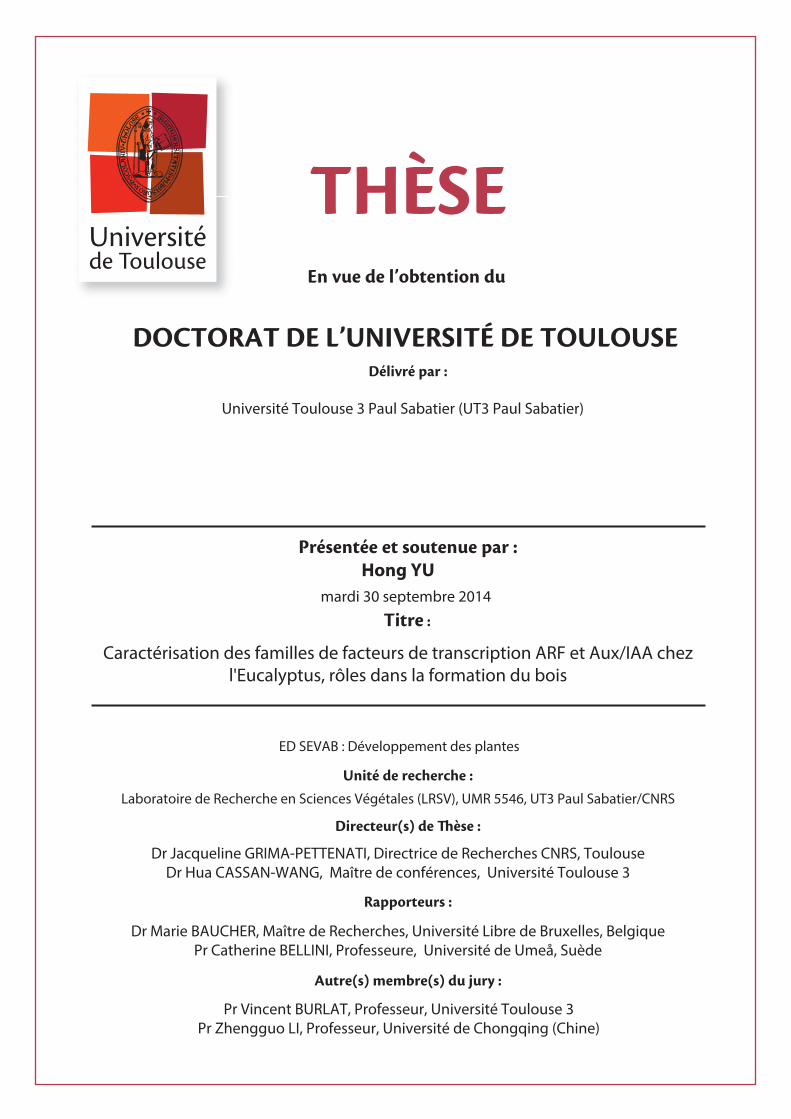
tre :
Université Toulouse 3 Paul Sabatier (UT3 Paul Sabatier)
ED SEVAB : Développement des plantes
Hong YUmardi 30 septembre 2014
Caractérisation des familles de facteurs de transcription ARF et Aux/IAA chezl'Eucalyptus, rôles dans la formation du bois
Laboratoire de Recherche en Sciences Végétales (LRSV), UMR 5546, UT3 Paul Sabatier/CNRS
Dr Jacqueline GRIMA-PETTENATI, Directrice de Recherches CNRS, ToulouseDr Hua CASSAN-WANG, Maître de conférences, Université Toulouse 3
Dr Marie BAUCHER, Maître de Recherches, Université Libre de Bruxelles, BelgiquePr Catherine BELLINI, Professeure, Université de Umeå, Suède
Pr Vincent BURLAT, Professeur, Université Toulouse 3Pr Zhengguo LI, Professeur, Université de Chongqing (Chine)

Acknowledgments
Foremost, I would like to express my sincere gratitude to my supervisors Dr. Jacqueline Grima-
Pettenati and Dr. Hua Cassan-Wang for allowing me to be part of their team, and thanks for
their guidance, advice, involvement and support in my work. They helped me in all the time of
research and writing of this thesis.
I am very grateful to the committee members: Dr. Marie Baucher, Dr. Catherine Bellini and Dr.
Laurent Laplaze for having accepted to judge my research work.
I would like also to acknowledge the help and advice of Prof. Mondher Bouzayen and Isabelle
Mila in GBF during my PhD. Thanks Dr. Edouard Pesquet and Junko Takahashi Schmidt for
hosting me in Umeå Plant Science Centre and for their precious help and advice with the Py-
GC/MS analysis. Thanks to Dr. Françoise Monéger and Dr. Didier Aldon for their precious
advice during my PhD. Thanks Yves Martinez for his help with the microscope analysis in
Plateforme Imagerie-TRI.
Thanks to Nathalie Ladouce for her help with my experiments especially for Fluidigm. Thanks
to Marçal Soler, for his stimulating discussions and sharing his knowledge with me. Thanks to
Hélene San-clemente, Dunand Christophe and Qiang Li, for their support of the bioinformatics.
I also would like to thank all members of the GFE team and all the friends studying and living
around me during these four years. Because of their support, advice and friendship, they all
contributed to the accomplishment of this thesis. And I would like to thank to all my colleagues
from Chongqing University for their friendship and support especially Prof. Zhengguo Li,
Zhongkang Wang and Youping Yin.
My scholarship was granted by China Scholarship Council in Beijing. Thank you for making
my staying in France possible.
And last but not the least, I would like to thank to my whole family for their loving support and
encouragement.

I
Résumé
L'auxine est une hormone centrale du développement chez les végétaux, impliquée dans
un large éventail de processus tels que l'organogenèse, la croissance racinaire, le
développement des fruits et le développement vasculaire. Chez les plantes ligneuses,
l'auxine joue un rôle clé dans la régulation de la formation du bois en stimulant l’activité
du cambium vasculaire et le développement du xylème. Les ARFs (Auxin-response
factors) et les Aux/IAAs (Auxin/Indole acetic acid) sont des régulateurs importants de
la voie de perception et de signalisation de l'auxine. La disponibilité récente de la
séquence du génome d’Eucalyptus grandis nous a permis d'étudier les caractéristiques
et l'histoire évolutive de ces deux familles de gènes chez cette plante ligneuse de grande
importance économique. Dans ce travail, nous avons identifié et caractérisé 17
EgrARFs et 24 EgrIAAs que nous avons nommés en fonction de leurs orthologues
putatifs chez Arabidopsis. Chez E. grandis, ces deux familles ont un peu moins de gènes
que chez la plupart des angiospermes étudiés jusqu'ici. L’analyse de phylogénie
comparative de génomes appartenant à des lignées taxonomiques pertinentes a révélé,
chez la famille ARF, la présence d'une clade trouvée préférentiellement chez les plantes
ligneuses pérennes. Des analyses d'expression à haut débit sur un large panel d’organes
et de tissus et en réponse à des signaux environnementaux ont mis en évidence des
gènes exprimés préférentiellement dans le cambium vasculaire et/ou le xylème en
différentiation, certains présentant des réponses à des stimuli abiotiques. Sur la base de
leurs profils d'expression, nous avons sélectionné certains gènes candidats prometteurs
et effectué leur caractérisation fonctionnelle afin d’évaluer leur rôle potentiel dans la
formation du bois. Nous avons utilisé Arabidopsis, qui peut dans certaines conditions
présenter une croissance secondaire, pour surexprimer des versions mutées et
stabilisées de EgrIAA4, 9 et 20. Les lignées transgéniques présentent des phénotypes
aberrants d’insensibilité à l'auxine. Parmi ceux-ci, des tailles de plantes réduites ou
naines, des racines qui croissent de façon agravitropique, la réduction ou même
l’absence de racines latérales et la stérilité partielle. Les analyses histochimiques
complétées par des analyses en pyrolyse ont montré des modifications évidentes dans

II
la différentiation des cellules du xylème et/ou des fibres interfasciculaires ainsi que
composition chimique de leurs parois secondaires. En conclusion, cette étude offre une
caractérisation complète de deux familles de régulateurs importants de l’auxine chez l'
Eucalyptus et met en évidence l'implication de certains membres dans la régulation de
la formation du bois.
III
Abstract
Auxin is a central hormone involved in a wide range of developmental processes
including organogenesis, tropic movement, root growth, fruit development, tissue and
organ patterning and vascular development. In woody plants, auxin has been proposed
to play a key role in regulating wood formation through its effects on cambial activity
and xylem development. Auxin Response Factors (ARF) and Auxin/Indole-3-Acetic
Acid (Aux/IAA) are important regulators of auxin responses in plants. The recent
availability of the Eucalyptus grandis genome allowed us to investigate the
characteristics and evolutionary history of these two gene families in a woody plant of
high economic importance. In this work, we identified and characterized 17 EgrARF
and 24 EgrIAA gene members and named them according to their putative orthologs in
Arabidopsis. Both of these two gene families are slightly contracted, as compared to
those of most angiosperms studied hitherto. Comparative phylogenetic analyses with
genomes of relevant taxonomic lineages revealed the presence of a new ARF clade
found preferentially in woody and/or perennial plants. High-throughput expression
profiling among different organs and tissues and in response to environmental cues
highlighted genes expressed in vascular cambium and/or developing xylem, in addition
to dynamic modifications in response to environmental stimuli. Based on their
expression profiles, we selected some promising candidates and carried out their
functional characterization to get insights into their potential roles in wood formation.
We used Arabidopsis, which was shown to undergo secondary growth in some
conditions, to overexpress stabilized versions of EgrIAA4m, 9Am and 20. The
transgenic lines exhibited auxin-related aberrant phenotypes, such as smaller size,
impaired root growth and development, and partial sterility. Histochemistry and Py-
GC/MS analysis revealed that the transgenic plants showed obvious modifications in
xylem and/or fiber cells differentiation and secondary cell wall composition. Altogether,
the present study provides a comprehensive characterization of the Eucalyptus ARF and
Aux/IAA gene families and highlights the involvement of some members in the
regulation of wood development.

IV
摘 要
生长素参与植物生长和发育诸多过程,包括器官的形成、植物的向地和向光反应、
根的发育生长、果实的发育,以及维管束组织的形成和分化。生长素在木材的形
成过程中也发挥着重要作用,主要通过对维管形成层及木质部细胞发育的影响来
调控其形成。生长素响应因子(ARF)和生长素/吲哚乙酸蛋白(Aux/IAA)是植
物生长素响应的主要调节蛋白。桉树作为木材在人类的生产生活中具有重要的经
济价值,其全基因组测序工作已于近期完成,这使得我们可以对桉树 ARF 和
Aux/IAA这两个家族进行全面的分析。在本论文中,我们一共分离出了 17个 ARF
与 24个 Aux/IAA 基因,并且根据其与拟南芥的同源性对它们进行了重命名。目
前为止,这两个基因家族的数量在桉树中较其它研究过的大多数的被子植物有所
减少。与其它物种的系统发育进化树分析表明在 ARF家族中存在一个木本植物植
物中占有优势的分支。对不同组织器官、环境因素及植物激素处理实验的高通量
表达分析显示有些基因在维管形成层和/或木质部中有优势表达,而且大部分基
因会对不同的环境因素及激素处理作出响应。为了深入了解桉树 ARF 与 Aux/IAA
在木材发育过程中的作用,我们选择了一些可能调控木质部发育的 ARF 与
Aux/IAA基因构建了转基因拟南芥。其中 EgrIAA4m, EgrIAA9Am 与 EgrIAA20 转
基因拟南芥表现出了一些与生长素缺陷相关的表型,比如矮小株型,根与侧根非
正常的生长发育,根向地性的丧失以及育性降低。组织化学及高温分解气相色谱
质朴分析表明转基因植株中木质部细胞的发育以及次生细胞壁的组成成分都发
生了明显的改变。总之,该研究全面分析了桉树 ARF 与 Aux/IAA 两个家族,并
通过反义基因功能分析揭示了有些成员在木质部的生长发育中起着重要的调控
作用。

V
Contents
Résumé ....................................................................................................................................... I
Abstract ................................................................................................................................... III
摘 要 ....................................................................................................................................... IV
List of Figures ......................................................................................................................... IX
List of Tables ...........................................................................................................................XI
Abbreviations ........................................................................................................................ .XII
General introduction .................................................................................................................. 1
Chapter I: Bibliographic review ................................................................................................ 4
Part I Wood or secondary xylem ....................................................................................... 4
1 Wood plays crucial roles for trees and mankind ................................................... 4
1.1 Wood role in trees ...................................................................................... 4
1.2 Wood is composed of lignified secondary cell walls of dead fibres .......... 5
1.3 Economic importance and end-uses of wood ............................................ 6
2. Wood plasticity ..................................................................................................... 7
3. Wood develops during secondary growth ............................................................ 9
4. Arabidopsis as a model to study secondary growth ........................................... 12
5. Regulation of cambium activity and wood specification ................................... 14
5.1 Regulation of cambium identity and activity .......................................... 14
5.2 Regulation of xylem specification ........................................................... 17
6. Secondary cell wall formation and its transcriptional regulation ....................... 18
6.1 Biosynthesis of the three main polymers................................................. 18
6.2 The SCW transcriptional network ........................................................... 22
Part II Auxin-key regulators of plant growth and development ..................................... 25
1 Auxin homeostasis .............................................................................................. 25
1.1 Auxin biosynthesis .................................................................................. 26
1.2 Auxin conjugation and degradation ......................................................... 27
1.3 Auxin transport ........................................................................................ 27
2 Auxin signaling ................................................................................................... 28
2.1 Auxin receptors........................................................................................ 29
2.2 Aux/IAAs family ..................................................................................... 31
2.3 ARFs (auxin response factors) family ..................................................... 34
2.4 Protein-protein interactions in auxin signaling ........................................ 36
3 Roles of auxin in plant development ................................................................... 38
3.1 Auxin promotes cell division, and cell expansion ................................... 38
3.2 Auxin roles in organ patterning ............................................................... 39
3.3 Typical auxin insensitive responses ......................................................... 43
4 Auxin signal and wood formation ....................................................................... 44
Part III The Eucalyptus, an evergreen angiosperm tree for wood formation .................. 50
VI
Chapter II: Genome-wide Characterization and Expression Profiling of the AUXIN
RESPONSE FACTOR (ARF) Gene Family in Eucalyptus grandis ......................................... 52
1 Introduction.................................................................................................................. 52
2 Materials and Methods ................................................................................................ 53
2.1 Identification of ARF gene family in Eucalyptus grandis and chromosomal
location ................................................................................................................... 53
2.2 Sequence, phylogenetic, gene structure analysis ............................................. 55
2.3 Plant material ................................................................................................... 55
2.4 Total RNA extraction, cDNA synthesis, quality controls and high throughput
quantitative RT-PCR .............................................................................................. 55
2.5 Transactivation analysis in single cell system .................................................. 55
3 Results and Discussion ................................................................................................ 55
3.1 Identification and chromosomal distribution of Eucalyptus ARF genes .......... 55
3.2 Comparative Phylogenetic analysis of the ARF family ................................... 57
3.3 Prediction of small regulatory RNAs and their potential ARF targets ............. 57
3.4 Expression of EgrARFs in different Eucalyptus organs and tissues and in
response to environmental cues ............................................................................. 58
3.5 Effects of long-term hormone treatments on EgrARFs transcript levels ......... 61
3.6 Transcriptional activities of EgrARF4, EgrARF 10 and EgrARF19A ............ 61
4 Conclusion ................................................................................................................... 61
5 References.................................................................................................................... 63
Chapter III: Comprehensive Genome-wide Analysis of Aux/IAA Gene Family in Eucalyptus:
Evidence for EgrIAA4’s Role in Wood Formation ................................................................. 65
1 Abstract ........................................................................................................................ 66
2 Introduction.................................................................................................................. 67
3 Results ......................................................................................................................... 70
3.1 Identification and sequence analysis of Aux/IAA gene family in E. grandis ... 70
3.2 Comparative phylogenetic analysis and chromosomal distribution of EgrIAA
genes ...................................................................................................................... 75
3.3 Expression profiling of EgrIAA genes in different tissues/organs and
environmental cues ................................................................................................ 78
3.4 Candidates EgrIAA genes potentially involved in wood formation ................. 79
3.5 Nuclear localization and transcriptional activity of EgrIAA4 .......................... 80
3.6 Overexpression of EgrIAA4m affects plant growth and development ............. 82
3.7 EgrIAA4m affects root development and gravitropism .................................... 83
3.8 EgrIAA4m negatively regulates xylem differentiation in Arabidopsis stem .... 84
4 Discussion .................................................................................................................... 85
5 Materials and Methods ................................................................................................ 89
5.1 Identification of Aux/IAA gene family in Eucalyptus....................................... 89
5.2 Sequence, gene structure and phylogenetic analysis ........................................ 89
5.3 Plant materials and growth conditions ............................................................. 90
5.4 RNA isolation, cDNA synthesis and qRT-PCR ................................................ 90
5.5 EgrIAA4 amplification and gain-of-function transgenic Arabidopsis construction
VII
................................................................................................................................ 91
5.6 Transient expression using a single cell system ............................................... 91
5.7 Microscopy analysis ......................................................................................... 92
Acknowledgements ......................................................................................................... 93
6 References.................................................................................................................... 94
Chapter IV: Wood formation associated IAAs/ARFs Candidate Genes selection and functional
characterization ..................................................................................................................... 100
1 Introduction................................................................................................................ 100
2 Results ....................................................................................................................... 105
2.1 Subcellular localization .................................................................................. 105
2.2 Transcriptional activity .................................................................................. 107
2.3 Strategies for functional characterization by heterologous expression of
Eucalyptus ARF or IAA transcription factors in Arabidopsis ............................... 109
2.3.1 ‘gain-of-function’ strategy for Aux/IAA candidate genes ................... 109
2.3.2 Appropriate reverse genetic strategies for three selected EgrARFs..... 111
2.4 Preliminary analyses of the IAA and ARF transgenic lines ............................. 111
2.5 Functional characterization of EgrIAA9A in transgenic Arabidopsis ............. 112
2.5.1 Over-expression of EgrIAA9Am in Arabidopsis led to smaller, shorter
plant with enhanced apical dominance ......................................................... 112
2.5.2 EgrIAA9Am transgenic plants showed altered root development and
reduced auxin sensitivity .............................................................................. 113
2.5.3 EgrIAA9Am_OE plants showed increased secondary cell wall deposition
and lignification proportion in Arabidopsis inflorescence ........................... 115
2.5.4 EgrIAA9Am greatly altered secondary xylem formation in Arabidopsis
hypocotyl ...................................................................................................... 117
2.5.5 EgrIAA9Am overexpression affects lignin composition of secondary cell
walls in Arabidopsis hypocotyls ................................................................... 118
2.6 Functional characterization of EgrIAA20 in transgenic Arabidopsis ............. 120
2.6.1 Overexpression of EgrIAA20 in Arabidopsis results in smaller, shorter
and bushier plants ........................................................................................ 120
2.6.2 Overexpression of EgrIAA20 affected root development and root
gravitropic response .................................................................................... 122
2.6.3 EgrIAA20 impaired vascular patterning in cotyledons ....................... 123
2.6.4 Overexpressing of EgrIAA20 altered xylem formation during secondary
growth in inflorescence stem and hypocotyl ............................................... 125
2.6.5 EgrIAA20 overexpression affects lignin composition of secondary cell
walls in Arabidopsis hypocotyls .................................................................. 127
3 Discussion .................................................................................................................. 128
4 Materials and Methods .............................................................................................. 132
4.1 Plant materials and culture condition ............................................................. 132
4.2 Gene expression analysis ............................................................................... 133
4.3 Transient expression of CGs in protoplast system for subcellular localization and
transcription activation analysis ........................................................................... 133
4.4 Genetic transformation in Arabidopsis (vectors, primers, OE, OE mutated

VIII
protein, dominant repression) .............................................................................. 134
4.5 Histochemical analysis ................................................................................... 135
4.6 Pyrolysis analysis ........................................................................................... 135
4.7 Protein-protein interaction analysis by yeast two hybrid system ................... 136
General discussion and perspectives ..................................................................................... 138
References ............................................................................................................................. 143
Annexes ................................................................................................................................. 164
IX
List of Figures
Pages
Figure I-1 Three-dimensional structure of the secondary cell wall of a
tracheid (xylem cell)
5
Figure I-2 Schematic model of xylem (wood) formation 9
Figure I-3 Terminology of wood-forming tissues 11
Figure I-4 Schematic illustration of the primary and secondary stem anatomy in
Arabidopsis
12
Figure I-5 Anatomy of primary and secondary vascular tissues in Arabidopsis
and in comparison with Populus wood-forming region
13
Figure I-6 A current model suggests that the procambial cell population is
maintained by the autoregulation loop of CLE41/44–PXY signalling
system in a non-cell-autonomous manner
15
Figure I-7 Phenylpropanoid and monolignol biosynthetic pathways. 21
Figure I-8 Transcriptional regulatory network controlling secondary cell wall
biosynthesis in Arabidopsis and Populus
23
Figure I-9 Presumptive pathways for IAA biosynthesis in plants 26
Figure I-10 Model of intercellular auxin transport 28
Figure I-11 ABP1 auxin signaling pathway 31
Figure I-12 Structural and functional domains of Aux/IAA proteins 32
Figure 1-13 The classification and structures of the ARF protein family in
Arabidopsis
34
Figure I-14 Auxin signaling in Arabidopsis 37
Figure I-15 The Arabidopsis ARF-Aux/IAA interaction map 38
Figure I-16 Schematic of lateral root (LR) formation regulated by SLR/IAA14–
ARF7–ARF19 and SHY2/IAA3–ARFs auxin-signalling modules
41
Figure I-17 Summary of the main biological effects of IAA on cambium
development and xylem and phloem differentiation processes
44
Figure I-18 Auxin concentration gradient across wood-forming tissue 47
Figure II-1 Gene structure of the EgrARF family 56
Figure II-2 Phylogenetic relationships of ARF proteins between Eucalyptus and
other species
58
Figure II-3 Expression profiles of 16 EgrARF genes in various organs and tissues 59
Figure II-4 Effect of environmental cues and developmental stages on EgrARF
expression
60
Figure II-5 Responsiveness of EgrARF genes to hormone treatment in seedling
stems
60
Figure II-6 EgrARF transcriptional activities in tobacco protoplasts 61
X
Figure III-1 Gene structure of the EgrIAA family 74
Figure III-2 Phylogenetic analysis between Eucalyptus, Arabidopsis and poplar
Aux/IAA proteins
76
Figure III-3 Expression profiles of 24 EgrIAA genes in various organs and tissues 78
Figure III-4 Effect of developmental and environmental cues on ten EgrIAA
genes with preferential expression in vascular tissues
80
Figure III-5 Subcellular localization and repressor activity of EgrIAA4 protein on
a synthetic DR5 promoter
81
Figure III-6 Phenotypic characterization of EgrIAA4 transgenic plants in short
days
83
Figure III-7 Histochemical analysis of cross sections of gain-of-function EgrIAA4
transgenic Arabidopsis base inflorescence stem
85
Figure IV-1 Subcellular localization of selected EgrARF and EgrIAA proteins 106
Figure IV-2 Subcellular localisation of EgrARF19A 107
Figure IV-3 Transcriptional activities of EgrIAA in tobacco protoplasts 108
Figure IV-4 Transcript levels of the candidate gene in transgenic Arabidopsis
under the control of the 35SCaMV promoter and the wild type by
RT-PCR
110
Figure IV-5 Phenotypic characteristics of EgrIAA9Am_OE transgenic plants in
short days
113
Figure IV-6 Root phenotype of the EgrIAA9Am_OE transgenic Arabidopsis 114
Figure IV-7 Microscopic analysis of cross sections of EgrIAA9Am_OE transgenic
and wild type Arabidopsis inflorescence base stem
116
Figure IV-8 Microscopic analysis of cross sections of EgrIAA9Am_OE transgenic
Arabidopsis and wild type hypocotyl
117
Figure IV-9 Chemical composition of EgrIAA9Am_OE transgenic Arabidopsis
and wild type hypocotyl by Py-GC/MS analysis
119
Figure IV-10 Phenotype of 42 days old EgrIAA20_OE transgenic plants and wild
type
120
Figure IV-11 Phenotypic characteristics of EgrIAA20_OE transgenic plants in
short days
121
Figure IV-12 Root phenotype of the EgrIAA20_OE transgenic Arabidopsis 123
Figure IV-13 Cotyledon venation pattern in EgrIAA20_OE transgenic plants 124
Figure IV-14 Protein-protein interaction between EgrIAA20 and AtARF5,
EgrARF5
124
Figure IV-15 Microscopic analysis of cross sections of EgrIAA20_OE transgenic
and wild type Arabidopsis inflorescence base stem
126
Figure IV-16 Microscopic analysis of cross sections of EgrIAA20_OE transgenic
Arabidopsis and wild type hypocotyl
127
Figure IV-17 Chemical composition of EgrIAA20_OE transgenic Arabidopsis and
wild type hypocotyl by Py-GC/MS analysis
128
XI
List of Tables
Pages
Table II-1 ARF gene family in Eucalyptus 54
Table II-2 Summary of ARF gene content in angiosperm species 57
Table III-1 IAA gene family in E. grandis 72
Table III-2 Number of Aux/IAA family gene members in angiosperm
species
75
Table IV-1 Wood formation associated CGs in Eucalyptus 101
Table IV-2 Bibliography summary and comparative analysis of ARF and
Aux/IAA genes potentially involved in wood formation
103

XII
Abbreviations
2, 4-D: 2,4-dichlorophenoxyacetic acid
4CL: 4-Hydroxycinnamoyl-CoA ligase
4-Cl-IAA: 4-Chloroindole-3-acetic
ABC: ATP-binding cassette
ABCB: ATP Binding Cassette subfamily B
ABP1: AUXIN BINDING PROTEIN 1
ACC: 1-aminocyclopropane-1-carboxylic-acid
AD: Activation domain
AFB: Auxin-signaling F-box proteins
ARF: Auxin response factors
AuxREs: Auxin response elements
Aux/IAA: Auxin/Indole-3-Acetic Acid
AUX1/LAX: AUXIN RESISTANT 1/LIKE AUX1
BD: Binding domain
C2O: Carbon dioxide
C4H: Cinnamate 4-hydroxylase
C3H: p-coumarate 3-hydroxylase
CAD: Cinnamyl alcohol dehydrogenase
CaMV: Cauliflower Mosaic Virus
CCoAOMT: Caffeoyl-CoA O-methyltransferase
CCR: Cinnamoyl-CoA reductase
cDNA: Complementary deoxyribonucleic acid
CDS: Coding sequence
CesA: Cellulose synthase
CGs: Candidate genes
COMT: Caffeic acid O-methyltransferase
CSC: Cellulose synthase complexes
CTAB: Cetyltrimethylammonium bromide
XIII
CTD: C-terminal dimerization domain
DBD: DNA binding domains
EAR: Ethylene-responsive element binding factor-associated Amphiphilic Repression
EST: Expression Sequence Tag
F5H: Ferulate 5-hydroxylase
G: Guaiacyl
GA: Gibberellic acid or gibberellin
GFP: Green Fluorescent Protein
GH3: Gretchen Hagen3
GTs: Glycosyltransferases
H: p-hydroxyphenyl
HD-ZIP III: Class III homeodomain-leucine zipper
IAA: Indole-3-Acetic Acid
IAM: Indole-3-acetamide
IAOX: Indole-3-acetaldoxime
IBA: Indole-3-butyric
IPA: Indole-3-pyruvate acid
MP: ARF5/MONOPTEROS
MR: Middle region
MS: Murashige and Skoog
NAA: 1-Naphthaleneacetic acid
NLS: Nuclear localization signal
OFP: Ovate Family Protein
PAA: Phenylacetic acid
PAL: Phenylalanine ammonia lyas
PAT: Auxin polar transport
PCD: Programed cell death
PCR: Polymerase chain reaction
PIN: PIN-FORMED
PILS: PIN-LIKES

XIV
Py-GC/MS: Pyrolysis–gas chromatography/mass spectrometry
qRT-PCR: Quantitative reverse transcription Polymerase chain reaction
RD: Repression domain
S: Syringyl
SAUR: Small Auxin-up RNA
SCF: SKP1-Cullin-F-box
SCW: Secondary cell wall
SD: Segmental duplication
TAM: Tryptamine
TD: Tandem duplication
TDIF: Tracheary element differentiation inhibitory factor
TDR/PXY: TDIF RECEPTOR/ PHLOEM INTERCALATED WITH XYLEM
TF: Transcription factor
TIR1: Transport inhibitor response 1
TPL: TOPLESS
Y2H: Yeast two-hybrid


General introduction

General introduction
1
General introduction
Scientific context
The plant hormone auxin plays a prominent role in regulation of plant growth in
response to diverse developmental and environmental cues such as organogenesis,
tropic movement, root growth, fruit development, tissue and organ patterning (Friml
2003). In woody plants, auxin has been proposed to play a key role in the development
of secondary xylem cells, a differentiation process involving cell division, expansion,
secondary cell wall formation and cell death (Miyashima et al. 2013; Sundberg et al.
2000). Auxin is believed to exert its function in wood formation through its
perception/signalling pathway, of which Auxin Response Factors (ARFs) and
Aux/IAAs are two well-known components regulating auxin responsive gene
expression (Guilfoyle and Hagen 2007; Tiwari et al. 2003). These two families have
been studied in several annual plants and more deeply in Arabidopsis, but remain
largely under investigated in tree species. The recent availability of Eucalyptus grandis
genome (Myburg et al. 2014), the second hardwood forest tree genome fully sequenced,
offers new opportunities to get insights into the regulation of secondary growth and
cambial activity by ARF and IAA. Eucalyptus is indeed the most planted hardwood in
the world, mainly for pulp and paper production but is also foreseen as a dedicated
energy crop for lignocellulosic biofuel production. My PhD work was supported by the
European Plant KBBE “Tree for Joules” project aiming at identifying candidate
transcription factors involved in the regulation of secondary cell wall formation for
improving their recalcitrance to degradation during the saccharification process.
Objectives of the thesis
The overall objective of my PhD was to better understand how auxin is involved in the
regulation of secondary xylem formation in Eucalyptus through the analysis of two
main components of the auxin-signalling pathway.
General introduction
2
The first overarching objective was to survey, identify and characterize all members of
the ARF and Aux/IAA families in E. grandis and to compare their evolutionary histories
to that of other genomes. The second aim was to analyse the expression patterns in a
wide range of tissues and organs and in response to environmental cues in order to
select the best candidates potentially involved in wood formation. The third objective
was to functionally characterize using reverse genetics some promising candidates to
get insights on their potential roles in wood formation.
Organization of the manuscript
This thesis manuscript comprises four main chapters. The first chapter consists in a
bibliographic review on wood formation and auxin. First, it presents actual knowledge
regarding the development of secondary xylem and its regulation at molecular level in
this chapter. It also describes auxin homeostasis, auxin signalling and the main roles of
auxin on regulation different plant development process and wood formation. At last, it
also explained why Eucalyptus was chosen as a model tree to study auxin action on
wood formation.
Chapter II is presented in the form of an article (accepted in Plos one) and presents a
genome-wide identification and characterization of the ARF family in E. grandis. With
17 members, the E. grandis ARF gene family is slightly contracted, as compared to
those of most angiosperms studied hitherto, lacking traces of duplication events.
Alternative splicing seemed to be a preeminent mechanism in shaping the functional
diversity of the ARF family in Eucalyptus. We identified a new ARF clade found
preferentially in woody plants. Finally, this study allowed identification of three ARF
candidates (ARF4, 10, 19A) potentially involved in the auxin-regulated transcriptional
program underlying wood formation
Chapter III is also presented in the form of an article (will be submitted to PCP) and
presents a comprehensive genome-wide analysis of Aux/IAA gene family in Eucalyptus
with evidence for EgrIAA4‘s role in wood formation. In this work, 24 functional
General introduction
3
EgrIAAs were identified. High-throughput expression profiling highlighted Aux/IAAs
expressed in vascular cambium and/or developing xylem, some showing differential
expression in response to developmental (juvenile versus mature) and/or to
environmental (tension stress) cues. We selected EgrIAA4 as the most promising
candidate gene. Overexpressing of EgrIAA4m in Arabidopsis strongly and negatively
affected both xylary and interfascicular fibers development, and lignified secondary
cell wall formation.
Chapter IV focuses on wood-associated ARFs and IAAs candidate gene selection and
their functional characterization by reverse genetic in Arabidopsis. Thirteen promising
candidate genes were selected and six of them have been overexpressed into
Arabidopsis hitherto. By now, EgrIAA4, EgrIAA9A and EgrIAA20 transgenic lines were
analyzed both exhibiting alternated lignification pattern in stems and hypocotyls.


Chapter I
Bibliographic review

Chapter I
4
Chapter I: Bibliographic review
From the numerous adaptations that land plants have developed during evolution, the
acquisition of the vascular system some 400 million years ago has been a crucial event
ensuring their successful earth colonization. Plant vascular systems are composed of
xylem and phloem. They provide physical strength to plant bodies and transport water,
nutrients and other substances required for growth and defense. They interconnect all
the plant body parts by their conductive function, from the root tip to the various organs
in the shoot. Xylem is the main tissue for transporting water and solute minerals,
whereas phloem is the route for distributing photosynthetic products and various
signaling molecules. These two conductive tissues consist of highly specialized cell
types that arise from undifferentiated stem cells located in lateral meristems via
asymmetric periclinal cell division (Eames and MacDaniels, 1947; Esau, 1965). In the
first part of the introduction, we will focus on secondary xylem formation and in a
second part on auxin and the roles it plays in the different steps of xylem ontogeny.
Part I Wood or secondary xylem
1 Wood plays crucial roles for trees and mankind
1.1 Wood role in trees
The major functions of wood are to conduct water from roots to the crown, to support
an ever-increasing mass of the growing tree, whilst adjusting to various environmental
cues (wind, snow, slope, light) and to contribute to tree growth over more than one year
by storing temporary reserves (Déjardin et al, 2010). In angiosperm trees, different cell
types fulfill these three functions. Vessels and fibres are involved, respectively, in water
conduction and mechanical support, while parenchyma cells, organized in rays, are
involved in the radial transfer of assimilates between phloem and xylem, their
temporary storage as starch or lipids, and their remobilization at the new season. As
fibres and vessels account for the major part of the xylem cell population, wood is
Chapter I
5
mostly made of the secondary cell walls of dead cells.
1.2 Wood is composed of lignified secondary cell walls of dead fibres
The majority of plant cells have only primary cell wall, while wood cells are
characterized by thick lignified secondary cell wall (SCW). The cell starts producing
the SCW after the primary cell wall is complete and the cell has stopped expanding.
The SCW is located between the primary cell wall and the plasma membrane and
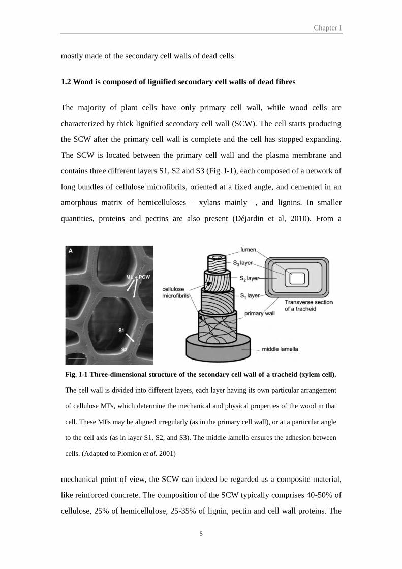
contains three different layers S1, S2 and S3 (Fig. I-1), each composed of a network of
long bundles of cellulose microfibrils, oriented at a fixed angle, and cemented in an
amorphous matrix of hemicelluloses – xylans mainly –, and lignins. In smaller
quantities, proteins and pectins are also present (Déjardin et al, 2010). From a
mechanical point of view, the SCW can indeed be regarded as a composite material,
like reinforced concrete. The composition of the SCW typically comprises 40-50% of
cellulose, 25% of hemicellulose, 25-35% of lignin, pectin and cell wall proteins. The
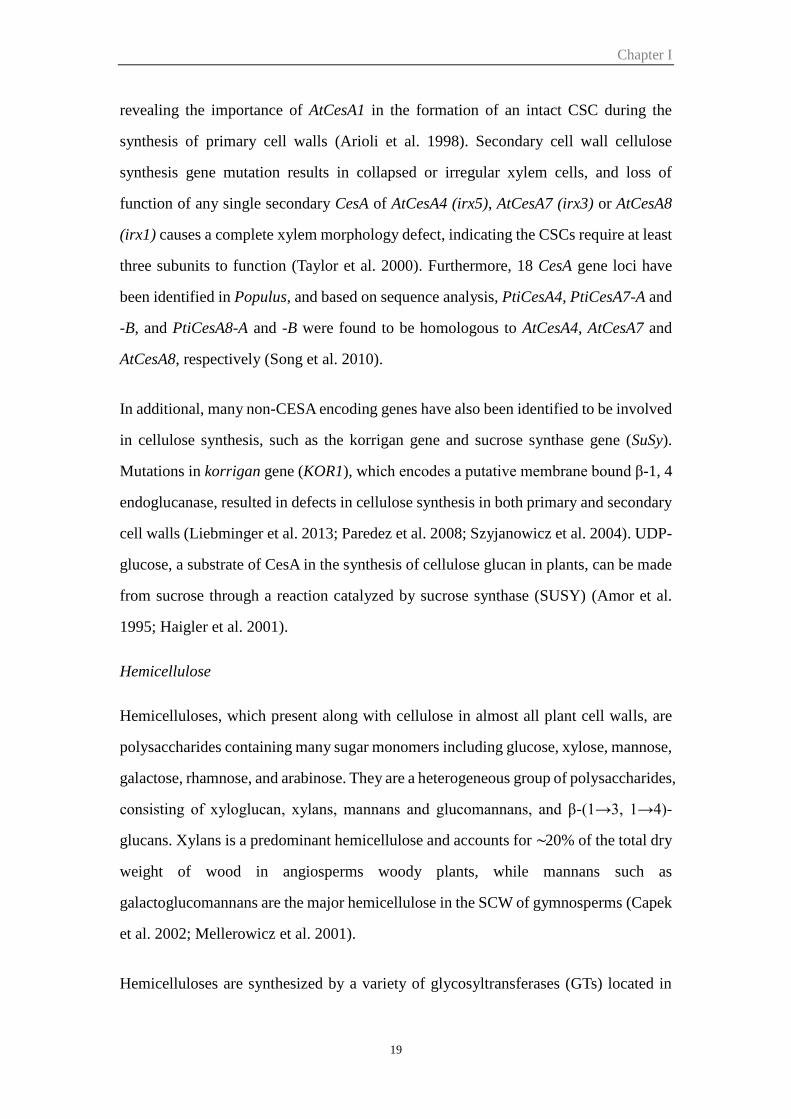
Fig. I-1 Three-dimensional structure of the secondary cell wall of a tracheid (xylem cell).
The cell wall is divided into different layers, each layer having its own particular arrangement
of cellulose MFs, which determine the mechanical and physical properties of the wood in that
cell. These MFs may be aligned irregularly (as in the primary cell wall), or at a particular angle
to the cell axis (as in layer S1, S2, and S3). The middle lamella ensures the adhesion between
cells. (Adapted to Plomion et al. 2001)
Chapter I
6
three layers differ from one another with respect to thickness and the orientation of their
cellulose microfibrils (Plomion et al. 2001). The microfibril angle (MFA) in the S2 layer,
which is the thickest one, is a parameter widely used in wood technology. The
biosynthesis of the main polymers will be presented below.
1.3 Economic importance and end-uses of wood
Wood represents the most important natural and endlessly renewable resource for
humans providing timber (e.g. for house building, furniture, packaging), fibres (for pulp,
paper, plywood) and energy (firewood). It is also an important sink for excess
atmospheric CO2, one of the major causes of global warming due to the greenhouse
effect (Demura and Fukuda 2007). Wood is expected to play a significant role in the
future as a renewable and environmentally cost effective alternative to fossil fuels. As
the world population is predicted to reach over 9 billion in 2050, the global demand for
wood for renewable energy, building and pulp and paper will grow rapidly (Mauriat et
al, 2014).
The major obstacle to the use of lignocellulosic biomass for the pulp industry and for
bioethanol production resides in its recalcitrance to degradation, due to the structure
and composition of the lignified secondary cell walls (SCW) (Séguin 2011). For the
plant, the presence of lignin confers rigidity and also protects the cell wall
polysaccharides from pathogens and microbial degradation (Vanholme et al. 2010).
This high resistance to degradation is also one of the most important industrial
limitations, where lignin impairs the accessibility of cellulose during kraft pulping as
well as during saccharification, reducing in the latter the yield of fermentable sugars
and rendering the whole process costly. The economic importance of wood for the pulp
industry and more recently for the production of bioethanol have driven many
researches worldwide aiming at improving SCW degradation (Pauly and Keegstra 2008;
Burton and Fincher 2014; Carpita 2012).
Chapter I
7
2. Wood plasticity
Trees are long-living organisms with a sessile lifestyle, which develop in a variable
environment and are subjected to developmental control. As a consequence, wood is a
complex and highly variable tissue, the formation of which is developmentally and
environmentally regulated (Plomion et al, 2001). Wood structure and composition show
large differences not only between hardwoods and softwood and between different tree
species, but individual trees also exhibit huge variations in their wood properties. The
variability occurs at the tissue level (proportion of different cell types) as well as at the
individual cell level (size, shape, wall structure, texture and chemical composition).
Anatomical, chemical and physical differences in wood characteristics are not only
common from tree to tree within the same area, but are also present within a single tree
(Plomion et al., 2001). This difference in wood properties is important for the end-uses
of wood as a raw material. Wood plasticity occurs during development and in responses
to environmental cues.
Developmental wood plasticity:
The age of the cambium has an important effect on the type of wood produced. Indeed,
juvenile wood is formed during the rapid early growth of a tree (usually first 10–12
years of the tree life but depending on the species). The young cambium located in the
crown of adult trees also produces juvenile wood. Once this period is finished another
type of wood, called mature wood, is produced by the mature cambium (Mauriat et al,
2014). Mature wood differs from juvenile wood by having thicker cell walls, narrower
cell lumens, larger cellulose microfibril angles and a higher specific density. In terms
of chemical composition, mature wood shows higher cellulose and lower lignin
contents (Mauriat et al, 2014; Zobel and Sprague 1998).
Environmental plasticity:
Environmental cues trigger the formation of different types of wood, which can be
found within a single tree genotype.

Chapter I
8
In reaction to gravitropic (wind, slope) or light stimuli, trees develop a reaction wood
at the upper side of the inclined axes in order to reorient their axes, trunks and branches,
and to allow their adaptation and harmonious development in their environment (Pilate
et al, 2004). In angiosperm trees, it is called tension wood (TW), because it is formed
in zones of the tree held in tension – the upper side of a leaning stem. At the
macroscopic level, a high eccentricity is often observed in the transverse section of stem,
suggesting that cell divisions are activated on tension wood side. Tension wood
generally has fewer, smaller vessels whereas fibres are significantly longer. The most
obvious and striking feature of TW is the presence in fibres of an inner gelatinous cell-
wall layer. This so-called G-layer consists of almost pure crystalline cellulose in parallel
microfibrils to the long cell axis (Jourez et al. 2001). The lignin content of the TW cell
walls is generally decreased (Pilate et al. 2004; Furuy et al. 1970; Habrant et al. 2003).
The changes in the biophysical properties and morphology of reaction wood cell walls
have a negative impact on the quality of wood products and their potential industrial
uses. For instance, TW is a problem for the solid wood industry as it increases
longitudinal, radial and tangential shrinkage during the drying process (Plomion et al.,
2001).
Nitrogen availability has been described to influence growth and development as well
as xylogenesis. Fiber morphology, SCW structure and composition were modified in
response to high N supply, including lignification pattern (Pitre et al. 2007). Further
mRNA profiles analysis showed that nitrogen fertilization had overlapping effects with
tension wood formation (Pitre et al. 2010). Moreover, using pedigree of pseudo-
backcrossed hybrid poplar (Populus trichocarpa × Populus deltoides), Novaes et al.
(2009) have shown that N fertilization significantly increased all growth traits as well
as the amount of cellulose and hemicelluloses in the wood whereas a decrease of the
lignin content was observed.
Other environmental cues such as temperature, drought have been shown to impact
wood properties (Gindl et al. 2014; Searson et al. 2004; Moura et al. 2010).
Chapter I
9
3. Wood develops during secondary growth
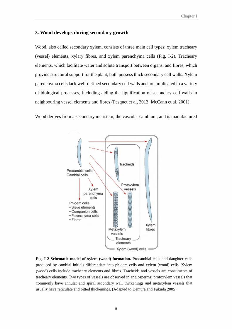
Wood, also called secondary xylem, consists of three main cell types: xylem tracheary
(vessel) elements, xylary fibres, and xylem parenchyma cells (Fig. I-2). Tracheary
elements, which facilitate water and solute transport between organs, and fibres, which
provide structural support for the plant, both possess thick secondary cell walls. Xylem
parenchyma cells lack well-defined secondary cell walls and are implicated in a variety
of biological processes, including aiding the lignification of secondary cell walls in
neighbouring vessel elements and fibres (Pesquet et al, 2013; McCann et al. 2001).
Wood derives from a secondary meristem, the vascular cambium, and is manufactured
Fig. I-2 Schematic model of xylem (wood) formation. Procambial cells and daughter cells
produced by cambial initials differentiate into phloem cells and xylem (wood) cells. Xylem
(wood) cells include tracheary elements and fibres. Tracheids and vessels are constituents of
tracheary elements. Two types of vessels are observed in angiosperms: protoxylem vessels that
commonly have annular and spiral secondary wall thickenings and metaxylem vessels that
usually have reticulate and pitted thickenings. (Adapted to Demura and Fukuda 2005)
Chapter I
10
by a succession of four major steps, including cell division, cell expansion (elongation
and radial enlargement), secondary cell wall thickening (involving cellulose,
hemicellulose, cell wall proteins, and lignin biosynthesis and deposition) (Plomion et
al. 2001). In addition, vessels undergo programmed cell death (PCD), resulting in a
continuous system of adjoining hollow cells that function in water/solute transport.
During fibre development, PCD is delayed, allowing for more extensive thickening and
lignification of secondary cell walls, consistent with a primary role for this cell type in
providing structural support.
The vascular cambium is derived from the procambium, and parenchyma cells that
regain the capacity to divide and form a layer of meristematic cells located between the
primary xylem and phloem in the vascular bundles. Whereas the procambium provides
a source of vascular stem cells during primary growth, the vascular cambium ensures
the same role during secondary growth, a prominent phenomena in woody plants where
it is responsible for the diametral growth of the plant. It is especially important for the
development of trees, ensuring their perennial life through the regular renewal of
functional xylem and phloem. An increase in the amount of vascular tissues mediated
by cambium is one of the characteristics that distinguish dicotyledons and
gymnosperms from monocots.
In woody species, the vascular cambium presents unique characteristics since it is
composed of two distinct cell types, the fusiform initials and the ray initials, which
differentiate into several particular cell types closely interconnected to constitute the
secondary xylem or wood, a complex tridimensional tissue (Chaffey, 2002). The
fusiform initials divide length-wise and produce secondary vascular tissues through
periclinal divisions in a position-dependent manner: on the inner side, wood elements
(mostly tracheids in gymnosperms, but also vessel elements, vessel-associated cells,
axial parenchyma and fibers in dicotyledons) and, on the outer side, phloem cells (sieve
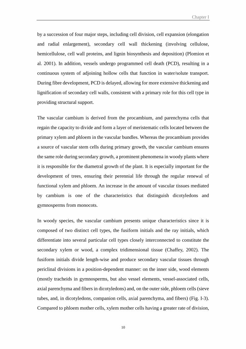
tubes, and, in dicotyledons, companion cells, axial parenchyma, and fibers) (Fig. I-3).
Compared to phloem mother cells, xylem mother cells having a greater rate of division,
Chapter I
11
so there is more secondary xylem than phloem in the tree. Anticlinal (radial) divisions
of the fusiform initials also produce daughter cells similar to mother cells and ensure
the harmonious increase in circumference of the cambium. Radial initials give rise to
rays that are essential to the translocation of nutrients between phloem and xylem (Du
and Groover 2010; Plomion et al. 2001).
The economic importance of secondary growth and wood formation has focused
considerable research attention on the function of the vascular cambium in tree species,
but this question has also been actively studied in Arabidopsis thaliana. With several
advantages such as genomic resources, Arabidopsis has emerged as a useful model for
investigating the secondary growth.
Fig. I-3 Terminology of wood-forming tissues. (Schrader et al. 2003)

Chapter I
12
4. Arabidopsis as a model to study secondary growth
Although this herbaceous plant does not normally undergo extensive secondary growth,
two regions of vascular cambium—zones of fascicular cambium and the neighbouring
zones of interfascicular cambium—are found within the Arabidopsis inflorescence
stem (Fig. I-4). Despite their proximity to each other and their apparent similarity, these
two cambial niches have different developmental origins. The fascicular cambium is
derived from the procambium that developed within the original vascular tissue as it
was formed during the primary growth of the stem. It is not surprising, therefore, that
radial differentiation of fascicular cambial cells gives rise to the full range of both xylem
and phloem cell types. The inflorescence stem fascicular cambium can thus be
considered functionally analogous to the circumferential vascular cambium of woody
plants.
The interfascicular cambium, on the other hand, is thought to arise through the de novo
recruitment of interfascicular parenchyma cells as primary growth in the stem slows. It
represents a specialized vascular meristem that gives rise exclusively to the structurally
important interfascicular fibres.
Fig. I-4 Schematic illustration of the primary and secondary stem anatomy in Arabidopsis.
The primary stem exhibits disconnected vascular bundles with procambium. In the secondary
developmental phase, this procambium turns into a fascicular cambium and the cells between
bundles become an interfascicular cambium. Fascicular and interfascicular cambia interconnect
to each other and establish a cambium in a circular form. (Miyashima et al. 2013)
Chapter I
13
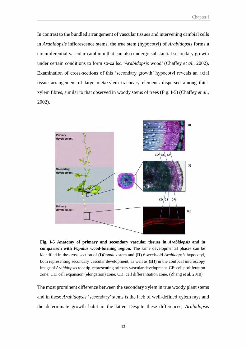
In contrast to the bundled arrangement of vascular tissues and intervening cambial cells
in Arabidopsis inflorescence stems, the true stem (hypocotyl) of Arabidopsis forms a
circumferential vascular cambium that can also undergo substantial secondary growth
under certain conditions to form so-called ‘Arabidopsis wood’ (Chaffey et al., 2002).
Examination of cross-sections of this ‘secondary growth’ hypocotyl reveals an axial
tissue arrangement of large metaxylem tracheary elements dispersed among thick
xylem fibres, similar to that observed in woody stems of trees (Fig. I-5) (Chaffey et al.,
2002).
The most prominent difference between the secondary xylem in true woody plant stems
and in these Arabidopsis ‘secondary’ stems is the lack of well-defined xylem rays and
the determinate growth habit in the latter. Despite these differences, Arabidopsis
Fig. I-5 Anatomy of primary and secondary vascular tissues in Arabidopsis and in
comparison with Populus wood-forming region. The same developmental phases can be
identified in the cross section of (I)Populus stem and (II) 6-week-old Arabidopsis hypocotyl,
both representing secondary vascular development, as well as (III) in the confocal microscopy
image of Arabidopsis root tip, representing primary vascular development. CP: cell proliferation
zone; CE: cell expansion (elongation) zone; CD: cell differentiation zone. (Zhang et al. 2010)
Chapter I
14
provides a useful model for understanding the process of secondary growth in woody
plants.
5. Regulation of cambium activity and wood specification
Plant hormones play important roles to regulate wood formation. For example, ethylene
and cytokinin acts as positive regulators of wood formation, promoting cambial cell
proliferation (Love et al. 2009; Nieminen et al. 2008); gibberellin promotes both cell
division and xylem fibers elongation (Mauriat and Moritz 2009); Auxin is involved in
different steps: formation and maintenance of the pro(cambium), cell division and cell
specification. Its role will be examined in more details in a specific paragraph. It is
worth noting that the different hormones also show crosstalking in control cambial
growth and differentiation (Sorce et al. 2013).
5.1 Regulation of cambium identity and activity
The procambium is formed and maintained by directed auxin flow in the precursors of
procambium cells (Donner et al. 2009; Ohashi-Ito and Fukuda 2010). In the leaf veins,
auxin flow is directed by polar localization of an auxin efflux carrier, PINFORMED 1
(PIN1) and the expression of PIN1 precedes differentiation of the procambium
precursor cells (Donner et al. 2009). ATHB8, a class III HD-ZIP transcription factor, is
required for preprocambial development and procambium differentiation, and its
expression depends on the activity of the auxin response factor MONOPTEROS (MP)
(Donner et al. 2009). There are five HD-ZIP III genes in Arabidopsis, PHB/ATHB14,
PHV/ATHB9, REV/IFL1, ATHB8 and CNA/ATHB15. They have been shown to regulate
the number of procambium cells by promoting xylem differentiation during vascular
development (Baima et al. 2001; Carlsbecker et al. 2010; Ilegems et al. 2010).
The maintenance of the pluripotent identity of the cambium is crucial for continuous
meristem activity. Current evidence indicates that a similar molecular mechanism
regulating shoot apical meristem (SAM) and root apical meristem (RAM) is likely
Chapter I
15
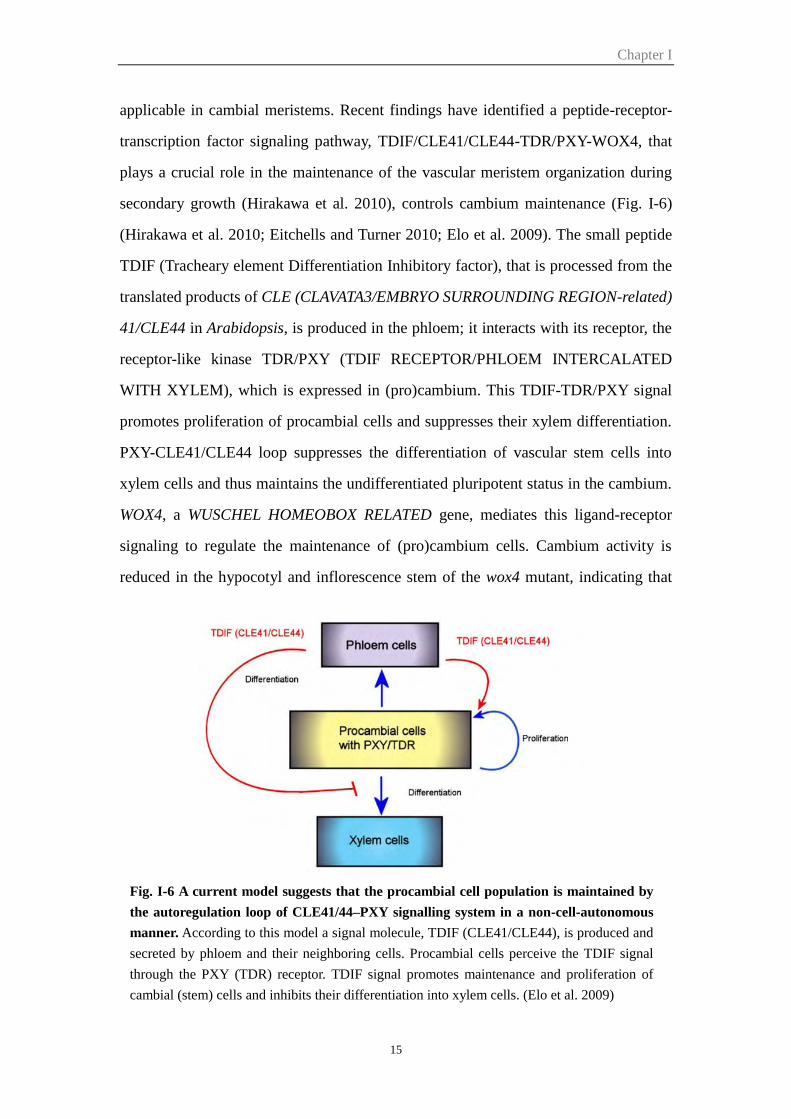
applicable in cambial meristems. Recent findings have identified a peptide-receptor-
transcription factor signaling pathway, TDIF/CLE41/CLE44-TDR/PXY-WOX4, that
plays a crucial role in the maintenance of the vascular meristem organization during
secondary growth (Hirakawa et al. 2010), controls cambium maintenance (Fig. I-6)
(Hirakawa et al. 2010; Eitchells and Turner 2010; Elo et al. 2009). The small peptide
TDIF (Tracheary element Differentiation Inhibitory factor), that is processed from the
translated products of CLE (CLAVATA3/EMBRYO SURROUNDING REGION-related)
41/CLE44 in Arabidopsis, is produced in the phloem; it interacts with its receptor, the
receptor-like kinase TDR/PXY (TDIF RECEPTOR/PHLOEM INTERCALATED
WITH XYLEM), which is expressed in (pro)cambium. This TDIF-TDR/PXY signal
promotes proliferation of procambial cells and suppresses their xylem differentiation.
PXY-CLE41/CLE44 loop suppresses the differentiation of vascular stem cells into
xylem cells and thus maintains the undifferentiated pluripotent status in the cambium.
WOX4, a WUSCHEL HOMEOBOX RELATED gene, mediates this ligand-receptor
signaling to regulate the maintenance of (pro)cambium cells. Cambium activity is
reduced in the hypocotyl and inflorescence stem of the wox4 mutant, indicating that
Fig. I-6 A current model suggests that the procambial cell population is maintained by
the autoregulation loop of CLE41/44–PXY signalling system in a non-cell-autonomous
manner. According to this model a signal molecule, TDIF (CLE41/CLE44), is produced and
secreted by phloem and their neighboring cells. Procambial cells perceive the TDIF signal
through the PXY (TDR) receptor. TDIF signal promotes maintenance and proliferation of
cambial (stem) cells and inhibits their differentiation into xylem cells. (Elo et al. 2009)
Chapter I
16
WOX4 regulates, but is not required to establish, the meristem (Hirakawa et al. 2010;
Suer et al. 2011). Another WOX-family gene, WOX14, acts redundantly with WOX4 to
regulate cambial cell proliferation (Etchells et al. 2013). The TDIF/CLE41/CLE44-
TDR/PXY-WOX4 signaling pathway appears to act downstream of auxin signaling in
regulating cambial cell proliferation and seems to be evolutionarily conserved between
both woody and herbaceous species, as it has been described in both Arabidopsis and
Populus (Schrader et al. 2004). Two other receptor-like kinases, REDUCED IN
LATERAL GROWTH1 (RUL1) and MORE LATERAL GROWTH1 (MOL1), were
also found to be activator and repressor of cambium activity, respectively (Agusti et al.
2011b).
In Arabidopsis, class I KNOX transcription factors are major regulators of SAM activity.
Plants overexpressing a class I KNOX gene, BREVIPEDICELLUS (BP)/KNAT1,
exhibited impaired lignin deposition while bp mutants show ectopic lignification in the
inflorescence stem, indicating that BP regulates xylem cell differentiation during
vascular development (Mele et al. 2003). In Populus, an ortholog of BP,
ARBORKNOX2 (ARK2), is expressed in both SAM and cambium region.
Overexpression of ARK2 in Populus results in the expansion of the cambium region
and inhibition of differentiation of tracheary elements and fibres in secondary xylem as
well as phloem fibres. Knock-down of ARK2 results in early appearance of lignified
secondary xylem and thicker SCWs (Du et al. 2009).
KANADI genes (KAN1-4) belong to one clade of the GARP transcription factor family.
Gain-of-function KANADI alleles result in a loss of cambium activity, while loss-of-
function result in increased cambium activity. For example, ectopic KAN1 expression
results in a complete loss of vascular tissue development. KANADI and Class III HD-
ZIP show function mutual antagonism through effects on the auxin flow (Ilegems et al.
2010).
Chapter I
17
5.2 Regulation of xylem specification
Several Arabidopsis mutants defective in the radial patterning of vascular tissues also
have defects in the radial (abaxial–adaxial) patterning of leaves, suggesting the
involvement of HD-ZIP III and KANADI transcription factors in the xylem cell type
patterning in root and stem (McConnell et al. 2001; Kerstetter et al. 2001; Emery et al.
2003; Cano-Delgado et al. 2010). Class III homeodomain-leucine zipper (HD-ZIP III)
transcription factors, including AtHB8, CORONA/AtHB15 (CNA), PHABULOSA
(PHB), PHAVOLUTA (PHV), and REVOLUTA (REV), are expressed in procambium,
cambium and developing xylem, and play important roles in cambium and xylem
differentiation in Arabidopsis. Loss-of-function mutants rev plants display disruption
of the differentiation of interfascicular fibers and secondary xylem, while AtTHB8 and
AtHB15 appear to have functions antagonistic to the REV in vascular formation (Prigge
et al. 2005). HD-ZIP III gene expression is positively regulated by auxin, and the
expression of AtHB8 is directly activated by the auxin-response transcription factor
MONOPTEROS (MP) at Arabidopsis preprocambial stages (Donner et al. 2009). The
stability of transcripts are regulated by microRNA165/166, and all the gain-of-function
mutations disrupt the miR165/166 target sequence (Demura and Fukuda 2007). In
additional, brassinosteroids can also activate HD-ZIP III expression and thus promote
xylem differentiation (Ohashi-Ito and Fukuda 2003). In Populus, knock down of
POPCORONA results in abnormal lignification, while overexpression of a miRNA-
resistant POPCORONA results in delayed lignification of xylem and phloem fibres
during secondary growth (Du et al. 2011). When the microRNA resistant form of
popREVOLUTA (PRE) (orthologous to AtREV) is overexpressed, it leads to abnormal
cambium formation in the cortex (Robischon et al. 2011).
Other transcription factors such as class I KNOX and bHLH TFs are also involved in
secondary growth. Populus ARK2, the ortholog of Arabidopsis BP/KNAT1, is expressed
in both SAM and cambium region, and ARK2 overexpression Populus displays
expansion of the cambium region and inhibited xylem differentiation. Knock-down of
Chapter I
18
ARK2 results in early appearance of lignified secondary xylem and thicker SCWs (Du
et al. 2009). TMO quadruple mutants completely lost vascular tissue in roots, and TMO5
and LHW co-expression plants induced dramatic periclinal divisions within the
vasculature of roots (De Rybel et al. 2013).
6. Secondary cell wall formation and its transcriptional regulation
6.1 Biosynthesis of the three main polymers
Cellulose
Cellulose, as a major structural of the cell wall, is the most abundant biopolymer
synthesized on land. It is consists of several linear polymer chains of ß-1, 4 linked
glucose residues. The fundamental structure units of cellulose are the microfibrils
(MFs), which are formed through intra- and inter-chain hydrogen bonds between the
different parallel linear glucan chains. In higher plants, the synthesis of cellulose is
believed to be catalyzed by cellulose synthase (CesA), organizing into cellulose
synthase complexes (CSCs) localized on the plasma membrane (Brett 2000; Saxena
and Brown 2005). CSCs exist as rosettes, containing six subunits arranged in a
hexagonal structure, and it has been postulated that each of the six rosette subunits
contains six cellulose synthase (CesA) proteins. It means that each rosette therefore has
a total of 36 CesA proteins (Doblin et al. 2002; Li et al. 2014).
CesAs are intEgral plasma membrane proteins with multiple transmembrane domains
and a central catalytic domain (Sethaphong et al. 2013; Slabaugh et al. 2014). The
Arabidopsis genome encodes ten CesA genes, which are roughly classified into two
groups: AtCesA1, AtCesA3, AtCesA6, AtCesA2, AtCesA5 and AtCesA9 are involved in
primary cell walls cellulose synthesis (Li et al. 2014); while AtCesA4, AtCesA7 and
AtCesA8 are required for cellulose synthesis in secondary cell walls (Taylor et al. 2003).
Among primary CesAs, AtCesA1 and AtCesA3 are essential, while AtCesA6 is
redundant with AtCesA2, AtCesA5 and AtCesA9. For example, the temperature
sensitive mutant, radial swelling root 1 (rsw1) caused the rosette to disintegrate,
Chapter I
19
revealing the importance of AtCesA1 in the formation of an intact CSC during the
synthesis of primary cell walls (Arioli et al. 1998). Secondary cell wall cellulose
synthesis gene mutation results in collapsed or irregular xylem cells, and loss of
function of any single secondary CesA of AtCesA4 (irx5), AtCesA7 (irx3) or AtCesA8
(irx1) causes a complete xylem morphology defect, indicating the CSCs require at least
three subunits to function (Taylor et al. 2000). Furthermore, 18 CesA gene loci have
been identified in Populus, and based on sequence analysis, PtiCesA4, PtiCesA7-A and
-B, and PtiCesA8-A and -B were found to be homologous to AtCesA4, AtCesA7 and
AtCesA8, respectively (Song et al. 2010).
In additional, many non-CESA encoding genes have also been identified to be involved
in cellulose synthesis, such as the korrigan gene and sucrose synthase gene (SuSy).
Mutations in korrigan gene (KOR1), which encodes a putative membrane bound β-1, 4
endoglucanase, resulted in defects in cellulose synthesis in both primary and secondary
cell walls (Liebminger et al. 2013; Paredez et al. 2008; Szyjanowicz et al. 2004). UDP-
glucose, a substrate of CesA in the synthesis of cellulose glucan in plants, can be made
from sucrose through a reaction catalyzed by sucrose synthase (SUSY) (Amor et al.
1995; Haigler et al. 2001).
Hemicellulose
Hemicelluloses, which present along with cellulose in almost all plant cell walls, are
polysaccharides containing many sugar monomers including glucose, xylose, mannose,
galactose, rhamnose, and arabinose. They are a heterogeneous group of polysaccharides,
consisting of xyloglucan, xylans, mannans and glucomannans, and β-(1→3, 1→4)-
glucans. Xylans is a predominant hemicellulose and accounts for ∼20% of the total dry
weight of wood in angiosperms woody plants, while mannans such as
galactoglucomannans are the major hemicellulose in the SCW of gymnosperms (Capek
et al. 2002; Mellerowicz et al. 2001).
Hemicelluloses are synthesized by a variety of glycosyltransferases (GTs) located in
Chapter I
20
the Golgi membranes. In Arabidopsis, many GT genes have been identified involved
in xylan biosynthesis through the characterization of collapsed xylem Arabidopsis
mutants, such as AtFRA8/IRX7, AtIRX8, AtIRX14, AtIRX14-L, AtF8H, AtPARVUS,
AtIRX10 and AtIRX10-Like (IRX10-L), AtIRX9, AtIRX9-L. AtIRX9, AtIRX14, AtIRX10
and AtIRX10-L are involved in synthesis of the β-D-(1→4)-xylan backbone elongation,
while the AtFRA8/IRX7, AtIRX8, AtF8H, and PARVUS are thought to play a role in
forming this oligosaccharide (Brown et al. 2009; Lee et al. 2007a; Lee et al. 2007b;
Pauly et al. 2013; Persson et al. 2007; Wu et al. 2009). In poplar, a few GT genes
involved in xylan biosynthesis have also been studied. PoGT47C, PoGT8D and
PoGT43B show high sequence similarity to AtFRA8, AtIRX8 and AtIRX9, respectively,
and it has also been shown that PoGT43B and PoGT47C can rescue the defects of
Arabidopsis Atirx9 and Atfra8 mutants, respectively. Moreover, poplar GT8E,
GT8F/PdGATL1.1 and PdGATL1.2 are identified as functional orthologs of
Arabidopsis PARVUS since they are able to rescue the Arabidopsis irregular xylem
phenotype (Kong et al. 2009; Lee et al. 2009a, b).
Lignin
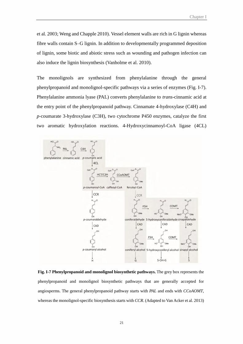
Lignin is phenolic biopolymer produced by the dehydrogenative polymerization of
essentially three different hydroxycinnamyl alcohols, the p-coumaryl, coniferyl, and
sinapyl alcohols, that differ in the degree of methoxylation at the C3 and C5 positions
of the aromatic ring giving raise to p-hydroxyphenyl (H), guaiacyl (G), syringyl (S),
and units, respectively (Fig. I-7) (Baucher et al. 2003).
Lignin embeds the polysaccharide matrix of the SCW to make them rigid and
impervious. Lignin content and monomeric composition vary widely among different
species, individuals, cell types, and cell wall layers and are influenced by
developmental and environmental cues. In general, lignins from gymnosperms and
related species are rich in G units and contains low amounts of H units, whereas dicots
lignins are mainly composed of G and S units (Weng and Chapple 2010). H-units are
elevated in softwood compression wood and may be slightly higher in grasses (Boerjan
Chapter I
21
et al. 2003; Weng and Chapple 2010). Vessel element walls are rich in G lignin whereas
fibre walls contain S–G lignin. In addition to developmentally programmed deposition
of lignin, some biotic and abiotic stress such as wounding and pathogen infection can
also induce the lignin biosynthesis (Vanholme et al. 2010).
The monolignols are synthesized from phenylalanine through the general
phenylpropanoid and monolignol-specific pathways via a series of enzymes (Fig. I-7).
Phenylalanine ammonia lyase (PAL) converts phenylalanine to trans-cinnamic acid at
the entry point of the phenylpropanoid pathway. Cinnamate 4-hydroxylase (C4H) and
p-coumarate 3-hydroxylase (C3H), two cytochrome P450 enzymes, catalyze the first
two aromatic hydroxylation reactions. 4-Hydroxycinnamoyl-CoA ligase (4CL)
Fig. I-7 Phenylpropanoid and monolignol biosynthetic pathways. The grey box represents the
phenylpropanoid and monolignol biosynthetic pathways that are generally accepted for
angiosperms. The general phenylpropanoid pathway starts with PAL and ends with CCoAOMT,
whereas the monolignol-specific biosynthesis starts with CCR. (Adapted to Van Acker et al. 2013)
Chapter I
22
activates p-coumaric acid to the activated thioester form p-coumaroyl CoA, which
represents the most important branchpoint within the central phenylpropanoid
biosynthesis in plants (Vogt 2010). Hydroxycinnamoyl-CoA: shikimate
hydroxycinnamoyl transferase (HCT), which belongs to the BAHD acyltransferase
superfamily, catalyzes the formation of p-coumarate esters using shikimate or quinate
as an acyl acceptor. Caffeoyl-CoA O-methyltransferase (CCoAOMT) catalyzes the first
transmethylation reaction in the phenylpropanoid pathway, synthesizing feruloyl-CoA
from caffeoyl-CoA (Fig. I-7). The first step of the monolignol-specific pathway starts
with cinnamoyl-CoA reductase (CCR) (Van Acker et al. 2014). CCR and Cinnamyl
alcohol dehydrogenase (CAD), two oxidoreductases, convert the hydroxycinnamoyl-
CoA esters to their corresponding alcohols (Fig. I-7). Ferulate 5-hydroxylase (F5H) and
caffeic acid O-methyltransferase (COMT) are two S lignin biosynthesis specific
enzymes.
The different monolignol subunits are synthesized in the cytoplasm and then
transported to the cell wall for subsequent polymerization (Wang et al. 2013). Recently,
it has been reported that AtABCG29, an ATP-binding cassette transporter, acts as a p-
coumaryl alcohol transporter (Alejandro et al. 2012). However, as a large transporter
gene family in plant, ABC transporters for coniferyl alcohol and sinapyl alcohol remain
to be identified and characterized (Wang et al. 2013). Following transport to the
secondary cell wall, monolignols are oxidized and subsequently polymerized by
peroxidases and laccases to form lignin (Baucher et al. 2003).
6.2 The SCW transcriptional network
The SCW formation is a critical step of wood formation. Through the recent studies in
Arabidopsis as well as other species such as Populus, Eucalyptus and pine, several key
transcriptional switches have been identified to regulate the entire differentiation
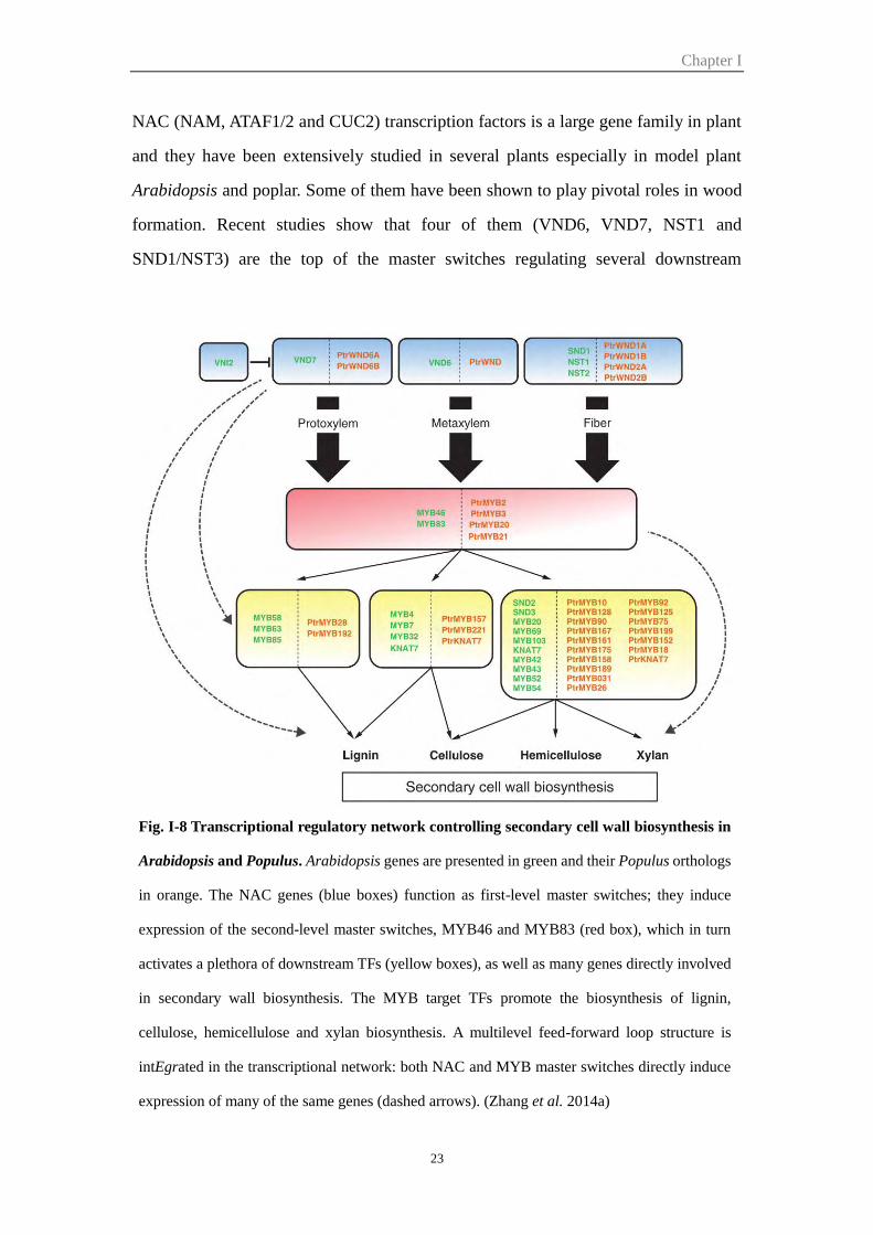
process. A simplified SCW regulatory network is shown in Fig. I-8.
NAC transcription factors
Chapter I
23
NAC (NAM, ATAF1/2 and CUC2) transcription factors is a large gene family in plant
and they have been extensively studied in several plants especially in model plant
Arabidopsis and poplar. Some of them have been shown to play pivotal roles in wood
formation. Recent studies show that four of them (VND6, VND7, NST1 and
SND1/NST3) are the top of the master switches regulating several downstream
Fig. I-8 Transcriptional regulatory network controlling secondary cell wall biosynthesis in
Arabidopsis and Populus. Arabidopsis genes are presented in green and their Populus orthologs
in orange. The NAC genes (blue boxes) function as first-level master switches; they induce
expression of the second-level master switches, MYB46 and MYB83 (red box), which in turn
activates a plethora of downstream TFs (yellow boxes), as well as many genes directly involved
in secondary wall biosynthesis. The MYB target TFs promote the biosynthesis of lignin,
cellulose, hemicellulose and xylan biosynthesis. A multilevel feed-forward loop structure is
intEgrated in the transcriptional network: both NAC and MYB master switches directly induce
expression of many of the same genes (dashed arrows). (Zhang et al. 2014a)
Chapter I
24
transcription factors which lead to the secondary cell wall formation. VASCULAR-
RELATED NAC-DOMAIN6 (VND6) and VND7 are transcription switches for plant
metaxylem and protoxylem vessel formation, respectively (Kubo et al. 2005), while
NAC SECONDARY WALL THICKENING PROMOTING FACTOR1 (NST1) and
SECONDARY WALL-ASSOCIATED NAC DOMAIN PROTEIN1 (SND1/NST3) are
two mater regulators promoting fiber differentiation (Mitsuda et al. 2007; Zhong et al.
2007). The other two NAC transcription factors SND2 and SND3, which are indirect
and direct targets of SND1 respectively, positively regulate the secondary wall
thickness of both xylary fibers and interfascicular fibers (Hussey et al. 2011; Zhong et
al. 2008). In Populus, PtrWND (PtVNS) genes, homologs of the four Arabidopsis NAC
transcription factors, which are highly expressed in developing secondary xylem tissue,
has been shown to be master switches activating the secondary cell wall biosynthesis
(Ohtani et al. 2011; Zhong and Ye 2010).
MYB transcription factors
Several MYB transcription factors have also been shown to be important regulators of
secondary cell wall formation. First, Eucalyptus EgMYB2 was found to bind the
EgCCR and EgCAD2 gene regulatory regions and act as a positive regulator of
secondary cell wall formation and lignin biosynthesis (Goicoechea et al. 2005). Then
in Arabidopsis, the MYB proteins have been identified as the direct target of secondary
wall NACs master switches regulating secondary wall biosynthesis. For example,
MYB46 and MYB83, both of which are SND1 direct targets, were found to function
redundantly as second-level master switches regulating secondary wall biosynthesis.
Over-expression of MYB46 or 83 in Arabidopsis induces activation of secondary wall
biosynthetic genes for cellulose, xylan and lignin and results in ectopic deposition of
secondary walls in cells that are generally parenchymatous, while mutations of MYB46
and 83 results in lack of secondary wall thickening (McCarthy et al., 2009; Zhong and
Ye, 2012). More recently, four Populus orthologs PtrMYB2, PtrMYB3, PtrMYB20 and
PtrMYB21 were also proved to be the direct target of the master regulators PtrWND
Chapter I
25
and function as second-level master switches of wood formation (McCarthy et al. 2010;
Zhong et al. 2013). In addition, other MYB transcription factors such as MYB58,
MYB63 and MYB85, were shown to be regulated by the SND1 close homologs NST1,
NST2, VND6, and VND7 and their downstream target MYB46 and/or MYB83 to
control biosynthesis of cellulose, hemicellulose, xylan and lignin (Zhong et al. 2008;
Zhou et al. 2009).
The Class II KNAT7 transcription factor
KNAT7, which has been shown to be one of the direct target of both SND1 and MYB46
(Ko et al. 2009; Zhong et al. 2008), is a regulator of secondary cell wall biosynthesis.
Loss-of-function knat7 mutants displayed exhibit both irx and enhanced fiber cell wall
thickness, while overexpression plants displayed opposite phenotype with decreased
secondary wall thickening in interfascicular fibres (Li et al. 2012). So KNAT7 acts as a
transcriptional repressor rather than an activator in regulating secondary wall
biosynthesis. KNAT7 can also interact with members of the Ovate Family Protein (OFP)
transcription co-regulators, such as OFP1 and OFP4. The ofp4 mutant exhibited similar
irx and fibre cell wall phenotypes as knat7, so it has been proposed that KNAT7 forms
a functional complex with OFP proteins to regulate aspects of secondary cell wall
formation (Li et al. 2011).
Part II Auxin-key regulators of plant growth and development
1 Auxin homeostasis
The plant hormone auxin has a crucial role in plant development, and this depends on
the graded distribution of auxin. The auxin homeostasis maintains the active auxin at
an optimal level for growth and development. This homeostasis depends on the
combined effects of all the process: biosynthesis, transport, conjugation and
degradation.
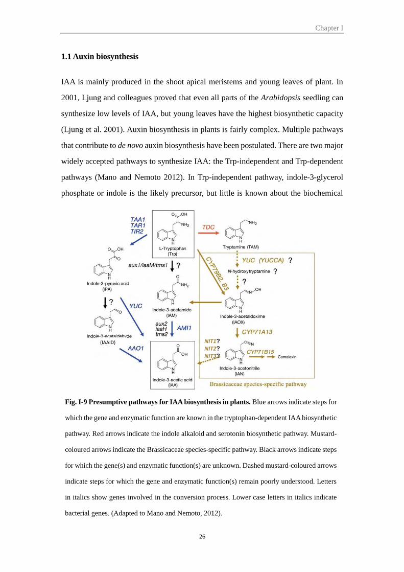
Chapter I
26
1.1 Auxin biosynthesis
IAA is mainly produced in the shoot apical meristems and young leaves of plant. In
2001, Ljung and colleagues proved that even all parts of the Arabidopsis seedling can
synthesize low levels of IAA, but young leaves have the highest biosynthetic capacity
(Ljung et al. 2001). Auxin biosynthesis in plants is fairly complex. Multiple pathways
that contribute to de novo auxin biosynthesis have been postulated. There are two major
widely accepted pathways to synthesize IAA: the Trp-independent and Trp-dependent
pathways (Mano and Nemoto 2012). In Trp-independent pathway, indole-3-glycerol
phosphate or indole is the likely precursor, but little is known about the biochemical
Fig. I-9 Presumptive pathways for IAA biosynthesis in plants. Blue arrows indicate steps for
which the gene and enzymatic function are known in the tryptophan-dependent IAA biosynthetic
pathway. Red arrows indicate the indole alkaloid and serotonin biosynthetic pathway. Mustard-
coloured arrows indicate the Brassicaceae species-specific pathway. Black arrows indicate steps
for which the gene(s) and enzymatic function(s) are unknown. Dashed mustard-coloured arrows
indicate steps for which the gene and enzymatic function(s) remain poorly understood. Letters
in italics show genes involved in the conversion process. Lower case letters in italics indicate
bacterial genes. (Adapted to Mano and Nemoto, 2012).
(IAAID) NIT1?
NIT2?
NIT3?
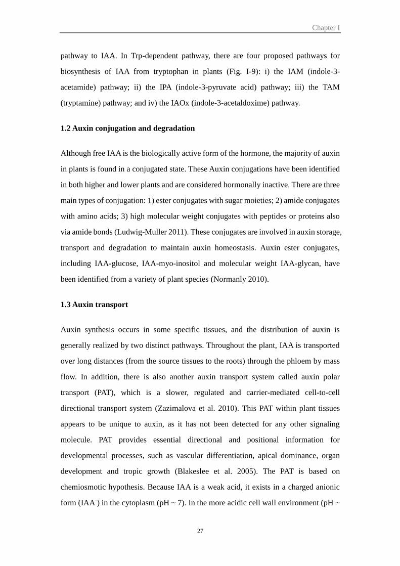
Chapter I
27
pathway to IAA. In Trp-dependent pathway, there are four proposed pathways for
biosynthesis of IAA from tryptophan in plants (Fig. I-9): i) the IAM (indole-3-
acetamide) pathway; ii) the IPA (indole-3-pyruvate acid) pathway; iii) the TAM
(tryptamine) pathway; and iv) the IAOx (indole-3-acetaldoxime) pathway.
1.2 Auxin conjugation and degradation
Although free IAA is the biologically active form of the hormone, the majority of auxin
in plants is found in a conjugated state. These Auxin conjugations have been identified
in both higher and lower plants and are considered hormonally inactive. There are three
main types of conjugation: 1) ester conjugates with sugar moieties; 2) amide conjugates
with amino acids; 3) high molecular weight conjugates with peptides or proteins also
via amide bonds (Ludwig-Muller 2011). These conjugates are involved in auxin storage,
transport and degradation to maintain auxin homeostasis. Auxin ester conjugates,
including IAA-glucose, IAA-myo-inositol and molecular weight IAA-glycan, have
been identified from a variety of plant species (Normanly 2010).
1.3 Auxin transport
Auxin synthesis occurs in some specific tissues, and the distribution of auxin is
generally realized by two distinct pathways. Throughout the plant, IAA is transported
over long distances (from the source tissues to the roots) through the phloem by mass
flow. In addition, there is also another auxin transport system called auxin polar
transport (PAT), which is a slower, regulated and carrier-mediated cell-to-cell
directional transport system (Zazimalova et al. 2010). This PAT within plant tissues
appears to be unique to auxin, as it has not been detected for any other signaling
molecule. PAT provides essential directional and positional information for
developmental processes, such as vascular differentiation, apical dominance, organ
development and tropic growth (Blakeslee et al. 2005). The PAT is based on
chemiosmotic hypothesis. Because IAA is a weak acid, it exists in a charged anionic
form (IAA-) in the cytoplasm (pH ~ 7). In the more acidic cell wall environment (pH ~
Chapter I
28
5.5), ~ 15% of the molecules are in the uncharged form (IAAH), which can enter the
cell by lipophilic diffusion across the plasma membrane (Zazimalova et al. 2010). This
pH differential between the cytoplasm and wall means that auxin can move into but not
out of the plant cells. So the auxin ion requires facilitators or transporters to exit cells
(Fig. I-10). The majority (85%) of IAA remains in its dissociated form (IAA-) and
would require a carrier for its active uptake across the cell. Three types of auxin
transporters have been studied. The AUX1/LAX (AUXIN RESISTANT 1/LIKE AUX1)
are influx auxin carriers (Bennett et al. 1996), while PIN (PIN-FORMED), and ABCB
(ATP Binding Cassette subfamily B) are efflux auxin carriers (Friml et al. 2002; Yang
and Murphy 2009). Recently, a novel PIN like family of auxin transport facilitators
termed PILS (PIN-LIKES) has been discovered by in silico studies and appears to be
involved in the regulation of auxin homeostasis in Arabidopsis (Barbez et al. 2012).
More recently, WALLS ARE THIN1 (WAT1) has also been demonstrated to be a
vacuolar auxin transport facilitator mediate auxin homoeostasis (Ranocha et al. 2013).
2 Auxin signaling
In different plant species, auxin can rapidly regulate gene expression in a few minutes
Fig. I-10 Model of intercellular auxin transport. (Adapted to Friml, 2010)
Chapter I
29
and control many plant developmental processes. Auxin acts by regulating transcription
through the action of at least three protein families called the TIR1/AFB F-box proteins,
the Aux/IAA transcriptional repressors, and the ARF transcription factors. The best
characterized pathway model is the SCFTIR1/AFB-proteasome complex mediated the
degradation of Aux/IAA which releases auxin response factor (ARF) to induce early
auxin-responsive gene expression (Chapman and Estelle 2009). Auxin signaling is also
involved in non-transcriptional regulation, but the mechanism is not as well
characterized. AUXIN BINDING PROTEIN 1 (ABP1) is the best characterized auxin
binding proteins and implicated in non-transcriptional auxin signaling.
2.1 Auxin receptors
By now, two distinct classes of auxin receptors have been recognized: TIR1 (transport
inhibitor response 1) and AFB (auxin-signaling F-box proteins) that control
transcriptional responses to auxin, and ABP1 (auxin binding protein 1), that controls a
wide variety of growth and developmental processes.
TIR1 (transport inhibitor response 1)/AFB (auxin-signaling F-box proteins)
TIR1 and additional five AFB proteins, AFB1 to AFB5, are six F-box proteins, and
function as auxin receptors (Dharmasiri et al. 2005a; Dharmasiri et al. 2005b). These
six proteins can be grouped into three subclusters according to the phylogenetic analysis,
TIR1/AFB1, AFB2/AFB3 and AFB4/AFB5. They form SCF (SKP1-Cullin-F-box)
protein complex with SUPPRESSOR OF KINETOCHORE PROTEIN 1 (SKP1 or
ASK1), CULLIN1 (CUL1) and RING BOX1 (RBX1) (Cardozo and Pagano 2004).
SCF complex is the largest family of E3 ubiquitin – protein ligases and catalyze the
ubiquitination of diverse regulatory and signalling protein destined for proteasomal
degradation.
Previous study showed TIR1/AFB1-3 function as auxin receptors and interact with
Aux/IAA in an auxin dependent manner. Genetic experiments indicate that reducing the
number of functional TIR1/AFB1-3 proteins in plant results in increasing resistance to
Chapter I
30
exogenous auxin. Comparison of single and multiple mutants in the TIR1, AFB1, AFB2,
and AFB3 genes and protein interaction analysis with Aux/IAA showed that TIR1
appears to make the largest contribution to auxin response followed by AFB2. Double,
triple and quadruple mutants showed a gradual decrease in auxin response and defects
in hypocotyl, cotyledon, root growth, leaf morphology and flow development.
Although TIR1/AFB1-3 show close relationships, their biochemically function are
distinct. For example, AFB1 and AFB2 do not rescue the tir1 mutant even regulated by
the TIR1 promoters (Parry et al. 2009).
Members of the TIR1 and AFB2 groups act as positive regulators of auxin signaling,
while both AFB4 and AFB5 function as auxin receptors based on in vitro assays
(Greenham et al. 2011; Hu et al. 2012). AFB4 and AFB5 are the major targets of the
picolinate herbicides in Arabidopsis. Genetic experiments showed that afb4-2 single
and afb4-2 afb5-5 double mutants displayed opposite effects compare to tir1 afb2
mutant, such as elongated petioles and longer hypocotyls. In addition, the afb4-2 mutant
had shorter roots and produced more lateral roots/primary root length, so it suggests
that AFB4 has a role in anchor or adventitious root production (Hu et al. 2012).
ABP1 (auxin binding protein 1)
ABP1 was discovered from maize coleoptiles 40 years ago (Hertel et al. 1972) and it
was a soluble 22kDa glycoprotein that specifically binds to auxin. The majority of
ABP1 protein localizes to the endoplasmic reticulum (ER), while some ABP1 (~22%)
is also found on the plasma membrane (Timpte 2001) and works as a functional receptor
for auxin perception. ABP1 is essential for early embryogenesis, root development, leaf
expansion, cell morphogenesis, and subcellular distribution of PIN auxin transporters.
In addition, auxin binding by ABP1 coordinates the cytoskeleton structure by regulating
the activity of two GTPases ROP2 and ROP6 (Sauer and Kleine-Vehn 2011). However,
it is not known how ABP1 transmits the auxin signal to regulate these cytoplasmic
responses, while the mechanism of the auxin recognition site is quite different from
TIR/AFB receptors. The crystal structure analysis of ABP1 suggested that ABP1 forms
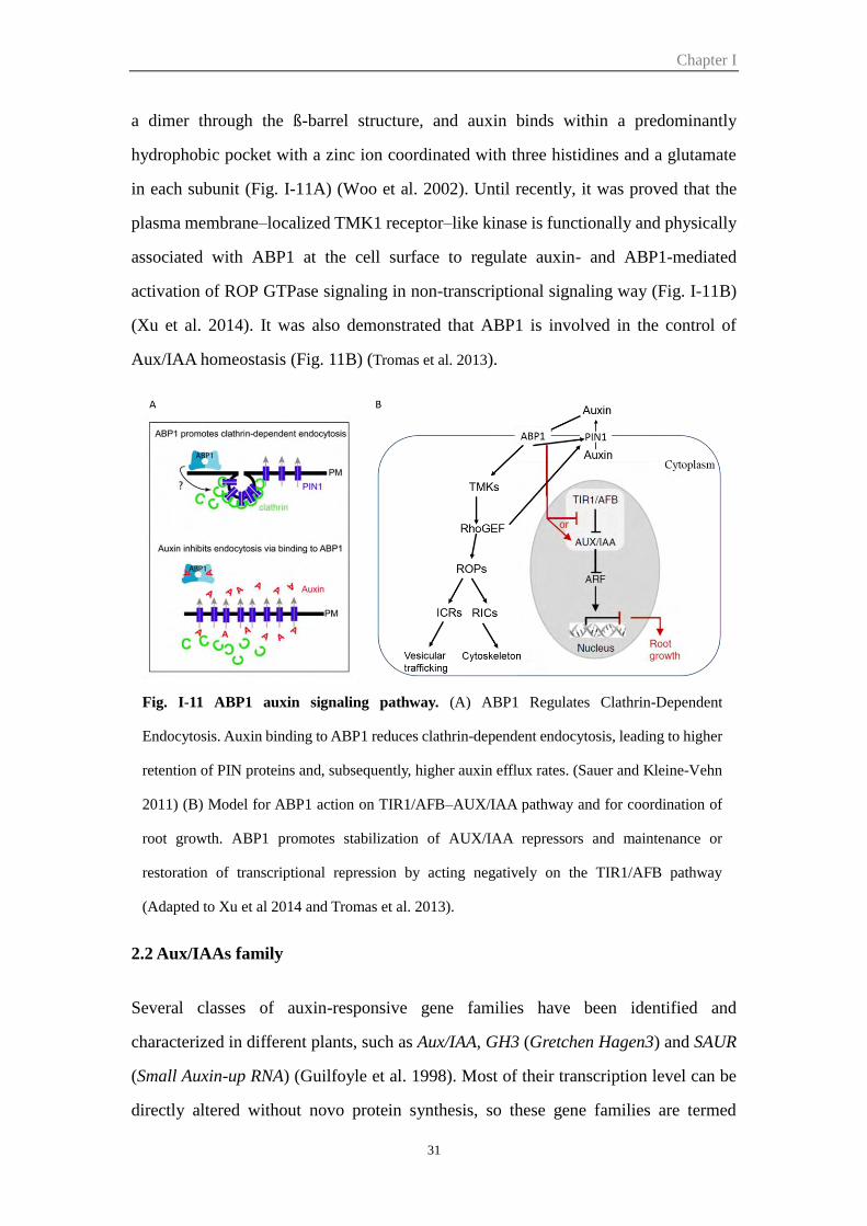
Chapter I
31
a dimer through the ß-barrel structure, and auxin binds within a predominantly
hydrophobic pocket with a zinc ion coordinated with three histidines and a glutamate
in each subunit (Fig. I-11A) (Woo et al. 2002). Until recently, it was proved that the
plasma membrane–localized TMK1 receptor–like kinase is functionally and physically
associated with ABP1 at the cell surface to regulate auxin- and ABP1-mediated
activation of ROP GTPase signaling in non-transcriptional signaling way (Fig. I-11B)
(Xu et al. 2014). It was also demonstrated that ABP1 is involved in the control of
Aux/IAA homeostasis (Fig. 11B) (Tromas et al. 2013).
2.2 Aux/IAAs family
Several classes of auxin-responsive gene families have been identified and
characterized in different plants, such as Aux/IAA, GH3 (Gretchen Hagen3) and SAUR
(Small Auxin-up RNA) (Guilfoyle et al. 1998). Most of their transcription level can be
directly altered without novo protein synthesis, so these gene families are termed
Fig. I-11 ABP1 auxin signaling pathway. (A) ABP1 Regulates Clathrin-Dependent
Endocytosis. Auxin binding to ABP1 reduces clathrin-dependent endocytosis, leading to higher
retention of PIN proteins and, subsequently, higher auxin efflux rates. (Sauer and Kleine-Vehn
2011) (B) Model for ABP1 action on TIR1/AFB–AUX/IAA pathway and for coordination of
root growth. ABP1 promotes stabilization of AUX/IAA repressors and maintenance or
restoration of transcriptional repression by acting negatively on the TIR1/AFB pathway
(Adapted to Xu et al 2014 and Tromas et al. 2013).

Chapter I
32
primary/early auxin-responsive genes (Abel and Theologis 1996). Aux/IAA gene as a
primary response to auxin was firstly identified in soybean and pea (Ainley et al. 1988;
Theologis et al. 1985; Walker and Key 1982). Then Aux/IAA genes have been found in
many plant species, including Arabidopsis (Overvoorde et al. 2005), poplar (Kalluri et
al. 2007), tomato (Audran-Delalande et al. 2012), maize (Wang et al. 2010) and rice
(Jain et al. 2006).
Canonical Aux/IAA proteins contain four highly conserved amino acid sequence motif
designated domain I, II, III and IV (Fig. I-12), although several proteins lacking one or
more of these domains are also included in the family. Domain I which contains a
conserved Leu repeat motif (LxLxLx) similarity within the EAR motif is responsible
for the repressing activity of the proteins (Tiwari et al. 2004), and it can also form co-
repressor with TOPLESS through this domain (Szemenyei et al. 2008). The instability
of Aux/IAA proteins involves conserved domain II composed by a hydrophobic motif
GWPPV (Worley et al. 2000), which mediates interaction with SCFTIR1 ubiquitin-ligase
complex for targeting of Aux/IAA to the 26S proteasome (Gray et al. 2001; Kepinski
and Leyser 2004). An amino acid substitution in the dEgron sequence (mutation of G
or P in GWPP motif) results in a stabilization of the affected protein and a decrease in
auxin response (Ouellet et al. 2001; Reed 2001; Tian et al. 2003). Domain III and IV
which are located in the carboxyl part of the protein, can mediate homo- and herero-
dimerization with other Aux/IAA members, as well as dimerization with ARFs which
contain similar domains (Kim et al. 1997; Ouellet et al. 2001; Ulmasov et al. 1997).
The Aux/IAA proteins do not contain DNA binding domain, so they cannot bind to the
Fig. I-12 Structural and functional domains of Aux/IAA proteins. (Colón-Carmona A et
al. 2000)
Chapter I
33
auxin-responsive elements (AuxREs) directly and regulate auxin-mediated gene
expression by interaction with ARF proteins, which have a B3-like DNA binding
domain (Guilfoyle and Hagen 2007). In addition to conserved domains, Aux/IAA
proteins display putative nuclear localization signals (NLS). NLS are composed by both
the bipartite structure of conserved basic doublet KR between domains I and II and
basic amino acids in domain II, and the SV40-type NLS located in domain IV.
Aux/IAA function has been mostly studied in Arabidopsis through the characterization
of gain-of-function mutants (mutation in domain II). axr5/iaa1 Arabidopsis mutant
showed defects in root and shoot tropisms, leaf morphology and inflorescence (Yang et
al. 2004). Both gain-of-function and loss-of-function mutant of AtIAA3 is characterized.
Gain-of-function shy2/iaa3 mutant showed a reduced lateral root formation and
increased root hair and slow root re-orientation, while loss-of-function mutant
displayed the opposite phenotype, indicating that IAA3 plays a central role in auxin
regulation of root growth and gravitropism. Shy2/iaa3 mutant also causes short
hypocotyls, leaf formation in dark and short inflorescence stems (Tian and Reed 1999).
axr2/iaa7 mutant causes agravitropic root and shoot growth, a short hypocotyl and stem,
and auxin-resistant root growth (Nagpal et al. 2000). The bdl/iaa12 Arabidopsis mutant
fails in initiating root meristem during early embryogenesis showing a specific role of
AtIAA12 during this process (Hamann et al. 2002). iaa28-1, slr/iaa14 and msg2-1/iaa19
suppress lateral root development (Fukaki et al. 2002; Rogg et al. 2001; Tatematsu et
al. 2004). The axr3/iaa17 Arabidopsis mutant presents shorter primary roots and
formation of adventitious roots with altered root gravitropism. Moreover, leaves are
darker and smaller than wild type and plants are dwarf showing altered apical
dominance (Leyser et al. 1996). 18 of 29 Aux/IAA T-DNA insertion mutants in
Arabidopsis have been characterized, but none of them displays visible developmental
defects, even for the double or triple mutants of closely homologs, suggesting that there
is an extensive functional redundancy among Aux/IAA gene family members
(Overvoorde et al. 2005).
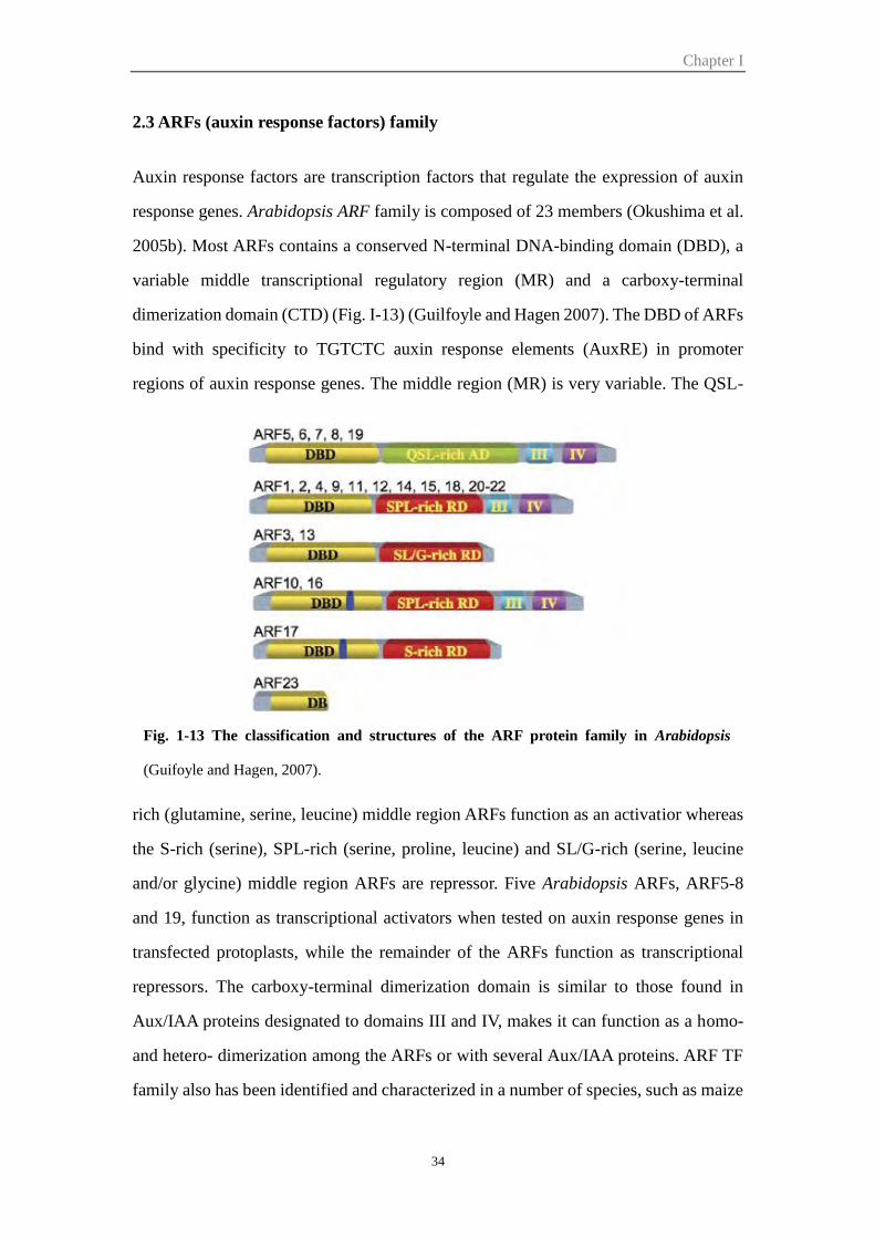
Chapter I
34
2.3 ARFs (auxin response factors) family
Auxin response factors are transcription factors that regulate the expression of auxin
response genes. Arabidopsis ARF family is composed of 23 members (Okushima et al.
2005b). Most ARFs contains a conserved N-terminal DNA-binding domain (DBD), a
variable middle transcriptional regulatory region (MR) and a carboxy-terminal
dimerization domain (CTD) (Fig. I-13) (Guilfoyle and Hagen 2007). The DBD of ARFs
bind with specificity to TGTCTC auxin response elements (AuxRE) in promoter
regions of auxin response genes. The middle region (MR) is very variable. The QSL-
rich (glutamine, serine, leucine) middle region ARFs function as an activatior whereas
the S-rich (serine), SPL-rich (serine, proline, leucine) and SL/G-rich (serine, leucine
and/or glycine) middle region ARFs are repressor. Five Arabidopsis ARFs, ARF5-8
and 19, function as transcriptional activators when tested on auxin response genes in
transfected protoplasts, while the remainder of the ARFs function as transcriptional
repressors. The carboxy-terminal dimerization domain is similar to those found in
Aux/IAA proteins designated to domains III and IV, makes it can function as a homo-
and hetero- dimerization among the ARFs or with several Aux/IAA proteins. ARF TF
family also has been identified and characterized in a number of species, such as maize
Fig. 1-13 The classification and structures of the ARF protein family in Arabidopsis
(Guifoyle and Hagen, 2007).
Chapter I
35
(Xing et al. 2011), rice (Wang et al. 2007), tomato (Kumar et al. 2011, Zouine et al.
2014), poplar (Kalluri et al. 2007), etc.
To understand their function in plant growth and development, the expression pattern
of ARF genes have been studied. ARF genes display specific expression pattern, ARF1
and ARF6 mainly expressed in developing flowers, ARF2 in floral organs and seedlings,
ARF3 and ARF4 in reproductive and vegetative tissues, ARF5 in embryos and vascular
tissues, ARF7 in roots, seedlings and embryos, ARF8 in seedlings and flowers, ARF12
in seeds, ARF16 in roots, leaves, vascular tissues and embryos, ARF19 in roots and
seedlings (Guilfoyle and Hagen 2007). A number of closely related ARFs (ARF12, -14,
-15, -17, -20-23) which located near the centromere of chromosome 1 were expressed
exclusively in the embryo (Rademacher et al. 2011). Moreover, lots of Arabidopsis arf
mutants have been studied. arf2 exhibits pleiotropic developmental phenotypes
including delayed flowing, leaf senescence and floral abscission, thick and long
inflorescence as well as defective apical hook formation, and arf1 mutations enhanced
many arf2 phenotypes (Ellis et al. 2005; Okushima et al. 2005a). arf3/ettin displays
abnormal development of flowers (Sessions et al. 1997). arf4 mutant shows no
phenotype whereas ett arf4 double mutants presents transformation of abaxial tissues
into adaxial ones in all aerial parts. It has been proven that ARF3 and ARF4 have
overlapping functions in leaf and floral organ patterning and cooperate with KANADI
genes to specify abaxial cell identity (Pekker et al. 2005). ARF5/ MONOPTEROS (MP)
gene is involved in embryo development and vascular tissue formation (Hardtke and
Berleth 1998). The AtARF7/NPH4 gene regulates hypocotyls tropism and the
sensitivity of auxin and ethylene (Harper et al. 2000). arf7 arf19 double mutant displays
strong auxin-related phenotype not observed in single mutant, including severely
impaired lateral roots formation and abnormal gravitropism in both hypocotyl and root
(Okushima et al. 2005b). AtARF6 and AtARF8 both regulate flowering process by
coordinating stamen development, petal expansion, anther dehiscence and gynoecium
maturation. arf6 arf8 double mutant has complete arrested flower development before
bud opening and are completely infertile, while arf6 and arf8 single mutants display
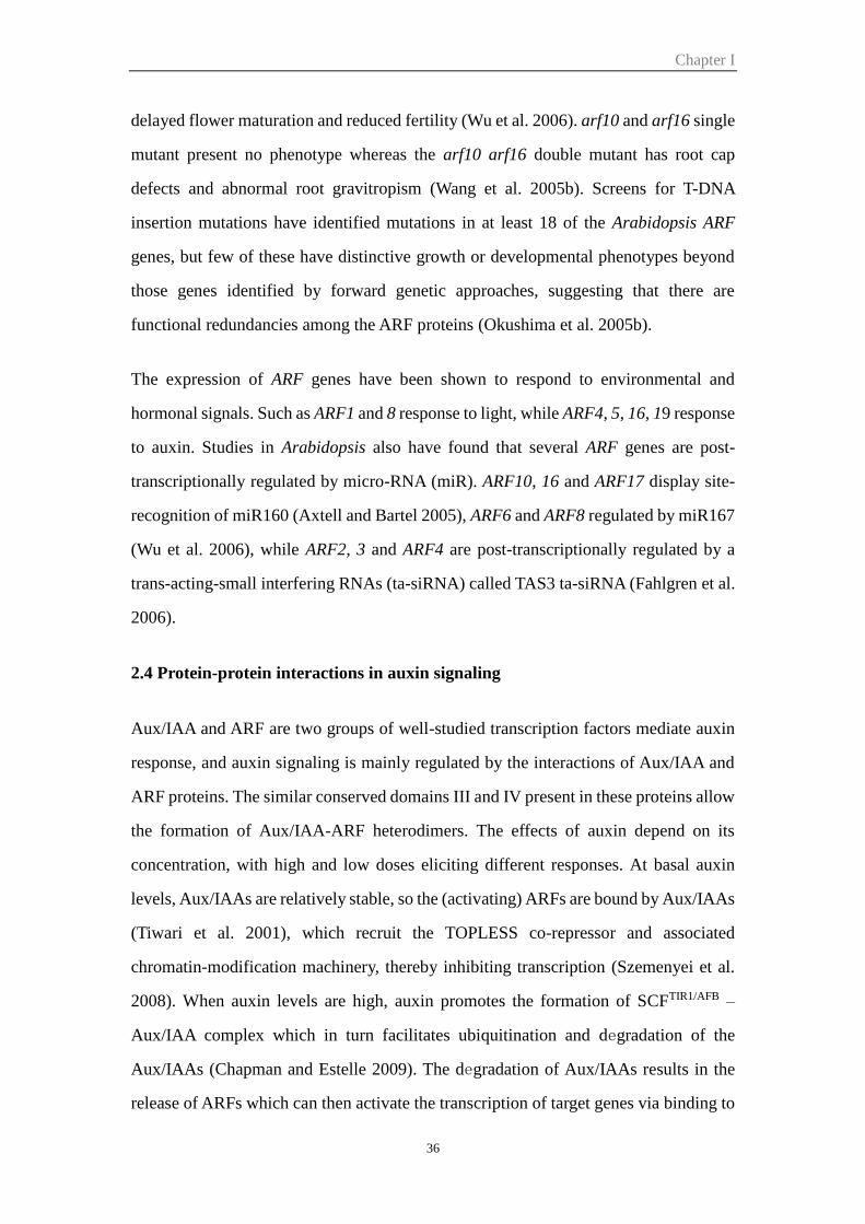
Chapter I
36
delayed flower maturation and reduced fertility (Wu et al. 2006). arf10 and arf16 single
mutant present no phenotype whereas the arf10 arf16 double mutant has root cap
defects and abnormal root gravitropism (Wang et al. 2005b). Screens for T-DNA
insertion mutations have identified mutations in at least 18 of the Arabidopsis ARF
genes, but few of these have distinctive growth or developmental phenotypes beyond
those genes identified by forward genetic approaches, suggesting that there are
functional redundancies among the ARF proteins (Okushima et al. 2005b).
The expression of ARF genes have been shown to respond to environmental and
hormonal signals. Such as ARF1 and 8 response to light, while ARF4, 5, 16, 19 response
to auxin. Studies in Arabidopsis also have found that several ARF genes are post-
transcriptionally regulated by micro-RNA (miR). ARF10, 16 and ARF17 display site-
recognition of miR160 (Axtell and Bartel 2005), ARF6 and ARF8 regulated by miR167
(Wu et al. 2006), while ARF2, 3 and ARF4 are post-transcriptionally regulated by a
trans-acting-small interfering RNAs (ta-siRNA) called TAS3 ta-siRNA (Fahlgren et al.
2006).
2.4 Protein-protein interactions in auxin signaling
Aux/IAA and ARF are two groups of well-studied transcription factors mediate auxin
response, and auxin signaling is mainly regulated by the interactions of Aux/IAA and
ARF proteins. The similar conserved domains III and IV present in these proteins allow
the formation of Aux/IAA-ARF heterodimers. The effects of auxin depend on its
concentration, with high and low doses eliciting different responses. At basal auxin
levels, Aux/IAAs are relatively stable, so the (activating) ARFs are bound by Aux/IAAs
(Tiwari et al. 2001), which recruit the TOPLESS co-repressor and associated
chromatin-modification machinery, thereby inhibiting transcription (Szemenyei et al.
2008). When auxin levels are high, auxin promotes the formation of SCFTIR1/AFB –
Aux/IAA complex which in turn facilitates ubiquitination and degradation of the
Aux/IAAs (Chapman and Estelle 2009). The degradation of Aux/IAAs results in the
release of ARFs which can then activate the transcription of target genes via binding to
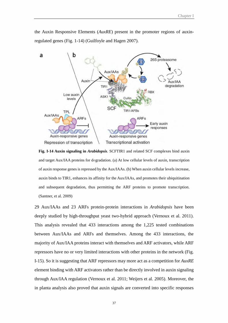
Chapter I
37
the Auxin Responsive Elements (AuxRE) present in the promoter regions of auxin-
regulated genes (Fig. 1-14) (Guilfoyle and Hagen 2007).
29 Aux/IAAs and 23 ARFs protein-protein interactions in Arabidopsis have been
deeply studied by high-throughput yeast two-hybrid approach (Vernoux et al. 2011).
This analysis revealed that 433 interactions among the 1,225 tested combinations
between Aux/IAAs and ARFs and themselves. Among the 433 interactions, the
majority of Aux/IAA proteins interact with themselves and ARF activators, while ARF
repressors have no or very limited interactions with other proteins in the network (Fig.
I-15). So it is suggesting that ARF repressors may more act as a competition for AuxRE
element binding with ARF activators rather than be directly involved in auxin signaling
through Aux/IAA regulation (Vernoux et al. 2011; Weijers et al. 2005). Moreover, the
in planta analysis also proved that auxin signals are converted into specific responses
Fig. I-14 Auxin signaling in Arabidopsis. SCFTIR1 and related SCF complexes bind auxin
and target Aux/IAA proteins for degradation. (a) At low cellular levels of auxin, transcription
of auxin response genes is repressed by the Aux/IAAs. (b) When auxin cellular levels increase,
auxin binds to TIR1, enhances its affinity for the Aux/IAAs, and promotes their ubiquitination
and subsequent degradation, thus permitting the ARF proteins to promote transcription.
(Santner, et al. 2009)
Chapter I
38
by matching pairs of co-expressed ARF and Aux/IAA proteins. AtIAA12 paralog
IAA13 could interact with ARF5, regulating embryonic root formation (Weijers et al.
2005), and the specific interaction between AtIAA14 and AtARF7 or AtARF19 is
essential for the inactivation of lateral root formation (Fukaki et al. 2006).
3 Roles of auxin in plant development
Auxin plays critical roles in plant cell growth and in a wide variety of developmental
processes. The effects of auxin was first documented by Darwin in 1880, then 45 years
later, by Went in 1926. The term generic term “auxin” in fact represents a family of
related compounds: indole-3-acetic acid (IAA) is considered to be the major
biologically active auxin, while a number of related compounds with auxin activity like
indole-3-butyric (IBA), 4-Chloroindole-3-acetic (4-Cl-IAA) and phenylacetic acid
(PAA) have also been identified in various plant species (Simon and Petrášek 2011).
3.1 Auxin promotes cell division, and cell expansion
On the cellular level, auxin influences aspects of cell division, cell elongation and cell
differentiation according to auxin concentration and cell/tissue-specific responses.
Fig. I-15 The Arabidopsis ARF-Aux/IAA interaction map. Most of Aux/IAAs are able to
interact with ARF activators while ARF repressors display very few interactions in
Arabidopsis. (Vernoux et al. 2011)
Chapter I
39
Auxin starvation of suspension cells causes cell division arrest while addition of auxin
to the arrest cells will lead to restoration of cell division (Perrot-Rechenmann 2010).
Auxin can induce the main gene expression promoted the cell division, such as Cyclin-
dependent kinase (CDKA) and D-type cyclins which plays a critical role in G1 to S and
G2 to M progression (Koroleva et al. 2004), while reduce the expression of cell division
inhibitors such as KRP (Himanen et al. 2002). One of the auxin receptors ABP1 is also
critical for auxin regulation of the cell cycle and expansion. Inactivation of ABP1 in
tobacco cells leads to cell-cycle arrest in G1 phase even in the presence of auxin (David
et al. 2007). Overexpression of ABP1 leads to an increased expansion of some leaf
tissues and the null Arabidopsis ABP1 mutant displays altered cell expansion (Chen et
al. 2001a; Chen et al. 2001b). Plant cell extension requires uptake of water and
irreversible extension of the cell wall, which includes wall loosening (short time frame)
and deposition of new wall material (long time frame). Auxin is one of the major stimuli
affecting these mechanisms, and this auxin-dependent cell expansion follows a dose-
response curve in which high concentrations are inhibitory (Barbier-Brygoo et al. 1991;
Evans et al. 1994).
3.2 Auxin roles in organ patterning
Auxin plays critical role in regulation of organ patterning, such as embryo and vascular
pattern, root initiation, apical dominance, tropism and fruit development. The role of
auxin in vascular patterning will be reviewed in the next paragraph.
Vascular patterning
Vascular tissues are generated during embryogenesis and organogenesis and response
for water and nutrients conduction. Auxin plays a key role in embryonic vascular
pattern, and disrupt auxin polar transport result in defects in leaf veins (reviewed in
Berleth and Mattsson 2000). In Arabidopsis, lax2 knockouts exhibited vascular breaks
in their cotyledons. Mutations in auxin response related genes such as MONOPTEROS
(MP)/AUXIN RESPONSE FACTOR 5 (AFR5), AUXIN RESISTANT 6 (AXR6), and
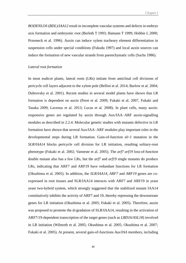
Chapter I
40
BODENLOS (BDL)/IAA12 result in incomplete vascular systems and defects in embryo
axis formation and embryonic root (Berleth T 1993; Hamann T 1999; Hobbie L 2000;
Przemeck et al. 1996). Auxin can induce xylem tracheary element differentiation in
suspension cells under special conditions (Fukuda 1997) and local auxin sources can
induce the formation of new vascular strands from parenchymatic cells (Sachs 1986).
Lateral root formation
In most eudicot plants, lateral roots (LRs) initiate from anticlinal cell divisions of
pericycle cell layers adjacent to the xylem pole (Bellini et al. 2014; Barlow et al. 2004;
Dubrovsky et al. 2001). Recent studies in several model plants have shown that LR
formation is dependent on auxin (Peret et al. 2009; Fukaki et al. 2007, Fukaki and
Tasaka 2009; Lavenus et al. 2013; Lucas et al. 2008). In plant cells, many auxin-
responsive genes are regulated by auxin through Aux/IAA–ARF auxin-signalling
modules as described in 2.2.4. Molecular genetic studies with mutants defective in LR
formation have shown that several Aux/IAA–ARF modules play important roles in the
developmental steps during LR formation. Gain-of-function slr-1 mutation in the
SLR/IAA14 blocks pericycle cell division for LR initiation, resulting solitary-root
phenotype (Fukaki et al. 2002; Vanneste et al. 2005). The arf7 arf19 loss-of-function
double mutant also has a few LRs, but the arf7 and arf19 single mutants do produce
LRs, indicating that ARF7 and ARF19 have redundant functions for LR formation
(Okushima et al. 2005). In addition, the SLR/IAA14, ARF7 and ARF19 genes are co-
expressed in root tissues and SLR/IAA14 interacts with ARF7 and ARF19 in yeast
yeast two-hybrid system, which strongly suggested that the stabilized mutant IAA14
constitutively inhibits the activity of ARF7 and 19, thereby repressing the downstream
genes for LR initiation (Okushima et al. 2005; Fukaki et al. 2005). Therefore, auxin
was proposed to promote the degradation of SLR/IAA14, resulting in the activation of
ARF7/19-dependent transcription of the target genes (such as LBD16/ASL18) involved
in LR initiation (Wilmoth et al. 2005; Okushima et al. 2005; Okushima et al. 2007;
Fukaki et al. 2005). At present, several gain-of-functions Aux/IAA members, including
Chapter I
41
IAA28, AXR5/IAA1, SHY3/IAA3, CRANE/IAA18 and MSG2/IAA19 also showed
decreased number of LRs, indicating that auxin signalling dependent on these Aux/IAAs
is necessary for LR formation (Overvoorde et al 2010). More recently, a multiple
AUX/IAA–ARF modules regulate lateral root formation has been demonstrated by Goh
et al. (2012) (Fig. I-16): IAA28 module regulates LR founder cell specification (De
Rybel et al. 2010), then SLR/IAA14-ARF7-ARF19 module regulates nuclear migration
and asymmetric cell divisions of the LR founder cells (Goh et al. 2012). Moreover, this
Fig. I-16 Schematic of lateral root (LR) formation regulated by SLR/IAA14–ARF7–ARF19
and SHY2/IAA3–ARFs auxin-signalling modules. LR initiation is controlled by the
SLR/IAA14–ARF7–ARF19 auxin-signalling module (orange box) by the activation of
LBD16/ASL18 and its related LBD/ASL proteins (red arrow 1). After initiation, the
SHY2/IAA3–ARFs signalling module (green box) plays a role not only for LR primordium
development and LR emergence after the SLR/IAA14–ARF7–ARF19 module (red arrows 2 and
3) but also for inhibition of SLR/IAA14–ARF7–ARF19-dependent LR initiation in the xylem
pericycle cells by affecting auxin homeostasis (blue dotted line). (Goh et al. 2012)
Chapter I
42
process is also regulated by BDL/IAA12-MP/ARF5 which represses ectopic pericycle
cell divisions (Goh et al. 2012). SHY2/IAA3-ARFs positively regulate LR primordium
development and in parallel inhibit LR formation by affecting auxin homeostasis (Goh
et al. 2012).
Flower and fruit development
As known, auxin plays a critical role in regulating flower and subsequent fruit
development (Dharmasiri et al., 2005b; Liscum and Reed, 2002). Auxin accumulation
controls the floral primordium initiation (Reinhardt et al., 2000); it is also essential for
anther development (Cecchetti et al. 2008) and involved in stamen and pollen
development. Several mutants disrupted in either auxin biosynthesis (yuc2yuc6),
transport (tir1afb), or signalling (arf6arf8, ettin/arf3, mp/arf5) exhibit flowering defects
that are variable but typically involve alterations in organ numbers, organ spacing, and
gynoecium morphology (Nagpal et al. 2005; Cecchetti et al. 2008).
Auxin plays a critical role in fruit development, beginning with flower formation,
through fruit set, fruit growth and ripening (de Jong et al. 2009b, Sundberg and
Ostergaard 2009, Ruan et al. 2012). In Arabidopsis alteration of AtARF8 expression
leads to the formation of parthenocarpic fruits (Goetz et al. 2007). In the loss-of-
function tomato iaa9 mutant ovary develops prior to pollination leading to precocious
fruit set and marker fruit parthenocarpy (Wang et al. 2005a). Moreover application of
auxin results in a delayed fruit ripening (Aharoni et al. 2002; Cohen 1996). Recent data
are also uncovering new roles for other Aux/IAA and ARF proteins in fruit growth after
fruit set. It was recently demonstrated that tomato IAA27 and ARF10 are involved in
the regulation of seed development and the size and shape of the fruit (Bassa et al. 2012,
Hendelman et al. 2012). Silencing of ARF4, which is expressed strongly in tomato
pericarp resulted in enhanced accumulation of starch and chlorophyll content, which
suggests the involvement of auxin signaling in the control of chloroplastic activity and
sugar metabolism in the fruit (Jones et al. 2002, Sagar et al. 2013).
Chapter I
43
Apical dominance
In intact plants, the shoot apex grows predominantly and inhibits outgrowth of axillary
buds. After decapitation of the shoot apex, outgrowth of axillary buds begins. This
phenomenon is called an apical dominance, and it is regulated by the plant hormones
auxin and cytokinin (CK). Auxin, derived from the shoot apex, inhibits the growth of
axillary buds, while CK, thought to be derived from the roots, promotes the growth of
axillary buds (Leyser 2003; Shimizu-Sato et al. 2001). Tanaka et al. (2006) proposed
that in intact plants, auxin is basipetally transported and represses expression of the IPT
gene (a key enzyme in CK biosynthesis) in the stem, then consequently, axillary buds
lack the ability to grow out. On the other hand, once the shoot apex is decapitated, the
auxin level in the stem decreases, repression of IPT gene expression is released, CK
levels increase and axillary buds grow out. After axillary buds grow out, de novo
synthesized IAA derived from a new shoot apex flows to the stem and again represses
IPT gene expression.
3.3 Typical auxin insensitive responses
Auxin regulates a host of plant developmental and physiological processes through the
SCFTIR1/AFB-mediated proteolysis of the Aux/IAA family of transcriptional regulators.
The domain II of the Aux/IAA is responsible for ubiquitin-mediated proteolysis, so
when the mutation happened in this domain, the Aux/IAA protein will be stabilized and
further resulted in altered auxin sensitivity and response. Most of the insight into the
biological function of Aux/IAA proteins gathered to date comes from this
characterization of dominant, gain-of-function mutations. Gain-of-function AtIAA12
mutant bdl is insensitive to auxin and displayed several auxin related phenotypes. The
strong lines displayed lacked hypocotyl, root and primary root meristem and the
vasculature of the cotyledons was reduced (Hamann et al. 1999). The bdl homozygous
mutant plants were short with rolled-up leaves and the apical dominance was reduced.
Semidominant AtIAA3 mutant shy2 showed less sensitive to auxin and adult plants were
extremely dwarfed with curled-up leaves. The seedlings displayed larger and curled up
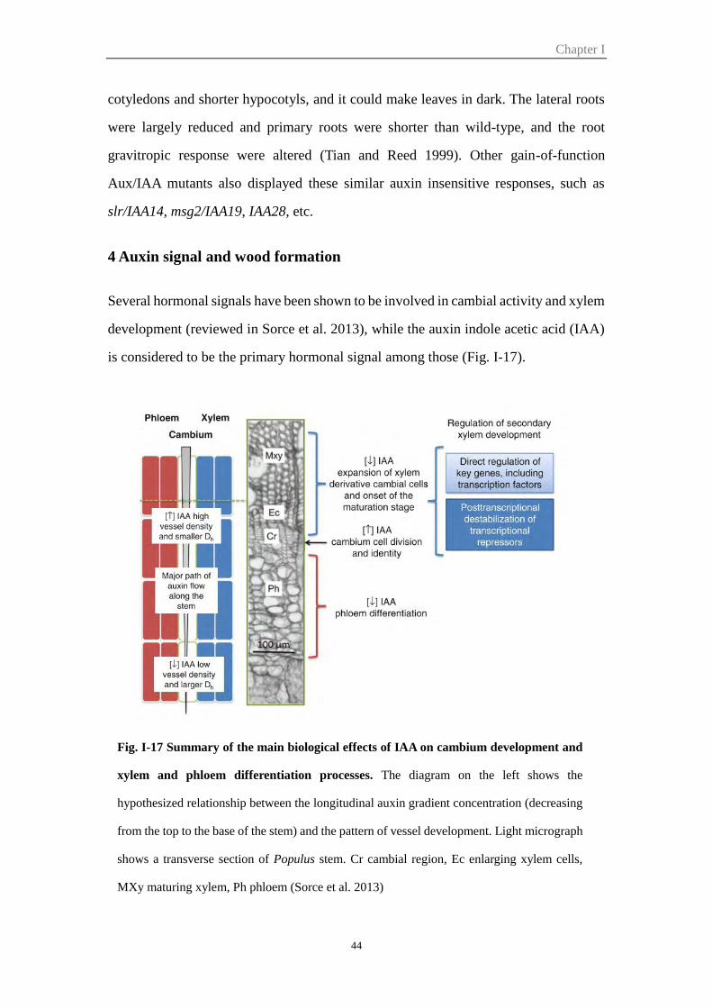
Chapter I
44
cotyledons and shorter hypocotyls, and it could make leaves in dark. The lateral roots
were largely reduced and primary roots were shorter than wild-type, and the root
gravitropic response were altered (Tian and Reed 1999). Other gain-of-function
Aux/IAA mutants also displayed these similar auxin insensitive responses, such as
slr/IAA14, msg2/IAA19, IAA28, etc.
4 Auxin signal and wood formation
Several hormonal signals have been shown to be involved in cambial activity and xylem
development (reviewed in Sorce et al. 2013), while the auxin indole acetic acid (IAA)
is considered to be the primary hormonal signal among those (Fig. I-17).
Fig. I-17 Summary of the main biological effects of IAA on cambium development and
xylem and phloem differentiation processes. The diagram on the left shows the
hypothesized relationship between the longitudinal auxin gradient concentration (decreasing
from the top to the base of the stem) and the pattern of vessel development. Light micrograph
shows a transverse section of Populus stem. Cr cambial region, Ec enlarging xylem cells,
MXy maturing xylem, Ph phloem (Sorce et al. 2013)
Chapter I
45
The phenomenon of recruiting existing parenchyma cells for differentiation into files
of procambial precursors is observed both during normal leaf vein development and in
response to rupture of existing vascular strands (Sachs 1986). Early experiments
established that ectopic application of the hormone auxin [indole-3-acetic acid (IAA)]
was sufficient to trigger the specification of vascular tissue, including proliferation of
cambial cells and final differentiation of vascular cell types (reviewed in Berleth and
Mattsson 2000). Auxin (supported by cytokinin) can induce xylem tracheary element
differentiation in suspension culture cells of Zinnia and Arabidopsis (Fukuda 1997). In
1981, Sachs proposed the ‘canalization of auxin flow hypothesis’ as a model for the
auxin-mediated formation of vascular tissues (Sachs, 1981). In this model, channels of
preferential auxin flow are created when a series of cells gradually become specialized
for directional auxin transport. Once established, these channels effectively drain auxin
from surrounding cells, resulting in localized concentration of auxin within distinct cell
files, and this shift in auxin distribution was hypothesized to subsequently induce
vascular tissue formation. The ‘canalization of auxin’ model thus provided a single
mechanism that could account for both the initial specification and the physical
contiguity of developing vascular tissues. The auxin-induced vascular differentiation
response is: (1) local, as vascular strand formation is initiated at the specific site of
auxin application; (2) polar, as vascular strand formation progresses from the auxin
source toward the basal side of the plant; and (3) continuous, as it generates
uninterrupted files of vascular cells (Scarpella et al. 2010). In normal plants, auxin is
predominantly produced in apical regions, such as young leaves, from which it is
transported basally to roots, such a steady polar flow of auxin induces the continuity of
vascular tissues along the plant.
The proposed canalization of auxin was supported by the fact that disruption of auxin
polar transport result in defects in leaf veins (Berleth and Mattsson 2000) and by the
identification of genes involved in auxin signalling and transport. Mutations in auxin
response related genes such as MONOPTEROS (MP)/AUXIN RESPONSE FACTOR 5
(AFR5), AUXIN RESISTANT 6 (AXR6), and BODENLOS (BDL)/IAA12 result in

Chapter I
46
incomplete vascular systems and defects in embryo axis formation and embryonic root
(Berleth 1993; Hamann 1999; Hobbie 2000; Przemeck et al. 1996). mp mutants also
have severely attenuated levels of expression of the PINFORMED1 (PIN1) gene
(Wenzel et al. 2007). Asymmetric localization of PIN1 in plant cells is thought to
establish a directional auxin flow. In the Arabidopsis leaf, PIN1 and MP are co-
expressed during very early stages of procambial cell specification, and their spatial
pattern of expression gradually changes from initially broader domains to a single file
of cells, as predicted for positive feedback onto the auxin canalization process
(Scarpella et al, 2006; Wenzel et al. 2007).
Recent findings have identified auxin-mediated basic helix-loop-helix (bHLH) TF
dimers as important factors regulating early vascular development, including TARGET
OF MONOPTEROS5 (TMO5), LONESOME HIGHWAY (LHW) and their closest
homologs (reviewed in Zhang et al. 2014a). Vascular tissue differentiation was totally
blocked in the roots of TMO quadruple mutants. By contrast, co-overexpression of
TMO5 and LHW induced dramatic periclinal divisions within the vasculature of roots.
Whether these factors interact to regulate secondary growth in different species is still
an open question.
The major path of auxin flow along the stems of pine and poplar trees is in the vascular
cambium, then auxin is distributed in a radial concentration gradient, with highest levels
in vascular cambium, where cell division takes place, then decline rapidly toward the
xylem and phloem (Fig. I-17, 18) (Uggla et al. 1998; Uggla et al. 1996). Thus, it has
been suggested that auxin acts as in a manner similar to a morphogen to provide
positional information for the division of wood-forming tissue into distinct
differentiation zones. High auxin concentrations may be interpreted as a signal for cell
division, intermediate levels may promote cell expansion and low levels may be read
out as a signal inducing the deposition of secondary cell walls. So auxin concentration
could define the fate of differentiating cells. High levels of auxin in cambium initial
mother cells would maintain cambial cells meristematic, whereas low levels would lead
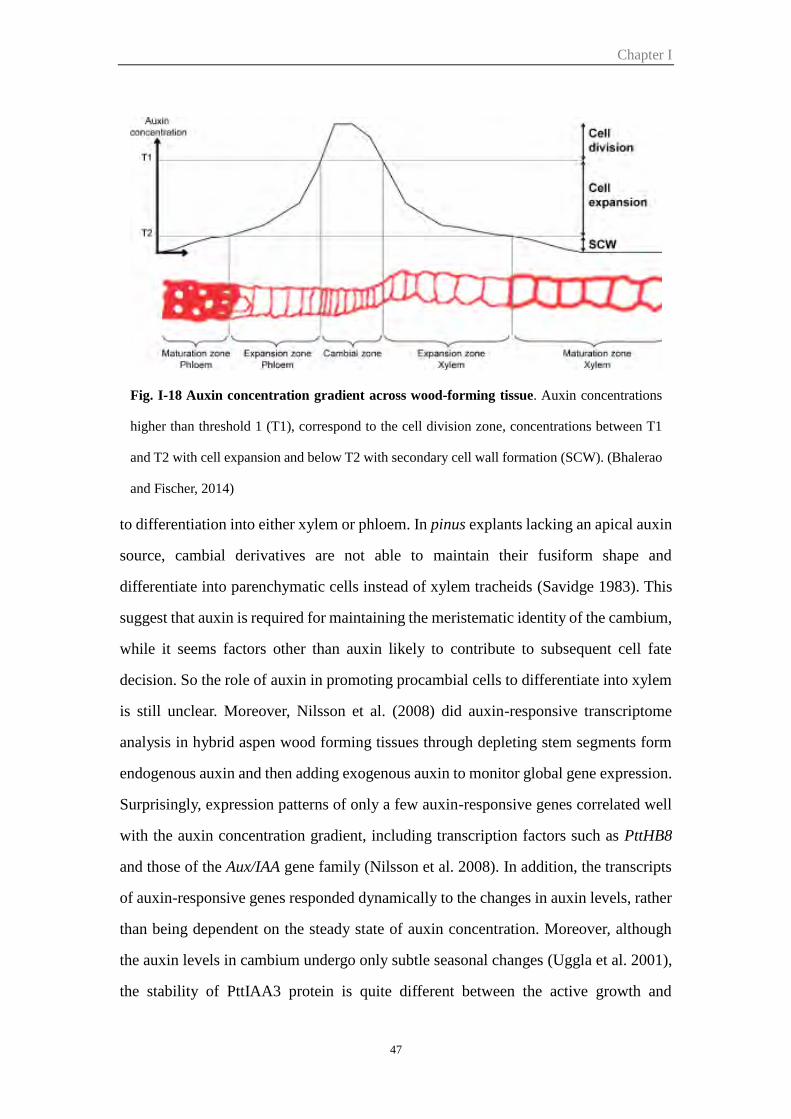
Chapter I
47
to differentiation into either xylem or phloem. In pinus explants lacking an apical auxin
source, cambial derivatives are not able to maintain their fusiform shape and
differentiate into parenchymatic cells instead of xylem tracheids (Savidge 1983). This
suggest that auxin is required for maintaining the meristematic identity of the cambium,
while it seems factors other than auxin likely to contribute to subsequent cell fate
decision. So the role of auxin in promoting procambial cells to differentiate into xylem
is still unclear. Moreover, Nilsson et al. (2008) did auxin-responsive transcriptome
analysis in hybrid aspen wood forming tissues through depleting stem segments form
endogenous auxin and then adding exogenous auxin to monitor global gene expression.
Surprisingly, expression patterns of only a few auxin-responsive genes correlated well
with the auxin concentration gradient, including transcription factors such as PttHB8
and those of the Aux/IAA gene family (Nilsson et al. 2008). In addition, the transcripts
of auxin-responsive genes responded dynamically to the changes in auxin levels, rather
than being dependent on the steady state of auxin concentration. Moreover, although
the auxin levels in cambium undergo only subtle seasonal changes (Uggla et al. 2001),
the stability of PttIAA3 protein is quite different between the active growth and
Fig. I-18 Auxin concentration gradient across wood-forming tissue. Auxin concentrations
higher than threshold 1 (T1), correspond to the cell division zone, concentrations between T1
and T2 with cell expansion and below T2 with secondary cell wall formation (SCW). (Bhalerao
and Fischer, 2014)
Chapter I
48
dormancy stage (Baba et al. 2011). These findings argue against auxin displaying
morphogen-like activity, and support the idea that auxin signaling controls cambial
activity by modulation of auxin responsiveness (Bhalerao and Fischer 2014). Nilsson
et al. (2008) hypothesized that auxin could regulate secondary xylem development
through two mechanisms: (1) direct regulation of the expression of a few key genes,
including some transcription factors; (2) posttranscriptional destabilization of
transcriptional repressors.
During the auxin concentration gradient formation, PIN-FORMED 1 (PIN1)-like auxin
efflux carries has been suggested to play an important role in trees (Hellgren et al. 2004;
Schrader et al. 2003). In order to redistribute polarly transported auxin from the
cambium into phloem and xylem, efflux carriers would need to be localized to the
lateral membrane of cambial cells and their derivatives. Although PIN proteins have
not yet been systematically localized in cambia, PIN1, which is strongly expressed in
Arabidopsis inflorescence stems, has been shown to localize not only to basal plasma
membranes but also in the lateral plasma membrane at the basal end of parenchymatic
xylem cells (Galweiler et al. 1998; Sauer et al. 2006). Similarly, PIN3, which is
involved in redirecting the auxin flux upon gravistimulation in the root tip, localizes to
the inner lateral membrane of the starch sheath of the Arabidopsis inflorescence stem
(Friml et al. 2002). In both a pin1 and pin3 mutant the radial extension of derivatives
of the interfascicular cambium is reduced (Agusti et al. 2011a). In stems of revolute
(rev) mutants, which are defective in the formation of interfascicular fibres, PIN3 and
PIN4 is dramatically reduced (Zhong and Ye 2001). Another auxin transporter which
be involved in this auxin gradient is the ATP-binding cassette (ABC) transporter
ABCB14. The abcb14 mutant displays reduced polar auxin transport and a mild radial
cell expansion phenotype in the xylem (Kaneda et al. 2011). More recently, WAT1 has
been demonstrated as a vacuolar auxin transporter. WAT1 mutants revealed two cell
wall-related phenotypes in Arabidopsis stems: a defect in cell elongation, resulting in a
dwarfed habit and little to no secondary cell walls in fibres. Secondary walls of vessel
elements were unaffected by the mutation (Ranocha et al. 2010). In summary,
Chapter I
49
phenotypes of auxin carrier mutants related to wood formation have either not been
reported or are difficult to interpret due to an absence of auxin distribution data in those
mutants (Agusti et al. 2011a, Kaneda et al. 2011).
The control of vessel size is an important trait for ensuring the ascent of water and
minerals from roots to leaves and adaptation of plants to the environment. Auxin
concentration has also been proposed to be involved in the regulation of vessel size and
density (Aloni and Zimmermann 1983). The hypothesis is that high auxin concentration
near the leaves stimulate rapid cell differentiation resulting in narrow vessels, while low
auxin levels further down result in wide vessels because of the slow differentiation,
which permits more cell expansion before secondary wall deposition. Thus, along the
tree axis, auxin induces gradual gradients of increasing vessel diameter and decreasing
vessel density from leaves to roots, which ensuring the ascent of water and minerals
from roots to leaves and the adaptation of plants to the environment (Aloni 2001).
Although the detailed mechanisms of how IAA would regulate growth and
differentiation are not fully understood, the aforesaid hypothesis is substantiated by
several observations: (1) the major sources of IAA are developing buds and young
shoots (Uggla et al. 1998); (2) IAA is transported basipetally from the leaves towards
the roots (Aloni 2001); (3) the major path of the IAA flow in tree stems is in the vascular
cambium (Sundberg et al. 2000); (4) there is a general decrease in the IAA
concentration towards the stem base (Aloni 2001).
As trees are long living organisms with sessile lifestyle, they have to adapt to changing
environmental conditions throughout their lifetimes which may span decades and
centuries in some cases. In particular, vascular stem cell activity shows plasticity in
response to mechanical stress which affects wood formation and quality. In angiosperm
woody species, a local increase in cambial cell division induces the formation of tension
wood in the upper side of the leaning tree stems (Paux et al. 2004; Hellgren et al. 2004).
Auxin has been proposed to be implicated in the tension response, and many different
experiments have indicated this (Little and Savidge 1987). For example, application of
Chapter I
50
either exogenous auxin or auxin transport inhibitors was shown to induce the gelatinous
G-fibres characteristics of tension wood (Pilate et al 2004). Although measurements of
endogenous auxin failed to reveal significant changes in auxin balance in the cambial
region tissues (Hellgren et al. 2004), a rather large set of auxin-related genes were found
to be differentially expressed in developing poplar tension wood (Andersson-Gunneras
et al. 2006).
Despite the fact that auxin has long been proposed as primary regulator of cambial
activity and wood formation (Uggla et al. 1996, Sundberg et al. 2000), the auxin-
regulated transcriptional programs underlying wood formation remain largely under
investigated.
Part III The Eucalyptus, an evergreen angiosperm tree for wood
formation
Eucalyptus species, commonly referred to as eucalypts, are native to Australia and the
islands to its north. It is a large genus of evergreen hardwood trees and comprises more
than 700 species, belonging to the angiosperm family Myrtaceae (Brooker 2000).
Eucalyptus species occur naturally from sea level to the alpine tree line, from high
rainfall to semi-arid zones and from the tropics to latitudes as high as 43° south. Species
of Eucalyptus are cultivated widely in the tropical and temperate world, though most
species do not tolerate frost. A mature Eucalyptus may take the form from a low
shrubby mallees (less than 10 m) to a straight-trunked forest up to 90 m tall.
Due to its wide adaptability, extremely fast growth rate, good form, and excellent wood
and fibres properties, Eucalyptus species have become the most valuable and most
widely planted forest trees on a world-wide basis. They are cultivated mainly in tropical
and temperate zones. Today Eucalyptus can be found in Brazil, India, Portugal, South
Africa, China, Uruguay, Ethiopia, California and more than 100 countries across six
continents about more than 20 million hectares. The rotation cycles have relatively
Chapter I
51
shorter (5-7 years) compared to teak (20-40 years). The rotation time also depends on
the trunk diameter desired, for example 4-5 years for heating wood, 7-10 years for paper
pulp and 15 years or more for furniture wood. The productivity of managed Eucalyptus
forests can reach to 20-50 m3 ha-1 yr-1 due to improved genetic breeding and silviculture,
against about 15 m3 ha-1 yr-1 for the classical coniferous forests.
Eucalyptus is the most common short fibres source for pulpwood to make pulp.
Eucalyptus globulus (in temperate climates) and the hybrid of Eucalyptus urophylla x
Eucalyptus grandis (in tropical climates) are the most used varieties in papermaking.
They are also utilized for a diverse array of products including sawn timber, mine props,
poles, firewood, essential oils, honey and tannin as well as for shade, shelter and soil
reclamation. In rural communities of many developing countries, eucalypt wood is an
important source of fuel and building material. In today’s "new carbon economy",
eucalypts are receiving attention as fast-growing, short-rotation, renewable biomass
crops for energy production. It is for instance listed as one of the U.S. Department of
Energy's candidate biomass energy crops.
The genome of Eucalyptus grandis BRASUZ1 has been fully sequenced and annotated
(Myburg et al. 2014), offering new opportunities to get insights into the regulation of
secondary growth and cambial activity by auxin-signalling mediators. Understanding
the mechanisms that underlie auxin regulation in Eucalyptus wood formation is of
interest both in the context of plant development and as a path to improve
lignocellulosic biomass production and quality as a dedicated energy crop for
lignocellulosic biofuel production. It is worth noting that Eucalyptus is an evergreen
tree that do not present dormancy in contrast to poplar a deciduous tree, allowing further
comparison of the regulation of cambium activity and xylem differentiation between
the two only forest genus sequenced hitherto.

Chapter II
Genome-wide Characterization and Expression
Profiling of the AUXIN RESPONSE FACTOR
(ARF) Gene Family in Eucalyptus grandis


Chapter II: Genome-Wide Characterization and Expression Profiling of the AUXIN RESPONSE FACTOR (ARF) Gene Family in Eucalyptus grandisHong Yu1, Marcal Soler1, Isabelle Mila2, Helene San Clemente1, Bruno Savelli1, Christophe Dunand1,
Jorge A. P. Paiva3,4, Alexander A. Myburg5, Mondher Bouzayen2, Jacqueline Grima-Pettenati1*,
Hua Cassan-Wang1
1 LRSV Laboratoire de Recherche en Sciences Vegetales, UMR5546, Universite Toulouse III/CNRS, Castanet Tolosan, France, 2 Universite de Toulouse, Institut National
Polytechnique-Ecole Nationale Superieure Agronomique de Toulouse, Laboratoire de Genomique et Biotechnologie des Fruits, Castanet-Tolosan, France, 3 Instituto de
Investigacao Cientıfica e Tropical (IICT/MCTES), Lisboa, Portugal, 4 Instituto de Biologia Experimental e Tecnologica (IBET), Oeiras, Portugal, 5 Department of Genetics,
Forestry and Agricultural Biotechnology Institute (FABI), Genomics Research Institute (GRI), University of Pretoria, Pretoria, South Africa
Abstract
Auxin is a central hormone involved in a wide range of developmental processes including the specification of vascularstem cells. Auxin Response Factors (ARF) are important actors of the auxin signalling pathway, regulating the transcriptionof auxin-responsive genes through direct binding to their promoters. The recent availability of the Eucalyptus grandisgenome sequence allowed us to examine the characteristics and evolutionary history of this gene family in a woody plant ofhigh economic importance. With 17 members, the E. grandis ARF gene family is slightly contracted, as compared to those ofmost angiosperms studied hitherto, lacking traces of duplication events. In silico analysis of alternative transcripts and genetruncation suggested that these two mechanisms were preeminent in shaping the functional diversity of the ARF family inEucalyptus. Comparative phylogenetic analyses with genomes of other taxonomic lineages revealed the presence of a newARF clade found preferentially in woody and/or perennial plants. High-throughput expression profiling among differentorgans and tissues and in response to environmental cues highlighted genes expressed in vascular cambium and/ordeveloping xylem, responding dynamically to various environmental stimuli. Finally, this study allowed identification ofthree ARF candidates potentially involved in the auxin-regulated transcriptional program underlying wood formation.
Citation: Yu H, Soler M, Mila I, San Clemente H, Savelli B, et al. (2014) Genome-Wide Characterization and Expression Profiling of the AUXIN RESPONSE FACTOR(ARF) Gene Family in Eucalyptus grandis. PLoS ONE 9(9): e108906. doi:10.1371/journal.pone.0108906
Editor: Marcel Quint, Leibniz Institute of Plant Biochemistry, Germany
Received July 2, 2014; Accepted August 27, 2014; Published September 30, 2014
Copyright: � 2014 Yu et al. This is an open-access article distributed under the terms of the Creative Commons Attribution License, which permits unrestricteduse, distribution, and reproduction in any medium, provided the original author and source are credited.
Data Availability: The authors confirm that all data underlying the findings are fully available without restriction. All Eucalyptus ARF sequences used in thispaper are available in phytozome http://www.phytozome.net/. E. globulus RNA Seq Illumina reads are provided in File S1.
Funding: This work was supported by the Centre National pour la Recherche Scientifique (CNRS), the University Paul Sabatier Toulouse III (UPS), the FrenchLaboratory of Excellence project ‘‘TULIP’’ (ANR-10-LABX-41; ANR-11-IDEX-0002-02) and the Plant KBBE TreeForJoules project (ANR-2010-KBBE-007-01 (FR) and P-KBBE/AGR_GPL/0001/2010 (FCT, PT)). HY was supported by PhD grants from the China Scholarship council. MS was supported by a Postdoc fellowship ‘‘Beatriu dePinos’’ thanks to the Departament d’Universitats, Recerca i Societat de la Informacio de la Generalitat de Catalunya. JAPP acknowledges Fundacao para a Ciencia ea Tecnologia (FCT, Portugal) and his research contract funded by Programa Ciencia 2008 and POPH (QREN). The funders had no role in study design, datacollection and analysis, decision to publish, or preparation of the manuscript.
Competing Interests: The authors declare that no competing interests exist.
* Email: [email protected]
1 Introduction
The plant hormone auxin plays a prominent role in the
regulation of plant growth in response to diverse developmental
and environmental cues such as organogenesis, tropic movement,
root growth, fruit development, tissue and organ patterning and
vascular development [1]. Auxin plays a crucial role in the
specification of vascular stem cells (procambium) and in cambial
activity [2]. Analysis of auxin distribution across the cambial
region in hybrid aspen trees showed a radial auxin gradient
reaching a peak level in the cambial zone or at the border between
the cambial zone and the expansion zone towards developing
wood cells [3,4]. The auxin gradient was indeed shown to overlap
with the sequential and numerous auxin-regulated genes respond-
ing dynamically to the change in auxin levels in wood forming cells
[5].
As trees are long living organisms with sessile lifestyle, they have
to adapt to changing environmental conditions throughout their
lifetimes which may span decades and centuries in some cases. In
particular, vascular stem cell activity shows plasticity in response to
mechanical stress which affects wood formation and quality. In
angiosperm woody species, a local increase in cambial cell division
induces the formation of tension wood in the upper side of the
leaning tree stems [6,7]. Auxin has been proposed to be implicated
in the tension response, and application of either exogenous auxin
or auxin transport inhibitors was shown to induce the gelatinous
G-fibres characteristics of tension wood [8]. Although measure-
ments of endogenous auxin failed to reveal significant changes in
auxin balance in the cambial region tissues, a rather large set of
auxin-related genes were found to be differentially expressed in
developing poplar tension wood [9]. A recent study indicated that
the auxin signalling pathway is significantly disrupted during
PLOS ONE | www.plosone.org 52 September 2014 | Volume 9 | Issue 9 | e108906
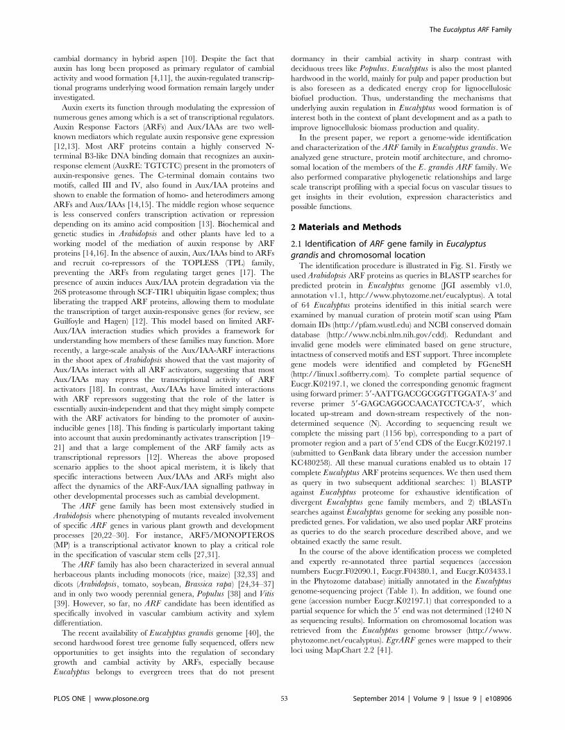
cambial dormancy in hybrid aspen [10]. Despite the fact that
auxin has long been proposed as primary regulator of cambial
activity and wood formation [4,11], the auxin-regulated transcrip-
tional programs underlying wood formation remain largely under
investigated.
Auxin exerts its function through modulating the expression of
numerous genes among which is a set of transcriptional regulators.
Auxin Response Factors (ARFs) and Aux/IAAs are two well-
known mediators which regulate auxin responsive gene expression
[12,13]. Most ARF proteins contain a highly conserved N-
terminal B3-like DNA binding domain that recognizes an auxin-
response element (AuxRE: TGTCTC) present in the promoters of
auxin-responsive genes. The C-terminal domain contains two
motifs, called III and IV, also found in Aux/IAA proteins and
shown to enable the formation of homo- and heterodimers among
ARFs and Aux/IAAs [14,15]. The middle region whose sequence
is less conserved confers transcription activation or repression
depending on its amino acid composition [13]. Biochemical and
genetic studies in Arabidopsis and other plants have led to a
working model of the mediation of auxin response by ARF
proteins [14,16]. In the absence of auxin, Aux/IAAs bind to ARFs
and recruit co-repressors of the TOPLESS (TPL) family,
preventing the ARFs from regulating target genes [17]. The
presence of auxin induces Aux/IAA protein degradation via the
26S proteasome through SCF-TIR1 ubiquitin ligase complex; thus
liberating the trapped ARF proteins, allowing them to modulate
the transcription of target auxin-responsive genes (for review, see
Guilfoyle and Hagen) [12]. This model based on limited ARF-
Aux/IAA interaction studies which provides a framework for
understanding how members of these families may function. More
recently, a large-scale analysis of the Aux/IAA-ARF interactions
in the shoot apex of Arabidopsis showed that the vast majority of
Aux/IAAs interact with all ARF activators, suggesting that most
Aux/IAAs may repress the transcriptional activity of ARF
activators [18]. In contrast, Aux/IAAs have limited interactions
with ARF repressors suggesting that the role of the latter is
essentially auxin-independent and that they might simply compete
with the ARF activators for binding to the promoter of auxin-
inducible genes [18]. This finding is particularly important taking
into account that auxin predominantly activates transcription [19–
21] and that a large complement of the ARF family acts as
transcriptional repressors [12]. Whereas the above proposed
scenario applies to the shoot apical meristem, it is likely that
specific interactions between Aux/IAAs and ARFs might also
affect the dynamics of the ARF-Aux/IAA signalling pathway in
other developmental processes such as cambial development.
The ARF gene family has been most extensively studied in
Arabidopsis where phenotyping of mutants revealed involvement
of specific ARF genes in various plant growth and development
processes [20,22–30]. For instance, ARF5/MONOPTEROS
(MP) is a transcriptional activator known to play a critical role
in the specification of vascular stem cells [27,31].
The ARF family has also been characterized in several annual
herbaceous plants including monocots (rice, maize) [32,33] and
dicots (Arabidopsis, tomato, soybean, Brassica rapa) [24,34–37]
and in only two woody perennial genera, Populus [38] and Vitis[39]. However, so far, no ARF candidate has been identified as
specifically involved in vascular cambium activity and xylem
differentiation.
The recent availability of Eucalyptus grandis genome [40], the
second hardwood forest tree genome fully sequenced, offers new
opportunities to get insights into the regulation of secondary
growth and cambial activity by ARFs, especially because
Eucalyptus belongs to evergreen trees that do not present
dormancy in their cambial activity in sharp contrast with
deciduous trees like Populus. Eucalyptus is also the most planted
hardwood in the world, mainly for pulp and paper production but
is also foreseen as a dedicated energy crop for lignocellulosic
biofuel production. Thus, understanding the mechanisms that
underlying auxin regulation in Eucalyptus wood formation is of
interest both in the context of plant development and as a path to
improve lignocellulosic biomass production and quality.
In the present paper, we report a genome-wide identification
and characterization of the ARF family in Eucalyptus grandis. We
analyzed gene structure, protein motif architecture, and chromo-
somal location of the members of the E. grandis ARF family. We
also performed comparative phylogenetic relationships and large
scale transcript profiling with a special focus on vascular tissues to
get insights in their evolution, expression characteristics and
possible functions.
2 Materials and Methods
2.1 Identification of ARF gene family in Eucalyptus grandis and chromosomal location
The identification procedure is illustrated in Fig. S1. Firstly we
used Arabidopsis ARF proteins as queries in BLASTP searches for
predicted protein in Eucalyptus genome (JGI assembly v1.0,
annotation v1.1, http://www.phytozome.net/eucalyptus). A total
of 64 Eucalyptus proteins identified in this initial search were
examined by manual curation of protein motif scan using Pfam
domain IDs (http://pfam.wustl.edu) and NCBI conserved domain
database (http://www.ncbi.nlm.nih.gov/cdd). Redundant and
invalid gene models were eliminated based on gene structure,
intactness of conserved motifs and EST support. Three incomplete
gene models were identified and completed by FGeneSH
(http://linux1.softberry.com). To complete partial sequence of
Eucgr.K02197.1, we cloned the corresponding genomic fragment
using forward primer: 59-AATTGACCGCGGTTGGATA-39 and
reverse primer 59-GAGCAGGCCAACATCCTCA-39, which
located up-stream and down-stream respectively of the non-
determined sequence (N). According to sequencing result we
complete the missing part (1156 bp), corresponding to a part of
promoter region and a part of 59end CDS of the Eucgr.K02197.1
(submitted to GenBank data library under the accession number
KC480258). All these manual curations enabled us to obtain 17
complete Eucalyptus ARF proteins sequences. We then used them
as query in two subsequent additional searches: 1) BLASTP
against Eucalyptus proteome for exhaustive identification of
divergent Eucalyptus gene family members, and 2) tBLASTn
searches against Eucalyptus genome for seeking any possible non-
predicted genes. For validation, we also used poplar ARF proteins
as queries to do the search procedure described above, and we
obtained exactly the same result.
In the course of the above identification process we completed
and expertly re-annotated three partial sequences (accession
numbers Eucgr.F02090.1, Eucgr.F04380.1, and Eucgr.K03433.1
in the Phytozome database) initially annotated in the Eucalyptusgenome-sequencing project (Table 1). In addition, we found one
gene (accession number Eucgr.K02197.1) that corresponded to a
partial sequence for which the 59 end was not determined (1240 N
as sequencing results). Information on chromosomal location was
retrieved from the Eucalyptus genome browser (http://www.
phytozome.net/eucalyptus). EgrARF genes were mapped to their
loci using MapChart 2.2 [41].
The Eucalyptus ARF Family
PLOS ONE | www.plosone.org 53 September 2014 | Volume 9 | Issue 9 | e108906
Ta
ble
1.
AR
Fg
en
efa
mily
inEu
caly
ptu
s.
Acc
ess
ion
no
.aA
rab
ido
psi
sb
est
hit
(bla
stp
)bA
min
oa
cid
ide
nti
ty%
cS
ho
rtn
am
ed
Nu
mb
er
of
pre
dic
ted
alt
ern
ati
ve
tra
nsc
rip
teC
hro
mo
som
ef
Ge
no
me
loca
tio
ng
OR
F(b
p)h
De
du
ced
po
lyp
eti
de
iE
xo
nN
o.
Le
ng
th(a
a)
MW
(kD
a)
PI
Eucg
r.G
0007
6.1
AtA
RF1
62
.2Eg
rAR
F14
78
48
,24
8..8
60
,08
72
02
86
75
75
.36
.06
14
Eucg
r.K
0219
7.1
AtA
RF2
59
.3Eg
rAR
F2A
**5
11
29
,23
2,7
70
..29
,23
9,1
03
25
08
83
59
3.1
36
.55
14
Eucg
r.B
0355
1.1
AtA
RF2
49
.5Eg
rAR
F2B
22
60
,13
6,6
97
..60
,14
3,1
20
23
64
78
78
8.0
96
.62
14
Eucg
r.D
0058
8.1
AtA
RF3
46
.2Eg
rAR
F32
41
0,8
35
,26
4..1
0,8
41
,97
72
17
27
23
79
.19
6.3
61
0
Eucg
r.B
0248
0.1
AtA
RF4
57
.8Eg
rAR
F43
24
6,9
50
,72
3..4
6,9
56
,74
72
39
47
97
88
.53
6.3
11
2
Eucg
r.F0
2090
.1A
tAR
F55
9.3
EgrA
RF5
*1
62
8,2
26
,78
9..2
8,2
33
,88
42
83
59
44
10
3.5
35
.43
14
Eucg
r.D
0026
4.1
AtA
RF6
69
.1Eg
rAR
F6A
24
4,2
24
,84
9..4
,23
2,2
95
26
94
89
79
9.0
46
.04
14
Eucg
r.A
0206
5.1
AtA
RF6
65
.6Eg
rAR
F6B
31
31
,46
3,7
16
..31
,47
3,7
22
26
13
87
09
6.6
46
.06
14
Eucg
r.D
0176
4.1
AtA
RF9
58
.6Eg
rAR
F9A
14
31
,58
9,3
33
..31
,59
3,9
48
19
71
65
67
3.5
36
.15
14
Eucg
r.E0
0888
.1A
tAR
F95
8.4
EgrA
RF9
B2
59
,30
3,5
73
..9,3
08
,20
92
06
46
87
76
.46
6.0
91
4
Eucg
r.J0
0923
.1A
tAR
F10
60
.1Eg
rAR
F10
11
01
0,0
70
,67
7..1
0,0
74
,84
02
13
97
12
77
.68
8.3
24
Eucg
r.G
0283
8.1
AtA
RF1
65
8Eg
rAR
F16A
17
46
,56
9,6
03
..46
,57
3,0
31
20
79
69
27
6.3
36
.80
3
Eucg
r.K
0124
0.1
AtA
RF1
65
2.5
EgrA
RF1
6B1
11
15
,58
4,7
70
..15
,58
8,7
16
21
24
70
77
7.9
16
.91
3
Eucg
r.F0
4380
.1A
tAR
F17
42
.1Eg
rAR
F17*
16
52
,73
0,9
40
..52
,73
3,8
56
18
81
62
66
7.7
57
.54
3
Eucg
r.C
0329
3.1
AtA
RF1
96
0.4
EgrA
RF1
9A2
36
2,4
60
,40
8..6
2,4
69
,13
23
36
01
11
91
24
.93
6.3
01
4
Eucg
r.C
0217
8.1
AtA
RF1
94
4.8
EgrA
RF1
9B2
33
9,5
05
,08
0..3
9,5
13
,06
63
36
01
11
91
23
.24
5.9
81
4
Eucg
r.K
0343
3.1
--
EgrA
RF2
4*1
11
43
,35
2,1
51
..43
,35
7,8
73
18
36
61
16
8.4
47
.24
14
*Usi
ng
FGe
ne
SHto
com
ple
teth
eco
mp
lete
seq
ue
nce
.**
usi
ng
spe
cifi
cp
rim
ers
toam
plif
yth
eg
en
om
icD
NA
toco
mp
lete
the
seq
ue
nce
.aG
en
em
od
el
of
Euca
lyp
tus
(ve
rsio
n1
.1)
inp
hyt
ozo
me
V8
.0.
bT
he
be
sth
ito
fEg
rAR
Fin
Ara
bid
op
sis
by
usi
ng
bla
stp
.cT
he
amin
oac
idid
en
tity
pe
rce
nta
ge
be
twe
en
EgrA
RF
and
corr
esp
on
din
gA
tAR
F.d
De
sig
nat
ion
rela
ted
toA
rab
ido
psi
sb
est
hit
.eT
he
nu
mb
er
of
pre
dic
ted
alte
rnat
ive
tran
scri
pts
of
EgrA
RF
inp
hyt
ozo
me
.f,
gLo
cati
on
of
the
EgrA
RF
ge
ne
sin
the
Ch
rom
oso
me
.hLe
ng
tho
fo
pe
nre
adin
gfr
ame
inb
ase
pai
rs.
i Th
en
um
be
ro
fam
ino
acid
s,m
ole
cula
rw
eig
ht
(kilo
dal
ton
s),
and
iso
ele
ctri
cp
oin
t(p
I)o
fth
ed
ed
uce
dp
oly
pe
pti
de
s.d
oi:1
0.1
37
1/j
ou
rnal
.po
ne
.01
08
90
6.t
00
1
The Eucalyptus ARF Family
PLOS ONE | www.plosone.org 54 September 2014 | Volume 9 | Issue 9 | e108906
2.2 Sequence, phylogenetic, gene structure analysisConserved protein motifs were determined by Pfam [42].
Multiple protein sequences alignment was performed using Clustal
X program (Version 2.0.11). Using full length sequences of all
predicted protein, phylogenetic trees were constructed with
MEGA5 program by neighbor-joining method with 1000 boot-
strap replicates. Their exon-intron structures were extracted from
Phytozome (http://www.phytozome.net/eucalyptus) and visual-
ized in Fancy Gene V1.4 (http://bio.ieo.eu/fancygene/). The
prediction of small RNA target sites in EgrARF genes was
performed through the web application psRNATarget (http://
plantgrn.noble.org/psRNATarget/). The stem-loop structures
were predicted using RNAfold web server (http://rna.tbi.univie.
ac.at/cgi-bin/RNAfold.cgi) and visualized by RNAstructure 5.3
program.
2.3 Plant materialThe plant materials provenance and preparation are described
in Cassan-Wang et al. [43]. Hormone treatments were performed
in an in vitro culture system. 10 mM NAA (1-Naphthaleneacetic
acid, for auxin), or 20 mM gibberellic acid or 100 mM ACC (1-
aminocyclopropane-1-carboxylic-acid, for ethylene) were added to
the medium of 65-d-old young trees, and trees were sampled 14
days after treatments.
2.4 Total RNA extraction, cDNA synthesis, quality controlsand high throughput quantitative RT-PCR
All the procedures used for the qRT-PCR, from the RNA
extraction to the calculation of transcript abundance are described in
Cassan-Wang et al. [43]. Only samples with a RNA integrity number
.7 (assessed by Agilent 2100 Bioanalyzer) were retained for reverse
transcription. cDNA quality was assessed as described by Udvardi et
al. [44] using housekeeping genes IDH and PP2A3 (primers see
Table S1). Primer pairs were designed using the software
QuantPrime (http://www.quantprime.de) [45], showing in Table
S1. qRT-PCR was performed by the Genotoul service in Toulouse
(http://genomique.genotoul.fr/) using the BioMark 96:96 Dynamic
Array integrated fluidic circuits (Fluidigm Corporation, San
Francisco, USA) described in Cassan-Wang et al. [43]. The specificity
of the PCR products was confirmed by analysing melting curves.
Only primers that produced a linear amplification and qPCR
products with a single-peak melting curves were used for further
analysis. The efficiency of each pair of primers was determined
from the data of amplification Ct value plot with a serial dilution of
mixture cDNA and the equation E = 10(-1/slope) -1. E-DDCt method
was used to calculate relative mRNA fold change compared
to control sample using formula (Etarget)DCt_target (control2sample)/
(Ereference)DCt_reference (control2sample) [46] and five reference genes
(IDH, PP2A1, PP2A3, EF-1a and SAND, Table S1) were used for
data normalization. We chose in vitro plantlets as control sample,
because it contains the main organs and tissues of our studies such as
stem, leaves, shoot tips, xylem, phloem and cambium, and it is a
relative stable and less variable sample as being grown under the
same in vitro culture condition from one experiment to another.
2.5 Transactivation analysis in single cell systemFor testing the ability of ARF transcription factors to up or
down regulate the expression of auxin responsive promoter DR5,
the full-length cDNAs of the ARF transcription factors were
cloned in pGreen vector under 35SCaMV promoter to create the
effector constructs. The reporter constructs use a synthetic auxin-
responsive promoter DR5 fused with the GFP reporter gene.
Tobacco BY-2 protoplasts were co-transfected with the reporter
and effector constructs as described in Audran-Delalande et al.
[47]. After 16 h incubation, GFP expression was quantified by
flow cytometry (LSR Fortessa, BD Biosciences). Data were
analysed using BD FacsDiva software. Transfection assays were
performed in three independent replicates and 3000–4000
protoplasts were gated for each sample. GFP fluorescence
corresponds to the average fluorescence intensity of the protoplasts
population after subtraction of auto-fluorescence determined with
non-transformed protoplasts. 50 mM 2, 4-D was used for auxin
treatment. We tested two independent protoplast preparations and
for each of them, we performed in three independent transfor-
mation replicates. Similar results were obtained with the
independent protoplast preparations and the data were represent-
ed by one of the preparations. For normalization, protoplasts were
transformed with the reporter vector and the effector plasmid
lacking the ARF gene.
3 Results and Discussion
3.1 Identification and chromosomal distribution of Eucalyptus ARF genes
The procedure to identify all members of the ARF family in the
E. grandis genome (JGI assembly v1.0, annotation v1.1 (http://
www.phytozome.net/cgi-bin/gbrowse/eucalyptus/), included ex-
pert manual curation as illustrated in Fig. S1. It allowed the
identification of 17 genes encoding full length Eucalyptus ARF
proteins (henceforth referred to as EgrARF genes). We named
these genes according to their potential orthologs in Arabidopsis(Table 1). Where two EgrARFs matched the same potential
Arabidopsis ortholog AtARFx, they were named as EgrARFxAand xB, with xA being the closest to the Arabidopsis ortholog; e.g.
EgrARF2A and EgrARF2B. The percentage of identity between
the Arabidopsis and the Eucalyptus predicted ARF protein
sequences, and among the Eucalyptus ARFs themselves are given
as Table S2 and S3, respectively. Eight Arabidopsis genes have no
corresponding Eucalyptus orthologs (AtARF12 to 15 & 20 to 23),
while only one EgrARF gene, EgrARF24, has no ortholog in
Arabidopsis (Table 1). In silico chromosomal mapping of the gene
loci revealed that the 17 EgrARF genes are scattered on nine of
the eleven chromosomes, with one to three EgrARF genes per
chromosome and with chromosomes 8 and 9 being devoid of ARFgenes (Fig. S2).
The predicted proteins encoded by the EgrARF genes ranged
from 593–1119 amino acid residues (Table 1), with PIs in the
range of 5.43–8.32, suggesting that they can work in very different
subcellular environments. Sequence analyses of the predicted
proteins and Pfam protein motif analysis showed that most of them
(14 of the 17 predicted proteins) harbour the typical ARF protein
structure comprising a highly conserved DNA-binding domain
(DBD) in the N-terminal region composed of a plant specific B3-
type subdomain and an ARF subdomain, a variable middle region
(MR) that functions as an activation or repression domain, and a
carboxy-terminal dimerization (CTD) domain consisting of two
highly conserved dimerization subdomains III and IV, similar to
those found in Aux/IAAs (Fig. 1). We analysed and aligned the
predicted amino acid sequences of the EgrARFs (Fig. 1 and Fig.
S3). Four out of the 17 EgrARFs (10, 16A, 16B and 17) exhibited
an additional short segment of amino acids (between 15 to 43
amino-acids) in their DBD, between the B3 and ARF subdomains
(Fig. 1 and Fig. S3). Such a feature has already been reported in
Arabidopsis and soybean [36]. At the end of the DBD domain, all
of the EgrARFs excepty EgrARF6A, 6B and 19A contain a
conserved putative mono-partite nuclear localization signal (NLS)
(Fig. S3) shown to direct the proteins into the nucleus [36,48].
The Eucalyptus ARF Family
PLOS ONE | www.plosone.org 55 September 2014 | Volume 9 | Issue 9 | e108906
Five EgrARFs (EgrARF5, 6A, 6B, 19A and 19B) harbour a
glutamine (Q)-rich middle region (Fig. S3) implying that these
proteins are likely transcriptional activators since glutamine
enrichment seems to be a distinctive feature of ARF activators
in all plant lineages [15,49]. The other 12 EgrARFs may function
as repressors based on their middle regions enriched either in S
(serine), SPL (Serine, Proline, Leucine) or SGL (Serine, Glycine,
Leucine) [36] (Fig. S3).
The predicted protein structures of EgrARF3 and EgrARF17
are lacking dimerization domains III and IV like their potential
orthologs in Arabidopsis (Fig. 1 and Fig. S3). EgrARF24, which
has no ortholog in Arabidopsis, has a truncated CTD since only
Aux/IAA subdomain III is present. The percentage of EgrARFs
displaying a truncated CTD (17.6%) is similar to that in
Arabidopsis (17.4%), but lower than in rapeseed (22.6%) or
tomato (28.6%) [24,37,50]. These truncated EgrARFs are
predicted to be unable to interact with Aux/IAA, a sequestration
mechanism which may regulate their activity, and hence, they are
likely to be insensitive to auxin. However, ARF repressors seem to
display very limited interactions with Aux/IAA proteins [18],
therefore the lack of domains III and/or IV could also have
consequences for the interaction of ARFs with other transcrip-
tional regulators [49].
Compared to Arabidopsis, the ARF family in Eucalyptus is
slightly contracted with 17 versus 23 members. It is worth noting
that we found the exact same number of ARF genes in another
Eucalyptus species, E. camaldulensis (http://www.kazusa.or.jp/
eucaly/). Indeed when comparing to other species, in which the
ARF family has been characterized (Table 2), Eucalyptus and
grapevine appeared to have the smallest families with 17 and 19
members respectively, whereas poplar and soybean had the largest
families with 39 and 51 members, respectively. We did not find
evidence that any of the 17 EgrARF genes arose by tandem,
segmental, or whole genome duplication, or even the more ancient
hexaploidization in the E. grandis genome [40] and it appears that
any such duplicates have been lost in Eucalyptus as is the case for
95% of whole-genome duplicates. This is sharply contrasting with
the intensive tandem duplication events found for ArabidopsisARF members [14,51], the segmental duplication found in
Populus [38], and the whole-genome duplication events in
soybean [36].
As duplication and alternative splicing are the two main
mechanisms involved in diversification of function within gene
families, sometimes viewed as opposite trends in gene family
evolution, we performed an in silico survey of the alternative
transcripts predicted in the E. grandis genome JGI assembly v1.0,
annotation v1.1 (http://www.phytozome.net/eucalyptus), and
compared them to those in Arabidopsis (Table 1 and Fig. S4).
More than half of the Eucalyptus ARF family members (10 out of
17) have evidence of alternative splicing (Fig. S4). Taking into
Figure 1. Gene structure of the EgrARF family. The information on exon–intron structure was extracted from the Phytozome database andvisualized by using the FancyGene software (http://bio.ieo.eu/fancygene/). The sizes of exons and introns are indicated by the scale at the top. Thedomains of EgrARF gene were predicted by Pfam (http://pfam.xfam.org/) and are indicated by different colours. The B3 together with ARFsubdomains constitute the DNA binding domain (DBD). The CTD contains two sub-domains III and IV. The TAS3 and microRNA target sites aremarked on the corresponding target genes. The triangles underline the insertion sites of additional short amino-acids segments between the B3 andARF subdomains.doi:10.1371/journal.pone.0108906.g001
The Eucalyptus ARF Family
PLOS ONE | www.plosone.org 56 September 2014 | Volume 9 | Issue 9 | e108906
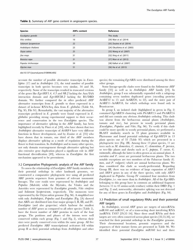
account the number of possible alternative transcripts in Euca-lyptus (17) and in Arabidopsis (15), the total number of possible
transcripts in both species becomes very similar, 34 and 38,
respectively. Some of the transcripts resulted in truncated versions
of the genes like EgrARF1.4, 4.3 and 9B.2 lacking the Aux/IAA
interaction domain and EgrARF2B.2 lacking the B3/DBD
domain. We further compared the in silico predicted ARFalternative transcripts from E. grandis to those expressed in a
dataset of in-house RNA-Seq data from E. globulus (Table S4,
Fig. S5, File S1). Remarkably, the vast majority of the alternative
transcripts predicted in E. grandis were found expressed in E.globulus providing strong experimental support to their occur-
rence and conservation in the two Eucalyptus species. The
importance of alternative splicing in the ARF family, has been
highlighted recently by Finet et al. [49], who have shown that two
Arabidopsis alternative transcripts of AtARF4 have very different
functions in flower development, and by Zouine et al. [35] who
have shown that in tomato, one third of the ARF members
displays alternative splicing as a mode of regulation during the
flower to fruit transition. In Arabidopsis and in many other species,
not only domain rearrangement through alternative splicing but
also extensive gene duplication played a significant role in ARFfunctional diversification [49], whereas in Eucalyptus the first
mechanism appeared to be preeminent.
3.2 Comparative Phylogenetic analysis of the ARF familyTo assess the relationship of Eucalyptus ARF family members to
their potential orthologs in other landmark genomes, we
constructed a comparative phylogenetic tree using all predicted
ARF protein sequences from genomes of relevant taxonomic
lineages. The core rosids were represented by Arabidopsis and
Populus (Malvids) while the Myrtales, the Vitales and the
Asterides were represented by Eucalyptus grandis, Vitis viniferaand Solanum lycopersicum, respectively. The monocots were
represented by the Oryza sativa genome (Fig. S6). A simplified tree
with only Arabidopsis, Populus and Eucalyptus (Fig. 2A) showed
that ARFs are distributed into four major groups I, II, III, and IV.
Eucalyptus (and also grapevine) which harbour the smallest
number of ARF genes as compared to all other species (Table 2),
have the fewest number of ARF proteins in each of the four
groups. The positions and phases of the introns were well
conserved within each group (Fig. 1 and Fig. 2), whereas their
sizes were poorly conserved even within the same group. All five
predicted Eucalyptus ARF transcriptional activators fell within
group II as their potential orthologs from Arabidopsis and other
species; the remaining EgrARFs were distributed among the three
other groups.
Some lineage-specific clades were found in the Solanaceae ARFfamily [35] as well as in Arabidopsis ARF family [24]. In
Arabidopsis, group I was substantially expanded with a subgroup
containing seven tandem duplicated genes (encoding proteins
AtARF12 to 15 and AtARF20, to 22), and the sister pair of
AtARF11–AtARF18, for which orthologs were found only in
Brassicaceae.
In group I, an isolated clade (highlighted in green in Fig. 2)
contained EgrARF24 clustering with PtrARF2.5 and PtrARF2.6
and did not contain any obvious Arabidopsis ortholog. This clade
was absent from the herbaceous annual plants (Arabidopsis,tomato and rice), but present in woody perennial plants
(Eucalyptus, Populus and Vitis; Fig. S6). To verify if this clade
could be more specific to woody perennial plants, we performed a
BLAST similarity search in 33 plant genomes available in
Phytozome and found potential orthologs of EgrARF24 in 13
plant species out of 33 (Table S5) which are presented in a
phylogenetic tree (Fig. 2B). Among these 13 plant species, 11 are
trees such as M. domestica, C. sinensis, C. clementina, P. persica,
or tree-like plants and shrubs such as C. papaya, T. cacao, G.raimondii, although the latter is often grown as an annual plant. A.coerulea and F. vesca are perennial herbaceous plants. The two
notable exceptions are two members of the Fabaceae family (G.max, and P. vulgaris) which are annual herbaceous plants. We
thus considered this clade as woody-preferential. Regarding
Group III, there was no evidence of large expansion of ARF3and ARF4 genes in any of the three species, with only ARF3duplicated in Populus. Group IV contained four members from
Eucalyptus, i.e. one more than in Arabidopsis. All of the EgrARFs
belonging to this group have in common an additional fragment
(between 15 to 43 amino-acids residues) within their DBD (Fig. 1
and Fig. 2) and, noteworthy, alternative splicing was not detected
for any of these genes in Eucalyptus and Arabidopsis (Fig. S4).
3.3 Prediction of small regulatory RNAs and their potential ARF targets
In Arabidopsis, several ARF genes are targets of microRNAs
miR160 and miR167, or of a trans-acting short interfering RNA
(tasiRNA) TAS3 [29,52–54]. Since these small RNAs and their
targets are very often conserved across plant species [32,55,56], we
searched for their potential orthologs in the Eucalyptus genome.
Their chromosomal locations, genomic sequences and the
sequences of their mature forms are presented in Table S6. We
identified three potential Eucalyptus miR160 loci and three
Table 2. Summary of ARF gene content in angiosperm species.
Species ARFs content Reference
Eucalyptus grandis 17 This study
Vitis vinifera 19 [39] Wan et al. (2014)
Solanum lycopersicon 22 [35] Zouine et al. (2014)
Arabidopsis thaliana 23 [24] Okushima et al. (2005)
Oryza sativa 25 [33] Wang et al. (2007)
Zea mays 31 [32] Xing et al. (2011)
Brassica rapa 31 [37] Mun et al. (2012)
Populus trichocarpa 39 [38] Kalluri et al. (2007)
Glycine max 51 [36] Ha et al. (2013)
doi:10.1371/journal.pone.0108906.t002
The Eucalyptus ARF Family
PLOS ONE | www.plosone.org 57 September 2014 | Volume 9 | Issue 9 | e108906
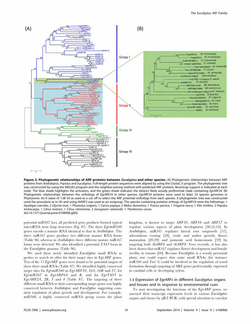
potential miR167 loci, all predicted gene products formed typical
microRNA stem–loop structures (Fig. S7). The three EgrmiR160genes encode a mature RNA identical to that in Arabidopsis. The
three miR167 genes produce two different mature RNA forms
(Table S6) whereas in Arabidopsis three different mature miR167
forms were detected. We also identified a potential TAS3 locus in
the Eucalyptus genome (Table S6).
We used these newly identified Eucalyptus small RNAs as
probes to search in silico for their target sites in EgrARF genes.
Ten of the 17 EgrARF genes were found to be potential targets of
these three small RNAs (Table S7). We identified highly conserved
target sites for EgrmiR160 in EgrARF10, 16A, 16B and 17, for
EgrmiR167 in EgrARF6A and B, and for EgrTAS3 in
EgrARF2A, 2B, 3 and 4 (Table S7). The targeting of three
different small RNA to their corresponding target genes was highly
conserved between Arabidopsis and Eucalyptus suggesting com-
mon regulation of plant growth and development. For example,
miR160, a highly conserved miRNA group across the plant
kingdom, is known to target ARF10, ARF16 and ARF17 to
regulate various aspects of plant development [30,52,53]. In
Arabidopsis, miR167 regulates lateral root outgrowth [57],
adventitious rooting [58], ovule and anther growth, flower
maturation [20,29] and jasmonic acid homeostasis [59] by
targeting both AtARF6 and AtARF8. Very recently, it has also
been shown that miR167 regulates flower development and female
sterility in tomato [60]. Because Eucalyptus is a woody perennial
plant, one could expect that some small RNAs (for instance
miR160 and Tasi 3) could be involved in the regulation of wood
formation through targeting of ARF genes preferentially expressed
in cambial cells or developing xylem.
3.4 Expression of EgrARFs in different Eucalyptus organs and tissues and in response to environmental cues
To start investigating the functions of the EgrARF genes, we
assessed their transcript expression levels in various Eucalyptusorgans and tissues by qRT-PCR, with special attention to vascular
Figure 2. Phylogenetic relationships of ARF proteins between Eucalyptus and other species. (A) Phylogenetic relationships between ARFproteins from Arabidopsis, Populus and Eucalyptus. Full-length protein sequences were aligned by using the Clustal_X program. The phylogenetic treewas constructed by using the MEGA5 program and the neighbor-joining method with predicted ARF proteins. Bootstrap support is indicated at eachnode. The blue shade highlights the activators, and the green shade indicates the distinct likely woody preferential clade containing EgrARF24. (B)Phylogenetic relationships between the orthologs of EgrARF24 in other species. EgrARF24 proteins were used to blast 33 species genomes inPhytozome. An E-value of 1.0E-50 as used as a cut off to select the ARF potential orthologs from each species. A phylogenetic tree was constructedused the procedure as in (A) and using AtARF2 was used as an outgroup. The species containing putative orthogs of EgrARF24 were the followings: 1Aquilegia coerulea, 2 Glycine max, 1 Phaseolus vulgaris, 1 Carica papaya, 2 Malus domestica, 1 Prunus persica, 1 Fragaria vesca, 1 Vitis vinifera, 2 Populustrichocarpa, 1 Citrus sinensis, 1 Citrus clementine, 2 Gossypium raimondii, 1 Theobroma cacao.doi:10.1371/journal.pone.0108906.g002
The Eucalyptus ARF Family
PLOS ONE | www.plosone.org 58 September 2014 | Volume 9 | Issue 9 | e108906
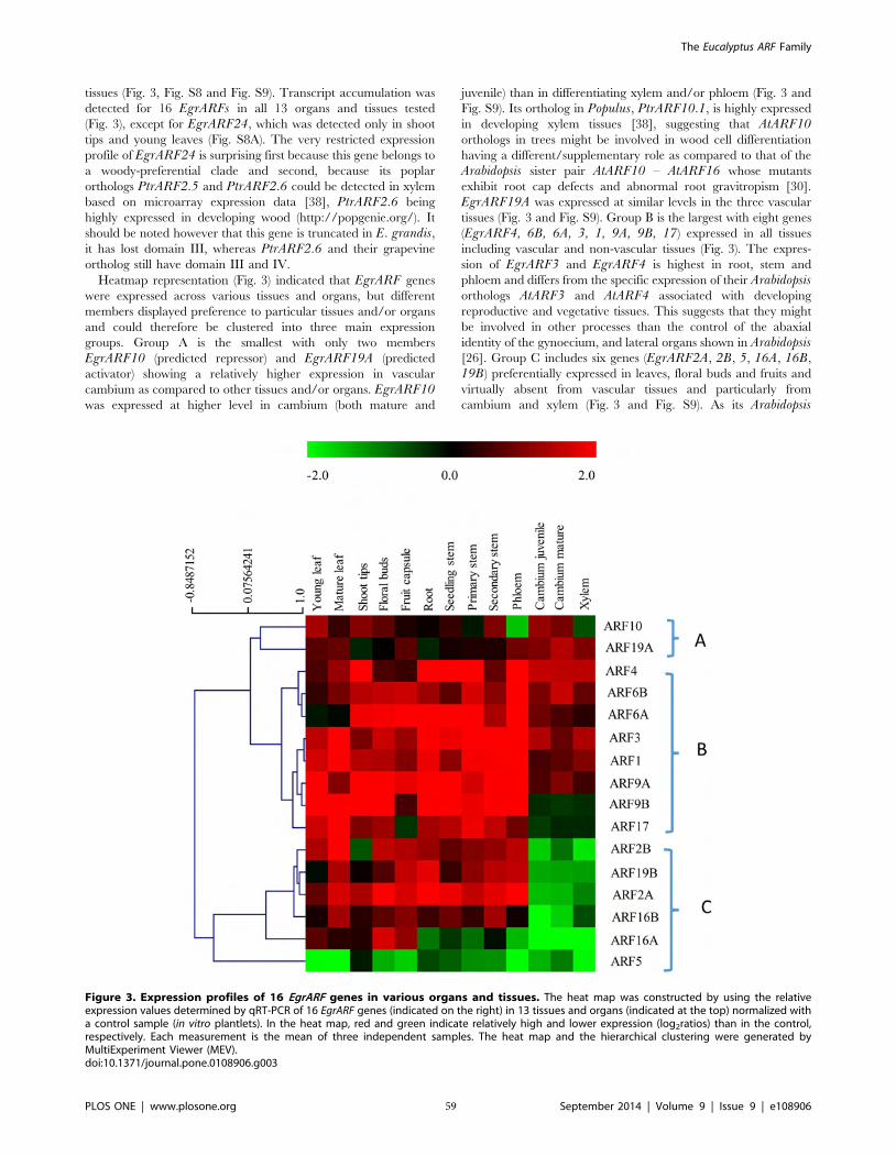
tissues (Fig. 3, Fig. S8 and Fig. S9). Transcript accumulation was
detected for 16 EgrARFs in all 13 organs and tissues tested
(Fig. 3), except for EgrARF24, which was detected only in shoot
tips and young leaves (Fig. S8A). The very restricted expression
profile of EgrARF24 is surprising first because this gene belongs to
a woody-preferential clade and second, because its poplar
orthologs PtrARF2.5 and PtrARF2.6 could be detected in xylem
based on microarray expression data [38], PtrARF2.6 being
highly expressed in developing wood (http://popgenie.org/). It
should be noted however that this gene is truncated in E. grandis,it has lost domain III, whereas PtrARF2.6 and their grapevine
ortholog still have domain III and IV.
Heatmap representation (Fig. 3) indicated that EgrARF genes
were expressed across various tissues and organs, but different
members displayed preference to particular tissues and/or organs
and could therefore be clustered into three main expression
groups. Group A is the smallest with only two members
EgrARF10 (predicted repressor) and EgrARF19A (predicted
activator) showing a relatively higher expression in vascular
cambium as compared to other tissues and/or organs. EgrARF10was expressed at higher level in cambium (both mature and
juvenile) than in differentiating xylem and/or phloem (Fig. 3 and
Fig. S9). Its ortholog in Populus, PtrARF10.1, is highly expressed
in developing xylem tissues [38], suggesting that AtARF10orthologs in trees might be involved in wood cell differentiation
having a different/supplementary role as compared to that of the
Arabidopsis sister pair AtARF10 – AtARF16 whose mutants
exhibit root cap defects and abnormal root gravitropism [30].
EgrARF19A was expressed at similar levels in the three vascular
tissues (Fig. 3 and Fig. S9). Group B is the largest with eight genes
(EgrARF4, 6B, 6A, 3, 1, 9A, 9B, 17) expressed in all tissues
including vascular and non-vascular tissues (Fig. 3). The expres-
sion of EgrARF3 and EgrARF4 is highest in root, stem and
phloem and differs from the specific expression of their Arabidopsisorthologs AtARF3 and AtARF4 associated with developing
reproductive and vegetative tissues. This suggests that they might
be involved in other processes than the control of the abaxial
identity of the gynoecium, and lateral organs shown in Arabidopsis[26]. Group C includes six genes (EgrARF2A, 2B, 5, 16A, 16B,
19B) preferentially expressed in leaves, floral buds and fruits and
virtually absent from vascular tissues and particularly from
cambium and xylem (Fig. 3 and Fig. S9). As its Arabidopsis
Figure 3. Expression profiles of 16 EgrARF genes in various organs and tissues. The heat map was constructed by using the relativeexpression values determined by qRT-PCR of 16 EgrARF genes (indicated on the right) in 13 tissues and organs (indicated at the top) normalized witha control sample (in vitro plantlets). In the heat map, red and green indicate relatively high and lower expression (log2ratios) than in the control,respectively. Each measurement is the mean of three independent samples. The heat map and the hierarchical clustering were generated byMultiExperiment Viewer (MEV).doi:10.1371/journal.pone.0108906.g003
The Eucalyptus ARF Family
PLOS ONE | www.plosone.org 59 September 2014 | Volume 9 | Issue 9 | e108906
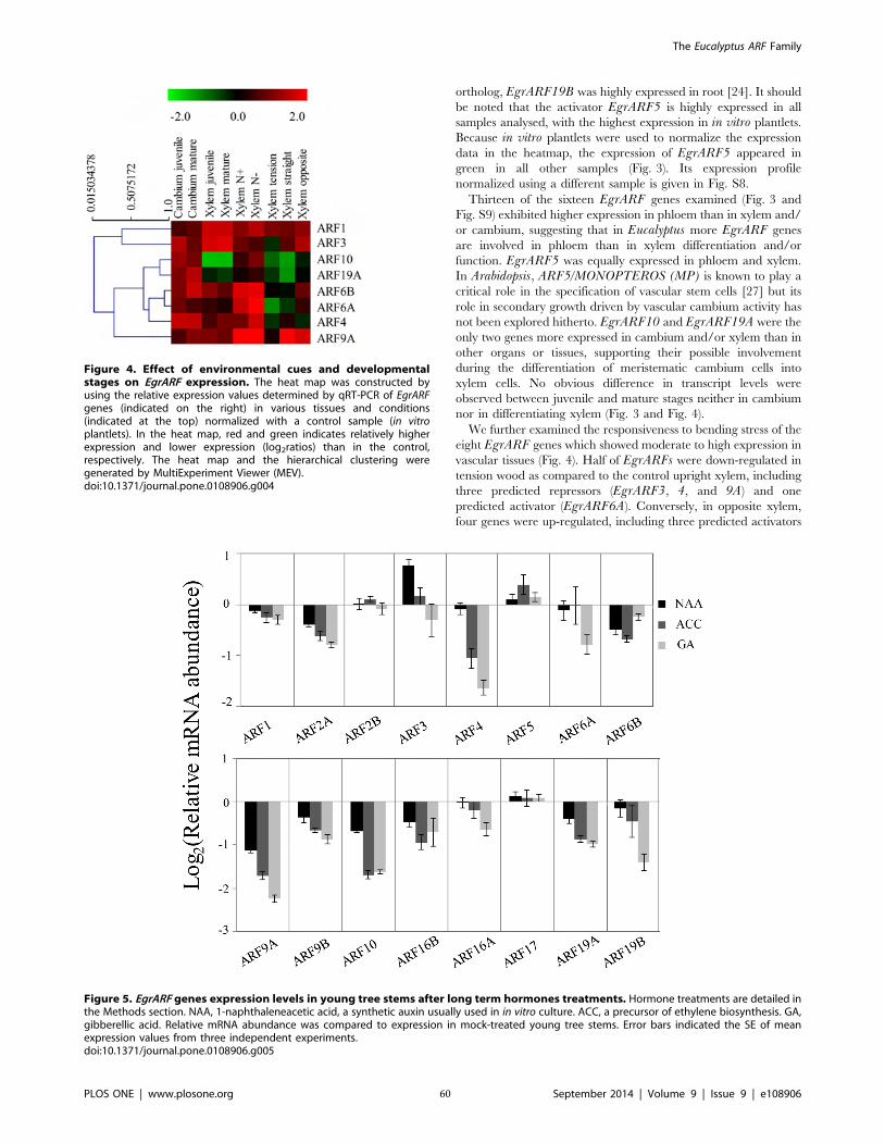
ortholog, EgrARF19B was highly expressed in root [24]. It should
be noted that the activator EgrARF5 is highly expressed in all
samples analysed, with the highest expression in in vitro plantlets.
Because in vitro plantlets were used to normalize the expression
data in the heatmap, the expression of EgrARF5 appeared in
green in all other samples (Fig. 3). Its expression profile
normalized using a different sample is given in Fig. S8.
Thirteen of the sixteen EgrARF genes examined (Fig. 3 and
Fig. S9) exhibited higher expression in phloem than in xylem and/
or cambium, suggesting that in Eucalyptus more EgrARF genes
are involved in phloem than in xylem differentiation and/or
function. EgrARF5 was equally expressed in phloem and xylem.
In Arabidopsis, ARF5/MONOPTEROS (MP) is known to play a
critical role in the specification of vascular stem cells [27] but its
role in secondary growth driven by vascular cambium activity has
not been explored hitherto. EgrARF10 and EgrARF19A were the
only two genes more expressed in cambium and/or xylem than in
other organs or tissues, supporting their possible involvement
during the differentiation of meristematic cambium cells into
xylem cells. No obvious difference in transcript levels were
observed between juvenile and mature stages neither in cambium
nor in differentiating xylem (Fig. 3 and Fig. 4).
We further examined the responsiveness to bending stress of the
eight EgrARF genes which showed moderate to high expression in
vascular tissues (Fig. 4). Half of EgrARFs were down-regulated in
tension wood as compared to the control upright xylem, including
three predicted repressors (EgrARF3, 4, and 9A) and one
predicted activator (EgrARF6A). Conversely, in opposite xylem,
four genes were up-regulated, including three predicted activators
Figure 4. Effect of environmental cues and developmentalstages on EgrARF expression. The heat map was constructed byusing the relative expression values determined by qRT-PCR of EgrARFgenes (indicated on the right) in various tissues and conditions(indicated at the top) normalized with a control sample (in vitroplantlets). In the heat map, red and green indicates relatively higherexpression and lower expression (log2ratios) than in the control,respectively. The heat map and the hierarchical clustering weregenerated by MultiExperiment Viewer (MEV).doi:10.1371/journal.pone.0108906.g004
Figure 5. EgrARF genes expression levels in young tree stems after long term hormones treatments. Hormone treatments are detailed inthe Methods section. NAA, 1-naphthaleneacetic acid, a synthetic auxin usually used in in vitro culture. ACC, a precursor of ethylene biosynthesis. GA,gibberellic acid. Relative mRNA abundance was compared to expression in mock-treated young tree stems. Error bars indicated the SE of meanexpression values from three independent experiments.doi:10.1371/journal.pone.0108906.g005
The Eucalyptus ARF Family
PLOS ONE | www.plosone.org 60 September 2014 | Volume 9 | Issue 9 | e108906
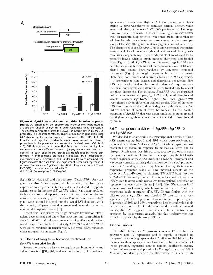
(EgrARF6A, 6B, 19A) and one repressor (EgrARF10). Only one
gene (EgrARF4) was repressed. In general, EgrARF gene
expression was repressed in tension xylem and induced in opposite
xylem, except in the case of EgrARF4, which was down-regulated
in both tension and opposite xylem (Fig. 4). These results are
consistent with a study performed in Populus where seven ARFgenes were detected in a poplar tension wood EST database, while
the majority of genes were down-regulated in tension wood as
compared to opposite wood [61].
Recent studies indicated that high nitrogen fertilization affects
xylem development and alters fibre structure and composition in
Populus [62,63] and induces some overlapping effects with tension
wood on xylem cell walls. Interestingly, EgrARF4 and EgrARF6Awere down regulated in tension wood, but were down regulated
when nitrogen was in excess (Fig. 4).
3.5 Effects of long-term hormone treatments on EgrARFs transcript levels
Several hormones are known to regulate cambium activity and
xylem formation [[31], [64] and references therein]. For instance,
application of exogenous ethylene (ACC) on young poplar trees
during 12 days was shown to stimulate cambial activity, while
xylem cell size was decreased [65]. We performed similar long-
term hormonal treatments (15 days) by growing young Eucalyptustrees on medium supplemented with either auxin, gibberellin or
ethylene in order to evaluate the consequences on the transcripts
levels of the EgrARF genes in stems (organs enriched in xylem).
The phenotypes of the Eucalyptus trees after hormonal treatments
were typical of each hormone: gibberellin stimulated plant growth
resulting in longer stems, ethylene reduced plant growth and led to
epinastic leaves, whereas auxin induced shortened and bolded
roots (Fig. S10). All EgrARF transcripts except EgrARF24 were
detected in young tree stems and the expression levels of 13 were
altered and mainly down-regulated by long-term hormonal
treatments (Fig. 5). Although long-term hormonal treatments
likely have both direct and indirect effects on ARFs expression,
it is interesting to note distinct and differential behaviours: Five
ARFs exhibited a kind of ‘‘hormonal preference’’ response since
their transcripts levels were altered in stems treated only by one of
the three hormones. For instance, EgrARF3 was up-regulated
only in auxin treated samples; EgrARF5, only in ethylene treated
samples, whereas EgrARF6A, EgrARF16A and EgrARF19Bwere altered only in gibberellin treated samples. Most of the other
ARFs were modulated at different degrees by the direct and/or
indirect actions of each of three hormones with the notable
exception of EgrARF4 that was down-regulated in stems treated
by ethylene and gibberellic acid but not affected in those treated
by auxin.
3.6 Transcriptional activities of EgrARF4, EgrARF 10 and EgrARF19A
We decided to characterize the transcriptional activity of three
ARF members: EgARF10 and 19A which were preferentially
expressed in cambium/xylem, and EgARF4 whose expression was
modulated in xylem in response to mechanical stress and to
nitrogen fertilization. For this purpose, tobacco protoplasts were
co-transfected with an effector construct expressing the full-length
coding sequence of the ARFs under the 35SCaMV promoter and
a reporter construct carrying the auxin-responsive DR5 promoter
fused to GFP coding sequence (Fig. 6A). DR5 is a synthetic auxin-
responsive promoter made of nine inverted repeats of the
conserved Auxin-Responsive Element, (TGTCTC box), fused to
a 35SCaMV minimal promoter. This reporter construct has been
widely used to assess auxin responsive transcriptional activation or
repression in vivo and in planta [15,47]. The DR5-driven GFP
showed low basal activity which was induced up to 4-fold by
exogenous auxin treatment (Fig. 6B). Co-transfection with the
effector genes EgrARF4 and EgrARF10 resulted in a very
significant (p,0.001) repression of auxin-induced reporter gene.
Expression of 80% and 38%, respectively hereby confirming their
predicted repressors roles. On the other hand, the values obtained
for EgrARF19A suggested that it could be an activator as
predicted by its sequence analysis, but this tendency was not
strongly supported by the student-T test.
4 Conclusions
The ARF family in E. grandis contains 17 members (5
activators and 12 repressors) and is slightly contracted as
compared to most angiosperm ARF families studied hitherto. In
contrast to these species, it is characterized by the absence of
whole genome, segmental and/or tandem duplication events.
Indeed, whole genome duplication in Eucalyptus occurred 109.9
Mya ago, considerably earlier than those detected in other rosids
Figure 6. EgrARF transcriptional activities in tobacco proto-plasts. (A) Schemes of the effector and reporter constructs used toanalyse the function of EgrARFs in auxin-responsive gene expression.The effector constructs express the EgrARF of interest driven by the 35Spromoter. The reporter construct consists of a reporter gene expressingGFP driven by the auxin-responsive promoter DR5 (DR5::GFP). (B)Effector and reporter constructs were co-expressed in tobaccoprotoplasts in the presence or absence of a synthetic auxin (50 mM 2,4-D). GFP fluorescence was quantified 16 h after transfection by flowcytometry. A mock effector construct (empty vector) was used as acontrol. In each experiment, protoplast transformations were per-formed in independent biological triplicates. Two independentexperiments were performed and similar results were obtained; thefigure indicates the data from one experiment. Error bars represent SEof mean fluorescence. Significant statistical differences (student T test,P,0.001) to control are marked with **.doi:10.1371/journal.pone.0108906.g006
The Eucalyptus ARF Family
PLOS ONE | www.plosone.org 61 September 2014 | Volume 9 | Issue 9 | e108906
and 95% of the paralogs were lost [40]. The absence of tandem
duplication is remarkable especially because E. grandis has the
largest number of genes in tandem repeats (34% of the total
number of genes) reported among sequenced plant genomes.
Indeed, tandem duplication shaped functional diversity in many
gene families in Eucalyptus. The ARF family thus evolved in a
very different way. Our data suggests that genomic truncation and
alternative splicing were preeminent mechanisms leading to the
diversity of domain architecture, shaping and increasing the
functional diversity of the ARF family in Eucalyptus, thereby
compensating for the lack of extensive gene duplication found in
other species. Comparative phylogenetic studies pointed out the
presence of a new clade, maintained preferentially in woody and
perennial plants. Finally, large scale expression profiling allowed
identifying candidates potentially involved in the auxin-regulated
transcriptional programs underlying wood formation.
Supporting Information
Figure S1 Procedure used for identifying ARF genes inEucalyptus grandis. Arabidopsis ARF protein sequences were
used to search their orthologs in the predicted Eucalyptusproteome by using in BLASTP. Sixty-four Eucalyptus proteins
identified in this initial search were further examined by manual
curation using protein motif scanning and the FgeneSH program
to complete partial sequences. Redundant and invalid genes were
eliminated based on gene structure, integrity of conserved motifs
and EST support. Manual curation resulted in 17 complete
Eucalyptus ARF protein sequences. These 17 protein sequences
were used in two subsequent additional searches: first, a BLASTP
search against the Eucalyptus proteome to identify exhaustively all
divergent Eucalyptus ARF gene family members and, second,
tBLASTn searches against the Eucalyptus genome for any possible
unpredicted genes. To confirm our findings, we used poplar ARF
proteins and repeated the complete search procedure described
above and obtained identical results.
(TIFF)
Figure S2 Locations of the 17 EgrARF genes on the 11Eucalyptus grandis chromosomes.
(TIFF)
Figure S3 Multiple sequence alignment of predictedamino acid sequences of EgrARF and AtARF proteins.The multiple sequence alignment was obtained with the
MUSCLE software [66]. The highly conserved domains and
nuclear localization signals (NLSs) proteins were noted on the
bottom of the alignment with different colours.
(PDF)
Figure S4 Comparative analysis of predicted ARFalternative variants between Eucalyptus grandis andArabidopsis thaliana. The alternative spliced protein sequenc-
es were extracted from Phytozome except for AtARF4 (obtained
from Finet et al. (2013), the motif structures were predicted by
Pfam (http://pfam.xfam.org/).
(PDF)
Figure S5 Structure of the ARF alternative transcripts inE. globulus. The E. globulus alternative transcripts were
obtained from a compendium of RNASeq data. The material
and methods are described in Table S4. The illumina reads
sequences are provided in File S1 in the FastQ format.
(PDF)
Figure S6 Comparative Phylogenetic relationships be-tween ARF proteins from poplar, Eucalyptus, grapevine,
Arabidopsis, tomato and rice. Full-length protein sequences
were aligned using the Clustal_X program. The phylogenetic tree
was constructed by using the MEGA5 program and the
neighbour-joining method with predicted full-length ARF pro-
teins. Bootstrap supports are indicated at each node.
(PDF)
Figure S7 Predicted stem-loop structures of threeEgrmiR160 and three EgrmiR167. The part of the stem-loop
from which the mature microRNA derives is highlighted in yellow.
(TIFF)
Figure S8 Expression profiles of EgrARF5 and EgrARF24in various organs and tissues. Relative mRNA abundance of
EgrARF5 and EgrARF24 was compared to expression in the
control sample of mature leaves and in vitro plantlets, respectively.
Error bars indicate the SE of mean expression values from three
independent experiments.
(TIFF)
Figure S9 Expression profiles of EgrARF genes intissues involved in secondary growth. Relative mRNA
abundance was compared to expression in the control sample (invitro plantlets).
(TIFF)
Figure S10 Young Eucalyptus grandis trees phenotypesin response to various long-term hormonal treatments.10 mM NAA, or 20 mM gibberellic acid or 100 mM ACC were
added to the medium of 65-d-old young tree, and phenotypes were
observed 14 days later.
(TIFF)
Table S1 Primers for EgrARF genes and reference genesused in qRT-PCR experiments.(PDF)
Table S2 Protein identity matrix between EgrARF andAtARF.(PDF)
Table S3 Protein identity matrix among EgrARF.(PDF)
Table S4 Comparison of the number of alternativetranscripts predicted in phytozome for E. grandis tothose found in a large compendium of transcriptomicdata from in E. globulus.(PDF)
Table S5 Comparison of the number of EgrARF24putative orthologs in other species.(PDF)
Table S6 Potential small RNAs targeting EgrARF genes.(PDF)
Table S7 Small RNAs target site prediction in EgrARFgenes.(PDF)
File S1 Sequences of the Illumina reads from RNA Seqused to predict the E. globulus alternative transcripts.The origin of the material and the procedure are described in
Table S4.
(ZIP)
Acknowledgments
The authors are grateful to J. P Combier for his help and advices for
miRNA and target prediction, to E. Camargo (Unicamp, Brazil), J.M.
The Eucalyptus ARF Family
PLOS ONE | www.plosone.org 62 September 2014 | Volume 9 | Issue 9 | e108906

Gion and E. Villar (CIRAD, FR), F. Melun and L. Harvengt (FCBA,
France), C. Araujo and L. Neves (AltriFlorestal, Portugal) and C. Marques
(RAIZ, Portugal) for kindly providing and/or allowing collection of
Eucalyptus organ and tissue samples, C. Graca, and V. Carocha for help
with sample collection and RNA extraction, N Ladouce for performing
Fluidigm experiments. Thanks to the Bioinfo Genotoul Platform (http://
bioinfo.genotoul.fr) for access to bioinformatic resources and to the
Genome and transcriptome Platform (http://get.genotoul.fr/) for advice
and technical assistance with high-throughput Biomark Fluidigm qRT-
PCR amplifications. Thanks to Y. Martinez (FR3450) for assistance with
microscopy analysis to C. Pecher and A. Zakaroff-Girard for technical
assistance and expertise in flow cytometry (Toulouse RIO imaging
platform http://tri.ups-tlse.fr).
Author Contributions
Conceived and designed the experiments: HY HCW MS JGP. Performed
the experiments: HY HSC CD IM BS. Analyzed the data: HY HCW MB
MS JGP. Contributed reagents/materials/analysis tools: JAPP AAM.
Wrote the paper: JGP YH HCW.
5 References
1. Woodward AW, Bartel B (2005) Auxin: regulation, action, and interaction. Ann
Bot 95: 707-735.
2. Miyashima S, Sebastian J, Lee JY and Helariutta Y (2013) Stem cell function
during plant vascular development. EMBO J 32: 178–193.
3. Tuominen H, Puech L, Fink S, Sundberg B (1997) A radial concentration
gradient of indole-3-acetic acid is related to secondary xylem development in
hybrid aspen. Plant Physiol 115: 577–585.
4. Uggla C, Moritz T, Sandberg G, Sundberg B (1996) Auxin as a positional signal
in pattern formation in plants. Proc Natl Acad Sci U S A 93: 9282–9286.
5. Nilsson J, Karlberg A, Antti H, Lopez-Vernaza M, Mellerowicz E, et al. (2008)
Dissecting the molecular basis of the regulation of wood formation by auxin in
hybrid aspen. Plant Cell 20: 843–855.
6. Pilate G, Dejardin A, Laurans F, Leple JC (2004) Tension wood as a model for
functional genomics of wood formation. New Phytol 164: 63–72.
7. Paux E, Carocha V, Marques C, Mendes de Sousa A, Borralho N, et al. (2005)
Transcript profiling of Eucalyptus xylem genes during tension wood formation.
New Phytol 167: 89–100.
8. Timell TE (1969) The chemical composition of tension wood. Svensk
Papperstidn 72: 173–181.
9. Andersson-Gunneras S, Mellerowicz EJ, Love J, Segerman B, Ohmiya Y, et al.
(2006) Biosynthesis of cellulose-enriched tension wood in Populus: global
analysis of transcripts and metabolites identifies biochemical and developmental
regulators in secondary wall biosynthesis. Plant J 45: 144–165.
10. Baba K, Karlberg A, Schmidt J, Schrader J, Hvidsten TR, et al. (2011) Activity-
dormancy transition in the cambial meristem involves stage-specific modulation
of auxin response in hybrid aspen. Proc Natl Acad Sci U S A 108: 3418–3423.
11. Sundberg B, Uggla C, Tuominen H (2000) Cambial growth and auxin gradients.
Oxford: BIOS Scientific Publishers. 169–188 p.
12. Guilfoyle TJ, Hagen G (2007) Auxin response factors. Curr Opin Plant Biol 10:
453–460.
13. Tiwari SB, Hagen G, Guilfoyle T (2003) The roles of auxin response factor
domains in auxin-responsive transcription. Plant Cell 15: 533–543.
14. Hagen G, Guilfoyle T (2002) Auxin-responsive gene expression: genes,
promoters and regulatory factors. Plant Mol Biol 49: 373–385.
15. Ulmasov T, Hagen G, Guilfoyle TJ (1999) Activation and repression of
transcription by auxin-response factors. Proc Natl Acad Sci U S A 96: 5844–
5849.
16. Gray WM, Kepinski S, Rouse D, Leyser O and Estelle M (2001) Auxin regulates
SCF(TIR1)-dependent degradation of AUX/IAA proteins. Nature 414: 271–
276.
17. Szemenyei H, Hannon M and Long JA (2008) TOPLESS mediates auxin-
dependent transcriptional repression during Arabidopsis embryogenesis. Science
319: 1384–1386.
18. Vernoux T, Brunoud G, Farcot E, Morin V, Van den Daele H, et al. (2011) The
auxin signalling network translates dynamic input into robust patterning at the
shoot apex. Mol Syst Biol 7: 508.
19. Paponov IA, Paponov M, Teale W, Menges M, Chakrabortee S, et al. (2008)
Comprehensive transcriptome analysis of auxin responses in Arabidopsis. Mol
Plant 1: 321–337.
20. Nagpal P, Ellis CM, Weber H, Ploense SE, Barkawi LS, et al. (2005) Auxin
response factors ARF6 and ARF8 promote jasmonic acid production and flower
maturation. Development 132: 4107–4118.
21. Pufky J, Qiu Y, Rao MV, Hurban P, Jones AM (2003) The auxin-induced
transcriptome for etiolated Arabidopsis seedlings using a structure/function
approach. Funct Integr Genomics 3: 135–143.
22. Ellis CM, Nagpal P, Young JC, Hagen G, Guilfoyle TJ, et al. (2005) AUXINRESPONSE FACTOR 1 and AUXIN RESPONSE FACTOR 2 regulate
senescence and floral organ abscission in Arabidopsis thaliana. Development
132: 4563–4574.
23. Okushima Y, Mitina I, Quach HL, Theologis A (2005) AUXIN RESPONSEFACTOR 2 (ARF2): a pleiotropic developmental regulator. Plant J 43: 29–46.
24. Okushima Y, Overvoorde PJ, Arima K, Alonso JM, Chan A, et al. (2005)
Functional genomic analysis of the AUXIN RESPONSE FACTOR gene family
members in Arabidopsis thaliana: unique and overlapping functions of ARF7and ARF19. Plant Cell 17: 444–463.
25. Sessions A, Nemhauser JL, McColl A, Roe JL, Feldmann KA, et al. (1997)
ETTIN patterns the Arabidopsis floral meristem and reproductive organs.
Development 124: 4481–4491.
26. Pekker I, Alvarez JP, Eshed Y (2005) Auxin response factors mediate Arabidopsisorgan asymmetry via modulation of KANADI activity. Plant Cell 17: 2899–
2910.
27. Hardtke CS, Berleth T (1998) The Arabidopsis gene MONOPTEROS encodes a
transcription factor mediating embryo axis formation and vascular development.
EMBO J 17: 1405–1411.
28. Harper RM, Stowe-Evans EL, Luesse DR, Muto H, Tatematsu K, et al. (2000)
The NPH4 locus encodes the auxin response factor ARF7, a conditional
regulator of differential growth in aerial Arabidopsis tissue. Plant Cell 12: 757–
770.
29. Wu MF, Tian Q, Reed JW (2006) Arabidopsis microRNA167 controls patterns
of ARF6 and ARF8 expression, and regulates both female and male
reproduction. Development 133: 4211–4218.
30. Wang JW, Wang LJ, Mao YB, Cai WJ, Xue HW, et al. (2005) Control of root
cap formation by MicroRNA-targeted auxin response factors in Arabidopsis.Plant Cell 17: 2204–2216.
31. Miyashima S, Sebastian J, Lee JY, Helariutta Y (2013) Stem cell function during
plant vascular development. EMBO J 32: 178–193.
32. Xing H, Pudake RN, Guo G, Xing G, Hu Z, et al. (2011) Genome-wide
identification and expression profiling of auxin response factor (ARF) gene family
in maize. BMC Genomics 12: 178.
33. Wang D, Pei K, Fu Y, Sun Z, Li S, et al. (2007) Genome-wide analysis of the
auxin response factors (ARF) gene family in rice (Oryza sativa). Gene 394: 13–24.
34. Kumar R, Tyagi AK, Sharma AK (2011) Genome-wide analysis of auxinresponse factor (ARF) gene family from tomato and analysis of their role in
flower and fruit development. Mol Genet Genomics 285: 245–260.
35. Zouine M, Fu Y, Chateigner-Boutin AL, Mila I, Frasse P, et al. (2014)
Characterization of the tomato ARF gene family uncovers a multi-levels post-
transcriptional regulation including alternative splicing. PloS one 9: e84203.
36. Ha CV, Le DT, Nishiyama R, Watanabe Y, Sulieman S, et al. (2013) The auxin
response factor transcription factor family in soybean: genome-wide identifica-
tion and expression analyses during development and water stress. DNA Res 20:
511–524.
37. Mun JH, Yu HJ, Shin JY, Oh M, Hwang HJ, et al. (2012) Auxin response factor
gene family in Brassica rapa: genomic organization, divergence, expression, and
evolution. Mol Genet Genomics 287: 765–784.
38. Kalluri UC, Difazio SP, Brunner AM, Tuskan GA (2007) Genome-wide analysis
of Aux/IAA and ARF gene families in Populus trichocarpa. BMC Plant Biol 7:
59.
39. Wan S, Li W, Zhu Y, Liu Z, Huang W, et al. (2014) Genome-wide
identification, characterization and expression analysis of the auxin response
factor gene family in Vitis vinifera. Plant Cell Rep 33: 1365–1375.
40. Myburg AA, Grattapaglia D, Tuskan GA, Hellsten U, Hayes RD, et al. (2014)
The genome of Eucalyptus grandis. Nature 510: 356–362.
41. Voorrips RE (2002) MapChart: software for the graphical presentation of
linkage maps and QTLs. J Hered 93: 77–78.
42. Finn RD, Bateman A, Clements J, Coggill P, Eberhardt RY, et al. (2014) Pfam:
the protein families database. Nucleic Acids Res 42: D222–230.
43. Cassan-Wang H, Soler M, Yu H, Camargo EL, Carocha V, et al. (2012)
Reference genes for high-throughput quantitative RT-PCR analysis of gene
expression in organs and tissues of Eucalyptus grown in various environmental
conditions. Plant Cell Physiol 53: 2101–2116.
44. Udvardi MK, Czechowski T, Scheible WR (2008) Eleven golden rules of
quantitative RT-PCR. Plant Cell 20: 1736–1737.
45. Arvidsson S, Kwasniewski M, Riano-Pachon DM, Mueller-Roeber B (2008)
QuantPrime-a flexible tool for reliable high-throughput primer design for
quantitative PCR. BMC Bioinformatics 9: 465.
46. Pfaffl MW (2001) A new mathematical model for relative quantification in real-
time RT-PCR. Nucleic Acids Res 29: e45.
47. Audran-Delalande C, Bassa C, Mila I, Regad F, Zouine M, et al. (2012)
Genome-wide identification, functional analysis and expression profiling of the
Aux/IAA gene family in tomato. Plant Cell Physiol 53: 659–672.
48. Shen C, Wang S, Bai Y, Wu Y, Zhang S, et al. (2010) Functional analysis of the
structural domain of ARF proteins in rice (Oryza sativa L.). J Exp Bot 61: 3971–
3981.
49. Finet C, Berne-Dedieu A, Scutt CP and Marletaz F (2013) Evolution of the ARFgene family in land plants: old domains, new tricks. Mol Biol Evol 30: 45–56.
The Eucalyptus ARF Family
PLOS ONE | www.plosone.org 63 September 2014 | Volume 9 | Issue 9 | e108906
50. Wu J, Wang F, Cheng L, Kong F, Peng Z, et al. (2011) Identification, isolation
and expression analysis of auxin response factor (ARF) genes in Solanumlycopersicum. Plant Cell Rep 30: 2059–2073.
51. Remington DL, Vision TJ, Guilfoyle TJ, Reed JW (2004) Contrasting modes of
diversification in the Aux/IAA and ARF gene families. Plant Physiol 135: 1738–
1752.
52. Liu PP, Montgomery TA, Fahlgren N, Kasschau KD, Nonogaki H, et al. (2007)
Repression of AUXIN RESPONSE FACTOR10 by microRNA160 is critical
for seed germination and post-germination stages. Plant J 52: 133–146.
53. Mallory AC, Bartel DP, Bartel B (2005) MicroRNA-directed regulation of
Arabidopsis AUXIN RESPONSE FACTOR17 is essential for proper develop-
ment and modulates expression of early auxin response genes. Plant Cell 17:
1360–1375.
54. Williams L, Carles CC, Osmont KS, Fletcher JC (2005) A database analysis
method identifies an endogenous trans-acting short-interfering RNA that targets
the Arabidopsis ARF2, ARF3, and ARF4 genes. Proc Natl Acad Sci U S A 102:
9703–9708.
55. Zhang B, Pan X, Cannon CH, Cobb GP, Anderson TA (2006) Conservation
and divergence of plant microRNA genes. Plant J 46: 243–259.
56. Lu S, Sun YH, Chiang VL (2008) Stress-responsive microRNAs in Populus.Plant J 55: 131–151.
57. Gifford ML, Dean A, Gutierrez RA, Coruzzi GM, Birnbaum KD (2008) Cell-
specific nitrogen responses mediate developmental plasticity. Proc Natl Acad
Sci U S A 105: 803–808.
58. Gutierrez L, Bussell JD, Pacurar DI, Schwambach J, Pacurar M, et al. (2009)
Phenotypic plasticity of adventitious rooting in Arabidopsis is controlled by
complex regulation of AUXIN RESPONSE FACTOR transcripts and
microRNA abundance. Plant Cell 21: 3119–3132.59. Gutierrez L, Mongelard G, Flokova K, Pacurar DI, Novak O, et al. (2012)
Auxin controls Arabidopsis adventitious root initiation by regulating jasmonic
acid homeostasis. Plant Cell 24: 2515–2527.60. Liu N, Wu S, Van Houten J, Wang Y, Ding B, et al. (2014) Down-regulation of
AUXIN RESPONSE FACTORS 6 and 8 by microRNA 167 leads to floraldevelopment defects and female sterility in tomato. J Exp Bot 65: 2507–2520.
61. Jin H, Do J, Moon D, Noh EW, Kim W, et al. (2011) EST analysis of functional
genes associated with cell wall biosynthesis and modification in the secondaryxylem of the yellow poplar (Liriodendron tulipifera) stem during early stage of
tension wood formation. Planta 234: 959–977.62. Pitre FE, Lafarguette F, Boyle B, Pavy N, Caron S, et al. (2010) High nitrogen
fertilization and stem leaning have overlapping effects on wood formation inpoplar but invoke largely distinct molecular pathways. Tree Physiol 30: 1273–
1289.
63. Pitre FE, Pollet B, Lafarguette F, Cooke JE, MacKay JJ, et al. (2007) Effects ofincreased nitrogen supply on the lignification of poplar wood. J Agri Food Chem
55: 10306–10314.64. Sorce C, Giovannelli A, Sebastiani L, Anfodillo T (2013) Hormonal signals
involved in the regulation of cambial activity, xylogenesis and vessel patterning
in trees. Plant Cell Rep 32: 885–898.65. Love J, Bjorklund S, Vahala J, Hertzberg M, Kangasjarvi J, et al. (2009)
Ethylene is an endogenous stimulator of cell division in the cambial meristem ofPopulus. Proc Natl Acad Sci U S A 106: 5984–5989.
66. Edgar RC (2004) MUSCLE: multiple sequence alignment with high accuracyand high throughput. Nucl Acids Res 32: 1792–1797.
The Eucalyptus ARF Family
PLOS ONE | www.plosone.org 64 September 2014 | Volume 9 | Issue 9 | e108906


Chapter III
Comprehensive Genome-wide Analysis of Aux/IAA
Gene Family in Eucalyptus: Evidence for EgrIAA4’s
Role in Wood Formation

Chapter III
65
Chapter III: Comprehensive Genome-wide Analysis of Aux/IAA Gene Family in
Eucalyptus: Evidence for EgrIAA4’s Role in Wood Formation
Hong Yu1, Marçal Soler1, Hélène San Clemente1, Isabelle Mila2,3, Jorge A. P. Paiva4,5,
Alexander A. Myburg6, Mondher Bouzayen2,3, Jacqueline Grima-Pettenati1(*) and Hua
Cassan-Wang1
1 LRSV Laboratoire de Recherche en Sciences Végétales, UMR5546, Université
Toulouse III, CNRS, BP 42617, Auzeville, 31326 Castanet Tolosan, France
² Université de Toulouse, INP-ENSA Toulouse, Génomique et Biotechnologie des Fruits,
Avenue de l'Agrobiopole BP 32607, Castanet-Tolosan F-31326, France
3 INRA, UMR990 Génomique et Biotechnologie des Fruits, Chemin de Borde Rouge,
Castanet-Tolosan, F-31326, France
4 Instituto de Investigação Científica e Tropical (IICT/MNE), Palácio Burnay, Rua da
Junqueira, 30, 1349-007 Lisboa, Portugal
5 IBET - Instituto de Biologia Experimental e Tecnológica, Apartado 12, 2781-901
Oeiras, Portugal
6 Department of Genetics, Forestry and Agricultural Biotechnology Institute (FABI),
Genomics Research Institute (GRI), University of Pretoria, Private Bag X20, Pretoria,
0028, South Africa
corresponding author:
Drs Jacqueline Grima-Pettenati
Laboratoire de Recherches en Sciences Végétales, LRSV
UMR 5546 CNRS/ Université Toulouse III (UPS)
24 Chemin de Borde Rouge, BP 42617, Auzeville,
31326 Castanet Tolosan, France
Emails: [email protected]
Tel: +33534323813
Chapter III
66
1 Abstract
Auxin plays a pivotal role in various plant growth and development processes,
including vascular differentiation. The modulation of auxin responsiveness through the
auxin perception and signaling machinery is believed to be a major regulatory
mechanism to control cambium activity and wood-formation. In order to get more
insights into the role of Aux/IAAs key regulators of auxin response in these processes,
we performed a genome-wide identification and characterization of Aux/IAA family in
Eucalyptus grandis a tree of high worldwide economic importance. The size of the
family in Eucalyptus is slightly contracted as compared to Populus and Arabidopsis,
but all the phylogenetic groups are represented. High-throughput expression profiling
among different organs and tissues highlighted several Aux/IAAs expressed in vascular
cambium and/or developing xylem, some showing differential expression in response
to developmental (juvenile versus mature) and/or to environmental (tension stress) cues.
Based on expression profiles, we selected the most promising candidate gene EgrIAA4
for functional characterization. We showed that EgrIAA4 was localized in nucleus and
functioned as an auxin-responsive repressor. Overexpressing a stabilized version of
EgrIAA4 in Arabidopsis impeded dramatically plant growth and fertility and induced
auxin-insensitive phenotypes such as inhibition of primary root elongation, lateral root
emergence and agravitropism. More interestingly, the lignified secondary walls of the
interfascicular fibers appeared very late whereas that of the xylary fibers was virtually
undetectable, indicating that EgrIAA4 plays crucial roles in fiber development and
secondary cell wall deposition.
Key words: Aux/IAA, auxin, Eucalyptus, expression analysis, wood formation
Abbreviations:
Aux/IAA, auxin/indole-3-acetic acid; TIR1, transport inhibitor response1; AFB, auxin
signaling F-box; ARF, auxin response factor; AuxRE, auxin-responsive cis-element;
CaMV, Cauliflower mosaic virus; EAR, ethylene-responsive element-binding factor-
associated amphiphilic repression; GFP, green fluorescent protein; MS medium,
Chapter III
67
Murashige and Skoog medium; NLS, nuclear localization signal; qRT–PCR,
quantitative reverse transcription–PCR.
2 Introduction
The plant hormone auxin plays an important role in regulating plant growth and
development processes like embryogenesis, apical dominance, lateral root formation,
tropism, fruit development and vascular differentiation (Friml 2003). Auxin also plays
a crucial role in the specification of vascular stem cells (Miyashima et al. 2013), and in
the regulation of the activity of the vascular cambium (for a review see Bhalerao and
Fischer (2014)] a lateral meristem that contributes to secondary radial growth of wood
in trees. Measurements of auxin levels across wood-forming tissues evidenced a radial
auxin concentration gradient where high auxin concentrations localize to the cambium,
intermediate concentrations to the xylem elongation zone and low auxin concentrations
to the maturation zone (Tuominen et al. 1997; Uggla et al. 1998; Uggla et al. 1996). It
was proposed that this gradient regulates cambial activity and differentiation of cambial
derivatives by providing positional information to cells within the tissue (Schrader et
al. 2003; Sundberg et al. 2000; Uggla et al. 1998). However, the hypothesis that auxin
acts as a kind of morphogen still lacks strong experimental support (Bhalerao and
Fischer 2014; Nilsson et al. 2008). Moreover, the expression patterns of most of the
auxin-responsive genes display limited correlation with the auxin concentration across
the wood-forming gradient, questioning a strong and direct impact of auxin levels on
radial patterning (Nilsson et al. 2008). In addition, there is only subtle variation of the
absolute auxin levels between the active and the dormant cambium in trees, suggesting
instead seasonal fluctuation of auxin sensitivity (Schrader et al. 2003; Schrader et al.
2004; Uggla et al. 2001; Uggla et al. 1996). Indeed, reduced auxin responsiveness of
the dormant cambium correlates with reduced expression levels of components of the
auxin perception machinery, implying that auxin signalling controls cambial activity by
modulation of auxin responsiveness (Baba et al. 2011).
Wood is a highly variable material both developmentally and environmentally regulated

Chapter III
68
(Plomion et al. 2001). For instance, in reaction to mechanical stress, a local increase in
cambial cell division induces the formation of tension wood in the upper side of the
leaning angiosperm tree stems. Tension wood had specific anatomical specificities as
the presence of a characteristic inner gelatinous cell-wall layer (G layer) (Pilate et al.
2004; Timell 1969). Auxin has been proposed to be implicated in the tension response,
and application of either exogenous auxin or auxin transport inhibitors was shown to
induce the formation of the G-fibers (Mellerowicz et al. 2001; Morey and Cronshaw
1968). For a long time, it has been assumed that auxin distribution was involved in the
regulation of tension wood but Hellgren et al. (2004) have shown that auxin levels were
homogeneously balanced under gravitational stress in Populus bended stems. Auxin
may rather exert its influence via components of its signalling pathway, as suggested
by changes in expression of a large set of auxin-related genes (Andersson-Gunneras et
al. 2006) including some members of the aspen Aux/IAA gene family (Moyle et al. 2002;
Paux et al. 2005).
The perception and signalling of auxin involve central regulators such as the Transport
Inhibitor Response 1 (TIR1)/Auxin Signalling F-Box (ABFs) proteins, Auxin/Indole
Acetic Acid (Aux/IAA) proteins, and Auxin Response Factor (ARF) proteins (Calderon
Villalobos et al. 2012; Mockaitis and Estelle 2008). Aux/IAA proteins are direct targets
of TIR1 and of its paralogs AFBs (Dharmasiri et al. 2005; Kepinski and Leyser 2005;
Tan et al. 2007). At low intracellular auxin concentrations, Aux/IAA proteins act as
transcriptional repressors of the auxin-mediated gene expression, by interacting and
sequestering ARF proteins thus preventing them from regulating the transcription of
their target genes (Guilfoyle and Hagen 2007). In contrast, high intracellular auxin
levels foster interactions between Aux/IAA proteins and TIR1 E3 ubiquitin–ligase
complexes, resulting in the degradation of Aux/IAA proteins by the 26S proteasome
(Gray et al. 2001; Tan et al. 2007; Woodward and Bartel 2005). As a consequence, ARF
proteins are released from their Aux/IAA interactants and can regulate the transcription
of their auxin-responsive target genes.
The Aux/IAA genes were first identified in soybean and pea and described as early
Chapter III
69
auxin-responsive genes (Ainley et al. 1988; Theologis et al. 1985; Walker and Key
1982). This plant specific transcription factor family displays 29 members in
Arabidopsis (Overvoorde et al. 2005), whose vast majority encode short-lived nuclear
proteins. Canonical Aux/IAA contains four highly conserved domains (called I, II III
and IV). Domain I contains a conserved Leu repeat motif (LxLxLx) similar to the EAR
motif, which is responsible for the repressing activity (Tiwari et al. 2004) and can also
interact with the co-repressor TOPLESS (Szemenyei et al. 2008). Domain II has a five
highly conserved amino acid motif (VGWPP) that leads to the rapid degradation of
Aux/IAA through interaction with a component of the ubiquitin–proteasome protein
(TIR1/AFBs) degradation pathway (Dharmasiri et al. 2005; Gray et al. 2001; Kepinski
and Leyser 2005). This interaction is abolished by mutations within the core sequence
of domain II (VGWPP) resulting in accumulation of the mutated protein and leading to
defects in auxin responses (Gray et al. 2001; Ouellet et al. 2001; Reed 2001; Tian et al.
2003). Domain III and IV can mediate homo-dimerization and hetero-dimerization with
other Aux/IAA family members, as well as dimerization with ARFs which also contain
these two similar domains (Kim et al. 1997; Ouellet et al. 2001; Ulmasov et al. 1997).
High-throughput protein-protein interactions study between 29 Aux/IAAs and 23 ARFs
in Arabidopsis showed that the majority of Aux/IAA proteins interact with themselves
and with ARF activators (Vernoux et al. 2011).
Our current knowledge of the diverse roles of Aux/IAA in plant growth and development
comes mainly from the characterization of gain-of-function mutants in Arabidopsis
(Fukaki et al. 2002; Rouse et al. 1998; Tian and Reed 1999; Watahiki and Yamamoto
1997; Yang et al. 2004) and from down-regulation in Solanaceae species (Su et al. 2014;
Bassa et al. 2012; Deng et al. 2012a; Deng et al. 2012b; Chaabouni et al. 2009; Wang
et al. 2005).
Although, several studies have stressed out the importance of the auxin perception and
signalling machinery in the regulation of cambial activity, cambial dormancy,
secondary cell wall deposition and tension wood formation (Bhalerao and Fischer
2014), the involvement of Aux/IAAs in these processes remains largely under

Chapter III
70
investigated. The work from Nilsson et al. (2008) showed that changes in endogenous
auxin levels in wood-forming tissues modulate the expression of a few key regulators
such as Aux/IAAs that control the global gene expression patterns essential for normal
secondary xylem development. Moreover, these authors have shown that
overexpressing a stabilized version of PttIAA3m (mutation in the degron domain II) in
Populus led to a reduction in cambium cell divisions and in a decrease of secondary
xylem’s width (Nilsson et al. 2008). To the best of our knowledge, this is the only
example demonstrating the role of an Aux/IAA gene in xylem development in a woody
species.
The Eucalyptus grandis genome (Myburg et al. 2014) was recently made available
being the second forest tree genome sequenced and offering unique opportunities to
analyze the characteristics of the Aux/IAA family in the most planted hardwood
worldwide, which in contrast to Populus does not present cambium dormancy. In this
study, we performed a comprehensive genome-wide identification and characterization
of the Aux/IAA gene family in E. grandis. In addition to comparative phylogenetic
analyses, genomic organization and prediction of protein structural motifs, we
investigated by RT-qPCR the expression profiles of the 24 Aux/IAA members among
different organ/tissues, at different developmental stages and in response to
environmental cues, with special focus on wood-forming tissues. Based on these
phylogenetic and expression results, we identified EgrIAA4 as the best candidate
potentially involved in the regulation of wood formation in Eucalyptus. Overexpression
of a stabilized version of EgrIAA4 (EgrIAA4m) in transgenic Arabidopsis led to auxin-
related aberrant phenotypes and strongly affected interfascicular and xylary fiber
formation, thereby confirming the hypothesized role of EgrIAA4 in the regulation of
cambium and wood forming tissues.
3 Results
3.1 Identification and sequence analysis of Aux/IAA gene family in E. grandis
The procedure to identify all members of Aux/IAA family in E. grandis is illustrated in
Chapter III
71
Fig. S1. After manual curation, a total of 26 E. grandis Aux/IAA genes were identified.
We named them according to their putative Arabidopsis orthologs (henceforth referred
to as EgrIAA genes) based on their phylogenetic relationships (Fig. S2). Two genes,
EgrIAA29A and 29B, encode protein sequences very similar to EgrIAA29 but both lack
domains III and IV that are crucial for Aux/IAA activity (Fig. S3).
The coding sequences of the vast majority (83%) of the EgrIAA genes are disrupted by
three or four introns, and the introns’ positions and phases are well conserved (Fig. 1).
However, variations were observed for some members, implicating mainly loss of one
or more introns (EgrIAA3A, 19 and 33A) and gain of one additional intron (EgrIAA9B).
The EgrIAA exons-introns patterns are similar to their orthologs in Arabidopsis (Fig.
S2).
The predicted EgrIAA proteins range in size from 154 (EgrIAA20) to 370 amino acids
(EgrIAA26B) with the corresponding molecular mass ranging from 17 to 41 kDa (Table
1). The theoretical isoelectric points also vary widely from 4.7 (EgrIAA33B) to 9.5
(EgrIAA31), indicating that they may function in different microenvironments. Pair-
wise analysis of EgrIAA protein sequences showed that the identity levels greatly vary
from 83.7% (EgrIAA3A and 3B) to 26.4% (EgrIAA9A and 28) (Table S1). A similar
wide variation of identity values was reported in Arabidopsis (Overvoorde et al. 2005)
and tomato (Audran-Delalande et al. 2012). Sequence alignment of the predicted
proteins and MEME protein motif analyses showed that 17 of the 24 EgrIAA proteins
harbour the typical four highly conserved domains of the canonical Aux/IAA proteins
(Fig. 1; Fig. S4), comprising (i) a N-terminal repressor domain I with a conserved
leucine repeat motif (LxLxLx) which can recruit the co-repressor TOPLESS, (ii) a
degron domain II, which is responsible for the stability of Aux/IAA proteins, and (iii) a
carboxy-terminal dimerization (CTD) domain, consisting of two highly conserved
dimerization subdomains III and IV, similar to those found in ARFs (Reed 2001).
However, some members displayed poor conservation or even missing domains I and/or
II. For instance, the consensus sequence T/LELrLGLPG in domain I, is not well in
domain II is not well conserved in EgrIAA31 (QDWPP) and is missing in EgrIAA20
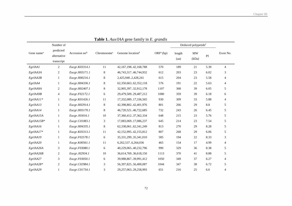
Chapter III
72
Table 1. Aux/IAA gene family in E. grandis
Gene namea
Number of
predicted
alternative
transcript
Accession nob. Chromosomec Genome locationd ORFe (bp)
Deduced polypetidef
Exon No. length
(aa)
MW
(kDa) PI
EgrIAA1 2 Eucgr.K03314.1 11 42,167,198..42,168,788 570 189 21 5.39 4
EgrIAA3A 2 Eucgr.H03171.1 8 46,743,317..46,744,932 612 203 23 6.02 3
EgrIAA3B 1 Eucgr.H00216.1 8 2,425,940..2,428,241 615 204 23 5.58 4
EgrIAA4 1 Eucgr.H04336.1 8 62,350,663..62,352,118 576 191 22 5.63 4
EgrIAA9A 2 Eucgr.H02407.1 8 32,905,397..32,912,178 1107 368 39 6.05 5
EgrIAA9B 4 Eucgr.F02172.1 6 29,479,509..29,487,212 1080 359 39 6.18 6
EgrIAA11* 1 Eucgr.K01426.1 11 17,332,089..17,336,565 930 309 33 5.88 4
EgrIAA13 1 Eucgr.H02914.1 8 42,398,802..42,401,976 801 266 29 8.8 5
EgrIAA14 1 Eucgr.H03170.1 8 46,720,521..46,722,685 732 243 26 6.45 5
EgrIAA15A 1 Eucgr.J03016.1 10 37,360,412..37,362,334 648 215 23 5.76 5
EgrIAA15B* 1 Eucgr.C01083.1 3 17,083,069..17,086,237 645 214 23 7.54 5
EgrIAA16 1 Eucgr.H04335.1 8 62,338,061..62,341,249 813 270 29 8.28 5
EgrIAA17* 1 Eucgr.K03313.1 11 42,152,995..42,155,812 807 268 29 6.06 5
EgrIAA19 1 Eucgr.F02578.1 6 35,331,299..35,341,010 585 194 22 8.33 3
EgrIAA20 1 Eucgr.K00561.1 11 6,262,537..6,264,036 465 154 17 4.99 4
EgrIAA26A 3 Eucgr.F03080.1 6 40,229,065..40,232,796 990 329 36 8.38 5
EgrIAA26B 2 Eucgr.J02934.1 10 36,614,769..36,618,150 1113 370 41 8.88 5
EgrIAA27 3 Eucgr.F03050.1 6 39,988,867..39,991,412 1050 349 37 6.27 4
EgrIAA28* 2 Eucgr.C02984.1 3 56,397,825..56,400,087 1044 347 38 6.72 5
EgrIAA29 1 Eucgr.C01734.1 3 29,257,063..29,258,993 651 216 25 6.6 4
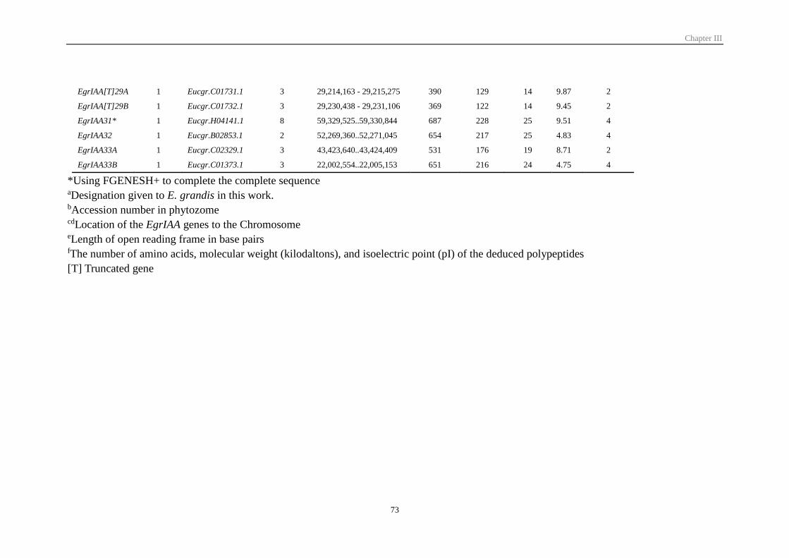
Chapter III
73
EgrIAA[T]29A 1 Eucgr.C01731.1 3 29,214,163 - 29,215,275 390 129 14 9.87 2
EgrIAA[T]29B 1 Eucgr.C01732.1 3 29,230,438 - 29,231,106 369 122 14 9.45 2
EgrIAA31* 1 Eucgr.H04141.1 8 59,329,525..59,330,844 687 228 25 9.51 4
EgrIAA32 1 Eucgr.B02853.1 2 52,269,360..52,271,045 654 217 25 4.83 4
EgrIAA33A 1 Eucgr.C02329.1 3 43,423,640..43,424,409 531 176 19 8.71 2
EgrIAA33B 1 Eucgr.C01373.1 3 22,002,554..22,005,153 651 216 24 4.75 4
*Using FGENESH+ to complete the complete sequence aDesignation given to E. grandis in this work. bAccession number in phytozome cdLocation of the EgrIAA genes to the Chromosome eLength of open reading frame in base pairs fThe number of amino acids, molecular weight (kilodaltons), and isoelectric point (pI) of the deduced polypeptides
[T] Truncated gene
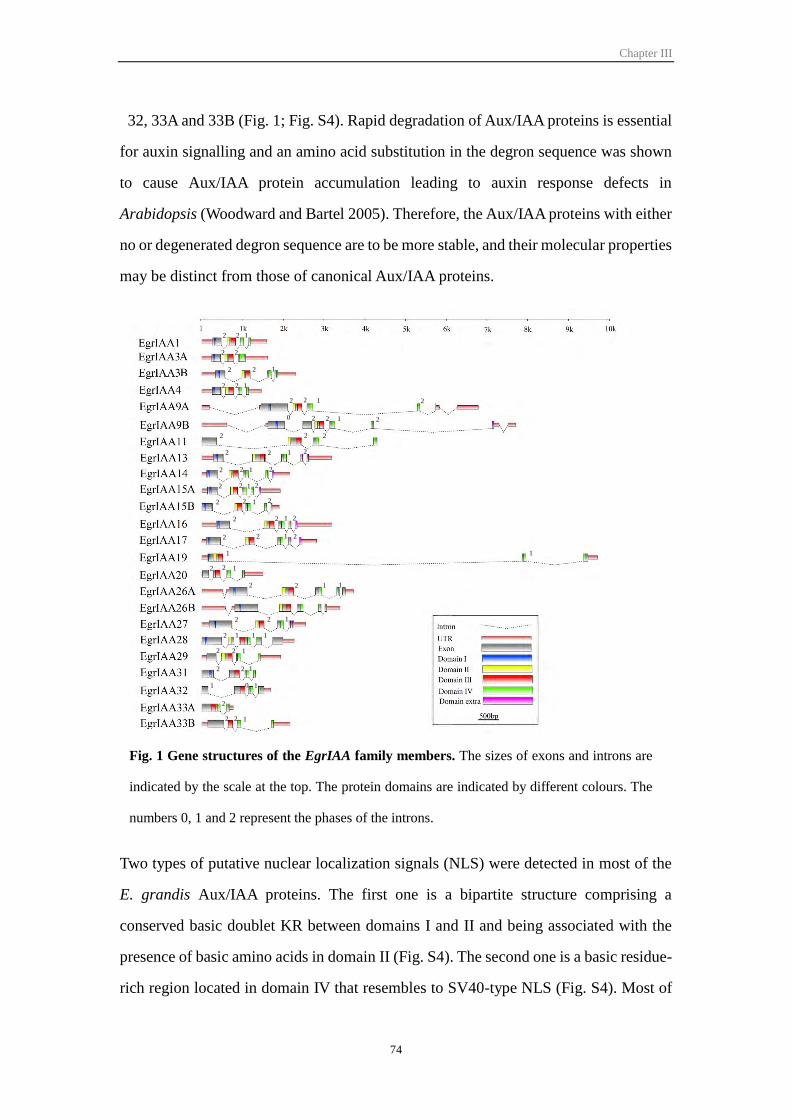
Chapter III
74
32, 33A and 33B (Fig. 1; Fig. S4). Rapid degradation of Aux/IAA proteins is essential
for auxin signalling and an amino acid substitution in the degron sequence was shown
to cause Aux/IAA protein accumulation leading to auxin response defects in
Arabidopsis (Woodward and Bartel 2005). Therefore, the Aux/IAA proteins with either
no or degenerated degron sequence are to be more stable, and their molecular properties
may be distinct from those of canonical Aux/IAA proteins.
Two types of putative nuclear localization signals (NLS) were detected in most of the
E. grandis Aux/IAA proteins. The first one is a bipartite structure comprising a
conserved basic doublet KR between domains I and II and being associated with the
presence of basic amino acids in domain II (Fig. S4). The second one is a basic residue-
rich region located in domain IV that resembles to SV40-type NLS (Fig. S4). Most of
Fig. 1 Gene structures of the EgrIAA family members. The sizes of exons and introns are
indicated by the scale at the top. The protein domains are indicated by different colours. The
numbers 0, 1 and 2 represent the phases of the introns.
2 2 1
2 2
2 2 1
1 1
2 2 1
2 2 1 2
2 2 1 20
2 2 2
2 2 1 2
2 2 1 2
2 2 1 2
2 2 1 2
2 2 1 2
2 2 1 2
2 2 1
2 2 11
2 2 1
2 1 1 1
2 2 1
2 2 1
1 0 1
2
2 2 1
Chapter III
75
the EgrIAA proteins contain the two types of NLS and are most likely targeted to the
nucleus consistent with their transcriptional activity. However, family members such as
EgrIAA29 and 33B contain a degenerated SV40-type NLS whereas EgrIAA20,
EgrIAA32, EgrIAA33A and EgrIAA33B lack the bipartite NLS. Using transient
expression assays with tomato Aux/IAA, Audran-Delalande et al. (2012) have shown
that despite of having a degenerated NLS, Sl-IAA29 protein was specifically targeted
to the nucleus. In contrast, in the absence of the bipartite structure like for instance in
Sl-IAA32, the accumulation of the protein was not restricted to the nuclear
compartment (Audran-Delalande et al. 2012).
3.2 Comparative phylogenetic analysis and chromosomal distribution of EgrIAA
genes
The Aux/IAA family in E. grandis is slightly contracted with only 24 members versus
29 and 35 in Arabidopsis and Populus, respectively. However, its size is quite similar
to that of tomato, which contains only 25 members (Table 2). To investigate the
phylogenetic relationships of Aux/IAA family members in different species, we
constructed a phylogenetic tree using predicted full-length amino acid sequences of
Aux/IAA from Eucalyptus, Arabidopsis and Populus. All the Aux/IAA members could
be grouped into 11 groups named A-K (Fig. 2), and EgrIAAs are distributed among all
groups and have representatives even within the four groups of non-canonical
Aux/IAAs (groups H, I, J and K) lacking domain I and/or II (Overvoorde et al. 2005).
As compared to Arabidopsis and Populus, Eucalyptus has the fewest members of
Table 2. Number of Aux/IAA family gene members in angiosperm species
Aux/IAA Reference
Eucalyptus grandis 24 This study
Solanum lycopersicon 25 Audran-Delalande et al. (2012)
Arabidopsis thaliana 29 Overvoorde et al. (2005)
Cucumis sativus 29 Gan et al. (2013)
Populus trichocarpa 35 Kalluri et al. (2007)
Zea mays 34 Ludwig et al. (2013)
Oryza sativa 31 Jain et al. (2006)
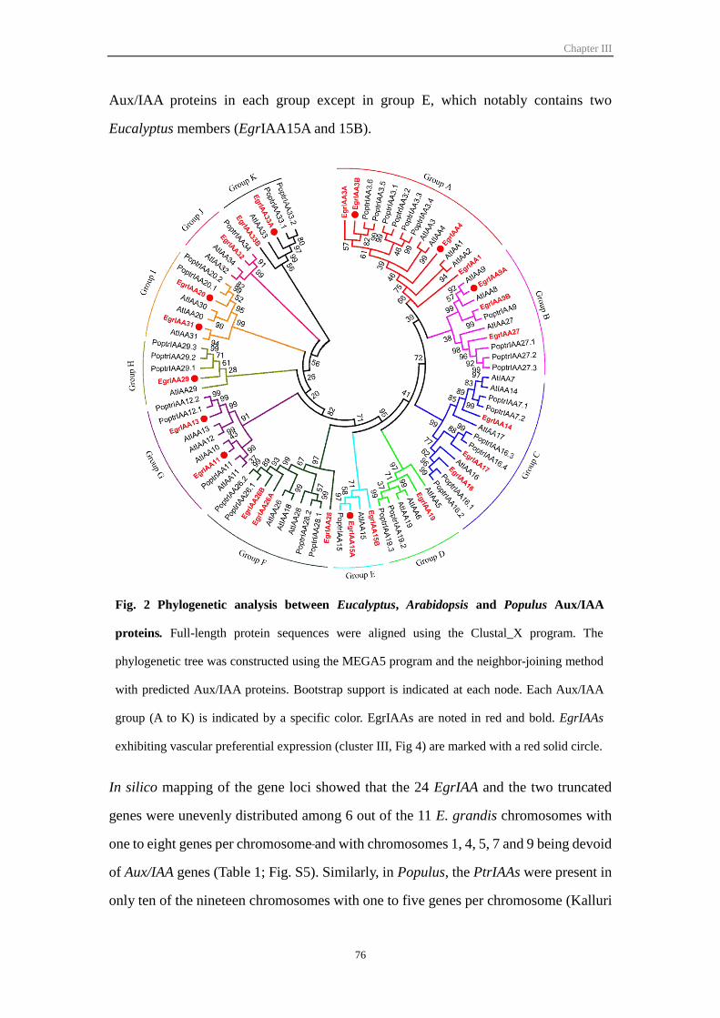
Chapter III
76
Aux/IAA proteins in each group except in group E, which notably contains two
Eucalyptus members (EgrIAA15A and 15B).
In silico mapping of the gene loci showed that the 24 EgrIAA and the two truncated
genes were unevenly distributed among 6 out of the 11 E. grandis chromosomes with
one to eight genes per chromosome and with chromosomes 1, 4, 5, 7 and 9 being devoid
of Aux/IAA genes (Table 1; Fig. S5). Similarly, in Populus, the PtrIAAs were present in
only ten of the nineteen chromosomes with one to five genes per chromosome (Kalluri
Fig. 2 Phylogenetic analysis between Eucalyptus, Arabidopsis and Populus Aux/IAA
proteins. Full-length protein sequences were aligned using the Clustal_X program. The
phylogenetic tree was constructed using the MEGA5 program and the neighbor-joining method
with predicted Aux/IAA proteins. Bootstrap support is indicated at each node. Each Aux/IAA
group (A to K) is indicated by a specific color. EgrIAAs are noted in red and bold. EgrIAAs
exhibiting vascular preferential expression (cluster III, Fig 4) are marked with a red solid circle.
Chapter III
77
et al. 2007), whereas Arabidopsis Aux/IAAs are scattered on all of the five chromosomes
(Overvoorde et al. 2005). Notably, one cluster of recent tandem was detected on
chromosome 3. EgrIAA29, 29A and 29B are located within a 45 kb fragment on
chromosome 3 and result from a recent tandem duplication event, which led to two
truncated versions and probably inactive versions of EgrIAA29 (Fig. S3). Three pairs
of EgrIAAs were identified as the results of segmental duplications (Myburg et al. 2014)
although they are located very close to each other (within a distance < 25 kb) on
chromosomes 8 (EgrIAA4/16 and EgrIAA3A/14) and 11 (EgrIAA1/17) respectively (Fig.
S5). These six genes are phylogenetically related, the more distant genes sharing 61%
protein identity. Members of protein pairs shares identity levels higher than 63% (Table
S1). However, each member of the protein pairs belongs to a distinct phylogenetic
group. EgrIAA1, 3A and 4 belong to group A, whereas EgrIAA17, 14 and 16 are
members of group C (Fig. S5 and Fig. S2). Altogether, these data suggest that one
ancestor gene went through ancient tandem duplication to give a first pair of EgrIAAs
that subsequently went through segmental duplication events to generate the other two
pairs. It seems that Aux/IAA family tandem duplication events in Eucalyptus occurred
prior to the chromosomal block duplication in Aux/IAA family in a similar way to that
proposed for Arabidopsis (Remington et al. 2004).
As duplication and alternative splicing are the two main mechanisms involved in
diversification of function within gene families, we performed an in silico survey of the
alternative transcripts predicted in the E. grandis genome JGI assembly v1.0,
annotation v1.1 (http://www.phytozome.net/eucalyptus). Among the 24 EgrIAAs, eight
of them showed more than one splice variant (Table 1). This proportion of alternative
transcripts is similar to that found in Arabidopsis. In Eucalyptus, most of the alternative
transcripts arise from members of groups B and F, while in Arabidopsis, the majority
of them belong to groups B and G (Table S2, Fig. 2). Some alternative transcripts are
lacking domains III and/or IV (Fig. 1, Fig. S6), likely contributing to shape the
functional diversity of the family.
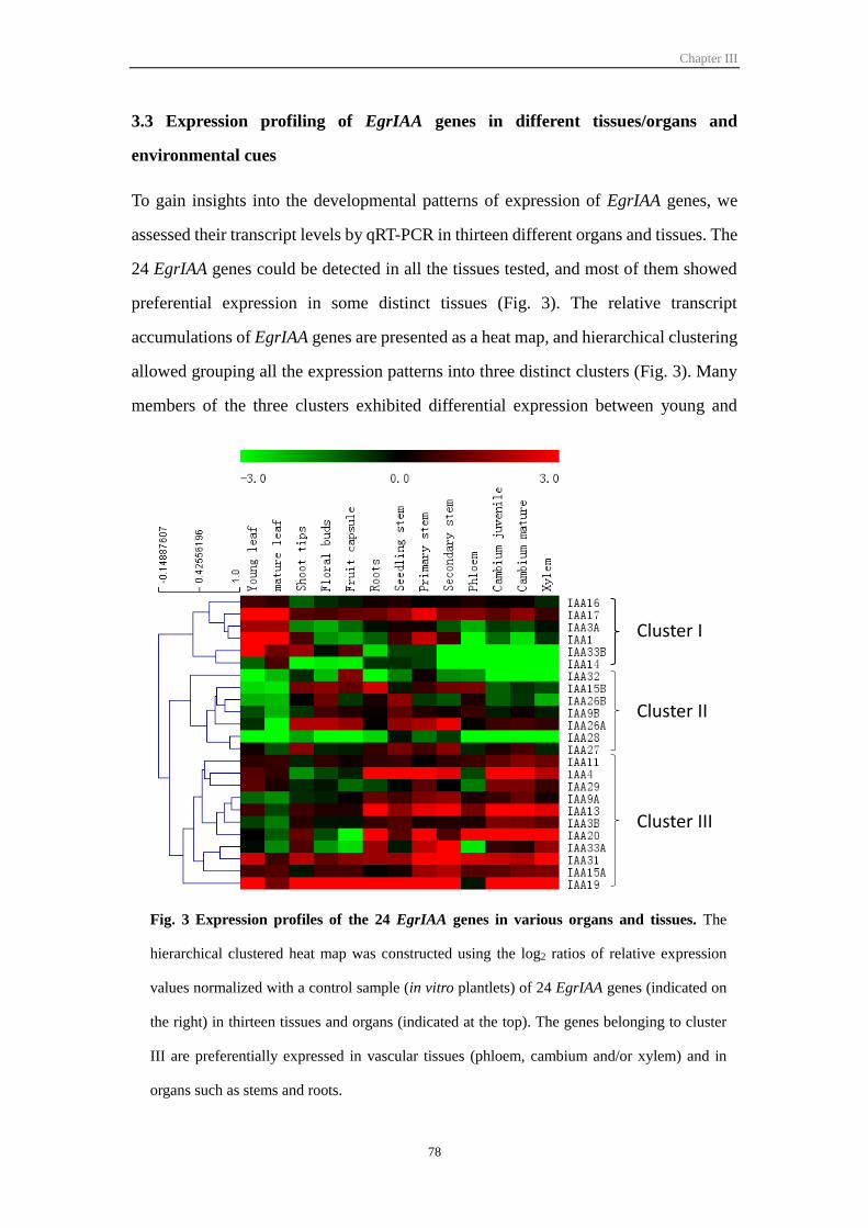
Chapter III
78
3.3 Expression profiling of EgrIAA genes in different tissues/organs and
environmental cues
To gain insights into the developmental patterns of expression of EgrIAA genes, we
assessed their transcript levels by qRT-PCR in thirteen different organs and tissues. The
24 EgrIAA genes could be detected in all the tissues tested, and most of them showed
preferential expression in some distinct tissues (Fig. 3). The relative transcript
accumulations of EgrIAA genes are presented as a heat map, and hierarchical clustering
allowed grouping all the expression patterns into three distinct clusters (Fig. 3). Many
members of the three clusters exhibited differential expression between young and
Fig. 3 Expression profiles of the 24 EgrIAA genes in various organs and tissues. The
hierarchical clustered heat map was constructed using the log2 ratios of relative expression
values normalized with a control sample (in vitro plantlets) of 24 EgrIAA genes (indicated on
the right) in thirteen tissues and organs (indicated at the top). The genes belonging to cluster
III are preferentially expressed in vascular tissues (phloem, cambium and/or xylem) and in
organs such as stems and roots.
Cluster I
Cluster II
Cluster III
Chapter III
79
mature leaves. Most of them showed higher expression in young leaves whereas only
EgrIAA14 displayed higher expression level in mature leaves. Members of both cluster
I and II displayed highest expression in non-vascular tissues, but they diverged
regarding their differential expression in leaves. The members of cluster I showed high
expression in leaves, while those of cluster II showed very low expression in these
organs. Cluster III was the largest cluster, containing eleven members that displayed
preferential expression in vascular tissues (phloem, cambium and xylem).
Within cluster III, EgrIAA13, 20 and 31 displayed relatively high expression in all three
vascular tissues whereas EgrIAA4 and 33A showed higher expression levels in
cambium and xylem than in phloem. EgrIAA29 was preferentially expressed in
cambium as compared to phloem and xylem. Most of the members of cluster III showed
higher expression in juvenile xylem versus mature xylem (Fig. 4A) whereas no obvious
differences in their expression patterns were detected between juvenile and mature
cambium (Fig. 3). Of particular note, EgrIAA4 was the only gene showing higher
expression in mature xylem as compared to juvenile xylem (Fig. 4A). Most of EgrIAA
genes from cluster III are responsive to bending mechanical stress (Fig. 4B). Four
EgrIAA genes were down-regulated (EgrIAA4, 11, 13 and 20) and two (EgrIAA31 and
33A) were up-regulated in tension wood as compared to straight wood (Fig. 4B).
Interestingly, EgrIAA11 and 20 were down-regulated in both tension and opposite
woods, whereas EgrIAA33A was up-regulated in these tissues (Fig. 4B).
3.4 Candidates EgrIAA genes potentially involved in wood formation
In order to identify the best candidate(s) potentially involved in wood formation for
functional characterization in planta, we defined several criteria based on (i) transcript
abundance in vascular tissues, (ii) high expression in cambium and/or xylem versus low
expression in phloem, (iii) responsiveness to bending, given that response to bending
involves dramatic changes both in xylem development and secondary cell wall structure
and composition, (iv) differential expression between mature versus juvenile xylem
known to exhibit different biochemical and physical properties. The combination of all
these criteria presented as a Venn diagram (Fig. S7) highlighted EgrIAA4 and
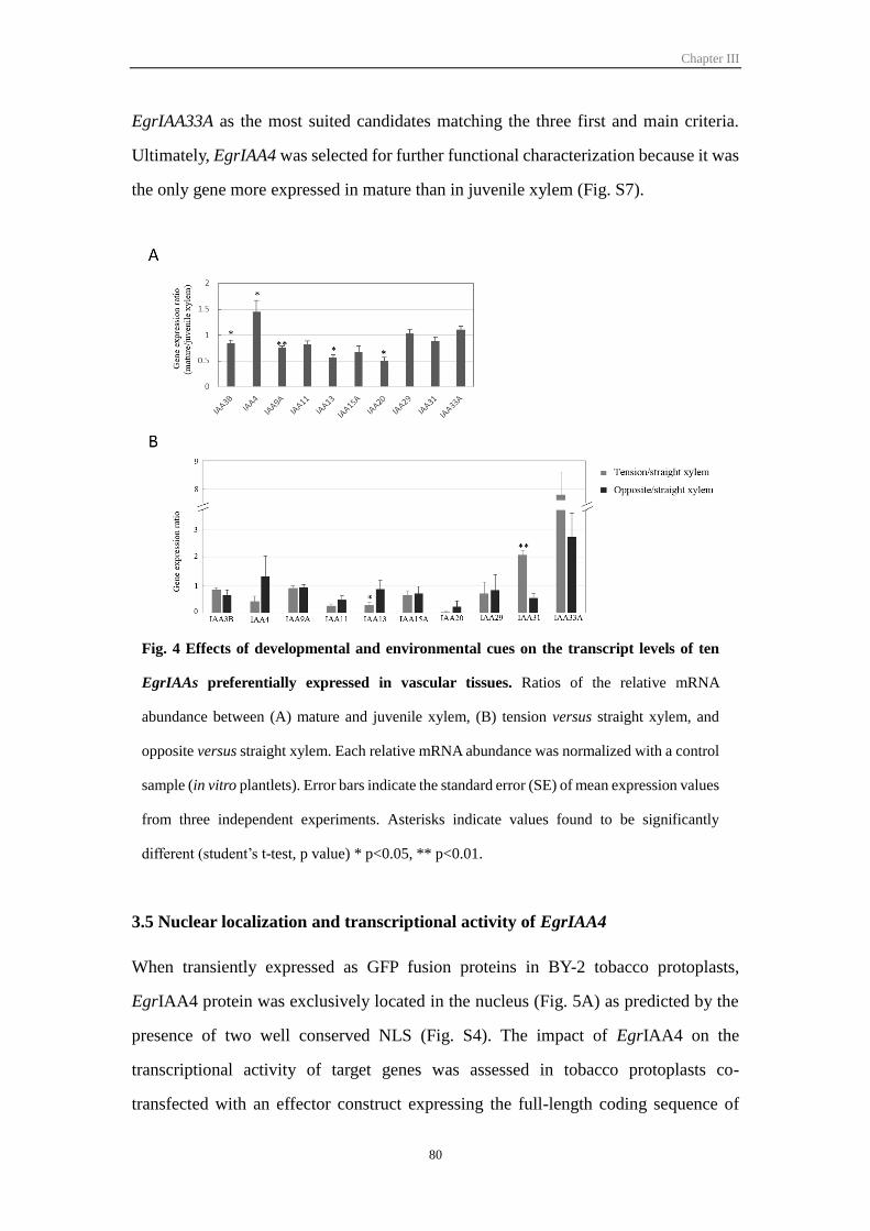
Chapter III
80
EgrIAA33A as the most suited candidates matching the three first and main criteria.
Ultimately, EgrIAA4 was selected for further functional characterization because it was
the only gene more expressed in mature than in juvenile xylem (Fig. S7).
3.5 Nuclear localization and transcriptional activity of EgrIAA4
When transiently expressed as GFP fusion proteins in BY-2 tobacco protoplasts,
EgrIAA4 protein was exclusively located in the nucleus (Fig. 5A) as predicted by the
presence of two well conserved NLS (Fig. S4). The impact of EgrIAA4 on the
transcriptional activity of target genes was assessed in tobacco protoplasts co-
transfected with an effector construct expressing the full-length coding sequence of
Fig. 4 Effects of developmental and environmental cues on the transcript levels of ten
EgrIAAs preferentially expressed in vascular tissues. Ratios of the relative mRNA
abundance between (A) mature and juvenile xylem, (B) tension versus straight xylem, and
opposite versus straight xylem. Each relative mRNA abundance was normalized with a control
sample (in vitro plantlets). Error bars indicate the standard error (SE) of mean expression values
from three independent experiments. Asterisks indicate values found to be significantly
different (student’s t-test, p value) * p<0.05, ** p<0.01.
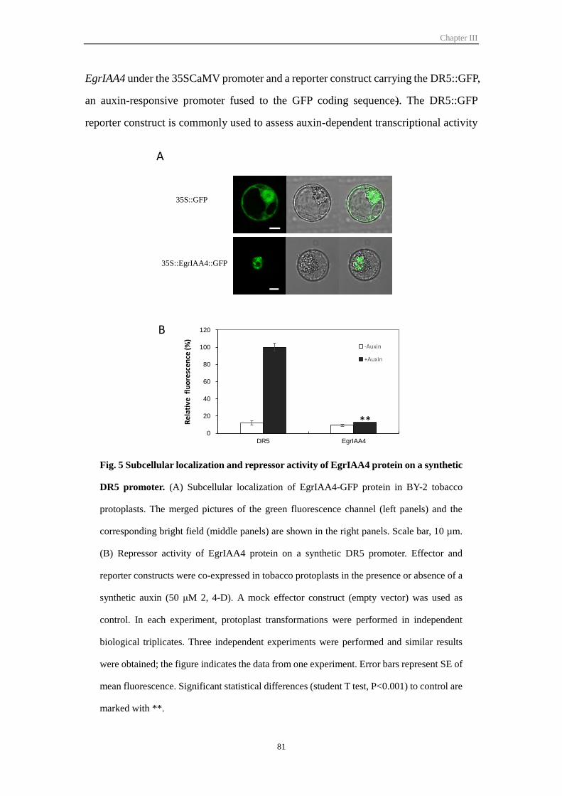
Chapter III
81
EgrIAA4 under the 35SCaMV promoter and a reporter construct carrying the DR5::GFP,
an auxin-responsive promoter fused to the GFP coding sequence). The DR5::GFP
reporter construct is commonly used to assess auxin-dependent transcriptional activity
Fig. 5 Subcellular localization and repressor activity of EgrIAA4 protein on a synthetic
DR5 promoter. (A) Subcellular localization of EgrIAA4-GFP protein in BY-2 tobacco
protoplasts. The merged pictures of the green fluorescence channel (left panels) and the
corresponding bright field (middle panels) are shown in the right panels. Scale bar, 10 µm.
(B) Repressor activity of EgrIAA4 protein on a synthetic DR5 promoter. Effector and
reporter constructs were co-expressed in tobacco protoplasts in the presence or absence of a
synthetic auxin (50 μM 2, 4-D). A mock effector construct (empty vector) was used as
control. In each experiment, protoplast transformations were performed in independent
biological triplicates. Three independent experiments were performed and similar results
were obtained; the figure indicates the data from one experiment. Error bars represent SE of
mean fluorescence. Significant statistical differences (student T test, P<0.001) to control are
marked with **.
B
35S::EgrIAA4::GFP
35S::GFP
A
**0
20
40
60
80
100
120
DR5 EgrIAA4
-Auxin
+Auxin
Re
lati
ve f
luo
resc
ence
(%
)
Chapter III
82
in planta (Audran-Delalande et al. 2012; Ulmasov et al. 1999). The DR5-driven GFP
showed a low basal activity which was induced up to 9-fold by exogenous auxin
treatment (Fig. 5B). Co-transfection with the effector plasmid EgrIAA4 resulted in a
strong repression (87%) of the auxin-induced expression of the reporter gene (Fig. 5B)
indicating that EgrIAA4 is able to mediate auxin response in vivo and that it functions
as a strong transcriptional repressor.
3.6 Overexpression of EgrIAA4m affects plant growth and development
To gain insights into the role of EgrIAA4, we overexpressed in Arabidopsis a mutated
version of the gene, referred to as EgrIAA4m (see Material and Methods and Fig. S8A),
encoding a stabilized form of the protein. The mutation introduced in the degron
domain was hypothesized to prevent auxin-mediated EgrIAA4 protein degradation.
Then EgrIAA4m was cloned under the control of the 35SCaMV promoter into the
Gateway pFAST-G02 expression vector and transformed into A. thaliana Col-0. This
pFAST-G02 vector contains an OLE1-GFP selection marker that allows direct screening
of transformed seeds displaying fluorescence under UV light (Shimada et al. 2010).
Among the ten seeds sown, six germinated giving independent EgrIAA4m
transformants, all exhibiting similar and dramatically reduced sizes as compared to
controls. We selected two phenotypically representative transformants (EgrIAA4m_1.3,
ErIAA4m_2.3) overexpressing EgrIAA4m (Fig. S8B) for further characterization. T2
seeds from both lines germinated and half of the seedlings exhibited a drastic phenotype
remaining very tiny (around one cm rosette only) and displaying very small and curled-
down leaves turning yellow or brown just before the plants die without flowering. (Fig.
S8C). The surviving transgenic plants (T2) exhibited several distinctive phenotypes in
their aerial parts (Fig. 6A, B). During the seedling stage, the cotyledons showed curled-
up shape, while the leaves had a curled-down shape and very short petioles (Fig. 6A).
During the vegetative stage, the EgrIAA4m transgenic plants from both lines showed
significantly reduced plant height and rosette diameter (Fig. 6B), and the inflorescence
stems were obviously thinner than those of wild type plants. In addition, EgrIAA4m
plants showed smaller siliques and much lower fertility than wild-type (no seeds or less
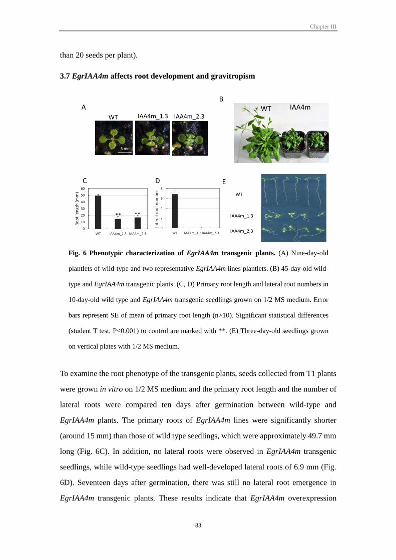
Chapter III
83
than 20 seeds per plant).
3.7 EgrIAA4m affects root development and gravitropism
To examine the root phenotype of the transgenic plants, seeds collected from T1 plants
were grown in vitro on 1/2 MS medium and the primary root length and the number of
lateral roots were compared ten days after germination between wild-type and
EgrIAA4m plants. The primary roots of EgrIAA4m lines were significantly shorter
(around 15 mm) than those of wild type seedlings, which were approximately 49.7 mm
long (Fig. 6C). In addition, no lateral roots were observed in EgrIAA4m transgenic
seedlings, while wild-type seedlings had well-developed lateral roots of 6.9 mm (Fig.
6D). Seventeen days after germination, there was still no lateral root emergence in
EgrIAA4m transgenic plants. These results indicate that EgrIAA4m overexpression
Fig. 6 Phenotypic characterization of EgrIAA4m transgenic plants. (A) Nine-day-old
plantlets of wild-type and two representative EgrIAA4m lines plantlets. (B) 45-day-old wild-
type and EgrIAA4m transgenic plants. (C, D) Primary root length and lateral root numbers in
10-day-old wild type and EgrIAA4m transgenic seedlings grown on 1/2 MS medium. Error
bars represent SE of mean of primary root length (n>10). Significant statistical differences
(student T test, P<0.001) to control are marked with **. (E) Three-day-old seedlings grown
on vertical plates with 1/2 MS medium.
5 mm
AB
WT IAA4m_1.3 IAA4m_2.3
E
WT
IAA4m_2.3
IAA4m_1.3
C
0
10
20
30
40
50
60
WT IAA4m_1.3 IAA4m_2.3
Ro
ot
len
gth
(m
m)
0
2
4
6
8
WT IAA4m_1.3 IAA4m_2.3
Late
ral r
oo
t n
um
ber
D
WT IAA4m
** **
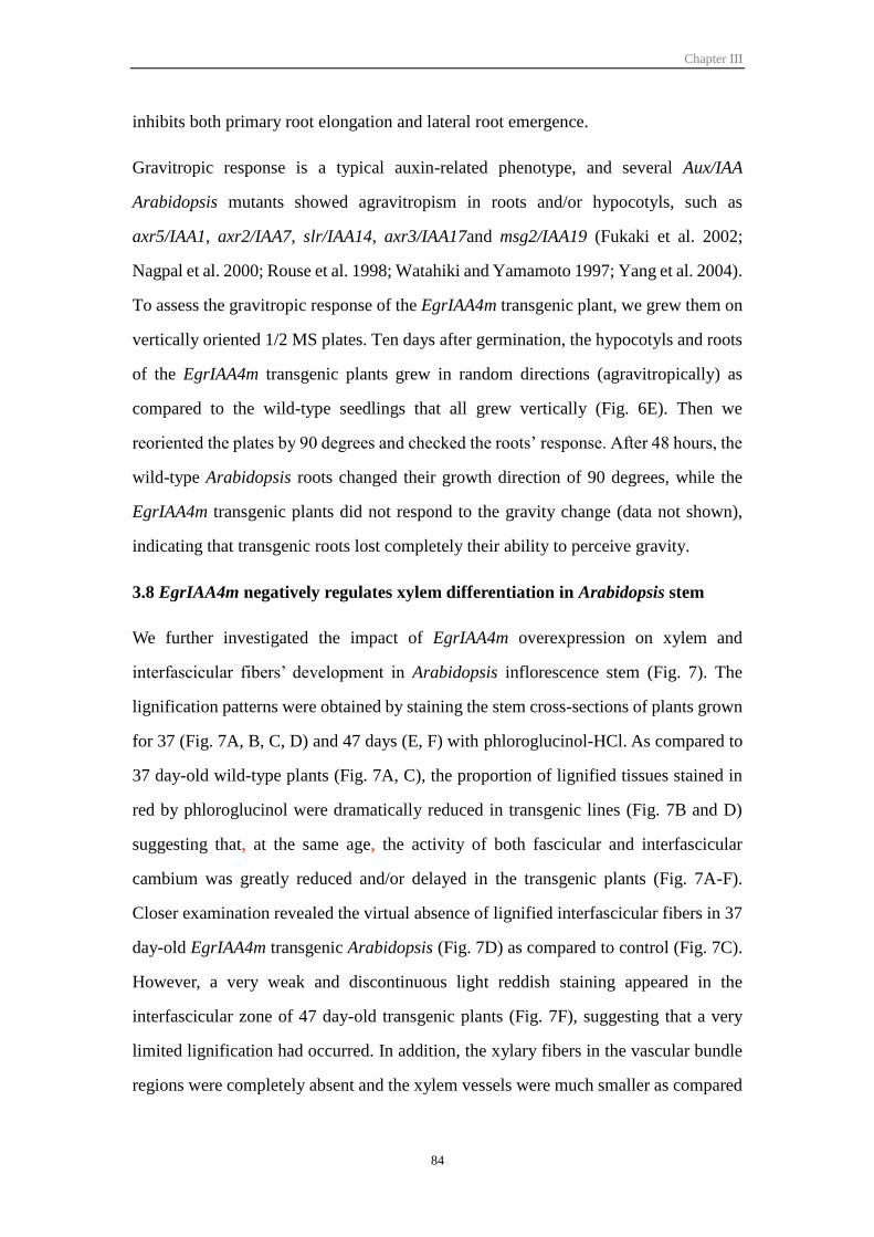
Chapter III
84
inhibits both primary root elongation and lateral root emergence.
Gravitropic response is a typical auxin-related phenotype, and several Aux/IAA
Arabidopsis mutants showed agravitropism in roots and/or hypocotyls, such as
axr5/IAA1, axr2/IAA7, slr/IAA14, axr3/IAA17and msg2/IAA19 (Fukaki et al. 2002;
Nagpal et al. 2000; Rouse et al. 1998; Watahiki and Yamamoto 1997; Yang et al. 2004).
To assess the gravitropic response of the EgrIAA4m transgenic plant, we grew them on
vertically oriented 1/2 MS plates. Ten days after germination, the hypocotyls and roots
of the EgrIAA4m transgenic plants grew in random directions (agravitropically) as
compared to the wild-type seedlings that all grew vertically (Fig. 6E). Then we
reoriented the plates by 90 degrees and checked the roots’ response. After 48 hours, the
wild-type Arabidopsis roots changed their growth direction of 90 degrees, while the
EgrIAA4m transgenic plants did not respond to the gravity change (data not shown),
indicating that transgenic roots lost completely their ability to perceive gravity.
3.8 EgrIAA4m negatively regulates xylem differentiation in Arabidopsis stem
We further investigated the impact of EgrIAA4m overexpression on xylem and
interfascicular fibers’ development in Arabidopsis inflorescence stem (Fig. 7). The
lignification patterns were obtained by staining the stem cross-sections of plants grown
for 37 (Fig. 7A, B, C, D) and 47 days (E, F) with phloroglucinol-HCl. As compared to
37 day-old wild-type plants (Fig. 7A, C), the proportion of lignified tissues stained in
red by phloroglucinol were dramatically reduced in transgenic lines (Fig. 7B and D)
suggesting that, at the same age, the activity of both fascicular and interfascicular
cambium was greatly reduced and/or delayed in the transgenic plants (Fig. 7A-F).
Closer examination revealed the virtual absence of lignified interfascicular fibers in 37
day-old EgrIAA4m transgenic Arabidopsis (Fig. 7D) as compared to control (Fig. 7C).
However, a very weak and discontinuous light reddish staining appeared in the
interfascicular zone of 47 day-old transgenic plants (Fig. 7F), suggesting that a very
limited lignification had occurred. In addition, the xylary fibers in the vascular bundle
regions were completely absent and the xylem vessels were much smaller as compared
Chapter III
85
to control plants (Fig. 7C-F). These results indicate that overexpression of EgrIAA4m
strongly and negatively affected xylem bundles and interfascicular fibers development.
4 Discussion
The Aux/IAA family in E. grandis contains 24 members and therefore is slightly
Fig. 7 Histochemical analysis of basal stem cross sections of EgrIAA4m
transgenic Arabidopsis. Sections of wild-type and EgrIAA4m transgenic plants
were stained with phloroglucinol-HCl. if, interfascicular fiber; xf, xylary fiber; xv,
xylem vessel. Scale bar: 50 μm.
C D
E F
if
if
if
xf
if
EgrIAA4mControl
xf
xv xv
xvxv
A B
Chapter III
86
contracted as compared to most angiosperms Aux/IAA families studied hitherto (Table
2). With 25 members, the size gene family in tomato is the closest to Eucalyptus but it
lacks non-canonical members in group I (Audran-Delalande et al. 2012), which were
shown here to be present in Eucalyptus. In fact, all the eleven phylogenetic groups
defined in Arabidopsis were represented in Eucalyptus but with less members in each
group except in group E that contains two members whereas Arabidopsis and Populus
have only one member each. In Arabidopsis, transcripts of AtIAA15, the unique member
of group E, were never been detected suggesting that it may be a pseudogene
(Remington et al. 2004), while in E. grandis the transcripts of both EgrIAA15A and 15B
were detected by qRT-PCR, indicating they are likely functional genes. Whereas in
Arabidopsis and Populus members of the Aux/IAA family arose predominantly through
large-scale genomic duplication events (Kalluri et al. 2007; Remington et al. 2004),
Aux/IAA family members in the E. grandis genome are concerned with only very few
segmental duplications events. One cluster of recent tandem duplication was detected
on chromosome 3, but the duplication of EgrIAA29 led to two truncated genes lacking
both domains III and IV. To the best of our knowledge, such a structure had never been
reported in other species and these truncated genes are most likely not functional.
The virtual absence of tandem duplication in the Eucalyptus Aux/IAA family is
especially striking because E. grandis has the largest number of genes in tandem repeats
reported among sequenced plant genomes (34% of the total number of genes; Myburg
et al. 2014) and tandem duplication shaped functional diversity in many gene families
in Eucalyptus such as in the MYB (Soler et al. 2014) and NAC (Hussey et al. 2014)
transcription factor families. On the other hand, this situation is quite similar to ARF
family in E. grandis whose size is also slightly contracted as compared to other
angiosperms genome studied (Yu et al. 2014). Because Aux/IAA proteins regulate
auxin-mediated gene expression through interaction with ARF proteins, a proper
dosage relationship is probably needed (Remington et al. 2004).
The expression profiles of EgrIAAs in various tissues and organs showed that some
genes have preferential expression patterns, contrasting to ARF family members that
Chapter III
87
exhibited more constitutive expression in the same panel of samples (Yu et al. 2014).
According to the current model, Aux/IAA proteins regulate auxin-mediated gene
expression by protein-protein interactions with ARFs, so the preferential expression
pattern of Aux/IAA genes may have a primary role in their physiological functions
(Muto et al. 2007). All the EgrIAA genes in groups G, H and I showed vascular tissue
preferential expression, and most of their orthologs in Arabidopsis and/or Populus also
showed high/preferential expression in xylem cells (Table S3) (Hruz et al. 2008; Kalluri
et al. 2007; Moyle et al. 2002; Nilsson et al. 2008), and/or induce vascular defects in
transgenic plants (Nilsson et al. 2008; Sato and Yamamoto 2008) suggesting that these
phylogenetic groups are regulating cambium activity and/or xylem development.
Overexpression of AtIAA20, 30 and 31 in Arabidopsis resulted in reduced and
discontinued vascular strands in cotyledons (Sato and Yamamoto 2008). Alteration of
auxin responsiveness through overexpression of a stabilized version of PttIAA3
(EgrIAA20 ‘s putative ortholog) in transgenic aspen reduced the cambial cell division
activity, caused spatial deregulation of cell division of the cambial initials and led to
reductions in not only radial but also axial dimensions of fibers and vessels (Nilsson et
al. 2008).
Most of the EgrIAAs in cluster III showed responsiveness to bending stress, but their
behaviour seems quite different from that of their potential orthologs in Populus (Moyle
et al. 2002). For example, in our conditions (three weeks of bending stress), EgrIAA20
was down-regulated in tension wood, whereas its Populus orthologs PttIAA3 and 4 were
not affected (Moyle et al. 2002). An opposite pattern was evidenced for EgrIAA9A that
was not affected by long-term bending, while its Populus ortholog PttIAA2 was down-
regulated in tension wood (Moyle et al. 2002). In fact, in a previous study, we had
shown that the transcript level of EgrIAA9A increased sharply between six and 24 hours
and then decreased between 24 h and one week, to reach a level similar to that of the
control (Paux et al. 2005). Aux/IAAs are thus responding to bending in a time-dependent
way explaining the apparent discrepancies between the responses of Populus and
Eucalyptus Aux/IAA orthologs. Indeed, Moyle et al. (2002) analysed an early response
Chapter III
88
to bending (from 30 minutes to eleven hours) whereas we analysed a late response after
three weeks of bending. Our results are indeed more consistent with those of Anderson-
Gunneras et al. (2006) who analyzed Populus gene expression after three weeks
bending. We found that EgrIAA4, 11 and 20 were all down-regulated in tension wood
as their Populus orthologs (Anderson-Gunneras et al. 2006).
EgrIAA4, which belongs to subgroup A, was considered as the best candidate to
regulate cambium activity and wood formation based on its expression profile, and was
chosen for functional characterization in planta. EgrIAA4 is the ortholog of Arabidopsis
sister pair AtIAA3 and AtIAA4. Only the function of AtIAA3 has been studied through
gain-of-function experiments. AtIAA3 mutation shy2 led to plants with shorter
hypocotyls, fewer lateral roots, and slower gravitropic response (Reed et al. 1998; Tian
and Reed 1999). AtIAA3 was shown recently to be part of the auxin-signalling module
(SLR/IAA14–ARF7–ARF19 and SHY2/IAA3–ARFs) regulating lateral root formation
(Goh et al. 2012). Likewise the down-regulation of Sl-IAA3, the ortholog of AtIAA3 in
the tomato, leads to reduced growth of primary and inhibition of lateral roots
(Chaabouni et al. 2009). Overexpression of an auxin-insensitive version of EgrIAA4 in
Arabidopsis led to very similar but more severe auxin aberrant phenotypes. The
gravitropic response was completely lost and the growth and development were
severely affected with strongly reduced height and diameter of the inflorescence stem,
reduced leaf size and fertility and absence of lateral roots. Notably, EgrIAA4m
overexpression also led to phenotypic alterations of the vascular system not reported
yet for its Arabidopsis’s ortholog. The development of both xylem and interfascicular
fibers were dramatically delayed and reduced suggesting a reduced cambium activity
in response to the altered auxin responsiveness induced by EgrIAA4m expression. In
the transgenic plants, the lignified secondary walls of the interfascicular fibers appeared
very late whereas that of the xylary fibers was virtually undetectable, indicating that
EgrIAA4 plays crucial roles in fiber development and secondary cell walls deposition
consistent with its expression profile.
Chapter III
89
5 Materials and Methods
5.1 Identification of Aux/IAA gene family in Eucalyptus
Firstly, we used 29 Arabidopsis Aux/IAA proteins as queries to do BLASTP against
Eucalyptus grandis genome (JGI assembly v1.0, annotation v1.1,
http://www.phytozome.net/eucalyptus), and identified 55 potential E. grandis Aux/IAA
proteins. Then, the Pfam database (http://pfam.sanger.ac.uk/search) and NCBI
conserved domain database (http://www.ncbi.nlm.nih.gov/cdd) web server were used
to examine the conserved domains (Finn et al. 2010; Marchler-Bauer et al. 2011). The
incomplete gene models were completed by FGENESH+ (http://linux1.softberry.com),
and redundant and invalid gene models were removed. The corrected E.grandis
Aux/IAA proteins were used as query sequences in two subsequent additional search:
1) BLASTP against E. grandis proteome for exhaustive identification of divergent E.
grandis gene family member, 2) TBLASTN searches against E. grandis genome for
seeking any possible no predicted genes. Finally 26 EgrIAAs were identified in E.
grandis genome (E.grandis genome V1.1, May 2012). For validation, we also used
Populus Aux/IAA proteins as queries to do the complete search procedure described
above, and we obtained exactly the same final 26 EgrAux/IAA genes. Gene information
on chromosomal location was retrieved from the E. grandis genome browser
(http://www.phytozome.net/eucalyptus) with manual curation and we mapped their loci
using MapChart 2.2 (Voorrips 2002). Basic physical and chemical parameters of
Aux/IAA proteins were calculated by online ProtParam tool
(http://web.expasy.org/protparam/).
5.2 Sequence, gene structure and phylogenetic analysis
Conserved protein motifs of EgrIAA were determined by MEME-MAST programs
(http://meme.sdsc.edu/meme) (Bailey et al. 2009). The exon-intron structures were
extracted from Phytozome with manual curation and visualized by Fancy Gene v1.4
(http://bio.ieo.eu/fancygene/). Multiple protein sequences alignment was performed
using Clustal_X2 program (Version 2.0.11). All predicted protein sequences were used
Chapter III
90
to do phylogenetic analysis and the phylogenetic trees were constructed with MEGA5
program by neighbour-joining method with 1000 bootstrap.
5.3 Plant materials and growth conditions
All Eucalyptus organ/tissues provenance and preparation are described in Cassan-Wang
et al. (2012). A. thaliana ecotype Col-0 plants were grown in growth chamber: 16 h
day/8 h night for long days, 22/20ºC day/night temperature, 70% relative humidity, 200
µmol photons m−1s−1 light intensity (intense luminosity). The plants were watered every
two days and fertilized weekly. Seeds were surface-sterilized for 1 min in 70% ethanol,
10 min in 25% bleach, rinsed five times in sterile water and plated on Murashige and
Skoog (MS) medium containing 1.0% sucrose solidified with 1% agar.
5.4 RNA isolation, cDNA synthesis and qRT-PCR
Total RNAs were extracted from 100–200 mg of frozen material as described by
(Southerton et al. 1998) and treated by Turbo DNA-freeTM kit (Ambion, Carlsbad, USA)
to remove the genomic DNA contamination. RNA concentration and purity were
determined by using a NanoDrop spectrophotometer ND-1000 (Thermo Scientific,
Waltham, USA) and the integrity was assessed by using the Agilent 2100 Bioanalyzer
(Agilent, Santa Clara, USA). Only samples with RNA Integrity Number (RIN) over 7
were used for reverse transcription. cDNA was reversed transcribed from 2 g total
RNA using the High Capacity cDNA Reverse Transcription Kit (Applied Biosystems,
Foster, USA).
Primers were designed using the software QuantPrime (qPCR primer design tool:
http://www.quantprime.de/ (Arvidsson et al. 2008) and the sequences are shown in
Table S4. Oligonucleotides were synthesized by Sigma Life Science. qRT-PCR was
performed by the Genotoul service in Toulouse (http://genomique.genotoul.fr/) using
the BioMark® 96:96 Dynamic Array integrated fluidic circuits (Fluidigm Corporation,
San Francisco, USA) as described in (Cassan-Wang et al. 2012). Only primers that
produced a linear amplification and qPCR products with a single-peak melting curves
were used for further analysis. The efficiency of each pair of primers was determined
Chapter III
91
from the data of amplification Ct value plot with a serial dilution of mixture cDNA and
the equation E =10(-1/slope) -1. E-∆∆Ct method was used to calculate relative mRNA fold
change compared to control sample using formula (Etarget)ΔCt_target
(control−sample)/(Ereference)ΔCt_reference (control−sample) (Pfaffl 2001) and five reference genes (IDH,
PP2A1, PP2A3, EF-1a and SAND, Table S4) were used for data normalization. In vitro
plantlets were used as control sample.
5.5 EgrIAA4 amplification and gain-of-function transgenic Arabidopsis
construction
The EgrIAA4 gene was amplified by polymerase chain reaction (PCR) using Phusion
Taq (Thermo, Waltham, USA) by a gene specific primer pair: 5’-
CACCATGGCAGCTCAAGGAGAGGAT-3’ and 5’-
AACCTCTGATGACCCTTTCATGATT-3’ which was based on the prediction from E.
grandis genome v.1.1 (Eucgr.H04336.1). To investigate the function of this gene, we
create a mutation in the auxin degradation domain changing the 80th amino acid from
proline to serine (P-to-S) in domain II by using overlap PCR. The overlap primers are
5’-ATCGGACCGGACTCCACCCCACGACTTGTGCCTTA-3’ and 5’-
CGTGGGGTGGAGTCCGGTCCGATCCTACCGAAA-3’. The underlined sequences
are the overlap region and the bold bases are the mutated nucleotides. The EgrIAA4m
fragment was cloned into the pENTR/D-TOPO vector (Invitrogen, Carlsbad, USA) to
sequence. After sequencing, we recombined EgrIAA4m fragment into the destination
vector pFAST-G02 using LR Clonase (Invitrogen, Carlsbad, USA). pFAST-EgIAA4m
vector was transformed into A. tumefaciens strain GV3101, and then transformed into
A. thaliana ecotype Col-0 using the floral dip method (Clough and Bent 1998).
5.6 Transient expression using a single cell system
Protoplast for transfection were obtained from suspension-cultured tobacco (Nicotiana
tabacum) BY-2 cells according to the method of Leclercq et al. (2005). Protoplast were
transfected by a modified polyethylene glycol method as described by Abel and
Theologis (1994). For nuclear localization of the selected Aux/IAA, the full length

Chapter III
92
cDNAs were fused in frame at the C-terminus with GFP in the pK7FWG2.0 vector
(Karimi et al. 2002) under the control of 35SCaMV promoter. Transfected protoplasts
were incubated for 16 h at 25ºC and examined for GFP florescence signals using a Leica
TCS SP2 laser scanning confocal microscope. Images were obtained with a x40 water
immersion objective. For co-transfection assays, the full-length cDNAs of the selected
Aux/IAA were cloned into pGreen vector under 35SCaMV promoter to create the
effector constructs. The reporter constructs used a synthetic auxin-responsive promoter
DR5 fused to the GFP reporter gene. Tobacco BY-2 protoplasts were co-transfected
with the reporter and effector constructs as described in Audran-Delalande et al. (2012).
After 16h incubation, GFP expression was quantified by flow cytometry (LSR Fortessa,
BD Bioscience), and the data were analysed using Cell Quest software BD FacsDiva
software. Transfection assays were performed in three independent replicates and 3000-
4000 protoplasts were gated for each sample. GFP fluorescence corresponds to the
average fluorescence intensity of the protoplasts population after subtraction of auto-
fluorescence determined with non-transformed protoplasts. 50 µM 2, 4-D was used for
auxin treatment.
5.7 Microscopy analysis
The Arabidopsis inflorescence stems at the basal end (~1 cm) and hypocotyls were
harvested at 37 and 47 days old, and then stored in 70% ethanol. The cross sections
were prepared using vibratome Leica VT1000 S (Leica, Paris, France). Lignin polymers
are the characteristic components of secondary cell wall (SCW) and are normally absent
from primary cell wall, therefore we used lignin deposition detection techniques to
screen for SCW phenotype. Cross sections of inflorescence stem and hypocotyl (~80
µm) were stained with phloroglucinol-HCl, which stains specifically lignin polymer
precursors coniferaldehyde and p-coumaraldehyde in the SCW giving violet-red color.
Phloroglucinol-HCl was directly applied on the slide and images were recorded with a
CCD camera (Photonic Science, Robertsbridge, UK, http://www.photonic-
science.co.uk).
Chapter III
93
Acknowledgements
This work was supported by the Centre National pour la Recherche Scientifique
(CNRS), the University Paul Sabatier Toulouse III (UPS), the French Laboratory of
Excellence project " TULIP" (ANR-10-LABX-41; ANR-11-IDEX-0002-02), the
Plant KBBE TreeForJoules project [ANR-2010-KBBE-007-01 (FR) and P-
KBBE/AGR_GPL/0001/2010 (FCT, PT)], and the project microEGo (PTDC/AGR-
GPL/098179/2008; FCT, PT). HY were supported by PhD grants from the China
Scholarship council. MS was supported by a Postdoc fellowship “Beatriu de Pinós”
thanks to the Departament d'Universitats, Recerca i Societat de la Informació de la
Generalitat de Catalunya. JAPP acknowledges FCT for the research contract Ciência
2008 program and the postdoctoral fellowship SFRH/BPD/92207/2013. The authors
are grateful to C. Dunand and Q. Li for the help of bioinformatics analysis, to E.
Camargo, J.M. Gion and E. Villar (CIRAD, FR), F. Melun and L. Harvengt (FCBA,
France), C. Araujo and L. Neves (AltriFlorestal, Portugal) and C. Marques (RAIZ,
Portugal) for kindly providing and/or allowing collection of Eucalyptus organ and
tissue samples, C. Graça, and N. Saidi for help with sample collection and RNA
extraction, N Ladouce and V. Carocha for performing Fluidigm experiments. Thanks to
the Bioinfo Genotoul Platform (http://bioinfo.genotoul.fr) for access to resources and
to the Genome and transcriptome Platform (http://get.genotoul.fr/) for advice and
technical assistance with high-throughput Biomark Fluidigm qRT-PCR amplifications.
Thanks to Y. Martinez (FR3450) for assistance with microscopy analysis (Plateforme
Imagerie TRI http://tri.ups-tlse.fr/). Thanks to C. Pecher and A. Zakaroff-Girard for
their technical assistance and expertise in flow cytometry (Cytometry and cell sorting
platform, INSERM UPS UMR 1048, Toulouse RIO imaging platform).
Chapter III
94
6 References
Abel, S. and Theologis, A. (1994) Transient transformation of Arabidopsis leaf
protoplasts: a versatile experimental system to study gene expression. Plant J. 5: 421-
427.
Ainley, W.M., Walker, J.C., Nagao, R.T. and Key, J.L. (1988) Sequence and
characterization of two auxin-regulated genes from soybean. J Biol Chem. 263:
10658-10666.
Andersson-Gunneras, S., Mellerowicz, E.J., Love, J., Segerman, B., Ohmiya, Y.,
Coutinho, P.M., et al. (2006) Biosynthesis of cellulose-enriched tension wood in
Populus: global analysis of transcripts and metabolites identifies biochemical and
developmental regulators in secondary wall biosynthesis. Plant J. 45: 144-165.
Arvidsson, S., Kwasniewski, M., Riano-Pachon, D.M. and Mueller-Roeber, B. (2008)
QuantPrime-a flexible tool for reliable high-throughput primer design for
quantitative PCR. BMC Bioinformatics 9: 465.
Audran-Delalande, C., Bassa, C., Mila, I., Regad, F., Zouine, M. and Bouzayen, M.
(2012) Genome-wide identification, functional analysis and expression profiling of
the Aux/IAA gene family in tomato. Plant Cell Physiol. 53: 659-672.
Baba, K., Karlberg, A., Schmidt, J., Schrader, J., Hvidsten, T.R., Bako, L., et al. (2011)
Activity-dormancy transition in the cambial meristem involves stage-specific
modulation of auxin response in hybrid aspen. Proc Natl Acad Sci U S A. 108: 3418-
3423.
Bailey, T.L., Boden, M., Buske, F.A., Frith, M., Grant, C.E., Clementi, L., et al. (2009)
MEME SUITE: tools for motif discovery and searching. Nucleic Acids Res. 37:
W202-208.
Bassa, C., Mila, I., Bouzayen, M. and Audran-Delalande, C. (2012) Phenotypes
associated with down-regulation of Sl-IAA27 support functional diversity among
Aux/IAA family members in the tomato. Plant Cell Physiol. 53: 1583-1595.
Bhalerao, R.P. and Fischer, U. (2014) Auxin gradients across wood-instructive or
incidental? Physiol Plant. 151: 43-51.
Calderon Villalobos, L.I., Lee, S., De Oliveira, C., Ivetac, A., Brandt, W., Armitage, L.,
et al. (2012) A combinatorial TIR1/AFB-Aux/IAA co-receptor system for differential
sensing of auxin. Nat Chem Biol. 8: 477-485.
Cassan-Wang, H., Soler, M., Yu, H., Camargo, E.L., Carocha, V., Ladouce, N., et al.
(2012) Reference genes for high-throughput quantitative reverse transcription-PCR
analysis of gene expression in organs and tissues of Eucalyptus grown in various
environmental conditions. Plant Cell Physiol. 53: 2101-2116.
Chapter III
95
Chaabouni, S., Jones, B., Delalande, C., Wang, H., Li, Z., Mila, I., et al. (2009) Sl-IAA3,
a tomato Aux/IAA at the crossroads of auxin and ethylene signalling involved in
differential growth. J Exp Bot. 60: 1349-1362.
Clough, S.J. and Bent, A.F. (1998) Floral dip: a simplified method for Agrobacterium-
mediated transformation of Arabidopsis thaliana. Plant J. 16: 735-743.
Deng, W., Yan, F., Liu, M., Wang, X. and Li, Z. (2012a) Down-regulation of SlIAA15
in tomato altered stem xylem development and production of volatile compounds in
leaf exudates. Plant Signal Behav. 7: 911-913.
Deng, W., Yang, Y., Ren, Z., Audran-Delalande, C., Mila, I., Wang, X., et al. (2012b)
The tomato SlIAA15 is involved in trichome formation and axillary shoot
development. New Phytol. 194: 379-390.
Dharmasiri, N., Dharmasiri, S. and Estelle, M. (2005) The F-box protein TIR1 is an
auxin receptor. Nature. 435: 441-445.
Finn, R.D., Mistry, J., Tate, J., Coggill, P., Heger, A., Pollington, J.E., et al. (2010) The
Pfam protein families database. Nucleic Acids Res. 38: D211-222.
Friml, J. (2003) Auxin transport - shaping the plant. Curr Opin Plant Biol. 6: 7-12.
Fukaki, H., Tameda, S., Masuda, H. and Tasaka, M. (2002) Lateral root formation is
blocked by a gain-of-function mutation in the SOLITARY-ROOT/IAA14 gene of
Arabidopsis. Plant J. 29: 153-168.
Gan, D., Zhuang, D., Ding, F., Yu, Z., and Zhao, Y. (2013) Identification and expression
analysis of primary auxin-responsive Aux/IAA gene family in cucumber (Cucumis
sativus). J Genet. 92: 513-21.
Goh, T., Kasahara, H., Mimura, T., Kamiya, Y. and Fukaki, H. (2012) Multiple
AUX/IAA-ARF modules regulate lateral root formation: the role of Arabidopsis
SHY2/IAA3-mediated auxin signalling. Philos Trans R Soc Lond B Biol Sci. 367:
1461-1468.
Gray, W.M., Kepinski, S., Rouse, D., Leyser, O. and Estelle, M. (2001) Auxin regulates
SCF(TIR1)-dependent degradation of AUX/IAA proteins. Nature 414: 271-276.
Guilfoyle, T.J. and Hagen, G. (2007) Auxin response factors. Curr Opin Plant Biol. 10:
453-460.
Hellgren, J.M., Olofsson, K. and Sundberg, B. (2004) Patterns of auxin distribution
during gravitational induction of reaction wood in poplar and pine. Plant Physiol.
135: 212-220.
Hruz, T., Laule, O., Szabo, G., Wessendorp, F., Bleuler, S., Oertle, L., et al. (2008)
Genevestigator v3: a reference expression database for the meta-analysis of
transcriptomes. Adv Bioinformatics. 2008: 420747.
Hussey, S.G., Saïdi, M.N., Hefer, C.A., Myburg, A.A. and Grima-Pettenati, J. (2014)
Chapter III
96
Structural, evolutionary and functional analysis of the NAC domain protein family
in Eucalyptus. New Phytol. in press.
Jain, M., Kaur, N., Garg, R., Thakur, J.K., Tyagi, A.K., and Khurana, J.P. (2006)
Structure and expression analysis of early auxin-responsive Aux/IAA gene family in
rice (Oryza sativa). Funct Integr Genomics. 6: 47-59.
Kalluri, U.C., Difazio, S.P., Brunner, A.M. and Tuskan, G.A. (2007) Genome-wide
analysis of Aux/IAA and ARF gene families in Populus trichocarpa. BMC Plant Biol.
7: 59.
Karimi, M., Inze, D. and Depicker, A. (2002) GATEWAY vectors for Agrobacterium-
mediated plant transformation. Trends Plant Sci. 7: 193-195.
Kepinski, S. and Leyser, O. (2005) The Arabidopsis F-box protein TIR1 is an auxin
receptor. Nature. 435: 446-451.
Kim, J., Harter, K. and Theologis, A. (1997) Protein-protein interactions among the
Aux/IAA proteins. Proc Natl Acad Sci U S A. 94: 11786-11791.
Leclercq, J., Ranty, B., Sanchez-Ballesta, M.T., Li, Z., Jones, B., Jauneau, A., et al.
(2005) Molecular and biochemical characterization of LeCRK1, a ripening-
associated tomato CDPK-related kinase. J Exp Bot. 56: 25-35.
Ludwig, Y., Zhang, Y., and Hochholdinger F. (2013) The maize (Zea mays L.)
AUXIN/INDOLE-3-ACETIC ACID gene family: phylogeny, synteny, and unique
root-type and tissue-specific expression patterns during development. PLoS One. 8:
e78859.
Marchler-Bauer, A., Lu, S., Anderson, J.B., Chitsaz, F., Derbyshire, M.K., DeWeese-
Scott, C., et al. (2011) CDD: a Conserved Domain Database for the functional
annotation of proteins. Nucleic Acids Res. 39: D225-229.
Mellerowicz, E.J., Baucher, M., Sundberg, B. and Boerjan, W. (2001) Unravelling cell
wall formation in the woody dicot stem. Plant Mol Biol. 47: 239-274.
Miyashima, S., Sebastian, J., Lee, J.Y. and Helariutta, Y. (2013) Stem cell function
during plant vascular development. EMBO J. 32: 178-193.
Mockaitis, K. and Estelle, M. (2008) Auxin receptors and plant development: a new
signaling paradigm. Annu Rev Cell Dev Biol. 24: 55-80.
Morey, P.R. and Cronshaw, J. (1968) Induction of tension wood by 2,4-dinitrophenol
and auxins. Protoplasma. 65: 393-405.
Moyle, R., Schrader, J., Stenberg, A., Olsson, O., Saxena, S., Sandberg, G., et al. (2002)
Environmental and auxin regulation of wood formation involves members of the
Aux/IAA gene family in hybrid aspen. Plant J. 31: 675-685.
Muto, H., Watahiki, M.K., Nakamoto, D., Kinjo, M. and Yamamoto, K.T. (2007)
Specificity and similarity of functions of the Aux/IAA genes in auxin signaling of
Chapter III
97
Arabidopsis revealed by promoter-exchange experiments among MSG2/IAA19,
AXR2/IAA7, and SLR/IAA14. Plant Physiol. 144: 187-196.
Myburg, A.A., Grattapaglia, D., Tuskan, G.A., Hellsten, U., Hayes, R.D., Grimwood,
J., et al. (2014) The genome of Eucalyptus grandis. Nature. 509: 356-362.
Nagpal, P., Walker, L.M., Young, J.C., Sonawala, A., Timpte, C., Estelle, M., et al.
(2000) AXR2 encodes a member of the Aux/IAA protein family. Plant Physiol. 123:
563-574.
Nilsson, J., Karlberg, A., Antti, H., Lopez-Vernaza, M., Mellerowicz, E., Perrot-
Rechenmann, C., et al. (2008) Dissecting the molecular basis of the regulation of
wood formation by auxin in hybrid aspen. Plant cell. 20: 843-855.
Ouellet, F., Overvoorde, P.J. and Theologis, A. (2001) IAA17/AXR3: biochemical
insight into an auxin mutant phenotype. Plant cell. 13: 829-841.
Overvoorde, P.J., Okushima, Y., Alonso, J.M., Chan, A., Chang, C., Ecker, J.R., et al.
(2005) Functional genomic analysis of the AUXIN/INDOLE-3-ACETIC ACID gene
family members in Arabidopsis thaliana. Plant cell. 17: 3282-3300.
Paux, E., Carocha, V., Marques, C., Mendes de Sousa, A., Borralho, N., Sivadon, P., et
al. (2005) Transcript profiling of Eucalyptus xylem genes during tension wood
formation. New Phytol. 167: 89-100.
Pfaffl, M.W. (2001) A new mathematical model for relative quantification in real-time
RT-PCR. Nucleic Acids Res. 29: e45.
Pilate, G., Déjardin, A., Laurans, F. and Leplé, J.C. (2004) Tension wood as a model
for functional genomics of wood formation. New Phytol. 164: 63-72.
Plomion, C., Leprovost, G. and Stokes, A. (2001) Wood formation in trees. Plant
Physiol. 127: 1513-1523.
Reed, J.W. (2001) Roles and activities of Aux/IAA proteins in Arabidopsis. Trends
Plant Sci. 6: 420-425.
Reed, J.W., Elumalai, R.P. and Chory, J. (1998) Suppressors of an Arabidopsis thaliana
phyB mutation identify genes that control light signaling and hypocotyl elongation.
Genetics 148: 1295-1310.
Remington, D.L., Vision, T.J., Guilfoyle, T.J. and Reed, J.W. (2004) Contrasting modes
of diversification in the Aux/IAA and ARF gene families. Plant Physiol. 135: 1738-
1752.
Rouse, D., Mackay, P., Stirnberg, P., Estelle, M. and Leyser, O. (1998) Changes in auxin
response from mutations in an AUX/IAA gene. Science. 279: 1371-1373.
Sato, A. and Yamamoto, K.T. (2008) Overexpression of the non-canonical Aux/IAA
genes causes auxin-related aberrant phenotypes in Arabidopsis. Physiol Plant. 133:
397-405.
Chapter III
98
Schrader, J., Baba, K., May, S.T., Palme, K., Bennett, M., Bhalerao, R.P., et al. (2003)
Polar auxin transport in the wood-forming tissues of hybrid aspen is under
simultaneous control of developmental and environmental signals. Proc Natl Acad
Sci U S A. 100: 10096-10101.
Schrader, J., Moyle, R., Bhalerao, R., Hertzberg, M., Lundeberg, J., Nilsson, P., et al.
(2004) Cambial meristem dormancy in trees involves extensive remodelling of the
transcriptome. Plant J. 40: 173-187.
Shimada, T.L., Shimada, T. and Hara-Nishimura, I. (2010) A rapid and non-destructive
screenable marker, FAST, for identifying transformed seeds of Arabidopsis thaliana.
Plant J. 61: 519-528.
Soler, M., Camargo, E.L.O., Carocha, V., Cassan-Wang, H., San Clemente, H., et al.
(2014) The Eucalyptus grandis R2R3-MYB transcription factor family: evidence for
woody growth related evolution and function. New Phytol. in press.
Southerton, S.G., Marshall, H., Mouradov, A. and Teasdale, R.D. (1998) Eucalypt
MADS-box genes expressed in developing flowers. Plant Physiol. 118: 365-372.
Su, L., Bassa C., Audran C., Cheniclet C., Chevalier C., Bouzayen M., et al. (2014) The
auxin Sl-IAA17 transcriptional repressor controls fruit size via the regulation of
endoreduplication-related cell expansion. Plant Cell Physiol. in press.
Sundberg, B., Uggla, C. and Tuominen, H. (2000) Cambial growth and auxin gradients.
Oxford, UK: BIOS Scientific Publishers: 169-188.
Szemenyei, H., Hannon, M. and Long, J.A. (2008) TOPLESS mediates auxin-
dependent transcriptional repression during Arabidopsis embryogenesis. Science.
319: 1384-1386.
Tan, X., Calderon-Villalobos, L.I., Sharon, M., Zheng, C., Robinson, C.V., Estelle, M.,
et al. (2007) Mechanism of auxin perception by the TIR1 ubiquitin ligase. Nature.
446: 640-645.
Theologis, A., Huynh, T.V. and Davis, R.W. (1985) Rapid induction of specific mRNAs
by auxin in pea epicotyl tissue. J Mol Biol. 183: 53-68.
Tian, Q., Nagpal, P. and Reed, J.W. (2003) Regulation of Arabidopsis SHY2/IAA3
protein turnover. Plant J. 36: 643-651.
Tian, Q. and Reed, J.W. (1999) Control of auxin-regulated root development by the
Arabidopsis thaliana SHY2/IAA3 gene. Development 126: 711-721.
Timell, T. (1969) The chemical composition of tension wood. Svensk Papperstidning
72: 173–181.
Tiwari, S.B., Hagen, G. and Guilfoyle, T.J. (2004) Aux/IAA proteins contain a potent
transcriptional repression domain. Plant cell. 16: 533-543.
Tuominen, H., Puech, L., Fink, S. and Sundberg, B. (1997) A radial concentration

Chapter III
99
gradient of indole-3-acetic acid is related to secondary xylem development in Hybrid
Aspen. Plant Physiol. 115: 577-585.
Uggla, C., Magel, E., Moritz, T. and Sundberg, B. (2001) Function and dynamics of
auxin and carbohydrates during earlywood/latewood transition in scots pine. Plant
Physiol. 125: 2029-2039.
Uggla, C., Mellerowicz, E.J. and Sundberg, B. (1998) Indole-3-acetic acid controls
cambial growth in scots pine by positional signaling. Plant Physiol. 117: 113-121.
Uggla, C., Moritz, T., Sandberg, G. and Sundberg, B. (1996) Auxin as a positional
signal in pattern formation in plants. Proc Natl Acad Sci U S A. 93: 9282-9286.
Ulmasov, T., Hagen, G. and Guilfoyle, T.J. (1999) Activation and repression of
transcription by auxin-response factors. Proc Natl Acad Sci U S A. 96: 5844-5849.
Ulmasov, T., Murfett, J., Hagen, G. and Guilfoyle, T.J. (1997) Aux/IAA proteins repress
expression of reporter genes containing natural and highly active synthetic auxin
response elements. Plant cell. 9: 1963-1971.
Vernoux, T., Brunoud, G., Farcot, E., Morin, V., Van den Daele, H., Legrand, J., et al.
(2011) The auxin signalling network translates dynamic input into robust patterning
at the shoot apex. Mol Syst Biol. 7: 508.
Voorrips, R.E. (2002) MapChart: software for the graphical presentation of linkage
maps and QTLs. J Hered. 93: 77-78.
Walker, J.C. and Key, J.L. (1982) Isolation of cloned cDNAs to auxin-responsive
poly(A)RNAs of elongating soybean hypocotyl. Proc Natl Acad Sci U S A. 79: 7185-
7189.
Wang, H., Jones, B., Li, Z., Frasse, P., Delalande, C., Regad, F., et al. (2005) The tomato
Aux/IAA transcription factor IAA9 is involved in fruit development and leaf
morphogenesis. Plant cell. 17: 2676-2692.
Watahiki, M.K. and Yamamoto, K.T. (1997) The massugu1 mutation of Arabidopsis
identified with failure of auxin-induced growth curvature of hypocotyl confers auxin
insensitivity to hypocotyl and leaf. Plant Physiol. 115: 419-426.
Woodward, A.W. and Bartel, B. (2005) Auxin: regulation, action, and interaction.
Annals of Botany 95: 707-735.
Yang, X., Lee, S., So, J.H., Dharmasiri, S., Dharmasiri, N., Ge, L., et al. (2004) The
IAA1 protein is encoded by AXR5 and is a substrate of SCF(TIR1). Plant J. 40: 772-
782.
Yu, H., Soler, M., Mila, I., San Clemente, H., Savelli, B., Dunand, C., et al. (2014)
Genome-Wide Characterization and Expression Profiling of the AUXIN RESPONSE
FACTOR (ARF) Gene Family in Eucalyptus grandis. PLoS One. 9: e108906.

Chapter IV
Wood formation associated IAAs/ARFs CGs
selection and functional characterization

Chapter IV
100
Chapter IV: Wood formation associated IAAs/ARFs candidate genes selection
and functional characterization
1 Introduction
The goal of the work presented in this chapter was refine the selection of the most
promising candidate genes involved in the differentiation of secondary xylem in order
to start their functional characterization in planta. Capitalizing on the genome-wide
analyses of the Aux/IAAs and ARFs families in the Eucalyptus grandis genome and on
their expression profiling in a large panel of organs, tissues and experimental conditions,
we were able to select 13 genes (3 ARFs and 10 IAAs presented in chapters II and III
and summarized in Table IV-1) highly and/or preferentially expressed in secondary
xylem and/or cambium and for some of them differentially expressed between
contrasting wood tissues. For instance, EgrIAA31 displayed a high expression in
tension wood while low expression in opposite wood, suggesting that this gene could
be involved in either the regulation of active cell division occurring in tension wood
and/or in the regulation of secondary cell wall formation since these contrasting woods
have very different SCW composition and structure. Although EgrARF10 showed a
relatively low expression in xylem, it was selected because it was highly expressed in
cambium, suggesting that it could be involved in the regulation of cambium identity
and/or xylem cell specification.
In addition, we seek for Eucalyptus Aux/IAA and ARF genes in EUCATOUL database
(Eucalyptus wood EST database: a dataset of 3928 wood-related unigenes (2479
contigs and 1449 singletons) (Paux et al. 2004; Foucart et al. 2006; Rengel et al. 2009).
Among the 17 EgrARF and 24 EgrIAA, only four genes were detected including three
IAA (EgIAA3B, 9B, 13) and one ARF (EgARF6A), and two of them (EgIAA3B, and 13)
were included in wood associated CGs (Table IV-1).

Chapter IV
101
Table IV-1 Wood formation associated CGs in Eucalyptus
In order to gather information on the expression patterns of their potential orthologs in
Arabidopsis, we performed an in silico gene expression profiling analysis by surveying
Genevestigator database for xylem-associated member of Aux/IAA and ARF families.
We seek for orthologs showing high and/or specific expression in xylem (hypocotyls of
adult plants) and/or differentially expressed during in vitro tracheary element (TE)
induction experiment. The later expression data have been obtained from an in vitro
xylem vessel element induction system from Arabidopsis (Kubo et al 2005). In this
system, about 50% of suspension cultured cells of Arabidopsis ecotype Col-0
differentiated into xylem vessel elements showing thickened SCW within 7 d in the
presence of 1 μM brassinolide and 10 mM boric acid. The data are reported on Table
IV-1.
Gene name Ortholog in
Arabidopsis
Expression in Eucalyptus
In silico
Bibliography EUCAWOOD
EST
Arabidopsis
Genevestigator*
xylem/
cambium
opposite/
tension
expression
in xylem TE
EgrIAA3B AtIAA3/4
++B 3 ++B -2.9
EgrIAA4 +++A 3.3
EgrIAA9A AtIAA8/9 ++B +++B 0.9 AtIAA8/SlIAA9
EgrIAA11 AtIAA11 ++B +++A 0.2
EgrIAA13 AtIAA12/13 +++A 3.2 2 +++A 1 AtIAA12/13
EgrIAA20 AtIAA20/30 +++A 7 AtIAA20/30/PttIAA3
EgrIAA29 AtIAA29 ++A +++A 2.5
EgrIAA31 AtIAA31 +++B 0.3 AtIAA31
EgrARF4 AtARF4 ++B +++A -0.5 AtARF4
EgrARF10 AtARF10 ++B +++A -2.9 AtARF10
EgrARF19A AtARF19 ++B ++A 0.9 AtARF19
A Preferential expression
B Non-preferential expression
++ Medium expression
+++ High expression
TE Tracheary element induction
* AtARF2, 3 and AtIAA2, 16, 18, 19, 26, 27, 28 also showed high and/or preferential expression in
xylem, but not their orthologs in Eucalyptus.
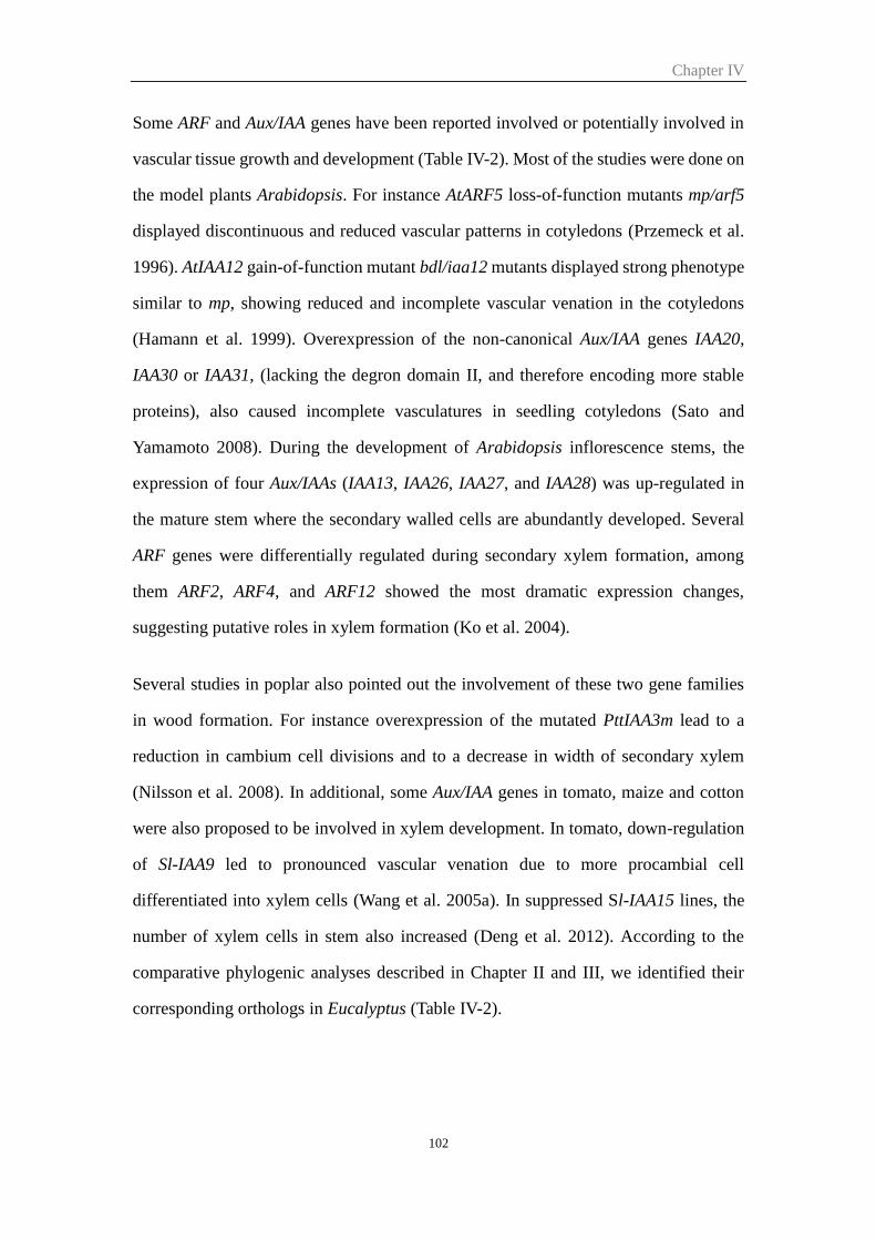
Chapter IV
102
Some ARF and Aux/IAA genes have been reported involved or potentially involved in
vascular tissue growth and development (Table IV-2). Most of the studies were done on
the model plants Arabidopsis. For instance AtARF5 loss-of-function mutants mp/arf5
displayed discontinuous and reduced vascular patterns in cotyledons (Przemeck et al.
1996). AtIAA12 gain-of-function mutant bdl/iaa12 mutants displayed strong phenotype
similar to mp, showing reduced and incomplete vascular venation in the cotyledons
(Hamann et al. 1999). Overexpression of the non-canonical Aux/IAA genes IAA20,
IAA30 or IAA31, (lacking the degron domain II, and therefore encoding more stable
proteins), also caused incomplete vasculatures in seedling cotyledons (Sato and
Yamamoto 2008). During the development of Arabidopsis inflorescence stems, the
expression of four Aux/IAAs (IAA13, IAA26, IAA27, and IAA28) was up-regulated in
the mature stem where the secondary walled cells are abundantly developed. Several
ARF genes were differentially regulated during secondary xylem formation, among
them ARF2, ARF4, and ARF12 showed the most dramatic expression changes,
suggesting putative roles in xylem formation (Ko et al. 2004).
Several studies in poplar also pointed out the involvement of these two gene families
in wood formation. For instance overexpression of the mutated PttIAA3m lead to a
reduction in cambium cell divisions and to a decrease in width of secondary xylem
(Nilsson et al. 2008). In additional, some Aux/IAA genes in tomato, maize and cotton
were also proposed to be involved in xylem development. In tomato, down-regulation
of Sl-IAA9 led to pronounced vascular venation due to more procambial cell
differentiated into xylem cells (Wang et al. 2005a). In suppressed Sl-IAA15 lines, the
number of xylem cells in stem also increased (Deng et al. 2012). According to the
comparative phylogenic analyses described in Chapter II and III, we identified their
corresponding orthologs in Eucalyptus (Table IV-2).
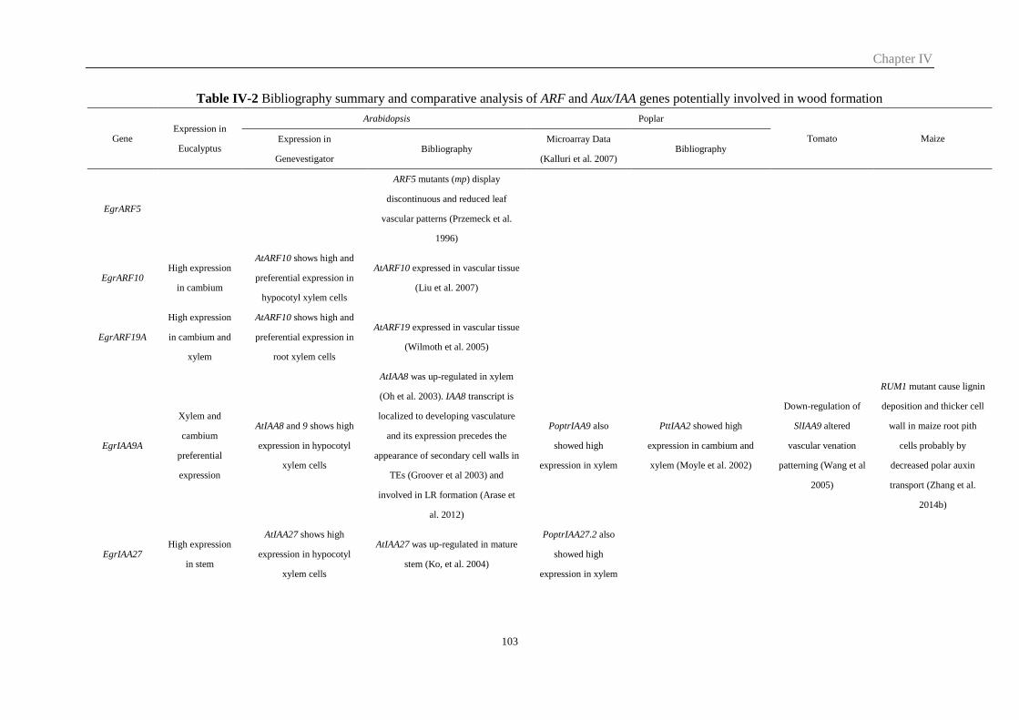
Chapter IV
103
Table IV-2 Bibliography summary and comparative analysis of ARF and Aux/IAA genes potentially involved in wood formation
Gene Expression in
Eucalyptus
Arabidopsis Poplar
Tomato Maize Expression in
Genevestigator Bibliography
Microarray Data
(Kalluri et al. 2007) Bibliography
EgrARF5
ARF5 mutants (mp) display
discontinuous and reduced leaf
vascular patterns (Przemeck et al.
1996)
EgrARF10 High expression
in cambium
AtARF10 shows high and
preferential expression in
hypocotyl xylem cells
AtARF10 expressed in vascular tissue
(Liu et al. 2007)
EgrARF19A
High expression
in cambium and
xylem
AtARF10 shows high and
preferential expression in
root xylem cells
AtARF19 expressed in vascular tissue
(Wilmoth et al. 2005)
EgrIAA9A
Xylem and
cambium
preferential
expression
AtIAA8 and 9 shows high
expression in hypocotyl
xylem cells
AtIAA8 was up-regulated in xylem
(Oh et al. 2003). IAA8 transcript is
localized to developing vasculature
and its expression precedes the
appearance of secondary cell walls in
TEs (Groover et al 2003) and
involved in LR formation (Arase et
al. 2012)
PoptrIAA9 also
showed high
expression in xylem
PttIAA2 showed high
expression in cambium and
xylem (Moyle et al. 2002)
Down-regulation of
SlIAA9 altered
vascular venation
patterning (Wang et al
2005)
RUM1 mutant cause lignin
deposition and thicker cell
wall in maize root pith
cells probably by
decreased polar auxin
transport (Zhang et al.
2014b)
EgrIAA27 High expression
in stem
AtIAA27 shows high
expression in hypocotyl
xylem cells
AtIAA27 was up-regulated in mature
stem (Ko, et al. 2004)
PoptrIAA27.2 also
showed high
expression in xylem
Chapter IV
104
EgrIAA11
Xylem and
cambium
preferential
expression
AtIAA11 showed
preferential expression in
root xylem cells
PoptrIAA11 showed
high expression in
xylem
PttIAA5 preferentially strong
expressed in secondary wall
forming xylem cells (Moyle et
al. 2002)
EgrIAA13
Vascular tissue
preferential
expression
AtIAA12 and AtIAA13
display high/specific
expression in hypocotyl
xylem cells
iaa12/bdl seeding showed
reduced cotyledons vasculature
(Hamann et al. 1999), AtIAA12 and
13 was up-regulated in xylem (Oh
et al. 2003), and up-regulated in
mature stem (Ko, et al. 2004)
EgrIAA15A High expression
in vascular tissue
Suppressed SlIAA15
lines increased xylem
cells in tomato stem
(Deng et al. 2012)
EgrIAA29
Preferential
expression in
xylem and
cambium
AtIAA29 showed
preferential expression in
root xylem cells
PoptrIAA29 showed
high expression in
xylem
EgrIAA20
High and
preferential
expression in
vascular tissue
overexpressing these three genes
(AtIAA20, 30, 31) result in reduced
and discontinued vascular in
cotyledons (Sato and Yamamoto
2008)
PoptrIAA20.2 showed
preferential expression
in xylem
PttIAA3 and 4 showed cambium
and differentiating xylem
preferential expression (Moyle
et al. 2002). PttIAA3m
transgenic poplar showed
reduced xylem development
(Nilsson et al. 2008).
EgrIAA31
Vascular
preferential
expression
Chapter IV
105
In the course of this PhD, we carried on the subcellular localization of eight of the 13
candidate genes, and assessed their transcriptional activities using co-transfection
assays tobacco protoplasts.
Heterologous expression in Arabidopsis was adopted to explore their physiological
roles in plants. We made overexpression constructs of either native or mutated versions
as well as dominant repression constructs. We analyzed in depth the effects of
EgrIAA4m (chapter III), EgrIAA9Am and EgrIAA20 (this chapter) in transgenic
Arabidopsis lines. Physiological and molecular experiments demonstrated the impacts
of over-expressing these genes on plant growth and development. Anatomical analyses
revealed altered vascular patterning and xylem development and biochemical analysis
further confirmed their impacts on plant cell wall compositions in transgenic plants.
2 Results
2.1 Subcellular localization
We examined the subcellular localisation of eight proteins including three ARF and five
Aux/IAA genes. When transiently expressed as GFP fusion proteins in tobacco
protoplasts, two ARFs (EgrARF4 and 10) and two Aux/IAA (9A and 13) were located
exclusively in the nucleus (Fig. IV-1), consistent with their predicted function as
transcription factors. However, EgrIAA3B and 20 were localized both in the nucleus
and cytoplasm, as reported for other Aux/IAA proteins (Arase et al. 2012; Audran-
Delalande et al 2013). Surprisingly, both the N- and C- terminal GFP fusion proteins of
EgrARF19A were not localized in the nucleus. Co-transformation with a nuclear
marker (simian virus 40 (SV40) large-T antigen) (David S. Goldfarb 1986) fused with
mRFP confirmed that EgrARF19A which harbours a weak bipartite NLS as its ortholog
in Arabidopsis, was localized outside the nucleus (Fig. IV-2). It is worth noting that
EgrARF19A localization was different to that of the control expressing GFP protein
alone which was localized everywhere including in the nucleus and the cytoplasm.
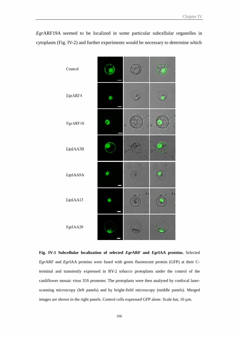
Chapter IV
106
EgrARF19A seemed to be localized in some particular subcellular organelles in
cytoplasm (Fig. IV-2) and further experiments would be necessary to determine which
Fig. IV-1 Subcellular localization of selected EgrARF and EgrIAA proteins. Selected
EgrARF and EgrIAA proteins were fused with green fluorescent protein (GFP) at their C-
terminal and transiently expressed in BY-2 tobacco protoplasts under the control of the
cauliflower mosaic virus 35S promoter. The protoplasts were then analysed by confocal laser-
scanning microscopy (left panels) and by bright-field microscopy (middle panels). Merged
images are shown in the right panels. Control cells expressed GFP alone. Scale bar, 10 µm.
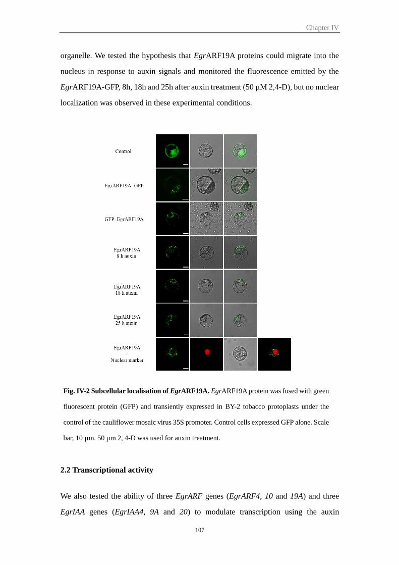
Chapter IV
107
organelle. We tested the hypothesis that EgrARF19A proteins could migrate into the
nucleus in response to auxin signals and monitored the fluorescence emitted by the
EgrARF19A-GFP, 8h, 18h and 25h after auxin treatment (50 µM 2,4-D), but no nuclear
localization was observed in these experimental conditions.
2.2 Transcriptional activity
We also tested the ability of three EgrARF genes (EgrARF4, 10 and 19A) and three
EgrIAA genes (EgrIAA4, 9A and 20) to modulate transcription using the auxin
Fig. IV-2 Subcellular localisation of EgrARF19A. EgrARF19A protein was fused with green
fluorescent protein (GFP) and transiently expressed in BY-2 tobacco protoplasts under the
control of the cauliflower mosaic virus 35S promoter. Control cells expressed GFP alone. Scale
bar, 10 µm. 50 µm 2, 4-D was used for auxin treatment.
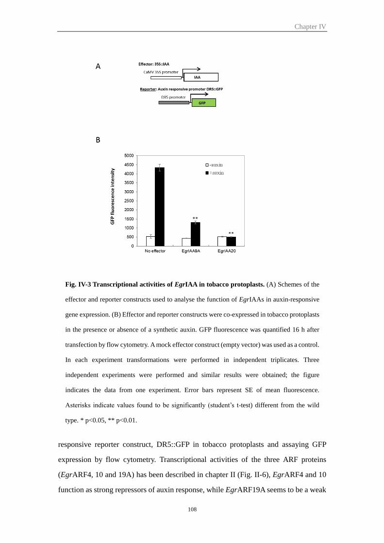
Chapter IV
108
responsive reporter construct, DR5::GFP in tobacco protoplasts and assaying GFP
expression by flow cytometry. Transcriptional activities of the three ARF proteins
(EgrARF4, 10 and 19A) has been described in chapter II (Fig. II-6), EgrARF4 and 10
function as strong repressors of auxin response, while EgrARF19A seems to be a weak
Fig. IV-3 Transcriptional activities of EgrIAA in tobacco protoplasts. (A) Schemes of the
effector and reporter constructs used to analyse the function of EgrIAAs in auxin-responsive
gene expression. (B) Effector and reporter constructs were co-expressed in tobacco protoplasts
in the presence or absence of a synthetic auxin. GFP fluorescence was quantified 16 h after
transfection by flow cytometry. A mock effector construct (empty vector) was used as a control.
In each experiment transformations were performed in independent triplicates. Three
independent experiments were performed and similar results were obtained; the figure
indicates the data from one experiment. Error bars represent SE of mean fluorescence.
Asterisks indicate values found to be significantly (student’s t-test) different from the wild
type. * p<0.05, ** p<0.01.
Chapter IV
109
activator. All three tested EgrIAAs mediated auxin response in vivo and functioned as
strong transcriptional repressors. EgrIAA4 (reported in Chapter III), EgrIAA9A and
EgrIAA20 repressed auxin-induced reporter gene expression by 87%, 70% and 88%
respectively as compared to the control co-transformed with an empty vector (Fig. IV-
3).
2.3 Strategies for functional characterization by heterologous expression of
Eucalyptus ARF or IAA transcription factors in Arabidopsis
We used reverse genetic approach to explore the role of some candidate genes using
heterologous expression in Arabidopsis. Despite its herbaceous nature, Arabidopsis is
a good model to study wood formation (Zhang et al. 2011; Chaffey et al. 2002;
Nieminen et al. 2004). We describe below the different construction we made
explaining the rationale behind them. These construction were made in the framework
of European project but only some of them were transformed into plants as detailed in
2.4.
2.3.1 ‘gain-of-function’ strategy for Aux/IAA candidate genes
In Arabidopsis, all of the Aux/IAA T-DNA insertion mutants characterized (12 out of
29) failed to show any obvious visible phenotype (Overvoorde et al. 2005). All other
loss-of-function mutants using RNAi or antisense exhibited only subtle phenotypes
(reviewed in Reed 2001), probably because of gene function redundancy or feedback
regulatory loops that enabled the mutant plants to compensate for the absence of a
particular Aux/IAA protein. Recent progress toward discovering the functions of
Aux/IAA genes has come from the fortuitous discovery of gain-of-function mutations in
several of these genes (IAA3/SHY2, IAA6/SHY1, IAA7/AXR2, IAA12/BDl, IAA14/SLR1,
IAA17/AXR3, IAA18, IAA19/MSG2, and IAA28). All these mutations occurred in the
highly conserved domain II and conferred increased stability to their corresponding
proteins. Aux/IAA proteins are very short living proteins, and their degradation is
essential for auxin signaling. Mutations in domain II increase protein stability. Some
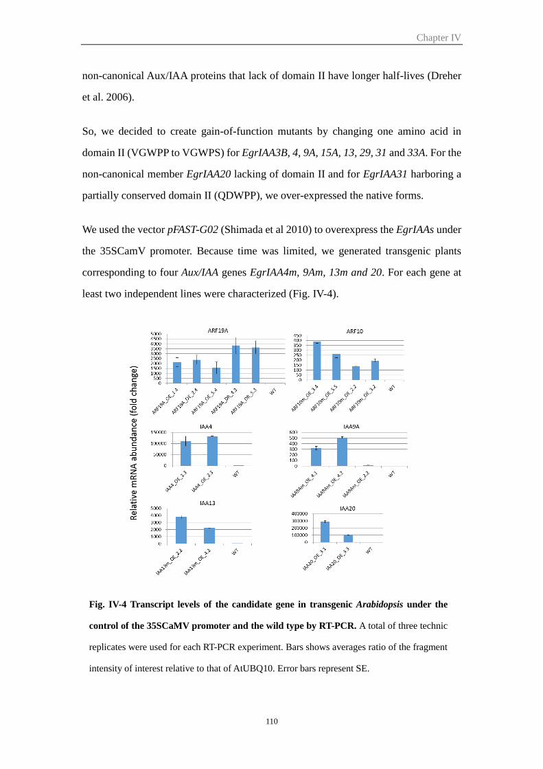
Chapter IV
110
non-canonical Aux/IAA proteins that lack of domain II have longer half-lives (Dreher
et al. 2006).
So, we decided to create gain-of-function mutants by changing one amino acid in
domain II (VGWPP to VGWPS) for EgrIAA3B, 4, 9A, 15A, 13, 29, 31 and 33A. For the
non-canonical member EgrIAA20 lacking of domain II and for EgrIAA31 harboring a
partially conserved domain II (QDWPP), we over-expressed the native forms.
We used the vector pFAST-G02 (Shimada et al 2010) to overexpress the EgrIAAs under
the 35SCamV promoter. Because time was limited, we generated transgenic plants
corresponding to four Aux/IAA genes EgrIAA4m, 9Am, 13m and 20. For each gene at
least two independent lines were characterized (Fig. IV-4).
Fig. IV-4 Transcript levels of the candidate gene in transgenic Arabidopsis under the
control of the 35SCaMV promoter and the wild type by RT-PCR. A total of three technic
replicates were used for each RT-PCR experiment. Bars shows averages ratio of the fragment
intensity of interest relative to that of AtUBQ10. Error bars represent SE.

Chapter IV
111
2.3.2 Appropriate reverse genetic strategies for three selected EgrARFs
Many loss-of-function mutants of ARF (T-DNA insertion mutant) did not show any
phenotypes, probably due to functional redundancy. Similarly, some transgenic plants
over expressing ARF failed to give any phenotype due to the endogenous post-
transcriptional regulation by miRNAs. Mutations in their miRNA target sites lead to
distinct phenotypes revealing their functions in plant growth and development (Liu et
al. 2007; Hendelman et al. 2012). Two of the selected Eucalyptus ARF genes, EgrARF4
and EgrARF10 were predicted to be targeted by miRNA; EgrARF4 has two TAS3 target
sites and EgrARF10 has one miR160 target site. Therefore, we choose to introduce
miRNA resistant-mutation in EgrARF4 and EgrARF10 to generate ‘gain-of-function’
transgenic plants. For the third selected gene EgrARF19A, no miRNA target site was
detected, we chose to overexpress the native form and also to make a dominant
repression construct to transform the TF in a strong repressor.
The transgenic Arabidopsis (overexpression, dominant repression, and miRNA
resistant mutations) were generated as described in Materials and Methods. We
obtained ten independent EgrIAA19A overexpression lines (EgrARF19A_OE) and
twelve EgrARF19A dominant repression lines (EgrARF19A_DR), four independent
miRNA resistant EgrARF10 lines (EgrARF10m) (Fig. IV-4). For technical reasons and
limited time, we failed the EgrARF4 overlap PCR because it contains two miRNA target
sites.
2.4 Preliminary analyses of the IAA and ARF transgenic lines
We generated transgenic Arabidopsis lines for four IAA (EgrIAA4, 9A, 13 and 20) and
two ARF (EgrARF10 and 19A). Among all these transgenic Arabidopsis, only three
mutants (EgrIAA4m, 9Am and 20) exhibited distinct xylem related phenotypes. The
functional characterization of EgrIAA4m was reported in Chapter III, and that of
EgrIAA9Am and EgrIAA20 will be addressed in the following sections. The two
EgrIAA13m transgenic lines grew smaller plants as compared to the wild type without
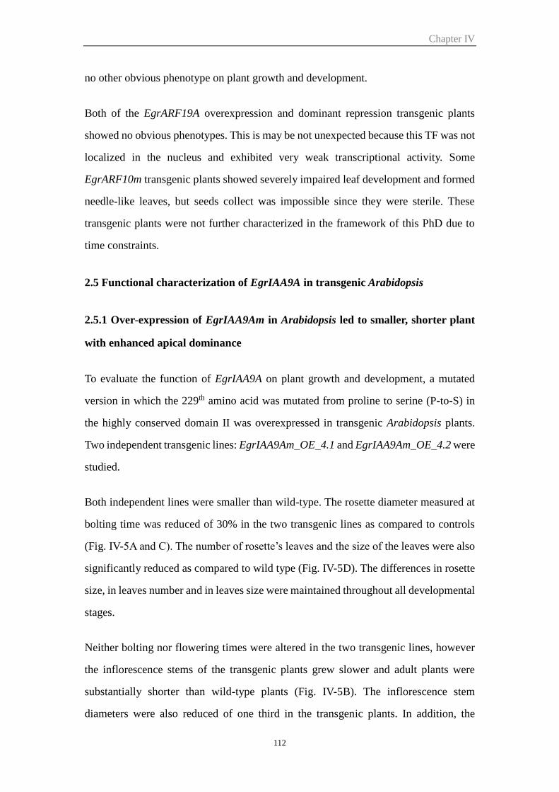
Chapter IV
112
no other obvious phenotype on plant growth and development.
Both of the EgrARF19A overexpression and dominant repression transgenic plants
showed no obvious phenotypes. This is may be not unexpected because this TF was not
localized in the nucleus and exhibited very weak transcriptional activity. Some
EgrARF10m transgenic plants showed severely impaired leaf development and formed
needle-like leaves, but seeds collect was impossible since they were sterile. These
transgenic plants were not further characterized in the framework of this PhD due to
time constraints.
2.5 Functional characterization of EgrIAA9A in transgenic Arabidopsis
2.5.1 Over-expression of EgrIAA9Am in Arabidopsis led to smaller, shorter plant
with enhanced apical dominance
To evaluate the function of EgrIAA9A on plant growth and development, a mutated
version in which the 229th amino acid was mutated from proline to serine (P-to-S) in
the highly conserved domain II was overexpressed in transgenic Arabidopsis plants.
Two independent transgenic lines: EgrIAA9Am_OE_4.1 and EgrIAA9Am_OE_4.2 were
studied.
Both independent lines were smaller than wild-type. The rosette diameter measured at
bolting time was reduced of 30% in the two transgenic lines as compared to controls
(Fig. IV-5A and C). The number of rosette’s leaves and the size of the leaves were also
significantly reduced as compared to wild type (Fig. IV-5D). The differences in rosette
size, in leaves number and in leaves size were maintained throughout all developmental
stages.
Neither bolting nor flowering times were altered in the two transgenic lines, however
the inflorescence stems of the transgenic plants grew slower and adult plants were
substantially shorter than wild-type plants (Fig. IV-5B). The inflorescence stem
diameters were also reduced of one third in the transgenic plants. In addition, the
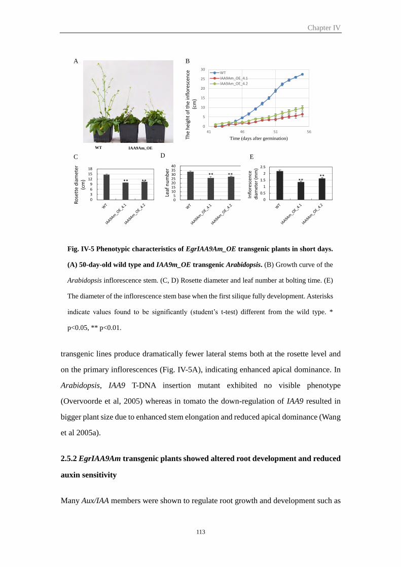
Chapter IV
113
transgenic lines produce dramatically fewer lateral stems both at the rosette level and
on the primary inflorescences (Fig. IV-5A), indicating enhanced apical dominance. In
Arabidopsis, IAA9 T-DNA insertion mutant exhibited no visible phenotype
(Overvoorde et al, 2005) whereas in tomato the down-regulation of IAA9 resulted in
bigger plant size due to enhanced stem elongation and reduced apical dominance (Wang
et al 2005a).
2.5.2 EgrIAA9Am transgenic plants showed altered root development and reduced
auxin sensitivity
Many Aux/IAA members were shown to regulate root growth and development such as
Fig. IV-5 Phenotypic characteristics of EgrIAA9Am_OE transgenic plants in short days.
(A) 50-day-old wild type and IAA9m_OE transgenic Arabidopsis. (B) Growth curve of the
Arabidopsis inflorescence stem. (C, D) Rosette diameter and leaf number at bolting time. (E)
The diameter of the inflorescence stem base when the first silique fully development. Asterisks
indicate values found to be significantly (student’s t-test) different from the wild type. *
p<0.05, ** p<0.01.
A B
0369
121518
Ro
sett
e d
iam
eter
(c
m) ** **
05
10152025303540
Leaf
nu
mb
er
** **
0
5
10
15
20
25
30
41 46 51 56
WT
IAA9Am_OE_4.1
IAA9Am_OE_4.2
C ED
WT IAA9Am_OE
Time (days after germination)The
hei
ght
of
the
infl
ore
scen
ce(c
m)
0
0.5
1
1.5
2
2.5
Infl
ore
scen
ced
iam
ete
r (m
m)
****
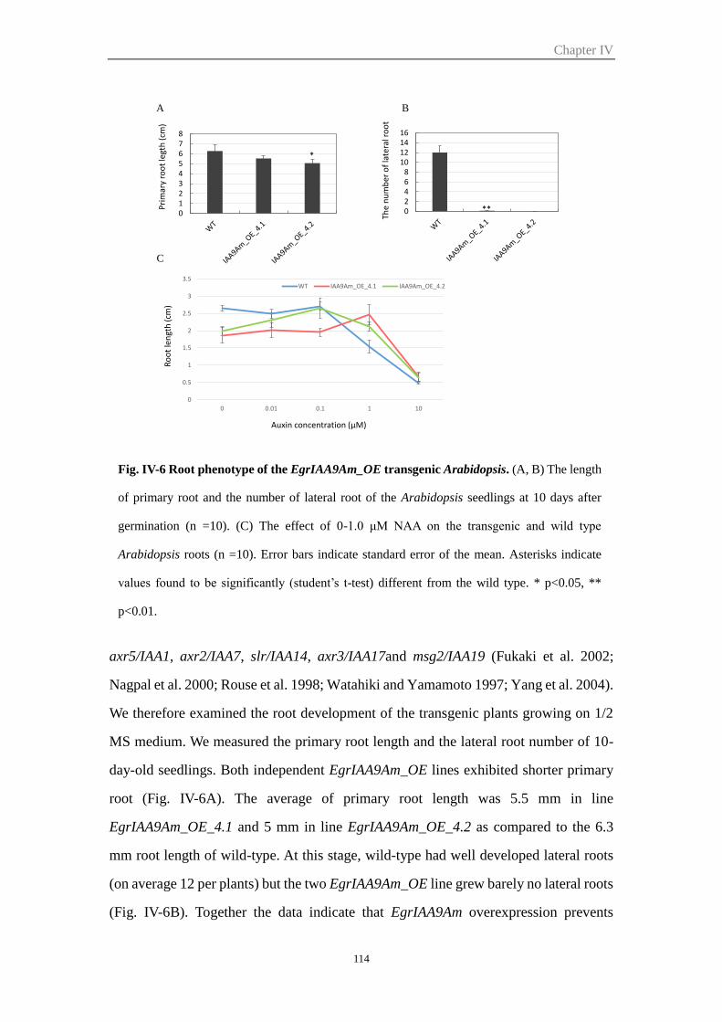
Chapter IV
114
axr5/IAA1, axr2/IAA7, slr/IAA14, axr3/IAA17and msg2/IAA19 (Fukaki et al. 2002;
Nagpal et al. 2000; Rouse et al. 1998; Watahiki and Yamamoto 1997; Yang et al. 2004).
We therefore examined the root development of the transgenic plants growing on 1/2
MS medium. We measured the primary root length and the lateral root number of 10-
day-old seedlings. Both independent EgrIAA9Am_OE lines exhibited shorter primary
root (Fig. IV-6A). The average of primary root length was 5.5 mm in line
EgrIAA9Am_OE_4.1 and 5 mm in line EgrIAA9Am_OE_4.2 as compared to the 6.3
mm root length of wild-type. At this stage, wild-type had well developed lateral roots
(on average 12 per plants) but the two EgrIAA9Am_OE line grew barely no lateral roots
(Fig. IV-6B). Together the data indicate that EgrIAA9Am overexpression prevents
Fig. IV-6 Root phenotype of the EgrIAA9Am_OE transgenic Arabidopsis. (A, B) The length
of primary root and the number of lateral root of the Arabidopsis seedlings at 10 days after
germination (n =10). (C) The effect of 0-1.0 μM NAA on the transgenic and wild type
Arabidopsis roots (n =10). Error bars indicate standard error of the mean. Asterisks indicate
values found to be significantly (student’s t-test) different from the wild type. * p<0.05, **
p<0.01.
02468
10121416
The
nu
mb
er o
f la
tera
l ro
ot
A B
012345678
Pri
mar
y ro
ot
legt
h (
cm)
*
C
Ro
ot
len
gth
(cm
)
0
0.5
1
1.5
2
2.5
3
3.5
0 0.01 0.1 1 10
WT IAA9Am_OE_4.1 IAA9Am_OE_4.2
Auxin concentration (µM)
**
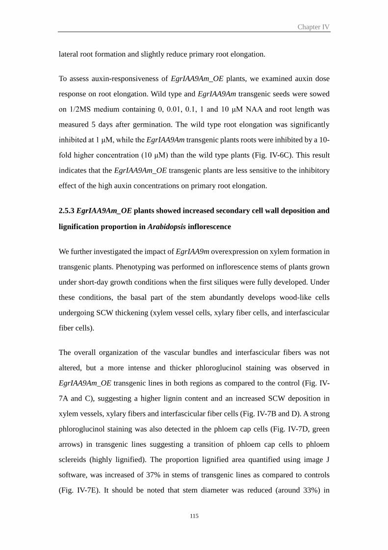
Chapter IV
115
lateral root formation and slightly reduce primary root elongation.
To assess auxin-responsiveness of EgrIAA9Am_OE plants, we examined auxin dose
response on root elongation. Wild type and EgrIAA9Am transgenic seeds were sowed
on 1/2MS medium containing 0, 0.01, 0.1, 1 and 10 μM NAA and root length was
measured 5 days after germination. The wild type root elongation was significantly
inhibited at 1 μM, while the EgrIAA9Am transgenic plants roots were inhibited by a 10-
fold higher concentration (10 μM) than the wild type plants (Fig. IV-6C). This result
indicates that the EgrIAA9Am_OE transgenic plants are less sensitive to the inhibitory
effect of the high auxin concentrations on primary root elongation.
2.5.3 EgrIAA9Am_OE plants showed increased secondary cell wall deposition and
lignification proportion in Arabidopsis inflorescence
We further investigated the impact of EgrIAA9m overexpression on xylem formation in
transgenic plants. Phenotyping was performed on inflorescence stems of plants grown
under short-day growth conditions when the first siliques were fully developed. Under
these conditions, the basal part of the stem abundantly develops wood-like cells
undergoing SCW thickening (xylem vessel cells, xylary fiber cells, and interfascicular
fiber cells).
The overall organization of the vascular bundles and interfascicular fibers was not
altered, but a more intense and thicker phloroglucinol staining was observed in
EgrIAA9Am_OE transgenic lines in both regions as compared to the control (Fig. IV-
7A and C), suggesting a higher lignin content and an increased SCW deposition in
xylem vessels, xylary fibers and interfascicular fiber cells (Fig. IV-7B and D). A strong
phloroglucinol staining was also detected in the phloem cap cells (Fig. IV-7D, green
arrows) in transgenic lines suggesting a transition of phloem cap cells to phloem
sclereids (highly lignified). The proportion lignified area quantified using image J
software, was increased of 37% in stems of transgenic lines as compared to controls
(Fig. IV-7E). It should be noted that stem diameter was reduced (around 33%) in
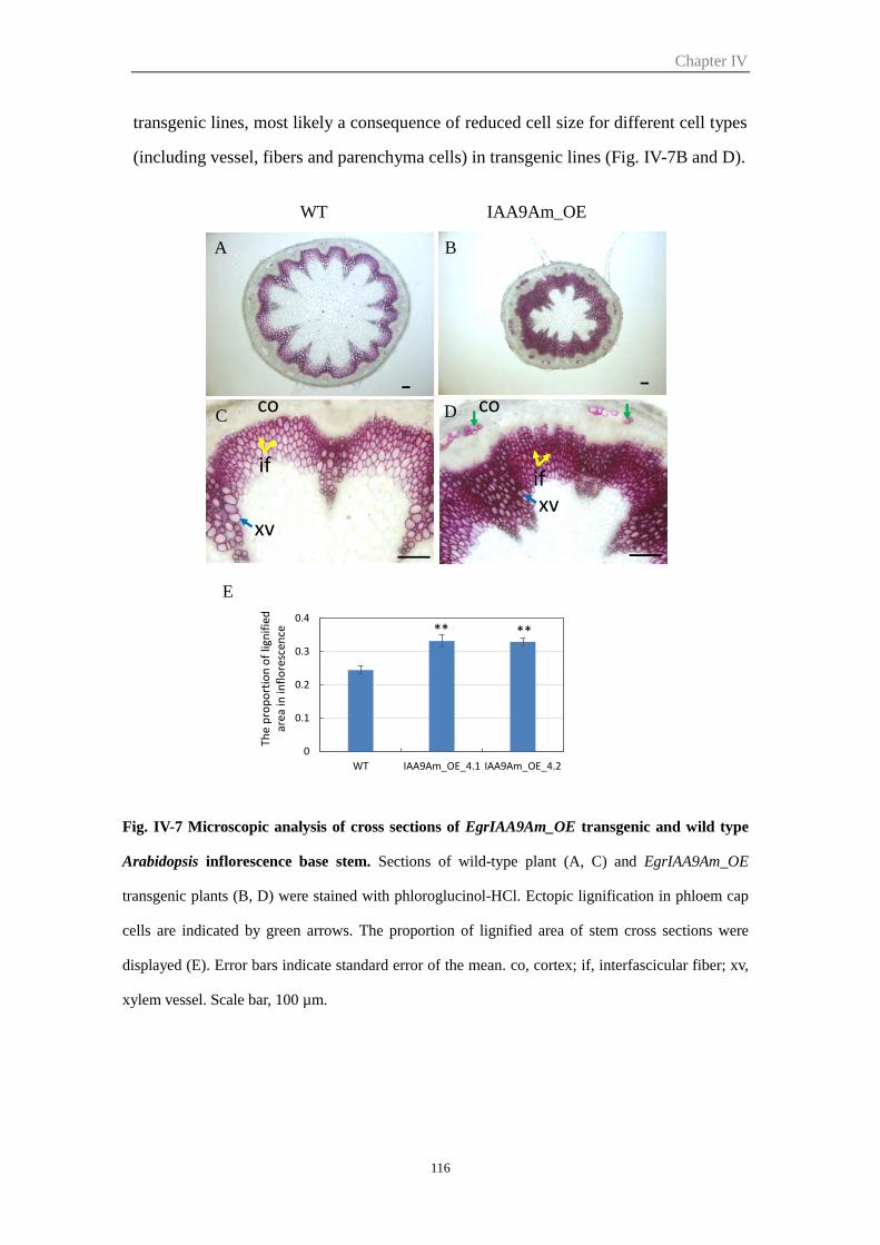
Chapter IV
116
transgenic lines, most likely a consequence of reduced cell size for different cell types
(including vessel, fibers and parenchyma cells) in transgenic lines (Fig. IV-7B and D).
Fig. IV-7 Microscopic analysis of cross sections of EgrIAA9Am_OE transgenic and wild type
Arabidopsis inflorescence base stem. Sections of wild-type plant (A, C) and EgrIAA9Am_OE
transgenic plants (B, D) were stained with phloroglucinol-HCl. Ectopic lignification in phloem cap
cells are indicated by green arrows. The proportion of lignified area of stem cross sections were
displayed (E). Error bars indicate standard error of the mean. co, cortex; if, interfascicular fiber; xv,
xylem vessel. Scale bar, 100 µm.
A
C
B
D
WT IAA9Am_OE
xvxv
ifif
co co
E
0
0.1
0.2
0.3
0.4
WT IAA9Am_OE_4.1 IAA9Am_OE_4.2
** **
The
pro
po
rtio
n o
f lig
nif
ied
ar
ea in
infl
ore
scen
ce
Chapter IV
117
2.5.4 EgrIAA9Am greatly altered secondary xylem formation in Arabidopsis
hypocotyl
The most extensive secondary xylem (wood formation) was found to develop in the
Arabidopsis hypocotyls grown under short-day conditions (Chaffey et al. 2002). In the
first phase of secondary xylem development, only vessel elements differentiate, and the
rest of the xylem cells remain as parenchyma cells. This phase I is visualized as a central
core in which only the vessels are stained by phloroglucinol due to their lignified SCW
(Fig. IV-8A and C). During phase II, all xylem cells develop into either vessels or fibers,
both have lignified thick SCW stained by phloroglucinol into red-violet. The fiber cells
are smaller and show brighter phloroglucinol staining than the vessels, due to the
Fig. IV-8 Microscopic analysis of cross sections of EgrIAA9Am_OE transgenic
Arabidopsis and wild type hypocotyl. Sections of wild-type plant (A, C) and
EgrIAA9Am_OE transgenic plants (B, D) were stained with phloroglucinol-HCl. I, xylem I;
II, xylem II; f, fiber; v, vessel. Scale bar, 100 µm.
Chapter IV
118
presence of Syringyl lignin (S) whereas the lignin in vessels are made mainly of G units
(Fig. IV-8A and C). It is during this later stage of secondary xylem development that
the anatomy of the Arabidopsis hypocotyl most closely resembles the xylem of woody
species (Chaffey et al. 2002).
EgrIAA9m_OE lines presented strikingly altered secondary xylem formation in
hypocotyls showing only phase I, in which much more vessel elements were
differentiated with smaller size as compared to controls (Fig. IV-8B and D). The phase
II of xylem development was completely absent in the transgenic lines at this stage
(approximately two weeks after bolting). It is also worth noting that much less lignified
phloem fibers were found in EgrIAA9m_OE lines as compared to the wild-type (Fig.
IV-8A, indicated by arrow).
Unexpectedly, these observations are in sharp contrast with those made at the basal part
of inflorescence stem which showed more lignified cells in EgrIAA9Am_OE lines.
These discrepancies in xylem patterning between the two organs suggest that different
regulating mechanisms may exist between stem and hypocotyl. These results underlie
the different origins of xylem in the two organs and deserve more investigations.
2.5.5 EgrIAA9Am overexpression affects lignin composition of secondary cell walls
in Arabidopsis hypocotyls
GC/MS pyrolysis (Py-GC/MS) analysis provides a chemical finger-print of the cell
walls and now is widely used for rapid determination of lignin chemistry (Gerber et al.
2012; Meier et al. 2005). We performed Py-GC/MS analysis to evaluate and identify
SCW modifications of the transgenic plants using hypocotyls as target material. The
Py-GC/MS chromatograms were evaluated using Simca software by multivariate
OPLS-DA method (orthogonal projections to latent structures discriminant analysis)
(Worley et al. 2013), which yielded in a clear separation of the two transgenic lines
from wild type (Fig. IV-9A).
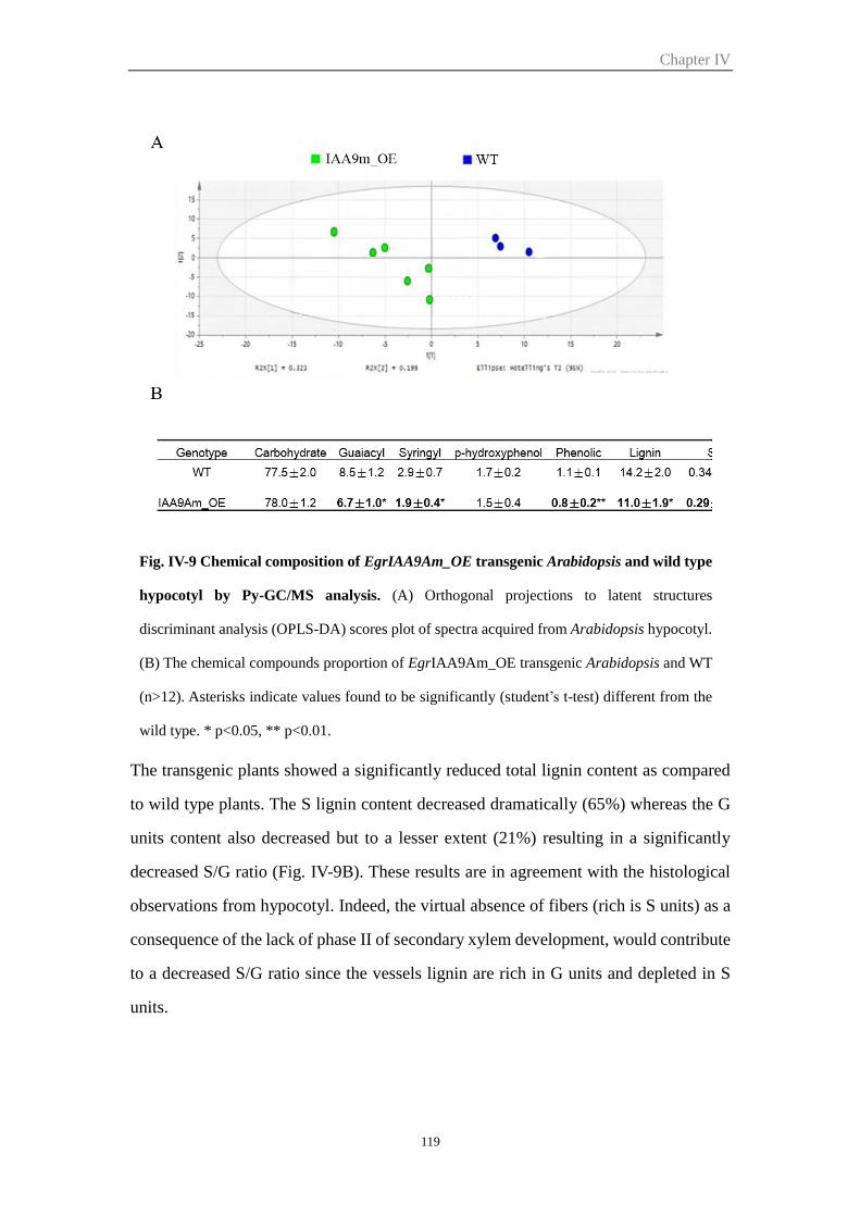
Chapter IV
119
The transgenic plants showed a significantly reduced total lignin content as compared
to wild type plants. The S lignin content decreased dramatically (65%) whereas the G
units content also decreased but to a lesser extent (21%) resulting in a significantly
decreased S/G ratio (Fig. IV-9B). These results are in agreement with the histological
observations from hypocotyl. Indeed, the virtual absence of fibers (rich is S units) as a
consequence of the lack of phase II of secondary xylem development, would contribute
to a decreased S/G ratio since the vessels lignin are rich in G units and depleted in S
units.
Fig. IV-9 Chemical composition of EgrIAA9Am_OE transgenic Arabidopsis and wild type
hypocotyl by Py-GC/MS analysis. (A) Orthogonal projections to latent structures
discriminant analysis (OPLS-DA) scores plot of spectra acquired from Arabidopsis hypocotyl.
(B) The chemical compounds proportion of EgrIAA9Am_OE transgenic Arabidopsis and WT
(n>12). Asterisks indicate values found to be significantly (student’s t-test) different from the
wild type. * p<0.05, ** p<0.01.
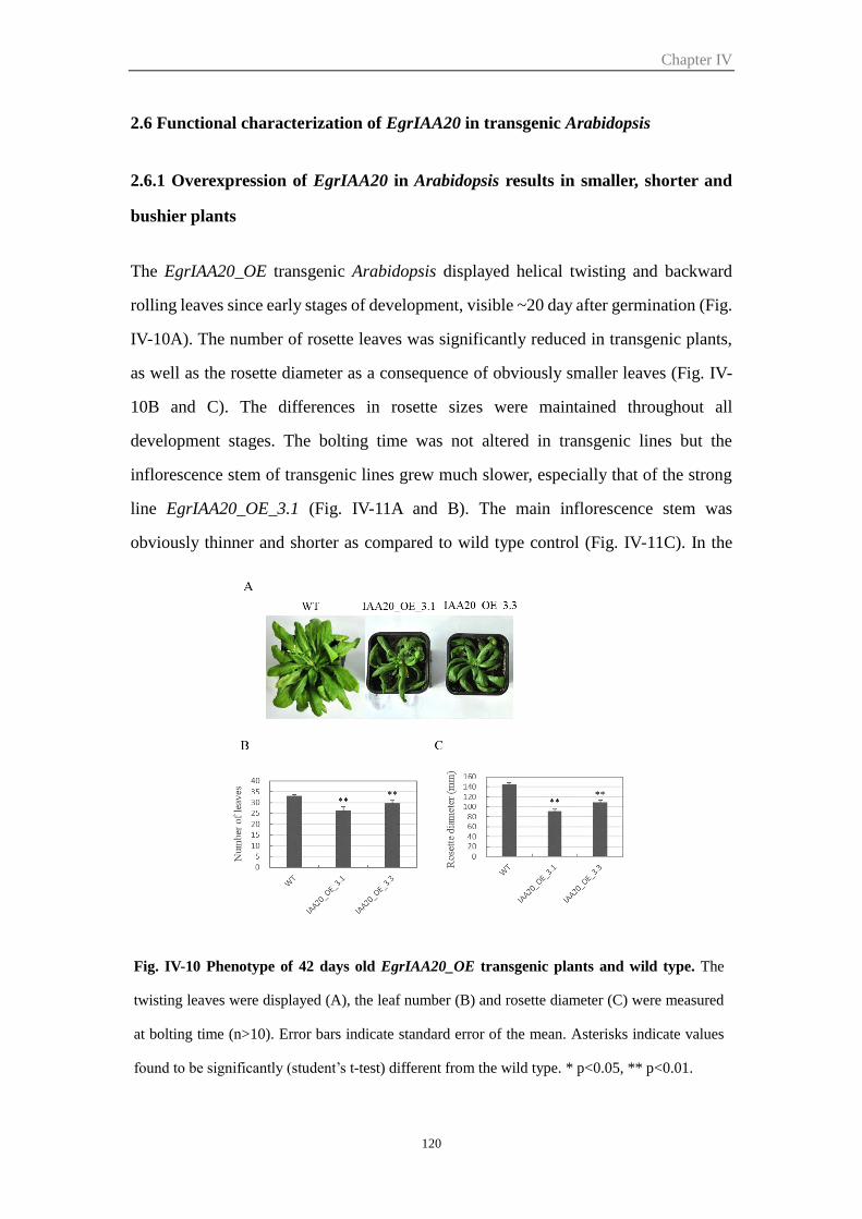
Chapter IV
120
2.6 Functional characterization of EgrIAA20 in transgenic Arabidopsis
2.6.1 Overexpression of EgrIAA20 in Arabidopsis results in smaller, shorter and
bushier plants
The EgrIAA20_OE transgenic Arabidopsis displayed helical twisting and backward
rolling leaves since early stages of development, visible ~20 day after germination (Fig.
IV-10A). The number of rosette leaves was significantly reduced in transgenic plants,
as well as the rosette diameter as a consequence of obviously smaller leaves (Fig. IV-
10B and C). The differences in rosette sizes were maintained throughout all
development stages. The bolting time was not altered in transgenic lines but the
inflorescence stem of transgenic lines grew much slower, especially that of the strong
line EgrIAA20_OE_3.1 (Fig. IV-11A and B). The main inflorescence stem was
obviously thinner and shorter as compared to wild type control (Fig. IV-11C). In the
Fig. IV-10 Phenotype of 42 days old EgrIAA20_OE transgenic plants and wild type. The
twisting leaves were displayed (A), the leaf number (B) and rosette diameter (C) were measured
at bolting time (n>10). Error bars indicate standard error of the mean. Asterisks indicate values
found to be significantly (student’s t-test) different from the wild type. * p<0.05, ** p<0.01.
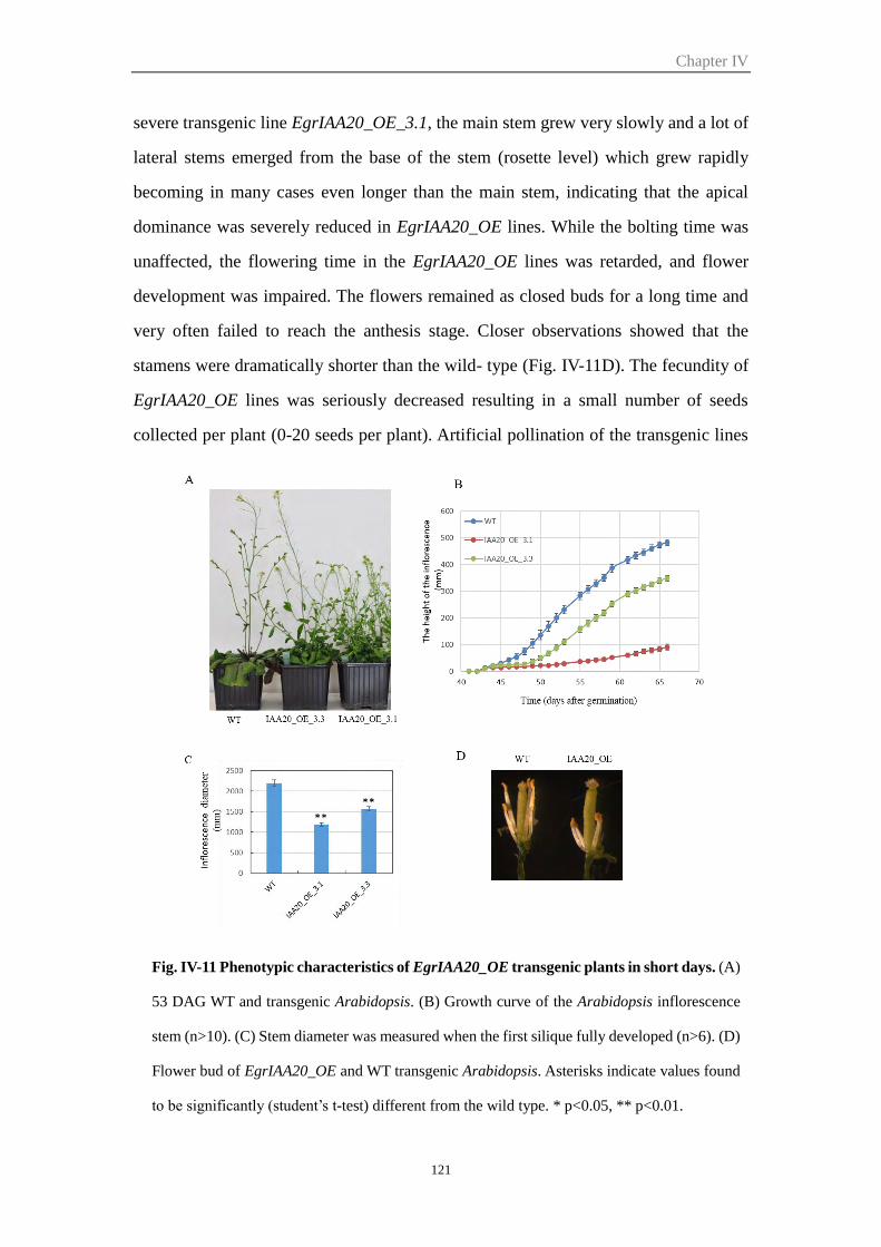
Chapter IV
121
severe transgenic line EgrIAA20_OE_3.1, the main stem grew very slowly and a lot of
lateral stems emerged from the base of the stem (rosette level) which grew rapidly
becoming in many cases even longer than the main stem, indicating that the apical
dominance was severely reduced in EgrIAA20_OE lines. While the bolting time was
unaffected, the flowering time in the EgrIAA20_OE lines was retarded, and flower
development was impaired. The flowers remained as closed buds for a long time and
very often failed to reach the anthesis stage. Closer observations showed that the
stamens were dramatically shorter than the wild- type (Fig. IV-11D). The fecundity of
EgrIAA20_OE lines was seriously decreased resulting in a small number of seeds
collected per plant (0-20 seeds per plant). Artificial pollination of the transgenic lines
Fig. IV-11 Phenotypic characteristics of EgrIAA20_OE transgenic plants in short days. (A)
53 DAG WT and transgenic Arabidopsis. (B) Growth curve of the Arabidopsis inflorescence
stem (n>10). (C) Stem diameter was measured when the first silique fully developed (n>6). (D)
Flower bud of EgrIAA20_OE and WT transgenic Arabidopsis. Asterisks indicate values found
to be significantly (student’s t-test) different from the wild type. * p<0.05, ** p<0.01.
Chapter IV
122
using the wild-type pollen allowed seed formation, indicating that the ovules were still
functional in the transgenic lines. Thus the reduced fertility could be a consequence of
an impaired stamen development and/or of the shorter stamens formed. Indeed, an un-
even position of stamen relative to the pistil is known to prevent the normal pollination
and fertilization.
2.6.2 Overexpression of EgrIAA20 affected root development and root gravitropic
response
We also examined the root growth phenotype in the transgenic lines. We compared the
primary root length and number of lateral roots between wild type and EgrIAA20_OE
lines 10 days after germination on 1/2 MS medium. The EgrIAA20_OE lines showed
significantly reduced primary roots elongation as compared to the wild-type (Fig. IV-
12A). Few lateral roots were formed in EgrIAA20_OE lines at this stage, in sharp
contrast to the wild type plants which had already 12 lateral roots in average (Fig. IV-
12B). This result indicate that the over expression EgrIAA20 gene inhibit primary root
elongation and lateral root formation.
Gravitropic response is a typical auxin-related phenotype, and several Aux/IAA
mutants show agravitropism in Arabidopsis roots and/or hypocotyl, such as axr5/IAA1,
axr2/IAA7, slr/IAA14, axr3/IAA17and msg2/IAA19 (Fukaki et al. 2002; Nagpal et al.
2000; Rouse et al. 1998; Watahiki and Yamamoto 1997; Yang et al. 2004). To assess
the gravitropic response of the EgrIAA20 transgenic plant, we grew them on vertically
oriented 1/2 MS medium plates. Eight days after germination we reoriented these plates
by 90 dEgrees to test their roots response to gravity. Fourty eight hours later, all the
wild type Arabidopsis roots changes 90 dEgrees according to the gravity changes, while
all the EgrIAA20 transgenic plants shows no changes to the gravity change (Fig. IV-
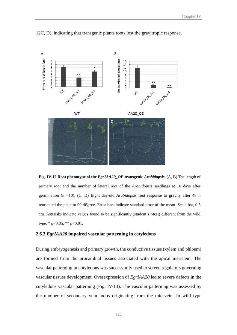
Chapter IV
123
12C, D), indicating that transgenic plants roots lost the gravitropic response.
2.6.3 EgrIAA20 impaired vascular patterning in cotyledons
During embryogenesis and primary growth, the conductive tissues (xylem and phloem)
are formed from the procambial tissues associated with the apical meristem. The
vascular patterning in cotyledons was successfully used to screen regulators governing
vascular tissues development. Overexpression of EgrIAA20 led to severe defects in the
cotyledons vascular patterning (Fig. IV-13). The vascular patterning was assessed by
the number of secondary vein loops originating from the mid-vein. In wild type
Fig. IV-12 Root phenotype of the EgrIAA20_OE transgenic Arabidopsis. (A, B) The length of
primary root and the number of lateral root of the Arabidopsis seedlings at 10 days after
germination (n =10). (C, D) Eight day-old Arabidopsis root response to gravity after 48 h
reoriented the plate to 90 dEgree. Error bars indicate standard error of the mean. Scale bar, 0.5
cm. Asterisks indicate values found to be significantly (student’s t-test) different from the wild
type. * p<0.05, ** p<0.01.
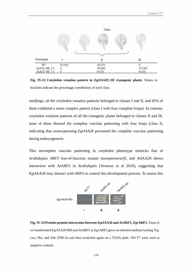
Chapter IV
124
seedlings, all the cotyledon venation patterns belonged to classes I and II, and 45% of
them exhibited a more complex pattern (class I with four complete loops). In contrast,
cotyledon venation patterns of all the transgenic plants belonged to classes II and III,
none of them showed the complete vascular patterning with four loops (class I),
indicating that overexpressing EgrIAA20 prevented the complete vascular patterning
during embryogenesis.
This incomplete vascular patterning in cotyledon phenotype mimicks that of
Arabidopsis ARF5 loss-of-function mutant monopteros/arf5, and AtIAA20 shows
interaction with AtARF5 in Arabidopsis (Vernoux et al 2010), suggesting that
EgrIAA20 may interact with ARF5 to control this development process. To assess this
Fig. IV-13 Cotyledon venation pattern in EgrIAA20_OE transgenic plants. Values in
brackets indicate the percentage contribution of each class.
Fig. IV-14 Protein-protein interaction between EgrIAA20 and AtARF5, EgrARF5. Yeast of
co-transformed EgrIAA20-BD and AtARF5 or EgrARF5 grew on selected medium lacking Trp,
Leu, His, and Ade (THLA) and then scratched again on a TLHA plate. AD-T7 were used as
negative controls.
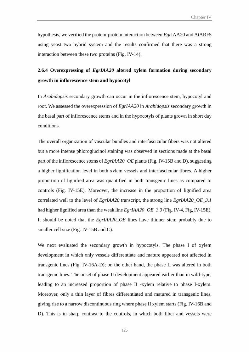
Chapter IV
125
hypothesis, we verified the protein-protein interaction between EgrIAA20 and AtARF5
using yeast two hybrid system and the results confirmed that there was a strong
interaction between these two proteins (Fig. IV-14).
2.6.4 Overexpressing of EgrIAA20 altered xylem formation during secondary
growth in inflorescence stem and hypocotyl
In Arabidopsis secondary growth can occur in the inflorescence stem, hypocotyl and
root. We assessed the overexpression of EgrIAA20 in Arabidopsis secondary growth in
the basal part of inflorescence stems and in the hypocotyls of plants grown in short day
conditions.
The overall organization of vascular bundles and interfascicular fibers was not altered
but a more intense phloroglucinol staining was observed in sections made at the basal
part of the inflorescence stems of EgrIAA20_OE plants (Fig. IV-15B and D), suggesting
a higher lignification level in both xylem vessels and interfascicular fibres. A higher
proportion of lignified area was quantified in both transgenic lines as compared to
controls (Fig. IV-15E). Moreover, the increase in the proportion of lignified area
correlated well to the level of EgrIAA20 transcript, the strong line EgrIAA20_OE_3.1
had higher lignified area than the weak line EgrIAA20_OE_3.3 (Fig. IV-4, Fig, IV-15E).
It should be noted that the EgrIAA20_OE lines have thinner stem probably due to
smaller cell size (Fig. IV-15B and C).
We next evaluated the secondary growth in hypocotyls. The phase I of xylem
development in which only vessels differentiate and mature appeared not affected in
transgenic lines (Fig. IV-16A-D); on the other hand, the phase II was altered in both
transgenic lines. The onset of phase II development appeared earlier than in wild-type,
leading to an increased proportion of phase II -xylem relative to phase I-xylem.
Moreover, only a thin layer of fibres differentiated and matured in transgenic lines,
giving rise to a narrow discontinuous ring where phase II xylem starts (Fig. IV-16B and
D). This is in sharp contrast to the controls, in which both fiber and vessels were
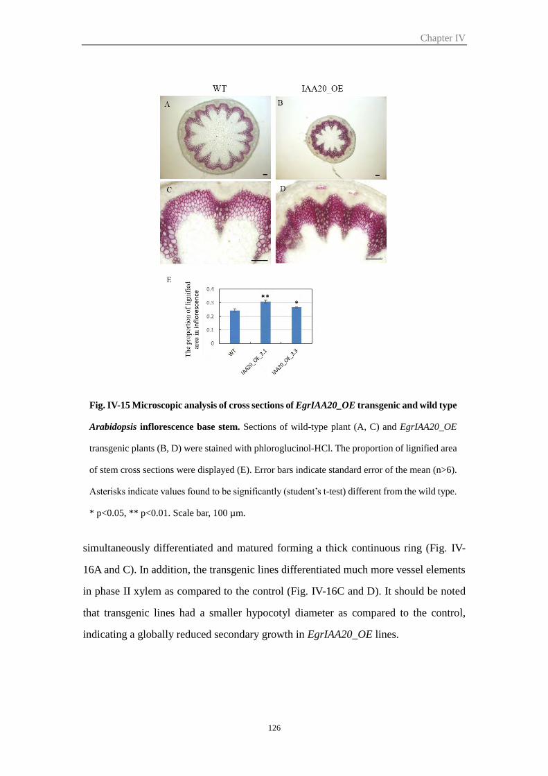
Chapter IV
126
simultaneously differentiated and matured forming a thick continuous ring (Fig. IV-
16A and C). In addition, the transgenic lines differentiated much more vessel elements
in phase II xylem as compared to the control (Fig. IV-16C and D). It should be noted
that transgenic lines had a smaller hypocotyl diameter as compared to the control,
indicating a globally reduced secondary growth in EgrIAA20_OE lines.
Fig. IV-15 Microscopic analysis of cross sections of EgrIAA20_OE transgenic and wild type
Arabidopsis inflorescence base stem. Sections of wild-type plant (A, C) and EgrIAA20_OE
transgenic plants (B, D) were stained with phloroglucinol-HCl. The proportion of lignified area
of stem cross sections were displayed (E). Error bars indicate standard error of the mean (n>6).
Asterisks indicate values found to be significantly (student’s t-test) different from the wild type.
* p<0.05, ** p<0.01. Scale bar, 100 µm.
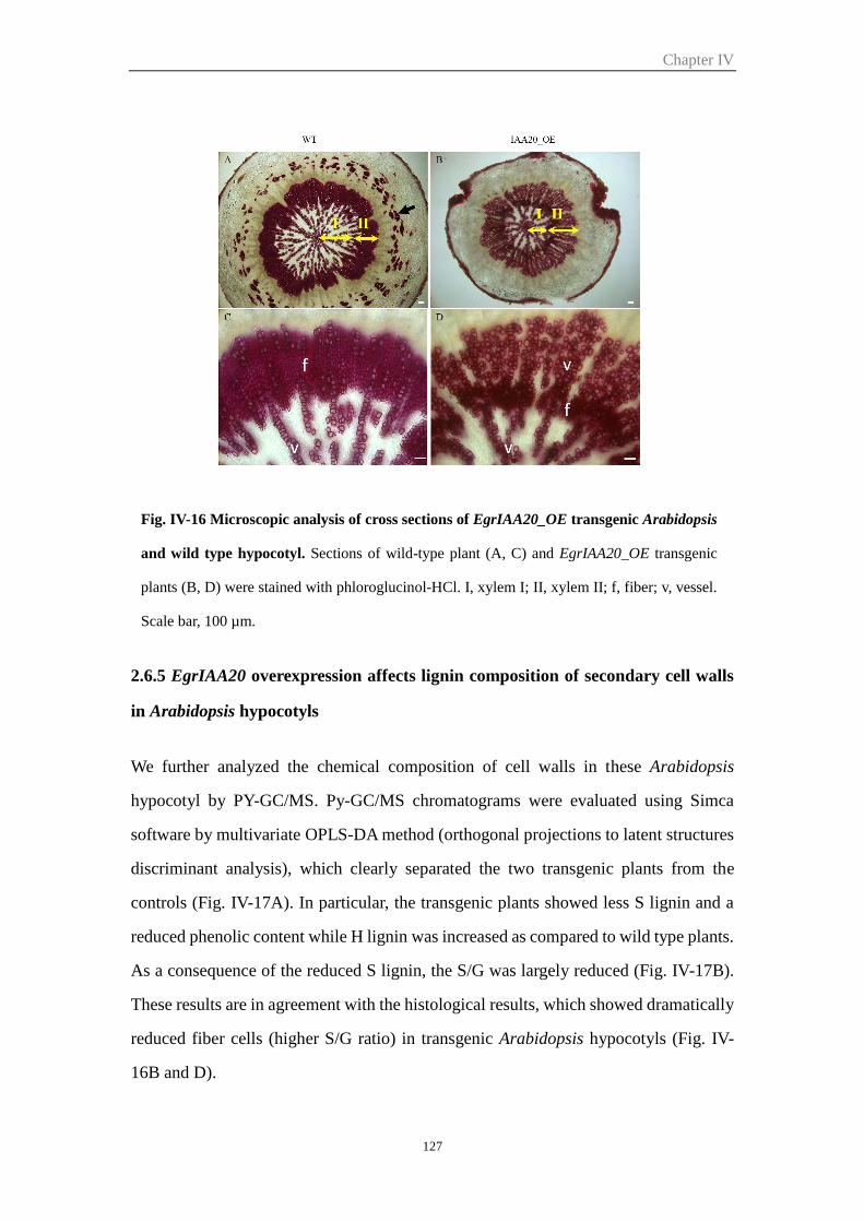
Chapter IV
127
2.6.5 EgrIAA20 overexpression affects lignin composition of secondary cell walls
in Arabidopsis hypocotyls
We further analyzed the chemical composition of cell walls in these Arabidopsis
hypocotyl by PY-GC/MS. Py-GC/MS chromatograms were evaluated using Simca
software by multivariate OPLS-DA method (orthogonal projections to latent structures
discriminant analysis), which clearly separated the two transgenic plants from the
controls (Fig. IV-17A). In particular, the transgenic plants showed less S lignin and a
reduced phenolic content while H lignin was increased as compared to wild type plants.
As a consequence of the reduced S lignin, the S/G was largely reduced (Fig. IV-17B).
These results are in agreement with the histological results, which showed dramatically
reduced fiber cells (higher S/G ratio) in transgenic Arabidopsis hypocotyls (Fig. IV-
16B and D).
Fig. IV-16 Microscopic analysis of cross sections of EgrIAA20_OE transgenic Arabidopsis
and wild type hypocotyl. Sections of wild-type plant (A, C) and EgrIAA20_OE transgenic
plants (B, D) were stained with phloroglucinol-HCl. I, xylem I; II, xylem II; f, fiber; v, vessel.
Scale bar, 100 µm.
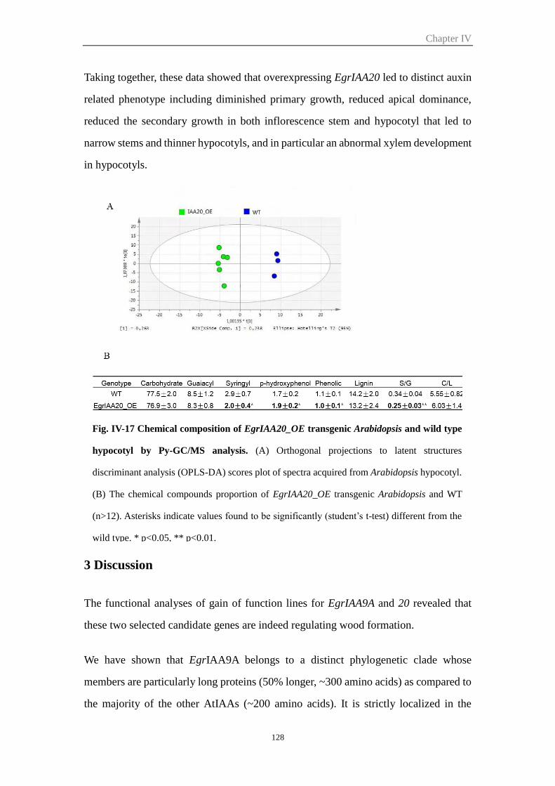
Chapter IV
128
Taking together, these data showed that overexpressing EgrIAA20 led to distinct auxin
related phenotype including diminished primary growth, reduced apical dominance,
reduced the secondary growth in both inflorescence stem and hypocotyl that led to
narrow stems and thinner hypocotyls, and in particular an abnormal xylem development
in hypocotyls.
3 Discussion
The functional analyses of gain of function lines for EgrIAA9A and 20 revealed that
these two selected candidate genes are indeed regulating wood formation.
We have shown that EgrIAA9A belongs to a distinct phylogenetic clade whose
members are particularly long proteins (50% longer, ~300 amino acids) as compared to
the majority of the other AtIAAs (~200 amino acids). It is strictly localized in the
Fig. IV-17 Chemical composition of EgrIAA20_OE transgenic Arabidopsis and wild type
hypocotyl by Py-GC/MS analysis. (A) Orthogonal projections to latent structures
discriminant analysis (OPLS-DA) scores plot of spectra acquired from Arabidopsis hypocotyl.
(B) The chemical compounds proportion of EgrIAA20_OE transgenic Arabidopsis and WT
(n>12). Asterisks indicate values found to be significantly (student’s t-test) different from the
wild type. * p<0.05, ** p<0.01.
Chapter IV
129
nucleus and behaves as a strong repressor of auxin-dependent transcription. It was
chosen for functional characterization because it belongs to IAA cluster III (more
expressed in xylem/cambium than in phloem, Chapter III) that also contains genes
involved in the biosynthesis of the main components of the SCWs (data not shown) and
also because it was more expressed in juvenile than in mature xylem, two types of wood
with distinct SCW properties. Its potential orthologs in Arabidopsis are the sister pair
AtIAA8/AtIAA9. No gain of function mutant of AtIAA8 nor AtIAA9 was reported,
whereas loss of function mutants (T-DNA insertion mutants iaa8-1, iaa9-1) and double
mutant iaa8/iaa9 did not exhibit any obvious phenotype (Overvoorde et al 2005) except
the presence of more lateral roots in iaa8-1 (Arase et al 2012). Despite the fact that no
phenotype was reported at the level of the vascular system, a large body of evidence
supports the hypothesis that the orthologs of EgrIAA9 in Arabidopsis and other species
are involved in the regulation of xylem formation: (i) both AtIAA8 and AtIAA9 show
high expression in hypocotyl xylem and inflorescence stem (Genevestigator database
survey), (ii) AtIAA8 and AtIAA9 transcript levels, measured at the base of the
inflorescence stem under artificial weight treatment shown to induce secondary growth,
were the highest among all other IAAs (Ko et al, 2004) but they underwent differential
expression strands suggesting different roles. AtIAA8 transcript level reaches it
maximum at a developmental stage corresponding to the beginning of the lignification
of the interfascicular fibres and decreases at a more mature stage, whereas AtIAA9
transcript level is highest at this latter stage (Ko et al 2004), (iii) the promoter of AtIAA8
is active in the vascular strands (Groover et al 2003), (iv) Zinnia zIAA8 is highly
induced during in vitro tracheary element induction (Groover et al 2003), (iv) Aspen
PttIAA2 shows high expression in cambium and xylem (Moyle et al 2002), (v) poplar
PtrIAA9 also displayed high expression in xylem (Kalluri et al 2007) and finally (vi)
down-regulation of the tomato SlIAA9 results in enhanced venation in leaves and thick
peduncles (Wang et al 2005). During the course of our study, it was reported that in the
monocot plant maize, the gain of function mutation in RUM1 (ortholog of EgrIAA9A)
led to defects in root xylem organisation and to ectopic lignification and wall thickening
in the pith cells (Zhang et al 2014).
Chapter IV
130
Overexpressing a stabilized version of EgrIAA9Am in Arabidopsis led to auxin-related
phenotypes, such as dwarfism, inhibition of lateral root emergence, reduced auxin
sensitivity in root growth, enhanced apical dominance. In line with EgrIAA9Am
expression profile, secondary xylem formation was altered in both the basal part of the
inflorescence stem and hypocotyl. Surprisingly, the alterations of secondary xylem
formation exhibit opposite patterns between the inflorescence stems and the hypocotyls:
secondary xylem formation was globally enhanced in the inflorescence stems of
transgenic lines showing an increased lignification staining both in the xylem bundles
and the interfascicular fibres, whereas in the hypocotyls, secondary xylem (fibres and
vessels) normally produced in the phase II was completely absent in transgenic lines
although in phase I xylem, more lignified vessels were produced in comparison to
controls. To the best of our knowledge such opposite vascular patterns between the
inflorescence stems and the hypocotyls has not been reported before, but histochemical
analysis of both organs is not systematically done and such distinct patterns may have
escaped to previous observations. These puzzling results suggest that different auxin-
regulated differentiation mechanisms may occur between inflorescence stems and
hypocotyls, the development of the latter resembling more to root ontogeny than to
stem ontogeny. Moreover, it also should be kept in mind that EgrIAA9Am was expressed
in Arabidopsis which contains two potential orthologs AtIAA8 and 9 having different
behaviours during secondary xylem formation in inflorescence stems artificially treated
to induce secondary growth (Ko et al 2004) and thus likely playing different roles
during this process. EgrIAA9m might interfere with both endogenous genes leading to
the complexes and antagonist vascular phenotypes, the most striking and dramatic
being the inhibition of secondary growth and xylem formation (Phase II) in hypocotyls.
These results are very promising although further investigations are needed to better
understand the role of EgrIAA9A and of its Arabidopsis orthologs. To this end, the
respective expression levels of Arabidopsis endogenous AtIAA8 and 9 will need to be
assessed as well as other auxin-related genes in EgrIAA9Am gain of function lines both
in hypocotyls and basal inflorescence stems and in parallel, auxin levels should be
evaluated at different development stages of xylem differentiation. In line with this, it

Chapter IV
131
is worth noting that co-expression analysis showed that AtIAA9 is tightly co-expressed
with WAT1 gene encoding a tonoplast localized auxin efflux facilitator. Mutations in
the fibre deficient mutant wat1 cause a reduction in polar auxin transport and
misregulation of genes associated with auxin response of which AtIAA9 was found to
be down-regulated (Ranocha et al. 2010, 2013).
EgrIAA20 encode a non-canonic IAA protein, as its orthologs in Arabidopsis, the sister
pair AtIAA20/AtIAA30, it lacks the domain II (dEgron), which interacts with the
SCFTIR1 complex and lead to the rapid degradation of Aux/IAA proteins by 26S
proteasome. In Arabidopsis mutation in domain II cause higher steady-state levels of
Aux/IAA proteins and the members who lack domain II (such AtIAA20 and AtIAA30)
are more stable proteins. Thus, EgrIAA20 was supposed to be more stable as compared
to the canonical EgrIAAs. We also showed that EgrIAA20 was not exclusively located
in the nucleus suggesting the possibility of a regulatory mechanism allowing its
migration to the nucleus to strongly repress the transcription of auxin-responsive genes.
In sharp contrast with AtIAA20 that shows very low basic expression in all tissues
examined (Genevestigator database survey), EgrIAA20 expression was found
preferentially expressed in vascular tissues, and particular highly expressed in vascular
cambium in Eucalyptus. No report described the loss of function mutant for AtIAA20
or AtIAA30 (no T-DNA insertion mutation for AtIAA20 was available and the AtIAA30
T-DNA insertion mutant iaa30-1 was not yet characterized (Overvoorde et al 2005).
Gain of function mutations by overexpressing AtIAA20 and AtIAA30 in Arabidopsis
resulted in semi-dwarf plants with agravitropic and impaired root growth. The
cotyledon venation was also impressively reduced (Sato and Kotaro 2008), mimicking
the phenotype of loss of function mutant for ARF5 (mp/arf5) and we also showed that
the EgrIAA20 protein was able to interact with ARF5. Overexpressing EgrIAA20
resulted in auxin related growth phenotypes such as reduced apical dominance,
inhibition of lateral root formation and agravitropic root response. The inflorescence
stems and hypocotyls were thinner and vascular patterning in cotyledons was impaired.
Histological analysis of hypocotyls sections showed altered secondary xylem formation
Chapter IV
132
formed during phase II, harboring virtually no fibers but more vessels than controls.
Biochemical analysis by Py-GC/MS further confirmed the significantly lower S lignin
content and the reduced S/G ratio due to fewer fibers rich in S units. These data strongly
suggest that EgrIAA20 regulates fibre development during secondary xylem
differentiation. It is worth noting that although the poplar PtrIAA20.1, PtrIAA20.2 and
aspen PttIAA3 are phylogenetically close to EgrIAA20, they contain the domain II in
contrast to EgrIAA20 and its Arabidopsis orthologs, suggesting that they are not stable,
and may have different roles in planta. Overexpressing a stabilized version of PttIAA3
(OE PttIAA3m) in poplar resulted in reduced secondary growth eg. decreased xylem
radial growth and reduced xylem/phloem ratio.
Although preliminary, the results obtained are new and promising suggesting that
IAA9A is regulating secondary xylem formation in hypocotyl whereas IAA20 is more
specifically involved in the regulation of fibres. Further investigations will be necessary
to get further insights into the roles and mechanisms of action of these two genes during
secondary xylem formation, and will be presented in the general discussion.
4 Materials and Methods
4.1 Plant materials and culture condition
The Arabidopsis seeds used for in vitro culture were surface-sterilized for 1 min in 70%
ethanol, 10 min in 25% bleach, rinsed five times in sterile water and plated on 1/2
Murashige and Skoog (MS) medium containing 1.0% sucrose solidified with 1% agar.
The culture conditions were: 16 h day/8 h night cycle, 22/20℃ day/night temperature,
40% relative humidity. For auxin dose-response experiments, sterilized seeds were
germinated on 1/2 MS medium containing 0, 0.01, 0.1, 1, 10 µM NAA respectively.
The primary root length was measured five days after germination. For gravitropic
response, sterilized seeds were planted on vertically oriented 1/2 MS plates. Eight days
after germination, we reoriented the plate by 90 dEgrees for another 48 h to check their
roots response to gravity.
Chapter IV
133
For pyrolysis and histochemical analysis, the Arabidopsis were planted in short day
conditions: 9 h day/15 h night cycle, 22/20℃ day/night temperature, 70% relative
humidity, 200 µmol photons m−1s−1 light intensity (intense luminosity). The plants were
watered every two days and fertilized weekly.
4.2 Gene expression analysis
All the Eucalyptus ARFs and Aux/IAAs gene expression in different tissues/organs were
performed in chapter II and III. In addition, Eucalyptus wood EST database
EUCATOUL (http://www.polebio.lrsv.ups-tlse.fr/eucatoul/db.php) was used for
checking their EST support. All the Arabidopsis ARF and Aux/IAA gene expression
profiles and their expression changes during the tracheary element (TE) induction
experiment were analyzed by surveying Genevestigator database
(https://www.genevestigator.com/gv/plant.jsp).
4.3 Transient expression of CGs in protoplast system for subcellular localization
and transcription activation analysis
For the subcellular localization, the CDS sequences of the EgrARF4, 10, 19A and
EgrIAA3B, 4, 9A, 13, 20 were first cloned into the Gateway entry vector pENTR-TOPO
(Fig. SIV-1), then recombined them into pK7FWG2 vector fused with GFP at C-
terminal and expressed under the control of the 35S CaMV promoter (Fig. SIV-2A).
EgrARF19A was also recombined into pK7WGF2 vector fused with GFP at N-terminal
(Fig. SIV-2B). The empty vector pK7WGF2.0 was used as control. The constructs were
introduced into tobacco BY-2 protoplasts by polyethylene glycol-mediated transfection
as described in Materials and Methods in chapter III. After 16 h incubation, the
transfected protoplasts were examined for green florescence signals using a Leica TCS
SP2 laser scanning confocal microscope. Images were obtained with a 40x 1.25
numerical aperture water-immersion objective.
For testing the ability of ARF transcription factors to up or down regulate the expression
Chapter IV
134
of auxin responsive promoter DR5, the full-length cDNAs of the EgrARF4, 10, 19A
and EgrIAA9A, 13, 20 transcription factors were cloned in pGreen vector under
CaMV35S promoter to create the effector constructs(Fig. SIV-3). The reporter
constructs use a synthetic auxin-responsive promoter DR5 followed by GFP reporter
gene. Tobacco BY-2 protoplasts were co-transfected with the reporter and effector
constructs as described in (Audran-Delalande et al. 2012). After 16 h incubation, GFP
expression was quantified by flow cytometry (LSR-Fortessa, BD Bioscience). Data
were analysed using Facsdiva software. Transfection assays were performed in three
independent replicates and 400-1000 protoplasts were gated for each sample. GFP
fluorescence corresponds to the average fluorescence intensity of the protoplasts
population after subtraction of auto-fluorescence determined with non-transformed
protoplasts. 50 µM 2, 4-D was used for auxin treatment.
4.4 Genetic transformation in Arabidopsis (vectors, primers, OE, OE mutated
protein, dominant repression)
Total RNA was isolated from Eucalyptus xylem using CTAB method as described by
Southerton et al. (1998). The cDNA first strand was synthesized from 2 μg total RNA
using SuperScript III reverse transcriptase (Invitrogen, USA), according to the
manufacturer’s instructions. We designed primer pairs of the corresponding gene
coding sequences using Primer3 v.04.0 (http://frodo.wi.mit.edu/), and the primers
sequences are shown in Table SIV-1. To get gain-of-function transgenic plants, we did
site target mutations in the miRNA target sites of EgrARF4 and EgrARF10, as well as
the domain II for EgrIAA3B, 4, 9A, 13 and 29. The mutation sites were introduced by
using overlap PCR. The CDS sequence was cloned into the Gateway entry vector
pENTR-TOPO, and then recombined into the overexpression vector pFAST-G02 (Fig.
SIV-4) (Shimada et al. 2010) and/or dominant repression vector pH35GEAR (Fig. SIV-
5) (Kubo et al. 2005), respectively. All binary expression constructs were transferred to
Agrobacterium tumefaciens strain GV3101 and the flower dip method was used to
transform Arabidopsis thaliana Col-0 (Clough and Bent 1998). For overexpression

Chapter IV
135
construction, the vector pFAST-G02 contains a GFP marker fused with a seed-specific
promoter OLE1, and this allowed us to select the transgenic seeds using the binocular
microscope under UV light (Shimada et al. 2010). For the dominant repression
construction, we used vector PH35GEAR which contains a repression domain
(LDLDLELRLG) after the target protein and hygromycin marker for transgenic plants
selection. PCR analysis of corresponding targeted genes were applied to confirm
transgenic plants for each line.
Underline sequences (CACC) represent the extra sequence needed for the pENTR/D-
TOPO cloning; sequences in red represent the overlap region for overlap PCR;
sequences highlight in yellow represent the mutation base.
4.5 Histochemical analysis
The histological comparative analysis of SCW between wild type and transgenic plants
was done at the stage of the first green siliques newly fully developed. At this stage, the
basal part of the inflorescence stem and hypocotyl abundantly develops cells
undergoing secondary wall thickening (xylem vessel cells, fascicular, and
interfascicular fiber cells). The Arabidopsis inflorescence stems at the basal end (~1 cm)
and hypocotyls were harvested and stored in 70% ethanol. The cross sections were
prepared using vibratome Leica VT1000 S. Lignin polymers are the characteristic
components of SCW and are normally absent from primary cell wall, therefore we used
lignin deposition detection techniques to screen for SCW phenotype. Cross sections of
inflorescence stem and hypocotyl were (~80 µm) were stained with phloroglucinol-HCl
which stains specifically lignin polymer precursors coniferaldehyde and p-
coumaraldehyde in the SCW giving violet-red color. Phloroglucinol-HCl was directly
applied on the slide and images were recorded with a CCD camera (Photonic Science,
http://www.photonic-science.co.uk).
4.6 Pyrolysis analysis
EgrIAA9A and 20 overexpression transgenic (two different lines for each) and wild type

Chapter IV
136
Arabidopsis were planted 3 different batches in short days (5 plants for each line in one
batch), and 10 days different between each batch. The hypocotyls were harvested in
liquid nitrogen when the first silique was fully development. Then the hypocotyls were
freeze-dried and ball-milled (MM400; Retsch) at 30 Hz in stainless steel jars (1.5 mL)
for 2 min with one ball (diameter of 7 mm). A total of 50 – 70 μg (XP6, Mettler-Toledo,
Switzerland) powder was transferred to auto sampler containers (Eco-cup SF, Frontier
Laboratories, Japan) for the Py-GC/MS. The sample was carried to oven pyrolyzer by
an auto sampler (PY-2020iD and AS-1020E, FrontierLabs, Japan) and analyzed by a
GC/MS system (Agilent, 7890A/5975C, Agilent Technologies AB, Sweden). The
pyrolysis oven was set to 450°C, the interface to 340°C and the injector to 320°C. The
pyrolysate was separated on a capillary column with a length of 30 m, diameter of 250
mm, and film thickness of 25 mm (JandW DB-5; Agilent Technologies Sweden). The
gas chromatography oven temperature program started at 40°C, followed by an
temperature ramp of 32°C/min to 100°C, 6°C/min to 118.75°C, 15°C/min to 250°C,
and 32°C/min to 320°C. Total run time was 19 min and full-scan spectra were recorded
in the range of 35 to 250 mass-to-charge ratio. Data processing, including peak
detection, intEgration, normalization, and identification, was done as described by
Gerber et al. (2012).
The relative amounts of S-, G-, and H-lignin and the carbohydrates were further
expressed as the percentage of the total compounds amounts. Orthogonal projections to
latent structures discriminant analysis (OPLS-DA) analysis of each individual replicate
was performed using SIMCA-P+ (12.0).
4.7 Protein-protein interaction analysis by yeast two hybrid system
For Y2H experiments, the cDNA sequence of EgrIAA20 was cloned into pBD vector
(Fig. SIV-5A), the cDNA sequence of EgrARF5 and AtARF5 were cloned into pAD
(Fig. SIV-5B) vectors, respectively. pBD Y2H vector (BD-bait) was used for the bait-
protein construction. It contains a functional copy of the Trp1 gene, thus restoring in
AH109 tryptophan autotrophy. pAD Y2H vector (AD-prey) was used for the prey-
Chapter IV
137
protein construction. It contains a functional copy of the Leu2 gene, thus restoring in
AH109 leucine autotrophy. Then, the BD-bait and AD-prey were co-transformed into
yeast AH109, co-transformants being selected on culture lacking both tryptophane and
leucine. If there is no interaction between the bait and prey protein, yeast will grow only
on the selection medium lacking Trp and Leu. If there was an interaction, yeast can
grow on the selection medium lacking Trp, Leu, His and Ade. Manipulation and
analysis of the Y2H assay followed the manufacturer’s instructions (Clontech Yeast
Protocols Handbook).

General discussion and perspectives

General discussion and perspectives
138
General discussion and perspectives
In woody plants, auxin has been proposed to play a key role in the development of
secondary xylem cells, a differentiation process involving cell division, expansion,
secondary cell wall formation and cell death (Miyashima et al. 2013; Sundberg et al.
2000). In comparison to the knowledge accumulated on the role of auxin in the control
of early vascular patterning in which some important actors and mechanisms involved
either in the maintenance of pro(cambial) identity or xylem specification have been
identified (reviewed in Berleth et al 2000; Elo et al 2009; Reinhardt 2003), our
knowledge of the auxin actors underlying secondary xylem differentiation is very
limited. Due to the growing importance of wood as a renewable and environmentally
cost effective alternative to fossil fuels, and the economic and ecological importance of
trees, a better understanding of the auxin regulation of wood formation is timely.
Auxin is believed to exert its function in wood formation through its
perception/signaling pathway, of which Auxin Response Factors (ARFs) and Aux/IAAs
are two well-known mediators of auxin responsive gene expression (Guilfoyle and
Hagen 2007; Tiwari et al. 2003). These two families have been studied in several annual
plants and more deeply in Arabidopsis, but remain largely under investigated in tree
species.
Taking advantage of the recent release of the E. grandis genome (Myburg et al, 2014),
we have performed a genome-wide comprehensive survey of two major transcription
families involved in the auxin perception and signaling pathway. One striking feature
of the E. grandis is the very high proportion of genes found in tandem repeats (34% of
the total number of genes). Indeed it is the highest rate of tandem duplication reported
among sequenced plant genomes (Myburg et al, 2014). In Eucalyptus, tandem
duplication shaped the functional diversity of many gene families like for instance the
MYB transcription factor family (Soler et al, 2014). However tandem duplication is
distributed unevenly between gene families and indeed, the ARF and IAA families
evolved in a very different way. Both families were slightly contracted as compared to
General discussion and perspectives
139
most angiosperms and monocots families studied hitherto, including the model plant
Arabidopsis. The functional diversity of the ARF family in Eucalyptus was mainly
shaped by alternative splicing and gene truncation leading to a diversity of domain
architecture, thereby compensating for the lack of extensive gene duplication
mechanisms found in other species. In contrast to these species, it is characterized by
the absence of whole genome, segmental and/or tandem duplication events. Indeed,
whole genome duplication in Eucalyptus occurred 109.9 Mya ago, considerably earlier
than those detected in other rosids and 95% of the paralogs were lost. The IAA family
has a different evolutionary history from that of the ARF, and the number of alternative
transcripts was similar to that of Arabidopsis.
Noteworthy, comparative phylogenetic studies pointed out the presence of a new clade
absent in Arabidopsis and maintained preferentially in woody and perennial plants. It
is likely that the genes belonging to this clade have been lost in herbaceous annual
species, and more work will be needed to decipher the role of these genes. Although in
our qRT-PCR experiments, EgrARF24 was not found to be expressed in xylem, unlike
its ortholog in poplar, it was found later to be expressed in this tissue in RNA-Seq data
(unpublished). This apparent discrepancies may be due to the presence of an alternative
transcript expressed in xylem and not detected by the primers chosen for the qRT-PCR,
this will need further verification.
We used large–scale expression profiling to identify ARF and Aux/IAA genes highly
and/or preferentially expressed in vascular cambium and/or xylem to identify those
potentially involved in the auxin-regulated transcriptional programs underlying wood
formation. Among the 13 genes exhibiting such expression patterns, we generated gain
of function transgenic Arabidopsis lines for six of them (four IAA (EgrIAA4, 9A, 13
and 20) and two ARF (EgrARF10 and 19A)). We functionally characterized lines
overexpressing EgrIAA4m, 9Am and 20. All three mutants exhibited distinct alterations
of the lignified vascular tissues:
The most dramatic macroscopic phenotype was obtained by overexpressing a stabilized

General discussion and perspectives
140
version of EgrIAA4, the plants were dwarf with strongly reduced fertility and exhibited
strong auxin insensitive phenotypes such as inhibition of primary root elongation and
lateral root emergence and roots agravitropism. EgrIAA4 is the potential ortholog of the
Arabidopsis sister pair AtIAA3/AtIAA4. Loss of function mutants (T-DNA insertion
mutant iaa4-1) exhibited no phenotype (Overvoorde et al 2005) whereas AtIAA3 gain
of function mutant (EMS mutation in the dEgron) exhibited a similar macroscopic
phenotype to the one induced by EgrIAA4m especially a reduced stature and short
hypocotyls (Reed et al., 1998; Tian and Reed, 1999). However, no phenotype was
reported at the level of the vascular system, probably because the authors did not look
specifically for it. Unfortunately, the hypocotyls were so tiny that we did not succeed
to obtain hypocotyl sections and the material was insufficient for chemical analyses. So,
we made sections to observe lignified vascular tissues patterning at two developmental
stages at the base of the inflorescence which is known to undergo secondary growth
(Altamura et al, 2001). Overexpression of EgrIAA4m strongly and negatively affected
the interfascicular fibres and the xylary fibres development, as shown by the virtual
absence of lignified secondary cell wall formation.
Similar phenotypes have been reported but for loss of function mutants suggesting
antagonist roles: (i) for the NAC master regulators of the SCW known to switch on the
expression of secondary wall genes in xylem fibres, (ii) for REV a class III
homeodomain leucine zipper transcription factor involved in organ polarity (ii) for
WAT1, which encodes for a tonoplast localized auxin efflux facilitator. In a nst1,
nst3/snd1 double mutant, secondary cell wall deposition in interfascicular fibres is
completely abolished (Mitsuda et al 2007; Mitsuda and Ohme-Takagi 2008). In Rev
loss-of-function mutants the development of interfascicular fibres is impaired (Zhong
and Ye 1999; Prigge et al 2005). REV is likely involved in cell fate determination rather
than directly promoting secondary cell wall deposition. As observed for rev mutants,
mutations in wat1 cause a similar reduction in polar auxin transport and misregulation
of genes associated with auxin response (Ranocha et al 2010, 2013). Secondary cell
wall deposition in xylary and interfascicular fibers is strongly reduced in wat1 mutants.
General discussion and perspectives
141
However, as there is residual lignification of fibres indicating fibre cell identity seems
not to be affected by wat1 mutation and that WAT1 acts more specifically on late fibre
differentiation.
Whether EgrIAA4 is involved more in interfascicular and xylary fibres specification or
in later differentiation and SCW deposition stages is yet to be solved. However, this
gene is differentially expressed between contrasting wood samples (tension versus
opposite wood, mature versus juvenile wood) whose secondary cell walls have very
different structure and composition. It is for instance, the only Eucalyptus IAA to be
more expressed in mature wood than in juvenile wood. These results together with the
residual lignification (although very light) of the SCW of the interfascicular fibres at
later stages of development in the EgrIAA4m lines, indirectly support a role in fibre
differentiation although more investigations are needed to decipher the mechanisms of
action of this gene.
Similar questions on the exact roles and mechanisms of action are still to be solved for
EgrIAA9A and EgrIAA20 which seem to regulate the whole secondary xylem formation
and secondary xylem fiber differentiation in hypocotyls, respectively.
Given the phenotypes of the three mutants, it would be important to assess endogenous
auxin distribution, this can be done by crossing the mutants lines to transgenic
Arabidopsis lines expressing the auxin induced marker DR5-Venus (Heisler et al 2005)
and visualization of its fluorescence at different stages of development of the vascular
system.
It would be also useful to assess the transcripts levels of genes involved in auxin
transport/signalling, those known to affect vascular system patterning as well of
transcription factors regulating fibre and vessels differentiation such the NACs, and
SCW biosynthetic genes. A systematic search for the transcripts levels of the three IAA
genes in the transcriptome of mutants such as Wat1, Rev, NAC… when available as
well as a without a priori search for their partners using an Eucalyptus two yeast hybrid
General discussion and perspectives
142
library available in the lab would help getting more insights into the regulation of the
three IAAs.
The use of an inducible promoter or even preferably of the promoter of the gene fused
to its coding sequence and tagged by a fluorescent protein would an interesting
alternative to the use of the 35SCamV promoter to allow less strong and pleiotropic
effects and in the latter case would allow to localize more precisely the sites of gene
expression. In the case of EgrIAA4m lines where the hypocotyls were particularly tiny,
it should allow comparative examination of xylem patterning between inflorescence
stems and hypocotyls. These strategies would be suitable at minima for the three genes
studied here and moreover a systematic comparison between inflorescence stems and
hypocotyls should be done for all transgenic lines as suggested by the highly contrasting
patterns of secondary xylem deposition in the two organs as observed in EgrIAA9m
overexpressing lines. These observations should be done at different stages of
development of the vascular system.
To overcome problems linked with heterologous transformation system, as well as the
frequent presence of sister pairs orthologs for one Eucalyptus IAA, a good alternative
would be to express the three IAA genes in an homologous system. Since the Eucalyptus
transformation is a very time consuming process with low efficiency of transformation,
our team has set up a hairy root transformation system and shown that secondary xylem
could be easily obtain. This promising and simplified tool will be valuable to further
functionally characterize the three IAA genes especially because the anatomy of the
hypocotyl vascular system is similar to that of the root (Chaffey et al 2002).


References

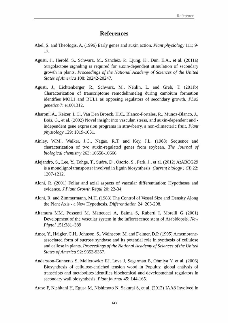
Reference
143
References
Abel, S. and Theologis, A. (1996) Early genes and auxin action. Plant physiology 111: 9-
17.
Agusti, J., Herold, S., Schwarz, M., Sanchez, P., Ljung, K., Dun, E.A., et al. (2011a)
Strigolactone signaling is required for auxin-dependent stimulation of secondary
growth in plants. Proceedings of the National Academy of Sciences of the United
States of America 108: 20242-20247.
Agusti, J., Lichtenberger, R., Schwarz, M., Nehlin, L. and Greb, T. (2011b)
Characterization of transcriptome remodelinmeleg during cambium formation
identifies MOL1 and RUL1 as opposing regulators of secondary growth. PLoS
genetics 7: e1001312.
Aharoni, A., Keizer, L.C., Van Den Broeck, H.C., Blanco-Portales, R., Munoz-Blanco, J.,
Bois, G., et al. (2002) Novel insight into vascular, stress, and auxin-dependent and -
independent gene expression programs in strawberry, a non-climacteric fruit. Plant
physiology 129: 1019-1031.
Ainley, W.M., Walker, J.C., Nagao, R.T. and Key, J.L. (1988) Sequence and
characterization of two auxin-regulated genes from soybean. The Journal of
biological chemistry 263: 10658-10666.
Alejandro, S., Lee, Y., Tohge, T., Sudre, D., Osorio, S., Park, J., et al. (2012) AtABCG29
is a monolignol transporter involved in lignin biosynthesis. Current biology : CB 22:
1207-1212.
Aloni, R. (2001) Foliar and axial aspects of vascular differentiation: Hypotheses and
evidence. J Plant Growth Regul 20: 22-34.
Aloni, R. and Zimmermann, M.H. (1983) The Control of Vessel Size and Density Along
the Plant Axis - a New Hypothesis. Differentiation 24: 203-208.
Altamura MM, Possenti M, Matteucci A, Baima S, Ruberti I, Morelli G (2001)
Development of the vascular system in the inflorescence stem of Arabidopsis. New
Phytol 151:381–389
Amor, Y., Haigler, C.H., Johnson, S., Wainscott, M. and Delmer, D.P. (1995) A membrane-
associated form of sucrose synthase and its potential role in synthesis of cellulose
and callose in plants. Proceedings of the National Academy of Sciences of the United
States of America 92: 9353-9357.
Andersson-Gunneras S, Mellerowicz EJ, Love J, Segerman B, Ohmiya Y, et al. (2006)
Biosynthesis of cellulose-enriched tension wood in Populus: global analysis of
transcripts and metabolites identifies biochemical and developmental regulators in
secondary wall biosynthesis. Plant journal 45: 144-165.
Arase F, Nishitani H, Egusa M, Nishimoto N, Sakurai S, et al. (2012) IAA8 Involved in
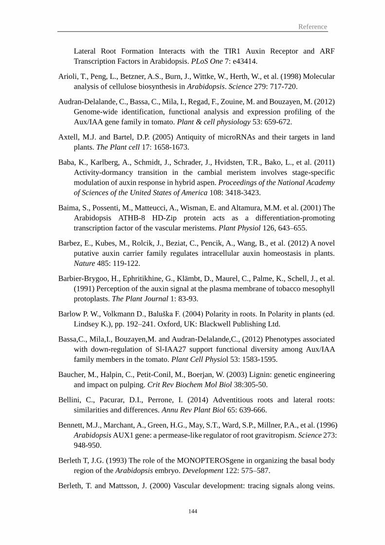
Reference
144
Lateral Root Formation Interacts with the TIR1 Auxin Receptor and ARF
Transcription Factors in Arabidopsis. PLoS One 7: e43414.
Arioli, T., Peng, L., Betzner, A.S., Burn, J., Wittke, W., Herth, W., et al. (1998) Molecular
analysis of cellulose biosynthesis in Arabidopsis. Science 279: 717-720.
Audran-Delalande, C., Bassa, C., Mila, I., Regad, F., Zouine, M. and Bouzayen, M. (2012)
Genome-wide identification, functional analysis and expression profiling of the
Aux/IAA gene family in tomato. Plant & cell physiology 53: 659-672.
Axtell, M.J. and Bartel, D.P. (2005) Antiquity of microRNAs and their targets in land
plants. The Plant cell 17: 1658-1673.
Baba, K., Karlberg, A., Schmidt, J., Schrader, J., Hvidsten, T.R., Bako, L., et al. (2011)
Activity-dormancy transition in the cambial meristem involves stage-specific
modulation of auxin response in hybrid aspen. Proceedings of the National Academy
of Sciences of the United States of America 108: 3418-3423.
Baima, S., Possenti, M., Matteucci, A., Wisman, E. and Altamura, M.M. et al. (2001) The
Arabidopsis ATHB-8 HD-Zip protein acts as a differentiation-promoting
transcription factor of the vascular meristems. Plant Physiol 126, 643–655.
Barbez, E., Kubes, M., Rolcik, J., Beziat, C., Pencik, A., Wang, B., et al. (2012) A novel
putative auxin carrier family regulates intracellular auxin homeostasis in plants.
Nature 485: 119-122.
Barbier-Brygoo, H., Ephritikhine, G., Klämbt, D., Maurel, C., Palme, K., Schell, J., et al.
(1991) Perception of the auxin signal at the plasma membrane of tobacco mesophyll
protoplasts. The Plant Journal 1: 83-93.
Barlow P. W., Volkmann D., Baluška F. (2004) Polarity in roots. In Polarity in plants (ed.
Lindsey K.), pp. 192–241. Oxford, UK: Blackwell Publishing Ltd.
Bassa,C., Mila,I., Bouzayen,M. and Audran-Delalande,C., (2012) Phenotypes associated
with down-regulation of Sl-IAA27 support functional diversity among Aux/IAA
family members in the tomato. Plant Cell Physiol 53: 1583-1595.
Baucher, M., Halpin, C., Petit-Conil, M., Boerjan, W. (2003) Lignin: genetic engineering
and impact on pulping. Crit Rev Biochem Mol Biol 38:305-50.
Bellini, C., Pacurar, D.I., Perrone, I. (2014) Adventitious roots and lateral roots:
similarities and differences. Annu Rev Plant Biol 65: 639-666.
Bennett, M.J., Marchant, A., Green, H.G., May, S.T., Ward, S.P., Millner, P.A., et al. (1996)
Arabidopsis AUX1 gene: a permease-like regulator of root gravitropism. Science 273:
948-950.
Berleth T, J.G. (1993) The role of the MONOPTEROSgene in organizing the basal body
region of the Arabidopsis embryo. Development 122: 575–587.
Berleth, T. and Mattsson, J. (2000) Vascular development: tracing signals along veins.
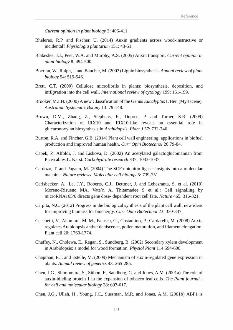
Reference
145
Current opinion in plant biology 3: 406-411.
Bhalerao, R.P. and Fischer, U. (2014) Auxin gradients across wood-instructive or
incidental? Physiologia plantarum 151: 43-51.
Blakeslee, J.J., Peer, W.A. and Murphy, A.S. (2005) Auxin transport. Current opinion in
plant biology 8: 494-500.
Boerjan, W., Ralph, J. and Baucher, M. (2003) Lignin biosynthesis. Annual review of plant
biology 54: 519-546.
Brett, C.T. (2000) Cellulose microfibrils in plants: biosynthesis, deposition, and
intEgration into the cell wall. International review of cytology 199: 161-199.
Brooker, M.I.H. (2000) A new Classification of the Genus Eucalyptus L'Her. (Myrtaceae).
Australian Systematic Botany 13: 79-148.
Brown, D.M., Zhang, Z., Stephens, E., Dupree, P. and Turner, S.R. (2009)
Characterization of IRX10 and IRX10-like reveals an essential role in
glucuronoxylan biosynthesis in Arabidopsis. Plant J 57: 732-746.
Burton, R.A. and Fincher, G.B. (2014) Plant cell wall engineering: applications in biofuel
production and improved human health. Curr Opin Biotechnol 26:79-84.
Capek, P., Alfoldi, J. and Liskova, D. (2002) An acetylated galactoglucomannan from
Picea abies L. Karst. Carbohydrate research 337: 1033-1037.
Cardozo, T. and Pagano, M. (2004) The SCF ubiquitin ligase: insights into a molecular
machine. Nature reviews. Molecular cell biology 5: 739-751.
Carlsbecker, A., Le, J.Y., Roberts, C.J., Dettmer, J. and Lehesranta, S. et al. (2010)
Moreno-Risueno MA, Vate n A, Thitamadee S et al.: Cell signalling by
microRNA165/6 directs gene dose- dependent root cell fate. Nature 465: 316-321.
Carpita, N.C. (2012) Progress in the biological synthesis of the plant cell wall: new ideas
for improving biomass for bioenergy. Curr Opin Biotechnol 23: 330-337.
Cecchetti, V., Altamura, M. M., Falasca, G., Costantino, P., Cardarelli, M. (2008) Auxin
regulates Arabidopsis anther dehiscence, pollen maturation, and filament elongation.
Plant cell 20: 1760-1774.
Chaffey, N., Cholewa, E., Regan, S., Sundberg, B. (2002) Secondary xylem development
in Arabidopsis: a model for wood formation. Physiol Plant 114:594-600.
Chapman, E.J. and Estelle, M. (2009) Mechanism of auxin-regulated gene expression in
plants. Annual review of genetics 43: 265-285.
Chen, J.G., Shimomura, S., Sitbon, F., Sandberg, G. and Jones, A.M. (2001a) The role of
auxin-binding protein 1 in the expansion of tobacco leaf cells. The Plant journal :
for cell and molecular biology 28: 607-617.
Chen, J.G., Ullah, H., Young, J.C., Sussman, M.R. and Jones, A.M. (2001b) ABP1 is
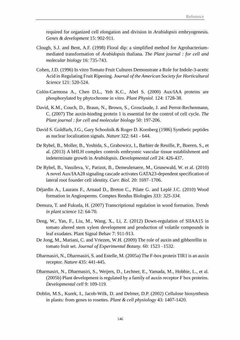
Reference
146
required for organized cell elongation and division in Arabidopsis embryogenesis.
Genes & development 15: 902-911.
Clough, S.J. and Bent, A.F. (1998) Floral dip: a simplified method for Agrobacterium-
mediated transformation of Arabidopsis thaliana. The Plant journal : for cell and
molecular biology 16: 735-743.
Cohen, J.D. (1996) In vitro Tomato Fruit Cultures Demonstrate a Role for Indole-3-acetic
Acid in Regulating Fruit Ripening. Journal of the American Society for Horticultural
Science 121: 520-524.
Colón-Carmona A., Chen D.L., Yeh K.C., Abel S. (2000) Aux/IAA proteins are
phosphorylated by phytochrome in vitro. Plant Physiol. 124: 1728-38.
David, K.M., Couch, D., Braun, N., Brown, S., Grosclaude, J. and Perrot-Rechenmann,
C. (2007) The auxin-binding protein 1 is essential for the control of cell cycle. The
Plant journal : for cell and molecular biology 50: 197-206.
David S. Goldfarb, J.G., Gary Schoolnik & Roger D. Kornberg (1986) Synthetic peptides
as nuclear localization signals. Nature 322: 641 - 644.
De Rybel, B., Moller, B., Yoshida, S., Grabowicz, I., Barbier de Reuille, P., Boeren, S., et
al. (2013) A bHLH complex controls embryonic vascular tissue establishment and
indeterminate growth in Arabidopsis. Developmental cell 24: 426-437.
De Rybel, B., Vassileva, V., Parizot, B., Demeulenaere, M., Grunewald, W. et al. (2010)
A novel Aux/IAA28 signaling cascade activates GATA23-dependent specification of
lateral root founder cell identity. Curr. Biol. 20: 1697–1706.
Déjardin A., Laurans F., Arnaud D., Breton C., Pilate G. and Leplé J.C. (2010) Wood
formation in Angiosperms. Comptes Rendus Biologies 333: 325-334.
Demura, T. and Fukuda, H. (2007) Transcriptional regulation in wood formation. Trends
in plant science 12: 64-70.
Deng, W., Yan, F., Liu, M., Wang, X., Li, Z. (2012) Down-regulation of SlIAA15 in
tomato altered stem xylem development and production of volatile compounds in
leaf exudates. Plant Signal Behav 7: 911-913.
De Jong, M., Mariani, C. and Vriezen, W.H. (2009) The role of auxin and gibberellin in
tomato fruit set. Journal of Experimental Botany. 60: 1523 –1532.
Dharmasiri, N., Dharmasiri, S. and Estelle, M. (2005a) The F-box protein TIR1 is an auxin
receptor. Nature 435: 441-445.
Dharmasiri, N., Dharmasiri, S., Weijers, D., Lechner, E., Yamada, M., Hobbie, L., et al.
(2005b) Plant development is regulated by a family of auxin receptor F box proteins.
Developmental cell 9: 109-119.
Doblin, M.S., Kurek, I., Jacob-Wilk, D. and Delmer, D.P. (2002) Cellulose biosynthesis
in plants: from genes to rosettes. Plant & cell physiology 43: 1407-1420.
Reference
147
Donner, T.J., Sherr, I. and Scarpella, E. (2009) Regulation of preprocambial cell state
acquisition by auxin signaling in Arabidopsis leaves. Development 136: 3235-3246.
Dreher, K.A., Brown, J., Saw, R.E. and Callis, J. (2006) The Arabidopsis Aux/IAA protein
family has diversified in degradation and auxin responsiveness. The Plant cell 18:
699-714.
Du, J. and Groover, A. (2010) Transcriptional regulation of secondary growth and wood
formation. Journal of intEgrative plant biology 52: 17-27.
Du, J., Mansfield, S.D. and Groover, A.T. (2009) The Populus homeobox gene
ARBORKNOX2 regulates cell differentiation during secondary growth. The Plant
journal : for cell and molecular biology 60: 1000-1014.
Du, J., Miura, E., Robischon, M., Martinez, C. and Groover, A. (2011) The Populus Class
III HD ZIP transcription factor POPCORONA affects cell differentiation during
secondary growth of woody stems. PloS one 6: e17458.
Dubrovsky, J. G., Rost, T. L., Colon-Carmona, A., Doerner, P. (2001) Early primordium
morphogenesis during lateral root initiation in Arabidopsis thaliana. Planta 214: 30-
36.
Eames A.J., MacDaniels L.H. (1947) An Introduction to Plant Anatomy 2nd edn. New
York: McGraw-Hill.
Ellis, C.M., Nagpal, P., Young, J.C., Hagen, G., Guilfoyle, T.J. and Reed, J.W. (2005)
AUXIN RESPONSE FACTOR1 and AUXIN RESPONSE FACTOR2 regulate
senescence and floral organ abscission in Arabidopsis thaliana. Development 132:
4563-4574.
Elo, A., Immanen, J., Nieminen, K., Helariutta, Y. (2009) Stem cell function during plant
vascular development. Semin Cell Dev Biol 20:1097-1106.
Esau K. (1965) Vascular Differentiation in Plants. New York: Holt, Rinehart, and Winston.
Etchells, J.P., Turner, S.R. (2010) The PXY-CLE41 receptor ligand pair defines a
multifunctional pathway that controls the rate and orientation of vascular cell
division. Development 137:767-774.
Etchells, J. P., Provost, C. M., Mishra, L. (2013) Turner, S. R. WOX4 and WOX14 act
downstream of the PXY receptor kinase to regulate plant vascular proliferation
independently of any role in vascular organisation. Development 140: 2224-2234.
Evans, M., Ishikawa, H. and Estelle, M. (1994) Responses of Arabidopsis roots to auxin
studied with high temporal resolution: Comparison of wild type and auxin-response
mutants. Planta 194: 215-222.
Fahlgren, N., Montgomery, T.A., Howell, M.D., Allen, E., Dvorak, S.K., Alexander, A.L.,
et al. (2006) Regulation of AUXIN RESPONSE FACTOR3 by TAS3 ta-siRNA
affects developmental timing and patterning in Arabidopsis. Curr Biol 16: 939-944.
Reference
148
Foucart, C., Paux, E., Ladouce, N., San-Clemente, H., Grima-Pettenati, J. and Sivadon, P.
(2006) Transcript profiling of a xylem vs phloem cDNA subtractive library identifies
new genes expressed during xylogenesis in Eucalyptus. New Phytol 170: 739-752.
Friml, J., Wisniewska, J., Benkova, E., Mendgen, K. and Palme, K. (2002) Lateral
relocation of auxin efflux regulator PIN3 mediates tropism in Arabidopsis. Nature
415: 806-809.
Fukaki, H., Tameda, S., Masuda, H. and Tasaka, M. (2002) Lateral root formation is
blocked by a gain-of-function mutation in the SOLITARY-ROOT/IAA14 gene of
Arabidopsis. Plant Journal 29: 153-168.
Fukaki, H., Nakao, Y., Okushima, Y., Theologis, A., Tasaka, M. (2005) Tissue-specific
expression of stabilized SOLITARY-ROOT/IAA14 alters lateral root development
in Arabidopsis. Plant Journal 44: 382-395.
Fukaki, H., Taniguchi, N. and Tasaka, M. (2006) PICKLE is required for SOLITARY-
ROOT/IAA14-mediated repression of ARF7 and ARF19 activity during Arabidopsis
lateral root initiation. The Plant journal : for cell and molecular biology 48: 380-389.
Fukaki, H., Okushima, Y., Tasaka, M. (2007) Auxin-mediated lateral root formation in
higher plants. Int. Rev. Cytol. 256: 111–137.
Fukaki, H. and Tasaka, M. (2009) Hormone interactions during lateral root formation.
Plant Mol. Biol. 69: 437–449.
Fukuda, H. (1997) Tracheary Element Differentiation. The Plant cell 9: 1147-1156.
Furuya N., Takahashi S., Mirayashi M., (1970) The chemical composition of gelatinous
layer from the tension wood of Populus eura-americana. Mokuzai Gakkaishi 16: 26–
30.
Galweiler, L., Guan, C.H., Muller, A., Wisman, E., Mendgen, K., Yephremov, A., et al.
(1998) Regulation of polar auxin transport by AtPIN1 in Arabidopsis vascular tissue.
Science 282: 2226-2230.
Gerber, L., Eliasson, M., Trygg, J., Moritz, T. and Sundberg, B. (2012) Multivariate curve
resolution provides a high-throughput data processing pipeline for pyrolysis-gas
chromatography/mass spectrometry. Journal of Analytical and Applied Pyrolysis 95:
95-100.
Gindl, W., Grabner, M., and Wimmer, R. (2014). The influence of temperature on
latewood lignin content in treeline Norway spruce compared with maximum density
and ring width. Trees 14: 409-414.
Goetz, M., Hooper, L.C., Johnson, S.D., Rodrigues, J.C., Vivian-Smith, A. and Koltunow,
A.M. (2007) Expression of aberrant forms of AUXIN RESPONSE FACTOR8
stimulates parthenocarpy in Arabidopsis and tomato. Plant Physiol 145: 351-366.
Goh, T., Kasahara, H., Mimura, T., Kamiya, Y. and Fukaki, H. (2012) Multiple AUX/IAA-
ARF modules regulate lateral root formation: the role of Arabidopsis SHY2/IAA3-
Reference
149
mediated auxin signalling. Philosophical transactions of the Royal Society of London.
Series B, Biological sciences 367: 1461-1468.
Goicoechea, M., Lacombe, E., Legay, S., Mihaljevic, S., Rech, P., Jauneau, A., et al. (2005)
EgMYB2, a new transcriptional activator from Eucalyptus xylem, regulates
secondary cell wall formation and lignin biosynthesis. The Plant journal : for cell
and molecular biology 43: 553-567.
Gray, W.M., Kepinski, S., Rouse, D., Leyser, O. and Estelle, M. (2001) Auxin regulates
SCF(TIR1)-dependent degradation of AUX/IAA proteins. Nature 414: 271-276.
Greenham, K., Santner, A., Castillejo, C., Mooney, S., Sairanen, I., Ljung, K., et al. (2011)
The AFB4 auxin receptor is a negative regulator of auxin signaling in seedlings.
Current biology : CB 21: 520-525.
Guilfoyle, T., Hagen, G., Ulmasov, T. and Murfett, J. (1998) How does auxin turn on genes?
Plant Physiol 118: 341-347.
Guilfoyle, T.J. and Hagen, G. (2007) Auxin response factors. Current opinion in plant
biology 10: 453-460.
Habrant, A., Laurans, F., Déjardin, A., Leplé, J.-C. and Pilate, G., et al. (2003) Physical
and chemical changes of poplar cell walls during tension wood formation, in: 12th
Int. Symp. on Wood and Pulping Chemistry (ISWPC), Madison, Wisconsin (USA),
8–12 June.
Haigler, C.H., Ivanova-Datcheva, M., Hogan, P.S., Salnikov, V.V., Hwang, S., Martin, K.,
et al. (2001) Carbon partitioning to cellulose synthesis. Plant molecular biology 47:
29-51.
Hamann, T., Benkova, E., Baurle, I., Kientz, M. and Jurgens, G. (2002) The Arabidopsis
BODENLOS gene encodes an auxin response protein inhibiting MONOPTEROS-
mediated embryo patterning. Genes Dev 16: 1610-1615.
Hamann, T., Mayer, U. and Jurgens, G. (1999) The auxin-insensitive bodenlos mutation
affects primary root formation and apical-basal patterning in the Arabidopsis embryo.
Development 126: 1387-1395.
Hardtke, C.S. and Berleth, T. (1998) The Arabidopsis gene MONOPTEROS encodes a
transcription factor mediating embryo axis formation and vascular development.
EMBO J 17: 1405-1411.
Harper, R.M., Stowe-Evans, E.L., Luesse, D.R., Muto, H., Tatematsu, K., Watahiki, M.K.,
et al. (2000) The NPH4 locus encodes the auxin response factor ARF7, a conditional
regulator of differential growth in aerial Arabidopsis tissue. Plant Cell 12: 757-770.
Heisler MG, Ohno C, Das P, Sieber P, Reddy GV, Long JA, Meyerowitz EM (2005)
Patterns of auxin transport and gene expression during primordium development
revealed by live imaging of the Arabidopsis inflorescence meristem. Curr Biol 15:
1899–1911.
Reference
150
Hellgren, J.M., Olofsson, K. and Sundberg, B. (2004) Patterns of auxin distribution during
gravitational induction of reaction wood in poplar and pine. Plant physiology 135:
212-220.
Hendelman, A., Buxdorf, K., Stav, R., Kravchik, M., Arazi, T. (2012) Inhibition of lamina
outgrowth following Solanum lycopersicum AUXIN RESPONSE FACTOR 10
(SlARF10) derepression. Plant Mol Biol 78: 561-576.
Hertel, R., Thomson, K.S. and Russo, V.E. (1972) In-vitro auxin binding to particulate
cell fractions from corn coleoptiles. Planta 107: 325-340.
Himanen, K., Boucheron, E., Vanneste, S., de Almeida Engler, J., Inze, D. and Beeckman,
T. (2002) Auxin-mediated cell cycle activation during early lateral root initiation.
The Plant cell 14: 2339-2351.
Hirakawa, Y., Kondo, Y. and Fukuda, H. (2010) TDIF peptide signaling regulates vascular
stem cell proliferation via the WOX4 homeobox gene in Arabidopsis. The Plant cell
22: 2618-2629.
Hobbie L, M.M., Hurwitz LR, Pierro A, Liu NY, Bandyopadhyay A, Estelle M. (2000)
The axr6mutants of Arabidopsis define a gene involved in auxin response and early
development. Development 127: 23–32.
Hu, Z., Keceli, M.A., Piisila, M., Li, J., Survila, M., Heino, P., et al. (2012) F-box protein
AFB4 plays a crucial role in plant growth, development and innate immunity. Cell
research 22: 777-781.
Hussey, S.G., Mizrachi, E., Spokevicius, A.V., Bossinger, G., Berger, D.K. and Myburg,
A.A. (2011) SND2, a NAC transcription factor gene, regulates genes involved in
secondary cell wall development in Arabidopsis fibres and increases fibre cell area
in Eucalyptus. BMC plant biology 11: 173.
Ilegems, M., Douet, V., Meylan-Bettex, M., Uyttewaal, M., Brand, L., Bowman, J.L., et
al. (2010) Interplay of auxin, KANADI and Class III HD-ZIP transcription factors in
vascular tissue formation. Development 137: 975-984.
Jain, M., Kaur, N., Garg, R., Thakur, J.K., Tyagi, A.K. and Khurana, J.P. (2006) Structure
and expression analysis of early auxin-responsive Aux/IAA gene family in rice
(Oryza sativa). Functional & intEgrative genomics 6: 47-59.
Jones, B., Frasse, P., Olmos, E., Zegzouti, H., Li, Z.G., Latche, A., et al. (2002) Down-
regulation of DR12, an auxin-response-factor homolog, in the tomato results in a
pleiotropic phenotype including dark green and blotchy ripening fruit. Plant J 32:
603-613.
Jourez B., Riboux A., Leclercq A. (2001) Anatomical characteristics of tension wood and
opposite wood in young inclined stems of poplar (Populus euramericana cv‘Ghoy’).
IAWA Journal 22: 133–157.
Kalluri, U.C., Difazio, S.P., Brunner, A.M. and Tuskan, G.A. (2007) Genome-wide
Reference
151
analysis of Aux/IAA and ARF gene families in Populus trichocarpa. BMC plant
biology 7: 59.
Kaneda, M., Schuetz, M., Lin, B.S., Chanis, C., Hamberger, B., Western, T.L., et al. (2011)
ABC transporters coordinately expressed during lignification of Arabidopsis stems
include a set of ABCBs associated with auxin transport. Journal of experimental
botany 62: 2063-2077.
Kepinski, S. and Leyser, O. (2004) Auxin-induced SCFTIR1-Aux/IAA interaction
involves stable modification of the SCFTIR1 complex. Proceedings of the National
Academy of Sciences of the United States of America 101: 12381-12386.
Kim, J., Harter, K. and Theologis, A. (1997) Protein-protein interactions among the
Aux/IAA proteins. Proceedings of the National Academy of Sciences of the United
States of America 94: 11786-11791.
Ko, J.H., Han, K.H., Park, S. and Yang, J. (2004) Plant body weight-induced secondary
growth in Arabidopsis and its transcription phenotype revealed by whole-
transcriptome profiling. Plant physiology 135: 1069-1083.
Kong, Y., Zhou, G., Avci, U., Gu, X., Jones, C., Yin, Y., et al. (2009) Two poplar
glycosyltransferase genes, PdGATL1.1 and PdGATL1.2, are functional orthologs to
PARVUS/AtGATL1 in Arabidopsis. Molecular plant 2: 1040-1050.
Koroleva, O.A., Tomlinson, M., Parinyapong, P., Sakvarelidze, L., Leader, D., Shaw, P.,
et al. (2004) CycD1, a putative G1 cyclin from Antirrhinum majus, accelerates the
cell cycle in cultured tobacco BY-2 cells by enhancing both G1/S entry and
progression through S and G2 phases. The Plant cell 16: 2364-2379.
Kubo, M., Udagawa, M., Nishikubo, N., Horiguchi, G., Yamaguchi, M., Ito, J., et al. (2005)
Transcription switches for protoxylem and metaxylem vessel formation. Genes Dev
19: 1855-1860.
Kumar, R., Tyagi, A.K. and Sharma, A.K. (2011) Genome-wide analysis of auxin
response factor (ARF) gene family from tomato and analysis of their role in flower
and fruit development. Mol Genet Genomics 285: 245-260.
Lavenus, J., Goh, .T, Roberts, I., Guyomarc'h, S., Laplaze, L. et al (2013) Lateral root
development in Arabidopsis: fifty shades of auxin. Trends Plant Sci 18:450-458.
Lee, C., O'Neill, M.A., Tsumuraya, Y., Darvill, A.G. and Ye, Z.H. (2007a) The irregular
xylem9 mutant is deficient in xylan xylosyltransferase activity. Plant & cell
physiology 48: 1624-1634.
Lee, C., Teng, Q., Huang, W., Zhong, R. and Ye, Z.H. (2009a) The F8H
glycosyltransferase is a functional paralog of FRA8 involved in glucuronoxylan
biosynthesis in Arabidopsis. Plant & cell physiology 50: 812-827.
Lee, C., Teng, Q., Huang, W., Zhong, R. and Ye, Z.H. (2009b) The poplar GT8E and
GT8F glycosyltransferases are functional orthologs of Arabidopsis PARVUS
Reference
152
involved in glucuronoxylan biosynthesis. Plant & cell physiology 50: 1982-1987.
Lee, C., Zhong, R., Richardson, E.A., Himmelsbach, D.S., McPhail, B.T. and Ye, Z.H.
(2007b) The PARVUS gene is expressed in cells undergoing secondary wall
thickening and is essential for glucuronoxylan biosynthesis. Plant & cell physiology
48: 1659-1672.
Leyser, H.M., Pickett, F.B., Dharmasiri, S. and Estelle, M. (1996) Mutations in the AXR3
gene of Arabidopsis result in altered auxin response including ectopic expression
from the SAUR-AC1 promoter. The Plant journal : for cell and molecular biology
10: 403-413.
Leyser, O. (2003) Regulation of shoot branching by auxin. Trends Plant Sci 8: 541–545.
Li, E., Bhargava, A., Qiang, W., Friedmann, M.C., Forneris, N., Savidge, R.A., et al. (2012)
The Class II KNOX gene KNAT7 negatively regulates secondary wall formation in
Arabidopsis and is functionally conserved in Populus. The New phytologist 194: 102-
115.
Li, E., Wang, S., Liu, Y., Chen, J.G. and Douglas, C.J. (2011) OVATE FAMILY
PROTEIN4 (OFP4) interaction with KNAT7 regulates secondary cell wall formation
in Arabidopsis thaliana. The Plant journal : for cell and molecular biology 67: 328-
341.
Li, S., Bashline, L., Lei, L. and Gu, Y. (2014) Cellulose synthesis and its regulation. The
Arabidopsis book / American Society of Plant Biologists 12: e0169.
Liebminger, E., Grass, J., Altmann, F., Mach, L. and Strasser, R. (2013) Characterizing
the link between glycosylation state and enzymatic activity of the endo-beta1,4-
glucanase KORRIGAN1 from Arabidopsis thaliana. The Journal of biological
chemistry 288: 22270-22280.
Liscum, E. and Reed, J. W. (2002) Genetics of Aux/IAA and ARF action in plant growth
and development. Plant Mol Biol 49:387-400.
Little, C.H.A. and Savidge, R.A. (1987) The Role of Plant-Growth Regulators in Forest
Tree Cambial Growth. Plant Growth Regul 6: 137-169.
Liu, P.P., Montgomery, T.A., Fahlgren, N., Kasschau, K.D., Nonogaki, H. and Carrington,
J.C. (2007) Repression of AUXIN RESPONSE FACTOR10 by microRNA160 is
critical for seed germination and post-germination stages. The Plant journal 52: 133-
146.
Ljung, K., Bhalerao, R.P. and Sandberg, G. (2001) Sites and homeostatic control of auxin
biosynthesis in Arabidopsis during vegetative growth. The Plant journal : for cell
and molecular biology 28: 465-474.
Love, J., Bjorklund, S., Vahala, J., Hertzberg, M., Kangasjarvi, J. and Sundberg, B. (2009)
Ethylene is an endogenous stimulator of cell division in the cambial meristem of
Populus. Proc Natl Acad Sci U S A 106: 5984-5989.
Reference
153
Lucas, M., Godin, C., Jay-Allemand, C., Laplaze, L. (2008) Auxin fluxes in the root apex
co-regulate gravitropism and lateral root initiation. J Exp Bot 59: 55-56.
Ludwig-Muller, J. (2011) Auxin conjugates: their role for plant development and in the
evolution of land plants. Journal of experimental botany 62: 1757-1773.
Mano, Y. and Nemoto, K. (2012) The pathway of auxin biosynthesis in plants. Journal of
experimental botany 63: 2853-2872.
Mauriat, M., Le, P.G., Rozenberg, P., Delzon S., Bréda, N. et al (2014) Tree biotechnology:
Wood formation in tress. CRC Press.
Mauriat, M. and Moritz, T. (2009) Analyses of GA20ox- and GID1-over-expressing aspen
suggest that gibberellins play two distinct roles in wood formation. The Plant journal :
for cell and molecular biology 58: 989-1003.
McCarthy, R.L., Zhong, R., Fowler, S., Lyskowski, D., Piyasena, H., Carleton, K., et al.
(2010) The poplar MYB transcription factors, PtrMYB3 and PtrMYB20, are
involved in the regulation of secondary wall biosynthesis. Plant & cell physiology
51: 1084-1090.
McCarthy, R.L., Zhong, R. and Ye, Z.H. (2009) MYB83 is a direct target of SND1 and
acts redundantly with MYB46 in the regulation of secondary cell wall biosynthesis
in Arabidopsis. Plant Cell Physiol 50: 1950-1964.
McCann, M.C., Stacey, N.J., Dahiya, P., Milioni, D. and Sado, P.E. et al. (2001) Zinnia.
Everybody needs good neighbors. Plant Physiology 127: 1380–1382.
Meier, D., Fortmann, I., Odermatt, J. and Faix, O. (2005) Discrimination of genetically
modified poplar clones by analytical pyrolysis–gas chromatography and principal
component analysis. Journal of Analytical and Applied Pyrolysis 74: 129-137.
Mele, G., Ori, N., Sato, Y., Hake, S. (2003) The knotted1-like homeobox gene
BREVIPEDICELLUS regulates cell differentiation by modulating metabolic
pathways. Genes Dev 17:2088-2093.
Mellerowicz, E.J., Baucher, M., Sundberg, B. and Boerjan, W. (2001) Unravelling cell
wall formation in the woody dicot stem. Plant molecular biology 47: 239-274.
Mitsuda, N., Iwase, A., Yamamoto, H., Yoshida, M., Seki, M., Shinozaki, K., et al. (2007)
NAC transcription factors, NST1 and NST3, are key regulators of the formation of
secondary walls in woody tissues of Arabidopsis. Plant Cell 19: 270-280.
Mitsuda N., Ohme-Takagi M. (2008) NAC transcription factors NST1 and NST3 regulate
pod shattering in a partially redundant manner by promoting secondary wall
formation after the establishment of tissue identity. Plant Journal 56:768-778.
Miyashima, S., Sebastian, J., Lee, J.Y. and Helariutta, Y. (2013) Stem cell function during
plant vascular development. The EMBO journal 32: 178-193.
Moura, J.C.M.S., Bonine, C.A.V., de Oliveira Fernandes Viana, J., Dornelas, M.C., and
Reference
154
Mazzafera, P. (2010). Abiotic and biotic stresses and changes in the lignin content
and composition in plants. J IntEgr Plant Biol 52: 360-366.
Myburg, A.A., Grattapaglia, D., Tuskan, G.A., Hellsten, U., Hayes, R.D., Grimwood, J.,
et al. (2014) The genome of Eucalyptus grandis. Nature 509: 356-362.
Nagpal, P., Walker, L.M., Young, J.C., Sonawala, A., Timpte, C., Estelle, M., et al. (2000)
AXR2 encodes a member of the Aux/IAA protein family. Plant physiology 123: 563-
574.
Nagpal, P., Ellis, C. M., Weber, H., Ploense, S. E., Barkawi, L. S. et al. (2005) Auxin
response factors ARF6 and ARF8 promote jasmonic acid production and flower
maturation. Development 132: 4107-4118.
Nieminen, K., Immanen, J., Laxell, M., Kauppinen, L., Tarkowski, P., Dolezal, K., et al.
(2008) Cytokinin signaling regulates cambial development in poplar. Proceedings of
the National Academy of Sciences of the United States of America 105: 20032-20037.
Nieminen, K.M., Kauppinen, L., Helariutta, Y. (2004) A weed for wood? Arabidopsis as
a genetic model for xylem development. Plant Physiol 135: 653-659.
Nilsson, J., Karlberg, A., Antti, H., Lopez-Vernaza, M., Mellerowicz, E., Perrot-
Rechenmann, C., et al. (2008) Dissecting the molecular basis of the regulation of
wood formation by auxin in hybrid aspen. The Plant cell 20: 843-855.
Normanly, J. (2010) Approaching cellular and molecular resolution of auxin biosynthesis
and metabolism. Cold Spring Harbor perspectives in biology 2: a001594.
Novaes, E., Osorio, L., Drost, D. R., Miles, B. L. and Boaventura-Novaes, C. R. (2009)
Quantitative genetic analysis of biomass and wood chemistry of Populus under
different nitrogen levels. New Phytol 182: 878-890.
Oh, S., Park, S., Han, K.H. (2003)Transcriptional regulation of secondary growth in
Arabidopsis thaliana. Journal of Experimental Botany 54: 2609-2722.
Ohashi-Ito, K. and Fukuda, H. (2003) HD-zip III homeobox genes that include a novel
member, ZeHB-13 (Zinnia)/ATHB-15 (Arabidopsis), are involved in procambium
and xylem cell differentiation. Plant & cell physiology 44: 1350-1358.
Ohashi-Ito, K. and Fukuda, H. (2010) Transcriptional regulation of vascular cell fates.
Curr Opin Plant Biol 13: 670-676.
Ohtani, M., Nishikubo, N., Xu, B., Yamaguchi, M., Mitsuda, N., Goue, N., et al. (2011) A
NAC domain protein family contributing to the regulation of wood formation in
poplar. The Plant journal : for cell and molecular biology 67: 499-512.
Okushima, Y., Mitina, I., Quach, H.L. and Theologis, A. (2005) AUXIN RESPONSE
FACTOR 2 (ARF2): a pleiotropic developmental regulator. Plant J 43: 29-46.
Okushima, Y., Overvoorde, P.J., Arima, K., Alonso, J.M., Chan, A., Chang, C., et al.
(2005b) Functional genomic analysis of the AUXIN RESPONSE FACTOR gene

Reference
155
family members in Arabidopsis thaliana: unique and overlapping functions of ARF7
and ARF19. The Plant cell 17: 444-463.
Ouellet, F., Overvoorde, P.J. and Theologis, A. (2001) IAA17/AXR3: biochemical insight
into an auxin mutant phenotype. The Plant cell 13: 829-841.
Overvoorde, P.J., Okushima, Y., Alonso, J.M., Chan, A., Chang, C., Ecker, J.R., et al.
(2005) Functional genomic analysis of the AUXIN/INDOLE-3-ACETIC ACID gene
family members in Arabidopsis thaliana. The Plant cell 17: 3282-3300.
Overvoorde, P., Fukaki, H., Beeckman, T. (2010) Auxin control of root development. Cold
Spring Harb Perspect Biol 6: a001537.
Paredez, A.R., Persson, S., Ehrhardt, D.W. and Somerville, C.R. (2008) Genetic evidence
that cellulose synthase activity influences microtubule cortical array organization.
Plant physiology 147: 1723-1734.
Parry, G., Calderon-Villalobos, L.I., Prigge, M., Peret, B., Dharmasiri, S., Itoh, H., et al.
(2009) Complex regulation of the TIR1/AFB family of auxin receptors. Proceedings
of the National Academy of Sciences of the United States of America 106: 22540-
22545.
Pauly M., Keegstra K. (2008) Cell-wall carbohydrates and their modification as a resource
for biofuels. The plant journal 54: 559-568.
Pauly, M., Gille, S., Liu, L., Mansoori, N., de Souza, A., Schultink, A., et al. (2013)
Hemicellulose biosynthesis. Planta 238: 627-642.
Paux, E., Tamasloukht, M., Ladouce, N., Sivadon, P. and Grima-Pettenati, J. (2004)
Identification of genes preferentially expressed during wood formation in Eucalyptus.
Plant Mol Biol 55: 263-280.
Pekker, I., Alvarez, J.P. and Eshed, Y. (2005) Auxin response factors mediate Arabidopsis
organ asymmetry via modulation of KANADI activity. Plant Cell 17: 2899-2910.
Peret, B., De Rybel, B., Casimiro, I., Benkova, E., Swarup, R. (2009) Arabidopsis lateral
root development: an emerging story. Trends Plant Sci 14: 399-408.
Perrot-Rechenmann, C. (2010) Cellular responses to auxin: division versus expansion.
Cold Spring Harbor perspectives in biology 2: a001446.
Persson, S., Caffall, K.H., Freshour, G., Hilley, M.T., Bauer, S., Poindexter, P., et al. (2007)
The Arabidopsis irregular xylem8 mutant is deficient in glucuronoxylan and
homogalacturonan, which are essential for secondary cell wall intEgrity. The Plant
cell 19: 237-255.
Pesquet, E., Zhang, B., Gorzsás, A., Puhakainen, T., Serk, H. (2013) Non-cell-autonomous
postmortem lignification of tracheary elements in Zinnia elegans. Plant cell 25:
1314-1328.
Pilate, G., Chabbert, B., Cathala, B., Yoshinaga, A. and Leplé, J.C. et al. (2004)

Reference
156
Lignification and tension wood. C R Biol 327:889-901.
Pitre, F.E., Pollet, B., Lafarguette, F., Cooke, J.E. and MacKay, J.J. et al. (2007) Effects
of increased nitrogen supply on the lignification of poplar wood. J Agric Food Chem
5:10306-1030614.
Pitre, F. E., Lafarguette, F., Boyle, B., Pavy, N. and Caron, S. et al. (2010) High nitrogen
fertilization and stem leaning have overlapping effects on wood formation in poplar
but invoke largely distinct molecular pathways. Tree Physiol 30:1273-1289.
Plomion, C., Leprovost, G. and Stokes, A. (2001) Wood formation in trees. Plant Physiol
127: 1513-1523.
Przemeck, G.K., Mattsson, J., Hardtke, C.S., Sung, Z.R. and Berleth, T. (1996) Studies on
the role of the Arabidopsis gene MONOPTEROS in vascular development and plant
cell axialization. Planta 200: 229-237.
Rademacher, E.H., Moller, B., Lokerse, A.S., Llavata-Peris, C.I., van den Berg, W. and
Weijers, D. (2011) A cellular expression map of the Arabidopsis AUXIN
RESPONSE FACTOR gene family. The Plant journal : for cell and molecular
biology 68: 597-606.
Ranocha P, Denancé N, Vanholme R, Freydier A, Martinez Y et al. (2010) Walls are thin
1 (WAT1), an Arabidopsis homolog of Medicago truncatula NODULIN21, is a
tonoplast-localized protein required for secondary wall formation in fibers. Plant
Journal 63: 469-483.
Ranocha, P., Dima, O., Nagy, R., Felten, J. and Corratge-Faillie, C. et al. (2013)
Arabidopsis WAT1 is a vacuolar auxin transport facilitator required for auxin
homoeostasis. Nature Communications 4: 2625
Reed, J.W. (2001) Roles and activities of Aux/IAA proteins in Arabidopsis. Trends in
plant science 6: 420-425.
Reinhardt, D., Mandel, T., Kuhlemeier, C. (2000) Auxin regulates the initiation and radial
position of plant lateral organs. Plant cell 12: 507-18.
Reinhardt D. (2003) Vascular patterning: more than just auxin? Curr Biol 13: R485–R487
Rengel, D., San Clemente, H., Servant, F., Ladouce, N., Paux, E., Wincker, P., et al. (2009)
A new genomic resource dedicated to wood formation in Eucalyptus. BMC Plant
Biol 9: 36.
Prigge, M.J., Otsuga, D., Alonso, J.M., Ecker, J.R., Drews, G.N. and Clark, S.E. (2005)
Class III homeodomain-leucine zipper gene family members have overlapping,
antagonistic, and distinct roles in Arabidopsis development. The Plant cell 17: 61-
76.
Robischon, M., Du, J., Miura, E. and Groover, A. (2011) The Populus class III HD ZIP,
popREVOLUTA, influences cambium initiation and patterning of woody stems.
Plant physiology 155: 1214-1225.
Reference
157
Rogg, L.E., Lasswell, J. and Bartel, B. (2001) A gain-of-function mutation in IAA28
suppresses lateral root development. The Plant cell 13: 465-480.
Rouse, D., Mackay, P., Stirnberg, P., Estelle, M. and Leyser, O. (1998) Changes in auxin
response from mutations in an AUX/IAA gene. Science 279: 1371-1373.
Ruan, Y. L., Patrick, J. W., Bouzayen, M., Osorio, S., Fernie, A. R. (2012) Molecular
regulation of seed and fruit set. Trends Plant Sci 17: 656-665.
Sachs, T. (1986) Cellular interactions in tissue and organ development. Symposia of the
Society for Experimental Biology 40: 181-210.
Sachs, T. (1981) The control of patterned differentiation of vascular tissues. Adv. Bot. Res.
9: 151–262.
Sagar, M., Chervin, C., Mila, I., Hao, Y., Roustan, J. P., et al. (2013) SlARF4, an auxin
response factor involved in the control of sugar metabolism during tomato fruit
development. Plant Physiol 161: 1362-1374.
Santner A., Calderon-Villalobos L. and Estelle M. (2009) Plant hormones are versatile
chemical regulators of plant growth. Nat Chem Biol. 5: 301-7.
Sato, A. and Yamamoto, K.T. (2008) Overexpression of the non-canonical Aux/IAA genes
causes auxin-related aberrant phenotypes in Arabidopsis. Physiologia plantarum 133:
397-405.
Sauer, M., Balla, J., Luschnig, C., Wisniewska, J., Reinohl, V., Friml, J., et al. (2006)
Canalization of auxin flow by Aux/IAA-ARF-dependent feedback regulation of PIN
polarity. Gene Dev 20: 2902-2911.
Sauer, M. and Kleine-Vehn, J. (2011) AUXIN BINDING PROTEIN1: the outsider. The
Plant cell 23: 2033-2043.
Savidge, R.A. (1983) The role of plant hormones in higher plant cellular differentiation.
II. Experiments with the vascular cambium, and sclereid and tracheid differentiation
in the pine, Pinus contorta. The Histochemical journal 15: 447-466.
Saxena, I.M. and Brown, R.M., Jr. (2005) Cellulose biosynthesis: current views and
evolving concepts. Annals of botany 96: 9-21.
Scarpella, E., Barkoulas, M. and Tsiantis, M. (2010) Control of Leaf and Vein
Development by Auxin. Cold Spring Harbor perspectives in biology 2.
Scarpella, E., Marcos, D., Friml, J., Berleth, T. (2006) Control of leaf vascular patterning
by polar auxin transport. Genes Dev 8: 1015-1027.
Schrader, J., Baba, K., May, S.T., Palme, K., Bennett, M., Bhalerao, R.P., et al. (2003)
Polar auxin transport in the wood-forming tissues of hybrid aspen is under
simultaneous control of developmental and environmental signals. Proceedings of
the National Academy of Sciences of the United States of America 100: 10096-10101.
Searson, M.J., Thomas, D.S., Montagu, K.D., and Conroy, J.P. (2004). Wood density and

Reference
158
anatomy of water-limited eucalypts. Tree Physiol 24: 1295-1302.
Séguin A. (2011) How could forest trees play an important role as feedstock for bioenergy
production? Current Opinion in Environmental Sustainability 3: 90-94.
Sessions, A., Nemhauser, J.L., McColl, A., Roe, J.L., Feldmann, K.A. and Zambryski, P.C.
(1997) ETTIN patterns the Arabidopsis floral meristem and reproductive organs.
Development 124: 4481-4491.
Sethaphong, L., Haigler, C.H., Kubicki, J.D., Zimmer, J., Bonetta, D., DeBolt, S., et al.
(2013) Tertiary model of a plant cellulose synthase. Proceedings of the National
Academy of Sciences of the United States of America 110: 7512-7517.
Shimada, T.L., Shimada, T. and Hara-Nishimura, I. (2010) A rapid and non-destructive
screenable marker, FAST, for identifying transformed seeds of Arabidopsis thaliana.
The Plant journal : for cell and molecular biology 61: 519-528.
Shimizu-Sato, S. and Mori, H. (2001) Control of outgrowth and dormancy in axillary buds.
Plant Physiol 127: 1405–1413.
Simon, S. and Petrášek, J. (2011) Why plants need more than one type of auxin. Plant
Science 180: 454-460.
Slabaugh, E., Davis, J.K., Haigler, C.H., Yingling, Y.G. and Zimmer, J. (2014) Cellulose
synthases: new insights from crystallography and modeling. Trends in plant science
19: 99-106.
Soler M, Camargo ELO, Carocha V, Cassan-Wang H, San Clemente H, Savelli B, Hefer
CA, Paiva JP; Myburg AA, Grima-Pettenati J. (2014). The Eucalyptus grandis R2R3-
MYB transcription factor family: evidence for woody growth related evolution and
function. New Phytol (in press).
Song, D., Shen, J. and Li, L. (2010) Characterization of cellulose synthase complexes in
Populus xylem differentiation. The New phytologist 187: 777-790.
Sorce, C., Giovannelli, A., Sebastiani, L. and Anfodillo, T. (2013) Hormonal signals
involved in the regulation of cambial activity, xylogenesis and vessel patterning in
trees. Plant cell reports 32: 885-898.
Suer, S., Agusti, J., Sanchez, P., Schwarz, M. and Greb, T. (2011) WOX4 imparts auxin
responsiveness to cambium cells in Arabidopsis. The Plant cell 23: 3247-3259.
Sundberg, B., Uggla, C. and Tuominen, H. (2000) Cambial growth and auxin gradients.
Exptl Biol Rev: 169.
Sundberg, E. Ostergaard, L. (2009) Distinct and dynamic auxin activities during
reproductive development. Cold Spring Harb Perspect Biol 1: a001628.
Szemenyei, H., Hannon, M. and Long, J.A. (2008) TOPLESS mediates auxin-dependent
transcriptional repression during Arabidopsis embryogenesis. Science 319: 1384-
1386.
Reference
159
Szyjanowicz, P.M., McKinnon, I., Taylor, N.G., Gardiner, J., Jarvis, M.C. and Turner, S.R.
(2004) The irregular xylem 2 mutant is an allele of korrigan that affects the secondary
cell wall of Arabidopsis thaliana. The Plant journal : for cell and molecular biology
37: 730-740.
Tanaka, M., Takei, K., Kojima, M., Sakakibara, H., Mori, H. (2006) Auxin controls local
cytokinin biosynthesis in the nodal stem in apical dominance. Plant Journal 45:
1028-1036.
Tatematsu, K., Kumagai, S., Muto, H., Sato, A., Watahiki, M.K., Harper, R.M., et al. (2004)
MASSUGU2 encodes Aux/IAA19, an auxin-regulated protein that functions
together with the transcriptional activator NPH4/ARF7 to regulate differential
growth responses of hypocotyl and formation of lateral roots in Arabidopsis thaliana.
The Plant cell 16: 379-393.
Taylor, N.G., Howells, R.M., Huttly, A.K., Vickers, K. and Turner, S.R. (2003)
Interactions among three distinct CesA proteins essential for cellulose synthesis.
Proceedings of the National Academy of Sciences of the United States of America
100: 1450-1455.
Taylor, N.G., Laurie, S. and Turner, S.R. (2000) Multiple cellulose synthase catalytic
subunits are required for cellulose synthesis in Arabidopsis. The Plant cell 12: 2529-
2540.
Theologis, A., Huynh, T.V. and Davis, R.W. (1985) Rapid induction of specific mRNAs
by auxin in pea epicotyl tissue. Journal of molecular biology 183: 53-68.
Tian, Q., Nagpal, P. and Reed, J.W. (2003) Regulation of Arabidopsis SHY2/IAA3 protein
turnover. The Plant journal : for cell and molecular biology 36: 643-651.
Tian, Q. and Reed, J.W. (1999) Control of auxin-regulated root development by the
Arabidopsis thaliana SHY2/IAA3 gene. Development 126: 711-721.
Timpte, C. (2001) Auxin binding protein: curiouser and curiouser. Trends in plant science
6: 586-590.
Tiwari, S.B., Hagen, G. and Guilfoyle, T. (2003) The roles of auxin response factor
domains in auxin-responsive transcription. The Plant cell 15: 533-543.
Tiwari, S.B., Hagen, G. and Guilfoyle, T.J. (2004) Aux/IAA proteins contain a potent
transcriptional repression domain. The Plant cell 16: 533-543.
Tiwari, S.B., Wang, X.J., Hagen, G. and Guilfoyle, T.J. (2001) AUX/IAA proteins are
active repressors, and their stability and activity are modulated by auxin. The Plant
cell 13: 2809-2822.
Tromas A., Paque S., Stierlé V., Quettier A.L., Muller P., Lechner E., et al. (2013) Auxin-
binding protein 1 is a negative regulator of the SCF(TIR1/AFB) pathway. Nat
Commun 4: 2496.
Uggla, C., Magel, E., Moritz, T. and Sundberg, B. (2001) Function and dynamics of auxin

Reference
160
and carbohydrates during earlywood/latewood transition in scots pine. Plant
physiology 125: 2029-2039.
Uggla, C., Mellerowicz, E.J. and Sundberg, B. (1998) Indole-3-acetic acid controls
cambial growth in scots pine by positional signaling. Plant physiology 117: 113-121.
Uggla, C., Moritz, T., Sandberg, G. and Sundberg, B. (1996) Auxin as a positional signal
in pattern formation in plants. Proc Natl Acad Sci U S A 93: 9282-9286.
Ulmasov, T., Murfett, J., Hagen, G. and Guilfoyle, T.J. (1997) Aux/IAA proteins repress
expression of reporter genes containing natural and highly active synthetic auxin
response elements. Plant Cell 9: 1963-1971.
Van Acker, R., Leple, J.C., Aerts, D., Storme, V., Goeminne, G., Ivens, B., et al. (2014)
Improved saccharification and ethanol yield from field-grown transgenic poplar
deficient in cinnamoyl-CoA reductase. Proceedings of the National Academy of
Sciences of the United States of America 111: 845-850.
Van Acker, R., Vanholme, R., Storme, V., Mortimer, J.C., Dupree, P. and Boerjan, W.
(2013) Lignin biosynthesis perturbations affect secondary cell wall composition and
saccharification yield in Arabidopsis thaliana. Biotechnol Biofuels. 6: 46.
Vanholme, R., Demedts, B., Morreel, K., Ralph, J. and Boerjan, W. (2010) Lignin
biosynthesis and structure. Plant physiology 153: 895-905.
Vanneste, S., De Rybel, B., Beemster, G. T., Ljung, K., De Smet, I. et al. (2005) Cell cycle
progression in the pericycle is not sufficient for SOLITARY ROOT/IAA14-mediated
lateral root initiation in Arabidopsis thaliana. Plant Cell 17: 3035-3050.
Vernoux, T., Brunoud, G., Farcot, E., Morin, V., Van den Daele, H., LEgrand, J., et al.
(2011) The auxin signalling network translates dynamic input into robust patterning
at the shoot apex. Molecular systems biology 7: 508.
Vogt, T. (2010) Phenylpropanoid biosynthesis. Molecular plant 3: 2-20.
Walker, J.C. and Key, J.L. (1982) Isolation of cloned cDNAs to auxin-responsive
poly(A)RNAs of elongating soybean hypocotyl. Proceedings of the National
Academy of Sciences of the United States of America 79: 7185-7189.
Wang, D., Pei, K., Fu, Y., Sun, Z., Li, S., Liu, H., et al. (2007) Genome-wide analysis of
the auxin response factors (ARF) gene family in rice (Oryza sativa). Gene 394: 13-
24.
Wang, H., Jones, B., Li, Z., Frasse, P., Delalande, C., Regad, F., et al. (2005a) The tomato
Aux/IAA transcription factor IAA9 is involved in fruit development and leaf
morphogenesis. The Plant cell 17: 2676-2692.
Wang, J.W., Wang, L.J., Mao, Y.B., Cai, W.J., Xue, H.W. and Chen, X.Y. (2005b) Control
of root cap formation by MicroRNA-targeted auxin response factors in Arabidopsis.
Plant Cell 17: 2204-2216.
Reference
161
Wang, Y., Chantreau, M., Sibout, R. and Hawkins, S. (2013) Plant cell wall lignification
and monolignol metabolism. Frontiers in plant science 4: 220.
Wang, Y., Deng, D., Bian, Y., Lv, Y. and Xie, Q. (2010) Genome-wide analysis of primary
auxin-responsive Aux/IAA gene family in maize (Zea mays. L.). Molecular biology
reports 37: 3991-4001.
Watahiki, M.K. and Yamamoto, K.T. (1997) The massugu1 mutation of Arabidopsis
identified with failure of auxin-induced growth curvature of hypocotyl confers auxin
insensitivity to hypocotyl and leaf. Plant physiology 115: 419-426.
Weijers, D., Benkova, E., Jager, K.E., Schlereth, A., Hamann, T., Kientz, M., et al. (2005)
Developmental specificity of auxin response by pairs of ARF and Aux/IAA
transcriptional regulators. The EMBO journal 24: 1874-1885.
Weng, J.K. and Chapple, C. (2010) The origin and evolution of lignin biosynthesis. The
New phytologist 187: 273-285.
Wenzel, C. L., Schuetz, M., Yu, Q., Mattsson, J. (2007) Dynamics of MONOPTEROS
and PIN-FORMED1 expression during leaf vein pattern formation in Arabidopsis
thaliana. Plant Journal 49: 387-398.
Wilmoth JC, Wang S, Tiwari SB, Joshi AD, Hagen G. (2005) NPH4/ARF7 and ARF19
promote leaf expansion and auxin-induced lateral root formation. Plant Journal 43:
118-30.
Woo, E.J., Marshall, J., Bauly, J., Chen, J.G., Venis, M., Napier, R.M., et al. (2002) Crystal
structure of auxin-binding protein 1 in complex with auxin. The EMBO journal 21:
2877-2885.
Worley, B., Halouska, S. and Powers, R. (2013) Utilities for quantifying separation in
PCA/PLS-DA scores plots. Analytical biochemistry 433: 102-104.
Worley, C.K., Zenser, N., Ramos, J., Rouse, D., Leyser, O., Theologis, A., et al. (2000)
degradation of Aux/IAA proteins is essential for normal auxin signalling. The Plant
journal : for cell and molecular biology 21: 553-562.
Wu, A.M., Rihouey, C., Seveno, M., Hornblad, E., Singh, S.K., Matsunaga, T., et al. (2009)
The Arabidopsis IRX10 and IRX10-LIKE glycosyltransferases are critical for
glucuronoxylan biosynthesis during secondary cell wall formation. The Plant
journal : for cell and molecular biology 57: 718-731.
Wu, M.F., Tian, Q. and Reed, J.W. (2006) Arabidopsis microRNA167 controls patterns of
ARF6 and ARF8 expression, and regulates both female and male reproduction.
Development 133: 4211-4218.
Xing, H., Pudake, R.N., Guo, G., Xing, G., Hu, Z., Zhang, Y., et al. (2011) Genome-wide
identification and expression profiling of auxin response factor (ARF) gene family
in maize. Bmc Genomics 12: 178.
Xu, T., Dai, N., Chen, J., Nagawa, S., Cao, M., Li, H., et al. (2014) Cell surface ABP1-
Reference
162
TMK auxin-sensing complex activates ROP GTPase signaling. Science 343: 1025-
1028.
Yang, H. and Murphy, A.S. (2009) Functional expression and characterization of
Arabidopsis ABCB, AUX 1 and PIN auxin transporters in Schizosaccharomyces
pombe. The Plant journal : for cell and molecular biology 59: 179-191.
Yang, X.Q., Lee, S., So, J.H., Dharmasiri, S., Dharmasiri, N., Ge, L., et al. (2004) The
IAA1 protein is encoded by AXR5 and is a substrate of SCFTIR1. Plant Journal 40:
772-782.
Zazimalova, E., Murphy, A.S., Yang, H., Hoyerova, K. and Hosek, P. (2010) Auxin
transporters--why so many? Cold Spring Harbor perspectives in biology 2: a001552.
Zhang, J., Elo, A. and Helariutta, Y. (2011) Arabidopsis as a model for wood formation.
Curr Opin Biotechnol 22: 293-299.
Zhang, J., Nieminen, K., Serra, J. A., Helariutta, Y. (2014a) The formation of wood and
its control. Curr Opin Plant Biol 17: 56-63.
Zhang, Y., Paschold, A., Marcon, C., Liu, S., Tai, H. et al. (2014b) he Aux/IAA gene rum1
involved in seminal and lateral root formation controls vascular patterning in maize
(Zea mays L.) primary roots. J Exp Bot 13: eru249.
Zhong, R., Lee, C., Zhou, J., McCarthy, R.L. and Ye, Z.H. (2008) A battery of
transcription factors involved in the regulation of secondary cell wall biosynthesis in
Arabidopsis. Plant Cell 20: 2763-2782.
Zhong, R., McCarthy, R.L., Haghighat, M. and Ye, Z.H. (2013) The poplar MYB master
switches bind to the SMRE site and activate the secondary wall biosynthetic program
during wood formation. PloS one 8: e69219.
Zhong, R., Richardson, E.A. and Ye, Z.H. (2007) Two NAC domain transcription factors,
SND1 and NST1, function redundantly in regulation of secondary wall synthesis in
fibers of Arabidopsis. Planta 225: 1603-1611.
Zhong R, Ye Z-H. (1999) IFL1, a gene regulating interfascicular fiber differentiation in
Arabidopsis, encodes a homeodomain-leucine zipper protein. Plant Cell 11:2139-
2152.
Zhong, R. and Ye, Z.H. (2010) The poplar PtrWNDs are transcriptional activators of
secondary cell wall biosynthesis. Plant Signal Behav 5: 469-472.
Zhong, R. and Ye, Z.H. (2012) MYB46 and MYB83 bind to the SMRE sites and directly
activate a suite of transcription factors and secondary wall biosynthetic genes. Plant
Cell Physiol 53: 368-380.
Zhong, R.Q. and Ye, Z.H. (2001) Alteration of auxin polar transport in the Arabidopsis
ifl1 mutants. Plant physiology 126: 549-563.
Zhou, J., Lee, C., Zhong, R. and Ye, Z.H. (2009) MYB58 and MYB63 are transcriptional

Reference
163
activators of the lignin biosynthetic pathway during secondary cell wall formation in
Arabidopsis. The Plant cell 21: 248-266.
Zobel, B.J. and J.R. Sprague. 1998. Juvenile wood in forest trees. Springer-Verlag, Berlin,
Germany.
Zouine M, Fu Y, Chateigner-Boutin AL, Mila I, Frasse P, et al. (2014) Characterization of
the tomato ARF gene family uncovers a multi-levels post-transcriptional regulation
including alternative splicing. PloS one 9: e84203.


Annexes

Annexes
164
Annexes
Publications
Supplementary data for chapter II
Figure SII-1. Procedure used for identifying ARF genes in Eucalyptus grandis.
Figure SII-2. Locations of the 17 EgrARF genes on the 11 Eucalyptus grandis chromosomes.
Figure SII-3. Multiple sequence alignment of predicted amino acid sequences of EgrARF
and AtARF proteins.
Figure SII-4. Comparative analysis of predicted ARF alternative variants between
Eucalyptus grandis and Arabidopsis thaliana.
Figure SII-5. Structure of the ARF alternative transcripts in E. globulus.
Figure SII-6. Comparative Phylogenetic relationships between ARF proteins from poplar,
Eucalyptus, grapevine, Arabidopsis, tomato and rice.
Figure SII-7. Figure SII-7. Predicted stem-loop structures of three EgrmiR160 and three
EgrmiR167.
Figure SII-8. Expression profiles of EgrARF5 and EgrARF24 in various organs and tissues.
Figure SII-9. Expression profiles of EgrARF genes in tissues involved in secondary growth.
Figure SII-10. Young Eucalyptus grandis trees phenotypes in response to various long-term
hormonal treatments.
Table SII-1. Primers for EgrARF genes and reference genes used in qRT-PCR experiments.
Table SII-2. Protein identity matrix between EgrARF and AtARF.
Table SII-3. Protein identity matrix among EgrARF.
Table SII-4. Comparison of the number of alternative transcripts predicted in phytozome for
E. grandis to those found in a large compendium of transcriptomic data from in E. globulus.
Table SII-5. The number of EgrARF24 orthologs in other species.
Table SII-6. Potential small RNAs targeting EgrARF genes.
Annexes
165
Table SII-7. Small RNAs target site prediction in EgrARF genes.
Supplementary data for chapter III
Supplementary Fig. SIII-1 Procedure used for identifying Aux/IAA genes in Eucalyptus
grandis.
Supplementary Fig. SIII-2 Phylogenetic analysis of E. grandis and Arabidopsis IAA proteins
and the exon–intron organization of corresponding genes.
Supplementary Fig. SIII-3 Sequencing alignment of EgrIAA29 and two putatively
pseudogenes, EgrIAA29A and 29B.
Supplementary Fig. SIII-4 Multiple sequence alignment of predicted amino acid sequences
of EgrIAA proteins.
Supplementary Fig. SIII-5 Chromosomal positions of the EgrIAA genes.
Supplementary Fig. SIII-6 Predicted proteins of all the transcripts of EgrIAA and their
conserved domains.
Supplementary Fig. SIII-7 Venn diagram of overall strategy to identify the best potential
candidate(s) involved in wood formation.
Supplementary Fig. SIII-8 Protein sequence of EgrIAA4 and the phenotype of gain-of-
function transgenic Arabidopsis.
Supplementary Table SIII-1. Protein identity matrix among EgrIAA.
Table SIII-2. Alternative transcript comparison between Eucalyptus and Arabidopsis.
Table SIII-3. Bibliography of the genes which are potentially involved in wood formation in
cluster III from the phylogenetic tree.
Supplementary Table SIII-4. The Primers for EgrARF genes and reference genes used in
qRT-PCR experiments.
Supplementary data for chapter IV
Fig. SIV-1 Maps and features of pENTR/D-TOPO which can cloning blunt-end PCR
products into the empty used for the gateway system.
Annexes
166
Fig. SIV-2 Maps and features of vectors pK7FWG2 (A) and pK7WGF2 (B) used for
subcellular localization.
Fig. SIV-3 Maps and features of pGreen vector used for transcription activation analysis.
Fig. SIV-4 Maps and features of pFAST-G02 vector used for overexpression construction by
gateway.
Fig. SIV-5 Maps and features of vector pH35GEAR used for dominant repression
construction.
Table SIV-1. The Primers for wood associated CGs amplification.

Annexes
Publications
Articles:
1. Yu H, Soler M, Mila I, San Clemente H, Dunand C, Paiva J, Myburg A, Bouzayen M,
Grima-Pettenati J and Cassan-Wang H (2014) Genome-wide Characterization and
Expression Profiling of the AUXIN RESPONSE FACTOR (ARF) Gene Family in
Eucalyptus grandis. PLoS one 9: e108906.
2. Li Q*, Yu H*, Cao PB*, Fawal N*, Mathé C, Azar S, Wang H, Grima-Pettenati J,
Marque C, Teulières C, Dunand C (2014) Explosive tandem and segmental duplications
of multigenic families in Eucalyptus grandis. (Co-first author, submitted in Genome
Biology and Evolution)
3. Yu H, Soler M, San Clemente H, Mila I, Myburg AA, Bouzayen M, Grima-Pettenati J
and Cassan-Wang H (2014) Comprehensive Genome-wide Analysis of Aux/IAA Gene
Family in Eucalyptus: Evidence for EgrIAA4’s Role in Wood Formation. (In preparation)
4. Cassan-Wang, H, Soler M, Yu H, Camargo EL, Carocha V, Ladouce N, Savelli B, Paiva
JA, Leple JC and Grima-Pettenati J. Reference Genes for High-Throughput Quantitative
Reverse Transcription-PCR Analysis of Gene Expression in Organs and Tissues of
Eucalyptus Grown in Various Environmental Conditions. Plant Cell Physiol. 2012, 53:
2101-2116.
5. Wang H, Soler M, Yu H, Camargo E, San Clemente H, Savelli B, Ladouce N, Paiva J,
Grima-Pettenati J (2011) Master regulators of wood formation in Eucalyptus. BMC
Proceedings. 5: P110.
Poster presentation:
1. Wang H, Soler M, Yu H, Plasencia A, San Clemente H, Ladouce N, Hefer C, Myburg A,
Paiva JAP, Grima-Pettenati J.(2013) New transcription factors regulating lignified
secondary cell wall formation in Eucalyptus. XIII Cell Wall Meeting, Nantes, July 7-12,

Annexes
2013.
2. Soler M, Yu H, Cassan-Wang H, Grima-Pettenati J (2013) New transcription factors
regulating secondary xylem différentiation In Eucalyptus. Third International Conference
on Plant Vascular Biology Helsinki (FI), July 26-30, 2013.
3. Wang H, Soler M, Saïdi N, Hussey S, Yu H, Carocha V, Camargo E, Ladouce N, San
Clemente H, Hefer C, Savelli B, Myburg Z, Paiva J, Grima-Pettenati J (2012) Master
Regulators of Wood Formation in Eucalyptus. International Plant and Animal genome
XX, San Diego (US-CA) January, 2012.
4. Wang H, Yu H, Soler M, San Clemente H, Camargo E, Saïdi N, Dunand C, Ladouce N,
Paiva J, Grima-Pettenati J (2011) Auxin Signaling Transcription Factors in Eucalyptus
Wood Formation. 9th SFBV meeting, Clermont-Ferrand, December 12-14, 2011.
5. Soler M, Camargo E, San Clemente H, Savelli B, Ladouce N, Bensussan M, Yu H, Hefer
CA, Myburg AA, Paiva J, Wang H, Grima-Pettenati J (2011) R2R3-MYB transcription
factors regulating wood formation in Eucalyptus. 9th SFBV meeting, Clermont-Ferrand,
December 12-14, 2011.
6. Soler M, Camargo E, Yu H, San Clemente H, Savelli B, Ladouce N, Paiva J, Wang H,
Grima-Pettenati J (2011) R2R3 MYB transcription factors controlling of wood formation
in Eucalyptus. IUFRO 2011 Eucalyptus: Joining silvicultural and genetic strategies to
minimize Eucalyptus environmental stresses, from research to practice, Porto Seguro
(BR), November 14-18, 2011.
7. Carocha VJ, Soler M, Saidi M, Yu H, Camargo E, Graça C, Pinto J, San Clemente H,
Ladouce N, Fevereiro P, Neves L, Araújo C, Wang H, Paiva J, Grima-Pettenati J (2011)
Identification, phylogeny and tissue-specific expression of some members of multigene
families involved in Eucalyptus lignin biosynthesis. IUFRO 2011 Eucalyptus: Joining
silvicultural and genetic strategies to minimize Eucalyptus environmental stresses, from
research to practice. Porto Seguro (BR), November 14-18, 2011.
8. Soler M, Camargo E, Yu H, San Clemente H, Savelli B, Ladouce N, Paiva J, Wang H,
Grima-Pettenati J (2011) R2R3 MYB transcription factors controlling of wood formation

Annexes
in Eucalyptus, IUFRO Tree Biotechnology, Arrial d'Ajuda (BR), June 26-July 2, 2011.
9. Yu H, San Clemente H, Dunand C, Soler M, Camargo E, Ladouce N, Paiva J, Grima-
Pettenati J and Wang H. Eucalyptus Auxin Responses Mediators in Wood Formation.
Poster, Nancy, France, 2011-05.
10. Soler M, Camargo E L O, San Clemente H, Savelli B, Ladouce N, Bensussan M, Yu H,
Paiva J, Wang H, Grima-Pettenati J. Eucalyptus MYB transcription factors regulating
wood formation. Poster, Nancy, France, 2011-05.

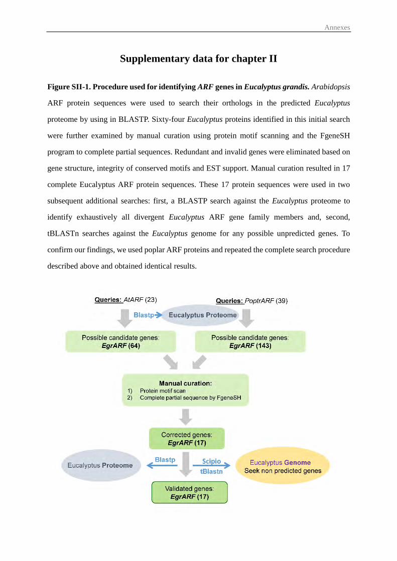
Annexes
Supplementary data for chapter II
Figure SII-1. Procedure used for identifying ARF genes in Eucalyptus grandis. Arabidopsis
ARF protein sequences were used to search their orthologs in the predicted Eucalyptus
proteome by using in BLASTP. Sixty-four Eucalyptus proteins identified in this initial search
were further examined by manual curation using protein motif scanning and the FgeneSH
program to complete partial sequences. Redundant and invalid genes were eliminated based on
gene structure, integrity of conserved motifs and EST support. Manual curation resulted in 17
complete Eucalyptus ARF protein sequences. These 17 protein sequences were used in two
subsequent additional searches: first, a BLASTP search against the Eucalyptus proteome to
identify exhaustively all divergent Eucalyptus ARF gene family members and, second,
tBLASTn searches against the Eucalyptus genome for any possible unpredicted genes. To
confirm our findings, we used poplar ARF proteins and repeated the complete search procedure
described above and obtained identical results.
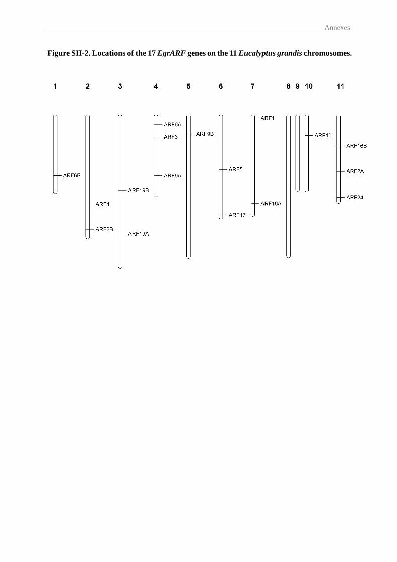
Annexes
Figure SII-2. Locations of the 17 EgrARF genes on the 11 Eucalyptus grandis chromosomes.
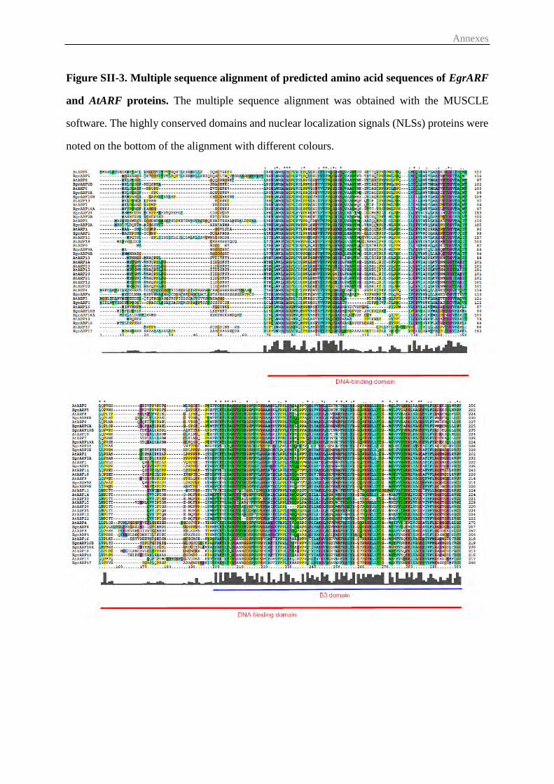
Annexes
Figure SII-3. Multiple sequence alignment of predicted amino acid sequences of EgrARF
and AtARF proteins. The multiple sequence alignment was obtained with the MUSCLE
software. The highly conserved domains and nuclear localization signals (NLSs) proteins were
noted on the bottom of the alignment with different colours.
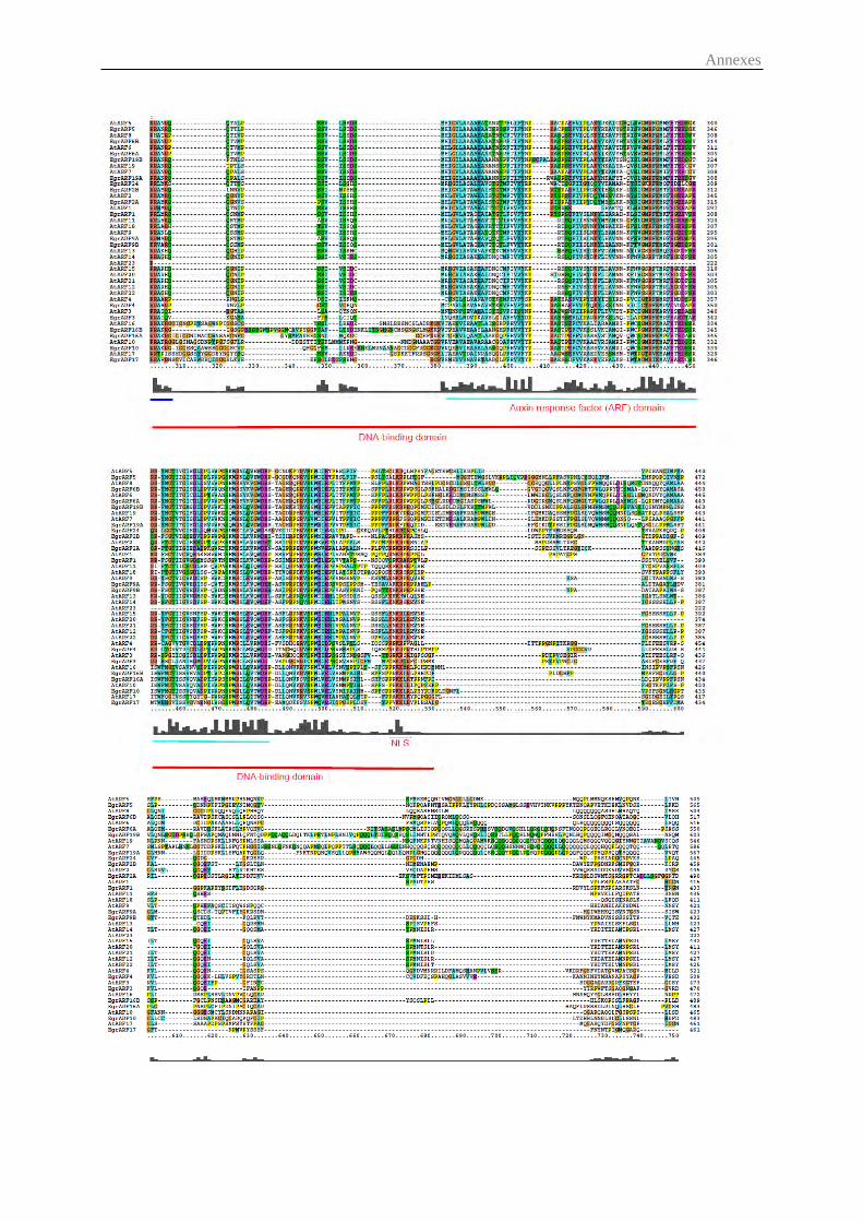
Annexes
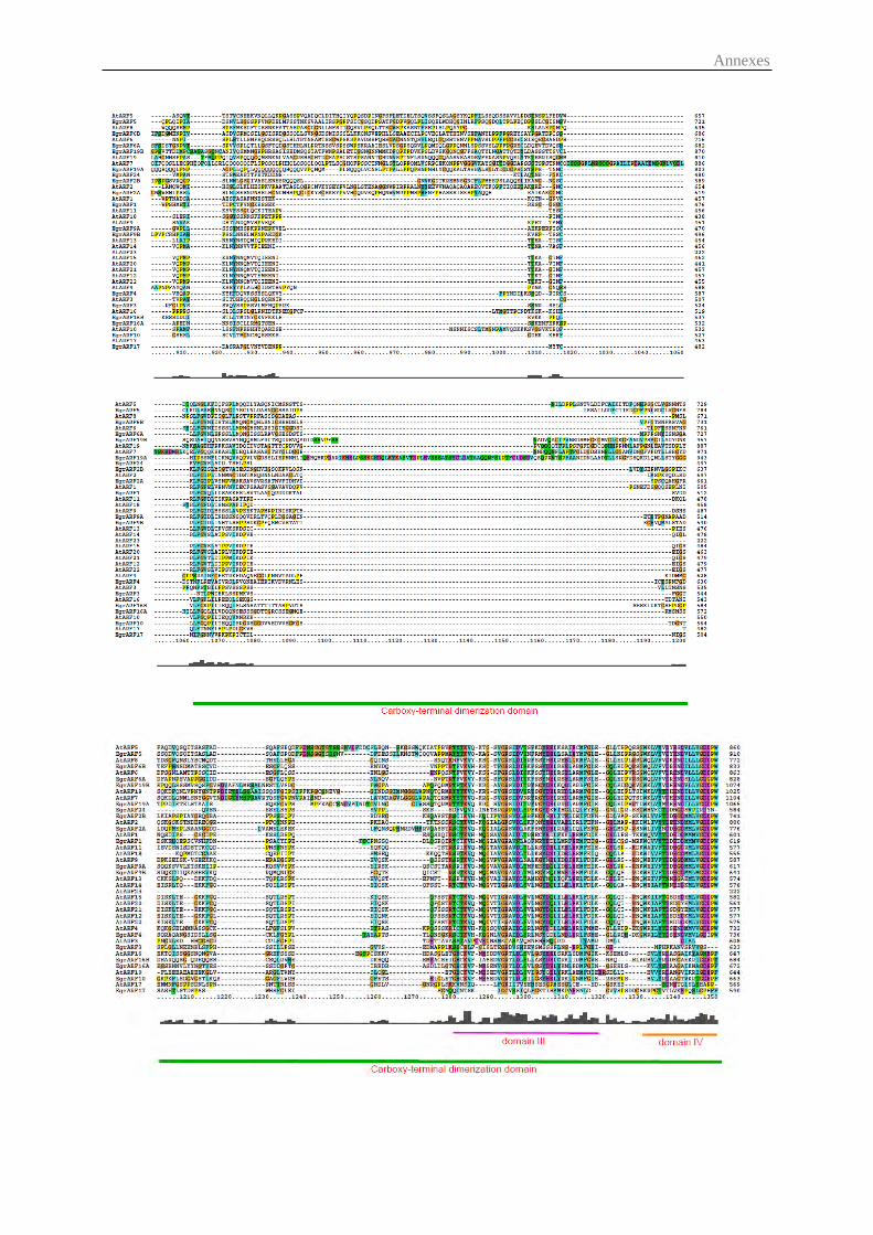
Annexes
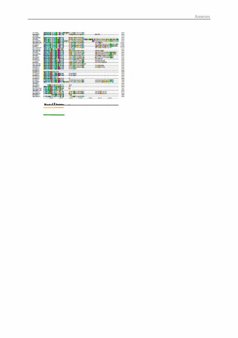
Annexes
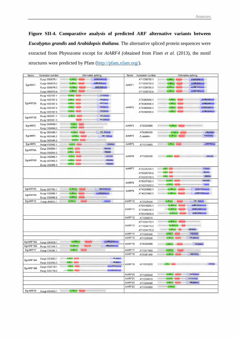
Annexes
Figure SII-4. Comparative analysis of predicted ARF alternative variants between
Eucalyptus grandis and Arabidopsis thaliana. The alternative spliced protein sequences were
extracted from Phytozome except for AtARF4 (obtained from Finet et al. (2013), the motif
structures were predicted by Pfam (http://pfam.xfam.org/).
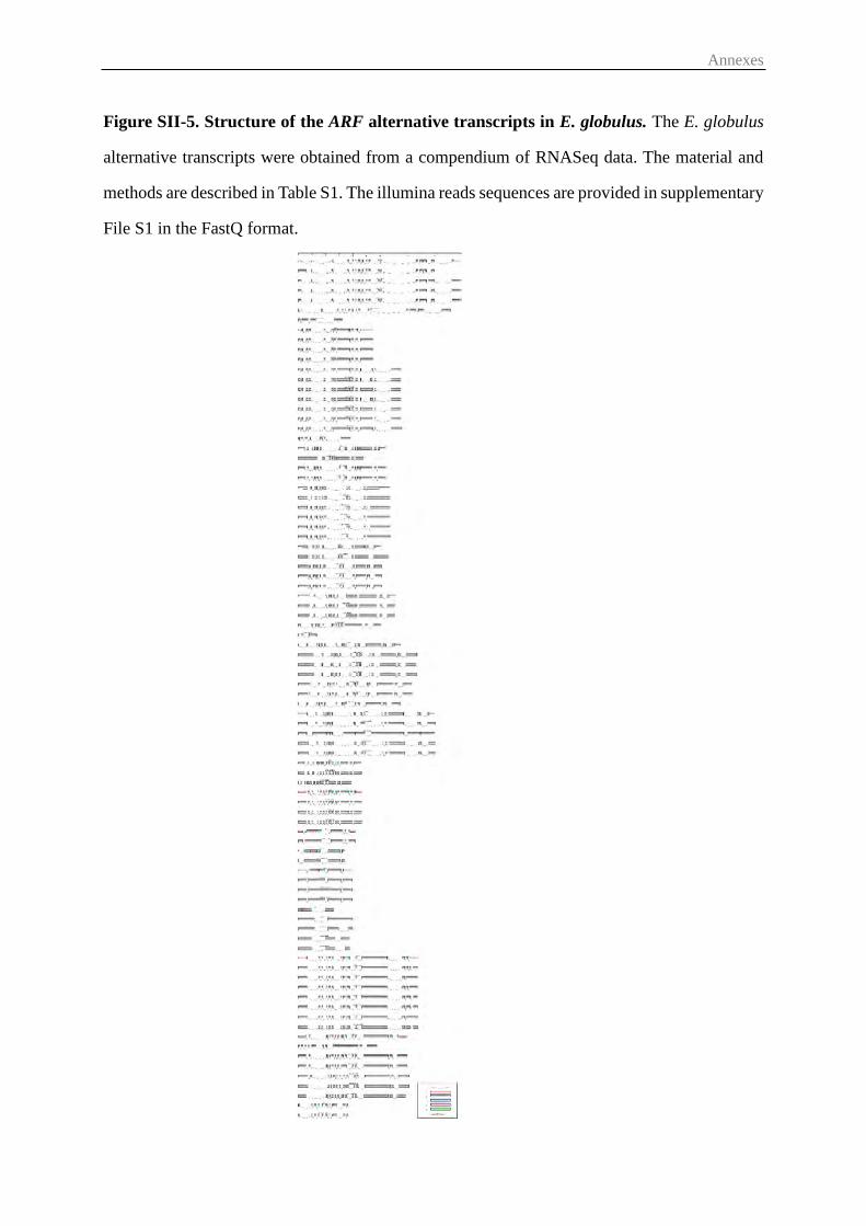
Annexes
Figure SII-5. Structure of the ARF alternative transcripts in E. globulus. The E. globulus
alternative transcripts were obtained from a compendium of RNASeq data. The material and
methods are described in Table S1. The illumina reads sequences are provided in supplementary
File S1 in the FastQ format.
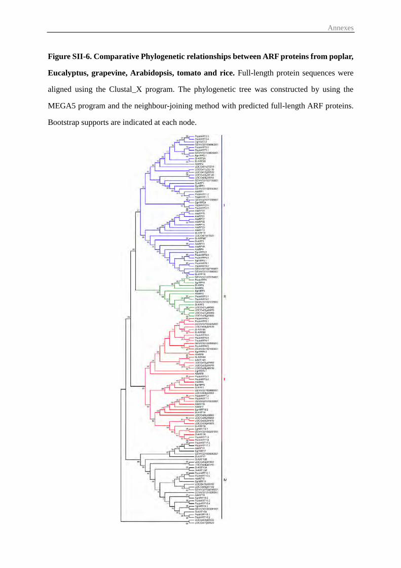
Annexes
Figure SII-6. Comparative Phylogenetic relationships between ARF proteins from poplar,
Eucalyptus, grapevine, Arabidopsis, tomato and rice. Full-length protein sequences were
aligned using the Clustal_X program. The phylogenetic tree was constructed by using the
MEGA5 program and the neighbour-joining method with predicted full-length ARF proteins.
Bootstrap supports are indicated at each node.
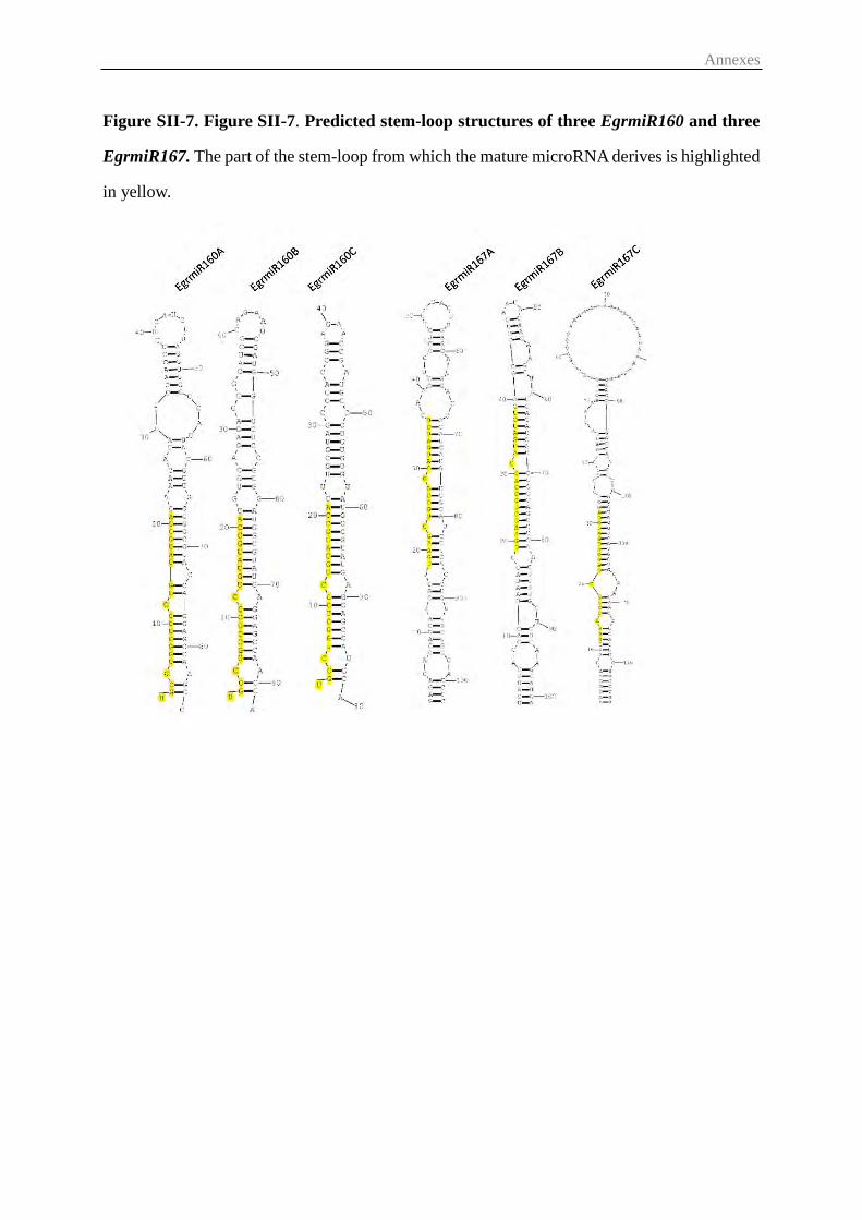
Annexes
Figure SII-7. Figure SII-7. Predicted stem-loop structures of three EgrmiR160 and three
EgrmiR167. The part of the stem-loop from which the mature microRNA derives is highlighted
in yellow.
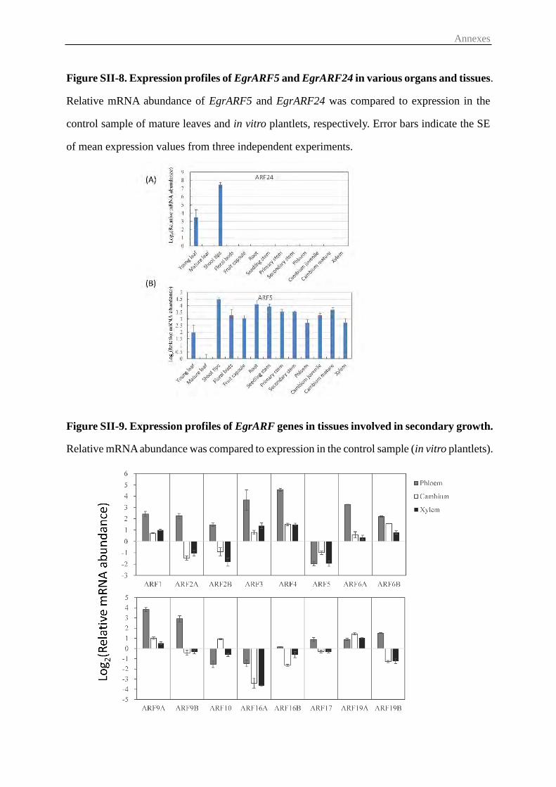
Annexes
Figure SII-8. Expression profiles of EgrARF5 and EgrARF24 in various organs and tissues.
Relative mRNA abundance of EgrARF5 and EgrARF24 was compared to expression in the
control sample of mature leaves and in vitro plantlets, respectively. Error bars indicate the SE
of mean expression values from three independent experiments.
Figure SII-9. Expression profiles of EgrARF genes in tissues involved in secondary growth.
Relative mRNA abundance was compared to expression in the control sample (in vitro plantlets).
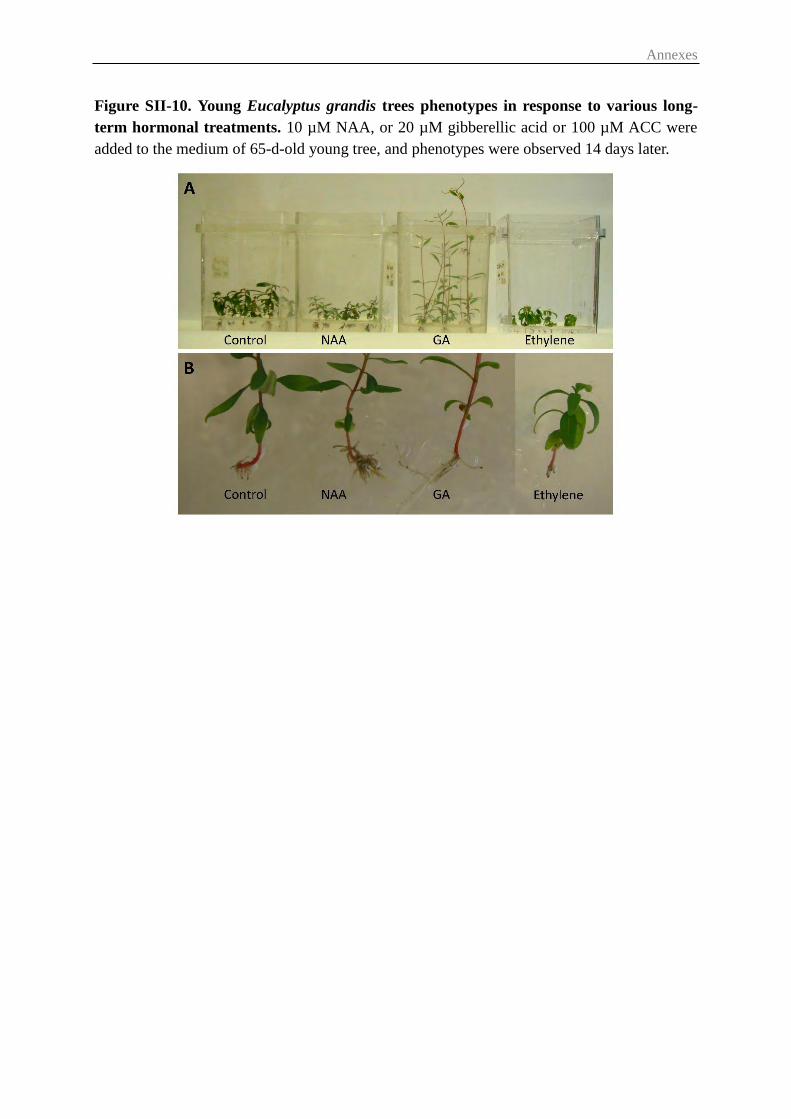
Annexes
Figure SII-10. Young Eucalyptus grandis trees phenotypes in response to various long-
term hormonal treatments. 10 µM NAA, or 20 µM gibberellic acid or 100 µM ACC were
added to the medium of 65-d-old young tree, and phenotypes were observed 14 days later.

Annexes
Table SII-1. Primers for EgrARF genes and reference genes used in qRT-PCR experiments
Name Accession No. Forward 5'-3' Reverse 5'-3' Efficiency
ARF1 Eucgr.G00076.1 TTTAAGAGCGCTGTGGTTCTCTG AAGCATCAGCAAACGCACCTTG 1.90
ARF2A Eucgr.K02197.1 GGCATGCCATTTCTACAGGAACC TCAGCAGGGCTTATCCTAGGTTTG 1.97
ARF2B Eucgr.B03551.1 ATTGAGGGCTGAACCAGAGACC TGGTTAGGCACCGGAAGCAAAG 1.95
ARF3 Eucgr.D00588.1 ACATATATACCGGGCAGGACAGC TCTTCCGGTTTACAAATGCACTCC 1.94
ARF4 Eucgr.B02480.1 AGCTGTCGCTAGTGCTGTATCC AACTCTGCATGGCTTGCCCTTG 1.96
ARF5 Eucgr.F02090.1 GCACATGGCAACAGCAAGTAGC ATCGACCTTCCGACTGATCCTG 1.97
ARF6A Eucgr.D00264.1 TGAGCTTGCTCGCATGTTTAGCC TGTTCACAAACTCCGGCCAAGG 1.89
ARF6B Eucgr.A02065.1 GCTGGCAGCTTGTATTTGTAGACC CCACACATTGTTGACGAACTCCTG 1.88
ARF9A Eucgr.D01764.1 TCCAAAGCAGTCTGTGGTTTCACC ATGCGACTCCTTGCAGTGGTAG 1.87
ARF9B Eucgr.E00888.1 GTGACTCGTTCGTGTTCTTAAGGG TGACGAGCAACTCGTTTCACTCC 2.00
ARF10 Eucgr.J00923.1 TAGCAGGGCGTGTCGTGTTATC AATCCGGACGCTTGCGTTTCTC 1.90
ARF16A Eucgr.G02838.1 AGGGACATGTTCGGCATAAACGG AATAGCACCGGCAGCATCTGAG 1.84
ARF16B Eucgr.K01240.1 CTCAGAAATCGGCCACTGCAAAG TGAGGCAAGCAACGAGAGATCG 1.97
ARF17 Eucgr.F04380.1 ATGGAGGTGGTGGTGAATGCAG AAACCACCTCGAACGGCAATCC 1.85
ARF19A Eucgr.C03293.1 TCAGTTTCAAGCGGATGAGAAGCC AAGGACAGCCACTCTGGGTTAG 1.78
ARF19B Eucgr.C02178.1 TCAAACAGGACCTGGCTCGTAG CGAACACAGTTCACGAAGTCCTC 1.93
ARF24 Eucgr.K03433.1 TCCAGCGACTTTGTGCAGGTTATC AGCCTTTGAATGGGCTGAGGTC 1.92
EF-1α Eucgr.B02473.1 ATGCGTCAGACTGTGGCTGTTG TTGGTCACCTTGGCTCCACTTG 1.84
SAND Eucgr.B02502.1 TTGATCCACTTGCGGACAAGGC TCACCCATTGACATACACGATTGC 2.11
PP2A1 Eucgr.B03386.1 TCGAGCTTTGGACCGCATACAAG ACCACAAGAGGTCACACATTGGC 1.98
PP2A3 5'end Eucgr.B03031.1 CGGAAGAACTGGGTGTGTTT CACAGAGGGTCTCCAATGGT 2.03
PP2A3 3'end Eucgr.B03031.1 CAGCGGCAAACAACTTGAAGCG ATTATGTGCTGCATTGCCCAGTC 2.02
IDH 5'end Eucgr.F02901.1 AATCGACCTGCTTCGACCCTTC TCGACCTTGATCTTCTCGAAACCC 1.92
IDH 3'end Eucgr.F02901.1 TGCTGTGGCAGCTGAACTCAAG ATGTTGTCCGCCAGTCACCTAC 1.86
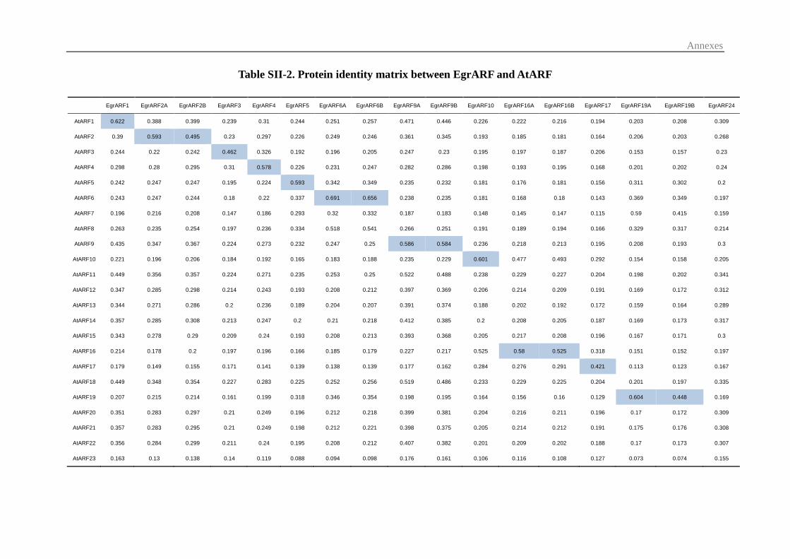
Annexes
Table SII-2. Protein identity matrix between EgrARF and AtARF
EgrARF1 EgrARF2A EgrARF2B EgrARF3 EgrARF4 EgrARF5 EgrARF6A EgrARF6B EgrARF9A EgrARF9B EgrARF10 EgrARF16A EgrARF16B EgrARF17 EgrARF19A EgrARF19B EgrARF24
AtARF1 0.622 0.388 0.399 0.239 0.31 0.244 0.251 0.257 0.471 0.446 0.226 0.222 0.216 0.194 0.203 0.208 0.309
AtARF2 0.39 0.593 0.495 0.23 0.297 0.226 0.249 0.246 0.361 0.345 0.193 0.185 0.181 0.164 0.206 0.203 0.268
AtARF3 0.244 0.22 0.242 0.462 0.326 0.192 0.196 0.205 0.247 0.23 0.195 0.197 0.187 0.206 0.153 0.157 0.23
AtARF4 0.298 0.28 0.295 0.31 0.578 0.226 0.231 0.247 0.282 0.286 0.198 0.193 0.195 0.168 0.201 0.202 0.24
AtARF5 0.242 0.247 0.247 0.195 0.224 0.593 0.342 0.349 0.235 0.232 0.181 0.176 0.181 0.156 0.311 0.302 0.2
AtARF6 0.243 0.247 0.244 0.18 0.22 0.337 0.691 0.656 0.238 0.235 0.181 0.168 0.18 0.143 0.369 0.349 0.197
AtARF7 0.196 0.216 0.208 0.147 0.186 0.293 0.32 0.332 0.187 0.183 0.148 0.145 0.147 0.115 0.59 0.415 0.159
AtARF8 0.263 0.235 0.254 0.197 0.236 0.334 0.518 0.541 0.266 0.251 0.191 0.189 0.194 0.166 0.329 0.317 0.214
AtARF9 0.435 0.347 0.367 0.224 0.273 0.232 0.247 0.25 0.586 0.584 0.236 0.218 0.213 0.195 0.208 0.193 0.3
AtARF10 0.221 0.196 0.206 0.184 0.192 0.165 0.183 0.188 0.235 0.229 0.601 0.477 0.493 0.292 0.154 0.158 0.205
AtARF11 0.449 0.356 0.357 0.224 0.271 0.235 0.253 0.25 0.522 0.488 0.238 0.229 0.227 0.204 0.198 0.202 0.341
AtARF12 0.347 0.285 0.298 0.214 0.243 0.193 0.208 0.212 0.397 0.369 0.206 0.214 0.209 0.191 0.169 0.172 0.312
AtARF13 0.344 0.271 0.286 0.2 0.236 0.189 0.204 0.207 0.391 0.374 0.188 0.202 0.192 0.172 0.159 0.164 0.289
AtARF14 0.357 0.285 0.308 0.213 0.247 0.2 0.21 0.218 0.412 0.385 0.2 0.208 0.205 0.187 0.169 0.173 0.317
AtARF15 0.343 0.278 0.29 0.209 0.24 0.193 0.208 0.213 0.393 0.368 0.205 0.217 0.208 0.196 0.167 0.171 0.3
AtARF16 0.214 0.178 0.2 0.197 0.196 0.166 0.185 0.179 0.227 0.217 0.525 0.58 0.525 0.318 0.151 0.152 0.197
AtARF17 0.179 0.149 0.155 0.171 0.141 0.139 0.138 0.139 0.177 0.162 0.284 0.276 0.291 0.421 0.113 0.123 0.167
AtARF18 0.449 0.348 0.354 0.227 0.283 0.225 0.252 0.256 0.519 0.486 0.233 0.229 0.225 0.204 0.201 0.197 0.335
AtARF19 0.207 0.215 0.214 0.161 0.199 0.318 0.346 0.354 0.198 0.195 0.164 0.156 0.16 0.129 0.604 0.448 0.169
AtARF20 0.351 0.283 0.297 0.21 0.249 0.196 0.212 0.218 0.399 0.381 0.204 0.216 0.211 0.196 0.17 0.172 0.309
AtARF21 0.357 0.283 0.295 0.21 0.249 0.198 0.212 0.221 0.398 0.375 0.205 0.214 0.212 0.191 0.175 0.176 0.308
AtARF22 0.356 0.284 0.299 0.211 0.24 0.195 0.208 0.212 0.407 0.382 0.201 0.209 0.202 0.188 0.17 0.173 0.307
AtARF23 0.163 0.13 0.138 0.14 0.119 0.088 0.094 0.098 0.176 0.161 0.106 0.116 0.108 0.127 0.073 0.074 0.155
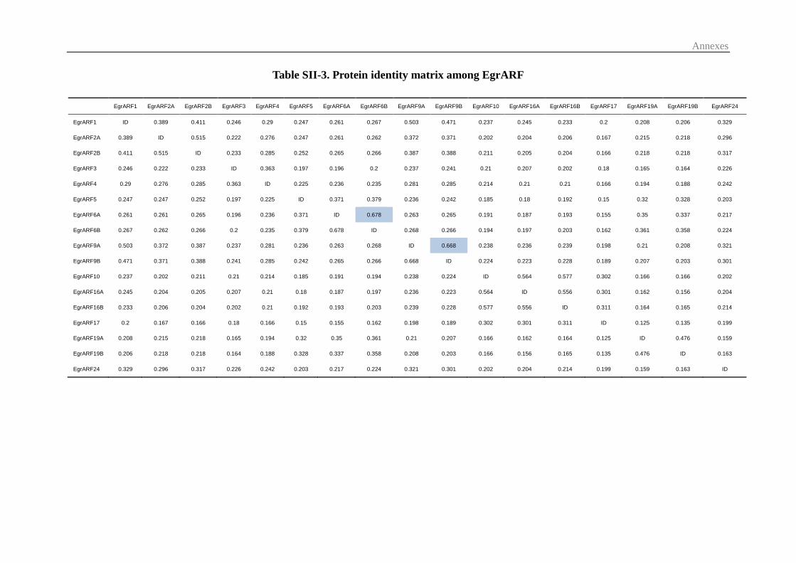
Annexes
Table SII-3. Protein identity matrix among EgrARF
EgrARF1 EgrARF2A EgrARF2B EgrARF3 EgrARF4 EgrARF5 EgrARF6A EgrARF6B EgrARF9A EgrARF9B EgrARF10 EgrARF16A EgrARF16B EgrARF17 EgrARF19A EgrARF19B EgrARF24
EgrARF1 ID 0.389 0.411 0.246 0.29 0.247 0.261 0.267 0.503 0.471 0.237 0.245 0.233 0.2 0.208 0.206 0.329
EgrARF2A 0.389 ID 0.515 0.222 0.276 0.247 0.261 0.262 0.372 0.371 0.202 0.204 0.206 0.167 0.215 0.218 0.296
EgrARF2B 0.411 0.515 ID 0.233 0.285 0.252 0.265 0.266 0.387 0.388 0.211 0.205 0.204 0.166 0.218 0.218 0.317
EgrARF3 0.246 0.222 0.233 ID 0.363 0.197 0.196 0.2 0.237 0.241 0.21 0.207 0.202 0.18 0.165 0.164 0.226
EgrARF4 0.29 0.276 0.285 0.363 ID 0.225 0.236 0.235 0.281 0.285 0.214 0.21 0.21 0.166 0.194 0.188 0.242
EgrARF5 0.247 0.247 0.252 0.197 0.225 ID 0.371 0.379 0.236 0.242 0.185 0.18 0.192 0.15 0.32 0.328 0.203
EgrARF6A 0.261 0.261 0.265 0.196 0.236 0.371 ID 0.678 0.263 0.265 0.191 0.187 0.193 0.155 0.35 0.337 0.217
EgrARF6B 0.267 0.262 0.266 0.2 0.235 0.379 0.678 ID 0.268 0.266 0.194 0.197 0.203 0.162 0.361 0.358 0.224
EgrARF9A 0.503 0.372 0.387 0.237 0.281 0.236 0.263 0.268 ID 0.668 0.238 0.236 0.239 0.198 0.21 0.208 0.321
EgrARF9B 0.471 0.371 0.388 0.241 0.285 0.242 0.265 0.266 0.668 ID 0.224 0.223 0.228 0.189 0.207 0.203 0.301
EgrARF10 0.237 0.202 0.211 0.21 0.214 0.185 0.191 0.194 0.238 0.224 ID 0.564 0.577 0.302 0.166 0.166 0.202
EgrARF16A 0.245 0.204 0.205 0.207 0.21 0.18 0.187 0.197 0.236 0.223 0.564 ID 0.556 0.301 0.162 0.156 0.204
EgrARF16B 0.233 0.206 0.204 0.202 0.21 0.192 0.193 0.203 0.239 0.228 0.577 0.556 ID 0.311 0.164 0.165 0.214
EgrARF17 0.2 0.167 0.166 0.18 0.166 0.15 0.155 0.162 0.198 0.189 0.302 0.301 0.311 ID 0.125 0.135 0.199
EgrARF19A 0.208 0.215 0.218 0.165 0.194 0.32 0.35 0.361 0.21 0.207 0.166 0.162 0.164 0.125 ID 0.476 0.159
EgrARF19B 0.206 0.218 0.218 0.164 0.188 0.328 0.337 0.358 0.208 0.203 0.166 0.156 0.165 0.135 0.476 ID 0.163
EgrARF24 0.329 0.296 0.317 0.226 0.242 0.203 0.217 0.224 0.321 0.301 0.202 0.204 0.214 0.199 0.159 0.163 ID

Annexes
Table SII-4 Comparison of the number of alternative transcripts predicted in phytozome
for E. grandis to those found in a large compendium of transcriptomic data from in E.
globulus.
Material and Methods for Alternative slipicing validation
TopHat and Cufflink Suite (Trapnell et al. 2012), currently used for differential gene and transcript expression
analysis of RNA-seq experiments, were used to validate the Eucalyptus ARF genes (E. grandis genome v162
annotation). Ten developing xylem (DX) RNA-Seq libraries were produced and sequenced FASTERIS SA
(Genève, CH; www.fasteris.com). RNA-Seq libraries were prepared from ten equimolar pools of high quality
individual total RNA: a) four seasonal libraries produced from total RNA extracted from DX samples collected in
2008 on three ramets of non-related tree genotypes at Herdade do Zambujal, Pegões (Portugal) [February 26 th
(Feb), May 23rd (May), September 5th (Sep) and December 3rd (Dec)] (Carocha et al., unpublished); b) four pulp
yield pools (five trees by pool) obtained by pooling the total RNA from samples. collected at Carregal Fundeiro,
Abrantes (Portugal), from 10 trees with very constrasting pulp yields in the coded AxB mapping population, and
10 trees with very constrasting pulp yields in a natural variation panel; c) two libraries from samples collected
from an adult tree and a juvenile one, (same genotype) at Herdade do Zambujal, Pegões (Portugal). Sampling
procedures and RNA extraction were described, respectively in Paux et al. (2004) and Cassan- Wang et al. (2012).
All samples were kindly provided by RAIZ Institute (Portugal). The TruSeq™ SBSv5 sequencing kit (Illumina)
was used for library sequencing, using the Illumina Hi-Seq 2000 instrument, on a multiplex runs with
1x100nt+7(index) cycles. For each RNA-Seq library, adaptors removal and a successive quality and contaminants
(ribosomal sequences) filters were applied. The resulting filtered high quality reads were then mapped to the E.
grandis genome sequence v162 using TopHat v1.3.1. Cufflinks v1.1.0 was used to determine the potential coding
regions and their intron-exon structures. Finally, Cuffcompare v1.1.0 were then used to compare the assembled
multiple libraries Cufflinks transcripts to the E. grandis v162 gene annotations. The sequences of the Illumina
reads from RNA Seq used to predict the E. globulus alternative transcripts are provided in FastaQ format in
supplementary File S1.
REF: Cole Trapnell, Adam Roberts, Loyal Goff, Geo Pertea, Daehwan Kim, David R Kelley, Harold Pimentel,
Steven L Salzberg, John L Rinn & Lior Pachter (2012) Differential gene and transcript expression analysis of
RNA-seq experiments with TopHat and Cufflinks. Nature Protocols 7: 562–578.
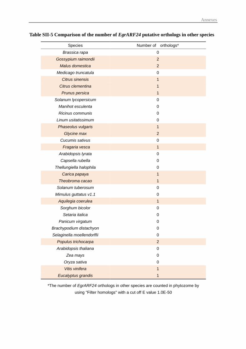
Annexes
Table SII-5 Comparison of the number of EgrARF24 putative orthologs in other species
Species Number of orthologs*
Brassica rapa 0
Gossypium raimondii 2
Malus domestica 2
Medicago truncatula 0
Citrus sinensis 1
Citrus clementina 1
Prunus persica 1
Solanum lycopersicum 0
Manihot esculenta 0
Ricinus communis 0
Linum usitatissimum 0
Phaseolus vulgaris 1
Glycine max 2
Cucumis sativus 0
Fragaria vesca 1
Arabidopsis lyrata 0
Capsella rubella 0
Thellungiella halophila 0
Carica papaya 1
Theobroma cacao 1
Solanum tuberosum 0
Mimulus guttatus v1.1 0
Aquilegia coerulea 1
Sorghum bicolor 0
Setaria italica 0
Panicum virgatum 0
Brachypodium distachyon 0
Selaginella moellendorffii 0
Populus trichocarpa 2
Arabidopsis thaliana 0
Zea mays 0
Oryza sativa 0
Vitis vinifera 1
Eucalyptus grandis 1
*The number of EgrARF24 orthologs in other species are counted in phytozome by
using "Filter homologs" with a cut off E value 1.0E-50
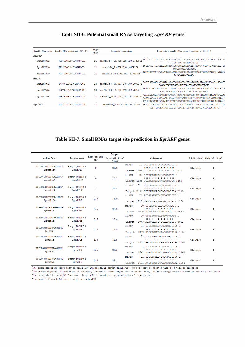
Annexes
Table SII-6. Potential small RNAs targeting EgrARF genes
Table SII-7. Small RNAs target site prediction in EgrARF genes
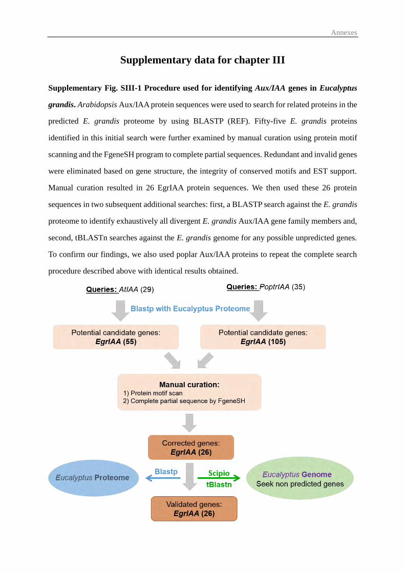
Annexes
Supplementary data for chapter III
Supplementary Fig. SIII-1 Procedure used for identifying Aux/IAA genes in Eucalyptus
grandis. Arabidopsis Aux/IAA protein sequences were used to search for related proteins in the
predicted E. grandis proteome by using BLASTP (REF). Fifty-five E. grandis proteins
identified in this initial search were further examined by manual curation using protein motif
scanning and the FgeneSH program to complete partial sequences. Redundant and invalid genes
were eliminated based on gene structure, the integrity of conserved motifs and EST support.
Manual curation resulted in 26 EgrIAA protein sequences. We then used these 26 protein
sequences in two subsequent additional searches: first, a BLASTP search against the E. grandis
proteome to identify exhaustively all divergent E. grandis Aux/IAA gene family members and,
second, tBLASTn searches against the E. grandis genome for any possible unpredicted genes.
To confirm our findings, we also used poplar Aux/IAA proteins to repeat the complete search
procedure described above with identical results obtained.
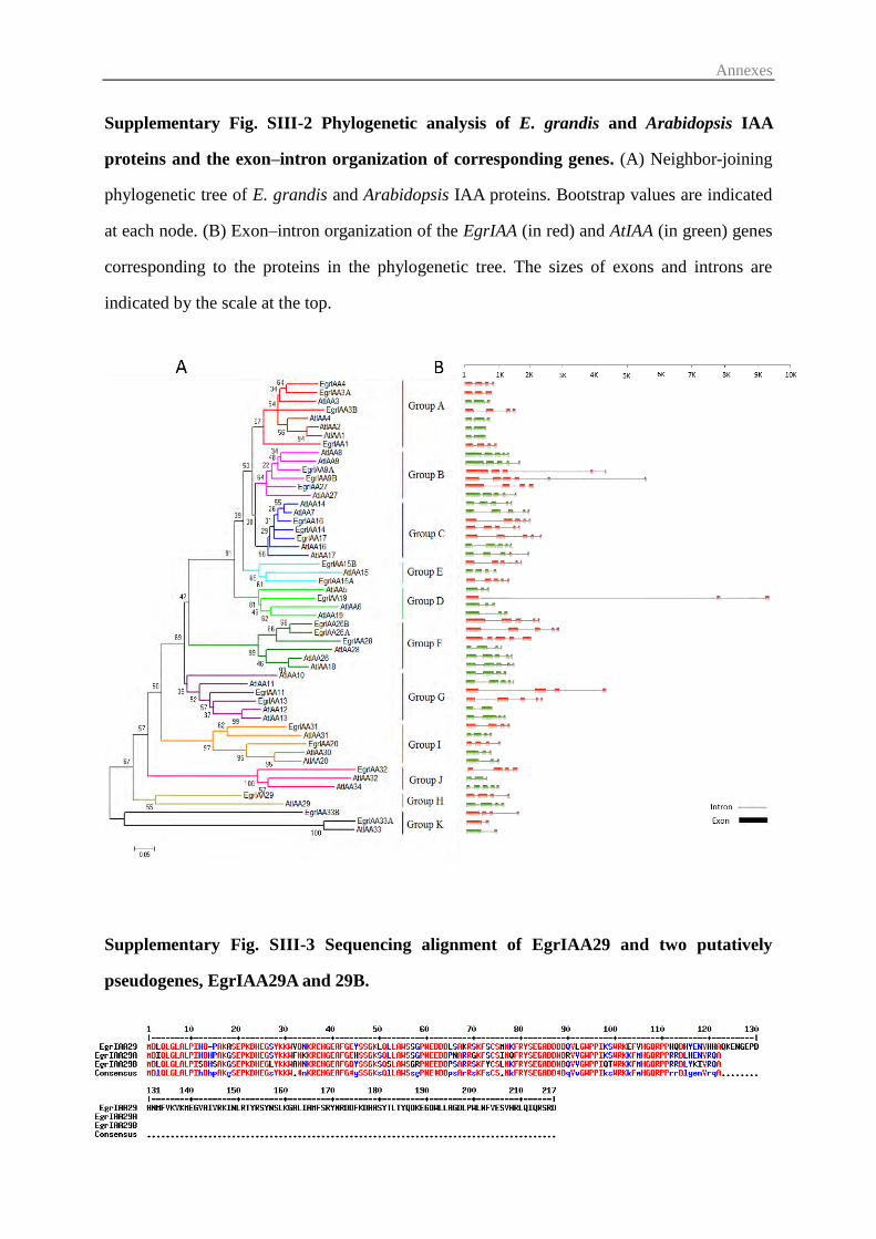
Annexes
Supplementary Fig. SIII-2 Phylogenetic analysis of E. grandis and Arabidopsis IAA
proteins and the exon–intron organization of corresponding genes. (A) Neighbor-joining
phylogenetic tree of E. grandis and Arabidopsis IAA proteins. Bootstrap values are indicated
at each node. (B) Exon–intron organization of the EgrIAA (in red) and AtIAA (in green) genes
corresponding to the proteins in the phylogenetic tree. The sizes of exons and introns are
indicated by the scale at the top.
Supplementary Fig. SIII-3 Sequencing alignment of EgrIAA29 and two putatively
pseudogenes, EgrIAA29A and 29B.
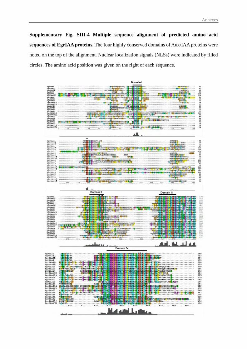
Annexes
Supplementary Fig. SIII-4 Multiple sequence alignment of predicted amino acid
sequences of EgrIAA proteins. The four highly conserved domains of Aux/IAA proteins were
noted on the top of the alignment. Nuclear localization signals (NLSs) were indicated by filled
circles. The amino acid position was given on the right of each sequence.
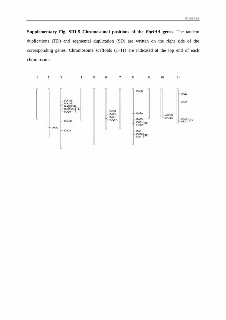
Annexes
Supplementary Fig. SIII-5 Chromosomal positions of the EgrIAA genes. The tandem
duplications (TD) and segmental duplication (SD) are written on the right side of the
corresponding genes. Chromosome scaffolds (1–11) are indicated at the top end of each
chromosome.
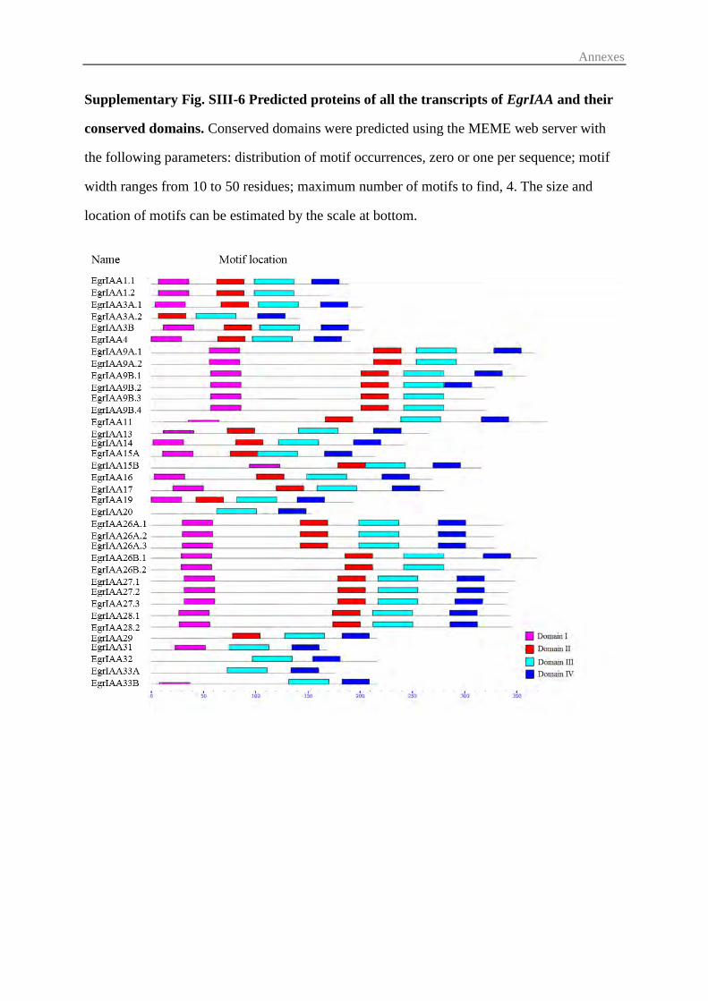
Annexes
Supplementary Fig. SIII-6 Predicted proteins of all the transcripts of EgrIAA and their
conserved domains. Conserved domains were predicted using the MEME web server with
the following parameters: distribution of motif occurrences, zero or one per sequence; motif
width ranges from 10 to 50 residues; maximum number of motifs to find, 4. The size and
location of motifs can be estimated by the scale at bottom.
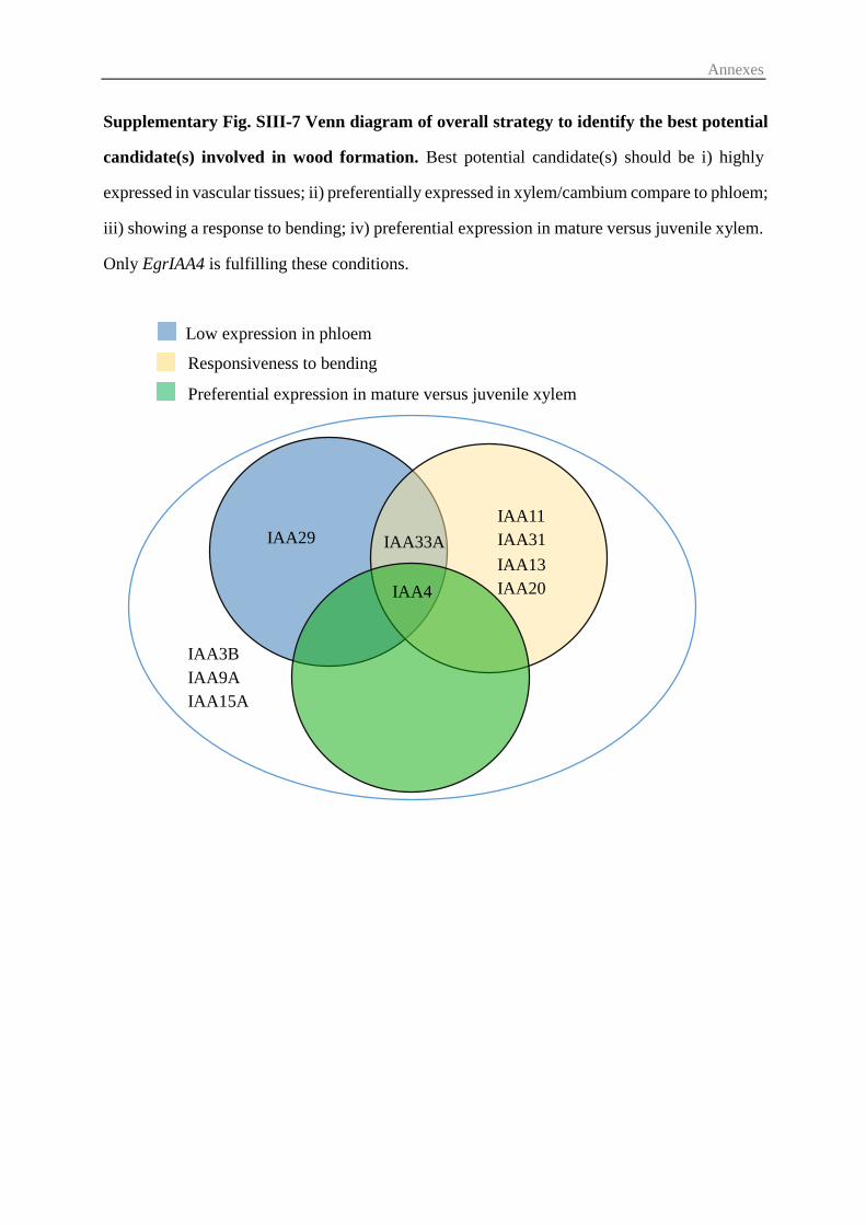
Annexes
Low expression in phloem
Responsiveness to bending
Preferential expression in mature versus juvenile xylem
IAA13
IAA20
IAA29 IAA11
IAA31
IAA4
IAA33A
IAA3B
IAA9A
IAA15A
Supplementary Fig. SIII-7 Venn diagram of overall strategy to identify the best potential
candidate(s) involved in wood formation. Best potential candidate(s) should be i) highly
expressed in vascular tissues; ii) preferentially expressed in xylem/cambium compare to phloem;
iii) showing a response to bending; iv) preferential expression in mature versus juvenile xylem.
Only EgrIAA4 is fulfilling these conditions.
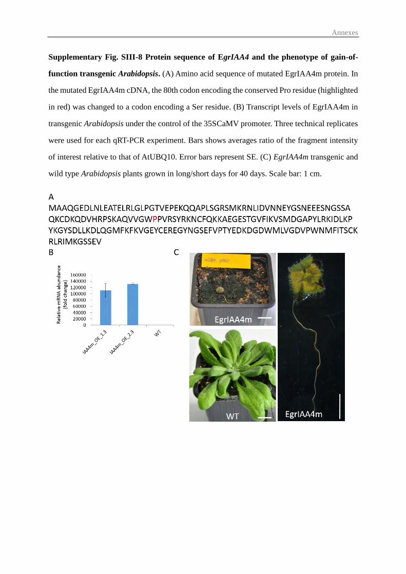
Annexes
Supplementary Fig. SIII-8 Protein sequence of EgrIAA4 and the phenotype of gain-of-
function transgenic Arabidopsis. (A) Amino acid sequence of mutated EgrIAA4m protein. In
the mutated EgrIAA4m cDNA, the 80th codon encoding the conserved Pro residue (highlighted
in red) was changed to a codon encoding a Ser residue. (B) Transcript levels of EgrIAA4m in
transgenic Arabidopsis under the control of the 35SCaMV promoter. Three technical replicates
were used for each qRT-PCR experiment. Bars shows averages ratio of the fragment intensity
of interest relative to that of AtUBQ10. Error bars represent SE. (C) EgrIAA4m transgenic and
wild type Arabidopsis plants grown in long/short days for 40 days. Scale bar: 1 cm.
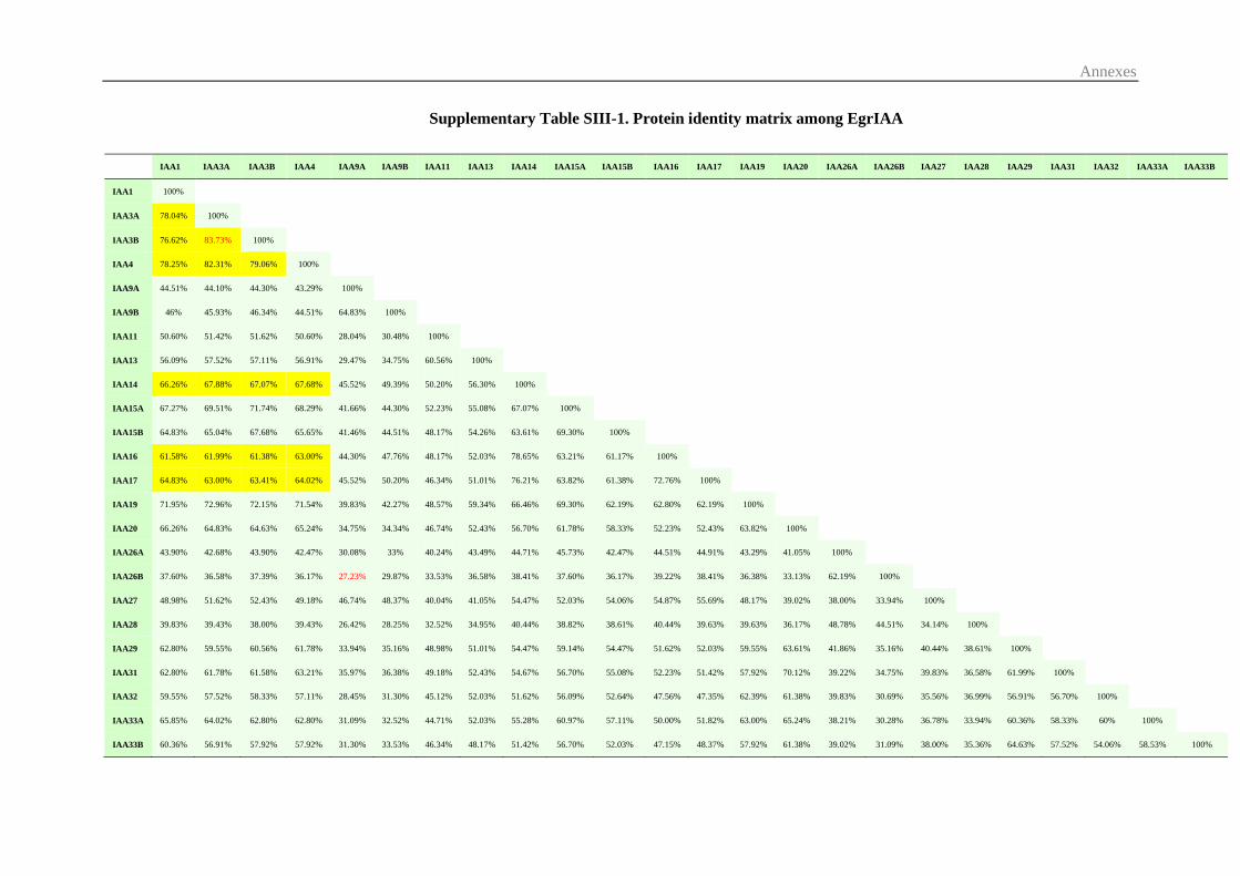
Annexes
Supplementary Table SIII-1. Protein identity matrix among EgrIAA
IAA1 IAA3A IAA3B IAA4 IAA9A IAA9B IAA11 IAA13 IAA14 IAA15A IAA15B IAA16 IAA17 IAA19 IAA20 IAA26A IAA26B IAA27 IAA28 IAA29 IAA31 IAA32 IAA33A IAA33B
IAA1 100%
IAA3A 78.04% 100%
IAA3B 76.62% 83.73% 100%
IAA4 78.25% 82.31% 79.06% 100%
IAA9A 44.51% 44.10% 44.30% 43.29% 100%
IAA9B 46% 45.93% 46.34% 44.51% 64.83% 100%
IAA11 50.60% 51.42% 51.62% 50.60% 28.04% 30.48% 100%
IAA13 56.09% 57.52% 57.11% 56.91% 29.47% 34.75% 60.56% 100%
IAA14 66.26% 67.88% 67.07% 67.68% 45.52% 49.39% 50.20% 56.30% 100%
IAA15A 67.27% 69.51% 71.74% 68.29% 41.66% 44.30% 52.23% 55.08% 67.07% 100%
IAA15B 64.83% 65.04% 67.68% 65.65% 41.46% 44.51% 48.17% 54.26% 63.61% 69.30% 100%
IAA16 61.58% 61.99% 61.38% 63.00% 44.30% 47.76% 48.17% 52.03% 78.65% 63.21% 61.17% 100%
IAA17 64.83% 63.00% 63.41% 64.02% 45.52% 50.20% 46.34% 51.01% 76.21% 63.82% 61.38% 72.76% 100%
IAA19 71.95% 72.96% 72.15% 71.54% 39.83% 42.27% 48.57% 59.34% 66.46% 69.30% 62.19% 62.80% 62.19% 100%
IAA20 66.26% 64.83% 64.63% 65.24% 34.75% 34.34% 46.74% 52.43% 56.70% 61.78% 58.33% 52.23% 52.43% 63.82% 100%
IAA26A 43.90% 42.68% 43.90% 42.47% 30.08% 33% 40.24% 43.49% 44.71% 45.73% 42.47% 44.51% 44.91% 43.29% 41.05% 100%
IAA26B 37.60% 36.58% 37.39% 36.17% 27.23% 29.87% 33.53% 36.58% 38.41% 37.60% 36.17% 39.22% 38.41% 36.38% 33.13% 62.19% 100%
IAA27 48.98% 51.62% 52.43% 49.18% 46.74% 48.37% 40.04% 41.05% 54.47% 52.03% 54.06% 54.87% 55.69% 48.17% 39.02% 38.00% 33.94% 100%
IAA28 39.83% 39.43% 38.00% 39.43% 26.42% 28.25% 32.52% 34.95% 40.44% 38.82% 38.61% 40.44% 39.63% 39.63% 36.17% 48.78% 44.51% 34.14% 100%
IAA29 62.80% 59.55% 60.56% 61.78% 33.94% 35.16% 48.98% 51.01% 54.47% 59.14% 54.47% 51.62% 52.03% 59.55% 63.61% 41.86% 35.16% 40.44% 38.61% 100%
IAA31 62.80% 61.78% 61.58% 63.21% 35.97% 36.38% 49.18% 52.43% 54.67% 56.70% 55.08% 52.23% 51.42% 57.92% 70.12% 39.22% 34.75% 39.83% 36.58% 61.99% 100%
IAA32 59.55% 57.52% 58.33% 57.11% 28.45% 31.30% 45.12% 52.03% 51.62% 56.09% 52.64% 47.56% 47.35% 62.39% 61.38% 39.83% 30.69% 35.56% 36.99% 56.91% 56.70% 100%
IAA33A 65.85% 64.02% 62.80% 62.80% 31.09% 32.52% 44.71% 52.03% 55.28% 60.97% 57.11% 50.00% 51.82% 63.00% 65.24% 38.21% 30.28% 36.78% 33.94% 60.36% 58.33% 60% 100%
IAA33B 60.36% 56.91% 57.92% 57.92% 31.30% 33.53% 46.34% 48.17% 51.42% 56.70% 52.03% 47.15% 48.37% 57.92% 61.38% 39.02% 31.09% 38.00% 35.36% 64.63% 57.52% 54.06% 58.53% 100%
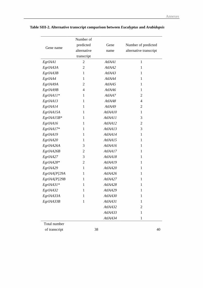
Annexes
Table SIII-2. Alternative transcript comparison between Eucalyptus and Arabidopsis
Gene name
Number of
predicted
alternative
transcript
Gene
name
Number of predicted
alternative transcript
EgrIAA1 2 AtIAA1 1
EgrIAA3A 2 AtIAA2 1
EgrIAA3B 1 AtIAA3 1
EgrIAA4 1 AtIAA4 1
EgrIAA9A 2 AtIAA5 1
EgrIAA9B 4 AtIAA6 1
EgrIAA11* 1 AtIAA7 2
EgrIAA13 1 AtIAA8 4
EgrIAA14 1 AtIAA9 2
EgrIAA15A 1 AtIAA10 1
EgrIAA15B* 1 AtIAA11 3
EgrIAA16 1 AtIAA12 2
EgrIAA17* 1 AtIAA13 3
EgrIAA19 1 AtIAA14 1
EgrIAA20 1 AtIAA15 1
EgrIAA26A 3 AtIAA16 1
EgrIAA26B 2 AtIAA17 1
EgrIAA27 3 AtIAA18 1
EgrIAA28* 2 AtIAA19 1
EgrIAA29 1 AtIAA20 1
EgrIAA[P]29A 1 AtIAA26 1
EgrIAA[P]29B 1 AtIAA27 1
EgrIAA31* 1 AtIAA28 1
EgrIAA32 1 AtIAA29 1
EgrIAA33A 1 AtIAA30 1
EgrIAA33B 1 AtIAA31 1
AtIAA32 2
AtIAA33 1
AtIAA34 1
Total number
of transcript 38 40

Annexes
Table SIII-3. Bibliography of the genes which are potentially involved in wood formation in cluster III from the phylogenetic tree
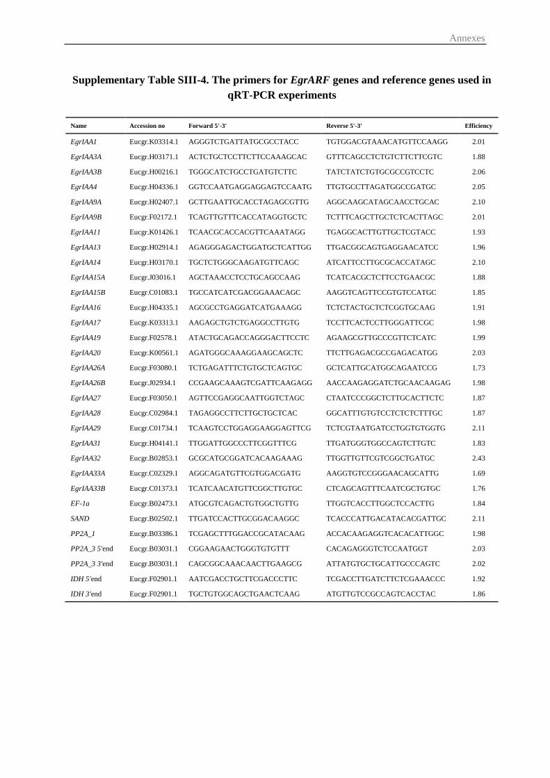
Annexes
Supplementary Table SIII-4. The primers for EgrARF genes and reference genes used in
qRT-PCR experiments
Name Accession no Forward 5'-3' Reverse 5'-3' Efficiency
EgrIAA1 Eucgr.K03314.1 AGGGTCTGATTATGCGCCTACC TGTGGACGTAAACATGTTCCAAGG 2.01
EgrIAA3A Eucgr.H03171.1 ACTCTGCTCCTTCTTCCAAAGCAC GTTTCAGCCTCTGTCTTCTTCGTC 1.88
EgrIAA3B Eucgr.H00216.1 TGGGCATCTGCCTGATGTCTTC TATCTATCTGTGCGCCGTCCTC 2.06
EgrIAA4 Eucgr.H04336.1 GGTCCAATGAGGAGGAGTCCAATG TTGTGCCTTAGATGGCCGATGC 2.05
EgrIAA9A Eucgr.H02407.1 GCTTGAATTGCACCTAGAGCGTTG AGGCAAGCATAGCAACCTGCAC 2.10
EgrIAA9B Eucgr.F02172.1 TCAGTTGTTTCACCATAGGTGCTC TCTTTCAGCTTGCTCTCACTTAGC 2.01
EgrIAA11 Eucgr.K01426.1 TCAACGCACCACGTTCAAATAGG TGAGGCACTTGTTGCTCGTACC 1.93
EgrIAA13 Eucgr.H02914.1 AGAGGGAGACTGGATGCTCATTGG TTGACGGCAGTGAGGAACATCC 1.96
EgrIAA14 Eucgr.H03170.1 TGCTCTGGGCAAGATGTTCAGC ATCATTCCTTGCGCACCATAGC 2.10
EgrIAA15A Eucgr.J03016.1 AGCTAAACCTCCTGCAGCCAAG TCATCACGCTCTTCCTGAACGC 1.88
EgrIAA15B Eucgr.C01083.1 TGCCATCATCGACGGAAACAGC AAGGTCAGTTCCGTGTCCATGC 1.85
EgrIAA16 Eucgr.H04335.1 AGCGCCTGAGGATCATGAAAGG TCTCTACTGCTCTCGGTGCAAG 1.91
EgrIAA17 Eucgr.K03313.1 AAGAGCTGTCTGAGGCCTTGTG TCCTTCACTCCTTGGGATTCGC 1.98
EgrIAA19 Eucgr.F02578.1 ATACTGCAGACCAGGGACTTCCTC AGAAGCGTTGCCCGTTCTCATC 1.99
EgrIAA20 Eucgr.K00561.1 AGATGGGCAAAGGAAGCAGCTC TTCTTGAGACGCCGAGACATGG 2.03
EgrIAA26A Eucgr.F03080.1 TCTGAGATTTCTGTGCTCAGTGC GCTCATTGCATGGCAGAATCCG 1.73
EgrIAA26B Eucgr.J02934.1 CCGAAGCAAAGTCGATTCAAGAGG AACCAAGAGGATCTGCAACAAGAG 1.98
EgrIAA27 Eucgr.F03050.1 AGTTCCGAGGCAATTGGTCTAGC CTAATCCCGGCTCTTGCACTTCTC 1.87
EgrIAA28 Eucgr.C02984.1 TAGAGGCCTTCTTGCTGCTCAC GGCATTTGTGTCCTCTCTCTTTGC 1.87
EgrIAA29 Eucgr.C01734.1 TCAAGTCCTGGAGGAAGGAGTTCG TCTCGTAATGATCCTGGTGTGGTG 2.11
EgrIAA31 Eucgr.H04141.1 TTGGATTGGCCCTTCGGTTTCG TTGATGGGTGGCCAGTCTTGTC 1.83
EgrIAA32 Eucgr.B02853.1 GCGCATGCGGATCACAAGAAAG TTGGTTGTTCGTCGGCTGATGC 2.43
EgrIAA33A Eucgr.C02329.1 AGGCAGATGTTCGTGGACGATG AAGGTGTCCGGGAACAGCATTG 1.69
EgrIAA33B Eucgr.C01373.1 TCATCAACATGTTCGGCTTGTGC CTCAGCAGTTTCAATCGCTGTGC 1.76
EF-1a Eucgr.B02473.1 ATGCGTCAGACTGTGGCTGTTG TTGGTCACCTTGGCTCCACTTG 1.84
SAND Eucgr.B02502.1 TTGATCCACTTGCGGACAAGGC TCACCCATTGACATACACGATTGC 2.11
PP2A_1 Eucgr.B03386.1 TCGAGCTTTGGACCGCATACAAG ACCACAAGAGGTCACACATTGGC 1.98
PP2A_3 5'end Eucgr.B03031.1 CGGAAGAACTGGGTGTGTTT CACAGAGGGTCTCCAATGGT 2.03
PP2A_3 3'end Eucgr.B03031.1 CAGCGGCAAACAACTTGAAGCG ATTATGTGCTGCATTGCCCAGTC 2.02
IDH 5'end Eucgr.F02901.1 AATCGACCTGCTTCGACCCTTC TCGACCTTGATCTTCTCGAAACCC 1.92
IDH 3'end Eucgr.F02901.1 TGCTGTGGCAGCTGAACTCAAG ATGTTGTCCGCCAGTCACCTAC 1.86
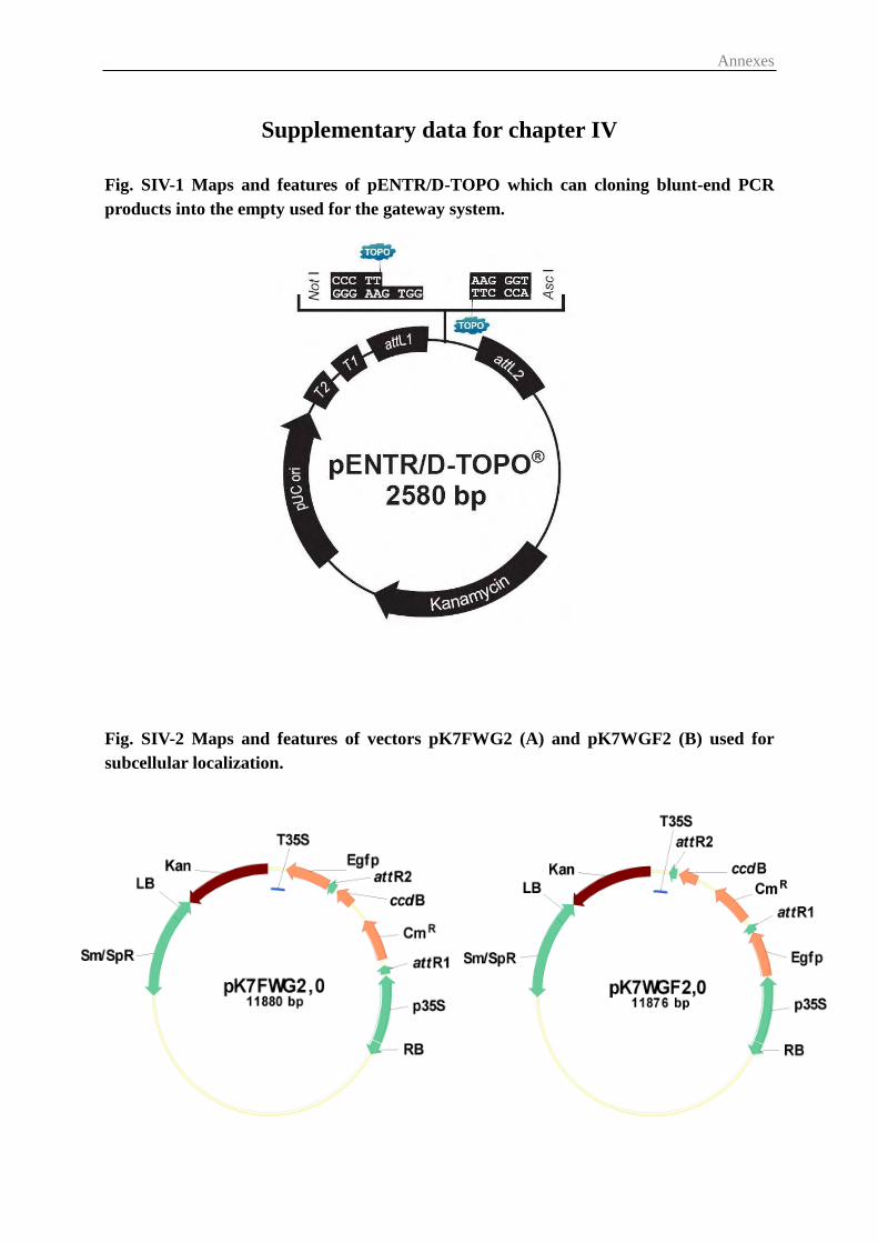
Annexes
Supplementary data for chapter IV
Fig. SIV-1 Maps and features of pENTR/D-TOPO which can cloning blunt-end PCR
products into the empty used for the gateway system.
Fig. SIV-2 Maps and features of vectors pK7FWG2 (A) and pK7WGF2 (B) used for
subcellular localization.
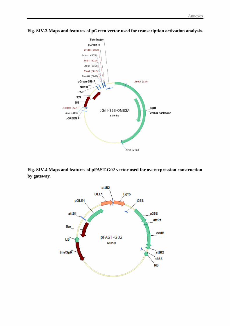
Annexes
pGrII-35S-OMEGA
5346 bpVector backbone
NptI
pGREEN F
pGreen R
pGreen-35S-F
New-R
35-F
35S
35S
Terminator
ApaLI (315)
EcoRI (5056)
HindIII (4174)
Sma I (5014)
XmaI (5012)
BamHI (5007)
BamHI (5016)
AvaI (2457)
AvaI (4153)
AvaI (5012)
Fig. SIV-3 Maps and features of pGreen vector used for transcription activation analysis.
Fig. SIV-4 Maps and features of pFAST-G02 vector used for overexpression construction
by gateway.
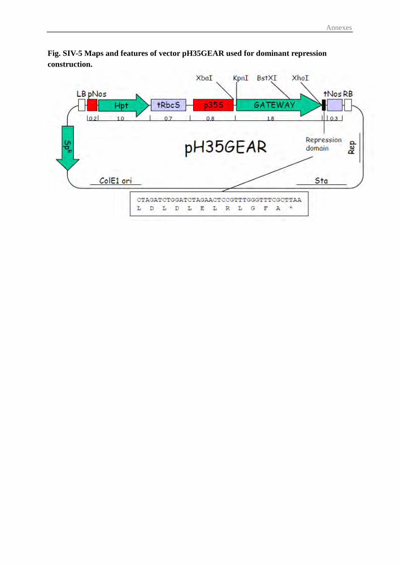
Annexes
Fig. SIV-5 Maps and features of vector pH35GEAR used for dominant repression
construction.
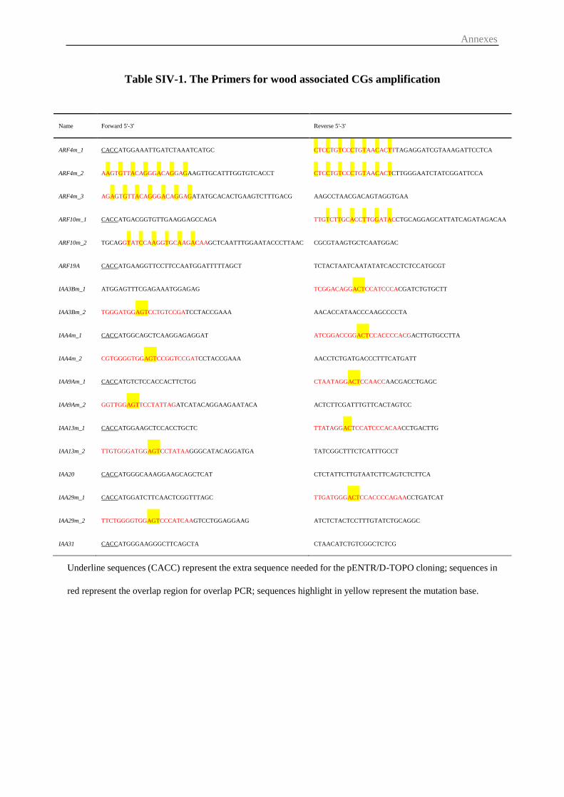
Annexes
Table SIV-1. The Primers for wood associated CGs amplification
Name Forward 5'-3' Reverse 5'-3'
ARF4m_1 CACCATGGAAATTGATCTAAATCATGC CTCCTGTCCCTGTAACACTTTAGAGGATCGTAAAGATTCCTCA
ARF4m_2 AAGTGTTACAGGGACAGGAGAAGTTGCATTTGGTGTCACCT CTCCTGTCCCTGTAACACTCTTGGGAATCTATCGGATTCCA
ARF4m_3 AGAGTGTTACAGGGACAGGAGATATGCACACTGAAGTCTTTGACG AAGCCTAACGACAGTAGGTGAA
ARF10m_1 CACCATGACGGTGTTGAAGGAGCCAGA TTGTCTTGCACCTTGGATACCTGCAGGAGCATTATCAGATAGACAA
ARF10m_2 TGCAGGTATCCAAGGTGCAAGACAAGCTCAATTTGGAATACCCTTAAC CGCGTAAGTGCTCAATGGAC
ARF19A CACCATGAAGGTTCCTTCCAATGGATTTTTAGCT TCTACTAATCAATATATCACCTCTCCATGCGT
IAA3Bm_1 ATGGAGTTTCGAGAAATGGAGAG TCGGACAGGACTCCATCCCACGATCTGTGCTT
IAA3Bm_2 TGGGATGGAGTCCTGTCCGATCCTACCGAAA AACACCATAACCCAAGCCCCTA
IAA4m_1 CACCATGGCAGCTCAAGGAGAGGAT ATCGGACCGGACTCCACCCCACGACTTGTGCCTTA
IAA4m_2 CGTGGGGTGGAGTCCGGTCCGATCCTACCGAAA AACCTCTGATGACCCTTTCATGATT
IAA9Am_1 CACCATGTCTCCACCACTTCTGG CTAATAGGACTCCAACCAACGACCTGAGC
IAA9Am_2 GGTTGGAGTTCCTATTAGATCATACAGGAAGAATACA ACTCTTCGATTTGTTCACTAGTCC
IAA13m_1 CACCATGGAAGCTCCACCTGCTC TTATAGGACTCCATCCCACAACCTGACTTG
IAA13m_2 TTGTGGGATGGAGTCCTATAAGGGCATACAGGATGA TATCGGCTTTCTCATTTGCCT
IAA20 CACCATGGGCAAAGGAAGCAGCTCAT CTCTATTCTTGTAATCTTCAGTCTCTTCA
IAA29m_1 CACCATGGATCTTCAACTCGGTTTAGC TTGATGGGACTCCACCCCAGAACCTGATCAT
IAA29m_2 TTCTGGGGTGGAGTCCCATCAAGTCCTGGAGGAAG ATCTCTACTCCTTTGTATCTGCAGGC
IAA31 CACCATGGGAAGGGCTTCAGCTA CTAACATCTGTCGGCTCTCG
Underline sequences (CACC) represent the extra sequence needed for the pENTR/D-TOPO cloning; sequences in
red represent the overlap region for overlap PCR; sequences highlight in yellow represent the mutation base.
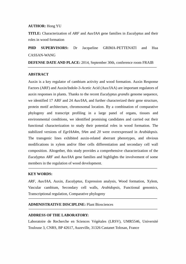
AUTHOR: Hong YU
TITLE: Characterization of ARF and Aux/IAA gene families in Eucalyptus and their
roles in wood formation
PHD SUPERVISORS: Dr Jacqueline GRIMA-PETTENATI and Hua
CASSAN-WANG
DEFENSE DATE AND PLACE: 2014, September 30th, conference room FRAIB
ABSTRACT
Auxin is a key regulator of cambium activity and wood formation. Auxin Response
Factors (ARF) and Auxin/Indole-3-Acetic Acid (Aux/IAA) are important regulators of
auxin responses in plants. Thanks to the recent Eucalyptus grandis genome sequence,
we identified 17 ARF and 24 Aux/IAA, and further characterized their gene structure,
protein motif architecture, chromosomal location. By a combination of comparative
phylogeny and transcript profiling in a large panel of organs, tissues and
environmental conditions, we identified promising candidates and carried out their
functional characterization to study their potential roles in wood formation. The
stabilized versions of EgrIAA4m, 9Am and 20 were overexpressed in Arabidopsis.
The transgenic lines exhibited auxin-related aberrant phenotypes, and obvious
modifications in xylem and/or fiber cells differentiation and secondary cell wall
composition. Altogether, this study provides a comprehensive characterization of the
Eucalyptus ARF and Aux/IAA gene families and highlights the involvement of some
members in the regulation of wood development.
KEY WORDS:
ARF, Aux/IAA, Auxin, Eucalyptus, Expression analysis, Wood formation, Xylem,
Vascular cambium, Secondary cell walls, Arabidopsis, Functional genomics,
Transcriptional regulation, Comparative phylogeny
ADMINISTRATIVE DISCIPLINE: Plant Biosciences
ADDRESS OF THE LABORATORY:
Laboratoire de Recherche en Sciences Végétales (LRSV), UMR5546, Université
Toulouse 3, CNRS, BP 42617, Auzeville, 31326 Castanet Tolosan, France
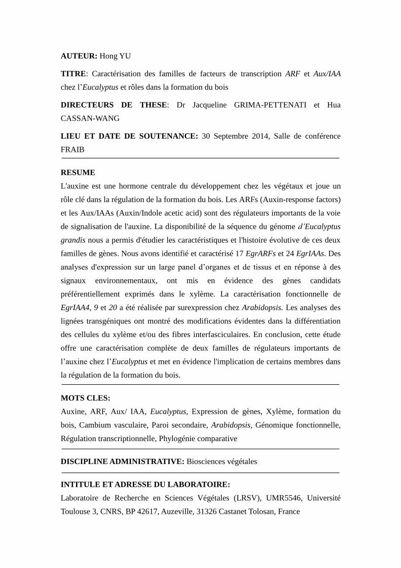
AUTEUR: Hong YU
TITRE: Caractérisation des familles de facteurs de transcription ARF et Aux/IAA
chez l’Eucalyptus et rôles dans la formation du bois
DIRECTEURS DE THESE: Dr Jacqueline GRIMA-PETTENATI et Hua
CASSAN-WANG
LIEU ET DATE DE SOUTENANCE: 30 Septembre 2014, Salle de conférence
FRAIB
RESUME
L'auxine est une hormone centrale du développement chez les végétaux et joue un
rôle clé dans la régulation de la formation du bois. Les ARFs (Auxin-response factors)
et les Aux/IAAs (Auxin/Indole acetic acid) sont des régulateurs importants de la voie
de signalisation de l'auxine. La disponibilité de la séquence du génome d’Eucalyptus
grandis nous a permis d'étudier les caractéristiques et l'histoire évolutive de ces deux
familles de gènes. Nous avons identifié et caractérisé 17 EgrARFs et 24 EgrIAAs. Des
analyses d'expression sur un large panel d’organes et de tissus et en réponse à des
signaux environnementaux, ont mis en évidence des gènes candidats
préférentiellement exprimés dans le xylème. La caractérisation fonctionnelle de
EgrIAA4, 9 et 20 a été réalisée par surexpression chez Arabidopsis. Les analyses des
lignées transgéniques ont montré des modifications évidentes dans la différentiation
des cellules du xylème et/ou des fibres interfasciculaires. En conclusion, cette étude
offre une caractérisation complète de deux familles de régulateurs importants de
l’auxine chez l’Eucalyptus et met en évidence l'implication de certains membres dans
la régulation de la formation du bois.
MOTS CLES:
Auxine, ARF, Aux/ IAA, Eucalyptus, Expression de gènes, Xylème, formation du
bois, Cambium vasculaire, Paroi secondaire, Arabidopsis, Génomique fonctionnelle,
Régulation transcriptionnelle, Phylogénie comparative
DISCIPLINE ADMINISTRATIVE: Biosciences végétales
INTITULE ET ADRESSE DU LABORATOIRE:
Laboratoire de Recherche en Sciences Végétales (LRSV), UMR5546, Université
Toulouse 3, CNRS, BP 42617, Auzeville, 31326 Castanet Tolosan, France