Embed Size (px)
Citation preview
Analysis of TGFBI gene mutations in Chinese patients with cornealdystrophies and review of the literature
Juhua Yang,1,2 Xiaoli Han,3 Dinggou Huang,3 Lin Yu,4 Yihua Zhu,3 Yi Tong,4 Binliang Zhu,5 Chuanbao Li,6
Mingshe Weng,7 Xu Ma2
(The first three authors contribute equally to the work)
1Biomedical Engineering Center, Fujian Medical University, Fuzhou, Fujian, China; 2Department of Genetics, National ResearchInstitute for Family Planning, Peking Union Medical College, Beijing, China; 3Department of Ophthalmology, The First AffiliatedHospital of Fujian Medical University, Fuzhou, Fujian, China; 4Fuzhou Southeast Eye Hospital, Fuzhou, Fujian, China;5Department of Ophthalmology, The First Hospital of Xi'an, Xi’an, Shanxi, China; 6Department of Ophthalmology, AffiliatedHospital of Jining Medical college, Jining, Shandong,China; 7Department of Ophthalmology, Jianou City Hospital, Jianou, Fujian,China
Purpose: To analyze human transforming growth factor b-induced (TGFBI) gene mutations in Chinese patients withcorneal dystrophies (CDs).Methods: Twenty-one families with corneal dystrophies were subjected to phenotypic and genotypic characterization.The corneal phenotypes of patients were documented by slit lamp photography. Mutation screening of the coding regionsof TGFBI was performed by direct sequencing. An additional 43 families and 3 sporadic patients with TGFBI dystrophiesfrom China reported in the literature were reviewed.Results: Five mutations of TGFBI were identified in 21 families with CDs, including one novel small deletion mutation,c.△1838–1849 (p.Δ613–616VAEP), responsible for one variant lattice CD (LCD) family and 4 known mutations, R555Wmutation for 10 granular cornea dystrophy type I (GCD1) families, R124H for 5 GCD type II (GCD2), R124C for 4 LCD1,and H626R for one variant LCD. In a cohort of Chinese patients (n=355) with TGFBI dystrophies from 64 families and3 sporadic cases, 19 distinct mutations were found in several different CD subtypes. The 3 most common phenotypeswere ranked as follows: GCD1, GCD2, and LCD1. Mutation hot spots at R124 and R555 occurred in >80% of thesefamilies.Conclusions: Our findings extend the mutational spectrum of TFGBI, and this is also the first extensively delineatedTGFBI mutation profile associated with the various corneal dystrophies in the Chinese population.
Corneal dystrophies (CDs) are characterized by theoccurrence of bilateral progressive opacities of the cornea thatoften arise from the deposition of insoluble material in thecorneal stroma and lead to visual impairment. Since 1997,multiple phenotypes have been identified that are related toallelic mutations of the human transforming growth factor b-induced (TGFBI) gene (OMIM 601692) on humanchromosome 5q31 [1]. This gene, originally named beta-ig-h3 (BIGH3), comprises 17 exons coding for a unique proteinof 683-amino acids, which is produced by both mesenchymaland epithelial cells and denoted as keratoepithelin [1]. Atpresent, more than 50 distinct disease-causing mutations havebeen identified in TFGBI that are associated with differentcorneal dystrophies, including granular cornea dystrophy(GCD) type I (GCD1; OMIM 121900), GCD type II (GCD2;
Correspondence to: Professor Xu Ma, Department of Genetics,National Research Institute for Family Planning, Peking UnionMedical College, 12 Da-hui-si, Hai Dian, Beijing, China, 100081;Phone: +86 1062179059; FAX: +86 10 62179059; email:[email protected]
OMIM 607541), lattice corneal dystrophy type I(LCD1;OMIM 122200), variant LCD, Thiel-Behnke cornealdystrophy (CDTB; OMIM 602082), Reis-Buckler cornealdystrophy (CDRB; OMIM 608470), and epithelial basementmembrane corneal dystrophy (EBMD; OMIM 121820). In thepresent study, we designated all of the cornea dystrophiesassociated with TGFBI mutations as TGFBI dystrophies.
Of these mutations, two mutational hot spots have beenidentified corresponding to R124 and R555 of the TGFBIprotein as being the most frequent sites of mutation in variouspopulations [2-7]. Despite TGFBI dystrophies represent aclinically heterogeneous group of disorders, a strongcorrelation between specific mutations at these two positionsand the observed phenotypes has been observed: GCD1/R555W, CDTB/R555Q, LCD1/R124C, GCD2/R124H, andCDRB/R124L. These above CDs were also classified as theclassic form of TGFBI dystrophies [8].
The TGFBI mutation spectrum and their clinicalconsequences have been investigated in patients with CDs indifferent ethnic populations [2-7]. However, the spectrum ofTGFBI mutations and the correlation between genotype and
Molecular Vision 2010; 16:1186-1193 <http://www.molvis.org/molvis/v16/a132>Received 11 March 2010 | Accepted 25 June 2010 | Published 30 June 2010
© 2010 Molecular Vision
1186
phenotype in the Chinese population has not been studiedextensively. In this study, we report our findings on 21 newCD families with TGFBI mutations, and an additional 43families and 3 sporadic patients with TGFBI dystrophies in aChinese population collected from the literature [9-31], anddelineate extensively the TGFBI mutation profile associatedwith the various corneal dystrophies in China.
METHODSPatients and subjects: As part of a genetic screening programfor genetic eye disorders, we collected materials from 26families diagnosed with corneal dystrophies from China. Aslit-lamp examination was performed for all participatingindividuals to determine if they were affected or unaffectedwith corneal dystrophies and to determine the diseasephenotype. Of these families, 21 were further classified into3 diagnostic categories: GCD1 in 10 families, LCD in 6families, and GCD2 in 5 families. The pedigrees of these 21families are shown in Figure 1, and the slit-lamp photographsof the representative patients with CDs are shown in Figure 2.After informed consent conforming to the tenets of theDeclaration of Helsinki and following the guidance of samplecollection of National Infrastructure Program of ChineseGenetic Resources (NICGR), 112 blood samples (58 samplesfrom patients and 54 from normal members of families) fromthe 21 families and other 50 samples from ethnically matchedcontrol individuals were obtained before the study.
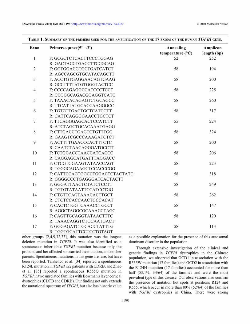
Genetic analysis: Total genomic DNA was extracted fromwhole blood using the Wizard Genomic DNA Purification Kit(Promega, Beijing, China) according to the manufacturer’sinstructions. Amplification of the coding regions of TGFBIfrom genomic DNA was performed with the primers asdescribed previously [1,2,22]. The sequences of the primersare listed in Table 1. Each PCR reaction was carried out in a50 µl reaction mixture containing 100-200 ng of genomicDNA, 0.25 µl of TaKaRa Ex TaqTM (5U/µl; TaKaRaBiotechnology Co., Ltd., Dalian, China), 4.0 µl of dNTPs (2.5mM each), 1.0 µl of each primer (10 pmol), 5 µl of 10× ExTaq Buffer (Mg2+ plus). PCR conditions were as follows: 94°C for 3 min; 30 cycles of 94 °C for 30 s, 52 °C or 55 °C or58 °C for 30 s, 72 °C for 1 min; and further extension step at72 °C for 5 min. PCR products were purified and sequencedon an ABI 3730XL Automated Sequencer (PE Biosystems,Foster City, CA), using the same PCR forward primers.
Publications selection: To evaluate the incidence of TFGBImutations in Chinese patients with TGFBI dystrophies, wealso reviewed published articles obtained by an electronicsearch on PubMed/MEDLINE, VIP Information, CNKI,WANGFANG, data and Google using the keywords “cornealdystrophy” and “TGFBI” or “BIGH3.” Articles were limitedto full papers in the English or Chinese language literature,and the search ended in February, 2010. When the same authorreported results obtained on a complete or partial similar
patient population in several publications, only the mostrecent or the most complete report was included in the analysisto avoid overlap. According to these criteria, 23 eligible trialspublished between 2002 and 2010 were selected [9-31]. Thearticles reported mutations of the TGFBI/BIGH3 geneassociated with corneal dystrophies in the Chinese population.All of these TGFBI dystrophies were reclassified as classic orvariant form according to Aldave and Sonmez’s [8] criterion.
RESULTSMutational analysis: We screened the coding regions ofTGFBI using PCR-based amplification followed by directsequencing. Mutations were identified in all patients from the21 unrelated Chinese families (Figure 1). Five distinctmutations of TGFBI were identified, including one novelsmall deletion mutation, c.Δ1838–1849 (p.Δ613–616VAEP),in exon 14 responsible for the HCD-022 family with variantLCD (Figure 1E and Figure 2I). This mutation was found inthe proband and her son, but not in her parents and 50 normalcontrols. The other four known mutations were R555Wmutation for 10 GCD1 families (Figure 1A and Figure 2A-C),R124H for 5 GCD2 families (Figure 1B and Figure 2D),R124C for 4 LCD1 families (Figure 1C and Figure 2G) andH626R for one variant LCD family (Figure 1D and Figure2H).Profile of TGFBI dystrophies: To delineate the TGFBImutation profile associated with the various cornealdystrophies in the Chinese population, an additional 43families and 3 sporadic cases with TGFBI dystrophiescollected from the literature were also included in theinvestigation. Thus, a total of 355 patients from 64 familiesand 3 sporadic patients were included in the statisticalanalysis. We reclassified these TGFBI dystrophies as classicor variant form, with GCD1/R555W, GCD2/R124H, LCD1/R124C, CDRB/R124L, and CDTB/R555Q as classic formsand the others as corresponding variants [8].
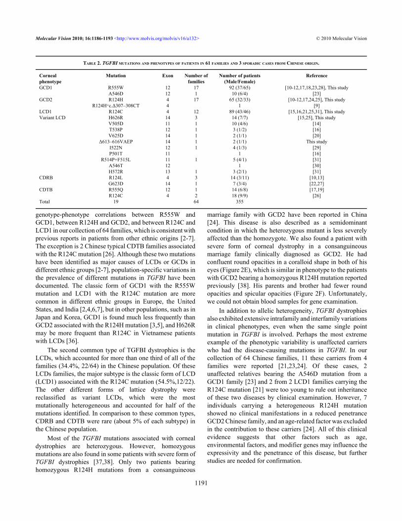
In this cohort of Chinese patients, 19 distinct TGFBImutations were identified in several different CD subtypes(Table 2). The most common phenotypes were the GCD1associated with the R555W mutation (26.6%, 17/64 families),GCD2 with the R124H mutation (26.6%,17/64families), andLCD1 with the R124C mutation (18.8%,12/64 families). Themutational hot spots at positions R124 and R555 occurred in53.1% (34/64) and 28.1% (18/64) of the families,respectively. Our observation also showed that the phenotypescorrelated strongly with specific mutations in TGFBI,including that between GCD1 and R555W, GCD2 andR124H, and LCD1 and R124C. The exception is two CDTBfamilies associated with the R124C mutation [26]. In addition,more than half (11/19) of these mutations were observed inChinese patients with variant LCDs (Table 2).
DISCUSSIONWe searched for mutations of TGFBI in 21 unrelated Chinesefamilies affected with GCDs or LCDs. Five distinct mutations
Molecular Vision 2010; 16:1186-1193 <http://www.molvis.org/molvis/v16/a132> © 2010 Molecular Vision
1187
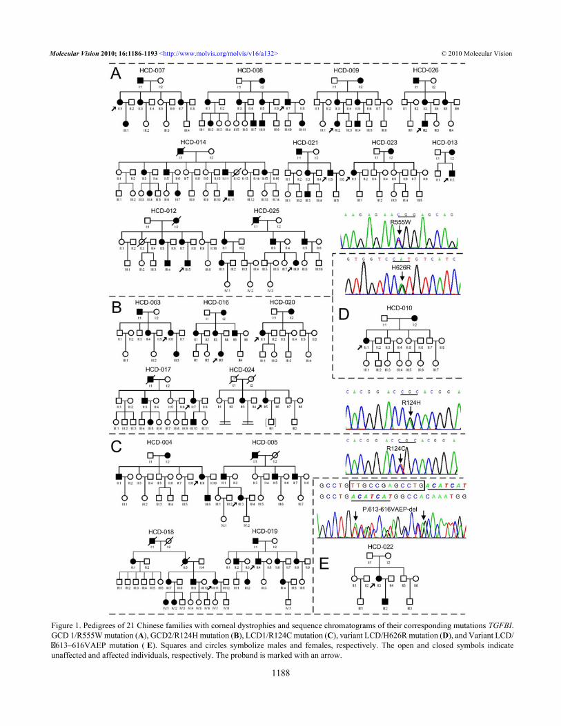
Figure 1. Pedigrees of 21 Chinese families with corneal dystrophies and sequence chromatograms of their corresponding mutations TGFBI.GCD 1/R555W mutation (A), GCD2/R124H mutation (B), LCD1/R124C mutation (C), variant LCD/H626R mutation (D), and Variant LCD/�613–616VAEP mutation ( E). Squares and circles symbolize males and females, respectively. The open and closed symbols indicateunaffected and affected individuals, respectively. The proband is marked with an arrow.
Molecular Vision 2010; 16:1186-1193 <http://www.molvis.org/molvis/v16/a132> © 2010 Molecular Vision
1188
of TGFBI were identified in all of the families (Figure 1), andthere have been perfect correlations between GCD1 andR555W, between GCD2 (ACD) and R124H, and betweenLCD1 and R124C. Among of these mutations, one novel 12
bp deletion mutation, c.Δ1838–1849 (p.Δ613–616VAEP), inexon 14 was found in the HCD-022 family (Figure 1E) withvariant LCD (Figure 2I). Compare with the five small deletionmutations (1~6 bp deleted) in TGFBI reported previously by
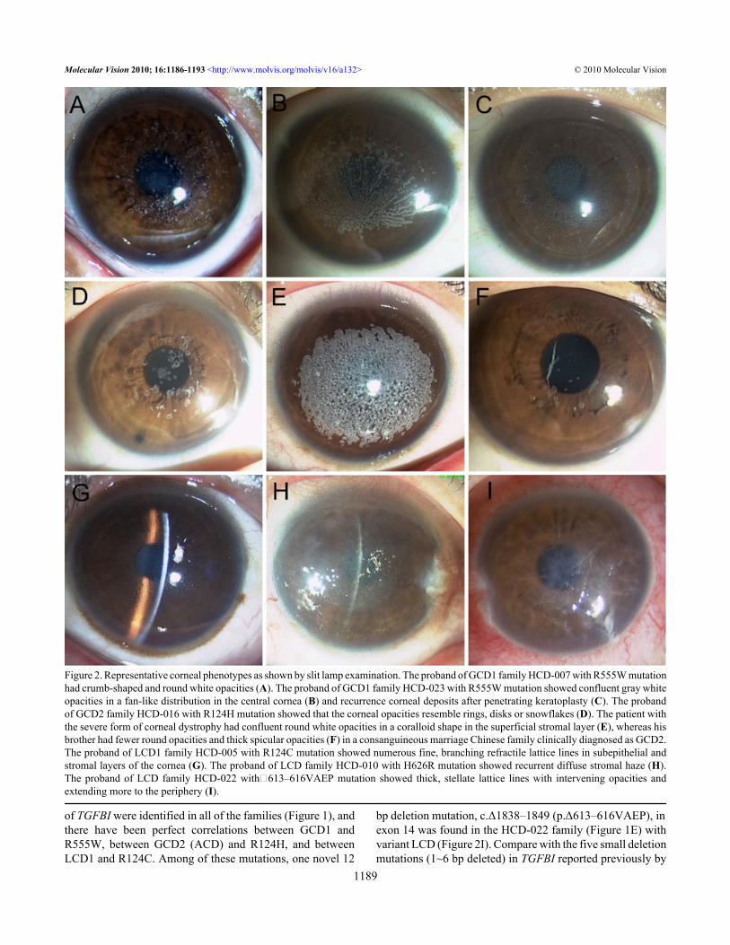
Figure 2. Representative corneal phenotypes as shown by slit lamp examination. The proband of GCD1 family HCD-007 with R555W mutationhad crumb-shaped and round white opacities (A). The proband of GCD1 family HCD-023 with R555W mutation showed confluent gray whiteopacities in a fan-like distribution in the central cornea (B) and recurrence corneal deposits after penetrating keratoplasty (C). The probandof GCD2 family HCD-016 with R124H mutation showed that the corneal opacities resemble rings, disks or snowflakes (D). The patient withthe severe form of corneal dystrophy had confluent round white opacities in a coralloid shape in the superficial stromal layer (E), whereas hisbrother had fewer round opacities and thick spicular opacities (F) in a consanguineous marriage Chinese family clinically diagnosed as GCD2.The proband of LCD1 family HCD-005 with R124C mutation showed numerous fine, branching refractile lattice lines in subepithelial andstromal layers of the cornea (G). The proband of LCD family HCD-010 with H626R mutation showed recurrent diffuse stromal haze (H).The proband of LCD family HCD-022 with△613–616VAEP mutation showed thick, stellate lattice lines with intervening opacities andextending more to the periphery (I).
Molecular Vision 2010; 16:1186-1193 <http://www.molvis.org/molvis/v16/a132> © 2010 Molecular Vision
1189
other groups [2,4,9,32,33], this mutation was the longestdeletion mutation in TGFBI. It was also identified as aspontaneous inheritable TGFBI mutation because only theproband and her affected son carried the mutation, and not herparents. Spontaneous mutations in this gene are rare, but havebeen reported. Tanhehco et al. [34] reported a spontaneousR124L mutation in TGFBI in 2 patients with CDRB, and Zhaoet al. [35] reported a spontaneous R555Q mutation inTGFBI in two unrelated families with Bowman's layer cornealdystrophies (CDTB and CDRB). Our finding not only extendsthe mutational spectrum of TFGBI, but also has historic value
as a possible explanation for the presence of this autosomaldominant disorder in the population.
Through extensive investigation of the clinical andgenetic findings in TGFBI dystrophies in the Chinesepopulation, we observed that GCD1 in association with theR555W mutation (17 families) and GCD2 in association withthe R124H mutation (17 families) accounted for more thanhalf (53.1%, 34/64) of the families and were the mostprevalent type of this disease. Our observations also confirmthe presence of mutation hot spots at positions R124 andR555, which occur in more than 80% (52/64) of the familieswith TGFBI dystrophies in China. There were strong
TABLE 1. SUMMARY OF THE PRIMERS USED FOR THE AMPLIFICATION OF THE 17 EXONS OF THE HUMAN TGFBI GENE.
Exon Primersequnce(5’→3') Annealingtemperature (°C)
Ampliconlength (bp)
1 F: GCGCTCTCACTTCCCTGGAG 52 252 R: GACTACCTGACCTTCCGCAG 2 F: GGTGGACGTGCTGATCATCT 58 194 R: AGCCAGCGTGCATACAGCTT 3 F: ACCTGTGAGGAACAGTGAAG 58 200 R: GCCTTTTATGTGGGTACTCC 4 F: CCCCAGAGGCCATCCCTCCT 58 225 R: CCGGGCAGACGGAGGTCATC 5 F: TAAACACAGAGTCTGCAGCC 58 260 R: TTCATTATGCACCAAGGGCC 6 F: TGTGTTGACTGCTCATCCTT 58 317 R: CATTCAGGGGAACCTGCTCT 7 F: TTCAGGGAGCACTCCATCTT 55 224 R: ATCTAGCTGCACAAATGAGG 8 F: CTTGACCTGAGTCTGTTTGG 58 324 R: GAAGTCGCCCAAAGATCTCT 9 F: ACTTTTGAACCCACTTTCTC 58 200 R: CAATCTAACAGGGATGCCTT
10 F: TCTGGACCTAACCATCACCC 58 206 R: CAGGAGCATGATTTAGGACC
11 F: CTCGTGGAAGTATAACCAGT 58 223 R: TGGGCAGAAGCTCCACCCGG
12 F: CATTCCAGTGGCCTGGACTCTACTATC 58 318 R: GGGGCCCTGAGGGATCACTACTT
13 F: GGGATTAACTCTATCTCCTT 58 249 R: TGTGTATAATTCCATCCTGG
14 F: CTGTTCAGTAAACACTTGCT 58 262 R: CTCTCCACCAACTGCCACAT
15 F: CACTCTGGTCAAACCTGCCT 58 147 R: AGGCTAGGCGCAAACCTAGC
16 F: CAGTTGCAGGTATAACTTTC 58 120 R: TAAACAGGTCTGCAATGACT
17 F: GGGAGATCTGCACCTATTTG 58 113 R: TGGTGCATTCCTCCTGTAGT
Molecular Vision 2010; 16:1186-1193 <http://www.molvis.org/molvis/v16/a132> © 2010 Molecular Vision
1190
genotype-phenotype correlations between R555W andGCD1, between R124H and GCD2, and between R124C andLCD1 in our collection of 64 families, which is consistent withprevious reports in patients from other ethnic origins [2-7].The exception is 2 Chinese typical CDTB families associatedwith the R124C mutation [26]. Although these two mutationshave been identified as major causes of LCDs or GCDs indifferent ethnic groups [2-7], population-specific variations inthe prevalence of different mutations in TGFBI have beendocumented. The classic form of GCD1 with the R555Wmutation and LCD1 with the R124C mutation are morecommon in different ethnic groups in Europe, the UnitedStates, and India [2,4,6,7], but in other populations, such as inJapan and Korea, GCD1 is found much less frequently thanGCD2 associated with the R124H mutation [3,5], and H626Rmay be more frequent than R124C in Vietnamese patientswith LCDs [36].
The second common type of TGFBI dystrophies is theLCDs, which accounted for more than one third of all of thefamilies (34.4%, 22/64) in the Chinese population. Of theseLCDs families, the major subtype is the classic form of LCD(LCD1) associated with the R124C mutation (54.5%,12/22).The other different forms of lattice dystrophy werereclassified as variant LCDs, which were the mostmutationally heterogeneous and accounted for half of themutations identified. In comparison to these common types,CDRB and CDTB were rare (about 5% of each subtype) inthe Chinese population.
Most of the TGFBI mutations associated with cornealdystrophies are heterozygous. However, homozygousmutations are also found in some patients with severe form ofTGFBI dystrophies [37,38]. Only two patients bearinghomozygous R124H mutations from a consanguineous
marriage family with GCD2 have been reported in China[24]. This disease is also described as a semidominantcondition in which the heterozygous mutant is less severelyaffected than the homozygote. We also found a patient withsevere form of corneal dystrophy in a consanguineousmarriage family clinically diagnosed as GCD2. He hadconfluent round opacities in a coralloid shape in both of hiseyes (Figure 2E), which is similar in phenotype to the patientswith GCD2 bearing a homozygous R124H mutation reportedpreviously [38]. His parents and brother had fewer roundopacities and spicular opacities (Figure 2F). Unfortunately,we could not obtain blood samples for gene examination.
In addition to allelic heterogeneity, TGFBI dystrophiesalso exhibited extensive intrafamily and interfamily variationsin clinical phenotypes, even when the same single pointmutation in TGFBI is involved. Perhaps the most extremeexample of the phenotypic variability is unaffected carrierswho had the disease-causing mutations in TGFBI. In ourcollection of 64 Chinese families, 11 these carriers from 4families were reported [21,23,24]. Of these cases, 2unaffected relatives bearing the A546D mutation from aGCD1 family [23] and 2 from 2 LCD1 families carrying theR124C mutation [21] were too young to rule out inheritanceof these two diseases by clinical examination. However, 7individuals carrying a heterogeneous R124H mutationshowed no clinical manifestations in a reduced penetranceGCD2 Chinese family, and an age-related factor was excludedin the contribution to these carriers [24]. All of this clinicalevidence suggests that other factors such as age,environmental factors, and modifier genes may influence theexpressivity and the penetrance of this disease, but furtherstudies are needed for confirmation.
TABLE 2. TGFBI MUTATIONS AND PHENOTYPES OF PATIENTS IN 61 FAMILIES AND 3 SPORADIC CASES FROM CHINESE ORIGIN.
Cornealphenotype
Mutation Exon Number offamilies
Number of patients(Male/Female)
Reference
GCD1 R555W 12 17 92 (37/65) [10-12,17,18,23,28], This study A546D 12 1 10 (6/4) [23]GCD2 R124H 4 17 65 (32/33) [10-12,17,24,25], This study R124H+c.Δ307–308CT 4 1 [9]LCD1 R124C 4 12 89 (43/46) [15,16,21,25,31], This studyVariant LCD H626R 14 3 14 (7/7) [15,25], This study V505D 11 1 10 (4/6) [14] T538P 12 1 3 (1/2) [16] V625D 14 1 2 (1/1) [20] Δ613–616VAEP 14 1 2 (1/1) This study I522N 12 1 4 (1/3) [29] P501T 11 1 [16] R514P+F515L 11 1 5 (4/1) [31] A546T 12 1 [30] H572R 13 1 3 (2/1) [31]CDRB R124L 4 3 14 (3/11) [10,13] G623D 14 1 7 (3/4) [22,27]CDTB R555Q 12 1 14 (6/8) [17,19] R124C 4 2 18 (9/9) [26]Total 19 64 355
Molecular Vision 2010; 16:1186-1193 <http://www.molvis.org/molvis/v16/a132> © 2010 Molecular Vision
1191
In summary, this study reported our findings in 21 newCD families with TGFBI mutations, and delineatedextensively the TGFBI mutation profile associated with thevarious corneal dystrophies in the Chinese population: bothGCD1 associated with the R555W mutation and GCD2 withthe R124H mutation are the most common forms of thisdisease. We also confirmed the presence of mutationalhotspots at positions R124 and R555, and a strong correlationbetween these two mutations and their phenotypes.
ACKNOWLEDGMENTSThe authors thank all patients and their family members fortaking part in this study. This work was supported by theNational Infrastructure Program of Chinese GeneticResources (2006DKA21301), National Science &Technology Pillar Program of China (2008BAH24B05),National Natural science Foundation of China (30872843),Natural Science Foundation of Fujian Province (C0810018),the Key Program of Scientific Research of Fujian MedicalUniversity (09ZD016–1), and Program for New CenturyExcellent Talents in Fujian Province University (JuhuaYang). Professor Xu Ma ([email protected]) and Dr.Juhua Yang ([email protected]) contributedequally to the research presented and can be considered asco-corresponding authors.
REFERENCES1. Munier FL, Korvatska E, Djemaï A, Le Paslier D, Zografos L,
Pescia G, Schorderet DF. Kerato-epithelin mutations in four5q31-linked corneal dystrophies. Nat Genet 1997;15:247-51. [PMID: 9054935]
2. Munier FL, Frueh BE, Othenin-Girard P, Uffer S, Cousin P,Wang MX, Héon E, Black GC, Blasi MA, Balestrazzi E,Lorenz B, Escoto R, Barraquer R, Hoeltzenbein M, Gloor B,Fossarello M, Singh AD, Arsenijevic Y, Zografos L,Schorderet DF. BIGH3 mutation spectrum in cornealdystrophies. Invest Ophthalmol Vis Sci 2002; 43:949-54.[PMID: 11923233]
3. Mashima Y, Yamamoto S, Inoue Y, Yamada M, Konishi M,Watanabe H, Maeda N, Shimomura Y, Kinoshita S.Association of autosomal dominantly inherited cornealdystrophies with BIGH3 gene mutations in Japan. Am JOphthalmol 2000; 130:516-7. [PMID: 11024425]
4. Chakravarthi SV, Kannabiran C, Sridhar MS, Vemuganti GK.TGFBI gene mutations causing lattice and granular cornealdystrophies in Indian patients. Invest Ophthalmol Vis Sci2005; 46:121-5. [PMID: 15623763]
5. Kim HS, Yoon SK, Cho BJ, Kim EK, Joo CK. BIGH3 genemutations and rapid detection in Korean patients with cornealdystrophy. Cornea 2001; 20:844-9. [PMID: 11685063]
6. Korvatska E, Munier FL, Djemaï A, Wang MX, Frueh B, ChiouAG, Uffer S, Ballestrazzi E, Braunstein RE, Forster RK,Culbertson WW, Boman H, Zografos L, Schorderet DF.Mutation hot spots in 5q31-linked corneal dystrophies. Am JHum Genet 1998; 62:320-4. [PMID: 9463327]
7. El-Ashry MF, Abd El-Aziz MM, Hardcastle AJ, BhattacharyaSS, Ebenezer ND. A Clinical and Molecular Genetic Studyof Autosomal-Dominant Stromal Corneal Dystrophy in
British Population. Ophthalmic Res 2005; 37:310-7. [PMID:16118514]
8. Aldave AJ, Sonmez B. Elucidating the Molecular Genetic Basisof the Corneal Dystrophies: Are We There Yet? ArchOphthalmol 2007; 125:177-86. [PMID: 17296893]
9. Pang CP, Lam DS. Differential occurrence of mutationscausative of eye diseases in the Chinese population. HumMutat 2002; 19:189-208. [PMID: 11857735]
10. Yu J, Zou LH, He JC, Liu NP, Zhang W, Lu L, Sun XG, DongDS, Wu YY, Yin XT. Analysis of mutation of BIGH3 genein Chinese patients with corneal dystrophies. Zhonghua YanKe Za Zhi 2003; 39:582-6. [PMID: 14766070]
11. Jin T, Zou LH, Yang L, Dong WL, Yu J, Lu L. Identificationof BIGH3 gene mutations in the patients with two types ofcorneal dystrophies. Zhonghua Yi Xue Yi Chuan Xue Za Zhi2004; 21:32-4. [PMID: 14767905]
12. Li Y, Sun XG, Ren HY, Dong B, Wang ZQ, Sun XY. Analysisof human transforming growth factor beta-induced genemutation in corneal dystrophy. Chin Med J (Engl) 2004;117:1418-21. [PMID: 15377440]
13. Tian X, Liu ZG, Li Q, Li B, Wang W, Xie PY, Fujiki K,Murakami A, Kanai A. Analysis of gene mutation in Chinesepatients with Reis-Bücklers corneal dystrophy. ZhonghuaYan Ke Za Zhi 2005; 41:239-42. [PMID: 15840366]
14. Tian X, Fujiki K, Wang W, Murakami A, Xie P, Kanai A, LiuZ. Novel mutation (V505D) of the TGFBI gene found in aChinese family with lattice corneal dystrophy, type I. Jpn JOphthalmol 2005; 49:84-8. [PMID: 15838722]
15. Dong WL, Zou LH, Pan ZQ, Jin T, Yu J. Molecular geneticstudy on patients with lattice corneal dystrophy in China.Zhonghua Yan Ke Za Zhi 2005; 41:523-6. [PMID: 16008913]
16. Yu P, Gu Y, Yang Y, Yan X, Chen L, Ge Z, Qi M, Si J, Guo L.A clinical and molecular-genetic analysis of Chinese patientswith lattice corneal dystrophy and novel Thr538Pro mutationin the TGFBI (BIGH3) gene. J Genet 2006; 85:73-6. [PMID:16809844]
17. Qi YH, He HD, Li Y, Lin H, Gu JZ, Su H, Huang SZ. A researchon TGFBI gene mutations in Chinese families with cornealdystrophies. Zhonghua Yi Xue Yi Chuan Xue Za Zhi 2006;23:310-2. [PMID: 16767671]
18. Yang J, Tong Y, Zhu Y, Xiao J, Chen Y, Lin J. Exon12 ofBIGH3 (R555W) mutation in granular corneal dystrophy ina Chinese kindred. J Fujian Medical University 2007;41:14-6.
19. Qi YH, He HD, Li Y, Wang L, Lin H, Su H, Gu JZ, Huang SZ.TGFBI gene mutation analysis in a Chinese family with Thiel-Behnke corneal dystrophy. Zhonghua Yan Ke Za Zhi 2007;43:718-21. [PMID: 18001570]
20. Tian X, Fujiki K, Zhang Y, Murakami A, Li Q, Kanai A, WangW, Hao Y, Ma Z. A novel variant lattice corneal dystrophycaused by association of mutation (V625D) in TGFBI gene.Am J Ophthalmol 2007; 144:473-5. [PMID: 17765440]
21. Liu Z, Wang YQ, Gong QH, Xie LX. An R124C mutation inTGFBI caused lattice corneal dystrophy type I with a variablephenotype in three Chinese families. Mol Vis 2008;14:1234-9. [PMID: 18615206]
22. Li D, Qi Y, Wang L, Lin H, Zhou N, Zhao L. An atypicalphenotype of Reis-Bücklers corneal dystrophy caused by theG623D mutation in TGFBI. Mol Vis 2008; 14:1298-302.[PMID: 18334967]
Molecular Vision 2010; 16:1186-1193 <http://www.molvis.org/molvis/v16/a132> © 2010 Molecular Vision
1192
23. Yu P, Gu Y, Jin F, Hu R, Chen L, Yan X, Yang Y, Qi M.p.Ala546 > Asp and p.Arg555 > Trp mutations of TGFBI geneand their clinical manifestations in two large Chinese familieswith granular corneal dystrophy type I. Genet Test 2008;12:421-5. [PMID: 18752451]
24. Cao W, Ge H, Cui X, Zhang L, Bai J, Fu S, Liu P. Reducedpenetrance in familial Avellino corneal dystrophy associatedwith TGFBI mutations. Mol Vis 2009; 15:70-5. [PMID:19145249]
25. Wang LM, Wang YC, Qiu DL, Ying M, Li ND. TGFBI genemutations in three Chinese families with autosomal dominantcorneal dystrophy. Zhonghua Yi Xue Yi Chuan Xue Za Zhi2009; 26:179-82. [PMID: 19350511]
26. Chang L, Zhiqun W, Shijing D, Chen Z, Qingfeng L, Li L,Xuguang S. Arg124Cys mutation of the TGFBI gene in 2Chinese families with Thiel-Behnke corneal dystrophy. ArchOphthalmol 2009; 127:641-4. [PMID: 19433713]
27. Li DD, Qi YH, Han Q, Lin H, Zhao LM, Zhang CM. Analysisof TGFBI gene mutation in a Chinese family with atypicalReis-Buckler corneal dystrophy. Zhonghua Yi Xue Yi ChuanXue Za Zhi 2009; 26:245-8. [PMID: 19504432]
28. Hu L, Xu F, Ma WJ, Zhang H, Sui RF. R555W mutation ofTGFbetaI related to granular corneal dystrophy in Chinesepatients. Chin Med J (Engl) 2009; 122:2691-4. [PMID:19951597]
29. Zhang C, Zeng G, Lin H, Li D, Zhao L, Zhou N, Qi Y. A novelmutation I522N within the TGFBI gene caused lattice cornealdystrophy I. Mol Vis 2009; 15:2498-502. [PMID: 19956413]
30. Chen L, Gu Y, Yu P. Application of molecular genetictechnique in classification of corneal dystrophy. Chin J PractOphthalmol 2006; 24:678-80.
31. Zhong X, Chen S, Huang W, Yang J, Chen X, Zhou Y, ZhouQ, Wang Y. Novel and known mutations of TGFBI, theirgenotype-phenotype correlation and structural modeling in 3Chinese families with lattice corneal dystrophy. Mol Vis2010; 16:224-30. [PMID: 20161820]
32. Aldave AJ, Rayner SA, Kim BT, Prechanond A, Yellore VS.Unilateral lattice corneal dystrophy associated with the novelHis572del mutation in the TGFBI gene. Mol Vis 2006;12:142-6. [PMID: 16541014]
33. Rozzo C, Fossarello M, Galleri G, Sole G, Serru A, Orzalesi N,Serra A, Pirastu M. A common beta ig-h3 gene mutation(delta f540) in a large cohort of Sardinian Reis Bücklerscorneal dystrophy patients. Mutations in brief no. 180. Online.Hum Mutat 1998; 12:215-6. [PMID: 10660331]
34. Tanhehco TY, Eifrig DE Jr, Schwab IR, Rapuano CJ,Klintworth GK. Two cases of Reis-Bucklers cornealdystrophy (granular corneal dystrophy type III) caused byspontaneous mutations in the TGFBI gene. Arch Ophthalmol2006; 124:589-93. [PMID: 16606891]
35. Zhao XC, Nakamura H, Subramanyam S, Stock LE, GilletteTE, Yoshikawa S, Ma X, Yee RW. Spontaneous andinheritable R555Q mutation in the TGFBI/BIGH3 gene in twounrelated families exhibiting Bowman's layer cornealdystrophy. Ophthalmology 2007; 114:e39-46. [PMID:17980739]
36. Chau HM, Ha NT, Cung LX, Thanh TK, Fujiki K, MurakamiA, Kanai A. H626R and R124C mutations of the TGFBI(BIGH3) gene caused lattice corneal dystrophy in Vietnamesepeople. Br J Ophthalmol 2003; 87:686-9. [PMID: 12770961]
37. Okada M, Yamamoto S, Watanabe H, Shimomura Y, Tano Y.Granular corneal dystrophy with homozygous mutations inthe kerato-epithelin gene. Am J Ophthalmol 2000;129:411-2. [PMID: 10755958]
38. Okada M, Yamamoto S, Inoue Y, Watanabe H, Maeda N,Shimomura Y, Ishii Y, Tano Y. Severe corneal dystrophyphenotype caused by homozygous R124H keratoepithelinmutations. Invest Ophthalmol Vis Sci 1998; 39:1947-53.[PMID: 9727418]
Molecular Vision 2010; 16:1186-1193 <http://www.molvis.org/molvis/v16/a132> © 2010 Molecular Vision
The print version of this article was created on 26 June 2010. This reflects all typographical corrections and errata to the articlethrough that date. Details of any changes may be found in the online version of the article.
1193