Embed Size (px)
Citation preview
OpenStax-CNX module: m46377 1
Anatomy of Selected Synovial
Joints*
OpenStax
This work is produced by OpenStax-CNX and licensed under the
Creative Commons Attribution License 3.0�
Abstract
By the end of this section, you will be able to:
• Describe the bones that articulate together to form selected synovial joints• Discuss the movements available at each joint• Describe the structures that support and prevent excess movements at each joint
Each synovial joint of the body is specialized to perform certain movements. The movements that areallowed are determined by the structural classi�cation for each joint. For example, a multiaxial ball-and-socket joint has much more mobility than a uniaxial hinge joint. However, the ligaments and muscles thatsupport a joint may place restrictions on the total range of motion available. Thus, the ball-and-socket jointof the shoulder has little in the way of ligament support, which gives the shoulder a very large range ofmotion. In contrast, movements at the hip joint are restricted by strong ligaments, which reduce its rangeof motion but confer stability during standing and weight bearing.
This section will examine the anatomy of selected synovial joints of the body. Anatomical names formost joints are derived from the names of the bones that articulate at that joint, although some joints, suchas the elbow, hip, and knee joints are exceptions to this general naming scheme.
1 Articulations of the Vertebral Column
In addition to being held together by the intervertebral discs, adjacent vertebrae also articulate with eachother at synovial joints formed between the superior and inferior articular processes called zygapophysialjoints (facet joints) (see ). These are plane joints that provide for only limited motions between the vertebrae.The orientation of the articular processes at these joints varies in di�erent regions of the vertebral columnand serves to determine the types of motions available in each vertebral region. The cervical and lumbarregions have the greatest ranges of motions.
In the neck, the articular processes of cervical vertebrae are �attened and generally face upward ordownward. This orientation provides the cervical vertebral column with extensive ranges of motion for �exion,extension, lateral �exion, and rotation. In the thoracic region, the downward projecting and overlappingspinous processes, along with the attached thoracic cage, greatly limit �exion, extension, and lateral �exion.However, the �attened and vertically positioned thoracic articular processes allow for the greatest rangeof rotation within the vertebral column. The lumbar region allows for considerable extension, �exion, andlateral �exion, but the orientation of the articular processes largely prohibits rotation.
*Version 1.4: Jun 28, 2013 10:04 am -0500�http://creativecommons.org/licenses/by/3.0/
http://cnx.org/content/m46377/1.4/
OpenStax-CNX module: m46377 2
The articulations formed between the skull, the atlas (C1 vertebra), and the axis (C2 vertebra) di�erfrom the articulations in other vertebral areas and play important roles in movement of the head. Theatlanto-occipital joint is formed by the articulations between the superior articular processes of the atlasand the occipital condyles on the base of the skull. This articulation has a pronounced U-shaped curvature,oriented along the anterior-posterior axis. This allows the skull to rock forward and backward, producing�exion and extension of the head. This moves the head up and down, as when shaking your head �yes.�
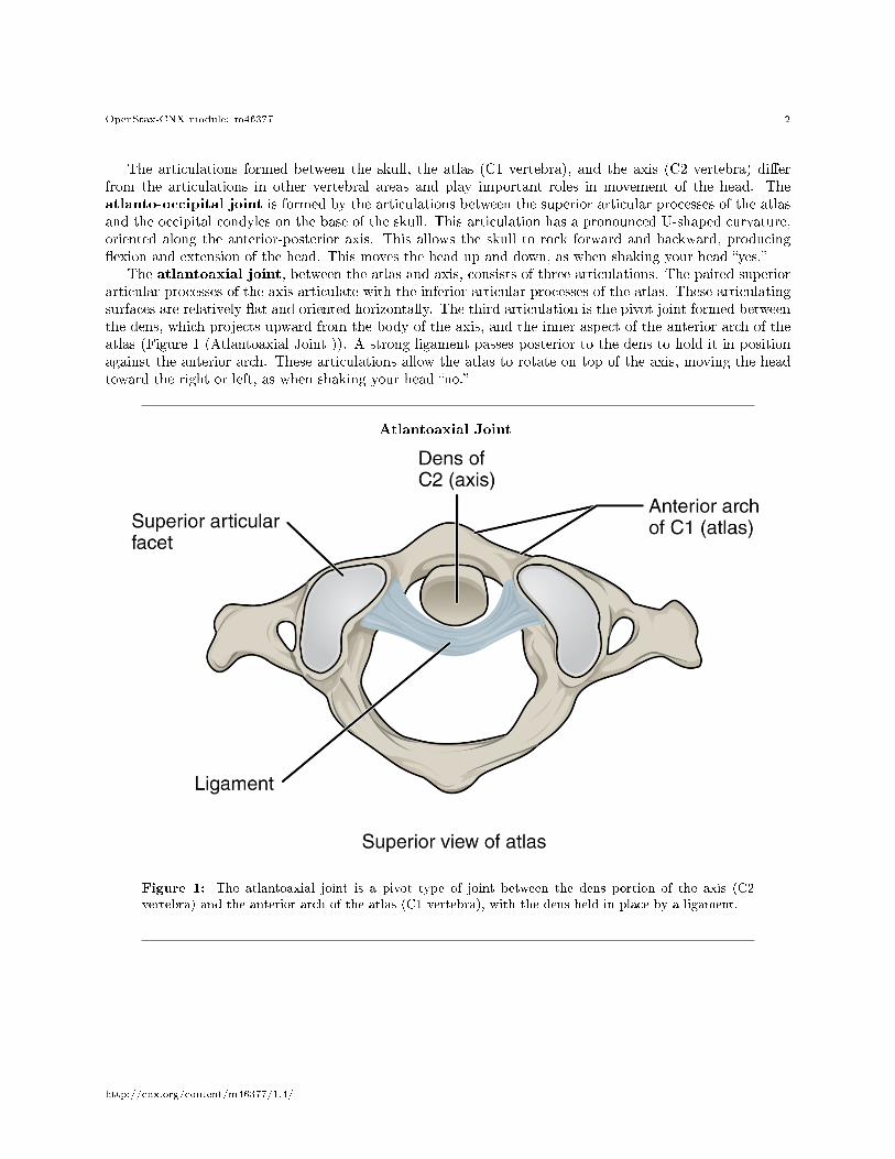
The atlantoaxial joint, between the atlas and axis, consists of three articulations. The paired superiorarticular processes of the axis articulate with the inferior articular processes of the atlas. These articulatingsurfaces are relatively �at and oriented horizontally. The third articulation is the pivot joint formed betweenthe dens, which projects upward from the body of the axis, and the inner aspect of the anterior arch of theatlas (Figure 1 (Atlantoaxial Joint )). A strong ligament passes posterior to the dens to hold it in positionagainst the anterior arch. These articulations allow the atlas to rotate on top of the axis, moving the headtoward the right or left, as when shaking your head �no.�
Atlantoaxial Joint
Figure 1: The atlantoaxial joint is a pivot type of joint between the dens portion of the axis (C2vertebra) and the anterior arch of the atlas (C1 vertebra), with the dens held in place by a ligament.
http://cnx.org/content/m46377/1.4/
OpenStax-CNX module: m46377 3
2 Temporomandibular Joint
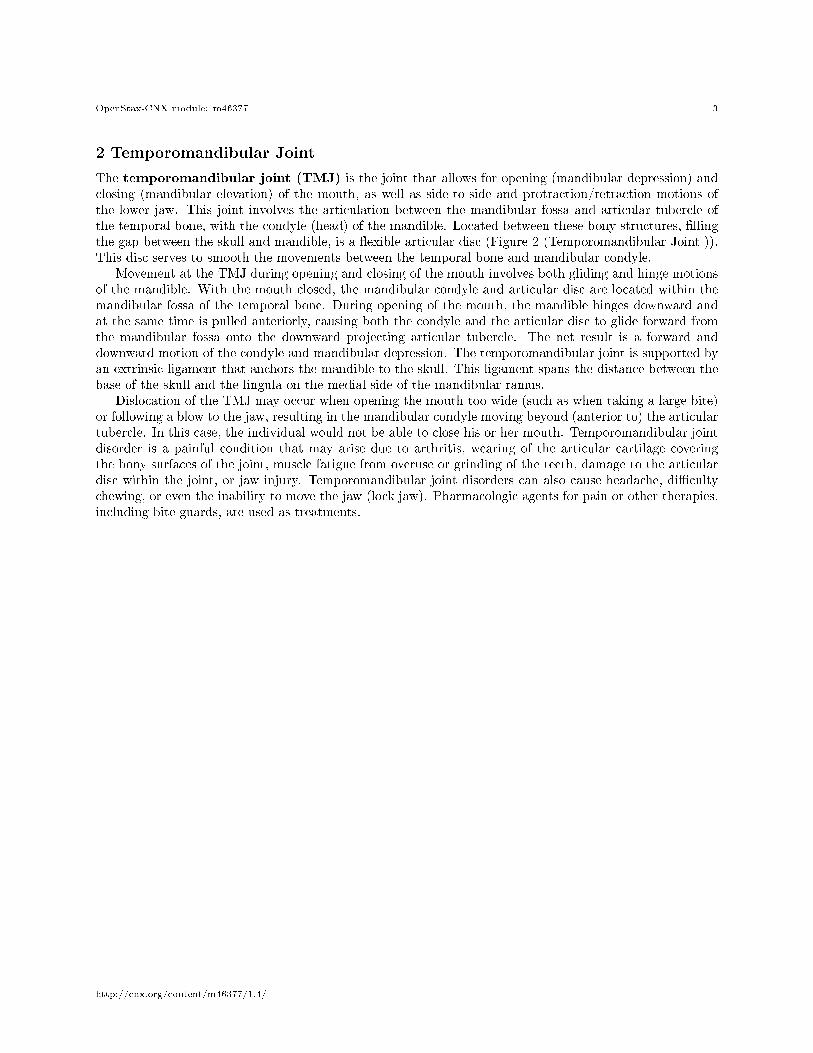
The temporomandibular joint (TMJ) is the joint that allows for opening (mandibular depression) andclosing (mandibular elevation) of the mouth, as well as side-to-side and protraction/retraction motions ofthe lower jaw. This joint involves the articulation between the mandibular fossa and articular tubercle ofthe temporal bone, with the condyle (head) of the mandible. Located between these bony structures, �llingthe gap between the skull and mandible, is a �exible articular disc (Figure 2 (Temporomandibular Joint )).This disc serves to smooth the movements between the temporal bone and mandibular condyle.
Movement at the TMJ during opening and closing of the mouth involves both gliding and hinge motionsof the mandible. With the mouth closed, the mandibular condyle and articular disc are located within themandibular fossa of the temporal bone. During opening of the mouth, the mandible hinges downward andat the same time is pulled anteriorly, causing both the condyle and the articular disc to glide forward fromthe mandibular fossa onto the downward projecting articular tubercle. The net result is a forward anddownward motion of the condyle and mandibular depression. The temporomandibular joint is supported byan extrinsic ligament that anchors the mandible to the skull. This ligament spans the distance between thebase of the skull and the lingula on the medial side of the mandibular ramus.
Dislocation of the TMJ may occur when opening the mouth too wide (such as when taking a large bite)or following a blow to the jaw, resulting in the mandibular condyle moving beyond (anterior to) the articulartubercle. In this case, the individual would not be able to close his or her mouth. Temporomandibular jointdisorder is a painful condition that may arise due to arthritis, wearing of the articular cartilage coveringthe bony surfaces of the joint, muscle fatigue from overuse or grinding of the teeth, damage to the articulardisc within the joint, or jaw injury. Temporomandibular joint disorders can also cause headache, di�cultychewing, or even the inability to move the jaw (lock jaw). Pharmacologic agents for pain or other therapies,including bite guards, are used as treatments.
http://cnx.org/content/m46377/1.4/
OpenStax-CNX module: m46377 4
Temporomandibular Joint
Figure 2: The temporomandibular joint is the articulation between the temporal bone of the skull andthe condyle of the mandible, with an articular disc located between these bones. During depression ofthe mandible (opening of the mouth), the mandibular condyle moves both forward and hinges downwardas it travels from the mandibular fossa onto the articular tubercle.
http://cnx.org/content/m46377/1.4/

OpenStax-CNX module: m46377 5
:
Watch this video1 to learn about TMJ. Opening of the mouth requires the combination of two mo-tions at the temporomandibular joint, an anterior gliding motion of the articular disc and mandibleand the downward hinging of the mandible. What is the initial movement of the mandible duringopening and how much mouth opening does this produce?
3 Shoulder Joint
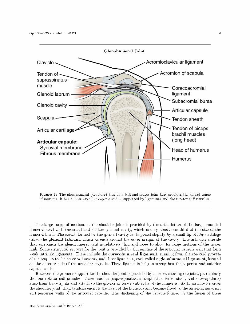
The shoulder joint is called the glenohumeral joint. This is a ball-and-socket joint formed by the articu-lation between the head of the humerus and the glenoid cavity of the scapula (Figure 3 (Glenohumeral Joint)). This joint has the largest range of motion of any joint in the body. However, this freedom of movementis due to the lack of structural support and thus the enhanced mobility is o�set by a loss of stability.
1http://openstaxcollege.org/l/TMJ
http://cnx.org/content/m46377/1.4/
OpenStax-CNX module: m46377 6
Glenohumeral Joint
Figure 3: The glenohumeral (shoulder) joint is a ball-and-socket joint that provides the widest rangeof motions. It has a loose articular capsule and is supported by ligaments and the rotator cu� muscles.
The large range of motions at the shoulder joint is provided by the articulation of the large, roundedhumeral head with the small and shallow glenoid cavity, which is only about one third of the size of thehumeral head. The socket formed by the glenoid cavity is deepened slightly by a small lip of �brocartilagecalled the glenoid labrum, which extends around the outer margin of the cavity. The articular capsulethat surrounds the glenohumeral joint is relatively thin and loose to allow for large motions of the upperlimb. Some structural support for the joint is provided by thickenings of the articular capsule wall that formweak intrinsic ligaments. These include the coracohumeral ligament, running from the coracoid processof the scapula to the anterior humerus, and three ligaments, each called a glenohumeral ligament, locatedon the anterior side of the articular capsule. These ligaments help to strengthen the superior and anteriorcapsule walls.
However, the primary support for the shoulder joint is provided by muscles crossing the joint, particularlythe four rotator cu� muscles. These muscles (supraspinatus, infraspinatus, teres minor, and subscapularis)arise from the scapula and attach to the greater or lesser tubercles of the humerus. As these muscles crossthe shoulder joint, their tendons encircle the head of the humerus and become fused to the anterior, superior,and posterior walls of the articular capsule. The thickening of the capsule formed by the fusion of these
http://cnx.org/content/m46377/1.4/
OpenStax-CNX module: m46377 7
four muscle tendons is called the rotator cu�. Two bursae, the subacromial bursa and the subscapularbursa, help to prevent friction between the rotator cu� muscle tendons and the scapula as these tendonscross the glenohumeral joint. In addition to their individual actions of moving the upper limb, the rotatorcu� muscles also serve to hold the head of the humerus in position within the glenoid cavity. By constantlyadjusting their strength of contraction to resist forces acting on the shoulder, these muscles serve as �dynamicligaments� and thus provide the primary structural support for the glenohumeral joint.
Injuries to the shoulder joint are common. Repetitive use of the upper limb, particularly in abductionsuch as during throwing, swimming, or racquet sports, may lead to acute or chronic in�ammation of thebursa or muscle tendons, a tear of the glenoid labrum, or degeneration or tears of the rotator cu�. Becausethe humeral head is strongly supported by muscles and ligaments around its anterior, superior, and posterioraspects, most dislocations of the humerus occur in an inferior direction. This can occur when force is appliedto the humerus when the upper limb is fully abducted, as when diving to catch a baseball and landing onyour hand or elbow. In�ammatory responses to any shoulder injury can lead to the formation of scar tissuebetween the articular capsule and surrounding structures, thus reducing shoulder mobility, a condition calledadhesive capsulitis (�frozen shoulder�).
http://cnx.org/content/m46377/1.4/

OpenStax-CNX module: m46377 8
:
Watch this video2 for a tutorial on the anatomy of the shoulder joint. What movements are availableat the shoulder joint?
2http://openstaxcollege.org/l/shoulderjoint1
http://cnx.org/content/m46377/1.4/
OpenStax-CNX module: m46377 9
:
Watch this video3 to learn more about the anatomy of the shoulder joint, including bones, joints,muscles, nerves, and blood vessels. What is the shape of the glenoid labrum in cross-section, andwhat is the importance of this shape?
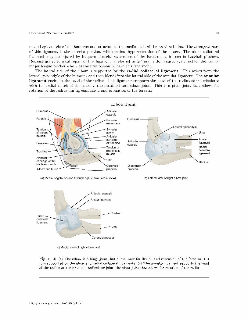
4 Elbow Joint
The elbow joint is a uniaxial hinge joint formed by the humeroulnar joint, the articulation betweenthe trochlea of the humerus and the trochlear notch of the ulna. Also associated with the elbow are thehumeroradial joint and the proximal radioulnar joint. All three of these joints are enclosed within a singlearticular capsule (Figure 4 (Elbow Joint )).
The articular capsule of the elbow is thin on its anterior and posterior aspects, but is thickened alongits outside margins by strong intrinsic ligaments. These ligaments prevent side-to-side movements andhyperextension. On the medial side is the triangular ulnar collateral ligament. This arises from the
3http://openstaxcollege.org/l/shoulderjoint2
http://cnx.org/content/m46377/1.4/
OpenStax-CNX module: m46377 10
medial epicondyle of the humerus and attaches to the medial side of the proximal ulna. The strongest partof this ligament is the anterior portion, which resists hyperextension of the elbow. The ulnar collateralligament may be injured by frequent, forceful extensions of the forearm, as is seen in baseball pitchers.Reconstructive surgical repair of this ligament is referred to as Tommy John surgery, named for the formermajor league pitcher who was the �rst person to have this treatment.
The lateral side of the elbow is supported by the radial collateral ligament. This arises from thelateral epicondyle of the humerus and then blends into the lateral side of the annular ligament. The annularligament encircles the head of the radius. This ligament supports the head of the radius as it articulateswith the radial notch of the ulna at the proximal radioulnar joint. This is a pivot joint that allows forrotation of the radius during supination and pronation of the forearm.
Elbow Joint
Figure 4: (a) The elbow is a hinge joint that allows only for �exion and extension of the forearm. (b)It is supported by the ulnar and radial collateral ligaments. (c) The annular ligament supports the headof the radius at the proximal radioulnar joint, the pivot joint that allows for rotation of the radius.
http://cnx.org/content/m46377/1.4/

OpenStax-CNX module: m46377 11
:
Watch this animation4 to learn more about the anatomy of the elbow joint. Which structures pro-vide the main stability for the elbow?
4http://openstaxcollege.org/l/elbowjoint1
http://cnx.org/content/m46377/1.4/
OpenStax-CNX module: m46377 12
:
Watch this video5 to learn more about the anatomy of the elbow joint, including bones, joints, mus-cles, nerves, and blood vessels. What are the functions of the articular cartilage?
5 Hip Joint
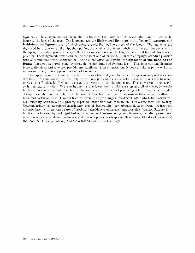
The hip joint is a multiaxial ball-and-socket joint between the head of the femur and the acetabulum of thehip bone (Figure 5 (Hip Joint )). The hip carries the weight of the body and thus requires strength andstability during standing and walking. For these reasons, its range of motion is more limited than at theshoulder joint.
The acetabulum is the socket portion of the hip joint. This space is deep and has a large articulationarea for the femoral head, thus giving stability and weight bearing ability to the joint. The acetabulumis further deepened by the acetabular labrum, a �brocartilage lip attached to the outer margin of theacetabulum. The surrounding articular capsule is strong, with several thickened areas forming intrinsic
5http://openstaxcollege.org/l/elbowjoint2
http://cnx.org/content/m46377/1.4/
OpenStax-CNX module: m46377 13
ligaments. These ligaments arise from the hip bone, at the margins of the acetabulum, and attach to thefemur at the base of the neck. The ligaments are the iliofemoral ligament, pubofemoral ligament, andischiofemoral ligament, all of which spiral around the head and neck of the femur. The ligaments aretightened by extension at the hip, thus pulling the head of the femur tightly into the acetabulum when inthe upright, standing position. Very little additional extension of the thigh is permitted beyond this verticalposition. These ligaments thus stabilize the hip joint and allow you to maintain an upright standing positionwith only minimal muscle contraction. Inside of the articular capsule, the ligament of the head of thefemur (ligamentum teres) spans between the acetabulum and femoral head. This intracapsular ligamentis normally slack and does not provide any signi�cant joint support, but it does provide a pathway for animportant artery that supplies the head of the femur.
The hip is prone to osteoarthritis, and thus was the �rst joint for which a replacement prosthesis wasdeveloped. A common injury in elderly individuals, particularly those with weakened bones due to osteo-porosis, is a �broken hip,� which is actually a fracture of the femoral neck. This may result from a fall,or it may cause the fall. This can happen as one lower limb is taking a step and all of the body weightis placed on the other limb, causing the femoral neck to break and producing a fall. Any accompanyingdisruption of the blood supply to the femoral neck or head can lead to necrosis of these areas, resulting inbone and cartilage death. Femoral fractures usually require surgical treatment, after which the patient willneed mobility assistance for a prolonged period, either from family members or in a long-term care facility.Consequentially, the associated health care costs of �broken hips� are substantial. In addition, hip fracturesare associated with increased rates of morbidity (incidences of disease) and mortality (death). Surgery for ahip fracture followed by prolonged bed rest may lead to life-threatening complications, including pneumonia,infection of pressure ulcers (bedsores), and thrombophlebitis (deep vein thrombosis; blood clot formation)that can result in a pulmonary embolism (blood clot within the lung).
http://cnx.org/content/m46377/1.4/
OpenStax-CNX module: m46377 14
Hip Joint
Figure 5: (a) The ball-and-socket joint of the hip is a multiaxial joint that provides both stability anda wide range of motion. (b�c) When standing, the supporting ligaments are tight, pulling the head ofthe femur into the acetabulum.
http://cnx.org/content/m46377/1.4/

OpenStax-CNX module: m46377 15
:
Watch this video6 for a tutorial on the anatomy of the hip joint. What is a possible consequencefollowing a fracture of the femoral neck within the capsule of the hip joint?
6http://openstaxcollege.org/l/hipjoint1
http://cnx.org/content/m46377/1.4/

OpenStax-CNX module: m46377 16
:
Watch this video7 to learn more about the anatomy of the hip joint, including bones, joints, muscles,nerves, and blood vessels. Where is the articular cartilage thickest within the hip joint?
6 Knee Joint
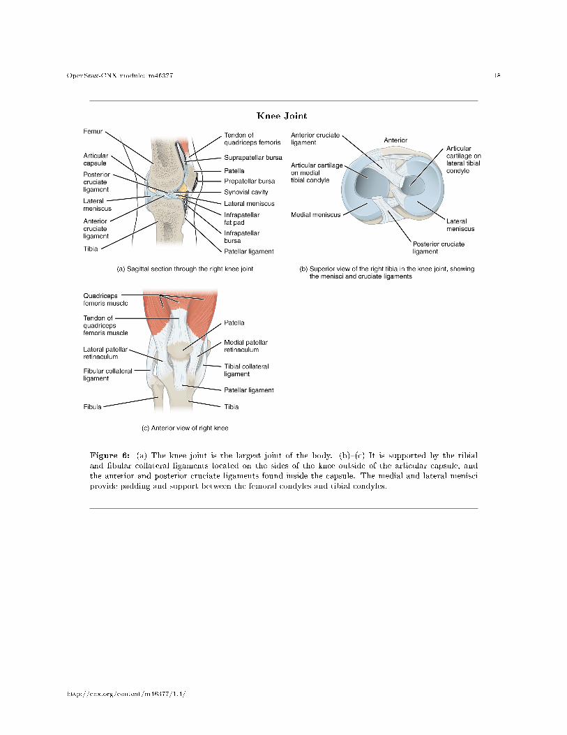
The knee joint is the largest joint of the body (Figure 6 (Knee Joint )). It actually consists of threearticulations. The femoropatellar joint is found between the patella and the distal femur. The medialtibiofemoral joint and lateral tibiofemoral joint are located between the medial and lateral condyles ofthe femur and the medial and lateral condyles of the tibia. All of these articulations are enclosed within asingle articular capsule. The knee functions as a hinge joint, allowing �exion and extension of the leg. Thisaction is generated by both rolling and gliding motions of the femur on the tibia. In addition, some rotationof the leg is available when the knee is �exed, but not when extended. The knee is well constructed for weight
7http://openstaxcollege.org/l/hipjoint2
http://cnx.org/content/m46377/1.4/
OpenStax-CNX module: m46377 17
bearing in its extended position, but is vulnerable to injuries associated with hyperextension, twisting, orblows to the medial or lateral side of the joint, particularly while weight bearing.
At the femoropatellar joint, the patella slides vertically within a groove on the distal femur. The patellais a sesamoid bone incorporated into the tendon of the quadriceps femoris muscle, the large muscle of theanterior thigh. The patella serves to protect the quadriceps tendon from friction against the distal femur.Continuing from the patella to the anterior tibia just below the knee is the patellar ligament. Acting viathe patella and patellar ligament, the quadriceps femoris is a powerful muscle that acts to extend the leg atthe knee. It also serves as a �dynamic ligament� to provide very important support and stabilization for theknee joint.
The medial and lateral tibiofemoral joints are the articulations between the rounded condyles of the femurand the relatively �at condyles of the tibia. During �exion and extension motions, the condyles of the femurboth roll and glide over the surfaces of the tibia. The rolling action produces �exion or extension, whilethe gliding action serves to maintain the femoral condyles centered over the tibial condyles, thus ensuringmaximal bony, weight-bearing support for the femur in all knee positions. As the knee comes into fullextension, the femur undergoes a slight medial rotation in relation to tibia. The rotation results because thelateral condyle of the femur is slightly smaller than the medial condyle. Thus, the lateral condyle �nishesits rolling motion �rst, followed by the medial condyle. The resulting small medial rotation of the femurserves to �lock� the knee into its fully extended and most stable position. Flexion of the knee is initiated bya slight lateral rotation of the femur on the tibia, which �unlocks� the knee. This lateral rotation motion isproduced by the popliteus muscle of the posterior leg.
Located between the articulating surfaces of the femur and tibia are two articular discs, the medialmeniscus and lateral meniscus (see Figure 6 (Knee Joint )b). Each is a C-shaped �brocartilage structurethat is thin along its inside margin and thick along the outer margin. They are attached to their tibialcondyles, but do not attach to the femur. While both menisci are free to move during knee motions, themedial meniscus shows less movement because it is anchored at its outer margin to the articular capsule andtibial collateral ligament. The menisci provide padding between the bones and help to �ll the gap betweenthe round femoral condyles and �attened tibial condyles. Some areas of each meniscus lack an arterial bloodsupply and thus these areas heal poorly if damaged.
The knee joint has multiple ligaments that provide support, particularly in the extended position (seeFigure 6 (Knee Joint )c). Outside of the articular capsule, located at the sides of the knee, are two extrinsicligaments. The �bular collateral ligament (lateral collateral ligament) is on the lateral side and spansfrom the lateral epicondyle of the femur to the head of the �bula. The tibial collateral ligament (medialcollateral ligament) of the medial knee runs from the medial epicondyle of the femur to the medial tibia. Asit crosses the knee, the tibial collateral ligament is �rmly attached on its deep side to the articular capsuleand to the medial meniscus, an important factor when considering knee injuries. In the fully extended kneeposition, both collateral ligaments are taut (tight), thus serving to stabilize and support the extended kneeand preventing side-to-side or rotational motions between the femur and tibia.
The articular capsule of the posterior knee is thickened by intrinsic ligaments that help to resist kneehyperextension. Inside the knee are two intracapsular ligaments, the anterior cruciate ligament andposterior cruciate ligament. These ligaments are anchored inferiorly to the tibia at the intercondylareminence, the roughened area between the tibial condyles. The cruciate ligaments are named for whetherthey are attached anteriorly or posteriorly to this tibial region. Each ligament runs diagonally upward toattach to the inner aspect of a femoral condyle. The cruciate ligaments are named for the X-shape formedas they pass each other (cruciate means �cross�). The posterior cruciate ligament is the stronger ligament. Itserves to support the knee when it is �exed and weight bearing, as when walking downhill. In this position,the posterior cruciate ligament prevents the femur from sliding anteriorly o� the top of the tibia. Theanterior cruciate ligament becomes tight when the knee is extended, and thus resists hyperextension.
http://cnx.org/content/m46377/1.4/
OpenStax-CNX module: m46377 18
Knee Joint
Figure 6: (a) The knee joint is the largest joint of the body. (b)�(c) It is supported by the tibialand �bular collateral ligaments located on the sides of the knee outside of the articular capsule, andthe anterior and posterior cruciate ligaments found inside the capsule. The medial and lateral menisciprovide padding and support between the femoral condyles and tibial condyles.
http://cnx.org/content/m46377/1.4/
OpenStax-CNX module: m46377 19
:
Watch this video8 to learn more about the �exion and extension of the knee, as the femur bothrolls and glides on the tibia to maintain stable contact between the bones in all knee positions.The patella glides along a groove on the anterior side of the distal femur. The collateral ligamentson the sides of the knee become tight in the fully extended position to help stabilize the knee.The posterior cruciate ligament supports the knee when �exed and the anterior cruciate ligamentbecomes tight when the knee comes into full extension to resist hyperextension. What are theligaments that support the knee joint?
8http://openstaxcollege.org/l/�exext
http://cnx.org/content/m46377/1.4/
OpenStax-CNX module: m46377 20
:
Watch this video9 to learn more about the anatomy of the knee joint, including bones, joints, mus-cles, nerves, and blood vessels. Which ligament of the knee keeps the tibia from sliding too farforward in relation to the femur and which ligament keeps the tibia from sliding too far backward?
: Joints
Injuries to the knee are common. Since this joint is primarily supported by muscles and ligaments,injuries to any of these structures will result in pain or knee instability. Injury to the posteriorcruciate ligament occurs when the knee is �exed and the tibia is driven posteriorly, such as fallingand landing on the tibial tuberosity or hitting the tibia on the dashboard when not wearing aseatbelt during an automobile accident. More commonly, injuries occur when forces are applied tothe extended knee, particularly when the foot is planted and unable to move. Anterior cruciateligament injuries can result with a forceful blow to the anterior knee, producing hyperextension, or
9http://openstaxcollege.org/l/kneejoint1
http://cnx.org/content/m46377/1.4/
OpenStax-CNX module: m46377 21
when a runner makes a quick change of direction that produces both twisting and hyperextensionof the knee.
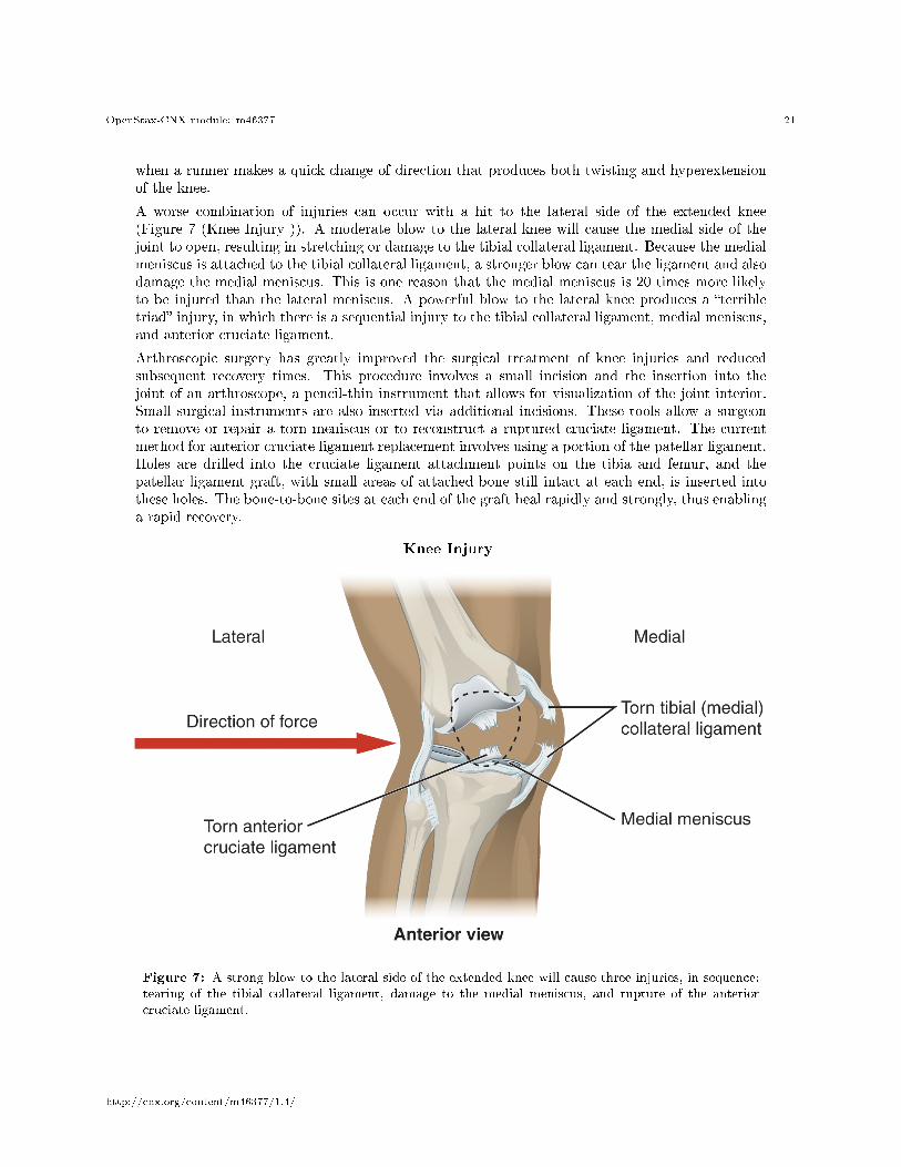
A worse combination of injuries can occur with a hit to the lateral side of the extended knee(Figure 7 (Knee Injury )). A moderate blow to the lateral knee will cause the medial side of thejoint to open, resulting in stretching or damage to the tibial collateral ligament. Because the medialmeniscus is attached to the tibial collateral ligament, a stronger blow can tear the ligament and alsodamage the medial meniscus. This is one reason that the medial meniscus is 20 times more likelyto be injured than the lateral meniscus. A powerful blow to the lateral knee produces a �terribletriad� injury, in which there is a sequential injury to the tibial collateral ligament, medial meniscus,and anterior cruciate ligament.
Arthroscopic surgery has greatly improved the surgical treatment of knee injuries and reducedsubsequent recovery times. This procedure involves a small incision and the insertion into thejoint of an arthroscope, a pencil-thin instrument that allows for visualization of the joint interior.Small surgical instruments are also inserted via additional incisions. These tools allow a surgeonto remove or repair a torn meniscus or to reconstruct a ruptured cruciate ligament. The currentmethod for anterior cruciate ligament replacement involves using a portion of the patellar ligament.Holes are drilled into the cruciate ligament attachment points on the tibia and femur, and thepatellar ligament graft, with small areas of attached bone still intact at each end, is inserted intothese holes. The bone-to-bone sites at each end of the graft heal rapidly and strongly, thus enablinga rapid recovery.
Knee Injury
Figure 7: A strong blow to the lateral side of the extended knee will cause three injuries, in sequence:tearing of the tibial collateral ligament, damage to the medial meniscus, and rupture of the anteriorcruciate ligament.
http://cnx.org/content/m46377/1.4/

OpenStax-CNX module: m46377 22
:
Watch this video10 to learn more about di�erent knee injuries and diagnostic testing of the knee.What are the most common causes of anterior cruciate ligament injury?
7 Ankle and Foot Joints
The ankle is formed by the talocrural joint (Figure 8 (Ankle Joint )). It consists of the articulationsbetween the talus bone of the foot and the distal ends of the tibia and �bula of the leg (crural = �leg�).The superior aspect of the talus bone is square-shaped and has three areas of articulation. The top of thetalus articulates with the inferior tibia. This is the portion of the ankle joint that carries the body weightbetween the leg and foot. The sides of the talus are �rmly held in position by the articulations with themedial malleolus of the tibia and the lateral malleolus of the �bula, which prevent any side-to-side motionof the talus. The ankle is thus a uniaxial hinge joint that allows only for dorsi�exion and plantar �exion ofthe foot.
10http://openstaxcollege.org/l/kneeinjury
http://cnx.org/content/m46377/1.4/
OpenStax-CNX module: m46377 23
Additional joints between the tarsal bones of the posterior foot allow for the movements of foot inversionand eversion. Most important for these movements is the subtalar joint, located between the talus andcalcaneus bones. The joints between the talus and navicular bones and the calcaneus and cuboid bones arealso important contributors to these movements. All of the joints between tarsal bones are plane joints.Together, the small motions that take place at these joints all contribute to the production of inversion andeversion foot motions.
Like the hinge joints of the elbow and knee, the talocrural joint of the ankle is supported by severalstrong ligaments located on the sides of the joint. These ligaments extend from the medial malleolus ofthe tibia or lateral malleolus of the �bula and anchor to the talus and calcaneus bones. Since they arelocated on the sides of the ankle joint, they allow for dorsi�exion and plantar �exion of the foot. They alsoprevent abnormal side-to-side and twisting movements of the talus and calcaneus bones during eversion andinversion of the foot. On the medial side is the broad deltoid ligament. The deltoid ligament supports theankle joint and also resists excessive eversion of the foot. The lateral side of the ankle has several smallerligaments. These include the anterior talo�bular ligament and the posterior talo�bular ligament,both of which span between the talus bone and the lateral malleolus of the �bula, and the calcaneo�bularligament, located between the calcaneus bone and �bula. These ligaments support the ankle and also resistexcess inversion of the foot.
http://cnx.org/content/m46377/1.4/

OpenStax-CNX module: m46377 24
Ankle Joint
Figure 8: The talocrural (ankle) joint is a uniaxial hinge joint that only allows for dorsi�exion orplantar �exion of the foot. Movements at the subtalar joint, between the talus and calcaneus bones,combined with motions at other intertarsal joints, enables eversion/inversion movements of the foot.Ligaments that unite the medial or lateral malleolus with the talus and calcaneus bones serve to supportthe talocrural joint and to resist excess eversion or inversion of the foot.
http://cnx.org/content/m46377/1.4/

OpenStax-CNX module: m46377 25
:
Watch this video11 for a tutorial on the anatomy of the ankle joint. What are the three ligamentsfound on the lateral side of the ankle joint?
11http://openstaxcollege.org/l/anklejoint1
http://cnx.org/content/m46377/1.4/
OpenStax-CNX module: m46377 26
:
Watch this video12 to learn more about the anatomy of the ankle joint, including bones, joints,muscles, nerves, and blood vessels. Which type of joint used in woodworking does the ankle jointresemble?
: Joints
The ankle is the most frequently injured joint in the body, with the most common injury beingan inversion ankle sprain. A sprain is the stretching or tearing of the supporting ligaments. Excessinversion causes the talus bone to tilt laterally, thus damaging the ligaments on the lateral side of theankle. The anterior talo�bular ligament is most commonly injured, followed by the calcaneo�bularligament. In severe inversion injuries, the forceful lateral movement of the talus not only rupturesthe lateral ankle ligaments, but also fractures the distal �bula.
Less common are eversion sprains of the ankle, which involve stretching of the deltoid ligament onthe medial side of the ankle. Forcible eversion of the foot, for example, with an awkward landing
12http://openstaxcollege.org/l/anklejoint2
http://cnx.org/content/m46377/1.4/
OpenStax-CNX module: m46377 27
from a jump or when a football player has a foot planted and is hit on the lateral ankle, can result ina Pott's fracture and dislocation of the ankle joint. In this injury, the very strong deltoid ligamentdoes not tear, but instead shears o� the medial malleolus of the tibia. This frees the talus, whichmoves laterally and fractures the distal �bula. In extreme cases, the posterior margin of the tibiamay also be sheared o�.
Above the ankle, the distal ends of the tibia and �bula are united by a strong syndesmosis formedby the interosseous membrane and ligaments at the distal tibio�bular joint. These connectionsprevent separation between the distal ends of the tibia and �bula and maintain the talus lockedinto position between the medial malleolus and lateral malleolus. Injuries that produce a lateraltwisting of the leg on top of the planted foot can result in stretching or tearing of the tibio�bularligaments, producing a syndesmotic ankle sprain or �high ankle sprain.�
Most ankle sprains can be treated using the RICE technique: Rest, Ice, Compression, and Elevation.Reducing joint mobility using a brace or cast may be required for a period of time. More severeinjuries involving ligament tears or bone fractures may require surgery.
http://cnx.org/content/m46377/1.4/
OpenStax-CNX module: m46377 28
:
Watch this video13 to learn more about the ligaments of the ankle joint, ankle sprains, and treat-ment. During an inversion ankle sprain injury, all three ligaments that resist excessive inversion ofthe foot may be injured. What is the sequence in which these three ligaments are injured?
8 Chapter Review
Although synovial joints share many common features, each joint of the body is specialized for certainmovements and activities. The joints of the upper limb provide for large ranges of motion, which give theupper limb great mobility, thus enabling actions such as the throwing of a ball or typing on a keyboard. Thejoints of the lower limb are more robust, giving them greater strength and the stability needed to supportthe body weight during running, jumping, or kicking activities.
The joints of the vertebral column include the symphysis joints formed by each intervertebral disc andthe plane synovial joints between the superior and inferior articular processes of adjacent vertebrae. Each
13http://openstaxcollege.org/l/anklejoint3
http://cnx.org/content/m46377/1.4/
OpenStax-CNX module: m46377 29
of these joints provide for limited motions, but these sum together to produce �exion, extension, lateral�exion, and rotation of the neck and body. The range of motions available in each region of the vertebralcolumn varies, with all of these motions available in the cervical region. Only rotation is allowed in thethoracic region, while the lumbar region has considerable extension, �exion, and lateral �exion, but rotationis prevented. The atlanto-occipital joint allows for �exion and extension of the head, while the atlantoaxialjoint is a pivot joint that provides for rotation of the head.
The temporomandibular joint is the articulation between the condyle of the mandible and the mandibularfossa and articular tubercle of the skull temporal bone. An articular disc is located between the bonycomponents of this joint. A combination of gliding and hinge motions of the mandibular condyle allows forelevation/depression, protraction/retraction, and side-to-side motions of the lower jaw.
The glenohumeral (shoulder) joint is a multiaxial ball-and-socket joint that provides �exion/extension,abduction/adduction, circumduction, and medial/lateral rotation of the humerus. The head of the humerusarticulates with the glenoid cavity of the scapula. The glenoid labrum extends around the margin of theglenoid cavity. Intrinsic ligaments, including the coracohumeral ligament and glenohumeral ligaments, pro-vide some support for the shoulder joint. However, the primary support comes from muscles crossing thejoint whose tendons form the rotator cu�. These muscle tendons are protected from friction against thescapula by the subacromial bursa and subscapular bursa.
The elbow is a uniaxial hinge joint that allows for �exion/extension of the forearm. It includes thehumeroulnar joint and the humeroradial joint. The medial elbow is supported by the ulnar collateral ligamentand the radial collateral ligament supports the lateral side. These ligaments prevent side-to-side movementsand resist hyperextension of the elbow. The proximal radioulnar joint is a pivot joint that allows for rotationof the radius during pronation/supination of the forearm. The annular ligament surrounds the head of theradius to hold it in place at this joint.
The hip joint is a ball-and-socket joint whose motions are more restricted than at the shoulder to providegreater stability during weight bearing. The hip joint is the articulation between the head of the femur andthe acetabulum of the hip bone. The acetabulum is deepened by the acetabular labrum. The iliofemoral,pubofemoral, and ischiofemoral ligaments strongly support the hip joint in the upright, standing position.The ligament of the head of the femur provides little support but carries an important artery that suppliesthe femur.
The knee includes three articulations. The femoropatellar joint is between the patella and distal femur.The patella, a sesamoid bone incorporated into the tendon of the quadriceps femoris muscle of the anteriorthigh, serves to protect this tendon from rubbing against the distal femur during knee movements. Themedial and lateral tibiofemoral joints, between the condyles of the femur and condyles of the tibia, aremodi�ed hinge joints that allow for knee extension and �exion. During these movements, the condyles ofthe femur both roll and glide over the surface of the tibia. As the knee comes into full extension, a slightmedial rotation of the femur serves to �lock� the knee into its most stable, weight-bearing position. Thereverse motion, a small lateral rotation of the femur, is required to initiate knee �exion. When the knee is�exed, some rotation of the leg is available.
Two extrinsic ligaments, the tibial collateral ligament on the medial side and the �bular collateral ligamenton the lateral side, serve to resist hyperextension or rotation of the extended knee joint. Two intracapsularligaments, the anterior cruciate ligament and posterior cruciate ligament, span between the tibia and theinner aspects of the femoral condyles. The anterior cruciate ligament resists hyperextension of the knee,while the posterior cruciate ligament prevents anterior sliding of the femur, thus supporting the knee whenit is �exed and weight bearing. The medial and lateral menisci, located between the femoral and tibialcondyles, are articular discs that provide padding and improve the �t between the bones.
The talocrural joint forms the ankle. It consists of the articulation between the talus bone and the medialmalleolus of the tibia, the distal end of the tibia, and the lateral malleolus of the �bula. This is a uniaxialhinge joint that allows only dorsi�exion and plantar �exion of the foot. Gliding motions at the subtalarand intertarsal joints of the foot allow for inversion/eversion of the foot. The ankle joint is supported onthe medial side by the deltoid ligament, which prevents side-to-side motions of the talus at the talocruraljoint and resists excessive eversion of the foot. The lateral ankle is supported by the anterior and posterior
http://cnx.org/content/m46377/1.4/

OpenStax-CNX module: m46377 30
talo�bular ligaments and the calcaneo�bular ligament. These support the ankle joint and also resist excessinversion of the foot. An inversion ankle sprain, a common injury, will result in injury to one or more ofthese lateral ankle ligaments.
9 Interactive Link Questions
Exercise 1 (Solution on p. 33.)
Watch this video14 to learn about TMJ. Opening of the mouth requires the combination of two mo-tions at the temporomandibular joint, an anterior gliding motion of the articular disc and mandibleand the downward hinging of the mandible. What is the initial movement of the mandible duringopening and how much mouth opening does this produce?
Exercise 2 (Solution on p. 33.)
Watch this video15 for a tutorial on the anatomy of the shoulder joint. What movements areavailable at the shoulder joint?
Exercise 3 (Solution on p. 33.)
Watch this video16 to learn about the anatomy of the shoulder joint, including bones, joints,muscles, nerves, and blood vessels. What is the shape of the glenoid labrum in cross-section, andwhat is the importance of this shape?
Exercise 4 (Solution on p. 33.)
Watch this animation17 to learn more about the anatomy of the elbow joint. What structuresprovide the main stability for the elbow?
Exercise 5 (Solution on p. 33.)
Watch this video18 to learn more about the anatomy of the elbow joint, including bones, joints,muscles, nerves, and blood vessels. What are the functions of the articular cartilage?
Exercise 6 (Solution on p. 33.)
Watch this video19 for a tutorial on the anatomy of the hip joint. What is a possible consequencefollowing a fracture of the femoral neck within the capsule of the hip joint?
Exercise 7 (Solution on p. 33.)
Watch this video20 to learn more about the anatomy of the hip joint, including bones, joints,muscles, nerves, and blood vessels. Where is the articular cartilage thickest within the hip joint?
Exercise 8 (Solution on p. 33.)
Watch this video21 to learn more about the �exion and extension of the knee, as the femur bothrolls and glides on the tibia to maintain stable contact between the bones in all knee positions.The patella glides along a groove on the anterior side of the distal femur. The collateral ligamentson the sides of the knee become tight in the fully extended position to help stabilize the knee.The posterior cruciate ligament supports the knee when �exed and the anterior cruciate ligamentbecomes tight when the knee comes into full extension to resist hyperextension. What are theligaments that support the knee joint?
Exercise 9 (Solution on p. 33.)
Watch this video22 to learn more about the anatomy of the knee joint, including bones, joints,muscles, nerves, and blood vessels. Which ligament of the knee keeps the tibia from sliding too far
14http://openstaxcollege.org/l/TMJ15http://openstaxcollege.org/l/shoulderjoint116http://openstaxcollege.org/l/shoulderjoint217http://openstaxcollege.org/l/elbowjoint118http://openstaxcollege.org/l/elbowjoint219http://openstaxcollege.org/l/hipjoint120http://openstaxcollege.org/l/hipjoint221http://openstaxcollege.org/l/�exext22http://openstaxcollege.org/l/kneejoint1
http://cnx.org/content/m46377/1.4/
OpenStax-CNX module: m46377 31
forward in relation to the femur and which ligament keeps the tibia from sliding too far backward?
Exercise 10 (Solution on p. 33.)
Watch this video23 to learn more about di�erent knee injuries and diagnostic testing of the knee.What are the most causes of anterior cruciate ligament injury?
Exercise 11 (Solution on p. 33.)
Watch this video24 for a tutorial on the anatomy of the ankle joint. What are the three ligamentsfound on the lateral side of the ankle joint?
Exercise 12 (Solution on p. 33.)
Watch this video25 to learn more about the anatomy of the ankle joint, including bones, joints,muscles, nerves, and blood vessels. The ankle joint resembles what type of joint used in woodwork-ing?
Exercise 13 (Solution on p. 33.)
Watch this video26 to learn about the ligaments of the ankle joint, ankle sprains, and treatment.During an inversion ankle sprain injury, all three ligaments that resist excessive inversion of thefoot may be injured. What is the sequence in which these three ligaments are injured?
10 Review Questions
Exercise 14 (Solution on p. 33.)
The primary support for the glenohumeral joint is provided by the ________.
a. coracohumeral ligamentb. glenoid labrumc. rotator cu� musclesd. subacromial bursa
Exercise 15 (Solution on p. 33.)
The proximal radioulnar joint ________.
a. is supported by the annular ligamentb. contains an articular disc that strongly unites the bonesc. is supported by the ulnar collateral ligamentd. is a hinge joint that allows for �exion/extension of the forearm
Exercise 16 (Solution on p. 33.)
Which statement is true concerning the knee joint?
a. The lateral meniscus is an intrinsic ligament located on the lateral side of the knee joint.b. Hyperextension is resisted by the posterior cruciate ligament.c. The anterior cruciate ligament supports the knee when it is �exed and weight bearing.d. The medial meniscus is attached to the tibial collateral ligament.
Exercise 17 (Solution on p. 33.)
The ankle joint ________.
a. is also called the subtalar joint
23http://openstaxcollege.org/l/kneeinjury24http://openstaxcollege.org/l/anklejoint125http://openstaxcollege.org/l/anklejoint226http://openstaxcollege.org/l/anklejoint3
http://cnx.org/content/m46377/1.4/

OpenStax-CNX module: m46377 32
b. allows for gliding movements that produce inversion/eversion of the footc. is a uniaxial hinge jointd. is supported by the tibial collateral ligament on the lateral side
Exercise 18 (Solution on p. 33.)
Which region of the vertebral column has the greatest range of motion for rotation?
a. cervicalb. thoracicc. lumbard. sacral
11 Critical Thinking Questions
Exercise 19 (Solution on p. 34.)
Discuss the structures that contribute to support of the shoulder joint.
Exercise 20 (Solution on p. 34.)
Describe the sequence of injuries that may occur if the extended, weight-bearing knee receives avery strong blow to the lateral side of the knee.
http://cnx.org/content/m46377/1.4/
OpenStax-CNX module: m46377 33
Solutions to Exercises in this Module
to Exercise (p. 30)The �rst motion is rotation (hinging) of the mandible, but this only produces about 20 mm (0.78 in) ofmouth opening.to Exercise (p. 30)The shoulder joint is a ball-and-socket joint that allows for �exion-extension, abduction-adduction, medialrotation, lateral rotation, and circumduction of the humerus.to Exercise (p. 30)The glenoid labrum is wedge-shaped in cross-section. This is important because it creates an elevated rimaround the glenoid cavity, which creates a deeper socket for the head of the humerus to �t into.to Exercise (p. 30)The structures that stabilize the elbow include the coronoid process, the radial (lateral) collateral ligament,and the anterior portion of the ulnar (medial) collateral ligament.to Exercise (p. 30)The articular cartilage functions to absorb shock and to provide an extremely smooth surface that makesmovement between bones easy, without damaging the bones.to Exercise (p. 30)An intracapsular fracture of the neck of the femur can result in disruption of the arterial blood supply tothe head of the femur, which may lead to avascular necrosis of the femoral head.to Exercise (p. 30)The articular cartilage is thickest in the upper and back part of the acetabulum, the socket portion of thehip joint. These regions receive most of the force from the head of the femur during walking and running.to Exercise (p. 30)There are �ve ligaments associated with the knee joint. The tibial collateral ligament is located on themedial side of the knee and the �bular collateral ligament is located on the lateral side. The anterior andposterior cruciate ligaments are located inside the knee joint.to Exercise (p. 30)The anterior cruciate ligament prevents the tibia from sliding too far forward in relation to the femur andthe posterior cruciate ligament keeps the tibia from sliding too far backward.to Exercise (p. 31)The anterior cruciate ligament (ACL) is most commonly injured when traumatic force is applied to the kneeduring a twisting motion or when side standing or landing from a jump.to Exercise (p. 31)The ligaments of the lateral ankle are the anterior and posterior talo�bular ligaments and the calcaneo�bularligament. These ligaments support the ankle joint and resist excess inversion of the foot.to Exercise (p. 31)Because of the square shape of the ankle joint, it has been compared to a mortise-and-tendon type of joint.to Exercise (p. 31)An inversion ankle sprain may injure all three ligaments located on the lateral side of the ankle. Thesequence of injury would be the anterior talo�bular ligament �rst, followed by the calcaneo�bular ligamentsecond, and �nally, the posterior talo�bular ligament third.to Exercise (p. 31)Cto Exercise (p. 31)Ato Exercise (p. 31)Dto Exercise (p. 31)C
http://cnx.org/content/m46377/1.4/
OpenStax-CNX module: m46377 34
to Exercise (p. 32)Bto Exercise (p. 32)The shoulder joint allows for a large range of motion. The primary support for the shoulder joint is providedby the four rotator cu� muscles. These muscles serve as �dynamic ligaments� and thus can modulate theirstrengths of contraction as needed to hold the head of the humerus in position at the glenoid fossa. Additionalbut weaker support comes from the coracohumeral ligament, an intrinsic ligament that supports the superioraspect of the shoulder joint, and the glenohumeral ligaments, which are intrinsic ligaments that support theanterior side of the joint.to Exercise (p. 32)A strong blow to the lateral side of the extended knee will cause the medial side of the knee joint to open,resulting in a sequence of three injuries. First will be damage to the tibial collateral ligament. Since themedial meniscus is attached to the tibial collateral ligament, the meniscus is also injured. The third structureinjured would be the anterior cruciate ligament.
Glossary
De�nition 8: acetabular labrumlip of �brocartilage that surrounds outer margin of the acetabulum on the hip bone
De�nition 8: annular ligamentintrinsic ligament of the elbow articular capsule that surrounds and supports the head of the radiusat the proximal radioulnar joint
De�nition 8: anterior cruciate ligamentintracapsular ligament of the knee; extends from anterior, superior surface of the tibia to the inneraspect of the lateral condyle of the femur; resists hyperextension of knee
De�nition 8: anterior talo�bular ligamentintrinsic ligament located on the lateral side of the ankle joint, between talus bone and lateralmalleolus of �bula; supports talus at the talocrural joint and resists excess inversion of the foot
De�nition 8: atlantoaxial jointseries of three articulations between the atlas (C1) vertebra and the axis (C2) vertebra, consistingof the joints between the inferior articular processes of C1 and the superior articular processes ofC2, and the articulation between the dens of C2 and the anterior arch of C1
De�nition 8: atlanto-occipital jointarticulation between the occipital condyles of the skull and the superior articular processes of theatlas (C1 vertebra)
De�nition 8: calcaneo�bular ligamentintrinsic ligament located on the lateral side of the ankle joint, between the calcaneus bone andlateral malleolus of the �bula; supports the talus bone at the ankle joint and resists excess inversionof the foot
De�nition 8: coracohumeral ligamentintrinsic ligament of the shoulder joint; runs from the coracoid process of the scapula to the anteriorhumerus
De�nition 8: deltoid ligamentbroad intrinsic ligament located on the medial side of the ankle joint; supports the talus at thetalocrural joint and resists excess eversion of the foot
De�nition 8: elbow jointhumeroulnar joint
http://cnx.org/content/m46377/1.4/
OpenStax-CNX module: m46377 35
De�nition 8: femoropatellar jointportion of the knee joint consisting of the articulation between the distal femur and the patella
De�nition 8: �bular collateral ligamentextrinsic ligament of the knee joint that spans from the lateral epicondyle of the femur to the headof the �bula; resists hyperextension and rotation of the extended knee
De�nition 8: glenohumeral jointshoulder joint; articulation between the glenoid cavity of the scapula and head of the humerus; mul-tiaxial ball-and-socket joint that allows for �exion/extension, abduction/adduction, circumduction,and medial/lateral rotation of the humerus
De�nition 8: glenohumeral ligamentone of the three intrinsic ligaments of the shoulder joint that strengthen the anterior articularcapsule
De�nition 8: glenoid labrumlip of �brocartilage located around the outside margin of the glenoid cavity of the scapula
De�nition 8: humeroradial jointarticulation between the capitulum of the humerus and head of the radius
De�nition 8: humeroulnar jointarticulation between the trochlea of humerus and the trochlear notch of the ulna; uniaxial hingejoint that allows for �exion/extension of the forearm
De�nition 8: iliofemoral ligamentintrinsic ligament spanning from the ilium of the hip bone to the femur, on the superior-anterioraspect of the hip joint
De�nition 8: ischiofemoral ligamentintrinsic ligament spanning from the ischium of the hip bone to the femur, on the posterior aspectof the hip joint
De�nition 8: lateral meniscusC-shaped �brocartilage articular disc located at the knee, between the lateral condyle of the femurand the lateral condyle of the tibia
De�nition 8: lateral tibiofemoral jointportion of the knee consisting of the articulation between the lateral condyle of the tibia and thelateral condyle of the femur; allows for �exion/extension at the knee
De�nition 8: ligament of the head of the femurintracapsular ligament that runs from the acetabulum of the hip bone to the head of the femur
De�nition 8: medial meniscusC-shaped �brocartilage articular disc located at the knee, between the medial condyle of the femurand medial condyle of the tibia
De�nition 8: medial tibiofemoral jointportion of the knee consisting of the articulation between the medial condyle of the tibia and themedial condyle of the femur; allows for �exion/extension at the knee
De�nition 8: patellar ligamentligament spanning from the patella to the anterior tibia; serves as the �nal attachment for thequadriceps femoris muscle
De�nition 8: posterior cruciate ligamentintracapsular ligament of the knee; extends from the posterior, superior surface of the tibia to theinner aspect of the medial condyle of the femur; prevents anterior displacement of the femur whenthe knee is �exed and weight bearing
http://cnx.org/content/m46377/1.4/
OpenStax-CNX module: m46377 36
De�nition 8: posterior talo�bular ligamentintrinsic ligament located on the lateral side of the ankle joint, between the talus bone and lateralmalleolus of the �bula; supports the talus at the talocrural joint and resists excess inversion of thefoot
De�nition 8: pubofemoral ligamentintrinsic ligament spanning from the pubis of the hip bone to the femur, on the anterior-inferioraspect of the hip joint
De�nition 8: radial collateral ligamentintrinsic ligament on the lateral side of the elbow joint; runs from the lateral epicondyle of humerusto merge with the annular ligament
De�nition 8: rotator cu�strong connective tissue structure formed by the fusion of four rotator cu� muscle tendons tothe articular capsule of the shoulder joint; surrounds and supports superior, anterior, lateral, andposterior sides of the humeral head
De�nition 8: subacromial bursabursa that protects the supraspinatus muscle tendon and superior end of the humerus from rubbingagainst the acromion of the scapula
De�nition 8: subscapular bursabursa that prevents rubbing of the subscapularis muscle tendon against the scapula
De�nition 8: subtalar jointarticulation between the talus and calcaneus bones of the foot; allows motions that contribute toinversion/eversion of the foot
De�nition 8: talocrural jointankle joint; articulation between the talus bone of the foot and medial malleolus of the tibia, distaltibia, and lateral malleolus of the �bula; a uniaxial hinge joint that allows only for dorsi�exion andplantar �exion of the foot
De�nition 8: temporomandibular joint (TMJ)articulation between the condyle of the mandible and the mandibular fossa and articular tubercleof the temporal bone of the skull; allows for depression/elevation (opening/closing of mouth),protraction/retraction, and side-to-side motions of the mandible
De�nition 8: tibial collateral ligamentextrinsic ligament of knee joint that spans from the medial epicondyle of the femur to the medialtibia; resists hyperextension and rotation of extended knee
De�nition 8: ulnar collateral ligamentintrinsic ligament on the medial side of the elbow joint; spans from the medial epicondyle of thehumerus to the medial ulna
De�nition 8: zygapophysial jointsfacet joints; plane joints between the superior and inferior articular processes of adjacent vertebraethat provide for only limited motions between the vertebrae
http://cnx.org/content/m46377/1.4/