Embed Size (px)
Citation preview
Anatomy of the wrist and
distal radioulnar joint
C. Fontaine (Lille)
EFORT Congress, June 3rd, 2009, Vienna (Austria)
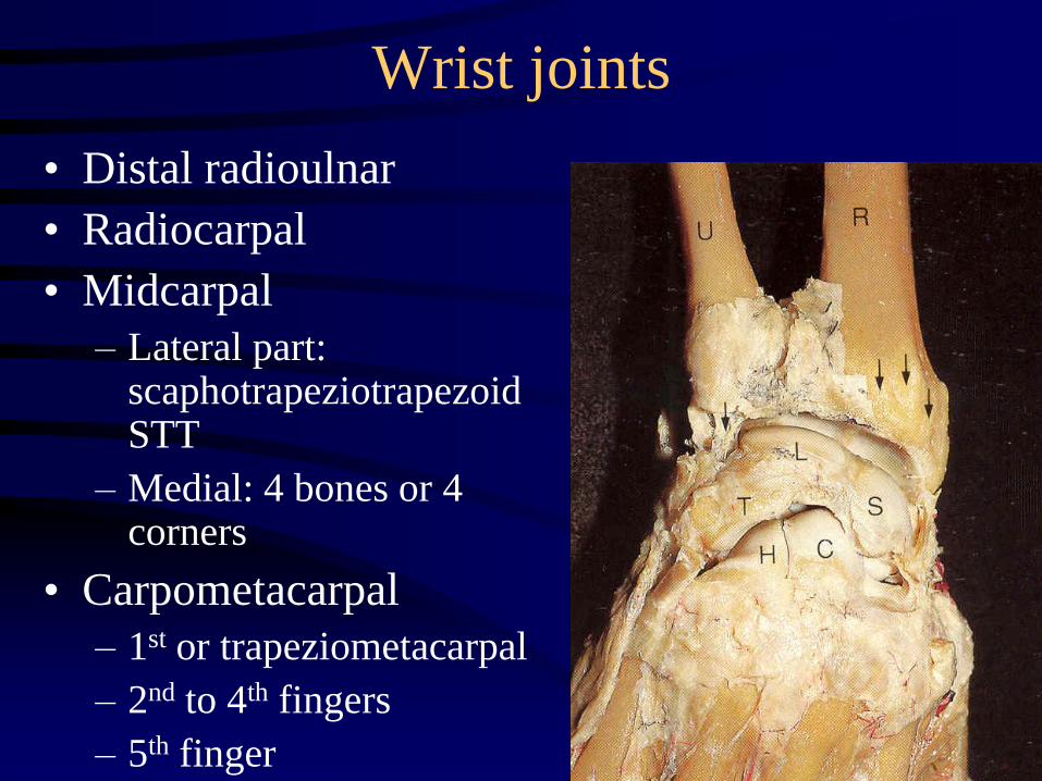
Wrist joints
• Distal radioulnar
• Radiocarpal
• Midcarpal
– Lateral part: scaphotrapeziotrapezoid STT
– Medial: 4 bones or 4 corners
• Carpometacarpal
– 1st or trapeziometacarpal
– 2nd to 4th fingers
– 5th finger
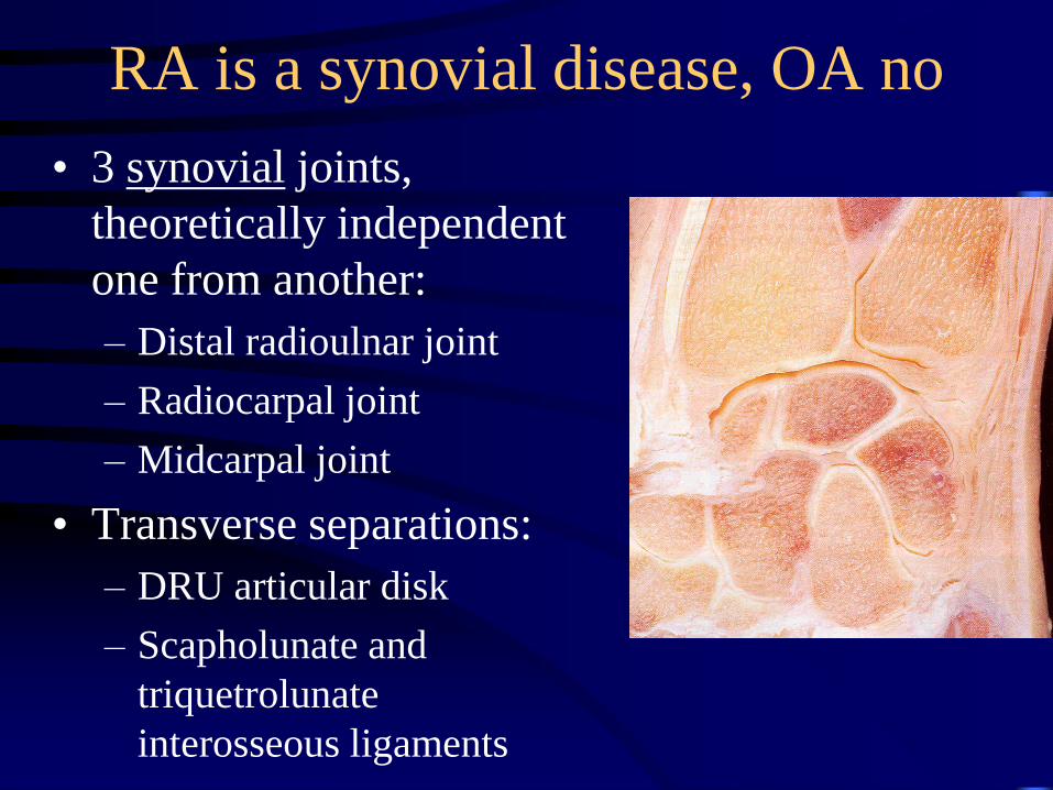
RA is a synovial disease, OA no
• 3 synovial joints,
theoretically independent
one from another:
– Distal radioulnar joint
– Radiocarpal joint
– Midcarpal joint
• Transverse separations:
– DRU articular disk
– Scapholunate and
triquetrolunate
interosseous ligaments
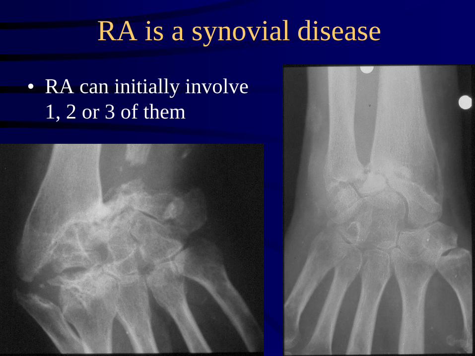
RA is a synovial disease
• RA can initially involve
1, 2 or 3 of them
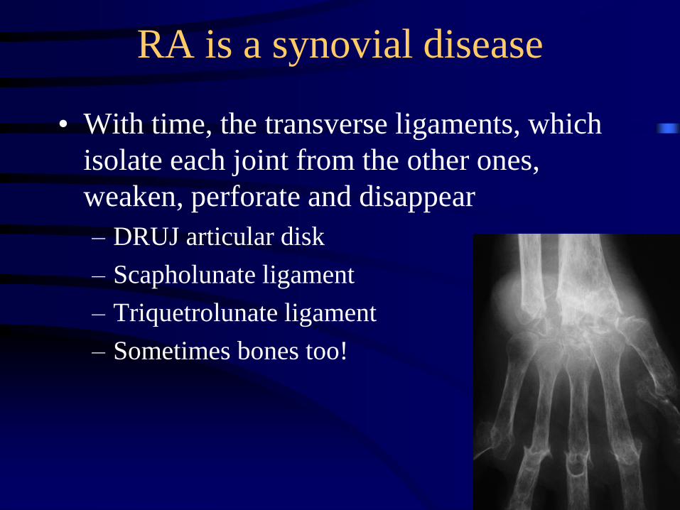
RA is a synovial disease
• With time, the transverse ligaments, which
isolate each joint from the other ones,
weaken, perforate and disappear
– DRUJ articular disk
– Scapholunate ligament
– Triquetrolunate ligament
– Sometimes bones too!
RA is a synovial disease
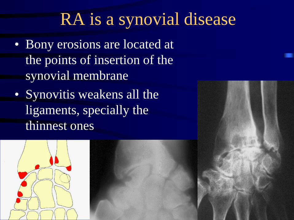
• Bony erosions are located at
the points of insertion of the
synovial membrane
• Synovitis weakens all the
ligaments, specially the
thinnest ones
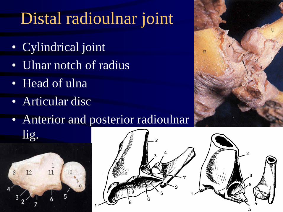
Distal radioulnar joint
• Cylindrical joint
• Ulnar notch of radius
• Head of ulna
• Articular disc
• Anterior and posterior radioulnar
lig.
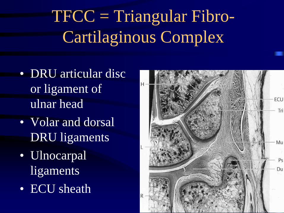
TFCC = Triangular Fibro-
Cartilaginous Complex
• DRU articular disc
or ligament of
ulnar head
• Volar and dorsal
DRU ligaments
• Ulnocarpal
ligaments
• ECU sheath
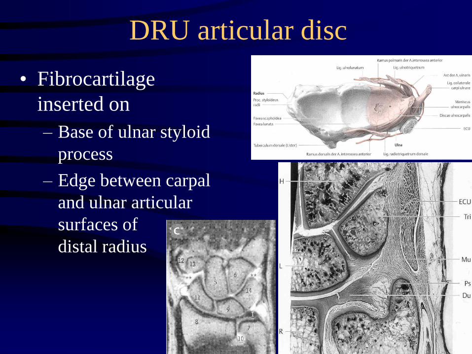
DRU articular disc
• Fibrocartilage
inserted on
– Base of ulnar styloid
process
– Edge between carpal
and ulnar articular
surfaces of
distal radius
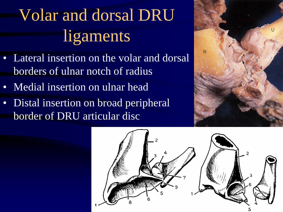
Volar and dorsal DRU
ligaments
• Lateral insertion on the volar and dorsal
borders of ulnar notch of radius
• Medial insertion on ulnar head
• Distal insertion on broad peripheral
border of DRU articular disc
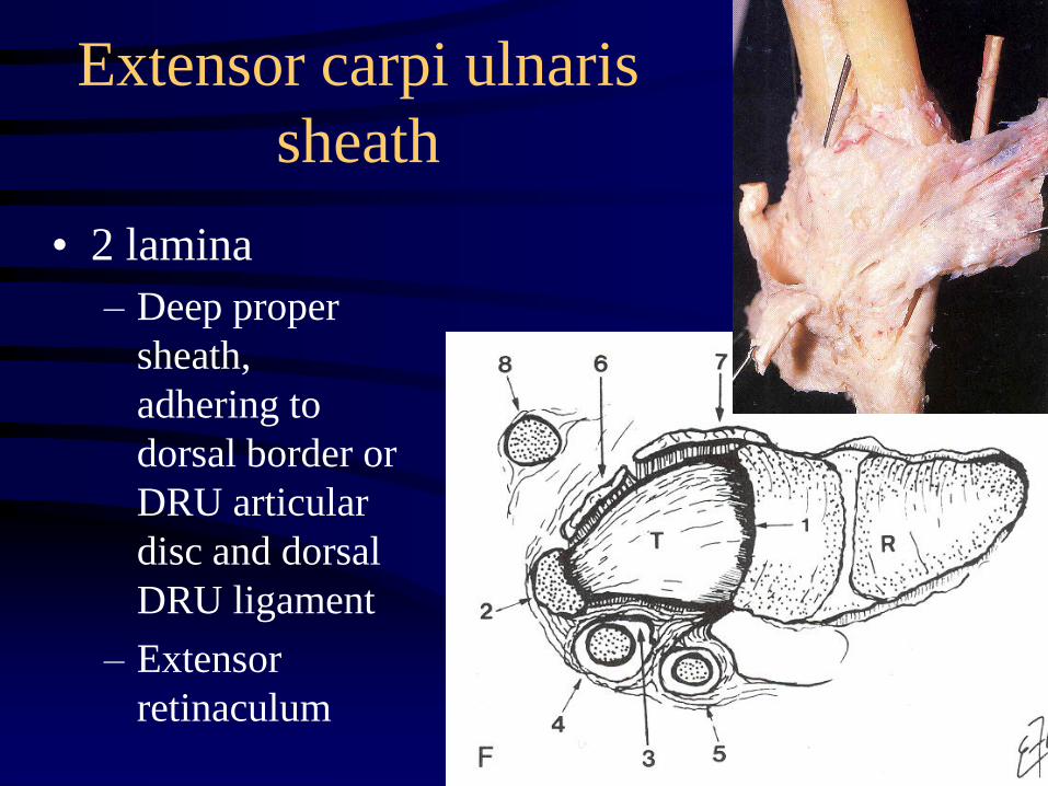
Extensor carpi ulnaris
sheath
• 2 lamina
– Deep proper
sheath,
adhering to
dorsal border or
DRU articular
disc and dorsal
DRU ligament
– Extensor
retinaculum
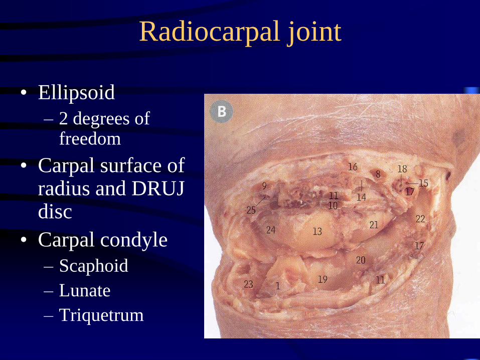
Radiocarpal joint
• Ellipsoid
– 2 degrees of freedom
• Carpal surface of radius and DRUJ disc
• Carpal condyle
– Scaphoid
– Lunate
– Triquetrum
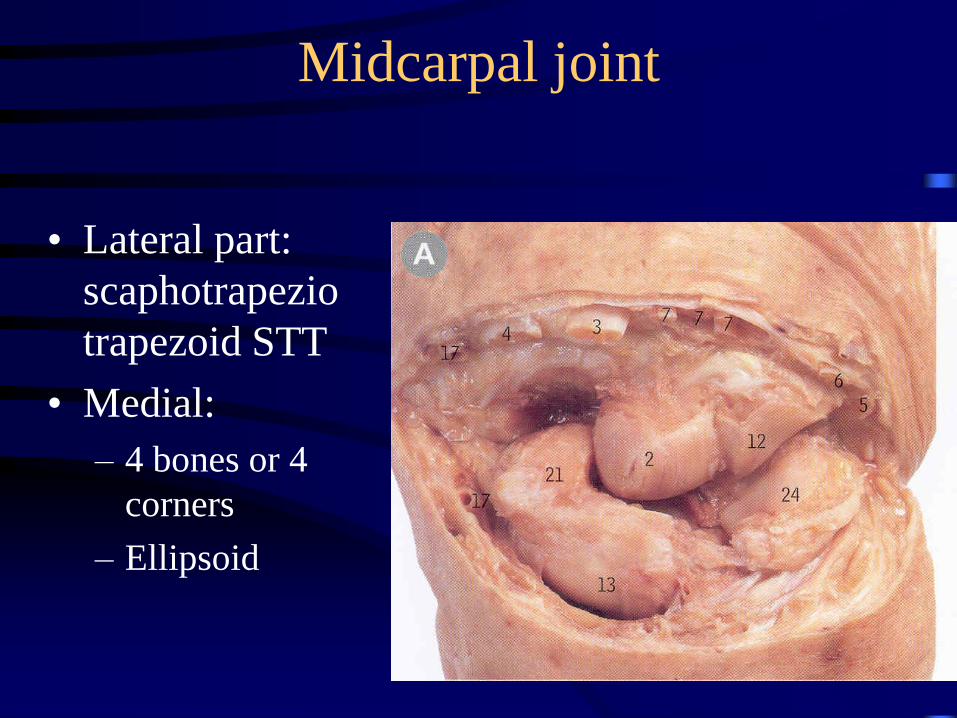
Midcarpal joint
• Lateral part:
scaphotrapezio
trapezoid STT
• Medial:
– 4 bones or 4
corners
– Ellipsoid
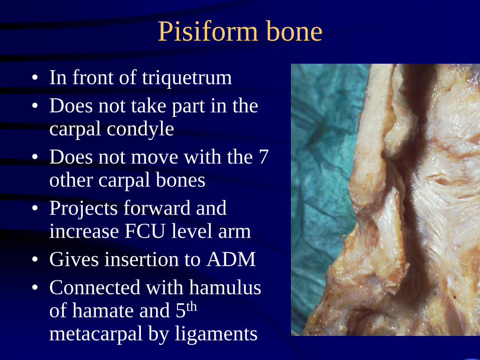
Pisiform bone
• In front of triquetrum
• Does not take part in the carpal condyle
• Does not move with the 7 other carpal bones
• Projects forward and increase FCU level arm
• Gives insertion to ADM
• Connected with hamulus of hamate and 5th
metacarpal by ligaments
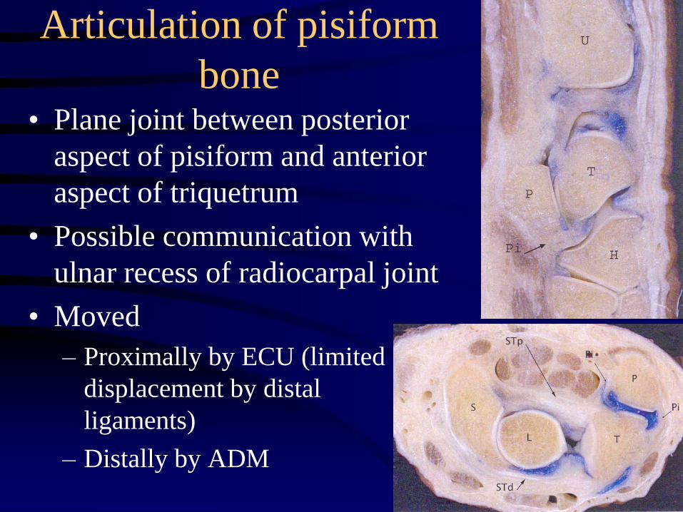
Articulation of pisiform
bone• Plane joint between posterior
aspect of pisiform and anterior
aspect of triquetrum
• Possible communication with
ulnar recess of radiocarpal joint
• Moved
– Proximally by ECU (limited
displacement by distal
ligaments)
– Distally by ADM
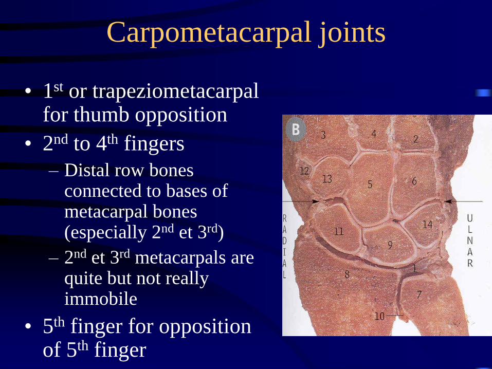
Carpometacarpal joints
• 1st or trapeziometacarpal for thumb opposition
• 2nd to 4th fingers
– Distal row bones connected to bases of metacarpal bones (especially 2nd et 3rd)
– 2nd et 3rd metacarpals are quite but not really immobile
• 5th finger for opposition of 5th finger
Means of union
• Capsular ligaments reinforcing capsule, joining mainly bones which do not belong to the same row
– Anterior or volar capsule
– Posterior or dorsal capsule
• Interosseous ligaments intraarticular, joining bones of the same row, strong and short (1-2 mm), whose surface is covered by fibrocartilage, in continuity with adjacent bone cartilage
• Distant ligaments : flexor retinaculum +/-extensor retinaculum
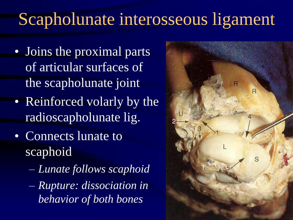
Scapholunate interosseous ligament
• Joins the proximal parts
of articular surfaces of
the scapholunate joint
• Reinforced volarly by the
radioscapholunate lig.
• Connects lunate to
scaphoid
– Lunate follows scaphoid
– Rupture: dissociation in
behavior of both bones
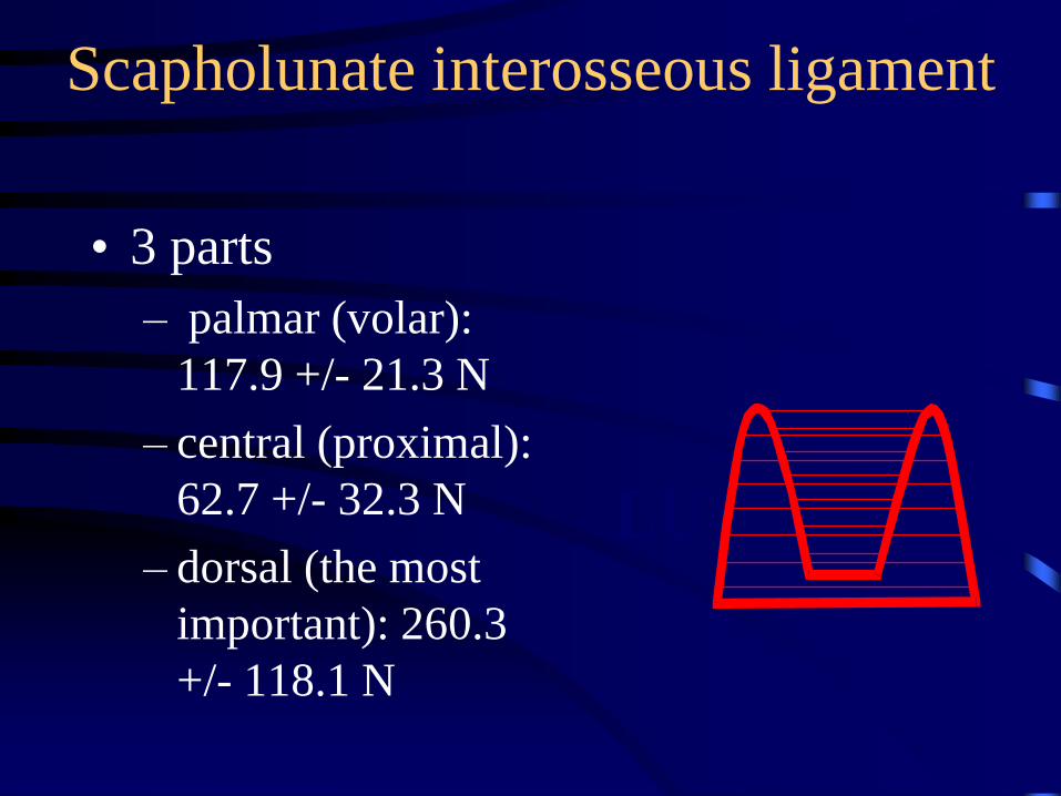
Scapholunate interosseous ligament
• 3 parts
– palmar (volar):
117.9 +/- 21.3 N
– central (proximal):
62.7 +/- 32.3 N
– dorsal (the most
important): 260.3
+/- 118.1 N
LU SC
RAD
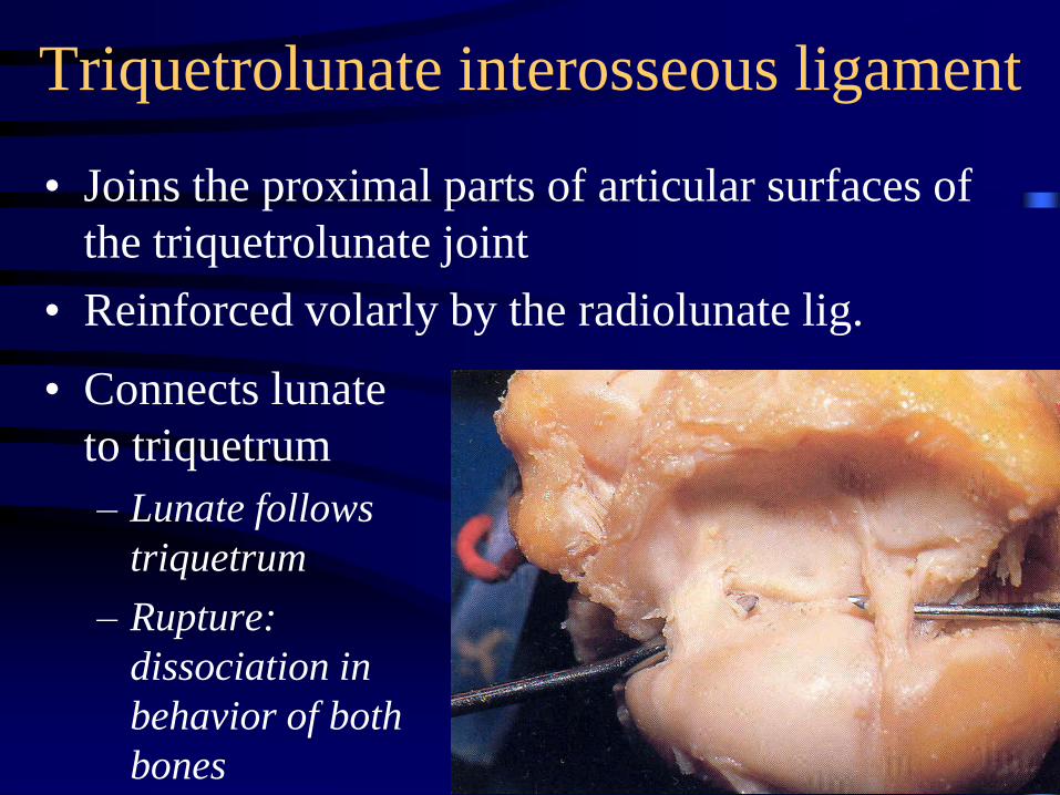
Triquetrolunate interosseous ligament
• Connects lunate
to triquetrum
– Lunate follows
triquetrum
– Rupture:
dissociation in
behavior of both
bones
• Joins the proximal parts of articular surfaces of
the triquetrolunate joint
• Reinforced volarly by the radiolunate lig.
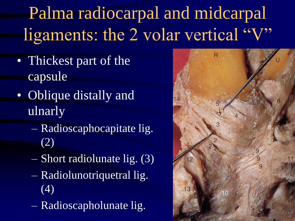
Palma radiocarpal and midcarpal
ligaments: the 2 volar vertical “V”
• Thickest part of the
capsule
• Oblique distally and
ulnarly
– Radioscaphocapitate lig.
(2)
– Short radiolunate lig. (3)
– Radiolunotriquetral lig.
(4)
– Radioscapholunate lig.
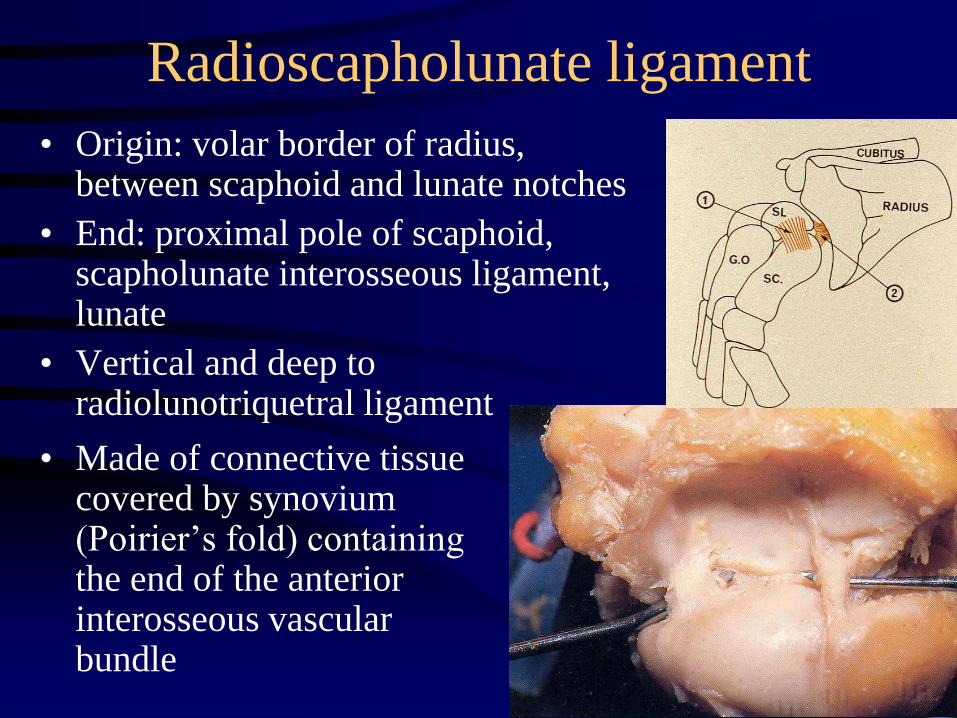
Radioscapholunate ligament
• Made of connective tissue covered by synovium (Poirier’s fold) containing the end of the anterior interosseous vascular bundle
• Origin: volar border of radius, between scaphoid and lunate notches
• End: proximal pole of scaphoid, scapholunate interosseous ligament, lunate
• Vertical and deep to radiolunotriquetral ligament
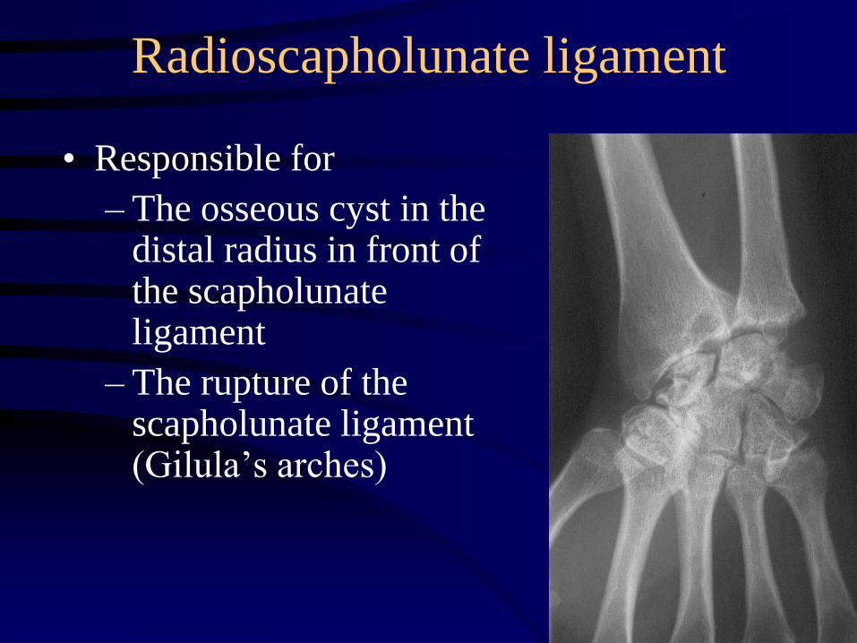
Radioscapholunate ligament
• Responsible for
– The osseous cyst in the distal radius in front of the scapholunate ligament
– The rupture of the scapholunate ligament (Gilula’s arches)
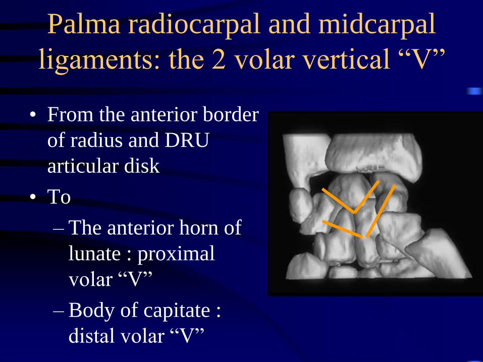
Palma radiocarpal and midcarpal
ligaments: the 2 volar vertical “V”
• From the anterior border
of radius and DRU
articular disk
• To
– The anterior horn of
lunate : proximal
volar “V”
– Body of capitate :
distal volar “V”
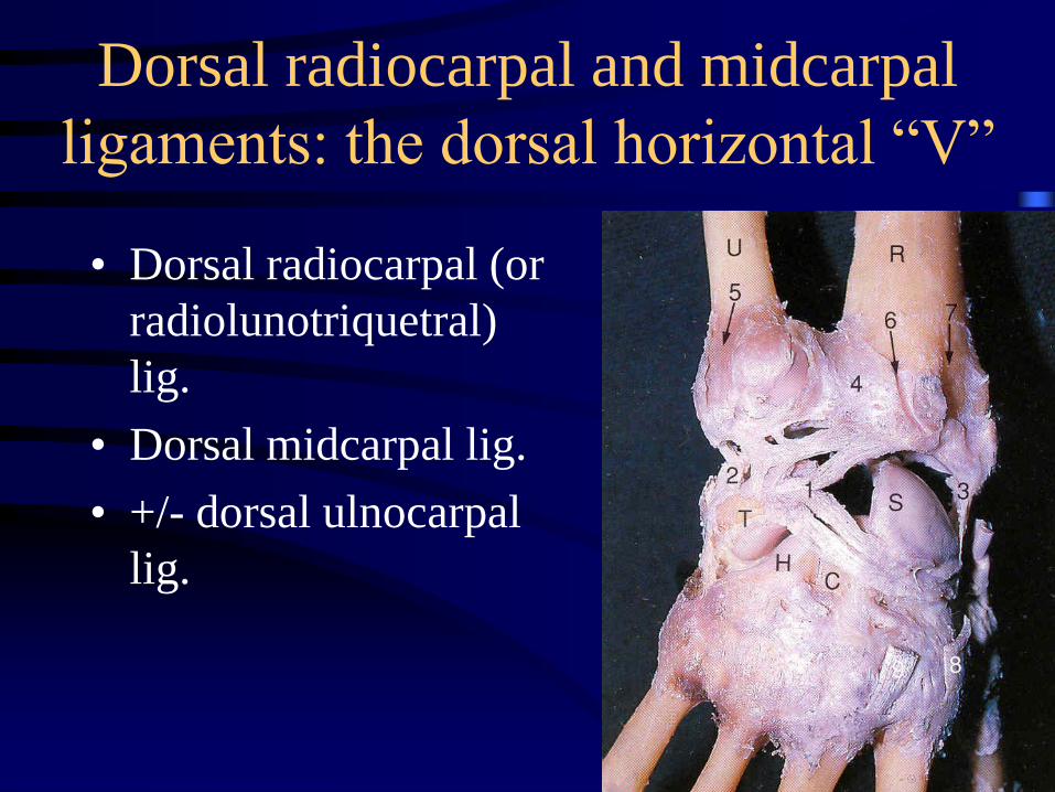
Dorsal radiocarpal and midcarpal
ligaments: the dorsal horizontal “V”
• Dorsal radiocarpal (or
radiolunotriquetral)
lig.
• Dorsal midcarpal lig.
• +/- dorsal ulnocarpal
lig.
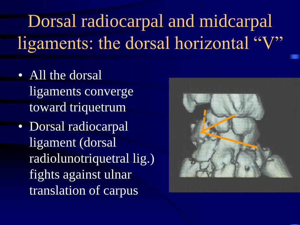
Dorsal radiocarpal and midcarpal
ligaments: the dorsal horizontal “V”
• All the dorsal
ligaments converge
toward triquetrum
• Dorsal radiocarpal
ligament (dorsal
radiolunotriquetral lig.)
fights against ulnar
translation of carpus
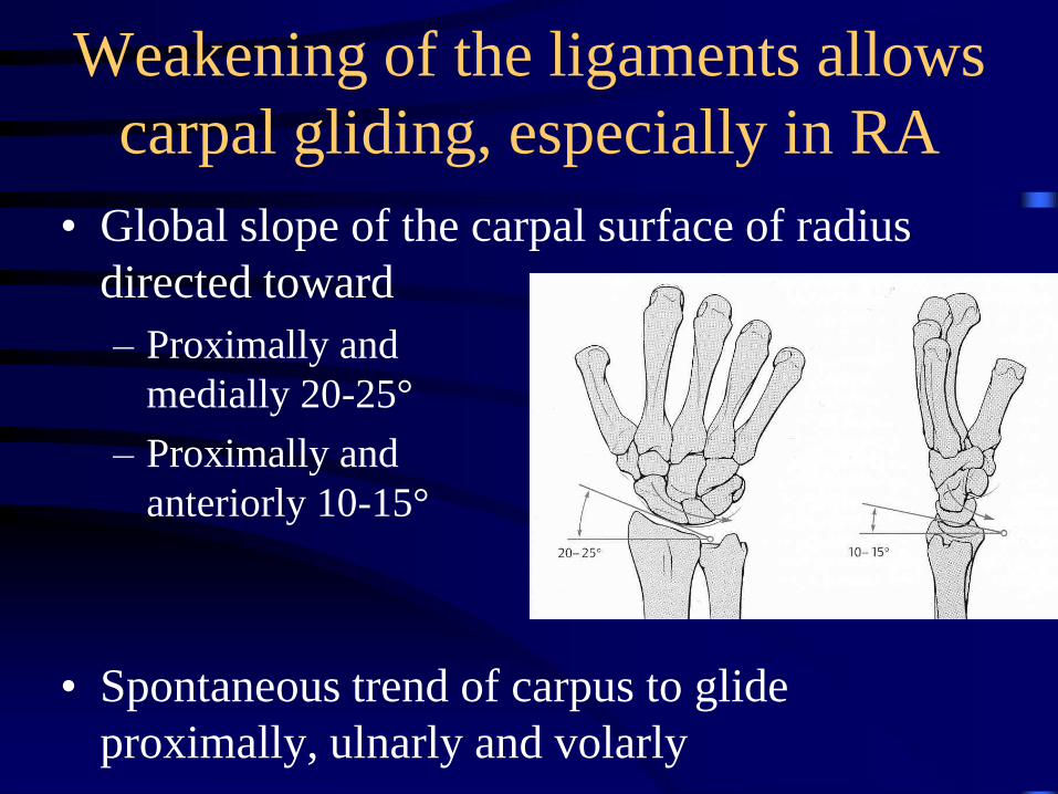
Weakening of the ligaments allows
carpal gliding, especially in RA
• Global slope of the carpal surface of radius
directed toward
– Proximally and
medially 20-25°
– Proximally and
anteriorly 10-15°
• Spontaneous trend of carpus to glide
proximally, ulnarly and volarly
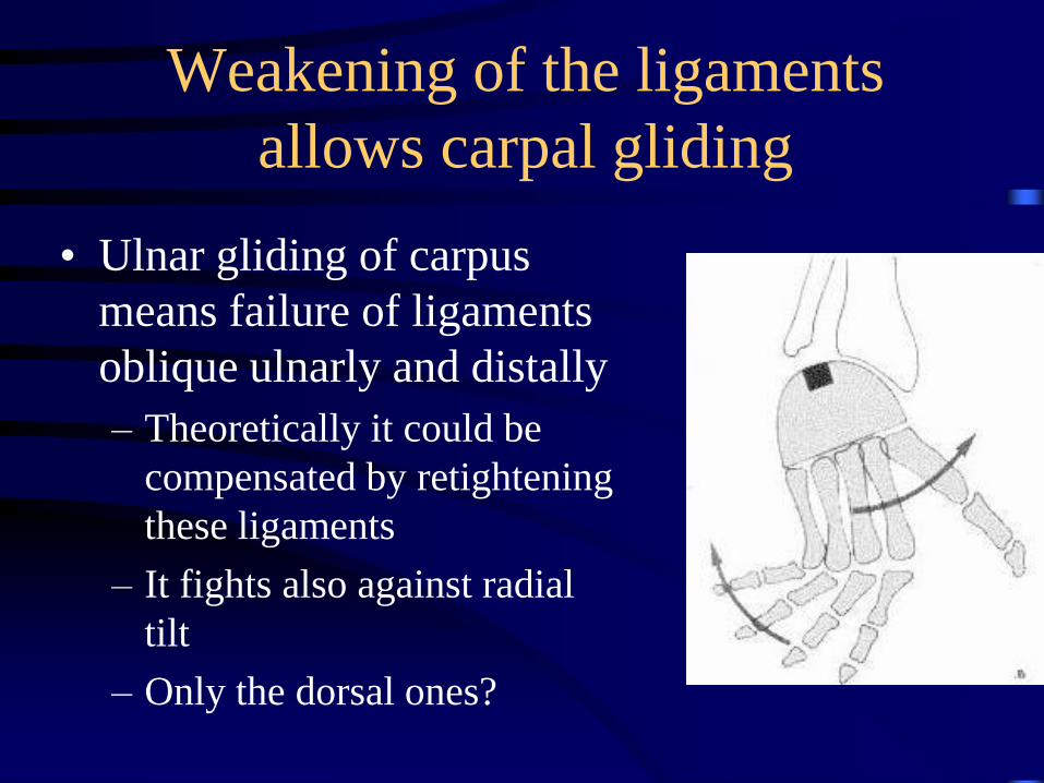
Weakening of the ligaments
allows carpal gliding
• Ulnar gliding of carpus
means failure of ligaments
oblique ulnarly and distally
– Theoretically it could be
compensated by retightening
these ligaments
– It fights also against radial
tilt
– Only the dorsal ones?
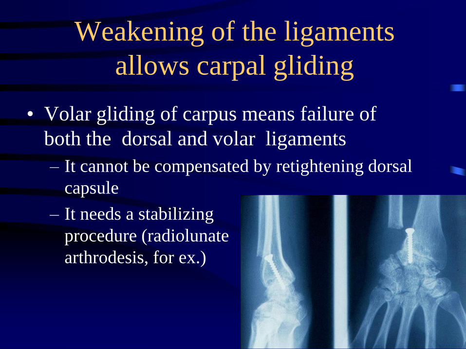
Weakening of the ligaments
allows carpal gliding
• Volar gliding of carpus means failure of
both the dorsal and volar ligaments
– It cannot be compensated by retightening dorsal
capsule
– It needs a stabilizing
procedure (radiolunate
arthrodesis, for ex.)
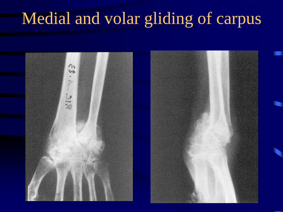
Medial and volar gliding of carpus
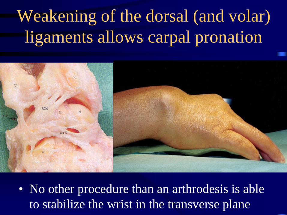
Weakening of the dorsal (and volar)
ligaments allows carpal pronation
• No other procedure than an arthrodesis is able
to stabilize the wrist in the transverse plane
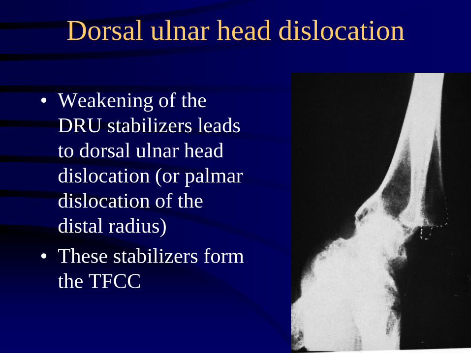
Dorsal ulnar head dislocation
• Weakening of the
DRU stabilizers leads
to dorsal ulnar head
dislocation (or palmar
dislocation of the
distal radius)
• These stabilizers form
the TFCC
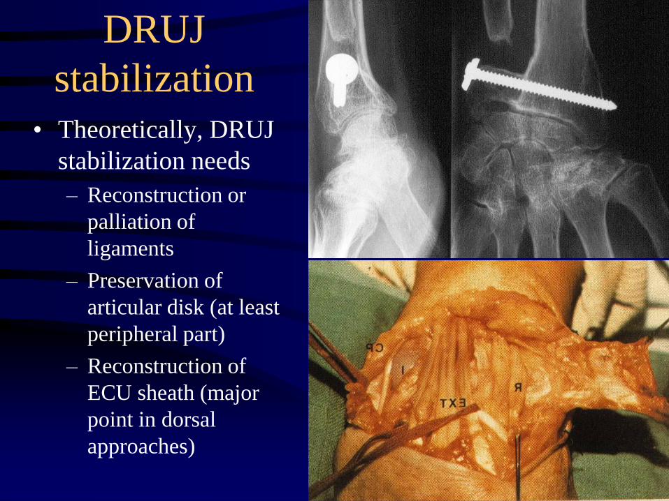
DRUJ
stabilization• Theoretically, DRUJ
stabilization needs
– Reconstruction or
palliation of
ligaments
– Preservation of
articular disk (at least
peripheral part)
– Reconstruction of
ECU sheath (major
point in dorsal
approaches)
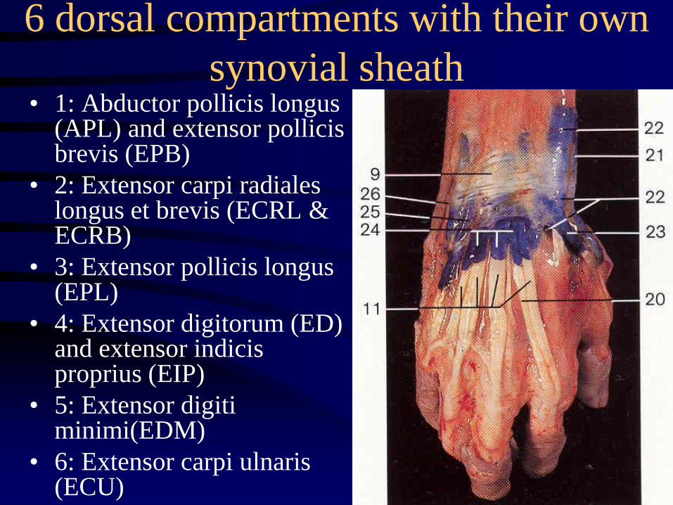
6 dorsal compartments with their own
synovial sheath• 1: Abductor pollicis longus
(APL) and extensor pollicis brevis (EPB)
• 2: Extensor carpi radiales longus et brevis (ECRL & ECRB)
• 3: Extensor pollicis longus (EPL)
• 4: Extensor digitorum (ED) and extensor indicis proprius (EIP)
• 5: Extensor digiti minimi(EDM)
• 6: Extensor carpi ulnaris (ECU)
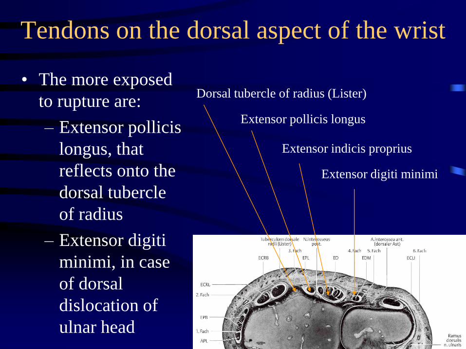
Tendons on the dorsal aspect of the wrist
• The more exposed
to rupture are:
– Extensor pollicis
longus, that
reflects onto the
dorsal tubercle
of radius
– Extensor digiti
minimi, in case
of dorsal
dislocation of
ulnar head
Dorsal tubercle of radius (Lister)
Extensor pollicis longus
Extensor indicis proprius
Extensor digiti minimi
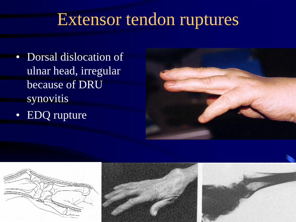
Extensor tendon ruptures
• Dorsal dislocation of
ulnar head, irregular
because of DRU
synovitis
• EDQ rupture
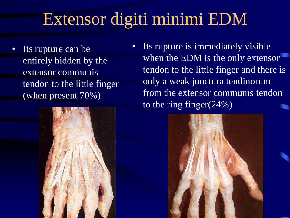
Extensor digiti minimi EDM
• Its rupture can be
entirely hidden by the
extensor communis
tendon to the little finger
(when present 70%)
• Its rupture is immediately visible
when the EDM is the only extensor
tendon to the little finger and there is
only a weak junctura tendinorum
from the extensor communis tendon
to the ring finger(24%)
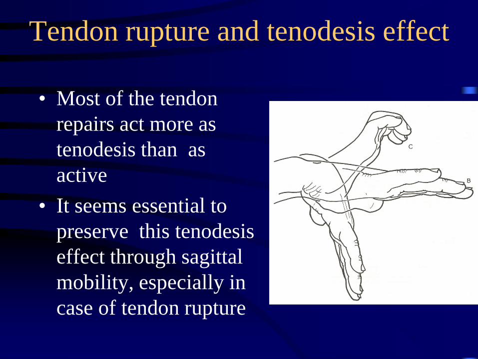
Tendon rupture and tenodesis effect
• Most of the tendon
repairs act more as
tenodesis than as
active
• It seems essential to
preserve this tenodesis
effect through sagittal
mobility, especially in
case of tendon rupture
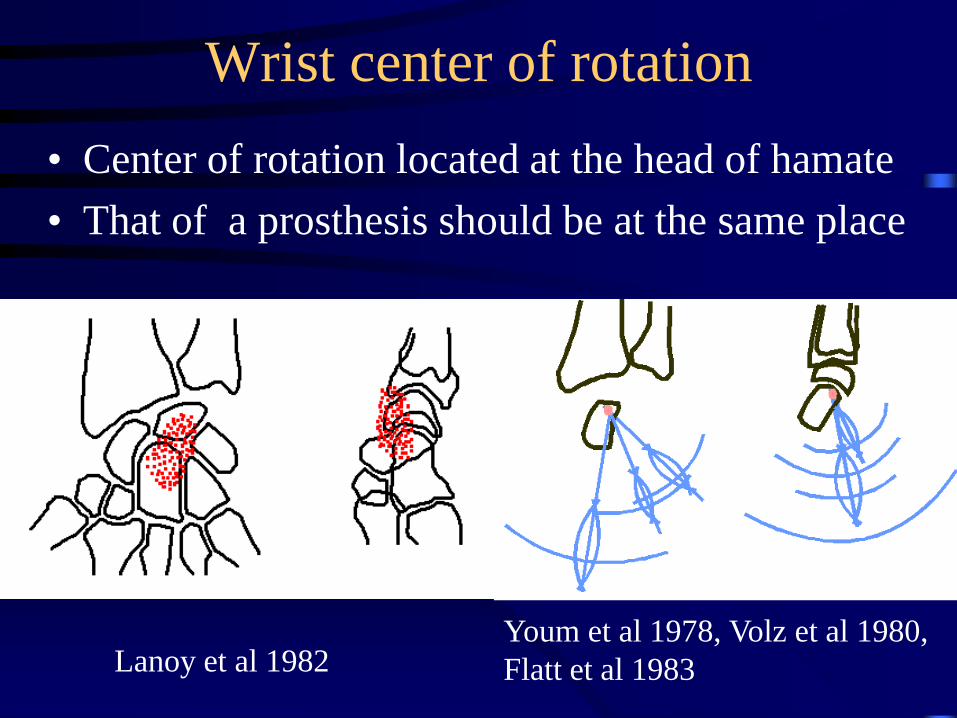
Wrist center of rotation
• Center of rotation located at the head of hamate
• That of a prosthesis should be at the same place
Lanoy et al 1982
Youm et al 1978, Volz et al 1980,
Flatt et al 1983
Wrist movements in the sagittal plane
• Flexion (palmar flexion) 85° according to
Kapandji (50° RC + 35° MC), 75° according
to Ryu
• Extension (dorsal flexion) 85° according to
Kapandji (35° MC + 50° MC), 70° according
to Ryu
• Partial carpal arthrodesis restricts mobility
asymmetrically
Wrist movements in the sagittal plane
• Unequal repartition between both joints : Part of radiocarpal joint in flexion
– 60% according to Kapandji (50 out of85°),
– 50% according to Viegas et al 1997, Patterson et al 1998
– 40% according to Sun et al 2000
• Unequal repartition between the 3 columns (Werner et al 1997)
– 90% in the lateral column between scaphoid and radius
– 65% in the medial column between radius and triquetrum
Wrist movements in the frontal plane
• Abduction (radial tilt) 25° according to Kapandji
(15° RC + 10° MC), 20° according to Ryu
• Adduction (ulnar tilt) 45° according to Kapandji
(20° RC + 25° MC), 40° according to Ryu
• Partial carpal arthrodesis restricts mobility
asymmetrically
Global wrist movements
• Useful mobility (Ryu et al)
– 80° in the sagittal plane
– 40° in the frontal plane
• Useful mobility (Brumfield 1984)
– Cleaning 10°/15°
– Daily living 5°/35°
• Most important movements occur in an oblique plane
– From extension and radial tilt
– To flexion and ulnar tilt
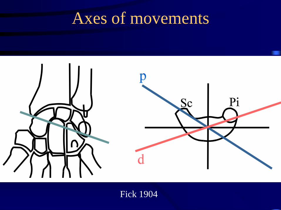
Axes of movements
Fick 1904
Static behavior of carpus in
neutral position
• Lunate distal surfaces turned volarly of
about the same value than the carpal surface
of radius
– Radiolunate angle 10° (from +15° to –20°)
– Capitolunate angle 10°
• Scaphoid belongs to both rows and bisects
between horizontal and vertical lines
– Scapholunate angle 55° (from 30° to 70°)
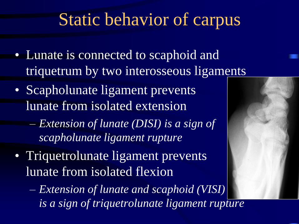
Static behavior of carpus
• Lunate is connected to scaphoid and
triquetrum by two interosseous ligaments
• Scapholunate ligament prevents
lunate from isolated extension
– Extension of lunate (DISI) is a sign of
scapholunate ligament rupture
• Triquetrolunate ligament prevents
lunate from isolated flexion
– Extension of lunate and scaphoid (VISI)
is a sign of triquetrolunate ligament rupture
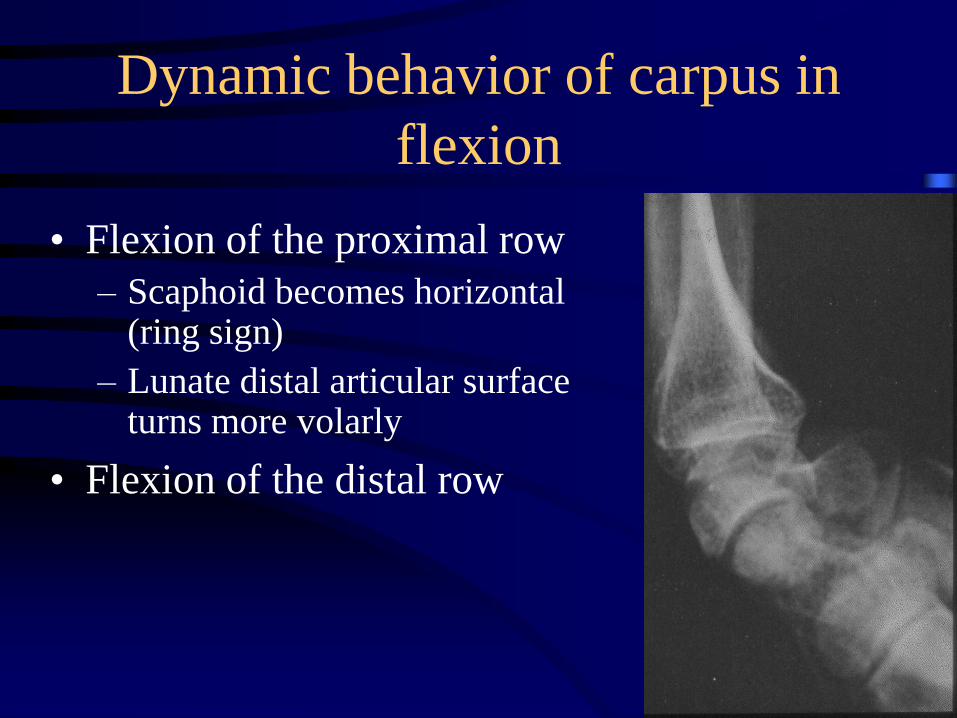
Dynamic behavior of carpus in
flexion
• Flexion of the proximal row
– Scaphoid becomes horizontal (ring sign)
– Lunate distal articular surface turns more volarly
• Flexion of the distal row
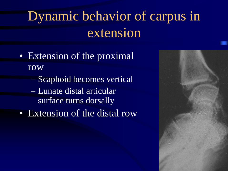
Dynamic behavior of carpus in
extension
• Extension of the proximal row
– Scaphoid becomes vertical
– Lunate distal articular surface turns dorsally
• Extension of the distal row
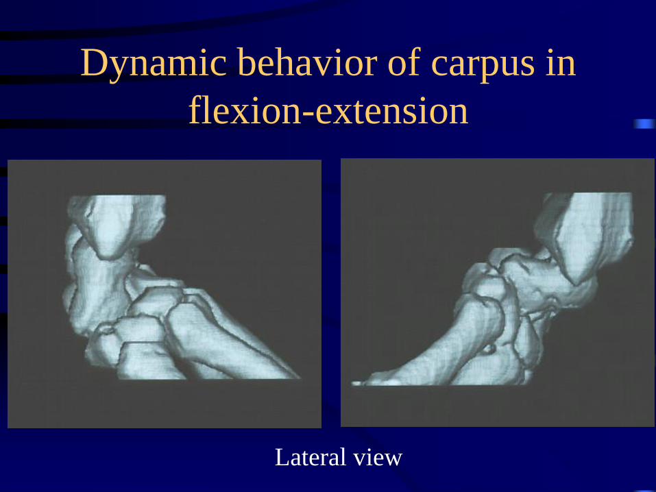
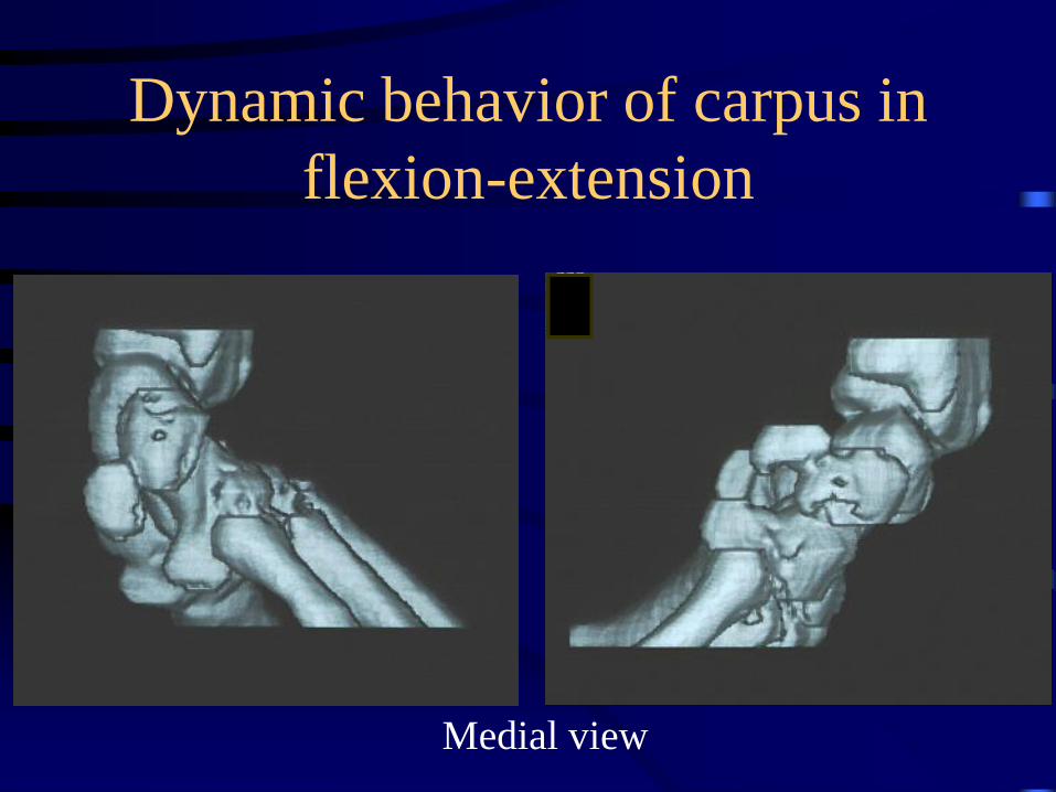
Dynamic behavior of carpus in
flexion-extension
Lateral view
Dynamic behavior of carpus in
flexion-extension
Medial view
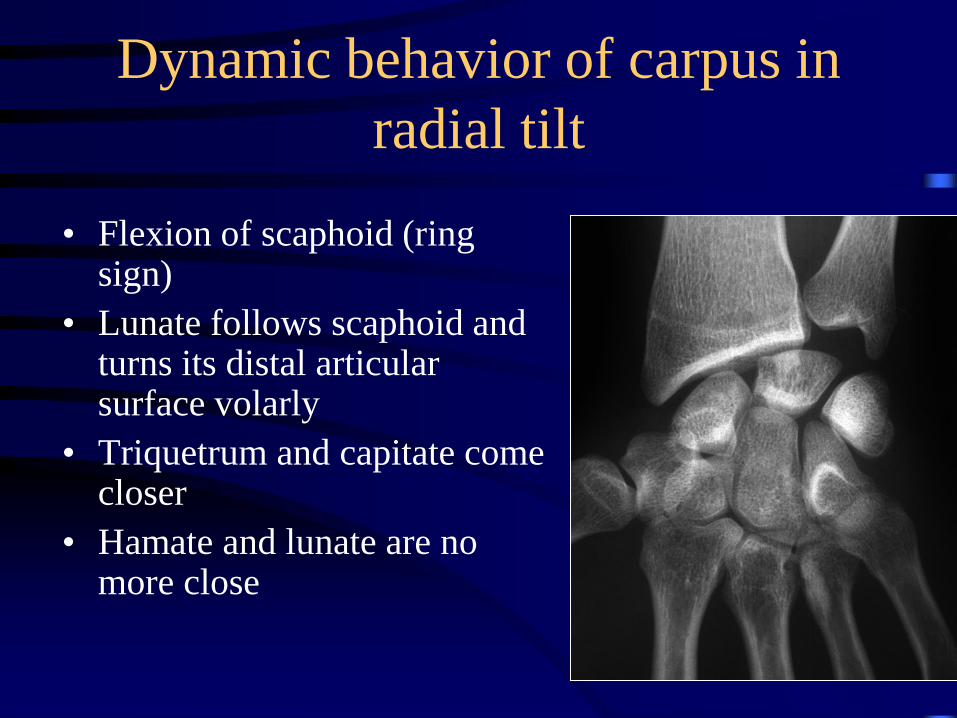
Dynamic behavior of carpus in
radial tilt
• Flexion of scaphoid (ring sign)
• Lunate follows scaphoid and turns its distal articular surface volarly
• Triquetrum and capitate come closer
• Hamate and lunate are no more close
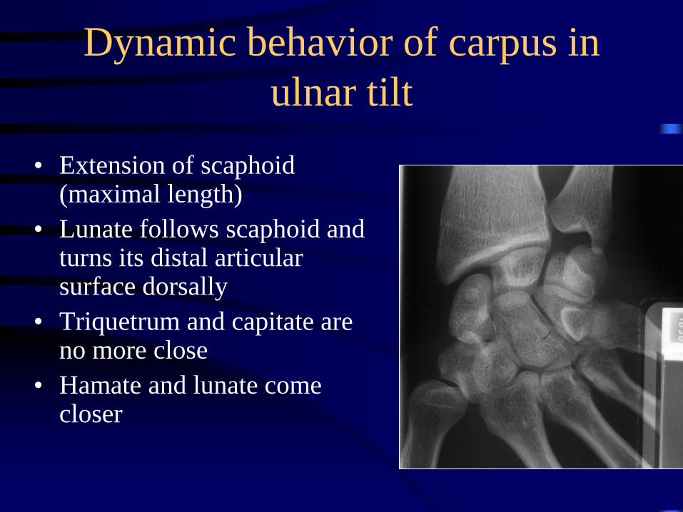
Dynamic behavior of carpus in
ulnar tilt
• Extension of scaphoid (maximal length)
• Lunate follows scaphoid and turns its distal articular surface dorsally
• Triquetrum and capitate are no more close
• Hamate and lunate come closer
Dynamic behavior of carpus
• The whole carpus changes its spatial arrangement,
but keeps a quite identical height
• Carpal height index (Youm et McMurtry): 0,54
+:- 0,03
• In case of destabilization, carpal bones rearrange
in order to present the radius the minimal height :
carpal collapse
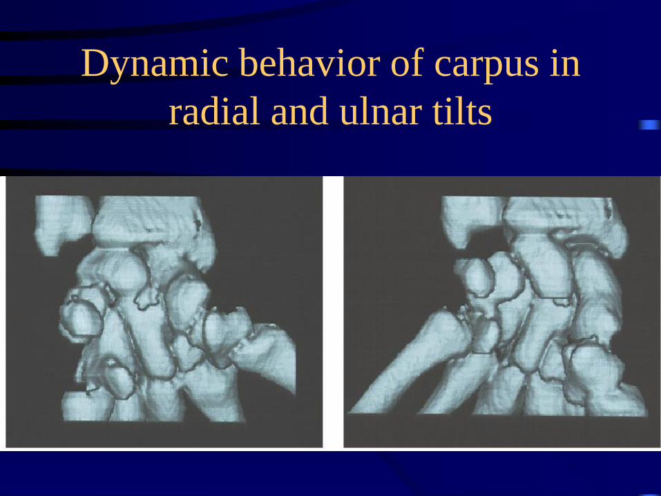
Dynamic behavior of carpus in
radial and ulnar tilts
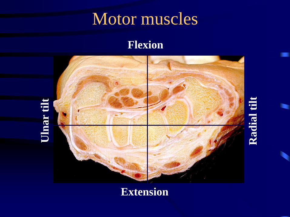
Motor muscles
Uln
ar
tilt
Rad
ial
tilt
Flexion
Extension
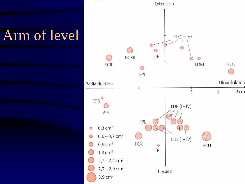
Arm of level
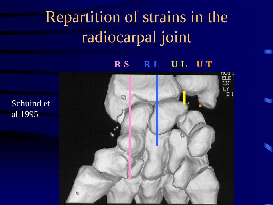
Repartition of strains in the
radiocarpal joint
Schuind et
al 1995
R-S R-L U-L U-T
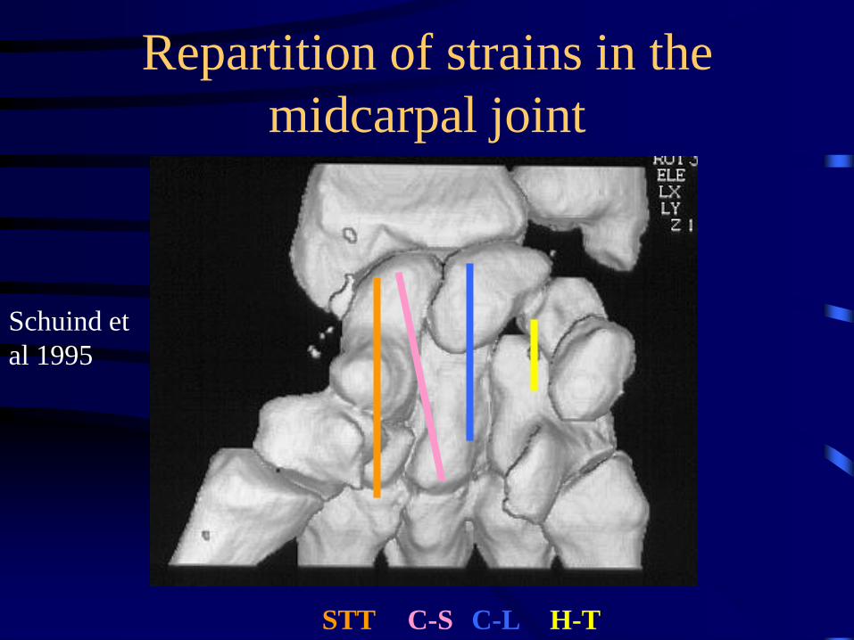
Repartition of strains in the
midcarpal joint
STT C-S C-L H-T
Schuind et
al 1995
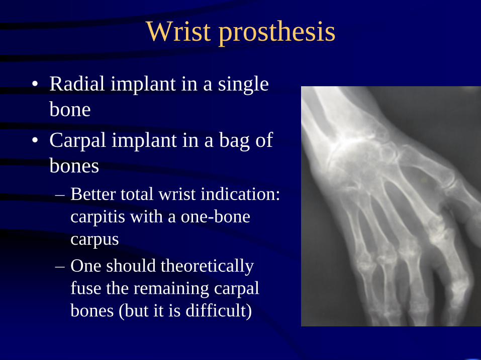
Wrist prosthesis
• Radial implant in a single
bone
• Carpal implant in a bag of
bones
– Better total wrist indication:
carpitis with a one-bone
carpus
– One should theoretically
fuse the remaining carpal
bones (but it is difficult)
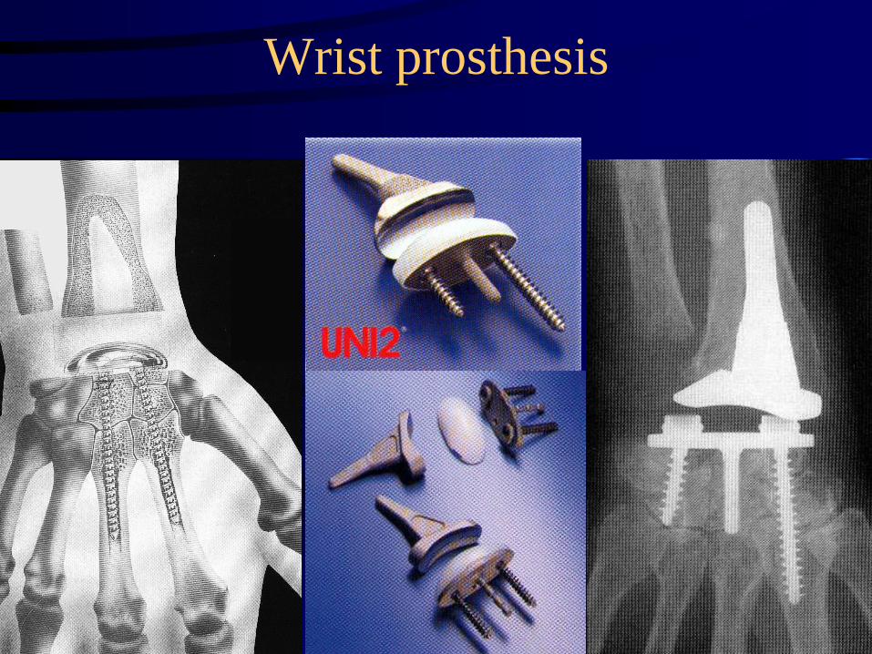
Wrist prosthesis
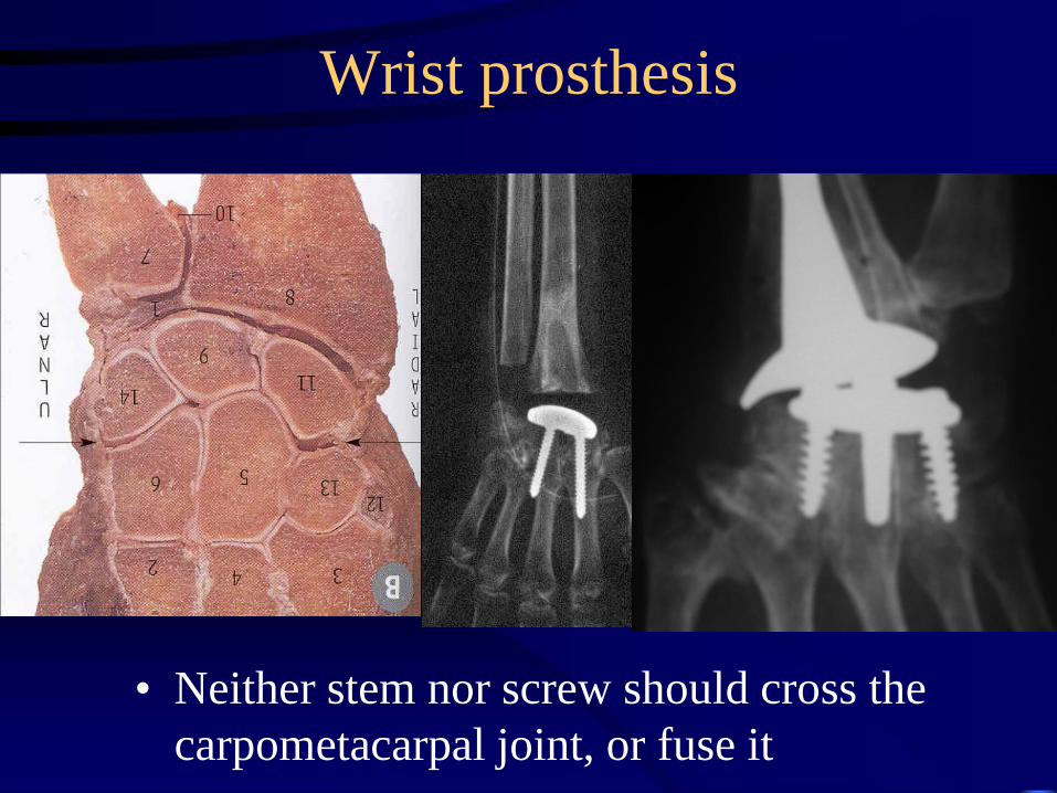
Wrist prosthesis
• Neither stem nor screw should cross the
carpometacarpal joint, or fuse it