Embed Size (px)
Citation preview
Chemical Geology, in press
Annealing radiation damage and the recoveryof cathodoluminescence
Lutz Nasdalaa, b, *, Christian L. Lengauerb, John M. Hancharc, Andreas Kronzd,Richard Wirthe, Philippe Blancf, Allen K. Kennedyg, Anne-Magali Seydoux-Guillaumee,†
a Institut für Geowissenschaften – Mineralogie, Johannes Gutenberg-Universität, D-55099 Mainz, Germany b Institut für Mineralogie und Kristallographie, Universität Wien – Geozentrum, Althanstr. 14, A-1090 Wien, Austria
c Department of Earth and Environmental Sciences, The George Washington University, Washington, DC 20052, U.S.A.d Göttinger Zentrum Geowissenschaften, Goldschmidtstr. 1, D-37077 Göttingen, Germany
e GeoForschungsZentrum Potsdam, Telegrafenberg, D-14473 Potsdam, Germanyf Université Pierre et Marie Curie, 4, place Jussieu, 75252 Paris Cedex 05, France
g Department of Applied Physics, Curtin University of Technology, Kent St., Bentley 6102 WA, Australia
* Corresponding author.Tel. (+49)-6131-3924781; FAX (+49)-6131-3923070; e-mail: [email protected]
† present address: Institut für Planetologie, Westfälische Wilhelms-Universität Münster, Wilhelm-Klemm-Str. 10, D-48149 Münster,Germany
ABSTRACT
The structural recovery upon heat-treatment of a highly metamict, actinide-rich zircon (U ≈ 6000 ppm) has been studied in detailby a complex of techniques including X-ray powder diffraction, Raman spectroscopy, SHRIMP ion probe, electron microprobe,transmission electron microscopy and cathodoluminescence analysis. The structural regeneration of the amorphous starting materialdepends on random nucleation. It starts between 800 and 950°C when amorphous ZrSiO4 decomposes to form crystalline ZrO2 andamorphous SiO2. At around 1100°C, well crystallised ZrSiO4 grows at the expense of the oxides. U has been retained in the newlygrown zircon whereas Pb was evaporated during the heat-treatment. This process is in marked opposition to the reconstitution ofmoderately metamict minerals, which experience a gradual recovery controlled by the epitaxial growth at the crystalline-amorphousboundaries. Both of these recovery processes are not the direct inverse of metamictisation. The structural regeneration was found to beconnected with a significant increase in the emission of CL. In all cases (annealing heavily damaged zircon and moderately damagedzircon and monazite) we observe that the final, well crystallised annealing products emit more intense CL than their radiation-damagedstarting minerals, even though having almost identical elemental composition. Our observations are taken as evidence that the CL is notonly determined by the chemical composition of the sample but is also strongly controlled by structural parameters such as crystallinityor the presence of defect centres.
Keywords: radiation damage; thermal annealing; cathodoluminescence; Raman spectroscopy; X-ray powder diffraction; SHRIMP ionprobe analysis; transmission electron microscopy
1. Introduction
Self-irradiation over geologic timescales due to theradioactive decay of incorporated actinide elements maytransform initially crystalline minerals into an amorphous statethat is called “metamict”. Even though the study of metamictminerals has a long history (e.g., Hurley and Fairbairn, 1953;Holland and Gottfried, 1955), the process of radiation damagegeneration and retention in mineral structures has not yet been
fully clarified. One of the main goals of such studies was tounderstand the causes of different properties of radiation-damaged minerals, such as their generally enhanced chemicalreactivity and decreased physical resistance, when comparedwith their crystalline analogues (e.g., Ewing, 1994; Lumpkin,2001). In the past, most such research was initiated bygeochronologists because understanding the structural causes ofthe strongly enhanced susceptibility of metamict minerals tosecondary Pb loss (Silver and Deutsch, 1963; Nasdala et al.,
2
1998) is crucial for the interpretation of U-Th-Pb dating results.The interest in studying radiation damage phenomena hasincreased in the past two decades, stimulated inter alia by thenecessity to find durable host materials for the safe long-termstorage of radioactive waste such as the highly radioactive andpoisonous Pu from disarmed nuclear weapons (e.g., Ewing,1999; Wang et al., 1999).
Natural actinide-bearing minerals such as zircon (e.g.,Murakami et al., 1991; Weber et al., 1994) and monazite(Meldrum et al. 1998) may cover the full range from littledamaged by radiation to entirely metamict. The real structure ofradiation-damaged minerals has been investigated with a widevariety of analytical techniques (Ewing, 1994). Additionalinformation is obtained by studying their behaviour when treatedwith high temperature, high pressure or in hydrothermalexperiments (e.g., Pidgeon et al., 1966; Mursic et al., 1992;Weber et al., 1994; Ellsworth et al., 1994). Annealingexperiments were initially done to eliminate the radiation damagestep-by-step and, thus, to retrace the formation of metamictstructures back to their crystalline origin. We have learned,however, that none of the different recovery processes (forexample, long-term annealing over geologic timescales, naturalrecrystallisation processes, or heat-treatment in the laboratory) isthe direct inverse of metamictisation. Annealing experiments are,nevertheless, a powerful tool for the study of metamict structures,particularly in view of the fact that the damage recovery must bestrongly controlled by the actual microstructure of the radiation-damaged mineral.
It has already been described in several annealing studies thatmoderately metamict samples, which have a domain structureconsisting of amorphous and little to heavily damaged crystallinenano-areas (Murakami et al., 1991), undergo a gradual recoveryprocess upon annealing (e.g., Colombo et al., 1999a; Zhang et al.,2000b). Their structural recovery is a complicated process thatinvolves, for example, random nucleation in the amorphousphase, epitaxial growth of the crystalline domains in favour oftheir surrounding remnant amorphous areas, recombination ofFrenkel defect pairs in the crystalline phase through point defectdiffusion (e.g., Weber et al., 1994), structural recovery as a resultof He diffusion and loss (cf. Seydoux-Guillaume et al., 2002a),and the escape of hydrous species (Nasdala et al., 2001b). Eventhough these processes may have different relationshipsdepending on temperature, duration of heating and otherconditions, the heating-induced gradual recovery of moderatelymetamict minerals is most probably strongly controlled by theepitaxial growth at crystalline-amorphous boundaries.
Highly amorphised minerals, however, react differently tothe heat-treatment. Because crystalline remnants are lacking,their epitaxial growth is impossible and the structural recoverymust depend on the nucleus formation. In addition, it has alreadybeen observed in several studies that the recovery of highlymetamict zircon involves an intermediate stage of ZrO2
formation (e.g., Weber, 1993; Weber et al., 1994; McLaren et al.,1994; Capitani et al., 2000; Begg et al., 2000; Zhang et al.,2000b, c). However, no comparative documentation of thisprocess comprising long-range and short-range methods has beendocumented so far. In the present paper we report a detailedinvestigation of the structural regeneration of a highly metamictzircon upon heat-treatment.
The products of our and previous annealing experimentswere also subjected to a CL (cathodoluminescence) investigation.It is still controversial as to which features of the chemicalcomposition and/or structural state affect the generation ofluminescence when irradiating minerals with an electron beam.Cathodoluminescence images of minerals allow one to recogniseinternal zones and micro-areas of different chemical composition(e.g., Vavra, 1990; Paterson et al., 1992; Hanchar and Miller,1993). It has also been demonstrated that the CL correlatesclosely with the pattern of heterogeneous metamictisation(Nasdala et al., 2001a). It has, however, not been clarified so farwhether (1) the latter is only an apparent correlation, caused bythe dependence of metamictisation on the chemical zoning thatcontrols the CL, or (2) the structural radiation damage has causaleffects on the CL emission. This question can be answered byinvestigating samples having the same chemical composition butdifferent degrees of radiation damage. Annealing products seemto be an ideal case for this and, therefore, we have studied themby means of CL imaging and spectral CL.
2. Experimental
In this paper we mainly investigate the annealing of a highlymetamict but, nevertheless, gemstone-quality zircon from SriLanka (sample number N17). Results of CL investigations forthree other samples that have been annealed and characterised inprevious studies (zircons K1 and K2, Nasdala et al., 2001b;monazite Moacir; Seydoux-Guillaume et al., 2002a) areadditionally reported.
Zircon N17 is a clear, dark green, cut stone of 4.6 ct (0.92 g)
weight and about 7.5 × 11 × 5 mm size. Slices between 200 µmand 1 mm in size were used for heating experiments in air. Wedid not do stepwise heating, rather we started always withfragments of the untreated zircon. The slices were placed in a 1ml Pt crucible and heated up to the desired annealing temperature
(500, 800, 950, 1100, 1250, 1400 and 1500°C) at a rate of ≈ 30degrees per minute. At the end of the 150 h run, the furnace wasswitched off. After about 1 h the temperature had decreased toca. 300-500 °C and then the furnace door was slightly opened,thus again increasing the cooling speed. The crucible was taken
out of the furnace after another 30 min (then at ≈100-150°C) andcooled down to room temperature within a few minutes.
Cathodoluminescence images were taken using an OXFORDINSTRUMENTS CL System interfaced to a JEOL 8900 RLelectron microprobe. The accelerating voltage was 20 kV and thebeam current was 50 nA. To avoid effects of varyingexperimental conditions on the observed CL intensity, samples tobe compared were brought simultaneously in the vacuumchamber and were measured under identical conditions, i.e.equally sized areas were scanned for the same time and imageswere obtained with constant signal amplification. Conclusionsabout absolute CL intensities are still limited, however, relativecomparison is possible between simultaneously obtained images.Because chemical compositions have been determined inprevious studies (Nasdala et al., 2001b; Seydoux-Guillaume etal., 2002a), only two fragments zircon N17 (untreated andannealed at 1400°C) were subjected to multiple electronmicroprobe analyses, to check again for potential compositionalchanges upon annealing
3
Cathodoluminescence spectra of samples N17 and Moacirwere obtained using a JEOL 840A scanning electron microscopeoperated at 35 kV and 50 nA. The luminescence signal was ledinto an external optical spectrometer by placing an OPEAparabolic collector in the beam path. Signal analysis was done bymeans of two different external systems. Spectra in the range200-500 nm were obtained using an INSTRUMENTS S. A.H10UV spectrometer equipped with GaAs cathode-basedphotomultiplier. The spectral resolution was 8 nm. Spectra in therange 300-900 nm were obtained using a JOBIN YVON TRIAXtriple monochromator equipped with liquid nitrogen-cooled CCD(charge-coupled device) detector, with a spectral resolution ofbetter than 4 nm. All spectra were normalised to the quantumefficiency of the respective detector.
U, Th and Pb contents and U-Pb isotopic ratios for thezircons were determined by means of the SHRIMP (SensitiveHigh mass Resolution Ion MicroProbe) II at the Department ofApplied Physics, Curtin University of Technology, Perth. Thegeneral SHRIMP technique has been described in more detailelsewhere (e.g., Compston et al., 1984; Vavra et al., 1996;Nelson, 1997) and the performance characteristics of the CurtinSHRIMP II was documented by De Laeter and Kennedy (1998).The SHRIMP was operated at 10 kV and with a mass resolution
(M/∆M) better than 5000, and the sensitivity for lead isotopeswas in the range 14 to 20 counts per second per ppm. Data werecalibrated using the Curtin University CZ3 standard zircon(Pidgeon et al., 1994). The 204Pb method was employed for thecorrection for common Pb. For further experimental details seeNasdala et al. (1999b).
Raman microprobe measurements were done using a JOBINYVON LabRam-HR (high resolution; this long-geometry versionof the LabRam system has, due to its focal length of 850 mm, athreefold improved spectral resolution). The Raman system was
equipped with OLYMPUS optical microscope (100× objective;numerical aperture 0.9). Spectra were excited with the He-Ne632.8 nm line (3 mW at the sample surface). The wavenumber
accuracy was ± 0.5 cm-1 and the spectral resolution was about 0.4cm-1. The degree of metamictisation of zircons was estimatedfrom the FWHM of the B1g Raman band at about 1000 cm-1
(Nasdala et al., 1995). Such quantitative estimation of theradiation damage is not possible in the case of monazite, becausechemical variations may greatly affect the Raman spectrum ofthis mineral. We have, however, used the FWHM of the
monazite A1g mode at ≈ 974 cm-1 as a relative measure of therecovery of the short-range order upon annealing (cf. Seydoux-Guillaume et al., 2002a). For further experimental details,correction of measured FWHMs for the apparatus function anderrors in the estimation of metamictisation from Raman data seeNasdala et al. (2001a).
X-ray powder data for zircon N17 and its annealing productswere collected on a PHILIPS PW 3050 based X'PERT MPDdiffractometer in Debye-Scherrer geometry equipped with an in-house built capillary spinner and a RAYTECH PSD (positionsensitive detector). Zircon fragments were ground in an agatemortar and pestle and filled into a Linde-glass capillary(Hilgenberg, no. 14) with an inner diameter of 0.28 mm. For the
monochromatic, parallel-beam Cu Kα X-ray source (λ = 1.54056Å) a ceramic-type X-ray copper tube (45 kV, 40 mA) with acombination of a multilayer X-ray mirror and a channel-cut Gecrystal (PHILIPS) was used. A soller slit of 1.15 ° axial
divergence was inserted on the primary side to reduce the peakasymmetry. The area of the primary X-ray beam irradiating the
glass capillary was limited to 0.7 × 6.0 mm2, thus resulting in anilluminated sample volume of roughly 0.4 mm3 per sample. Thegoniometer radius of the secondary side was 343.8 mm, which
corresponds to an angular resolution of 6 mm/° 2θ and a physical
detector resolution of 0.011 °2θ. Measurements were performedat 24(2)°C in a continuous mode, using a twofold data collection,
each set over the range 10-100 °2θ with a step interval of 0.011
°2θ, a scan time of 11.6 h, and an angular PSD opening of 4 °2θ.This was equivalent to a summed counting time of 3600 sec/step.The determination of cell parameters and FWHMs was doneusing the PC-Rietveld Plus package of Fischer et al. (1993).Standard deviations were corrected according to Berar andLelann (1991). The background was set manually and theabsorption correction was calculated with the values forcylindrical samples according to Dwiggins (1975). For the peak-shape simulation, the pseudo-Voigt function was used. Angulardependencies of FWHMs were detemined using the formula ofCagliotti et al. (1959). Semiquantitative results were calculatedas described by Hill and Howard (1987). For the Rietveldrefinements the atomic coordinates and displacement factorsgiven by Mursic et al. (1992) and Howard et al. (1988) wereused. Ionic X-ray scattering factors were taken from Cromer andWaber (1974; for Zr4+ and Si4+) and Hovestreydt (1983; for O2-).
TEM (transmission electron microscope) investigations weredone by means of a PHILIPS CM200 system equipped withEDAX X-ray analyser and GATAN imaging filter. Samples werefirst grind and then prepared by conventional Ar ion beamthinning and subsequently carbon coated. The electronmicroscope was operated at a voltage of 200 kV. For moreexperimental details see Wirth et al. (2001a, b).
3. General characterisation of samples
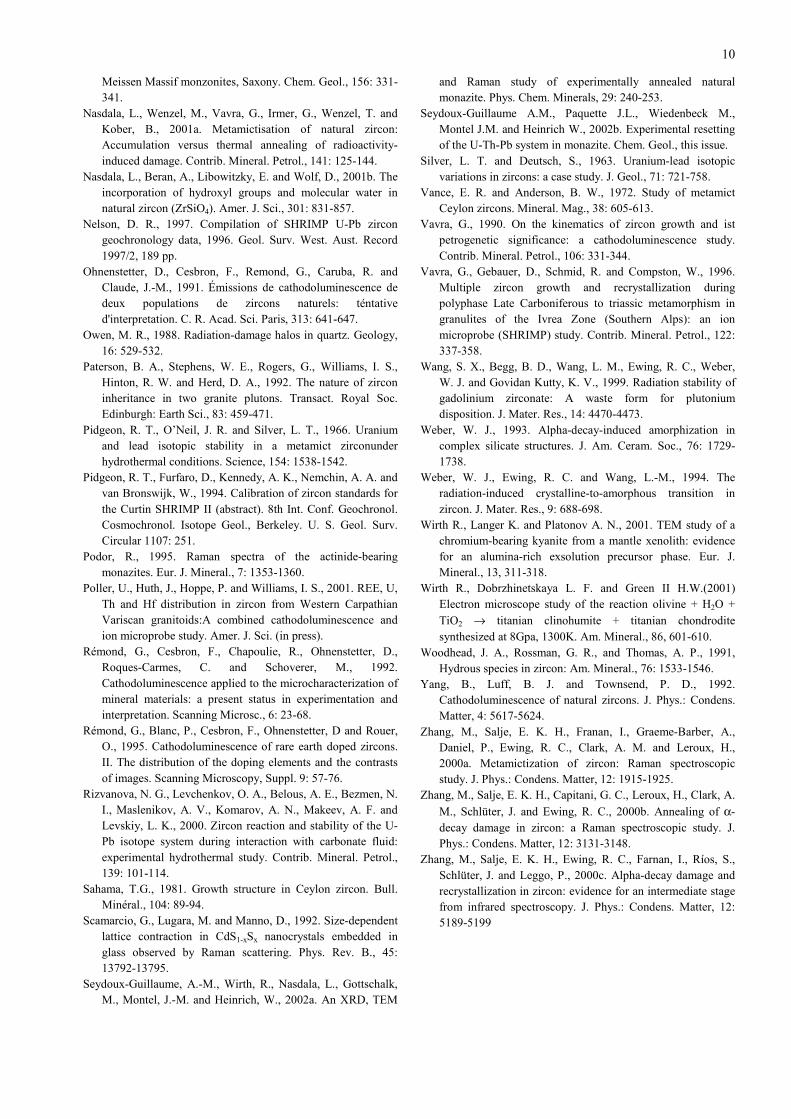
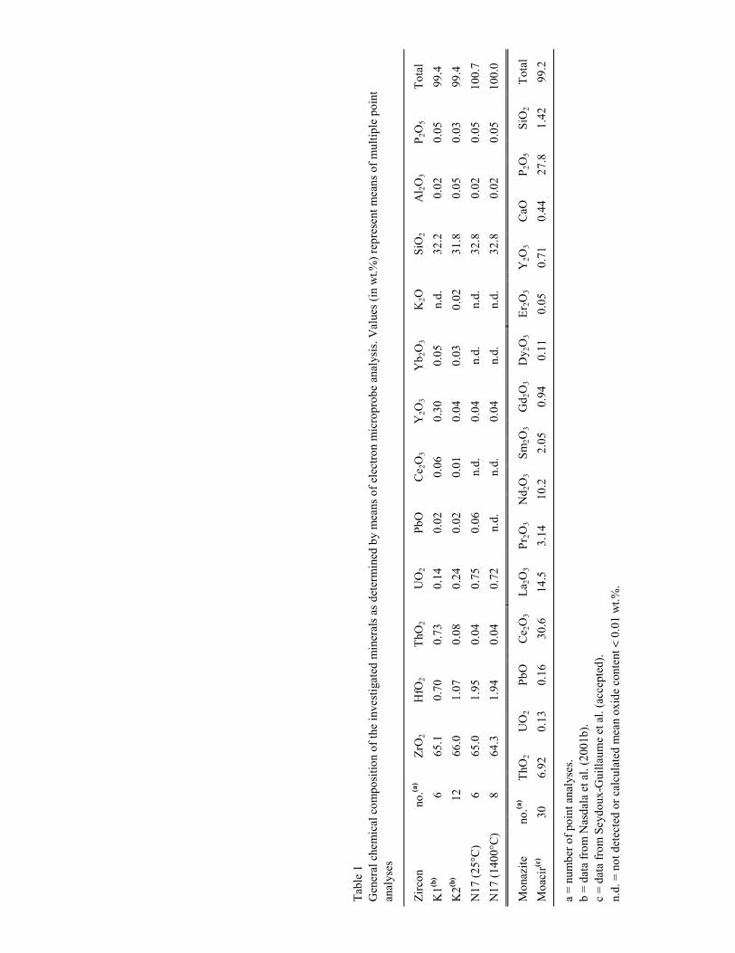
A general characterisation of the chemical compositions ofthe four mineral samples under investigation is presented inTable 1 and structural parameters (powder diffraction and Ramandata) are listed in Table 2. Note the comparably high actinidecontent of all four samples.
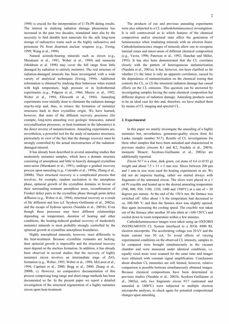
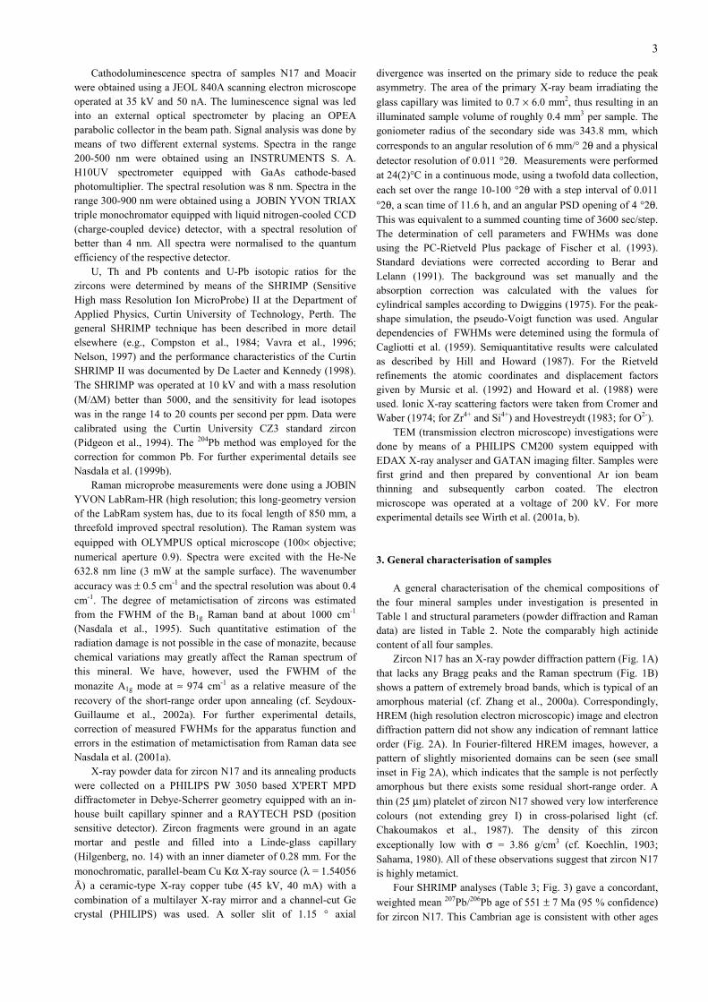
Zircon N17 has an X-ray powder diffraction pattern (Fig. 1A)that lacks any Bragg peaks and the Raman spectrum (Fig. 1B)shows a pattern of extremely broad bands, which is typical of anamorphous material (cf. Zhang et al., 2000a). Correspondingly,HREM (high resolution electron microscopic) image and electrondiffraction pattern did not show any indication of remnant latticeorder (Fig. 2A). In Fourier-filtered HREM images, however, apattern of slightly misoriented domains can be seen (see smallinset in Fig 2A), which indicates that the sample is not perfectlyamorphous but there exists some residual short-range order. A
thin (25 µm) platelet of zircon N17 showed very low interferencecolours (not extending grey I) in cross-polarised light (cf.Chakoumakos et al., 1987). The density of this zircon
exceptionally low with σ = 3.86 g/cm3 (cf. Koechlin, 1903;Sahama, 1980). All of these observations suggest that zircon N17is highly metamict.
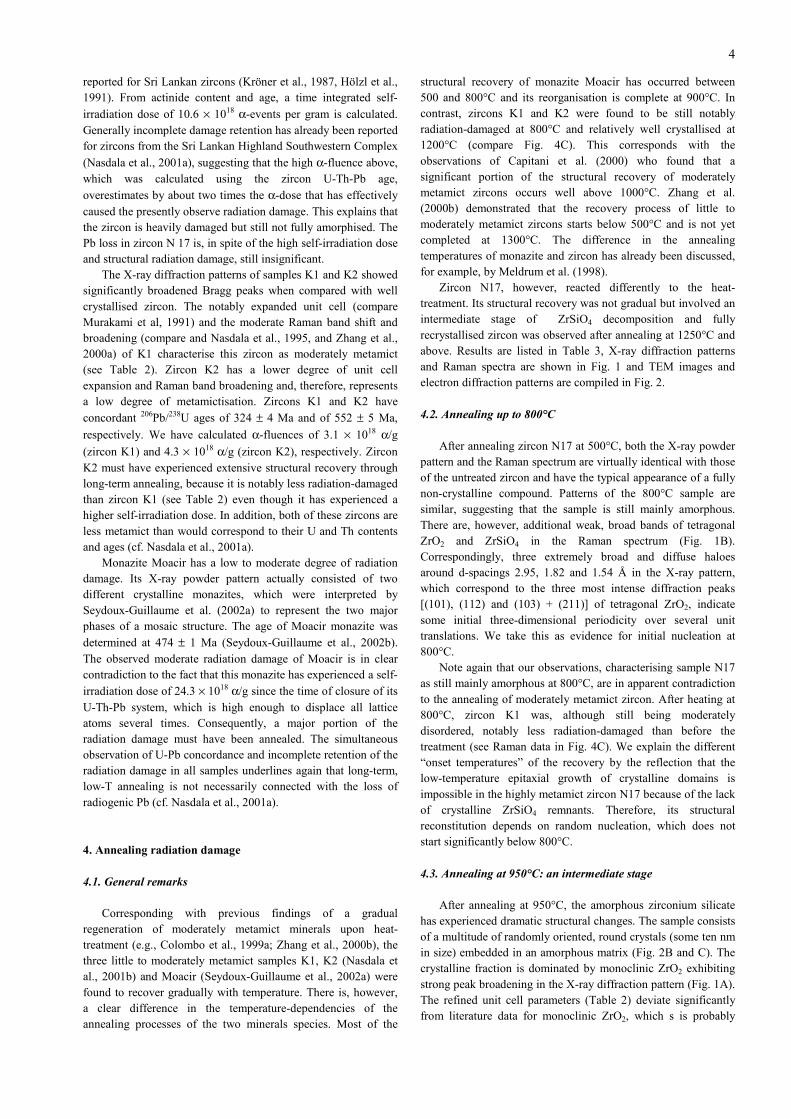
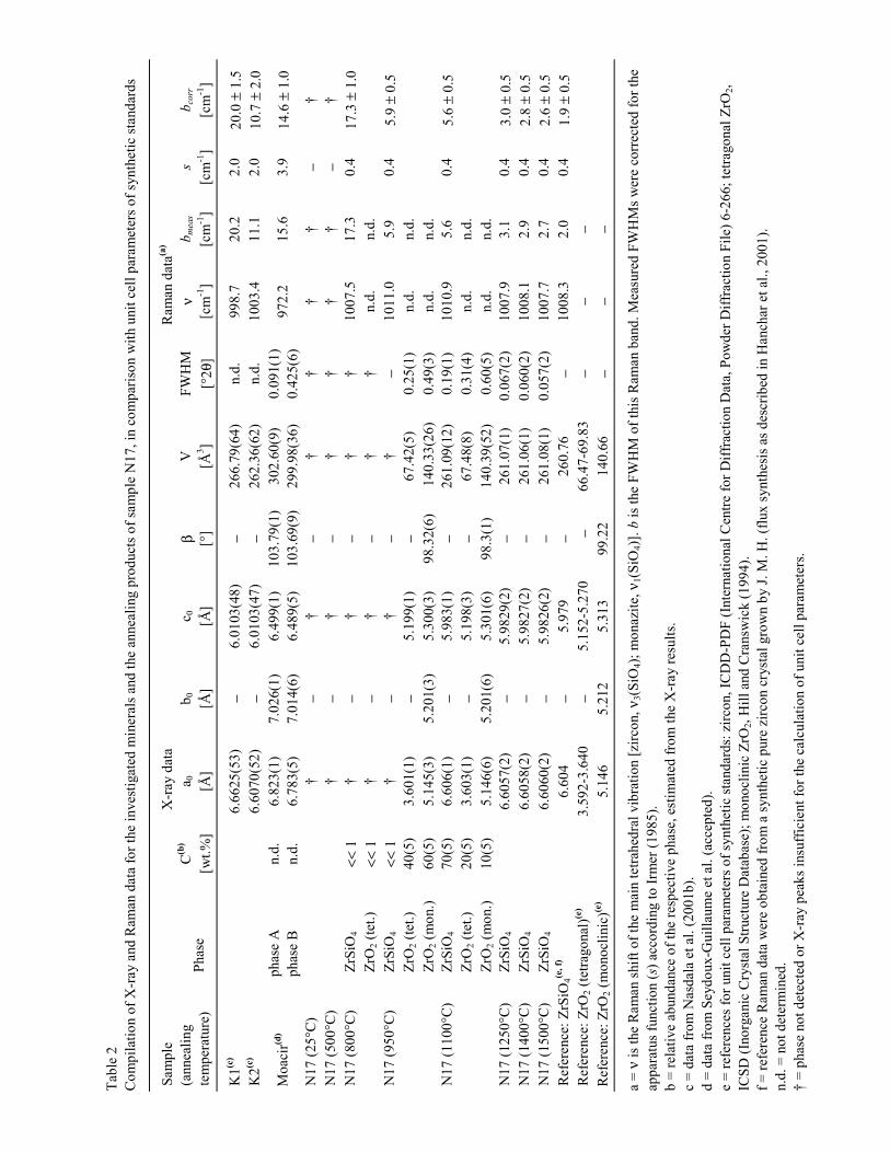
Four SHRIMP analyses (Table 3; Fig. 3) gave a concordant,
weighted mean 207Pb/206Pb age of 551 ± 7 Ma (95 % confidence)for zircon N17. This Cambrian age is consistent with other ages
4
reported for Sri Lankan zircons (Kröner et al., 1987, Hölzl et al.,1991). From actinide content and age, a time integrated self-
irradiation dose of 10.6 × 10¹⁸ α-events per gram is calculated.Generally incomplete damage retention has already been reportedfor zircons from the Sri Lankan Highland Southwestern Complex
(Nasdala et al., 2001a), suggesting that the high α-fluence above,which was calculated using the zircon U-Th-Pb age,
overestimates by about two times the α-dose that has effectivelycaused the presently observe radiation damage. This explains thatthe zircon is heavily damaged but still not fully amorphised. ThePb loss in zircon N 17 is, in spite of the high self-irradiation doseand structural radiation damage, still insignificant.
The X-ray diffraction patterns of samples K1 and K2 showedsignificantly broadened Bragg peaks when compared with wellcrystallised zircon. The notably expanded unit cell (compareMurakami et al, 1991) and the moderate Raman band shift andbroadening (compare and Nasdala et al., 1995, and Zhang et al.,2000a) of K1 characterise this zircon as moderately metamict(see Table 2). Zircon K2 has a lower degree of unit cellexpansion and Raman band broadening and, therefore, representsa low degree of metamictisation. Zircons K1 and K2 have
concordant 206Pb/238U ages of 324 ± 4 Ma and of 552 ± 5 Ma,
respectively. We have calculated α-fluences of 3.1 × 10¹⁸ α/g
(zircon K1) and 4.3 × 10¹⁸ α/g (zircon K2), respectively. ZirconK2 must have experienced extensive structural recovery throughlong-term annealing, because it is notably less radiation-damagedthan zircon K1 (see Table 2) even though it has experienced ahigher self-irradiation dose. In addition, both of these zircons areless metamict than would correspond to their U and Th contentsand ages (cf. Nasdala et al., 2001a).
Monazite Moacir has a low to moderate degree of radiationdamage. Its X-ray powder pattern actually consisted of twodifferent crystalline monazites, which were interpreted bySeydoux-Guillaume et al. (2002a) to represent the two majorphases of a mosaic structure. The age of Moacir monazite was
determined at 474 ± 1 Ma (Seydoux-Guillaume et al., 2002b).The observed moderate radiation damage of Moacir is in clearcontradiction to the fact that this monazite has experienced a self-
irradiation dose of 24.3 × 10¹⁸ α/g since the time of closure of itsU-Th-Pb system, which is high enough to displace all latticeatoms several times. Consequently, a major portion of theradiation damage must have been annealed. The simultaneousobservation of U-Pb concordance and incomplete retention of theradiation damage in all samples underlines again that long-term,low-T annealing is not necessarily connected with the loss ofradiogenic Pb (cf. Nasdala et al., 2001a).
4. Annealing radiation damage
4.1. General remarks
Corresponding with previous findings of a gradualregeneration of moderately metamict minerals upon heat-treatment (e.g., Colombo et al., 1999a; Zhang et al., 2000b), thethree little to moderately metamict samples K1, K2 (Nasdala etal., 2001b) and Moacir (Seydoux-Guillaume et al., 2002a) werefound to recover gradually with temperature. There is, however,a clear difference in the temperature-dependencies of theannealing processes of the two minerals species. Most of the
structural recovery of monazite Moacir has occurred between500 and 800°C and its reorganisation is complete at 900°C. Incontrast, zircons K1 and K2 were found to be still notablyradiation-damaged at 800°C and relatively well crystallised at1200°C (compare Fig. 4C). This corresponds with theobservations of Capitani et al. (2000) who found that asignificant portion of the structural recovery of moderatelymetamict zircons occurs well above 1000°C. Zhang et al.(2000b) demonstrated that the recovery process of little tomoderately metamict zircons starts below 500°C and is not yetcompleted at 1300°C. The difference in the annealingtemperatures of monazite and zircon has already been discussed,for example, by Meldrum et al. (1998).
Zircon N17, however, reacted differently to the heat-treatment. Its structural recovery was not gradual but involved anintermediate stage of ZrSiO4 decomposition and fullyrecrystallised zircon was observed after annealing at 1250°C andabove. Results are listed in Table 3, X-ray diffraction patternsand Raman spectra are shown in Fig. 1 and TEM images andelectron diffraction patterns are compiled in Fig. 2.
4.2. Annealing up to 800°C
After annealing zircon N17 at 500°C, both the X-ray powderpattern and the Raman spectrum are virtually identical with thoseof the untreated zircon and have the typical appearance of a fullynon-crystalline compound. Patterns of the 800°C sample aresimilar, suggesting that the sample is still mainly amorphous.There are, however, additional weak, broad bands of tetragonalZrO2 and ZrSiO4 in the Raman spectrum (Fig. 1B).Correspondingly, three extremely broad and diffuse haloesaround d-spacings 2.95, 1.82 and 1.54 Å in the X-ray pattern,which correspond to the three most intense diffraction peaks[(101), (112) and (103) + (211)] of tetragonal ZrO2, indicatesome initial three-dimensional periodicity over several unittranslations. We take this as evidence for initial nucleation at800°C.
Note again that our observations, characterising sample N17as still mainly amorphous at 800°C, are in apparent contradictionto the annealing of moderately metamict zircon. After heating at800°C, zircon K1 was, although still being moderatelydisordered, notably less radiation-damaged than before thetreatment (see Raman data in Fig. 4C). We explain the different“onset temperatures” of the recovery by the reflection that thelow-temperature epitaxial growth of crystalline domains isimpossible in the highly metamict zircon N17 because of the lackof crystalline ZrSiO4 remnants. Therefore, its structuralreconstitution depends on random nucleation, which does notstart significantly below 800°C.
4.3. Annealing at 950°C: an intermediate stage
After annealing at 950°C, the amorphous zirconium silicatehas experienced dramatic structural changes. The sample consistsof a multitude of randomly oriented, round crystals (some ten nmin size) embedded in an amorphous matrix (Fig. 2B and C). Thecrystalline fraction is dominated by monoclinic ZrO2 exhibitingstrong peak broadening in the X-ray diffraction pattern (Fig. 1A).The refined unit cell parameters (Table 2) deviate significantlyfrom literature data for monoclinic ZrO2, which s is probably
5
mainly due to the poor crystallinity. Furthermore, relatively sharppeaks of (relatively well ordered) tetragonal ZrO2 are resolved inX-ray and Raman patterns. The formation of tetragonal ZrO2
normally occurs only well above 1100°C, however, it is alsoknown that metastable, tetragonal ZrO2 is preferentially formedat very small crystallite sizes (Garvie, 1965; Mitsuhashi et al.,1974). It must also be cautiously considered that impurities maypotentially stabilise the tetragonal ZrO2 at lower temperatures.
It seems a bit surprising that only the tetragonal ZrO2 isobserved in the Raman spectrum whereas the monoclinic ZrO2 isnot (compare the spectrum of the 950°C sample in Fig. 1B withthat of the monoclinic ZrO2 reference). We suspect that theRaman signal of the poorly crystallised monoclinic ZrO2 isobscured by the much more intense signal of the relatively wellcrystallised tetragonal ZrO2. This conclusion is supported by theobservation of dramatic intensity losses (about two orders ofmagnitude) of Raman bands with decreasing crystallinity (forzircon documented by Nasdala et al., 2001a).
None of the further known cubic, orthorhombic, orrhombohedral ZrO2 polymorphs (cf. Howard et al., 1990) werefound by consecutive Rietveld refinement trials. The minorpresence of ZrSiO4 is indicated by low-intensity zircon Ramanbands (Fig. 1B) and a small deviation from the background noiseat d-spacing 3.30 Å in the X-ray diffractogram, corresponding tothe main zircon (200) peak. Calculation of cell parameters,however, was impossible.
A crystalline SiO2 polymorph or another SiO4-bearing phaseis neither observed in the X-ray diffraction pattern nor in theRaman spectrum. Energy dispersive element analyses and darkfield images obtained in the TEM, however, showed that Si isconcentrated the amorphous phase whereas Zr was only detectedin the round nanocrystals (compare Figs. 2B and C). Theamorphous ZrSiO4 has obviously decomposed to form crystallineZrO2 and amorphous SiO2. Capitani et al. (2000) explained thisby considering that the ionic bond character of ZrO2 stronglyfavours the crystalline state whereas bonding of SiO2 ispredominantly covalent.
The decomposition of zircon into oxides has different effectsto non-formula elements. U is preferentially incorporated in theZrO2 phase whereas Pb is concentrated in the SiO2 phase. Thiscan be seen from the behaviour of the 950°C sample whenanalysed in the SHRIMP. The impact of the oxygen beam causesenhanced sputtering of amorphous SiO2 whereas the crystallineZrO2 is more resistant. Too much Pb is thus released and weobtain, as an analytical artefact, apparently increased Pbconcentrations (Table 3). Consequently, U-Pb isotopic ratiosdetermined with the normal calibration equations show strongreverse discordance (Fig. 3; compare McLaren et al., 1994). Itwill be worthwhile to subject intermediate annealing products toprecise chemical analyses, in order to check whether the entirePb content of the original zircon has been completely transferredinto the SiO2 phase or Pb has already partially escaped.
4.4. Annealing at 1100°C
At 1100 °C the two ZrO2 polymorphs have become minorconstituents with decreased crystallinity (see FWHMs Table 2)and ZrSiO4 is the main phase. Zircon Bragg peaks are alsosignificantly broadened (Fig. 1A). The hypothesis that thisbroadening may be due to moderate radiation damage and, thus,
indicate incomplete damage recovery at this stage is disproved bythe facts that (1) this zircon is newly grown and (2) its unit cellparameters are identical within their errors with those of the fullyreorganised zircon (Table 2). Instead, we explain the broadeningof Bragg peaks by the very small crystal size of the newly grown
ZrSiO4, which is probably on the order of ≤ 10 nm.This interpretation is supported by the Raman results. In both
the 950°C and the 1100°C Raman spectrum, the frequency of the
ν3(SiO4) band shows a notable shift by about 2 cm-1 towardhigher wavenumbers, when compared with crystalline zircon(Table 2). Recall that the loss of crystallinity uponmetamictisation causes vibrational frequencies to shift toward
lower wavenumbers (Nasdala et al., 1995). For instance, ν3(SiO4)
may decrease from ≈ 1008 cm-1 (Dawson et al., 1971; Hoskin
and Rodgers, 1996) to < 990 cm-1. When annealing moderatelymetamict zircon, the gradual structural recovery is connected
with gradual recovery of the band frequencies and ν3(SiO4)
finally increases back to the initial value of ≈ 1008 cm-1 (seeZhang et al., 2001b). It has, however, never been reported beforethat vibrational frequencies of intermediate annealing productscan surpass those of well crystallised zircon. The now observed
ν3(SiO4) frequency of 1011 cm-1 is, therefore, most remarkable.We explain the slight shift toward higher vibrational energies bythe compressive strain in small ZrSiO4 particles, which increaseswith decreasing particle size. This effect has already beenreported for other materials, e.g., by Scamarcio et al. (1992) andHwang et al. (1996).
4.5. Annealing at 1200°C and above
All three samples that were heat-treated at 1250°C and aboveconsist of crystalline ZrSiO4 with a crystal size on the order ofsome hundred nm (Fig. 2D). Because the scale factors of theRietveld refinements are very similar (Table 2), we may assumethat almost all previous non-crystalline parts of the sample arerecrystallised at 1250°C, in favour of the remnant ZrO2. Smalldifferences can only be observed for the FWHM, which is,however, very close to the theoretical resolution of our X-ray
measurements (0.047 °2θ). The cell parameters of therecrystallised zircon are slightly increased in comparison tosynthetic ZrSiO4 (see Table 3). We explain this difference by thenon-ideal chemistry of the zircon .
Analogously, the Raman spectra at 1250°C and above showonly the bands of well crystallised zircon and differencesbetween the three annealing products do not extend beyond theanalytical uncertainties. Note that the Raman parameters of theannealing product show only minor variations from those of
pure, synthetic zircon. In particular the ν3(SiO4) FWHM deviatesby less than + 1 cm-1 from pure ZrSiO4 (Table 2). It is obviousthat the mere presence of some 103 ppm U at the [8]Zr4+ sites hasonly minor effects on the Raman spectrum. In contrast to naturalmonazites, where internal PO4 vibrations may be stronglybroadened depending on potentially extensive chemicalvariations (Podor, 1995; Nasdala et al., 1999a), Raman bandbroadening in natural zircons is mainly due to structural effectssuch as radiation damage and chemical effects can mostly beexcluded. This confirms again that the Raman band broadeningin zircon can be used to estimate the degree of structuralradiation damage.
6
Multiple electron microprobe analyses on the untreated,highly metamict sample and the recrystallised zircon (Table 1)demonstrate that the heat-treatment did not cause major changesof the chemical composition, except for the escape of Pb.Considering this observation and the fact that the elementalcomposition of zircons is relatively easily altered inhydrothermal experiments (Pidgeon et al. 1966; Rizvanova et al.,2000; Geisler et al., 2001), we suppose that both the lack of asuitable transporting medium (e.g., chemically “aggressive”fluids, etc.) and the lack of diffusion pathways may haveprevented notable migration of elements. The degree of Pb loss,however, is obvious from the SHRIMP results. Calculated Pb/Uratios are close to zero, with huge errors greatly extending thevalues. Obviously the heat-treatment has fully reset the U-Pbsystem of the zircon.
This reset may also account for the occurrence of tiny SiO2
inclusions in the recrystallised zircon (Fig. 2E). Due to the Pbescape, the initial equilibrium between large (Zr, U, Pb) andsmall (Si) cations is disturbed and the sample has now a slightlynon-stoichiometric composition. Obviously the excess Si was notincorporated in the newly grown zircon but has formed a separatesilica phase.
4.6. Discussion
The reconstitution of a highly metamict zircon involves anintermediate stage of decomposition into oxides. An energeticexplanation for this phase separation was given by Ellsworth etal. (1994) who discussed that the difference in enthalpy betweenhighly radiation-damaged and crystalline zircon is much greaterthan between metamict zircon and a mixture of tetragonal ZrO2
and glassy SiO2. Also, Ellsworth et al. (1994) stated that theseparation into oxides would be favoured if the metamict startingmaterial had a non-uniform structure comprising ZrO2- and SiO2-rich domains, because ZrO2-rich regions would recrystallisemore easily upon heat-treatment. Such structure was described,for example, by Vance and Anderson (1972) who explained theanomalous optical absorption of metamict zircon by the presenceof tiny crystalline ZrO2 particles in the radiation-damagedZrSiO4. Decomposition into oxides, however, does not seem todepend on the hypothetical presence of ZrO2 domains in thestarting material, because it was also observed when annealinghighly radiation-damaged zircons without this feature (Capitaniet al., 2000; this study).
We found the formation of well crystallised ZrSiO4 to benearly completed at 1250°C in our experiments. We have alsoobserved at 1100°C that the oxide content is lowered and thecontent of zircon nanocrystals is greatly increased with respect tothe 950°C sample, so the ZrSiO4 formation must have startedeven below 1100°C. Consequently, our results suggest that thetemperature interval in which the intermediate oxide stage isrealised extends between > 800°C / < 950°C and roughly1100°C. This is in agreement with the results of Begg et al.(2000) who reported that amorphous, Pu-substituted zircon haddecomposed into oxides at 1000°C whereas at 1200°C zircon hadfully recrystallised.
Our results, however, do not correspond with the results ofMcLaren et al. (1994). These authors observed roundedmonoclinic ZrO2 crystallites in a glassy silica matrix afterannealing a highly metamict zircon for 5 h at 1250°C. A detailed
explanation for these different results cannot be given. One maysuspect that stepwise annealing and annealing of untreated zirconmay give different results. It seems to be most likely that ZrSiO4
formation after phase separation at lower temperatures requireshigher energies than ZrSiO4 recrystallisation directly fromamorphous zircon (e.g., Begg et al., 2000). When heatingamorphous zircon to 1250°C, it is most probably quickly driventhrough the temperature range of oxide nucleation and, therefore,ZrSiO4 nucleation in the amorphous zircon will become moreimportant. However, this potentially different behaviour cannotbe applied to explain the different results of McLaren et al.(1994) and the present work, because untreated fragments wereheated in both studies. There is certainly some doubt about theaccuracy of the temperature calibration (i.e. whether or not theirand our 1250°C experiments were really done at exactly the sametemperature). Also, one might suspect that extending theannealing times from 5 h (McLaren et al.) to 150 h (his study)may have allowed for slow recrystallisational processes. Eventhough annealing experiments have often been done for only < 2h (e.g., Woodhead et al., 1991; Biagini et al., 1997; Colombo etal., 1999a), it is well known that structural parameters shownotable changes when extending the annealing time (Ellsworth etal., 1994; Colombo et al., 1999b; Zhang et al., 2000b). Thissuggests that annealing times of only a few hours may result inincomplete reaction.
Another apparent contradiction is found when comparing ourresults with the results of Capitani et al. (2000). These authorsfound zirconia grains in an amorphous silica matrix after heat-treating a highly metamict, natural zircon at 1400 K (1127°C) butdid not report zircon at this temperature. Their sample, however,was richer in actinides (UO2 0.86 wt-%) and also even moreradiation-damaged than the zircon studied here, which may havecaused slightly different annealing behaviours. The differentexperimental conditions may also have affected the results:Capitani annealed their zircon for 16 h and in an N2 atmospherewhereas in the present work the experiments lasted 150 h andwere done in an open furnace (air).
5. Cathodoluminescence results
Our and previous heating experiments (Nasdala et al., 2001b;Seydoux-Guillaume et al., 2002a) have produced small series ofmineral samples with almost identical chemical compositions butdifferent structural states. Apparently complete Pb escape duringthe high-temperature treatment is the only major chemicaldifference observed in the present study. It should, however, becautiously considered that many trace elements, whichpotentially may also cause or suppress luminescence, were belowthe detection limit of the electron microprobe. In spite of thisuncertainty, comparison of crystalline annealing products andtheir radiation-damaged starting materials allows us toinvestigate the dependence of CL on metamictisation withouthaving to consider major chemical effects.
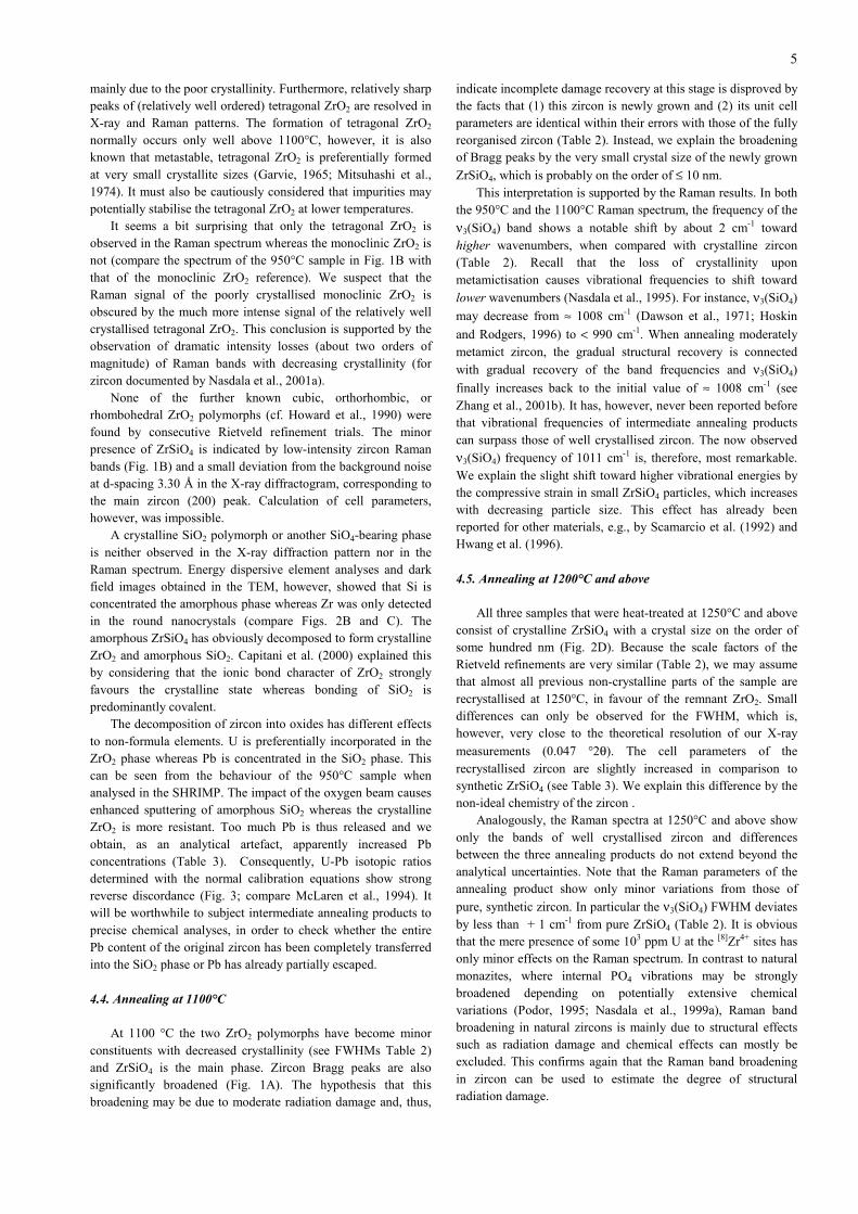
To check for general changes of the total CL intensity, CLimages were obtained for fragments of the same sample annealedat different temperatures (Fig. 4C). We found that the total CLsignal of the annealed fragments is always higher in intensitywhen compared with the image of the respective untreated,radiation-damaged sample. This observation suggests that the CL
7
emission is generally suppressed in radiation-damaged structures.The CL recovery seems to be almost completed at 800°C inMoacir monazite (Seydoux et al., 2002a) whereas the CL ofzircons K1 and K2 shows significant changes above thistemperature (Fig. 4C). This difference in CL recovery concurswith the different annealing behaviours of the two mineralsdiscussed above.
More precise conclusions, however, require the spectralanalysis of the CL emission. This was done for several chips ofsamples N17 (and, in addition, also for monazite Moacir). For theinterpretation of spectra, it is necessary to reconsider briefly theorigin of CL. The CL emission commonly observed in crustalzircon crystals comprises two different types. The first type is an“intrinsic” broad-band emission centred either in the blue oryellow region of the electromagnetic spectrum. Its origin is stillcontroversial. Most probably it is defect-related, for example topoint defects or local charge-imbalance due to impurities (e.g.,Ohnenstetter et al., 1991; Kempe et al., 2000) or electronicdefects related to SiO4 tetrahedra (Cesbron et al., 1993; 1995).The second type are comparably narrow REE (rare earthelement) emission peaks (e.g., 4f electronic transitions oftrivalent REE). These emissions are often observed in clusters ofbands, then pointing to a variety of REE site symmetries, andmay either be superimposed on the broad-band emission or a flatbackground. Among the REEs, emission bands of Dy3+ are aparticularly typical feature of zircon (Mariano, 1978; 1988; 1989;Remond et al., 1992; Hanchar and Rudnick, 1995). Trivalent Tb,Gd3+ and Y 3+ may also be CL-activated elements in this mineral(e.g., Ohnenstetter et al., 1991; Yang et al., 1992; Blanc et al.,2000).
Fig. 5 shows the CL spectra obtained from zircon N17. Afterstructural reorganisation through annealing at 1400°C, theintegrated CL intensity has increased by about two orders ofmagnitude. No significant yellow broad-band emission isdetected in the 1400°C spectrum and the blue broad-bandintrinsic emission is comparably low in intensity and is centred inthe ultraviolet. The spectrum is dominated by narrow emissionsrelated to Gd3+, Dy3+ and Tb3+. The fact that these bands areextremely low in intensity in the 25°C and 1100°C spectrum,even though the same amount of REEs is present, mayhypothetically be explained in two different ways. Firstly, it maybe assumed these REEs are not CL-activated elements when inthe metamict zircon, which could, for example, be due toionisation effects caused by the self-irradiation. Secondly, it ispossible that the CL is “quenched” in the damaged structure, forexample by the highly disturbed site symmetries of the REEs orelectronic defects in their neighbouring range. As Pb loss fromthe 1400°C sample was observed, it cannot be ruled out thattraces of radiogenic Pb may have some suppressing influence on
the CL signal. Even the presence of He (implanted α-particles) inthe low-luminescent, metamict zircon, which has certainly alsoescaped at 1400°C, should be considered. Dehydration of solidsmay also increase their CL (e.g., Gutzov and Peneva, 1995),however, this effect is of minor importance in the studied zirconsbecause of their comparably low content of hydrous species(Nasdala et al., 2001b).
For comparison, CL spectra of monazite Moacir were alsoobtained. All spectra (Fig. 6) have a similar pattern that isdominated by five Nd3+ bands in the range 860-895 nm. The CLintensity of the annealed sample is clearly enhanced, when
compared with the untreated, radiation-damaged sample. It iswell known that minor CL intensity variations may be affectedby the crystal orientation of zircon (Cesbron et al., 1995) andapatite (Barbarand and Pagel, 2001). The intensity gain observedhere, however, surpasses potential orientation effects. The
intensity gain is only about 3 × for the Nd3+ bands but 10 × forthe 330 nm lattice emission and must be due to the reconstitutionof the monazite structure. Note the clear correlation between theCL intensity and the recovery of short-range order, in Fig. 6demonstrated with the decreasing FWHM of internal PO4
vibrations.Poller et al. (2001) have proposed that “U (and possibly Hf)
and related radiation damage” may suppress the CL intensity.Our results disprove the hypothesis that the presence of U is themain reason for decreased CL emission of natural zircons. Recallthat the CL intensity of zircon N17 is greatly increased afterannealing at 1400°C, even though the U content is about thesame. We suspect that the anti-correlation of CL intensity and Ucontent observed by Poller et al. (2001) is most probably a linkbetween the radiation damage caused by U and the CLsuppression due to radiation damage. In particularly actinide-richminerals such as natural monazites, however, high levels of U4+
may play a role in quenching the CL emission.Consideration of the crystallinity-dependence of CL may
perhaps also prove helpful for the interpretation of apparentlycontradicting BSE (backscattered electrons) and CL images. BSEimages reveal, greatly simplified, contrasts in average atomicnumber of a phase. The element primarily responsible for theseBSE variations in crustal zircons is Hf, with U having asecondary effect (Hanchar and Miller, 1993). It is well knownfrom numerous BSE/CL imaging studies (e.g., Vavra et al., 1990;Paterson et al., 1992; Hanchar and Miller, 1993; Koschek, 1993;Hanchar and Rudnick, 1995; Rémond et al., 1995) that bothtechniques reveal mostly the same internal features, however,usually the bright areas in CL are dark in BSE and vise versa (seeFig. 4A and B). Observations of positively correlated BSE andCL in patchily recrystallised zircons (see Fig. 2c-d in Kempe etal., 2000) or differing BSE and CL patterns can be explained ifwe consider that processes causing heterogeneous structuralchanges may have different effects on the CL and BSE patterns.
The results of our CL study give strong evidence that the CLis not only controlled by the presence of REEs and otherchemical factors but has also great structural dependence. This isconsistent with previous findings that the CL emission greatlyincreases when amorphous solids crystallise (e.g., Gutzov et al.,1998). Cathodoluminescence generation is, however, probablyeven more complicated than has previously been thought. In ourstudy, we have observed that the CL is notably suppressed insignificantly radiation-damaged samples and recovers uponannealing. The opposite case, however, is also possible. Partialreorganisation of highly radiation-damaged zircon under theimpact of an electron beam is not connected with CL recoverybut, in contrast, the CL emission is even more suppressed in thebetter crystallised area (Fig. 4 D and E). It is also well knownthat radiation damage in minerals may result in enhanced CLemission (for example, see Owen, 1988; Meunier et al., 1990).Correct assignment of radioactivity-induced CL changes will,therefore, require a much more detailed investigation of CLspectra obtained from well characterised, natural and syntheticmineral samples.
8
Acknowledgements
We are most grateful to W. Hofmeister (Institute ofGemstone Research, Idar-Oberstein and Mainz) for the gemstonezircon N17. The other samples described in this study werekindly provided by A. Massanek (Freiberg) and J.-M. Montel(Toulouse). Sample preparation was done by A. Wagner (Wien),D. Dettmar (Bochum) and K. Paech (Potsdam). Thanks are dueto A. A. Nemchin (Perth) for assisting the SHRIMP ion probework at Curtin University and to B. Schulz-Dobrick and N.Groschopf (both Mainz) for help with electron microprobeanalysis. We are indebted to J. Bethke, A. v. Lemel and B. Punte(Philips Analytical X-Ray, Almelo) who made a high resolutionpowder diffractometer available for analysis. We are also gratefulto R. Kleeberg (Freiberg) for the X-ray analysis of zircons K1and K2 and to E. Fuchs (Idar-Oberstein) for the densitymeasurement. Helpful comments by G. Irmer (Freiberg), W. J.Weber (Richland, WA), D. Ohnenstetter (Nancy), and ananonymous reviewer are greatly appreciated. Thanks are also dueto A. Kröner (Mainz) and W. Heinrich (Potsdam) for financialsupport. Funding for this research was provided by DeutscheForschungsgemeinschaft (grants Na 284/3-1-4), and byUniversität Wien during a guest professorship of L. N. in the2001 Spring term.
References
Barbarand, J. and Pagel, M., 2001. Cathodoluminescence studyof apatite crystals. Am. Mineral., 86: 473-484.
Begg, B. D., Hess, N. J., Weber, W. J., Conradson, S. D.,Schweiger, M. J. and Ewing, R. C., 2000. XAS and XRDstudy of 238Pu- and 239Pu-substituted zircons(Zr0.92Pu0.08SiO4). J. Nucl. Mat., 278: 212-224.
Begun, G. M., Beall, G. W., Boatner, L. A. and Gregor, W. J.,1981. Raman spectra of the rare earth orthophosphates. J.Raman Spectrosc., 11: 273-278.
Berar, J.-F. and Lelann, P., 1991. E.S.D.s and estimated probableerrors obtained in Rietveld refinements with localcorrelations. J. Appl. Cryst., 24: 1-5.
Biagini, R., Memmi, I. and Olmi, F., 1997. Radiation damage inzircons. N. Jahrb. Mineral. Mh., 1997: 257-270.
Blanc, P., Baumer, A., Cesbron, F., Ohnenstetter, D., Panczer, G.and Rémond, G., 2000. Systematic cathodouminescencespectral analysis of synthetic doped minerals anhydrite,apatite, calcite, fluorite, scheelite and zircon. In: M. Pagel, V.Barbin, P. Blanc and D. Ohnenstetter (eds.),Cathodoluminescence in Geosciences, Springer, Heidelberg,pp. 127-175.
Cagliotti, G., Paoletti, A. and Ricci, F. P., 1959. The choice ofcollimators for a crystal spectrometer for neutron diffraction.Nucl. Instr., 3: 223-228.
Capitani, G. C., Leroux, H., Doukhan, J. C., Ríos, S., Zhang, M.and Salje, E. K. H., 2000. A TEM investigation of naturalmetamict zircons: structure and recovery of amorphousdomains. Phys. Chem. Minerals., 27: 545-556.
Cesbron, F., Ohnenstetter, D., Blanc, P., Rouer, O. and Sichere,M. C., 1993. Incorporation de terres rares dans des zircons de
synthése: étude par cathodoluminescence. C. R. Acad. Sci.Paris, 316: 1231-1238.
Cesbron, F., Blanc, P., Ohnenstetter, D and Rémond, G., 1995.Cathodoluminescence of rare earth doped zircons. I Theirpossible use as reference materials. Scanning Microscopy,Suppl. 9: 35-56.
Chakoumakos, B. C., Murakami, T., Lumpkin, G. R. and Ewing,
R. C., 1987. Alpha-Decay−Induced Fracturing in Zircon: TheTransition from the Crystalline to the Metamict State.Science, 236: 1556-1559.
Colombo, M., Chrosch, J. and Salje, E. K. H., 1999a. Annealingmetamict zircon: a powder X-ray diffraction study of ahighly defective phase. J. Am. Ceram. Soc., 82: 2711-2716.
Colombo, M., Chrosch, J., Biagini, R. and Memmi, I., 1999b. AnIR analysis of the role of SiO4 tetrahedra in thermallyannealed ZrSiO4. N. Jb. Miner. Mh., 1999: 113-122.
Compston, W., Williams, I. S. and Meyer, C., 1984. U-PbGeochronology of zircons from lunar breccia 73217 using aSensitive High Mass-Resolution Ion Micorprobe. J. Geophys.Res. 89 (Suppl.): B525-B534.
Cromer, D. T. and Waber, J. T., 1974. Atomic scattering factorsfor X-rays. In: International Tables for X-rayCrystallography, vol. IV, pp. 71-148. Kynoch Press,Birmingham.
Dawson, P., Hargreave, M. M. and Wilkinson, G. R., 1971. Thevibrational spectrum of zircon (ZrSiO4). J. Phys. C: SolidState Phys., 4: 240-256.
De Leater, J. R. and Kennedy, A. K., 1998. A double focusingmass spectrometer for geochronology. Int. J. Mass Spectr.,178: 43-50.
Dwiggins, C. W., 1975. Rapid calculation of X-ray absorptioncorrection factors for cylinders to an accuracy of 0.1%. ActaCryst., A31: 146.
Ellsworth, S., Navrotsky, A. and Ewing, R. C., 1994. Energeticsof radiation damage in natural zircon (ZrSiO4). Phys. Chem.Minerals, 21: 140-149.
Ewing, R. C., 1994. The metamict state: 1993 – the centennial.Nucl. Instr. Meth. Phys. Res., B 91: 22-29.
Ewing, R. C., 1999. Nuclear waste forms for actinides. Proc.Natl. Acad. Sci., 96: 3432-3439.
Fischer, R. X., Lengauer, C. L., Tillmanns, E., Ensink, R. J.,Reiss, C. A. and Fantner, E. J, 1993. PC-Rietveld Plus, acomprehensive Rietveld analysis package for PC. Mater. Sci.Forum, 133-136: 287-292.
Garvie, R. C., 1965. The occurrence of metastable tetragonalzirconia as a crystallite size effect. J. Phys. Chem., 69: 1238-1243.
Geisler, T., Ulonska, M., Schleicher, H., Pidgeon, R.T. and vanBronswijk, W., 2001. Leaching and differentialrecrystallization of metamict zircon under experimentalhydrothermal conditions. Contrib. Mineral. Petrol., 141: 53-65.
Gutzov, S. and Peneva, S. K., 1995. Structure and properties ofhydrous zirconium oxide. Bulgarian Chem. Commun., 28:744-751.
Gutzov, S., Bredol, M. and Wasgestian, F. 1998.Cathodoluminescence study of europium doped zirconia andcassiterite powders. J. Phys. Chem. Solids, 59: 69-74.
Hanchar, J. M. and Miller, C. F., 1993. Zircon zonation patternsas revealed by cathodoluminescence and backscattered
9
electron images: Implications for interpretation of complexcrustal histories. Chem. Geol., 110: 1-13.
Hanchar, J. M. and Rudnick, R. L., 1995. Revealing hiddenstructures: The application of cathodoluminescence andback-scattered electron imaging to dating zircons from lowercrustal xenoliths. Lithos, 36: 289-303.
Hanchar, J. M., Finch, R. J., Hoskin, P. W. O., Watson, E. B.,Cherniak, D. J. and Mariano, A. N., 2001. Rare earthelements in natural zircon: Part 1. Synthesis, and rare earthelement and phosphorous doping. Am. Mineral., 86: 667-680.
Hawthorne, F. C., Groat, L. A., Raudsepp, M., Ball, N. A.,Kimata, M., Spike, F. D., Gaba, R., Halden, N. M., Lumpkin,G. R., Ewing, R. C., Greegor, R. B., Lytle, F. W., Ercit, T.C., Rossman, G. R., Wicks, F. J., Ramik, R. A., Sherriff, B.L., Fleet, M. E. and McCammon, C., 1991. Alpha-decaydamage in titanite. Am. Mineral., 76: 370-396.
Hill, R. J. and Howard, C. J., 1987. Quantitative phase analysisfrom neutron powder diffraction data using the Rietveldmethod. J. Appl. Cryst., 20: 467-474.
Hill, R. .J. and Cranswick, L. M. D., 1994. Rietveld refinementround robin. II. Analysis of monoclinic ZrO2. J. Appl. Cryst.,27: 802-844.
Holland, H. D. and Gottfried, D., 1955. The effect of nuclearradiation on the structure of zircon. Acta Cryst., 8: 291-300.
Hölzl, S., Köhler, H., Kröner, A., Jäckel, P. and Liew, T.C.,1991. Geochronology of the Sri Lankan basement. In:Kröner, A. (ed.) The crystalline crust of Sri Lanka. Part I.Summary of Research of the German-Sri LankanConsortium. Geol. Surv. Dept. of Sri Lanka, Prof. Paper 5:pp 237-257.
Hoskin, P. W. O. and Rodgers, K. A., 1996. Raman spectral shiftin the isomorphous series (Zr1-xHfx)SiO4. Eur. J. Solid StateInorg. Chem., 33: 1111-1121.
Howard, C. J., Hill, R. J. and Reichert, B. E., 1988. Structures ofthe ZrO2 polymorphs at room temperature by high-resolutionneutron powder diffraction. Acta Cryst., B44: 116-120.
Hovestreydt, E., 1983. On the atomic scattering factor for O2-.Acta Cryst., A39: 268-269.
Hurley, P. M. and Fairbairn, H. F., 1953. Radiation damage inzircon: a possible age method. Bull. Geol. Soc. Am., 64: 659-674.
Hwang, Y.-N., Shin, S., Park, H. L., Park, S.-H., Kim, U., Jeong,H. S., Shin, E. and Kim, D., 1996. Effect of latticecontraction on the Raman shifts of CdSe quantum dots inglass matrices. Phys. Rev. B, 54: 15120-15124.
Irmer, G., 1985. Zum Einfluß der Apparatefunktion auf dieBestimmung von Streuquerschnitten und Lebensdauern ausoptischen Phononenspektren. Exper. Techn. Phys., 33: 501-506.
Kawashima, Y. and Katagiri, G., 1995. Fundamentals, overtones,and combinations in the Raman spectrum of graphite. Phys.Rev. B, 52: 10053–10059.
Kempe, U., Gruner, T., Nasdala, L. and Wolf, D., 2000.Relevance of cathodoluminescence for the interpretation ofU-Pb zircon ages, with an example of an application to astudy of zircons from the Saxonian Granulite Complex,Germany. In: M. Pagel, V. Barbin, P. Blanc and D.Ohnenstetter (eds.), Cathodoluminescence in Geosciences,Springer, Heidelberg, pp 415-455.
Koechlin, R., 1903. Über Zirkon. Tscherm. Min. Petr. Mitt., 22:368-372.
Kolesov, B. A., Geiger, C. A. and Armbruster T., 2001. Thedynamic properties of zircon studied by single-crystal X-raydiffraction and Raman spectroscopy. Eur. J. Mineral., 13:939-948.
Koschek, G., 1993. Origin and significance of the SEMcathodoluminescence from zircon. J. Microsc., 171: 223-232.
Kröner, A., Williams, I. S., Compston, W., Baur, N., Vitanage,P.W. and Perera, L. R. L., 1987. Zircon ion microprobedating of high-grade rocks in Sri Lanka. J. Geol., 95: 775-791.
Lumpkin, G. R., 2001. Alpha-decay damage and aqueousdurability of actinide host phases in natural systems. J. Nucl.Mater., 189: 136-166.
Mariano, A. N., 1978. The application of cathodoluminescencefor carbonitite exploration and characterization. In: C. J.Braga (ed.), Proc. Int. Symp. Carbonitites, 1st, Pocos deCaldas, Brazil, June, 1976. Brasil Depart. Nacional daProducão Mineral, Brasilia, pp. 39-57.
Mariano, A. N., 1988. Some further geologic applications ofcathodoluminescence. In: D. J. Marshall (ed.),Cathodoluminescence of Geologic materials. Unwin Hyman,Boston, pp. 94-123.
Mariano, A. N., 1989. Cathodoluminescence emission spectra ofrare earth element activators in minerals. In: B. R. Lipin andG. A. McKay (eds.), Geochemistry and Mineralogy of RareEarth Elements. Mineral. Soc. Am., Rev. Mineral., 21: 339-348.
McLaren, A. C., Fitz Gerald, J. D. and Williams, I. S., 1994. Themicrostructure of zircon and its influence on the agedetermination from Pb/U isotopic ratios measured by ionmicroprobe. Geochim. Cosmochim. Acta, 58: 993-1005.
Meldrum, A., Boatner, L. A., Weber, W. J. and Ewing, R. C.,1998. Radiation damage in zircon and monazite. Geochim.Cosmochim. Acta, 62: 2509-2520.
Meunier, J. D., Sellier, E. and Pagel, M., 1990. Radiation-damage rims in quartz from uranium-bearing sandstones. J.Sediment. Petrol., 60: 53-58.
Mitsuhashi, T., Ichihara, M. and Tatsuke, U., 1974.Characterization and stabilization of metastable tetragonalZrO2. J. Amer. Ceram. Soc., 57: 97-101.
Murakami, T., Chakoumakos, B. C., Ewing, R. C., Lumpkin, G.R. and Weber, W. J., 1991. Alpha-decay event damage inzircon. Am. Mineral., 76: 1510-1532.
Mursic, Z., Vogt, T., Boysen, H. and Frey, F., 1992. Single-crystal neutron diffraction study of metamict zircon up to2000 K. J. Appl. Cryst., 25: 519-523.
Nasdala, L., Irmer, G. and Wolf, D., 1995: The degree ofmetamictization in zircons: a Raman spectroscopic study.Eur. J. Mineral., 7: 471-478.
Nasdala, L., Pidgeon, R. T., Wolf, D. and Irmer, G., 1998.Metamictization and U-Pb isotopic discordance in singlezircons: a combined Raman mircoprobe and SHRIMP ionprobe study. Mineral. Petrol., 62: 1-27.
Nasdala, L., Finger, F. and Kinny, P., 1999a. Can monazitebecome metamict? Eur. J. Mineral., 11, Beih. 1: 164.
Nasdala, L., Wenzel, T., Pidgeon, R. T. and Kronz, A., 1999b.Internal structures and dating of complex zircons from
10
Meissen Massif monzonites, Saxony. Chem. Geol., 156: 331-341.
Nasdala, L., Wenzel, M., Vavra, G., Irmer, G., Wenzel, T. andKober, B., 2001a. Metamictisation of natural zircon:Accumulation versus thermal annealing of radioactivity-induced damage. Contrib. Mineral. Petrol., 141: 125-144.
Nasdala, L., Beran, A., Libowitzky, E. and Wolf, D., 2001b. Theincorporation of hydroxyl groups and molecular water innatural zircon (ZrSiO4). Amer. J. Sci., 301: 831-857.
Nelson, D. R., 1997. Compilation of SHRIMP U-Pb zircongeochronology data, 1996. Geol. Surv. West. Aust. Record1997/2, 189 pp.
Ohnenstetter, D., Cesbron, F., Remond, G., Caruba, R. andClaude, J.-M., 1991. Émissions de cathodoluminescence dedeux populations de zircons naturels: téntatived'interpretation. C. R. Acad. Sci. Paris, 313: 641-647.
Owen, M. R., 1988. Radiation-damage halos in quartz. Geology,16: 529-532.
Paterson, B. A., Stephens, W. E., Rogers, G., Williams, I. S.,Hinton, R. W. and Herd, D. A., 1992. The nature of zirconinheritance in two granite plutons. Transact. Royal Soc.Edinburgh: Earth Sci., 83: 459-471.
Pidgeon, R. T., O’Neil, J. R. and Silver, L. T., 1966. Uraniumand lead isotopic stability in a metamict zirconunderhydrothermal conditions. Science, 154: 1538-1542.
Pidgeon, R. T., Furfaro, D., Kennedy, A. K., Nemchin, A. A. andvan Bronswijk, W., 1994. Calibration of zircon standards forthe Curtin SHRIMP II (abstract). 8th Int. Conf. Geochronol.Cosmochronol. Isotope Geol., Berkeley. U. S. Geol. Surv.Circular 1107: 251.
Podor, R., 1995. Raman spectra of the actinide-bearingmonazites. Eur. J. Mineral., 7: 1353-1360.
Poller, U., Huth, J., Hoppe, P. and Williams, I. S., 2001. REE, U,Th and Hf distribution in zircon from Western CarpathianVariscan granitoids:A combined cathodoluminescence andion microprobe study. Amer. J. Sci. (in press).
Rémond, G., Cesbron, F., Chapoulie, R., Ohnenstetter, D.,Roques-Carmes, C. and Schoverer, M., 1992.Cathodoluminescence applied to the microcharacterization ofmineral materials: a present status in experimentation andinterpretation. Scanning Microsc., 6: 23-68.
Rémond, G., Blanc, P., Cesbron, F., Ohnenstetter, D and Rouer,O., 1995. Cathodoluminescence of rare earth doped zircons.II. The distribution of the doping elements and the contrastsof images. Scanning Microscopy, Suppl. 9: 57-76.
Rizvanova, N. G., Levchenkov, O. A., Belous, A. E., Bezmen, N.I., Maslenikov, A. V., Komarov, A. N., Makeev, A. F. andLevskiy, L. K., 2000. Zircon reaction and stability of the U-Pb isotope system during interaction with carbonate fluid:experimental hydrothermal study. Contrib. Mineral. Petrol.,139: 101-114.
Sahama, T.G., 1981. Growth structure in Ceylon zircon. Bull.Minéral., 104: 89-94.
Scamarcio, G., Lugara, M. and Manno, D., 1992. Size-dependentlattice contraction in CdS1-xSx nanocrystals embedded inglass observed by Raman scattering. Phys. Rev. B., 45:13792-13795.
Seydoux-Guillaume, A.-M., Wirth, R., Nasdala, L., Gottschalk,M., Montel, J.-M. and Heinrich, W., 2002a. An XRD, TEM
and Raman study of experimentally annealed naturalmonazite. Phys. Chem. Minerals, 29: 240-253.
Seydoux-Guillaume A.M., Paquette J.L., Wiedenbeck M.,Montel J.M. and Heinrich W., 2002b. Experimental resettingof the U-Th-Pb system in monazite. Chem. Geol., this issue.
Silver, L. T. and Deutsch, S., 1963. Uranium-lead isotopicvariations in zircons: a case study. J. Geol., 71: 721-758.
Vance, E. R. and Anderson, B. W., 1972. Study of metamictCeylon zircons. Mineral. Mag., 38: 605-613.
Vavra, G., 1990. On the kinematics of zircon growth and istpetrogenetic significance: a cathodoluminescence study.Contrib. Mineral. Petrol., 106: 331-344.
Vavra, G., Gebauer, D., Schmid, R. and Compston, W., 1996.Multiple zircon growth and recrystallization duringpolyphase Late Carboniferous to triassic metamorphism ingranulites of the Ivrea Zone (Southern Alps): an ionmicroprobe (SHRIMP) study. Contrib. Mineral. Petrol., 122:337-358.
Wang, S. X., Begg, B. D., Wang, L. M., Ewing, R. C., Weber,W. J. and Govidan Kutty, K. V., 1999. Radiation stability ofgadolinium zirconate: A waste form for plutoniumdisposition. J. Mater. Res., 14: 4470-4473.
Weber, W. J., 1993. Alpha-decay-induced amorphization incomplex silicate structures. J. Am. Ceram. Soc., 76: 1729-1738.
Weber, W. J., Ewing, R. C. and Wang, L.-M., 1994. Theradiation-induced crystalline-to-amorphous transition inzircon. J. Mater. Res., 9: 688-698.
Wirth R., Langer K. and Platonov A. N., 2001. TEM study of achromium-bearing kyanite from a mantle xenolith: evidencefor an alumina-rich exsolution precursor phase. Eur. J.Mineral., 13, 311-318.
Wirth R., Dobrzhinetskaya L. F. and Green II H.W.(2001)Electron microscope study of the reaction olivine + H2O +
TiO2 → titanian clinohumite + titanian chondroditesynthesized at 8Gpa, 1300K. Am. Mineral., 86, 601-610.
Woodhead, J. A., Rossman, G. R., and Thomas, A. P., 1991,Hydrous species in zircon: Am. Mineral., 76: 1533-1546.
Yang, B., Luff, B. J. and Townsend, P. D., 1992.Cathodoluminescence of natural zircons. J. Phys.: Condens.Matter, 4: 5617-5624.
Zhang, M., Salje, E. K. H., Franan, I., Graeme-Barber, A.,Daniel, P., Ewing, R. C., Clark, A. M. and Leroux, H.,2000a. Metamictization of zircon: Raman spectroscopicstudy. J. Phys.: Condens. Matter, 12: 1915-1925.
Zhang, M., Salje, E. K. H., Capitani, G. C., Leroux, H., Clark, A.
M., Schlüter, J. and Ewing, R. C., 2000b. Annealing of α-decay damage in zircon: a Raman spectroscopic study. J.Phys.: Condens. Matter, 12: 3131-3148.
Zhang, M., Salje, E. K. H., Ewing, R. C., Farnan, I., Ríos, S.,Schlüter, J. and Leggo, P., 2000c. Alpha-decay damage andrecrystallization in zircon: evidence for an intermediate stagefrom infrared spectroscopy. J. Phys.: Condens. Matter, 12:5189-5199
Tab
le 1
Gen
eral
che
mic
al c
ompo
siti
on o
f th
e in
vest
igat
ed m
iner
als
as d
eter
min
ed b
y m
eans
of
elec
tron
mic
ropr
obe
anal
ysis
. Val
ues
(in
wt.%
) re
pres
ent m
eans
of
mul
tipl
e po
int
anal
yses
Zir
con
no.(a
)Z
rO2
HfO
2T
hO2
UO
2P
bOC
e 2O
3Y
2O3
Yb 2
O3
K2O
SiO
2A
l 2O
3P
2O5
Tot
al
K1(b
)6
65.1
0.70
0.73
0.14
0.02
0.06
0.30
0.05
n.d.
32.2
0.02
0.05
99.4
K2(b
)12
66.0
1.07
0.08
0.24
0.02
0.01
0.04
0.03
0.02
31.8
0.05
0.03
99.4
N17
(25
°C)
665
.01.
950.
040.
750.
06n.
d.0.
04n.
d.n.
d.32
.80.
020.
0510
0.7
N17
(14
00°C
)8
64.3
1.94
0.04
0.72
n.d.
n.d.
0.04
n.d.
n.d.
32.8
0.02
0.05
100.
0
Mon
azit
eno
.(a)
ThO
2U
O2
PbO
Ce 2
O3
La 2
O3
Pr 2
O3
Nd 2
O3
Sm
2O3
Gd 2
O3
Dy 2
O3
Er 2
O3
Y2O
3C
aOP
2O5
SiO
2T
otal
Moa
cir(c
)30
6.92
0.13
0.16
30.6
14.5
3.14
10.2
2.05
0.94
0.11
0.05
0.71
0.44
27.8
1.42
99.2
a =
num
ber
of p
oint
ana
lyse
s.b
= d
ata
from
Nas
dala
et a
l. (2
001b
).c
= d
ata
from
Sey
doux
-Gui
llau
me
et a
l. (a
ccep
ted)
.n.
d. =
not
det
ecte
d or
cal
cula
ted
mea
n ox
ide
cont
ent <
0.0
1 w
t.%.
Tab
le 2
Com
pila
tion
of
X-r
ay a
nd R
aman
dat
a fo
r th
e in
vest
igat
ed m
iner
als
and
the
anne
alin
g pr
oduc
ts o
f sa
mpl
e N
17, i
n co
mpa
riso
n w
ith
unit
cel
l par
amet
ers
of s
ynth
etic
sta
ndar
ds
Sam
ple
X-r
ay d
ata
Ram
an d
ata(a
)
C(b
)a 0
b 0c 0
βV
FW
HM
νb meas
sb corr
(ann
eali
ngte
mpe
ratu
re)
Pha
se[w
t.%]
[Å]
[Å]
[Å]
[°]
[Å3 ]
[°2θ
][c
m-1
][c
m-1
][c
m-1
][c
m-1
]
K1(c
)6.
6625
(53)
−6.
0103
(48)
−26
6.79
(64)
n.d.
998.
720
.22.
020
.0 ±
1.5
K2(c
)6.
6070
(52)
−6.
0103
(47)
−26
2.36
(62)
n.d.
1003
.411
.12.
010
.7 ±
2.0
Moa
cir(d
)ph
ase
Aph
ase
Bn.
d.n.
d.6.
823(
1)6.
783(
5)7.
026(
1)7.
014(
6)6.
499(
1)6.
489(
5)10
3.79
(1)
103.
69(9
)30
2.60
(9)
299.
98(3
6)0.
091(
1)0.
425(
6)97
2.2
15.6
3.9
14.6
± 1
.0
N17
(25
°C)
†−
†−
††
††
−†
N17
(50
0°C
)†
−†
−†
††
†−
†Z
rSiO
4<<
1†
−†
−†
†10
07.5
17.3
0.4
17.3
± 1
.0N
17 (
800°
C)
ZrO
2 (t
et.)
<< 1
†−
†−
††
n.d.
n.d.
ZrS
iO4
<< 1
†−
†−
†−
1011
.05.
90.
45.
9 ±
0.5
ZrO
2 (t
et.)
40(5
)3.
601(
1)−
5.19
9(1)
−67
.42(
5)0.
25(1
)n.
d.n.
d.N
17 (
950°
C)
ZrO
2 (m
on.)
60(5
)5.
145(
3)5.
201(
3)5.
300(
3)98
.32(
6) 1
40.3
3(26
)0.
49(3
)n.
d.n.
d.N
17 (
1100
°C)
ZrS
iO4
70(5
)6.
606(
1)−
5.98
3(1)
−26
1.09
(12)
0.19
(1)
1010
.95.
60.
45.
6 ±
0.5
ZrO
2 (t
et.)
20(5
)3.
603(
1)−
5.19
8(3)
−67
.48(
8)0.
31(4
)n.
d.n.
d.Z
rO2
(mon
.)10
(5)
5.14
6(6)
5.20
1(6)
5.30
1(6)
98.3
(1)
140.
39(5
2)0.
60(5
)n.
d.n.
d.N
17 (
1250
°C)
ZrS
iO4
6.60
57(2
)−
5.98
29(2
)−
261.
07(1
) 0
.067
(2)
1007
.93.
10.
43.
0 ±
0.5
N17
(14
00°C
)Z
rSiO
46.
6058
(2)
−5.
9827
(2)
−26
1.06
(1)
0.0
60(2
)10
08.1
2.9
0.4
2.8
± 0.
5N
17 (
1500
°C)
ZrS
iO4
6.60
60(2
)−
5.98
26(2
)−
261.
08(1
) 0
.057
(2)
1007
.72.
70.
42.
6 ±
0.5
Ref
eren
ce: Z
rSiO
4(e, f
)6.
604
−5.
979
−26
0.76
−10
08.3
2.0
0.4
1.9
± 0.
5R
efer
ence
: ZrO
2 (t
etra
gona
l)(e
)3.
592-
3.64
0−
5.15
2-5.
270
−66
.47-
69.8
3−
−−
Ref
eren
ce: Z
rO2
(mon
ocli
nic)
(e)
5.14
65.
212
5.31
399
.22
140.
66−
−−
a =
ν is
the
Ram
an s
hift
of
the
mai
n te
trah
edra
l vib
rati
on [
zirc
on, ν
3(S
iO4)
; mon
azit
e, ν
1(S
iO4)
]. b
is th
e F
WH
M o
f th
is R
aman
ban
d. M
easu
red
FW
HM
s w
ere
corr
ecte
d fo
r th
eap
para
tus
func
tion
(s)
acc
ordi
ng to
Irm
er (
1985
).b
= r
elat
ive
abun
danc
e of
the
resp
ecti
ve p
hase
, est
imat
ed f
rom
the
X-r
ay r
esul
ts.
c =
dat
a fr
om N
asda
la e
t al.
(200
1b).
d =
dat
a fr
om S
eydo
ux-G
uill
aum
e et
al.
(acc
epte
d).
e =
ref
eren
ces
for
unit
cel
l par
amet
ers
of s
ynth
etic
sta
ndar
ds: z
irco
n, I
CD
D-P
DF
(In
tern
atio
nal C
entr
e fo
r D
iffr
acti
on D
ata,
Pow
der
Dif
frac
tion
Fil
e) 6
-266
; tet
rago
nal Z
rO2,
ICS
D (
Inor
gani
c C
ryst
al S
truc
ture
Dat
abas
e); m
onoc
lini
c Z
rO2,
Hil
l and
Cra
nsw
ick
(199
4).
f =
ref
eren
ce R
aman
dat
a w
ere
obta
ined
fro
m a
syn
thet
ic p
ure
zirc
on c
ryst
al g
row
n by
J. M
. H. (
flux
syn
thes
is a
s de
scri
bed
in H
anch
ar e
t al.,
200
1).
n.d.
= n
ot d
eter
min
ed.
† =
pha
se n
ot d
etec
ted
or X
-ray
pea
ks in
suff
icie
nt f
or th
e ca
lcul
atio
n of
uni
t cel
l par
amet
ers.
Tab
le 3
SH
RIM
P io
n pr
obe
data
for
zir
con
N17
(co
rrec
ted
for
com
mon
Pb)
. Iso
tope
val
ues
are
give
n w
ith
1 σ
erro
rs
Zir
con
Th
(ppm
)U
(ppm
)T
h/U
Pb
(ppm
)20
6 Pb/
238 U
Age
206
/ 238
(Ma)
207 P
b/23
5 UA
ge 2
07/ 2
35
(Ma)
207 P
b/20
6 Pb
Age
207
/ 206
(Ma)
N17
(25
°C)
351
5601
0.06
475
0.08
950
± 0.
0011
555
2 ±
70.
7235
± 0
.010
155
3 ±
60.
0585
3 ±
0.00
022
549
± 8
N17
(25
°C)
303
5437
0.06
460
0.08
881
± 0.
0013
854
8 ±
70.
7168
± 0
.009
954
9 ±
60.
0584
3 ±
0.00
022
546
± 8
N17
(25
°C)
348
5599
0.06
456
0.08
772
± 0.
0011
154
2 ±
70.
7116
± 0
.009
8454
6 ±
60.
0587
2 ±
0.00
024
557
± 9
N17
(25
°C)
374
5637
0.07
420
0.08
399
± 0.
0010
252
0 ±
60.
6813
± 0
.009
5252
7 ±
60.
0584
5 ±
0.00
034
566
± 12
N17
(95
0°C
)30
955
850.
0612
980.
2809
8 ±
0.00
288
1596
± 1
42.
2618
± 0
.026
812
00 ±
80.
0583
4 ±
0.00
029
543
± 11
N17
(95
0°C
)36
454
030.
0789
50.
2002
3 ±
0.00
197
1177
± 1
11.
6225
± 0
.029
197
9 ±
110.
0587
7 ±
0.00
081
559
± 30
N17
(95
0°C
)34
752
840.
0711
250.
2556
7 ±
0.00
262
1468
± 1
32.
0563
± 0
.024
511
34 ±
80.
0583
2 ±
0.00
028
542
± 10
N17
(14
00°C
)38
257
530.
07†
††
††
††
† =
no
U-P
b is
otop
e ra
tios
are
giv
en f
or th
e 14
00°C
sam
ple.
The
obs
erve
d P
b co
unt r
ate
was
insi
gnif
ican
t, re
sult
ing
in a
ppar
entl
y m
eani
ngle
ss is
otop
e ra
tios
wit
h hu
ge e
rror
s.
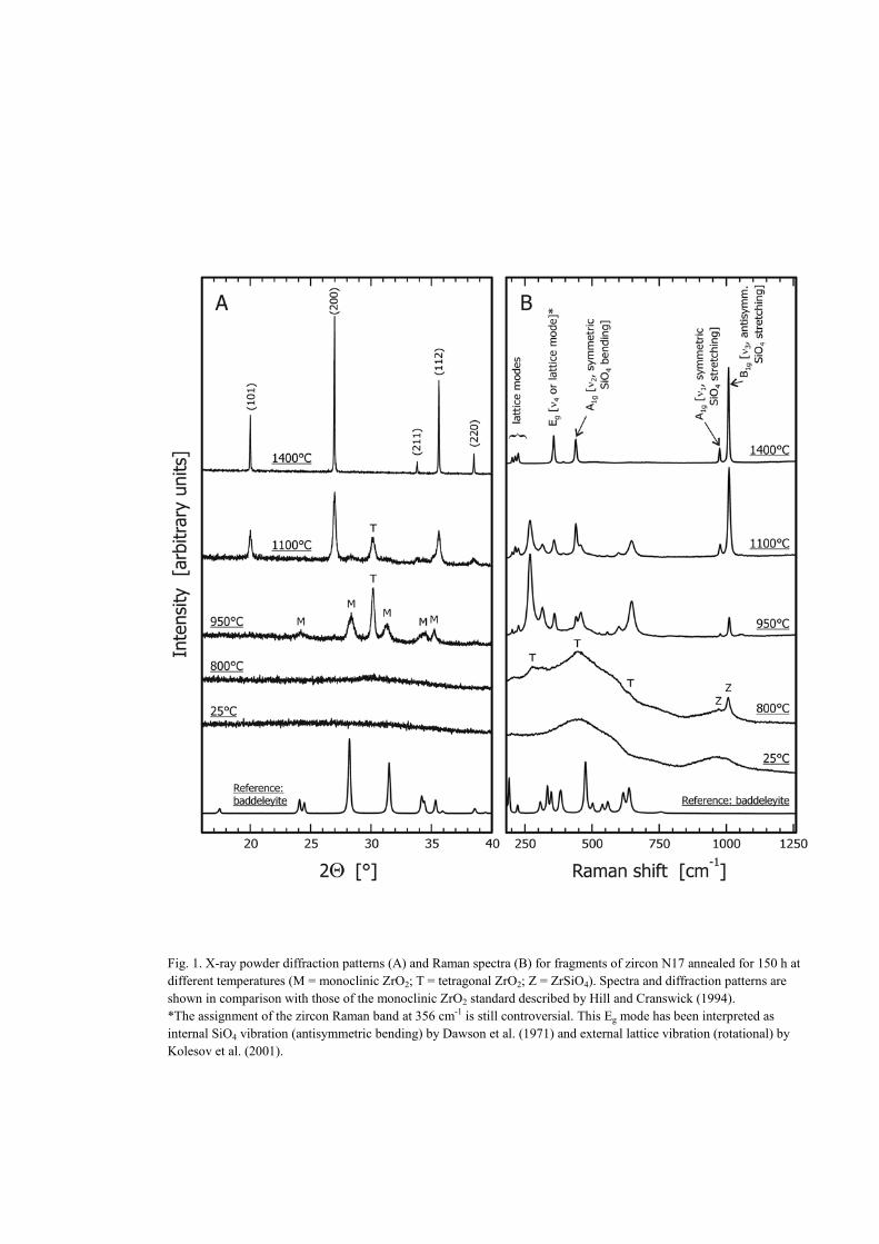
Fig. 1. X-ray powder diffraction patterns (A) and Raman spectra (B) for fragments of zircon N17 annealed for 150 h atdifferent temperatures (M = monoclinic ZrO2; T = tetragonal ZrO2; Z = ZrSiO4). Spectra and diffraction patterns areshown in comparison with those of the monoclinic ZrO2 standard described by Hill and Cranswick (1994).*The assignment of the zircon Raman band at 356 cm-1 is still controversial. This Eg mode has been interpreted asinternal SiO4 vibration (antisymmetric bending) by Dawson et al. (1971) and external lattice vibration (rotational) byKolesov et al. (2001).
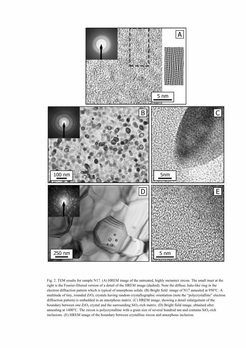
Fig. 2. TEM results for sample N17. (A) HREM image of the untreated, highly metamict zircon. The small inset at theright is the Fourier-filtered version of a detail of the HREM image (dashed). Note the diffuse, halo-like ring in theelectron diffraction pattern which is typical of amorphous solids. (B) Bright field image of N17 annealed at 950°C. Amultitude of tiny, rounded ZrO2 crystals having random crystallographic orientation (note the “polycrystalline” electrondiffraction pattern) is embedded in an amorphous matrix. (C) HREM image, showing a detail enlargement of theboundary between one ZrO2 crystal and the surrounding SiO2-rich matrix. (D) Bright field image, obtained afterannealing at 1400°C. The zircon is polycrystalline with a grain size of several hundred nm and contains SiO2-richinclusions. (E) HREM image of the boundary between crystalline zircon and amorphous inclusion.
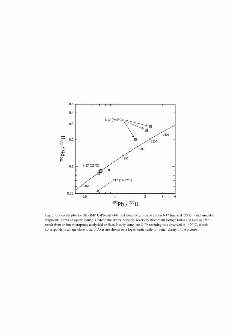
Fig. 3. Concordia plot for SHRIMP U-Pb data obtained from the untreated zircon N17 (marked “25°C”) and annealedfragments. Sizes of square symbols extend the errors. Strongly reversely discordant isotope ratios and ages at 950°Cresult from an ion microprobe analytical artifact. Nearly complete U-Pb resetting was observed at 1400°C, whichcorresponds to an age close to zero. Axes are shown on a logarithmic scale for better clarity of the picture.
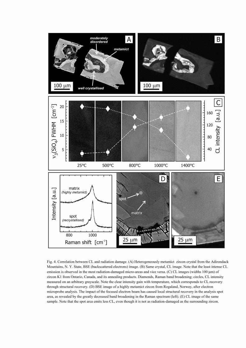
Fig. 4. Correlation between CL and radiation damage. (A) Heterogeneously metamict zircon crystal from the AdirondackMountains, N. Y. State, BSE (backscattered electrons) image. (B) Same crystal, CL image. Note that the least intense CL
emission is observed in the most radiation-damaged micro-areas and vice versa. (C) CL images (widths 100 µm) ofzircon K1 from Ontario, Canada, and its annealing products. Diamonds, Raman band broadening; circles, CL intensitymeasured on an arbitrary grayscale. Note the clear intensity gain with temperature, which corresponds to CL recoverythrough structural recovery. (D) BSE image of a highly metamict zircon from Rogaland, Norway, after electronmicroprobe analysis. The impact of the focused electron beam has caused local structural recovery in the analysis spotarea, as revealed by the greatly decreased band broadening in the Raman spectrum (left). (E) CL image of the samesample. Note that the spot area emits less CL, even though it is not as radiation-damaged as the surrounding zircon.
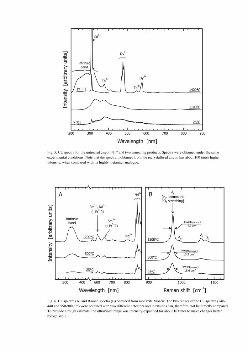
Fig. 5. CL spectra for the untreated zircon N17 and two annealing products. Spectra were obtained under the sameexperimental conditions. Note that the spectrum obtained from the recrystallised zircon has about 100 times higherintensity, when compared with its highly metamict analogue.
Fig. 6. CL spectra (A) and Raman spectra (B) obtained from monazite Moacir. The two ranges of the CL spectra (240-440 and 550-900 nm) were obtained with two different detectors and intensities can, therefore, not be directly compared.To provide a rough estimate, the ultraviolet range was intensity-expanded for about 10 times to make changes betterrecognisable.