Embed Size (px)
Citation preview
Calcitonin Gene-related Peptide and Muscle Activity Regulate Acetylcholine Receptor a-Subul t mRNA Levels by Distinct Intracellular Pathways Ber t rand Fontaine, Andr6 Klarsfeld, and Jean-Pier re Changeux
Unit6 de Neurobiologie Mol6culaire et Unit6 Associ6e au Centre National de la Recherche Scientifique UA 041149. Interactions Mol~culaires et Cellulaires, D6partement des Biotechnologies, Institut Pasteur, 75724 Paris C6dex 15, France
Abstract. In cultured chicken myotubes, calcitonin gene-related peptide (CGRP), a peptide present in spi- nal cord motoneurons, increased by 1.5-fold the num- ber of surface acetylcholine receptors (AChRs) and by threefold AChR ~t-subunit mRNA level without affect- ing the level of muscular t~-actin mRNA. Cholera toxin (CT), an activator of adenylate cyclase, produced a similar effect, which did not add up with that of CGRP. In contrast, tetrodotoxin, a blocker of voltage- sensitive Na § channels, elevated the level of AChR a-subunit mRNA on top of the increase caused by ei- ther CGRP or CT.
L2-O-Tetradecanoyl phorbol-B-acetate (TPA), an acti- vator of protein kinase C, markedly decreased the cell surface and total content of [L25I]ctBGT-binding sites
and reduced the rate of appearance of AChR at the surface of the myotubes without reducing the level of AChR ~t-subunit mRNA. Moreover, TPA inhibited the increase of AChR ~t-subunit mRNA caused by tetro- dotoxin without affecting that produced by CGRP or CT. Under the same conditions, TPA decreased the level of muscular ~t-actin mRNA and increased that of nonmuscular 13- and "/-actins mRNA.
These data suggest that distinct second messengers are involved in the regulation of AChR biosynthesis by CGRP and muscle activity and that these two path- ways may contribute to the development of different patterns of AChR gene expression in junctional and extrajunctional areas of the muscle fiber.
T He. nicotinic acetylcholine receptor (AChR) ~ is an in- tegral membrane protein, composed of five transmem- brane subunits (~t213)'8) (8). Under the nerve endings,
the number of receptor molecules steadily increases during the maturation of the neuromuscular junction, and then re- mains very high in the adult junction; in contrast, extra- synaptic receptor density falls to negligible values (9, 15, 29). The decline of extrajunctional AChR has been related to the repression of AChR biosynthesis by the neurally evoked elec- trical activity of the developing muscle fiber (9, 15, 29). What then are the mechanisms that would account for the persistence of AChR incorporationat the level of the growing subneural domain? Several attempts have been made to iden- tify neural factors that might plausibly be involved in this process, by following the increase of AChR levels in cultured muscle cells caused by extracts of neural tissues and/or by a variety of pharmacological agents (reviewed in 29), includ- ing, most recently, the neuropeptide calcitonin gene-related peptide (CGRP) (14, 25).
CGRP is a widely distributed peptide generated by differential RNA processing from a single genomic locus
1. Abbreviations used in this paper: AChR, acetylcholine receptors; [125I]tt-BGT, 125I-labeled a-bungarotoxin; CGRP, calcitonin gene-related peptide; CT, cholera toxin; TTX, tetrodotoxin.
which also codes for calcitonin (28). CGRP exerts several biological actions such as cardiovascular effects, inhibition of gastric secretion, and ingestive behavior (13) and has been proposed to act as a neurotransmitter in the sensory tract (33). Interestingly, CGRP has been localized by immuno- histochemistry in chicken motoneurons (14, 25) and in mouse (32) or rat tongue (31) motor end-plates. Moreover, the peptide was found to be colocalized with acetylcholine in rat neurons of several motor systems (31). CGRP may thus serve as a coexisting neuronal messenger (7, 16). Consistent with such a role, CGRP is released from cultures of rat trigeminal cells (21) and enhances skeletal muscle contrac- tion (32). Furthermore, it was recently shown that addition of CGRP to chick muscle cells in primary cultures signifi- cantly increases the level of surface AChR (14, 25).
In this paper, we demonstrate that CGRP significantly ele- vates AChR ~t-subunit mRNA levels in primary chick myo- tubes in culture. To further identify the intracellular path- ways involved in this effect, a variety of pharmacological agents were tested. We report that cholera toxin (CT), an ac- tivator ofadenylate cyclase, and 12-O-tetradecanoyl phorbol- B-acetate (TPA), an activator of protein kinase C (6, 27), exert different effects on the levels of AChR and AChR a-sub- unit mRNA. Similar experiments with CT or TPA were also
�9 The Rockefeller University Press, 0021-9525/8710911337/6 $2.00 The Journal of Cell Biology, Volume 105, September 1987 1337-1342 1337
on April 11, 2019jcb.rupress.org Downloaded from http://doi.org/10.1083/jcb.105.3.1337Published Online: 1 September, 1987 | Supp Info:
performed in the presence of CGRP or tetrodotoxin (TTX), a Mocker of muscle electrical activity. The data are inter- preted in terms of a model of AChR genes regulation during the development of the neuromuscular junction according to which CGRP and muscle electrical activity might respec- tively contribute to the maintenance of AChR gene expres- sion in junctional areas and to the disappearance of AChR in extrajunctional areas via distinct intracellular pathways.
Materials and Methods
Myotube Culture
Myoblasts were obtained from the hind limbs of H-d-old chick embryos (Centre Avicole de rile de France, Arpajon) by mechanical dissociation and cultured as described (2). Briefly, the cells were seeded in gelatine-coated plastic dishes at a density of 104 cells/ram 2 and cultivated in a 3:1 mixture of MEM (Gibco, Grand Island, NY) and medium 199 (Gibco), containing 10% horse serum (Gibco) and 1% chick embryo extract. From 48 to 96 h of culture, the cells were treated with 10 -5 M cytosine arabinnside (Sigma Chemical Co., St. Louis, MO) to minimize fibroblast proliferation. Media were then reneged every 2 or 3 d.
Treatments with Pharmacological Agents
The agents were added after 6 d of culture directly into the medium. Syn- thetic rat CGRP (10 -7 M final concentration) was purchased from Bachem Feinchemikalien (Bubendorf, Switzerland), CT (25 ng/ml final concentra- tion), a gift of Dr. A. Monneron, was from Makor Chemicals (Jerusalem, Israel) and TTX (0.1 IJg/ml final concentration) was from Sigma Chemical Co. After 24-40 h of incubation with cultured myotubes, CGRP-supple- mented media were still able to increase the level of AChR when transferred to a nontreated culture dish (data not shown). Moreover, AChR levels did not further increase upon adding a fresh CGRP solution to the culture medium, indicating that CGRP was not or only slightly inactivated during the experiment.
TPA (Sigma Chemical Co.) and 4-a-phorbol 12-13 didecanoate (gift of Dr. Castagna) were dissolved at 1 mg/rnl in 1% DMSO. 1,000-fold dilution of a 1% DMSO solution into the culture medium had no effect on myotubes as judged by phase-contrast microscopy and surface AChR number.
Quantification of AChR Levels with I uJI]-a-Bungarotoxin Surface AChR levels (2) were measured by t2SI-labeled a-bungarotoxin ([mI]aBGT; sp act = 200 Ci/mmol; Amersham Corp., Arlington Heights, IL) binding to duplicate 35-ram dishes. Non-specific binding was deter- mined after 30 rain of preincubation with, and in the subsequent presence of, 10 -5 M decamethonium, and was subtracted from all values (5-10% de- pending on the [ml]aBGT batches).
The metabolic rate of degradation of surface AChR was estimated by fol- lowing the life-time of the AChR-aBGT complex. Cultures treated with 20 ng/ml TPA for 24 h and control cultures were labeled in parallel with [t~I]aBGT. After 0.5 h at 37~ the cells were washed free of unbound toxin and cultured in TPA-supplemented or control medium. At different time intervals, duplicate dishes were removed and the amount of remaining bound radioactivity was determined.
The rate of insertion of AChR into the sarcolemmai membrane was deter- mined after 24 h of preineubation with TPA (20 ng/ml) or CGRP (10 -7 M). The cells were incubated with 10 -7 M aBGr (Signm Chemical Co.) for 0.5 h in MEM at 370C, washed four times with MEM, and incubated again in the medium that had been removed before incubation with aBGT (sup- plemented with CGRP or TPA where necessary). At different time intervals, cells were incubated with 10 nM [ml]aBGT for 0.5 h. After four washes with PBS (pH 7), radioactivity of a 1% Triton X-100-1 M NaOH extract was determined in a gamma counter (LKB-WalIac, Turku, Finland).
Total receptor content was determined in 1% Triton X-100-10 mM NaCI- 10 mM Tris-HC! (pH 7.4) extracts (12). After 5 rain of incubation in this solution, cells were homogenized with a glass-teflon homogenizer. Cen- trifugation at 7,000 g during 30 rain was performed to remove insoluble ma- terial. The supernatant was then incubated with 10 nM [mI]aBGr during 1 h at 370C. Nonspecitic binding of aBGT was determined by the binding
of [mI]aBGT after 1 h of preineubation in the presence of 10 -7 M aBGT. Each sample was then filtered through three stacked DES1 filters (Whatman Inc., Clifton, NJ), which were washed with 5-10 mt of a solution containing 1% Triton X-100, 10 mM NaCI, 10 mM Tris-HC1 (pH 7.4).
Total Protein Content and Incorporation of pSS]Methionine Total protein content was measured in extracts by the Coomassie Blue tech- nique (Bio-Rad Laboratories, Richmond, CA). The rate of incorporation of [35Slmethionine was determined in triplicate 20 ng/ml TPA-supplemented and control dishes. The medium was replaced by medium free of methio- nine, with TPA where necessary. [35S]melhionine (100 txCitml; Amersham Corp.) was then added and incorporation was allowed to proceed for 2-4 h. The cells were washed twice with PBS (pH 7) and extracted in t M NaOH containing 5 mM L-methionine. The 10% TeA-insoluble radioactiv- ity was determined in aliquots by a glass fiber filter assay procedure and ex- pressed relative to the total radioactivity measured in the same extract. Results were corrected for the nonspecific binding of [35Slmethionine which was determined at time zero.
Northern Blot Analysis
Total RNA was prepared from pools of four 100-mm culture dishes. After two washings with PBS (pH 7) and one with 0.25 M NaCI-10 mM Tris-HCl (pH 7.5)-10 mM MgCI2, the cells were scraped with a rubber policeman, and lysis was performed in 4 M urea-2 M LiCI by slowly pipetting through a syringe. Precipitation of RNA by LiCI was allowed to proceed for 6-24 h at 0~ After centrifugation, the pellet was dissolved in 10 mM Tris HC1 (pH 7.5) - 0.5% SDS-1 mM ethylenedinitrilotetraacetic acid, extracted once with phenol and once with phenol-ehloroform-isoamylalcobol (25:24:1) before ethanol precipitation (la). Yields varied between 10to 30 I~g of RNA per dish. RNA samples (8-12 [.tg) were subjected to electrophoresis in 1.2 % formaldehyde-agarose gels and transferred onto a nylon membrane (Hy- bond N; Arnersham Corp.) by standard procedures (20). Hybridization and final washes of the blots wore performed according to manuals of membrane suppliers. The AChR a-subunit single-stranded probe (3) was synthesized from a DNA fragment of 500 nucleotides containing 120 nucleotides of exon P2 of the chicken AChR a-subunit gene (17) and labeled by a[32P]dATP (800 Ci/mmol; Amersham Corp.). The actin probe (giR of Dr. S. Alonso) was a 1,150-bp mouse eDNA insert in plasmid pAIA1 derived from cyto- skeletal I~-aetin mRNA, as determined from its 3' untranslated sequence (1, 24). It was labeled according to the Amersham multiprime procedure by a[32P]dCrP (Amersham Corp.; 800 Ci/mmol). Autoradiograms of the hy- bridized blots were scanned with a densitometer using appropriate expo- sures to obtain calibration curves.
Results
Effects of CGRP, CT, and TTX on the Levels Of AChR and AChR a-Subunit mRNA As previously described (14, 25), exposure of primary cul- tures of chick myotubes to 10 -7 M CGRP increased the level of surface AChR by ,~30%. Parallel results were obtained
Table L Parallel Variation of Total and Surface [ml]aBGT-binding Sites in CGRP- or TPA-treated Chick Myotubes in Culture
Total ['25I]aBGT sites* Surface [125I]aBGT sites*
(fraction of control) (fraction of control)
Control 1 1 w CGRP 1.25 + 0.02 1.26 + 0.05 TPA 0.60 • 0.10 0.47 5 :0 .05
* Results are averages :t: SD of two separate experiments with three culture dishes in each experiment. * Results are averages 5: SD of two separate experiments with two culture dishes in each experiment. w For example, in one experiment surface [l~I]aBGT-binding sites were ,070 fmol and total [12sI]aBGT-binding sites ,0135 fmol per control 35-mm dishes.
The Journal of Cell Biology, Volume 105, 1987 1338
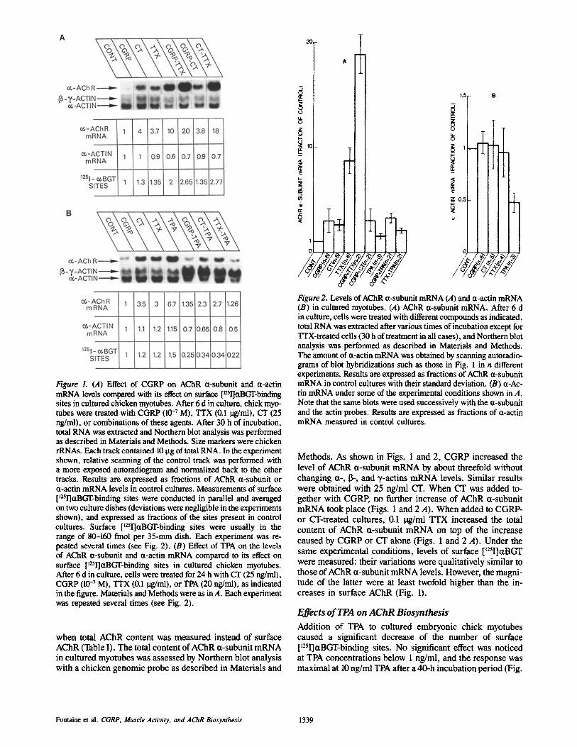
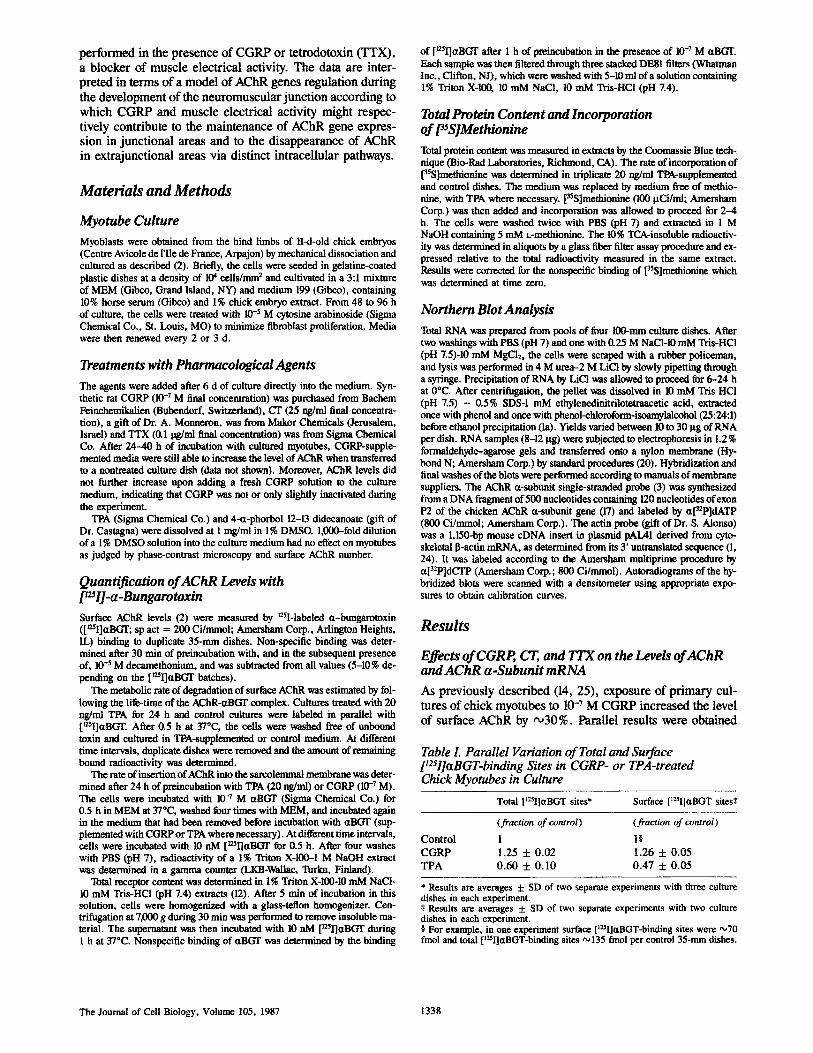
Figure L (A) Effect of CGRP on AChR a-subunit and a-actin mRNA levels compared with its effect on surface [mI]aBGT-binding sites in cultured chicken myotubes. After 6 d in culture, chick myo- tubes were treated with CGRP (10 -7 M), TTX (0.1 ~,/ml), CT (25 ng/ml), or combinations of these agents. After 30 h of incubation, total RNA was extracted and Northern blot analysis was performed as described in Materials and Methods. Size markers were chicken rRNAs. Each track contained 10 pg of total RNA. In the experiment shown, relative scanning of the control track was performed with a more exposed autoradiogram and normalized back to the other tracks. Results are expressed as fractions of AChR r or a-actin mRNA levels in control cultures. Measurements of surface [~5I]aBGT-binding sites were conducted in parallel and averaged on two culture dishes (deviations were negligible in the experiments shown), and expressed as fractions of the sites present in control cultures. Surface [t251]aBGT-binding sites were usually in the range of 80-I60 fmol per 35-mm dish. Each experiment was re- peated several times (see Fig. 2). (B) Effect of TPA on the levels of AChR (x-subunit and a-actin mRNA compared to its effect on surface ['25I]aBGT-binding sites in cultured chicken myotubes. After 6 d in culture, cells were treated for 24 h with CT (25 ng/ml), CGRP (10 -7 M), TTX (0.1 lag/ml), or TPA (20 ng/ml), as indicated in the figure. Materials and Methods were as in A. Each experiment was repeated several times (see Fig. 2).
when total AChR content was measured instead of surface AChR (Table I). The total content of AChR a-subunit mRNA in cultured myotubes was assessed by Northern blot analysis with a chicken genomic probe as described in Materials and
^
Z 0 o
10 E
n r
E
z 1 E Z_
1,5 B
Figure 2. Levels of AChR 0t-subunit mRNA (A) and u-actin mRNA (B) in cultured myotubes. (,4) AChR u-subunit mRNA. After 6 d in culture, cells were treated with different compounds as indicated, total RNA was extracted after various times of incubation except for TTX-treated cells (30 h of treatment in all cases), and Northern blot analysis was performed as described in Materials and Methods. The amount of ct-actin mRNA was obtained by scanning autoradio- grams of blot hybridizations such as those in Fig. 1 in n different experiments. Results are expressed as fractions of AChR a-subunit mRNA in control cultures with their standard deviation. (B) r tin mRNA under some of the experimental conditions shown in A. Note that the same blots were used successively with the a-subunit and the actin probes. Results are expressed as fractions of ct-actin mRNA measured in control cultures.
Methods. As shown in Figs. 1 and 2, CGRP increased the level of AChR a-subunit mRNA by about threefold without changing a-, 13-, and 7-actins mRNA levels. Similar results were obtained with 25 ng/ml CT. When CT was added to- gether with CGRP, no further increase of AChR ct-subunit mRNA took place (Figs. 1 and 2 A). When added to CGRP- or CT-treated cultures, 0.1 lag/ml TTX increased the total content of AChR a-subunit mRNA on top of the increase caused by CGRP or CT alone (Figs. 1 and 2 A). Under the same experimental conditions, levels of surface [~25I]r were measured: their variations were qualitatively similar to those of AChR ct-subunit mRNA levels. However, the magni- tude of the latter were at least twofold higher than the in- creases in surface AChR (Fig. 1).
Effects of TPA on AChR Biosynthesis
Addition of TPA to cultured embryonic chick myotubes caused a significant decrease of the number of surface [~2~I]aBGT-binding sites. No significant effect was noticed at TPA concentrations below 1 ng/ml, and the response was maximal at 10 ng/ml TPA after a 40-h incubation period (Fig.
Fontaine et al, CGRP, Muscle Activity, and AChR Biosynthesis 1339
10017 ....
8
0,1
~oo
A
[ l I 1 lO TPA(ng/ml) 100
I I I
10 hours 20 30
Figure 3. Effect of TPA on surface AChR. (A) Dose-response curve of the effect of TPA on [~2~I]ctBGT-binding sites in cultured myo- tubes after a 40-h incubation period. Number of surface AChR sites are expressed as the percentage of [~2~I]ttBGT sites in parallel un- treated cultures. (B) Time course of the evolution of [125I]ctBGT- binding sites in TPA (20 ng/ml)-treated and control cultures. Each of these experiments was repeated at least twice. Each point is a mean of two different culture dishes, with the bar indicating the variation between the two.
3 A). 3 h after the addition of TPA (20 ng/ml), this effect was already noticeable, and it reached its maximum after 24 h of treatment (Fig. 3 B). No further decrease was found between 30 and 40 h (not shown). The reduction in the number of [12~I]ctBGT-binding sites caused by TPA averaged 45% (25-60% of control values in nine separate experiments). A similar but smaller effect was obtained on total AChR (Table I). The 4-ct-phorbol 12-13 didecanoate (20 ng/ml), which does not activate protein kinase C (27), had no significant effect on AChR number in these cultured muscle cells.
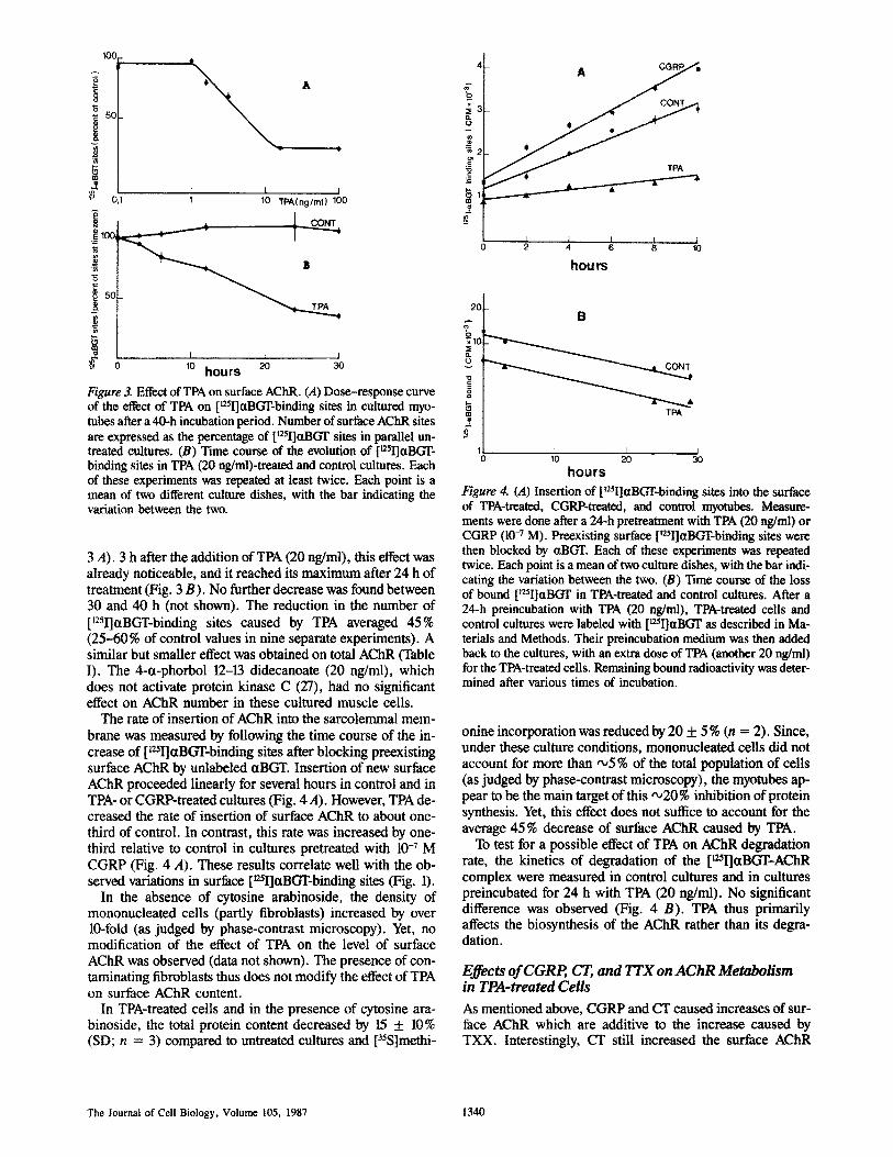
The rate of insertion of AChR into the sarcolemmal mem- brane was measured by following the time course of the in- crease of [L~ctBGT-binding sites after blocking preexisting surface AChR by unlabeled ~tBGT. Insertion of new surface AChR proceeded linearly for several hours in control and in TPA- or CGRP-treated cultures (Fig. 4 A). However, TPA de- creased the rate of insertion of surface AChR to about one- third of control. In contrast, this rate was increased by one- third relative to control in cultures pretreated with 10 -7 M CGRP (Fig. 4 A). These results correlate well with the ob- served variations in surface [~25I]ctBGT-binding sites (Fig. 1).
In the absence of cytosine arabinoside, the density of mononucleated cells (partly fibroblasts) increased by over 10-fold (as judged by phase-contrast microscopy). Yet, no modification of the effect of TPA on the level of surface AChR was observed (data not shown). The presence of con- taminating fibroblasts thus does not modify the effect of TPA on surface AChR content.
In TPA-treated cells and in the presence of cytosine ara- binoside, the total protein content decreased by 15 + 10% (SD; n = 3) compared to untreated cultures and [35S]methi-
x
o
g
25
?
* *~' r P A
I t t I I 2 4 6 8 10
hours
20
~o
m TPA
I I I 0 10 2 0 3 0
hours
Figure 4. (A) Insertion of [12~I]otBGT-binding sites into the surface of TPA-treated, CGRP-treated, and control myotubes. Measure- merits were done after a 24-h pretreatment with TPA (20 ng/ml) or CGRP (10 -7 M). Preexisting surface [~25I]~-binding sites were then blocked by ctBGT. Each of these experiments was repeated twice. Each point is a mean of two culture dishes, with the bar indi- cating the variation between the two. (B) Time course of the loss of bound [t25I]ctBGT in TPA-treated and control cultures. After a 24-h preincubation with TPA (20 ng/ml), TPA-treated cells and control cultures were labeled with [~25I]~tBGT as described in Ma- terials and Methods. Their preincubation medium was then added back to the cultures, with an extra dose of TPA (another 20 ng/ml) for the TPA-treated cells. Remaining bound radioactivity was deter- mined after various times of incubation.
onine incorporation was reduced by 20 + 5 % (n = 2). Since, under these culture conditions, mononucleated cells did not account for more than ,x,5 % of the total population of cells (as judged by phase-contrast microscopy), the myotubes ap- pear to be the main target of this •20% inhibition of protein synthesis. Yet, this effect does not suffice to account for the average 45 % decrease of surface AChR caused by TPA.
To test for a possible effect of TPA on AChR degradation rate, the kinetics of degradation of the [~25I]aBGT-AChR complex were measured in control cultures and in cultures preincubated for 24 h with TPA (20 ng/ml). No significant difference was observed (Fig. 4 B). TPA thus primarily affects the biosynthesis of the AChR rather than its degra- dation.
Effects of CGRP, CT, and TTX on AChR Metabolism in TPA-treated Cells
As mentioned above, CGRP and CT caused increases of sur- face AChR which are additive to the increase caused by TXX. Interestingly, CT still increased the surface AChR
The Journal of Cell Biology, Volume 105, 1987 1340

content in TPA-treated cells (Fig. 1 B). However, a similar effect for CGRP was only noticed in two out of the five ex- periments (Fig. 1 B). In contrast, in the presence of TPA, the effect of TTX on surface AChR was systematically abol- ished (Fig. 1 B).
Parallel analysis of mRNA content by Northern blots re- vealed that the level of AChR ~t-subunit mRNA was not affected by TPA (or slightly increased) relative to AChR a-subunit mRNA level in control cultures (Figs. 1 B and 2 A). In contrast, the level of ~t-actin mRNA decreased by "-,50% (Figs. 1 B and 2 B) while that of 13- and 3,-actins in- creased ,~fivefold (Fig. 2 B). CGRP and CT still increased the content in AChR ct-subunit mRNA in the presence of TPA (Figs. 1 B and 2 A). Consistent with a previous study (17), TTX augmented up to 10-fold the content of AChR ~t-subunit mRNA (after 30 h of treatment). However, in TPA- treated cells, this effect of TTX on AChR ~t-subunit mRNA content was totally abolished (Figs. 1 B and 2 A).
Discussion
CGRP, a neuropeptide which coexists with acetylcholine in spinal cord motor neurons, has been previously shown to in- crease the number of AChR sites in primary cultures of chick skeletal muscle (14, 25). The present results further demon- strate that, correlatively, CGRP increases (by about three- fold) the level of AChR r mRNA without signifi- cantly affecting the level of r mRNA, a major muscular component. Such an increase may result from a specific stimulation of ~t-subunit gene transcription or from a stabili- zation of the r mRNA (11). Nuclear run-on experi- ments should decide in favor of one of these two possibilities.
The amplitude of this increase in mRNA levels appears significantly larger (about twofold) than the increase of total and surface aBGT-binding sites as well as of the rate of AChR appearance at the surface of the myotubes. A similar difference between AChR r mRNA and AChR pro- tein content has already been noticed in the case of TTX blockade of the spontaneous electrical activity of cultured myotubes (17). Posttranscriptional events which include the covalent modification and conformational maturation of AChR subunits, their assembly into the a2137~ pentamer, and their transport to the cell surface via the Golgi apparatus may account for this difference (23).
Electrical activity of muscle fibers in culture and in vivo is known to repress AChR biosynthesis (9, 15, 29). In cell cultures, CGRP causes an increase in AChR number which appears independent of the electrical activity of the muscle fibers, suggesting that CGRP and electrical activity regulate AChR biosynthesis via different intraceUular pathways (14). These results have now been extended to the AChR r mRNA level. Moreover, in our initial observations (14), it was noticed that CI', an activator of adenytate cyclase, also increased AChR number (as already described with dibuty- ryl cyclic AMP [2] and with CT [4, 19]), but this effect was not found additive with that of CGRP. Interestingly, the same observations hold at the mRNA level, further supporting a contribution of cyclic AMP to the regulation of AChR bio- synthesis by CGRP. Consistent with such a mechanism, CGRP has recently been shown to increase cyclic AMP level in cultured myotubes (18) and in mouse diaphragm (30), and
to activate adenylate cyclase in chick muscle membranes (18).
To investigate the possible involvement of protein kinase C in AChR biosynthesis, the phorbol ester TPA, an activator of protein kinase C (6, 27), was applied to chick myotube cul- tures. TPA has already been shown to exert a pleiotropic ac- tion on cultured myotubes (10) and to modulate their terminal differentiation. This effect could account for the observed in- crease of nonmuscular I~- and T-actin mRNA levels and for the decrease of muscular ct-actin mRNA level. An increase of l~ and ~t-actin mRNA levels has also been recently re- ported in BC3H1 ceils exposed to other mitogenic factors: 20% FCS and fibroblast growth factor (35). Of particular in- terest for the present study, TPA caused a specific decrease of surface and total AChR content in cultured myotubes as well as a correlative decrease of the rate of AChR insertion into the sarcolemmal membrane. Yet, general protein syn- thesis was hardly diminished; AChR lifetime did not change and AChR a-subunit mRNA level remained constant (or slightly increased). Under these culture conditions, TPA thus primarily affects posttranscriptional events in the matu- ration of AChR. Posttranscriptional regulation of AChR bio- synthesis has also been observed with differentiated BC3H-1 cells in culture. When forced back into the cell cycle by refeeding with 20% FCS, AChR a-subunit mRNA level in- creased although AChR expression at the cell surface was completely blocked (23). Also, an inhibition of AChR as- sembly by electrical activity has been suggested in primary cultures of embryonic rat muscle cells (5).
It is noteworthy that muscle electrical activity has been proposed to stimulate phosphatidylinositide metabolism (34), and, thus, may enhance protein kinase C activity via the resulting production of diacylglycerol (27). Exposure of the cells to TPA might then reproduce the effect of electrical stimulation on AChR biosynthesis. Although TPA did not significantly modify the level of AChR mRNA in cells where AChR expression is already repressed by spontaneous elec- trical activity (9, 15, 29), TPA did completely block the in- crease of AChR biosynthesis caused by TTX. In contrast, the accumulation of AChR a-subunit mRNA elicited by CGRP and CT persisted in TPA-treated myotubes. Therefore, the present data do not rule out the possible involvement of pro- tein kinase C in linking muscle electrical activity to AChR r mRNA content. Moreover, calcium has also been shown to play a significant role in the regulation of AChR biosythesis by muscle activity (15, 29), and the interactions between calcium and protein kinase C are now well docu- mented (27). In any case, these data further strengthen the conclusion that CGRP and electrical activity regulate AChR gene expression via different second messenger systems (14).
Our results are consistent with a model (9) according to which, (a) in the adult muscle, the sarcolemmal nuclei that underlie the endplate are in a state of differentiation different from those located in extrajunctional cytoplasm (22), and (b) the development and maintenance of this pattern involves distinct intracellular signaling pathways in extrajunctional and junctional areas. Tentatively, one may thus suggest that, during development, CGRP, co-released with acetylcholine by the motor nerve ending, would increase the expression of the genes coding for the AChR via the cyclic AMP cascade in subneural areas while, in extrajunctional nuclei, muscle membrane electrical activity would repress their transcrip-
Fontaine et aL CGRP, Muscle Activity, and AChR Biosynthesis 1341
tion by an intracellular pathway involving calcium (19) and, more speculatively, the activation of protein kinase C. Whereas the in vivo role of muscle electrical activity in AChR gene regulation is now well established, the actual rel- evance of CGRP to the development of the neuromuscular junction remains to be derhonstrated. Interestingly, a possi- ble contribution of cyclic AMP to synapse stabilization has been proposed on the basis of in vitro results (26) and CGRP appears to be one candidate "anterograde factor" (7, 9) which, among others, could mediate this regulation.
The skillful technical assistance of Mrs. Hem'iette Nuret is gratefully ac- knowledged. We thank Professor Tomas H6kfelt for helpful suggestions and our laboratory colleagues for help and advice especially Drs. R. Laufer, C. Henderson, E Revah, O. Heidmann, and A. Devillers-Thiery. We thank Dr. M. Castagna for the gift of 4-tt-phorbol 12-13 didecanoate; Dr. A. Mon- neron for the gift of cholera toxin; Dr. S. Alonso for the gift of plasmid pAL 41; and Dr. M. H. Buc-Caron for the gift of [35S]methionine and advice for its use.
This work was supported by grants from the Muscular Dystrophy Associ- ation of America, the Collbge de France, the Minist~re dc la Recherche et de la Technologie, the Centre National de la Recherche Scientifique, and the Commissariat ~t rEncrgie Atomique. Bertrand Fontainc is an "Interne de SI~cialit~ de Paris ct dc la rdgion lle de France" and is recipient of an "annde recherche " fellowship from the "Assistance Publique des H6pilanx de Paris 7
Received for publication 10 March 1987, and in revised form 14 April 1987.
Rt~l~nce$
1. Alonso, S., A. Mint'y, Y. Bourlet, and M. Buckingham. 1986. Comparison of three actin-coding sequences in the mouse; evolutionary relationships between the aetin genes of warm-blooded vertebrates. J. MoL Biol. 23:11-22.
la. Auffray, C., and F. Rougeon. 1980. Pttrifieation of mouse immunoglobu- lin heavy-chain messenger RNAs from total myeloma tumor RNA. Eur. J. Biochem. 107:303-314.
2. Bet.z, H., and J-P. Changeux. 1979. Regulation of muscle acetylcholine receptor synthesis /n vitro by cyclic nucleotide derivatives. Nature (Lond.). 278:749-752.
3. Biggin, M., P. J. Farrell, and B. G. Barrel. 1984. Transcription and DNA sequence of the Barn HI L fragment of B 95-8 Epstein-Ban" virus. EMBO (Eur. MoL Biol. Organ.)J. 3:1083-1090.
4. Blosser, L C., and S. H. AppeL 1980. Regulation of acetylchuline receptor by cyclic AMP../. BioL (?hem. 255:1235-1238.
5. Carlin, B. E., J. R. Lawrence, L M. Lindstrom, and J. P. Merlie. 1986. Inhibition of acetylcholine receptor assembly by activity in primary cul- tures of embryonic rat muscle cells..L Biol. Chem. 261:5180-5186.
6. Castagna, M., Y. Takai, K. Kailmchi, K. Sano, Y. Kikk~wa, and Y. Nish- izuka. 1982. Direct activation of calcium-activated phospholipid-depen- dent protein kinase by tumor promoting phorhol esters. J. Biol. Chem. 257:7847-7851.
7. Changeux, J-P. 1986. Coexistence of neuronal messengers and molecular selection. In Coexistence of neuronal messengers: a new principle in chemical transmission. Prog. Brain lies. 68:373--403.
8. Changeux, J-P., A. Devillers-Thi6ry, and P. Chemoallli. 1984. The acetyl- choline receptor: an allosteric protein. Science (wash. DC). 225:1335- 1345.
9. Changeux, J-P., A. Klarsfeld, and T. Heddmann. 1987. The acetyleholine receptor and molecular models for short and long term learning. Dahlem Korfferenzen. In The Cellular and Molecular Bases of Learning. J. P. Changeux, and M. Konishi, editors. John Wiley & Sons, London. 31-83.
10. Crnop, J., G. Dubrynk, Y. Toyama, A. Dlugosz, A. Scarpa, and H. Holt- zer. 1982. Effects of 12-O-tetradecanocylphorbol- 13-acotate on myofibril integrity and Ca ++ content in developing myotubes. Dee. BioL 89:460- 474.
11. Darnell, J. E., Jr. 1982. Variety in the level of gene control in eukaryotic cells. Nature (Land.). 297:365-371.
12. Devreotes, P. N., and D. M. Fambrough. 1975. Acetylcholine receptor turnover in membranes of developing muscle fibers. J. Cell Biol. 65: 335-358.
13. Fischer, J. A., and W. Born. 1985. Novel peptides from the calcitonin
geue: expression, receptors and biologic~ function. Peptides. 3(Suppl.): 265-271.
14. Fontaine, B., A. Klarsfeld, T. H6kfelt, and J. P. Changeux. 1986. Calcito- nin gene-related peptide, a peptide present in spinal cord motoneurons, increases the number of acetylcholine receptors in primary cultures of chick embryo myotubes. Neurosci. Lett. 71:59-65.
15. Fambrough, D. M. 1979. Control of acetylcholine receptors in skeletal muscle. Physiol. Rev. 159:165-227.
16. H6kfelt, T., V. R. Holets, W. Staines, B. Meister, T. Melander, M. Schall- ing, M. Schultzberg, J. Freedman, H. Bj6rldund, L. Olson, B. Lindh, L. G. Elfvin, J. M. Lundberg, J. A. Lindgren, B. Samuelsson, B. Per- now, L. Terenius, C. Post, B. Everitt, and M. Goldstein. 1986. Coexis- tence of neuronal messengers- an overview. In Coexistence of neuronal messengers: a new principle in chemical transmission. Prog. Brain Res. 68:33-70.
17. Klarsfeld, A., and J. P. Changeax. 1985. Activity regulates the levels of acetylcholine receptor ct-subenit mRNA in cultured chicken myotubes. Proc. Natl. Acad. Sci. USA. 82:4558-4562.
18. Laufer, R., and J. P. Changeux. 1987. Calcitonin geue-related peptide ele- vates cyclic AMP levels in chick skeletal muscle: possible neurotrophic role for a coexisting neuronal messenger. EMBO (Eur. Mol. Biol. Or- gan.) Y. 6:901-906.
19. Mac Manaman, J. L., J. C. Blosser, and S. H. Appel. t982. Inhihitors of membrane depolarization regulate acetylcholine receptor synthesis by a calcium-dependent cyclic nucleotide-independeat mechanism. Biochim. Biophys. Acta. 720:8-35.
20. Maniatis, T., E. F. Fritsch, and M. Sambrook. 1982. Molecular Cloning. A Laboratory Manual. Cold Spring Harbor Press, Cold Spring Harbor, N.Y.
21. Mason, R. T., R. A. Peteffreund, P. E. Saw Chenko, A. Z. Corrigan, J. E. Rivier, and W. W. Yale. 1984. Release of the predicted calcitonin gene- related peptide from rat trigeminal ganglion cells. Nature (Lond.). 308:653--655.
22. Merlie, J. P., and J. R. Sanes. 1985. Concentration of acetylcholine recep- tor mRNA in synaptic regions of adult muscle fibers. Nature (Lond.). 317:66-68.
23. Merlie, J. P., and M. M. Smith, 1986. Synthesis and assembly ofacetylcho- line receptor, a multisuhunit membrane glycoprntein. J. Membr. Biol. 91:1-10.
24. Minty, A., S. Alonso, J. L. Gu~net, and M. E. Buckingham. 1983. Number and organization of actin-related sequences in the mouse genome. J. Mol. Biol. 167:77-101.
25. New, H. V., and A. W. Mudge. 1986. Calcitonin gene-related peptide regulates muscle acetylcholine receptor synthesis. Nature (Land.). 323: 809-811.
26. Nirenberg, M., S. Wilson, H. Higashida, A. Rotter, K. Krueger, N. Busis, R. Ray, J. G. Kenimer, and M. Adler. 1983. Modulation of synapse for- mation by cyclic adenosine monophosphate. Science (Wash. DC). 222: 794-799.
27. Nishizuka, Y. 1986. Studies and perspectives of protein kinase C. Science (Wash. DC). 233:305-312.
28. Rosenfeld, M. G., J. J. Mermod, S. G. Amara, L. W. Swanson, P. E. San- chenko, J. Rivier, W. W. Vale, and R. M. Evans. 1983. Production of a novel neuropeptide encoded by the caltitonin gene via tissue-specific RNA processing. Nature (Lond.). 304:129-135.
29. Salpeter, M. M., and R. H. Loring. 1985. Nicotinic acetylcholine receptors in vertebrate muscle: properties, distribution and neural control. Prog. NeurobioL (Oxf.). 25:297-325.
30. Takaml, K., K. Hashimoto, S. Uchida, M. Tohyama, and H. Yoshida. 1986. Effect of calcitonin gene-related peptide on the cyclic AMP level of isolated mouse diaphragm. Jpn. J. PharmacoL 42:345-350.
31. Takami, K., Y. Kawai, S. Shio Saka, Y. Lee, S. Girgis, C. J. Hillyard, I. Maelntyre, P. C. Emson, and M. Tohyama. 1985. Immunohistochemi- cal evidence for the coexistence of ealcitonin gene-related peptide- and choline acyltransferase-like immunoreactivity in neurons of the rate hypoglossal, facial and ambigus nuelesi. Brain Res. 328:386-389.
32. Takami, K., Y. Kawai, S. Uchida, M. Tokyama, Y. Shiotani, H. Akami, H. Yoshida, P. C. Emson, S. H. Girgis, C. J. Hitlyard, and I. Maclntyre. 1985. Effect of calcitonin gene-related peptide on contraction of striated muscle in the mouse. Neurasci. Lett. 60:227-230.
33. Tschopp, F. A., H. Henke, J. B. Peterman, P. H. Tobler, R. Janzer, T. H6kfelt, J. M. Lundberg, C. Cuello, and J. A. Fischer. 1985. CGRP and its binding sites in the human central nervous system and pituitary. Proc. Natl. Acad. Sci. USA. 82:248-252.
34. Vergara, J., R. Y. Tsien, and M. Delay. 1985. Inositoi 1,4,5-triphosphate: a possible chemical link in excitation-contracture coupling in muscle. Proc. Natl. Acad. Sci. USA. 82:6352-6356.
35. Wice, B., J. Hilbrandt, and L. Glaser. 1987. Control of muscle differentia- tion in BC3 H1 cells by fibroblast growth factor and vanadate. J. BioL Chem. 262:1810-1817.
The Journal of Cell Biology, Volume 105, 1987 1342