Embed Size (px)
Citation preview
Molecular interactions of the physiological anti-hypertensive peptidecatestatin with the neuronal nicotinic acetylcholine receptorBhavani S. Sahu1,*, Jagan Mohan1,*, Giriraj Sahu1,*, Pradeep K. Singh2, Parshuram J. Sonawane1, Binu K. Sasi1,Prasanna K. R. Allu1, Samir K. Maji2, Amal K. Bera1,`, Sanjib Senapati1,` and Nitish R. Mahapatra1,`
Department of Biotechnology, Bhupat and Jyoti Mehta School of Biosciences Building, Indian Institute of Technology Madras, Chennai 600036, IndiaDepartment of Biosciences and Bioengineering, Indian Institute of Technology Bombay, Powai, Mumbai 400076, India
*These authors contributed equally to this workAuthors for correspondence ([email protected]; [email protected]; [email protected])
Journal of Cell Science 125, 27872012. Published by The Company of Biologists Ltd
doi: 10.1242/jcs.114389
There was an error published in J. Cell Sci. 125, 2323-2337.
The third corresponding author was inadvertently omitted from the title page.
All corresponding authors are now indicated above.
We apologise for this error.
Erratum 2787
Molecular interactions of the physiologicalanti-hypertensive peptide catestatin with the neuronalnicotinic acetylcholine receptor
Bhavani S. Sahu1,*, Jagan M. Obbineni1,*, Giriraj Sahu1,*, Pradeep K. Singh2, Parshuram J. Sonawane1,Binu K. Sasi1, Prasanna K. R. Allu1, Samir K. Maji2, Amal K. Bera1,`, Sanjib Senapati1 and Nitish R. Mahapatra1,`
1Department of Biotechnology, Bhupat and Jyoti Mehta School of Biosciences Building, Indian Institute of Technology Madras, Chennai 600036, India2Department of Biosciences and Bioengineering, Indian Institute of Technology Bombay, Powai, Mumbai 400076, India
*These authors contributed equally to this work`Authors for correspondence ([email protected]; [email protected])
Accepted 2 January 2012Journal of Cell Science 125, 2323–2337� 2012. Published by The Company of Biologists Ltddoi: 10.1242/jcs.103176
SummaryCatestatin (CST), a chromogranin-A-derived peptide, is a potent endogenous inhibitor of the neuronal nicotinic acetylcholine receptor(nAChR). It exerts an anti-hypertensive effect by acting as a ‘physiological brake’ on transmitter release into the circulation. However,
the mechanism of interaction of CST with nAChR is only partially understood. To unravel molecular interactions of the wild-typehuman CST (CST-WT) as well as its naturally occurring variants (CST-364S and CST-370L, which have GlyRSer and ProRLeusubstitutions, respectively) with the human a3b4 nAChR, we generated a homology-modeled human a3b4 nAChR structure and
solution structures of CST peptides. Docking and molecular dynamics simulations showed that ,90% of interacting residues werewithin 15 N-terminal residues of CST peptides. The rank order of binding affinity of these peptides with nAChR was: CST-370L.CST-WT.CST-364S; the extent of occlusion of the receptor pore by these peptides was also in the same order. In corroboration with
computational predictions, circular dichroism analysis revealed significant differences in global structures of CST peptides (e.g. theorder of a-helical content was: CST-370L.CST-WT.CST-364S). Consistently, CST peptides blocked various stages of nAChR signaltransduction, such as nicotine- or acetylcholine-evoked inward current, rise in intracellular Ca2+ and catecholamine secretion in or from
neuron-differentiated PC12 cells, in the same rank order. Taken together, this study shows molecular interactions between human CSTpeptides and human a3b4 nAChR, and demonstrates that alterations in the CST secondary structure lead to the gain of potency for CST-370L and loss of potency for CST-364S. These findings have implications for understanding the nicotinic cholinergic signaling inhumans.
Key words: Hypertension, Structure–function, nAChR, Catestatin, Human variation
IntroductionCatestatin (CST) is an endogenous 21 amino acid (human
sequence: SSMKLSFRARAYGFRGPGPQL) peptide (Mahapatra,
2008; Mahata et al., 1997). It is generated from chromogranin A
(CHGA) (Biswas et al., 2009; Helle, 2010), a major protein co-
stored and co-released with catecholamines from secretory granules
of chromaffin cells and adrenergic neurons (Sahu et al., 2010;
Taupenot et al., 2003). Although CHGA is overexpressed in genetic
hypertension (Friese et al., 2005; O’Connor et al., 1999;
Takiyyuddin et al., 1995), the plasma CST level is diminished
not only in established hypertensive patients but also in their
normotensive offspring (O’Connor et al., 2002). Infusion of CST
into dorsal hand veins triggers dose-dependent vasodilation in
human subjects (Fung et al., 2010). Consistently, targeted ablation
of the CHGA gene in mice leads to hypertension that can be rescued
by administration of human CST (Mahapatra et al., 2005).
CST blocks nicotinic cholinergic-agonist-evoked exocytotic
secretion from adrenal medullary chromaffin cells (Mahata et al.,
2004) and cultured neurons (Rao et al., 2007). Interestingly, CST
does not block the secretion caused by agents that raise
intracellular Ca2+ without involving the neuronal nicotinic
acetylcholine receptor [nAChR] (e.g. the calcium ionophore
ionomycin; KCl, which opens voltage-gated Ca2+ channels; ATP,
which acts on P2x receptors) (Mahapatra et al., 2006). Moreover,
CST blocks nicotine-induced entry of extracellular Na+ into
chromaffin cells, the initial step in the nicotinic cationic
signal transduction (Mahata et al., 2004). These observations
established CST as a specific antagonist at nAChRs, the
physiological trigger to efferent autonomic outflow, and hence
a regulator of catecholamine secretion into the circulation by a
negative-feedback mechanism. We have also shown that any
dose of nicotine cannot overcome the inhibition of catecholamine
secretion by CST, suggesting the non-competitive nature of
blockade of nAChRs by this peptide (Mahapatra, 2008; Mahata
et al., 2004). However, the precise site at which CST interacts
with nAChRs remains unknown.
While probing human genetic variations at the CHGA locus,
we discovered two non-synonymous substitutions within the CST
region: Gly364Ser and Pro370Leu (Wen et al., 2004); whereas
364Ser has not been detected in any other mammals, 370Leu is a
reversion of the wild-type Pro residue to Leu residue seen in
several non-primate mammals. Interestingly, the 364Ser variant
Research Article 2323
Journ
alof
Cell
Scie
nce
(CST-364S) is ,4.5-times less potent, whereas the 370Leu
variant (CST-370L) is ,2.2-times more potent than the wild-type
peptide (CST-WT) in inhibiting nicotine-evoked catecholamine
release from PC12 cells (Mahapatra, 2008; Mahata et al., 2004).
However, the molecular basis for such differential activities
of these naturally occurring CST variants has not yet been
investigated.
Here, we analyzed interactions of human CST peptides with the
human a3b4 nAChR subtype, a major subtype expressed in
chromaffin cells and adrenergic neurons harboring catecholamine-
storage vesicles (Rezvani et al., 2010; Sala et al., 2008), by
computational (docking and molecular dynamics simulations) as
well as experimental (biochemical, biophysical, cell biology and
electrophysiology) techniques. We observed molecular interactions
between each human CST peptide and a3b4 nAChR, and
demonstrated that alterations in the peptide secondary structure
(especially, a-helical content) cause the gain of potency for CST-
370L and loss of potency for CST-364S over CST-WT.
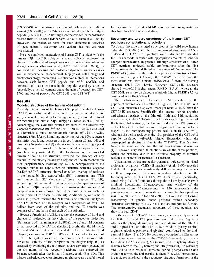
ResultsModeled structure of the human a3b4 nAChR
To study interactions of the human CST peptide with the human
nAChR, an in silico model structure for the human a3b4 receptor
subtype was developed by following a recently reported protocol
for modeling the human a4b2 subtype (Haddadian et al., 2008).
Accordingly, the available refined structure of the electric ray
Torpedo marmorata (a)2bcd nAChR (PDB ID: 2BG9) was used
as a template to build the pentameric human (a3)2(b4)3 nAChR
structure (Fig. 1A) by homology modeling. About 60% sequence
similarity was found between the target (human a3 and b4) and
template (Torpedo a and b) subunits sequences, ensuring a good
starting point to model the human a3b4 receptor structure
(supplementary material Fig. S1). Evaluation of the modeled
human a3b4 nAChR structure revealed the absence of any
residue in the strictly disallowed regions of the Ramachandran
Plot (supplementary material Fig. S2). Superimposition of the
modeled human (a3)2(b4)3 nAChR structure with the Torpedo
(a)2bcd nAChR structure showed excellent overlap of residues
in the ligand binding extracellular (EC), transmembrane (TM)
and intracellular (IC) domains of these receptors (Fig. 1B),
suggesting that the model provides a reasonable representative of
the human a3b4 receptor. The EC domain of the human a3b4
receptor was mainly constituted of b-strands (13 for each a3
subunit and 11 for each b4 subunit). Additionally, one a-helix
was also present towards the N-terminus of both subunit types.
The TM domain of the receptor was comprised of four TM
helices from each of the subunits, and the IC domain was
comprised of one helix from each of the subunits.
Because functional nAChRs require the presence of lipid and
cholesterol molecules in the vicinity of the receptor molecules
(Barrantes, 2004; Brannigan et al., 2008), the EC and TM domains
of the modeled a3b4 nAChR structure (specifically, the M1, M2,
M3 and M4 helices) were embedded in the equilibrated lipid
bilayer (composed of POPC, POPA and cholesterol in the ratio of
3:1:1). The system was then equilibrated for 80 nanoseconds.
Structural stability of the receptor in the bilayer (Fig. 1C) as
assessed by evaluating the root-mean-square deviation (RMSD) of
the Ca atoms of the receptor backbone was observed for
80 nanoseconds after the initial 10 nanoseconds (Fig. 1D). This
bilayer-embedded receptor structure might serve as a useful model
for docking with a3b4 nAChR agonists and antagonists forstructure–function analysis studies.
Secondary and tertiary structures of the human CSTpeptides: computational predictions
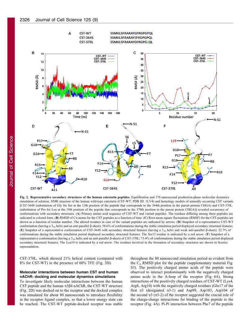
To obtain the time-averaged structures of the wild type humancatestatin (CST-WT) and that of the derived structures of CST-364S and CST-370L, the peptides were individually simulated
for 180 nseconds in water with appropriate amounts of ions forcharge neutralization. In general, although structures of all threeCST peptides achieved stable conformations after the first
20 nanoseconds, they differed in the extent of fluctuations. TheRMSD of Ca atoms in these three peptides as a function of timeare shown in Fig. 2B. Clearly, the CST-WT structure was the
most stable one, with a mean RMSD of 4.1A from the startingstructure (PDB ID: 1LV4). However, CST-364S structureshowed ,twofold higher mean RMSD (8.5 A), whereas the
CST-370L structure displayed a relatively higher RMSD (5.5 A)compared with the CST-WT.
The root-mean-square fluctuation (RMSF) values of the
peptide structures are illustrated in Fig. 2C. The CST-WT andCST-370L structures displayed lower per residue RMSF than theCST-364S structure. Specifically, the leucine, serine, arginine
and alanine residues at the 5th, 6th, 10th and 11th positions,respectively, in the CST-364S structure showed a high degree offluctuation. Interestingly, the leucine residue at the 19th positionof the CST-370L peptide showed insignificant fluctuation (with
respect to the corresponding proline residue in the CST-WT),whereas the serine residue at the 13th position of the CST-364Speptide displayed ,fivefold higher fluctuation (than the
corresponding glycine residue in the CST-WT). The first twoN-terminal residues (SS) and the last two C-terminal residues(QL) showed very high fluctuations in all the three peptides,
which are probably due to the intrinsic property of terminalresidues in proteins or peptides to fluctuate.
Visualization of the molecular dynamics trajectories in visual
molecular dynamics (VMDs) (Humphrey et al., 1996) revealedthat the three peptides significantly differed among themselvesin their propensities to adopt secondary structures in the
following order: CST-370L.CST-WT.CST-364S. Specifically,considering the conformations during the relatively stable (withminimal fluctuations) 80 nanosecond time window of thesimulation (from 40 nanoseconds to 120 nanoseconds), the
percentage occurrence of secondary structures for these peptideswas 73.4, 58.6 and 22.7 for CST-370L, CST-WT and CST-364S,respectively. In general, these peptides formed secondary
structures comprising of a 310 helix and an anti-parallel b-sheet.The representative secondary structures of these peptides areshown in Fig. 2D–F.
In the case of CST-WT, the arginine, alanine and tyrosine atthe 10th, 11th and 12th positions contributed to a 310 helixwhereas the phenylalanine, arginine and alanine at the 7th, 8th
and 9th positions, and the 14th to 18th residues (phenylalanine,arginine, glycine, proline and glycine) contributed to the anti-parallel b-sheet (Fig. 2D). By contrast, a completely different set
of residues were involved in the CST-364S secondary structureformation: the 5th (leucine), 6th (serine) and 7th (phenylalanine)residues formed the 310 helices; the 8th (arginine), 9th (alanine)
and 12th to 15th residues (tyrosine, serine, phenyl alanine andarginine) formed the anti-parallel b-sheet (Fig. 2E). Interestingly,the residues involved in the secondary structure formation in the
Journal of Cell Science 125 (9)2324
Journ
alof
Cell
Scie
nce
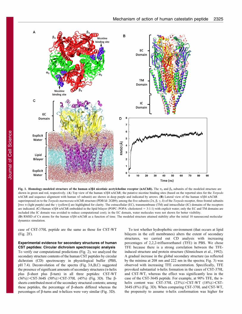
case of CST-370L peptide are the same as those for CST-WT
(Fig. 2F).
Experimental evidence for secondary structures of human
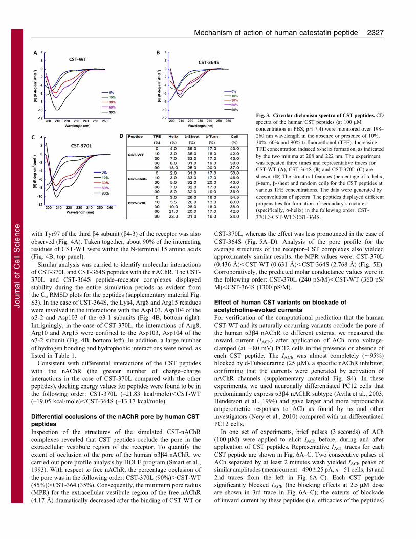
CST peptides: Circular dichroism spectroscopic analysis
To verify our computational predictions (Fig. 2), we analyzed the
secondary structure contents of the human CST peptides by circular
dichroism (CD) spectroscopy in physiological buffer (PBS,
pH 7.4). Deconvolution of the spectra (Fig. 3A,B,C) suggested
the presence of significant amounts of secondary structures (a-helix
plus b-sheet plus b-turn) in all three peptides: CST-WT
(56%).CST-364S (50%).CST-370L (45%) (Fig. 3D). The b-
sheets contributed most of the secondary structural contents; among
these peptides, the percentage of b-sheets differed whereas the
percentages of b-turns and a-helices were very similar (Fig. 3D).
To test whether hydrophobic environment (that occurs at lipid
bilayers in the cell membranes) alters the extent of secondary
structures, we carried out CD analysis with increasing
percentages of 2,2,2-trifluoroethanol (TFE) in PBS. We chose
TFE because there is a strong correlation between the TFE-
induced structure and protein structure (Sonnichsen et al., 1992).
A gradual increase in the global secondary structure (as reflected
by the minima at 208 nm and 222 nm in the spectra; Fig. 3) was
observed with increasing TFE concentration. Specifically, TFE
provoked substantial a-helix formation in the cases of CST-370L
and CST-WT, whereas the effect was significantly less in the
case of the CST-364S peptide. For example, at 90% TFE, the a-
helix content was: CST-370L (23%).CST-WT (18%).CST-
364S (8%) (Fig. 3D). When comparing CST-370L and CST-WT,
the propensity to assume a-helix conformation was higher for
Fig. 1. Homology-modeled structure of the human a3b4 nicotinic acetylcholine receptor (nAChR). The a3 and b4 subunits of the modeled structure are
shown in green and red, respectively. (A) Top view of the human a3b4 nAChR; the putative nicotine binding sites (based on the reported sites for the Torpedo
nAChR and sequence alignment with human a3 subunit) are shown in deep purple and indicated by arrows. (B) Lateral view of the human a3b4 nAChR
superimposed on to the Torpedo marmorata nAChR structure (PDB Id: 2GB9); among the five subunits (2a, b, c, d) of the Torpedo receptor, three frontal subunits
[two a (light purple) and the c (yellow)] are highlighted for clarity. The extracellular (EC), transmembrane (TM) and intracellular (IC) domains of the receptors
are indicated. (C) Human a3b4 nAChR embedded in the lipid bilayer (POPC: POPA: cholesterol 5 3:1:1) with explicit water; only the EC and TM domains are
included (the IC domain was avoided to reduce computational cost); in the EC domain, water molecules were not shown for better visibility.
(D) RMSD of Ca atoms for the human a3b4 nAChR as a function of time. The modeled structure attained stability after the initial 10 nanosecond molecular
dynamics simulation.
Mechanism of action of human catestatin peptide 2325
Journ
alof
Cell
Scie
nce
CST-370L, which showed 21% helical content (compared with8% for CST-WT) in the presence of 60% TFE (Fig. 3D).
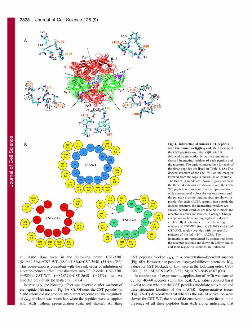
Molecular interactions between human CST and humannAChR: docking and molecular dynamics simulations
To investigate likely molecular interactions between the humanCST peptide and the human a3b4 nAChR, the CST-WT structure(Fig. 2D) was docked on to the receptor and the docked complex
was simulated for about 80 nanoseconds to introduce flexibilityin the receptor–ligand complex, so that a lower energy state canbe reached. The CST-WT peptide-docked receptor was stable
throughout the 80 nanosecond simulation period as evident from
the Ca RMSD plot for the peptide (supplementary material Fig.
S3). The positively charged amino acids of the peptide were
observed to interact predominantly with the negatively charged
amino acids in the A-loop of the receptor (Fig. 4A). Strong
interactions of the positively charged residues of CST-WT (Lys4,
Arg8, Arg10) with the negatively charged residues (Glu17 of the
first a3 (designated a3-1) and Asp99, Asp103, Asp104 of
the second a3 (a3-2) of the receptor suggested the crucial role of
the charge-charge interactions for binding of the peptide to the
receptor (Fig. 4A). Pi-Pi interaction between Phe7 of the peptide
Fig. 2. Representative secondary structures of the human catestatin peptides. Equilibration and 170 nanosecond production-phase molecular dynamics
simulation of solution, NMR structure of the human wild-type catestatin (CST-WT; PDB ID: 1LV4) and homology models of naturally occurring CST variants
[CST-364S (substitution of Gly for Ser at the 13th position of the peptide that corresponds to the 364th position in the parent protein CHGA) and CST-370L
(substitution of Pro for Leu at the 19th position of the peptide that corresponds to the 370th position in the parent protein CHGA)] revealed occurrence of
conformations with secondary structures. (A) Primary amino acid sequence of CST-WT and variant peptides. The residues differing among these peptides are
indicated in colored fonts. (B) RMSD of Ca atoms for the CST peptides as a function of time. (C) Root mean square fluctuations (RMSF) for the CST-peptides are
shown as a function of residue number. The altered residues in case of the variant peptides are indicated by arrows. (D) Snapshot of a representative CST-WT
conformation (having a 310 helix and an anti-parallel b-sheet); 58.6% of conformations during the stable simulation period displayed secondary structural features.
(E) Snapshot of a representative conformation of CST-364S with secondary structural features (having a 310 helix and weak anti-parallel b-sheet); 22.7% of
conformations during the stable simulation period displayed secondary structural features. The Ser13 residue is indicated by a red arrow. (F) Snapshot of a
representative conformation (having a 310 helix and an anti-parallel b-sheet) of CST-370L; 73.4% of conformations during the stable simulation period displayed
secondary structural features. The Leu19 is indicated by a red arrow. The residues involved in the formation of secondary structures are shown in licorice
representation.
Journal of Cell Science 125 (9)2326
Journ
alof
Cell
Scie
nce
with Tyr97 of the third b4 subunit (b4-3) of the receptor was also
observed (Fig. 4A). Taken together, about 90% of the interacting
residues of CST-WT were within the N-terminal 15 amino acids
(Fig. 4B, top panel).
Similar analysis was carried to identify molecular interactions
of CST-370L and CST-364S peptides with the nAChR. The CST-
370L and CST-364S peptide–receptor complexes displayed
stability during the entire simulation periods as evident from
the Ca RMSD plots for the peptides (supplementary material Fig.
S3). In the case of CST-364S, the Lys4, Arg8 and Arg15 residues
were involved in the interactions with the Asp103, Asp104 of the
a3-2 and Asp103 of the a3-1 subunits (Fig. 4B, bottom right).
Intriguingly, in the case of CST-370L, the interactions of Arg8,
Arg10 and Arg15 were confined to the Asp103, Asp104 of the
a3-2 subunit (Fig. 4B, bottom left). In addition, a large number
of hydrogen bonding and hydrophobic interactions were noted, as
listed in Table 1.
Consistent with differential interactions of the CST peptides
with the nAChR (the greater number of charge–charge
interactions in the case of CST-370L compared with the other
peptides), docking energy values for peptides were found to be in
the following order: CST-370L (–21.83 kcal/mole),CST-WT
(–19.05 kcal/mole),CST-364S (–13.17 kcal/mole).
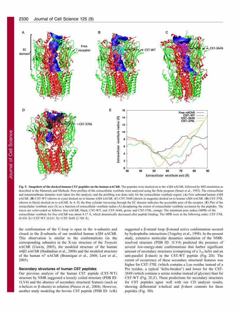
Differential occlusions of the nAChR pore by human CST
peptides
Inspection of the structures of the simulated CST-nAChR
complexes revealed that CST peptides occlude the pore in the
extracellular vestibule region of the receptor. To quantify the
extent of occlusion of the pore of the human a3b4 nAChR, we
carried out pore profile analysis by HOLE program (Smart et al.,
1993). With respect to free nAChR, the percentage occlusion of
the pore was in the following order: CST-370L (90%).CST-WT
(85%).CST-364 (35%). Consequently, the minimum pore radius
(MPR) for the extracellular vestibule region of the free nAChR
(4.17 A) dramatically decreased after the binding of CST-WT or
CST-370L, whereas the effect was less pronounced in the case of
CST-364S (Fig. 5A–D). Analysis of the pore profile for the
average structures of the receptor–CST complexes also yielded
approximately similar results; the MPR values were: CST-370L
(0.436 A),CST-WT (0.631 A),CST-364S (2.768 A) (Fig. 5E).
Corroboratively, the predicted molar conductance values were in
the following order: CST-370L (240 pS/M),CST-WT (360 pS/
M),CST-364S (1300 pS/M).
Effect of human CST variants on blockade of
acetylcholine-evoked currents
For verification of the computational prediction that the human
CST-WT and its naturally occurring variants occlude the pore of
the human a3b4 nAChR to different extents, we measured the
inward current (IACh) after application of ACh onto voltage-
clamped (at 280 mV) PC12 cells in the presence or absence of
each CST peptide. The IACh was almost completely (,95%)
blocked by d-Tubocurarine (25 mM), a specific nAChR inhibitor,
confirming that the currents were generated by activation of
nAChR channels (supplementary material Fig. S4). In these
experiments, we used neuronally differentiated PC12 cells that
predominantly express a3b4 nAChR subtype (Avila et al., 2003;
Henderson et al., 1994) and gave larger and more reproducible
amperometric responses to ACh as found by us and other
investigators (Nery et al., 2010) compared with un-differentiated
PC12 cells.
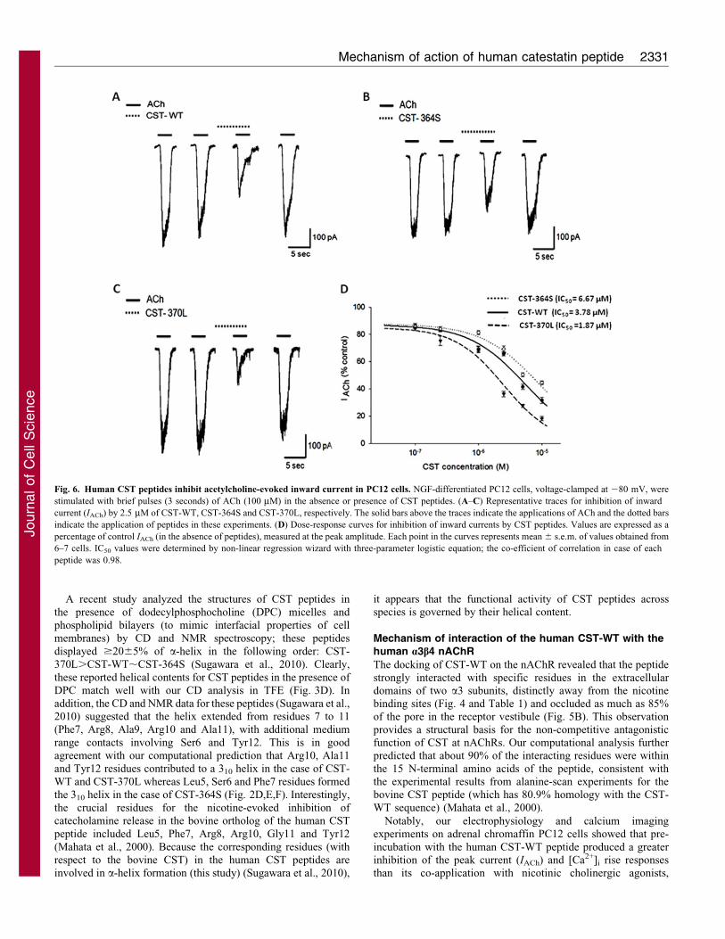
In one set of experiments, brief pulses (3 seconds) of ACh
(100 mM) were applied to elicit IACh before, during and after
application of CST peptides. Representative IACh traces for each
CST peptide are shown in Fig. 6A–C. Two consecutive pulses of
ACh separated by at least 2 minutes wash yielded IACh peaks of
similar amplitudes (mean current5490625 pA, n551 cells; 1st and
2nd traces from the left in Fig. 6A–C). Each CST peptide
significantly blocked IACh (the blocking effects at 2.5 mM dose
are shown in 3rd trace in Fig. 6A–C); the extents of blockade
of inward current by these peptides (i.e. efficacies of the peptides)
Fig. 3. Circular dichroism spectra of CST peptides. CD
spectra of the human CST peptides (at 100 mM
concentration in PBS, pH 7.4) were monitored over 198–
260 nm wavelength in the absence or presence of 10%,
30%, 60% and 90% trifluoroethanol (TFE). Increasing
TFE concentration induced a-helix formation, as indicated
by the two minima at 208 and 222 nm. The experiment
was repeated three times and representative traces for
CST-WT (A), CST-364S (B) and CST-370L (C) are
shown. (D) The structural features (percentage of a-helix,
b-turn, b-sheet and random coil) for the CST peptides at
various TFE concentrations. The data were generated by
deconvolution of spectra. The peptides displayed different
propensities for formation of secondary structures
(specifically, a-helix) in the following order: CST-
370L.CST-WT.CST-364S.
Mechanism of action of human catestatin peptide 2327
Journ
alof
Cell
Scie
nce
at 10 mM dose were in the following order: CST-370L
(81.961.5%).CST-WT (68.061.8%).CST-364S (55.461.3%).
This observation is consistent with the rank order of inhibition of
nicotine-induced 22Na+ translocation into PC12 cells: CST-370L
(,98%).CST-WT (,87.0%).CST-364S (,74%), as we
reported previously (Mahata et al., 2004).
Interestingly, the blocking effect was reversible after washout of
the peptide (4th trace in Fig. 6A–C). Of note, the CST peptides (at
5 mM) alone did not produce any current response and the magnitude
of IACh blockade was much less when the peptides were co-applied
with ACh without pre-incubation (data not shown). All three
CST peptides blocked IACh in a concentration-dependent manner
(Fig. 6D). However, the peptides displayed different potencies; IC50
values for CST blockade of IACh were in the following order: CST-
370L (1.86 mM),CST-WT (3.87 mM),CST-364S (6.67 mM).
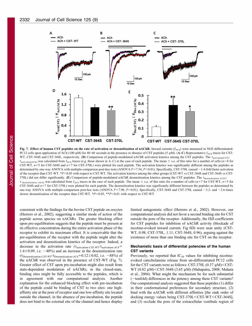
In another set of experiments, application of ACh was carried
out for 40–60 seconds (until the peak IAch value reduced basal
levels) to test whether the CST peptides modulate activation and
desensitization kinetics of the nAChR. Representative traces
(Fig. 7A–C) demonstrate that whereas the rate of activation were
slower for CST-WT, the rates of desensitization were faster in the
presence of all three peptides than ACh alone, indicating that
Fig. 4. Interaction of human CST peptides
with the human (a3)2(b4)3 nAChR. Docking of
the CST peptides onto the a3b4 nAChR,
followed by molecular dynamics simulations
showed interacting residues of each peptide and
the receptor. The various interactions for each of
the three peptides are listed in Table 1. (A) The
docked structure of the CST-WT on the receptor
(viewed from the top) is shown, as an example.
The two a3 subunits are shown in green whereas
the three b4 subunits are shown in red; the CST-
WT peptide is shown in licorice representation
with conventional colors for various atoms and
the putative nicotine binding sites are shown in
purple. For each nAChR subunit, just outside the
docked structure, the interacting residues are
shown: peptide residues are labeled in black and
receptor residues are labeled in orange. Charge–
charge interactions are highlighted in dotted
circles. (B) A schematic of the interacting
residues of CST-WT (top), CST-364S (left) and
CST-370L (right) peptides with the specific
residues of the (a3)2(b4)3 nAChR. The
interactions are represented by connecting lines;
the receptor residues are shown in yellow circles
and their respective subunits are indicated.
Journal of Cell Science 125 (9)2328
Journ
alof
Cell
Scie
nce
these peptides, in general, blunted the nAChR function. Among
the three peptides, the rate of activation of nAChR was
significantly faster in the presence of CST-370L, as reflected
by the significantly lower tactivation(CST):tactivation(ACh) ratio
compared with the ratio for CST-WT (0.9860.05 vs
1.4160.09, P50.011; Fig. 7D). In addition, the rate of nAChR
desensitization was slower in the case of CST-364S, as reflected
by the significantly higher tdesensitization(CST):tdesensitization(ACh)
ratio, in comparison with CST-WT (0.3860.08 vs 0.1260.02,
P50.024; Fig. 7E).
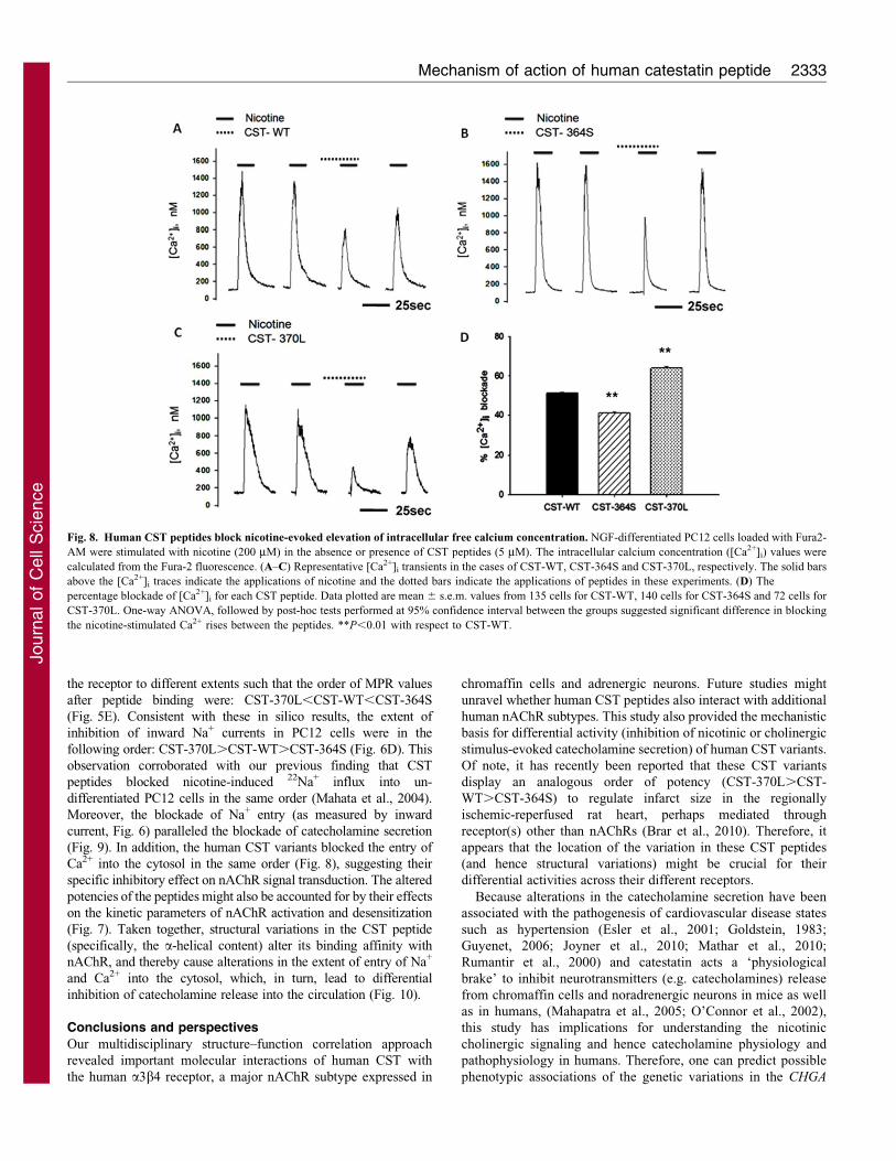
Effect of human CST variants on intracellular Ca2+
concentration
Because the human CST peptides blocked the inward current
elicited by ACh to different extents (Fig. 6), we hypothesized that
intracellular free calcium concentration ([Ca2+]i) would also be
modulated differently by these peptides. To test this hypothesis,
neuronally differentiated PC12 cells were stimulated with two
brief pulses (15–30 seconds) of nicotine followed by an incubation
period of 2 minutes with a peptide. One CST peptide and nicotine
were then co-applied that was followed by a recovery dose. The 1st
and 2nd traces in Fig. 8A–C show the fast and transient increase of
the [Ca2+]i after nicotine. The basal and nicotine-stimulated [Ca2+]i
levels were 7765 nM (n549 cells) and 1072653 nM (n549
cells), respectively. Each CST peptide blocked the nicotine-
stimulated [Ca2+]i rise (Fig. 8A–C, 3rd trace); pre-incubation with
the peptide was required to obtain a clear and significant effect
on [Ca2+]i signal. The extents of blockade of [Ca2+]i rise
(Fig. 8D) were different among the CST peptides: CST-370L
(,65%).CST-WT (,52%).CST-364S (,42%). Interestingly,
blocking effects were recovered upon re-stimulation of cells by
nicotine, although to different extents (Fig. 8A–C, 4th trace);
specifically, in the cases of CST-370L and CST-WT, the recovery
was incomplete.
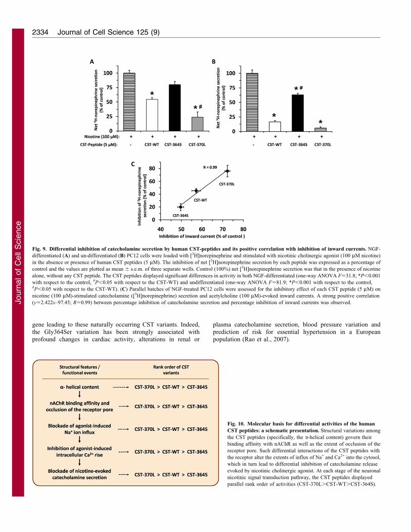
Differential inhibition of catecholamine secretion by CSTpeptides
To determine whether the human CST variants display
differential efficacies in blocking catecholamine release from
neuronally differentiated PC12 cells, we measured the exogenous
norepinephrine secretion stimulated by nicotine in the presence
or absence of each CST peptide. Consistent with the rank order of
blockade of [Ca2+]i rise (Fig. 8), the CST peptides blocked
[3H]norepinephrine secretion from NGF-differentiated PC12
cells in the following order: CST-370L (,76%).CST-WT
(,46%).CST-364S (,19%) (Fig. 9A). In another set of
experiments, we also assessed the efficacy of these peptides to
block nicotine-evoked [3H]norepinephrine secretion from
undifferentiated PC12 cells, which displayed a similar effect
(albeit, a relatively stronger response than in differentiated cells)
and blocked [3H]norepinephrine secretion in the same order:
CST-370L (,94%).CST-WT (,84%).CST-364S (,37%)
(Fig. 9B). In neuronally differentiated PC12 cells, the
inhibitory effect of each CST peptide on nicotinic-cholinergic-
induced catecholamine secretion (the final step of nicotinic
signal transduction) correlated (correlation co-efficient, R50.99;
Fig. 9C) with the blockade of ACh-evoked inward current (the
initial step of the signal transduction), reflecting the specific
interaction of these peptides at nAChRs.
DiscussionStructure of the human a3b4 nAChR
Owing to the non-availability of high-resolution structures of
any human nAChR subtype, mechanistic details of molecular
interactions between the physiological anti-hypertensive peptide
CST, a potent and specific inhibitor of nAChR (Mahapatra et al.,
2006; Mahata et al., 1997), remained elusive. In this study, we
generated a homology-modeled structure of the human a3b4
nAChR based on the Torpedo nAChR structure. To our
knowledge, this is the first report of an all-atomic model of
the human a3b4 nAChR (Fig. 1). The structure, embedded in
lipid bilayer attained stability after 10 nanosecond molecular
dynamics simulations. Previously, a homology model of the
extracellular domain of the a3b4 nAChR was reported using the
structure of ACh binding proteins from Aplysia californica and
Lymnaea stagnalis (Rana et al., 2009); qualitative comparison of
this model with the extracellular domain of our model showed
similar locations for various residues in both structures. Of note,
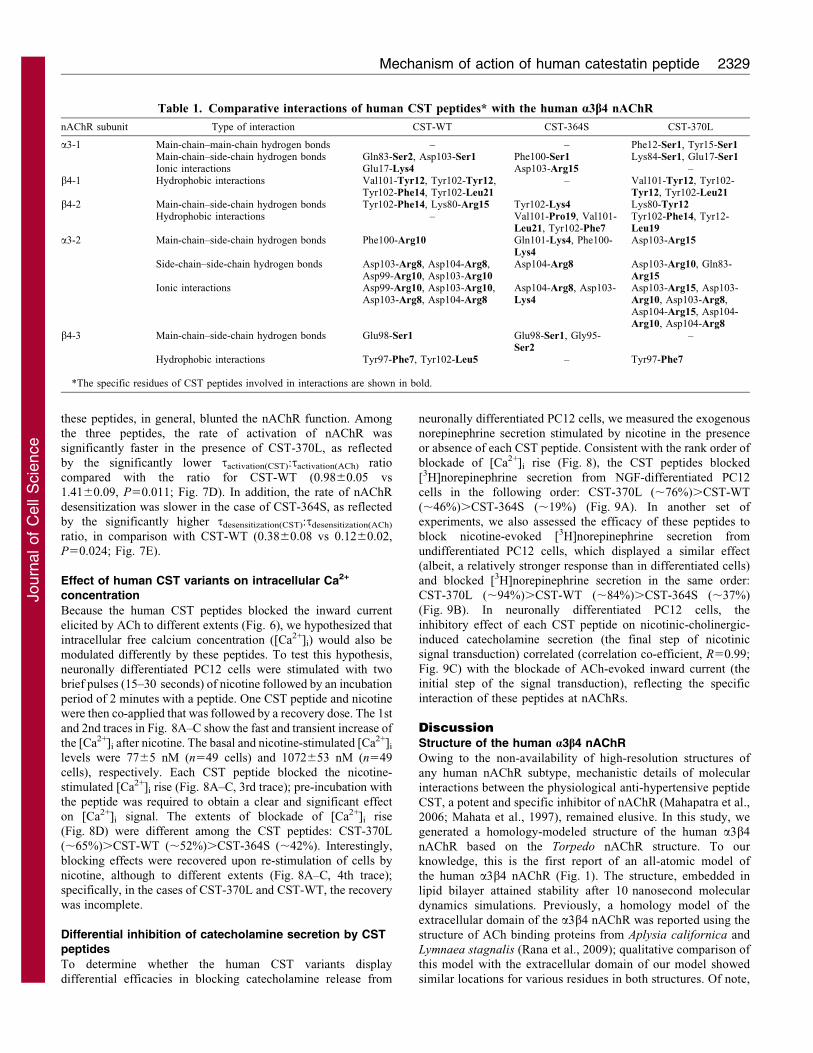
Table 1. Comparative interactions of human CST peptides* with the human a3b4 nAChR
nAChR subunit Type of interaction CST-WT CST-364S CST-370L
a3-1 Main-chain–main-chain hydrogen bonds – – Phe12-Ser1, Tyr15-Ser1Main-chain–side-chain hydrogen bonds Gln83-Ser2, Asp103-Ser1 Phe100-Ser1 Lys84-Ser1, Glu17-Ser1Ionic interactions Glu17-Lys4 Asp103-Arg15 –
b4-1 Hydrophobic interactions Val101-Tyr12, Tyr102-Tyr12,Tyr102-Phe14, Tyr102-Leu21
– Val101-Tyr12, Tyr102-Tyr12, Tyr102-Leu21
b4-2 Main-chain–side-chain hydrogen bonds Tyr102-Phe14, Lys80-Arg15 Tyr102-Lys4 Lys80-Tyr12Hydrophobic interactions – Val101-Pro19, Val101-
Leu21, Tyr102-Phe7Tyr102-Phe14, Tyr12-Leu19
a3-2 Main-chain–side-chain hydrogen bonds Phe100-Arg10 Gln101-Lys4, Phe100-Lys4
Asp103-Arg15
Side-chain–side-chain hydrogen bonds Asp103-Arg8, Asp104-Arg8,Asp99-Arg10, Asp103-Arg10
Asp104-Arg8 Asp103-Arg10, Gln83-Arg15
Ionic interactions Asp99-Arg10, Asp103-Arg10,Asp103-Arg8, Asp104-Arg8
Asp104-Arg8, Asp103-Lys4
Asp103-Arg15, Asp103-Arg10, Asp103-Arg8,Asp104-Arg15, Asp104-Arg10, Asp104-Arg8
b4-3 Main-chain–side-chain hydrogen bonds Glu98-Ser1 Glu98-Ser1, Gly95-Ser2
–
Hydrophobic interactions Tyr97-Phe7, Tyr102-Leu5 – Tyr97-Phe7
*The specific residues of CST peptides involved in interactions are shown in bold.
Mechanism of action of human catestatin peptide 2329
Journ
alof
Cell
Scie
nce
the conformation of the C-loop is open in the a-subunits and
closed in the b-subunits of our modeled human a3b4 nAChR.
This observation is similar to the conformations (in the
corresponding subunits) in the X-ray structure of the Torpedo
nAChR (Unwin, 2005), the modeled structure of the human
a4b2 nAChR (Haddadian et al., 2008) and the modeled structure
of the human a7 nAChR (Brannigan et al., 2008; Law et al.,
2005).
Secondary structures of human CST peptides
Our previous analysis of the human CST peptide (CST-WT)
structure by NMR suggested a loosely coiled structure (PDB ID:
1LV4) and the absence of secondary structural features (such as
a-helices or b-sheets) in solution (Preece et al., 2004). However,
another study modeling the bovine CST peptide (PDB ID: 1cfk)
suggested a b-strand–loop–b-strand active conformation secured
by hydrophobic interactions (Tsigelny et al., 1998). In the present
study, extensive molecular dynamics simulation of the NMR-
resolved structure (PDB ID: 1LV4) predicted the presence of
several low-energy-state conformations that harbor significant
amount of secondary structures (comprising of a 310 helix and an
anti-parallel b-sheet) in the CST-WT peptide (Fig. 2D). The
extent of occurrence of these secondary structural features was
higher for CST-370L (which contains a Leu residue instead of a
Pro residue, a typical ‘helix-breaker’) and lower for the CST-
364S (which contains a serine residue instead of glycine) than for
CST-WT (Fig. 2E,F). These predictions for secondary structures
for CST peptides agree well with our CD analysis results,
showing differential a-helical and b-sheet contents for these
peptides (Fig. 3D).
Fig. 5. Snapshots of the docked human CST peptides on the human nAChR. The peptides were docked on to the a3b4 nAChR, followed by MD simulation as
described in the Materials and Methods. Pore profiles of the extracellular vestibule were analyzed using the Hole program (Smart et al., 1993). The extracellular
and transmembrane domains were taken for this analysis; and the profiling was done only for the extracellular vestibule region. (A) Free unbound human a3b4
nAChR. (B) CST-WT (shown in cyan) docked on to human a3b4 nAChR. (C) CST-364S (shown in magenta) docked on to human a3b4 nAChR. (D) CST-370L
(shown in black) docked on to nAChR. In A–D, the blue cylinder traversing through the EC domain indicates the accessible pore of the receptor. (E) Plot of the
extracellular vestibule axis (A) as a function of extracellular vestibule radius (A) deciphering the extent of extracellular vestibule occlusion by the peptides. The
traces are color-coded as follows: free nAChR, black; CST-WT, red; CST-364S, green; and CST-370L, orange. The minimum pore radius (MPR) of the
extracellular vestibule for free nAChR was about 4.17 A, which dramatically decreased after peptide binding. The MPR were in the following order: CST-370L
(0.436 A),CST-WT (0.631 A),CST-364S (2.768 A).
Journal of Cell Science 125 (9)2330
Journ
alof
Cell
Scie
nce
A recent study analyzed the structures of CST peptides in
the presence of dodecylphosphocholine (DPC) micelles and
phospholipid bilayers (to mimic interfacial properties of cell
membranes) by CD and NMR spectroscopy; these peptides
displayed $2065% of a-helix in the following order: CST-
370L.CST-WT,CST-364S (Sugawara et al., 2010). Clearly,
these reported helical contents for CST peptides in the presence of
DPC match well with our CD analysis in TFE (Fig. 3D). In
addition, the CD and NMR data for these peptides (Sugawara et al.,2010) suggested that the helix extended from residues 7 to 11
(Phe7, Arg8, Ala9, Arg10 and Ala11), with additional medium
range contacts involving Ser6 and Tyr12. This is in good
agreement with our computational prediction that Arg10, Ala11
and Tyr12 residues contributed to a 310 helix in the case of CST-
WT and CST-370L whereas Leu5, Ser6 and Phe7 residues formed
the 310 helix in the case of CST-364S (Fig. 2D,E,F). Interestingly,
the crucial residues for the nicotine-evoked inhibition of
catecholamine release in the bovine ortholog of the human CST
peptide included Leu5, Phe7, Arg8, Arg10, Gly11 and Tyr12
(Mahata et al., 2000). Because the corresponding residues (with
respect to the bovine CST) in the human CST peptides are
involved in a-helix formation (this study) (Sugawara et al., 2010),
it appears that the functional activity of CST peptides acrossspecies is governed by their helical content.
Mechanism of interaction of the human CST-WT with thehuman a3b4 nAChR
The docking of CST-WT on the nAChR revealed that the peptide
strongly interacted with specific residues in the extracellulardomains of two a3 subunits, distinctly away from the nicotinebinding sites (Fig. 4 and Table 1) and occluded as much as 85%of the pore in the receptor vestibule (Fig. 5B). This observation
provides a structural basis for the non-competitive antagonisticfunction of CST at nAChRs. Our computational analysis furtherpredicted that about 90% of the interacting residues were within
the 15 N-terminal amino acids of the peptide, consistent withthe experimental results from alanine-scan experiments for thebovine CST peptide (which has 80.9% homology with the CST-
WT sequence) (Mahata et al., 2000).
Notably, our electrophysiology and calcium imagingexperiments on adrenal chromaffin PC12 cells showed that pre-
incubation with the human CST-WT peptide produced a greaterinhibition of the peak current (IACh) and [Ca2+]i rise responsesthan its co-application with nicotinic cholinergic agonists,
Fig. 6. Human CST peptides inhibit acetylcholine-evoked inward current in PC12 cells. NGF-differentiated PC12 cells, voltage-clamped at 280 mV, were
stimulated with brief pulses (3 seconds) of ACh (100 mM) in the absence or presence of CST peptides. (A–C) Representative traces for inhibition of inward
current (IACh) by 2.5 mM of CST-WT, CST-364S and CST-370L, respectively. The solid bars above the traces indicate the applications of ACh and the dotted bars
indicate the application of peptides in these experiments. (D) Dose-response curves for inhibition of inward currents by CST peptides. Values are expressed as a
percentage of control IACh (in the absence of peptides), measured at the peak amplitude. Each point in the curves represents mean 6 s.e.m. of values obtained from
6–7 cells. IC50 values were determined by non-linear regression wizard with three-parameter logistic equation; the co-efficient of correlation in case of each
peptide was 0.98.
Mechanism of action of human catestatin peptide 2331
Journ
alof
Cell
Scie
nce
consistent with the findings for the bovine CST peptide on oocytes
(Herrero et al., 2002), suggesting a similar mode of action of the
peptide across species on nAChRs. The greater blocking effect
upon pre-equilibration suggests that the peptide must be present at
its effective concentration during the entire activation phase of the
receptor to exhibit its maximum effect. It is conceivable that the
pre-equilibration of the receptor with the peptide might alter the
activation and desensitization kinetics of the receptor. Indeed, a
decrease in the activation rate (tactivation-CST-WT:tactivation-ACh5
1.460.09, i.e. ,40%) and an increase in the desensitization rate
(tDesensitization-CST-WT:tDesensitization-ACh50.1260.02, i.e. ,88%) of
the nAChR was observed in the presence of CST-WT (Fig. 7).
Greater effect of CST upon pre-incubation might also result from
state-dependent modulation of nAChRs; in the closed-state,
binding sites might be fully accessible to the peptides, which is
in agreement with our computational analysis. Another
explanation for the enhanced blocking effect with pre-incubation
of the peptide could be binding of CST to two sites: one high-
affinity site at the pore of receptor and one low-affinity site located
outside the channel; in the absence of pre-incubation, the peptide
does not bind to the external site of the channel and hence display
limited antagonistic effect (Herrero et al., 2002). However, our
computational analysis did not favor a second binding site for CST
outside the pore of the receptor. Additionally, the Hill coefficients
for CST peptides for inhibition of nAChR activity (blockade of
nicotine-evoked inward current; Fig 6D) were near unity (CST-
WT, 0.98; CST-370L, 1.11; CST-364S, 0.96), arguing against the
existence of more than one binding site for CST on the receptor.
Mechanistic basis of differential potencies of the human
CST variants
Previously, we reported that IC50 values for inhibiting nicotine-
evoked catecholamine release from un-differentiated PC12 cells
for the CST variants were as follows: CST-370L (0.37 mM),CST-
WT (0.82 mM),CST-364S (3.65 mM) (Mahapatra, 2008; Mahata
et al., 2004). What might the mechanism be for such substantial
(,tenfold) differences in the potency among these CST variants?
Our computational analysis suggested that these peptides (1) differ
in their conformational preferences for secondary structure, (2)
bind with the nAChR with different affinities [the rank order of
docking energy values being CST-370L,CST-WT,CST-364S],
and (3) occlude the pore of the extracellular vestibule region of
Fig. 7. Effect of human CST peptides on the rate of activation or desensitization of nAChR. Inward currents (IACh) were measured in NGF-differentiated-
PC12 cells upon application of ACh (100 mM) for 40–60 seconds in the presence or absence of CST peptides (5 mM). (A–C) Representative IACh traces for CST-
WT, CST-364S and CST-360L, respectively. (D) Comparison of peptide-modulated nAChR activation kinetics among the CST peptides. The tactivation(CST):
tactivation(ACh) was calculated from IACh traces (e.g. those shown in A–C) in the case of each peptide. The mean 6 s.e. of this ratio for a number of cells (n58 for
CST-WT, n55 for CST-364S and n57 for CST-370L) were plotted for each peptide. The activation kinetics was significantly different among the peptides as
determined by one-way ANOVA with multiple-comparison post-hoc tests (ANOVA F57.54, P,0.01). Specifically, CST-370L caused ,1.4-fold faster activation
of the receptor than CST-WT. *P,0.05 with respect to CST-WT. The activation kinetics among the other groups (CST-WT vs CST-364S and CST-364S vs CST-
370L) did not differ significantly. (E) Comparison of peptide-modulated nAChR desensitization kinetics among the CST peptides. The tdesensitization (CST):
t desensitization (ACh) was calculated from IACh traces in the case of each peptide. The mean 6 s.e. of this ratio for a number of cells (n57 for CST-WT, n55 for
CST-364S and n57 for CST-370L) were plotted for each peptide. The desensitization kinetics was significantly different between the peptides as determined by
one-way ANOVA with multiple-comparison post-hoc tests (ANOVA F57.90, P,0.01). Specifically, CST-364S and CST-370L caused ,3.2- and ,2.6-times
slower desensitization of the receptor than CST-WT. *P,0.05, **P,0.01 with respect to CST-WT.
Journal of Cell Science 125 (9)2332
Journ
alof
Cell
Scie
nce
the receptor to different extents such that the order of MPR values
after peptide binding were: CST-370L,CST-WT,CST-364S
(Fig. 5E). Consistent with these in silico results, the extent of
inhibition of inward Na+ currents in PC12 cells were in the
following order: CST-370L.CST-WT.CST-364S (Fig. 6D). This
observation corroborated with our previous finding that CST
peptides blocked nicotine-induced 22Na+ influx into un-
differentiated PC12 cells in the same order (Mahata et al., 2004).
Moreover, the blockade of Na+ entry (as measured by inward
current, Fig. 6) paralleled the blockade of catecholamine secretion
(Fig. 9). In addition, the human CST variants blocked the entry of
Ca2+ into the cytosol in the same order (Fig. 8), suggesting their
specific inhibitory effect on nAChR signal transduction. The altered
potencies of the peptides might also be accounted for by their effects
on the kinetic parameters of nAChR activation and desensitization
(Fig. 7). Taken together, structural variations in the CST peptide
(specifically, the a-helical content) alter its binding affinity with
nAChR, and thereby cause alterations in the extent of entry of Na+
and Ca2+ into the cytosol, which, in turn, lead to differential
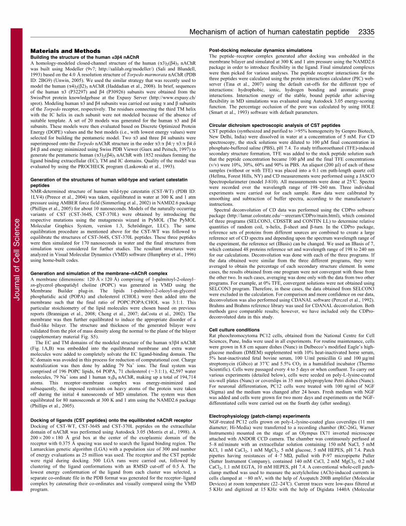
inhibition of catecholamine release into the circulation (Fig. 10).
Conclusions and perspectives
Our multidisciplinary structure–function correlation approach
revealed important molecular interactions of human CST with
the human a3b4 receptor, a major nAChR subtype expressed in
chromaffin cells and adrenergic neurons. Future studies might
unravel whether human CST peptides also interact with additional
human nAChR subtypes. This study also provided the mechanistic
basis for differential activity (inhibition of nicotinic or cholinergic
stimulus-evoked catecholamine secretion) of human CST variants.
Of note, it has recently been reported that these CST variants
display an analogous order of potency (CST-370L.CST-
WT.CST-364S) to regulate infarct size in the regionally
ischemic-reperfused rat heart, perhaps mediated through
receptor(s) other than nAChRs (Brar et al., 2010). Therefore, it
appears that the location of the variation in these CST peptides
(and hence structural variations) might be crucial for their
differential activities across their different receptors.
Because alterations in the catecholamine secretion have been
associated with the pathogenesis of cardiovascular disease states
such as hypertension (Esler et al., 2001; Goldstein, 1983;
Guyenet, 2006; Joyner et al., 2010; Mathar et al., 2010;
Rumantir et al., 2000) and catestatin acts a ‘physiological
brake’ to inhibit neurotransmitters (e.g. catecholamines) release
from chromaffin cells and noradrenergic neurons in mice as well
as in humans, (Mahapatra et al., 2005; O’Connor et al., 2002),
this study has implications for understanding the nicotinic
cholinergic signaling and hence catecholamine physiology and
pathophysiology in humans. Therefore, one can predict possible
phenotypic associations of the genetic variations in the CHGA
Fig. 8. Human CST peptides block nicotine-evoked elevation of intracellular free calcium concentration. NGF-differentiated PC12 cells loaded with Fura2-
AM were stimulated with nicotine (200 mM) in the absence or presence of CST peptides (5 mM). The intracellular calcium concentration ([Ca2+]i) values were
calculated from the Fura-2 fluorescence. (A–C) Representative [Ca2+]i transients in the cases of CST-WT, CST-364S and CST-370L, respectively. The solid bars
above the [Ca2+]i traces indicate the applications of nicotine and the dotted bars indicate the applications of peptides in these experiments. (D) The
percentage blockade of [Ca2+]i for each CST peptide. Data plotted are mean 6 s.e.m. values from 135 cells for CST-WT, 140 cells for CST-364S and 72 cells for
CST-370L. One-way ANOVA, followed by post-hoc tests performed at 95% confidence interval between the groups suggested significant difference in blocking
the nicotine-stimulated Ca2+ rises between the peptides. **P,0.01 with respect to CST-WT.
Mechanism of action of human catestatin peptide 2333
Journ
alof
Cell
Scie
nce
gene leading to these naturally occurring CST variants. Indeed,the Gly364Ser variation has been strongly associated withprofound changes in cardiac activity, alterations in renal or
plasma catecholamine secretion, blood pressure variation andprediction of risk for essential hypertension in a Europeanpopulation (Rao et al., 2007).
Fig. 9. Differential inhibition of catecholamine secretion by human CST-peptides and its positive correlation with inhibition of inward currents. NGF-
differentiated (A) and un-differentiated (B) PC12 cells were loaded with [3H]norepinephrine and stimulated with nicotinic cholinergic agonist (100 mM nicotine)
in the absence or presence of human CST peptides (5 mM). The inhibition of net [3H]norepinephrine secretion by each peptide was expressed as a percentage of
control and the values are plotted as mean 6 s.e.m. of three separate wells. Control (100%) net [3H]norepinephrine secretion was that in the presence of nicotine
alone, without any CST peptide. The CST peptides displayed significant differences in activity in both NGF-differentiated (one-way ANOVA F531.8; *P,0.001
with respect to the control, #P,0.05 with respect to the CST-WT) and undifferentiated (one-way ANOVA F581.9; *P,0.001 with respect to the control,#P,0.05 with respect to the CST-WT). (C) Parallel batches of NGF-treated PC12 cells were assessed for the inhibitory effect of each CST peptide (5 mM) on
nicotine (100 mM)-stimulated catecholamine ([3H]norepinephrine) secretion and acetylcholine (100 mM)-evoked inward currents. A strong positive correlation
(y52.422x–97.45; R50.99) between percentage inhibition of catecholamine secretion and percentage inhibition of inward currents was observed.
Fig. 10. Molecular basis for differential activities of the human
CST peptides: a schematic presentation. Structural variations among
the CST peptides (specifically, the a-helical content) govern their
binding affinity with nAChR as well as the extent of occlusion of the
receptor pore. Such differential interactions of the CST peptides with
the receptor alter the extents of influx of Na+ and Ca2+ into the cytosol,
which in turn lead to differential inhibition of catecholamine release
evoked by nicotinic cholinergic agonist. At each stage of the neuronal
nicotinic signal transduction pathway, the CST peptides displayed
parallel rank order of activities (CST-370L.CST-WT.CST-364S).
Journal of Cell Science 125 (9)2334
Journ
alof
Cell
Scie
nce
Materials and MethodsBuilding the structure of the human a3b4 nAChR
A homology-modeled closed-channel structure of the human (a3)2(b4)3 nAChR
was built using Modeller (9v7; http://salilab.org/modeller/) (Sali and Blundell,1993) based on the 4.0 A resolution structure of Torpedo marmorata nAChR (PDB
ID: 2BG9) (Unwin, 2005). We used the similar strategy that was recently used tomodel the human (a4)2(b2)3 nAChR (Haddadian et al., 2008). In brief, sequences
of the human a3 (P32297) and b4 (P30926) subunits were obtained from theSwissProt protein knowledgebase at the Expasy Server (http://www.expasy.ch/
sprot). Modeling human a3 and b4 subunits was carried out using a and b subunitsof the Torpedo receptor, respectively. The residues connecting the third TM helix
with the IC helix in each subunit were not modeled because of the absence ofsuitable template. A set of 20 models was generated for the human a3 and b4
subunits. These models were then evaluated based on Discrete Optimized ProteinEnergy (DOPE) values and the best models (i.e., with lowest energy values) were
selected for building the pentameric model. Two a3 and three b4 subunits weresuperimposed onto the Torpedo nAChR structure in the order a3:a b4:c a3:a b4:db4:b and energy minimized using Swiss PDB Viewer (Guex and Peitsch, 1997) togenerate the pentameric human (a3)2(b4)3 nAChR with 1852 residues forming theligand binding extracellular (EC), TM and IC domains. Quality of the model was
evaluated by using the PROCHECK program (Laskowski et al., 1993).
Generation of the structures of human wild-type and variant catestatin
peptides
NMR-determined structure of human wild-type catestatin (CST-WT) (PDB ID:1LV4) (Preece et al., 2004) was taken, equilibrated in water at 300 K and 1 atm
pressure using AMBER force field (Simmerling et al., 2002) in NAMD2.6 package(Phillips et al., 2005) for about 10 nanoseconds. Models of the naturally occurring
variants of CST (CST-364S, CST-370L) were obtained by introducing therespective mutations using the mutagenesis wizard in PyMOL (The PyMOL
Molecular Graphics System, version 1.3, Schrodinger, LLC). The sameequilibration procedure as mentioned above for the CST-WT was followed toequilibrate the structures of CST-364S, CST-370L peptides. These three peptides
were then simulated for 170 nanoseconds in water and the final structures fromsimulation were considered for further studies. The resultant structures were
analyzed in Visual Molecular Dynamics (VMD) software (Humphrey et al., 1996)using home-built codes.
Generation and simulation of the membrane–nAChR complex
A membrane (dimensions: 120 A6120 A) comprising of 1-palmitoyl-2-oleoyl–sn-glycerol–phospatidyl choline (POPC) was generated in VMD using the
Membrane Builder plug-in. The lipids 1-palmitoyl-2-oleoyl-sn-glycerolphosphatidic acid (POPA) and cholesterol (CHOL) were then added into the
membrane such that the final ratio of POPC:POPA:CHOL was 3:1:1. Thisparticular stoichiometry of the lipid molecules were chosen based on previousreports (Brannigan et al., 2008; Cheng et al., 2007; daCosta et al., 2002). The
membrane was then further equilibrated to induce the appropriate disorder of afluid-like bilayer. The structure and thickness of the generated bilayer were
validated from the plot of mass density along the normal to the plane of the bilayer(supplementary material Fig. S5).
The EC and TM domains of the modeled structure of the human a3b4 nAChR(Fig. 1A,B) was embedded into the equilibrated membrane and extra water
molecules were added to completely solvate the EC ligand-binding domain. TheIC domain was avoided in this process for reduction of computational cost. Charge
neutralization was then done by adding 79 Na+ ions. The final system wascomprised of 196 POPC lipids, 64 POPA, 71 cholesterol (,3:1:1), 42,597 water
molecules, 79 Na+ ions and 1 human a3b4 nAChR, making up a total of 194,714atoms. This receptor–membrane complex was energy-minimized and
subsequently, the imposed restraints on heavy atoms of the protein were takenoff during the initial 4 nanoseconds of MD simulation. The system was then
equilibrated for 80 nanoseconds at 300 K and 1 atm using the NAMD2.6 package(Phillips et al., 2005).
Docking of ligands (CST peptides) onto the equilibrated nAChR receptor
Docking of CST-WT, CST-364S and CST-370L peptides on the extracellular
domain of nAChR was performed using Autodock 3.05 (Morris et al., 1998). A20062006180 A grid box at the center of the exoplasmic domain of the
receptor with 0.375 A spacing was used to search the ligand binding region. TheLamarckian genetic algorithm (LGA) with a population size of 300 and number
of energy evaluations as 25 million was used. The receptor and the CST peptidewere rigid during docking. 500 LGA runs were carried out, followed by
clustering of the ligand conformations with an RMSD cut-off of 0.5 A. Thelowest energy conformation of the ligand from each cluster was selected, a
separate co-ordinate file in the PDB format was generated for the receptor–ligandcomplex by catenating their co-ordinates and visually compared using the VMD
program.
Post-docking molecular dynamics simulations
The peptide–receptor complex generated after docking was embedded in themembrane bilayer and simulated at 300 K and 1 atm pressure using the NAMD2.6package in order to introduce flexibility in the ligand. Final simulated complexeswere then picked for various analyses. The peptide receptor interactions for thethree peptides were calculated using the protein interactions calculator (PIC) web-server (Tina et al., 2007) using the default cut-offs for the different type ofinteractions: hydrophobic, ionic, hydrogen bonding and aromatic groupinteractions. Interaction energy of the stable, bound peptide after achievingflexibility in MD simulations was evaluated using Autodock 3.05 energy-scoringfunction. The percentage occlusion of the pore was calculated by using HOLE(Smart et al., 1993) software with default parameters.
Circular dichroism spectroscopic analysis of CST peptides
CST peptides (synthesized and purified to .95% homogeneity by Genpro Biotech,New Delhi, India) were dissolved in water at a concentration of 5 mM. For CDspectroscopy, the stock solutions were diluted to 100 mM final concentration inphosphate-buffered saline (PBS), pH 7.4. To study trifluoroethanol (TFE)-inducedsecondary structure formation, TFE was added to the stock peptide solution suchthat the peptide concentration became 100 mM and the final TFE concentrations(v/v) were 10%, 30%, 60% and 90% in PBS. An aliquot (200 ml) of each of thesesamples (without or with TFE) was placed into a 0.1 cm path-length quartz cell(Hellma, Forest Hills, NY) and CD measurements were performed using a JASCOSpectropolarimeter (model J-810). All measurements were done at 25 C. Spectrawere recorded over the wavelength range of 198–260 nm. Three individualexperiments were carried out for each sample. Raw data were calibrated bysmoothing and subtraction of buffer spectra, according to the manufacturer’sinstructions.
Spectral deconvolution of CD data was performed using the CDPro softwarepackage (http://lamar.colostate.edu/,sreeram/CDPro/main.html), which consistedof three programs (SELCON3, CDSSTR and CONTIN LL) to determine relativequantities of random coil, a-helix, b-sheet and b-turn. In the CDPro package,reference sets of proteins from different sources are combined to create a largereference set of CD spectra and depending upon the spectrum wavelength range ofthe experiment, the reference set (IBasis) can be changed. We used an IBasis of 7,which contained 48 proteins reference set and wavelength range of 198 to 240 nmfor our calculations. Deconvolution was done with each of the three programs. Ifthe data obtained were similar from the three different programs, they wereaveraged to obtain the percentage of each secondary structure element. In somecases, the results obtained from one program were not convergent with those fromthe other two. In such cases, averaging was done only with the data from two otherprograms. For example, at 0% TFE, convergent solutions were not obtained usingSELCON3 program. Therefore, in these cases, the data obtained from SELCON3were excluded in the calculation. For comparison and more confidence on our data,deconvolution was also performed using CDANAL software (Perczel et al., 1992).Brahms and Brahms reference library was used for CDANAL deconvolution. Bothmethods gave comparable results; however, we have included only the CDPro-deconvoluted data in this study.
Cell culture conditions
Rat pheochromocytoma PC12 cells, obtained from the National Centre for CellSciences, Pune, India were used in all experiments. For routine maintenance, cellswere grown in 8.8 cm square dishes (Nunc) in Dulbecco’s modified Eagle’s high-glucose medium (DMEM) supplemented with 10% heat-inactivated horse serum,5% heat-inactivated fetal bovine serum, 100 U/ml penicillin G and 100 mg/mlstreptomycin (Gibco) at 37 C and 5.5% CO2 in a humidified incubator (ThermoScientific). Cells were passaged every 4 to 5 days or when confluent. To carry outvarious experiments (detailed below), cells were seeded on poly-L-lysine-coatedsix-well plates (Nunc) or coverslips in 35 mm polypropylene Petri dishes (Nunc).For neuronal differentiation, PC12 cells were treated with 100 ng/ml of NGF(Sigma) and the medium was changed after 24 hours. Fresh medium with NGFwas added and cells were grown for two more days and experiments on the NGF-differentiated cells were carried out on the fourth day (after seeding).
Electrophysiology (patch-clamp) experiments
NGF-treated PC12 cells grown on poly-L-lysine-coated glass coverslips (11 mmdiameter; Hi-Media) were transferred to a recording chamber (RC-26G, WarnerInstruments) mounted on the stage of an Olympus IX71 inverted microscopeattached with ANDOR CCD camera. The chamber was continuously perfused at5–8 ml/minute with an extracellular solution containing 150 mM NaCl, 5 mMKCl, 1 mM CaCl2, 1 mM MgCl2, 5 mM glucose, 5 mM HEPES, pH 7.4. Patchpipettes having resistances of 4–7 MV, pulled with P-97 micropipette Puller(Sutter Instrument Company), contained 140 mM CsCl, 2 mM MgCl2, 0.2 mMCaCl2, 1.1 mM EGTA, 10 mM HEPES, pH 7.4. A conventional whole-cell patch-clamp method was used to measure the acetylcholine (ACh)-induced currents incells clamped at 280 mV, with the help of Axopatch 200B amplifier (MolecularDevices) at room temperature (22–24 C). Current traces were low-pass filtered at5 KHz and digitized at 15 KHz with the help of Digidata 1440A (Molecular
Mechanism of action of human catestatin peptide 2335
Journ
alof
Cell
Scie
nce
Devices). Data acquisition and analysis were carried out with Pclamp 10 software(Molecular Devices).
Currents were elicited by brief applications (,3 seconds) of ACh (acetylcholinechloride, 100 mM; Sigma). For measuring desensitization kinetics, the duration ofACh application was prolonged to 40–60 seconds. Two consecutive pulses of AChseparated by at least 2 minutes of wash were given to check the rundown of thecurrents. To monitor the effect of wild-type CST (CST-WT) and its variants (CST-360S and CST-370L), cells were incubated with the peptides for 2 minutes,followed by a pulse of ACh with the peptide. Cells showing no run-down in currentamplitudes were considered for treatment with the CST peptides. To determine thepotencies (as IC50 for each CST peptide that elicits the half-maximal of inwardcurrent), experiments were carried out with several doses of the peptide (100 nM,250 nM, 1 mM, 2.5 mM, 5 mM and 10 mM). Efficacies of the peptides weredetermined as percentage inhibition of the inward current at 5 mM and 10 mMpeptide doses. Experiments were carried out at three different times and n57 cellswere considered for data analysis. SigmaPlot version 11.0 (Systat Software) wasused for data fitting. The IC50 values and Hill coefficients for different peptideswere calculated by non-linear regression wizard with a three-parameter logisticequation. The activation and desensitization time constants (tactivation andtdesensitization) were determined by fitting the current traces with standardexponential function with single parameter, using Pclamp 10 software(Molecular Devices). The graphs were plotted for the ratio tpeptide:tACh both foractivation and desensitization. For example, in the case of the activation graph, theplot was generated using tactivation(peptide):tactivation(ACh) values. Similarly, thedesensitization plot was generated using tdesensitization(peptide):tdesensitization(ACh)
values.
Measurement of intracellular calcium concentrations
[Ca2+]i was measured by a ratiometric method using Fura 2-AM dye (Invitrogen).In brief, NGF-treated PC12 cells grown on coverslips were incubated with 5 mMFura 2-AM (Invitrogen) in bathing solution (120 mM NaCl, 5 mM KCl, 5 mMCaCl2, 2 mM MgCl2, 10 mM glucose, 10 mM HEPES, pH 7.4) at roomtemperature (22–24 C) for 30 minutes, followed by washing with the bathingsolution for 30 minutes. Cells were illuminated with dual-excitation wavelengths(at 340 nm and 380 nm) and emission was captured at 510 nm with appropriateexcitation and emission filters. Images were acquired by an Andor CCD cameraattached with Olympus IX71, controlled through Andor iQ software (AndorTechnology).
Nicotine-induced Ca2+ rise was measured by applying 200 mM of nicotinebitartarate (Sigma) in bathing solution. To see the effect of CST peptides, cellswere pre-incubated with 5 mM of a peptide for 2 minutes, followed by its co-application with nicotine. [Ca2+]i rise in the presence of each peptide wascompared with the corresponding control (basal level). Regions of interest weredrawn to define borders of cells and fluorescence ratios (340 nm/380 nm) werecalculated from the background subtracted images. The apparent [Ca2+]i
concentration was derived by using the equation: [Ca2+]i 5Kd(R–Rmin)/(Rmax–R)b(Grynkiewicz et al., 1985), where Kd was the dissociation constant of Fura-2-Ca2+
complex, R was the emission ratio of fluorescent intensities at 340 and 380 nm(F340/F380), Rmin and Rmax were the emission ratio of F340/F380 at zero calcium (i.e.in the presence of 0 mM CaCl2 plus 3 mM ionomycin [Sigma]) and saturatedcalcium (i.e. in the presence 10 mM CaCl2 plus 3 mM ionomycin) levelsrespectively, b (58.2) was the ratio of Fura-2 fluorescence at 380 nm, in the freestate (zero calcium) to the bound state (saturated calcium condition). For measuringRmin, Fura-2 loaded cells were first incubated in calcium-free bath solutioncontaining 5 mM EGTA for 15–20 min, followed by the application of samesolution supplemented with 3 mM ionomycin for 2–3 minutes. Rmax was measuredafter incubating the cells in bath solution containing 3 mM ionomycin and 10 mMCaCl2 for 3–5 minutes. The Rmin and Rmax values were 0.73 and 12.8, respectively.The Kd value was taken as 274 nM as per previous reports (Reber et al., 1993;Shuttleworth and Thompson, 1991). Statistical analysis was carried out using thedata from n5135 cells for CST-WT, n5140 cells for CST-364S and n572 cells forCST-370L peptides, generated from at least three independent experiments.
Catecholamine secretion experiments
Catecholamine secretion was assayed as described previously (Mahapatra et al.,2006) with slight modifications. In brief, undifferentiated or NGF-differentiatedPC12 cells grown in six-well plates and labeled with 0.5 mCi of L-[7-3H]norepinephrine (PerkinElmer) for 2 hours at 37 C were washed once for2 minutes with release buffer (150 mM NaCl, 5 mM KCl, 2 mM CaCl2, 10 mMHEPES, pH 7) and once (for 20 minutes) with DMEM, and incubated for20 minutes at 37 C in release buffer with or without nicotine (100 mM).Supernatant was collected 20 minutes after treatment, and then cells were lysedin lysis buffer [150 mM NaCl, 5 mM KCl, 2mM CaCl2, 10 mM HEPES, pH 7,0.1% (v/v) Triton X-100]. Secretion media and cell lysates were assayed for[3H]norepinephrine counts by using a PerkinElmer Tricarb liquid scintillationcounter and results were expressed as percentage secretion: (amount released/[amount released + amount in cell lysate])6100. Net secretion was calculated asstimulated release minus basal or unstimulated release of catecholamines.
Data presentation and statistical analysis
The experimental data presented in this report are representatives of three to fiveseparate experiments. Results are expressed as mean 6 s.e.m. values fromreplicate cells or wells as indicated in the respective figure legends. One-wayANOVA and Post-hoc tests (Gabriel, Hochberg and Games–Howell) wereperformed at 95% confidence interval using SPSS. P#0.05 was chosen forstatistical significance between the groups.
FundingThis study was supported by grants from the Department ofBiotechnology [grant numbers BT/PR12820/BRB/10/726/2009 toN.R.M. and S.S.]; and the Department of Science and Technology[grant number SR/SO/BB-47/2008 to N.R.M. and A.K.B.] of theGovernment of India.
Supplementary material available online at
http://jcs.biologists.org/lookup/suppl/doi:10.1242/jcs.103176/-/DC1
ReferencesAvila, A. M., Davila-Garcıa, M. I., Ascarrunz, V. S., Xiao, Y. and Kellar, K. J.
(2003). Differential regulation of nicotinic acetylcholine receptors in PC12 cells bynicotine and nerve growth factor. Mol. Pharmacol. 64, 974-986.
Barrantes, F. J. (2004). Structural basis for lipid modulation of nicotinic acetylcholinereceptor function. Brain Res. Brain Res. Rev. 47, 71-95.
Biswas, N., Rodriguez-Flores, J. L., Courel, M., Gayen, J. R., Vaingankar, S. M.,Mahata, M., Torpey, J. W., Taupenot, L., O’Connor, D. T. and Mahata, S. K.
(2009). Cathepsin L colocalizes with chromogranin a in chromaffin vesicles togenerate active peptides. Endocrinology 150, 3547-3557.
Bond, C. S. and Schuttelkopf, A. W. (2009). ALINE: a WYSIWYG protein-sequencealignment editor for publication-quality alignments. Acta Crystallogr. D Biol.
Crystallogr. 65, 510-512.
Brannigan, G., Henin, J., Law, R., Eckenhoff, R. and Klein, M. L. (2008). Embeddedcholesterol in the nicotinic acetylcholine receptor. Proc. Natl. Acad. Sci. USA 105,14418-14423.
Brar, B. K., Helgeland, E., Mahata, S. K., Zhang, K., O’Connor, D. T., Helle, K. B.
and Jonassen, A. K. (2010). Human catestatin peptides differentially regulate infarctsize in the ischemic-reperfused rat heart. Regul. Pept. 165, 63-70.
Cheng, M. H., Liu, L. T., Saladino, A. C., Xu, Y. and Tang, P. (2007). Moleculardynamics simulations of ternary membrane mixture: phosphatidylcholine, phosphatidicacid, and cholesterol. J. Phys. Chem. B 111, 14186-14192.
daCosta, C. J. B., Ogrel, A. A., McCardy, E. A., Blanton, M. P. and Baenziger, J. E.
(2002). Lipid-protein interactions at the nicotinic acetylcholine receptor. A functionalcoupling between nicotinic receptors and phosphatidic acid-containing lipid bilayers.J. Biol. Chem. 277, 201-208.
Esler, M., Rumantir, M., Kaye, D., Jennings, G., Hastings, J., Socratous, F. and
Lambert, G. (2001). Sympathetic nerve biology in essential hypertension. Clin. Exp.
Pharmacol. Physiol. 28, 986-989.
Friese, R. S., Mahboubi, P., Mahapatra, N. R., Mahata, S. K., Schork, N. J.,Schmid-Schonbein, G. W. and O’Connor, D. T. (2005). Common geneticmechanisms of blood pressure elevation in two independent rodent models of humanessential hypertension. Am. J. Hypertens. 18, 633-652.
Fung, M. M., Salem, R. M., Mehtani, P., Thomas, B., Lu, C. F., Perez, B., Rao, F.,Stridsberg, M., Ziegler, M. G., Mahata, S. K. et al. (2010). Direct vasoactiveeffects of the chromogranin A (CHGA) peptide catestatin in humans in vivo. Clin.
Exp. Hypertens. 32, 278-287.
Goldstein, D. S. (1983). Plasma catecholamines and essential hypertension. Ananalytical review. Hypertension 5, 86-99.
Grynkiewicz, G., Poenie, M. and Tsien, R. Y. (1985). A new generation of Ca2+
indicators with greatly improved fluorescence properties. J. Biol. Chem. 260, 3440-3450.
Guex, N. and Peitsch, M. C. (1997). SWISS-MODEL and the Swiss-PdbViewer: anenvironment for comparative protein modeling. Electrophoresis 18, 2714-2723.
Guyenet, P. G. (2006). The sympathetic control of blood pressure. Nat. Rev. Neurosci.
7, 335-346.
Haddadian, E. J., Cheng, M. H., Coalson, R. D., Xu, Y. and Tang, P. (2008). In silicomodels for the human alpha4beta2 nicotinic acetylcholine receptor. J. Phys. Chem. B
112, 13981-13990.
Helle, K. B. (2010). Regulatory peptides from chromogranin A and secretogranin II.Cell. Mol. Neurobiol. 30, 1145-1146.
Henderson, L. P., Gdovin, M. J., Liu, C., Gardner, P. D. and Maue, R. A. (1994).Nerve growth factor increases nicotinic ACh receptor gene expression and currentdensity in wild-type and protein kinase A-deficient PC12 cells. J. Neurosci. 14, 1153-1163.
Herrero, C. J., Ales, E., Pintado, A. J., Lopez, M. G., Garcıa-Palomero, E., Mahata,
S. K., O’Connor, D. T., Garcıa, A. G. and Montiel, C. (2002). Modulatorymechanism of the endogenous peptide catestatin on neuronal nicotinic acetylcholinereceptors and exocytosis. J. Neurosci. 22, 377-388.
Humphrey, W., Dalke, A. and Schulten, K. (1996). VMD: visual molecular dynamics.J. Mol. Graph. 14, 33-38, 27-28.
Journal of Cell Science 125 (9)2336
Journ
alof
Cell
Scie
nce
Joyner, M. J., Charkoudian, N. and Wallin, B. G. (2010). Sympathetic nervoussystem and blood pressure in humans: individualized patterns of regulation and theirimplications. Hypertension 56, 10-16.
Laskowski, R. A., MacArthur, M. W., Moss, D. S. and Thornton, J. M. (1993).PROCHECK: a program to check the stereochemical quality of protein structures. J.
Appl. Cryst. 26, 283-291.
Law, R. J., Henchman, R. H. and McCammon, J. A. (2005). A gating mechanismproposed from a simulation of a human alpha7 nicotinic acetylcholine receptor. Proc.
Natl. Acad. Sci. USA 102, 6813-6818.
Mahapatra, N. R. (2008). Catestatin is a novel endogenous peptide that regulatescardiac function and blood pressure. Cardiovasc. Res. 80, 330-338.
Mahapatra, N. R., O’Connor, D. T., Vaingankar, S. M., Hikim, A. P., Mahata, M.,
Ray, S., Staite, E., Wu, H., Gu, Y., Dalton, N. et al. (2005). Hypertension fromtargeted ablation of chromogranin A can be rescued by the human ortholog. J. Clin.
Invest. 115, 1942-1952.
Mahapatra, N. R., Mahata, M., Mahata, S. K. and O’Connor, D. T. (2006). Thechromogranin A fragment catestatin: specificity, potency and mechanism to inhibitexocytotic secretion of multiple catecholamine storage vesicle co-transmitters. J.
Hypertens. 24, 895-904.
Mahata, S. K., O’Connor, D. T., Mahata, M., Yoo, S. H., Taupenot, L., Wu, H., Gill,
B. M. and Parmer, R. J. (1997). Novel autocrine feedback control of catecholaminerelease. A discrete chromogranin a fragment is a noncompetitive nicotinic cholinergicantagonist. J. Clin. Invest. 100, 1623-1633.
Mahata, S. K., Mahata, M., Wakade, A. R. and O’Connor, D. T. (2000). Primarystructure and function of the catecholamine release inhibitory peptide catestatin(chromogranin A(344-364)): identification of amino acid residues crucial for activity.Mol. Endocrinol. 14, 1525-1535.
Mahata, S. K., Mahata, M., Wen, G., Wong, W. B., Mahapatra, N. R., Hamilton,
B. A. and O’Connor, D. T. (2004). The catecholamine release-inhibitory‘‘catestatin’’ fragment of chromogranin a: naturally occurring human variants withdifferent potencies for multiple chromaffin cell nicotinic cholinergic responses. Mol.
Pharmacol. 66, 1180-1191.
Mathar, I., Vennekens, R., Meissner, M., Kees, F., Van der Mieren, G., Camacho
Londono, J. E., Uhl, S., Voets, T., Hummel, B., van den Bergh, A. et al. (2010).Increased catecholamine secretion contributes to hypertension in TRPM4-deficientmice. J. Clin. Invest. 120, 3267-3279.
Morris, G. M., Goodsell, D. S., Halliday, R. S., Huey, R., Hart, W. E., Belew, R. K.
and Olson, A. J. (1998). Automated docking using a Lamarckian genetic algorithmand an empirical binding free energy function. J. Comput. Chem. 19, 1639-1662.
Nery, A. A., Resende, R. R., Martins, A. H., Trujillo, C. A., Eterovic, V. A. and
Ulrich, H. (2010). Alpha 7 nicotinic acetylcholine receptor expression and activityduring neuronal differentiation of PC12 pheochromocytoma cells. J. Mol. Neurosci.
41, 329-339.
O’Connor, D. T., Takiyyuddin, M. A., Printz, M. P., Dinh, T. Q., Barbosa, J. A.,
Rozansky, D. J., Mahata, S. K., Wu, H., Kennedy, B. P., Ziegler, M. G. et al.
(1999). Catecholamine storage vesicle protein expression in genetic hypertension.Blood Press. 8, 285-295.
O’Connor, D. T., Kailasam, M. T., Kennedy, B. P., Ziegler, M. G., Yanaihara, N.
and Parmer, R. J. (2002). Early decline in the catecholamine release-inhibitorypeptide catestatin in humans at genetic risk of hypertension. J. Hypertens. 20, 1335-1345.
Perczel, A., Park, K. and Fasman, G. D. (1992). Analysis of the circular dichroismspectrum of proteins using the convex constraint algorithm: a practical guide. Anal.
Biochem. 203, 83-93.
Phillips, J. C., Braun, R., Wang, W., Gumbart, J., Tajkhorshid, E., Villa, E., Chipot, C.,
Skeel, R. D., Kale, L. and Schulten, K. (2005). Scalable molecular dynamics withNAMD. J. Comput. Chem. 26, 1781-1802.
Preece, N. E., Nguyen, M., Mahata, M., Mahata, S. K., Mahapatra, N. R., Tsigelny, I.
and O’Connor, D. T. (2004). Conformational preferences and activities of peptidesfrom the catecholamine release-inhibitory (catestatin) region of chromogranin A.Regul. Pept. 118, 75-87.
Rana, B. K., Wessel, J., Mahboubi, V., Rao, F., Haeller, J., Gayen, J. R., Eskin, E.,Valle, A. M., Das, M., Mahata, S. K. et al. (2009). Natural variation within theneuronal nicotinic acetylcholine receptor cluster on human chromosome 15q24:influence on heritable autonomic traits in twin pairs. J. Pharmacol. Exp. Ther. 331,419-428.
Rao, F., Wen, G., Gayen, J. R., Das, M., Vaingankar, S. M., Rana, B. K., Mahata, M.,Kennedy, B. P., Salem, R. M., Stridsberg, M. et al. (2007). Catecholamine release-inhibitory peptide catestatin (chromogranin A(352-372)): naturally occurring aminoacid variant Gly364Ser causes profound changes in human autonomic activity andalters risk for hypertension. Circulation 115, 2271-2281.
Reber, B. F., Stucki, J. W. and Reuter, H. (1993). Unidirectional interaction betweentwo intracellular calcium stores in rat phaeochromocytoma (PC12) cells. J. Physiol.
468, 711-727.Rezvani, K., Teng, Y. and De Biasi, M. (2010). The ubiquitin-proteasome system
regulates the stability of neuronal nicotinic acetylcholine receptors. J. Mol. Neurosci.
40, 177-184.Rice, P., Longden, I. and Bleasby, A. (2000). EMBOSS: The European molecular
biology open software suite. Trends Genet. 16, 276-277.Rumantir, M. S., Jennings, G. L., Lambert, G. W., Kaye, D. M., Seals, D. R. and
Esler, M. D. (2000). The ’adrenaline hypothesis’ of hypertension revisited: evidencefor adrenaline release from the heart of patients with essential hypertension. J.
Hypertens. 18, 717-723.Sahu, B. S., Sonawane, P. J. and Mahapatra, N. R. (2010). Chromogranin A: a novel
susceptibility gene for essential hypertension. Cell. Mol. Life Sci. 67, 861-874.Sala, F., Nistri, A. and Criado, M. (2008). Nicotinic acetylcholine receptors of adrenal
chromaffin cells. Acta Physiol. (Oxf.) 192, 203-212.Sali, A. and Blundell, T. L. (1993). Comparative protein modelling by satisfaction of
spatial restraints. J. Mol. Biol. 234, 779-815.Shuttleworth, T. J. and Thompson, J. L. (1991). Effect of temperature on receptor-
activated changes in [Ca2+]i and their determination using fluorescent probes. J. Biol.
Chem. 266, 1410-1414.Simmerling, C., Strockbine, B. and Roitberg, A. E. (2002). All-atom structure
prediction and folding simulations of a stable protein. J. Am. Chem. Soc. 124, 11258-11259.
Smart, O. S., Goodfellow, J. M. and Wallace, B. A. (1993). The pore dimensions ofgramicidin A. Biophys. J. 65, 2455-2460.
Sonnichsen, F. D., Van Eyk, J. E., Hodges, R. S. and Sykes, B. D. (1992). Effect oftrifluoroethanol on protein secondary structure: an NMR and CD study using asynthetic actin peptide. Biochemistry 31, 8790-8798.
Sugawara, M., Resende, J. M., Moraes, C. M., Marquette, A., Chich, J.-F., Metz-Boutigue, M. H. and Bechinger, B. (2010). Membrane structure and interactions ofhuman catestatin by multidimensional solution and solid-state NMR spectroscopy.FASEB J. 24, 1737-1746.
Takiyyuddin, M. A., Parmer, R. J., Kailasam, M. T., Cervenka, J. H., Kennedy, B.,Ziegler, M. G., Lin, M.-C., Li, J., Grim, C. E., Wright, F. A. et al. (1995).Chromogranin A in human hypertension. Influence of heredity. Hypertension 26, 213-220.
Taupenot, L., Harper, K. L. and O’Connor, D. T. (2003). The chromogranin-secretogranin family. N. Engl. J. Med. 348, 1134-1149.
Tina, K. G., Bhadra, R. and Srinivasan, N. (2007). PIC: protein interactionscalculator. Nucleic Acids Res. 35, W473-W476.
Tsigelny, I., Mahata, S. K., Taupenot, L., Preece, N. E., Mahata, M., Khan, I.,Parmer, R. J. and O’Connor, D. T. (1998). Mechanism of action of chromogranin Aon catecholamine release: molecular modeling of the catestatin region reveals a beta-strand/loop/beta-strand structure secured by hydrophobic interactions and predictiveof activity. Regul. Pept. 77, 43-53.
Unwin, N. (2005). Refined structure of the nicotinic acetylcholine receptor at 4Aresolution. J. Mol. Biol. 346, 967-989.
Wen, G., Mahata, S. K., Cadman, P., Mahata, M., Ghosh, S., Mahapatra, N. R.,Rao, F., Stridsberg, M., Smith, D. W., Mahboubi, P. et al. (2004). Both rare andcommon polymorphisms contribute functional variation at CHGA, a regulator ofcatecholamine physiology. Am. J. Hum. Genet. 74, 197-207.
Mechanism of action of human catestatin peptide 2337
Journ
alof
Cell
Scie
nce


![POUSSEES HYPERTENSIVES DE L’ADULTEcours des urgences hypertensives [2]. Une urgence hypertensive accompagnée d’une rétinopathie hypertensive de stade III (signe du croisement](https://img.pdfslide.fr/doc/110x75/6002dfaca9e7ac55f449489e/poussees-hypertensives-de-laadulte-cours-des-urgences-hypertensives-2-une-urgence.jpg)