Embed Size (px)
Citation preview
Aix Marseille Université
Caractérisation des interactions physico-
chimiques entre le cuivre et les racines
comme base de développement d'un modèle
d'évaluation de la phytodisponibilité
des éléments traces
Thèse pour obtenir le grade de
Docteur de Aix-Marseille Université
Ecole doctorale : Sciences de l’Environnement
Mention : Géosciences de l’Environnement
Soutenue publiquement par
Stéphanie GUIGUES
Le 10 mars 2015
Jury
M. Marc BENEDETTI Professeur, Université Paris Diderot Rapporteur
M. Erik SMOLDERS Professeur, Katholieke Universiteit Leuven Rapporteur
M. Antonio BISPO Ingénieur ADEME Examinateur
M. Cédric GARNIER Maître de Conférences, Université de Toulon Examinateur
Mme Marie-Pierre ISAURE Maître de Conférences, Université de Pau et des pays
de l’Adour Examinatrice
Mme Catherine KELLER Professeure, Université Aix-Marseille Examinatrice
M. Emmanuel DOELSCH Chargé de recherche, CIRAD Directeur de thèse
M. Matthieu BRAVIN Chargé de recherche, CIRAD Co-directeur de thèse

I

II

III
IV
Remerciements
Derrière ces travaux de thèse, il y a la contribution de nombreuses personnes que je
souhaite remercier ici, espérant être aussi exhaustive que possible.
Mes premiers remerciements s’adressent à mon directeur de thèse Emmanuel Doelsch et à
mon co-directeur de thèse Matthieu Bravin. Leur disponibilité, leur écoute attentive, leurs
critiques constructives et leurs idées d’amélioration m’ont été d’une grande aide pour mener
à bien ce projet. En particulier, Emmanuel, je te remercie de m’avoir donné l’occasion de
vivre un « run » synchrotron, c’est une expérience inoubliable ; Matthieu, je te remercie pour
ton investissement lors du développement du modèle, notamment à travers les nombreuses
réunions par skype.
Je remercie Catherine Keller d’avoir accepté de présider mon jury de thèse, Marie-Pierre
Isaure d’être venue évaluer mes travaux ainsi que Marc Benedetti et Erik Smolders pour leur
relecture de mon manuscrit et pour leurs remarques.
Je remercie également Antonio Bispo pour sa disponibilité, son suivi durant la thèse et pour
sa présence le jour de la soutenance.
Je remercie très chaleureusement Cédric Garnier de m’avoir accordé du temps tout au long
de cette thèse. Il a été d’une grande aide pour l’élaboration et la réalisation de plusieurs
expérimentations. Je tiens également à le remercier pour les longues discussions que nous
avons eues sur les résultats et pour ses conseils. Ce fût un plaisir de le retrouver dans mon
jury de thèse.
Je profite de cette occasion pour remercier également le laboratoire PROTEE de m’avoir
accueillie à plusieurs reprises pour mener à bien mes travaux.
Mille mercis (et cela semble bien peu) à Patrick Cazevieille, épaulé par Claire Chevassus-
Rosset, pour leur aide logistique lors des périodes de culture de blé et de tomate(s) et pour
leur soutien pendant mes séjours à Montpellier.
Un grand merci à Brigitte Mayor, toujours disponible et de bonne humeur.
Merci également à toute l’équipe Persyst pour son accueil, je garde d’excellents souvenirs
de mes passages au CIRAD.
V
Je souhaite également adresser des remerciements à Armand Masion pour ses conseils sur
la résonance magnétique nucléaire et pour les quelques longues réunions dédiées à
l’exploitation des spectres et à leur interprétation. Merci également à Marie Tella pour son
aide pour la préparation des « runs » synchrotron et les petits conseils de manip toujours
bien venus. Merci à Mélanie Auffan, Perrine Chaurand et Jérome Labille pour leur aide au
quotidien dans les labos et les agréables moments passés pendant la pose de midi. Un
grand merci également à Hélène Miche et Bernard Angeletti pour leur disponibilité et
respectivement pour l’aide à l’utilisation de l’ICP-AES et pour les analyses d’ICP-MS. Merci
également à l’ensemble de l’équipe Interfast de m’avoir accueillie dans ses locaux. Un grand
merci à Isabelle Hammad pour sa gentillesse et pour avoir facilité les nombreuses formalités
administratives.
Je tiens également à remercier mon ancienne co-bureau Margot Chapuis pour ses bons
conseils, les petits délires de fin de journée, les carottes bio et pour son soutien à distance,
même après son départ. Merci à Mattéo Vacchi, mon co-bureau pendant quelques mois,
pour sa gentillesse. Un grand merci à Anne-Eléonore Paquier pour sa bonne humeur, les
repas dans la pinède et sa compassion lorsque je lui ai appris mon départ pour le grand
nord. Merci de tout cœur à Fabrice Cuoq pour sa présence tôt le matin et tard le soir dans
les labos, pour ses recommandations et pour tous les bons moments passés à Aix. Merci
également à Marie-Eve Krapf pour les sympathiques moments passés au CEREGE et à Aix.
Un grand merci à Laureline Février, sans qui je n’aurais probablement jamais fait cette thèse,
parce qu’elle m’a appris énormément de choses que j’ai pu mettre en pratique lors de mes
travaux et parce qu’elle a toujours su être disponible pour discuter.
Enfin, mes derniers remerciements seront pour ma famille, toujours présente à mes côtés,
que ce soit pour les bons moments ou pendant les coups durs. En particulier, merci à ma
moitié Jean-Christophe, pour tout !

VI

VII
VIII
Sommaire
Liste des figures ................................................................................................................. XII
Liste des tableaux .............................................................................................................. XV
Introduction ............................................................................................... 1
1ère
partie ............................................................................................................. 5
Chapitre 1 : Phytodisponibilité des éléments traces dans les sols – généralités ...... 7
I. Origine et teneurs des éléments traces dans les sols ................................................. 7
II. Deux catégories d’éléments traces : essentiels ou non-essentiels .............................. 8
III. Toxicité des éléments traces ................................................................................... 9
III.1. Fraction phytodisponible ...................................................................................... 9
III.2. Phytotoxicité des éléments traces .......................................................................10
Chapitre 2 : Caractérisation expérimentale des interactions physico-chimiques éléments traces - racines ................................................................................................. 15
I. Transfert racinaire des éléments traces .....................................................................15
I.1. Les voies apoplasmiques et symplasmiques ......................................................15
I.2. Localisation des éléments traces dans les racines ..............................................16
II. Le compartiment apoplasmique .................................................................................18
II.1. La lamelle moyenne et les parois apoplasmiques ...............................................18
II.2. Composition chimique des parois apoplasmiques ...............................................20
II.3. Capacité de complexation de l’apoplasme racinaire ...........................................24
III. Les membranes plasmiques ...................................................................................26
III.1. Composition chimique des membranes plasmiques ...........................................26
III.2. Capacité de complexation des membranes plasmiques ......................................27
IV. Spéciation des éléments traces dans les racines ...................................................28
IX
Chapitre 3 : Modélisation des interactions éléments traces - racines ..................... 31
I. Le modèle électrostatique ..........................................................................................31
I.1. Principales hypothèses et applications ...............................................................32
I.2. Principales limites ...............................................................................................33
II. Les modèles de complexation ....................................................................................34
II.1. Principales hypothèses communes .....................................................................34
II.2. Le TBLM .............................................................................................................36
II.3. Les modèles basés sur les substances humiques ..............................................37
II.4. Limites des modèles de complexation ................................................................38
Chapitre 4 : Objectifs et stratégies de recherche ......................................................... 41
I. Hypothèse de travail ..................................................................................................41
II. Objectifs.....................................................................................................................41
III. Modèles expérimentaux .........................................................................................42
III.1. Le cuivre : élément trace modèle ........................................................................42
III.2. La tomate et le blé : espèces végétales modèles ................................................43
IV. Approches expérimentales et modélisation ............................................................43
2ème
partie .......................................................................................................... 57
Chapitre 5 : Origine des propriétés de complexation des racines ............................. 59
I. Introduction ................................................................................................................62
II. Material and methods ................................................................................................64
II.1. Plant growth ........................................................................................................64
II.2. Isolation of root cell walls ....................................................................................64
II.3. Determination of the cation exchange capacity of roots and cell walls ................65
II.4. Characterization of the acidic properties of roots and cell walls by potentiometric
titration ..........................................................................................................................65
II.5. Identification of the chemical structure of roots and cell walls by solid-state 13C-
NMR spectroscopy ........................................................................................................66
X
III. Results ...................................................................................................................67
III.1. Loss of mineral elements during the isolation procedure .....................................67
III.2. Cation exchange capacity of roots and cell walls ................................................68
III.3. Acidic properties of roots and cell walls ..............................................................68
III.4. 13C-NMR spectra of roots and cell walls ..............................................................70
IV. Discussion ..............................................................................................................72
IV.1. Efficiency of the cell wall isolation procedure ...................................................72
IV.2. Limited contribution of cell walls to the total binding capacity of roots ..............74
IV.3. Distinct acidic properties of roots and cell walls related to the chemical nature
of binding sites ..............................................................................................................75
V. Conclusion .................................................................................................................77
Chapitre 6 : Rôle des acides aminés dans la complexation du cuivre au sein du continuum parois apoplasmiques – membranes plasmiques .................................... 85
I. Introduction ................................................................................................................88
II. Material and methods ................................................................................................89
II.1. Plant growth and isolation of root cell walls .........................................................89
II.2. Experimental batches of copper sorption on roots and cell walls ........................90
II.2.a. Experiment 1 ...............................................................................................90
II.2.b. Experiment 2 ...............................................................................................90
II.2.c. Experiment 3 ...............................................................................................91
II.3. Copper speciation in roots and cell walls by X-ray absorption spectroscopy .......91
II.4. Identification of Cu binding functional groups by NMR ........................................91
II.5. Modeling copper sorption on roots, cell walls and plasma membranes ...............92
III. Results ...................................................................................................................93
III.1. Copper speciation in roots and cell walls ............................................................93
III.2. Identification of functional groups involved in Cu binding by 13C-NMR ................96
III.3. Modeling the acidic properties of roots, cell walls and plasma membranes .........99
III.4. Copper sorption on roots, cell walls and plasma membranes............................ 101
III.5. Modeling copper sorption on roots, cell walls and plasma membranes ............. 101
XI
IV. Discussion ............................................................................................................ 104
IV.1. Dual copper coordination with carboxyl and amine groups ............................ 104
IV.2. High-affinity copper complexation .................................................................. 105
IV.3. Relative contribution of cell walls and plasma membranes ............................ 106
IV.4. Interspecific comparison ................................................................................ 107
IV.5. The HLScale under-estimates the contribution of amino acids ...................... 107
V. Conclusion ............................................................................................................... 108
VI. Supporting information ......................................................................................... 109
3ème
partie ........................................................................................................ 123
Chapitre 7 : Développement d’un modèle de prédiction de la complexation du cuivre dans les racines ..................................................................................................125
I. Introduction .............................................................................................................. 128
II. Experimental approach ............................................................................................ 130
II.1. Plant root material ............................................................................................. 130
II.2. Potentiometric titration ...................................................................................... 130
II.3. Copper sorption experiments ............................................................................ 130
III. Modeling approach ............................................................................................... 131
IV. Results and discussion ......................................................................................... 133
IV.1. Ability of WHAM-THP to predict root acidic properties ................................... 133
IV.2. Ability of WHAM-THP to predict copper binding on roots ............................... 136
IV.2.a. Root copper binding affinity ....................................................................... 136
IV.2.b. Influence of ionic strength .......................................................................... 138
IV.2.c. Proton competition ..................................................................................... 140
IV.2.d. Calcium and zinc competition .................................................................... 142
IV.3. Perspectives for the application of WHAM-THP in predictive ecotoxicology .. 144
V. Supporting information ............................................................................................. 146
Conclusion et perspectives ............................................. 160
XII
Liste des figures
Figure 1 : Courbe dose-réponse d’un élément essentiel (A) et d’un élément uniquement toxique (B ; d’après Adriano 2001 (modifiée). ........................................................................ 9
Figure 2 : Diminution de la biomasse de l’origan (Origanum vulgare subsp. Hirtum) avec l’augmentation de la concentration en Cu dans le sol ...........................................................10
Figure 3 : Symptômes de rhizotoxicité.. ................................................................................11
Figure 4 : Coupe d’une racine présentant les deux voies de transfert des minéraux depuis la solution de sol jusqu’au xylème (site internet biologie-forums.com). .....................................15
Figure 5 : Localisation des ET dans les racines.. ..................................................................17
Figure 6 : Représentation schématique d’une cellule végétale entourée des parois apoplasmiques (A) (site internet phschool.com) ; image MET (microscope électronique à transmission) de parois apoplasmiques racinaires d’Arabidopsis thaliana (B). .....................18
Figure 7 : Représentation 3D de l’apoplasme racinaire d’une jeune cellule (site internet : wpclipart.com). ...............................................................................................19
Figure 8 : Structure chimique des principaux constituants des parois apoplasmiques (monomères à gauche et polymères respectifs à droite ; Sarkar et al. 2009). .......................22
Figure 9 : Formation de gel pectique par l’intermédiaire de structures en « egg-box » (d’après Carpita and Gibeaut 1993). ..................................................................................................23
Figure 10 : Schéma représentant l’adsorption des ET (ici Pb) au sein de chaînes de polymères pectiques (Krzesłowska 2011). ............................................................................25
Figure 11 : Représentation schématique d’une membrane plasmique (extrait de Taiz and Zeiger 2006). .........................................................................................27
Figure 12 : Capacité de l’activité de Cu2+ en solution (a) et de l’activité de Cu2+ à la surface des membranes plasmiques (b) à décrire l’élongation racinaire relative chez Vigna unguiculata L. Walp (Kopittke et al. 2011b). .........................................................................33
Figure 13 : Représentation schématique des équilibres chimiques qui existent entre le cation métallique, les constituants du sol et de la solution de sol et les sites réactifs situés à la surface de l’organisme (ici dénommés Biotic Ligand, Thakali et al. 2006a). .........................34
Figure 14 : Courbe dose-réponse présentant l’évolution du taux d’élongation racinaire d’Hordeum vulgare en fonction de la quantité de sites occupés par du Ni (Thakali et al. 2006a). .................................................................................................................................35
Figure 15 : Elongation racinaire prédite avec (i) le TMM (considération de la concentration totale en Cu du sol), (ii) le FIAM (considération de l’activité de Cu2+ libre dans la solution de sol) et (iii) le TBLM (avec prise en compte de la compétition avec le proton) en fonction de l’élongation racinaire mesurée chez l’orge (Hordeum vulgare cv. Regina, exposé à des sols présentant différents niveaux de contamination en Cu ainsi que des propriétés physico-chimiques variées ;Thakali et al. 2006a). ..............................................................................37
Figure 16 : Percentage of the mass of copper (Cu), iron (Fe), calcium (Ca), phosphorus (P) and potassium (K) in roots lost in cell walls after the 30-d isolation procedure for wheat (filled bars) and tomato (empty bars). ............................................................................................67
XIII
Figure 17 : Cation exchange capacity (CEC) of roots (filled bars) and cell walls (empty bars) of wheat and tomato.. ...........................................................................................................68
Figure 18 : Potentiometric titrations for roots (filled symbols) and cell walls (empty symbols) of wheat (squares) and tomato (circles) expressed in charge (Q) corrected by the initial charge (Q0). .........................................................................................................................70
Figure 19 : 13C-NMR spectra of roots (solid line) and cell walls (dotted line) of wheat (a) and tomato (b). .....................................................................................................................71
Figure 20 : Cell wall to root signal ratios for the different 13C-NMR spectra regions for wheat (filled bars) and tomato (empty bars). ...................................................................................72
Figure 21 : Normalized Cu K-edge X-ray absorption near-edge spectroscopy (XANES) spectra for wheat and tomato roots (R) and cell walls (CW) and for two reference compounds, Cu(I)-cysteine and Cu(II)-malate. .....................................................................94
Figure 22 : Cu K-edge k2-weighted extended X-ray absorption fine structure (EXAFS) spectra (a) and their radial distribution function (RDF) (b) for wheat and tomato roots (R) and cell walls (CW) and for three reference compounds, Cu(II)-histidine, Cu(II)-formate and Cu(I)-cysteine. ...............................................................................................................................95
Figure 23 : Proportion for each Cu ligands resulting in the best linear combination fitting of the Cu K-edge k2-weighted extended X-ray absorption fine structure (EXAFS) spectra of roots (R) and cell walls (CW) of wheat and tomato. ..............................................................96
Figure 24 : 13C-NMR spectra of (a) wheat roots containing copper bound at 0 (black line), 4855 (orange line) and 8023 (green line) mg.kg-1 dry mass and (b) tomato roots containing copper bound at 0 (black line),832 (red line), 2785 (blue line) and 9864 (purple line) mg.kg-1 dry mass. ..............................................................................................................................98
Figure 25 : Potentiometric titrations for roots (filled symbols) and cell walls (empty symbols) of wheat (squares) and tomato (circles) expressed in charge (Q) corrected by the initial charge (Q0). ..........................................................................................................................99
Figure 26 : Copper binding by roots (filled symbols) and cell walls (empty symbols) of wheat (a) and tomato (b). .............................................................................................................. 103
Figure 27 : Potentiometric titrations of wheat (squares) and tomato (circles) expressed in charge (Q) corrected by the initial charge (Q0). .................................................................. 134
Figure 28 : Copper binding on wheat (squares, a) and tomato (circles, b) roots at pH 4.7 (± 0.2) and ionic strength of 0.03 M. ....................................................................................... 137
Figure 29 : Copper binding on wheat (squares, a) and tomato (circles, b) roots at an ionic strength of 0.6 mM (blue symbols) or 0.3 M (red symbols) and at pH 4.7 (± 0.2) (see experiment 2 in section II.3). ............................................................................................... 139
Figure 30 : Copper binding on wheat (squares, a) and tomato (circles, b) roots at pH 6.3 (± 0.1) (blue symbols) and pH 4.1 (± 0.1) (red symbols) with an ionic strength of 0.03 M (see experiment 3 in section II.3). ............................................................................................... 141
Figure 31 : Copper binding on wheat (squares, a) and tomato (circles, b) in the presence of Ca (empty symbols) and Zn (filled symbols) at an ionic strength of 0.03 M and pH 5.1 (± 0.4) and 4.7 (± 0.1), respectively. .............................................................................................. 143
Figure S 1 : Potentiometric titrations for roots (filled symbols) and cell walls (empty symbols) of wheat (squares) and tomato (circles) expressed in charge (Q) corrected by the initial charge (Q0) ......................................................................................................................... 113
XIV
Figure S 2 : Theoretical potentiometric titrations for wheat (triangles) and tomato (cross) plasma membranes expressed in charge (Q) corrected by the initial charge (Q0) ............... 114
Figure S 3 : Theoretical copper binding (Cuads) by wheat (triangles) and tomato (cross) plasma membranes. ........................................................................................................... 115
Figure S 4 : Comparison of copper binding (Cuads) between experiment 1 (grey symbols), experiment 2 (star symbols) and 3 (colorful symbols) in wheat (a) and tomato (c) roots and wheat (b) and tomato (d) cell walls. .................................................................................... 116
Figure S 5 : Visible shift (arrows) in the first oscillation of the Cu K-edge k2-weighted extended X-ray absorption fine structure (EXAFS) spectra for wheat (black line) and tomato (grey line) roots, as similarly observed between the two reference compounds, Cu(II)-histidine (dotted black line) and Cu(II)-formate (dotted grey line). ....................................... 117
Figure S 6 : Distribution of copper between HAI (black line) and HAII (dotted line) in wheat roots (a), cell walls (c) and plasma membranes (e) and tomato roots (b), cell walls (d) and plasma membranes (f). ....................................................................................................... 118
Figure S 7 : Charge borne by wheat (squares) and tomato (circles) roots as calculated with WHAM for terrestrial higher plants parameterized independently for each species (WHAM-THPips) and parameterized for both species concomitantly (WHAM-THP) ......................... 150
Figure S 8 : Copper adsorbed by wheat (squares) and tomato (circles) roots at pH 4.7 (± 0.2) as calculated with WHAM for terrestrial higher plants parameterized independently for each species (WHAM-THPips) and parameterized for both species concomitantly (WHAM-THP) ........................................................................................................................................... 151
Figure S 9 : Copper adsorbed by wheat (squares) and tomato (circles) roots at different pH (pH 4.1 (± 0.1) (red) ; and pH 6.3 (± 0.1) (blue)) as calculated with WHAM for terrestrial higher plants parameterized independently for each species (WHAM-THPips) and parameterized for both species concomitantly (WHAM-THP) ............................................. 152
Figure S 10 : Copper adsorbed by wheat (squares) and tomato (circles) roots at different ionic strength (0.3 M (red) and 0.6 mM (blue)) as calculated with WHAM for terrestrial higher plants parameterized independently for each species (WHAM-THPips) and parameterized for both species concomitantly (WHAM-THP) .......................................................................... 153
Figure S 11 : Copper adsorbed by wheat (squares) and tomato (circles) roots in presence of Zn (filled symbols) and Ca (empty symbols) as calculated with WHAM for terrestrial higher plants parameterized independently for each species (WHAM-THPips) and parameterized for both species concomitantly (WHAM-THP) .......................................................................... 154
Figure S 12 : Relationship between the cation exchange capacity (CEC) and the total binding site density (HAT) of wheat cell walls (green), wheat roots (blue), tomato cell walls (purple), ray-grass roots (yellow), tomato roots (red) and the roots of tomato sitiens mutant (black). 155
XV
Liste des tableaux
Tableau 1 : Constantes de complexation déterminées pour l’orge. .......................................39
Table 2 : Acidic properties of wheat and tomato roots and cell walls. ...................................69
Table 3 : Acidic properties modelled from experimental data for wheat and tomato roots, cell walls (CW) and derived from theoretical potentiometric titrations for plasma membranes (PM).. ................................................................................................................................. 100
Table 4 : Copper sorption properties modelled from experimental data for wheat and tomato roots, cell walls (CW) and derived from theoretical sorption data for plasma membranes (PM). . ............................................................................................................................... 102
Table 5 : Total site densities (LHi, cmolc.kg-1), proton dissociation constants (pKai) and distribution terms (ΔpKai) of wheat and tomato roots parameterized in WHAM and WHAM-THP (see section III for further rationale). ........................................................................... 135
Tableau 6 : Intrinsic equilibrium constants (KM,i) and heterogeneity parameters (ΔLK2M,i) of copper (Cu), calcium (Ca) and zinc (Zn) binding on wheat and tomato roots parameterized in WHAM and WHAM-THP (see section III for further rationale). ............................................ 138
Table S 1 : Initial (pCuT) and at equilibrium (pCueq) copper concentration and pH for each data point of experiment 3. ................................................................................................. 111
Table S 2 : Concentration of Cu bound (Cuads) and proportion for each Cu ligands resulting in the best linear fitting of the Cu K-edge extended X-ray absorption fine structure (EXAFS) spectra for wheat and tomato roots (R) and cell walls (CW), depending the initial copper concentration (pCuT) .......................................................................................................... 112
Table S 3 : Initial (pMT) and final/equilibrium (pMeq) concentration of copper, calcium and zinc, pH measured and ionic strength (IS) fixed in each batch experiment of copper binding on wheat roots (see section II.3 for further information). ..................................................... 147
Table S 4 : Initial (pMT) and final/equilibrium (pMeq) concentration of copper, calcium and zinc and pH measured and ionic strength (IS) fixed in each batch experiment of copper binding on tomato roots (see section II.3 for further information).......................................................... 148
Table S 5 : Total site densities (LHi, cmolc.kg-1), proton dissociation constants (pKai) and distribution terms (ΔpKai) of wheat and tomato roots parameterized in WHAM-2HA. ......... 149


Introduction
1
Introduction
Le sol est un compartiment essentiel de notre environnement. Milieu complexe et
multifonctionnel, il abrite une importante biodiversité et joue un rôle clé dans les cycles
biogéochimiques (Calvet 2003). Du fait de son processus de formation extrêmement lent, le
sol constitue une ressource non renouvelable à l’échelle humaine. Il est cependant sujet à de
nombreux processus de dégradation dont les causes sont multiples (érosion, diminution de
la matière organique, tassements, salinisation, etc.), et dont les conséquences peuvent être
dramatiques pour l’Homme et l’environnement.
La conséquence des activités humaines sur la qualité des sols est une préoccupation
récente. La contamination fait partie des principales menaces qui pèsent sur les sols. En
2006, le nombre de sites potentiellement contaminés dans l’Europe des 25 avait ainsi été
estimé à 3,5 millions (CEE 2006). Parmi les différents contaminants répertoriés, les éléments
traces (ET) sont les contaminants du sol que l’on retrouve le plus fréquemment sur ces sites.
Les industries (exploitations minières, fonderies, etc.), l’agriculture (par l’emploi d’engrais, de
pesticides, etc.) et le trafic urbain sont autant de sources de contamination ponctuelles ou
diffuses en ET (Nagajyoti et al. 2010; Belon et al. 2012; Zhou et al. 2013).
N’étant soumis à aucun phénomène de dégradation et étant relativement peu sujets aux
transferts verticaux vers les aquifères, les ET s’accumulent dans les sols où ils peuvent
atteindre des concentrations toxiques pour les organismes vivants (faune, flore et
microflore). En contexte agricole, la contamination des sols peut engendrer deux types de
conséquences. Ayant une forte propension à s’accumuler dans la plante tout en n’altérant
pas ou peu leur croissance à ces niveaux de concentration, les ET caractérisés de phyto-
accumulables, tels que le cadmium (Cd) ou le plomb (Pb), peuvent entraîner une
contamination de la chaine alimentaire et atteindre l’Homme. A l’inverse, par leur caractère
phytotoxique, certains ET tels que le cuivre (Cu) ou le zinc (Zn), peuvent provoquer une
baisse des rendements des cultures (Adriano 2001).
Les racines des plantes sont le premier organe exposé aux contaminations métalliques des
sols et sont de ce fait le premier site d’expression de la toxicité d’un ET (i.e. rhizotoxicité).
Les mécanismes biochimiques entraînant une toxicité ne sont pas connus avec exactitude.
Néanmoins, de nombreux travaux ont mis en évidence le fait que la toxicité d’un métal n’est
pas directement reliée à sa concentration totale dans le milieu mais à une fraction dite
Introduction
2
disponible, susceptible d’interagir avec les racines des plantes (Harmsen 2007). Cette
interaction implique plusieurs étapes dont le processus-clé est l’adsorption de l’ET sur les
surfaces racinaires.
L’intérêt d’étudier les mécanismes physico-chimiques de l’interaction ET – surface racinaire
réside dans le fait de vouloir comprendre et prédire la conséquence d’une contamination sur
une plante. Cette capacité de prédiction passe par la construction d’un formalisme
mathématique modélisant les effets d’une contamination du sol sur la nutrition et la
croissance d’une plante. Différents modèles ont été développés à ce jour et donnent de bons
résultats. Cependant, le manque de généricité de certains de leurs paramètres et la
multiplication des cas particuliers où leur application est limitée témoignent d’une nécessité
de remettre en cause certaines hypothèses, initialement simplifiées pour faciliter leur
développement.
Ces travaux de thèse proposent donc une nouvelle approche de modélisation, centrée sur
l’étude de la complexation de l’ET au niveau des surfaces racinaires plutôt que sur
l’observation d’un effet toxique, comme cela est classiquement le cas. Ils apportent des
éléments de réponse sur les principaux sites d’interaction entre l’ET et la racine,
problématique peu étudiée jusqu’à présent. Ils présentent également un nouvel outil de
prédiction dédié à l’étude de la complexation des ET au sein des racines.
Ce manuscrit s’articule en trois parties. La première partie est dédiée à l’état de l’art sur les
interactions entre les éléments traces et les racines des plantes et sur les modèles de
prédiction existants. La deuxième partie est consacrée à l’étude des mécanismes de
complexation racinaire. Quant à la dernière partie, elle présente le modèle de prédiction de
la complexation du Cu dans les racines que j’ai développé.
Introduction
3
Références bibliographiques
Adriano DC (2001) Trace Elements in Terrestrial Environments : Biogeochemistry,
Bioavailability, and Risks of Metals. Springer Edition:867 pp.
Belon E, Boisson M, Deportes IZ, Eglin TK, Feix I, Bispo AO, Galsomies L, Leblond S,
Guellier CR (2012) An inventory of trace elements inputs to French agricultural soils.
Science of The Total Environment 439 (0):87-95.
doi:http://dx.doi.org/10.1016/j.scitotenv.2012.09.011.
CEE Commission des Communautés Européennes 2006 Communication de la comission au
conseil, au parlement européen, au comité économique et social européen et au
comité des régions - Stratégie thématique en faveur de la protection de sols,
Bruxelles. 13 p.
Calvet R (2003) Le sol : propriétés et fonctions. Tome 1 : constitution et structure,
phénomènes aux interfaces. . Editions France Agricole
Harmsen J (2007) Measuring Bioavailability: From a Scientific Approach to Standard
Methods. J Environ Qual 36 (5):1420-1428. doi:10.2134/jeq2006.0492
Nagajyoti P, Lee K, Sreekanth T (2010) Heavy metals, occurrence and toxicity for plants: a
review. Environmental Chemistry Letters 8 (3):199-216. doi:10.1007/s10311-010-
0297-8
Zhou H, Zeng M, Zhou X, Liao B-H, Liu J, Lei M, Zhong Q-Y, Zeng H (2013) Assessment of
heavy metal contamination and bioaccumulation in soybean plants from mining and
smelting areas of southern Hunan Province, China. Environmental Toxicology and
Chemistry 32 (12):2719-2727. doi:10.1002/etc.2389

4
Synthèse bibliographique
5
1ère
partie
Synthèse
bibliographique

Synthèse bibliographique
6
Synthèse bibliographique
7
Chapitre 1 : Phytodisponibilité des éléments
traces dans les sols – généralités
Les sols sont naturellement riches en éléments qui proviennent de l’héritage géologique et
de la pédogénèse. Ces éléments peuvent être classés en deux catégories : les éléments
majeurs et les éléments traces. Les éléments majeurs sont au nombre de 12 (oxygène (O),
silicium (Si), aluminium (Al), fer (Fe), calcium (Ca), sodium (Na), potassium (K), magnésium
(Mg), titane (Ti), hydrogène (H), phosphore (P) et manganèse (Mn)) et constituent 99,4 % en
masse de la croute terrestre. Dans les sols, on les trouve généralement à des concentrations
moyennes supérieures à 100 mg.kg-1. Les éléments traces sont au nombre de 68 (parmi
lesquels nous pouvons citer le chlore (Cl), le nickel (Ni), Cu, Pb ou encore Zn) et ne
représentent, à eux tous, que 0,6 % en masse de la croute terrestre. A l’inverse des
éléments majeurs, leur concentration moyenne dans les sols n’excède pas 100 mg.kg-1
(Adriano 2001; Hooda 2010; Alloway 2013).
Le terme « éléments traces » (ET) regroupe indistinctement les métaux, les métalloïdes et
les non-métaux, quelle que soit leur fonction dans le système sol - plante - animal. Il se
substitute depuis quelques années au terme « métaux lourds » dont la classification faisait
débat étant donné son emploi pour certains métaux (potentiellement) toxiques qui ne sont
pas « lourds » (comme Zn par exemple) ou pour certains éléments toxiques non métalliques
(comme l’arsenic (As) qui est un métalloïde ; Miquel et al. 2001 ; Hooda 2010).
I. Origine et teneurs des éléments traces dans les sols
Dans un horizon de sol donné, les concentrations naturelles en ET constituent le fond
pédogéochimique naturel local (Doelsch et al. 2006a). Diverses sources naturelles d’ET
peuvent conduire à une augmentation de cette concentration. Par exemple, les éruptions
volcaniques émettent de grande quantité de Cu, mercure (Hg), Ni, Pb et Zn. Quant aux vents
chauds provenant des régions désertiques comme le Sahara, ils sont source de chrome (Cr),
Ni, Pb et Zn (Nagajyoti et al. 2010).
D’autres sources, d’origines anthropiques, participent également à l’enrichissement des sols
en ET. Les retombées atmosphériques liées aux activités industrielles (incinération de
déchets, poussières industrielles, etc.) et aux trafics urbains sont sources de cadmium (Cd),
Pb, étain (Sn) et Zn. Les exploitations minières constituent également des sources
d’éléments variés : les stériles, générés après extraction de l’or par exemple, sont riches en
Synthèse bibliographique
8
Cu, Hg, Pb et Zn. Certains intrants agricoles classiquement utilisés comme les engrais
minéraux, les produits phytosanitaires ou les produits résiduaires organiques sont sources
de Cd, Cr, Cu, Hg, Pb et Zn (Antwi-Agyei 2009; Nagajyoti et al. 2010; Zhou et al. 2013;
Romeo et al. 2014). Une récente étude a ainsi révélé que Cu et Zn sont les deux ET les plus
apportés sur les sols agricoles français, avec des apports annuels moyens de 4869 t et
15190 t respectivement (Belon et al. 2012).
Contrairement aux micropolluants organiques, les ET ne sont soumis à aucun phénomène
de dégradation. Ils présentent également la particularité d’être relativement peu sujets aux
transferts verticaux vers les aquifères. Ils ont donc tendance à s’accumuler dans les sols.
II. Deux catégories d’éléments traces : essentiels ou non-
essentiels
Les ET peuvent être classés en deux catégories (Baize 1997; Appenroth 2010).
La première catégorie regroupe les ET essentiels pour les plantes, faisant partie de la famille
des oligo-éléments. Ce sont le bore (B), Cu, le cobalt (Co) (pour les légumineuses), le
molybdène (Mo), Ni et Zn. Ces ET sont dits essentiels car ils participent à de nombreux
processus physiologiques (Nagajyoti et al. 2010). Le bore, par exemple, entre en jeu dans
les processus de division cellulaire et de synthèse des protéines et est un acteur de la
migration des sucres dans la plante. Le molybdène, quant à lui, est un élément essentiel
pour la fixation de l’azote et la formation des enzymes (Moore 2001). Ces éléments sont
importants pour le bon développement de la plante et doivent être apportés à une
concentration optimale. En deçà d’une concentration critique basse, le métabolisme
cellulaire peut être perturbé, provoquant une déficience. A l’inverse, si la concentration
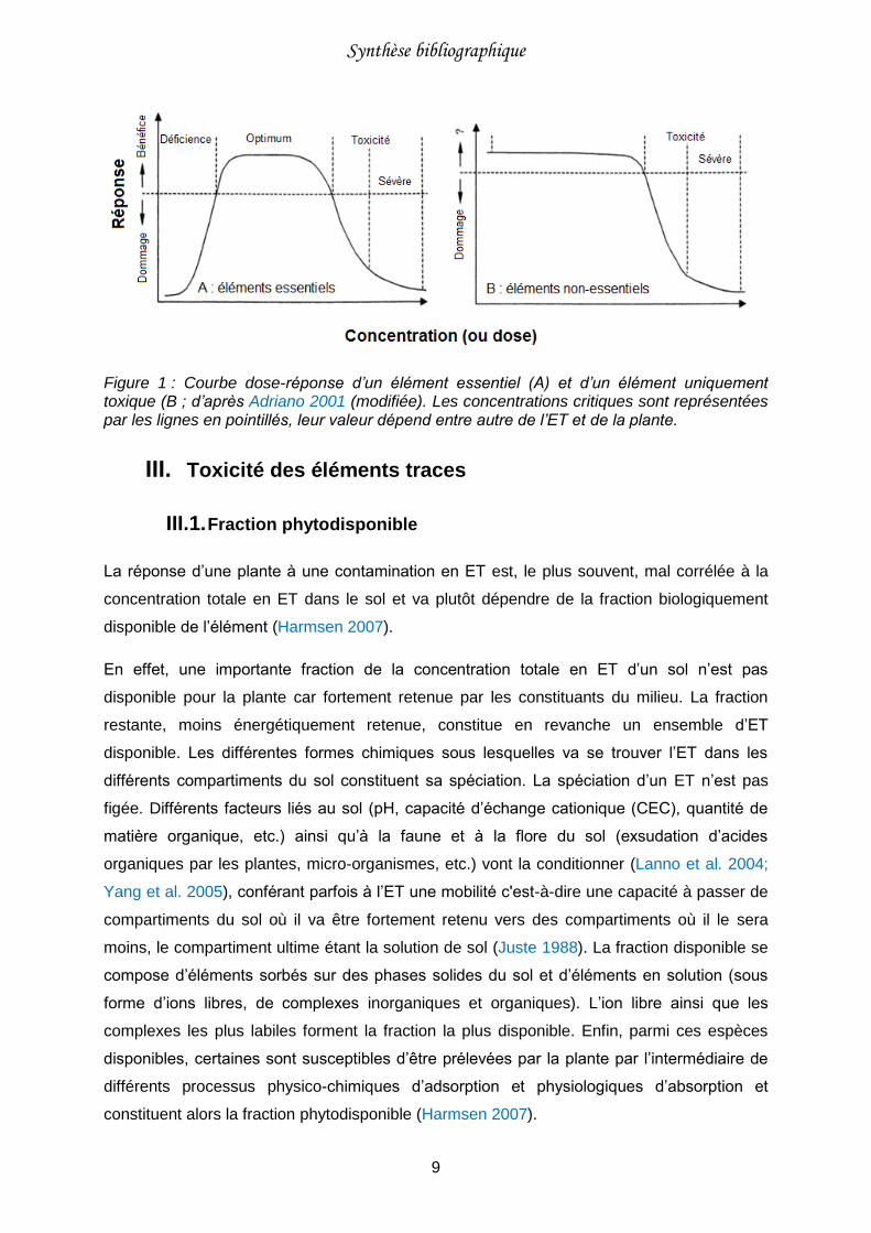
apportée dépasse la concentration critique haute, l’ET devient phytotoxique (Figure 1A).
Dans les deux cas extrêmes, les conséquences sont la perte des différentes fonctions
physiologiques et la mort de la plante.
La seconde catégorie est constituée d’ET uniquement toxiques. On y retrouve, entre autres,
Cd, Hg, Pb et Sn. Ces éléments ne présentent aucune fonction pour l’organisme végétal et
peuvent seulement être toxiques. Le cadmium par exemple, présente des similitudes avec
certains éléments essentiels (Zn ou Fe entre autres) et il est supposé qu’il entre facilement
dans la plante en empruntant les mêmes transporteurs que ces derniers (Krzesłowska et al.
2004; Wójcik et al. 2005). Au-delà d’une concentration seuil, ils conduisent à une inhibition
de la croissance et peuvent entrainer des dommages irréversibles conduisant à la mort de la
plante (Figure 1B ; Nagajyoti et al. 2010).
Synthèse bibliographique
9
Figure 1 : Courbe dose-réponse d’un élément essentiel (A) et d’un élément uniquement toxique (B ; d’après Adriano 2001 (modifiée). Les concentrations critiques sont représentées par les lignes en pointillés, leur valeur dépend entre autre de l’ET et de la plante.
III. Toxicité des éléments traces
III.1. Fraction phytodisponible
La réponse d’une plante à une contamination en ET est, le plus souvent, mal corrélée à la
concentration totale en ET dans le sol et va plutôt dépendre de la fraction biologiquement
disponible de l’élément (Harmsen 2007).
En effet, une importante fraction de la concentration totale en ET d’un sol n’est pas
disponible pour la plante car fortement retenue par les constituants du milieu. La fraction
restante, moins énergétiquement retenue, constitue en revanche un ensemble d’ET
disponible. Les différentes formes chimiques sous lesquelles va se trouver l’ET dans les
différents compartiments du sol constituent sa spéciation. La spéciation d’un ET n’est pas
figée. Différents facteurs liés au sol (pH, capacité d’échange cationique (CEC), quantité de
matière organique, etc.) ainsi qu’à la faune et à la flore du sol (exsudation d’acides
organiques par les plantes, micro-organismes, etc.) vont la conditionner (Lanno et al. 2004;
Yang et al. 2005), conférant parfois à l’ET une mobilité c'est-à-dire une capacité à passer de
compartiments du sol où il va être fortement retenu vers des compartiments où il le sera
moins, le compartiment ultime étant la solution de sol (Juste 1988). La fraction disponible se
compose d’éléments sorbés sur des phases solides du sol et d’éléments en solution (sous
forme d’ions libres, de complexes inorganiques et organiques). L’ion libre ainsi que les
complexes les plus labiles forment la fraction la plus disponible. Enfin, parmi ces espèces
disponibles, certaines sont susceptibles d’être prélevées par la plante par l’intermédiaire de
différents processus physico-chimiques d’adsorption et physiologiques d’absorption et
constituent alors la fraction phytodisponible (Harmsen 2007).
Synthèse bibliographique
10
C’est par les racines que la plante prélève l’eau et les ET présents dans le sol. Les surfaces
racinaires présentent la particularité de pouvoir complexer les éléments. Ce processus
participe à la régulation qu’opère la plante au niveau racinaire pour contrôler le transfert des
ET vers les autres compartiments (pousses, feuilles, etc. ; Briat and Lebrun 1999 ; DalCorso
et al. 2014).
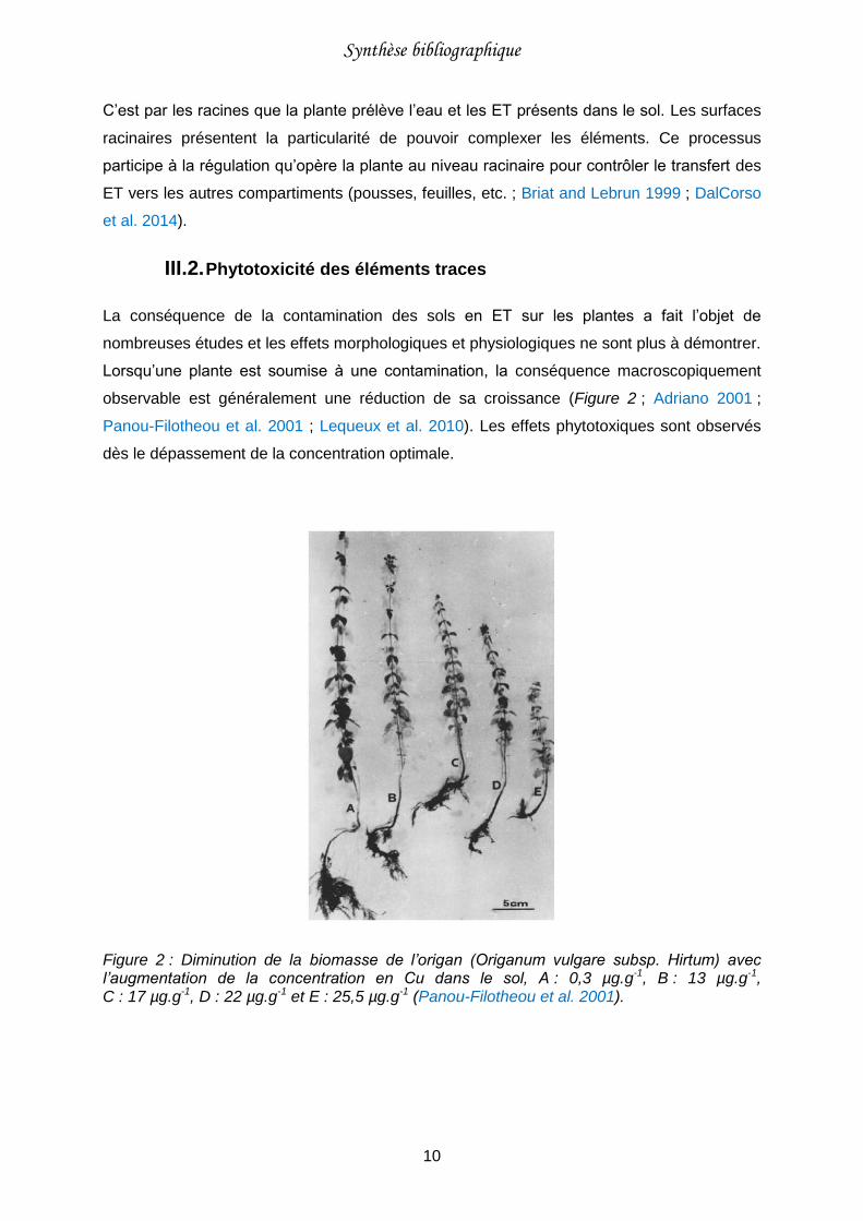
III.2. Phytotoxicité des éléments traces
La conséquence de la contamination des sols en ET sur les plantes a fait l’objet de
nombreuses études et les effets morphologiques et physiologiques ne sont plus à démontrer.
Lorsqu’une plante est soumise à une contamination, la conséquence macroscopiquement
observable est généralement une réduction de sa croissance (Figure 2 ; Adriano 2001 ;
Panou-Filotheou et al. 2001 ; Lequeux et al. 2010). Les effets phytotoxiques sont observés
dès le dépassement de la concentration optimale.
Figure 2 : Diminution de la biomasse de l’origan (Origanum vulgare subsp. Hirtum) avec l’augmentation de la concentration en Cu dans le sol, A : 0,3 µg.g-1, B : 13 µg.g-1, C : 17 µg.g-1, D : 22 µg.g-1 et E : 25,5 µg.g-1 (Panou-Filotheou et al. 2001).
Synthèse bibliographique
11
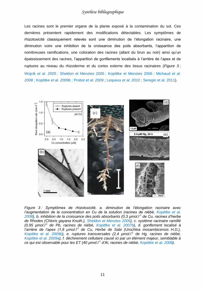
Les racines sont le premier organe de la plante exposé à la contamination du sol. Ces
dernières présentent rapidement des modifications détectables. Les symptômes de
rhizotoxicité classiquement relevés sont une diminution de l’élongation racinaire, une
diminution voire une inhibition de la croissance des poils absorbants, l’apparition de
nombreuses ramifications, une coloration des racines (allant du brun au noir) ainsi qu’un
épaississement des racines, l’apparition de gonflements localisés à l’arrière de l’apex et de
ruptures au niveau du rhizoderme et du cortex externe des tissus racinaires (Figure 3 ;
Wojcik et al. 2005 ; Sheldon et Menzies 2005 ; Kopittke et Menzies 2006 ; Michaud et al.
2008 ; Kopittke et al. 2009b ; Probst et al. 2009 ; Lequeux et al. 2010 ; Seregin et al. 2011).
Figure 3 : Symptômes de rhizotoxicité. a. diminution de l’élongation racinaire avec l’augmentation de la concentration en Cu de la solution (racines de niébé, Kopittke et al. 2008), b. inhibition de la croissance des poils absorbants (0,3 µmol.l-1 de Cu, racines d’herbe de Rhodes (Chloris gayana Knuth.), Sheldon et Menzies 2005), c. système racinaire ramifié (0,95 µmol.l-1 de Pb, racines de niébé, Kopittke et al. 2007b), d. gonflement localisé à l’arrière de l’apex (1,9 µmol.l-1 de Cu, Herbe de Sabi (Urochloa mosambicensis H.D.), Kopittke et al. 2009b), e. ruptures transversales (2,4 µmol.l-1 de Hg, racines de niébé, Kopittke et al. 2009a), f. déchirement cellulaire causé ici par un élément majeur, semblable à ce qui est observable pour les ET (40 µmol.l-1 d’Al, racines de niébé, Kopittke et al. 2008).
Synthèse bibliographique
12
Tous les ET induisent des effets rhizotoxiques liés à un processus de complexation sur les
surfaces racinaires. Ces effets peuvent différer d’un élément à l’autre car ils sont fonction de
l’affinité de complexation de l’élément pour les surfaces (Kopittke et al. 2014b). Ainsi, d’après
l’étude portant sur la rhizotoxicité de 26 éléments métalliques pour le niébé (Vigna
unguiculata L.), Cu (troisième cation métallique le plus toxique parmi les 26) est plus toxique
que Ni, qui est lui-même plus toxique que Pb (Kopittke et al. 2011a). Il a été constaté que Ni
inhibe la croissance des racines latérales du niébé, contrairement à Cu et Pb qui les
favorisent. Le cuivre, quant à lui, entraine des déformations importantes des racines
(formation de vrilles) et l’apparition de ruptures, symptômes qui n’ont pas été constatés avec
Pb et Ni (Kopittke and Menzies 2006; Kopittke et al. 2007a; Kopittke et al. 2007b).
En résumé, à l’interface solution de sol-racines, on retrouve les deux processus suivants :
- (i) diffusion des ET (sous forme d’ion libre ou de complexe métallique) depuis la solution
vers les surfaces racinaires ;
- (ii) adsorption des ET sur les surfaces racinaires, au niveau des sites de complexation.
Ce deuxième processus peut être suivi d’un processus d’absorption, l’élément traverse alors
la membrane biologique. L’ensemble de ces processus conditionne la nature et l’intensité
des interactions entre les racines des plantes et les ET. Néanmoins, le processus
d’adsorption des ET sur les surfaces racinaires semble être un facteur clé, tant dans le
déterminisme de l’absorption des ET par les cellules racinaires que de la rhizotoxicité des ET
(Reid 2001; Kopittke et al. 2014b).

Synthèse bibliographique
13

Synthèse bibliographique
14
Synthèse bibliographique
15
Chapitre 2 : Caractérisation expérimentale
des interactions physico-chimiques éléments
traces - racines
I. Transfert racinaire des éléments traces
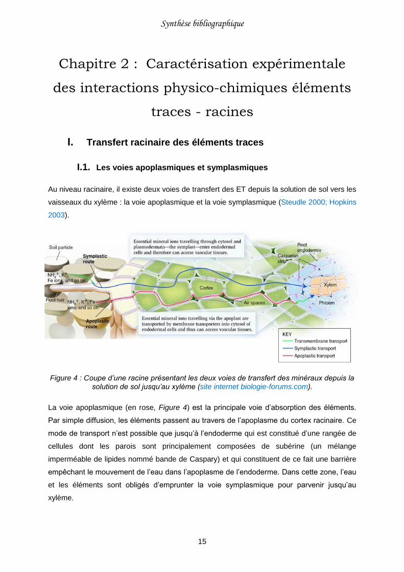
I.1. Les voies apoplasmiques et symplasmiques
Au niveau racinaire, il existe deux voies de transfert des ET depuis la solution de sol vers les
vaisseaux du xylème : la voie apoplasmique et la voie symplasmique (Steudle 2000; Hopkins
2003).
Figure 4 : Coupe d’une racine présentant les deux voies de transfert des minéraux depuis la solution de sol jusqu’au xylème (site internet biologie-forums.com).
La voie apoplasmique (en rose, Figure 4) est la principale voie d’absorption des éléments.
Par simple diffusion, les éléments passent au travers de l’apoplasme du cortex racinaire. Ce
mode de transport n’est possible que jusqu’à l’endoderme qui est constitué d’une rangée de
cellules dont les parois sont principalement composées de subérine (un mélange
imperméable de lipides nommé bande de Caspary) et qui constituent de ce fait une barrière
empêchant le mouvement de l’eau dans l’apoplasme de l’endoderme. Dans cette zone, l’eau
et les éléments sont obligés d’emprunter la voie symplasmique pour parvenir jusqu’au
xylème.
Synthèse bibliographique
16
La voie symplasmique (en bleu, Figure 4) désigne le passage de l’eau et des éléments de
cellules en cellules, par l’intermédiaire des plasmodesmes (canaux traversant les parois
cellulaires et reliant deux cellules entre elles). Avant de parvenir dans le symplasme
racinaire, les éléments doivent d’abord traverser l’apoplasme puis les membranes
plasmiques des cellules végétales grâce à des canaux ou des transporteurs membranaires.
Quelle que soit la voie empruntée, l’ET traverse à minima les parois apoplasmiques et une
membrane plasmique avant d’être distribué dans la plante. Ces deux compartiments jouent
donc un rôle important dans les processus de nutrition et de toxicité.
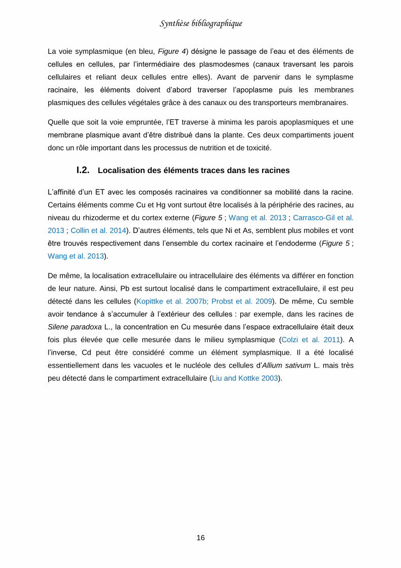
I.2. Localisation des éléments traces dans les racines
L’affinité d’un ET avec les composés racinaires va conditionner sa mobilité dans la racine.
Certains éléments comme Cu et Hg vont surtout être localisés à la périphérie des racines, au
niveau du rhizoderme et du cortex externe (Figure 5 ; Wang et al. 2013 ; Carrasco-Gil et al.
2013 ; Collin et al. 2014). D’autres éléments, tels que Ni et As, semblent plus mobiles et vont
être trouvés respectivement dans l’ensemble du cortex racinaire et l’endoderme (Figure 5 ;
Wang et al. 2013).
De même, la localisation extracellulaire ou intracellulaire des éléments va différer en fonction
de leur nature. Ainsi, Pb est surtout localisé dans le compartiment extracellulaire, il est peu
détecté dans les cellules (Kopittke et al. 2007b; Probst et al. 2009). De même, Cu semble
avoir tendance à s’accumuler à l’extérieur des cellules : par exemple, dans les racines de
Silene paradoxa L., la concentration en Cu mesurée dans l’espace extracellulaire était deux
fois plus élevée que celle mesurée dans le milieu symplasmique (Colzi et al. 2011). A
l’inverse, Cd peut être considéré comme un élément symplasmique. Il a été localisé
essentiellement dans les vacuoles et le nucléole des cellules d’Allium sativum L. mais très
peu détecté dans le compartiment extracellulaire (Liu and Kottke 2003).
Synthèse bibliographique
17
Figure 5 : Localisation des ET dans les racines. a, b, c et d. cartographies 2D des éléments présents dans des racines de niébé obtenues par micro-fluorescence à rayons X (racines exposées à 1,5 µM de Cu, 1 µM de Hg, 5 µM de Ni et 20 µM d’As ; Wang et al. 2013) ; e. cartographie élémentaire d’une section racinaire de Phyllostachys fastuosa exposée à 100 µM de Cu, obtenue par micro-fluorescence à rayons X (Cu en vert, K en bleu et Si en rouge ; Collin et al. 2014) ; f, g et h. tomographies de sections racinaires de niébé situées à 2 mm de l’apex (signal élémentaire en rouge, signal Compton en vert ; racines exposées à 1 µM de Hg, 5 µM de Ni et 20 µM d’As ; Wang et al. 2013).
Il est toutefois important de garder à l’esprit que les techniques employées pour localiser les
ET dans les racines ont une limite de sensibilité et qu’il est possible de ne pas détecter un
élément dans une région racinaire parce qu’il est présent en trop faible concentration. De
plus, s’il est possible de localiser un ET à l’intérieur ou à l’extérieur de la cellule, il est difficile
en revanche d’être plus précis et de faire la différence entre une localisation dans les parois
Synthèse bibliographique
18
apoplasmiques et au niveau de la surface des membranes plasmiques des cellules. Une
telle différenciation s’avère pourtant nécessaire pour une meilleure compréhension des
mécanismes de toxicité puisqu’il semblerait que certains éléments comme Pb, qui sont
supposés être localisés dans les parois apoplasmiques, sont complexés à la fois avec des
composés pariétaux et avec des composés des présents sur les surfaces des membranes
plasmiques (Seregin et al. 2002).
II. Le compartiment apoplasmique
Le terme apoplasme a été inventé en 1930 par le botaniste Ernst Münch et regroupe
l’ensemble des compartiments situés au-delà de la membrane plasmique (Sattelmacher
2001). Etant localisé à l’interface entre le milieu extérieur et les membranes plasmiques des
cellules végétales, l’apoplasme racinaire est un passage obligé pour tous les ET depuis la
solution de sol vers le symplasme racinaire (Hall 2002). Ce compartiment végétal est
constitué d’eau, de gaz et de parois. Ces dernières sont composées de petits polymères
distincts, étroitement tissés, qui forment un maillage en 3 dimensions avec une organisation
architecturale spécifique (Sarkar et al. 2009).
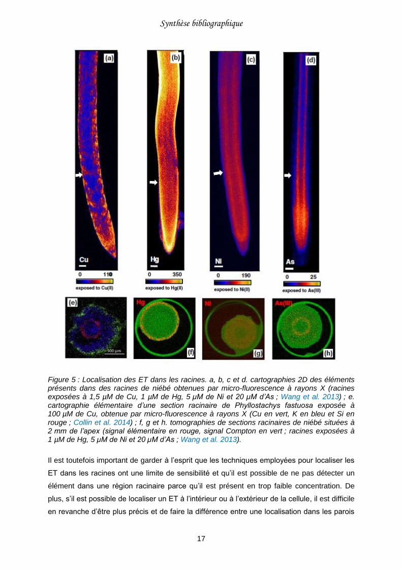
II.1. La lamelle moyenne et les parois apoplasmiques
On distingue 3 couches nommées lamelle moyenne, paroi primaire et paroi secondaire
(Figure 6 ; Sattelmacher (2001)).
Figure 6 : Représentation schématique d’une cellule végétale entourée des parois apoplasmiques (A) (site internet phschool.com) ; image MET (microscope électronique à transmission) de parois apoplasmiques racinaires d’Arabidopsis thaliana (B ; membrane plasmique (pm), paroi secondaire (sw), lamelle moyenne (ml), paroi primaire (pw), cytosol (c) et vacuole (v), d’après Caffal and Mohnen 2009).
Synthèse bibliographique
19
La lamelle moyenne est commune à deux cellules adjacentes et en assure la cohésion. Elle
est formée lors de la mitose (processus de division cellulaire permettant l’obtention de deux
cellules filles identiques à partir d’une cellule mère) et est principalement composée de
pectines ayant différents degrés de méthylation (Sattelmacher 2001).
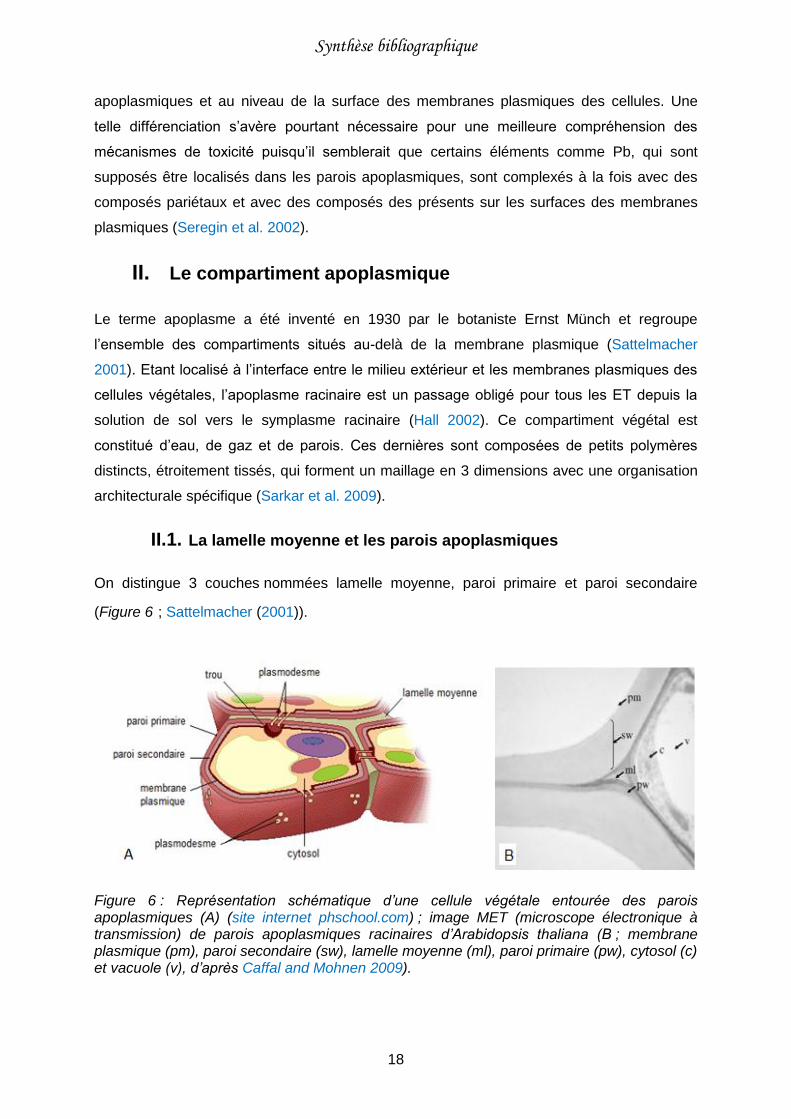
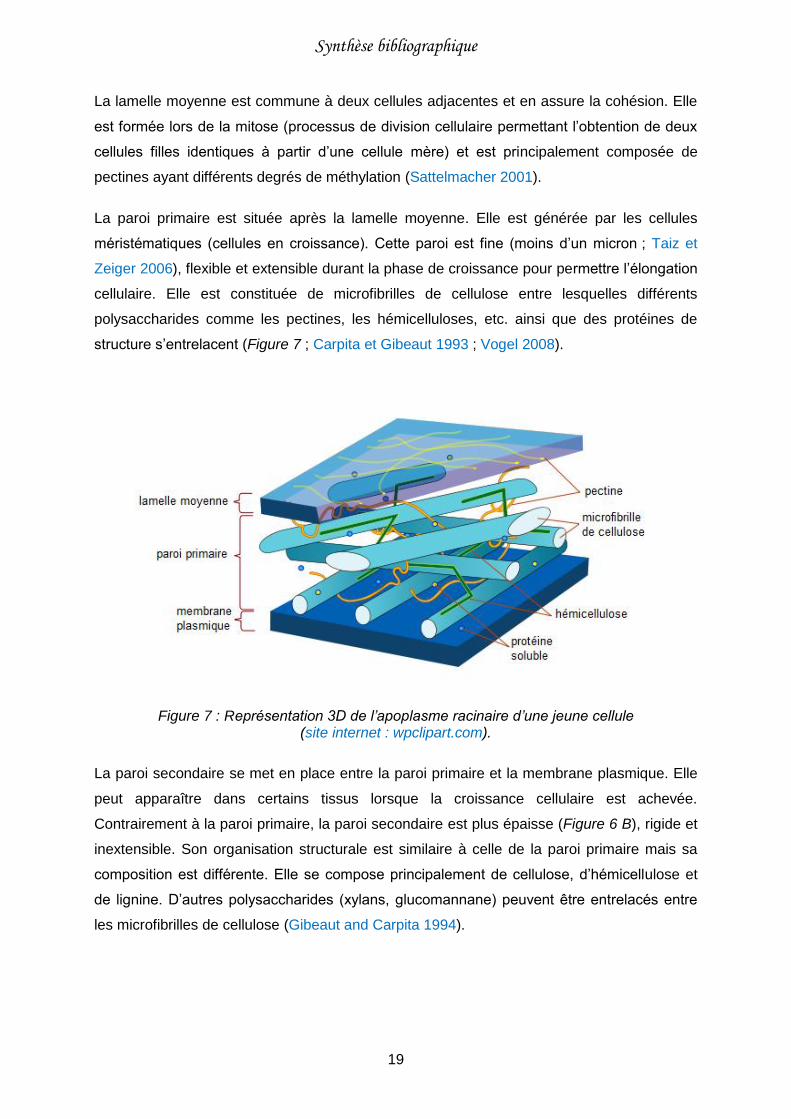
La paroi primaire est située après la lamelle moyenne. Elle est générée par les cellules
méristématiques (cellules en croissance). Cette paroi est fine (moins d’un micron ; Taiz et
Zeiger 2006), flexible et extensible durant la phase de croissance pour permettre l’élongation
cellulaire. Elle est constituée de microfibrilles de cellulose entre lesquelles différents
polysaccharides comme les pectines, les hémicelluloses, etc. ainsi que des protéines de
structure s’entrelacent (Figure 7 ; Carpita et Gibeaut 1993 ; Vogel 2008).
Figure 7 : Représentation 3D de l’apoplasme racinaire d’une jeune cellule (site internet : wpclipart.com).
La paroi secondaire se met en place entre la paroi primaire et la membrane plasmique. Elle
peut apparaître dans certains tissus lorsque la croissance cellulaire est achevée.
Contrairement à la paroi primaire, la paroi secondaire est plus épaisse (Figure 6 B), rigide et
inextensible. Son organisation structurale est similaire à celle de la paroi primaire mais sa
composition est différente. Elle se compose principalement de cellulose, d’hémicellulose et
de lignine. D’autres polysaccharides (xylans, glucomannane) peuvent être entrelacés entre
les microfibrilles de cellulose (Gibeaut and Carpita 1994).
Synthèse bibliographique
20
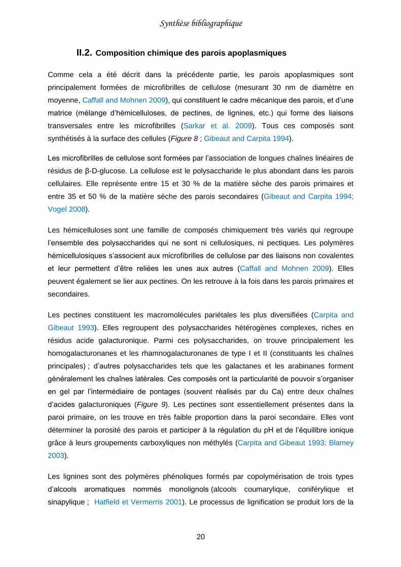
II.2. Composition chimique des parois apoplasmiques
Comme cela a été décrit dans la précédente partie, les parois apoplasmiques sont
principalement formées de microfibrilles de cellulose (mesurant 30 nm de diamètre en
moyenne, Caffall and Mohnen 2009), qui constituent le cadre mécanique des parois, et d’une
matrice (mélange d’hémicelluloses, de pectines, de lignines, etc.) qui forme des liaisons
transversales entre les microfibrilles (Sarkar et al. 2009). Tous ces composés sont
synthétisés à la surface des cellules (Figure 8 ; Gibeaut and Carpita 1994).
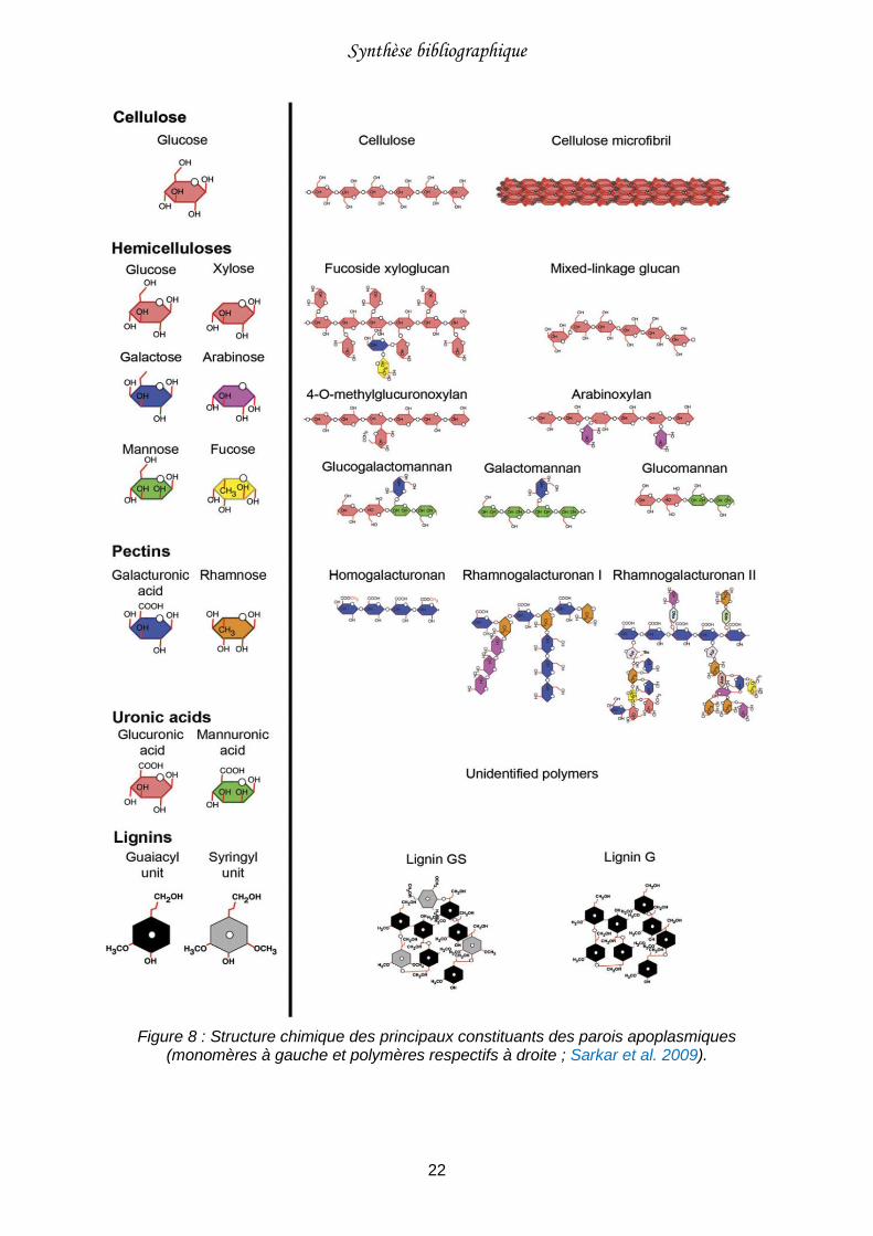
Les microfibrilles de cellulose sont formées par l’association de longues chaînes linéaires de
résidus de β-D-glucose. La cellulose est le polysaccharide le plus abondant dans les parois
cellulaires. Elle représente entre 15 et 30 % de la matière sèche des parois primaires et
entre 35 et 50 % de la matière sèche des parois secondaires (Gibeaut and Carpita 1994;
Vogel 2008).
Les hémicelluloses sont une famille de composés chimiquement très variés qui regroupe
l’ensemble des polysaccharides qui ne sont ni cellulosiques, ni pectiques. Les polymères
hémicellulosiques s’associent aux microfibrilles de cellulose par des liaisons non covalentes
et leur permettent d’être reliées les unes aux autres (Caffall and Mohnen 2009). Elles
peuvent également se lier aux pectines. On les retrouve à la fois dans les parois primaires et
secondaires.
Les pectines constituent les macromolécules pariétales les plus diversifiées (Carpita and
Gibeaut 1993). Elles regroupent des polysaccharides hétérogènes complexes, riches en
résidus acide galacturonique. Parmi ces polysaccharides, on trouve principalement les
homogalacturonanes et les rhamnogalacturonanes de type I et II (constituants les chaînes
principales) ; d’autres polysaccharides tels que les galactanes et les arabinanes forment
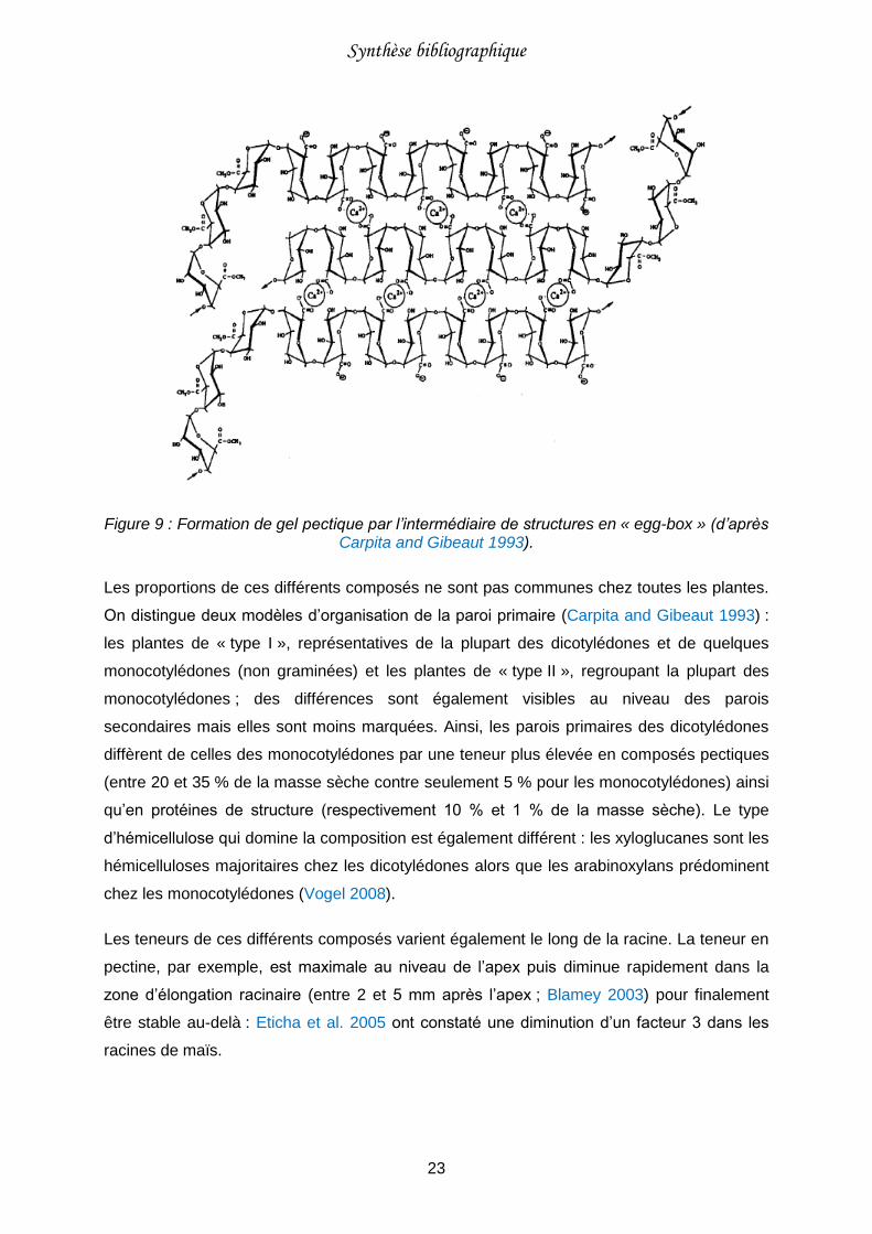
généralement les chaînes latérales. Ces composés ont la particularité de pouvoir s’organiser
en gel par l’intermédiaire de pontages (souvent réalisés par du Ca) entre deux chaînes
d’acides galacturoniques (Figure 9). Les pectines sont essentiellement présentes dans la
paroi primaire, on les trouve en très faible proportion dans la paroi secondaire. Elles vont
déterminer la porosité des parois et participer à la régulation du pH et de l’équilibre ionique
grâce à leurs groupements carboxyliques non méthylés (Carpita and Gibeaut 1993; Blamey
2003).
Les lignines sont des polymères phénoliques formés par copolymérisation de trois types
d’alcools aromatiques nommés monolignols (alcools coumarylique, coniférylique et
sinapylique ; Hatfield et Vermerris 2001). Le processus de lignification se produit lors de la
Synthèse bibliographique
21
formation de la paroi secondaire (Gibeaut and Carpita 1994). La lignine va s’incruster dans
les trois couches pariétales, assurant ainsi une rigidité à l’ensemble et empêchant la
dégradation des polysaccharides pariétaux.
On trouve également dans la paroi primaire des protéines (de structure ou enzymatiques) qui
participent à son élaboration, son remodelage et à la défense contre les agents pathogènes
(Cassab 1998; Caffall and Mohnen 2009).
Synthèse bibliographique
22
Figure 8 : Structure chimique des principaux constituants des parois apoplasmiques (monomères à gauche et polymères respectifs à droite ; Sarkar et al. 2009).
Synthèse bibliographique
23
Figure 9 : Formation de gel pectique par l’intermédiaire de structures en « egg-box » (d’après Carpita and Gibeaut 1993).
Les proportions de ces différents composés ne sont pas communes chez toutes les plantes.
On distingue deux modèles d’organisation de la paroi primaire (Carpita and Gibeaut 1993) :
les plantes de « type I », représentatives de la plupart des dicotylédones et de quelques
monocotylédones (non graminées) et les plantes de « type II », regroupant la plupart des
monocotylédones ; des différences sont également visibles au niveau des parois
secondaires mais elles sont moins marquées. Ainsi, les parois primaires des dicotylédones
diffèrent de celles des monocotylédones par une teneur plus élevée en composés pectiques
(entre 20 et 35 % de la masse sèche contre seulement 5 % pour les monocotylédones) ainsi
qu’en protéines de structure (respectivement 10 % et 1 % de la masse sèche). Le type
d’hémicellulose qui domine la composition est également différent : les xyloglucanes sont les
hémicelluloses majoritaires chez les dicotylédones alors que les arabinoxylans prédominent
chez les monocotylédones (Vogel 2008).
Les teneurs de ces différents composés varient également le long de la racine. La teneur en
pectine, par exemple, est maximale au niveau de l’apex puis diminue rapidement dans la
zone d’élongation racinaire (entre 2 et 5 mm après l’apex ; Blamey 2003) pour finalement
être stable au-delà : Eticha et al. 2005 ont constaté une diminution d’un facteur 3 dans les
racines de maïs.

Synthèse bibliographique
24
II.3. Capacité de complexation de l’apoplasme racinaire
L’apoplasme racinaire remplit de nombreuses fonctions chez la plante (e.g. squelette de
soutien, régulateur de croissance, voie de transport pour l’eau, les nutriments, les hormones
végétales, etc.) et en particulier, il joue un rôle central dans la nutrition minérale en
constituant un réservoir d’ions pour la plante, permettant ainsi de temporiser leur transport
(Grignon and Sentenac 1991; Sakurai 1998; Briat and Lebrun 1999; Carpita 2007).
Cette particularité provient de la composition des parois. Leur capacité de complexation est
généralement associée à leurs teneurs en groupements carboxyliques non méthylés, portés
entre autre par les polysaccharides acides tels que les pectines. La constante d’acidité (pKa)
de ces groupements est estimée entre 3 et 4,4 ce qui signifie qu’aux pH physiologiques de
l’apoplasme (généralement 5-6) entre 80 et 97 % des groupements carboxyliques sont
dissociés (Allan and Jarrell 1989; Wehr et al. 2010) et donc particulièrement disponibles pour
des réactions de complexation avec des cations. Toutefois, les titrages acido-basiques de
parois isolées de blé (Triticum aestivum L.), de pois (Pisum sativum L.) et de lupin (Lupinus
albus L.) ont révélé une diversité de sites réactifs plus importante avec des pKa plus élevés
(Meychik and Yermakov 1999, 2001). D’autres composés pariétaux peuvent donc également
participer à la complexation des cations. Les cations peuvent être adsorbés de manière
spécifique (Cu, Mn ou Zn ; Sattelmacher 2001 ou non spécifique (Ca, K ; sous forme
d’interactions électrostatiques).
La faculté d’une espèce végétale à adsorber les cations est évaluée par sa capacité
d’échange cationique racinaire (CECR). La CECR est dépendante de nombreux paramètres
tels que l’âge de la plante (Heintze 1961; Ram 1980). Elle est généralement plus élevée
chez les dicotylédones (comprise entre 20 et 50 cmolc.kg-1) que chez les monocotylédones
(comprise entre 10 et 20 cmolc.kg-1 ; Crooke et al. 1960 ; Heintze 1961 ; Straczek et al.
2008). Cette particularité provient du fait que les compositions des parois apoplasmiques
sont différentes (cf. Chapitre 2 : II.2), les dicotylédones étant plus riches en composés
pectiques que les monocotylédones. Les groupements carboxyliques non méthylés portés
par les pectines sont généralement considérés comme étant la principale source de charges
négatives des racines (Krzesłowska 2011), 70 à 90 % de la CECR leur étant attribué
(Haynes 1980).
La complexation en excès des ET dans les parois apoplasmiques est l’une des principales
hypothèses avancées pour expliquer les effets rhizotoxiques observés (cf. Chapitre 1 : III.2,
Figure 3). Il a en effet été constaté que les propriétés physiques des parois apoplasmiques
sont altérées lorsque le Ca est remplacé par un ET pour la formation des gels pectiques
Synthèse bibliographique
25
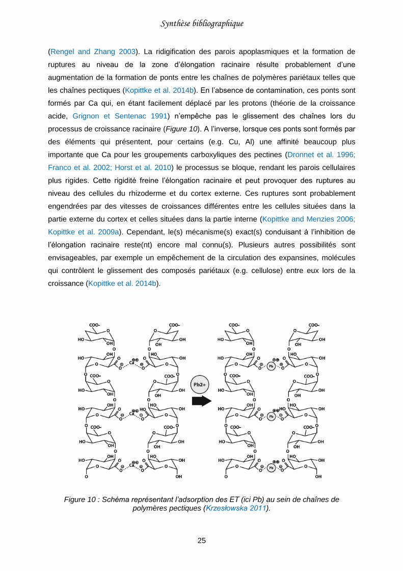
(Rengel and Zhang 2003). La ridigification des parois apoplasmiques et la formation de
ruptures au niveau de la zone d’élongation racinaire résulte probablement d’une
augmentation de la formation de ponts entre les chaînes de polymères pariétaux telles que
les chaînes pectiques (Kopittke et al. 2014b). En l’absence de contamination, ces ponts sont
formés par Ca qui, en étant facilement déplacé par les protons (théorie de la croissance
acide, Grignon et Sentenac 1991) n’empêche pas le glissement des chaînes lors du
processus de croissance racinaire (Figure 10). A l’inverse, lorsque ces ponts sont formés par
des éléments qui présentent, pour certains (e.g. Cu, Al) une affinité beaucoup plus
importante que Ca pour les groupements carboxyliques des pectines (Dronnet et al. 1996;
Franco et al. 2002; Horst et al. 2010) le processus se bloque, rendant les parois cellulaires
plus rigides. Cette rigidité freine l’élongation racinaire et peut provoquer des ruptures au
niveau des cellules du rhizoderme et du cortex externe. Ces ruptures sont probablement
engendrées par des vitesses de croissances différentes entre les cellules situées dans la
partie externe du cortex et celles situées dans la partie interne (Kopittke and Menzies 2006;
Kopittke et al. 2009a). Cependant, le(s) mécanisme(s) exact(s) conduisant à l’inhibition de
l’élongation racinaire reste(nt) encore mal connu(s). Plusieurs autres possibilités sont
envisageables, par exemple un empêchement de la circulation des expansines, molécules
qui contrôlent le glissement des composés pariétaux (e.g. cellulose) entre eux lors de la
croissance (Kopittke et al. 2014b).
Figure 10 : Schéma représentant l’adsorption des ET (ici Pb) au sein de chaînes de polymères pectiques (Krzesłowska 2011).
Synthèse bibliographique
26
D’autres hypothèses ont également été avancées pour expliquer la rhizotoxicité des ET,
notamment une complexation des éléments en excès au niveau des membranes plasmiques
et un effet toxique engendré par leur altération (modification de leur perméabilité ; Ishikawa
et al. 2001 ; Kopittke et al. 2014b).
III. Les membranes plasmiques
Les cellules végétales sont délimitées par une enveloppe appelée membrane plasmique qui
permet la séparation entre le cytoplasme et le milieu extérieur (Figure 11).
III.1. Composition chimique des membranes plasmiques
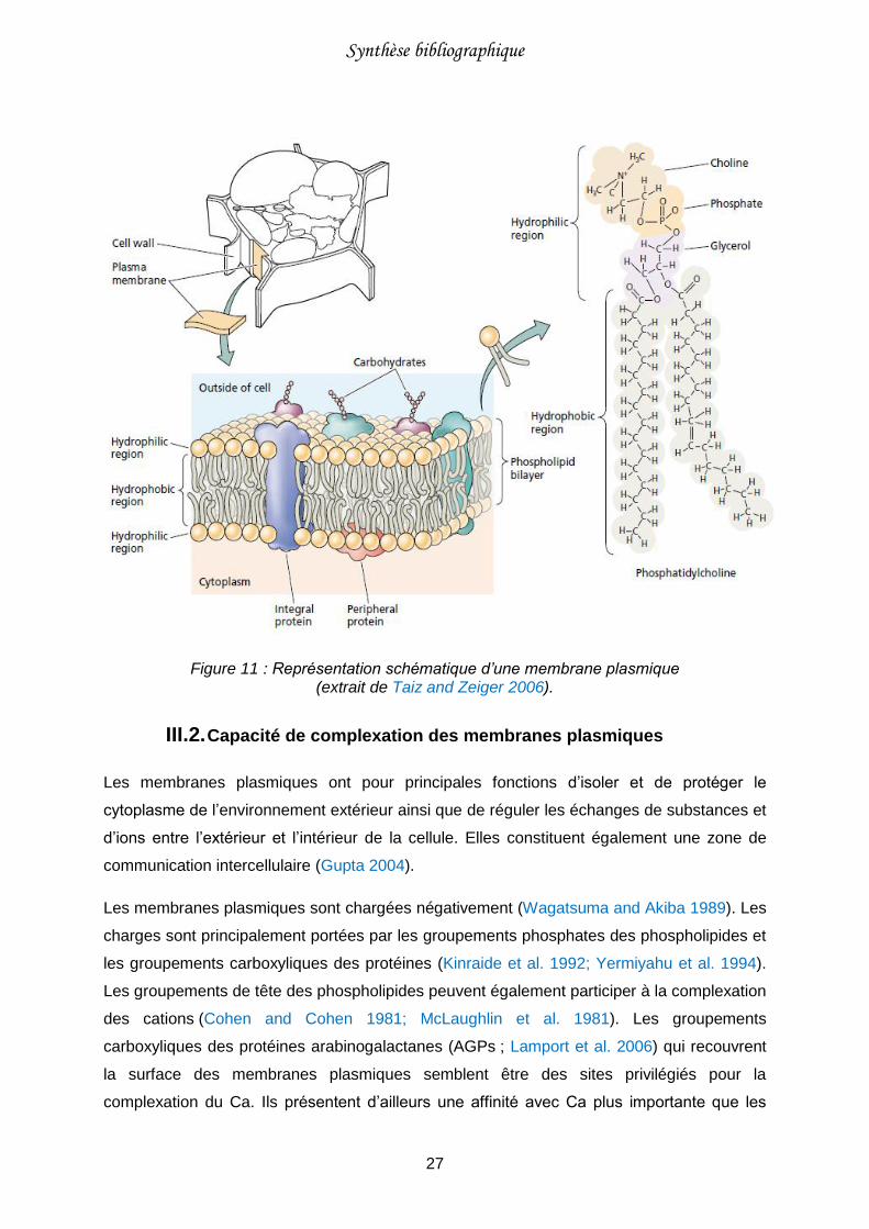
Toutes les membranes biologiques ont la même organisation moléculaire. Elles sont formées
d’une double couche de phospholipides et de protéines (Figure 11).
Les phospholipides sont des molécules complexes constituées de deux chaînes d’acides
gras apolaires hydrophobes et d’une tête polaire hydrophile. La tête se compose d’une
molécule de glycérol (faisant le lien entre les deux chaînes d’acides gras), d’un groupement
phosphate et d’une molécule de tête qui peut être une molécule de choline, comme sur la
figure 11, ou une autre molécule (sérine, glycérol, inositol, ethanolamine, etc. ; Hopkins
2003 ; Taiz et Zeiger 2006).
Les protéines sont des macromolécules composées d’une ou plusieurs chaînes d’acides
aminés liées entre eux par des liaisons peptidiques. Elles peuvent représenter jusqu’à la
moitié de la masse membranaire (Hopkins 2003; Taiz and Zeiger 2006). Les protéines
membranaires peuvent être classées en deux catégories : les protéines intégrées
(enchâssées dans la bicouche) et les protéines périphériques (liées à la surface hydrophile ;
Figure 11).
Synthèse bibliographique
27
Figure 11 : Représentation schématique d’une membrane plasmique (extrait de Taiz and Zeiger 2006).
III.2. Capacité de complexation des membranes plasmiques
Les membranes plasmiques ont pour principales fonctions d’isoler et de protéger le
cytoplasme de l’environnement extérieur ainsi que de réguler les échanges de substances et
d’ions entre l’extérieur et l’intérieur de la cellule. Elles constituent également une zone de
communication intercellulaire (Gupta 2004).
Les membranes plasmiques sont chargées négativement (Wagatsuma and Akiba 1989). Les
charges sont principalement portées par les groupements phosphates des phospholipides et
les groupements carboxyliques des protéines (Kinraide et al. 1992; Yermiyahu et al. 1994).
Les groupements de tête des phospholipides peuvent également participer à la complexation
des cations (Cohen and Cohen 1981; McLaughlin et al. 1981). Les groupements
carboxyliques des protéines arabinogalactanes (AGPs ; Lamport et al. 2006) qui recouvrent
la surface des membranes plasmiques semblent être des sites privilégiés pour la
complexation du Ca. Ils présentent d’ailleurs une affinité avec Ca plus importante que les
Synthèse bibliographique
28
pectines. Il est supposé que la complexation de Ca sur les AGPs participe à la régulation du
processus d’élongation racinaire, selon la même théorie de la croissance acide utilisée pour
expliquer le rôle des pectines (Lamport and Várnai 2013). Par analogie, le remplacement de
Ca par d’autres cations métalliques pourrait ainsi perturber le processus d’élongation
racinaire et ce d’autant plus que certains cations métalliques comme Al et Cu semblent
présenter une affinité élevée avec les sites réactifs présents à la surface des membranes
plasmiques (Vulkan et al. 2004; Kudo et al. 2011).
IV. Spéciation des éléments traces dans les racines
Les composés racinaires impliqués dans les réactions de complexation sont de diverses
natures et la spéciation peut fortement différer d’un ET à un autre.
Le plomb, préférentiellement localisé dans le milieu extracellulaire, a uniquement été trouvé
en association avec des ligands oxygène dans les racines de différentes plantes (Avena
pratensis, Festuca ovina, etc.). L’hypothèse d’une participation majoritaire des
polysaccharides acides tels que les pectines a été retenue (Bovenkamp et al. 2013).
Le cuivre (II) est soit majoritairement trouvé en association avec des ligands oxygène
(Polette et al. 2000; Kopittke et al. 2011c), soit associé à des ligands oxygène et azote (Shi
et al. 2008; Collin et al. 2014). Dans le premier cas, cela suggère une complexation du Cu
par les pectines (comme dans le cas du Pb, d’autant que Cu est un élément qui a tendance
à s’accumuler dans le milieu extracellulaire). Cela peut aussi révéler la participation de petits
acides organiques tels que le malate ou le citrate, principalement localisé dans le
symplasme. Dans le second cas, cela suppose l’implication d’acides aminés présents à la
fois dans le milieu extracellulaire (résidus de protéines enchâssées dans les parois
apoplasmiques ou dans les membranes plasmiques) et dans le milieu intracellulaire (petits
acides aminés). Une récente étude a montré que les protéines riches en résidus histidine
sont le principal site de complexation du Cu dans les parois apoplasmiques de Thlaspi
arvense (Manceau et al. 2013). Du cuivre (I) a également été détecté dans certaines racines
et trouvé en association avec du souffre (Polette et al. 2000; Shi et al. 2008; Collin et al.
2014).
La réduction du Cu(II) en Cu(I) dans les racines est un processus permettant à la plante
d’internaliser le Cu mais il est également supposée être un mécanisme de protection de la
plante, Cu étant complexé par des protéines puis acheminé vers des tissus moins sensibles.
Synthèse bibliographique
29
Le cobalt semble également avoir une affinité particulière pour les acides aminés puisque de
l’oxygène et de l’azote ont été détectés dans son environnement dans des racines de blé
(Triticum aestivum L.) et de tomate (Lycopersicon esculentum M.) (Collins et al. 2010).
Contrairement au Cu(II) ou au Pb, Cd et Hg sont quasi-exclusivement trouvés en association
avec du souffre dans les racines (Pickering et al. 1999; Isaure et al. 2006; Carrasco-Gil et al.
2013). Si pour Hg, qui est un élément plutôt extracellulaire, la présence de souffre peut être
associée à des protéines riches en résidus type cystéine enchâssées dans les parois
apoplasmiques ; pour Cd, élément plutôt symplasmique, cela suggère une participation de
petits acides aminés tels que la cystéine ou le glutathion.
Le zinc, quant à lui, est quelque fois trouvé en association avec des ligands oxygène mais il
présente la particularité d’être le plus souvent entouré de ligand oxygène et phosphore
(Sarret et al. 2001; Sarret et al. 2009; Straczek et al. 2008; Kopittke et al. 2011c). Alors qu’un
environnement uniquement oxygène suppose, comme pour d’autres éléments, qu’il est
complexé par des acides organiques, un environnement présentant à la fois de l’oxygène et
du phosphore permet de supposer qu’il y a participation de groupements phosphates et donc
participation soit de petits acides caractéristiques tels que l’acide phytique, soit des
groupements phosphate des phospholipides portés par les membranes plasmiques.

Synthèse bibliographique
30
Synthèse bibliographique
31
Chapitre 3 : Modélisation des interactions
éléments traces - racines
Les études menées entre 1960 et 1970 sur la toxicité de nombreux éléments traces pour les
organismes aquatiques ont conduit à la formulation de deux mécanismes principaux
(Erickson 2013) :
(i) la toxicité d’un ET varie en fonction de sa spéciation chimique dans la solution
baignant l’organisme ; elle est souvent étroitement corrélée à l’activité de l’ion
libre, bien que d’autres espèces métalliques puissent contribuer ;
(ii) les cations comme Ca2+ ou H+ peuvent réduire la toxicité de l’élément trace.
Les modèles actuellement développés pour les plantes s’appuient sur ces deux
mécanismes.
Une analyse de la littérature révèle l’existence de deux familles de modèles : certains sont
centrés sur la description de la complexation de l’ET au niveau des sites réactifs et
l’établissement des constantes associées, d’autres s’attachent à décrire les phénomènes
électrostatiques présents au niveau de la zone d’interaction, pour lesquels l’établissement de
constantes de complexation n’est pas centrale (le modèle y étant peu sensible). On constate
également des divergences quant à la principale zone d’interaction entre l’ET et la plante : il
peut s’agir soit des parois apoplasmiques, soit des membranes plasmiques, soit des deux.
I. Le modèle électrostatique
Le modèle électrostatique est développé depuis plus de 20 ans. Ce modèle fait l’hypothèse
que les phénomènes d’absorption, les effets toxiques ainsi que les compétitions entre
éléments traces et cations ou anions majeurs qui ont lieu au sein des racines sont
directement reliés à la concentration en ions située à la surface des membranes plasmiques.
Le formalisme mathématique employé est fondé sur le modèle de la double couche
électrique (également connu sous le nom de modèle Gouy-Chapman-Stern), initialement
consacré à l’étude du comportement des surfaces chargées en contact avec des solutions.
Synthèse bibliographique
32
I.1. Principales hypothèses et applications
Cette partie résume les principales hypothèses du modèle électrostatique, sans description
mathématique. Le lecteur est invité à consulter les articles de Kinraide et co-auteurs pour
plus d’informations sur ces aspects mathématiques (Kinraide et al. 1998; Kinraide 2001).
Les principales hypothèses de ce modèle peuvent être résumées ainsi.
Les sites chargés négativement à la surface externe des membranes plasmiques vont créer
un potentiel électrique de surface (noté ψ0). Ce potentiel électrique de surface contrôle
l’activité des ions à la surface des membranes plasmiques et donc les effets toxiques (Wang
et al. 2009). Les parois apoplasmiques n’ont que peu d’effet sur l’activité d’un ion au niveau
de la surface des membranes plasmiques (Kinraide 2004) ; elles ne sont donc pas prises en
compte dans le modèle (Kopittke et al. 2014a). La description des phénomènes de
complexation n’est employée que pour moduler les charges négatives à la surface des
membranes plasmiques et donc moduler le potentiel électrique de surface. La qualité des
simulations est donc peu sensible aux valeurs fixées pour les constantes de complexation
des ET contrairement à celles des anions et cations majeurs (Kinraide 2004).
La composition ionique du milieu dans lequel se trouve les membranes plasmiques a une
influence sur la densité de sites de surface chargés ainsi que sur le potentiel électrique de
surface (Kinraide 2001). Par formation de liaisons covalentes ou électrostatiques, les cations
tels que H+, Ca2+ et Mg2+ réduisent la négativité de ψ0 (Kinraide et al. 1992). L’ampleur de la
réduction de la négativité du potentiel de surface est fonction de la charge de l’ion ainsi que
de sa force de liaison avec les sites de surface (Kinraide 2006). La concentration en cations
métalliques à la surface des membranes plasmiques est directement liée au potentiel
électrique de surface. Par conséquent, une réduction du potentiel affecte la toxicité des
cations métalliques (Wang et al. 2009). Ainsi, une diminution de la toxicité de l’aluminium et
du Cu pour le blé (Triticum aestivum L. cv Scout 66) a été constatée respectivement en
présence de Ca2+ (Kinraide et al. 1998) et en présence de Ca2+ et de Mg2+ (Wang et al.
2008).
L’activité des cations métalliques à la surface des membranes plasmiques permet ainsi une
meilleure prédiction de la diminution du taux d’élongation racinaire que l’activité des cations
métalliques en solution (Figure 12 ; Kinraide 2003 ; Wang et al. 2011 ; Kopittke et al. 2011b).
Ce modèle permet également de prédire la toxicité de certains métalloïdes anioniques
comme l’arsenic (V) ou le sélénate (Yermiyahu 2005; Wang et al. 2008), contrairement aux
autres modèles de prédiction de la toxicité qui ne sont pas paramétrés pour les anions.
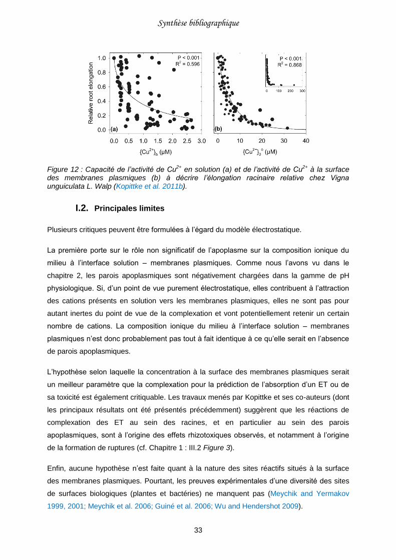
Synthèse bibliographique
33
Figure 12 : Capacité de l’activité de Cu2+ en solution (a) et de l’activité de Cu2+ à la surface des membranes plasmiques (b) à décrire l’élongation racinaire relative chez Vigna unguiculata L. Walp (Kopittke et al. 2011b).
I.2. Principales limites
Plusieurs critiques peuvent être formulées à l’égard du modèle électrostatique.
La première porte sur le rôle non significatif de l’apoplasme sur la composition ionique du
milieu à l’interface solution – membranes plasmiques. Comme nous l’avons vu dans le
chapitre 2, les parois apoplasmiques sont négativement chargées dans la gamme de pH
physiologique. Si, d’un point de vue purement électrostatique, elles contribuent à l’attraction
des cations présents en solution vers les membranes plasmiques, elles ne sont pas pour
autant inertes du point de vue de la complexation et vont potentiellement retenir un certain
nombre de cations. La composition ionique du milieu à l’interface solution – membranes
plasmiques n’est donc probablement pas tout à fait identique à ce qu’elle serait en l’absence
de parois apoplasmiques.
L’hypothèse selon laquelle la concentration à la surface des membranes plasmiques serait
un meilleur paramètre que la complexation pour la prédiction de l’absorption d’un ET ou de
sa toxicité est également critiquable. Les travaux menés par Kopittke et ses co-auteurs (dont
les principaux résultats ont été présentés précédemment) suggèrent que les réactions de
complexation des ET au sein des racines, et en particulier au sein des parois
apoplasmiques, sont à l’origine des effets rhizotoxiques observés, et notamment à l’origine
de la formation de ruptures (cf. Chapitre 1 : III.2 Figure 3).
Enfin, aucune hypothèse n’est faite quant à la nature des sites réactifs situés à la surface
des membranes plasmiques. Pourtant, les preuves expérimentales d’une diversité des sites
de surfaces biologiques (plantes et bactéries) ne manquent pas (Meychik and Yermakov
1999, 2001; Meychik et al. 2006; Guiné et al. 2006; Wu and Hendershot 2009).
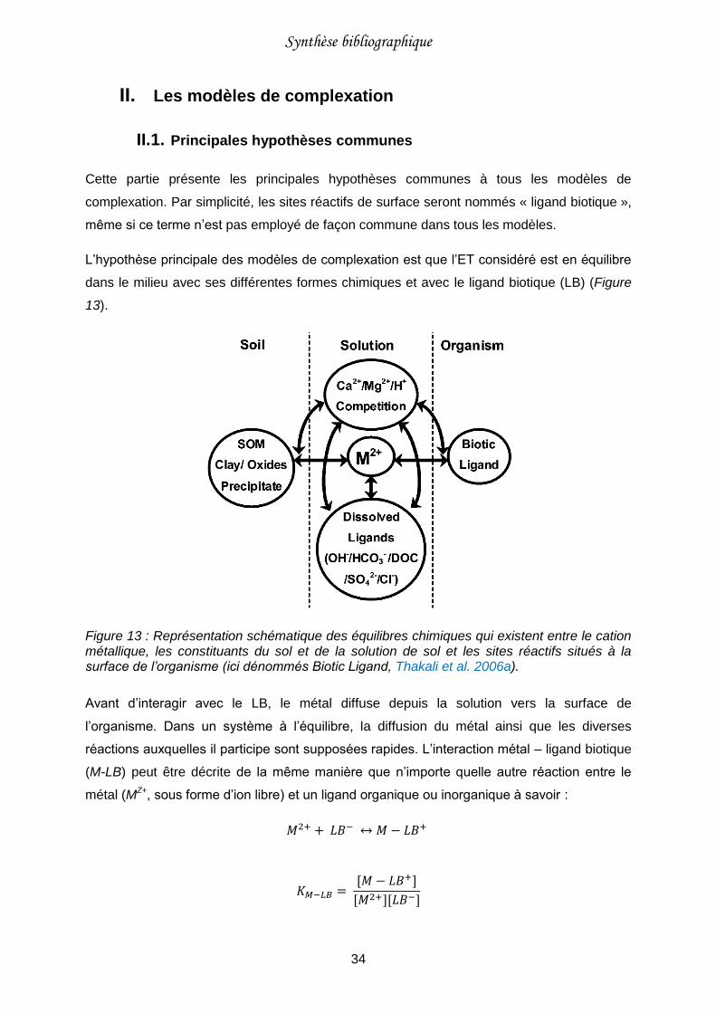
Synthèse bibliographique
34
II. Les modèles de complexation
II.1. Principales hypothèses communes
Cette partie présente les principales hypothèses communes à tous les modèles de
complexation. Par simplicité, les sites réactifs de surface seront nommés « ligand biotique »,
même si ce terme n’est pas employé de façon commune dans tous les modèles.
L’hypothèse principale des modèles de complexation est que l’ET considéré est en équilibre
dans le milieu avec ses différentes formes chimiques et avec le ligand biotique (LB) (Figure
13).
Figure 13 : Représentation schématique des équilibres chimiques qui existent entre le cation métallique, les constituants du sol et de la solution de sol et les sites réactifs situés à la surface de l’organisme (ici dénommés Biotic Ligand, Thakali et al. 2006a).
Avant d’interagir avec le LB, le métal diffuse depuis la solution vers la surface de
l’organisme. Dans un système à l’équilibre, la diffusion du métal ainsi que les diverses
réactions auxquelles il participe sont supposées rapides. L’interaction métal – ligand biotique
(M-LB) peut être décrite de la même manière que n’importe quelle autre réaction entre le
métal (MZ+, sous forme d’ion libre) et un ligand organique ou inorganique à savoir :
𝑀2+ + 𝐿𝐵− ↔ 𝑀 − 𝐿𝐵+
𝐾𝑀−𝐿𝐵 = [𝑀 − 𝐿𝐵+]
[𝑀2+][𝐿𝐵−]
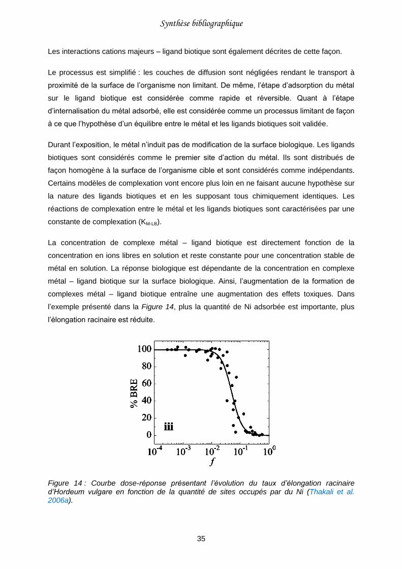
Synthèse bibliographique
35
Les interactions cations majeurs – ligand biotique sont également décrites de cette façon.
Le processus est simplifié : les couches de diffusion sont négligées rendant le transport à
proximité de la surface de l’organisme non limitant. De même, l’étape d’adsorption du métal
sur le ligand biotique est considérée comme rapide et réversible. Quant à l’étape
d’internalisation du métal adsorbé, elle est considérée comme un processus limitant de façon
à ce que l’hypothèse d’un équilibre entre le métal et les ligands biotiques soit validée.
Durant l’exposition, le métal n’induit pas de modification de la surface biologique. Les ligands
biotiques sont considérés comme le premier site d’action du métal. Ils sont distribués de
façon homogène à la surface de l’organisme cible et sont considérés comme indépendants.
Certains modèles de complexation vont encore plus loin en ne faisant aucune hypothèse sur
la nature des ligands biotiques et en les supposant tous chimiquement identiques. Les
réactions de complexation entre le métal et les ligands biotiques sont caractérisées par une
constante de complexation (KM-LB).
La concentration de complexe métal – ligand biotique est directement fonction de la
concentration en ions libres en solution et reste constante pour une concentration stable de
métal en solution. La réponse biologique est dépendante de la concentration en complexe
métal – ligand biotique sur la surface biologique. Ainsi, l’augmentation de la formation de
complexes métal – ligand biotique entraîne une augmentation des effets toxiques. Dans
l’exemple présenté dans la Figure 14, plus la quantité de Ni adsorbée est importante, plus
l’élongation racinaire est réduite.
Figure 14 : Courbe dose-réponse présentant l’évolution du taux d’élongation racinaire d’Hordeum vulgare en fonction de la quantité de sites occupés par du Ni (Thakali et al. 2006a).
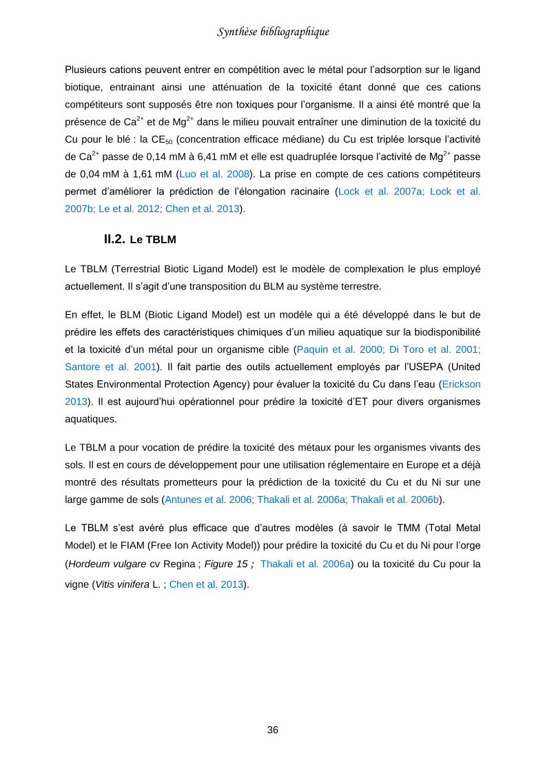
Synthèse bibliographique
36
Plusieurs cations peuvent entrer en compétition avec le métal pour l’adsorption sur le ligand
biotique, entrainant ainsi une atténuation de la toxicité étant donné que ces cations
compétiteurs sont supposés être non toxiques pour l’organisme. Il a ainsi été montré que la
présence de Ca2+ et de Mg2+ dans le milieu pouvait entraîner une diminution de la toxicité du
Cu pour le blé : la CE50 (concentration efficace médiane) du Cu est triplée lorsque l’activité
de Ca2+ passe de 0,14 mM à 6,41 mM et elle est quadruplée lorsque l’activité de Mg2+ passe
de 0,04 mM à 1,61 mM (Luo et al. 2008). La prise en compte de ces cations compétiteurs
permet d’améliorer la prédiction de l’élongation racinaire (Lock et al. 2007a; Lock et al.
2007b; Le et al. 2012; Chen et al. 2013).
II.2. Le TBLM
Le TBLM (Terrestrial Biotic Ligand Model) est le modèle de complexation le plus employé
actuellement. Il s’agit d’une transposition du BLM au système terrestre.
En effet, le BLM (Biotic Ligand Model) est un modèle qui a été développé dans le but de
prédire les effets des caractéristiques chimiques d’un milieu aquatique sur la biodisponibilité
et la toxicité d’un métal pour un organisme cible (Paquin et al. 2000; Di Toro et al. 2001;
Santore et al. 2001). Il fait partie des outils actuellement employés par l’USEPA (United
States Environmental Protection Agency) pour évaluer la toxicité du Cu dans l’eau (Erickson
2013). Il est aujourd’hui opérationnel pour prédire la toxicité d’ET pour divers organismes
aquatiques.
Le TBLM a pour vocation de prédire la toxicité des métaux pour les organismes vivants des
sols. Il est en cours de développement pour une utilisation réglementaire en Europe et a déjà
montré des résultats prometteurs pour la prédiction de la toxicité du Cu et du Ni sur une
large gamme de sols (Antunes et al. 2006; Thakali et al. 2006a; Thakali et al. 2006b).
Le TBLM s’est avéré plus efficace que d’autres modèles (à savoir le TMM (Total Metal
Model) et le FIAM (Free Ion Activity Model)) pour prédire la toxicité du Cu et du Ni pour l’orge
(Hordeum vulgare cv Regina ; Figure 15 ; Thakali et al. 2006a) ou la toxicité du Cu pour la
vigne (Vitis vinifera L. ; Chen et al. 2013).
Synthèse bibliographique
37
Figure 15 : Elongation racinaire prédite avec (i) le TMM (considération de la concentration totale en Cu du sol), (ii) le FIAM (considération de l’activité de Cu2+ libre dans la solution de sol) et (iii) le TBLM (avec prise en compte de la compétition avec le proton) en fonction de l’élongation racinaire mesurée chez l’orge (Hordeum vulgare cv. Regina, exposé à des sols présentant différents niveaux de contamination en Cu ainsi que des propriétés physico-chimiques variées ;Thakali et al. 2006a).
II.3. Les modèles basés sur les substances humiques
D’autres modèles de complexation, développés en parallèle du TBLM, sont utilisés pour
prédire les effets toxiques des ET pour divers organismes aquatiques (plantes, bactéries,
macro-invertébrés, etc.). Ces modèles emploient des formalismes mathématiques
initialement développés pour étudier la complexation des ET avec les matières organiques
naturelles de type substances humiques (acides fulviques et humiques).
Le formalisme mathématique le plus employé actuellement est celui de WHAM (Windermere
Humic Aqueous Model). Il s’agit d’une approche de modélisation discrète. La distribution des
sites est bimodale ; généralement, les sites caractérisés par des pKa inférieurs à 7 sont dits
de type « carboxylique » et les sites caractérisés par des pKa supérieurs à 7 sont dits de
type « phénolique ». Chaque type de sites regroupe 4 sites distincts, présents en égale
quantité. Ce modèle permet la prise en compte d’interactions spécifiques (mono-, bi- et tri-
dendates) et d’interactions non spécifiques (électrostatiques), l’accumulation de contre-ions
dans la double couche de diffusion étant estimée avec une expression type Donnan. Pour de
plus amples détails ainsi qu’une description mathématique complète du modèle, le lecteur
est invité à consulter les deux articles suivants : Tipping 1998 et Tipping et al. 2011.
Dans la littérature, on trouve deux applications du formalisme mathématique de WHAM. La
première, WHAM-FTox, fait l’hypothèse que la réactivité de l’organisme cible peut être
directement assimilée à la réactivité d’un acide humique ; seule la densité totale de sites
réactifs est ajustée. WHAM-FTox a été employé avec succès pour prédire la toxicité de
différents éléments métalliques pour des macro-invertébrés (Stockdale et al. 2010; Stockdale
et al. 2014a), du zooplancton (Stockdale et al. 2014b) ainsi que pour des bactéries et des
Synthèse bibliographique
38
organismes aquatiques (Tipping and Lofts 2013). La seconde application a permis une
bonne prédiction de la toxicité du Cu pour les lentilles d’eau (Lemna minor ; Antunes et al.
2012). Contrairement à l’application précédente, quelques paramètres ont été ajustés pour
permettre au modèle WHAM de correctement représenter les données expérimentales.
L’autre formalisme mathématique employé est celui du modèle NICA-Donnan (NICA pour
Non-Ideal Competitive Adsorption). Il s’agit d’une approche de modélisation continue. De
même qu’avec le modèle WHAM, la distribution des sites est bimodale, il est fait la distinction
entre les sites dits « type carboxylique » et les sites dits « type phénoliques ». Ce modèle
permet également la prise en compte d’interactions spécifiques (monodendates) et non
spécifiques (accumulation de contre-ions dans un volume de Donnan). Pour de plus amples
détails ainsi qu’une description mathématique complète du modèle, le lecteur est invité à
consulter les deux articles suivants : Kinniburgh et al. 1996 et Benedetti et al. 1996. Dans le
cadre de l’application à l’étude des interactions ET – organismes vivants, ce formalisme
mathématique a été employé une seule fois pour prédire les effets de compétition observés
au niveau des parois de bactéries entre Zn, Cd, Ca et H (Plette et al. 1996).
A noter que d’autres modèles ont été ponctuellement développés et adaptés à une étude en
particulier. C’est le cas par exemple, du modèle de prédiction de l’adsorption du Cu dans des
parois apoplasmiques de maïs et de soja, développé par Allan et Jarrell 1989. Ce modèle est
basé sur un modèle de complexation de surface initialement utilisé pour décrire la réactivité
d’oxydes métalliques. Autre exemple, le modèle développé par Postma et al. 2005 qui décrit
la compétition entre Al, Ca et H pour la formation de complexes au niveau des sites des
parois apoplasmiques de tomate. Ce modèle est basé sur le CESS (Cation Exchange/
Specific Sorption model), un modèle généralement dédié à l’étude des réactions d’échanges
cationiques dans le sol.
II.4. Limites des modèles de complexation
La critique majeure qui peut être faite au TBLM est le manque de généricité de certaines
constantes de complexation, qui altère les possibilités d’application du modèle. En effet, la
comparaison des constantes de complexation trouvées dans la littérature montre certains
écarts importants, parfois de plusieurs ordres de grandeurs pour un même métal et pour une
même plante (Tableau 1). De tels écarts sont également visibles pour un même métal entre
différentes espèces végétales.
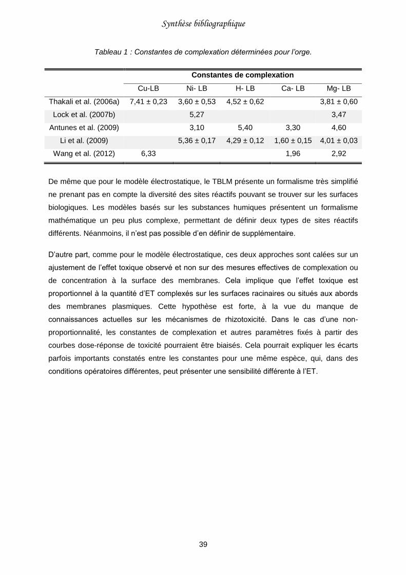
Synthèse bibliographique
39
Tableau 1 : Constantes de complexation déterminées pour l’orge.
Constantes de complexation
Cu-LB Ni- LB H- LB Ca- LB Mg- LB
Thakali et al. (2006a) 7,41 ± 0,23 3,60 ± 0,53 4,52 ± 0,62 3,81 ± 0,60
Lock et al. (2007b) 5,27 3,47
Antunes et al. (2009) 3,10 5,40 3,30 4,60
Li et al. (2009) 5,36 ± 0,17 4,29 ± 0,12 1,60 ± 0,15 4,01 ± 0,03
Wang et al. (2012) 6,33 1,96 2,92
De même que pour le modèle électrostatique, le TBLM présente un formalisme très simplifié
ne prenant pas en compte la diversité des sites réactifs pouvant se trouver sur les surfaces
biologiques. Les modèles basés sur les substances humiques présentent un formalisme
mathématique un peu plus complexe, permettant de définir deux types de sites réactifs
différents. Néanmoins, il n’est pas possible d’en définir de supplémentaire.
D’autre part, comme pour le modèle électrostatique, ces deux approches sont calées sur un
ajustement de l’effet toxique observé et non sur des mesures effectives de complexation ou
de concentration à la surface des membranes. Cela implique que l’effet toxique est
proportionnel à la quantité d’ET complexés sur les surfaces racinaires ou situés aux abords
des membranes plasmiques. Cette hypothèse est forte, à la vue du manque de
connaissances actuelles sur les mécanismes de rhizotoxicité. Dans le cas d’une non-
proportionnalité, les constantes de complexation et autres paramètres fixés à partir des
courbes dose-réponse de toxicité pourraient être biaisés. Cela pourrait expliquer les écarts
parfois importants constatés entre les constantes pour une même espèce, qui, dans des
conditions opératoires différentes, peut présenter une sensibilité différente à l’ET.

Synthèse bibliographique
40
Synthèse bibliographique
41
Chapitre 4 : Objectifs et stratégies de
recherche
I. Hypothèse de travail
Les racines des plantes sont le premier organe à être exposé à une contamination du sol en
ET. Les mécanismes à l’origine des effets rhizotoxiques sont encore mal connus.
Néanmoins, le mécanisme de sorption de l’ET semble être au cœur du processus de
rhizotoxicité. En particulier, la complexation de l’ET au niveau des surfaces racinaires,
première interaction entre l’élément et la plante, semble être une étape importante. Deux
sites de complexation racinaires peuvent être distingués : les parois apoplasmiques d’une
part et la face externe des membranes plasmiques d’autre part. Tous deux présentent une
capacité élevée pour complexer les cations métalliques. Cependant, leur contribution
respective dans les propriétés de complexation des racines n’est pas déterminée.
Des modèles visant à prédire l’effet toxique engendré par les ET chez les plantes se
développent et donnent des résultats prometteurs. Mais la méconnaissance des
mécanismes précis générant un effet toxique associée à la volonté de proposer des cadres
de modélisation relativement simples entraîne la formulation de nombreuses hypothèses,
pouvant paraître parfois trop simplificatrices ou non vérifiées et potentiellement limitantes
pour l’application et la généricité des modèles. Il est donc nécessaire à l’heure actuelle de
remettre en cause certaines de ces hypothèses de façon à outrepasser ces limites.
L’hypothèse centrale de cette étude est qu’une amélioration des modèles de prédiction de la
phytotoxicité passe par une modification de la démarche de modélisation employée, avec un
intérêt porté sur les réactions de complexation plutôt que sur les effets toxiques observés.
II. Objectifs
Ainsi, l’objectif général de la thèse est de reconsidérer les bases mécanistiques de la
complexation des ET dans les racines des plantes et de modéliser cette complexation.
Une première étape consiste à caractériser la réactivité des racines. Il s’agit :
(i) d’identifier la nature des sites réactifs ;
(ii) de déterminer leur localisation c’est-à-dire leur répartition entre l’apoplasme
racinaire et la surface externe des membranes plasmiques des cellules.
Synthèse bibliographique
42
La seconde étape est dédiée à l’étude de la complexation d’un ET modèle au sein des
racines. Il s’agit :
(i) d’identifier les sites réactifs participant à sa complexation ;
(ii) d’étudier les effets des conditions physico-chimiques du milieu (pH, force ionique,
cations compétiteurs) sur la complexation.
L’ensemble des résultats doit permettre de développer un modèle prédisant la complexation
de cet ET modèle au sein des racines.
III. Modèles expérimentaux
III.1. Le cuivre : élément trace modèle
Comme cela est évoqué dans le chapitre 1, le Cu est un élément essentiel pour les plantes.
Il est, avec B, Mn et Zn, l’un des éléments que l’on retrouve en grande quantité dans les
tissus végétaux (Saur 1990). Il joue un rôle fondamental dans de nombreux processus
physiologiques. Par exemple, Cu intervient dans le processus de photosynthèse et dans la
régulation du transfert hydrique de la plante. In vivo, il peut être présent sous différents états
d’oxydation (i.e. Cu+ et Cu2+) et ainsi contribuer aux réactions d’oxydoréduction des
systèmes enzymatiques (Saur 1990; Marschner 1995; Yruela 2009).
Plusieurs raisons nous amènent à choisir le Cu comme élément trace modèle pour cette
étude. Du point de vue de la contamination des sols, le Cu est le deuxième élément le plus
apporté sur les sols agricoles français après Zn : son apport annuel moyen est estimé à
4900 t. Il provient majoritairement des effluents d’origine animale et des pesticides employés
(Belon et al. 2012). Il s’agit également de l’élément essentiel le plus toxique, d’après un
examen de la littérature datant de la période 1975 - 2009 sur les effets de 9 éléments
essentiels et non-essentiel sur la croissance des plantes en culture hydroponique, avec une
concentration toxique médiane de 2 µM (Kopittke et al. 2010). Enfin, il présente enfin une
importante affinité aussi bien avec les sites des parois apoplasmiques (en particulier les
pectines (Kartel et al. 1999; Franco et al. 2002)) qu’avec les sites situés sur les membranes
plasmiques de cellules (Vulkan et al. 2004).
Le zinc a été sélectionné comme élément trace compétiteur modèle. Tout comme Cu, Zn est
un élément essentiel, impliqué dans divers processus physiologiques comme la synthèse
des hormones de croissance et la synthèse de la chlorophylle (Broadley et al. 2007). Notre
choix s’est porté sur Zn car, comme évoqué précédemment, il s’agit de l’élément le plus
Synthèse bibliographique
43
apporté sur les sols agricoles français, avec un apport annuel moyen estimé autour de
15190 t, principalement issu des effluents d’origine animale (Belon et al. 2012).
III.2. La tomate et le blé : espèces végétales modèles
D’après la littérature, les capacités de complexation des racines peuvent fortement différer
selon l’espèce végétale (Heintze 1961). Les dicotylédones présentent généralement une
capacité de complexation plus élevée (comprise entre 20 et 50 cmolc.kg-1) que les
monocotylédones (plutôt comprise entre 10 et 20 cmolc.kg-1 ; Crooke et al. 1960 ; Heintze
1961 ; Straczek et al. 2008). Ces différences sont associées à la composition des parois
apoplasmiques qui, comme nous l’avons vu au paragraphe II.2 du chapitre 2, diffère d’une
espèce végétale à l’autre. En particulier, les dicotylédones sont plus riches en composés
pectiques que les monocotylédones (Carpita and Gibeaut 1993).
De manière à disposer d’un large éventail de données et pouvoir ainsi développer un modèle
qui soit le plus générique possible, nous avons choisi de travailler avec une dicotylédone et
une monocotylédone. La tomate et le blé ont été sélectionnés parmi cinq espèces végétales
pour leur capacité de complexation bien différenciées.
IV. Approches expérimentales et modélisation
Comme évoqué précédemment, plusieurs études se sont attachées à démontrer que les
parois apoplasmiques et les membranes plasmiques présentent toutes deux la capacité de
complexer les ET. Cependant, la contribution respective des parois apoplasmiques et des
membranes plasmiques dans la réactivité des racines vis-à-vis des cations est inconnue.
Dans cette étude, nous avons donc fait le choix de travailler d’une part avec des parois
apoplasmiques isolées, de manière à étudier leurs propriétés de complexation, et d’autre
part avec des racines entières sèches, correspondant à des racines ayant gardées à la fois
les parois apoplasmiques et les membranes plasmiques mais ne présentant pas d’activité
biologique. La contribution des membranes plasmiques est alors obtenue par différence
entre les racines sèches et les parois isolées.
Plusieurs techniques analytiques ont été mises en œuvre sur ces deux matériaux racinaires
(parois apoplasmiques et racines sèches). La spectroscopie de résonance magnétique
nucléaire du carbone 13 (RMN 13C) a permis d’identifier les groupements fonctionnels
présents dans les apoplasmes isolés et les racines sèches. Les titrages acido-basiques ont
apportés des informations sur leur réactivité. Le croisement de ces résultats a permis d’une
part d’évaluer la contribution respective des parois apoplasmiques et des membranes
Synthèse bibliographique
44
plasmiques dans les propriétés de complexation des racines et d’autre part d’identifier les
principaux sites complexant (Deuxième partie, Chapitre 5).
L’étape suivante a consisté en la caractérisation de l’adsorption du Cu au sein des racines
sèches et des apoplasmes isolés. La spéciation du Cu dans ces deux matériaux racinaires a
été étudiée par spectroscopie d’absorption des rayons X. Quant à son affinité de
complexation, il a été étudié en réalisant des expérimentations en batch et en modélisant les
résultats obtenus pour chaque plante et chaque matériau racinaire (Deuxième partie,
Chapitre 6).
Enfin, sur la base d’un modèle existant destiné à l’étude de la réactivité des matières
organiques dissoutes, un modèle décrivant la réactivité du Cu vis-à-vis des racines a été
développé (Troisième partie, Chapitre 7).
Synthèse bibliographique
45
Références bibliographiques
Adriano DC (2001) Trace Elements in Terrestrial Environments : Biogeochemistry, Bioavailability, and Risks of Metals. Springer Edition:867 pp.
Allan DL, Jarrell WM (1989) Proton and Copper Adsorption to Maize and Soybean Root Cell
Walls. Plant Physiol 89 (3):823-832. doi:10.1104/pp.89.3.823 Alloway BJ (2013) Heavy Metal in Soils. Springer Edition:613 pp. Antunes PMC, Berkelaar EJ, Boyle D, Hale BA, Hendershot W, Voigt A (2006) The biotic
ligand model for plants and metals: Technical challenges for field application. Environmental Toxicology and Chemistry 25 (3):875-882. doi:10.1897/04-586r.1
Antunes PMC, Kreager NJ (2009) Development of the terrestrial biotic ligand model for
predicting nickel toxicity to barley (Hordeum Vulgare): Ion effects at low pH. Environmental Toxicology and Chemistry 28 (8):1704-1710. doi:10.1897/08-387.1
Antunes PMC, Scornaienchi ML, Roshon HD (2012) Copper toxicity to Lemna minor
modelled using humic acid as a surrogate for the plant root. Chemosphere 88 (4):389-394. doi:10.1016/j.chemosphere.2012.02.052
Antwi-Agyei P, et al. (2009) Trace elements contamination of soils around gold mine tailings
dams at Obuasi, Ghana. African Journal of Environmental Science and Technology 3 (11):pp. 353-359
Appenroth K-J (2010) What are “heavy metals” in Plant Sciences? Acta Physiologiae
Plantarum 32 (4):615-619. doi:10.1007/s11738-009-0455-4 Baize B (1997) Teneurs totales en éléments traces métalliques dans les sols : France.
Editions Quae:408 pp. Belon E, Boisson M, Deportes IZ, Eglin TK, Feix I, Bispo AO, Galsomies L, Leblond S,
Guellier CR (2012) An inventory of trace elements inputs to French agricultural soils. Science of The Total Environment 439 (0):87-95. doi:http://dx.doi.org/10.1016/j.scitotenv.2012.09.011
Benedetti MF, Van Riemsdijk WH, Koopal LK (1996) Humic Substances Considered as a
Heterogeneous Donnan Gel Phase. Environmental Science & Technology 30 (6):1805-1813. doi:10.1021/es950012y
Blamey F (2003) A role for pectin in the control of cell expansion. Soil Science and Plant
Nutrition 49 (6):775 - 783 Bovenkamp GL, Prange A, Schumacher W, Ham K, Smith AP, Hormes J (2013) Lead
Uptake in Diverse Plant Families: A Study Applying X-ray Absorption Near Edge Spectroscopy. Environmental Science & Technology 47 (9):4375-4382. doi:10.1021/es302408m
Briat J-F, Lebrun M (1999) Plant responses to metal toxicity. Comptes Rendus de l'Académie
des Sciences - Series III - Sciences de la Vie 322 (1):43-54. doi:10.1016/s0764-4469(99)80016-x
Synthèse bibliographique
46
Broadley MR, White PJ, Hammond JP, Zelko I, Lux A (2007) Zinc in plants. New Phytologist 173 (4):677-702. doi:10.1111/j.1469-8137.2007.01996.x
Caffall KH, Mohnen D (2009) The structure, function, and biosynthesis of plant cell wall
pectic polysaccharides. Carbohydrate Research 344 (14):1879-1900. doi:10.1016/j.carres.2009.05.021
Carpita N (2007) Cell wall-ion interactions. In: Sattelmacher, B, Horst, W, editors The
apoplast of higher plants: compartment of storage, transport, and reactions Dordrecht: Kluwer Academic Publishers; 2007.:15-17
Carpita NC, Gibeaut DM (1993) Structural models of primary cell walls in flowering plants:
consistency of molecular structure with the physical properties of the walls during growth. The Plant Journal 3 (1):1-30. doi:10.1111/j.1365-313X.1993.tb00007.x
Carrasco-Gil S, Siebner H, LeDuc DL, Webb SM, Millán R, Andrews JC, Hernández LE
(2013) Mercury Localization and Speciation in Plants Grown Hydroponically or in a Natural Environment. Environmental Science & Technology 47 (7):3082-3090. doi:10.1021/es303310t
Cassab GI (1998) PLANT CELL WALL PROTEINS. Annual Review of Plant Physiology and
Plant Molecular Biology 49 (1):281-309. doi:doi:10.1146/annurev.arplant.49.1.281 Chen B-C, Ho P-C, Juang K-W (2013) Alleviation effects of magnesium on copper toxicity
and accumulation in grapevine roots evaluated with biotic ligand models. Ecotoxicology 22 (1):174-183. doi:10.1007/s10646-012-1015-z
Cohen JA, Cohen M (1981) Adsorption of monovalent and divalent cations by phospholipid
membranes. The monomer-dimer problem. Biophysical journal 36 (3):623-651 Collin B, Doelsch E, Keller C, Cazevieille P, Tella M, Chaurand P, Panfili F, Hazemann J-L,
Meunier J-D (2014) Evidence of sulfur-bound reduced copper in bamboo exposed to high silicon and copper concentrations. Environmental Pollution 187 (0):22-30. doi:http://dx.doi.org/10.1016/j.envpol.2013.12.024
Collins RN, Bakkaus E, Carrière M, Khodja H, Proux O, Morel J-L, Gouget B (2010) Uptake,
Localization, and Speciation of Cobalt in Triticum aestivum L. (Wheat) and Lycopersicon esculentum M. (Tomato). Environmental Science & Technology 44 (8):2904-2910. doi:10.1021/es903485h
Colzi I, Doumett S, Del Bubba M, Fornaini J, Arnetoli M, Gabbrielli R, Gonnelli C (2011) On
the role of the cell wall in the phenomenon of copper tolerance in Silene paradoxa L. Environmental and Experimental Botany 72 (1):77-83. doi:DOI: 10.1016/j.envexpbot.2010.02.006
Crooke WM, Knight AH, Macdonald IR (1960) Cation exchange properties and pectin content
of storage-tissue disks. Plant and Soil 13 (1):55-67. doi:10.1007/bf01666476 DalCorso G, Manara A, Piasentin S, Furini A (2014) Nutrient metal elements in plants.
Metallomics. doi:10.1039/c4mt00173g Di Toro DM, Allen HE, Bergman HL, Meyer JS, Paquin PR, Santore RC (2001) Biotic ligand
model of the acute toxicity of metals. 1. Technical Basis. Environmental Toxicology and Chemistry 20 (10):2383-2396. doi:10.1002/etc.5620201034
Synthèse bibliographique
47
Doelsch E, Van de Kerchove V, Saint Macary H (2006a) Heavy metal content in soils of Réunion (Indian Ocean). Geoderma 134 (1–2):119-134. doi:http://dx.doi.org/10.1016/j.geoderma.2005.09.003
Dronnet VM, Renard CMGC, Axelos MAV, Thibault JF (1996) Characterisation and
selectivity of divalent metal ions binding by citrus and sugar-beet pectins. Carbohydrate Polymers 30 (4):253-263. doi:Doi: 10.1016/s0144-8617(96)00107-5
Erickson RJ (2013) The biotic ligand model approach for addressing effects of exposure
water chemistry on aquatic toxicity of metals: Genesis and challenges. Environmental Toxicology and Chemistry 32 (6):1212-1214. doi:10.1002/etc.2222
Eticha D, Stass A, Horst WJ (2005) Cell-wall pectin and its degree of methylation in the
maize root-apex: significance for genotypic differences in aluminium resistance. Plant, Cell & Environment 28 (11):1410-1420. doi:10.1111/j.1365-3040.2005.01375.x
Franco CR, Chagas AP, Jorge RA (2002) Ion-exchange equilibria with aluminum pectinates.
Colloids and Surfaces A: Physicochemical and Engineering Aspects 204 (1-3):183-192. doi:Doi: 10.1016/s0927-7757(01)01134-7
Gibeaut DM, Carpita NC (1994) Biosynthesis of plant cell wall polysaccharides. The FASEB
Journal 8 (12):904-915 Grignon C, Sentenac H (1991) pH and Ionic Conditions in the Apoplast. Annual Review of
Plant Physiology and Plant Molecular Biology 42 (1):103-128. doi:doi:10.1146/annurev.pp.42.060191.000535
Guigues S, Bravin M, Garnier C, Masion A, Doelsch E (2014) Isolated cell walls exhibit
cation binding properties distinct from those of plant roots. Plant and Soil:1-13. doi:10.1007/s11104-014-2138-1
Guiné V, Spadini L, Sarret G, Muris M, Delolme C, Gaudet JP, Martins JMF (2006) Zinc
Sorption to Three Gram-Negative Bacteria: Combined Titration, Modeling, and EXAFS Study. Environmental Science & Technology 40 (6):1806-1813. doi:10.1021/es050981l
Gupta GP (2004) Plant Cell Biology. Discovery Publishing House, New Delhi Hall JL (2002) Cellular mechanisms for heavy metal detoxification and tolerance. Journal of
Experimental Botany 53 (366):1-11. doi:10.1093/jexbot/53.366.1 Harmsen J (2007) Measuring Bioavailability: From a Scientific Approach to Standard
Methods. J Environ Qual 36 (5):1420-1428. doi:10.2134/jeq2006.0492 Hatfield R, Vermerris W (2001) Lignin Formation in Plants. The Dilemma of Linkage
Specificity. Plant Physiology 126 (4):1351-1357. doi:10.1104/pp.126.4.1351 Haynes RJ (1980) Ion exchange properties of roots and ionic interactions within the root
apoplasm: Their role in ion accumulation by plants. Bot Rev 46 (1):75-99. doi:10.1007/bf02860867
Heintze SG (1961) Studies on cation-exchange capacities of roots. Plant and Soil 13 (4):365-
383. doi:10.1007/bf01394648 Hooda PS (2010) Trace Elements in Soils. Blackwell Publishing, Ltd
Synthèse bibliographique
48
Hopkins WG (2003) Physiologie végétale. De Boeck Université:515 pp. Horst WJ, Wang Y, Eticha D (2010) The role of the root apoplast in aluminium-induced
inhibition of root elongation and in aluminium resistance of plants: a review. Annals of Botany 106 (1):185-197. doi:10.1093/aob/mcq053
Isaure M-P, Fayard B, Sarret G, Pairis S, Bourguignon J (2006) Localization and chemical
forms of cadmium in plant samples by combining analytical electron microscopy and X-ray spectromicroscopy. Spectrochimica Acta Part B: Atomic Spectroscopy 61 (12):1242-1252. doi:DOI: 10.1016/j.sab.2006.10.009
Ishikawa S, Wagatsuma T, Takano T, Tawaraya K, Oomata K (2001) The Plasma Membrane
Intactness of Root-Tip Cells Is a Primary Factor for Al-Tolerance in Cultivars of Five Species. Soil Science and Plant Nutrition 47 (3):489-501
Juste C (1988) Apprécition de la mobilité et de la biodisponibilité des éléments en traces du
sol. Science du sol 26 (2):103-112 Kartel MT, Kupchik LA, Veisov BK (1999) Evaluation of pectin binding of heavy metal ions in
aqueous solutions. Chemosphere 38 (11):2591-2596. doi:Doi: 10.1016/s0045-6535(98)00466-4
Kinniburgh DG, Milne CJ, Benedetti MF, Pinheiro JP, Filius J, Koopal LK, Van Riemsdijk WH
(1996) Metal Ion Binding by Humic Acid: Application of the NICA-Donnan Model. Environmental Science & Technology 30 (5):1687-1698. doi:10.1021/es950695h
Kinraide TB (2001) Ion fluxes considered in terms of membrane-surface electrical potentials.
Australian Journal of Plant Physiology 28 (7):605-616. doi:10.1071/pp01019 Kinraide TB (2003) The controlling influence of cell-surface electrical potential on the uptake
and toxicity of selenate (SeO42–). Physiologia Plantarum 117 (1):64-71. doi:10.1111/j.1365-3016.1991.tb00723.x
Kinraide TB (2004) Possible Influence of Cell Walls upon Ion Concentrations at Plasma
Membrane Surfaces. Toward a Comprehensive View of Cell-Surface Electrical Effects upon Ion Uptake, Intoxication, and Amelioration. Plant Physiol 136 (3):3804-3813. doi:10.1104/pp.104.043174
Kinraide TB (2006) Plasma membrane surface potential (ψpm) as a determinant of ion
bioavailability: A critical analysis of new and published toxicological studies and a simplified method for the computation of plant ψpm. Environmental Toxicology and Chemistry 25 (12):3188-3198. doi:10.1897/06-103r.1
Kinraide TB, Ryan PR, Kochian LV (1992) Interactive Effects of Al3+, H+, and Other Cations
on Root Elongation Considered in Terms of Cell-Surface Electrical Potential. Plant Physiol 99 (4):1461-1468. doi:10.1104/pp.99.4.1461
Kinraide TB, Yermiyahu U, Rytwo G (1998) Computation of Surface Electrical Potentials of
Plant Cell Membranes . Correspondence to Published Zeta Potentials from Diverse Plant Sources. Plant Physiol 118 (2):505-512. doi:10.1104/pp.118.2.505
Kopittke P, Asher C, Menzies N (2007a) Toxic effects of Ni<sup>2+</sup> on
growth of cowpea (<i>Vigna unguiculata</i>). Plant and Soil 292 (1):283-289. doi:10.1007/s11104-007-9226-4
Synthèse bibliographique
49
Kopittke P, Blamey F, Menzies N (2008) Toxicities of soluble Al, Cu, and La include ruptures to rhizodermal and root cortical cells of cowpea. Plant and Soil 303 (1):217-227. doi:10.1007/s11104-007-9500-5
Kopittke P, McKenna B, Blamey F, Wehr J, Menzies N (2009a) Metal-induced cell rupture in
elongating roots is associated with metal ion binding strengths. Plant and Soil 322 (1):303-315. doi:10.1007/s11104-009-9917-0
Kopittke P, Menzies N (2006) Effect of Cu Toxicity on Growth of Cowpea (<i>Vigna
unguiculata</i>). Plant and Soil 279 (1):287-296. doi:10.1007/s11104-005-1578-z
Kopittke P, Wang P, Menzies N, Naidu R, Kinraide T (2014a) A web-accessible computer
program for calculating electrical potentials and ion activities at cell-membrane surfaces. Plant and Soil 375 (1-2):35-46. doi:10.1007/s11104-013-1948-x
Kopittke PM, Asher CJ, Blamey FPC, Menzies NW (2009b) Toxic effects of Cu2+ on growth,
nutrition, root morphology, and distribution of Cu in roots of Sabi grass. Science of The Total Environment 407 (16):4616-4621. doi:10.1016/j.scitotenv.2009.04.041
Kopittke PM, Asher CJ, Kopittke RA, Menzies NW (2007b) Toxic effects of Pb2+ on growth of cowpea (Vigna unguiculata). Environmental Pollution 150 (2):280-287. doi:10.1016/j.envpol.2007.01.011
Kopittke PM, Blamey FPC, Asher CJ, Menzies NW (2010) Trace metal phytotoxicity in
solution culture: a review. Journal of Experimental Botany 61 (4):945-954. doi:10.1093/jxb/erp385
Kopittke PM, Blamey FPC, McKenna BA, Wang P, Menzies NW (2011a) Toxicity of metals to
roots of cowpea in relation to their binding strength. Environmental Toxicology and Chemistry 30 (8):1827-1833. doi:10.1002/etc.557
Kopittke PM, Kinraide TB, Wang P, Blamey FPC, Reichman SM, Menzies NW (2011b)
Alleviation of Cu and Pb Rhizotoxicities in Cowpea (Vigna unguiculata) as Related to Ion Activities at Root-Cell Plasma Membrane Surface. Environmental Science & Technology 45 (11):4966-4973. doi:10.1021/es1041404
Kopittke PM, Menzies NW, de Jonge MD, McKenna BA, Donner E, Webb RI, Paterson DJ,
Howard DL, Ryan CG, Glover CJ, Scheckel KG, Lombi E (2011c) In Situ Distribution and Speciation of Toxic Copper, Nickel, and Zinc in Hydrated Roots of Cowpea. Plant Physiology 156 (2):663-673. doi:10.1104/pp.111.173716
Kopittke PM, Menzies NW, Wang P, McKenna BA, Wehr JB, Lombi E, Kinraide TB, Blamey
FPC (2014b) The rhizotoxicity of metal cations is related to their strength of binding to hard ligands. Environmental Toxicology and Chemistry 33 (2):268-277. doi:10.1002/etc.2435
Krzesłowska M (2011) The cell wall in plant cell response to trace metals: polysaccharide
remodeling and its role in defense strategy. Acta Physiologiae Plantarum 33 (1):35-51. doi:10.1007/s11738-010-0581-z
Krzesłowska M, Woźny A, Konieczna-Koperska J (2004) Calcium Ameliorates Effects of
Lead in Protonema of <i>Funaria hygrometrica</i> Hedw. Biologia Plantarum 48 (4):569-574. doi:10.1023/b:biop.0000047154.16119.32
Synthèse bibliographique
50
Kudo H, Kudo K, Ambo H, Uemura M, Kawai S (2011) Cadmium sorption to plasma membrane isolated from barley roots is impeded by copper association onto membranes. Plant Science 180 (2):300-305. doi:http://dx.doi.org/10.1016/j.plantsci.2010.09.008
Lamport DTA, Kieliszewski MJ, Showalter AM (2006) Salt stress upregulates periplasmic
arabinogalactan proteins: using salt stress to analyse AGP function*. New Phytologist 169 (3):479-492. doi:10.1111/j.1469-8137.2005.01591.x
Lamport DTA, Várnai P (2013) Periplasmic arabinogalactan glycoproteins act as a calcium
capacitor that regulates plant growth and development. New Phytologist 197 (1):58-64. doi:10.1111/nph.12005
Lanno R, Wells J, Conder J, Bradham K, Basta N (2004) The bioavailability of chemicals in
soil for earthworms. Ecotoxicology and Environmental Safety 57 (1):39-47. doi:http://dx.doi.org/10.1016/j.ecoenv.2003.08.014
Le TTY, Peijnenburg WJGM, Hendriks AJ, Vijver MG (2012) Predicting effects of cations on
copper toxicity to lettuce (Lactuca sativa) by the biotic ligand model. Environmental Toxicology and Chemistry 31 (2):355-359. doi:10.1002/etc.736
Lequeux H, Hermans C, Lutts S, Verbruggen N (2010) Response to copper excess in
Arabidopsis thaliana: Impact on the root system architecture, hormone distribution, lignin accumulation and mineral profile. Plant Physiology and Biochemistry 48 (8):673-682. doi:10.1016/j.plaphy.2010.05.005
Li B, Zhang X, Wang X, Ma Y (2009) Refining a biotic ligand model for nickel toxicity to
barley root elongation in solution culture. Ecotoxicology and Environmental Safety 72 (6):1760-1766. doi:10.1016/j.ecoenv.2009.05.003
Liu D, Kottke I (2003) Subcellular localization of Cd in the root cells ofAllium sativum by
electron energy loss spectroscopy. J Biosci 28 (4):471-478. doi:10.1007/bf02705121 Lock K, De Schamphelaere KAC, Becaus S, Criel P, Van Eeckhout H, Janssen CR (2007a)
Development and validation of a terrestrial biotic ligand model predicting the effect of cobalt on root growth of barley (Hordeum vulgare). Environmental Pollution 147 (3):626-633. doi:DOI: 10.1016/j.envpol.2006.10.003
Lock K, Van Eeckhout H, De Schamphelaere KAC, Criel P, Janssen CR (2007b)
Development of a biotic ligand model (BLM) predicting nickel toxicity to barley (Hordeum vulgare). Chemosphere 66 (7):1346-1352. doi:DOI: 10.1016/j.chemosphere.2006.07.008
Luo X-S, Li L-Z, Zhou D-M (2008) Effect of cations on copper toxicity to wheat root:
Implications for the biotic ligand model. Chemosphere 73 (3):401-406. doi:10.1016/j.chemosphere.2008.05.031
Manceau A, Simionovici A, Lanson M, Perrin J, Tucoulou R, Bohic S, Fakra SC, Marcus MA,
Bedell J-P, Nagy KL (2013) Thlaspi arvense binds Cu(ii) as a bis-(l-histidinato) complex on root cell walls in an urban ecosystem. Metallomics 5 (12):1674-1684. doi:10.1039/c3mt00215b
Marschner H (1995) Mineral Nutrition of higher plants. Academic Press, second edition
Synthèse bibliographique
51
McLaughlin S, Mulrine N, Gresalfi T, Vaio G, McLaughlin A (1981) Adsorption of divalent cations to bilayer membranes containing phosphatidylserine. The Journal of General Physiology 77 (4):445-473. doi:10.1085/jgp.77.4.445
Meychik N, Nikolaeva Y, Yermakov I (2006) Ion-exchange properties of cell walls of
<i>Spinacia oleracea</i> L. roots under different environmental salt conditions. Biochemistry (Moscow) 71 (7):781-789. doi:10.1134/s000629790607011x
Meychik NR, Yermakov IP (1999) A new approach to the investigation on the tonogenic
groups of root cell walls. Plant and Soil 217 (1):257-264. doi:10.1023/a:1004675309128
Meychik NR, Yermakov IP (2001) Ion exchange properties of plant root cell walls. Plant and
Soil 234 (2):181-193. doi:10.1023/a:1017936318435 Michaud A, Chappellaz C, Hinsinger P (2008) Copper phytotoxicity affects root elongation
and iron nutrition in durum wheat (Triticum turgidum durum L.). Plant and Soil 310 (1-2):151-165. doi:10.1007/s11104-008-9642-0
Miquel et al. (2001) Les effets des métaux lourds sur l'environnement et la santé. Rapport
pour l'office parlementaire d'évaluation des choix scientifiques et technologiques n° 261
Moore G (2001) Soilguide: a handbook for understanding and managing agricultural soils.
The National Library of Australia:381 pp. Nagajyoti P, Lee K, Sreekanth T (2010) Heavy metals, occurrence and toxicity for plants: a
review. Environmental Chemistry Letters 8 (3):199-216. doi:10.1007/s10311-010-0297-8
Panou-Filotheou H, Bosabalidis AM, Karataglis S (2001) Effects of Copper Toxicity on
Leaves of Oregano (Origanum vulgare subsp. hirtum). Annals of Botany 88 (2):207-214. doi:10.1006/anbo.2001.1441
Paquin PR, Santore RC, Wu KB, Kavvadas CD, Di Toro DM (2000) The biotic ligand model:
a model of the acute toxicity of metals to aquatic life. Environmental Science & Policy 3 (Supplement 1):175-182. doi:Doi: 10.1016/s1462-9011(00)00047-2
Pickering IJ, Prince RC, George GN, Rauser WE, Wickramasinghe WA, Watson AA,
Dameron CT, Dance IG, Fairlie DP, Salt DE (1999) X-ray absorption spectroscopy of cadmium phytochelatin and model systems. Biochimica et Biophysica Acta (BBA) - Protein Structure and Molecular Enzymology 1429 (2):351-364. doi:http://dx.doi.org/10.1016/S0167-4838(98)00242-8
Plette ACC, Benedetti MF, van Riemsdijk WH (1996) Competitive Binding of Protons,
Calcium, Cadmium, and Zinc to Isolated Cell Walls of a Gram-Positive Soil Bacterium. Environmental Science & Technology 30 (6):1902-1910. doi:10.1021/es950568l
Polette LA, Gardea-Torresdey JL, Chianelli RR, George GN, Pickering IJ, Arenas J (2000)
XAS and microscopy studies of the uptake and bio-transformation of copper in Larrea tridentata (creosote bush). Microchemical Journal 65 (3):227-236. doi:10.1016/s0026-265x(00)00055-2
Synthèse bibliographique
52
Postma JWM, Keltjens WG, van Riemsdijk WH (2005) Calcium-(Organo)aluminum-proton Competition for Adsorption to Tomato Root Cell Walls: Experimental Data and Exchange Model Calculations. Environmental Science & Technology 39 (14):5247-5254. doi:10.1021/es048138v
Probst A, Liu H, Fanjul M, Liao B, Hollande E (2009) Response of Vicia faba L. to metal
toxicity on mine tailing substrate: Geochemical and morphological changes in leaf and root. Environmental and Experimental Botany 66 (2):297-308. doi:DOI: 10.1016/j.envexpbot.2009.02.003
Ram L (1980) Cation exchange capacity of plant roots in relation to nutrients uptake by shoot
and grain as influenced by age. Plant and Soil 55 (2):215-224. doi:10.1007/bf02181801
Reid RJ (2001) Mechanisms of micronutrient uptake in plants. Functional Plant Biology 28
(7):661-668. doi:http://dx.doi.org/10.1071/PP01037 Rengel Z, Zhang WH (2003) Role of dynamics of intracellular calcium in aluminium-toxicity
syndrome. New Phytologist 159 (2):295-314. doi:10.1046/j.1469-8137.2003.00821.x Romeo A, Vacchina V, Legros S, Doelsch E (2014) Zinc fate in animal husbandry systems.
Metallomics. doi:10.1039/c4mt00062e Sakurai N (1998) Dynamic function and regulation of apoplast in the plant body. Journal of
Plant Research 111 (1):133-148. doi:10.1007/bf02507160 Santore RC, Di Toro DM, Paquin PR, Allen HE, Meyer JS (2001) Biotic ligand model of the
acute toxicity of metals. 2. Application to acute copper toxicity in freshwater fish and Daphnia. Environmental Toxicology and Chemistry 20 (10):2397-2402. doi:10.1002/etc.5620201035
Sarkar P, Bosneaga E, Auer M (2009) Plant cell walls throughout evolution: towards a
molecular understanding of their design principles. Journal of Experimental Botany 60 (13):3615-3635. doi:10.1093/jxb/erp245
Sarret G, Vangronsveld J, Manceau A, Musso M, D'Haen J, Menthonnex J-J, Hazemann J-L
(2001) Accumulation Forms of Zn and Pb in Phaseolus vulgaris in the Presence and Absence of EDTA. Environmental Science & Technology 35 (13):2854-2859. doi:10.1021/es000219d
Sarret G, Willems G, Isaure MP, Marcus MA, Fakra SC, Frérot H, Pairis S, Geoffroy N,
Manceau A, Saumitou-Laprade P (2009) Zinc distribution and speciation in Arabidopsis halleri × Arabidopsis lyrata progenies presenting various zinc accumulation capacities. New Phytologist 184 (3):581-595. doi:10.1111/j.1469-8137.2009.02996.x
Sattelmacher B (2001) The apoplast and its significance for plant mineral nutrition. New
Phytologist 149 (2):167-192. doi:10.1046/j.1469-8137.2001.00034.x Saur E (1990) Mise au point bibliographique, au sujet de la nutrition oligo-minérale des
plantes supérieures. Carences et toxicités chez les conifères. Ann For Sci 47 (4):367-389
Seregin I, Kozhevnikova A, Gracheva V, Bystrova E, Ivanov V (2011) Tissue zinc distribution
in maize seedling roots and its action on growth. Russian Journal of Plant Physiology 58 (1):109-117. doi:10.1134/s1021443711010171
Synthèse bibliographique
53
Seregin IV, Pekhov VM, Ivanov VB (2002) Plasmolysis as a Tool to Reveal Lead Localization
in the Apoplast of Root Cells. Russian Journal of Plant Physiology 49 (2):283-285. doi:10.1023/a:1014822127865
Sheldon A, Menzies N (2005) The Effect of Copper Toxicity on the Growth and Root
Morphology of Rhodes Grass (<i>Chloris gayana</i> Knuth.) in Resin Buffered Solution Culture. Plant and Soil 278 (1):341-349. doi:10.1007/s11104-005-8815-3
Shi J, Wu B, Yuan X, Yy C, Chen X, Chen Y, Hu T (2008) An X-ray absorption spectroscopy
investigation of speciation and biotransformation of copper in <i>Elsholtzia splendens</i>. Plant and Soil 302 (1):163-174. doi:10.1007/s11104-007-9463-6
Steudle E (2000) Water uptake by plant roots: an integration of views. Plant and Soil 226 (1):45-56. doi:10.1023/a:1026439226716
Stockdale A, Tipping E, Fjellheim A, Garmo ØA, Hildrew AG, Lofts S, Monteith DT, Ormerod
SJ, Shilland EM (2014a) Recovery of macroinvertebrate species richness in acidified upland waters assessed with a field toxicity model. Ecological Indicators 37, Part B (0):341-350. doi:http://dx.doi.org/10.1016/j.ecolind.2011.11.002
Stockdale A, Tipping E, Lofts S, Fott J, Garmo ØA, Hruska J, Keller B, Löfgren S, Maberly
SC, Majer V, Nierzwicki-Bauer SA, Persson G, Schartau A-K, Thackeray SJ, Valois A, Vrba J, Walseng B, Yan N (2014b) Metal and proton toxicity to lake zooplankton: A chemical speciation based modelling approach. Environmental Pollution 186 (0):115-125. doi:http://dx.doi.org/10.1016/j.envpol.2013.11.012
Stockdale A, Tipping E, Lofts S, Ormerod SJ, Clements WH, Blust R (2010) Toxicity of
proton–metal mixtures in the field: Linking stream macroinvertebrate species diversity to chemical speciation and bioavailability. Aquatic Toxicology 100 (1):112-119. doi:http://dx.doi.org/10.1016/j.aquatox.2010.07.018
Straczek A, Sarret G, Manceau A, Hinsinger P, Geoffroy N, Jaillard B (2008) Zinc distribution
and speciation in roots of various genotypes of tobacco exposed to Zn. Environmental and Experimental Botany 63 (1-3):80-90. doi:DOI: 10.1016/j.envexpbot.2007.10.034
Taiz L, Zeiger E (2006) Plant Physiology, 4th Edition. Sinauer Associates Sunderland, MA Thakali S, Allen HE, Di Toro DM, Ponizovsky AA, Rooney CP, Zhao F-J, McGrath SP
(2006a) A Terrestrial Biotic Ligand Model. 1. Development and Application to Cu and Ni Toxicities to Barley Root Elongation in Soils. Environmental Science & Technology 40 (22):7085-7093. doi:10.1021/es061171s
Thakali S, Allen HE, Di Toro DM, Ponizovsky AA, Rooney CP, Zhao F-J, McGrath SP, Criel
P, Van Eeckhout H, Janssen CR, Oorts K, Smolders E (2006b) Terrestrial Biotic Ligand Model. 2. Application to Ni and Cu Toxicities to Plants, Invertebrates, and Microbes in Soil. Environmental Science & Technology 40 (22):7094-7100. doi:10.1021/es061173c
Tipping E (1998) Humic Ion-Binding Model VI: An Improved Description of the Interactions of
Protons and Metal Ions with Humic Substances. Aquatic Geochemistry 4 (1):3-47. doi:10.1023/a:1009627214459
Synthèse bibliographique
54
Tipping E, Lofts S (2013) Metal mixture toxicity to aquatic biota in laboratory experiments: Application of the WHAM-FTOX model. Aquatic Toxicology 142–143 (0):114-122. doi:http://dx.doi.org/10.1016/j.aquatox.2013.08.003
Tipping E, Lofts S, Sonke JE (2011) Humic Ion-Binding Model VII: a revised parameterisation
of cation-binding by humic substances. Environmental chemistry 8 (3):225-235. doi:http://dx.doi.org/10.1071/EN11016
Vogel J (2008) Unique aspects of the grass cell wall. Current Opinion in Plant Biology 11 (3):301-307. doi:http://dx.doi.org/10.1016/j.pbi.2008.03.002
Vulkan R, Yermiyahu U, Mingelgrin U, Rytwo G, Kinraide TB (2004) Sorption of Copper and
Zinc to the Plasma Membrane of Wheat Root. Journal of Membrane Biology 202 (2):97-104. doi:10.1007/s00232-004-0722-7
Wagatsuma T, Akiba R (1989) Low surface negativity of root protoplasts from aluminum-
tolerant plant species. Soil Science and Plant Nutrition 35 (3):443-452. doi:10.1080/00380768.1989.10434777
Wang P, Kinraide TB, Zhou D, Kopittke PM, Peijnenburg WJGM (2011) Plasma Membrane
Surface Potential: Dual Effects upon Ion Uptake and Toxicity. Plant Physiol 155 (2):808-820. doi:10.1104/pp.110.165985
Wang P, Menzies NW, Lombi E, McKenna BA, de Jonge MD, Donner E, Blamey FPC, Ryan
CG, Paterson DJ, Howard DL, James SA, Kopittke PM (2013) Quantitative determination of metal and metalloid spatial distribution in hydrated and fresh roots of cowpea using synchrotron-based X-ray fluorescence microscopy. Science of The Total Environment 463–464 (0):131-139. doi:http://dx.doi.org/10.1016/j.scitotenv.2013.05.091
Wang P, Zhou D-M, Li L-Z, Li D-D (2009) What role does cell membrane surface potential
play in ion-plant interactions <!--EndFragment-->. Plant signaling & behavior 4 (1):42-43
Wang P, Zhou D, Kinraide TB, Luo X, Li L, Li D, Zhang H (2008) Cell Membrane Surface
Potential (psi0) Plays a Dominant Role in the Phytotoxicity of Copper and Arsenate. Plant Physiol 148 (4):2134-2143. doi:10.1104/pp.108.127464
Wang X, Hua L, Ma Y (2012) A biotic ligand model predicting acute copper toxicity for barley
(Hordeum vulgare): Influence of calcium, magnesium, sodium, potassium and pH. Chemosphere 89 (1):89-95. doi:10.1016/j.chemosphere.2012.04.022
Wehr JB, Blamey FPC, Menzies NW (2010) Comparison between Methods using Copper,
Lanthanum, and Colorimetry for the Determination of the Cation Exchange Capacity of Plant Cell Walls. Journal of Agricultural and Food Chemistry 58 (8):4554-4559. doi:10.1021/jf100097k
Wójcik M, Vangronsveld J, D'Haen J, Tukiendorf A (2005) Cadmium tolerance in Thlaspi
caerulescens: II. Localization of cadmium in Thlaspi caerulescens. Environmental and Experimental Botany 53 (2):163-171. doi:DOI: 10.1016/j.envexpbot.2004.03.011
Wu Y, Hendershot W (2009) Cation Exchange Capacity and Proton Binding Properties of
Pea (<i>Pisum sativum</i> L.) Roots. Water, Air, & Soil Pollution 200 (1):353-369. doi:10.1007/s11270-008-9918-2
Synthèse bibliographique
55
Yang X, Feng Y, He Z, Stoffella PJ (2005) Molecular mechanisms of heavy metal hyperaccumulation and phytoremediation. Journal of Trace Elements in Medicine and Biology 18 (4):339-353. doi:DOI: 10.1016/j.jtemb.2005.02.007
Yermiyahu U, Kinraide, T.B. (2005) Binding and electrostatic attraction of trace elements to
plant root surfaces. Biogeochemistry of Trace Elements in the Rhizosphere Chapter 12
Yermiyahu U, Nir S, Ben-Hayyim G, Kafkafi U (1994) Quantitative competition of calcium with sodium or magnesium for sorption sites on plasma membrane vesicles of melon (Cucumis melo L.) root cells. J Membarin Biol 138 (1):55-63. doi:10.1007/bf00211069
Yruela I (2009) Copper in plants: acquisition, transport and interactions. Functional Plant
Biology 36 (5):409-430. doi:http://dx.doi.org/10.1071/FP08288 Zhang Q, Smith A, Sekimoto H, Reid R (2001) Effect of membrane surface charge on nickel
uptake by purified mung bean root protoplasts. Planta 213 (5):788-793. doi:10.1007/s004250100555
Zhou H, Zeng M, Zhou X, Liao B-H, Liu J, Lei M, Zhong Q-Y, Zeng H (2013) Assessment of
heavy metal contamination and bioaccumulation in soybean plants from mining and smelting areas of southern Hunan Province, China. Environmental Toxicology and Chemistry 32 (12):2719-2727. doi:10.1002/etc.2389

56
57
2ème
partie
mécanismes de
complexation
racinaire

58
Chapitre 5 : Isolated cell walls exhibit cation binding properties distinct from those of plant roots
59
Chapitre 5 : Origine des propriétés de
complexation des racines
Dans le chapitre 2, nous avons vu que les parois apoplasmiques et les membranes
plasmiques portent divers groupements fonctionnels. Ces groupements sont à l’origine de
leur réactivité et en particulier de leur capacité à complexer les cations. Ainsi, les propriétés
de complexation des racines proviennent à la fois des parois apoplasmiques et des
membranes plasmiques. Toutefois, le principal site de complexation racinaire est encore
indéterminé, la contribution respective des parois apoplasmiques et des membranes
plasmiques n’a jamais été clairement établie, en partie parce que ces deux compartiments
ont toujours été étudiés séparément.
L’article Isolated cell walls exhibit cation binding properties distinct from those of plant roots,
constituant ce chapitre 5, apporte un élément de réponse sur ce point en comparant les
propriétés de complexation de parois apoplasmiques isolées à celles de racines sèches
c’est-à-dire des racines ne présentant plus aucune activité biologique mais conservant à la
fois les membranes plasmiques et les parois apoplasmiques.
Après avoir vérifié l’efficacité de la procédure sélectionnée pour l’isolation des parois
apoplasmiques, nous avons montré que cette isolation est accompagnée d’une diminution
de la capacité de complexation d’un facteur 2,4 pour le blé et d’un facteur 3,4 pour la tomate.
Sur les spectres RMN 13C de ces matériaux racinaires, nous avons constaté une diminution
du signal dans certaines régions spectrales après l’isolation. Cette perte de signal est
attribuable à la perte de groupements chimiques caractéristiques (groupements
carboxylique, amine et phosphate), portés par le matériel racinaire ayant disparu lors de
l’isolation des parois apoplasmiques à savoir les membranes plasmiques. Il a ainsi été
montré que pour ces deux espèces végétales, les propriétés de complexation des racines
proviennent majoritairement des groupements fonctionnels portés par les membranes
plasmiques ; ces dernières contribuant à hauteur de 60 % dans les racines de blé et de 70 %
dans les racines de tomate.
Cet article a été publié dans la revue Plant and Soil.
Chapitre 5 : Isolated cell walls exhibit cation binding properties distinct from those of plant roots
60
Chapitre 5 : Isolated cell walls exhibit cation binding properties distinct from those of plant roots
61
Isolated cell walls exhibit cation binding
properties distinct from those of plant roots
Stéphanie Guigues1,2, Matthieu N. Bravin3, Cédric Garnier4, Armand Masion
and Emmanuel Doelsch1
1 CIRAD, UPR Recyclage et risque, F-34398 Montpellier, France
2 ADEME, 20 avenue du Grésillé, BP-90406, Angers cedex 01, France
3 CIRAD, UPR Recyclage et risque, F-97408 Saint-Denis, Réunion, France
4 Université de Toulon, PROTEE, EA 3819, 83957 La Garde, France
5 Aix-Marseille Université, CNRS, IRD, CEREGE UM34, 13545 Aix-en-Provence, France
DOI : 10.1007/s11104-014-2138-1
Abstract
The principal contributor to the cation binding properties of roots is currently considered to be
the cell wall or, alternatively, the plasma membrane. The aim of this study was to highlight
their respective contributions in the binding properties. Cell walls of a dicotyledon (Solanum
lycopersicum L.) and monocotyledon (Triticum aestivum L.) were isolated from roots and
their binding properties were compared to those of their respective roots. Cell wall and root
binding capacities were evaluated by potentiometric titrations and cation exchange capacity
measurements, while their biochemical composition was analyzed by 13C-NMR
spectroscopy. The lower binding capacity of isolated cell walls compared to roots revealed
that cell plasma membranes had a higher binding site density than cell walls. The significant
decrease in some NMR signals, i.e. carbonyl C, N alkyl/ methoxyl C and alkyl C regions,
suggested that carboxyl, amine and phosphate binding sites, borne by proteins and
phospholipid plasma membranes, contribute to the binding capacity. Cell walls and plasma
membranes were found to be jointly involved in root binding properties and their respective
contributions seemed vary between plants.
Keywords: apoplast; cell plasma membrane; heavy metal; binding capacity; potentiometric
titration; 13C-NMR spectroscopy.
Chapitre 5 : Isolated cell walls exhibit cation binding properties distinct from those of plant roots
62
I. Introduction
Trace elements in soil were originally derived from the weathering of parent rock (Doelsch et
al. 2006a), but the development of industrial and agricultural activities and urbanization gave
rise to other sources. Mining, agricultural practices such as organic waste spreading, refuse
incineration and urban traffic account for substantial anthropogenic contamination (Doelsch
et al. 2006b; Nagajyoti et al. 2010; Legros et al. 2013; Zhou et al., 2013).
Roots are the first plant organs exposed to trace elements in soil. Plants may show various
rhizotoxicity symptoms, such as root growth inhibition, root hair damage, local swelling and
ruptures in the rhizodermis and outer cortex (Sheldon and Menzies 2005; Kopittke and
Menzies 2006; Kopittke et al. 2008). In recent years, root binding properties have often been
put forward as a major driver of micronutrient uptake and trace element toxicity in plants
(Reid 2001; Thakali et al. 2006a; Kopittke et al. 2009a). The root apoplast, i.e. the
compartment consisting of water, gas and cell walls, and root cell plasma membranes are,
alternatively, considered to be a major contributor to root binding properties (Sattelmacher
2001; Kinraide 2004).
Cell walls exhibit many binding sites associated with carboxylic, phenolic, amine and thiol
functional groups borne by a range of chemical polymers tightly woven into the tridimensional
meshwork (Sarkar et al. 2009; Kzreslowska 2011). Binding properties closely depend on the
cell wall composition, which varies notably with plant age and between species. The root
binding capacity is usually higher in dicots (20-50 cmolc.kg-1 or meq.100 g-1) than in
monocots (10-20 cmolc.kg-1) (Ram 1980; Sattelmacher 2001; Straczek et al. 2008). This
difference in binding capacity is attributed to a higher pectin content in dicot cell walls (20-
35% of the dry mass) than in monocot cell walls (5% of the dry mass) (Vogel 2008). Free
carboxylic groups borne by pectins are usually considered to be responsible for 70-90% of
the root binding properties (Haynes 1980). Experimental studies have shown that these free
carboxyl groups play a crucial role in aluminum (Al) accumulation in roots and in the
induction of Al rhizotoxicity (Horst et al. 2010). Beyond pH 7, phenolic groups also contribute
significantly to root cell wall binding properties (Meychik and Yermakov 2001). Considering
that cell wall compounds, especially pectins, seem to control root cation binding properties,
model formalisms have been based on descriptions of interactions between trace elements
and isolated cell walls, thus overlooking the possible contribution of other root compartments
such as cell plasma membranes (Allan and Jarrell 1989; Postma et al. 2005).
However, the plasma membrane bears cation binding sites provided by phospholipid
phosphate groups and protein-containing amino acids entrapped in the plasma membrane
Chapitre 5 : Isolated cell walls exhibit cation binding properties distinct from those of plant roots
63
(Kinraide et al. 1992). The negative charges provided by these sites create a negative
plasma membrane electrical potential that controls the activity of ions at the plasma
membrane surface (Kinraide 2006). Experimental studies on root protoplasts showed that
metals such as copper (Cu), nickel (Ni) or Al have high binding affinity for root cell plasma
membranes (Zhang et al. 2001; Vulkan et al. 2004; Kudo et al. 2011). The development of a
plant-ion interaction model based on the binding properties of the plasma membranes while
ignoring those of cell walls has markedly enhanced prediction of plant uptake and
rhizotoxicity of a broad range of ions (Kopittke et al. 2011b; Wang et al. 2011). These authors
have consistently suggested that plasma membranes substantially contribute to the binding
properties of whole roots.
Ultimately, the respective contributions of cell walls and plasma membranes to the binding
properties of whole roots have not been clearly established, especially as they are always
tested separately. Studies carried out on cation distributions within whole roots using current
imaging techniques have not provided any further information on this point. Elements
accumulated outside and within the cells can be clearly differentiated (Liu and Kottke 2003;
Kopittke et al. 2007b; Kopittke et al. 2009b), but it is much harder to distinguish between ions
bound in cell walls from those bound on plasma membranes. Nanoscale secondary ion mass
spectrometry (NanoSIMS), an analytical tool that provide high spatial resolution combined
with high elemental sensitivity (Hermann et al. 2007), would seem to be able to make such
distinctions, but to our knowledge this technique has never been used for this purpose
(Smart et al. 2010; Moore et al. 2011).
The present study thus aimed to highlight the respective contributions of cell walls and
plasma membranes to the binding properties of whole roots. The properties and chemical
nature of cell wall and plasma membrane cation binding sites were assessed by comparing
cell walls isolated from roots with cell walls and plasma membranes still in the roots. Multiple
procedures using chemical or physical treatments have been previously described for
isolating cell walls (Sentenac and Grignon 1981; Masion and Bertsch 1997; Bastías et al.
2010; Yang et al. 2010). All of these procedures are assumed to be capable of removing
plasma membranes and the cytoplasm from roots while preserving the cell wall composition
and binding properties. The isolation procedure used in this study allowed us to recover cell
walls while preserving the shapes and dimensions they originally had in the roots.
Experiments were performed on roots of two plant species representative of dicots i.e.
Solanum lycopersicum L. and monocots i.e. Triticum aestivum L.. The cell wall and root
binding properties were estimated by measuring the cation exchange capacity (CEC) and
Chapitre 5 : Isolated cell walls exhibit cation binding properties distinct from those of plant roots
64
performing potentiometric titrations. The cell wall and root compositions were characterized
by solid-state 13C nuclear magnetic resonance (13C-NMR).
II. Material and methods
II.1. Plant growth
Bread wheat (T. aestivum L. cv. Premio) and tomato (S. lycopersicum L. cv. Moneymaker)
were grown in hydroponic conditions for 21 days with an experimental procedure adapted
from Bravin et al. (2010). Briefly, wheat and tomato seeds (5 and 20 seeds per pot,
respectively) were germinated in darkness for 7 d in 600 µM CaCl2 and 2 µM H3BO3.
Seedlings were then grown for 14 d in a nutrient solution (µM): Ca(NO3)2 2000, KNO3 2000,
MgSO4 1000, KH2PO4 500, NaFe(III)EDTA 100, H3BO3 10, MnCl2 2, CuCl2 1, ZnSO4 1 and
Na2MoO4 0.05. The solution was renewed every 2-3 days. The growth chamber parameters
were set at (day/night): 25/20°C, 75/70% relative humidity and 16/8h with a photon flux
density of 450 µmol photons m-2 s-1 during the day. At harvest, roots were blotted with paper
towels before being subdivided into homogenous subsamples and then stored frozen. After
thawing, roots were subdivided into two groups: (i) root subsamples were rinsed with 1 mM
Ca(NO3)2 to eliminate cytosolic compounds released during thawing and oven-dried at 50°C
(until a steady mass) and are hereafter referred to as roots; and (ii) remaining root
subsamples were maintained moist for subsequent analysis (see below).
II.2. Isolation of root cell walls
Cell walls were isolated from wheat and tomato roots using Triton X 100, a non-ionic
detergent which enabled us to remove the cellular components by solubilizing plasma
membrane phospholipids. The procedure was adapted from Cathala et al. (1978). Thawed
wheat and tomato roots were cut into 1-2 cm long pieces, then immersed for 30 d in a
solution containing 1% v/v Triton X 100 and 1 mM Ca(NO3)2. Triton X 100 was then removed
by washing with a 1 mM Ca(NO3)2 solution that was renewed every day for 10 d. The whole
procedure was carried out at 6 (± 1)°C under stirring (60 rpm). The isolated cell walls,
hereafter referred as to cell walls, were stored at 4°C in 1 mM Ca(NO3)2.
To assess the efficiency of the isolation procedure, the loss of calcium (Ca), copper (Cu),
iron (Fe), phosphorus (P) and potassium (K) in the cell walls during the 30-d of the procedure
was determined. Initial roots and the root material obtained after 30 d of the isolation
procedure was oven-dried at 50°C until a steady mass was achieved, and then ground using
a porcelain mortar. A subsample of each root material was digested in a microwave oven
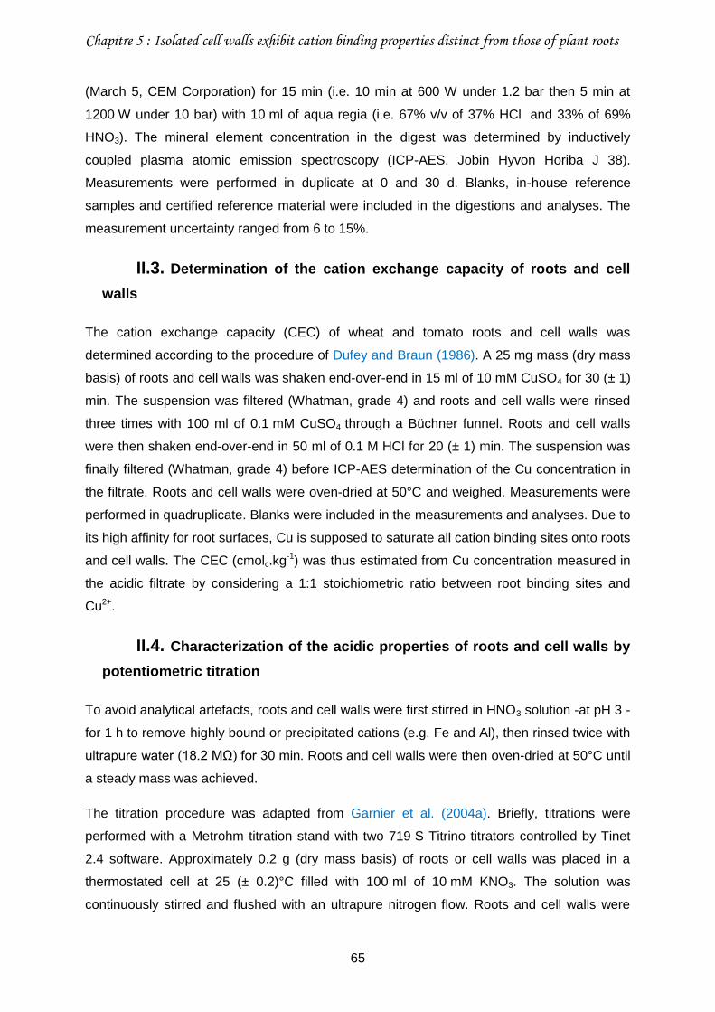
Chapitre 5 : Isolated cell walls exhibit cation binding properties distinct from those of plant roots
65
(March 5, CEM Corporation) for 15 min (i.e. 10 min at 600 W under 1.2 bar then 5 min at
1200 W under 10 bar) with 10 ml of aqua regia (i.e. 67% v/v of 37% HCl and 33% of 69%
HNO3). The mineral element concentration in the digest was determined by inductively
coupled plasma atomic emission spectroscopy (ICP-AES, Jobin Hyvon Horiba J 38).
Measurements were performed in duplicate at 0 and 30 d. Blanks, in-house reference
samples and certified reference material were included in the digestions and analyses. The
measurement uncertainty ranged from 6 to 15%.
II.3. Determination of the cation exchange capacity of roots and cell
walls
The cation exchange capacity (CEC) of wheat and tomato roots and cell walls was
determined according to the procedure of Dufey and Braun (1986). A 25 mg mass (dry mass
basis) of roots and cell walls was shaken end-over-end in 15 ml of 10 mM CuSO4 for 30 (± 1)
min. The suspension was filtered (Whatman, grade 4) and roots and cell walls were rinsed
three times with 100 ml of 0.1 mM CuSO4 through a Büchner funnel. Roots and cell walls
were then shaken end-over-end in 50 ml of 0.1 M HCl for 20 (± 1) min. The suspension was
finally filtered (Whatman, grade 4) before ICP-AES determination of the Cu concentration in
the filtrate. Roots and cell walls were oven-dried at 50°C and weighed. Measurements were
performed in quadruplicate. Blanks were included in the measurements and analyses. Due to
its high affinity for root surfaces, Cu is supposed to saturate all cation binding sites onto roots
and cell walls. The CEC (cmolc.kg-1) was thus estimated from Cu concentration measured in
the acidic filtrate by considering a 1:1 stoichiometric ratio between root binding sites and
Cu2+.
II.4. Characterization of the acidic properties of roots and cell walls by
potentiometric titration
To avoid analytical artefacts, roots and cell walls were first stirred in HNO3 solution -at pH 3 -
for 1 h to remove highly bound or precipitated cations (e.g. Fe and Al), then rinsed twice with
ultrapure water (18.2 MΩ) for 30 min. Roots and cell walls were then oven-dried at 50°C until
a steady mass was achieved.
The titration procedure was adapted from Garnier et al. (2004a). Briefly, titrations were
performed with a Metrohm titration stand with two 719 S Titrino titrators controlled by Tinet
2.4 software. Approximately 0.2 g (dry mass basis) of roots or cell walls was placed in a
thermostated cell at 25 (± 0.2)°C filled with 100 ml of 10 mM KNO3. The solution was
continuously stirred and flushed with an ultrapure nitrogen flow. Roots and cell walls were
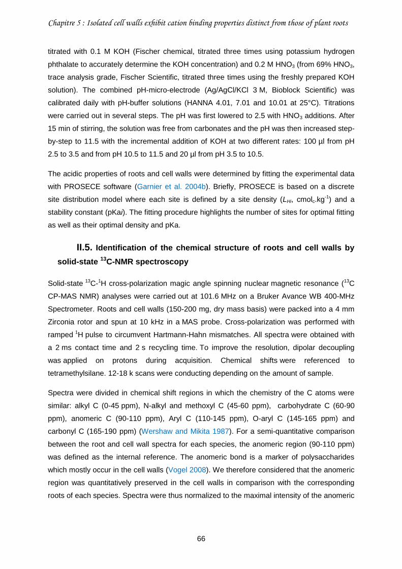
Chapitre 5 : Isolated cell walls exhibit cation binding properties distinct from those of plant roots
66
titrated with 0.1 M KOH (Fischer chemical, titrated three times using potassium hydrogen
phthalate to accurately determine the KOH concentration) and 0.2 M HNO3 (from 69% HNO3,
trace analysis grade, Fischer Scientific, titrated three times using the freshly prepared KOH
solution). The combined pH-micro-electrode (Ag/AgCl/KCl 3 M, Bioblock Scientific) was
calibrated daily with pH-buffer solutions (HANNA 4.01, 7.01 and 10.01 at 25°C). Titrations
were carried out in several steps. The pH was first lowered to 2.5 with HNO3 additions. After
15 min of stirring, the solution was free from carbonates and the pH was then increased step-
by-step to 11.5 with the incremental addition of KOH at two different rates: 100 µl from pH
2.5 to 3.5 and from pH 10.5 to 11.5 and 20 µl from pH 3.5 to 10.5.
The acidic properties of roots and cell walls were determined by fitting the experimental data
with PROSECE software (Garnier et al. 2004b). Briefly, PROSECE is based on a discrete
site distribution model where each site is defined by a site density (LHi, cmolc.kg-1) and a
stability constant (pKai). The fitting procedure highlights the number of sites for optimal fitting
as well as their optimal density and pKa.
II.5. Identification of the chemical structure of roots and cell walls by
solid-state 13C-NMR spectroscopy
Solid-state 13C-1H cross-polarization magic angle spinning nuclear magnetic resonance (13C
CP-MAS NMR) analyses were carried out at 101.6 MHz on a Bruker Avance WB 400-MHz
Spectrometer. Roots and cell walls (150-200 mg, dry mass basis) were packed into a 4 mm
Zirconia rotor and spun at 10 kHz in a MAS probe. Cross-polarization was performed with
ramped 1H pulse to circumvent Hartmann-Hahn mismatches. All spectra were obtained with
a 2 ms contact time and 2 s recycling time. To improve the resolution, dipolar decoupling
was applied on protons during acquisition. Chemical shifts were referenced to
tetramethylsilane. 12-18 k scans were conducting depending on the amount of sample.
Spectra were divided in chemical shift regions in which the chemistry of the C atoms were
similar: alkyl C (0-45 ppm), N-alkyl and methoxyl C (45-60 ppm), carbohydrate C (60-90
ppm), anomeric C (90-110 ppm), Aryl C (110-145 ppm), O-aryl C (145-165 ppm) and
carbonyl C (165-190 ppm) (Wershaw and Mikita 1987). For a semi-quantitative comparison
between the root and cell wall spectra for each species, the anomeric region (90-110 ppm)
was defined as the internal reference. The anomeric bond is a marker of polysaccharides
which mostly occur in the cell walls (Vogel 2008). We therefore considered that the anomeric
region was quantitatively preserved in the cell walls in comparison with the corresponding
roots of each species. Spectra were thus normalized to the maximal intensity of the anomeric
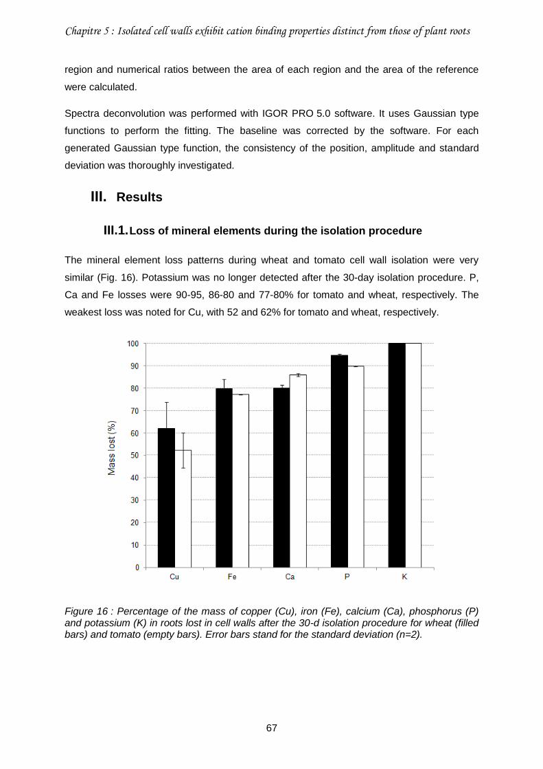
Chapitre 5 : Isolated cell walls exhibit cation binding properties distinct from those of plant roots
67
region and numerical ratios between the area of each region and the area of the reference
were calculated.
Spectra deconvolution was performed with IGOR PRO 5.0 software. It uses Gaussian type
functions to perform the fitting. The baseline was corrected by the software. For each
generated Gaussian type function, the consistency of the position, amplitude and standard
deviation was thoroughly investigated.
III. Results
III.1. Loss of mineral elements during the isolation procedure
The mineral element loss patterns during wheat and tomato cell wall isolation were very
similar (Fig. 16). Potassium was no longer detected after the 30-day isolation procedure. P,
Ca and Fe losses were 90-95, 86-80 and 77-80% for tomato and wheat, respectively. The
weakest loss was noted for Cu, with 52 and 62% for tomato and wheat, respectively.
Figure 16 : Percentage of the mass of copper (Cu), iron (Fe), calcium (Ca), phosphorus (P) and potassium (K) in roots lost in cell walls after the 30-d isolation procedure for wheat (filled bars) and tomato (empty bars). Error bars stand for the standard deviation (n=2).
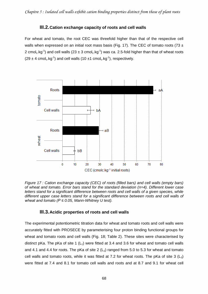
Chapitre 5 : Isolated cell walls exhibit cation binding properties distinct from those of plant roots
68
III.2. Cation exchange capacity of roots and cell walls
For wheat and tomato, the root CEC was threefold higher than that of the respective cell
walls when expressed on an initial root mass basis (Fig. 17). The CEC of tomato roots (73 ±
2 cmolc.kg-1) and cell walls (23 ± 3 cmolc.kg-1) was ca. 2.5-fold higher than that of wheat roots
(29 ± 4 cmolc.kg-1) and cell walls (10 ±1 cmolc.kg-1), respectively.
Figure 17 : Cation exchange capacity (CEC) of roots (filled bars) and cell walls (empty bars) of wheat and tomato. Error bars stand for the standard deviation (n=4). Different lower case letters stand for a significant difference between roots and cell walls of a given species, while different upper case letters stand for a significant difference between roots and cell walls of wheat and tomato (P ≤ 0.05, Mann-Whitney U test).
III.3. Acidic properties of roots and cell walls
The experimental potentiometric titration data for wheat and tomato roots and cell walls were
accurately fitted with PROSECE by parameterising four proton binding functional groups for
wheat and tomato roots and cell walls (Fig. 18; Table 2). These sites were characterised by
distinct pKa. The pKa of site 1 (L1) were fitted at 3.4 and 3.6 for wheat and tomato cell walls
and 4.1 and 4.4 for roots. The pKa of site 2 (L2) ranged from 5.0 to 5.3 for wheat and tomato
cell walls and tomato roots, while it was fitted at 7.2 for wheat roots. The pKa of site 3 (L3)
were fitted at 7.4 and 8.1 for tomato cell walls and roots and at 8.7 and 9.1 for wheat cell
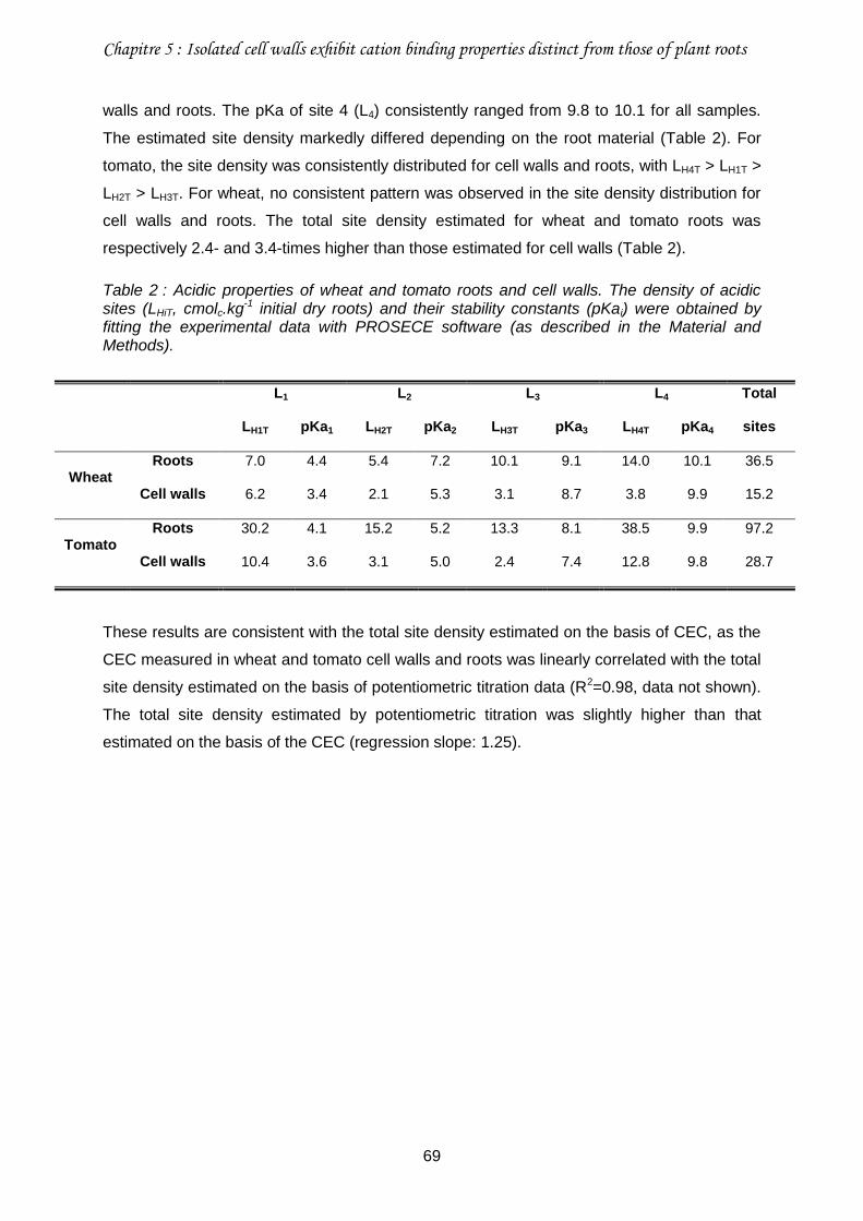
Chapitre 5 : Isolated cell walls exhibit cation binding properties distinct from those of plant roots
69
walls and roots. The pKa of site 4 (L4) consistently ranged from 9.8 to 10.1 for all samples.
The estimated site density markedly differed depending on the root material (Table 2). For
tomato, the site density was consistently distributed for cell walls and roots, with LH4T > LH1T >
LH2T > LH3T. For wheat, no consistent pattern was observed in the site density distribution for
cell walls and roots. The total site density estimated for wheat and tomato roots was
respectively 2.4- and 3.4-times higher than those estimated for cell walls (Table 2).
Table 2 : Acidic properties of wheat and tomato roots and cell walls. The density of acidic sites (LHiT, cmolc.kg-1 initial dry roots) and their stability constants (pKai) were obtained by fitting the experimental data with PROSECE software (as described in the Material and Methods).
L1 L2 L3 L4 Total
sites LH1T pKa1 LH2T pKa2 LH3T pKa3 LH4T pKa4
Wheat Roots 7.0 4.4 5.4 7.2 10.1 9.1 14.0 10.1 36.5
Cell walls 6.2 3.4 2.1 5.3 3.1 8.7 3.8 9.9 15.2
Tomato Roots 30.2 4.1 15.2 5.2 13.3 8.1 38.5 9.9 97.2
Cell walls 10.4 3.6 3.1 5.0 2.4 7.4 12.8 9.8 28.7
These results are consistent with the total site density estimated on the basis of CEC, as the
CEC measured in wheat and tomato cell walls and roots was linearly correlated with the total
site density estimated on the basis of potentiometric titration data (R2=0.98, data not shown).
The total site density estimated by potentiometric titration was slightly higher than that
estimated on the basis of the CEC (regression slope: 1.25).
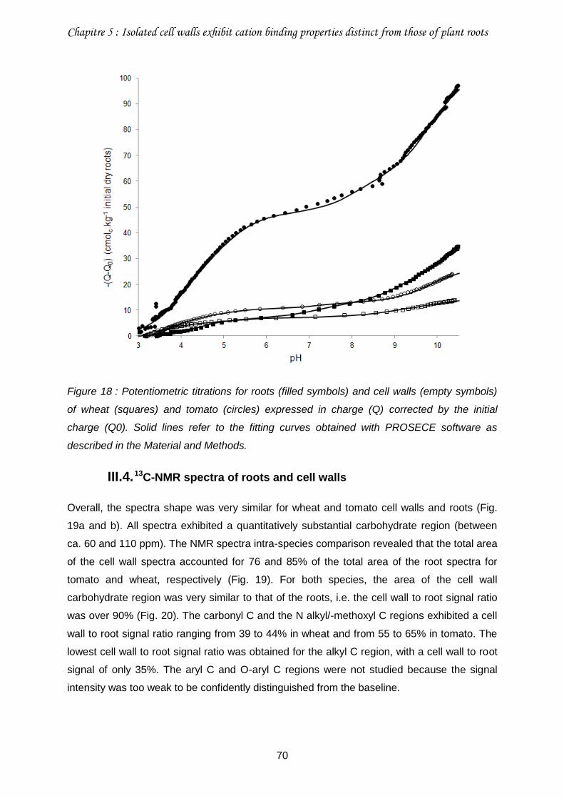
Chapitre 5 : Isolated cell walls exhibit cation binding properties distinct from those of plant roots
70
Figure 18 : Potentiometric titrations for roots (filled symbols) and cell walls (empty symbols)
of wheat (squares) and tomato (circles) expressed in charge (Q) corrected by the initial
charge (Q0). Solid lines refer to the fitting curves obtained with PROSECE software as
described in the Material and Methods.
III.4. 13C-NMR spectra of roots and cell walls
Overall, the spectra shape was very similar for wheat and tomato cell walls and roots (Fig.
19a and b). All spectra exhibited a quantitatively substantial carbohydrate region (between
ca. 60 and 110 ppm). The NMR spectra intra-species comparison revealed that the total area
of the cell wall spectra accounted for 76 and 85% of the total area of the root spectra for
tomato and wheat, respectively (Fig. 19). For both species, the area of the cell wall
carbohydrate region was very similar to that of the roots, i.e. the cell wall to root signal ratio
was over 90% (Fig. 20). The carbonyl C and the N alkyl/-methoxyl C regions exhibited a cell
wall to root signal ratio ranging from 39 to 44% in wheat and from 55 to 65% in tomato. The
lowest cell wall to root signal ratio was obtained for the alkyl C region, with a cell wall to root
signal of only 35%. The aryl C and O-aryl C regions were not studied because the signal
intensity was too weak to be confidently distinguished from the baseline.
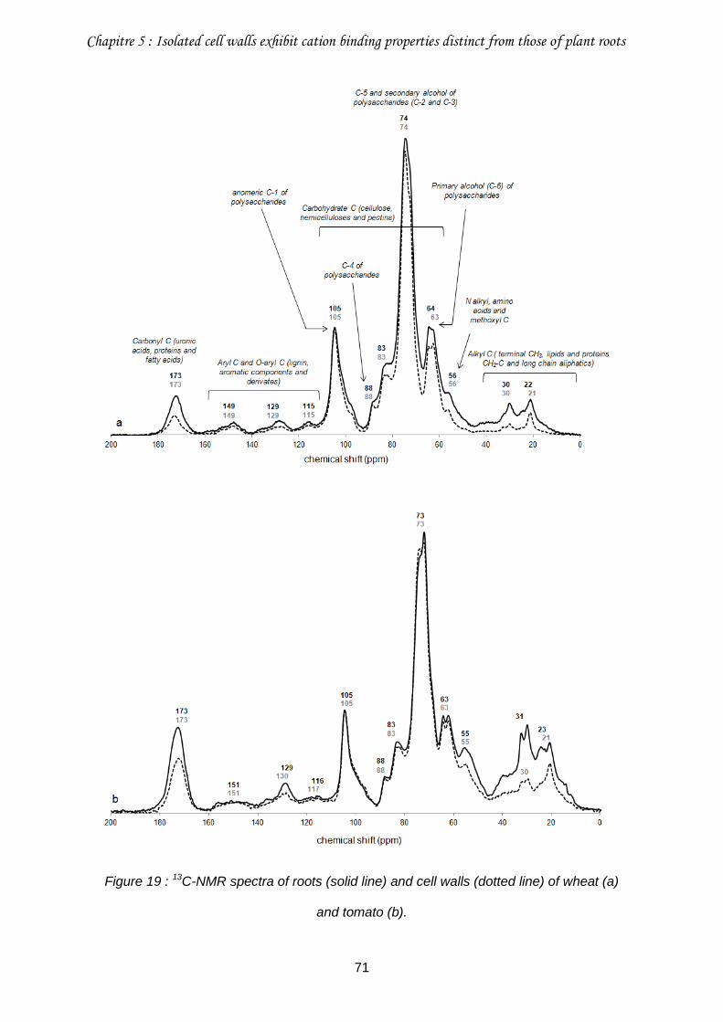
Chapitre 5 : Isolated cell walls exhibit cation binding properties distinct from those of plant roots
71
Figure 19 : 13C-NMR spectra of roots (solid line) and cell walls (dotted line) of wheat (a)
and tomato (b).
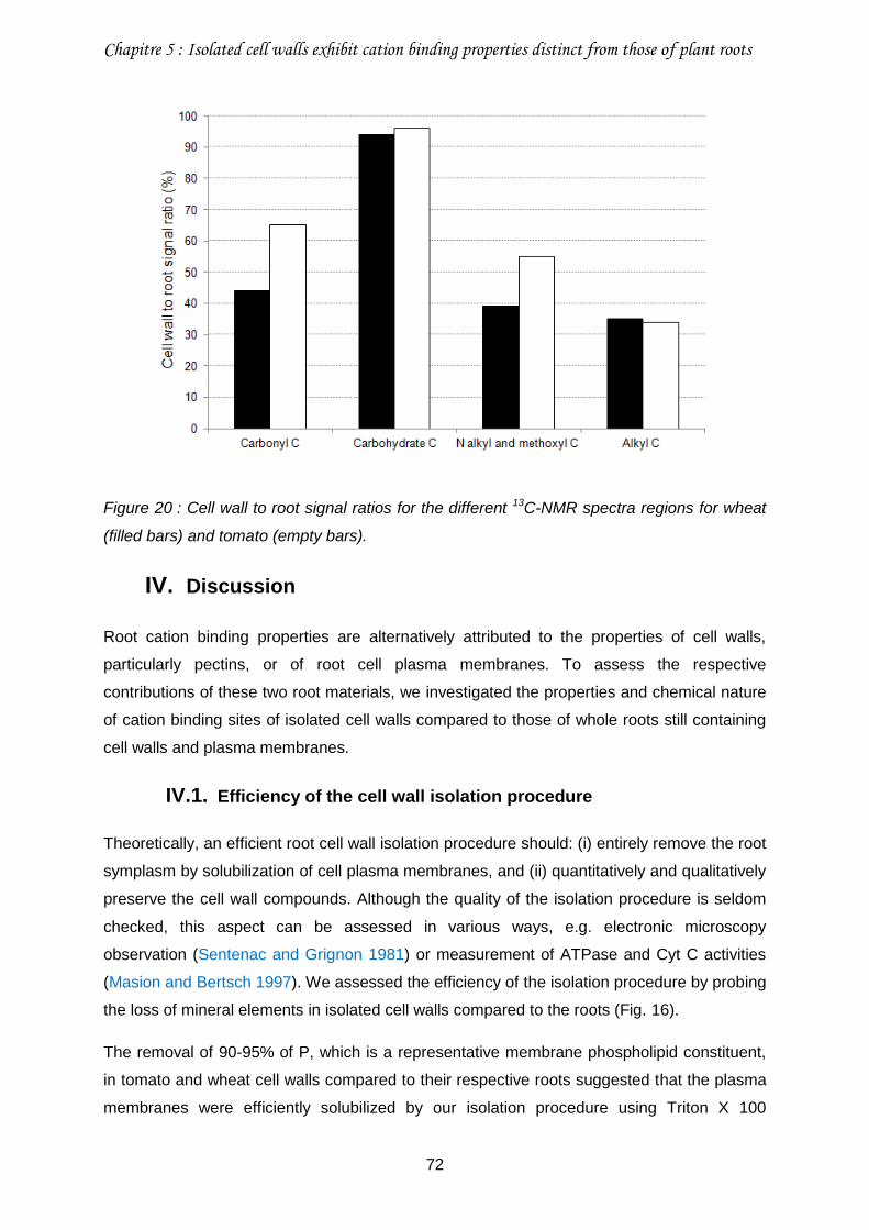
Chapitre 5 : Isolated cell walls exhibit cation binding properties distinct from those of plant roots
72
Figure 20 : Cell wall to root signal ratios for the different 13C-NMR spectra regions for wheat
(filled bars) and tomato (empty bars).
IV. Discussion
Root cation binding properties are alternatively attributed to the properties of cell walls,
particularly pectins, or of root cell plasma membranes. To assess the respective
contributions of these two root materials, we investigated the properties and chemical nature
of cation binding sites of isolated cell walls compared to those of whole roots still containing
cell walls and plasma membranes.
IV.1. Efficiency of the cell wall isolation procedure
Theoretically, an efficient root cell wall isolation procedure should: (i) entirely remove the root
symplasm by solubilization of cell plasma membranes, and (ii) quantitatively and qualitatively
preserve the cell wall compounds. Although the quality of the isolation procedure is seldom
checked, this aspect can be assessed in various ways, e.g. electronic microscopy
observation (Sentenac and Grignon 1981) or measurement of ATPase and Cyt C activities
(Masion and Bertsch 1997). We assessed the efficiency of the isolation procedure by probing
the loss of mineral elements in isolated cell walls compared to the roots (Fig. 16).
The removal of 90-95% of P, which is a representative membrane phospholipid constituent,
in tomato and wheat cell walls compared to their respective roots suggested that the plasma
membranes were efficiently solubilized by our isolation procedure using Triton X 100
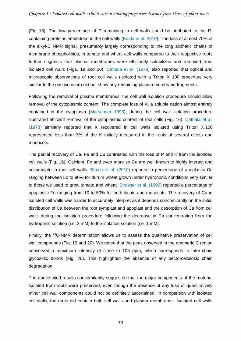
Chapitre 5 : Isolated cell walls exhibit cation binding properties distinct from those of plant roots
73
(Fig. 16). The low percentage of P remaining in cell walls could be attributed to the P-
containing proteins embedded in the cell walls (Kaida et al. 2010). The loss of almost 70% of
the alkyl-C NMR signal, presumably largely corresponding to the long aliphatic chains of
membrane phospholipids, in tomato and wheat cell walls compared to their respective roots
further suggests that plasma membranes were efficiently solubilized and removed from
isolated cell walls (Figs. 19 and 20). Cathala et al. (1978) also reported that optical and
microscopic observations of root cell walls (isolated with a Triton X 100 procedure very
similar to the one we used) did not show any remaining plasma membrane fragments.
Following the removal of plasma membranes, the cell wall isolation procedure should allow
removal of the cytoplasmic content. The complete loss of K, a soluble cation almost entirely
contained in the cytoplasm (Marschner 1995), during the cell wall isolation procedure
illustrated efficient removal of the cytoplasmic content of root cells (Fig. 16). Cathala et al.
(1978) similarly reported that K recovered in cell walls isolated using Triton X 100
represented less than 3% of the K initially measured in the roots of several dicots and
monocots.
The partial recovery of Ca, Fe and Cu contrasted with the loss of P and K from the isolated
cell walls (Fig. 16). Calcium, Fe and even more so Cu are well-known to highly interact and
accumulate in root cell walls. Bravin et al. (2010) reported a percentage of apoplastic Cu
ranging between 50 to 80% for durum wheat grown under hydroponic conditions very similar
to those we used to grow tomato and wheat. Strasser et al. (1999) reported a percentage of
apoplastic Fe ranging from 10 to 50% for both dicots and monocots. The recovery of Ca in
isolated cell walls was harder to accurately interpret as it depends concomitantly on the initial
distribution of Ca between the root symplast and apoplast and the desorption of Ca from cell
walls during the isolation procedure following the decrease in Ca concentration from the
hydroponic solution (i.e. 2 mM) to the isolation solution (i.e. 1 mM).
Finally, the 13C-NMR determination allows us to assess the qualitative preservation of cell
wall compounds (Fig. 19 and 20). We noted that the peak observed in the anomeric C region
conserved a maximum intensity of close to 105 ppm, which corresponds to inter-chain
glycosidic bonds (Fig. 20). This highlighted the absence of any pecto-cellulosic chain
degradation.
The above-cited results concomitantly suggested that the major components of the material
isolated from roots were preserved, even though the absence of any loss of quantitatively
minor cell wall components could not be definitely ascertained. In comparison with isolated
cell walls, the roots did contain both cell walls and plasma membranes. Isolated cell walls
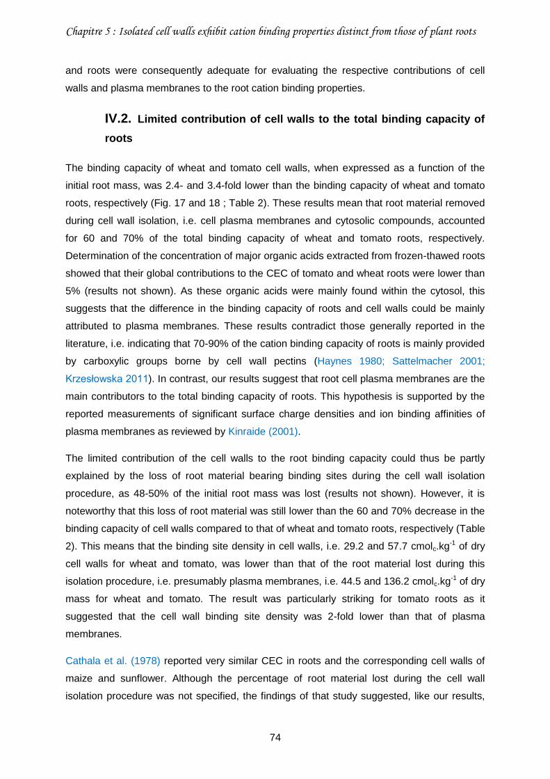
Chapitre 5 : Isolated cell walls exhibit cation binding properties distinct from those of plant roots
74
and roots were consequently adequate for evaluating the respective contributions of cell
walls and plasma membranes to the root cation binding properties.
IV.2. Limited contribution of cell walls to the total binding capacity of
roots
The binding capacity of wheat and tomato cell walls, when expressed as a function of the
initial root mass, was 2.4- and 3.4-fold lower than the binding capacity of wheat and tomato
roots, respectively (Fig. 17 and 18 ; Table 2). These results mean that root material removed
during cell wall isolation, i.e. cell plasma membranes and cytosolic compounds, accounted
for 60 and 70% of the total binding capacity of wheat and tomato roots, respectively.
Determination of the concentration of major organic acids extracted from frozen-thawed roots
showed that their global contributions to the CEC of tomato and wheat roots were lower than
5% (results not shown). As these organic acids were mainly found within the cytosol, this
suggests that the difference in the binding capacity of roots and cell walls could be mainly
attributed to plasma membranes. These results contradict those generally reported in the
literature, i.e. indicating that 70-90% of the cation binding capacity of roots is mainly provided
by carboxylic groups borne by cell wall pectins (Haynes 1980; Sattelmacher 2001;
Krzesłowska 2011). In contrast, our results suggest that root cell plasma membranes are the
main contributors to the total binding capacity of roots. This hypothesis is supported by the
reported measurements of significant surface charge densities and ion binding affinities of
plasma membranes as reviewed by Kinraide (2001).
The limited contribution of the cell walls to the root binding capacity could thus be partly
explained by the loss of root material bearing binding sites during the cell wall isolation
procedure, as 48-50% of the initial root mass was lost (results not shown). However, it is
noteworthy that this loss of root material was still lower than the 60 and 70% decrease in the
binding capacity of cell walls compared to that of wheat and tomato roots, respectively (Table
2). This means that the binding site density in cell walls, i.e. 29.2 and 57.7 cmolc.kg-1 of dry
cell walls for wheat and tomato, was lower than that of the root material lost during this
isolation procedure, i.e. presumably plasma membranes, i.e. 44.5 and 136.2 cmolc.kg-1 of dry
mass for wheat and tomato. The result was particularly striking for tomato roots as it
suggested that the cell wall binding site density was 2-fold lower than that of plasma
membranes.
Cathala et al. (1978) reported very similar CEC in roots and the corresponding cell walls of
maize and sunflower. Although the percentage of root material lost during the cell wall
isolation procedure was not specified, the findings of that study suggested, like our results,
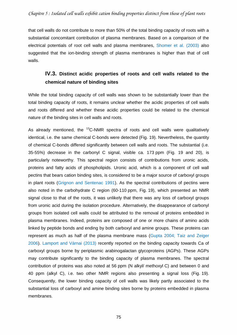
Chapitre 5 : Isolated cell walls exhibit cation binding properties distinct from those of plant roots
75
that cell walls do not contribute to more than 50% of the total binding capacity of roots with a
substantial concomitant contribution of plasma membranes. Based on a comparison of the
electrical potentials of root cell walls and plasma membranes, Shomer et al. (2003) also
suggested that the ion-binding strength of plasma membranes is higher than that of cell
walls.
IV.3. Distinct acidic properties of roots and cell walls related to the
chemical nature of binding sites
While the total binding capacity of cell walls was shown to be substantially lower than the
total binding capacity of roots, it remains unclear whether the acidic properties of cell walls
and roots differed and whether these acidic properties could be related to the chemical
nature of the binding sites in cell walls and roots.
As already mentioned, the 13C-NMR spectra of roots and cell walls were qualitatively
identical, i.e. the same chemical C-bonds were detected (Fig. 19). Nevertheless, the quantity
of chemical C-bonds differed significantly between cell walls and roots. The substantial (i.e.
35-55%) decrease in the carbonyl C signal, visible ca. 173 ppm (Fig. 19 and 20), is
particularly noteworthy. This spectral region consists of contributions from uronic acids,
proteins and fatty acids of phospholipids. Uronic acid, which is a component of cell wall
pectins that bears cation binding sites, is considered to be a major source of carboxyl groups
in plant roots (Grignon and Sentenac 1991). As the spectral contributions of pectins were
also noted in the carbohydrate C region (60-110 ppm, Fig. 19), which presented an NMR
signal close to that of the roots, it was unlikely that there was any loss of carboxyl groups
from uronic acid during the isolation procedure. Alternatively, the disappearance of carbonyl
groups from isolated cell walls could be attributed to the removal of proteins embedded in
plasma membranes. Indeed, proteins are composed of one or more chains of amino acids
linked by peptide bonds and ending by both carboxyl and amine groups. These proteins can
represent as much as half of the plasma membrane mass (Gupta 2004; Taiz and Zeiger
2006). Lamport and Várnai (2013) recently reported on the binding capacity towards Ca of
carboxyl groups borne by periplasmic arabinogalactan glycoproteins (AGPs). These AGPs
may contribute significantly to the binding capacity of plasma membranes. The spectral
contribution of proteins was also noted at 56 ppm (N alkyl/ methoxyl C) and between 0 and
40 ppm (alkyl C), i.e. two other NMR regions also presenting a signal loss (Fig. 19).
Consequently, the lower binding capacity of cell walls was likely partly associated to the
substantial loss of carboxyl and amine binding sites borne by proteins embedded in plasma
membranes.
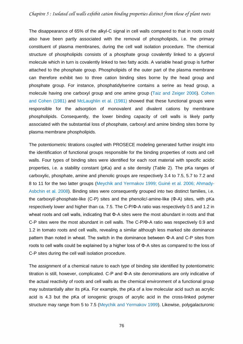
Chapitre 5 : Isolated cell walls exhibit cation binding properties distinct from those of plant roots
76
The disappearance of 65% of the alkyl-C signal in cell walls compared to that in roots could
also have been partly associated with the removal of phospholipids, i.e. the primary
constituent of plasma membranes, during the cell wall isolation procedure. The chemical
structure of phospholipids consists of a phosphate group covalently linked to a glycerol
molecule which in turn is covalently linked to two fatty acids. A variable head group is further
attached to the phosphate group. Phospholipids of the outer part of the plasma membrane
can therefore exhibit two to three cation binding sites borne by the head group and
phosphate group. For instance, phosphatidylserine contains a serine as head group, a
molecule having one carboxyl group and one amine group (Taiz and Zeiger 2006). Cohen
and Cohen (1981) and McLaughlin et al. (1981) showed that these functional groups were
responsible for the adsorption of monovalent and divalent cations by membrane
phospholipids. Consequently, the lower binding capacity of cell walls is likely partly
associated with the substantial loss of phosphate, carboxyl and amine binding sites borne by
plasma membrane phospholipids.
The potentiometric titrations coupled with PROSECE modeling generated further insight into
the identification of functional groups responsible for the binding properties of roots and cell
walls. Four types of binding sites were identified for each root material with specific acidic
properties, i.e. a stability constant (pKa) and a site density (Table 2). The pKa ranges of
carboxylic, phosphate, amine and phenolic groups are respectively 3.4 to 7.5, 5.7 to 7.2 and
8 to 11 for the two latter groups (Meychik and Yermakov 1999; Guiné et al. 2006; Ahmady-
Asbchin et al. 2008). Binding sites were consequently grouped into two distinct families, i.e.
the carboxyl/-phosphate-like (C-P) sites and the phenolic/-amine-like (Φ-A) sites, with pKa
respectively lower and higher than ca. 7.5. The C-P/Φ-A ratio was respectively 0.5 and 1.2 in
wheat roots and cell walls, indicating that Φ-A sites were the most abundant in roots and that
C-P sites were the most abundant in cell walls. The C-P/Φ-A ratio was respectively 0.9 and
1.2 in tomato roots and cell walls, revealing a similar although less marked site dominance
pattern than noted in wheat. The switch in the dominance between Φ-A and C-P sites from
roots to cell walls could be explained by a higher loss of Φ-A sites as compared to the loss of
C-P sites during the cell wall isolation procedure.
The assignment of a chemical nature to each type of binding site identified by potentiometric
titration is still, however, complicated. C-P and Φ-A site denominations are only indicative of
the actual reactivity of roots and cell walls as the chemical environment of a functional group
may substantially alter its pKa. For example, the pKa of a low molecular acid such as acrylic
acid is 4.3 but the pKa of ionogenic groups of acrylic acid in the cross-linked polymer
structure may range from 5 to 7.5 (Meychik and Yermakov 1999). Likewise, polygalacturonic
Chapitre 5 : Isolated cell walls exhibit cation binding properties distinct from those of plant roots
77
acid carboxylic groups may have a pKa in the range of that of Φ-A sites (Lenoble et al.
2008). Accurate knowledge on the chemical structure is thus necessary. The NMR signal
loss effectively reflected the decrease in the total site density observed by both
potentiometric titration and CEC measurement. However, due to the substantial chemical
heterogeneity in the roots and cell walls, the NMR spectra did not generate more accurate
results or highlight a straightforward link between the identified functional groups and the
pKa.
V. Conclusion
While, based on previous reports, it is often commonly considered that root binding
properties are mainly dependent on the cell wall composition, our results conversely suggest
that cell walls does not contribute to more than 50% of the total binding capacity of roots.
Because of the apparent efficiency of the cell wall isolation procedure in removing plasma
membranes, we noted that binding sites borne by plasma membranes contributed markedly
to the total binding capacity of roots.
The distinct binding properties of isolated cell walls and roots should no longer be neglected
and should be considered carefully in studies focused on root-ion interactions. Cell walls and
plasma membranes should be separated especially to gain insight into processes taking
place within cell walls, such as root elongation mechanisms. Conversely, cell walls and
plasma membranes have to be seen as a continuum in the overall issue of rhizotoxicity
because of their respective contributions in the binding properties of roots. From a more
operational standpoint, the biotic ligand model will be more effective if cell walls and plasma
membranes are considered as separate cation binding sites. Further in-depth studies are
nevertheless required to investigate the binding of trace elements by each type of root
material in order to identify the extents of participation of their respective binding sites.
Acknowledgements
The authors are grateful to French Environment and Energy Management Agency (ADEME)
and the French Centre of Agricultural Research for Development (CIRAD) for funding the
PhD scholarship of Stéphanie Guigues and INSU (CNRS) for funding the study via the
EC2CO-CYTRIX call. The authors thank Patrick Cazevieille and Claire Chevassus-Rosset
(CIRAD) for their technical support during the plant growth phase, Hélène Miche (CEREGE)
for providing access to ICP-AES and Jean-Claude Davidian (Montpellier SupAgro) for his
advice on root cell wall isolation.
Chapitre 5 : Isolated cell walls exhibit cation binding properties distinct from those of plant roots
78
Références bibliographiques
Ahmady-Asbchin S, Andrès Y, Gérente C, Cloirec PL (2008) Biosorption of Cu(II) from aqueous solution by Fucus serratus: Surface characterization and sorption mechanisms. Bioresource Technology 99 (14):6150-6155. doi:10.1016/j.biortech.2007.12.040
Allan DL, Jarrell WM (1989) Proton and Copper Adsorption to Maize and Soybean Root Cell
Walls. Plant Physiol 89 (3):823-832. doi:10.1104/pp.89.3.823 Bastías E, Alcaraz-López C, Bonilla I, Martínez-Ballesta MC, Bolaños L, Carvajal M (2010)
Interactions between salinity and boron toxicity in tomato plants involve apoplastic calcium. Journal of Plant Physiology 167 (1):54-60. doi:http://dx.doi.org/10.1016/j.jplph.2009.07.014
Bravin M, Merrer B, Denaix L, Schneider A, Hinsinger P (2010) Copper uptake kinetics in
hydroponically-grown durum wheat (Triticum turgidum durum L.) as compared with soil’s ability to supply copper. Plant and Soil 331 (1-2):91-104. doi:10.1007/s11104-009-0235-3
Brunner I, Luster J, Günthardt-Goerg MS, Frey B (2008) Heavy metal accumulation and
phytostabilisation potential of tree fine roots in a contaminated soil. Environmental Pollution 152 (3):559-568. doi:DOI: 10.1016/j.envpol.2007.07.006
Cathala N, Ghorbal, M. H., Lamant, A., Salsac, L. (1978) Obtention de parois cellulosiques à
l'aide d'un détergent : étude préliminaire de leur composition minérale. C R Acad Sc Paris t. 286:1025-1027
Cohen JA, Cohen M (1981) Adsorption of monovalent and divalent cations by phospholipid
membranes. The monomer-dimer problem. Biophysical journal 36 (3):623-651 Crooke WM, Knight AH, Macdonald IR (1960) Cation exchange properties and pectin content
of storage-tissue disks. Plant and Soil 13 (1):55-67. doi:10.1007/bf01666476 Dœlsch E, Deroche B, Van de Kerchove V (2006b) Impact of sewage sludge spreading on
heavy metal speciation in tropical soils (Réunion, Indian Ocean). Chemosphere 65 (2):286-293. doi:http://dx.doi.org/10.1016/j.chemosphere.2006.02.046
Doelsch E, Van de Kerchove V, Saint Macary H (2006a) Heavy metal content in soils of
Réunion (Indian Ocean). Geoderma 134 (1–2):119-134. doi:http://dx.doi.org/10.1016/j.geoderma.2005.09.003
Dufey JE, Braun R (1986) Cation Exchange Capacity of Roots : Tirtation Sum of
Exchangeable Cations, Copper Adsorption. Journal of Plant Nutrition 9 (8):1147-1155 Garnier C, Mounier S, Benaïm JY (2004a) Influence of dissolved organic carbon content on
modelling natural organic matter acid–base properties. Water Research 38 (17):3685-3692. doi:http://dx.doi.org/10.1016/j.watres.2004.05.019
Garnier C, Pižeta I, Mounier S, Benaım JY, Branica M (2004b) Influence of the type of
titration and of data treatment methods on metal complexing parameters determination of single and multi-ligand systems measured by stripping voltammetry. Analytica Chimica Acta 505 (2):263-275. doi:http://dx.doi.org/10.1016/j.aca.2003.10.066
Chapitre 5 : Isolated cell walls exhibit cation binding properties distinct from those of plant roots
79
Grignon C, Sentenac H (1991) pH and Ionic Conditions in the Apoplast. Annual Review of Plant Physiology and Plant Molecular Biology 42 (1):103-128. doi:doi:10.1146/annurev.pp.42.060191.000535
Guiné V, Spadini L, Sarret G, Muris M, Delolme C, Gaudet JP, Martins JMF (2006) Zinc
Sorption to Three Gram-Negative Bacteria: Combined Titration, Modeling, and EXAFS Study. Environmental Science & Technology 40 (6):1806-1813. doi:10.1021/es050981l
Gupta GP (2004) Plant Cell Biology. Discovery Publishing House, New Delhi Haynes RJ (1980) Ion exchange properties of roots and ionic interactions within the root
apoplasm: Their role in ion accumulation by plants. Bot Rev 46 (1):75-99. doi:10.1007/bf02860867
Heintze SG (1961) Studies on cation-exchange capacities of roots. Plant and Soil 13 (4):365-
383. doi:10.1007/bf01394648 Herrmann AM, Ritz K, Nunan N, Clode PL, Pett-Ridge J, Kilburn MR, Murphy DV, O’Donnell
AG, Stockdale EA (2007) Nano-scale secondary ion mass spectrometry — A new analytical tool in biogeochemistry and soil ecology: A review article. Soil Biology and Biochemistry 39 (8):1835-1850. doi:http://dx.doi.org/10.1016/j.soilbio.2007.03.011
Horst WJ, Wang Y, Eticha D (2010) The role of the root apoplast in aluminium-induced
inhibition of root elongation and in aluminium resistance of plants: a review. Annals of Botany 106 (1):185-197. doi:10.1093/aob/mcq053
Kaida R, Serada S, Norioka N, Norioka S, Neumetzler L, Pauly M, Sampedro J, Zarra I,
Hayashi T, Kaneko TS (2010) Potential Role for Purple Acid Phosphatase in the Dephosphorylation of Wall Proteins in Tobacco Cells. Plant Physiology 153 (2):603-610. doi:10.1104/pp.110.154138
Kinraide TB (2001) Ion fluxes considered in terms of membrane-surface electrical potentials.
Australian Journal of Plant Physiology 28 (7):605-616. doi:10.1071/pp01019 Kinraide TB (2004) Possible Influence of Cell Walls upon Ion Concentrations at Plasma
Membrane Surfaces. Toward a Comprehensive View of Cell-Surface Electrical Effects upon Ion Uptake, Intoxication, and Amelioration. Plant Physiol 136 (3):3804-3813. doi:10.1104/pp.104.043174
Kinraide TB (2006) Plasma membrane surface potential (ψpm) as a determinant of ion
bioavailability: A critical analysis of new and published toxicological studies and a simplified method for the computation of plant ψpm. Environmental Toxicology and Chemistry 25 (12):3188-3198. doi:10.1897/06-103r.1
Kinraide TB, Ryan PR, Kochian LV (1992) Interactive Effects of Al3+, H+, and Other Cations
on Root Elongation Considered in Terms of Cell-Surface Electrical Potential. Plant Physiol 99 (4):1461-1468. doi:10.1104/pp.99.4.1461
Kopittke P, Blamey F, Menzies N (2008) Toxicities of soluble Al, Cu, and La include ruptures
to rhizodermal and root cortical cells of cowpea. Plant and Soil 303 (1):217-227. doi:10.1007/s11104-007-9500-5
Chapitre 5 : Isolated cell walls exhibit cation binding properties distinct from those of plant roots
80
Kopittke P, McKenna B, Blamey F, Wehr J, Menzies N (2009a) Metal-induced cell rupture in elongating roots is associated with metal ion binding strengths. Plant and Soil 322 (1):303-315. doi:10.1007/s11104-009-9917-0
Kopittke P, Menzies N (2006) Effect of Cu Toxicity on Growth of Cowpea (<i>Vigna
unguiculata</i>). Plant and Soil 279 (1):287-296. doi:10.1007/s11104-005-1578-z
Kopittke PM, Asher CJ, Blamey FPC, Menzies NW (2009b) Toxic effects of Cu2+ on growth,
nutrition, root morphology, and distribution of Cu in roots of Sabi grass. Science of The Total Environment 407 (16):4616-4621. doi:10.1016/j.scitotenv.2009.04.041
Kopittke PM, Asher CJ, Kopittke RA, Menzies NW (2007) Toxic effects of Pb2+ on growth of
cowpea (Vigna unguiculata). Environmental Pollution 150 (2):280-287. doi:10.1016/j.envpol.2007.01.011
Kopittke PM, Kinraide TB, Wang P, Blamey FPC, Reichman SM, Menzies NW (2011)
Alleviation of Cu and Pb Rhizotoxicities in Cowpea (Vigna unguiculata) as Related to Ion Activities at Root-Cell Plasma Membrane Surface. Environmental Science & Technology 45 (11):4966-4973. doi:10.1021/es1041404
Krzesłowska M (2011) The cell wall in plant cell response to trace metals: polysaccharide
remodeling and its role in defense strategy. Acta Physiologiae Plantarum 33 (1):35-51. doi:10.1007/s11738-010-0581-z
Kudo H, Kudo K, Ambo H, Uemura M, Kawai S (2011) Cadmium sorption to plasma
membrane isolated from barley roots is impeded by copper association onto membranes. Plant Science 180 (2):300-305. doi:http://dx.doi.org/10.1016/j.plantsci.2010.09.008
Lamport DTA, Várnai P (2013) Periplasmic arabinogalactan glycoproteins act as a calcium
capacitor that regulates plant growth and development. New Phytologist 197 (1):58-64. doi:10.1111/nph.12005
Legros S, Doelsch E, Feder F, Moussard G, Sansoulet J, Gaudet JP, Rigaud S, Doelsch IB,
Macary HS, Bottero JY (2013) Fate and behaviour of Cu and Zn from pig slurry spreading in a tropical water–soil–plant system. Agriculture, Ecosystems & Environment 164 (0):70-79. doi:http://dx.doi.org/10.1016/j.agee.2012.09.008
Lenoble V, Garnier C, Masion A, Ziarelli F, Garnier JM (2008) Combination of 13C/113Cd
NMR, potentiometry, and voltammetry in characterizing the interactions between Cd and two models of the main components of soil organic matter. Analytical and Bioanalytical Chemistry 390 (2):749-757. doi:10.1007/s00216-007-1678-0
Liu D, Kottke I (2003) Subcellular localization of Cd in the root cells ofAllium sativum by
electron energy loss spectroscopy. J Biosci 28 (4):471-478. doi:10.1007/bf02705121 Marschner H (1995) Mineral Nutrition of higher plants. Academic Press, second edition Masion A, Bertsch PM (1997) Aluminium speciation in the presence of wheat root cell walls:
a wet chemical study. Plant, Cell & Environment 20 (4):504-512. doi:10.1046/j.1365-3040.1997.d01-86.x
Chapitre 5 : Isolated cell walls exhibit cation binding properties distinct from those of plant roots
81
McLaughlin S, Mulrine N, Gresalfi T, Vaio G, McLaughlin A (1981) Adsorption of divalent cations to bilayer membranes containing phosphatidylserine. The Journal of General Physiology 77 (4):445-473. doi:10.1085/jgp.77.4.445
Meychik NR, Yermakov IP (1999) A new approach to the investigation on the tonogenic
groups of root cell walls. Plant and Soil 217 (1):257-264. doi:10.1023/a:1004675309128
Meychik NR, Yermakov IP (2001) Ion exchange properties of plant root cell walls. Plant and
Soil 234 (2):181-193. doi:10.1023/a:1017936318435 Moore KL, Schröder M, Wu Z, Martin BGH, Hawes CR, McGrath SP, Hawkesford MJ, Feng
Ma J, Zhao F-J, Grovenor CRM (2011) High-Resolution Secondary Ion Mass Spectrometry Reveals the Contrasting Subcellular Distribution of Arsenic and Silicon in Rice Roots. Plant Physiology 156 (2):913-924. doi:10.1104/pp.111.173088
Nagajyoti P, Lee K, Sreekanth T (2010) Heavy metals, occurrence and toxicity for plants: a
review. Environmental Chemistry Letters 8 (3):199-216. doi:10.1007/s10311-010-0297-8
Postma JWM, Keltjens WG, van Riemsdijk WH (2005) Calcium-(Organo)aluminum-proton
Competition for Adsorption to Tomato Root Cell Walls: Experimental Data and Exchange Model Calculations. Environmental Science & Technology 39 (14):5247-5254. doi:10.1021/es048138v
Ram LC (1980) Cation exchange capacity of plant roots in relation to nutrients uptake by
shoot and grain as influenced by age. Plant and Soil 55 (2):215-224. doi:10.1007/bf02181801
Reid RJ (2001) Mechanisms of micronutrient uptake in plants. Functional Plant Biology 28
(7):661-668. doi:http://dx.doi.org/10.1071/PP01037 Sarkar P, Bosneaga E, Auer M (2009) Plant cell walls throughout evolution: towards a
molecular understanding of their design principles. Journal of Experimental Botany 60 (13):3615-3635. doi:10.1093/jxb/erp245
Sattelmacher B (2001) The apoplast and its significance for plant mineral nutrition. New
Phytologist 149 (2):167-192. doi:10.1046/j.1469-8137.2001.00034.x Sentenac H, Grignon C (1981) A Model for Predicting Ionic Equilibrium Concentrations in
Cell Walls. Plant Physiol 68:415-419 Sheldon A, Menzies N (2005) The Effect of Copper Toxicity on the Growth and Root
Morphology of Rhodes Grass (<i>Chloris gayana</i> Knuth.) in Resin Buffered Solution Culture. Plant and Soil 278 (1):341-349. doi:10.1007/s11104-005-8815-3
Shomer I, Novacky AJ, Pike SM, Yermiyahu U, Kinraide TB (2003) Electrical Potentials of
Plant Cell Walls in Response to the Ionic Environment. Plant Physiology 133 (1):411-422. doi:10.1104/pp.103.024539
Smart KE, Smith JAC, Kilburn MR, Martin BGH, Hawes C, Grovenor CRM (2010) High-
resolution elemental localization in vacuolate plant cells by nanoscale secondary ion mass spectrometry. The Plant Journal 63 (5):870-879. doi:10.1111/j.1365-313X.2010.04279.x
Chapitre 5 : Isolated cell walls exhibit cation binding properties distinct from those of plant roots
82
Straczek A, Sarret G, Manceau A, Hinsinger P, Geoffroy N, Jaillard B (2008) Zinc distribution and speciation in roots of various genotypes of tobacco exposed to Zn. Environmental and Experimental Botany 63 (1-3):80-90. doi:DOI: 10.1016/j.envexpbot.2007.10.034
Strasser O, Köhl K, Römheld V (1999) Overestimation of apoplastic Fe in roots of soil grown
plants. Plant and Soil 210 (2):179-189. doi:10.1023/a:1004650506592 Taiz L, Zeiger E (2006) Plant Physiology, 4th Edition. Sinauer Associates Sunderland, MA Thakali S, Allen HE, Di Toro DM, Ponizovsky AA, Rooney CP, Zhao F-J, McGrath SP (2006)
A Terrestrial Biotic Ligand Model. 1. Development and Application to Cu and Ni Toxicities to Barley Root Elongation in Soils. Environmental Science & Technology 40 (22):7085-7093. doi:10.1021/es061171s
Vogel J (2008) Unique aspects of the grass cell wall. Current Opinion in Plant Biology 11
(3):301-307. doi:http://dx.doi.org/10.1016/j.pbi.2008.03.002 Vulkan R, Yermiyahu U, Mingelgrin U, Rytwo G, Kinraide TB (2004) Sorption of Copper and
Zinc to the Plasma Membrane of Wheat Root. Journal of Membrane Biology 202 (2):97-104. doi:10.1007/s00232-004-0722-7
Wang P, Kinraide TB, Zhou D, Kopittke PM, Peijnenburg WJGM (2011) Plasma Membrane
Surface Potential: Dual Effects upon Ion Uptake and Toxicity. Plant Physiol 155 (2):808-820. doi:10.1104/pp.110.165985
Wershaw R, Mikita M (1987) NMR of humic substances and coal. Lewis Publishers, Chelsea,
MI Yang Z-B, Eticha D, Rao IM, Horst WJ (2010) Alteration of cell-wall porosity is involved in
osmotic stress-induced enhancement of aluminium resistance in common bean (Phaseolus vulgaris L.). Journal of Experimental Botany 61 (12):3245-3258. doi:10.1093/jxb/erq146
Zhang Q, Smith A, Sekimoto H, Reid R (2001) Effect of membrane surface charge on nickel
uptake by purified mung bean root protoplasts. Planta 213 (5):788-793. doi:10.1007/s004250100555
Zhou H, Zeng M, Zhou X, Liao B-H, Liu J, Lei M, Zhong Q-Y, Zeng H (2013) Assessment of
heavy metal contamination and bioaccumulation in soybean plants from mining and smelting areas of southern Hunan Province, China. Environmental Toxicology and Chemistry 32 (12):2719-2727. doi:10.1002/etc.2389

83

84
Chapitre 6 : Implication of amino acids in high-affinity copper complexation in the apoplast of tomato and wheat roots : spectroscopic and thermodynamic evidences
85
Chapitre 6 : Rôle des acides aminés dans la
complexation du cuivre au sein du continuum
parois apoplasmiques – membranes plasmiques
Dans le chapitre précédent (chapitre 5), nous avons vu que les parois apoplasmiques et les
membranes plasmiques présentent toutes deux des propriétés de complexation vis-à-vis des
cations métalliques, les membranes plasmiques portant d’ailleurs une densité de sites
complexant plus élevée que les parois apoplasmiques. Néanmoins, l’induction par les
cations métalliques d’une rhizotoxicité est généralement associée à leur complexation par
les groupements carboxyliques portés par les pectines. Le cuivre fait cependant partie des
cations métalliques qui présentent une rhizotoxicité particulièrement élevée, au-delà de ce
qui est prédit par l’affinité du cuivre pour les groupements carboxyliques (Kopittke et al.
2014). Cela suggère que le cuivre pourrait être complexé par d’autres groupements réactifs
des parois apoplasmiques.
L’article Implication of amino acids in high-affinity copper complexation in the apoplast of
tomato and wheat roots: spectroscopic and thermodynamic evidences, constituant ce
chapitre 6, est une étude de la complexation du cuivre au sein du compartiment
extracellulaire racinaire à savoir le continuum parois apoplasmiques – membranes
plasmiques. Comme dans le chapitre précédent (chapitre 5), les contributions respectives
des parois apoplasmiques et des membranes plasmiques sont déduites de la comparaison
parois apoplasmiques isolées – racines sèches.
Contrairement aux résultats attendus d’après les conclusions du chapitre 5, les parois
apoplasmiques semblent contribuer au moins autant, voire plus (entre 50 et 60% du cuivre
total complexé), que les membranes plasmiques à la complexation du cuivre sur les surfaces
racinaires. En croisant différentes techniques analytiques (spectroscopie d’absorption des
rayons X et RMN 13C) avec la modélisation de la réactivité des racines de blé et de tomate
vis-à-vis du cuivre, nous avons mis en évidence une spéciation du cuivre partagée, de façon
presque égale, entre des ligands oxygènes présentant une faible affinité, et des ligands
oxygène / azote présentant une affinité élevée. Ces deux familles de ligands traduisent
respectivement la contribution des groupements carboxyliques et des acides aminés portés
à la fois au niveau des parois apoplasmiques et des membranes plasmiques.
Chapitre 6 : Implication of amino acids in high-affinity copper complexation in the apoplast of tomato and wheat roots : spectroscopic and thermodynamic evidences
86
Chapitre 6 : Implication of amino acids in high-affinity copper complexation in the apoplast of tomato and wheat roots : spectroscopic and thermodynamic evidences
87
Implication of amino acids in high-affinity copper complexation in the apoplast
of tomato and wheat roots : spectroscopic and thermodynamic evidences
Stéphanie Guigues 1,2
, Matthieu N. Bravin 3, Cédric Garnier
4, Armand Masion
5, Claire
Chevassus-Rosset 1, Patrick Cazevieille
1 and Emmanuel Doelsch
1
1 CIRAD, UPR Recyclage et risque, F-34398 Montpellier, France
2 ADEME, 20 avenue du Grésillé, BP-90406, Angers cedex 01, France
3 CIRAD, UPR Recyclage et risque, F-97408 Saint-Denis, Réunion, France
4 Université de Toulon, PROTEE, EA 3819, 83957 La Garde, France
5 Aix-Marseille Université, CNRS, IRD, CEREGE UM34, 13545 Aix-en-Provence, France
Abstract
Carboxylic groups located in plant cell walls (CW) are generally considered as the main
copper binding sites in plant roots, despite the presence of other functional groups which
contribute to root binding properties. The aim of this study was to investigate sites
responsible of copper binding in root apoplast i.e. the cell walls – outer surface of plasma
membranes (PM) continuum. The apoplastic localization of binding sites was investigated by
comparing isolated CW of a monocotyledon (Triticum aestivum L.) and dicotyledon (Solanum
lycopersicum L.) with their respective whole roots. Copper speciation was examined by X-ray
absorption and 13C-nuclar magnetic resonance spectroscopies while the affinity of ligands
involved in copper binding was investigated by modeling copper sorption isotherms.
Homogeneous speciation of copper was found in root apoplast. Nitrogen/oxygen ligands
associated to CW and PM amino acids were identified in slightly higher proportion than
single oxygen ligand assimilated to CW polysaccharides. Furthermore, low- and high-affinity
binding sites almost contributed in equivalent proportion to copper complexation in the root
apoplast of wheat and tomato. Jointly to CW polysaccharides, amino acids embedded in CW
and PM participate in copper binding on roots.
Keywords: cell walls; plasma membrane; trace metal; modeling; binding site; X-ray
absorption spectroscopy
Chapitre 6 : Implication of amino acids in high-affinity copper complexation in the apoplast of tomato and wheat roots : spectroscopic and thermodynamic evidences
88
I. Introduction
As seven other micronutrients, copper (Cu) is essential for the development of plants
(Marschner 1995). Copper is a redox-active transition metal that exists as Cu2+ and Cu+
depending on physiological conditions. It participates in many biological processes such as
photosynthesis, mitochondrial respiration, perception of ethylene and cell wall (CW)
remodeling. The optimum range of Cu concentration in plant is narrow, as deficiency and
toxicity are usually expected below 5 and above 20 µg.g-1 dry weight in shoots respectively.
Consequently, plants must regulate finely Cu uptake and homeostasis (Burkhead 2009). The
substantial and repetitive inputs of Cu on agricultural areas (fungicides, organic waste
spreading, etc.) associated with the low mobility of Cu in soil lead to large-scale Cu
contamination of soils that questions the potential occurrence of Cu phytotoxicity (Belon et al.
2012). Due to their location within soil, roots are primarily exposed to Cu contamination and
are thus known to be particularly sensitive to Cu rhizotoxicity. Indeed, Cu(II) (hereafter
referred as to Cu) was found to be the third most rhizotoxic metal cation out of 26 (Kopittke et
al. 2011a).
Kopittke et al. (2014) suggested that the minimum rhizotoxicity of metal cations, including Cu,
would mainly be based on a non-specific mechanism related to the binding strength of metal
cations to hard ligands (e.g. carboxyl, phosphate, carbonyl, phenolic and amine groups) in
the CW, especially to the carboxylic groups of galacturonic acids located within the pectic
matrix. As other metal cations showing a large affinity for hard ligands, Cu accumulates
almost entirely within the rhizodermis and the outer cortex of roots (Kopittke et al. 2011c;
Collin et al. 2014) and is mainly located in the apoplastic compartment (Bravin et al. 2010;
Colzi et al. 2011). Measurements in X-ray absorption spectroscopy (XAS) and pectinase
treatment independently suggest that Cu was mainly bound to polygalacturonate-like groups
located in CW (Konno et al. 2005; Kopittke et al. 2011c). Taken together, these experimental
considerations support the hypothesis of an increased rigidity of CW of outer root tissues due
to the substantial and strong binding of Cu to pectins, which results in a slower elongation
rate of outer cells compared to the cells located in the stele and inner cortex and finally to the
appearance of ruptures in the rhizodermis and the outer cortex of roots. However, Cu
belongs to the four metal cations exhibiting “an unexpectedly high rhizotoxicity”, i.e. a
rhizotoxicity higher than that estimated from the binding strength of Cu to the hard ligand
scale (HLScale) mainly represented by carboxyl groups, that requires further investigation
(Kopittke et al. 2014). Reexamining this theory involving hard ligands, we intended to
investigate the speciation and the sorption of Cu in root apoplast to evaluate the two
following hypotheses: (i) Cu ligands such as phosphate and amine groups in CW and the
Chapitre 6 : Implication of amino acids in high-affinity copper complexation in the apoplast of tomato and wheat roots : spectroscopic and thermodynamic evidences
89
outer surface of plasma membranes (PM; that we consider herein as the inner borderline of
apoplast) significantly contribute to Cu binding in addition to carboxyl groups borne by
pectins and (ii) the Cu binding strength of root apoplast is under-estimated by the current
HLScale owing to the under-representation of amine groups as high-affinity Cu binding sites.
To evaluate the relevance of the two above-stated hypotheses, we combine the
determination of the speciation and the sorption of Cu in root apoplast. Tomato (Solanum
lycopersicum L.) and wheat (Triticum aestivum L.) were chosen as the model species of
dicots and monocots respectively as it is well known that dicots exhibit a higher pectin
concentration and a higher total density of binding sites than monocots. Copper sorption
experiments were carried out both on CW isolated from roots and on the whole roots still
containing CW and PM, to evaluate the respective contribution of CW and PM to the whole
root Cu binding ability. The affinity of root materials for Cu were estimated by fitting sorption
data with a geochemical model dedicated to the binding of metal cations on humic
substances. The main functional groups involved in Cu sorption within the root were further
identified by XAS and nuclear magnetic resonance (NMR).
II. Material and methods
II.1. Plant growth and isolation of root cell walls
The procedures followed to get healthy plants and isolated root CW were already detailed by
Guigues et al. (2014). Briefly, seeds of bread wheat (cv. Premio) and tomato (cv.
Moneymaker) were grown in hydroponic conditions for 21 days. At harvest, roots were
subdivided into homogenous subsamples and then stored frozen. After thawing, a portion of
roots were rinsed with 1 mM Ca(NO3)2 to eliminate the cytosolic compounds related to
membrane leakage during thawing and oven-dried at 50°C (until a steady mass). Dried roots,
hereafter referred as to roots, did contain both CW and PM. The remaining subsamples of
rinsed roots were maintained moist for the isolation of CW.
Cell walls were isolated from wheat and tomato roots by using Triton X 100 (1% v/v) for 30 d.
Isolated CW were then washed for 10 d in1 mM Ca(NO3)2 and finally stored at 4 °C. The
efficiency of the isolation procedure was checked by measuring the loss of calcium, Cu, iron,
phosphorus and potassium and by performing NMR measurements on roots and CW
(Guigues et al. 2014).
Chapitre 6 : Implication of amino acids in high-affinity copper complexation in the apoplast of tomato and wheat roots : spectroscopic and thermodynamic evidences
90
II.2. Experimental batches of copper sorption on roots and cell walls
Before copper sorption experiments, roots and CW were stirred in HNO3 solution – at pH 3 –
for 1 h to remove highly bound or precipitated cations (e.g. iron and aluminium), then rinsed
twice with ultrapure water (18.2 MΩ) for 30 min and finally oven-dried at 50°C until a steady
mass was achieved.
II.2.a. Experiment 1
An equivalent of 50 mg (dry mass basis) of roots or CW of wheat and tomato was shaken
end-over-end during 24h at 25 °C in 250 ml of 0.05 M NaNO3 at pH 4.7 (± 0.3) with an initial
Cu concentration varying from pCuT 6.2 to 4.8 (pCuT = -log10 [CuT]). The suspension was
filtered (Whatman, grade 4) and roots and CW were rinsed 3 times with 100 ml of 0.05
NaNO3 through a Büchner funnel. Sorption experiments were performed in triplicate. The
replicate reserved for XAS analysis were immediately frozen in liquid N2 and stored at -18°C.
The two remaining replicates were oven-dried at 50 °C until a steady mass was achieved,
ground using a porcelain mortar and digested for Cu analysis using hot mixing of 69% HNO3
and 30% H2O2 (TraceSELECT ultra, Sigma-Aldrich). Copper concentration in the digest was
determined by ICP-MS. Blanks and certified reference materials (rye-grass roots ERM-CD
281, EnviroMAT Driking water EP-L-3 and groundwater ES-H-2) were included in the
digestions and analyses procedures. The measurement uncertainty was lower than 15%.
II.2.b. Experiment 2
An equivalent of 50 mg (dry mass basis) of wheat and tomato roots was shaken end-over-
end during 24h at 25°C in 250 ml of 0.05 M NaNO3 at pH 4.5 (±0.2) with an initial Cu
concentration varying from pCuT 5.1 to 3. The suspension was filtered (Whatman, grade 4)
and roots were rinsed 3 times with 100 ml of 0.05 NaNO3 through a Büchner funnel. Copper
concentration in initial and at equilibrium solution was determined by inductively coupled
plasma atomic emission spectroscopy (ICP-AES, Jobin Hyvon Horiba J 38). Blanks and in-
house reference samples were included in the analyses. The measurement uncertainty was
lower than 15%. The amount of Cu bound to wheat and tomato roots was calculated by
making difference in the quantity of Cu recovered in the initial solution and the solution in
chemical equilibrium with roots.
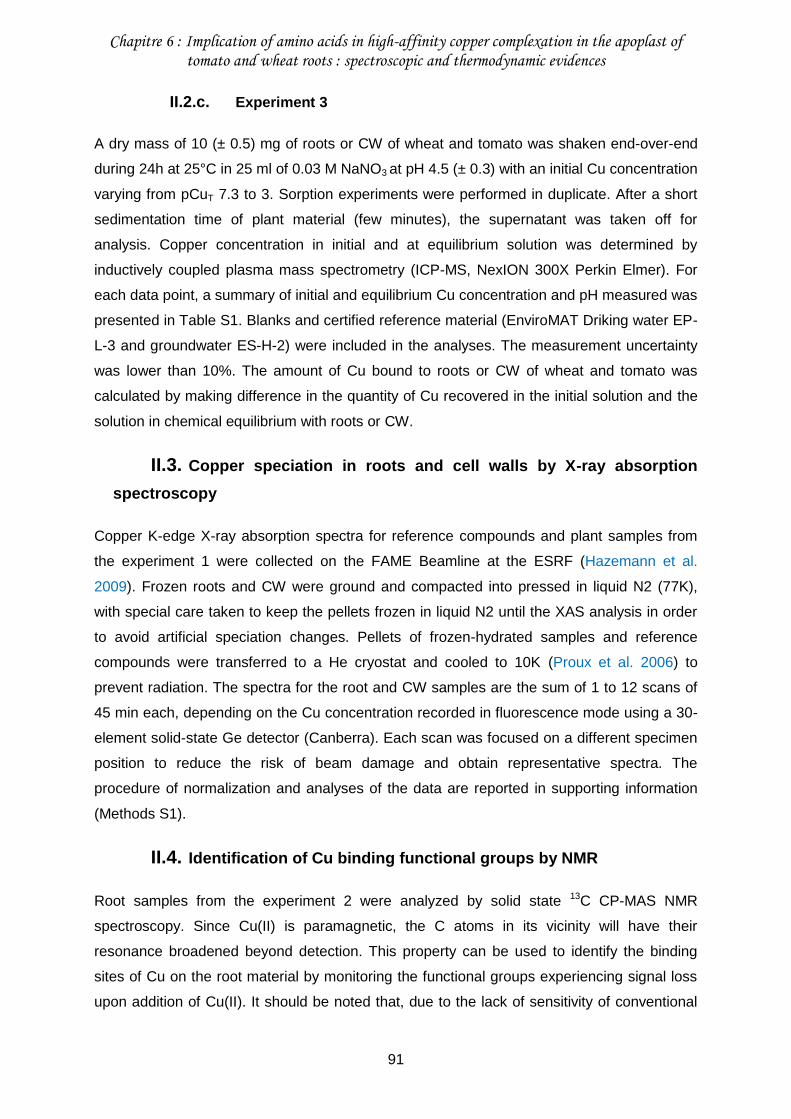
Chapitre 6 : Implication of amino acids in high-affinity copper complexation in the apoplast of tomato and wheat roots : spectroscopic and thermodynamic evidences
91
II.2.c. Experiment 3
A dry mass of 10 (± 0.5) mg of roots or CW of wheat and tomato was shaken end-over-end
during 24h at 25°C in 25 ml of 0.03 M NaNO3 at pH 4.5 (± 0.3) with an initial Cu concentration
varying from pCuT 7.3 to 3. Sorption experiments were performed in duplicate. After a short
sedimentation time of plant material (few minutes), the supernatant was taken off for
analysis. Copper concentration in initial and at equilibrium solution was determined by
inductively coupled plasma mass spectrometry (ICP-MS, NexION 300X Perkin Elmer). For
each data point, a summary of initial and equilibrium Cu concentration and pH measured was
presented in Table S1. Blanks and certified reference material (EnviroMAT Driking water EP-
L-3 and groundwater ES-H-2) were included in the analyses. The measurement uncertainty
was lower than 10%. The amount of Cu bound to roots or CW of wheat and tomato was
calculated by making difference in the quantity of Cu recovered in the initial solution and the
solution in chemical equilibrium with roots or CW.
II.3. Copper speciation in roots and cell walls by X-ray absorption
spectroscopy
Copper K-edge X-ray absorption spectra for reference compounds and plant samples from
the experiment 1 were collected on the FAME Beamline at the ESRF (Hazemann et al.
2009). Frozen roots and CW were ground and compacted into pressed in liquid N2 (77K),
with special care taken to keep the pellets frozen in liquid N2 until the XAS analysis in order
to avoid artificial speciation changes. Pellets of frozen-hydrated samples and reference
compounds were transferred to a He cryostat and cooled to 10K (Proux et al. 2006) to
prevent radiation. The spectra for the root and CW samples are the sum of 1 to 12 scans of
45 min each, depending on the Cu concentration recorded in fluorescence mode using a 30-
element solid-state Ge detector (Canberra). Each scan was focused on a different specimen
position to reduce the risk of beam damage and obtain representative spectra. The
procedure of normalization and analyses of the data are reported in supporting information
(Methods S1).
II.4. Identification of Cu binding functional groups by NMR
Root samples from the experiment 2 were analyzed by solid state 13C CP-MAS NMR
spectroscopy. Since Cu(II) is paramagnetic, the C atoms in its vicinity will have their
resonance broadened beyond detection. This property can be used to identify the binding
sites of Cu on the root material by monitoring the functional groups experiencing signal loss
upon addition of Cu(II). It should be noted that, due to the lack of sensitivity of conventional
Chapitre 6 : Implication of amino acids in high-affinity copper complexation in the apoplast of tomato and wheat roots : spectroscopic and thermodynamic evidences
92
pulse NMR, the quantities of Cu necessary to observe a significant effect are well above the
concentrations to be expected in a natural environment. However, these experiments can
help identify trends and differences in behavior between the root material samples. Spectra
were obtained with a Bruker Avance WB 400-MHz spectrometer at 101.6 MHz. Typical
aquisition parameters include spin rate of 10kHz, 2 ms contact time and 2 s recycling delays.
Depending on the sample, 10-18k scans were recorded. The FIDs were processed with the
MestReNova software; data treatment consisted in a 50 Hz line broadening and standard
baseline and phase correction. Semi quantitative analysis was performed by spectra
decomposition into gaussian peaks using the IGOR PRO 5.0 software.
In parallel, attempts to obtain usable 15N NMR data failed because N accounts for only 5%
and 3% weight of the root material of tomato and wheat respectively (results not shown),
which, considering the low sensitivity of the 15N nucleus, leads to unreasonably long
acquisition times.
II.5. Modeling copper sorption on roots, cell walls and plasma
membranes
The sorption of Cu on roots and CW of wheat and tomato was characterized by simulating
the data from the experiment 3 (see Methods S2 for details on the modelling procedure) with
the Humic Ion-Binding Model VII, hereafter referenced as to Model VII (Tipping (1998) and
Tipping et al. 2011). Model VII was originally designed to simulate the sorption properties of
two humic substances, namely a fulvic and a humic acid, depicted as a regular array of
binding sites of type 1 and 2. The density (LHi, cmolc.kg-1) of type-1 sites is arbitrarily fixed as
twice higher than the density of the type-2 sites (i.e. LH1 = 2 LH2). Protons and metal ions
compete for sorption on these sites with metal ions being able to form mono-, bi- and tri-
dendate complexes. Electrostatic effects are accounted for in Model VII by approximating the
diffuse layer/bulk solution system with a Donnan model. As roots and CW are solid organic
matters, we parameterized their sorption properties on the basis of the default conformation
parameters defined for the humic acid (HA) in Model VII. The partial pressure of CO2 was
assumed to be that of the ambient atmosphere (10-3.5 atm) and the temperature was fixed at
25 °C for calculations. The modelling procedure (Methods S2) was further implemented on
the theoretical potentiometric titrations and Cu sorption isotherms of the outer surface of root
cell PM for wheat and tomato that were calculated by making the difference between roots
and CW data (Figs. S2 and S3).
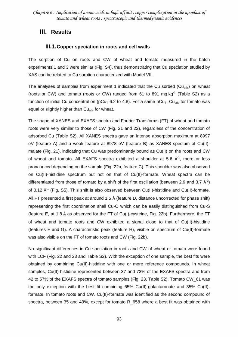
Chapitre 6 : Implication of amino acids in high-affinity copper complexation in the apoplast of tomato and wheat roots : spectroscopic and thermodynamic evidences
93
III. Results
III.1. Copper speciation in roots and cell walls
The sorption of Cu on roots and CW of wheat and tomato measured in the batch
experiments 1 and 3 were similar (Fig. S4), thus demonstrating that Cu speciation studied by
XAS can be related to Cu sorption characterized with Model VII.
The analyses of samples from experiment 1 indicated that the Cu sorbed (Cuads) on wheat
(roots or CW) and tomato (roots or CW) ranged from 61 to 891 mg.kg-1 (Table S2) as a
function of initial Cu concentration (pCuT 6.2 to 4.8). For a same pCuT, Cuads for tomato was
equal or slightly higher than Cuads for wheat.
The shape of XANES and EXAFS spectra and Fourier Transforms (FT) of wheat and tomato
roots were very similar to those of CW (Fig. 21 and 22), regardless of the concentration of
adsorbed Cu (Table S2). All XANES spectra gave an intense absorption maximum at 8997
eV (feature A) and a weak feature at 8978 eV (feature B) as XANES spectum of Cu(II)-
malate (Fig. 21), indicating that Cu was predominantly bound as Cu(II) on the roots and CW
of wheat and tomato. All EXAFS spectra exhibited a shoulder at 5.6 Å-1, more or less
pronounced depending on the sample (Fig. 22a, feature C). This shoulder was also observed
on Cu(II)-histidine spectrum but not on that of Cu(II)-formate. Wheat spectra can be
differentiated from those of tomato by a shift of the first oscillation (between 2.9 and 3.7 Å-1)
of 0.12 Å-1 (Fig. S5). This shift is also observed between Cu(II)-histidine and Cu(II)-formate.
All FT presented a first peak at around 1.5 Å (feature D, distance uncorrected for phase shift)
representing the first coordination shell Cu-O which can be easily distinguished from Cu-S
(feature E, at 1.8 Å as observed for the FT of Cu(I)-cysteine, Fig. 22b). Furthermore, the FT
of wheat and tomato roots and CW exhibited a signal close to that of Cu(II)-histidine
(features F and G). A characteristic peak (feature H), visible on spectrum of Cu(II)-formate
was also visible on the FT of tomato roots and CW (Fig. 22b).
No significant differences in Cu speciation in roots and CW of wheat or tomato were found
with LCF (Fig. 22 and 23 and Table S2). With the exception of one sample, the best fits were
obtained by combining Cu(II)-histidine with one or more reference compounds. In wheat
samples, Cu(II)-histidine represented between 37 and 73% of the EXAFS spectra and from
42 to 57% of the EXAFS spectra of tomato samples (Fig. 23, Table S2). Tomato CW_61 was
the only exception with the best fit combining 65% Cu(II)-galacturonate and 35% Cu(II)-
formate. In tomato roots and CW, Cu(II)-formate was identified as the second compound of
spectra, between 35 and 49%, except for tomato R_658 where a best fit was obtained with
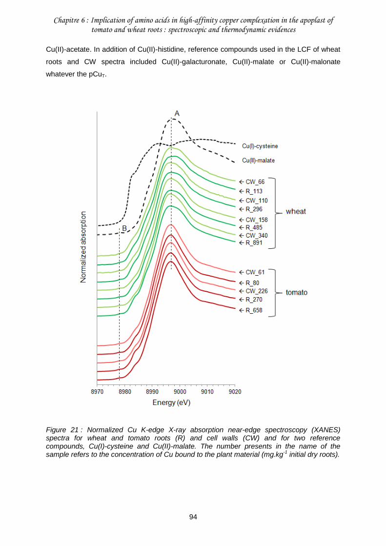
Chapitre 6 : Implication of amino acids in high-affinity copper complexation in the apoplast of tomato and wheat roots : spectroscopic and thermodynamic evidences
94
Cu(II)-acetate. In addition of Cu(II)-histidine, reference compounds used in the LCF of wheat
roots and CW spectra included Cu(II)-galacturonate, Cu(II)-malate or Cu(II)-malonate
whatever the pCuT.
Figure 21 : Normalized Cu K-edge X-ray absorption near-edge spectroscopy (XANES) spectra for wheat and tomato roots (R) and cell walls (CW) and for two reference compounds, Cu(I)-cysteine and Cu(II)-malate. The number presents in the name of the sample refers to the concentration of Cu bound to the plant material (mg.kg-1 initial dry roots).
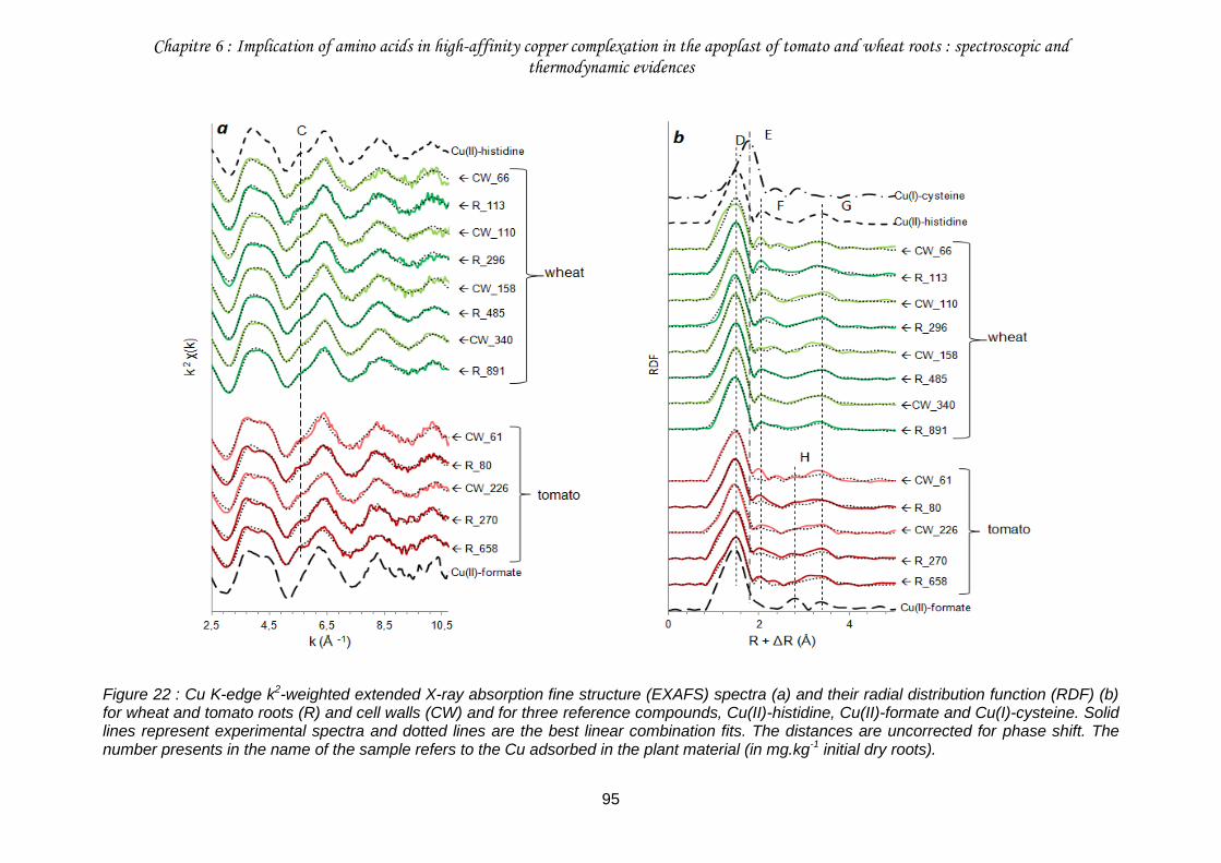
Chapitre 6 : Implication of amino acids in high-affinity copper complexation in the apoplast of tomato and wheat roots : spectroscopic and thermodynamic evidences
95
Figure 22 : Cu K-edge k2-weighted extended X-ray absorption fine structure (EXAFS) spectra (a) and their radial distribution function (RDF) (b) for wheat and tomato roots (R) and cell walls (CW) and for three reference compounds, Cu(II)-histidine, Cu(II)-formate and Cu(I)-cysteine. Solid lines represent experimental spectra and dotted lines are the best linear combination fits. The distances are uncorrected for phase shift. The number presents in the name of the sample refers to the Cu adsorbed in the plant material (in mg.kg-1 initial dry roots).
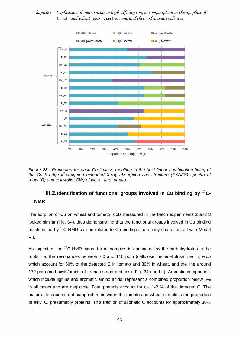
Chapitre 6 : Implication of amino acids in high-affinity copper complexation in the apoplast of tomato and wheat roots : spectroscopic and thermodynamic evidences
96
Figure 23 : Proportion for each Cu ligands resulting in the best linear combination fitting of the Cu K-edge k2-weighted extended X-ray absorption fine structure (EXAFS) spectra of roots (R) and cell walls (CW) of wheat and tomato.
III.2. Identification of functional groups involved in Cu binding by 13C-
NMR
The sorption of Cu on wheat and tomato roots measured in the batch experiments 2 and 3
looked similar (Fig. S4), thus demonstrating that the functional groups involved in Cu binding
as identified by 13C-NMR can be related to Cu binding site affinity characterized with Model
VII.
As expected, the 13C-NMR signal for all samples is dominated by the carbohydrates in the
roots, i.e. the resonances between 60 and 110 ppm (cellulose, hemicellulose, pectin, etc.)
which account for 60% of the detected C in tomato and 80% in wheat, and the line around
172 ppm (carboxyls/amide of uronates and proteins) (Fig. 24a and b). Aromatic compounds,
which include lignins and aromatic amino acids, represent a combined proportion below 5%
in all cases and are negligible. Total phenols account for ca. 1-2 % of the detected C. The
major difference in root composition between the tomato and wheat sample is the proportion
of alkyl C, presumably proteins. This fraction of aliphatic C accounts for approximately 30%
Chapitre 6 : Implication of amino acids in high-affinity copper complexation in the apoplast of tomato and wheat roots : spectroscopic and thermodynamic evidences
97
in the tomato roots (Fig. 24b) whereas this proportion is only ca. 15% in the case of wheat
(Fig. 24a). It should be kept in mind however that these are rough estimates since aliphatic
amino acids such as valine (Val), threonine (Thr), proline (Pro) and isoleucine (Ile) have
resonances between 60 and 70 ppm, i.e. the carbohydrate region, and under our
experimental conditions cannot be distinguished from cellulose type material.
In the case of the tomato roots, the addition of increasing amount of Cu leads to diminished
intensities of the peaks in the alkyl- and carbonyl/carboxyl regions (Fig. 24b). The intensity
variations observed between 0 and approximately 60 ppm cannot be converted with
sufficient confidence into variations of proportions since, assuming that they are due to Cu
binding by proteins, only a limited fraction of C is potentially impacted; also there is an
overlap between the contribution around 55 and 60 ppm which complicates the determination
of alkyl- vs. carbohydrate compounds. Semi-quantitative analysis is possible however for the
variations of the carboxyl resonance around 172 ppm: when the Cu concentration is
increased from 0 to 9864 mg.kg-1 (dry mass), the surface area of the peak is diminished by
approximately 30%. In the case of wheat, equivalent amount of added Cu(II) caused only a
ca. 10% reduction of the contribution around 172 ppm showing that for this material Cu
binding to carboxyl is less prevalent (Fig. 24a). Similarly to the case of tomato, the intensity
variations in the alkyl region are difficult to translate into meaningful variations of proportions.
An interesting feature is the decrease in intensity of the line around 62 ppm. As it seems
unlikely that the primary alcohol of a hexose moiety specifically binds Cu, this decrease can
be attributed to the loss of signal of the amino acids Val, The, Pro and Ile, thereby pointing to
a specific composition of root proteins.
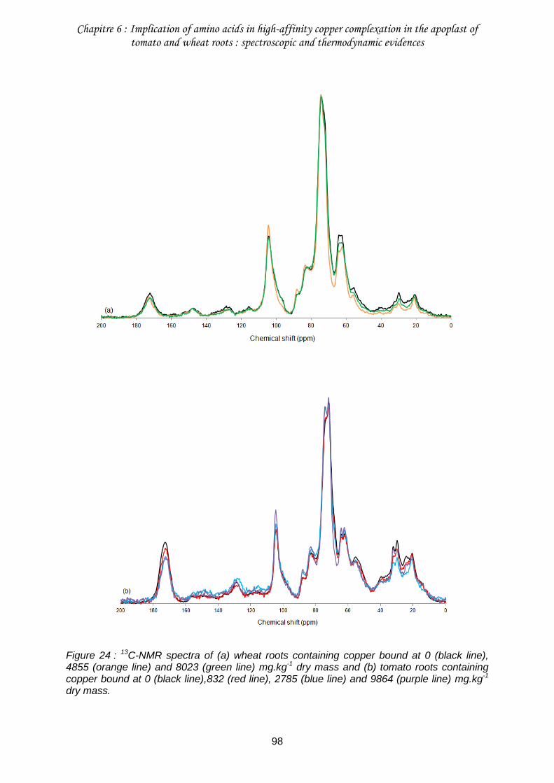
Chapitre 6 : Implication of amino acids in high-affinity copper complexation in the apoplast of tomato and wheat roots : spectroscopic and thermodynamic evidences
98
Figure 24 : 13C-NMR spectra of (a) wheat roots containing copper bound at 0 (black line), 4855 (orange line) and 8023 (green line) mg.kg-1 dry mass and (b) tomato roots containing copper bound at 0 (black line),832 (red line), 2785 (blue line) and 9864 (purple line) mg.kg-1 dry mass.
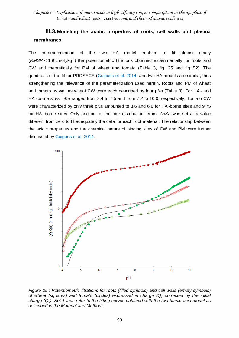
Chapitre 6 : Implication of amino acids in high-affinity copper complexation in the apoplast of tomato and wheat roots : spectroscopic and thermodynamic evidences
99
III.3. Modeling the acidic properties of roots, cell walls and plasma
membranes
The parameterization of the two HA model enabled to fit almost neatly
(RMSR < 1.9 cmolc.kg-1) the potentiometric titrations obtained experimentally for roots and
CW and theoretically for PM of wheat and tomato (Table 3, fig. 25 and fig. S2). The
goodness of the fit for PROSECE (Guigues et al. 2014) and two HA models are similar, thus
strengthening the relevance of the parameterization used herein. Roots and PM of wheat
and tomato as well as wheat CW were each described by four pKa (Table 3). For HAI- and
HAII-borne sites, pKa ranged from 3.4 to 7.5 and from 7.2 to 10.0, respectively. Tomato CW
were characterized by only three pKa amounted to 3.6 and 6.0 for HAI-borne sites and 9.75
for HAII-borne sites. Only one out of the four distribution terms, ΔpKa was set at a value
different from zero to fit adequately the data for each root material. The relationship between
the acidic properties and the chemical nature of binding sites of CW and PM were further
discussed by Guigues et al. 2014.
Figure 25 : Potentiometric titrations for roots (filled symbols) and cell walls (empty symbols) of wheat (squares) and tomato (circles) expressed in charge (Q) corrected by the initial charge (Q0). Solid lines refer to the fitting curves obtained with the two humic-acid model as described in the Material and Methods.
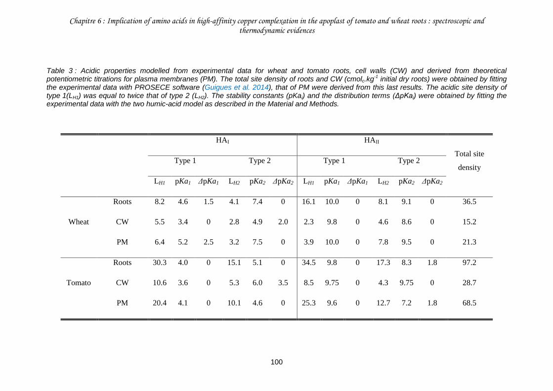
Chapitre 6 : Implication of amino acids in high-affinity copper complexation in the apoplast of tomato and wheat roots : spectroscopic and thermodynamic evidences
100
Table 3 : Acidic properties modelled from experimental data for wheat and tomato roots, cell walls (CW) and derived from theoretical potentiometric titrations for plasma membranes (PM). The total site density of roots and CW (cmolc.kg-1 initial dry roots) were obtained by fitting the experimental data with PROSECE software (Guigues et al. 2014), that of PM were derived from this last results. The acidic site density of type 1(LH1) was equal to twice that of type 2 (LH2). The stability constants (pKai) and the distribution terms (ΔpKai) were obtained by fitting the experimental data with the two humic-acid model as described in the Material and Methods.
HAI HAII
Total site
density Type 1 Type 2 Type 1 Type 2
LH1 pKa1 ΔpKa1 LH2 pKa2 ΔpKa2 LH1 pKa1 ΔpKa1 LH2 pKa2 ΔpKa2
Wheat
Roots 8.2 4.6 1.5 4.1 7.4 0 16.1 10.0 0 8.1 9.1 0 36.5
CW 5.5 3.4 0 2.8 4.9 2.0 2.3 9.8 0 4.6 8.6 0 15.2
PM 6.4 5.2 2.5 3.2 7.5 0 3.9 10.0 0 7.8 9.5 0 21.3
Tomato
Roots 30.3 4.0 0 15.1 5.1 0 34.5 9.8 0 17.3 8.3 1.8 97.2
CW 10.6 3.6 0 5.3 6.0 3.5 8.5 9.75 0 4.3 9.75 0 28.7
PM 20.4 4.1 0 10.1 4.6 0 25.3 9.6 0 12.7 7.2 1.8 68.5
Chapitre 6 : Implication of amino acids in high-affinity copper complexation in the apoplast of tomato and wheat roots : spectroscopic and thermodynamic evidences
101
III.4. Copper sorption on roots, cell walls and plasma membranes
Whatever the root material considered in the batch experiment 3, the concentration of Cu
adsorbed increased with the increasing total Cu concentration in solution (Fig. 26). When
expressed in dry mass of roots and CW respectively, the quantity of Cu bound to roots and
CW was the same, reflecting an identical binding behavior of these two plant materials
(results not shown). In accordance with binding site density (Table 3), roots and CW of wheat
bound less Cu at a given Cu concentration in equilibrated solution and were more rapidly
saturated in Cu than roots and CW of tomato (Fig. 26). Very similar observations can be
done for the theoretical Cu sorption data calculated for PM (Fig. S3).
III.5. Modeling copper sorption on roots, cell walls and plasma
membranes
The two HA model was able to fit almost neatly the sorption of Cu on roots, CW and PM of
wheat (RMSR = 76 mg.kg-1) over the entire range of Cu concentration in solution and of
tomato (RMSR = 4.5 mg.kg-1) for pCuT > 4.5 (Fig. 26, Fig. S3). For pCuT < 4.5, the two HA
model deviated from the experimental data points for roots, CW and PM of tomato. While the
two HA model can be specifically parameterized to fit the tomato data for pCuT < 4.5 in
solution (data not shown), the modeling results were discussed below on the basis of the
parameterization used to fit the data for pCuT > 4.5 that corresponds to the environmentally-
relevant range of root-exposed Cu concentrations in solution (Table 4). For HAI sites, logKCu,1
and logKCu,2 ranged from 1.9 to 2.45 and from 2.1 to 3.9, respectively (Table 4). The
intraspecific comparison revealed that the HAI sites of PM had lower intrinsic equilibrium
constants than those of roots and CW for both wheat and tomato. For HAII sites, logKCu,1 and
logKCu,2 ranged from 6.0 to 7.5 and from 4.5 to 6.7, respectively. For the HAII sites of tomato,
intrinsic equilibrium constants obtained for PM were lower than those of roots and CW. For
the HAII sites of wheat, the logKCu,1 of PM was equal to those of roots and CW and the
logKCu,2 of PM was slightly higher than those of roots and CW. Interspecific comparison
revealed that equilibrium constants of roots and PM were higher for wheat than for tomato.
Contrarily, equilibrium constants of CW were higher for tomato than for wheat. The
heterogeneity parameters (ΔLK2Cu) for both HAI and HAII sites were fairly low, i.e. equals to
0.8 for wheat roots or not different from zero for all other root material of wheat and tomato.
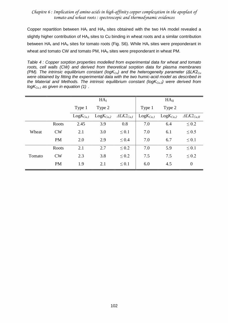
Chapitre 6 : Implication of amino acids in high-affinity copper complexation in the apoplast of tomato and wheat roots : spectroscopic and thermodynamic evidences
102
Copper repartition between HAI and HAII sites obtained with the two HA model revealed a
slightly higher contribution of HAII sites to Cu binding in wheat roots and a similar contribution
between HAI and HAII sites for tomato roots (Fig. S6). While HAI sites were preponderant in
wheat and tomato CW and tomato PM, HAII sites were preponderant in wheat PM.
Table 4 : Copper sorption properties modelled from experimental data for wheat and tomato roots, cell walls (CW) and derived from theoretical sorption data for plasma membranes (PM). The intrinsic equilibrium constant (logKCu) and the heterogeneity parameter (ΔLK2Cu were obtained by fitting the experimental data with the two humic-acid model as described in the Material and Methods. The intrinsic equilibrium constant (logKCu,2) were derived from logKCu,1 as given in equation (1) .
HAI HAII
Type 1 Type 2 Type 1 Type 2
LogKCu,1 LogKCu,2 ΔLK2Cu,I LogKCu,1 LogKCu,2 ΔLK2Cu,II
Wheat
Roots 2.45 3.9 0.8 7.0 6.4 ≤ 0.2
CW 2.1 3.0 ≤ 0.1 7.0 6.1 ≤ 0.5
PM 2.0 2.9 ≤ 0.4 7.0 6.7 ≤ 0.1
Tomato
Roots 2.1 2.7 ≤ 0.2 7.0 5.9 ≤ 0.1
CW 2.3 3.8 ≤ 0.2 7.5 7.5 ≤ 0.2
PM 1.9 2.1 ≤ 0.1 6.0 4.5 0
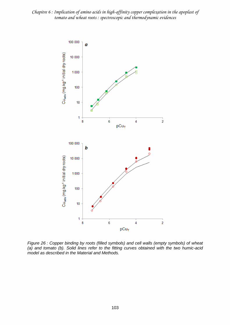
Chapitre 6 : Implication of amino acids in high-affinity copper complexation in the apoplast of tomato and wheat roots : spectroscopic and thermodynamic evidences
103
Figure 26 : Copper binding by roots (filled symbols) and cell walls (empty symbols) of wheat (a) and tomato (b). Solid lines refer to the fitting curves obtained with the two humic-acid model as described in the Material and Methods.
Chapitre 6 : Implication of amino acids in high-affinity copper complexation in the apoplast of tomato and wheat roots : spectroscopic and thermodynamic evidences
104
IV. Discussion
IV.1. Dual copper coordination with carboxyl and amine groups
There is increasing evidences that hard ligands other than carboxylic groups could be
significantly involved in metal sorption within root apoplast. The potentiometric titrations of
root CW enabled to identify the occurrence of cation binding sites with pKa higher than ca.
7.5, i.e. presumably attributed to phenolic and amine groups (Meychik and Yermakov 2001).
Complementary to CW, the role of cation binding sites borne by the outer surface of root PM
can be also evoked. Based on the investigation of the paramagnetic effect of cobalt (Co) on
the 31P- and 13C-NMR signals of phosphatidylserine, McLaughlin (1982) estimated that Co(II)
was bound to the surface of the model membrane as phosphate complex for 13 %, amine-
carboxyl chelate complex for 32 % and carboxyl complex for 54 %. Hence, it is conceivable
that a significant amount of the Cu binding sites borne by root-cell plasma membranes may
involve not only carboxyl groups but also phosphate, phenolic and amine groups.
For both tomato and wheat, roots and CW, and whatever the level of Cu loading, the
chemical environment of Cu revealed by XAS analysis looked very similar, thus suggesting
that Cu speciation was rather homogeneous in root apoplast (Fig. 22a and b). While it is
usually assumed that the distinction between Cu-N and Cu-O coordinations for biomolecules
is difficult to make with XAS, Manceau et al. (2013) recently succeed in showing that Cu was
mainly bound to histidine residue in the root CW of T. arvense. In line with this result, we
found that Cu(II)-histidine like species were ubiquitous in all the samples but one and
(co-)dominated Cu speciation in 10 out of the 13 samples of roots and CW (Fig. 23, Table
S2). In the Cu(II)-histidine reference compound, Cu was simultaneously bound to oxygen
and nitrogen atoms carried by carboxylic and amine groups (Evertson 1969). This reflects
the involvement of amino acid residues, embedded in CW (Cassab 1998) and at the outer
surface of PM (Taiz and Zeiger 2006). Copper was also associated with O atoms in roots
and CW of wheat and tomato (Fig. 23, Table S2). While the Cu-O local environment was
ubiquitous in all samples, it (co-)dominated Cu speciation in only 6 out of the 13 samples.
Copper was likely bound to both carboxyl and alcohol groups, as in Cu(II)-galacturonate and
Cu(II)-malate. Such environment most likely reflects the participation of CW polysaccharides
as pectins in Cu complexation. Such CW sites had already been identified as Cu binding
sites in roots of cowpea exposed to Cu during 24h (Kopittke et al. 2011c). Beyond the
contribution of Cu-N and Cu-O first coordination shells, no other Cu ligand was detected by
XAS analysis in roots and CW of wheat and tomato. The absence of Cu-phosphate bond
suggests that the PM phospholipids were not involved in Cu binding or in too low proportion
Chapitre 6 : Implication of amino acids in high-affinity copper complexation in the apoplast of tomato and wheat roots : spectroscopic and thermodynamic evidences
105
to be detected (i.e. below 10%). Indeed, Cu has a low affinity for phosphate groups, in
contrast to zinc (Sarret et al. 2001; Kopittke et al. 2011c). We have not detected Cu-sulfur
bond either. This bond has been detected for plants which cope with Cu by internal
sequestration via several S-rich ligands (e.g. metallothioneins) or by biomineralization of
copper sulfide (Collin et al. 2014). As the dry roots and CW we studied were metabolically-
inactive, these physiologically-driven processes were likely not active.
The homogeneity in Cu speciation whatever the plant species, the root apoplastic
compartment and the level of apoplastic Cu loading considered is further supported by the
values fitted for ΔLK2Cu, in the two HA model. This parameter was designed to account for
the occurrence at low concentration of binding sites exhibiting a very high affinity for metals
in humic substances, thus enabling to simulate strong binding behaviors at low metal
concentration (Tipping et al. 2011). The ΔLK2Cu values fitted in the two HA model (≤ 0.8;
Table 4) were much lower than the values reported for humic substances (2.34 for Cu),
suggesting that this kind of sites is either almost absent from root apoplast or can be
neglected.
IV.2. High-affinity copper complexation
XAS analysis showed that Cu is coordinated in root apoplast either with a single O ligand
mainly involving carboxyl groups or with both O and N ligands involving carboxyl and amine
groups of amino acids. Copper has a much higher affinity for synthetic and natural ligands
with both carboxylic and amine groups (e.g. ethylenediaminetetraacetic acid, histidine,
nicotianamine) than for ligands with carboxyl groups only (Fry et al. 2002; Karlsson et al.
2008). In line with these considerations, the two HA model was parameterized for wheat and
tomato roots by considering that 34 to 47 % of binding sites were low-pKa sites (i.e. type 1
and 2 in HAI) presumably corresponding to carboxyl-like groups and exhibiting low-affinity Cu
binding constants ranging from logKCu 2.1 to 3.9, whereas 53 to 66 % of binding sites were
high-pKa sites (i.e. type 1 and 2 in HAII) presumably corresponding to amine-like groups and
exhibiting high-affinity Cu binding constants ranging from logKCu 5.9 to 7 (Tables 3 and 4). It
is noteworthy that these high-affinity Cu binding sites presumably corresponding to amine
sites exhibit Cu binding constants even higher than those (i.e. logKCu 4.8-5.6) of similar high-
pKa sites usually described in humic susbtances that presumably correspond to phenolic
sites (Tipping et al. 2011). The two HA model further suggested that the high-affinity Cu
binding sites significantly contributed to the actual complexation of Cu, especially when
considering the whole root surfaces where these high-affinity sites bound 40 to 60 % of total
Cu complexed (Fig. S6a and b).
Chapitre 6 : Implication of amino acids in high-affinity copper complexation in the apoplast of tomato and wheat roots : spectroscopic and thermodynamic evidences
106
IV.3. Relative contribution of cell walls and plasma membranes
The comparison of Cu sorption in roots and CW revealed that PM bound only 38 to 44% of
Cu in wheat and tomato roots. This lower quantitative contribution of PM over CW to Cu
complexation is surprising at first sight as PM bore 60 to 70% of the total density of binding
sites in wheat and tomato roots (Guigues et al. 2014). This first suggests that cation binding
sites in CW exhibit a much higher affinity for Cu that those located at the outer surface of
PM. This hypothesis works for tomato for which the logKCu of CW sites were higher by 0.4 to
3 units than the logKCu of PM sites (Table 4). However, for wheat the logKCu of CW sites were
either weakly higher by 0 to 0.1 unit (for HAI sites and type-1 sites of HAII) or lower by 0.6 unit
than the logKCu of PM sites. The weak Cu binding strength of PM in wheat roots could be
alternatively related to electrostatic effects that were showed to strongly influence ion
concentration at PM surfaces (Wang et al. 2010; Kopittke et al. 2011). Indeed, the net
negative charge of isolated CW was higher than that of roots for wheat when solution pH was
lower than 5.5 (Fig. 25), thus leading to calculate that PM were positively charged in sorption
experiments performed at pH 4.5-5. Plasma membranes in wheat roots may hence have
exerted a repulsive effect on Cu2+ that lowered their Cu binding strength. An additional
hypothesis that may explain the greater contribution of CW over PM to Cu complexation is
the spatial arrangement of these two compartments. By design, any metal cation interacting
with roots first interacts and binds within CW and after passing through the remaining non-
bound metal cation then interacts and binds on the outer surface of PM. Consequently, we
hypothesize that Cu which exhibits a high affinity for binding sites in root apoplast first and
thus mainly accumulates in CW even if CW have a lower density and Cu-affinity of their
binding sites than PM. The larger contribution of CW to Cu complexation we observed in root
apoplast apparently contradict the previous findings of Shomer et al. (2003) and Kinraide
(2004) who suggested that metal cations would bind much more strongly to PM sites than to
CW sites and that CW would have a negligible influence on the metal cation concentration at
PM surfaces. However, these authors did not effectively measure the binding of metal
cations to CW and PM but measured the electrical potential in CW and at PM surfaces. The
modeling approach then implemented was thus very sensitive to the binding site density of
CW but conversely almost insensitive to the binding strength of metal cations in CW
(Kinraide 2004; Kinraide et al. 2004). Consequently, the electrostatic model developed by
Kinraide and co-workers is likely relevant to calculate the ion concentration at PM surfaces
but would most likely fail to calculate the effective binding of metal cations in root apoplast
that as we showed involves the substantial contribution of CW.
Chapitre 6 : Implication of amino acids in high-affinity copper complexation in the apoplast of tomato and wheat roots : spectroscopic and thermodynamic evidences
107
IV.4. Interspecific comparison
Tomato roots exhibited a 2.7-fold higher site density than wheat roots (Guigues et al. 2014).
Calculations performed with the two HA model suggest that binding sites were exhibiting a
higher affinity for Cu (i.e. higher logKCu) in wheat roots than in tomato roots (Table 4). The
higher mean contribution of the Cu(II)-histidine reference in the LCF of EXAFS spectra
(Fig. 23) for wheat (62 14 %) than for tomato (55 3 %) consistently suggest a higher
contribution of amino acids to root Cu complexation in wheat over tomato. This preferential
binding to N groups in wheat is also supported by the NMR data. Indeed, the addition of
Cu(II) leads to modification primarily in the spectral region below 60 ppm, i.e. where signal
from amino acids is expected, even though the modifications are difficult to quantify (N < 3%
weight, results not shown), whereas the resonance of carboxyl C around 172 ppm remains
largely unaffected (Fig. 24a and b). Although the amount of N in tomato roots is
approximately twice as high as in wheat (result not shown), the present spectroscopic data
does not support a preferential binding of Cu to N-groups. This suggests that the nature and
thus the affinity for Cu, and/or (steric) accessibility of N binding sites are different in tomato
and wheat roots.
IV.5. The HLScale under-estimates the contribution of amino acids
It is questionable how the HLScale which is defined with a single value is able to account for
both low- and high-affinity sites we identified in root apoplast? The HLScale may correlate
both with low- and high-affinity sites defined for a range of metal cations. However, this
assertion cannot be tested as we focused our investigation on Cu only. Alternatively, a look
on the classification of Cu binding strength in the HLScale shows that Cu2+ is one of the
divalent cations exhibiting the highest binding strength toward hard ligands but that it remains
(much) lower than the binding strength of trivalent cations (Kinraide 2009). This behavior is
typical from the binding strength of Cu to most hard ligands with the notable exception of
some amino acids that are known to play an important role in metal transport to and within
plants. For instance, Cu exhibits an exceptionally high affinity for phytosiderophores
exudated by the roots of monocots and some histidine-rich sequences of metal-transport
proteins located at the outer surface of PM (Murakami et al. 1989; Grossoehme et al. 2006).
In comparison with synthetic N ligands, Cu exhibits with these biologically-relevant amino
acids a binding strength that approaches or even exceeds that of trivalent cations such as
Fe3+. Owing to the substantial contribution of amino acid residue in Cu binding in root
apoplast, we may infer that the HLScale likely under-estimates the high affinity of these
Chapitre 6 : Implication of amino acids in high-affinity copper complexation in the apoplast of tomato and wheat roots : spectroscopic and thermodynamic evidences
108
binding sites for Cu, which may partly explain the under-prediction of the unexpectedly high
Cu rhizotoxicity underlined by Kopittke et al. (2014).
V. Conclusion
By crossing modeling and spectroscopic investigations, we found a dual local environment of
Cu in the root apoplast of wheat and tomato roots. As expected, CW pectins were identified
as one of the major Cu binding sites. We found no evidence of the involvement of PM
phospholipids. More surprisingly, Cu speciation was (co)-dominated by amino acids
embedded in CW and PM. These amino acids exhibited a high affinity for Cu and hence
significantly contributed to the actual Cu complexation in root apoplast. We further infer that
a re-evaluation of the HLScale to better account for the contribution of amino acids may help
to explain the unexpectedly high rhizotoxicity exhibited by Cu.
Although this study gives a consistent and updated picture of Cu binding in root apoplast, we
investigated only non-metabolically-active materials. Hence, it would be interesting to confirm
these results with fresh plants.
Acknowledgments
The authors are grateful to French Environment and Energy Management Agency (ADEME)
and the French Centre of Agricultural Research for Development (CIRAD) for funding the
PhD scholarship of Stéphanie Guigues and INSU (CNRS) for funding the study via the
EC2CO-CYTRIX call. The authors thank Hélène Miche (CEREGE) for providing access to
ICP-AES and Bernard Angeletti (CEREGE) for ICP-MS analyses. The authors also
acknowledge support from Spectropole, the Analytical Facility of Aix-Marseille University, by
allowing a special access to the instruments purchased with European Funding (FEDER
OBJ2142-3341) and especially Fabio Ziarelli for his assistance with the NMR experiments.
Chapitre 6 : Implication of amino acids in high-affinity copper complexation in the apoplast of tomato and wheat roots : spectroscopic and thermodynamic evidences
109
VI. Supporting information
Methods S1: Procedure of normalization and analyses of the X-ray absorption spectroscopy
data
A Cu foil was used to calibrate the X-ray energy (threshold energy taken at the zero-crossing
point of the second derivative spectrum).
The data were normalized using Athena software (Ravel and Newville 2005). The k2-
weighted EXAFS (2.5 to 10.7 Å-1) recorded on plant samples were fitted by linear
combination fitting (LCF) using a library of Cu reference compounds consisting of organic
and mineral species.
Principal component analysis (PCA) was applied to EXAFS spectra to determine the number
of species contained in the samples, but the PCA indicator value failed to reach a minimum.
Thus, for each plant spectrum, LCFs using one, two and three reference compounds were
tested successively. The LCFs with n + 1 components was retained if the normalized sum-
squares residual (Table S2) was decreased by more than 20% compared to the fit with n
components. Satisfactory fits were obtained with a combination of two or three references. A
part of the reference compound database used was described previously in Collin et al.
(2014) and compounds described below were added. Cu(II)-gluconate, Cu(II)-phthalocyanine
and libethenite (Cu2PO4OH) were purchased from Sigma-Aldrich. Cu(II)-galacturonate was
synthesized according to the procedure of Synytsya et al. (2004). Cu(II)-methionine and
Cu(II)-phenylalanine were prepared in compliance with the protocol of Stanila et al. (2004).
And Cu(II)-phytate was obtained in the same way as described in Kopittke et al. (2011c).
Chapitre 6 : Implication of amino acids in high-affinity copper complexation in the apoplast of tomato and wheat roots : spectroscopic and thermodynamic evidences
110
Methods S2: Modeling procedure (Model VII)
In a first step, Model VII was used to fit the potentiometric titrations previously done by
Guigues et al. (2014) on roots and cell walls of wheat and tomato. These titrations were
previously interpreted with the dedicated software PROSECE (Guigues et al. 2014) which
enabled for each root material to fix the total site density as an input parameter and to give
indicative values to fit the intrinsic proton dissociation constant, pKai, and the corresponding
distribution term, ΔpKai, for sites of type 1 and 2. Preliminary investigations showed that it
was not possible to fit adequately the titration data of tomato roots and wheat and tomato cell
walls when considering a single HA. This was mainly due to the following condition imposed
by Model VII: LH1 = 2 LH2 (Fig. S1). To relax the consequences of this condition, we fit the
titration data by representing each root material with two HA independently parameterized
(HAI and HAII) that stand for the low-and the high-pKa sites respectively. In a second step,
the data of Cu sorption from the experiment 2 were simulated for roots and cell walls of
wheat and tomato with the two HA model by fitting the intrinsic equilibrium constant, logKCu,1,
for the type-1 sites and the heterogeneity parameter, ΔLK2Cu,1,. The intrinsic equilibrium
constant for the type-2 sites, logKCu,2, was derived from logKCu,1 (Tipping et al. 2011) :
𝑙𝑜𝑔𝐾𝐶𝑢,2 = 𝑙𝑜𝑔𝐾𝐶𝑢,1 ×𝑝𝐾𝑎2
𝑝𝐾𝑎1 (1)
While Model VII can account for the complexation of the free ionic form and the first
hydrolysis product of each metal, we accounted for Cu2+ only as a preliminary speciation
calculation showed that Cu2+ stood for > 99 % of total Cu in solution at pH ≤ 5.
The best fit was determined by minimizing the root mean square residual (RMSR),
calculated as follows:
𝑅𝑀𝑆𝑅 = √∑ (𝑥𝑖,𝑒𝑥𝑝−𝑥𝑖,𝑚𝑜𝑑𝑒𝑙)2𝑛
𝑖=1
𝑛 (2)
where 𝑥𝑖,𝑒𝑥𝑝 is an experimental data point, 𝑥𝑖,𝑚𝑜𝑑𝑒𝑙 its corresponding calculated point with the
two humic-acid model and n the total number of experimental data points.
This modelling procedure was further implemented on the theoretical potentiometric titrations
and Cu sorption isotherms of the outer surface of root cell plasma membranes for wheat and
tomato that were calculated by making the difference between roots and cell walls data (Figs.
S2 and S3).
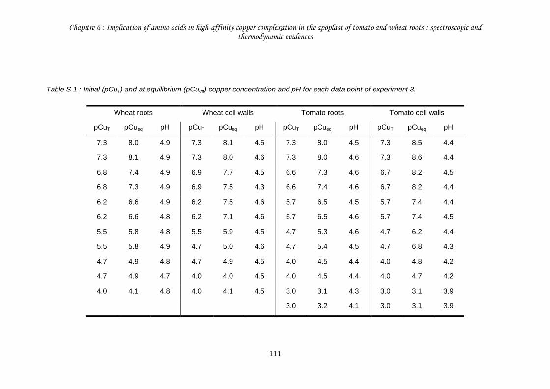
Chapitre 6 : Implication of amino acids in high-affinity copper complexation in the apoplast of tomato and wheat roots : spectroscopic and thermodynamic evidences
111
Table S 1 : Initial (pCuT) and at equilibrium (pCueq) copper concentration and pH for each data point of experiment 3.
Wheat roots Wheat cell walls Tomato roots Tomato cell walls
pCuT pCueq pH pCuT pCueq pH pCuT pCueq pH pCuT pCueq pH
7.3 8.0 4.9 7.3 8.1 4.5 7.3 8.0 4.5 7.3 8.5 4.4
7.3 8.1 4.9 7.3 8.0 4.6 7.3 8.0 4.6 7.3 8.6 4.4
6.8 7.4 4.9 6.9 7.7 4.5 6.6 7.3 4.6 6.7 8.2 4.5
6.8 7.3 4.9 6.9 7.5 4.3 6.6 7.4 4.6 6.7 8.2 4.4
6.2 6.6 4.9 6.2 7.5 4.6 5.7 6.5 4.5 5.7 7.4 4.4
6.2 6.6 4.8 6.2 7.1 4.6 5.7 6.5 4.6 5.7 7.4 4.5
5.5 5.8 4.8 5.5 5.9 4.5 4.7 5.3 4.6 4.7 6.2 4.4
5.5 5.8 4.9 4.7 5.0 4.6 4.7 5.4 4.5 4.7 6.8 4.3
4.7 4.9 4.8 4.7 4.9 4.5 4.0 4.5 4.4 4.0 4.8 4.2
4.7 4.9 4.7 4.0 4.0 4.5 4.0 4.5 4.4 4.0 4.7 4.2
4.0 4.1 4.8 4.0 4.1 4.5 3.0 3.1 4.3 3.0 3.1 3.9
3.0 3.2 4.1 3.0 3.1 3.9
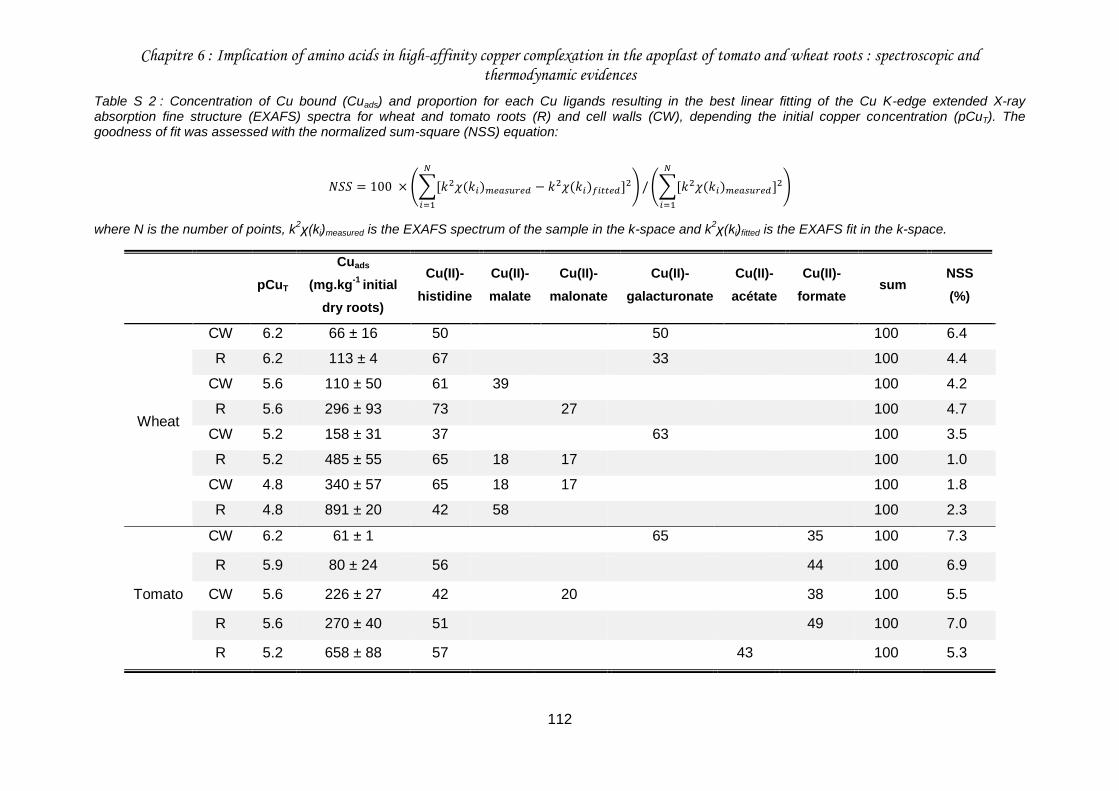
Chapitre 6 : Implication of amino acids in high-affinity copper complexation in the apoplast of tomato and wheat roots : spectroscopic and thermodynamic evidences
112
Table S 2 : Concentration of Cu bound (Cuads) and proportion for each Cu ligands resulting in the best linear fitting of the Cu K-edge extended X-ray absorption fine structure (EXAFS) spectra for wheat and tomato roots (R) and cell walls (CW), depending the initial copper concentration (pCuT). The goodness of fit was assessed with the normalized sum-square (NSS) equation:
𝑁𝑆𝑆 = 100 × (∑[𝑘2𝜒(𝑘𝑖)𝑚𝑒𝑎𝑠𝑢𝑟𝑒𝑑 − 𝑘2𝜒(𝑘𝑖)𝑓𝑖𝑡𝑡𝑒𝑑]2
𝑁
𝑖=1
) / (∑[𝑘2𝜒(𝑘𝑖)𝑚𝑒𝑎𝑠𝑢𝑟𝑒𝑑]2
𝑁
𝑖=1
)
where N is the number of points, k2χ(ki)measured is the EXAFS spectrum of the sample in the k-space and k
2χ(ki)fitted is the EXAFS fit in the k-space.
pCuT
Cuads
(mg.kg-1
initial
dry roots)
Cu(II)-
histidine
Cu(II)-
malate
Cu(II)-
malonate
Cu(II)-
galacturonate
Cu(II)-
acétate
Cu(II)-
formate sum
NSS
(%)
Wheat
CW 6.2 66 ± 16 50 50 100 6.4
R 6.2 113 ± 4 67 33 100 4.4
CW 5.6 110 ± 50 61 39 100 4.2
R 5.6 296 ± 93 73 27 100 4.7
CW 5.2 158 ± 31 37 63 100 3.5
R 5.2 485 ± 55 65 18 17 100 1.0
CW 4.8 340 ± 57 65 18 17 100 1.8
R 4.8 891 ± 20 42 58 100 2.3
Tomato
CW 6.2 61 ± 1 65 35 100 7.3
R 5.9 80 ± 24 56 44 100 6.9
CW 5.6 226 ± 27 42 20 38 100 5.5
R 5.6 270 ± 40 51 49 100 7.0
R 5.2 658 ± 88 57 43 100 5.3
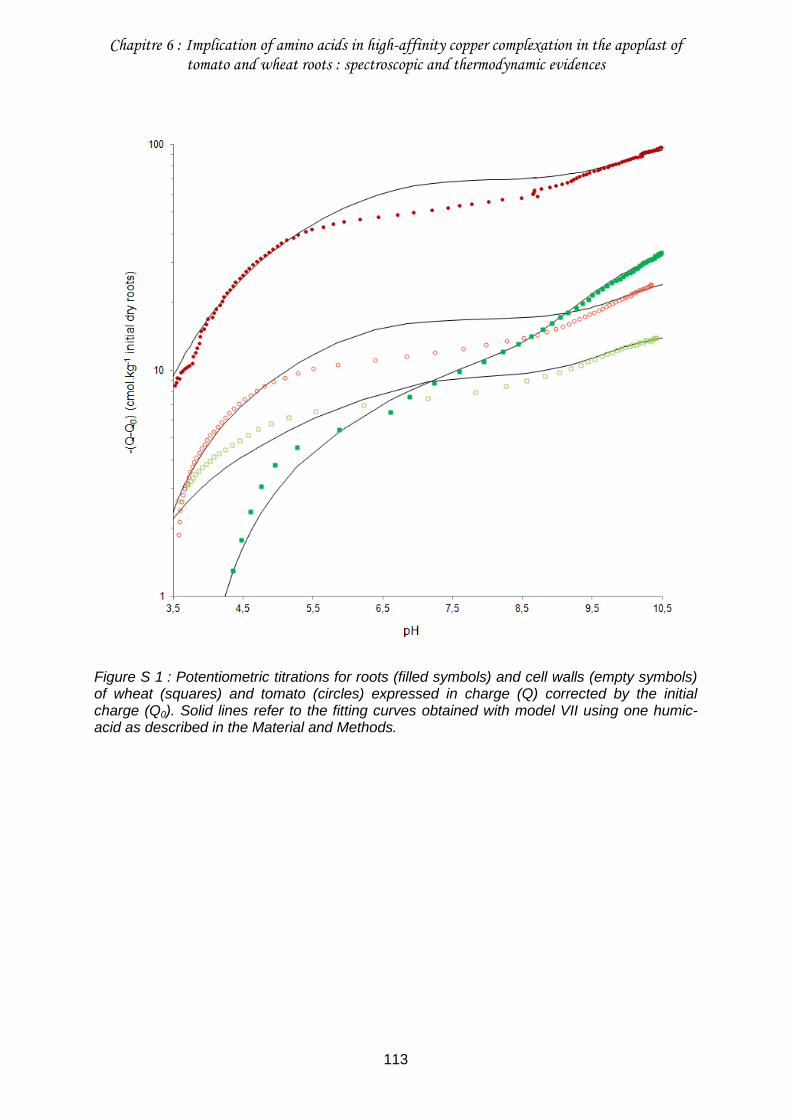
Chapitre 6 : Implication of amino acids in high-affinity copper complexation in the apoplast of tomato and wheat roots : spectroscopic and thermodynamic evidences
113
Figure S 1 : Potentiometric titrations for roots (filled symbols) and cell walls (empty symbols) of wheat (squares) and tomato (circles) expressed in charge (Q) corrected by the initial charge (Q0). Solid lines refer to the fitting curves obtained with model VII using one humic-acid as described in the Material and Methods.
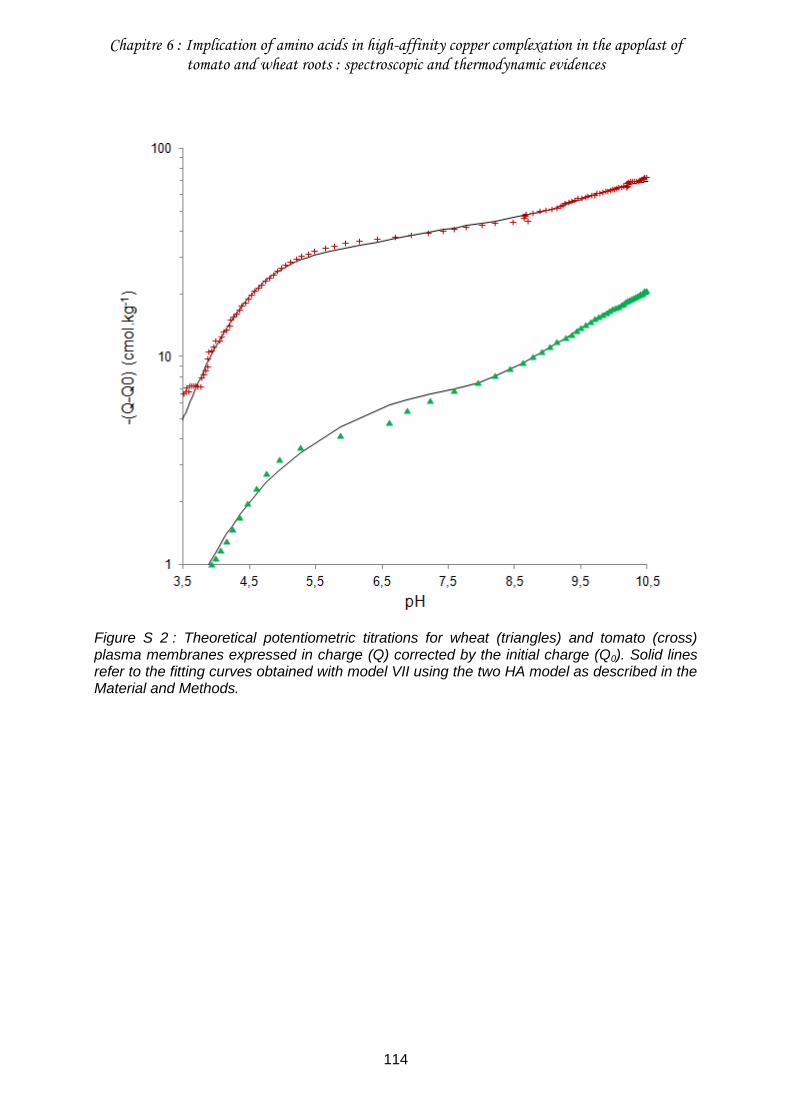
Chapitre 6 : Implication of amino acids in high-affinity copper complexation in the apoplast of tomato and wheat roots : spectroscopic and thermodynamic evidences
114
Figure S 2 : Theoretical potentiometric titrations for wheat (triangles) and tomato (cross) plasma membranes expressed in charge (Q) corrected by the initial charge (Q0). Solid lines refer to the fitting curves obtained with model VII using the two HA model as described in the Material and Methods.
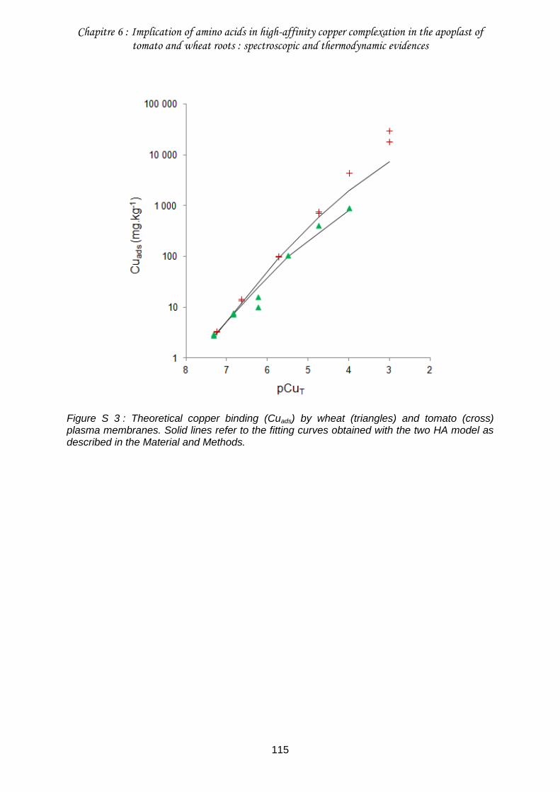
Chapitre 6 : Implication of amino acids in high-affinity copper complexation in the apoplast of tomato and wheat roots : spectroscopic and thermodynamic evidences
115
Figure S 3 : Theoretical copper binding (Cuads) by wheat (triangles) and tomato (cross) plasma membranes. Solid lines refer to the fitting curves obtained with the two HA model as described in the Material and Methods.
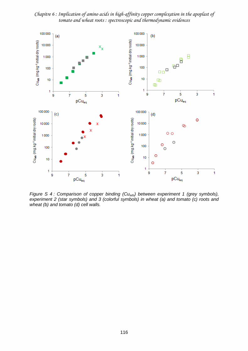
Chapitre 6 : Implication of amino acids in high-affinity copper complexation in the apoplast of tomato and wheat roots : spectroscopic and thermodynamic evidences
116
Figure S 4 : Comparison of copper binding (Cuads) between experiment 1 (grey symbols), experiment 2 (star symbols) and 3 (colorful symbols) in wheat (a) and tomato (c) roots and wheat (b) and tomato (d) cell walls.
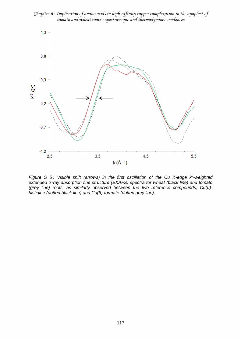
Chapitre 6 : Implication of amino acids in high-affinity copper complexation in the apoplast of tomato and wheat roots : spectroscopic and thermodynamic evidences
117
Figure S 5 : Visible shift (arrows) in the first oscillation of the Cu K-edge k2-weighted extended X-ray absorption fine structure (EXAFS) spectra for wheat (black line) and tomato (grey line) roots, as similarly observed between the two reference compounds, Cu(II)-histidine (dotted black line) and Cu(II)-formate (dotted grey line).
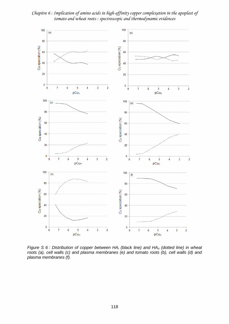
Chapitre 6 : Implication of amino acids in high-affinity copper complexation in the apoplast of tomato and wheat roots : spectroscopic and thermodynamic evidences
118
Figure S 6 : Distribution of copper between HAI (black line) and HAII (dotted line) in wheat roots (a), cell walls (c) and plasma membranes (e) and tomato roots (b), cell walls (d) and plasma membranes (f).
Chapitre 6 : Implication of amino acids in high-affinity copper complexation in the apoplast of tomato and wheat roots : spectroscopic and thermodynamic evidences
119
Références bibliographiques
Belon E, Boisson M, Deportes IZ, Eglin TK, Feix I, Bispo AO, Galsomies L, Leblond S, Guellier CR (2012) An inventory of trace elements inputs to French agricultural soils. Science of The Total Environment 439 (0):87-95. doi:http://dx.doi.org/10.1016/j.scitotenv.2012.09.011
Bravin M, Merrer B, Denaix L, Schneider A, Hinsinger P (2010) Copper uptake kinetics in
hydroponically-grown durum wheat (Triticum turgidum durum L.) as compared with soil’s ability to supply copper. Plant and Soil 331 (1-2):91-104. doi:10.1007/s11104-009-0235-3
Burkhead JL, Gogolin Reynolds KA, Abdel-Ghany SE, Cohu CM, Pilon M (2009) Copper
homeostasis. New Phytologist 182 (4):799-816. doi:10.1111/j.1469-8137.2009.02846.x
Cassab GI (1998) PLANT CELL WALL PROTEINS. Annual Review of Plant Physiology and
Plant Molecular Biology 49 (1):281-309. doi:doi:10.1146/annurev.arplant.49.1.281 Collin B, Doelsch E, Keller C, Cazevieille P, Tella M, Chaurand P, Panfili F, Hazemann J-L,
Meunier J-D (2014) Evidence of sulfur-bound reduced copper in bamboo exposed to high silicon and copper concentrations. Environmental Pollution 187 (0):22-30. doi:http://dx.doi.org/10.1016/j.envpol.2013.12.024
Colzi I, Doumett S, Del Bubba M, Fornaini J, Arnetoli M, Gabbrielli R, Gonnelli C (2011) On
the role of the cell wall in the phenomenon of copper tolerance in Silene paradoxa L. Environmental and Experimental Botany 72 (1):77-83. doi:DOI: 10.1016/j.envexpbot.2010.02.006
Evertsson B (1969) The crystal structure of bis-l-histidinecopper(II) dinitrate dihydrate. Acta
Crystallographica Section B 25 (1):30-41. doi:doi:10.1107/S0567740869001737 Fry SC, Miller JG, Dumville JC (2002) A proposed role for copper ions in cell wall loosening.
Plant and Soil 247 (1):57-67. doi:10.1023/a:1021140022082 Grossoehme NE, Akilesh S, Guerinot ML, Wilcox DE (2006) Metal-Binding Thermodynamics
of the Histidine-Rich Sequence from the Metal-Transport Protein IRT1 of Arabidopsis thaliana. Inorganic Chemistry 45 (21):8500-8508. doi:10.1021/ic0606431
Guigues S, Bravin M, Garnier C, Masion A, Doelsch E (2014) Isolated cell walls exhibit
cation binding properties distinct from those of plant roots. Plant and Soil:1-13. doi:10.1007/s11104-014-2138-1
Hazemann J-L, Proux O, Nassif V, Palancher H, Lahera E, Da Silva C, Braillard A,
Testemale D, Diot M-A, Alliot I, Del Net W, Manceau A, Gelebart F, Morand M, Dermigny Q, Shukla A (2009) High-resolution spectroscopy on an X-ray absorption beamline. Journal of Synchrotron Radiation 16 (2):283-292. doi:doi:10.1107/S0909049508043768
Karlsson T, Elgh-Dalgren K, Skyllberg U (2008) Modeling Copper(II) Complexation in a Peat
Soil Based on Spectroscopic Structural Information All rights reserved. Soil Sci Soc Am J 72 (5):1286-1291. doi:10.2136/sssaj2007.0390
Chapitre 6 : Implication of amino acids in high-affinity copper complexation in the apoplast of tomato and wheat roots : spectroscopic and thermodynamic evidences
120
Kinraide TB (2004) Possible Influence of Cell Walls upon Ion Concentrations at Plasma Membrane Surfaces. Toward a Comprehensive View of Cell-Surface Electrical Effects upon Ion Uptake, Intoxication, and Amelioration. Plant Physiol 136 (3):3804-3813. doi:10.1104/pp.104.043174
Kinraide TB (2009) Improved scales for metal ion softness and toxicity. Environmental
Toxicology and Chemistry 28 (3):525-533. doi:10.1897/08-208.1 Kinraide TB, Pedler JF, Parker DR (2004) Relative effectiveness of calcium and magnesium
in the alleviation of rhizotoxicity in wheat induced by copper, zinc, aluminum, sodium, and low pH. Plant and Soil 259 (1):201-208. doi:10.1023/b:plso.0000020972.18777.99
Konno H, Nakato T, Nakashima S, Katoh K (2005) Lygodium japonicum fern accumulates
copper in the cell wall pectin. Journal of Experimental Botany 56 (417):1923-1931. doi:10.1093/jxb/eri187
Kopittke PM, Blamey FPC, McKenna BA, Wang P, Menzies NW (2011a) Toxicity of metals to
roots of cowpea in relation to their binding strength. Environmental Toxicology and Chemistry 30 (8):1827-1833. doi:10.1002/etc.557
Kopittke PM, Kinraide TB, Wang P, Blamey FPC, Reichman SM, Menzies NW (2011b)
Alleviation of Cu and Pb Rhizotoxicities in Cowpea (Vigna unguiculata) as Related to Ion Activities at Root-Cell Plasma Membrane Surface. Environmental Science & Technology 45 (11):4966-4973. doi:10.1021/es1041404
Kopittke PM, Menzies NW, de Jonge MD, McKenna BA, Donner E, Webb RI, Paterson DJ,
Howard DL, Ryan CG, Glover CJ, Scheckel KG, Lombi E (2011c) In Situ Distribution and Speciation of Toxic Copper, Nickel, and Zinc in Hydrated Roots of Cowpea. Plant Physiology 156 (2):663-673. doi:10.1104/pp.111.173716
Kopittke PM, Menzies NW, Wang P, McKenna BA, Wehr JB, Lombi E, Kinraide TB, Blamey
FPC (2014) The rhizotoxicity of metal cations is related to their strength of binding to hard ligands. Environmental Toxicology and Chemistry 33 (2):268-277. doi:10.1002/etc.2435
Legros S, Doelsch E, Feder F, Moussard G, Sansoulet J, Gaudet JP, Rigaud S, Doelsch IB,
Macary HS, Bottero JY (2013) Fate and behaviour of Cu and Zn from pig slurry spreading in a tropical water–soil–plant system. Agriculture, Ecosystems & Environment 164 (0):70-79. doi:http://dx.doi.org/10.1016/j.agee.2012.09.008
Manceau A, Marcus MA, Tamura N (2002) Quantitative Speciation of Heavy Metals in Soils
and Sediments by Synchrotron X-ray Techniques. Reviews in Mineralogy and Geochemistry 49 (1):341-428. doi:10.2138/gsrmg.49.1.341
Manceau A, Simionovici A, Lanson M, Perrin J, Tucoulou R, Bohic S, Fakra SC, Marcus MA,
Bedell J-P, Nagy KL (2013) Thlaspi arvense binds Cu(ii) as a bis-(l-histidinato) complex on root cell walls in an urban ecosystem. Metallomics 5 (12):1674-1684. doi:10.1039/c3mt00215b
Marschner H (1995) Mineral Nutrition of higher plants. Academic Press, second edition Meychik NR, Yermakov IP (2001) Ion exchange properties of plant root cell walls. Plant and
Soil 234 (2):181-193. doi:10.1023/a:1017936318435
Chapitre 6 : Implication of amino acids in high-affinity copper complexation in the apoplast of tomato and wheat roots : spectroscopic and thermodynamic evidences
121
Moore KL, Schröder M, Wu Z, Martin BGH, Hawes CR, McGrath SP, Hawkesford MJ, Feng Ma J, Zhao F-J, Grovenor CRM (2011) High-Resolution Secondary Ion Mass Spectrometry Reveals the Contrasting Subcellular Distribution of Arsenic and Silicon in Rice Roots. Plant Physiology 156 (2):913-924. doi:10.1104/pp.111.173088
Murakami T, Ise K, Hayakawa M, Kamei S, Takagi S-i (1989) Stabilities of Metal Complexes
of Mugineic Acids and Their Specific Affinities for Iron(III). Chemistry Letters 18 (12):2137-2140. doi:10.1246/cl.1989.2137
Proux O, Nassif V, Prat A, Ulrich O, Lahera E, Biquard X, Menthonnex J-J, Hazemann J-L
(2006) Feedback system of a liquid-nitrogen-cooled double-crystal monochromator: design and performances. Journal of Synchrotron Radiation 13 (1):59-68. doi:doi:10.1107/S0909049505037441
Ravel B, Newville M (2005) ATHENA, ARTEMIS, HEPHAESTUS : data analysis for X-ray
absorption spectroscopy using IFEFFIT. Journal of Synchrotron Radiation 12:537 - 541.
Sarret G, Vangronsveld J, Manceau A, Musso M, D'Haen J, Menthonnex J-J, Hazemann J-L
(2001) Accumulation Forms of Zn and Pb in Phaseolus vulgaris in the Presence and Absence of EDTA. Environmental Science & Technology 35 (13):2854-2859. doi:10.1021/es000219d
Shomer I, Novacky AJ, Pike SM, Yermiyahu U, Kinraide TB (2003) Electrical Potentials of
Plant Cell Walls in Response to the Ionic Environment. Plant Physiology 133 (1):411-422. doi:10.1104/pp.103.024539
Stanila A, Marcu A, Rusu D, Rusu M, David L (2007) Spectroscopic studies of some
copper(II) complexes with amino acids. Journal of Molecular Structure 834–836 (0):364-368. doi:10.1016/j.molstruc.2006.11.048
Synytsya A, Urbanová M, Setnička V, Tkadlecová M, Havlíček J, Raich I, Matějka P,
Synytsya A, Čopıková J, Volka K (2004) The complexation of metal cations by d-galacturonic acid: a spectroscopic study. Carbohydrate Research 339 (14):2391-2405. doi:10.1016/j.carres.2004.07.008
Taiz L, Zeiger E (2006) Plant Physiology, 4th Edition. Sinauer Associates Sunderland, MA Tipping E (1998) Humic Ion-Binding Model VI: An Improved Description of the Interactions of
Protons and Metal Ions with Humic Substances. Aquatic Geochemistry 4 (1):3-47. doi:10.1023/a:1009627214459
Tipping E, Lofts S, Sonke JE (2011) Humic Ion-Binding Model VII: a revised parameterisation
of cation-binding by humic substances. Environmental chemistry 8 (3):225-235. doi:http://dx.doi.org/10.1071/EN11016
Vulkan R, Yermiyahu U, Mingelgrin U, Rytwo G, Kinraide TB (2004) Sorption of Copper and
Zinc to the Plasma Membrane of Wheat Root. Journal of Membrane Biology 202 (2):97-104. doi:10.1007/s00232-004-0722-7

122

123
3ème
partie
Développement
d’un modèle de
complexation
racinaire

124
Chapitre 7 : Predicting copper competitive binding on terrestrial plant roots using a two humic-acid model
125
Chapitre 7 : Développement d’un modèle de
prédiction de la complexation du cuivre
dans les racines
Comme présenté dans le chapitre 3, divers modèles de prédiction sont actuellement
disponibles dans la littérature. Parmi ces modèles, le TBLM (Terrestrial Biotic Ligand Model)
est le plus communément employé. Les études utilisant le TBLM présentent des résultats
encourageants lorsqu’elles sont individuellement considérées. Cependant, leur comparaison
révèle un manque de généricité de certains paramètres et la multiplication des cas
particuliers où l’application du TBLM est limitée témoigne d’une nécessité de remettre en
cause certaines hypothèses du formalisme mathématique sous-jacent.
L’article Predicting copper competitive binding on terrestrial plant roots using a two humic-
acid model, constituant ce chapitre 7, présente le modèle de prédiction de la complexation
du cuivre dans les racines que j’ai développé. Ce nouveau modèle est fondé sur un
formalisme mathématique plus élaboré que celui du modèle TBLM (différentes natures de
sites réactifs, prise en compte des effets électrostatiques, etc.). Il remet également en cause
la démarche de modélisation classiquement employée puisqu’il n’a pas été paramétré par
l’ajustement de données expérimentales de toxicité mais par l’ajustement de données
expérimentales concernant le processus effectivement modélisé, c’est à dire la complexation
du Cu sur les racines.
Ainsi, nous avons montré que la complexation du cuivre dans les racines des deux plantes
modèles ne peut pas être décrite de manière simple. L’inaptitude de WHAM (Windermere
Humic Acid Model) paramétré par défaut à représenter cette complexation se traduit comme
une impossibilité d’assimiler la réactivité des racines des plantes à celle des substances
humiques, comme cela a pu être fait pour les organismes aquatiques. A partir du formalisme
mathématique de WHAM, nous avons donc développé un modèle plus élaboré, adapté aux
plantes supérieures terrestres : le WHAM-Terrestrial Higher Plants (WHAM-THP). Grâce à
une prédiction fine des propriétés acides des racines, ce nouveau modèle a été capable de
prédire l’adsorption du cuivre sur les racines de blé et de tomate, et pour des
environnements chimiques très variables (force ionique, pH et cations compétiteurs). Malgré
les différences de réactivité qui existent entre les racines de blé et de tomate (cf. chapitre 6),
nous avons réussi à proposer un jeu de paramètres communs pour les deux espèces. Dans
un souci de simplicité d’utilisation, nous avons également proposé, sur la base d’une
Chapitre 7 : Predicting copper competitive binding on terrestrial plant roots using a two humic-acid model
126
comparaison entre des mesures de capacité d’échange cationique racinaire (CECR) et des
titrages acido-basiques, une relation qui permettra au futur utilisateur d’estimer assez
précisément la densité totale de sites racinaires à partir d’une simple mesure de CECR
pouvant être réalisée en routine.
Chapitre 7 : Predicting copper competitive binding on terrestrial plant roots using a two humic-acid model
127
Predicting copper competitive binding on
terrestrial plant roots
using a two humic-acid model
Stéphanie Guigues 1,2, Matthieu N. Bravin 3, Cédric Garnier 4 and Emmanuel Doelsch 1
1 CIRAD, UPR Recyclage et risque, F-34398 Montpellier, France
2 ADEME, 20 avenue du Grésillé, BP-90406, Angers cedex 01, France
3 CIRAD, UPR Recyclage et risque, F-97408 Saint-Denis, Réunion, France
4 Université de Toulon, PROTEE, EA 3819, 83957 La Garde, France
Abstract
The Terrestrial Biotic Ligand Model (TBLM) is widely used to predict the toxic effect of trace
metals for various soil organisms including higher plants. However, its formalism remains
based on strong assumptions that may partly explain inconsistent results observed in the
fitted binding constants of trace metals to plant roots. To improve this, we aimed at adapting
the parameterization of the humic acid defined in the Windermere Humic Aqueous Model
(WHAM) for terrestrial higher plants (THP). Contrary to WHAM parameterized by default,
WHAM-THP was able with a unique set of parameters to predict adequately the acidic
properties and the binding of copper on the root surfaces of wheat (Triticum aestivum L.) and
tomato (Solanum lycopersicum L.), while accounting for the electrostatic effect and the
competition with proton, calcium and zinc.
Keywords: WHAM; complexation; trace element; modeling; heavy metal
Chapitre 7 : Predicting copper competitive binding on terrestrial plant roots using a two humic-acid model
128
I. Introduction
Soil contamination still remains a major environmental concern. Among contaminants, the
particular case of trace metals is worrying as e.g. they exhibit the highest occurrence in
European soils (European Environmental Agency 2007). Atmospheric deposition related to
industrial activities, urban traffic, mining activities and agricultural practices (organic waste
spreading, use of pesticides and fungicides) contribute to the enrichment of trace metals in
soils (Hooda 2010). Hence, a major challenge for the environmental risk assessment of trace
metals in soil is the development of predictive ecotoxicological tools able to estimate the toxic
effect of trace metals on soil organisms. Toxicity to soil organisms is usually poorly correlated
to the total concentration of trace metals in soil but rather depends on the speciation of trace
metals, especially in the soil solution where trace metals are taken up (Harmsen 2007). The
toxic effect is further determined by the interaction of trace metals with the outer surface of
soil organisms. This interaction is strongly influenced by the chemical parameters of the soil
solution such as pH, ionic strength and the concentrations of other competitive metal cations
(Brown and Markich 2000).
Among existing models, the biotic ligand model (BLM) is increasingly used in predictive
ecotoxicology. Based on the determination of trace metals speciation in solution, the BLM is
designed to estimate trace metal toxicity as a function of site-specific competitions between
cations (e.g. H+, Ca2+ and Mg2+) and trace metals for binding to the biotic ligand borne by the
outer surface of an organism (Di Toro et al. 2001). Initially dedicated to the prediction of trace
element toxicity to aquatic organisms (Paquin et al. 2002), a terrestrial BLM (TBLM) was
developed in the past decades for soil organisms such as plants, invertebrates and
microorganisms (Thakali et al. 2006; Ardestani et al. 2014). Among these soil organisms, the
TBLM seems to be an approach particularly promising for plants for which the mechanistic
basis is particularly consistent with the major mechanism involved in the occurrence of trace
metal toxicity to plant roots, i.e. rhizotoxicity. Indeed, Kopittke et al. (2014) recently
contended “that the primary mechanism of rhizotoxicity of many metal cations is nonspecific
and that the magnitude of toxic effects is positively related to the strength with which they
bind to hard ligands, especially carboxylate ligands of the cell-wall pectic matrix”.
Consequently, the ability of the TBLM formalism to describe accurately the binding of trace
metals on plant root surface is a crucial issue.
However, the mathematical formalism of the TBLM is based on several strong assumptions
that question its relevancy for application in predictive ecotoxicological risk assessment
(Slaveykova and Wilkinson 2005; Erickson 2013). The TBLM notably assumes (i) the
homogeneity of the nature, the distribution and the affinity of the binding sites, (ii) that
Chapitre 7 : Predicting copper competitive binding on terrestrial plant roots using a two humic-acid model
129
electrostatic effects do not significantly alter trace metal binding and (iii) that trace metals
bind to the biotic ligand only with a 1:1, i.e. monodentate, coordination stoichiometry. The
validity of these three assumptions are clearly questionable since they were all rejected and
reconsidered in the current modeling approaches of trace metal binding on humic
substances, i.e. another kind of complex and natural organic matters (Milne et al. 2003;
Tipping et al. 2011). An additional concern in the implementation of the TBLM is the
widespread practice of fitting binding constants from the measurements of toxic endpoints
rather than actual binding. It is therefore not surprising to find binding constants varying over
several orders of magnitude, even when restricting the comparison to the same plant
species, exposure condition and toxic endpoint (Ardestani et al. 2014). For example, the
binding constant of Cu to the biotic ligand determined for the root elongation of Hordeum
vulgare in hydroponics ranged between logK ≤ 3.9 (Antunes et al. 2007) and logK=6.3 (Wang
et al. 2012b). The considerable variability in the parameterization and the apparent
weaknesses in the underlying hypothesis of the mathematical formalism concomitantly
suggest the necessity to revisit and likely to further improve the implementation of the TBLM
(Erickson 2013).
Accordingly, geochemical models initially designed to predict trace metal binding on humic
susbtances were applied in predictive ecotoxicology (Plette et al. 1996; Tipping et al. 2008).
The Windermere Humic Aqueous Model (WHAM) more particularly was used successfully in
the past few years to predict the binding and toxicity of trace metals to a range of aquatic
organisms including bacteria, algae, invertebrates and lower and higher plants (Tipping et al.
2008; Antunes et al. 2012; Iwasaki et al. 2013). We recently characterized the Cu binding
properties of the root surface of two higher plants, namely wheat (Triticum aestivum L.) and
tomato (Solanum lycopersicum L.), by implementing a two humic-acid (HA) model based on
WHAM (Chapter 6). Consistently with spectroscopic investigations, this two-HA model
enabled us to demonstrate that Cu binding on plant roots was driven by two types of sites
occurring in ca. equivalent amount, i.e. low- and high-affinity sites corresponding to
carboxylic and amine functional groups respectively. Foreshadowing potential improvement
of the current TBLM formalism, the application of WHAM for terrestrial higher plants,
hereafter called WHAM-THP, should deserve further attention.
Consequently, the present study aimed at developing a generic WHAM-THP for Cu by
parameterizing its complexation on the root surface of wheat and tomato under varying
chemical conditions (i.e. ionic strength, pH and competitive cations). Wheat and tomato were
chosen as the two model plant species to be respectively representative of monocots and
dicots which are known to differ substantially in the composition and the cation binding
properties of their root surfaces (Sattelmacher 2001; Vogel 2008).
Chapitre 7 : Predicting copper competitive binding on terrestrial plant roots using a two humic-acid model
130
II. Experimental approach
II.1. Plant root material
Wheat (cv. Premio) and tomato (cv. Moneymaker) were grown in hydroponics for 21 days,
i.e. 7 days of germination in darkness and 14 days of growth (see Guigues et al. (2014) for
additional information). The growth chamber parameters were set at (day/night): 25/20°C,
75/70% relative humidity and 16/8h with a photon flux density of 450 µmol photons m-2 s-1
during the day. At harvest, roots were separated from shoots, then blotted with paper towels
before being subdivided into homogenous subsamples and finally stored frozen.
After thawing, roots were rinsed with 1 mM Ca(NO3)2 to eliminate vacuolar compounds
released during thawing. Therefore, the root material (hereafter are referred to as roots)
obtained was assumed to do not exhibit any biological activity, allowing us to overcome
cellular absorption phenomena and to study only binding reactions. Furthermore, these roots
did contain both cell walls and plasma membranes, i.e. two plant compartments that give to
roots their binding properties (Guigues et al. 2014; Chapter 6). Before any further
experiment, roots were stirred in HNO3 solution at pH 3 for 1 h to remove highly bound or
precipitated cations (e.g. Fe and Al), then rinsed twice with ultrapure water (18.2 MΩ) for
30 min. Roots were then oven-dried at 50°C until a steady mass was achieved.
II.2. Potentiometric titration
Potentiometric titration experiments were extensively described by Guigues et al. (2014).
Briefly, ca. 0.2 g (dry mass basis) of roots was placed in 100 ml of 10 mM KNO3. During
titrations, pH was first lowered to 2.5 with 0.2 M HNO3 additions and was then increased
step-by-step to 11.5 with the incremental additions of 0.2 M KOH of either 100 µl in the pH
ranges 2.5-3.5 and 10.5-11.5 or 20 µl in the pH range 3.5-10.5.
II.3. Copper sorption experiments
A dry mass of 10 (± 0.5) mg of wheat and tomato roots was shaken end-over-end for 24 h at
25°C in 25 ml of solution of varying chemical composition (Tables S3 and S4). In the
experiment 1, the solution contained an initial, total Cu concentration varying from pCuT 7.3
to 3.0 (pCuT = -log10[Cu]T) with an ionic strength and a pH fixed at 0.03 M with NaNO3 and
4.7 (± 0.2), respectively.
Chapitre 7 : Predicting copper competitive binding on terrestrial plant roots using a two humic-acid model
131
In the experiment 2, the solution contained an ionic strength fixed at 0.6 mM or 0.3 M with
NaNO3, an initial, total Cu concentration fixed at pCuT 4.2, 5.2 or 6.2 and a pH fixed at 4.7
(± 0.2).
In the experiment 3, the solution contained a pH fixed at 4.1 (± 0.1) or 6.3 (± 0.1), an initial,
total Cu concentration fixed at pCuT 4.2, 5.2 or 6.2 and an ionic strength fixed at 0.03 M with
NaNO3.
In the experiment 4, the solution contained an initial, total Ca concentration varying from
pCaT 2.0 to 4.0 but with an initial, total Cu concentration, an ionic strength and a pH fixed at
pCuT 6.3, 0.03 M with NaNO3 and 5.1 (± 0.4), respectively.
In the experiment 5, the solution contained an initial, total Zn concentration varying from
pZnT 4.5 to 7.2 but with an initial, total Cu concentration, an ionic strength and a pH fixed at
pCuT 6.3, 0.03 M with NaNO3 and 4.7 (± 0.1), respectively.
The pH was buffered with 1 mM 2-[N-morpholino] ethane sulfonic acid and adjusted by
NaOH or HNO3. Sorption experiments were performed in duplicate and blanks were included
in the measurements. Copper concentration in initial and final (i.e. at equilibrium) solution
was determined by inductively coupled plasma mass spectrometry (ICP-MS, NexION 300X
Perkin Elmer). Blanks and certified reference material (EnviroMAT Driking water EP-L-3 and
groundwater ES-H-2) were included in the analyses. The measurement uncertainty was
lower than 10%. The amount of Cu bound to wheat and tomato roots was calculated by
subtracting the final to the initial Cu concentration measured in each batch.
III. Modeling approach
The modeling approach was based on the formalism of the Humic Ion-Binding Model
included in WHAM VII. This formalism was extensively described by Tipping (1998) and
Tipping et al. (2011). To summarize, WHAM is able to simulate the sorption properties of
humic substances depicted as a regular array of binding sites of type 1 and 2. The density
(LHi, cmolc.kg-1) of type-1 sites is arbitrarily fixed as twice higher than the density of the type-2
sites (i.e. LH1 = 2 LH2). Protons and metal ions compete for sorption on these sites with
metal ions being able to form mono-, bi- and tri-dendate complexes. Proton sorption to humic
substances were characterized by one intrinsic proton dissociation constant for each type of
sites, i.e. pKa1 and pKa2, and the corresponding distribution terms, ΔpKa1 and ΔpKa2. Metal
sorption to humic substances was characterized by one intrinsic equilibrium constant for
each type of sites, i.e. KM,1 and KM,2, and a heterogeneity parameter, ΔLK2M.
Chapitre 7 : Predicting copper competitive binding on terrestrial plant roots using a two humic-acid model
132
The parameter KM,2 was calculated as follows (Tipping et al. 2011) :
𝐿𝑜𝑔𝐾𝑀,2 = 𝐿𝑜𝑔𝐾𝑀,1 ×𝑝𝐾𝑎2
𝑝𝐾𝑎1 (1)
Electrostatic effects are accounted for in WHAM by approximating the diffuse layer/bulk
solution system with a Donnan model. While WHAM can account for the complexation of the
free ionic form and the first hydrolysis product of each metal, we only accounted for Cu2+ as a
preliminary speciation calculation showed that Cu2+ stood for > 95 % of total Cu in solution
with pH ≤ 6.3. The partial pressure of CO2 was assumed to be that of the ambient
atmosphere (10-3.5 atm) and the temperature was fixed at 25 °C for calculations.
Toward the development of a predictive tool, our modeling approach aimed at fitting the
experimental data as accurately as possible with the most squeezed and generic set of
parameters for the two plant species tested. Accordingly, WHAM was firstly implemented by
representing root binding properties with a single HA exhibiting the default parameterization
given in WHAM except for the total site density of wheat and tomato roots that was derived
from a previous work (Guigues et al. 2014). WHAM was also configured with two HA
(configuration hereafter referred as to WHAM-2HA) to agree with the actual distribution of
low- and high-pKa sites on wheat and tomato roots. In WHAM-2HA (table S5), the first HA
represented the low-pKa sites while the second HA represented the high-pKa sites, each HA
respectively exhibiting the same parameterization as the type-1 and type-2 sites defined in
WHAM. Some unsatisfactory fits (see Results and discussion section for rationale) led
however to develop a more complex model.
WHAM-THP was thus implemented by representing root binding properties with two
concomitant HA (HAI and HAII) independently parameterized and that stand for the low-and
the high-pKa sites, respectively. The total density of HAI and HAII sites was derived from a
previous work (Guigues et al. 2014). The implementation of WHAM-THP started with the
parameterization of pKa1, pKa2, ΔpKa1 and ΔpKa2 for HAI and HAII by fitting experimental
titration curves of wheat and tomato roots. Then, KCu,1, KCu,2 and ΔLK2Cu was parameterized
by fitting Cu sorption data from experiment 1. The ability of WHAM-THP to predict ionic
strength and pH effects on Cu binding was tested by fitting Cu sorption data from
experiments 2 and 3. Finally, the ability of WHAM-THP to account for the competitive effect
of Ca and Zn was assessed by parameterizing KCa,1, KCa,2, ΔLK2Ca, KZn,1, KZn,2 and ΔLK2Zn
using Cu sorption data from experiments 4 and 5.
WHAM, WHAM-2HA and WHAM-THP best fits were determined by minimizing the root mean
square residual.
Chapitre 7 : Predicting copper competitive binding on terrestrial plant roots using a two humic-acid model
133
IV. Results and discussion
Initially developed to simulate trace metal binding on humic substances, WHAM was applied
in the past few years in predictive ecotoxicology by suggesting humic acid as a relevant
surrogate of the biotic ligand. On this principle, the present study was focused on the
development of WHAM for terrestrial higher plants (i.e. WHAM-THP) to predict Cu binding on
root surfaces under varying chemical conditions.
IV.1. Ability of WHAM-THP to predict root acidic properties
WHAM failed to fit the experimental data for both wheat and tomato. WHAM overestimated
the binding site density depicted by experimental data over the major part of the pH range
investigated (up to 9 cmolc.kg-1 for wheat and 14 cmolc.kg-1 for tomato) with a shape that
differed substantially from that of experimental data (Fig. 27; table 5). This inadequacy of
WHAM partly came from the imposed relationship between the density of type-1 and type-2
sites, i.e. LH1 = 2 LH2. Indeed, the distribution of low- and high-pKa sites on wheat and
tomato roots did not meet this requirement as low to high-pKa sites ratio was equal to 0.4
and 0.9, respectively (Guigues et al. 2014). Similarly, other biotic ligands from other plant
roots and bacteria cell walls exhibited low to high-pKa sites ratio different from 2, ranging
from 1.4 to 3 (Plette et al. 1995; Ginn et al. 2008; Wu and Hendershot 2009; Mishra et al.
2010). Such results mean that the nature of binding sites borne by biotic ligands differs
substantially from the nature of binding sites borne by humic substances.
WHAM-2HA only improved satisfactorily the fit of potentiometric data for tomato roots at
pH ≤ 6 while potentiometric data for tomato roots at pH > 6 and wheat roots remained very
poorly fitted (Fig. 27). Hence, the parameters describing proton dissociation (i.e. pKa and
pKa) of HA in WHAM were also inadequate for wheat and tomato roots. This led us to
conclude that the acidic properties of plant roots are much different from those of HA and
thus require to be specifically parameterized for terrestrial higher plants.
Accordingly, WHAM-THP was specifically parameterized and thus allowed to precisely
represent potentiometric titration of wheat and tomato roots with a unique set of parameters
for both plant species (Fig. 27, table 5). Beyond its specific parameterization, the better fits
obtained with WHAM-THP also partly came from the larger number of parameters fitted,
which resulted in a more flexible model. Indeed, while eight different pKa were defined in
WHAM and WHAM-2HA, thirteen different pKa were defined in WHAM-THP (tables 5
and S5).
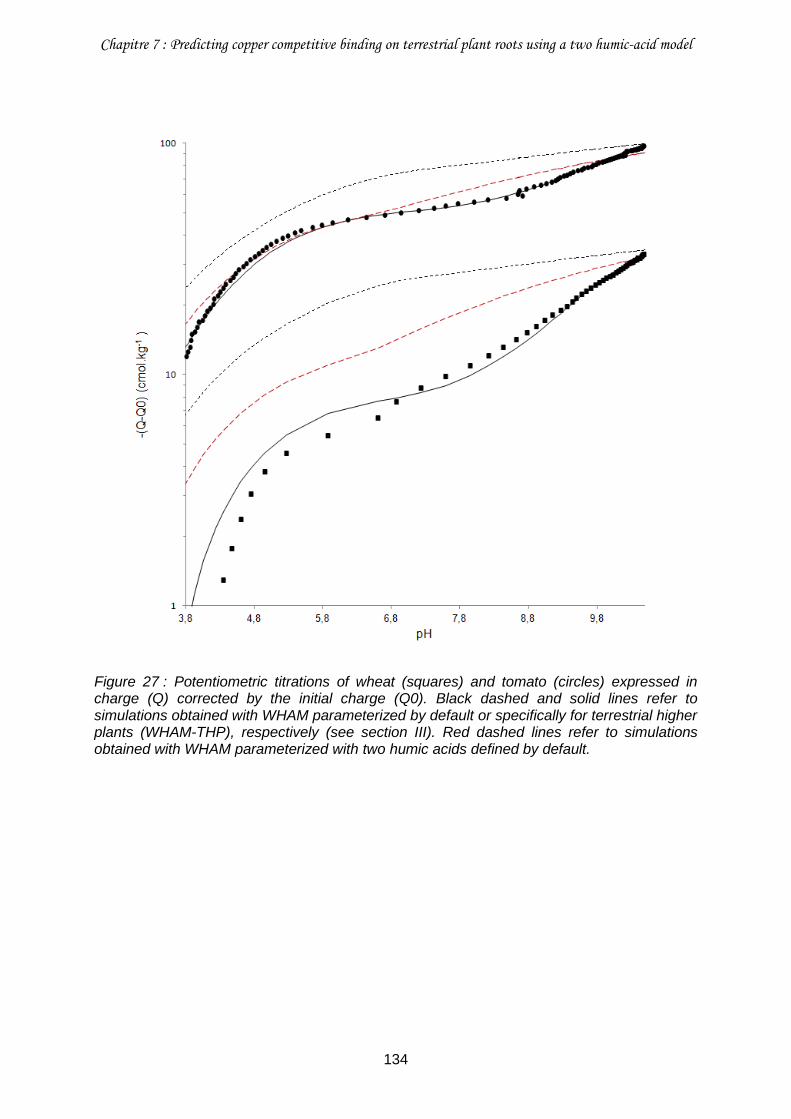
Chapitre 7 : Predicting copper competitive binding on terrestrial plant roots using a two humic-acid model
134
Figure 27 : Potentiometric titrations of wheat (squares) and tomato (circles) expressed in charge (Q) corrected by the initial charge (Q0). Black dashed and solid lines refer to simulations obtained with WHAM parameterized by default or specifically for terrestrial higher plants (WHAM-THP), respectively (see section III). Red dashed lines refer to simulations obtained with WHAM parameterized with two humic acids defined by default.
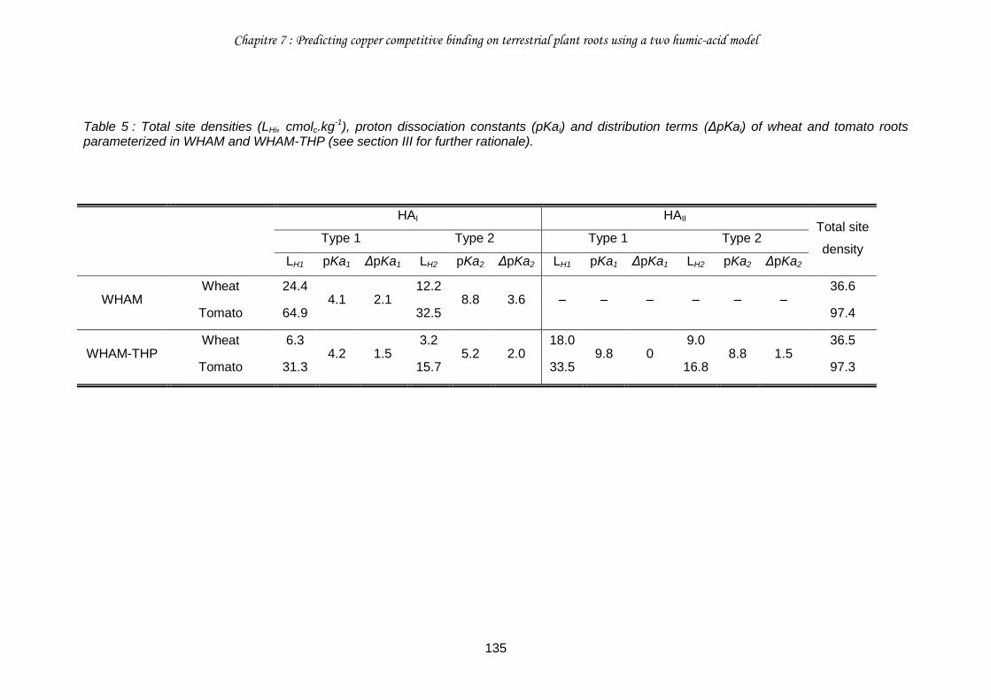
Chapitre 7 : Predicting copper competitive binding on terrestrial plant roots using a two humic-acid model
135
Table 5 : Total site densities (LHi, cmolc.kg-1), proton dissociation constants (pKai) and distribution terms (ΔpKai) of wheat and tomato roots parameterized in WHAM and WHAM-THP (see section III for further rationale).
HAI HAII Total site
density Type 1 Type 2 Type 1 Type 2
LH1 pKa1 ΔpKa1 LH2 pKa2 ΔpKa2 LH1 pKa1 ΔpKa1 LH2 pKa2 ΔpKa2
WHAM Wheat 24.4
4.1 2.1 12.2
8.8 3.6 ‒ ‒ ‒ ‒ ‒ ‒ 36.6
Tomato 64.9 32.5 97.4
WHAM-THP Wheat 6.3
4.2 1.5 3.2
5.2 2.0 18.0
9.8 0 9.0
8.8 1.5 36.5
Tomato 31.3 15.7 33.5 16.8 97.3
Chapitre 7 : Predicting copper competitive binding on terrestrial plant roots using a two humic-acid model
136
IV.2. Ability of WHAM-THP to predict copper binding on roots
IV.2.a. Root copper binding affinity
Although Cu sorption on wheat and tomato roots was contrasted, WHAM fitted Cu sorption
data for both species (Fig. 28) much better than it fitted potentiometric data (Fig. 27).
However, WHAM overestimated Cu bound on wheat roots by 67% on average, with an
increase of the overestimation as Cu concentration in solution increases (Fig. 28a). WHAM-
THP fitted very precisely (deviation < 10%) Cu binding on wheat roots. Both WHAM and
WHAM-THP fitted adequately Cu binding on tomato roots (i.e. with an average deviation of
20% and < 10%, respectively) for pCuT > 4.5 (i.e. pCueq > 5; table S4), a concentration range
relevant for Cu rhizotoxicity as it encompasses the usual concentration (equivalent to
pCueq > 5) that was shown to cause a 50% reduction in the elongation rate of cowpea roots
(Kopittke et al. 2010). However, a two-fold gap with experimental data was observed for
WHAM and WHAM-THP at pCuT < 4.5 (Fig. 28b; Chapter 6). Similar trends were also
observed for WHAM-2HA (Fig. 28). However, the fit obtained with WHAM-2HA at elevated
concentrations of Cu in solution was better than that obtained with WHAM (but not than that
obtained with WHAM-THP) for wheat roots. For tomato roots, the fit obtained with WHAM-
2HA was worse than that obtained with WHAM and WHAM-THP with a higher
underestimation of Cu binding.
The logKCu,1 of HAI sites (i.e. 2.4) fixed by default in WHAM was intermediate between the
logKCu,1 (i.e. 2.2) and the logKCu,2 (i.e. 2.7) of HAI sites specifically defined for WHAM-THP
(table 6). However, the logKCu,2 (i.e. 5.1) of HAI sites fixed by default in WHAM was much
lower than the logKCu,1 (i.e. 6.0) and the logKCu,2 (i.e. 6.7) of HAII sites specifically defined for
WHAM-THP (table 6). This difference between WHAM and WHAM-THP can be attributed to
the nature of the binding sites exhibiting a high affinity for Cu which are typically phenolic
groups on HA (Tipping 1998), while they were shown to be amine groups on wheat and
tomato roots (chapter 6). Even if both phenolic and amines groups have a higher Cu affinity
than carboxylic groups, amine groups presented a particularly high Cu affinity (Fry et al.
2002), even higher than that of phenolic groups. The heterogeneity parameter, ΔLK2Cu,I (i.e.
2.3) defined by default in WHAM is however much higher than the two ΔLK2Cu specifically
defined in WHAM-THP as they were both fixed at zero (table 6). This heterogeneity
parameter is used in WHAM to account for the occurrence of binding sites typically
corresponding to sulfhydryl and amine groups borne by HA that occurred at low
concentration but that exhibited a high affinity towards Cu (Tipping 1998). Whereas amine
groups is a major type of binding sites on wheat and tomato roots accounted for in WHAM-
THP by the parameterization of the HAII, sulfhydryl groups would not participate significantly
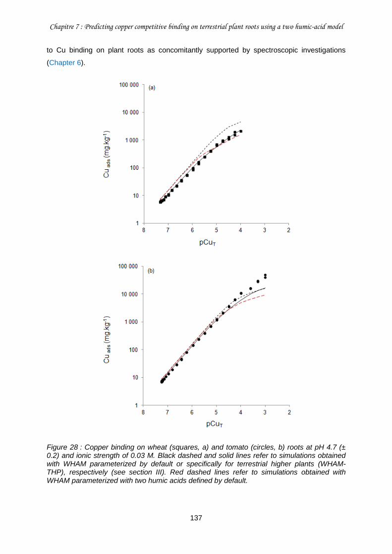
Chapitre 7 : Predicting copper competitive binding on terrestrial plant roots using a two humic-acid model
137
to Cu binding on plant roots as concomitantly supported by spectroscopic investigations
(Chapter 6).
Figure 28 : Copper binding on wheat (squares, a) and tomato (circles, b) roots at pH 4.7 (± 0.2) and ionic strength of 0.03 M. Black dashed and solid lines refer to simulations obtained with WHAM parameterized by default or specifically for terrestrial higher plants (WHAM-THP), respectively (see section III). Red dashed lines refer to simulations obtained with WHAM parameterized with two humic acids defined by default.
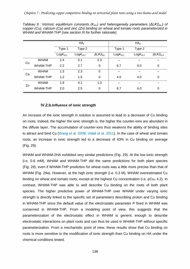
Chapitre 7 : Predicting copper competitive binding on terrestrial plant roots using a two humic-acid model
138
Tableau 6 : Intrinsic equilibrium constants (KM,i) and heterogeneity parameters (ΔLK2M,i) of copper (Cu), calcium (Ca) and zinc (Zn) binding on wheat and tomato roots parameterized in WHAM and WHAM-THP (see section III for further rationale).
HAI HAII
Type 1 Type 2 Type 1 Type 2
LogKM,1 LogKM,2 ΔLK2M,I LogKM,1 LogKM,2 ΔLK2M,II
Cu WHAM 2.4 5.1 2.3 ‒ ‒ ‒
WHAM-THP 2.2 2.7 0 6.7 6.0 0
Ca WHAM 1.3 2.3 0 ‒ ‒ ‒
WHAM-THP 1.2 1.5 0 4.5 4.0 0
Zn WHAM 1.9 4.1 1.3 ‒ ‒ ‒
WHAM-THP 2.0 2.5 0 6.7 6.0 0
IV.2.b.Influence of ionic strength
An increase of the ionic strength in solution is assumed to lead to a decrease of Cu binding
on roots. Indeed, the higher the ionic strength is, the higher the counter-ions are abundant in
the diffuse layer. The accumulation of counter-ions thus weakens the ability of binding sites
to attract and bind Cu (Wang et al. 2008; Vidali et al. 2011). In the case of wheat and tomato
roots, an increase in ionic strength led to a decrease of 43% in Cu binding on average
(Fig. 29).
WHAM and WHAM-2HA exhibited very similar predictions (Fig. 29). At the low ionic strength
(i.e. 0.6 mM), WHAM and WHAM-THP did the same predictions for both plant species
(Fig. 29), even if WHAM-THP prediction for wheat roots was a little more precise than that of
WHAM (Fig. 29a). However, at the high ionic strength (i.e. 0.3 M), WHAM overestimated Cu
binding on wheat and tomato roots, except at the highest Cu concentration (i.e. pCuT 4.2). In
contrast, WHAM-THP was able to well describe Cu binding on the roots of both plant
species. The higher predictive power of WHAM-THP over WHAM under varying ionic
strength is directly linked to the specific set of parameters describing proton and Cu binding
in WHAM-THP since the default value of the electrostatic parameter P fixed in WHAM was
conserved in WHAM-THP. From a modeling point of view, this suggests that the
parameterization of the electrostatic effect in WHAM is generic enough to describe
electrostatic interactions on plant roots and can thus be used in WHAM-THP without specific
parameterization. From a mechanistic point of view, these results show that Cu binding on
roots is more sensitive to the modification of ionic strength than Cu binding on HA under the
chemical conditions tested.
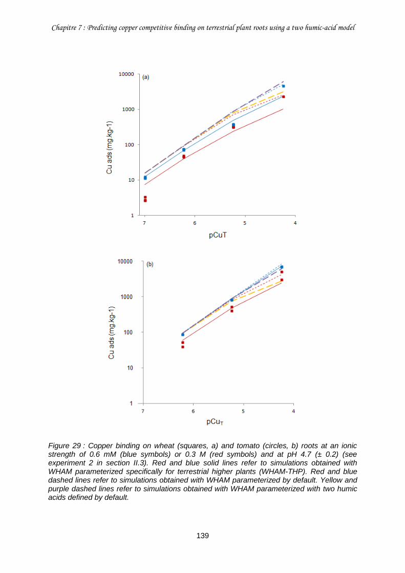
Chapitre 7 : Predicting copper competitive binding on terrestrial plant roots using a two humic-acid model
139
Figure 29 : Copper binding on wheat (squares, a) and tomato (circles, b) roots at an ionic strength of 0.6 mM (blue symbols) or 0.3 M (red symbols) and at pH 4.7 (± 0.2) (see experiment 2 in section II.3). Red and blue solid lines refer to simulations obtained with WHAM parameterized specifically for terrestrial higher plants (WHAM-THP). Red and blue dashed lines refer to simulations obtained with WHAM parameterized by default. Yellow and purple dashed lines refer to simulations obtained with WHAM parameterized with two humic acids defined by default.
Chapitre 7 : Predicting copper competitive binding on terrestrial plant roots using a two humic-acid model
140
IV.2.c. Proton competition
As pH increases, amount of Cu bound to roots would increase because root surfaces
become more negatively charged, thus resulting in a lower competition between H+ and Cu2+
for binding on roots (Haque et al. 2007; Bulgariu and Bulgariu 2012). Indeed, the quantity of
Cu bound on wheat and tomato roots increased on average by 41% (Fig. 30a) and 16%
(Fig. 30b), respectively, when pH increased. The competition between H+ and Cu2+ was more
pronounced for highest Cu concentration in solution and for wheat roots than for tomato
roots.
At pH 6, WHAM, WHAM-2HA and WHAM-THP similarly overestimated Cu binding on wheat
and tomato roots by 30% on average (Fig. 30). At pH 4, WHAM and WHAM-2HA
overestimated Cu binding on wheat and tomato roots by 37% on average, except for WHAM-
2HA at the highest Cu concentration in solution that adequately fit the experimental data. In
comparison, WHAM-THP tended to underestimated Cu binding on wheat and tomato roots
by 33% on average. Consequently, WHAM and WHAM-2HA predicted a very weak
competition between H+ and Cu2+ for binding on wheat and tomato roots in the range of pH 4
to 6, thus leading to a consistent overestimation of the amount of Cu bound as determined
experimentally. Conversely, WHAM-THP accounted for a substantial competition between H+
and Cu2+ for binding on wheat and tomato roots, even if the simulated changes in Cu bound
between pH 4 and 6 overestimated the changes observed experimentally. Similarly, Wu and
Hendershot (2010) had to lower the logK of Cu binding on roots of Pisum sativum L. by ca.
1 unit to fit the proton competitive effect from pH 4 to 6.
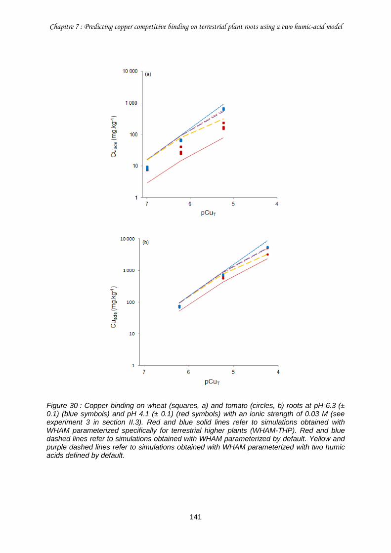
Chapitre 7 : Predicting copper competitive binding on terrestrial plant roots using a two humic-acid model
141
Figure 30 : Copper binding on wheat (squares, a) and tomato (circles, b) roots at pH 6.3 (± 0.1) (blue symbols) and pH 4.1 (± 0.1) (red symbols) with an ionic strength of 0.03 M (see experiment 3 in section II.3). Red and blue solid lines refer to simulations obtained with WHAM parameterized specifically for terrestrial higher plants (WHAM-THP). Red and blue dashed lines refer to simulations obtained with WHAM parameterized by default. Yellow and purple dashed lines refer to simulations obtained with WHAM parameterized with two humic acids defined by default.
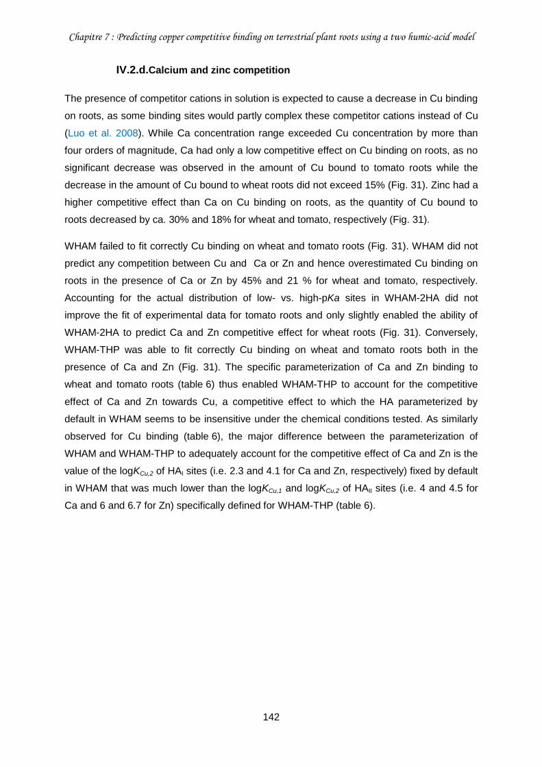
Chapitre 7 : Predicting copper competitive binding on terrestrial plant roots using a two humic-acid model
142
IV.2.d.Calcium and zinc competition
The presence of competitor cations in solution is expected to cause a decrease in Cu binding
on roots, as some binding sites would partly complex these competitor cations instead of Cu
(Luo et al. 2008). While Ca concentration range exceeded Cu concentration by more than
four orders of magnitude, Ca had only a low competitive effect on Cu binding on roots, as no
significant decrease was observed in the amount of Cu bound to tomato roots while the
decrease in the amount of Cu bound to wheat roots did not exceed 15% (Fig. 31). Zinc had a
higher competitive effect than Ca on Cu binding on roots, as the quantity of Cu bound to
roots decreased by ca. 30% and 18% for wheat and tomato, respectively (Fig. 31).
WHAM failed to fit correctly Cu binding on wheat and tomato roots (Fig. 31). WHAM did not
predict any competition between Cu and Ca or Zn and hence overestimated Cu binding on
roots in the presence of Ca or Zn by 45% and 21 % for wheat and tomato, respectively.
Accounting for the actual distribution of low- vs. high-pKa sites in WHAM-2HA did not
improve the fit of experimental data for tomato roots and only slightly enabled the ability of
WHAM-2HA to predict Ca and Zn competitive effect for wheat roots (Fig. 31). Conversely,
WHAM-THP was able to fit correctly Cu binding on wheat and tomato roots both in the
presence of Ca and Zn (Fig. 31). The specific parameterization of Ca and Zn binding to
wheat and tomato roots (table 6) thus enabled WHAM-THP to account for the competitive
effect of Ca and Zn towards Cu, a competitive effect to which the HA parameterized by
default in WHAM seems to be insensitive under the chemical conditions tested. As similarly
observed for Cu binding (table 6), the major difference between the parameterization of
WHAM and WHAM-THP to adequately account for the competitive effect of Ca and Zn is the
value of the logKCu,2 of HAI sites (i.e. 2.3 and 4.1 for Ca and Zn, respectively) fixed by default
in WHAM that was much lower than the logKCu,1 and logKCu,2 of HAII sites (i.e. 4 and 4.5 for
Ca and 6 and 6.7 for Zn) specifically defined for WHAM-THP (table 6).
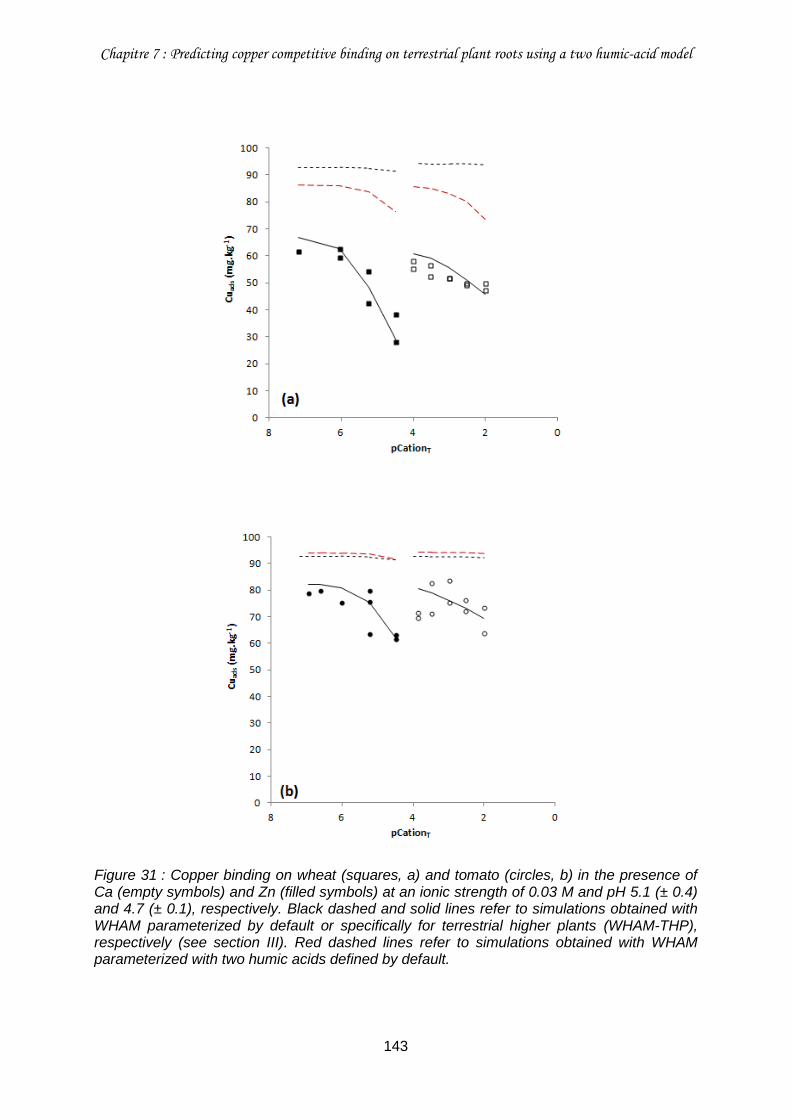
Chapitre 7 : Predicting copper competitive binding on terrestrial plant roots using a two humic-acid model
143
Figure 31 : Copper binding on wheat (squares, a) and tomato (circles, b) in the presence of Ca (empty symbols) and Zn (filled symbols) at an ionic strength of 0.03 M and pH 5.1 (± 0.4) and 4.7 (± 0.1), respectively. Black dashed and solid lines refer to simulations obtained with WHAM parameterized by default or specifically for terrestrial higher plants (WHAM-THP), respectively (see section III). Red dashed lines refer to simulations obtained with WHAM parameterized with two humic acids defined by default.
Chapitre 7 : Predicting copper competitive binding on terrestrial plant roots using a two humic-acid model
144
IV.3. Perspectives for the application of WHAM-THP in predictive
ecotoxicology
In line with theoretical considerations about biotic ligands, the present study demonstrated
that Cu binding on plant roots is sensitive to variations in the ionic strength, pH and the
concentration of competitive metal cations such as Ca and Zn in solution. Although WHAM
as configured by default was shown to be able to account satisfactorily for electrostatic effect
as well as the competitive effect of proton and cations on trace metal binding on aquatic
organisms (Tipping et al. 2008; Tipping and Lofts 2013), similar conclusion cannot be
reached for plant roots. If WHAM was grossly able to predict Cu binding to wheat and tomato
roots at a fixed pH ca. 4.5 and an ionic strength of 0.03 M, WHAM was however unable to
predict Cu binding on plant roots under varying ionic strength or pH. Moreover, WHAM did
not account at all for the competitive effect of Ca and Zn. These results concomitantly
evidenced that the HA defined in WHAM cannot be used as a straightforward surrogate for
terrestrial plant roots. Alternatively, the mathematical formalism used in WHAM can serve as
a basis for the specific parameterization of HA to predict the Cu binding properties of
terrestrial plant roots. Accordingly, we developed WHAM Terrestrial Higher Plants
independently parameterize species model (WHAM-THPips) by parameterizing independently
two HA for each plant (see chapter 6 for additional information and for model parameters).
The similarity of parameters obtained for the two plants allowed to define a unique set of
parameters without degrading the quality of predictions (Fig. S7 to S11). Thus, we developed
WHAM-THP and then demonstrated the ability of WHAM-THP to predict adequately proton
and Cu binding on wheat and tomato roots under varying ionic strength, pH and
concentration of competitive Ca and Zn that were representative of the usual chemical
properties of soil solution. A similar approach was implemented by Plette et al. (1995 and
1996) who successfully reparameterized the NICA-Donnan model for predicting the
competitive binding of protons, calcium, cadmium and Zn on the cell walls of a soil
bacterium. The main interest of WHAM-THP is to suggest a unique parameterization for both
wheat and tomato. These plant species were respectively representative of monocots and
dicots and accordingly their roots exhibited distinct composition and binding properties
(Guigues et al. 2014). This leads to suggest that WHAM-THP could be the first step toward a
more generic TBLM for plants. Nonetheless, the application of WHAM-THP in predictive
ecotoxicology is still constraints by several limitations.
The main difficulty that can appear upon application of WHAM-THP relates to the
parameterization of the density and the distribution of binding sites. As exemplify by tomato
and wheat roots, the density of binding sites was specific to each plant species.
Chapitre 7 : Predicting copper competitive binding on terrestrial plant roots using a two humic-acid model
145
Potentiometric titration is the referenced procedure to finely measure the density of binding
sites. However, potentiometric titration remains a laborious and time-consuming
methodology in the perspective of predictive ecotoxicology. To facilitate the parameterization
of WHAM-THP, we propose to determine the density of root binding sites through the
determination of the root cation exchange capacity (CEC) measured with a routine procedure
based on the adsorption and desorption of Cu from roots (Dufay and Braun 1986). We found
a highly significant, linear relationship (Fig. S12) between the density of binding sites
determined from root CEC and that determined from potentiometric titration that works for
three plant species (i.e. wheat, ray-grass and tomato) and two types of root material (i.e.
roots and isolated cell walls). Thus, by performing a CEC measurement and applying the
proposed relationship, it is possible to estimate fairly accurately the density of root binding
sites. Beyond the density of binding sites, the distribution of low- and high-pKa sites was also
different for wheat and tomato roots and our attempt to parameterize WHAM-THP with a
single low- to high-pKa sites ratio led to unsatisfactory fits (results not shown). If the
distribution of low- and high-pKa sites may differ for each plant species, it likely also depends
on the composition of root tissues, particularly that of the cell walls. The composition of root
cell walls is known to be particularly different between dicots and monocots (Vogel 2008). In
a first approximation, we may suggest to parameterize the low- to high-pKa sites ratio in
WHAM-THP by using the ratio determined in tomato roots for dicots and in wheat roots for
monocots.
To date, WHAM-THP was only tested in solution with pH below 6.5. Above this pH, Cu
speciation will be progressively dominated by Cu hydroxyl species and then Cu carbonates
that were not parameterized in WHAM-THP but that were suspected to be significantly
involved in the induction of Cu rhizotoxicity (Wang et al. 2012a; Wang et al. 2012b). In
addition, WHAM-THP was only tested for the competitive effect of proton, calcium and zinc
while recent implementations of the BLM suggested that magnesium could be also an
efficient competitor towards Cu (Luo et al. 2008; Wang et al. 2012a; Chen et al. 2013). A
more complete parameterization of WHAM-THP for Cu and even more so for other trace
metals should thus deserve further attention.
Finally, the next step in the implementation of WHAM-THP relies on its application in
ecotoxicological studies related to Cu rhizotoxocity.
Chapitre 7 : Predicting copper competitive binding on terrestrial plant roots using a two humic-acid model
146
Acknowledgments
The authors are grateful to French Environment and Energy Management Agency (ADEME)
and the French Centre of Agricultural Research for Development (CIRAD) for funding the
PhD scholarship of Stéphanie Guigues and INSU (CNRS) for funding the study via the
EC2CO-CYTRIX call. The authors thank Patrick Cazevieille and Claire Chevassus-Rosset
(CIRAD) for their technical support during the plant growth phase, Bernard Angeletti
(CEREGE) for ICP-MS analyzes.
V. Supporting information
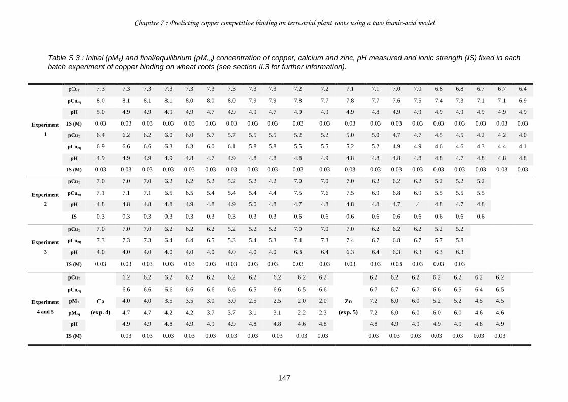
Chapitre 7 : Predicting copper competitive binding on terrestrial plant roots using a two humic-acid model
147
Table S 3 : Initial (pMT) and final/equilibrium (pMeq) concentration of copper, calcium and zinc, pH measured and ionic strength (IS) fixed in each batch experiment of copper binding on wheat roots (see section II.3 for further information).
Experiment
1
pCuT 7.3 7.3 7.3 7.3 7.3 7.3 7.3 7.3 7.3 7.2 7.2 7.1 7.1 7.0 7.0 6.8 6.8 6.7 6.7 6.4
pCueq 8.0 8.1 8.1 8.1 8.0 8.0 8.0 7.9 7.9 7.8 7.7 7.8 7.7 7.6 7.5 7.4 7.3 7.1 7.1 6.9
pH 5.0 4.9 4.9 4.9 4.9 4.7 4.9 4.9 4.7 4.9 4.9 4.9 4.8 4.9 4.9 4.9 4.9 4.9 4.9 4.9
IS (M) 0.03 0.03 0.03 0.03 0.03 0.03 0.03 0.03 0.03 0.03 0.03 0.03 0.03 0.03 0.03 0.03 0.03 0.03 0.03 0.03
pCuT 6.4 6.2 6.2 6.0 6.0 5.7 5.7 5.5 5.5 5.2 5.2 5.0 5.0 4.7 4.7 4.5 4.5 4.2 4.2 4.0
pCueq 6.9 6.6 6.6 6.3 6.3 6.0 6.1 5.8 5.8 5.5 5.5 5.2 5.2 4.9 4.9 4.6 4.6 4.3 4.4 4.1
pH 4.9 4.9 4.9 4.9 4.8 4.7 4.9 4.8 4.8 4.8 4.9 4.8 4.8 4.8 4.8 4.8 4.7 4.8 4.8 4.8
IS (M) 0.03 0.03 0.03 0.03 0.03 0.03 0.03 0.03 0.03 0.03 0.03 0.03 0.03 0.03 0.03 0.03 0.03 0.03 0.03 0.03
Experiment
2
pCuT 7.0 7.0 7.0 6.2 6.2 5.2 5.2 5.2 4.2 7.0 7.0 7.0 6.2 6.2 6.2 5.2 5.2 5.2
pCueq 7.1 7.1 7.1 6.5 6.5 5.4 5.4 5.4 4.4 7.5 7.6 7.5 6.9 6.8 6.9 5.5 5.5 5.5
pH 4.8 4.8 4.8 4.8 4.9 4.8 4.9 5.0 4.8 4.7 4.8 4.8 4.8 4.7 ∕ 4.8 4.7 4.8
IS 0.3 0.3 0.3 0.3 0.3 0.3 0.3 0.3 0.3 0.6 0.6 0.6 0.6 0.6 0.6 0.6 0.6 0.6
Experiment
3
pCuT 7.0 7.0 7.0 6.2 6.2 6.2 5.2 5.2 5.2 7.0 7.0 7.0 6.2 6.2 6.2 5.2 5.2
pCueq 7.3 7.3 7.3 6.4 6.4 6.5 5.3 5.4 5.3 7.4 7.3 7.4 6.7 6.8 6.7 5.7 5.8
pH 4.0 4.0 4.0 4.0 4.0 4.0 4.0 4.0 4.0 6.3 6.4 6.3 6.4 6.3 6.3 6.3 6.3
IS (M) 0.03 0.03 0.03 0.03 0.03 0.03 0.03 0.03 0.03 0.03 0.03 0.03 0.03 0.03 0.03 0.03 0.03
Experiment
4 and 5
pCuT
Ca
(exp. 4)
6.2 6.2 6.2 6.2 6.2 6.2 6.2 6.2 6.2 6.2
Zn
(exp. 5)
6.2 6.2 6.2 6.2 6.2 6.2 6.2
pCueq 6.6 6.6 6.6 6.6 6.6 6.6 6.5 6.6 6.5 6.6 6.7 6.7 6.7 6.6 6.5 6.4 6.5
pMT 4.0 4.0 3.5 3.5 3.0 3.0 2.5 2.5 2.0 2.0 7.2 6.0 6.0 5.2 5.2 4.5 4.5
pMeq 4.7 4.7 4.2 4.2 3.7 3.7 3.1 3.1 2.2 2.3 7.2 6.0 6.0 6.0 6.0 4.6 4.6
pH 4.9 4.9 4.8 4.9 4.9 4.9 4.8 4.8 4.6 4.8 4.8 4.9 4.9 4.9 4.9 4.8 4.9
IS (M) 0.03 0.03 0.03 0.03 0.03 0.03 0.03 0.03 0.03 0.03 0.03 0.03 0.03 0.03 0.03 0.03 0.03
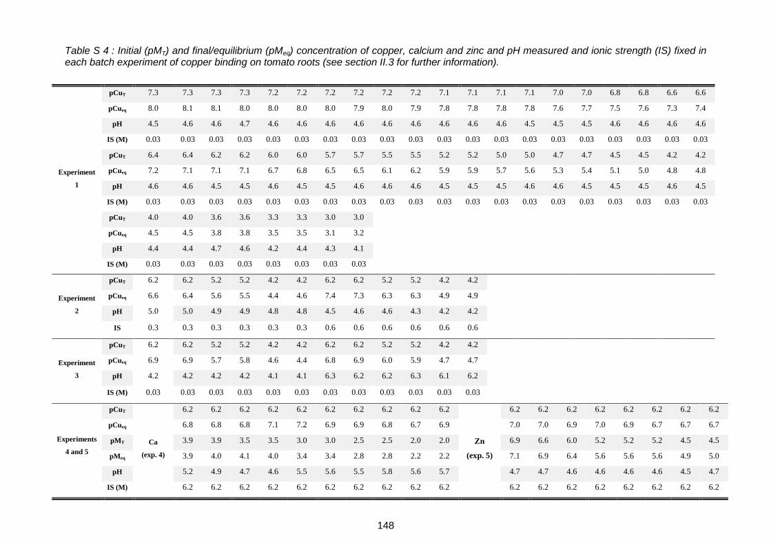
148
Table S 4 : Initial (pMT) and final/equilibrium (pMeq) concentration of copper, calcium and zinc and pH measured and ionic strength (IS) fixed in each batch experiment of copper binding on tomato roots (see section II.3 for further information).
Experiment
1
pCuT 7.3 7.3 7.3 7.3 7.2 7.2 7.2 7.2 7.2 7.2 7.1 7.1 7.1 7.1 7.0 7.0 6.8 6.8 6.6 6.6
pCueq 8.0 8.1 8.1 8.0 8.0 8.0 8.0 7.9 8.0 7.9 7.8 7.8 7.8 7.8 7.6 7.7 7.5 7.6 7.3 7.4
pH 4.5 4.6 4.6 4.7 4.6 4.6 4.6 4.6 4.6 4.6 4.6 4.6 4.6 4.5 4.5 4.5 4.6 4.6 4.6 4.6
IS (M) 0.03 0.03 0.03 0.03 0.03 0.03 0.03 0.03 0.03 0.03 0.03 0.03 0.03 0.03 0.03 0.03 0.03 0.03 0.03 0.03
pCuT 6.4 6.4 6.2 6.2 6.0 6.0 5.7 5.7 5.5 5.5 5.2 5.2 5.0 5.0 4.7 4.7 4.5 4.5 4.2 4.2
pCueq 7.2 7.1 7.1 7.1 6.7 6.8 6.5 6.5 6.1 6.2 5.9 5.9 5.7 5.6 5.3 5.4 5.1 5.0 4.8 4.8
pH 4.6 4.6 4.5 4.5 4.6 4.5 4.5 4.6 4.6 4.6 4.5 4.5 4.5 4.6 4.6 4.5 4.5 4.5 4.6 4.5
IS (M) 0.03 0.03 0.03 0.03 0.03 0.03 0.03 0.03 0.03 0.03 0.03 0.03 0.03 0.03 0.03 0.03 0.03 0.03 0.03 0.03
pCuT 4.0 4.0 3.6 3.6 3.3 3.3 3.0 3.0
pCueq 4.5 4.5 3.8 3.8 3.5 3.5 3.1 3.2
pH 4.4 4.4 4.7 4.6 4.2 4.4 4.3 4.1
IS (M) 0.03 0.03 0.03 0.03 0.03 0.03 0.03 0.03
Experiment
2
pCuT 6.2 6.2 5.2 5.2 4.2 4.2 6.2 6.2 5.2 5.2 4.2 4.2
pCueq 6.6 6.4 5.6 5.5 4.4 4.6 7.4 7.3 6.3 6.3 4.9 4.9
pH 5.0 5.0 4.9 4.9 4.8 4.8 4.5 4.6 4.6 4.3 4.2 4.2
IS 0.3 0.3 0.3 0.3 0.3 0.3 0.6 0.6 0.6 0.6 0.6 0.6
Experiment
3
pCuT 6.2 6.2 5.2 5.2 4.2 4.2 6.2 6.2 5.2 5.2 4.2 4.2
pCueq 6.9 6.9 5.7 5.8 4.6 4.4 6.8 6.9 6.0 5.9 4.7 4.7
pH 4.2 4.2 4.2 4.2 4.1 4.1 6.3 6.2 6.2 6.3 6.1 6.2
IS (M) 0.03 0.03 0.03 0.03 0.03 0.03 0.03 0.03 0.03 0.03 0.03 0.03
pCuT
Ca
(exp. 4)
6.2 6.2 6.2 6.2 6.2 6.2 6.2 6.2 6.2 6.2
Zn
(exp. 5)
6.2 6.2 6.2 6.2 6.2 6.2 6.2 6.2
pCueq 6.8 6.8 6.8 7.1 7.2 6.9 6.9 6.8 6.7 6.9 7.0 7.0 6.9 7.0 6.9 6.7 6.7 6.7
Experiments
4 and 5
pMT 3.9 3.9 3.5 3.5 3.0 3.0 2.5 2.5 2.0 2.0 6.9 6.6 6.0 5.2 5.2 5.2 4.5 4.5
pMeq 3.9 4.0 4.1 4.0 3.4 3.4 2.8 2.8 2.2 2.2 7.1 6.9 6.4 5.6 5.6 5.6 4.9 5.0
pH 5.2 4.9 4.7 4.6 5.5 5.6 5.5 5.8 5.6 5.7 4.7 4.7 4.6 4.6 4.6 4.6 4.5 4.7
IS (M) 6.2 6.2 6.2 6.2 6.2 6.2 6.2 6.2 6.2 6.2 6.2 6.2 6.2 6.2 6.2 6.2 6.2 6.2
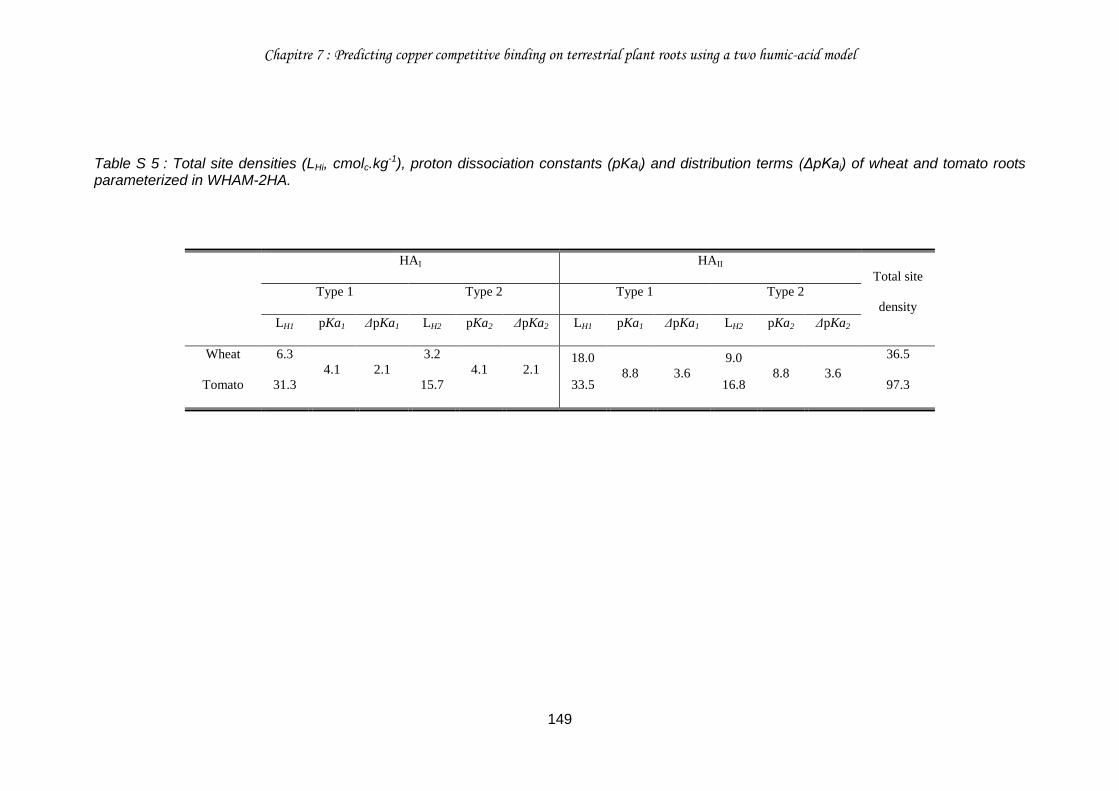
Chapitre 7 : Predicting copper competitive binding on terrestrial plant roots using a two humic-acid model
149
Table S 5 : Total site densities (LHi, cmolc.kg-1), proton dissociation constants (pKai) and distribution terms (ΔpKai) of wheat and tomato roots parameterized in WHAM-2HA.
HAI HAII
Total site
density
Type 1 Type 2 Type 1 Type 2
LH1 pKa1 ΔpKa1 LH2 pKa2 ΔpKa2 LH1 pKa1 ΔpKa1 LH2 pKa2 ΔpKa2
Wheat 6.3
4.1 2.1
3.2
4.1 2.1 18.0
8.8 3.6 9.0
8.8 3.6
36.5
Tomato 31.3 15.7 33.5 16.8 97.3
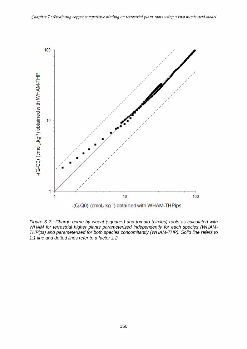
Chapitre 7 : Predicting copper competitive binding on terrestrial plant roots using a two humic-acid model
150
Figure S 7 : Charge borne by wheat (squares) and tomato (circles) roots as calculated with WHAM for terrestrial higher plants parameterized independently for each species (WHAM-THPips) and parameterized for both species concomitantly (WHAM-THP). Solid line refers to
1:1 line and dotted lines refer to a factor 2.
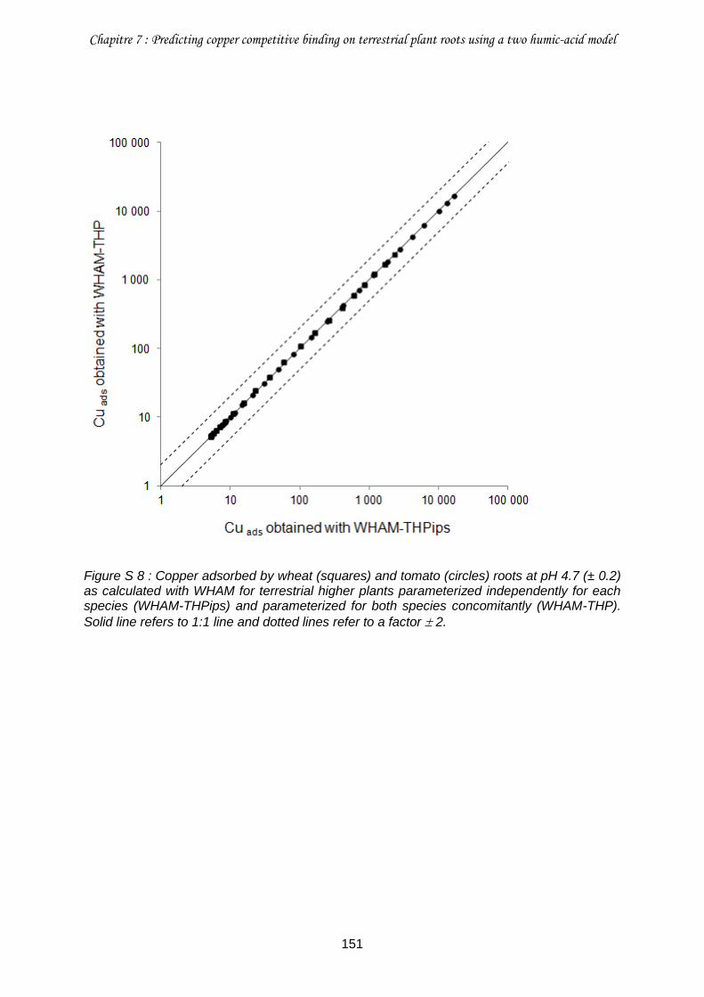
Chapitre 7 : Predicting copper competitive binding on terrestrial plant roots using a two humic-acid model
151
Figure S 8 : Copper adsorbed by wheat (squares) and tomato (circles) roots at pH 4.7 (± 0.2) as calculated with WHAM for terrestrial higher plants parameterized independently for each species (WHAM-THPips) and parameterized for both species concomitantly (WHAM-THP).
Solid line refers to 1:1 line and dotted lines refer to a factor 2.
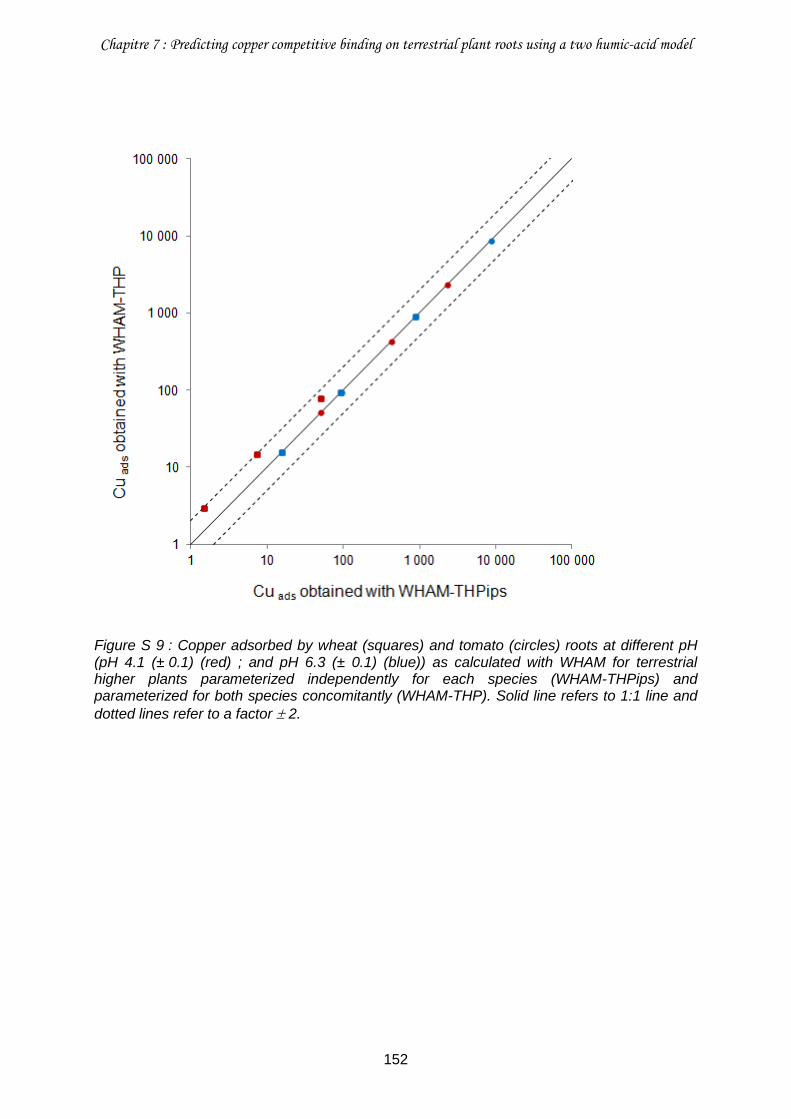
Chapitre 7 : Predicting copper competitive binding on terrestrial plant roots using a two humic-acid model
152
Figure S 9 : Copper adsorbed by wheat (squares) and tomato (circles) roots at different pH (pH 4.1 (± 0.1) (red) ; and pH 6.3 (± 0.1) (blue)) as calculated with WHAM for terrestrial higher plants parameterized independently for each species (WHAM-THPips) and parameterized for both species concomitantly (WHAM-THP). Solid line refers to 1:1 line and
dotted lines refer to a factor 2.
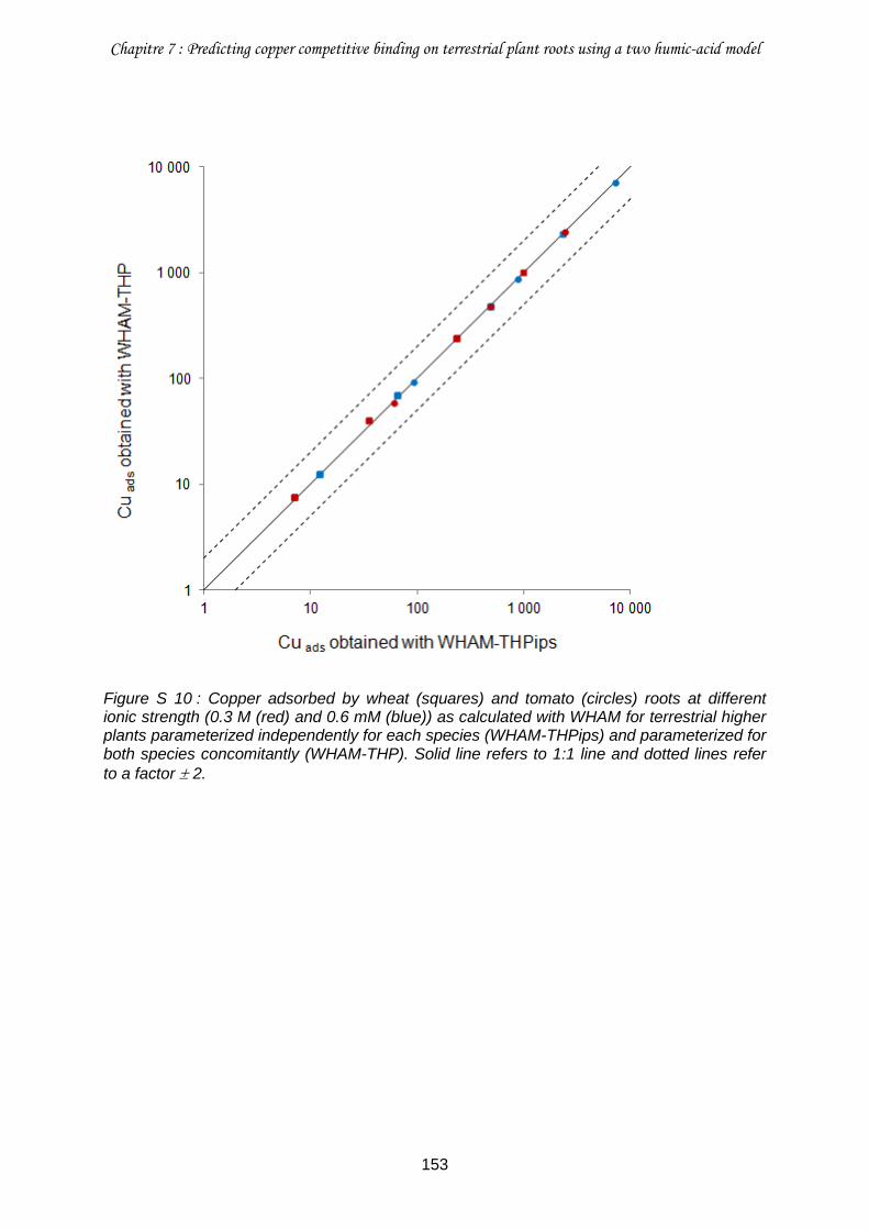
Chapitre 7 : Predicting copper competitive binding on terrestrial plant roots using a two humic-acid model
153
Figure S 10 : Copper adsorbed by wheat (squares) and tomato (circles) roots at different ionic strength (0.3 M (red) and 0.6 mM (blue)) as calculated with WHAM for terrestrial higher plants parameterized independently for each species (WHAM-THPips) and parameterized for both species concomitantly (WHAM-THP). Solid line refers to 1:1 line and dotted lines refer
to a factor 2.
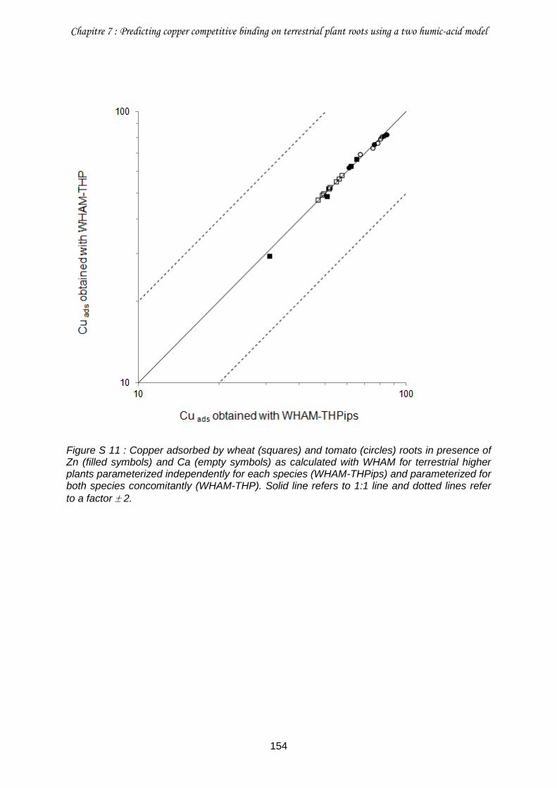
Chapitre 7 : Predicting copper competitive binding on terrestrial plant roots using a two humic-acid model
154
Figure S 11 : Copper adsorbed by wheat (squares) and tomato (circles) roots in presence of Zn (filled symbols) and Ca (empty symbols) as calculated with WHAM for terrestrial higher plants parameterized independently for each species (WHAM-THPips) and parameterized for both species concomitantly (WHAM-THP). Solid line refers to 1:1 line and dotted lines refer
to a factor 2.
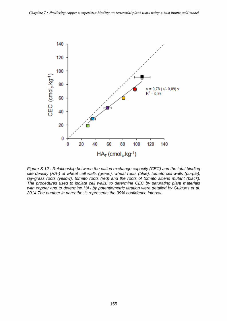
Chapitre 7 : Predicting copper competitive binding on terrestrial plant roots using a two humic-acid model
155
Figure S 12 : Relationship between the cation exchange capacity (CEC) and the total binding site density (HAT) of wheat cell walls (green), wheat roots (blue), tomato cell walls (purple), ray-grass roots (yellow), tomato roots (red) and the roots of tomato sitiens mutant (black). The procedures used to isolate cell walls, to determine CEC by saturating plant materials with copper and to determine HAT by potentiometric titration were detailed by Guigues et al. 2014.The number in parenthesis represents the 99% confidence interval.
Chapitre 7 : Predicting copper competitive binding on terrestrial plant roots using a two humic-acid model
156
Références bibliographiques
Antunes PMC, Hale BA, Ryan AC (2007) Toxicity versus accumulation for barley plants exposed to copper in the presence of metal buffers: Progress towards development of a terrestrial biotic ligand model. Environmental Toxicology and Chemistry 26 (11):2282-2289. doi:10.1897/06-641r.1
Antunes PMC, Scornaienchi ML, Roshon HD (2012) Copper toxicity to Lemna minor
modelled using humic acid as a surrogate for the plant root. Chemosphere 88 (4):389-394. doi:10.1016/j.chemosphere.2012.02.052
Ardestani MM, van Straalen NM, van Gestel CAM (2014) The relationship between metal
toxicity and biotic ligand binding affinities in aquatic and soil organisms: A review. Environmental Pollution 195 (0):133-147. doi:http://dx.doi.org/10.1016/j.envpol.2014.08.020
Brown PL, Markich SJ (2000) Evaluation of the free ion activity model of metal-organism
interaction: extension of the conceptual model. Aquatic Toxicology 51 (2):177-194. doi:Doi: 10.1016/s0166-445x(00)00115-6
Bulgariu D, Bulgariu L (2012) Equilibrium and kinetics studies of heavy metal ions biosorption
on green algae waste biomass. Bioresource Technology 103 (1):489-493. doi:10.1016/j.biortech.2011.10.016
Chen B-C, Ho P-C, Juang K-W (2013) Alleviation effects of magnesium on copper toxicity
and accumulation in grapevine roots evaluated with biotic ligand models. Ecotoxicology 22 (1):174-183. doi:10.1007/s10646-012-1015-z
Di Toro DM, Allen HE, Bergman HL, Meyer JS, Paquin PR, Santore RC (2001) Biotic ligand
model of the acute toxicity of metals. 1. Technical Basis. Environmental Toxicology and Chemistry 20 (10):2383-2396. doi:10.1002/etc.5620201034
Dufey JE, Braun R (1986) Cation Exchange Capacity of Roots : Tirtation Sum of
Exchangeable Cations, Copper Adsorption. Journal of Plant Nutrition 9 (8):1147-1155 Erickson RJ (2013) The biotic ligand model approach for addressing effects of exposure
water chemistry on aquatic toxicity of metals: Genesis and challenges. Environmental Toxicology and Chemistry 32 (6):1212-1214. doi:10.1002/etc.2222
Fry SC, Miller JG, Dumville JC (2002) A proposed role for copper ions in cell wall loosening.
Plant and Soil 247 (1):57-67. doi:10.1023/a:1021140022082 Ginn BR, Szymanowski JS, Fein JB (2008) Metal and proton binding onto the roots of
Fescue rubra. Chemical Geology 253 (3-4):130-135. doi:10.1016/j.chemgeo.2008.05.001
Guigues S, Bravin M, Garnier C, Masion A, Doelsch E (2014) Isolated cell walls exhibit
cation binding properties distinct from those of plant roots. Plant and Soil:1-13. doi:10.1007/s11104-014-2138-1
Haque MN, Morrison GM, Perrusquía G, Gutierréz M, Aguilera AF, Cano-Aguilera I, Gardea-
Torresdey JL (2007) Characteristics of arsenic adsorption to sorghum biomass. Journal of Hazardous Materials 145 (1-2):30-35. doi:10.1016/j.jhazmat.2006.10.080
Chapitre 7 : Predicting copper competitive binding on terrestrial plant roots using a two humic-acid model
157
Harmsen J (2007) Measuring Bioavailability: From a Scientific Approach to Standard Methods. J Environ Qual 36 (5):1420-1428. doi:10.2134/jeq2006.0492
Hooda PS (2010) Trace Elements in Soils. Blackwell Publishing, Ltd Iwasaki Y, Cadmus P, Clements WH (2013) Comparison of different predictors of exposure
for modeling impacts of metal mixtures on macroinvertebrates in stream microcosms. Aquatic Toxicology 132–133 (0):151-156. doi:http://dx.doi.org/10.1016/j.aquatox.2013.02.007
Kopittke PM, Blamey FPC, Asher CJ, Menzies NW (2010) Trace metal phytotoxicity in
solution culture: a review. Journal of Experimental Botany 61 (4):945-954. doi:10.1093/jxb/erp385
Kopittke PM, Menzies NW, Wang P, McKenna BA, Wehr JB, Lombi E, Kinraide TB, Blamey
FPC (2014) The rhizotoxicity of metal cations is related to their strength of binding to hard ligands. Environmental Toxicology and Chemistry 33 (2):268-277. doi:10.1002/etc.2435
Luo X-S, Li L-Z, Zhou D-M (2008) Effect of cations on copper toxicity to wheat root:
Implications for the biotic ligand model. Chemosphere 73 (3):401-406. doi:10.1016/j.chemosphere.2008.05.031
Milne CJ, Kinniburgh DG, van Riemsdijk WH, Tipping E (2003) Generic NICA−Donnan Model
Parameters for Metal-Ion Binding by Humic Substances. Environmental Science & Technology 37 (5):958-971. doi:10.1021/es0258879
Mishra B, Boyanov M, Bunker BA, Kelly SD, Kemner KM, Fein JB (2010) High- and low-
affinity binding sites for Cd on the bacterial cell walls of Bacillus subtilis and Shewanella oneidensis. Geochimica et Cosmochimica Acta 74 (15):4219-4233. doi:http://dx.doi.org/10.1016/j.gca.2010.02.019
Paquin PR, Gorsuch JW, Apte S, Batley GE, Bowles KC, Campbell PGC, Delos CG, Di Toro
DM, Dwyer RL, Galvez F, Gensemer RW, Goss GG, Hogstrand C, Janssen CR, McGeer JC, Naddy RB, Playle RC, Santore RC, Schneider U, Stubblefield WA, Wood CM, Wu KB (2002) The biotic ligand model: a historical overview. Comparative Biochemistry and Physiology Part C: Toxicology & Pharmacology 133 (1-2):3-35. doi:Doi: 10.1016/s1532-0456(02)00112-6
Plette ACC, Benedetti MF, van Riemsdijk WH (1996) Competitive Binding of Protons,
Calcium, Cadmium, and Zinc to Isolated Cell Walls of a Gram-Positive Soil Bacterium. Environmental Science & Technology 30 (6):1902-1910. doi:10.1021/es950568l
Plette ACC, van Riemsdijk WH, Benedetti MF, van der Wal A (1995) pH Dependent
Charging Behavior of Isolated Cell Walls of a Gram-Positive Soil Bacterium. Journal of Colloid and Interface Science 173 (2):354-363. doi:DOI: 10.1006/jcis.1995.1335
Sattelmacher B (2001) The apoplast and its significance for plant mineral nutrition. New
Phytologist 149 (2):167-192. doi:10.1046/j.1469-8137.2001.00034.x Slaveykova VI, Wilkinson KJ (2005) Predicting the Bioavailability of Metals and Metal
Complexes : Critical Review of the Biotic Ligand Model. Environmental chemistry 2:9-24
Chapitre 7 : Predicting copper competitive binding on terrestrial plant roots using a two humic-acid model
158
Thakali S, Allen HE, Di Toro DM, Ponizovsky AA, Rooney CP, Zhao F-J, McGrath SP (2006) A Terrestrial Biotic Ligand Model. 1. Development and Application to Cu and Ni Toxicities to Barley Root Elongation in Soils. Environmental Science & Technology 40 (22):7085-7093. doi:10.1021/es061171s
Tipping E (1998) Humic Ion-Binding Model VI: An Improved Description of the Interactions of
Protons and Metal Ions with Humic Substances. Aquatic Geochemistry 4 (1):3-47. doi:10.1023/a:1009627214459
Tipping E, Lofts S (2013) Metal mixture toxicity to aquatic biota in laboratory experiments:
Application of the WHAM-FTOX model. Aquatic Toxicology 142–143 (0):114-122. doi:http://dx.doi.org/10.1016/j.aquatox.2013.08.003
Tipping E, Lofts S, Sonke JE (2011) Humic Ion-Binding Model VII: a revised parameterisation
of cation-binding by humic substances. Environmental chemistry 8 (3):225-235. doi:http://dx.doi.org/10.1071/EN11016
Tipping E, Vincent CD, Lawlor AJ, Lofts S (2008) Metal accumulation by stream bryophytes,
related to chemical speciation. Environmental Pollution 156 (3):936-943. doi:http://dx.doi.org/10.1016/j.envpol.2008.05.010
Vidali R, Remoundaki E, Tsezos M (2011) An Experimental and Modelling Study of Cu2+
Binding on Humic Acids at Various Solution Conditions. Application of the NICA-Donnan Model. Water, Air, & Soil Pollution 218 (1-4):487-497. doi:10.1007/s11270-010-0662-z
Vogel J (2008) Unique aspects of the grass cell wall. Current Opinion in Plant Biology 11
(3):301-307. doi:http://dx.doi.org/10.1016/j.pbi.2008.03.002 Wang P, Menzies N, Wang Y-M, Zhou D-M, Zhao F-J, Kopittke P (2012a) Identifying the
species of copper that are toxic to plant roots in alkaline nutrient solutions. Plant and Soil 361 (1-2):317-327. doi:10.1007/s11104-012-1260-1
Wang P, Zhou D, Kinraide TB, Luo X, Li L, Li D, Zhang H (2008) Cell Membrane Surface
Potential (psi0) Plays a Dominant Role in the Phytotoxicity of Copper and Arsenate. Plant Physiol 148 (4):2134-2143. doi:10.1104/pp.108.127464
Wang X, Hua L, Ma Y (2012b) A biotic ligand model predicting acute copper toxicity for
barley (Hordeum vulgare): Influence of calcium, magnesium, sodium, potassium and pH. Chemosphere 89 (1):89-95. doi:10.1016/j.chemosphere.2012.04.022
Wu Y, Hendershot W (2009) Cation Exchange Capacity and Proton Binding Properties of
Pea (<i>Pisum sativum</i> L.) Roots. Water, Air, & Soil Pollution 200 (1):353-369. doi:10.1007/s11270-008-9918-2
Wu Y, Hendershot WH (2010) The effect of calcium and pH on nickel accumulation in and
rhizotoxicity to pea (Pisum sativum L.) root–empirical relationships and modeling. Environmental Pollution 158 (5):1850-1856. doi:10.1016/j.envpol.2009.10.046

159

160
Conclusion et perspectives
161
Conclusion et perspectives
Depuis quelques années, les études développant ou employant un modèle de prédiction de
la toxicité se multiplient. Parmi les formalismes mathématiques actuellement disponibles, le
Biotic Ligand Model (BLM, ou Terrestrial BLM lorsqu’il est utilisé pour des études de toxicité
menées sur des organismes terrestres) fait figure de référence et est, de ce fait, le plus
utilisé. En particulier, le TBLM s’avère être un modèle adapté à l’étude d’effets toxiques chez
les plantes. En effet, dans ce modèle, la réponse biologique observée est supposée être
engendrée par la complexation de l’élément trace avec les ligands biotiques situés à la
surface de l’organisme cible (Thakali et al. 2006), tout comme les effets rhizotoxiques
observés sont supposés être engendrés par la complexation de l’élément trace sur les
surfaces racinaires (Kopittke et al. 2014).
Lorsqu’elles sont considérées individuellement, les études employant le TBLM donnent des
résultats prometteurs. Cependant, leur comparaison révèle souvent un manque de généricité
de certains des paramètres et les cas particuliers où l’application du TBLM est limitée se
multiplient. Ces limites trouvent leur origine dans la formulation d’hypothèses trop
simplificatrices, résultant d’une connaissance encore incomplète des mécanismes générant
l’effet toxique. La solution, pour dépasser ces limites et ainsi améliorer les modèles de
prédiction, est de ne pas se contenter d’une simple application du formalisme mathématique
classique du BLM pour l’ajustement de données expérimentales de toxicité mais de revenir à
l’étude des mécanismes (Erickson 2013).
C’est dans ce contexte que s’inscrivent mes travaux de recherche dont l’objectif principal
était de proposer une démarche de modélisation différente, centrée sur les réactions de
complexation se produisant à la surface de l’organisme cible plutôt que sur l’effet toxique
observé. Cette approche a été employée pour prédire l’adsorption du cuivre sur les racines
de deux plantes modèles, le blé et la tomate, sélectionnées pour la capacité contrastée de
leurs racines à complexer les cations métalliques. L’étude de la nature et des propriétés des
sites de complexation du Cu des racines a permis de valider les paramètres obtenus par
modélisation.
Conclusion et perspectives
162
Complexation du cuivre dans les racines
D’après la littérature, les propriétés de complexation des racines peuvent être attribuées aux
groupements fonctionnels portés principalement soit par les parois apoplasmiques (et en
particulier par les polysaccharides pectiques) soit par les membranes plasmiques (Kinraide
2004 ; Krzeslowska 2011). Les études portant sur ce sujet sont peu nombreuses et
uniquement consacrées à l’un ou à l’autre de ces compartiments de sorte que leur
contribution respective dans les propriétés de complexation des surfaces racinaires n’a
jamais été définie.
La première étape a donc été de caractériser la réactivité des racines des deux plantes
modèles, c'est-à-dire localiser et identifier les sites réactifs racinaires. Nous avons ainsi
montré que l’isolement des parois apoplasmiques entraine une diminution de la capacité de
complexation des racines, d’un facteur 2,4 pour le blé et d’un facteur 3,4 pour la tomate. Ces
résultats révèlent une contribution majoritaire du matériel racinaire ayant disparu lors de
l’isolation des parois apoplasmiques, à savoir les membranes plasmiques, dans les
propriétés de complexation racinaire. Cette contribution a été évaluée à hauteur de 60 %
dans les racines de blé et de 70 % dans les racines de tomate. Cette diminution de capacité
de complexation après isolement a été accompagnée de la perte de groupements chimiques
caractéristiques tels que des groupements carboxyliques, amines et phosphates. Ces
groupements, portés par les protéines et les phospholipides des membranes plasmiques,
sont donc impliqués dans les propriétés de complexation des racines. Ainsi, contrairement à
ce qui est communément supposé, les groupements carboxyliques des polysaccharides
pectiques, et d’une façon générale l’ensemble des groupements portés par les parois
apoplasmiques, contribue pour moins de 50 % dans la capacité de complexation des racines
des deux plantes étudiées.
Toutefois, les contributions respectives des parois apoplasmiques et des membranes
plasmiques dans les propriétés de complexation des racines que nous avons déterminées
sont fondées sur l‘hypothèse que la réactivité des parois apoplasmiques est similaire en
présence et en l’absence de membranes plasmiques. Il serait intéressant de vérifier cette
hypothèse, en étudiant d’une part les parois apoplasmiques et les membranes plasmiques
séparément et d’autre part, en étudiant les racines « entières ». Le protocole employé dans
ces travaux pour isoler les parois apoplasmiques semble convenir et pourrait donc être à
nouveau utilisé. En effet, nous avons montré qu’il ne restait plus aucune trace des
membranes plasmiques après la procédure d’isolation et que le protocole n’entrainait pas de
dégradation des polysaccharides pariétaux. L’isolation des membranes plasmiques est
Conclusion et perspectives
163
également possible ; il s’agit d’ailleurs du matériel végétal classiquement employé pour le
développement du modèle électrostatique (Wang et al. 2008).
Une autre possibilité, pour étudier la contribution respective des parois apoplasmiques et des
membranes plasmiques dans les propriétés de complexation racinaire, est de procéder à
une localisation de l’élément cible (par exemple Cu) au sein des racines grâce à une
technique d’imagerie. Une étude aussi fine devrait pouvoir être réalisée grâce à la nanoSIMS
(nano-scale Secondary Ion Mass Spectrometry), une technique analytique qui offre une
résolution spatiale élevée combinée à une analyse élémentaire très sensible (Herrmann et
al. 2007). A ce jour, les études nano-SIMS sont surtout employées pour étudier la distribution
des éléments entre le compartiment apoplasmique et le compartiment symplasmique,
néanmoins la distinction entre parois apoplasmiques et membranes plasmiques semble
possible (Moore et al. 2011).
La seconde étape de ces travaux a été dédiée à l’étude de la complexation du cuivre au sein
des racines. Même s’il est parfois détecté dans le milieu symplasmique, le cuivre est connu
pour être un élément s’accumulant préférentiellement dans le compartiment extracellulaire à
savoir le continuum parois apoplasmiques – faces externes des membranes plasmiques
(Colzi et al. 2011). Nous avons donc cherché, parmi les sites réactifs identifiés
précédemment, ceux qui participent à la complexation du cuivre. Nous avons montré que
plus de 50 % des sites participant à sa complexation sont situés dans les parois
apoplasmiques. Etant donné que la densité de sites des parois apoplasmiques est moins
élevée que celle des membranes plasmiques, cette contribution majoritaire des parois
apoplasmiques dans la complexation du Cu peut provenir d’une plus grande affinité du Cu
pour les sites pariétaux des racines de blé et de tomate que pour les sites membranaires.
Une autre explication pourrait être directement liée à la géométrie des racines, Cu
rencontrant en premier lieu les parois apoplasmiques et, seulement après avoir traversé ces
dernières, les membranes plasmiques. L’organisation spatiale des tissus racinaires serait,
dans ce cas, déterminante. Il n’est pas exclu, cependant, qu’il s’agisse d’une action conjointe
de ces deux hypothèses. Nous avons également montré que plus de la moitié des sites
racinaires participant à la complexation du Cu ont une réactivité assimilable à un groupement
amine, seulement 40 % environ présentent une réactivité proche de celle d’un acide
carboxylique. Cette constatation a été confortée par la spéciation du cuivre identifiée dans
les racines. En effet, Cu a été trouvé sous la forme de Cu(II)-O/N organique de façon très
légèrement prépondérante par rapport à la forme Cu(II)-O organique c'est-à-dire associé à
des acides aminés pouvant être enchâssés soit dans les parois apoplasmiques en
association avec des polysaccharides pariétaux, soit au niveau des membranes plasmiques.
Conclusion et perspectives
164
De tels résultats modifient significativement la vision actuelle du mécanisme conduisant à la
génération d’effets rhizotoxiques. En effet, il est communément admis que la complexation
des éléments traces avec les groupements carboxyliques portés par les polysaccharides
pariétaux en est la principale cause, les autres groupements fonctionnels présents dans les
racines étant supposés avoir une contribution minoritaire (Krzeslowska 2011). Une
contribution significative d’autres composés, tels que les protéines, dans la complexation du
Cu au sein de racines pourrait expliquer l’incohérence entre la rhizotoxicité observée pour
Cu et l’échelle d’affinité avec les ligands forts proposée par Kopittke et al. 2014.
Validité du modèle WHAM-THP pour prédire l’adsorption du cuivre dans les racines
Développés en parallèle du BLM, d’autres modèles de complexation sont utilisés pour
prédire les effets toxiques des éléments traces pour les organismes aquatiques. Ces
modèles emploient des formalismes mathématiques initialement développés pour étudier la
complexation des éléments traces avec les matières organiques naturelles de type
substances humiques. Le formalisme mathématique du modèle WHAM est le plus employé
actuellement. Ce modèle semble être un outil adéquat pour prédire la toxicité des éléments
traces pour les organismes aquatiques (Tipping et al. 2008 ; Antunes et al. 2012) mais il
n’avait encore jamais été employé pour des études de complexation menées avec des
plantes supérieures.
Contrairement au cas des organismes aquatiques, le modèle WHAM tel qu’initialement
paramétré ne s’est pas avéré efficace pour prédire la réactivité des racines. Nous avons
montré que cela provenait d’une part de la distribution imposée des sites réactifs (ratio de
sites présentant un pKa faible et un pKa élevé fixe) et d’autre part de la valeur des
constantes, définies initialement pour des substances humiques. Le développement d’un
modèle plus complexe a donc été nécessaire pour prédire correctement la complexation du
Cu dans les racines de blé et de tomate. Le modèle WHAM-Terrestrial Higher Plants
(WHAM-THP) a été capable de prédire l’adsorption du Cu dans les racines de blé et de
tomate pour différents pH, différentes forces ioniques et en présence des deux cations
compétiteurs que sont Ca et Zn. Malgré les différences de propriétés de complexation
observées entre les racines de blé et de tomate, il a été possible d’établir un jeu de
paramètres commun pour ces deux espèces. Nous avons également cherché à rendre
l’application du modèle la plus simple possible en proposant une relation mathématique
permettant de déterminer, rapidement et sans équipement spécifique, la densité totale de
sites racinaire.
Conclusion et perspectives
165
Le modèle WHAM-THP a donné des résultats prometteurs pour modéliser des réactions de
complexation du Cu au sein des racines de blé et tomate. Cependant, son usage reste
encore très limité et il serait intéressant d’étendre son domaine d’application. En premier lieu,
il serait intéressant de compléter le modèle existant pour Cu. En effet, WHAM-THP ne
permet, à ce jour, que de représenter l’adsorption du Cu sur une gamme de pH allant jusqu’à
6,5. Au-delà, la spéciation du Cu est plus diversifiée (CuOH+, CuHCO3, CuOH2, etc.) et les
constantes de complexation de ces espèces de Cu n’ont pas été déterminées. L’hypothèse
d’une participation d‘autres espèces métalliques que l’ion libre dans l’induction d’un effet
toxique tend à se confirmer au travers de différentes études (Wang et al. 2009 ; Wang et al.
2012). L’ajout de nouvelles constantes de complexation pour d’autres espèces de Cu
permettra une application plus étendue du modèle. De même, Cu avait été sélectionné
comme élément modèle dans cette étude mais le modèle WHAM-THP doit être développé
pour d’autres éléments traces (par exemple, Pb, Ni ou Cd) et testé sur d’autres espèces
végétales (par exemple l’orge, qui est une plante modèle souvent employée dans les études
TBLM). Le protocole employé pour réaliser les expérimentations en batch est simple et
efficace et devrait permettre l’acquisition rapide de nouveaux jeux de données. La
description des propriétés acides, communes au blé et à la tomate, doit pouvoir être
appliquée à n’importe quelle autre plante, l’utilisateur n’ayant qu’à déterminer la densité
totale de sites. De même, le logiciel WHAM, à partir duquel sont obtenus les résultats de
modélisation, permet de définir d’autres espèces chimiques très simplement.
Il serait également nécessaire d’éprouver la capacité du modèle WHAM-THP à prédire
l’adsorption de Cu dans les racines de blé et de tomate lorsque ces plantes sont exposées à
une contamination en cuivre. En effet, la principale critique qui peut être faite à ces travaux
de recherche est l’absence de résultats in vivo. En effet, ces travaux ont été menés avec du
matériel végétal racinaire ne présentant pas d’activité biologique. Une telle configuration
nous a permis d’axer notre étude sur les réactions de complexation se produisant dans les
racines tout en nous affranchissant des phénomènes d’absorption. Mais le modèle WHAM-
THP doit pouvoir être employé pour prédire la fraction de Cu adsorbé dans les racines d’une
plante exposée à une contamination. Cela nécessite donc de réaliser une étape
intermédiaire qui est de valider les constantes de complexation pour le blé et la tomate ou
d’estimer un écart entre les prédictions et les observations expérimentales de façon à affiner
le modèle. Pour commencer, une culture hydroponique devrait permettre de contrôler assez
finement les conditions expérimentales. La quantité de Cu adsorbée dans les racines de blé
et de tomate, après exposition à une contamination, pourra directement être comparée à la
prédiction du modèle WHAM-THP.
Conclusion et perspectives
166
Références bibliographiques
Antunes PMC, Scornaienchi ML, Roshon HD (2012) Copper toxicity to Lemna minor
modelled using humic acid as a surrogate for the plant root. Chemosphere 88 (4):389-394. doi:10.1016/j.chemosphere.2012.02.052
Colzi I, Doumett S, Del Bubba M, Fornaini J, Arnetoli M, Gabbrielli R, Gonnelli C (2011) On
the role of the cell wall in the phenomenon of copper tolerance in Silene paradoxa L. Environmental and Experimental Botany 72 (1):77-83. doi:DOI: 10.1016/j.envexpbot.2010.02.006
Erickson RJ (2013) The biotic ligand model approach for addressing effects of exposure
water chemistry on aquatic toxicity of metals: Genesis and challenges. Environmental Toxicology and Chemistry 32 (6):1212-1214. doi:10.1002/etc.2222
Herrmann AM, Ritz K, Nunan N, Clode PL, Pett-Ridge J, Kilburn MR, Murphy DV, O’Donnell
AG, Stockdale EA (2007) Nano-scale secondary ion mass spectrometry — A new analytical tool in biogeochemistry and soil ecology: A review article. Soil Biology and Biochemistry 39 (8):1835-1850. doi:http://dx.doi.org/10.1016/j.soilbio.2007.03.011
Kinraide TB (2004) Possible Influence of Cell Walls upon Ion Concentrations at Plasma
Membrane Surfaces. Toward a Comprehensive View of Cell-Surface Electrical Effects upon Ion Uptake, Intoxication, and Amelioration. Plant Physiol 136 (3):3804-3813. doi:10.1104/pp.104.043174
Kopittke PM, Menzies NW, Wang P, McKenna BA, Wehr JB, Lombi E, Kinraide TB, Blamey
FPC (2014) The rhizotoxicity of metal cations is related to their strength of binding to hard ligands. Environmental Toxicology and Chemistry 33 (2):268-277. doi:10.1002/etc.2435
Krzesłowska M (2011) The cell wall in plant cell response to trace metals: polysaccharide
remodeling and its role in defense strategy. Acta Physiologiae Plantarum 33 (1):35-51. doi:10.1007/s11738-010-0581-z
Moore KL, Schröder M, Wu Z, Martin BGH, Hawes CR, McGrath SP, Hawkesford MJ, Feng
Ma J, Zhao F-J, Grovenor CRM (2011) High-Resolution Secondary Ion Mass Spectrometry Reveals the Contrasting Subcellular Distribution of Arsenic and Silicon in Rice Roots. Plant Physiology 156 (2):913-924. doi:10.1104/pp.111.173088
Thakali S, Allen HE, Di Toro DM, Ponizovsky AA, Rooney CP, Zhao F-J, McGrath SP (2006)
A Terrestrial Biotic Ligand Model. 1. Development and Application to Cu and Ni Toxicities to Barley Root Elongation in Soils. Environmental Science & Technology 40 (22):7085-7093. doi:10.1021/es061171s
Tipping E, Vincent CD, Lawlor AJ, Lofts S (2008) Metal accumulation by stream bryophytes,
related to chemical speciation. Environmental Pollution 156 (3):936-943. doi:http://dx.doi.org/10.1016/j.envpol.2008.05.010
Wang P, Menzies N, Wang Y-M, Zhou D-M, Zhao F-J, Kopittke P (2012) Identifying the
species of copper that are toxic to plant roots in alkaline nutrient solutions. Plant and Soil 361 (1-2):317-327. doi:10.1007/s11104-012-1260-1
Conclusion et perspectives
167
Wang P, Zhou D, Kinraide TB, Luo X, Li L, Li D, Zhang H (2008) Cell Membrane Surface Potential (psi0) Plays a Dominant Role in the Phytotoxicity of Copper and Arsenate. Plant Physiol 148 (4):2134-2143. doi:10.1104/pp.108.127464
Wang X, Ma Y, Hua L, McLaughlin MJ (2009) Identification of hydroxyl copper toxicity to
barley (Hordeum vulgare) root elongation in solution culture. Environmental Toxicology and Chemistry 28 (3):662-667. doi:10.1897/07-641.1


Résumé
Cette étude a été consacrée au développement d’une nouvelle approche de modélisation de la phytodisponibilité des éléments traces, basée sur leurs réactions de complexation au sein des racines des plantes. Cette approche a été développée pour prédire l’adsorption du cuivre (Cu) sur les racines de deux plantes, le blé et la tomate. Plusieurs techniques analytiques (titrages acido-basiques, résonance magnétique nucléaire du carbone 13, spectroscopie d’absorption des rayons X) ont été employées et croisées avec les résultats de modélisation. Dans un premier temps, la réactivité des racines a été caractérisée. Les racines étant constituées de parois apoplasmiques et de membranes plasmiques, la contribution respective de ces deux compartiments aux propriétés de complexation des racines a été évaluée. L’étude a ensuite été focalisée sur la complexation du Cu au sein des racines et sur l’évolution de cette complexation en fonction des conditions physico-chimiques du milieu (pH, force ionique, cations compétiteurs). Grâce aux résultats obtenus sur la caractérisation des racines et à l’acquisition d’un jeu variéde données expérimentales sur la complexation du Cu, le modèle a pu être paramétré. Il a été montré que les propriétés de complexation des racines de blé et de tomate proviennent conjointement des membranes plasmiques et des parois apoplasmiques. La spéciation du Cu au sein des racines était partagée, de façon presque égale, entre les composés pectiques des parois apoplasmiques et les protéines enchâssées à la fois dans les parois apoplasmiques et les membranes plasmiques. Un modèle propre aux racines a pu être développé sur la base d’un modèle existant dédié à la réactivité des substances humiques. Le modèle WHAM-Terrestrial Higher Plants, présenté dans cette étude, est un premier pas vers un nouvel outil d’évaluation de la disponibilité des éléments traces pour les plantes.
summary
This study has been dedicated to the development of a new modeling approach of trace element phytoavailability, focusing on binding reactions between trace element and plant roots. This approach was used to predict copper (Cu) adsorption on wheat and tomato roots. Several analytical techniques (acid-base titrations, nuclear magnetic resonance of carbon 13, X-ray absorption spectroscopy) were used and crossed with modeling results. At first, plant root reactivity was characterized. Because plant roots are consist of cell walls and plasma membranes, the relative contribution of these two compartments in root binding properties was evaluated. The study was then focused on Cu binding reactions on roots and the effects of physico-chemical conditions (pH, ionic strength, presence of cations) on copper binding. The model has been set thanks to results on root characterization obtained and the acquisition of a set of experimental data on Cu binding. It has been shown that binding properties of wheat and tomato roots came from both cell walls and plasma membranes. Copper speciation in roots was shared, almost evenly, between cell wall pectic compounds and proteins embedded in cell walls and plasma membranes. A model, specific to plant roots, has been developed on the basis of a current model dedicated to the humic substances reactivity. The WHAM-Terrestrial Higher Plants model presented in this studyis a first step towards a new tool for assessing the availability of trace elements for plants.