Embed Size (px)
Citation preview
Carbon Nanotubes as Structural Nanofibers for Hyaluronic AcidHydrogel Scaffolds
Sanjib Bhattacharyya,*,†,§ Samuel Guillot,† Hinda Dabboue,† Jean-François Tranchant,‡
and Jean-Paul Salvetat*,†
Centre de Recherche sur la Matière Divisée, UMR 6619, CNRS-Université d’Orléans, 1b rue de laFérollerie, 45071 Orléans Cedex 2, France, and Laboratoire de Physicochimie, Parfums Christian Dior,
F-45804 St Jean de Braye, France
Received September 7, 2007; Revised Manuscript Received November 22, 2007
We have successfully dispersed functionalized single-walled carbon nanotubes (SWNTs) within hyaluronicacid–water solutions. Hybrid hyaluronic acid (HA) hydrogels with SWNTs were then formed by cross-linkingwith divinyl sulfone. We have found a considerable change in the morphology of the lyophilized hybrid hydrogelscompared to HA hydrogels. The high water uptake capacity, an important property of HA hydrogels, remainedalmost unchanged after 2 wt % SWNT (vs HA) incorporation, despite a dramatic enhancement in the dynamicmechanical properties of the hybrid hydrogels compared to native ones. We have found a 300% enhancement inthe storage modulus of hybrid hydrogel with only 2 wt % of SWNTs vs HA (0.06 wt % vs total weight includingwater content). This apparent contradiction can be explained by a networking effect between SWNTs, mediatedby HA chains. As in biological tissue, HA plays a dual role of matrix and linker for the rigid reinforcing nanofibers.
Introduction
In recent years carbon nanotubes (CNTs) have found a placein a variety of fields including electronics and nanocompositesas well as in biomedical research due to their remarkableelectrical and mechanical properties and chemical stability.1 Oneof the most exciting areas of research to emerge around carbonnanotubes is in the biomedical field.2–5 There are a number ofpublications showing the capability of nanotubes to deliverbiomolecules and therapeutic molecules,6–8 to sense the presenceof DNA and proteins in solution9–11 and to enhance cellproliferation and growth.12–14 However, due to the chemicalinertness of CNTs, it is always difficult to manipulate andintegrate them with other materials such as polymers. One ofthe main reasons is the poor dispersion of CNTs within wateror organic solvents. There has been much progress in thefunctionalization chemistry of CNTs for solubilization anddispersion within different solvents.15–18
Biopolymers are often studied for polymeric scaffolds in thefield of tissue engineering,19,20 and nanotechnology has beenused recently in the field.21,22 Polysaccharides are of particularinterest in joint-related repairs based on their role as glycosami-noglycans in mammalian extracellular matrices. Hyaluronic acid,alginates, and chitosan hydrogels are the most intensivelystudied, due to their ease of processing into useful structures.23–31
Among the required properties for successful application of thesehydrogels in the tissue scaffolds are mechanical stability alongwith porous structure and water intake capacity. Hydrogels withimproved mechanical properties are required to provide me-chanical support during new tissue formation in vitro and invivo and to provide mechanical signaling to cells in vitro and
in vivo for optimization of the structural and mechanicalfunctions of the new extracellular matrix being formed. In thiscontext, functionalized water-soluble CNTs can be very goodcandidates to reinforce the biopolymer hydrogels withoutaltering their chemical properties. The interplay of the physicalproperties of CNTs with a biopolymer can give birth to a newgeneration of tissue-engineered implants to reconstruct naturaltissues that are subjected to high mechanical stress or requireelectrical and thermal conductivity at physiological state.Unfortunately there are very few reports on incorporation ofCNTs within biocompatible polymer hydrogels.32–34 Li et al.35
have shown the successful incorporation of multiwalled carbonnanotubes (MWNTs) within gelatin hydrogel without changingthe swelling characteristics of native hydrogel. Recently Wanget al.36 have reported the synthesis of supramolecular Pluronichydrogels hybridized with single-wall carbon nanotubes (SWNTs).They have shown that the resultant hybrid hydrogels retainedthe basic characters of supramolecular hydrogel and pristineSWNTs, especially the shear-thinning property, which is veryimportant in drug delivery and controlled release. Surprisinglythey have observed a decline in the mechanical properties ofthe hybridized hydrogel, which they explain as being due tothe mechanism of hydrogel formation. We know only of tworeports addressing the incorporation of CNTs within biopolymerhydrogels and both are with alginate.37,38
In this article we have demonstrated a successful way todisperse SWNTs within hyaluronic acid (HA) solution andinduce the formation of reinforced hydrogel in the presence ofcross-linking reagent divinyl sulfone (DVS).39 We have ob-served a 4-fold increase in the storage modulus of the hydrogelby incorporation of only 0.06 wt % SWNTs (vs total weightincluding water) without significant change in the water intakecapacity and shear thinning behavior of the native hydrogel.
Materials and Methods
High molecular weight (1000 kDa) hyaluronic acid was a generousgift from LVMH, Orléans, France. Divinyl sulfone (purum g98%) was
* Corresponding authors S. Bhattacharyya, e-mail [email protected],and J.-P. Salvetat, e-mail [email protected].
† Centre de Recherche sur la Matière Divisée, UMR 6619, CNRS-Université d’Orléans.
‡ Laboratoire de Physicochimie, Parfums Christian Dior.§ Present address: Center for Bioactive Materials & Tissue Engineering,
Bioengineering Department, University of Pennsylvania, Philadelphia, PA19104.
Biomacromolecules 2008, 9, 505–509 505
10.1021/bm7009976 CCC: $40.75 2008 American Chemical SocietyPublished on Web 01/11/2008
purchased from Fluka, France, and used without further purification.SWNTs were purchased from Carbon Nanotechnology Inc., USA. Theywere purified and carboxylated prior to use. First they were heat treatedat 350 °C in air for 2 h to remove the amorphous carbon and thenrefluxed with 3 M nitric acid (100 mL of acid per gram of SWNTs) at130 °C for 36 h to remove the metallic catalytic particles and to createthe carboxyl functionalities on the surface. Formation of carboxyl groupshas been verified by Fourier transformed infrared spectroscopy andX-ray photoelectron spectroscopy (data not shown).
The carboxylated SWNTs prepared via above method was used toprepare the gel with different amounts of SWNTs. To prepare HA gelhybridized with 2 wt % (vs HA) functionalized SWNTs, first 6 mg ofSWNTs was dispersed in 10 mL of double distilled water in a bathsonicator for 30 min and then 300 mg of HA was dissolved in it bymagnetic stirring. At this stage, the concentration of SWNTs vs wateris approximately 0.06 wt %. The pH of the solution was maintained at9–10 by adding an appropriate amount of 2 M NaOH for efficient cross-linking. DVS was used to cross-link the HA and hybrid HA/SWNTssolution keeping the ratio HA:DVS 1:1. After 2 h the gel was thoroughlywashed to get rid of any unreacted DVS and NaOH while keeping thetotal water amount constant. Three hydrogel samples with 0, 0.03, and0.06 wt % of SWNT were prepared to compare the results.
Success of the cross-linking of HA with and without SWNTs wasverified by Fourier transform infrared (FTIR) spectroscopy on a Nicolet710 FT-IR spectrophotometer with a resolution of 8 cm-1 by usingthe KBr method.
For morphological characterization by scanning electron microscopy(SEM), hydrogels were frozen to rupture and the transverse sectionswere observed in Hitachi 4200 operated at 1 kV.
The swelling behavior of the hydrogels was measured in phosphatebuffer solution (pH 7.4). The starting materials were not fully driedand contained the same amount of water (10 mL for 300 mg of HA).Approximately 4 g of hydrogel samples was immersed in 50 mL ofphosphate buffer solution for different time intervals. Then the hydratedgels were removed from the buffer solution and quickly dried onblotting paper to eliminate residual water on the surface. Results areexpressed as percentage of swelling (S%) and were calculated by using
S % ) 100 × [(Wht -W0) ⁄ W0]
where W0 is the initial weight of the gel at time 0 and Wht is the weightof the hydrated gel at time t.
Anton Paar Physica MRC 300 modular compact rheometer was usedin the parallel plate geometry, with a 25 mm plate and a gap size of 1mm. Constant values of deformation, around 2 mrad, were maintainedthroughout each frequency sweep of 0.2–15 Hz. The steady flowbehaviors were measured at shear rate range from 0.1 to 100 s-1. Allthe dynamic tests were performed at a constant strain of 5%.
Results and Discussion
To obtain a proper integration of nanofillers within thebiopolymer gels, it is very important to stabilize a gooddispersion of the fillers within the polymer–water solution and
keep this dispersion stable up to the gelation point after a cross-linker is added. From optical observations (see photo in Figure1 as an illustration) it was evident that the acid-treated SWNTswere well dispersed within the HA solution as it gave ahomogeneous black color of the solution without any floccula-tion-induced turbidity. (Note that our acid-treated SWNTs werealso well dispersed in pure water due to hydrophilic chemicalgroups grafted at the surface.) Atomic force microscopyobservations showed that individual nanotubes coexisted withsmall bundles in HA water solutions. We do not have quantified,however, to what extend exfoliation of SWNT bundles occurred.(Additional experiments would be needed to address that point.)After 30 min of the addition of the cross-linking agent DVS,HA solutions both with and without SWNTs form the gel shownin Figure 1.
It should be noted that after gelification with SWNTs coloris still homogeneous indicating no macroscopic phase separationbetween cross-linked HA and SWNTs. With DVS, the cross-
Figure 1. Photograph of HA solution (transparent, on the left) and dispersion of 0.06% SWNTs by mass within HA solution (dark, on the right).Arrow leads to photograph showing invertible HA gels, native (white) and hybridized (dark), formed after 30 min of reaction with DVS.
Scheme 1. Cross-Linking Reaction between HA and DVS
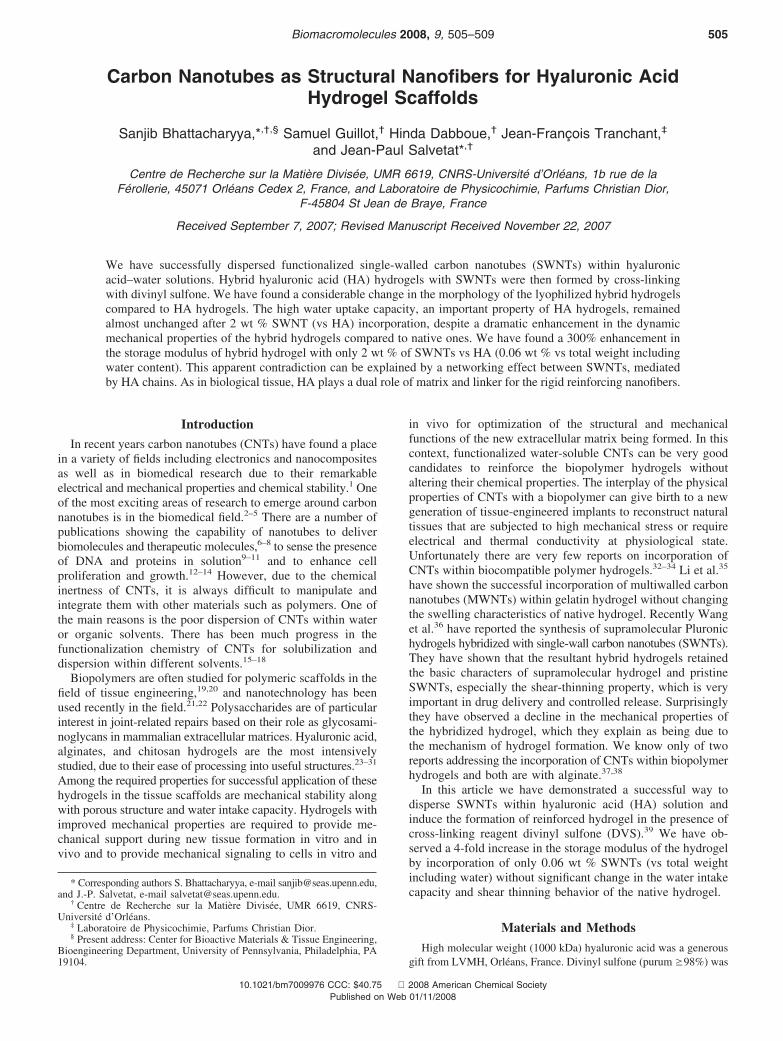
Figure 2. FTIR spectra of starting HA, oxidized SWNT, and DVScross-linked HA gels with 2 wt % (vs HA) SWNTs. (Spectra are shiftedvertically for a better view.)
506 Biomacromolecules, Vol. 9, No. 2, 2008 Bhattacharyya et al.
linking occurs via the hydroxyl groups of HA forming an etherbond as shown in Scheme 1. Due to cross-linking through thehydroxyl group, the intensity of the peak of the OH group inFTIR spectra (Figure 2, at around 3400 cm-1) decreased forthe cross-linked HA gel with and without SWNTs, whichindicates the presence of SWNTs did not hinder the cross-linkingreaction. There is also an appearance of two enhanced peaks inthe case of cross-linked gels at around 1290 and 1120 cm-1
which are coming both from C-O stretch modes involved inthe HA-DVS bonding (1260 cm-1 and 1110 cm-1) and fromSdO stretch modes of DVS (1316 and 1124 cm-1). Acontribution from C-H bend modes should also be presentaround 1270 cm-1. We did not noticed any modification of the1640 cm-1 peak, corresponding to the symmetric CdO stretchmode of HA, which further confirmed that the main reactionbetween HA and DVS was between the hydroxyl groups. Thesedata confirm the successful cross-linking of HA despite thepresence of SWNTs. It was not possible, however, to assess byFTIR whether covalent reactions occurred between oxidizedSWNTs and HA via DVS.
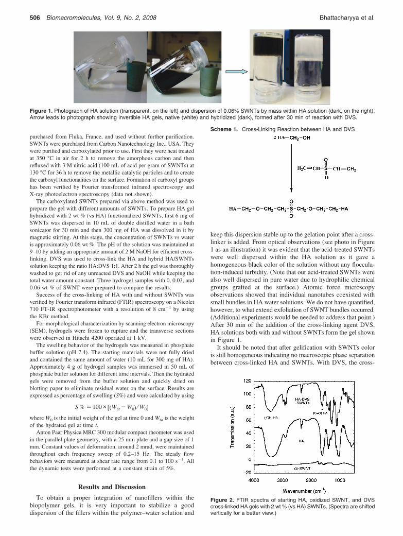
Morphology of Freeze-Dried Hydrogels. Panels a and b ofFigure 3 show the typical interconnected porous morphologyfor the native hydrogel having uniform pore sizes ranging from6 to 9 µm. We observed a considerable change in themorphology of the hybrid hydrogels. In panels c and d of Figure3, the material constituting the original honeycombed orspongelike structure seems to have ”relaxed” and separated intoseparate fibers or slivers of material, although the chamberedstructure can still be recognized. Pores seem, surprisingly, larger
than those in the pristine structure but are filled with filamentousmaterial. Mechanically rigid SWNTs are probably involved inthe backbone of the fibrous substructure, which suggests thatthey interact strongly with HA during the cross-linking reactionwith DVS. We may wonder whether the spontaneous liquidcrystal phase separation of nanotubes in HA reported byMoulton can be involved in the filamentous structure forma-tion.40 However, the ratio between SWNT and HA concentration(0.06:3) wt % is not in favor of a nematic phase according tothe phase diagram established by the authors.40
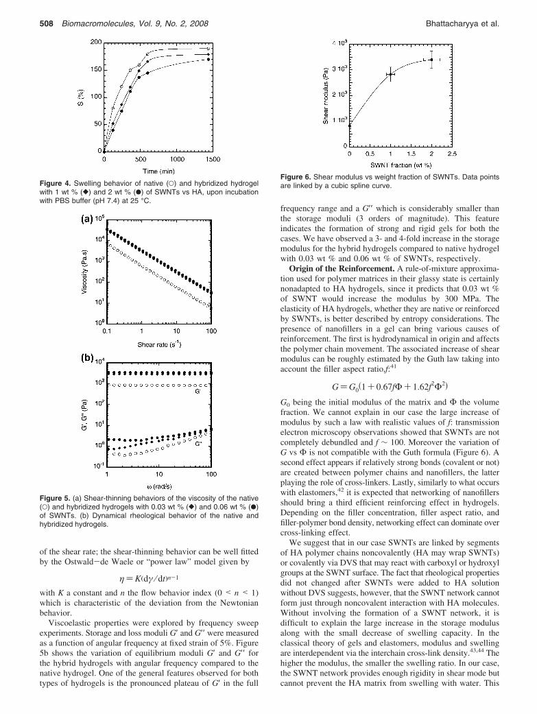
Swelling Behavior. From Figure 4, it can be clearly seenthat the hybrid and native hydrogels show the same generalswelling trend. After 24 h of water absorption we have observedonly 12% difference in the equilibrium water uptake capacitybetween native (190%) and hybrid (170%) hydrogel with 2%(wt/wt HA) of SWNTs. These results demonstrate that thepresence of SWNTs has very little influence on the water uptakecapacity of the gel.
Rheological Properties of the Native and Hybrid Hydro-gels. To examine the influence of SWNTs on the dynamicalviscosity and moduli of the hydrogels, two samples withdifferent concentrations of SWNTs were employed for therheology test. The steady flow results showed a considerableincrease in the viscosity due to the incorporation of SWNTs(Figure 5a). Both the native and hybrid hydrogels exhibited ashear-thinning behavior (decrease of the viscosity with increas-ing the shear rate), which is a typical character of supramolecularhydrogels.26 In Figure 5a, we plotted the viscosity as a function
Figure 3. SEM images of the morphologies of native hydrogels (a, b) and hydrogels hybridized with 0.06 wt % SWNTs (c, d). Scale bars inpanels a and c are 30 µm, and those in panels b and d are 10 µm.
CNTs as Structural Nanofibers for Scaffolds Biomacromolecules, Vol. 9, No. 2, 2008 507
of the shear rate; the shear-thinning behavior can be well fittedby the Ostwald-de Waele or “power law” model given by
η)K(dγ ⁄ dt)n-1
with K a constant and n the flow behavior index (0 < n < 1)which is characteristic of the deviation from the Newtonianbehavior.
Viscoelastic properties were explored by frequency sweepexperiments. Storage and loss moduli G′ and G′′ were measuredas a function of angular frequency at fixed strain of 5%. Figure5b shows the variation of equilibrium moduli G′ and G′′ forthe hybrid hydrogels with angular frequency compared to thenative hydrogel. One of the general features observed for bothtypes of hydrogels is the pronounced plateau of G′ in the full
frequency range and a G′′ which is considerably smaller thanthe storage moduli (3 orders of magnitude). This featureindicates the formation of strong and rigid gels for both thecases. We have observed a 3- and 4-fold increase in the storagemodulus for the hybrid hydrogels compared to native hydrogelwith 0.03 wt % and 0.06 wt % of SWNTs, respectively.
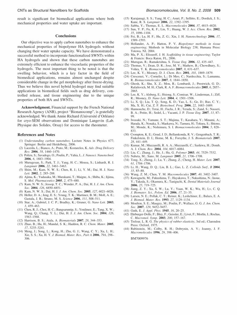
Origin of the Reinforcement. A rule-of-mixture approxima-tion used for polymer matrices in their glassy state is certainlynonadapted to HA hydrogels, since it predicts that 0.03 wt %of SWNT would increase the modulus by 300 MPa. Theelasticity of HA hydrogels, whether they are native or reinforcedby SWNTs, is better described by entropy considerations. Thepresence of nanofillers in a gel can bring various causes ofreinforcement. The first is hydrodynamical in origin and affectsthe polymer chain movement. The associated increase of shearmodulus can be roughly estimated by the Guth law taking intoaccount the filler aspect ratio,f:41
G)G0(1+ 0.67fΦ+ 1.62f2Φ2)
G0 being the initial modulus of the matrix and Φ the volumefraction. We cannot explain in our case the large increase ofmodulus by such a law with realistic values of f: transmissionelectron microscopy observations showed that SWNTs are notcompletely debundled and f ∼ 100. Moreover the variation ofG vs Φ is not compatible with the Guth formula (Figure 6). Asecond effect appears if relatively strong bonds (covalent or not)are created between polymer chains and nanofillers, the latterplaying the role of cross-linkers. Lastly, similarly to what occurswith elastomers,42 it is expected that networking of nanofillersshould bring a third efficient reinforcing effect in hydrogels.Depending on the filler concentration, filler aspect ratio, andfiller-polymer bond density, networking effect can dominate overcross-linking effect.
We suggest that in our case SWNTs are linked by segmentsof HA polymer chains noncovalently (HA may wrap SWNTs)or covalently via DVS that may react with carboxyl or hydroxylgroups at the SWNT surface. The fact that rheological propertiesdid not changed after SWNTs were added to HA solutionwithout DVS suggests, however, that the SWNT network cannotform just through noncovalent interaction with HA molecules.Without involving the formation of a SWNT network, it isdifficult to explain the large increase in the storage modulusalong with the small decrease of swelling capacity. In theclassical theory of gels and elastomers, modulus and swellingare interdependent via the interchain cross-link density.43,44 Thehigher the modulus, the smaller the swelling ratio. In our case,the SWNT network provides enough rigidity in shear mode butcannot prevent the HA matrix from swelling with water. This
Figure 4. Swelling behavior of native (O) and hybridized hydrogelwith 1 wt % ([) and 2 wt % (b) of SWNTs vs HA, upon incubationwith PBS buffer (pH 7.4) at 25 °C.
Figure 5. (a) Shear-thinning behaviors of the viscosity of the native(O) and hybridized hydrogels with 0.03 wt % ([) and 0.06 wt % (b)of SWNTs. (b) Dynamical rheological behavior of the native andhybridized hydrogels.
Figure 6. Shear modulus vs weight fraction of SWNTs. Data pointsare linked by a cubic spline curve.
508 Biomacromolecules, Vol. 9, No. 2, 2008 Bhattacharyya et al.
result is significant for biomedical applications where bothmechanical properties and water uptake are important.
Conclusions
Our objective was to apply carbon nanotubes to enhance themechanical properties of biopolymer HA hydrogels withoutchanging their water uptake capacity. We have demonstrated asuccessful method to incorporate functionalized SWNTs withinHA hydrogels and shown that these carbon nanotubes areextremely efficient to enhance the viscoelastic properties of thehydrogels. The most important thing to be noted is that theswelling behavior, which is a key factor in the field ofbiomedical applications, remains almost unchanged despiteconsiderable change in the gel morphology after freeze-drying.Thus we believe this novel hybrid hydrogel may find suitableapplications in biomedical fields such as drug delivery, con-trolled release, and tissue engineering due to the uniqueproperties of both HA and SWNTs.
Acknowledgment. Financial support by the French NationalResearch Agency (ANR), project “Bionanocomp”, is gratefullyacknowledged. We thank Annie Richard (Université d’Orléans)for cryo-SEM observations and Dominique Langevin (Lab.Physique des Solides, Orsay) for access to the rheometer.
References and Notes(1) Understanding carbon nanotubes; Lecture Notes in Physics 677;
Springer: Berlin and Heidelberg, 2006.(2) Lacerda, L.; Bianco, A.; Prato, M.; Kostarelos, K. AdV. Drug DeliVery
ReV. 2006, 58, 1460–1470.(3) Polizu, S.; Savadogo, O.; Poulin, P.; Yahia, L. J. Nanosci. Nanotechnol.
2006, 6, 1883–1904.(4) Murugesan, S.; Park, T. J.; Yang, H. C.; Mousa, S.; Linhardt, R. J.
Langmuir 2006, 22, 3461–3463.(5) Shim, M.; Kam, N. W. S.; Chen, R. J.; Li, Y. M.; Dai, H. J. Nano
Lett. 2002, 2, 285–288.(6) Ajima, K.; Yudasaka, M.; Murakami, T.; Maigne, A.; Shiba, K.; Iijima,
S. Mol. Pharmaceutics 2005, 2, 475–480.(7) Kam, N. W. S.; Jessop, T. C.; Wender, P. A.; Dai, H. J. J. Am. Chem.
Soc. 2004, 126, 6850–6851.(8) Kam, N. W. S.; Dai, H. J. J. Am. Chem. Soc. 2005, 127, 6021–6026.(9) Heller, D. A.; Jeng, E. S.; Yeung, T. K.; Martinez, B. M.; Moll, A. E.;
Gastala, J. B.; Strano, M. S. Science 2006, 311, 508–511.(10) Star, A.; Gabriel, J. C. P.; Bradley, K.; Gruner, G. Nano Lett. 2003,
3, 459–463.(11) Chen, R. J.; Choi, H. C.; Bangsaruntip, S.; Yenilmez, E.; Tang, X. W.;
Wang, Q.; Chang, Y. L.; Dai, H. J. J. Am. Chem. Soc. 2004, 126,1563–1568.
(12) Harrison, B. S.; Atala, A. Biomaterials 2007, 28, 344–353.(13) Zhao, B.; Hu, H.; Mandal, S. K.; Haddon, R. C. Chem. Mater. 2005,
17, 3235–3241.(14) Meng, J.; Song, L.; Kong, H.; Zhu, G. J.; Wang, C. Y.; Xu, L. H.;
Xie, S. S.; Xu, H. Y. J. Biomed. Mater. Res., Part A 2006, 79A, 298–306.
(15) Karajanagi, S. S.; Yang, H. C.; Asuri, P.; Sellitto, E.; Dordick, J. S.;Kane, R. S. Langmuir 2006, 22, 1392–1395.
(16) Sabba, Y.; Thomas, E. L. Macromolecules 2004, 37, 4815–4820.(17) Sun, Y. P.; Fu, K. F.; Lin, Y.; Huang, W. J. Acc. Chem. Res. 2002,
35, 1096–1104.(18) Fei, B.; Lu, H. F.; Hu, Z. G.; Xin, J. H. Nanotechnology 2006, 17,
1589–1593.(19) Hollander, A. P.; Hatton, P. V. Biopolymer methods in tissue
engineering; Methods in Molecular Biology 238; Humana Press:Totowa, NJ, 2004.
(20) Ma, P. X.; Elisseeff, J. H. Scaffolding in tissue engineering; Taylor& Francis: Boca Raton, FL, 2006.
(21) Murugan, R.; Ramakrishna, S. Tissue Eng. 2006, 12, 435–447.(22) Thomas, V.; Dean, D. R.; Jose, M. V.; Mathew, B.; Chowdhury, S.;
Vohra, Y. K. Biomacromolecules 2007, 8, 631–637.(23) Lee, K. Y.; Mooney, D. J. Chem. ReV. 2001, 101, 1869–1879.(24) Crescenzi, V.; Cornelio, L.; Di Meo, C.; Nardecchia, S.; Lamanna,
R. Biomacromolecules 2007, 8, 1844–1850.(25) Ghosh, K.; Shu, X. Z.; Mou, R.; Lombardi, J.; Prestwich, G. D.;
Rafailovich, M. H.; Clark, R. A. F. Biomacromolecules 2005, 6, 2857–2865.
(26) Lee, K. Y.; Alsberg, E.; Hsiong, S.; Comisar, W.; Linderman, J.; Ziff,R.; Mooney, D. Nano Lett. 2004, 4, 1501–1506.
(27) Li, X. Q.; Liu, T. Q.; Song, K. D.; Yao, L. S.; Ge, D.; Bao, C. Y.;Ma, X. H.; Cui, Z. F. Biotechnol. Prog. 2006, 22, 1683–1689.
(28) Hannouche, D.; Terai, H.; Fuchs, J. R.; Terada, S.; Zand, S.; Nasseri,B. A.; Petite, H.; Sedel, L.; Vacanti, J. P. Tissue Eng. 2007, 13, 87–99.
(29) Iwasaki, N.; Yamane, S. T.; Majima, T.; Kasahara, Y.; Minami, A.;Harada, K.; Nonaka, S.; Maekawa, N.; Tamura, H.; Tokura, S.; Shiono,M.; Monde, K.; Nishimura, S. I. Biomacromolecules 2004, 5, 828–833.
(30) Crompton, K. E.; Goud, J. D.; Bellamkonda, R. V.; Gengenbach, T. R.;Finkelstein, D. I.; Horne, M. K.; Forsythe, J. S. Biomaterials 2007,28, 441–449.
(31) Kumar, M.; Muzzarelli, R. A. A.; Muzzarelli, C.; Sashiwa, H.; Domb,A. J. Chem. ReV. 2004, 104, 6017–6084.
(32) Liu, C.; Zhang, J.; He, J.; Hu, G. Polymer 2003, 44, 7529–7532.(33) Nabeta, M.; Sano, M. Langmuir 2005, 21, 1706–1708.(34) Tong, X.; Zheng, J.; Lu, Y.; Zhang, Z.; Cheng, H. Mater. Lett. 2007,
61, 1704–1706.(35) Li, H.; Wang, D. Q.; Liu, B. L.; Gao, L. Z. Colloids Surf., B 2004,
33, 85–88.(36) Wang, Z. M.; Chen, Y. M. Macromolecules 2007, 40, 3402–3407.(37) Kawaguchi, M.; Fukushima, T.; Hayakawa, T.; Nakashima, N.; Inoue,
Y.; Takeda, S.; Okamura, K.; Taniguchi, K. Dental Materials Journal2006, 25, 719–725.
(38) Jiang, Z. Y.; Xu, S. W.; Lu, Y.; Yuan, W. K.; Wu, H.; Lv, C. Q.J. Biomater. Sci., Polym. Ed. 2006, 17, 21–35.
(39) Larsen, N. E.; Pollak, C. T.; Reiner, K.; Leshchiner, E.; Balazs, E. A.J. Biomed. Mater. Res. 1993, 27, 1129–1134.
(40) Moulton, S. E.; Maugey, M.; Poulin, P.; Wallace, G. G. J. Am. Chem.Soc. 2007, 129, 9452–9457.
(41) Guth, E. J. Appl. Phys. 1945, 16, 20–25.(42) Ehrburger-Dolle, F.; Bley, F.; Geissler, E.; Livet, F.; Morfin, I.; Rochas,
C. Macromol. Symp. 2003, 200, 157–167.(43) Treloar, L. R. G. The physics of rubber elasticity, 3rd ed.; Clarendon
Press: Oxford, 1975.(44) Rubinstein, M.; Colby, R. H.; Dobrynin, A. V.; Joanny, J. F.
Macromolecules 1996, 29, 398–406.
BM7009976
CNTs as Structural Nanofibers for Scaffolds Biomacromolecules, Vol. 9, No. 2, 2008 509