Embed Size (px)
Citation preview
採択課題番号 24009 BNCT用照射場におけるQA/QCの確立 共同通常
(京大・原子炉)櫻井良憲、田中浩基、吉永尚生(京大院・工)藤井孝明、武川哲也、内田良平
CO7-1 Establishment of QA/QC for BNCT Neutron Irradiation Field
Y. Sakurai, T. Fujii1, R. Uchida
1 and H. Tanaka
Research Reactor Institute, Kyoto University 1Graduate School of Engineering, Kyoto University
INTRODUCTION: In May 2010, the operation of
Kyoto University Reactor (KUR) restarted, which had
been suspended during four years for the
fuel-low-enrichment. Concurrently with the KUR restart,
clinical irradiation of boron neutron capture therapy
(BNCT) at the Heavy Water Neutron Irradiation Facility
(HWNIF) also restarted [1]. After the restart, 170 BNCT
irradiations have already been carried out as of May 2013.
In the while, Cyclotron-based BNCT Epi-thermal Neu-
tron Source (C-BENS) was installed in this institute in
the end of 2008 [2]. In November 2012, the BNCT clini-
cal trial using C-BENS started. Thus, this institute be-
came a special institute in the world, in where BNCT is
performed at the two-type neutron sources such as reac-
tor-based one and accelerator-based one. It is one of the
important subjects that the consistent dose-estimation is
performed between the both neutron sources, and then the
equivalence and homogeneity for the deposited dose
during the clinical irradiation are assured. The aim of this
research is the establishment of quality assurance and
quality control (QA/QC) for BNCT neutron irradiation
field. In 2012, one of the important tools for QA/QC,
“Multi Ionization Chamber System (MICS)”, was pre-
pared by way of trial [3], and its characteristics were es-
timated and its efficacy was confirmed.
METHODS: The prototype of MICS consists of (i) a
chamber of silicon-nitride wall and nitrogen gas for the
thermal neutron component (Si3N4(N2)), (ii) a chamber of
boron-evaporation-coated polyethylene wall and nitrogen
gas, covered with 6LiF shield, for the epi-thermal neutron
component (PolyB(N2)), (iii) a chamber of polyethylene
wall and methane gas for the fast neutron component
(Poly(CH4)), and (iv) a chamber of graphite wall and
argon gas for the gamma-ray component (G(Ar)). These
chambers were placed on the bismuth-layer side of the
collimator on the remote patient carrier, as shown in
photo 1. The experiment for the characteristic estimation
was performed for the epi-thermal neutron irradiation
mode.
RESULTS AND DISCUSSIONS: Figure 1 shows the
changes of the separate-estimated values with time for
the four components, such as thermal neutron,
epi-thermal neutron, fast neutron, and gamma ray, ob-
tained using MICS. These values are for the KUR power
of 1 MW. The vertical axis is in flux. The solid lines are
for the experimentally-estimated values and the broken
lines are for the estimated values by simulation. In the
epi-thermal neutron irradiation mode, the thermal neutron
flux was practically zero, because it was lower than the
limit for the separate-estimation. For the epi-thermal and
fast neutrons, those experimental values were in good
agreement with the simulation values, in the uncertainty
of 4% and 12%, respectively. For the gamma ray, the
experimental value overestimated the simulation value at
63%. The flux dispersion in the experiment was 2%, 26%
and 27%, respectively for epi-thermal neutron, fast neu-
tron and gamma ray. It was confirmed that the accuracy
in the separate-estimation was better and the response
was more stable for the component with the larger re-
sponse.
CONCLUSION: The efficacy of the prototype MICS
was confirmed. The possibility of the separate-estimation
for the thermal neutron component is expected, as the
KUR power is 5 MW, during the actual BNCT.
REFERENCES: [1] Y. Sakurai et al., Nucl. Instr. Meth. A 453 (2000)
569-596.
[2] H. Tanaka et al., Nucl. Instr. Meth. B 267 (2009)
1970-1977.
[3] T. Fujii et al., Appl. Radiat. Isot. 69 (2011)
1862-1865.
Photo 1. Placement of MICS on the collimator.
Fig. 1. Changes of the separate-estimated values
with time, obtained using MICS.
採択課題番号 24012 血管内皮細胞を標的とする放射線耐性腫瘍の克服にむけて 共同通常
(東北大・加齢研)桑原義和、福本基、福本学(京大・原子炉)鈴木実、小野公二
CO7-2 To Conquer the Clinically Relevant Radioresistant Cell Tumors
Targeting Tumor Endohtelial Cells Y. Kuwahara, M. Fukumoto, Y. Sakurai
1, M. Suzuki
1,
K. Ono1 and M. Fukumoto
Department of Pathology, Institute of Development, Ag-
ing and Cancer, Tohoku University. 1Department of Radiation Life Science and Radiation
Medical Science, Research Reactor Institute, Kyoto Uni-
versity INTRODUCTION: Radiotherapy is one of the major therapeutic modalities for eradicating malignant tumors. However, the existence of radioresistant cells remains one of the most critical obstacles in radiotherapy. To un-derstand the characteristics of radioresistant cells and to develop more effective radiotherapy, we have established clinically relevant radioresistant (CRR) cell lines. Be-cause tumor tissues of CRR cells transplanted into nude mice were richer in tumor blood vessels compared with their radiosensitive parental cell lines. So, we performed boron neutron capture (BNC) method targeting tumor endothelial cells using PEG-10B. Growth rate of HeLa-R (CRR of HeLa) tumors were not significantly different from that of non-irradiated control in 4 weeks after irra-diation. Tumors of parental cells treated with BNC using PEG-10B were significantly smaller than those without radiation. Seven days after BNC CD31 positive blood vessels were destroyed in BNCT treated HeLa tumors compared to HeLa tumor without BNCT but were not in HeLa-R tumors irrespective of BNCT. Moreover, to our surprise, 4 weeks after BNC the density of CD34, posi-tive blood vessels were almost the same irrespective of BNCT in both HeLa-R and HeLa tumors.
We need further studies to confirm how the density of blood vessels contributes to tumor radiotherapy.
EXPERIMENTS: Three days before experiments, 1x10
6 cells of HeLa and HeLa-R were injected subcuta-
neously into hind legs of male Balb/c nude mice (4 weeks old). The day of the exposure experiment tumor diameter was approximately 3-4 mm. For irradiation of tumor en-dothelial cells by α-particles, PEG-
10B were administered.
The compound was suspended in physiological saline at a concentration of 2500 ppm and was injected via the tail vein. Three hours after the administration, mice were exposed to neutron radiation at the Research Reactor In-stitute, Kyoto University (KURRI).
Tumor size was determined by caliper measurements every three days. Endothelial cells of blood vessels in tumor tissues were immunohistochemically stained for CD34 and CD31. Type Ⅳcollagen for pericytes and functional blood vessels for injected tomatolectin from tails. We counted the number of vessels in 10 high power field (x 400) and calculated the average (n = 3).
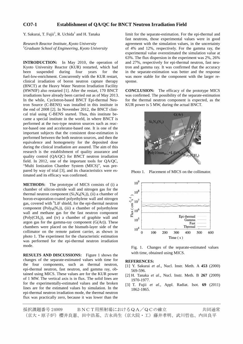
RESULTS: We first examined the size of HeLa tumors and HeLa-R daily after BNCT. Within 24 days after BNCT, the size of HeLa-R tumors was not different irre-spective BNCT. But the size of HeLa tumors was signif-icantly decreased compare to control tumors. Mice were sacrificed on day 30 after BNCT. Histological examination showed that HeLa-R tumors were richer in tumor blood vessels compared with their radiosensitive parental cell tumors. CD34 positive blood vessels were also more abundant in SAS-R tumors than in SAS-tumors (Data not shown).
After BNC using PEG-10B, the vessels of HeLa-R tumors were recovered more rapidly in 7 days. On the other hand, those of parental tumors were destroyed.
However, 4 weeks after BNC, the density of CD34 positive blood vessels was almost the same irrespective of BNCT in both HeLa-R tumors and HeLa tumors. DISCUSSION: In this research, we tried to target tu-mor endothelial cells of radioresistant HeLa-R tumors, because our preliminary experiment showed that the den-sity of blood vessels in SAS-R tumors was higher than that in SAS tumors. We selectively exposed endothelial cells to α-particles, using BNC with PEG-
10B. The tumor
volume of HeLa-R was not significantly different after exposure to 8 Gy of α-particles for the examination peri-od. But that of HeLa was significantly decreased compare to control tumors.
7 days after BNC the density of CD31 positive blood vessels were destroyed in HeLa tumor exposed to α-particles compared to control HeLa tumor but did not in HeLa-R tumor irrespective of α-particles exposure. Moreover, to our surprise, 4 weeks after BNC the density of CD34, positive blood vessels were almost same irre-spective of α-particles exposure in both HeLa-R tumor and HeLa.
Therefore, further studies are needed to confirm how blood vessel density contributes to tumor radiotherapy.
採択課題番号 24014 極少検出器を用いた中性子計測手法の高度化に関する研究 共同通常
(名大・工)瓜谷 章、渡辺賢一、山﨑 淳、川端勇矢、牛田雅人
(京大・原子炉)櫻井良憲、田中浩基
CO7-3 Study on Advanced Neutron Measurements Using a Small Size Neutron Scintillator
K. Watanabe, Y. Kawabata, M. Ushida, A. Uritani,
A. Yamazaki, Y. Sakurai1 and H. Tanaka
1
Graduate School of Engineering, Nagoya University 1Research Reactor Institute, Kyoto University
INTRODUCTION: The Boron Neutron Capture
Therapy (BNCT) has been developed as one of the
promising radiotherapies. The neutron dose evaluation
for the BNCT is quite important. Optical fiber type de-
tectors as one of the on-line and small neutron flux mon-
itors have been developed. The conventional optical
fiber neutron detectors, however, show a continuous dis-
tribution without a characteristic shape, such as the full
energy peak corresponding to the neutron induced reac-
tion, in a pulse height spectrum due to large fluctuation of
collected scintillation photons based on their poor light
collection efficiency[1-3]. The sensitivity of these de-
tectors depends on the detector signal gain. We, there-
fore, develop the advanced optical fiber type neutron de-
tector using a small piece of Eu doped LiCaAlF6 scintil-
lator. This detector can show an obvious neutron ab-
sorption peak and suppress the gamma-ray sensitivity.
In this report, we characterize the developed neutron de-
tector at the Heavy Water Thermal Neutron Irradiation
Facility (HWTNIF) of Kyoto University Research Reac-
tor (KUR).
DEVELOPED DETECTOR: We fabricated the opti-
cal fiber type neutron detector using a small Eu:LiCaAlF6
scintillator. The fabricated detector consists of a small
piece of Eu doped LiCaAlF6 scintillator, a plastic optical
fiber, a photomultiplier tube (PMT) and signal processing
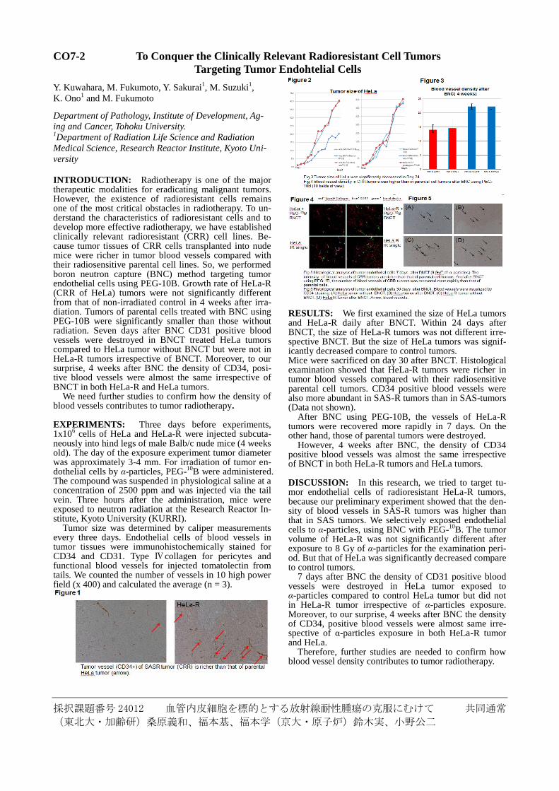
circuits. A bulk Eu:LiCaAlF6 scintillator suffers from
influence of gamma rays because of its relatively low /b
ratio. Figure 1 shows the pulse height spectra obtained
from a bulk Eu:LiCaAlF6 scintillator. Gamma-ray sig-
nals are confirmed to interfere the neutron absorption
peak.
0 100 200 300 400 500
10-2
10-1
100
pulse height [channel]
cou
nti
ng r
ate
[cp
s]
252
Cf
60Co
BG
Fig. 1. Pulse height spectra obtained from a bulk
Eu:LiCaAlF6 scintillator.
Figure 2 shows the pulse height spectra obtained from
an optical fiber detector with a small LiCaAlF6 scintilla-
tor. The pulse height of signals induced only by gamma
rays can be suppressed compared with neutron induced
signals. This is because of differences in ranges of
charged particles produced by neutrons and gamma rays.
0 50 100 150
10-3
10-2
10-1
BG
252
Cf線源60
Co線源
波高値 [channel]
計数率
[cp
s]
Pulse height (ch)
Co
un
t ra
te (
cps)
Cf-252
Co-60
BG
Fig. 2. Pulse height spectra obtained from an optical fiber
detector with a small Eu:LiCaAlF6 scintillator.
EXPERIMENTS AT HWTNIF OF KUR: We char-
acterize the fabricated detector at the HWTNIF of KUR.
The detector head was placed at various distances from
the bismuth filter surface. An example of the pulse
height spectrum obtained from the fabricated detector in
experiments at the HWTMIF of KUR is shown in Fig. 3,
where the distance from the bismuth filter surface was
120 cm. A clear neutron absorption peak was obviously
observed.
0 100 200 300 400 5000
50
100
Pulse height (ch)
Count
rate
(cps)
Fig. 3. Example of the pulse height spectrum obtained
from the fabricated detector in experiments at the HWT-
NIF of KUR.
REFERENCES: [1] M. Ishikawa et al., Appl. Rad. Isotopes, 61 (2004)
775-779.
[2] M. Ishikawa et al., Nucl. Instr. Meth. A, 551 (2005)
448-457.
[3] Y. Ito et al., Radiat. Prot. Dosim., 110 (2004)
619-622.
採択課題番号 24023 中性子捕捉治療のための新規ホウ素薬剤の開発 共同通常
(学習院大・理)中村 浩之、立川 将士、小金井 逸人
(京大・原子炉)鈴木 実、増永 慎一郎、小野 公二
CO7-4 High Boron Content Liposomes and Their Promising Antitumor Effect for BNCT
H. Koganei, S. Tachikawa, M. Suzuki1, S. Masunaga
1,
K. Ono1 and H. Nakamura
Faculty of Science, Gakushuin University 1Research Reactor Institute, Kyoto University
INTRODUCTION: Boron neutron capture therapy
(BNCT) functions as a double targeting therapy for can-
cer. Its therapeutic effect is realized by neutron beam
irradiation and a boron delivery system (BDS). BNCT
uses the nuclear reaction of two species, boron-10 (10
B)
and thermal neutrons. Although the low-energy thermal
neutrons (0.025 eV) are employed, the resulting α
-particle and Li nuclei are high linear energy transfer
(LET) particles that travel a short distance (approximate-
ly 5−9 μm) to destroy cells containing 10
B. If 10
B atoms
were selectively delivered to intracellular regions of tu-
mor tissue, it would be possible to kill tumor cells selec-
tively without seriously damaging adjacent healthy tis-
sues.
In this study, we focused on lipophilic boron com-
pounds embedded in a liposome bilayer. This strategy is
an attractive means to increase the overall incorporation
efficiency of boron containing species, as well as to raise
the gross boron content of liposomes [1]. We developed
high boron content liposomes by incorporating boron into
both the interior aqueous core and the membrane of lipo-
somes. Indeed, this strategy yielded significant antitumor
effect on tumor-bearing mice after neutron irradiation, as
well as a reduction of the total liposome dose, revealing
that the current boronated liposome is one of the most
promising candidates for practical use in BDSs for
BNCT.
EXPERIMENTS: DSPC (MC-8080) and DSPE-PEG
(Sunbright DSPE-020CN) were purchased from Nippon
Oil and Fats (Tokyo, Japan). Cholesterol (Chol) was
purchased from Kanto Chemical (Tokyo, Japan). 10
B-enriched BSH and S–cyanoethyl protected 10
B
-enriched BSH were purchased from Stella Pharma Co.
(Osaka, Japan). Boron lipid (DSBL) was synthesized
according to the previously described procedures with
modification [2].
BSH-encapsulated DSBL-10% liposomes, which were
prepared from 10
B-enriched DSBL, DSPC, Chol, and
DSPE-PEG (0.1:0.9:1:0.11, molar ratio) and 125 mM
BSH aqueous solution according to the REV method pre-
viously described [3], were injected into colon 26 tumor
bearing mice (female, 6−7 weeks old, 16−20 g, 5 mice in
each group) via the tail vein at doses of 15 and 30 mg 10
B
/kg (1500 and 3000 ppm of 10
B concentration; 200 μL of
boronated liposome solution). The mice were placed in
an acrylic mouse holder 36 h after i.v. injection. The mice
were irradiated in KUR. The antitumor effects of BNCT
were evaluated on the basis of the changes in tumor
volume of the mice. Mortality was monitored daily and
tumor volume was measured at intervals of a few days.
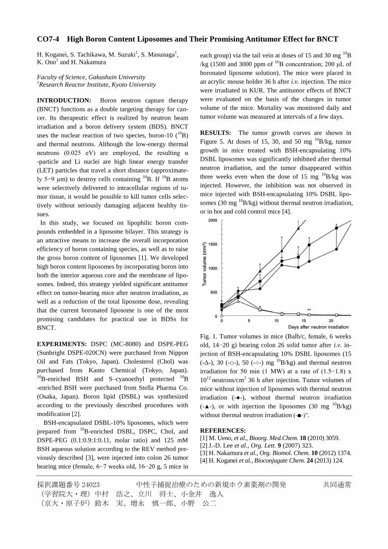
RESULTS: The tumor growth curves are shown in
Figure 5. At doses of 15, 30, and 50 mg 10
B/kg, tumor
growth in mice treated with BSH-encapsulating 10%
DSBL liposomes was significantly inhibited after thermal
neutron irradiation, and the tumor disappeared within
three weeks even when the dose of 15 mg 10
B/kg was
injected. However, the inhibition was not observed in
mice injected with BSH-encapsulating 10% DSBL lipo-
somes (30 mg 10
B/kg) without thermal neutron irradiation,
or in hot and cold control mice [4].
Fig. 1. Tumor volumes in mice (Balb/c, female, 6 weeks
old, 14−20 g) bearing colon 26 solid tumor after i.v. in-
jection of BSH-encapsulating 10% DSBL liposomes (15
(-Δ-), 30 (-□-), 50 (-○-) mg 10
B/kg) and thermal neutron
irradiation for 50 min (1 MW) at a rate of (1.5−1.8) x
1012
neutrons/cm2 36 h after injection. Tumor volumes of
mice without injection of liposomes with thermal neutron
irradiation (-●-), without thermal neutron irradiation
(-▲-), or with injection the liposomes (30 mg 10
B/kg)
without thermal neutron irradiation (-■-)”.
REFERENCES: [1] M. Ueno, et al., Bioorg. Med.Chem. 18 (2010) 3059.
[2] J.-D. Lee et al., Org. Lett. 9 (2007) 323.
[3] H. Nakamura et al., Org. Biomol. Chem. 10 (2012) 1374.
[4] H. Koganei et al., Bioconjugate Chem. 24 (2013) 124.
採択課題番号 24029 明細胞肉腫に対するホウ素中性子捕捉療法の検討 共同通常
(京大・原子炉)鈴木実、櫻井良憲、小野公二(神戸学院大・薬)安藤 徹、市川秀喜
(兵庫県立がんセンター・整形外科)藤本卓也
CO7-5 Long-Term Result of BNCT for Different Types of Human Clear Cell Sarcoma
in Mouse Model
T. Fujimoto, T. Andoh1, Y. Sakurai
2, M. Suzuki
2,
H. Ichikawa1 and K. Ono
2
Hyogo Cancer Center,
Department of Orthopaedic Surgery 1Faculty of Pharmaceutical Sciences and Cooperative
Research Center of Life Sciences,
Kobe Gakuin University 2Research Reactor Institute, Kyoto University
INTRODUCTION: Clear cell sarcoma (CCS) of ten-
dons and aponeuroses is a rare neoplasm with no effec-
tive treatment other than surgery. Furthermore, this ma-
lignant tumor has a predilection for young adults and its
prognosis is poor [1]. Clearly, therefore, new therapeutic
strategies are required. In a previous study, we have
shown that the high accumulation of 10
B both in cultured
human CCS cell lines and in CCS-bearing animal models,
is potentially propitious in boron neutron capture therapy
(BNCT) with the use of p-boronophenylalanine (BPA)
[2,3]. We have recently also demonstrated the
effectiveness of short-term BNCT in the human
CCS-bearing nude mouse model [4]. Thus BNCT could
become a potential, new therapeutic option for the
treatment of human CCS. Consequently, in this study, we
evaluated the long-term efficacy of BNCT for CCS, with
the use of nude mice intramuscularly transplanted with
human CCS cell lines.
EXPERIMENTS: (1) Tumor cell line: Human CCS
cell lines, MP-CCS-SY [5], SU-CCS-1 [6] and HS-MM
[7] were grown in RPMI 1640 and DMEM (for HS-MM)
with fetal bovine serum in a 5% CO2 humidified
incubator at 37˚C.
(2) BNCT for CCS-bearing animal: All animal experi-
ments were carried out according to the regulations of the
Animal Care and Use Committee. Cells of CCS cell lines
were transplanted into the left femoral region of BALB/c
nude mice. When CCS tumors grew to about 10-20mm in
diameter, the animals were divided into 3 BNCT groups
and 3 control groups for each cell line. The animals, un-
der anesthesia and through the femoral vein, were intra-
venously administered BPA-Fr (24mg 10
B/kg; the BNCT
groups) or saline (the control groups), and then immedi-
ately placed in a chamber for thermal neutron irradiation
experiments. Thermal neutrons (1MW) were delivered
from the dorsum of the mouse, in the heavy water facility
at KURRI. LiF tiles were used to shield parts of the body
other than the left leg. After the irradiation, the size of the
tumor was measured as the maximum elliptical area of
the tumor mass as follows: Maximum tumor area=(minor
axis)×(major axis), and the “Area ratio” was defined as
the area of each divided by the area of day 0. Tissue sam-
ples of the tumor mass resected under anesthesia from the
control groups on day 45, and from the BNCT groups on
day 90 were fixed in 10% buffered-formalin solution and
embedded in paraffin according to standard protocols.
Sections were then stained with hematoxylin-eosin (HE)
for histological examination.
RESULTS: The doses absorbed (Gy) by the mice in-
tramuscularly transplanted with the CCS cells were 7.2
(HS-MM), 6.8 (MP-CCS-SY) and 7.1 (SU-CCS-1). After
irradiation, the size of the decreased time-dependently in
the BNCT groups until around day 20, and then regrew
time-dependently. In the control groups, the growth was
not suppressed by thermal neutron irradiation; the tumor
mass simply increased with time [Fig.1]. Histological
examination of the BNCT groups on day 90 revealed
regrowth of the CCS tumor of each cell line, with no
damage to normal surrounding tissue.
Fig.1. After the neutron irradiation, the size of the tumor
was measured as the maximum elliptical area of the tu-
mor mass. BNCT groups (ΔMP BNCT; MP-CCS-SY,
□HS BNCT; HS-MM, ○SU BNCT; SU-CCS-1) and con-
trol groups (▲MP Hot control; MP-CCS-SY, ■HS Hot
control; HS-MM, ●SU Hot control; SU-CCS-1).
REFERENCES: [1] F.M. Enzinger, Cancer, 18 (1965) 1163-1174.
[2] T. Fujimoto et al., Appl. Radiat. Isot., 69 (2011)
1713-1716.
[3] T. Andoh et al., Appl. Radiat. Isot., 69 (2011)
1721-1724.
[4] T. Fujimoto et al., Appl. Radiat. Isot., 73 (2013)
96-100.
[5] H. Moritake et al., Cancer Genet. Cytogenet.,
135 (2002) 48-56.
[6] H. Sonobe et al., J. Pathology, 169 (1993) 317-322.
[7] A.L. Epstein et al., Cancer Res., 44 (1984) 1265-1274.
採択課題番号 24030 炭化ホウ素粒子を利用した硼素中性子捕捉療法の可能性 共同通常
(産総研)越崎直人 (香川大・工)石川善恵(京大原子炉実験所)鈴木 実
Application of B4C Nanoparticles for Boron Neutron Capture Therapy
N. Koshizaki, Y. Ishikawa1 and M. Suzuki2
Nanosystem Research Institute (NRI), National Institute of Ad-
vanced Industrial Science and Technology (AIST) 1Kagawa University, Faculty of Engineering 2Research Reactor Institute, Kyoto University
INTRODUCTION: In carcinoma in situ (CIS) in the bladder, the cancer cells are still only in the mucosa of the bladder, but are in flat sheets that look a bit like moss. CIS bladder cancer is called a high risk, early bladder cancer be-cause if it is not treated it is very likely to spread into the deeper layers of the bladder. Nonradioactive isotope 10B atoms that absorb low-energy (<0.5 eV) neutrons (thermal neutrons) disintegrate into an alpha (4He) particle and a recoiled lithium nucleus (7Li). These particles deposit high en-ergy along their very short path (<10 µm). Thus, only malignant cells with 10B are destroyed following ther-mal neutron irradiation. Theoretically, any normal cells abutting the cancer cells are spared from high linear energy transfer irradiation by 4He and 7Li particles. We have been working on a nanoparticle fabrication technique by irradiating laser light onto a plate placed or powder dispersed in liquid media. This technique has several advantages over conventional nanoparticle preparation in liquid phase, highly pure nanoparticles with less use of surfactant molecules mostly toxic in biological systems, and crystallized nanoparticle for-mation due to the transient high temperature process induced by pulsed laser irradiation. Thus, these fea-tures are expected to be suitable for B4C nanoparticle preparation by pulsed laser irradiation of B4C particles dispersed in ethyl acetate under atmospheric pressure and room temperature [1]. In the treatment of BNCT for CIS bladder cancer, intravesical administration of B4C nanoparticles may be appropriate drug delivery system as Bacille de Calmette et Guérin (BCG) vaccine treatment since car-cinoma cells in CIS bladder cancer is exposed directly by B4C nanoparticles applied to bladder. We carried out a preliminary experiment investigat-ing availability of B4C nanoparticles in the treatment of BNCT for CIS bladder cancer. EXPERIMENTS:
HeLa cells were used in this study. The cells were dispersed in the 96-well microplates at the concentration of 10,000 cells per well the day before neutron irradiation. In this experiment, B-10 rich nanoparticles (10B4C nano-particles) were used in the BNCT treatment groups. The treatment conditions were sorted into 5 groups as follows. 1. No treatment
2. 10B4C exposure without neutron irradiation3. Neutron irradiation without 10B4C exposure.4. BNCT with 10B4C nanoparticles (no rinse).5. BNCT following 1 h-exposure with 10B4C and rinse
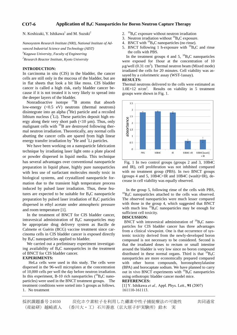
the cells with PBS. In the treatment groups 4 and 5, 10B4C nanoparticles were exposed for 1hour at the concentration of 10 g/well (0.31 cm2). Thermal neutron beam (Mixed mode) irradiated the cells for 20 minutes. Cell viability was as-sayed by a colorimetric assay (WST-1assay). RESULTS: Thermal neutrons delivered to the cells were estimated as 1.0E+12 n/cm2. Results on viability in 5 treatment groups were shown in Fig. 1.
Fig. 1 In two control groups (groups 2 and 3, 10B4C and IR), cell proliferation was not inhibited compared with no treatment group (PBS). In two BNCT groups (groups 4 and 5, 10B4C+IR and 10B4C (wash)+IR), de-crease in cell viability was equally observed.
In the group 5, following rinse of the cells with PBS, 10B4C nanoparticles attached to the cells was observed. The observed nanoparticles were much lesser compared with those in the group 4, which suggested that BNCT with much less 10B4C nanoparticles may be enough for sufficient cell toxicity. DISCUSSION: BNCT with intravesical administration of 10B4C nano-particles for CIS bladder cancer has three advantages from a clinical viewpoint. One is that occurrence of sys-temic toxicity derived from the newly-developed boron compound is not necessary to be considered. Second is that the irradiated doses to rectum or small intestine around the bladder is very low since no boron compound distributed in these normal organs. Third is that 10B4C nanoparticles are more economically prepared compared with other boron compounds, boronophenylalanine (BPA) and borocaptate sodium. We have planned to carry out in vivo BNCT experiments with 10B4C nanoparticles using orthotropic bladder cancer model mice. REFERENCES: [1] Y. Ishikawa et al., Appl. Phys. Lett., 91 (2007) 161110-161113.
CO7-6
採択課題番号 24036 ホウ素中性子捕捉療法の新規ホウ素薬剤の開発研究 共同通常
(阪府大院・生命環境)谷森 紳治、門野 尚之 (京大・原子炉)小野 公二、増永 慎一郎、
鈴木 実 (阪府大・21機構)切畑 光統、椋本 麻里、服部 能英
Development of Novel Boron Compounds for BNCT
S. Tanimori, M. Kirihata1, Y. Hattori
1, M. Mukumoto
1, N.
Kadono, M. Suzuki2, S. Masunaga
2 and K. Ono
2
Graduate School of Life and Environmental Sciences,
Osaka Prefecture University 1Research Organization for 21st Century, Osaka Prefec-
ture University 2Research Reactor Institute, Kyoto University
INTRODUCTION: Boron neutron capture therapy
(BNCT) is an attractive technique for cancer treatment.
Although many kinds of boron compounds such as amino
acid, nucleic acid and peptides have been reported as
boron carrier for BNCT [1], only two compounds,
p-borono-L-phenylalanine (BPA) and mercapto-closo-
undecahydrododecaborate (BSH) are clinically used in
cure of cancer with BNCT. As part of our developing
studies on new boron delivery agents for BNCT, we have
designed and synthesized thiododecaborate
([B12H11]2-
-S-) unit containing L-amino acids (1a–c),
which constitute a new class of tumor-seeking and water
soluble amino acids (Fig. 1). In vitro evaluation studies
on the cytotoxicity, killing effects by neutron irradiation,
and micro distribution analysis performed previously by
our group suggested that 1a–c might be potential delivery
agents for BNCT [2]. On the other hand, recently, ra-
dio-labeled with 18
F ,-cycloalkyl amino acids such as
1-aminocyclobutane-1-carboxylic acid (ACBC) are
highly noted as useful PET (positron emission tomogra-
phy) tracers for diagnosis of cancer, since unusual amino
acids having ,- alkyl ring are incorporated selectively
and temporarily retained by cancer cells.
To develop practical materials utilizing 10
B carrier, we
have newly synthesized dodecdaboratethio-unit contain-
ing , -cycloalkyl amino acids such as cis/trans-
[1-amino-3-(thiododecaboranyl)methyl]cyclobutane-1-
carboxylic acid (ACBC-BSH, 2a and 2b) bearing no
asymmetric carbon atom, by extension of our reported
method. Here, we report the tumor cell killing effects of
2 against cultivated cancer cells.
MATERIAL and METHOD: Cultures were inoculated
with 1.0 x 10⁶ cells/dish, and cells were grown for 24 h in
DMEM. The medium was replaced with the each me-
dium containing each boron amino acid (final concentra-
tion was 1.0 mM in each case). The cells were cultured
for 24 h, and the medium was removed by aspiration. The
cells were washed with PBS, harvested by trypsini-
zation, and then cell numbers were counted. After cen-
trifugation, the tripsin was removed by aspiration, and to
the residual cells was added DMEM. The suspension of
the cell in DMEM (5.0 x 10³
cells/mL, 1mL) was irradi-ated with thermal neutron for 0 - 90 min in column-shape
tube. The thermal neutron fluence was determined by
averaging two gold foils symmetrically attached to the
surface of the column-shape tube along the direction of
incidence of thermal neutrons. After thermal neutron
exposure, 600 cells were placed in three Corning 60 mm
tissue culture dishes containing 3 mL DMEM to examine
colony formation. Seven days later, the colonies were
fixed with ethanol and stained with 0.1% crystal violet
for quantitative visualization by the naked eye.
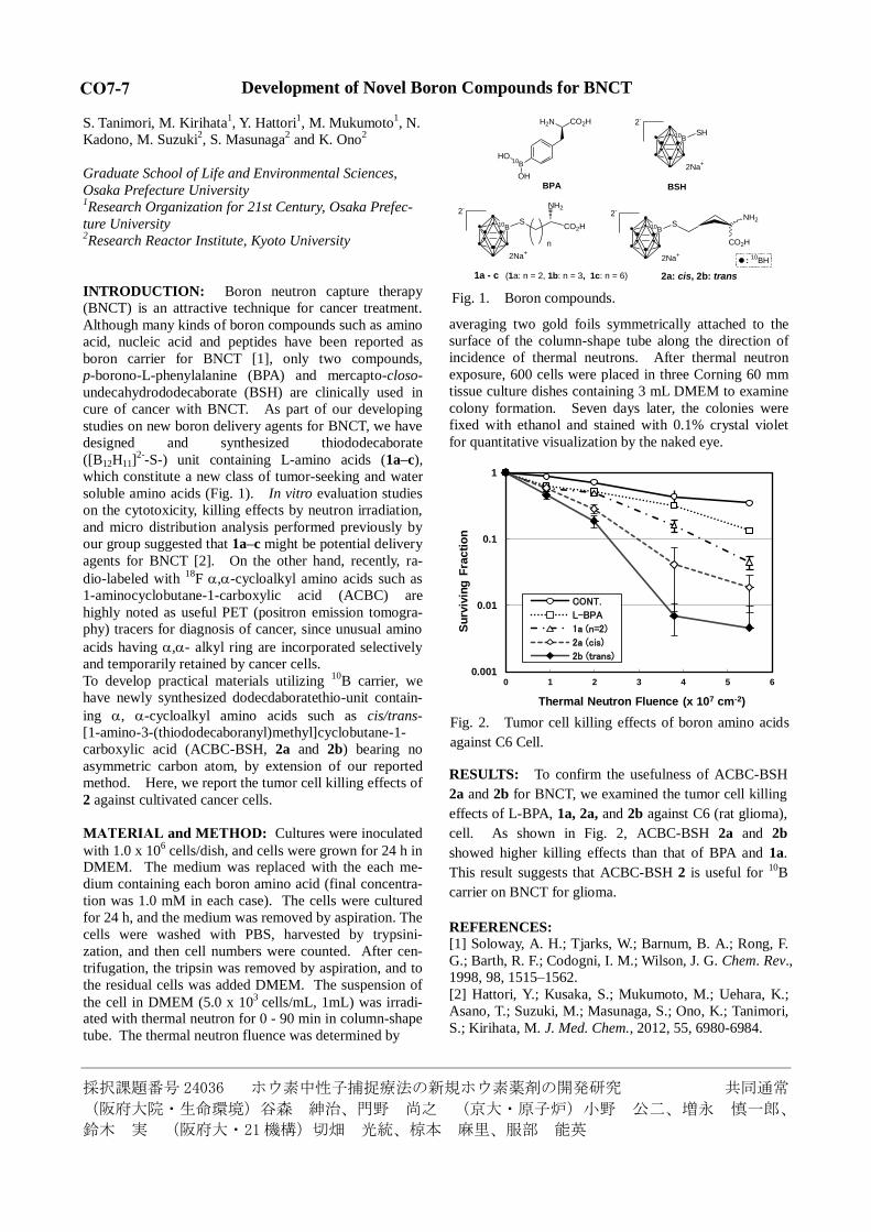
RESULTS: To confirm the usefulness of ACBC-BSH
2a and 2b for BNCT, we examined the tumor cell killing
effects of L-BPA, 1a, 2a, and 2b against C6 (rat glioma),
cell. As shown in Fig. 2, ACBC-BSH 2a and 2b
showed higher killing effects than that of BPA and 1a.
This result suggests that ACBC-BSH 2 is useful for 10
B
carrier on BNCT for glioma.
REFERENCES: [1] Soloway, A. H.; Tjarks, W.; Barnum, B. A.; Rong, F.
G.; Barth, R. F.; Codogni, I. M.; Wilson, J. G. Chem. Rev.,
1998, 98, 1515–1562.
[2] Hattori, Y.; Kusaka, S.; Mukumoto, M.; Uehara, K.;
Asano, T.; Suzuki, M.; Masunaga, S.; Ono, K.; Tanimori,
S.; Kirihata, M. J. Med. Chem., 2012, 55, 6980-6984.
0.001
0.01
0.1
1
0 1 2 3 4 5 6
Su
rviv
ing
Fra
cti
on
Thermal Neutron Fluence (x 107 cm-2)
CONT.
L-BPA
1a (n=2)
2a (cis)
2b (trans)
S10B
CO2H
NH22
-
SH10B
2-
2Na+
H2N CO2H
10BHO
OH2Na
+
BSHBPA
2a: cis, 2b: trans
: 10
BH
NH2
CO2HS10B
2-
n
2Na+
1a - c (1a: n = 2, 1b: n = 3, 1c: n = 6)
Fig. 1. Boron compounds.
Fig. 2. Tumor cell killing effects of boron amino acids
against C6 Cell.
CO7-7
採択課題番号 24037 中性子補足療法適応癌腫のプロトコル確率 共同通常
(川崎医大)平塚純一、原田保、粟飯原輝人、宇野雅子、森田倫正、牧野英一、笹岡俊輔、小西圭、
神谷伸彦(京大・原子炉)小野公二、丸橋晃
CO7-8 BNCT for Malignant Melanoma and Head and Neck Cancer
N. Kamitani, K. Konishi, E. Yoden, S. Sasaoka1,
M. Uno2, N. Morita
2, T. Harada
2, T. Aihara
3, Y. Sakurai
4,
A. Maruhashi4, K. Ono
4 and J. Hiratsuka
Kawasaki Medical School
Department of Radiology 1Department of Dermatology
2Department of Otolaryngology
3University of Tsukuba Hospital
4Research Reactor Institute, Kyoto University
INTRODUCTION: Advanced head and neck carci-
noma (AHNC) and recurrent head and neck cancer
(RHNC) are often radio-/chemo-resistant and show ex-
tensive growth, requiring a wide resection including sur-
rounding normal tissues. To avoid severe impairment of
head and neck structures, it is necessary to explore new
treatment for AHNC. Mishima first proposed employing
BNCT for malignant melanomas utilizing the specific
melanin synthesis activity of melanoma cells [1]. Kato et
al. [2] began BNCT using both BSH and BPA for recur-
rent parotid gland carcinoma for the first time and re-
ported excellent preliminary results. On the basis of the
encouraging results of their pioneering clinical trial, our
many years’ experience with melanoma BNCT and the
trend toward emphasizing the quality of life after treat-
ment, we also began treating our patients with BNCT
using BPA alone [3-4]. This report is a summary of
treatment by BNCT using BPA alone in 2012.
16 cases are consulted to Kawasaki Medical School
Hospital as referral visit for BNCT in 2012. 10 patients
were treated, consisting 4 men, 6 women; a median age
of 60 years old (range 37-86 years). The number of head
and neck recurrent carcinomas was 7, and that of cutane-
ous malignant melanomas was 3. 6 in head and neck re-
current cancer were squamous-cell carcinoma (SCC) re-
currence, and one was the neck lymph node metastasis of
the malignant melanoma. The local recurrence was five,
and the number of neck lymph node metastasis was one
among SCC. All cutaneous malignant melanomas treated
BNCT as first choice. Patients of head and neck recurrent
carcinoma have undergone the operation in the past ex-
cept the one of malignant melanoma. The local recur-
rence was five, and the number of neck lymph node me-
tastasis was one among SCC. All cutaneous malignant
melanomas treated BNCT as first choice. Patients of head
and neck recurrent carcinoma have undergone the opera-
tion in the past except the one of malignant melanoma.
RESULT: Although the malignant melanoma faded, it
still remains. Neither a recurrence nor progression is ac-
cepted. Any adverse event is not occurred. One of the
head and neck recurrent carcinomas which is neck lymph
node metastasis of the malignant melanoma died in a
month after BNCT cause of rapid progression of cancer.
In two patients of head and neck cancer (SCC), the local
recurrence occurred within two months after BNCT.
The shortage of doses was able to be considered as
acause. And two of four patients which carried out partial
control accepted the recurrence outside the exposure field.
Although the partial control by BNCT is good, it does
not contribute to overall survival.
Conclusion: We are considering combined use with
systemic therapy may also be useful to find the better
result.
Details:
Age (median) 60 y.o.
(37-86 y.o.)
male 4
Female 6
Head and neck carcinoma 7
SCC 6
nasopharynx 1
hypopharynx 1
buccal mucosa 1
maxillary sinus 1
external auditory canal 2
malignant melanoma 1
local recurrence (SCC) 5
cervical lymph node metastasis 2
external auditory canal 1
malignant melanoma 1
cutaneous malignant melanomas 3
heel 2
upper lip 1
Result:
local recurrence 2
metastasis 2
REFERENCES: [1] Y. Mishima, Pigment Cell Res., 1 (1973) 215-221.
[2] I. Kato et.al., Appl. Radiat. Isot., 61 (2004)
1069-1073.
[3] T.Aihara et al., Head Neck., 28 (2006) 850-855.
[4] T.Aihara et al., New Challenges in Neutron capture
therapy, (2010) 25-29.
採択課題番号 24043 中性子照射による半導体デバイスエラーに関する研究 一般通常
(京大・原子炉)田中浩基、櫻井良憲、堀順一
CO7-9 Study on the Semiconductor Device Error Irradiated with Thermal Neutrons
H. Tanaka, Y. Sakurai, J. Hori, M. Suzuki, S. Masunaga,
N. Kondo, M. Narabayashi, Y. Kinashi, K. Ono and
A. Maruhashi
Research Reactor Institute, Kyoto University
INTRODUCTION: Recently, the irradiation experi-
ments of fast neutrons for semiconductor devices have
been performed using cyclotron facility of Research
Center for Nuclear Physics, Osaka University [1]. How-
ever, irradiation test using thermal neutron for semicon-
ductor device has not been systematically performed. If
the nuclide with large capture cross section of thermal
neutrons is contained in semiconductor devices, charged
particles are produced in semiconductor devises. Charged
particles create electron-hole pair toward those tracks that
become the current of noise in semiconductor devises.
There is fear that the current in semiconductor devices
triggers incorrect operation. Furthermore, the miniaturi-
zation of semiconductor device is proceeding and the
current of signal is becoming small. Thus, the sensitivity
for electronic noise becomes large. The aim of this study
is the investigation of the influence for semiconductor
device of thermal neutron irradiation.
EXPERIMENTS: The sample of semiconductor de-
vice was irradiated at Heavy Water Neutron Irradiation
Facility (HWNIF) of KUR [2]. To investigate the influ-
ence of thermal neutrons, the difference of the number of
error (bit flip of memory) for epithermal mode
(CO-0000F) and mix mode (OO-0000F) was recorded.
Fig. 1 shows the neutron spectrum of two irradiation
mode. Epithermal mode can produce the epithermal and
fast neutrons without thermal neutrons using a cadmium
filter.
A number of semiconductor devices were irradiated
simultaneously. The irradiated thermal neutron distribu-
tion was not uniform because of its large irradiation area.
In order to measure thermal neutron flux, gold wire was
set at the surface of each semiconductor device. Gold
wire was taken from the surface of a semiconductor de-
vice to determine thermal neutron flux. The number of
error of each semiconductor device was recorded during
the irradiation of thermal neutron. Next, the number of
error was also recorded for epithermal and fast neutron
irradiation.
RESULTS: Table 1 shows the number of error of each
semiconductor for each irradiation mode and thermal
neutron flux. The deviation of thermal neutron fluxes for
each semiconductor device was shown in this table. The
importance of the measurement of thermal neutron fluxes
was revealed according to these results. Fig. 2 shows the
comparison of the number of error, that was removed the
influence of epithermal and fast neutrons, per thermal
neutron fluence. As shown in Fig. 2, the number of error
for sample D, E, F was larger than other samples. We
established the thermal neutron irradiation field for the
investigation of error of semiconductor devices.
REFERENCES:
[1]Y. Iwamoto, M. Fukuda, Y. Sakamoto, A. Tamii, et al.,
Nuclear Technology, 173(2), 210-217(2007)
[2] Y. Sakurai, T. Kobayashi, Nucl. Instrum. Meth. A453,
569-596(2000)
Fig.1. Neutron spectrum of CO-0000F and OO-0000F
mode at the surface of gamma ray shield of HWNIF.
OO-0000F CO-0000F
Sample A 1.0E+8 53 21
Sample B 1.0E+8 39 17
Sample C 9.1E+7 110 54
Smaple D 2.0E+8 7378 293
Sample E 2.1E+8 3959 110
Sample F 2.1E+8 3537 92
Sample G 1.2E+8 39 12
The number of errorThermal neutron
Flux [n/cm/s]
Table 1. the number of error of each semiconductor
for each irradiation mode and thermal neutron fluxes
0
500
1000
1500
2000
2500
3000
3500
4000
A B C D E F G
Nu
mb
er
of
the
err
or
pe
r th
erm
al n
eu
tro
n
flu
en
ce
(a.u
)
Fig.2. The comparison of the number of error for each
sample.
採択番号 24046 口腔癌に対する硼素中性子補足療法の基礎的研究 共同通常
(京大・原子炉)鈴木実 (大阪医科大学・口腔外科)伊藤雄一、有吉靖則、木村吉宏、中島世市
郎、武井祐子、植野高章
CO7-10 Tissue Changes by BNCT of the Oral Cancer Tissue at Having Used
Hyaluronan Conjugated PEG Liposome
Y. Ito, Y. Ariyoshi, Y. Kimura, Y. Nakajima, Y. Takei,
T. Ueno and M. Suzuki1
Department of Dentistry & Oral Surgery, Division of
Medicine for Function & Morphology of Sensory Organs,
Osaka Medical College 1Research Reactor Institute, Kyoto University
INTRODUCTION: In Boron neutron capture therapy
(BNCT), selective and highly concentrated boron accu-
mulation in tumor cells is important. A study was carried
out in our division using various liposomes [1, 2].
Hyaluronan is a ligand of CD44 and RHAMM, which is
excessively expressed in tumor cells, and Hyalu-
ronan-conjugated PEG Liposome (HA PEG Liposome) is
an active targeting candidate for tumor cells.
In this study, the effect of neutron irradiation was in-
vestigated by histological observation, in order to con-
sider BSH transport using HA PEG Liposomes in mice
with oral squamous cancer cells.
EXPERIMENTS: Tumor bearing mice were prepared
by injecting cultured SAS cells into the right thigh of
BALB/c mice. When tumor size reached approximately
10 mm, HA-PEG-Liposomal BSH was intravenously
administered from the tail vein. This injection was carried
out at the point in time when boron concentration in the
tumor tissue could be maintained at a comparatively
higher condition than the surrounding tissue than that of
previous experiments, in other words, two days before of
neutron irradiation. For reference, a control group which
used liposomes without boron was also set. After neutron
irradiation, the mice were allowed to grow for 8 weeks.
At the 8 weeks, all mice were euthanized, and tissue
samples were taken. Formalin fixation was carried out on
the samples, and the samples were set as a formalin fixed
paraffin sections according to the standard method, HE
dyed, and observed by a microscope.
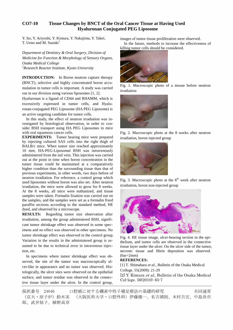
RESULTS: Regarding tumor size observation after
irradiation, among the group administered BSH, signifi-
cant tumor shrinkage effect was observed in some spec-
imens and no effect was observed in other specimens. No
tumor shrinkage effect was observed in the control group.
Variation in the results in the administered group is as-
sumed to be due to technical error in intravenous injec-
tion, etc.
In specimens where tumor shrinkage effect was ob-
served, the site of the tumor was macroscopically ul-
cer-like in appearance and no tumor was observed. His-
tologically, the ulcer sites were observed on the epithelial
surface, and tumor residue was observed in the connec-
tive tissue layer under the ulcer. In the control group,
images of tumor tissue proliferation were observed.
In the future, methods to increase the effectiveness of
killing tumor cells should be considered.
Fig. 1. Macroscopic photo of a mouse before neutron
irradiation
Fig. 2. Macroscopic photo at the 8 weeks after neutron
irradiation, boron injected group
Fig. 3. Macroscopic photo at the 8th
week after neutron
irradiation, boron non-injected group
Fig. 4. HE tissue image, ulcer-bearing section in the epi-
thelium, and tumor cells are observed in the connective
tissue layer under the ulcer. On the ulcer side of the tumor,
necrotic tissue and fibrin deposition was observed.
(bar=2mm)
REFERENCES:
[1] T. Shimahara et al., Bulletin of the Osaka Medical
College. 55(2009): 21-29
[2] Y. Kimura et al., Bulletin of the Osaka Medical
Col-lege. 56(2010): 65-7
採択課題番号24047 高い腫瘍選択性を有するボロン含有ナノ粒子の創製と 共同通常
中性子捕捉療法への展開
(筑波大・数理)長崎幸夫、矢口達也、角谷省吾、高振宇、堀口諭吉(名古屋大学)野田祥子、
中西勇介、篠原久典(京大・原子炉)鈴木実、小野公二
Development of Water-Soluble Gd Metallofullerenes/PEG-b-Polyamine Complexed Nanoparticles for Neutron Capture Therapy
Y. Nagasaki1-3, T. Yaguchi1, Z. Gao1, S. Sumitani1, Y. Horiguchi1, S. Noda,4 Y. Nakanishi,4 H. Shinohara,4 M. Suzuki5and K. Ono5
1Institute of Materials Science, Graduate School of Pure and Applied Sciences, University of Tsukuba, 2Master’s School of Medical Sciences, Graduate School of Comprehensive Human Sciences, University of Tsu-kuba 3Satellite Laboratory, International Center for Materials Nanoarchitectonics (WPI-MANA), National Institute of Materials Sci-ence (NIMS) 4Graduate School of Science Division of Material Sci-ence, Nagoya University 5Radiation Oncology Research Laboratory, Research Reactor Institute, Kyoto University
INTRODUCTION: Gadolinium element is one of promising candidates for neutron capture therapy (NCT) because of high neutron capture cross section. The essen-tial toxicity of Gd ion is one of the problems to solve. Gadolinium chelates with specific ligand such as diethy-lenetriaminepentaacetic acid (DTPA), and 1,4,7,10-tetraazacyclododecane-1,4,7,10-tetraacetic acid (DOTA), have been proposed to reduce toxicity. Howev-er, these Gd chelates still show toxicity and are not enough for NCT, because high dose is required for NCT treatment. Gadolinium endohedral metallofullerene (Gd@C82) are emerging compounds and promising as new biomaterials to suppress the toxicity of Gd by full-erene cage. In order to use Gd@C82 compounds in bio-logical fluid, delivatization of Gd@C82 has been done in most cases, which causes unwanted bioactivity. Thus, we have been used fullerenes without delivatization. To sol-ubilize fullerenes in aqueous media, poly(ethylene gly-col)-block-poly(2-(N,N-diethylamino)ethyl methacrylate (PEG–b–PAMA) were employed in our previous work[1]. The result of the NCT experiment in vitro showed that Gd@82/PEG–b–PAMA complexed nanoparicle was ef-fective NCT agent. To develop the more safety wa-ter-soluble Gd@C82 nanoparticles as NCT agent, radical containing amphiphilic block copolymer (PEG–b–PMNT) was used as dispersion agent [2]. PEG–b–PMNT is low toxic polymer (LD50 > 600 mg/Kg), and it acts as reactive oxygen species (ROS) scavenger to suppress the unwanted immune response induced by ROS after neutron irradiation. In this report, we investigate the NCT by Gd@C82/PEG–b–PMNT complexed nanoparti-cles (GdNPs). To confirm the tumor treatment, tumor
sizes of the mice were measured after neutron irradiation.
EXPERIMENTS: Preparation of Gd@C82 nanoparticles: 10 mg of Gd@C82 was dissolved in 50 mL of DMF and sonicated for 1 h. 50 mg of PEG–b–PMNT was added to the mix-ture and the sonication continued for 3 h. The mixture was transferred to a dialysis tube and dialyzed against 2 L of water. The external water was changed 5 times at t = 3, 15, 39, 63 and 87 h. The solution was condensed by ul-trafiltration and dispersed in PBS for in vivo experiment. Preparation of tumor bearing mice (BLAB/c, male, 5weeks): Tumors were prepared in mice legs by hypo-dermic injection of colon-26 cells (1,000,000 cells per mouse). This procedure was carried out a week before the neutron irradiation. Neutron irradiation: The administration of the GdNPs were carried out via tail vein (1 mg of Gd@C82 per mouse). After 2 d, the mice were irradiated thermal neu-trons for 120 min at a rate of 9.4 − 9.8 × 10¹² neu-trons/cm2). After the neutron irradiation, the tumor sizes of mice were measured for 26 d.
RESULTS: Evaluation of obtained Gd@C82 nanoparticles: The size of the GdNP was evaluated by DLS. The result showed that the average size of GdNP was ca. 40 nm and the dispersion property of the particles is quite stable enough to keep the dispersion for over a year. Neutron Irradiation: The tumor grew up to 6 – 7 cm3 after 26 d without GdNPs and no thermal neutron irradia-tion (n = 4). On the other hand, the growth was effec-tively suppressed in the mice treated with GdNPs with the irradiation of thermal neutron (average size was 2 cm3, n = 4). The suppression of tumor growth was also obser-ved in the mice with the irradiation of thermal neutron without GdNPs (average size was approx. 4 cm3, n = 5), the suppression efficacy of tumor growth of the mice with GdNPs with the irradiation was higher than that of the mice without GdNPs with the irradiation (p-value = 0.144). Our results demonstrate that the rational material design of GdNPs holds promise for the future of GdNCT.
REFERENCES: [1] Y. Horiguchi, S. Kudo, Y. Nagasaki, Sci. Technol. Adv. Mater., 12 (2011) 044607. [2] T. Yoshitomi, D. Miyamoto, Y. Nagasaki: Biom-acromolecules, 10 (2009) 596.
CO7-11
採択課題番号 24050 中性子捕捉療法の一般外科領域癌への展開に向けた基礎的研究 共同通常
(東大・原子力国際専攻)高橋浩之、柳衛宏宣、Novriana Dewi(東大・獣医)柳川将志、飯塚智
也(東大・心臓外科)櫻井由里子、毛利きくえ(大阪市大・バイオ)長崎 健、李 家暐、朱 宇
翔、河崎 陸(京大・原子炉)小野公二、増永慎一郎、鈴木 実、櫻井良憲
CO7-12 Pilot Biodistribution Study of Poly-ion Complex Mediated
Gadolinium Delivery System to Cancer Model In vivo
H. Yanagie
1,2, N. Dewi
1, Y. Sakurai
2, M. Yanagawa
3,
T. Iizuka3, T. Nagasaki
4, M. Suzuki
5, Y. Sakurai
5,
S. Masunaga5, K. Ono
5 and H. Takahashi
1,2
1Department of Nuclear Engineering & Management,
The University of Tokyo 2Cooperative Unit of Medicine & Engineering, The Uni-
versity of Tokyo Hospital 3Department of Veternary Surgery, The University of To-
kyo 4Department of Bioengineering, Osaka City University,
5Research Reactor Institute, Kyoto University
INTRODUCTION:
Tumour cell destruction in gadolinium (Gd) neu-
tron-capture therapy (GdNCT) is due to the nuclear reac-
tion between Gd atoms and thermal neutrons. It is nec-
essary for effective neutron capture therapy to accumu-
late Gd atoms in the tumour cells without affecting adja-
cent healthy cells.
We have developed novel efficient gene transfection
system, comprising the plasmid/polycation complex core
and the outer polyanion-coating. We used Polyethylene-
imine (PEI) as polycationic polymer, and Polyethylene
glycol derivatives having carboxylic acid [1,2]. We had
also applied this poly-ion complex as heavy ion delivery
systems. In this study, we prepare Gadoteridol en-
trapped poly-ion complex for selective intravenous or
intratumoural injection for mouse colon cancer model
applicating to GdNCT, and evaluate the poly-ion com-
plex as selective Gd delivery carrier to cancer tissues.
EXPERIMENTS:
(1) Cell: Mouse colon cancer cell line: Colon 26
(2) Polyethyleneimine: ExGen 500 (Fermentas Ltd.) was
used. ExGen 500 is linear shaped 22 kDa PEI. The effi-
cient gene transfer activity of ExGen 500 is related to its
capacity for condensing DNA, interacting with anionic
proteoglycans of the cell membrane, protecting DNA and
inducing endosomal swelling and rupture before DNA
degradation.
(3) PEG-C: Polyethylene glycol derivatives having car-
boxylic acid was synthesized (MW of PEG was 8940,
17.7 carboxylic acid groups were binding per PEG mol-
ecule).
(4) JTS-1: pH dependent fusogenic peptide was kindly
gifted by Professor Leaf Huang, Department of Pharmacy,
University of Pittsburgh, PA, USA.
(5) Gadoteridol: (±) -10 - (2-hydroxypropyl) -1,4,7,
10- tetraazacyclo-dodecane -1,4,7- triacetatogadolinium
[III]( C17H29GdN4O7 ) (MW: 558.69, 1396.5 mg/5ml) .
(6) Gadolinium Delivery in the Colon 26 tumours
①Colon 26 cells (2 x 10⁶) were injected subcutaneously
into the back of female BALB/c mice. At 10–14 days
after injection, when the estimated tumour weight
reached about 500 mg, the recipient animals were inject-
ed intravenously or intratumorally with either Gd/PEI, Gd/PEI/PEG-C/JTS-1, or Gd solution.
②One, and three days after injections, the Gd concentra-
tions of the tumor nodules, blood, and normal organs
were measured. The Gd concentrations of tissues were
determinated by ICP- Mass Spectroscopy of Juntendo
University.
RESULTS: The Gd concentration in tumour by intratumoural (IT)
delivery with Gd/PEI was 53.7 ppm, 22.5 ppm, after 2
hours and 12 hours, respectively. The Gd concentration in
tumour by IT delivery with Gd/PEI/PEG-C/JTS-1
was 21.9 ppm, 12.3 ppm, after 2 hours and 12 hours, re-
spectively. The Gd concentration in tumour by IT deliv-
ery with Gd solution was 17.4 ppm, 6.8 ppm, after 2
hours, 12 hours, respectively.
The Gd concentrations in Colon 26 tumour on delivery
with poly-ion complex (Gd/P EI) was 3 times superior to
simple Gd solution after 2 hours IT injection.
The Gd concentration in tumour by intravenous (IV)
delivery with Gd/PEI was 4.0 ppm, 1.2 ppm, after 2
hours, 12 hours, respectively. The Gd concentration in
tumour by IV delivery with Gd/PEI /PEG-C/JTS-1
was 9.8 ppm, 2.7 ppm, after 2 hours, 12 hours, respec-
tively. The Gd concentration in tumour by IV delivery
with Gd so lut ion was 1.1 ppm, 0.0 ppm, after 2 hours,
12 hours, respectively.
The Gd concentrations in Colon 26 tumour on delivery
with poly-ion complex (Gd/P EI) was 4 times superior to
simple Gd solution after 2 hours IV injection, and also
9 times superior to control groups in the usage of
poly-ion complex (Gd/PEI/PEG-C/JTS-1) .
CONCLUSION:
Poly-ion complex (Gd/PEI /PEG-C/JTS-1) can be
applied to the Gd delivery systems with the retention ac-
tivity and dispersion activity. We are ongoing to evaluate
the suppressive activity with these complexes by thermal
neutron irradiation.
REFERENCES:
[1] M. Sakae et al., Biomed Pharmacother., 62 (2008)
448-453.
[2] H. Yanagie et al., IGCC 2013 Abstract., (2010) P4-7
採択課題番号 24051 中性子捕捉療法の一般外科領域癌への展開に向けた臨床的研究 共同通常
(東大・原子力国際専攻)高橋浩之、柳衛宏宣(宏仁会メディカルシティ東部病院)東 秀史、
瀬口浩司(結核予防会 新山手病院)江里口正純、丸山正二、小山和行(啓愛会 宝陽病院)野中
泰政(京大・原子炉)小野公二、増永慎一郎、鈴木 実、櫻井良憲
CO7-13 Pilot Clinical Study of Boron Neutron Capture Therapy
for Recurreced Hepatic Cancer
H. Yanagie1,2, S. Higashi3, K. Seguchi3, I. Ikushima4, K.
Oyama5, Y. Nonaka6, S. Maruyama5, R. Hatae5, M. Su-
zuki7, S. Masunaga7, T. Kinashi7, Y. Sakurai7, H. Tanaka7,
A. Maruhashi7, K. Ono7, J. Nakajima2,8, M. Ono2,8, M.
Eriguchi5 and H. Takahashi1,2 1The University of Tokyo, Department of Nuclear Engi-
neering & Management 2Cooperative Unit of Medicine & Engineering, The Uni-
versity of Tokyo Hospital, Tokyo 3Kojinkai Medical City East Hospital, Department of
Surgery, Miyazaki 4Miyakonojyo Metropolitan Hospital, Department of Ra-
diology, Miyazaki 5Japan Anti-Tuberculosis Association, Shin-Yamate Hos-
pital, Department of Surgery, Tokyo 6Keiaikai Hoyo Hospital, Department of Surgery, Iwate 7Kyoto University, Research Reactor Institute, Osaka 8The University of Tokyo Hospital, Department of Cardi-
othoracic Surgery, Tokyo, JAPAN
INTRODUCTION:
Applications of boron neutron-capture therapy (BNCT)
has been increased clinically in patients with a lot of
cancers in hole body. The main two 10Boron com-
pounds(sodium mercaptoundecahydro dodeca- bo-
rate : 10BSH, 10B-p borono- phenylalanine (10BPA) and
its fructose complex ) are used to clinical trials. Tumour
cell destruction in BNCT is due to the nuclear reaction
between 10Boron and thermal neutrons. For effective
BNCT therapy, it is necessary to accumulate 10B atoms in
the tumour cells without affecting adjacent healthy cells.
Most of hepatocellular carcinomas (HCC) are thought
to be incurable, and limited surgical operation, chemo-
therapy, or radiation therapies are available for a pro-
longed survival. Suzuki et al. had reported that the in-
tra-arterial administration of a boron compound with
IPSO is technically an application of chemoembolization,
which has been widely used for the treatment of liver
tumours. They also reported the clinical results of the
first patient with multiple hepatocellular carcinomas
(HCCs) treated with BNCT. Higashi et al. prepared a
long term inseparable, water-in-oil-water emulsion
(WOW) containing 8-60 mg of epirubicin for use in arte-
rial injection therapy to treat patients with HCC. The
WOW was prepared by membrane emulsification tech-
nique using a controlled pore glass.
We started the pilot clinical studies of BNCT to recur-
rence breast cancer, hepatic cancer, and gastrointestinal
cancers. In this paper, we present pilot clinical study in
patients of hepatic cancer.
RESULT [Case 1]
In accordance with the clinical results of Higashi and
colleagues, water-in- oil-in-water (WOW) emulsion has
been used as the carrier of anti-cancer agents on in-
tra-arterial injections in clinical trials. We would like to
apply BNCT for the treatment of HCC in order to in-
crease the selection of therapies available for HCC pa-
tients. We developed a 10BSH containing WOW emulsion
using a double emulsification technique.
A 63-year-old man with multiple HCCs was enrolled
as the first patient in a pilot study for treating BNCT with 10BSH containing WOW emulsion. The patient had been
performed right hepatectomy in 6 years ago. Hepatic
arterial chemotherapies with epirubicin containing WOW
emulsion were performed in the recurrence stages. The
multiple tumours in the left liver lobe were treated with
BNCT by selective intra-arterial infusion of 10BSH con-
taining WOW emulsion. The pre-BNCT dosimetry was
performed using SERA(mean tumour fluence is 12Gy-Eq
on 56 minutes BNCT (Maximum 19Gy-Eq on tumour),
and maximum fluence of normal mucosa is 5.0 Gy-Eq).
The tumour size was remained stable during 3 months
after BNCT. No adverse effect as a result of BNCT was
observed during the treatment and follow-up period. The
BNCT-treated tumours showed regrowth 3 months after
BNCT, so the patient has continued the repeated hepatic
arterial chemotherapy of epirubicin containing WOW
emulsion.
The present results showed that 10B-containing WOW
emulsion can be applied as a novel intra-arterial boron
carrier for BNCT for HCC.
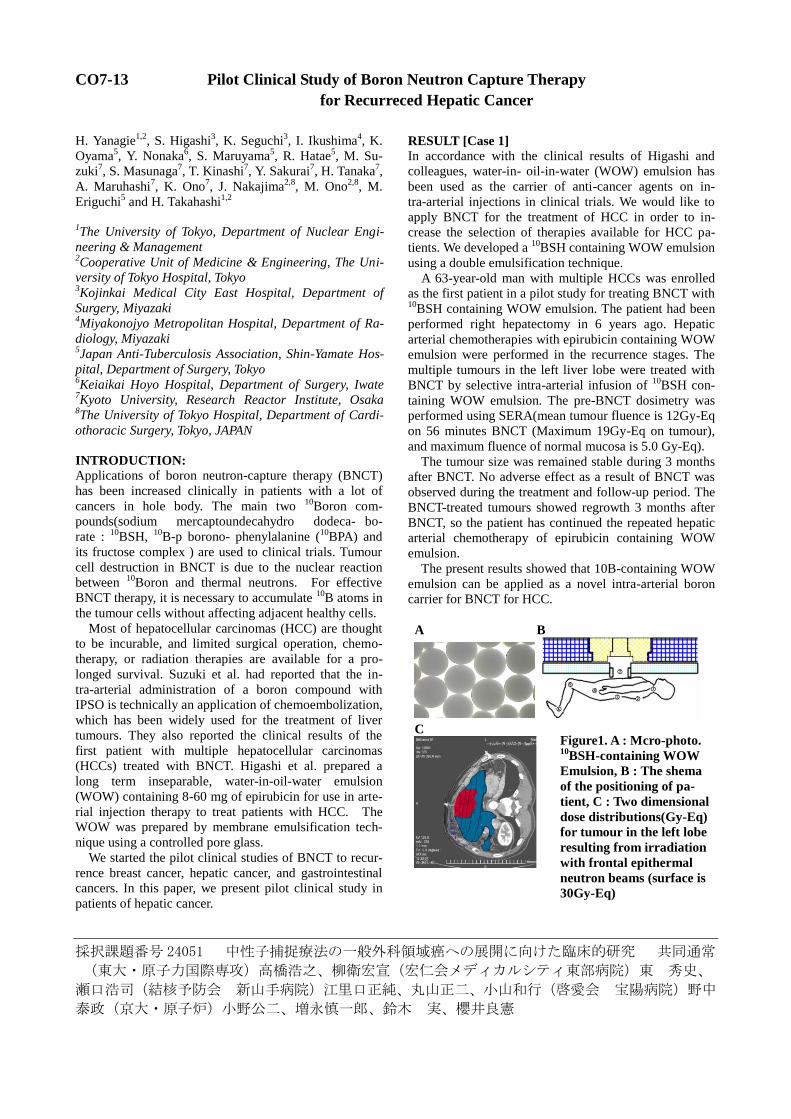
A B
C
Figure1. A : Mcro-photo. 10BSH-containing WOW
Emulsion, B : The shema
of the positioning of pa-
tient, C : Two dimensional
dose distributions(Gy-Eq)
for tumour in the left lobe
resulting from irradiation
with frontal epithermal
neutron beams (surface is
30Gy-Eq)
採択課題番号 24052 新規化合物および細胞内濃度変調による中性子捕捉反応による 共同通常
細胞生存試験および腫瘍増殖抑制効果
(筑波大学・医学医療系)中井 啓、山本哲哉、吉田文代、白川 真、山本陽平、上前洋二、佐藤
英介(京大・原子炉)増永慎一郎、櫻井良憲、田中浩基
CO7-14 Development of Boronated Liposome for Boron Neutron Capture Therapy
M. Shirakawa1, K. Nakai
1,2, Y. Yamamoto
1, F. Yoshida
1,2,
T. Yamamoto1,2
, A. Matsumura1,2
, H. Tanaka3, M. Suzuki
3
and S. Masunaga3
1Department of Graduate School of Comprehensive Hu-
man Sciences, University of Tsukuba 2Department of Neurosurgery, Faculty of Medicine, Uni-
versity of Tsukuba 3Particle Radiation Oncology Research Center, Research
Reactor Institute, Kyoto University
INTRODUCTION: Boron Neutron Capture Therapy
(BNCT) has cell selective radiation therapy theoreti-
cally. Therapeutic effect of the boron compound is based
on alpha particles produced by the 10
B (n, α) 7Li
reaction
and tumor selectivity [1]. We developed novel boronated
liposome for BNCT, analyzed the neutron capture effect
for the cancer cell line and tumor bearing mouse model
[2].
EXPERIMENTS: Synthesized the new boronated lipid
compound (PBL). Boronated lipid and other lipids
(DSPC: cholesterol: PBL = 1:1:0.12) were dissolved in
organic solvent and assembled as a liposome of 100 μm
diameter using lipid film methods and extruder.
i) In Vitro Colony forming assay using V79 and 379A
was performed with three groups of samples; (1) Boro-
nated liposome without medium change, (2) Boronated
liposome with medium change, (3) no boron argents
groups. Each cell suspesions were irradiated at KUR.
Irradiation time of each groups were 15min, 30min, and
45min. After 1week incubation, the colonies were count-
ed.
ii) In Vivo tumor growth inhibitory test with animal ex-
perimental tumor bearing model. 107 of CT26 colon can-
cer cells were injected subcutaneously to right thigh of
BALB/c mice. 2week after the injection, 5% PBL lipo-
some was administrated via tail vein. The Boron dose
was 10mg/kg, volume of 100µl. Concentration of the
boron was 2000ppm.
Mice were devided four groups; (1) 5% PBL lipsome
group, (2) BSH water solutions, (3) Neutron only and (4)
no treatment groups.
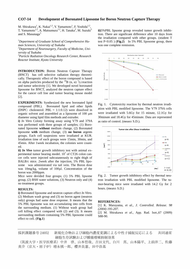
RESULTS: i) Boronated liposome and neutron capture effect In Vitro.
(2) Medium wash group and (3) no boron agent (neutron
only) groups had same dose response. It means that the
5% PBL liposome was not accumulating into cells from
the surrounding medium. (1) Without wash group had
cell killing effect compared with (2) and (3). It means
surrounding medium containing 5% PBL liposome could
effect to cell. (Fig.1)
ii)5%PBL lipsome group revealed tumor growth inhibi-
tion. There are significant difference after 10 days from
the irradiation compared with other groups. (student T
test P<0.05 ) (Fig.2) In 5% PBL liposome group, there
was one complete remission.
Fig. 1. Cytotoxicity reaction by thermal neutron irradi-
ation with PBL modified liposome. The V79 379A cells
were irradiated with 5.6Gy for 15 minute, 12.1Gy for
30minute and 18.4Gy for 45minute. Data are represented
as ratio of control. (mean± S.D.).
Fig. 2. Tumor growth inhibitory effect by thermal neu-
tron irradiation with PBL modified liposome. The tu-
mor-bearing mice were irradiated with 14.2 Gy for 2
hours. (mean± S.D.)
REFERENCES: [1] K. Maruyama, et al., J. Controlled. Release. 98
(2004) 195-207.
[2] M. Shirakawa et al., App. Rad. Isot.,67 (2009)
S88-90.
採択課題番号 24054 新規硼素化合物の有用性と腫瘍幹細胞の克服 共同通常
(大阪医大)宮武伸一、弘田祐己、川端信司、平松亮、頼経英倫那
(岡山大)道上宏之、森亜希子、王飛霏
(熊本大)富澤一仁
(京大原子炉)小野公二
The Benefit of High LET-Radiation to Glioma Stem-Like Cells
Y. Hirota, S. Masunaga1, N. Kondo
1, T. Fujishiro
2,
S. Kawabata2, A. Fujimori
3, H. Hirakawa
3, H Yajima
3,
K. Ono1, T. Kuroiwa
2 and S. Miyatake
2
Graduate School of Neurosurgery, Osaka Medical Col-
lege 1Research Reactor Institute, Kyoto University
2Department of Neurosurgery, Osaka Medical College
3National Institute of Radiological science, Heavy-ion
Radiobiology Research group, Research Center for
Charged Particle Therapy
PURPOSE:
Radiation therapy with surgery and chemotherapy is the
standard treatment for glioblastoma multiforme (GBM)
[1]. However, almost half of GBM patients cannot sur-
vive one year after diagnosis, and the prognosis of pa-
tients with GBM has not been improved over the past
decades. Recently, there have been some reports showing
the presence of glioma stem cells (GSCs) in malignant
gliomas which are regarded as highly radio-resistant to
low linear energy transfer (LET) photons[1, 2]. On the
other hand, we have applied boron neutron capture ther-
apy (BNCT) for GBM. This is a unique tumor-selective
particle radiotherapy using neutron irradiation, especially
thermal neutron irradiation. Boron-10 (10
B) releases alpha
(4He) and
7Li particles by 10
B(n,α)7Li reaction. The key
players of anti-tumor effects in BNCT are these high lin-
ear energy transfer (LET) particles. With BNCT, good
results have been achieved for patients with GBM and
recurrent malignant glioma[3, 4]. Here we analyzed the
benefit of high LET particles to GSCs.
MATERIALS AND METHODS: Glioma stem-like cells (GSLCs) were induced from
GBM cell line A172 in stem cell-culture medium [5]. The
phenotype of these GSLCs and wild type cell lines were
confirmed by western blot analysis and fluores-
cence-activated cell sorting (FACS) using stem cell
markers. These cells were irradiated with 60
Co gamma
rays or neutron beams. Radio-sensitivity was assessed by
a colony-forming assay[6] and the number of DNA dou-
ble strand breaks (DSBs) using histone gamma-H2AX
foci detection assay[7, 8].
RESULTS: In stem cell-cultured medium, GSLCs could form
neurosphere-like spheroid cells. And GSLCs expressed
neural stem cell markers more frequently in western blot
analysis and the ratio of CD133 positive cells increased
day by day. GSLCs were radio-resistant to gamma-rays in
comparison with parental cultured cell lines, but neutron
beams could overcome the resistance. Twenty-four hours
after irradiation with gamma-rays, the number of gam-
ma-H2AX foci in GSLCs was significantly less than that
of parental cells, while there was no apparent difference
in the number of these foci between GSLCs and parental
cultured cell lines following neutron beam irradiation. In
addition, neutron beam irradiation induced larger gam-
ma-H2AX foci than those observed after gamma-ray ir-
radiation in both types of A172 cells.
CONCLUSION:
Neutron beams can induce elastic scattering and nitrogen
neutron capture reaction, and produce proton particle
(H+). This particle is high LET radiation and it could
overcome radioresistance of GSLCs with unrepairable
DSBs. So we could demonstrate that high LET radiation
may be able to overcome GSCs that are resistant to low
LET radiation. It is necessary to further investigate the
usefulness of high LET radiation to control GSCs, and
high LET radiation therapy such as BNCT has a very
important role in further treatment for therapy-resistant
GBM.
REFERENCES:
[1]S. Bao et al, Nature 444 (2006) 756-760.
[2]C. Charn-jung et al, Biochemical and Biophysical Re-
search Communications, 380 (2009) 236-242.
[3]S. Miyatake et al, Journal of Neuro-oncology, 91(2009) 199-206.[4]S. Kawabata et al, Journal of Radiation Research, 50(2009) 51-60.[5]Q. Lei et al, Cancer Letters 279 (2009) 13-21.[6]A. Maria et al, International Journal of Radiation On-
cology Biology Physics, 79 (2011) 262-268.[7]A. Osma et al, International Journal of Radiation On-
cology Biology Physics, 75 (2009) 1216-1225.[8]Y. Kinashi et al, Radiation Oncology, 6 (2011) 106.
CO7-15
採択課題番号 24057 悪性脳腫瘍のための熱外中性子捕捉療法の基礎的研究 共同通常 (藍野大)高垣政雄(東邦大・医)東丸貴信(京大・原子炉)増永慎一郎
CO7-16 A Biodistribution Study of Carboran Sugar
M. Takagaki, I. Snajdr1, R. Satapathy
1, NS. Hosmane
1
and S. Masunaga2
College of Nursing, Aino Univ 1Dept Chem & Biochem, Northern Illinois Univ
2Research Reactor Institute, Kyoto University
Carboran sugar (carboranyl-thio-d-glucose: TDG)
has been chemically modified via novel approach
and reduction of its IC50 has been achieved to be
very low 5.3x10⁻²
M that is almost half of that of
BSH (2.75x10⁻²
M) [1].
In this study, biodistribution of TDG derivative has
been preliminary investigated via -auto-
radiography. C6 tumor cells were implanted via
stereotactic maneuvers into Wister’s rat brain. 2
weeks after the implantation, 100mg/kg body
weight of the compound was injected into
peritoneal cavity. 3hs after injection, whole brain
was removed and rapidly frozen in liquid nitrogen.
Frozen sections were mounted onto the solid-state
track detectors: Kodak LR 115 and were exposed
by thermal neutrons. The detector were then etched
in 10% NaOH solution at 60℃ to emerge - and/or
recoil 7Li particles tracks of
10B(n,)
7Li that could
be numerically evaluated via an ordinary light
microscope.
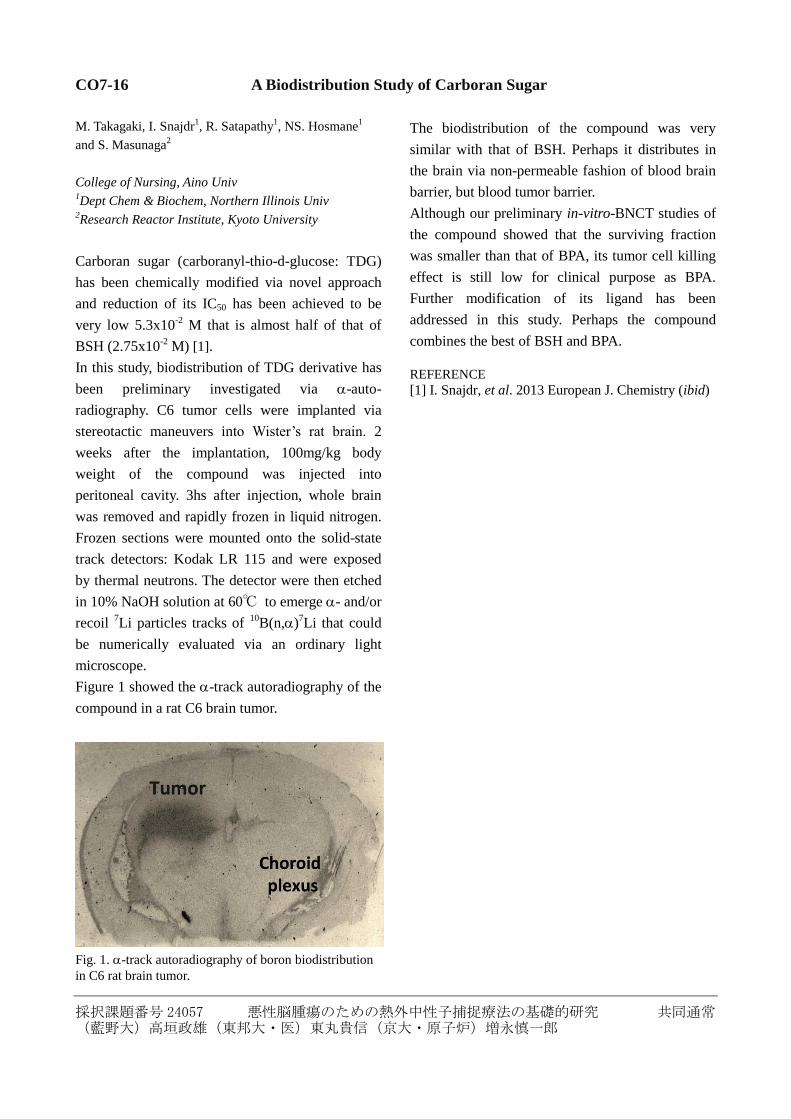
Figure 1 showed the -track autoradiography of the
compound in a rat C6 brain tumor.
Fig. 1.-track autoradiography of boron biodistribution
in C6 rat brain tumor.
The biodistribution of the compound was very
similar with that of BSH. Perhaps it distributes in
the brain via non-permeable fashion of blood brain
barrier, but blood tumor barrier.
Although our preliminary in-vitro-BNCT studies of
the compound showed that the surviving fraction
was smaller than that of BPA, its tumor cell killing
effect is still low for clinical purpose as BPA.
Further modification of its ligand has been
addressed in this study. Perhaps the compound
combines the best of BSH and BPA.
REFERENCE
[1] I. Snajdr, et al. 2013 European J. Chemistry (ibid)
採択課題番号 24088 頭頸部悪性腫瘍におけるホウ素中性子捕捉療法の臨床的研究 共同通常
(阪大・2口外)加藤逸郎、岩井聡一、墨 哲郎、中澤光博、由良義明(阪大・工)村田 勲(慶
大)岡本正人(長崎大)梅田博昭、柳本惣市(りんくう医療セ)大前政利(東大阪総合)千足浩久
(市立池田)大西徹郎(田中クリニック)田中 善(京大・原子炉)田中浩基、鈴木 実、櫻井良
憲、増永慎一郎、丸橋晃、小野公二
CO7-17 Clinical Studies on BNCT for 5 Cases of Head and Neck Cancer
I. Kato, N. Yamamoto1, Y. Fujita
2, M. Ohmae
3, M. Suzu-
ki7, S. Masunaga
7, M. Nakazawa, T. Sumi, A. Maruhashi
7,
Y. Imahori5, M. Kirihata
6 and K. Ono
7
Dept. of Oral &Max.fac. Surg. II, Grad. Sch. of Dent., Osaka Univ 1
Dept. of Oral &Max.fac. Surg., Saiseikai-Senri Hpsp 2Dept. of Oral &Max.fac.Surg., Higashiosaka General
Hosp 3Oral &Max. fac. Surg., Rinku,General Med. Center
4Dept. of Oral &Max.fac. Surg., Rinku General Medical
Center 5CICS Inc
6Graduate School of Agriculture and Life Science, Osaka
Prefecture University 7Research Reactor Institute, Kyoto University
INTRODUCTION: We had first reported that six pa-
tients with head and neck cancer (HNC) had been treated
with BNCT [1]. We also report long term (more than
5-year) clinical outcomes of our 26 patients with recur-
rent HNC treated with BNCT [2]. We summarized 5 pa-
tients with HNM who had treated with BNCT at KUR in
last year in Table 1.
PURPOSES: The purpose of this study was to estimate
safety and effectiveness of BNCT for patients with ad-
vanced/ recurrent HNC for which there were no other
treatment options.
RESULTS: We also report here latest clinical outcomes
of 35 patients with recurrent HNC
All cases are advanced such as 17 (53%) out of 35 pa-
tients had developed regional lymph node metastases.
Distant metastases were developed in 10 cases (29%)
during treatment. (1)10
B concentration of tumor/normal
tissue ratios (T/N ratio) of FBPA-PET studies were SCC:
1.8-6.0, sarcoma: 2.5-4.0, parotid tumor: 2.5-3.7. (1) Re-
gression rates were CR: 13cases (51%), PR: 13cases
(37%), PD: 3cases (9%), NE (not evaluated):1case. Re-
sponse rate was 88%. (2) Mean Survival time was
24.2months. 2-year overall survival rate (OS) and 6-year
OS were 42% and 36%, respectively. (3)BNCT improved
QOL, PS and survival periods. (4)Survival periods after
BNCT were 1-84 months. (5) Adverse events were brain
necrosis, osteomyelitis and transient mucositis and alo-
pecia and so on.
Case 1: A 40 year-old female with recurrent adenoid
cystic carcinoma of nasal cancer (rT0N3M0, ACC), who
had got a skull-base operation and irradiated RT 60Gy
post operatively in 2004. About 5-year later, she had got
another surgical operation because of intra-orbital recur-
rence. In 2011 she had developed another recurrence just
after she had got endoscopic surgery for a solitary lung
metastasis. FBPA-PET study revealed 10
B concentration
of T/B ratio was 2.0. Then she has been treated with BPA
(500mg/kg) mediated BNCT at KUR in June 2012 and
she has been disease free survival for so far 11-month.
Case 5: A 65-year old woman with SCC at Lt-WK
(T4N0M0) had got surgery with microscopic forearm
reconstruction in March 2011 and she had got surgery of
Rt-RND and postoperative radiotherapy (54Gy) in June,
Lt-RND in December. She had got bilateral selective
intra-artery chemotherapy (CDDP+TXT) after having
developed recurrence in February 2012. Again she had
developed cervical LN metastases (L-Level II:4cm,
R-Level V:1.5cm) so she had weekly treated with [Ce-
tuximub (400mg/m2,250mg/m
2)+Paclitaxel:60mg/m
2]x7.
FBPA-PET study resulted that T/B ratio=4.0. Just before
BNCT, the left of level II LN had grown to 5cm with
undefined margin which was infiltrated into pa-
ra-pharyngeal area. The LN was necrotic and skin was
ruptured and discharged cancer milk. She treated BNCT
in February, 2013. After BNCT the LN had completely
disappeared and had covered with normal skin. Then she
has been disease free survival for so far 5-month.
REFERENCES: [1] I. Kato, et al., Appl. Radiat. Isot., 61 (2004) 1069-1073. [2] R. Barth, et al., Radiat. Oncol., in press.
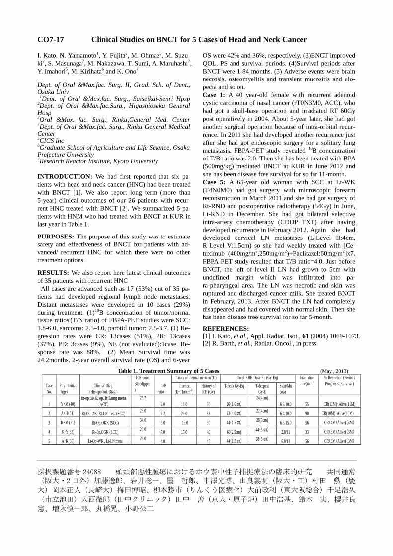
Table 1. Treatment Summary of 5 Cases (May , 2013)
Case
No.
Pt‘s Initial
(Age)
Clinical Diag.
(Histopathol. Diag.)
10B-conc.
Blood(ppm)
T/B
ratio
T-max of thermal neutron (D) Total-RBE-Dose Eq (Gy-Eq) Irradiation time(min.)
% Reduction (Period) Prognosis (Survival) Fluence
(E+11n/cm-2) History of RT: (Gy)
T-Peak Gy-Eq T-deepest Gy-E
Skin/Mucosa
1 Y・M (40) Rt-op.OKK, op. lt-Lung meta
(ACC)
25.7
2.0 18.0 50 26(1.6 ㎝)
24(4cm)
6.9/18.0 55 CR(11M)・Alive(11M)
2 A・H(51) Rt-Op. ZK, Rt-LN meta.(SCC) 28.0
2.2 23.0 63 23(4.0 ㎝) 22(4cm)
6.4/10.0 90 CR(10M)・Alive(10M)
3 K・M (71) Rt-Op.OKK (SCC) 34.0
6.0 13.0 50 44(1.5 ㎝) 28(5cm)
6.8/15.0 56 CR(4M)Alive((4M)
4 K・Y(83) Rt-0p.OGK (SCC) 28.0
7.0 15.0 40 60(2.5cm) 44(5 ㎝) 2.8/11 33 CR(3M)Alive((3M)
5 A・K(60) Lt-Op-WK、Lt-LN meta 23.0
4.0 45 44(1.5 ㎝) 28(5 ㎝) 6.8/12 56 CR(3M)Alive((3M)
採択課題番号 24089 口腔悪性腫瘍におけるホウ素中性子捕捉療法に関する基礎的研究 共同通常
(阪大・歯)岩上隆紀、加藤逸郎、由良義明(済生会千里病院)山本直典(京大・原子炉)田中浩基、
櫻井良憲、増永慎一郎、鈴木実、丸橋晃、小野公二
CO7-18 B4C Particle as a Boron Compound for BNCT
T. Iwagami, I. Kato, N. Yamamoto1, Y. Ishikawa
2,
N. Koshizaki3, H. Tanaka
4, Y. Sakurai
4, S. Masunaga
4,
M. Suzuki4, A. Maruhashi
4, K. Ono
4 and Y. Yura
Graduate School of Denistry, Osaka University 1Department of Oral Surgery, Saiseikai Senri Hospital
2National Institute of Advanced Industrial Science and
Technology 3 Graduate School of Engineering, Hokkaido University
4Research Reactor Institute, Kyoto University
INTRODUCTION: B4C nano particles are produced
by liquid phase laser irradiation method [1]. The diameter
of the particle is 200 nm. Sonoporation is a low ultra-
sound which makes small transient holes in the cell
membrane and introduces external materials such as drug
and gene into the cell [2]. In this study, we investigated
whether sonoporation could be used to introduce B4C
particles into the oral squamous cell carcinoma (SCC)
cells.
EXPERIMENTS: SAS cells derived from oral SCC
were used. Cells were exposed to thermal neutron at
Kyoto University Reactor (KUR) [3]. An ultrasound ma-
chine, Sonitron 2000V, and a microbubble, SV-25, were
used [4]. The cell viability was examined by MTT assay.
The cell surface was observed using a scanning electron
microscope.
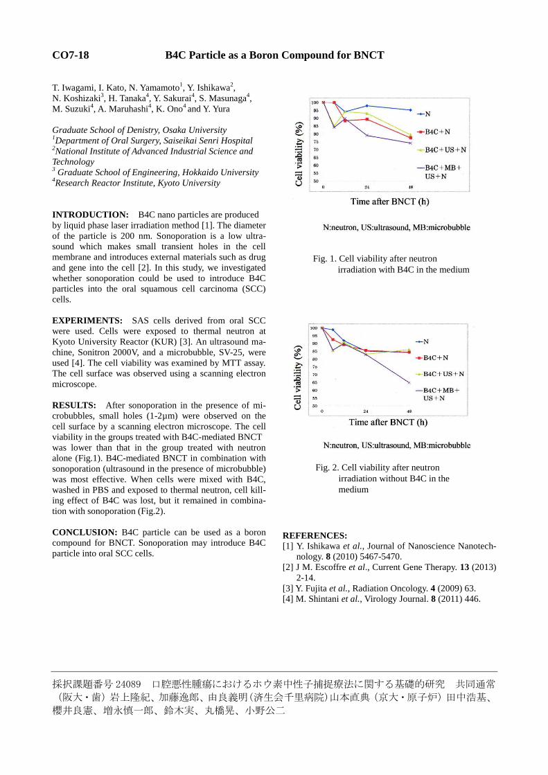
RESULTS: After sonoporation in the presence of mi-
crobubbles, small holes (1-2µm) were observed on the
cell surface by a scanning electron microscope. The cell
viability in the groups treated with B4C-mediated BNCT
was lower than that in the group treated with neutron
alone (Fig.1). B4C-mediated BNCT in combination with
sonoporation (ultrasound in the presence of microbubble)
was most effective. When cells were mixed with B4C,
washed in PBS and exposed to thermal neutron, cell kill-
ing effect of B4C was lost, but it remained in combina-
tion with sonoporation (Fig.2).
CONCLUSION: B4C particle can be used as a boron
compound for BNCT. Sonoporation may introduce B4C
particle into oral SCC cells.
REFERENCES: [1] Y. Ishikawa et al., Journal of Nanoscience Nanotech-
nology. 8 (2010) 5467-5470.
[2] J M. Escoffre et al., Current Gene Therapy. 13 (2013)
2-14.
[3] Y. Fujita et al., Radiation Oncology. 4 (2009) 63.
[4] M. Shintani et al., Virology Journal. 8 (2011) 446.
Fig. 1. Cell viability after neutron
irradiation with B4C in the medium
Fig. 2. Cell viability after neutron
irradiation without B4C in the
medium
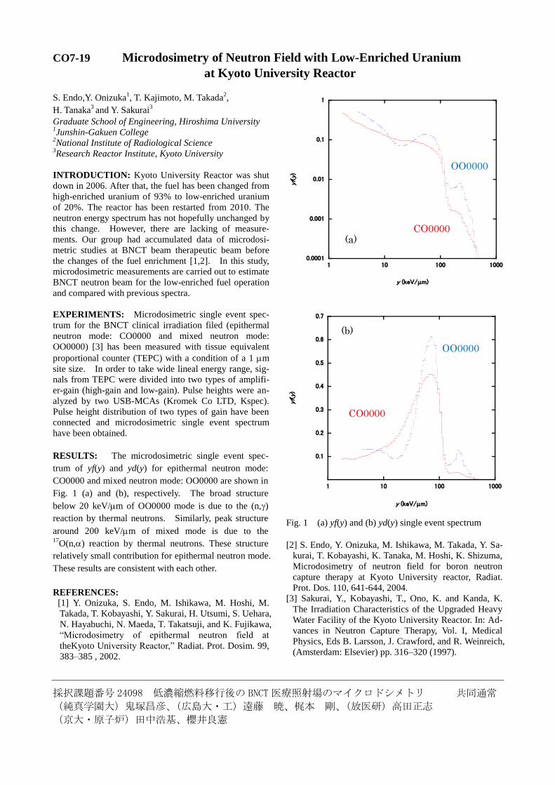
採択課題番号 24098 低濃縮燃料移行後の BNCT医療照射場のマイクロドシメトリ 共同通常
(純真学園大)鬼塚昌彦、(広島大・工)遠藤 暁、梶本 剛、(放医研)高田正志
(京大・原子炉)田中浩基、櫻井良憲
CO7-19 Microdosimetry of Neutron Field with Low-Enriched Uranium
at Kyoto University Reactor
S. Endo,Y. Onizuka1, T. Kajimoto, M. Takada
2,
H. Tanaka3 and Y. Sakurai
3
Graduate School of Engineering, Hiroshima University 1Junshin-Gakuen College
2National Institute of Radiological Science
3Research Reactor Institute, Kyoto University
INTRODUCTION: Kyoto University Reactor was shut
down in 2006. After that, the fuel has been changed from
high-enriched uranium of 93% to low-enriched uranium
of 20%. The reactor has been restarted from 2010. The
neutron energy spectrum has not hopefully unchanged by
this change. However, there are lacking of measure-
ments. Our group had accumulated data of microdosi-
metric studies at BNCT beam therapeutic beam before
the changes of the fuel enrichment [1,2]. In this study,
microdosimetric measurements are carried out to estimate
BNCT neutron beam for the low-enriched fuel operation
and compared with previous spectra.
EXPERIMENTS: Microdosimetric single event spec-
trum for the BNCT clinical irradiation filed (epithermal
neutron mode: CO0000 and mixed neutron mode:
OO0000) [3] has been measured with tissue equivalent
proportional counter (TEPC) with a condition of a 1 m
site size. In order to take wide lineal energy range, sig-
nals from TEPC were divided into two types of amplifi-
er-gain (high-gain and low-gain). Pulse heights were an-
alyzed by two USB-MCAs (Kromek Co LTD, Kspec).
Pulse height distribution of two types of gain have been
connected and microdosimetric single event spectrum
have been obtained.
RESULTS: The microdosimetric single event spec-
trum of yf(y) and yd(y) for epithermal neutron mode:
CO0000 and mixed neutron mode: OO0000 are shown in
Fig. 1 (a) and (b), respectively. The broad structure
below 20 keV/m of OO0000 mode is due to the (n,)
reaction by thermal neutrons. Similarly, peak structure
around 200 keV/m of mixed mode is due to the 17
O(n,) reaction by thermal neutrons. These structure
relatively small contribution for epithermal neutron mode.
These results are consistent with each other.
REFERENCES: [1] Y. Onizuka, S. Endo, M. Ishikawa, M. Hoshi, M.
Takada, T. Kobayashi, Y. Sakurai, H. Utsumi, S. Uehara,
N. Hayabuchi, N. Maeda, T. Takatsuji, and K. Fujikawa,
“Microdosimetry of epithermal neutron field at
theKyoto University Reactor,” Radiat. Prot. Dosim. 99,
383–385 , 2002.
0.0001
0.001
0.01
0.1
1
1 10 100 1000
CO0001 OO0001
yf(y
)
y (keV/m)
0.1
0.2
0.3
0.4
0.5
0.6
0.7
1 10 100 1000
CO0001 OO0001
yf(y
)
y (keV/m)
Fig. 1 (a) yf(y) and (b) yd(y) single event spectrum
[2] S. Endo, Y. Onizuka, M. Ishikawa, M. Takada, Y. Sa-
kurai, T. Kobayashi, K. Tanaka, M. Hoshi, K. Shizuma,
Microdosimetry of neutron field for boron neutron
capture therapy at Kyoto University reactor, Radiat.
Prot. Dos. 110, 641-644, 2004.
[3] Sakurai, Y., Kobayashi, T., Ono, K. and Kanda, K.
The Irradiation Characteristics of the Upgraded Heavy
Water Facility of the Kyoto University Reactor. In: Ad-
vances in Neutron Capture Therapy, Vol. I, Medical
Physics, Eds B. Larsson, J. Crawford, and R. Weinreich,
(Amsterdam: Elsevier) pp. 316–320 (1997).
(a)
(b)
CO0000
OO0000
CO0000
OO0000
(b)
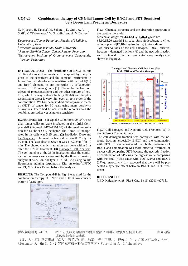
採択課題番号 24103 BNCTと光線力学治療の併用療法に両用の増感剤を使用した 共同通常 癌培養細胞共同実験
(福井大・医)三好憲雄(京大・原子炉)田中浩基、櫻井正憲、小野公二(ロシア国立がんセンター)Alexander A. Shtil(ロシア国立有機材料物質研究所)Valentina A. Ol'shevskaya
CO7-20 Combination therapy of C6 Glial Tumor Cell by BNCT and PDT Sensitized by a Boron Lich Porphyrin Derivative
N. Miyoshi, H. Tanaka1, M. Sakurai
1, K. Ono
1, A. A.
Shtil2, V. Ol'shevskaya
3, V. N. Kalini
3 and A. V. Zaitsev
3
Department of Tumor Pathology, Faculty of Medicine,
University of Fukui 1 Research Reactor Institute, Kyoto University
2 Russian Blokhim Cancer Center, Russian Federation
3Nesmeyanov Institute of Organoelement Compounds,
Russian Federation
INTRODUCTION: The distribution of BNCT as one of clinical cancer treatments will be spread by the pro-gress of the sensitizers and the compact instruments in future. We had developed a sensitizer with lich of F(16) and B(44) elements in one molecules by collaboration research of Russian groups [1]. The molecule has both effects of photosensitizing and the other capture of neu-tron, which is easy water-soluble (>10mM) and the pho-tosensitizing effect is very high even at ppm order of the concentration. We had been studied photodynamic thera-py (PDT) of cancer for 30 years using many porphyrin derivatives. There had be not seen the reports about the combination studies yet using one sensitizer.
EXPERIMENTS: (1) Uptake Conditions: 2x10⁶ C6 rat
glial tumor cells/ ml were incubated in the 10M Com-
pound-B (Figure-1: MW=1564.63) of the medium solu-
tion for 14.5hr at CO2 incubator. The Boron-10 incorpo-
rated in the cells was 3.15 ppm. (2) Irradiation Dose and
the Sequence: The neutron beam dose was 0.575Gy for
10 min. The laser dose at 665 nm was 115.2 J/cm2 for 10
min. The photodynamic irradiation was done within 2 hr
after the BNCT treatment. (3) Damaged Cell Analysis:
The cell number at the 36 hr incubation after the combi-
nation treatments were measured by the flow cytometory
analysis (FACS Canto-II type, BD Ltd. Co.) using double
fluorescent staining (Apoptosis Kit: annexine-V/FITC
and PI, MBL Co.) 15 min before the analysis.
RESULTS: The Compound-B in Fig. 1 was used for the
combination therapy of BNCT and PDT at low concen-
tration of 3.15 ppm.
Fig.1. Chemical structure and the absorption spectrum of
the capture molecule.
Molecular weight =1564.63,C48H60B44F16N4Na4=
{5,10,15,20-terakis[4-(1-caba-closo-dodecabrane-1-yl)tet
rafluorophenyl]17,18-dihydrophyrin}} tetrasodium.
Two observations of the cell damages, 100% - survival
fraction = damaged fraction (%) and the necrotic fraction
were obtained from the flow cytometory analysis as
shown in Figure 2.
Fig.2. Cell damaged and Necrotic Cell Fractions (%) in
the Different Treated Groups.
The cell damaged fraction was correlated with the ne-
crotic fraction, especially BNCT and the combination
with PDT. It was considered that both treatments of
BNCT and combination was more effective treatment of
cancer cell comparing PDT because the necrotic fraction
of combination of 51% was the highest value comparing
with the total (41%) value with PDT (21%) and BNCT
(27%), respectively. It is expected that there will be pre-
sented a synergic effect between BNCT and PDT treat-
ments.
REFERENCES: [1] D. Kaluzhny et al., PLoS One, 6 (11) (2011) e27151.