Embed Size (px)
Citation preview
COMMUNAUTE FRANCAISE DE BELGIQUE
ACADEMIE UNIVERSITAIRE WALLONIE-EUROPE
UNIVERSITE DE LIEGE — GEMBLOUX AGRO-BIO TECH
CONTRIBUTION A L’ETUDE DE LA RESISTANCE AU SECHAGE
DES BACTERIES LACTIQUES
Ibourahema COULIBALY
Dissertation originale présentée en vue de l’obtention du grade de docteur en sciences
agronomiques et ingénierie biologique
Promoteurs : Professeur Philippe THONART
Docteur Jacqueline DESTAIN
2010

Copyright.
Aux termes de la loi belge du 30 juin 1994, sur le droit d’auteur et les droits des voisins,
seul l’auteur a le droit de reproduire partiellement ou complètement cet ouvrage de
quelque façon et forme que ce soit. Toute photocopie ou reproduction sous autre forme
est donc faite de la dite loi et des modifications ultérieures.
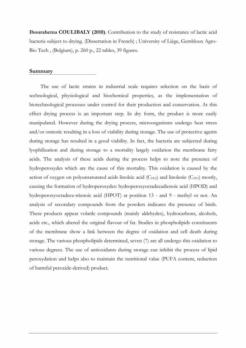
Ibourahema COULIBALY (2010). Contribution à l’étude de la résistance au séchage
des bactéries lactiques (thèse de doctorat en français) ; Université de Liège, Gembloux
Agro-Bio Tech, (Belgique), 260 p., 22 tableaux, 39 figures.
Résumé
L’utilisation de souches lactiques, à l’échelle industrielle, nécessite une sélection sur
base des propriétés technologiques, physiologiques et biochimiques, ainsi que la mise en
œuvre de procédés biotechnologiques bien maîtrisés pour leur production et leur
conservation. A cet effet, le séchage est une étape importante. Sous forme sèche, le
produit est plus facilement manipulable. Cependant au cours du séchage, les micro-
organismes subissent un stress thermique et/ou osmotique qui se traduit par une perte de
viabilité pendant le stockage. L’utilisation d’agents protecteurs au cours du stockage a
permis d’obtenir une bonne viabilité. En effet, les bactéries sont soumises pendant la
lyophilisation et au cours du stockage à une mortalité liée en grande partie à l’oxydation
des acides gras membranaires. L’analyse de ces acides au cours des différents procédés de
fabrication et de stockage permet de noter la présence des hydroperoxydes. Cette
oxydation est engendrée par l’action de l’oxygène sur les acides gras polyinsaturés
principalement l’acide linoléique (C18:2) et linolénique (C18:3) ce qui engendre la formation
des hydroperoxydes : acide hydroperoxyoctadecadienoique (HPOD) et acide hydro-
peroxyoctadecatrienoique (HPOT) méthylés ou non en position 13- et 9-. Une analyse des
composés secondaires émanant des poudres de bactéries lactiques indique la présence de
composés volatils. Parmi ceux-ci, apparaissent des molécules volatiles (principalement des
aldéhydes), qui modifient la flaveur d’origine des corps gras. Les études menées au niveau
des constituants phospholipidiques de la membrane montrent un lien entre le degré
d’oxydation et la mortalité cellulaire au cours du stockage. Les différents phospholipides
déterminés au nombre de sept (7) subissent tous cette oxydation à des degrés divers.
L’utilisation d’antioxydant au cours du stockage permet d’inhiber les phénomènes de
peroxydation des lipides et permet également de maintenir la valeur nutritionnelle (teneur
en AGPI, réduction des dérivés peroxydés nocifs) du produit.
Ibourahema COULIBALY (2010). Contribution to the study of resistance of lactic acid
bacteria subject to drying. (Dissertation in French) ; University of Liège, Gembloux Agro-
Bio Tech , (Belgium), p. 260 p., 22 tables, 39 figures.
Summary
The use of lactic strains in industrial scale requires selection on the basis of
technological, physiological and biochemical properties, as the implementation of
biotechnological processes under control for their production and conservation. At this
effect drying process is an important step. In dry form, the product is more easily
manipulated. However during the drying process, microorganisms undergo heat stress
and/or osmotic resulting in a loss of viability during storage. The use of protective agents
during storage has resulted in a good viability. In fact, the bacteria are subjected during
lyophilization and during storage to a mortality largely oxidation the membrane fatty
acids. The analysis of these acids during the process helps to note the presence of
hydroperoxydes which are the cause of this mortality. This oxidation is caused by the
action of oxygen on polyunsaturated acids linoleic acid (C18:2) and linolenic (C18:3) mostly,
causing the formation of hydroperoxydes: hydroperoxyoctadecadienoic acid (HPOD) and
hydroperoxyoctadeca-trienoic acid (HPOT) at position 13 - and 9 - methyl or not. An
analysis of secondary compounds from the powders indicates the presence of birds.
These products appear volatile compounds (mainly aldehydes), hydrocarbons, alcohols,
acids etc., which altered the original flavour of fat. Studies in phospholipids constituents
of the membrane show a link between the degree of oxidation and cell death during
storage. The various phospholipids determined, seven (7) are all undergo this oxidation to
various degrees. The use of antioxidants during storage can inhibit the process of lipid
peroxydation and helps also to maintain the nutritional value (PUFA content, reduction
of harmful peroxide-derived) product.
Dédicace
A Bintou, mon épouse. Ce travail te doit beaucoup. Qu’il soit pour toi le témoignage de mon
infinie reconnaissance pour ces années de durs labeurs, de compréhensions et de tolérances, de
privations et d’efforts communs.
A toi Mohamed Lamine Junior, notre fils, tout étonné que son papa ait enfin fini d’aller à
« l’école » comme lui !
A notre fille Oriane Fatim Manaka, ta naissance vient comme un cadeau de Dieu pour
couronner cette thèse.
A mon père, décédé ce Lundi matin d’un mois de Janvier 1983 et qui me disait un jour « je
n’ai pas mis mes enfants à l’école pour la police, ni la gendarmerie mais pour aller à
l’université, le temple du savoir », trouve ici la réalisation de ton vœux, mon immense
reconnaissance et que le paradis soit ta demeure.
A ma mère pour l’absence prolongée de son fils aîné et la peine endurée.
Remerciements
La réalisation de cette thèse fut une occasion merveilleuse de rencontrer et d’échanger avec
de nombreuses personnes. Je ne saurais pas les citer toutes sans dépasser le nombre de pages
raisonnablement admis dans ce genre de travail. Il me tient à cœur de remercier tous ceux
qui y ont contribué de près ou de loin.
Je pense particulièrement à Monsieur le Professeur Philippe THONART, mon promoteur,
pour la finesse de ses attitudes sur le plan aussi bien humain que scientifique. Ses remarques
successives ont permis d’améliorer les différentes versions de ce travail. Il a toujours trouvé
comme promoteur les mots justes et un soutien total et sans faille dans les moments délicats.
De lui, j’ai toujours reçu non seulement les encouragements dont le doctorant a tant besoin,
mais aussi les précieux conseils pratiques que seul un homme, ayant des qualités humaines
comme lui, peut amener à prodiguer. Grâce à son approche respectueuse de la personne
humaine, je me suis continuellement senti à l’aise. Je lui en sais infiniment gré.
Je remercie très sincèrement Madame le Docteur Jacqueline DESTAIN co-promoteur, pour
avoir lu ce travail et pour m’avoir continuellement donner des conseils très avisés dans la
rédaction des différents articles scientifiques de cette thèse.
Il m’est agréable de remercier Messieurs les Professeurs Karim AMIGHI de l’Université
Libre de Bruxelles (U.L.B) et DOMMES Jacques de l’Université de Liége (U.Lg) et
Madame le Docteur Marie-Laure FAUCONNIER de l’unité de biologie végétale de
gembloux (Gx-ABT) pour m’avoir permis de travailler dans leurs différents laboratoires.
Je tiens à adresser ma gratitude aux membres du jury, en particulier à Monsieur le
Professeur Georges LOGNAY de me faire l’honneur de juger cette thèse, et à Messieurs les
Professeurs François BERA, Daniel PORTETELLE pour leur disponibilité.
J’adresse mes sincères remerciements à Madame le Professeur Carmen SOCACIU et Madame
le Docteur Sabine DANTHINE pour avoir accepté d’être les rapporteurs de cette thèse.
Je remercie particulièrement Monsieur le Docteur Robin DUBOIS-DAUPHIN qui a su
faire grandir en moi la rigueur scientifique et qui a accepté de m’accorder un temps précieux
dans la rédaction de cette thèse et des articles qui la composent.
Mes remerciements s’adressent également à tout le personnel du bureau 22.73 en occurrence
Mme Dominique ROBLAIN pour son aide, ses conseils et sa disponibilité. Ce fut très
agréable de travailler avec toi.
Je tiens à remercier particulièrement le personnel du secrétariat du CWBI, que je nomme
affectueusement la « tour de contrôle du CWBI », Marina CHANET, Marguerite DEMI et
Cécile EK pour leur accueil chaleureux, leur disponibilité et surtout leur bonne humeur qui
n’a jamais fait défaut tout au long de ces années.
Je tiens ici à remercier tout le personnel scientifique et technique qui œuvre chaque jour pour
la renommée du CWBI, en particulier aux Docteurs Franck DELVIGNE, Marc OGENA,
Jean-Marc ALDRIC, Hakim GHALFI, Carine DORTU ; Mlles Magali MASSON, Lamia
MAJAD, Martine ALLGAERT, Annick SALVE, Cathy HELWUS, Pascale VANHAL,
Danielle DUBALLET, Fabienne PISCART et Messieurs Samuel TELEK, Christophe
THIRY, BenoîT MASSAUX, Pol, Laurent, Frederick, Thami el MEJDOUB, pour leur aide
et leur amitié et surtout leur compréhension pour ces années formidables passées ensemble.
Ma gratitude aux personnels de chimie organique et analytique en occurrence Mmes Maryse
HARDENNE, Céline PIERART et Messieurs Vincent HOTE et Dany TRISTMAN pour
leur aide et surtout leur disponibilité.
Mes six années d’études au CWBI, se sont déroulées dans un environnement convivial, c’est
pourquoi je tiens à remercier tous les doctorants avec lesquels j’ai passé ces journées au
CWBI à l’unité de Bio-industries de Gembloux : Ali BAYANE, Maîmouna SOW,
N’DOYE Bassirou, Pascal KAKANA, Wazé Mireille ALLOUE, Antoine ASSAMOI,
Francine AKE, Philomène KABRAN, Privat KOUAKOU, Ana AGUILAR-GALVEZ,
Mohamed CISSE, Charles YAPO, Michel MUSONI, Michel DIOP, Eugène KARENZI,
Venant NIHORIMERE, Roland FOMA, Khady BA, Guillaume HENRY, Annick
LEJEUNE, Mohamed ALCHIHAB, Thambi KAR pour leur marque de sympathie et leur
amitié sans faille durant cette période.
Je tiens à remerciement solennellement l’Etat de Côte d’Ivoire pour m’avoir soutenu
financièrement par l’octroi d’une bourse d’études durant ces six années.
En dehors de tout cadre professionnel, je voudrais remercier certaines personnes avec qui j’ai
partagé des relations humaines fortes durant cette thèse. Merci à ALABI Fabrice Taofic et
à SILUE Souleymane pour leur fraternité. Grâce à vous, en plus de la thèse j’ai eu des frères
sur le chantier de DIEU. Merci pour le réconfort lorsque le moral n’était pas au rendez-
vous, votre compagnie quotidienne a fait de mon séjour à Gembloux des moments de joies et
de souvenirs heureux tout en apportant une guidée spirituelle. Je n’oublierais pas mes jeunes
frères TRAORE Souleymane, Gaoussou KARAMOGO et CISSE Mohamed pour leur
respect et leur sympathie.
Je remercie réligeusement El Hadj Marouane pour les efforts dont il déploie chaque jour au
sein de notre communauté musulmane de Gembloux.
Je pense également aux familles : BAMBA (Adja, Youssouf et leur fille) ; CISSE
(Souleymane imam, Maimouna et enfant) ; COULIBALY (Saran, Ibrahim et enfants),
SILUE (Abdoulaye et enfants), DIABY (koro, Adama et enfants) et Kamagaté (Ahmed,
Aline et enfants) à Bruxelles. Un remerciement sincère aux familles BAMBA (Arouna,
Mariam et enfants) à Kuurne, KONE (Yaya, Matogoma et enfant) à Quaregnon, Sylla à
Amay ; KONE (Abdoulaye, Awa et enfants) à Flénu et Kaba, CISSE mohamed à Mons ;
OUATTARA Ibrahim à Bruxelles et à tous les membres et symphatisants du CNI Belgique.
Mes sincères amitiés à la famille FREH (Abdessalem, Emilienne, Samy et Sabry), à Hèlène
Aboubakar FOUREIRA et enfants ainsi qu’à Guido Claudine et enfants à Gembloux.
Mes sincères remerciements à nos aînés ASSIE Kouakou Lazare, AMBE Guy- Alain et Eric AKPA pour leurs conseils avisés. Je remercie également les membres de l’association des étudiants ivoiriens en Belgique
(ASETIB) et plus particulièrement ceux de la section de Gembloux pour leur dynamisme,
leur convivialité et leur esprit d’entraide.
Enfin, je voudrais dire un grand merci à mon épouse COULIBALY Bintou, à nos enfants
COULIBALY Mohamed Lamine Junior et sa sœur COULIBALY Oriane Fatim Manaka
pour leur patience, ainsi qu’à mes parents et aux familles proches pour leur soutien de tout
temps.
Que toutes les personnes que j’ai oublié de citer trouvent dans ce dernier
paragraphe mes sincères reconnaissances et mes remerciements distingués
Ibourahema

Table des matières
Table des matières
Table des matières
LISTE DES PUBLICATIONS................................................................................................. A
I. PUBLICATIONS ACCEPTEES DANS DES REVUES A COMITE DE LECTURE .................................................A
II. PUBLICATION EN COURS DE SOUMISSION..................................................................................................A
III. CONFERENCES SEMINAIRES ET CONGRES................................................................................................B
IV. FORMATIONS AU COURS DU DOCTORAT...................................................................................................C
LISTE DES ABREVIATIONS.................................................................................................D
INTRODUCTION GENERALE
INTRODUCTION GENERALE ......................................................................2
CHAPITRE I
SYNTHESE BIBLIOGRAPHIQUE ..................................................................9
I. SYNTHESE BIBLIOGRAPHIQUE – PREMIERE PARTIE ............................................... 10
I-1. CONTEXTE ET OBJECTIFS.........................................................................................................................10
I-2. INCIDENCES DE LA LYOPHILISATION SUR LA VIABILITE DES BACTERIES LACTIQUES AU COURS DU
STOCKAGE : REVUE..........................................................................................................................................11
RESUME .............................................................................................................................................................12
ABSTRACT .........................................................................................................................................................13
I-2.1. GENERALITES SUR LE SECHAGE DES MICROORGANISMES................................................................14
I-2.1.1. Introduction ............................................................................................................................................... 14
I-2.1.2. Types de séchage dans les industries agro-alimentaires.................................................................................. 14
I-2.2. LYOPHILISATION DES BACTERIES LACTIQUES....................................................................................16
I-2.2.1. Description et principes de la lyophilisation ................................................................................................. 17
I-2.2.2. Facteurs agissants sur la conservation des cellules séchées ............................................................................. 21
I-2.2.3. Conservation du produit sec ........................................................................................................................ 22
Table des matières
I-2.3. UTILISATION DES CRYOPROTECTEURS................................................................................................23
I-2.3.1. Cryoprotection des bactéries lactiques........................................................................................................... 23
I-2.3.2. Mécanismes d’action des cryoprotecteurs ...................................................................................................... 24
I-2.4. ACIDES GRAS ET OXYDATION CELLULAIRE ........................................................................................25
I-2.4.1. Rôles des acides gras au niveau des bactéries................................................................................................ 25
I-2.4.2. Mécanismes d’oxydation des lipides ............................................................................................................ 26
I-2.4.3. Autooxydation........................................................................................................................................... 27
I-2.4.4. Photooxydation .......................................................................................................................................... 31
I-2.4.5. Oxydation enzymatique ............................................................................................................................. 32
I-2.5. FACTEURS INFLUENÇANTS L'OXYDATION DES LIPIDES ....................................................................33
I-2.5.1. Principaux facteurs de l’oxydation des lipides ............................................................................................. 33
I-2.5.2. Impacts de l’oxydation des lipides sur les starters ........................................................................................ 34
I-2.6. DIFFERENTES APPROCHES POUR INHIBER L’OXYDATION DES STARTERS LACTIQUES
LYOPHILISES AU COURS DU STOCKAGE .........................................................................................................36
I-2.6.1. Mécanismes d’action des antioxydants préventifs ......................................................................................... 36
I-2.6.2 Mécanismes d’action des antioxydants de type « chaîne rompue et chain breaking » ...................................... 37
I-2.7. REMERCIEMENTS ...................................................................................................................................39
I-2.8. CONCLUSIONS ........................................................................................................................................39
I-2.9. REFERENCES BIBLIOGRAPHIQUES .......................................................................................................39
I. SYNTHESE BIBLIOGRAPHIQUE – DEUXIEME PARTIE .............................................. 45
I-3. CONTEXTE ET OBJECTIFS.........................................................................................................................45
I.4. FUNCTIONAL ASPECT OF Lactobacillus SPP. : A REVIEW...........................................................................46
RESUME .............................................................................................................................................................47
ABSTRACT .........................................................................................................................................................48
I-4.1. INTRODUCTION......................................................................................................................................49
I-4.2. LACTOBACILLUS GENUS ........................................................................................................................50
I-4.2.1. Ecology ...................................................................................................................................................... 50
I-4.2.2. Metabolism................................................................................................................................................ 50
I-4.3. CHARACTERISTICS OF GRAM POSSITIVE BACTERIA ............................................................................52
I-4.3.1. Generalities................................................................................................................................................ 52
I-4.3.2. Membrane structure ................................................................................................................................... 53
I-4.4. ADHESION AND RESISTANCE IN THE HOST ........................................................................................55
I-4.4.1. Anchor types.............................................................................................................................................. 55
I-4.4.2. Enzymes.................................................................................................................................................... 57
I-4.4.3. Adherence proteins and binding domains .................................................................................................... 57
Table des matières
I-4.4.4. The cell-surface hydrolase (CSH) domain ................................................................................................... 58
I-4.5. Phospholipids and fatty acids of Lactobacillus spp.......................................................................................... 59
I-4.5.1. Structure of phospholipids........................................................................................................................... 59
I-4.5.2. Fatty acids ................................................................................................................................................. 61
I-4.6. PROBIOTICS AND BIOTHERAPEUTICS ..................................................................................................61
I-4.6.1. Probiotics and food production .................................................................................................................... 61
I-4.6.2. Biotherapeutics and potential benefits.......................................................................................................... 63
I-4.6.3. Benefits for health ...................................................................................................................................... 64
I-4.7. CONCLUSIONS ........................................................................................................................................66
I-4.8. REFERENCES...........................................................................................................................................67
I-5. CONCLUSIONS : INCIDENCES DE LA LYOPHILISATION SUR LA VIABILITE DES BACTERIES
LACTIQUES AU COURS DU STOCKAGE............................................................................................................73
Table des matières
CHAPITRE II
SELECTION ET IDENTIFICATION DE NOUVELLES SOUCHES
RESISTANTES DE BACTERIES LACTIQUES.............................................. 74
II. SELECTION ET IDENTIFICATION DE NOUVELLES SOUCHES RESISTANTES DE
BACTERIES LACTIQUES .................................................................................................... 75
II.1. CONTEXTE ET OBJECTIFS .......................................................................................................................75
II.2. CHARACTERIZATION OF LACTIC ACID BACTERIA ISOLATED FROM POULTRY FARMS IN SENEGAL76
RESUME .............................................................................................................................................................77
ABSTRACT .........................................................................................................................................................78
II-2.1. INTRODUCTION ....................................................................................................................................79
II-2.2. MATERIALS AND METHODS ................................................................................................................80
II-2.2.1. Isolation of LAB strains, culture media and growth conditions.................................................................. 80
II-2.2.2. Determination of lactic acid isomers .......................................................................................................... 81
II-2.2.3. Assay of residual glucose........................................................................................................................... 82
II-2.2.4. Behaviour for tolerance to high temperatures, high concentrations of lactic acid and sodium chloride, and low
pH.......................................................................................................................................................................... 82
II-2.2.5. Determination of the turbidity................................................................................................................... 83
II-2.3. RESULTS AND DISCUSSION...................................................................................................................83
II-2.3.1. Screening of lactic acid bacteria.................................................................................................................. 83
II-2.3.2. Rapid screening for tolerance to high temperatures, high concentrations of lactic acid and sodium chloride, and
low pH ................................................................................................................................................................... 84
II-2.3.3. Growth and lactic acid production profiles from time-course studies ............................................................ 87
II-2.3.4. Optical isomer of lactic acid comparison..................................................................................................... 90
II-2.4. ACKNOWLEDGEMENTS .......................................................................................................................91
II-2.5. REFERENCES .........................................................................................................................................91
II-3. CONCLUSIONS : SELECTION ET IDENTIFICATION DE NOUVELLES SOUCHES RESISTANTES DE
BACTERIES LACTIQUES ....................................................................................................................................94
Table des matières
CHAPITRE III
EFFETS DES CRYOPROTECTEURS SUR LA COMPOSITION EN ACIDES
GRAS CELLULAIRES DE Lactobacillus plantarum CWBI-B1419..................... 96
III. EFFETS DES CRYOPROTECTEURS SUR LA COMPOSITION EN ACIDES ACIDES GRAS CELLULAIRES DE
L. plantarum CWBI-B1419...............................................................................................................................96
III-1. CONTEXTE ET OBJECTIFS......................................................................................................................96
III-2. THE RESISTANCE TO FREEZE-DRYING AND TO STORAGE WAS DETERMINED AS THE CELLULAR
ABILITY TO RECOVER ITS SURVIVAL RATE AND ACIDIFICATION ACTIVITY ..............................................97
RESUME .............................................................................................................................................................98
ABSTRACT .........................................................................................................................................................99
III-2.1. INTRODUCTION.................................................................................................................................100
III-2.2. MATERIALS AND METHODS .............................................................................................................101
III-2.2.1. Microorganisms and growth conditions...................................................................................................101
III-2.2.2. Rehydration/enumeration .....................................................................................................................103
III-2.2.3. Acidification activity and water content..................................................................................................103
III-2.2.4. Fatty acids analyses (FAME)..............................................................................................................103
III-2.2.5. Statistical Analysis. ..............................................................................................................................104
III-2.3. RESULTS AND DISCUSSION ...............................................................................................................104
III-2.3.1. Fatty acid composition of freeze-dried L. plantarum CWBI-B1419 and L. paracasei ssp. paracasei
LMG9192T.........................................................................................................................................................104
III-2.3.2. Impacts of protectives agents and storage time on membrane fatty acid composition of L. plantarum CWBI-
B1419 and L. paracasei sp. paracasei LMG9192T. ............................................................................................105
III-2.3.3. Effects of sorbitol, monosodium glutamate and glycerol, on the survival and resistance to freeze-dried storage
of L. plantarum CWBI-B1419 ...........................................................................................................................108
III-2.3.4. Relationship between the resistance, freeze-dried L. plantarum CWBI-B1419 storage and its membrane
fatty acid composition.............................................................................................................................................113
III-2.4. CONCLUSIONS ...................................................................................................................................114
III-2.5. ACKNOWLEDGEMENTS ....................................................................................................................115
III-2.6. REFERENCES......................................................................................................................................115
III-3. CONCLUSIONS : EFFETS DES CRYOPROTECTEURS SUR LA COMPOSITION EN ACIDES GRAS
CELLULAIRES DE Lactobacillus plantarum CWBI-B1419 ................................................................................119
Table des matières
CHAPITRE IV
SUIVI DE LA DEGRADATION DES ACIDES GRAS POLYINSATURES..... 120
IV. SUIVI DE LA DEGRADATION DES ACIDES GRAS POLYINSATURES ......................................................121
IV.1. CONTEXTE ET OBJECTIFS ....................................................................................................................121
IV.2. SURVIVAL OF FREEZE-DRIED Leuconostoc mesenteroides KMROG AND Lactobacillus plantarum CWBI-
B1419 RELATED TO THEIR CELLULAR FATTY ACIDS COMPOSITION DURING STORAGE........................122
RESUME ...........................................................................................................................................................123
ABSTRACT .......................................................................................................................................................124
IV-2.1. INTRODUCTION.................................................................................................................................125
IV-2.2. MATERIALS AND METHODS .............................................................................................................126
IV-2.2.1. Micro-organisms and cultivation ...........................................................................................................126
IV-2.2.2. Productions ..........................................................................................................................................126
IV-2.2.3. Storage conditions .................................................................................................................................127
IV-2.2.4. Dry cell weight and water content determinations...................................................................................127
IV-2.2.5. Water activity measurements.................................................................................................................127
IV-2.2.6. Survival rate ........................................................................................................................................127
IV-2.2.7. Analysis of cellular fatty acids ..............................................................................................................128
IV-2.2.8. Statistical analysis................................................................................................................................129
IV-2.3. RESULTS AND DISCUSSION ...............................................................................................................129
IV.2.3.1. Survival of lactic acid bacteria after freeze-drying ...................................................................................129
IV-2.3.2. Influence of water activity on survival rate. ............................................................................................130
IV-2.3.3. Survival of L. mesenteroides and Lb. plantarum during storage ............................................................130
IV-2.3.4. Cellular fatty acid relative contents after freeze-drying and changes in fatty acid composition ...................134
IV-2.4. CONCLUSIONS ...................................................................................................................................142
IV-2.5. ACKNOWLEDGEMENTS ....................................................................................................................142
IV-2.6. REFERENCES......................................................................................................................................143
IV.3. CONCLUSIONS : SUIVI DE LA DEGRADATION DES ACIDES GRAS POLYINSATURES .......................146
Table des matières
CHAPITRE V
CARACTERISATION DES COMPOSES VOLATILS ISSUS DE LA
DEGRADATION DES ACIDES GRAS DE BACTERIES LACTIQUES..........151
V. CARACTERISATION DES COMPOSES VOLATILS ISSUS DE LA DEGRADATION DES ACIDES GRAS DE
BACTERIES LACTIQUES : L. plantarum CWBI-B1419 ..................................................................................148
V.1. CONTEXTE ET OBJECTIFS......................................................................................................................148
V.2. CHARACTERIZATION OF VOLATILES COMPOUNDS EMITTED FROM FREEZE-DRIED L. plantarum
CWBI-B1419 PRODUCT DURING STORAGE. ...............................................................................................149
RESUME ...........................................................................................................................................................150
ABSTRACT .......................................................................................................................................................151
V-2.1. INTRODUCTION ..................................................................................................................................152
V-2.2. MATERIALS AND METHODS ...............................................................................................................153
V-2.2.1. Microorganism and inoculum .................................................................................................................153
V-2.2.2. Productions and storage conditions..........................................................................................................153
V-2.2.3. Dry cell weight, water Content and survival rate .....................................................................................153
V-2.2.4. Data analysis of SPME........................................................................................................................154
V-2.2.5. GC–MS operating parameters...............................................................................................................154
V-2.3. RESULTS AND DISCUSSION.................................................................................................................155
V-2.3.1. Viability of L. planatrum CWBI-B1419 during storage. .....................................................................155
V-2.3.2. Volatile compound analysis....................................................................................................................156
V-2.4. CONCLUSIONS .....................................................................................................................................162
V-2.5. AKNOWLEGE.......................................................................................................................................163
V-2.6. REFERENCES .......................................................................................................................................163
V-3. CONCLUSIONS : CARACTERISATION DES COMPOSES VOLATILS ISSUS DE LA DEGRADATION DES
ACIDES GRAS DE BACTERIES LACTIQUES.....................................................................................................166
Table des matières
CHAPITRE VI
ANALYSE DES PHOSPHOLIPIDES DE Lactobacillus plantarum CWBI-B1419
AU COURS DU STOCKAGE ........................................................................ 167
VI. ANALYSE DES PHOSPHOLIPIDES DE L. plantarum CWBI-B1419 AU COURS DU STOCKAGE ...........168
VI-1. CONTEXTE ET OBJECTIFS....................................................................................................................168
VI-2. EFFECT OF STORAGE TEMPERATURE ON PHOSPHOLIPIDS COMPOSITIONS AND VIABILITY UPON
Lactobacillus plantarum CWB-B1419................................................................................................................169
RESUME ...........................................................................................................................................................170
ABSTRACT .......................................................................................................................................................171
VI-2.1. INTRODUCTION.................................................................................................................................172
VI-2.2. MATERIALS AND METHODS .............................................................................................................173
VI-2.2.1. Strain and culture ................................................................................................................................173
VI-2.2.2. Solvents and standards .........................................................................................................................173
VI-2.2.3. Lipid extraction ...................................................................................................................................174
VI-2.2.4. Solid-phase extraction (SPE) ...............................................................................................................174
VI.2.2.5. HPLC- ELSD of the PL fraction ......................................................................................................175
VI-2.3. RESULTS..............................................................................................................................................176
VI-2.3.1. L. plantarum CWBI-B1419 growth characteristics..............................................................................176
VI-2.3.2. Biochemical behaviour of L. plantarum CWBI-B1419 during storage .................................................177
VI-2.3.3. Free fatty acids and lipids compositions .................................................................................................178
VI-2.3.4. Fatty acids distribution in lipids classes.................................................................................................179
VI-2.3.5. HPLC procedure for the separation and quantification of phospholipids in the PL fraction ...................180
VI-2.4. DISCUSSION .......................................................................................................................................182
VI-2.5.CONCLUSION ......................................................................................................................................183
VI-2.6. AKWNOLEDGE ..................................................................................................................................183
VI-2.7. REFERENCES......................................................................................................................................183
VI-3. CONCLUSIONS : ANALYSE DES PHOSPHOLIPIDES DE L. plantarum CWBI-B1419 AU COURS DU
STOCKAGE ......................................................................................................................................................187
Table des matières
CHAPITRE VII
DISCUSSIONS GENERALES ...................................................................... 188
Discussions générales........................................................................................................... 189
CHAPITRE VIII
CONCLUSIONS GENERALES ET PERSPECTIVES....................................211
Conclusions générales ...........................................................................................................211
Perspectives ......................................................................................................................... 214
ANNEXES
ANNEXES.................................................................................................. 217
Annexe 1 : Article publié dans la revue « African Journal of Biotechnology » .........................................218
Annexe 2 : Article publié dans la revue « Applied Biochemistry And Biotechnology » .........................219
Annexe 3 : Article publié dans la revue « Applied Microbiology and Biotechnology » ..........................220
Annexe 4 : Article publié dans la revue « International Journal of Microbiology » .................................221
Annexe 5 : Montage de la chromatographie HPLC couplé au ELSD et le chromatogramme ...............222
Annexe 6: Voies d’utilisation intracellulaire des acides gras et synthèse des phospholipides ................223
Annexe 7: Dispostif HPLC-SPME pour l’analyse des composés volatils ................................................224
Annexe 8 : Formule type milieu MRS..........................................................................................................225
Annexe 9 : Appareillage pour la concentration et le séchage des cellules.................................................226
Annexe 10 : Appareil seravnt à la réaction de polymérisation en chaîne (PCR).......................................227
TABLEAUX ET FIGURES
TABLEAUX ET FIGURES ....................................................................... 228
Liste des tableaux................................................................................................................. 229
Liste des figures ................................................................................................................... 231
LISTE DES MEMBRES DU JURY ............................................................... 235
Publications & abréviations
Liste des publications scientifiques et des séminaires
a
Liste des publications
I. Publications acceptées dans des revues à comité de lecture
Coulibaly I., Robin D-D., Destain J., and Thonart Ph., 2008. Characterization of lactic
acid bacteria isolated from poultry farms in Senegal. Afr. J. Biotechnol., 7, 2006-
2012.
Coulibaly I., Yao A.A., Lognay G.,. Fauconnier M.L and Ph. Thonart., 2009. Survival of
freeze-dried Leuconostoc mesenteroides and Lactobacillus plantarum related to their
cellular fatty acids composition during storage. Appl. Biochem. Biotechnol., 157,
70-84.
Yao A.A., Coulibaly I., Lognay G., Fauconnier M.L and Ph. Thonart., 2008. Impact of
polyunsaturated fatty acid degradation on survival and acidification activity of
freeze-dried Weissella paramesenteroides LC11 during storage. Appl. Microbiol.
Biotechnol., 79, 1045-1052.
Coulibaly I., Robin D-D., Destain J., Fauconnier M.L., Lognay G and Ph. Thonart., 2010.
The resistance to freeze-drying and to storagewas determined as the cellular
ability to recover its survival rate and acidification activity. Int.l J. Microbiol. 2010,
doi:10.1155/2010/625239, 9 pages.
II. Publications en cours de soumission
Coulibaly I., Robin D. D., Destain J., Danthine S., Béra F., Majad L., Mejoub T., Wathelet
J.P. and Thonart Ph., 2010. Incidences de la lyophilisation sur la viabilité des
bactéries lactiques au cours du stockage : In press (revue BASE).
Coulibaly I., Béra F., Robin D. D., Destain J., Majad L., Mejoub T., Wathelet J.P. and
Thonart Ph., 2010. Functional aspects of Lactobacillus sp. Synthèse
bibliographique. (Soumis à African Journal of Biotechnology).
Liste des publications scientifiques et des séminaires
b
III. Conférences, séminaires et congrès
Participation aux journées scientifiques “Biomedica 2009” du 1er au 2 Avril 2009 au
Palais des Congrès de Liège - Poster: Coulibaly I., Yao A. A., Lognay G.,
Fauconnier M-L, Thonart P., 2009. Determination of phospholipids extracted
from freeze-dried Lactobacillus plantarum and during the subsequent storage.
Participation à “Intensive Program food and health” Cluj-Napoca Roumanie, February
2007, University of Agricultural Sciences and Veterinary Medicine - Poster:
Ibourahema Coulibaly, Maimouna Sow N’deye, Robin Dubois Dauphin and
Philippe Thonart. 2007. Identification of new strain used as probiotic.
Participation au séminaire de formation “Maîtrise des équipements : les pipettes
automatiques” le 1er Février 2006 à l’auditoire ZT1 de la Faculté des sciences
Agronomiques de Gembloux. Formateurs : Mme Callens et M. Muydermans -
Société : VWR.
Participation au séminaire de formation “Les outils de la communication” le 29 Mars
2006 à l’auditoire ZT1 de la Faculté des sciences Agronomiques de Gembloux -
Formateur : M. Lahousse - Société : LAPIERRE & LIBERT s.c.r.l.
Participation au séminaire de formation “Maîtrise des dispositifs de mesure: les ph-
mètres” le 24 Mai 2005 à l’auditoire CA de la Faculté des sciences Agronomiques
de Gembloux - Formateur: Pr. BARTHELEMY Jean-paul. - Laboratoire: Unité
de Chimie Analytique et Phytopharmacie.
Participation au séminaire de formation “Principes de stérilisation en laboratoire” le 16
Décembre 2004 à l’auditoire ZT1 de la Faculté des sciences Agronomiques de
Gembloux - Formateurs : LAMBERT David - Société : H+P Labortechnik AG
Liste des publications scientifiques et des séminaires
c
IV. Formations au cours du doctorat
Participation aux formations spécifiques TechnofuturTIC “Initiation au language html”
du 16 Décembre 2008 au 20 Janvier 2009. Cours donné par TechnofuturTIC en
formation à distance - Formateurs : Anne-Sophie PATINY & Baya AZOUIGH
Participation aux formations spécifiques BioWin “Bonnes Pratiques de Validation” du 26
au 27 Mai 2009 à la salle de réunion de Cardio3 Biosciences SA Axis parc Business
Center Rue Edouard Belin, 12 B-1435 Mont Saint Guibert Belgique - Formateur :
CAMBIER Jacques en collaboration avec Cefochim asbl Zoning de Seneffe, Zone
C, 7180 Seneffe Belgique.
Participation aux formations spécifiques BioWin “Introduction à la Bioinformatique” du
02 au 03 Juin 2009 au centre de formation en biotechnologies- Tour GIGA B34
2eme Etage Avenue de l’hôpital 1, 4000 Liège - Formateurs : Xavier TORDOIR
& Alexandre IRRTHUM
Participation au séminaire de formation “Virologie - Module de base” les 17 et 23
Mars 2010 à l’auditoire de Culture in vivo asbl, Rue du Progrès, 4 1400 Nivelles –
Belgique. Formateur : Vincent MERIAUX
Participation au séminaire de formation “Tests immunologiques” le 24 Mars et le 1er
Avril 2010 à l’auditoire de Culture in vivo asbl, Rue du Progrès, 4 1400 Nivelles –
Belgique. Formateur : Vincent MERIAUX
Participation au séminaire de formation “ELISA Validation” le 30 Avril 2010 à
l’auditoire de Culture in vivo asbl, Rue du Progrès, 4 1400 Nivelles – Belgique.
Formateurs : Vincent MERIAUX, FAUCONNIER Allan
Liste des abréviations
d
Liste des abréviations
aw : water activity
C : cell suspension without protective coumpound
CFA : cellular fatty acid
CL : cell suspension with 8% lithothamne400®
ELSD : evaporative light scattering detection
FD : freeze-drying
FFA : free fatty acid
GxABT : Gembloux Agro-Bio Tech
HOD : hydroperoxyde acid
HPOD : hydroperoxy octadecadienoic acid
HPOT : hydroperoxy octadecatrienoic acid
K : inactivation rate
Ka : acidification loss rate
Kg : kilogramme
L : Leuconostoc
LAB : lactic acid bacteria
Lb, L : Lactobacillus
LPC : lysophosphatidylcholine
m : mètre
Me : methyl esterified
mL : millilitre
MUFA : monounsaturated fatty acid
NL : neutral lipid
P : cell without cryoprotectant
PA : phosphatidic acid
Pc : cell with cryoprotectants
PC : phosphatidylcholine
PE : phosphatidylethanolamine
Liste des abréviations
e
PG : phosphatidyl-DL-glycerol
PI : phosphatidylinositol
PL : phospholipids
PUFA : polyunsaturated fatty acids
R2 : determination coefficient
RH : relative humidity
SFA : saturated fatty acids
SM : sphingomyeline
SPE : solid phase extraction
Tg : température de transition vitreuse
TL : total lipid
Ts : température de stockage
UFA : unsaturated fatty acids
ULg : Université de Liège
Introduction générale
Introduction générale
2
Introduction générale
Représenté, comme l’un des marchés lucratifs, le plus grand en production de
biomasse, les bactéries lactiques, après les levures sont de nos jours utilisées sous diverses
formulations. Elles sont utilisées principalement pour des applications dans l’industrie
alimentaire, comme la fabrication des fromages, des laits fermentés, de certains légumes et
produits carnés fermentés et de certains vins. Comme starters, elles occupent aussi une
place de choix dans l’industrie chimique pour la production d’acide lactique et de
biopolymères. Depuis quelques années, elles ont acquis un rôle croissant en santé animale
et humaine, proposées sous le terme de probiotiques. En combinaison avec des
prébiotiques (oligosaccharides), on obtient des synbiotiques qui sont très exploités en
alimentation humaine. Utilisés dans les fermentations industrielles, ces bactéries
interviennent dans plusieurs aspects de la vie humaine. Le pouvoir antibactérien de l’acide
lactique produit dans les cultures est exploité dans plusieurs formulations alimentaires in
vitro (conservations) et in vivo (probiotiques). En alimentation, la production de ces
microorganismes représente une activité industrielle importante de plus en plus
croissante. Dans le groupe des lactiques, les espèces appartenant au genre Lactobacillus
sont les plus couramment utilisées dans les fermentations alimentaires (laiteries,
fromagerie…) (Collins et al., 1998 ; Gomez et al., 1999). La plupart des espèces
appartenant à ce genre bactérien ont d’importants besoins nutritionnels et possèdent peu
de résistance aux procédés industriels de transformation (séchage, stockage, etc.), de
conditionnement ainsi qu’à leur application in vivo (propriétés physicochimiques du
tractus digestif) Buckenhüskes, (1993). Cette situation amène les industries avec les
microbiologistes à rechercher de nouveaux isolats de bactéries lactiques capables
d'apporter plus de satisfaction en supportant l’ensemble des procédés de production, de
stockage et de formulation sans que ne soient perdues leurs propriétés fonctionnelles
(Berner et al., 1998 ; Klein et al., 1998 ; Schillinger, 1999). Au cours des opérations de
post-fermentation, les bactéries lactiques sont soumises à différentes opérations unitaires
industrielles que sont : le refroidissement, la concentration, la cryoprotection, la
congélation ou la lyophilisation et le stockage.
Introduction générale
3
L’objectif de cette filière industrielle microbiologiste est de produire des concentrés
bactériens de qualité élevée, c’est-à-dire, comportant un nombre élevé de cellules viables,
capables d'être conservés le plus longtemps possible et bien reproductibles. Force est de
constater que ces opérations unitaires induisent différents types de stress (chimique ou
biochimique, thermique, osmotique, mécanique, oxydatif) qui sont à l’origine d’une
dégradation de l’état physiologique des cellules, avec des conséquences directes sur la
qualité technologique des ferments.
La maîtrise de l’ensemble des opérations pour obtenir des starters présentant des
propriétés technologiques optimales lors de leur utilisation est indispensable. Pour
garantir de telles performances technologiques (résitance, viabilité, etc.) et métaboliques
au moment de leur utilisation, l’optimisation des conditions de production et de
conservation des souches bactériennes est indéniable. Les procédés de conservation
utilisés à l’échelle industrielle (lyophilisation, atomisation, fluidisation) sont de plus en plus
performants, les paramètres opérationnels étant bien maîtrisés. Une fois transformées
sous forme séchée, les bactéries lactiques sont souvent aménées à être congelées ou
réfrigérées pour garantir une stabilité dans le temps. C’est pour cette raison que la
déshydratation est souvent citée comme la principale cause de cette mortalité lors du
stockage. Le maintien des propriétés probiotiques et organoleptiques et les impératifs de
rentabilité impliquent en amont une sélection rigoureuse des souches destinées à la
production industrielle de starter. Ainsi, la sélection de souches microbiennes (bactéries
lactiques), technologiquement performantes et naturellement plus résistantes aux
procédés technologiques est un moyen fiable de s’assurer la survie et la stabilité de ces
souches lors du stockage même à température ambiante. Cette sélection est basée sur les
critères de thermo-résistance et de la résistance à la dessiccation de bactéries lactiques
isolées à partir d’échantillons de divers supports (sol, volaille, fientes etc.) provenant de
pays chauds et arides. L’adaptation de ces souches à de telles conditions drastiques est la
preuve qu’elles possèdent des systèmes d’adaptation capables d’améliorer leur capacité de
résistance aux stress thermiques et osmotiques. La mise en contact des cellules avec un
stress modéré leur permet également de s’adapter aux conditions défavorables, et donc de
mieux résister à une situation de stress plus intense, comme celle qu’elles rencontrent lors
de la congélation.
Introduction générale
4
Après la sélection, les nouvelles souches destinées à la production industrielle,
doivent faire l’objet d’une identification rigoureuse. Une validation permet de vérifier la
conformité des critères d’activité, de performances et de pureté spécifiques à chaque
application. Le contrôle de l’identité des espèces industriellement exploitées est fonction
de ces caractères génotypes.
Des essais plus approfondis (recherches) visant à comprendre les mécanismes
physiologiques à l’origine de ces dégradations (oxydations) ont été effectués. Par exemple,
les cellules produites dans un environnement acide ou à basse température développent
des mécanismes d’adaptation et résistent mieux (meilleure survie et meilleure activité) à la
congélation et leur temps de survie au cours du stockage est prolongé. Par réaction à ces
différentes conditions, les cellules développent des réponses physiologiques d’adaptation
au niveau membranaire (acides gras membranaires) et au niveau cytoplasmique (synthèse
de protéines de stress). Ces compositions en acides gras ouvrent de nouvelles voies
d’investigation pour améliorer la conservation des cellules. Les résultats actuellement
disponibles sur l’oxydation des acides gras décrivent principalement l’effet de conditions
environnementales que sont : le pH, la température, la composition du milieu au cours de
la fermentation, pour modifier la résistance des bactéries lactiques à la congélation et à la
lyophilisation. Cependant, l’effet de stress modérés et de l’oxydation des acides gras
membranaires liés aux opérations unitaires de production et les conditions de stockage, de
même que l’état physiologique des bactéries lactiques, manquent de recherches et
d’investigations.
C’est pour cette raison que cette étude prend toute son importance, en ce sens
qu’elle a pour but de comprendre comment la résistance de L. plantarum CWBI-B1419,
soumise à un stress au cours de la lyophilisation et pendant le stockage, est influencée par
les étapes du procédé. Ce mémoire de thèse est présenté sous forme d’essai structuré
autour d’articles scientifiques. En accord avec les réglements de l’université de Liège
campus de Gembloux Agro-Bio Tech, les chapitres en anglais sont précédés d’un résumé
en français.
Le document est subdivisé en huit (8) chapitres. Le chapitre I, intitulé “Synthèse
bibliographique ”, subdivisé en deux parties, porte sur la problématique de la recherche
Introduction générale
5
et résume les connaissances actuelles sur les ferments et les bactéries lactiques, de même
que les défis à relever pour produire et exploiter ces bactéries comme probiotiques. La
première partie (Part – I) a fait l’objet d’un article sous la forme de revue soumise à
Biotechnology, Agronomy, Society and Environment (BASE). Cette première partie
de synthèse bibliographique (revue) (Part-I) : intitulée, “Incidences de la lyophilisation
sur la viabilité des bactéries lactiques au cours du stockage : revue ”, résume les
techniques de conservation, avec une vue d'ensemble des techniques de séchage par
atomisation et par fluidisation et surtout par lyophilisation. Cette dernière est la plus
couramment utilisée en industrie agroalimentaire et se révèle comme une méthode de
choix, malgré quelques inconvénients pour la production de ferments lactiques
probiotiques. Quant à la deuxième partie (Part-II), rédigée en anglais “Functional aspect
of Lactobacillus spp. a review ”, elle met en relation la viabilité bactérienne et les
produits de dégradation des acides gras cellulaires au cours du stockage. Elle décrit
également les enzymes impliquées dans ces phénomènes de détérioration. Les
phospholipides membranaires des bactéries lactiques ont aussi été étudiés. L’importance
de ces produits au niveau des bactéries probiotiques a ainsi été démontrée. Suite à la
synthèse bibliographique, les hypothèses, buts et objectifs de recherche sont posés.
Dans le souci d’assurer d’excellentes qualités probiotiques et organoleptiques, les
impératifs de rentabilité impliquent une sélection rigoureuse des souches destinées à la
production industrielle. La sélection de starters microbiens technologiquement perfor-
mants et naturellement plus résistants est un moyen fiable pour améliorer la survie et la
stabilité des souches lors du stockage. C’est qui est traité dans le chapitre II, intitulé
“Sélection, identification et caractérisation de nouvelles souches de bactéries
lactiques thermorésistantes”, où est étudié l’isolement de souches lactiques plus
résistantes afin d’améliorer la conservation des starters au cours du stockage. Ces isolats
de bactéries sont soumis à plusieurs séries de tests (acidité, pH, température, NaCl, etc.)
afin de retenir les souches qui sont les plus résistantes dans ces conditions drastiques
(température élevée, sécheresse). Ces souches seront capables de garantir une viabilité
commercialement satisfaisante, après un stockage de longue durée. Ce chapitre constitue
la première étape de l’étude de la résistance des bactéries lactiques (probiotiques) et a été
publié dans le journal “African Journal of Biotechnology”, sous le titre de
Introduction générale
6
« Characterization of lactic acid bacteria isolated from poultry farms in Senegal », Afr. J. Biotechnol. 7,
2006-2012 : 2008.
L’étude des performances technologiques, au cours de la conservation à
température ambiante des souches isolées, de même que leurs propriétés en tant que
probiotiques constitue le troisième thème de ce mémoire. Le chapitre III est intitulé
“Effets des cryoprotecteurs sur la composition des acides gras cellulaires chez
Lactobacillus plantarum CWBI-B1419 au cours du stockage à température
ambiante”. Dans ce chapitre, les résultats présentés portent sur l’effet des additifs au
cours de la lyophilisation et pendant le stockage. Nos résultats montrent qu’il n’y a pas
d’effets notables de ces protecteurs pendant la lyophilisation mais un effet positif au cours
du stockage en agissant sur la composition des acides gras membranaires et la viabilité
cellulaire. Les résultats de ces travaux ont été soumis à la revue “International Journal of
Microbiology ” avec l’intitulé «The resistance to freeze-drying and to storage was determined as the
cellular ability to recover its survival rate and acidification activity», Int. J. Microbiol., : 2010, article
ID 625239, 9 pages. D’autres mécanismes permettant de comprendre la mortalité
cellulaire pendant le stockage et la conservation ont fait l’objet de notre attention.
Les résultats présentés dans le chapitre IV, qui a pour titre “Décomposition des
acides gras polyinsaturés au cours du stockage” montrent la dégradation des acides
gras cellulaires. L’analyse des produits primaires de dégradation des acides gras
polyinsaturés au cours du stockage chez Lactobacillus plantarum CWBI-B1419 a été
investiguée en comparaison avec la souche de Leuconostoc mesenteroides. Les résultats de ces
travaux ont fait l’objet d’une publication et ont permis de comprendre le phénomène de
mortalité lié à l’oxydation cellulaire. Cet article a été soumis dans le journal “Applied
Microbiology and Biotechnology”, intitulé « Survival of freeze-dried Leuconostoc mesenteroides
and Lactobacillus plantarum CWB-B1419 related to their cellular fatty acids composition during storage
», Appl. Biochem. Biotechnol., 157, 70-84 : 2009. Les produits primaires de décompositions
des acides gras ont été déterminés, il s’agit des oxylipines. Les recherches se sont ensuites
focalisées sur la décomposition de ces dernières en composés volatils. Les différents
aspects sont passés en revue dans le chapitre V, intitulé “Détermination des produits
volatils chez Lactobacillus plantarum CWBI-B1419”.
Introduction générale
7
Les produits secondaires volatils se dégageant, lors de la dégradation de poudres
lyophilisées, pendant le stockage ont été caractérisés. Ces travaux basés sur la méthode de
la SPME ont donné des résultats intéressants. Cette technique a permis de répertorier les
différentes classes de produits volatils qui sont responsables des odeurs au cours de
l’oxydation cellulaire. Les résultats de ces analyses ont fait l’objet d’un article soumis dans
la revue “Biotechnology and Applied Biochemistry”. Avec l’intitulé « Characterization of
volatiles compounds emitted from freeze-dried L. plantarum CWBI-B1419 product during storage», in
press.
La cinquième partie porte sur l’étude de la composition et le suivi des
phospholipides membranaires au cours du stockage. Après extraction en phase solide
(SPE) sur une cartouche aminopropyl (Varian Bond Elut NH2), les échantillons ont été
analysés par chromatographie HPLC couplée à un Evaporative Light Scattering Detection
signal (SPME-ELSD). Le chapitre VI, intitulé “Détermination des phospholipides
chez Lactobacillus plantarum CWBI-B1419 au cours du stockage” traite des
modifications physiologiques observées lors du stockage au niveau des classes de
phospholipides (acide phosphatidique, phosphatidyléthanolamine, phosphatidylinositol,
phosphatidylcholine, sphingomyéline, esters de stérol, phosphatidyl-DL-glycérol et
lysophosphatidylcholine (PA, PE, PI, PC, SM, ES et LPC).
Les composés phospholipidiques chez Lactobacillus plantarum CWBI-B1419 sont au
nombre de sept. Au cours du stockage, ces différents composés sont soumis à
réarrangements des acides gras. Le suivi au cours de 120 jours de conservation, a mis en
évidence une corrélation entre ces modifications et la mortalité cellulaire de ces composés.
Les résultats ont été discutés et l’importance de ces produits au niveau des bactéries
probiotiques a été ainsi démontrée. Le resultat de ces travaux a été soumis dans
“Microbiology and Applied Biotechnology” sous le titre «Determination of phospholipids
in lipids extracted from Lactobacillus plantarum CWBI-B1419 after freeze-drying and during the
subsequent storage», in press.
La discussion générale, chapitre VII, resume et discute l’ensemble des résultats
des travaux qui ont donné lieu à la redaction de publications. Les travaux de sélection ont
Introduction générale
8
abouti à l’isolement et à l’identification de Lactobacillus plantarum CWBI-B1419 grâce à la
combinaison de techniques de biologie moléculaire (PCR) et des tests biochimiques (API
CHL50).
Ensuite, l’addition de certains additifs utilisés comme cryoprotecteurs a montré un
effet positif sur la conservation des bactéries au cours du stockage. Notre travail n’a pas
permis de montrer un effet notable pendant la lyophilisation. La discussion porte alors sur
la dégradation des acides gras polyinsaturés au niveau de poudres lyophilisées de
Lactobacillus plantarum CWBI-B1419 pendant le stockage à différentes températures. Le
chapitre de la discussion générale se conclut sur la détection et l’analyse des
phospholipides au cours du stockage en relation avec la survie des bactéries.
Le chapitre VIII, présente les conclusions générales les plus importantes de
cette étude et les principales perspectives envisagées à la poursuite des recherches dans
ce domaine.
Chapitre I
Synthèse bibliographique
Chapitre premier
Incidences de la lyophilisation sur la viabilité des bactéries lactiques
10
I. Synthèse bibliographique – Première partie
I-1. Contexte et objectifs
Les bactéries lactiques naturellement présentes dans notre environnement, sont
exploitées dans le monde entier, depuis l’antiquité, de façon empirique dans l’alimentation
humaine et animale. Les industries alimentaires, pharmaceutiques et diététiques sont de
plus en plus intéressées par la sélection et l’utilisation de bactéries lactiques présentant des
propriétés spécifiques profitables aux consommateurs, tant sur le plan organoleptique que
sur celui de la santé. Pour garantir de telles performances technologiques et métaboliques
au moment de leur utilisation, il est important d’optimiser les conditions de production et
de conservation des souches bactériennes, de contrôler leur viabilité et leurs aptitudes
technologiques au cours du stockage. Les procédés de conservation utilisés à l’échelle
industrielle (lyophilisation, atomisation, fluidisation, etc.) sont de plus en plus
performants, les paramètres opérationnels étant bien maîtrisés. Les bactéries lactiques
doivent encore être congelées ou réfrigérées sous forme de poudres ou de granulés, pour
leur garantir une stabilité à long terme car la présence d’eau (activité d’eau élévée) est
souvent citée comme la principale cause de mortalité lors du stockage. Cette revue
présente les travaux déjà effectués dans le domaine, les procédés d’optimisation de la
production et de la conservation des starters lactiques.
Chapitre premier
Incidences de la lyophilisation sur la viabilité des bactéries lactiques
11
I-2. Incidences de la lyophilisation sur la viabilité des bactéries
lactiques au cours du stockage : revue
Publication 1 – Biotechnol. Agr. Soc. Environ
Biotechnology, Agronomy, Society and Environment
Coulibaly, I(1♣)., Dubois-Dauphin, R(1)., Danthine, S(3)., Majad, L (1).,, Mejoub, T(2).,
Destain, J(1)., Bera, F(3)., Wathelet, J.P(4)., Thonart, Ph (1, 2)
(1)Wallon Center for Industrial Biology, Bio-Industry Unit, Gembloux
Agricultural University, Passage des déportés 2, 5030 Gembloux, Belgium.
(2)Wallon Center for Industrial Biology, Microbial Technology Unit, University
of Liège, Sart-Tilman B40, 4000 Liège, Belgium.
(3)Food Technology Unit, Gembloux Agricultural University, Passages des
déportés 2, 5030 Gembloux, Belgium.
(4)Organic Chemistry Unit, Gembloux Agricultural University, Passages des
déportés 2, 5030 Gembloux, Belgium
♣ Corresponding Author : I. Coulibaly, Phone : +3281622305, Fax : +32816614222 E-mail : [email protected]
Chapitre premier
Incidences de la lyophilisation sur la viabilité des bactéries lactiques
12
Résumé
La plupart des starters lactiques ont d’importants besoins nutritionnels et
possèdent peu de résistance vis-à-vis des conditions environnementales entourant leur
production (séchage, stockage...) et leur utilisation in vivo (propriétés physicochimiques du
tractus digestif). Cette situation amène régulièrement les industriels à développer avec les
microbiologistes, des projets de recherche de nouvelles bactéries lactiques capables de
supporter l’ensemble des procédés de production, de stockage et de formulation sans
perdre leurs propriétés fonctionnelles. Parmi les différentes méthodes de séchage
(atomisation, fluidisation et lyophilisation), la lyophilisation permet d'obtenir une
déshydratation poussée compatible avec des durées de conservation très longues. Cette
méthode implique des changements de la température du produit assez agressifs pour les
micro-organismes car elle nécessite une congélation, ce qui n’est pas sans conséquence
pour les cellules. Dans certains cas, elles occasionnent des altérations cellulaires
(peroxydation des acides gras) et génétiques (modification des protéines). L’utilisation de
cryoprotecteurs au cours de la lyophilisation et d’antioxydants pendant le stockage
augmente sensiblement le taux de viabilité de ces cellules.
Mots-clés. Probiotiques, lyophilisation, radicaux libres, acides gras cellulaires,
hydroperoxydes, peroxydation, cryoprotecteurs.
Chapitre premier
Incidences de la lyophilisation sur la viabilité des bactéries lactiques
13
Abstract
Preservation of industrial’s lactic acid bacteria (probiotics) by freeze-drying.
Majority of the lactic acid bacteria have important nutritional needs and do not have
resistance against the environmental conditions surrounding their production (drying,
storage…) and their use in vivo (physico-chemical properties of the digestive tract). In
this condition, industrialists and microbiologists develop regularly research projects of
new lactic bacteria able to support the whole of the processes of production, storage and
formulation without losing their functional properties. Among the various methods of
drying (atomization, fluidization and freeze-drying), freeze-drying makes it possible to
obtain a thorough dehydration compatible with very long storage times. This method
involve changes in product temperature and are enough aggressive for micro-organisms
because they require freezing that is not without consequences for cells In otherwise, they
cause cellular deteriorations (peroxydation of the fatty-acids) and genetics (modification
of proteins). The use of cryoprotecteurs during freeze-drying and antioxidants during
storage appreciably increases the rate of viability of these cells.
Keywords. Probiotics, freeze-drying, cellular fatty acid, free radicals, hydroperoxyds,
peroxydation, cryoprotectants.
Chapitre premier
Incidences de la lyophilisation sur la viabilité des bactéries lactiques
14
I-2.1. Généralités sur le séchage des micro-organismes
I-2.1.1. Introduction
La commercialisation de souches microbiennes, qu’il s’agisse de bactéries, de
levures ou de moisissures, nécessite de les conditionner sous une forme durable stable
(Leslie et al., 1995). En effet en raison de leur physiologie, les cellules microbiennes ne se
conservent pas à l’état natif dans leur milieu de culture. Leur développement est souvent
assuré en fermenteur dans un substrat liquide. Conservées dans ce milieu après leur
croissance, les cellules y consomment les derniers nutriments disponibles et expriment
pour la plupart, un métabolisme fermentaire qui nuit à la qualité du produit (modification
du pH, émanation d’odeurs, etc.). De plus, les cellules après avoir brûlé leurs réserves,
meurent en grand nombre (Zhao et al., 2005). Il est donc indispensable de stabiliser la
population microbienne. Le séchage des cellules apparaît alors comme une solution
pratique. En éliminant l’eau, le métabolisme cellulaire est bloqué, les cellules sont figées
dans un état physiologique déterminé. Outre la conservation des micro-organismes, le
séchage facilite la manipulation et le stockage et en réduit les coûts. Les méthodes de
séchage seront passées en revue, surtout les méthodes appliquées à l’échelle industrielle à
savoir l’atomisation, la fluidisation et la lyophilisation. Elles sont assez agressives vis-à-vis
des micro-organismes car elles impliquent de fortes variations de la température du
produit (Mille et al., 2005). L’utilisation de cryoprotecteurs au cours de la lyophilisation et
d’antioxydants pendant le stockage augmente significativement le taux de viabilité des
cellules.
I-2.1.2. Types de séchage dans les industries agro-alimentaires
Concentration, dessiccation, séchage, déshydratation : autant de termes que l’on
regroupera sous le terme général d’élimination d’eau. Toutes ces opérations obéissent en
effet, fondamentalement, aux mêmes lois et leurs objectifs sont les mêmes. Alors que la
concentration traite un produit liquide pour aboutir à un liquide, le séchage part d’un
produit liquide ou solide, pour aboutir à un solide. Le séchage est un procédé de
conservation extrêmement ancien qui, privant l’aliment d’eau libre, interdit toute activité
microbienne ou enzymatique. La concentration ne donne lieu qu'à une élimination d’eau
Chapitre premier
Incidences de la lyophilisation sur la viabilité des bactéries lactiques
15
partielle, mais elle permet d’obtenir un produit dont la pression osmotique est parfois
suffisante pour entraver tout développement microbien (Thonart, 2004). C’est ainsi que
l’élimination d’eau permet de tamponner les caractères saisonniers de certaines activités
agricoles (fenaison) ou industrielles (concentrés de jus de pommes en cidrerie). Des
produits secs tels que le lait en poudre, se conservent pendant des années. Le séchage
consiste à éliminer un solvant emprisonné dans un solide en l'évaporant dans la phase
gazeuse qui le baigne (Aller et al., 1998). Pour cela, il faut apporter de l'énergie pour
compenser à la fois l'énergie de liaison solide-liquide (due à des forces de Van der waals)
et la chaleur latente de vaporisation du solvant (Bayrock et al., 1997a). Cet apport peut
être fait par la phase gazeuse (on parle alors de séchage par convection), ou par une
source externe (chauffage par effet Joule, par radiation infrarouge, ou par courants de
haute fréquence). Il faut aussi éviter la saturation en solvant de la phase gazeuse, ce qui est
assuré par un balayage de l’atmosphère, qui maintient la pression partielle en solvant au
voisinage du solide, inférieure à la pression de vapeur saturante. Les techniques de
séchage couramment utilisées sont les suivantes :
Atomisation : en agroalimentaire, l'atomisation est une méthode de déshydratation
d'un liquide (jus, lait, ...). Lors de la déshydratation par atomisation, les aliments liquides
sont projetés dans une chambre chauffante sous forme de fines gouttelettes ce qui permet
de faire évaporer l'eau et de recueillir l'aliment déshydraté en poudre à la base de l'appareil.
Cette méthode est utilisée notamment pour la déshydratation du lait. Elle se déroule en
quatre grandes étapes : la nébulisation ou la dispersion en un spray homogène du liquide,
le contact du spray avec l’air chaud (150°C), l’évaporation de l’eau des gouttelettes et la
séparation du produit déshydraté (50-60°C) et de l’air. Elle a l'inconvénient de faire perdre
aux aliments leurs arômes qui sont entraînés avec la vapeur d'eau et aussi, par suite du
chauffage, de dénaturer certains composants car lorsque les cellules sont séchées avec une
teneur faible en eau, un certain nombre des composants cellulaires comme l'ADN ou
l'ARN et des protéines sont affligées (Santivarangkna et al., 2008).
Fluidisation : cette technique s’applique aux produits solides sous formes de
particules ou susceptibles de se désagréger par frottement. La fluidisation consiste à faire
passer une phase fluide (très souvent un gaz) à travers un lit de particules, supportées par
Chapitre premier
Incidences de la lyophilisation sur la viabilité des bactéries lactiques
16
une grille, pour les mettre en suspension. Le terme fluidisation vient du fait que la
suspension gaz/solide est amenée dans un état semblable à celui des fluides. La technique
du séchage par lit fluidisé est la conjonction de deux idées : le séchage et la fluidisation. Si
le courant est envoyé à une vitesse suffisante, elle provoque une désagrégation du solide,
par contre, si cette vitesse n'est pas trop forte, les grains, au lieu d'être entraînés, ont une
agitation désordonnée comparable à l'agitation moléculaire (Rasul et al., 2000). C'est ce
qu'on appelle un lit fluidisé. Le but de la fluidisation est notamment d'augmenter la
surface de contact gaz-solide pour faciliter le séchage. Il existe trois grands types de lit
fluidisé : lit fluidisé simple, l'air assure à la fois la fluidisation, l'apport de chaleur et
l'évacuation des vapeurs désorbées ; lit fluidisé vibré, l'apport de chaleur est encore assuré
par l'air, mais la fluidisation se fait cette fois par un procédé mécanique ; lit fluidisé à
échangeur interne, l'air n'assure que la fluidisation et l'évacuation des vapeurs. L’apport de
chaleur est effectué par un échangeur externe, placé à l'intérieur du lit (Caillet et al., 2007).
Lyophilisation : la technique consiste à ôter l'eau d'un produit liquide, pâteux ou
solide, à l'aide de l'action combinée du froid et du vide. Le principe de base est que
lorsqu'on réchauffe de l'eau à l'état solide à très basse pression, l'eau se sublime, c'est à
dire qu'elle passe directement de l'état solide à l'état gazeux (De Beer et al., 2006). La
vapeur d'eau (ou de tout autre solvant) quitte le produit et on la capture par congélation à
l'aide d'un condensateur, ou piège. Cette technique permet de conserver à la fois le
volume et l'aspect du produit traité. Elle peut avoir lieu naturellement (séchage en
montagne), ou, plus rapidement, dans un lyophilisateur (Chouvenc et al., 2004). La
lyophilisation comporte généralement trois étapes : la congélation, la sublimation et la
dessiccation secondaire.
I-2.2. Lyophilisation des bactéries lactiques
La lyophilisation s’est développée pendant la seconde guerre mondiale pour le
stockage du plasma sanguin (Pegg, 2002). Ainsi, pour des vaccins comme pour un grand
nombre de médicaments et de micro-organismes la lyophilisation est la seule technique
permettant une conservation à long terme des principes actifs à l’état sec (Tang et al.,
2004). Dans le domaine des industries alimentaires, les ferments lactiques (probiotiques et
starters) sont de plus en plus utilisés sous la forme lyophilisée (Pegg, 2002).
Chapitre premier
Incidences de la lyophilisation sur la viabilité des bactéries lactiques
17
I-2.2.1. Description et principes de la lyophilisation
La première phase de l’opération consiste à congeler le produit, c'est-à-dire à amener
l’eau qu’il contient à l’état solide ; ceci implique de lui soustraire de l’énergie. La seconde
phase consiste à extraire l’eau du produit par volatilisation en lui fournissant de l’énergie
thermique (Shalaev et al., 2002). Cette seconde opération se déroule dans une enceinte où
règne une pression très largement inférieure à la pression atmosphérique (de l’ordre de 0,1
à 1 mBar).
Congélation : au cours de la congélation, la solution liquide de départ est supposée
homogène, une partie de l’eau se sépare des substances dissoutes pour cristalliser à l’état
pur. Le reste de l’eau se solidifie ensuite peu à peu en mélange avec les solutés pour
former une matière interstitielle (entre les cristaux de glace) qui adopte une structure
amorphe (non cristalline ou vitreuse) (Simatos et al., 1994). En général, à partir d’une
solution complexe, les substances dissoutes ne cristallisent pas (Searles, 2001 ; Shuck et
al., 2004). Un paramètre important à prendre en compte pour l’étape de la congélation est
la vitesse de congélation qui conditionne la dimension des cristaux de glace. En effet, plus
les cristaux de glace formés sont grands, plus le diamètre des pores est important, et par
conséquent, plus la vitesse de dessiccation est élevée. Il est donc important du point de
vue technologique, de produire de petits cristaux de glace par une congélation rapide
conduisant à l’obtention de petits cristaux intracellulaires non dommageables pour les
cellules, (De Angelis et al., 2004).
Dessiccation : elle se déroule en deux temps : d’abord la sublimation de l’eau
cristallisée (séchage primaire, l’eau passe directement de l’état solide à l’état gazeux), puis
la phase de désorption et d’évaporation de l’eau liée qui a adopté, lors de la congélation,
une forme non cristalline ou vitreuse (séchage secondaire). Celle-ci résulte de la
solidification sans réarrangement des molécules en raison de la très grande augmentation
de la viscosité (Caillet et al., 2007). Ces états (cristallin et vitreux) peuvent coexister dans
des proportions variables pour un même échantillon, la proportion d’eau à l’état vitreux
étant d’autant plus grande que la vitesse de congélation et la viscosité sont grandes
(Hausman et al., 2005). Les cavités laissées par les cristaux après la sublimation de l’eau
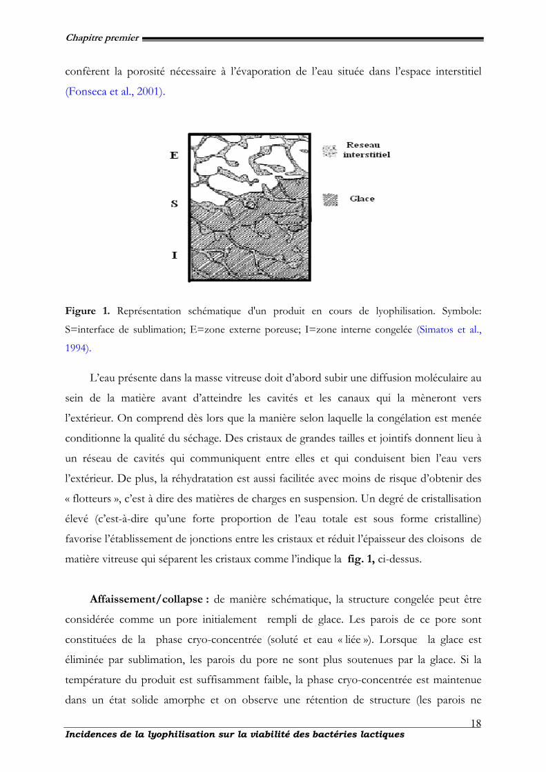
Chapitre premier
Incidences de la lyophilisation sur la viabilité des bactéries lactiques
18
confèrent la porosité nécessaire à l’évaporation de l’eau située dans l’espace interstitiel
(Fonseca et al., 2001).
Figure 1. Représentation schématique d'un produit en cours de lyophilisation. Symbole:
S=interface de sublimation; E=zone externe poreuse; I=zone interne congelée (Simatos et al.,
1994).
L’eau présente dans la masse vitreuse doit d’abord subir une diffusion moléculaire au
sein de la matière avant d’atteindre les cavités et les canaux qui la mèneront vers
l’extérieur. On comprend dès lors que la manière selon laquelle la congélation est menée
conditionne la qualité du séchage. Des cristaux de grandes tailles et jointifs donnent lieu à
un réseau de cavités qui communiquent entre elles et qui conduisent bien l’eau vers
l’extérieur. De plus, la réhydratation est aussi facilitée avec moins de risque d’obtenir des
« flotteurs », c’est à dire des matières de charges en suspension. Un degré de cristallisation
élevé (c’est-à-dire qu’une forte proportion de l’eau totale est sous forme cristalline)
favorise l’établissement de jonctions entre les cristaux et réduit l’épaisseur des cloisons de
matière vitreuse qui séparent les cristaux comme l’indique la fig. 1, ci-dessus.
Affaissement/collapse : de manière schématique, la structure congelée peut être
considérée comme un pore initialement rempli de glace. Les parois de ce pore sont
constituées de la phase cryo-concentrée (soluté et eau « liée »). Lorsque la glace est
éliminée par sublimation, les parois du pore ne sont plus soutenues par la glace. Si la
température du produit est suffisamment faible, la phase cryo-concentrée est maintenue
dans un état solide amorphe et on observe une rétention de structure (les parois ne
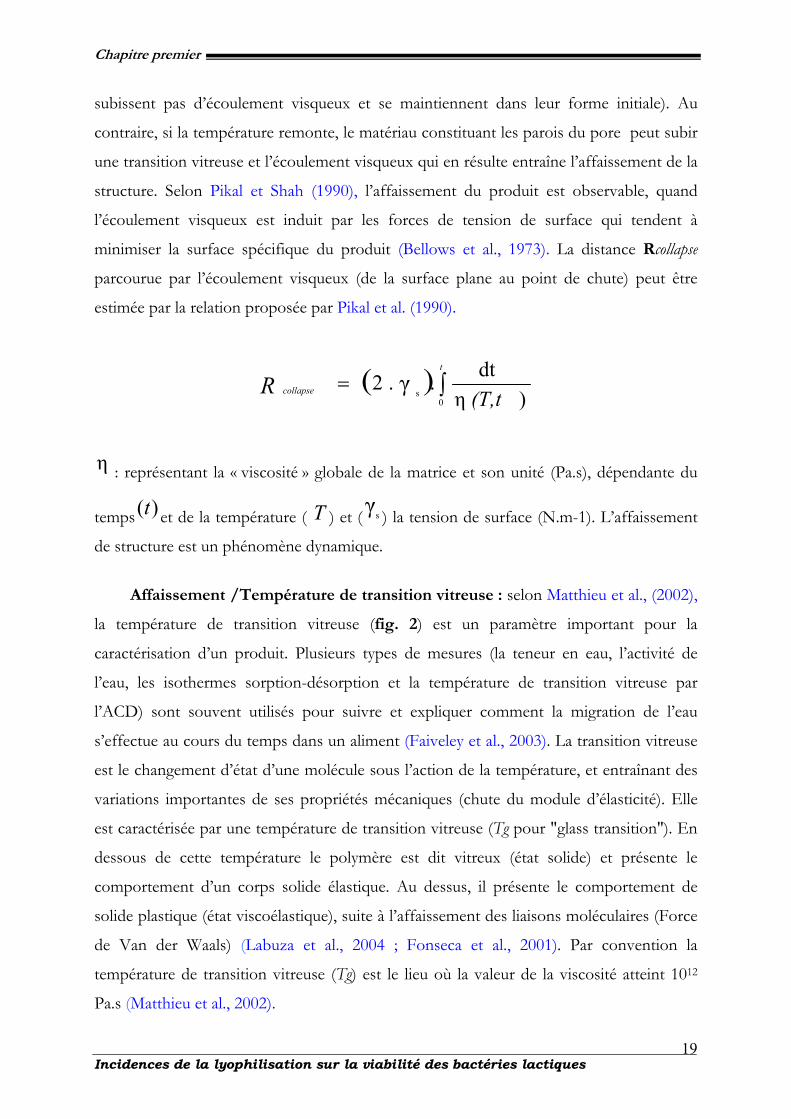
Chapitre premier
Incidences de la lyophilisation sur la viabilité des bactéries lactiques
19
subissent pas d’écoulement visqueux et se maintiennent dans leur forme initiale). Au
contraire, si la température remonte, le matériau constituant les parois du pore peut subir
une transition vitreuse et l’écoulement visqueux qui en résulte entraîne l’affaissement de la
structure. Selon Pikal et Shah (1990), l’affaissement du produit est observable, quand
l’écoulement visqueux est induit par les forces de tension de surface qui tendent à
minimiser la surface spécifique du produit (Bellows et al., 1973). La distance Rcollapse
parcourue par l’écoulement visqueux (de la surface plane au point de chute) peut être
estimée par la relation proposée par Pikal et al. (1990).
( ) ∫=t
collapse (T,tR0
s )ηdt..2 γ
η : représentant la « viscosité » globale de la matrice et son unité (Pa.s), dépendante du
temps )(t et de la température ( Τ ) et (γs ) la tension de surface (N.m-1). L’affaissement
de structure est un phénomène dynamique.
Affaissement /Température de transition vitreuse : selon Matthieu et al., (2002),
la température de transition vitreuse (fig. 2) est un paramètre important pour la
caractérisation d’un produit. Plusieurs types de mesures (la teneur en eau, l’activité de
l’eau, les isothermes sorption-désorption et la température de transition vitreuse par
l’ACD) sont souvent utilisés pour suivre et expliquer comment la migration de l’eau
s’effectue au cours du temps dans un aliment (Faiveley et al., 2003). La transition vitreuse
est le changement d’état d’une molécule sous l’action de la température, et entraînant des
variations importantes de ses propriétés mécaniques (chute du module d’élasticité). Elle
est caractérisée par une température de transition vitreuse (Tg pour "glass transition″). En
dessous de cette température le polymère est dit vitreux (état solide) et présente le
comportement d’un corps solide élastique. Au dessus, il présente le comportement de
solide plastique (état viscoélastique), suite à l’affaissement des liaisons moléculaires (Force
de Van der Waals) (Labuza et al., 2004 ; Fonseca et al., 2001). Par convention la
température de transition vitreuse (Tg) est le lieu où la valeur de la viscosité atteint 1012
Pa.s (Matthieu et al., 2002).
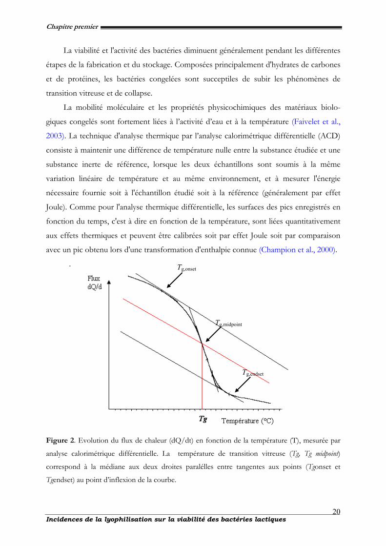
Chapitre premier
Incidences de la lyophilisation sur la viabilité des bactéries lactiques
20
La viabilité et l'activité des bactéries diminuent généralement pendant les différentes
étapes de la fabrication et du stockage. Composées principalement d'hydrates de carbones
et de protéines, les bactéries congelées sont succeptiles de subir les phénomènes de
transition vitreuse et de collapse.
La mobilité moléculaire et les propriétés physicochimiques des matériaux biolo-
giques congelés sont fortement liées à l’activité d’eau et à la température (Faivelet et al.,
2003). La technique d'analyse thermique par l’analyse calorimétrique différentielle (ACD)
consiste à maintenir une différence de température nulle entre la substance étudiée et une
substance inerte de référence, lorsque les deux échantillons sont soumis à la même
variation linéaire de température et au même environnement, et à mesurer l'énergie
nécessaire fournie soit à l'échantillon étudié soit à la référence (généralement par effet
Joule). Comme pour l'analyse thermique différentielle, les surfaces des pics enregistrés en
fonction du temps, c'est à dire en fonction de la température, sont liées quantitativement
aux effets thermiques et peuvent être calibrées soit par effet Joule soit par comparaison
avec un pic obtenu lors d'une transformation d'enthalpie connue (Champion et al., 2000).
.
Figure 2. Evolution du flux de chaleur (dQ/dt) en fonction de la température (T), mesurée par
analyse calorimétrique différentielle. La température de transition vitreuse (Tg, Tg midpoint)
correspond à la médiane aux deux droites paralélles entre tangentes aux points (Tgonset et
Tgendset) au point d’inflexion de la courbe.
Tg,onset
Tg,midpoint
Tg,endset
Chapitre premier
Incidences de la lyophilisation sur la viabilité des bactéries lactiques
21
Ces diagrammes sont habituellement établis avec des mesures effectuées par analyse
calorimétrique différentielle (ACD). Associés aux isothermes de sorption d'eau des
produits secs, ils sont souvent employés pour expliquer la stabilité de ces produits
pendant le stockage (Roos et al., 1991).
La transition vitreuse est aussi définie comme le changement d’état d’une molécule
sous l’action de la température et entraînant des variations importantes de ses propriétés
mécaniques (chute du module d’élasticité). Elle est caractérisée par une température de
transition vitreuse (Tg pour “glass transition”) (Hausman et al., 2005). En revanche, le
phénomène d’affaissement intervient dans la couche sèche, en cours de dessiccation.
L’étude des propriétés physiques susmentionnées réalisée sur les bactéries lactiques est
susceptible d’expliquer certains changements qui se produisent pendant leur traitement et
leur stockage
I-2.2.2. Facteurs agissants sur la conservation des cellules séchées
La maîtrise des procédés de congélation ou de lyophilisation ne doit pas se borner
aux conditions de changements de l’état de l’eau ; le succès est intimement lié à l’état dans
lequel se trouve le produit biologique au moment du traitement. Les nombreux facteurs
qui influencent directement la survie des souches sont représentés sur la figure 3 :
facteurs physiques de refroidissement, de congélation et de décongélation ou de
dessiccation mais aussi facteurs biologiques d’adaptabilité du microorganisme au
traitement (Besson et al., 1987 ; Roméo et al., 2001). Le nombre et la diversité de ces
facteurs expliquent la difficulté d’obtenir des résultats reproductibles de la survie d’un
microorganisme, d’autant plus qu’il est mal aisé de les étudier isolément. D’autres facteurs
comme le rayonnement U.V ou solaire et l’oxygène favorisent les réactions d’oxydation.
Pour une conservation optimale, un conditionnement sous vide ou sous atmosphère
inerte (azote pur) en flacon scellé ou bouché hermétiquement est recommandé (King et
al., 1998). La perte de viabilité des cellules sèches est une conséquence des dommages
causés aux cellules au niveau de différentes cibles, à savoir la paroi cellulaire, la membrane
cellulaire et l'ADN (Teixeira et al. , 1995a). Le séchage est responsable de différentes
formes de dommages cellulaires, principalement en raison des changements dans l'état
physique des lipides membranaires et dans la structure des protéines sensibles qui
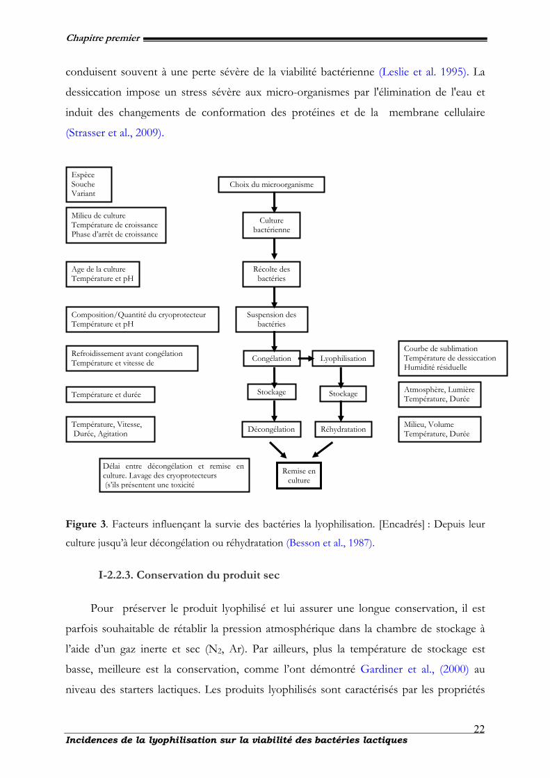
Chapitre premier
Incidences de la lyophilisation sur la viabilité des bactéries lactiques
22
conduisent souvent à une perte sévère de la viabilité bactérienne (Leslie et al. 1995). La
dessiccation impose un stress sévère aux micro-organismes par l'élimination de l'eau et
induit des changements de conformation des protéines et de la membrane cellulaire
(Strasser et al., 2009).
Figure 3. Facteurs influençant la survie des bactéries la lyophilisation. [Encadrés] : Depuis leur
culture jusqu’à leur décongélation ou réhydratation (Besson et al., 1987).
I-2.2.3. Conservation du produit sec
Pour préserver le produit lyophilisé et lui assurer une longue conservation, il est
parfois souhaitable de rétablir la pression atmosphérique dans la chambre de stockage à
l’aide d’un gaz inerte et sec (N2, Ar). Par ailleurs, plus la température de stockage est
basse, meilleure est la conservation, comme l’ont démontré Gardiner et al., (2000) au
niveau des starters lactiques. Les produits lyophilisés sont caractérisés par les propriétés
Espèce Souche Variant
Milieu de culture Température de croissance Phase d’arrêt de croissance
Age de la culture Température et pH
Composition/Quantité du cryoprotecteur Température et pH
Refroidissement avant congélation Température et vitesse de
Température et durée
Température, Vitesse, Durée, Agitation
Délai entre décongélation et remise en culture. Lavage des cryoprotecteurs (s’ils présentent une toxicité
Choix du microorganisme
Culture bactérienne
Récolte des bactéries
Suspension des bactéries
Congélation
Stockage
Décongélation
Lyophilisation
Stockage
Réhydratation
Remise en culture
Courbe de sublimation Température de dessiccation Humidité résiduelle
Atmosphère, Lumière Température, Durée
Milieu, Volume Température, Durée
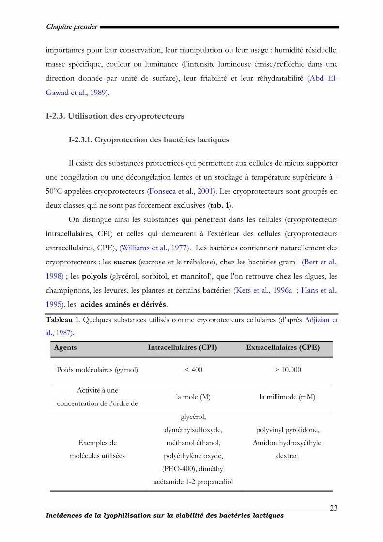
Chapitre premier
Incidences de la lyophilisation sur la viabilité des bactéries lactiques
23
importantes pour leur conservation, leur manipulation ou leur usage : humidité résiduelle,
masse spécifique, couleur ou luminance (l’intensité lumineuse émise/réfléchie dans une
direction donnée par unité de surface), leur friabilité et leur réhydratabilité (Abd El-
Gawad et al., 1989).
I-2.3. Utilisation des cryoprotecteurs
I-2.3.1. Cryoprotection des bactéries lactiques
Il existe des substances protectrices qui permettent aux cellules de mieux supporter
une congélation ou une décongélation lentes et un stockage à température supérieure à -
50°C appelées cryoprotecteurs (Fonseca et al., 2001). Les cryoprotecteurs sont groupés en
deux classes qui ne sont pas forcement exclusives (tab. 1).
On distingue ainsi les substances qui pénètrent dans les cellules (cryoprotecteurs
intracellulaires, CPI) et celles qui demeurent à l’extérieur des cellules (cryoprotecteurs
extracellulaires, CPE), (Williams et al., 1977). Les bactéries contiennent naturellement des
cryoprotecteurs : les sucres (sucrose et le tréhalose), chez les bactéries gram+ (Bert et al.,
1998) ; les polyols (glycérol, sorbitol, et mannitol), que l'on retrouve chez les algues, les
champignons, les levures, les plantes et certains bactéries (Kets et al., 1996a ; Hans et al.,
1995), les acides aminés et dérivés.
Tableau 1. Quelques substances utilisés comme cryoprotecteurs cellulaires (d’après Adjizian et
al., 1987).
Agents Intracellulaires (CPI) Extracellulaires (CPE)
Poids moléculaires (g/mol) < 400 > 10.000
Activité à une
concentration de l’ordre de la mole (M) la millimode (mM)
Exemples de
molécules utilisées
glycérol,
dyméthylsulfoxyde,
méthanol éthanol,
polyéthylène oxyde,
(PEO-400), diméthyl
acétamide 1-2 propanediol
polyvinyl pyrolidone,
Amidon hydroxyéthyle,
dextran
Chapitre premier
Incidences de la lyophilisation sur la viabilité des bactéries lactiques
24
(bétaïne et carnitine), chez plusieurs groupes de bactéries (Csonka, 1989) et l'acide
tetrahydroxypyrimidine carboxylique (hydroxyectoïne) chez les bactéries halophiliques
(Del Moral et al., 1994), le glutamate, la glutamine, proline et l’alanine chez Escherichia coli,
Staphylococcus aureus, Pseudomonas et d'autres micro-organismes (Jewell et al., 1991) et les
acides aminés bêta chez les bactéries méthanogènes (Lai et al., 1991).
I-2.3.2. Mécanismes d’action des cryoprotecteurs
Les mécanismes de la cryoprotection sont l’objet de nombreuses études de nos
jours. Les modes d’action les plus probables sont : un accroissement du volume de la
phase liquide interstitielle, à une température donnée, par dépression du point de
congélation commençante et donc réduction des effets liés à un refroidissement lent ; une
diminution de la taille des cristaux de glace par abaissement de la température de
nucléation et une réduction de leur vitesse de croissance et une stabilisation de la
configuration native des protéines (Leslie et al., 1995). Lorsque les premiers cristaux de
glace apparaissent, les CPE se concentrent uniquement à l’extérieur des cellules,
contrairement aux autres solutés qui se concentrent à l’intérieur et l’extérieur des cellules.
La perte d’eau des cellules conduit donc à une concentration en autre soluté, plus faible
qu’en absence de CPE, minimisant ainsi les effets de l’accroissement de concentration de
la solution interstitielle (Fonseca et al., 2004). Au niveau des membranes cellulaires
séchées, des sucres comme le tréhalose et le saccharose peuvent remplacer les molécules
d'eau dans leur liaison avec les groupes polaires des phospholipides empêchant ainsi les
dommages durant la réhydratation (Leslie et al., 1995 ; Aguilera & Karel, 1997). Le
tréhalose et le saccharose stabilisent la structure des protéines intracellulaires par ce
phénomène de remplacement de l'eau (Carpenter et al., 1993). D’autres études ont montré
que les cryoprotecteurs étaient indispensables lors de la congélation quelles que soient les
courbes de refroidissement. Tous les cryoprotecteurs ne pénètrent pas dans les cellules. Il
est d'usage de les séparer en cryoprotecteurs non diffusants (la plupart des sucres ajoutés)
et diffusants (le glycérol, l'éthylène glycol, par exemple) (Pegg et al., 1988). Les
cryoprotecteurs diffusants vont se substituer à une partie de l'eau intracellulaire et
permettre ainsi une déshydratation partielle des cellules en plus de limiter la formation de
cristaux de glace intracellulaire. Ils réduisent également la vitesse de croissance de ces
Chapitre premier
Incidences de la lyophilisation sur la viabilité des bactéries lactiques
25
cristaux, abaissent la température de solidification de l'eau intracellulaire, tel un « antigel »,
et modifient la forme des cristaux de glace (Hey et al. 1998 ; Palasz et al., 1996). Quand
aux cryoprotecteurs pénétrants, ils protègent aussi au cours de la congélation et de la
décongélation en réduisant la taille des cristaux de glace et en induisant des formes de
cristaux moins traumatisantes. Ils permettent, via l'augmentation de la viscosité du milieu,
la diminution de la rapidité des mouvements de l'eau et de chocs osmotiques importants.
De plus, ils permettent, via l'augmentation de la pression osmotique, la réduction de la
quantité de cryoprotecteurs pénétrants nécessaires à une bonne conservation (Meryman,
1974).
I-2.4. Acides gras et oxydation cellulaire
I-2.4.1. Rôles des acides gras au niveau des bactéries
Les lipides membranaires sont le seuil d’intenses modifications en vue de maintenir
une fluidité et ainsi assurer la perméabilité des membranes cellulaires.
Tableau 2. Effet de la température de culture sur la composition en acides gras (%) chez L.
plantarum (Russell et al., 1989)
Acides gras Noms communs Noms
scientifiques 10°C 30°C 40°C
C16 :1 Acide
palmitoléique
9-hexadecenoic
acid 11 18 2
C16:0 Acide palmitique hexadecanoic
acid 15 30 56
C17 :0cyc Acide margarique acide
heptadécanoïque<1 tr tr
C18 :1 Acide oléique acide 9-
octadecenoique56 34 11
C18:0 Acide stéarique acide
octadecanoique10 4 13
C19 :0cyc Acide
nonadécyclique
acide
nonadécanoïque6 8 16
cyc: cyclopropane fatty acid ; tr : trace.
Chapitre premier
Incidences de la lyophilisation sur la viabilité des bactéries lactiques
26
Cette activité est indispensable pour le bon fonctionnement des cellules en cas de
stress (Russell et al., 1989). D’autres travaux (tab. 2), similaires effectués par Russell et al.
(1995) ont mis en évidence l’impact de la température d’incubation des cultures sur L.
plantarum. Cette expérience a montré que l’augmentation de la synthèse des acides gras
insaturés (C18:1 principalement) d’une part et d’autre part une augmentation de la synthèse
des C16:0, afin de réguler la fluidité membranaire était fortement liée à une élévation de la
température d’incubation.
Pour L. monocytogenes, une baisse de la synthèse des C15:0 et une augmentation de la
synthèse des C 17:0 ainsi que des C 18:1 a été mis en évidence dans les conditions similaires
(Russell et al., 1995).
I-2.4.2. Mécanismes d’oxydation des lipides
Après leur lyophilisation, au cours du stockage, les bactéries lactiques subissent
d’intenses phénomènes de dégradations, liés pour la plupart à l’oxydation des lipides
membranaires. Ces réactions d’oxydations sont les principaux facteurs qui déterminent la
durée de vie des cellules contenant ces lipides. Les produits (substrats) de ces réactions
sont principalement des acides gras insaturés (Rousch et al., 2003). Leur degré d’oxydation
et la vitesse de celui-ci sont fonction de leur insaturation. Une différence fondamentale
existe entre les acides gras saturés et les acides gras polyinsaturés. Tandis que les acides
gras saturés s’oxydent à une température supérieure à 60°C, les acides gras polyinsaturés
s’oxydent même lors de l’entreposage des aliments à l’état congelé (Niki et al., 2005).
Plusieurs mécanismes naturels de contrôle de l’oxydation se déroulent au niveau des
cellules. Ces mécanismes ont pour but de prévenir la destruction oxydative des lipides
membranaires, des protéines et des acides nucléiques (Pereira et al., 2003). Les
mécanismes de régulation des systèmes pro-oxydants et anti-oxydants sont mis en place
pour maintenir l’équilibre entre les facteurs impliqués dans les réactions d’oxydation. Mais
cette régulation des facteurs pro- et anti-oxydants est perturbée durant les processus de
transformation et de stockage des produits, ce qui favorise le développement des
réactions d’oxydation (Niki et al., 2000). Selon les travaux effectués par Steinberg et
collaborateurs en 1989, les facteurs qui influencent, ou qui initient, l’oxydation des lipides
sont de deux types : - des facteurs intrinsèques tels que la composition en acides gras des
Chapitre premier
Incidences de la lyophilisation sur la viabilité des bactéries lactiques
27
lipides (nombre et position des insaturations), la présence de pro-oxydants (ions
métalliques, enzymes, etc.) ou d’antioxydants naturels (tocophérols, caroténoïdes, etc.) et -
des facteurs externes tels que la température, la lumière, la pression partielle en oxygène,
l’activité de l’eau, les conditions de stockage et de transformation (Steinberg et al., 1989).
La classification de l’oxydation est fonction des agents initiateurs, ainsi on distingue 3
types d’oxydation des lipides :
• l’auto-oxydation catalysée par la température, les ions métalliques et les radicaux libres,
• la photo-oxydation, initiée par la lumière en présence de photosensibilisateurs,
• l’oxydation enzymatique initiée par la présence des enzymes d'oxydation.
Parmi les travaux effectués sur les phénomènes d’oxydation, celui des lipides pose de
sérieux problèmes qui résident dans la dégradation des propriétés biochimiques, organo-
leptiques (formation de composés volatils d’odeur désagréable : rancissement) d’une part
et d’autre part dans les qualités nutritionnelles (par interaction des produits d'oxydation
avec les acides aminés) des produits lyophilisés (Takebe et al., 2002). Les réactions
d'oxydation conduisent à la formation de radicaux libres puis d’hydroperoxydes,
composés intermédiaire, qui à leur tour se décomposent en donnant naissance à des
composés volatils, des composés furaniques et surtout des aldéhydes saturés et insaturés
qui sont des molécules cancérigènes et toxiques aussi bien pour les bactéries que pour
l’homme (Chena et al., 2006). Ces réactions nécessitent la présence d’oxygène, qui doit
être activé en oxygène singulet soit sous l’action des photons (lumière), soit de radicaux
libres déjà présents dans le milieu, soit d’enzymes (peroxydases, lipoxygénases, etc.). Elles
se développent en milieu anhydre ou à très faible activité de l’eau pour des valeurs (aw <
0.2), dans le cas de l’auto-oxydation et à l’interface eau-milieu apolaire dans le cas de
l’oxydation catalysée par les enzymes (Bradley et al., 1992).
I-2.4.3. Autooxydation
Dans la nature, il existe un certain nombre d'autooxydations, dont celle du
dioxygène (O2) qui donne naissance à l'ozone (O3). Dans l'organisme, l'auto-oxydation ou
oxydation spontanée de certains composants, forme essentiellement des peroxydes,
dégradés ensuite par des enzymes spécifiques (fig. 4) : les peroxydases.
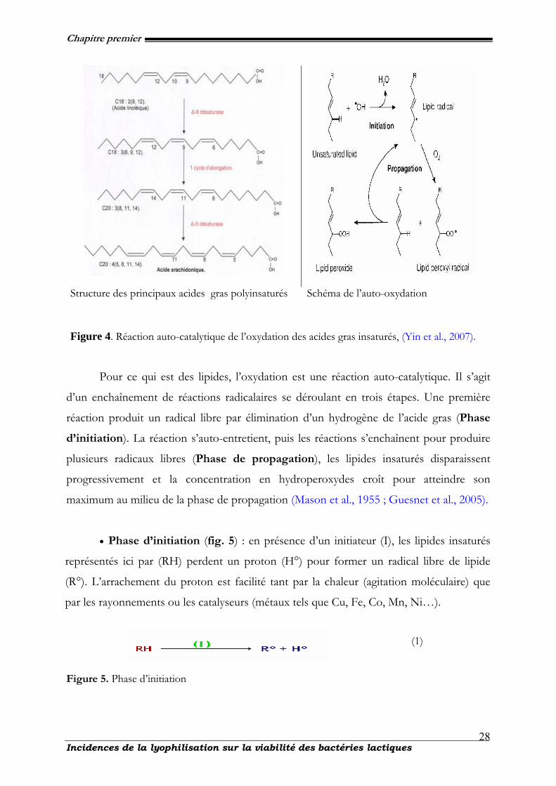
Chapitre premier
Incidences de la lyophilisation sur la viabilité des bactéries lactiques
28
Structure des principaux acides gras polyinsaturés
Schéma de l’auto-oxydation
Figure 4. Réaction auto-catalytique de l’oxydation des acides gras insaturés, (Yin et al., 2007).
Pour ce qui est des lipides, l’oxydation est une réaction auto-catalytique. Il s’agit
d’un enchaînement de réactions radicalaires se déroulant en trois étapes. Une première
réaction produit un radical libre par élimination d’un hydrogène de l’acide gras (Phase
d’initiation). La réaction s’auto-entretient, puis les réactions s’enchaînent pour produire
plusieurs radicaux libres (Phase de propagation), les lipides insaturés disparaissent
progressivement et la concentration en hydroperoxydes croît pour atteindre son
maximum au milieu de la phase de propagation (Mason et al., 1955 ; Guesnet et al., 2005).
• Phase d’initiation (fig. 5) : en présence d’un initiateur (I), les lipides insaturés
représentés ici par (RH) perdent un proton (H°) pour former un radical libre de lipide
(R°). L’arrachement du proton est facilité tant par la chaleur (agitation moléculaire) que
par les rayonnements ou les catalyseurs (métaux tels que Cu, Fe, Co, Mn, Ni…).
(1)
Figure 5. Phase d’initiation
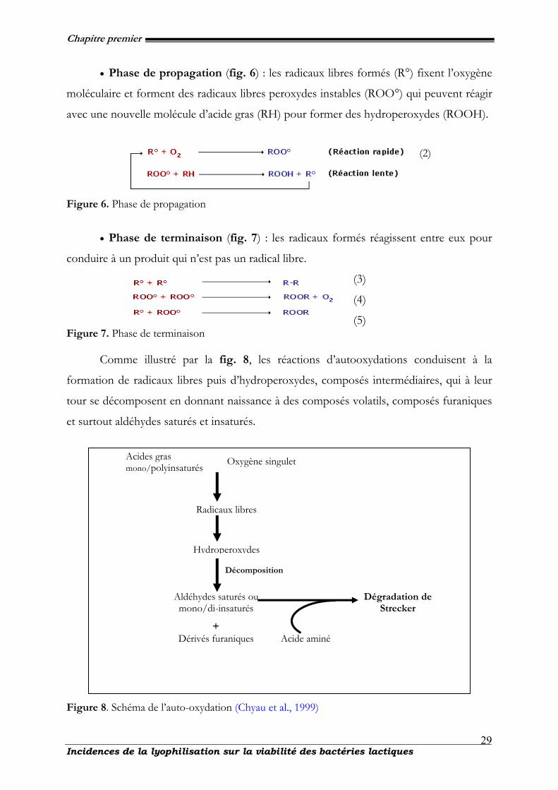
Chapitre premier
Incidences de la lyophilisation sur la viabilité des bactéries lactiques
29
• Phase de propagation (fig. 6) : les radicaux libres formés (R°) fixent l’oxygène
moléculaire et forment des radicaux libres peroxydes instables (ROO°) qui peuvent réagir
avec une nouvelle molécule d’acide gras (RH) pour former des hydroperoxydes (ROOH).
(2)
Figure 6. Phase de propagation
• Phase de terminaison (fig. 7) : les radicaux formés réagissent entre eux pour
conduire à un produit qui n’est pas un radical libre.
(3)
(4)
(5) Figure 7. Phase de terminaison
Comme illustré par la fig. 8, les réactions d’autooxydations conduisent à la
formation de radicaux libres puis d’hydroperoxydes, composés intermédiaires, qui à leur
tour se décomposent en donnant naissance à des composés volatils, composés furaniques
et surtout aldéhydes saturés et insaturés.
Figure 8. Schéma de l’auto-oxydation (Chyau et al., 1999)
Acides gras mono/polyinsaturés
Oxygène singulet
Radicaux libres
Hydroperoxydes
Dérivés furaniques
Aldéhydes saturés ou mono/di-insaturés
Décomposition
Dégradation de Strecker
+ Acide aminé
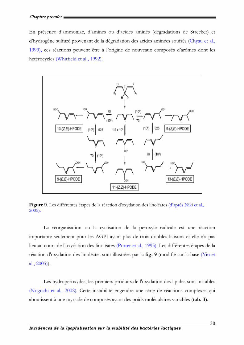
Chapitre premier
Incidences de la lyophilisation sur la viabilité des bactéries lactiques
30
En présence d’ammoniac, d’amines ou d’acides aminés (dégradations de Strecker) et
d’hydrogène sulfuré provenant de la dégradation des acides aminées soufrés (Chyau et al.,
1999), ces réactions peuvent être à l’origine de nouveaux composés d’arômes dont les
hétérocycles (Whitfield et al., 1992).
Figure 9. Les différentes étapes de la réaction d'oxydation des linoléates (d’après Niki et al., 2005).
La réorganisation ou la cyclisation de la peroxyle radicale est une réaction
importante seulement pour les AGPI ayant plus de trois doubles liaisons et elle n'a pas
lieu au cours de l'oxydation des linoléates (Porter et al., 1995). Les différentes étapes de la
réaction d'oxydation des linoléates sont illustrées par la fig. 9 (modifié sur la base (Yin et
al., 2005)).
Les hydroperoxydes, les premiers produits de l'oxydation des lipides sont instables
(Noguchi et al., 2002). Cette instabilité engendre une série de réactions complexes qui
aboutissent à une myriade de composés ayant des poids moléculaires variables (tab. 3).
Chapitre premier
Incidences de la lyophilisation sur la viabilité des bactéries lactiques
31
Tableau 3. Peroxidation des lipides (produits des linoléates). Les caractéristiques des réactions
chimiques et les produits spécifiques issus de l'oxydation des linoléates par différents
mécanismes.
Isomères des HPODE Type Caractéristiques
Régio Stéreo Enantio
Enzymatique (15-LOX) catalytique spécifique 13 cis, trans S
Non enzymatique, oxydation
à chaîne radicalaire (LO2*)
réaction en chaîne
9 , 13
cis, trans,
trans, trans
9-ct=13-ct,
9-tt=13-tt
R = S
(racemic)
Non enzymatique, oxydation
non radicalaire (1O2) stochiométrie
9, 10, 12,
13 cis, trans
I-2.4.4. Photooxydation
Comme évoqué pour l’auto-oxydation, la photooxydation qui est une oxydation
induite en grande partie par la présence de la lumière et de photosensibilisateurs tels que
les hémoprotéines, la chlorophylle ou la riboflavine est aussi une voie importante de
production d’hydroperoxydes en présence d’oxygène. Pour ce qui est du mécanisme, les
photosensibilisateurs (Sens) absorberaient l’énergie lumineuse et afin d’atteindre un état
de triplet excité (Sens3). Ces photosensibilisateurs interviennent dans l’oxydation des
lipides selon deux types de mécanismes (Frankel et al., 1979 ; Hatch et al., 2003)
Le premier type de mécanisme est induit par des photosensibilisateurs (type I),
telle que la riboflavine, qui agissent comme des radicaux libres initiateurs. Dans
leur état triplet, elles arrachent un atome d’hydrogène ou un électron aux
molécules lipidiques pour former un radical capable de réagir avec l’oxygène (1).
Sens3 + RH —› Sens H + R° (1),
Selon le second mécanisme, les molécules photosensibles de type II, telles que la
chlorophylle et l’érythrosine, réagissent dans leur état excité (Sens3) avec l’oxygène
triplet auquel elles transfèrent leur énergie pour donner de l’oxygène singulet (1O2)
(2).
Sens3 + 3O2 —› 1O2 + Sens (2),
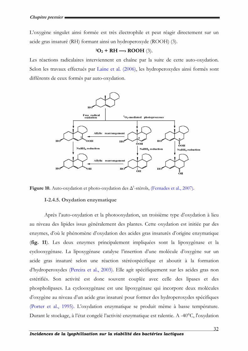
Chapitre premier
Incidences de la lyophilisation sur la viabilité des bactéries lactiques
32
L’oxygène singulet ainsi formée est très électrophile et peut réagir directement sur un
acide gras insaturé (RH) formant ainsi un hydroperoxyde (ROOH) (3). 1O2 + RH —› ROOH (3).
Les réactions radicalaires interviennent en chaîne par la suite de cette auto-oxydation.
Selon les travaux effectués par Laine et al. (2006), les hydroperoxydes ainsi formés sont
différents de ceux formés par auto-oxydation.
Figure 10. Auto-oxydation et photo-oxydation des ∆5-stérols, (Fernades et al., 2007).
I-2.4.5. Oxydation enzymatique
Après l’auto-oxydation et la photooxydation, un troisième type d’oxydation à lieu
au niveau des lipides issus généralement des plantes. Cette oxydation est initiée par des
enzymes, d’où le phénomène d’oxydation des acides gras insaturés d’origine enzymatique
(fig. 11). Les deux enzymes principalement impliquées sont la lipoxygénase et la
cyclooxygénase. La lipoxygénase catalyse l’insertion d’une molécule d’oxygène sur un
acide gras insaturé selon une réaction stéréospécifique et aboutit à la formation
d’hydroperoxydes (Pereira et al., 2003). Elle agit spécifiquement sur les acides gras non
estérifiés. Son activité est donc souvent couplée avec celle des lipases et des
phospholipases. La cyclooxygénase est une lipoxygénase qui incorpore deux molécules
d’oxygène au niveau d’un acide gras insaturé pour former des hydroperoxydes spécifiques
(Porter et al., 1995). L’oxydation enzymatique se produit même à basse température.
Durant le stockage, à l’état congelé l’activité enzymatique est ralentie. A -40°C, l'oxydation
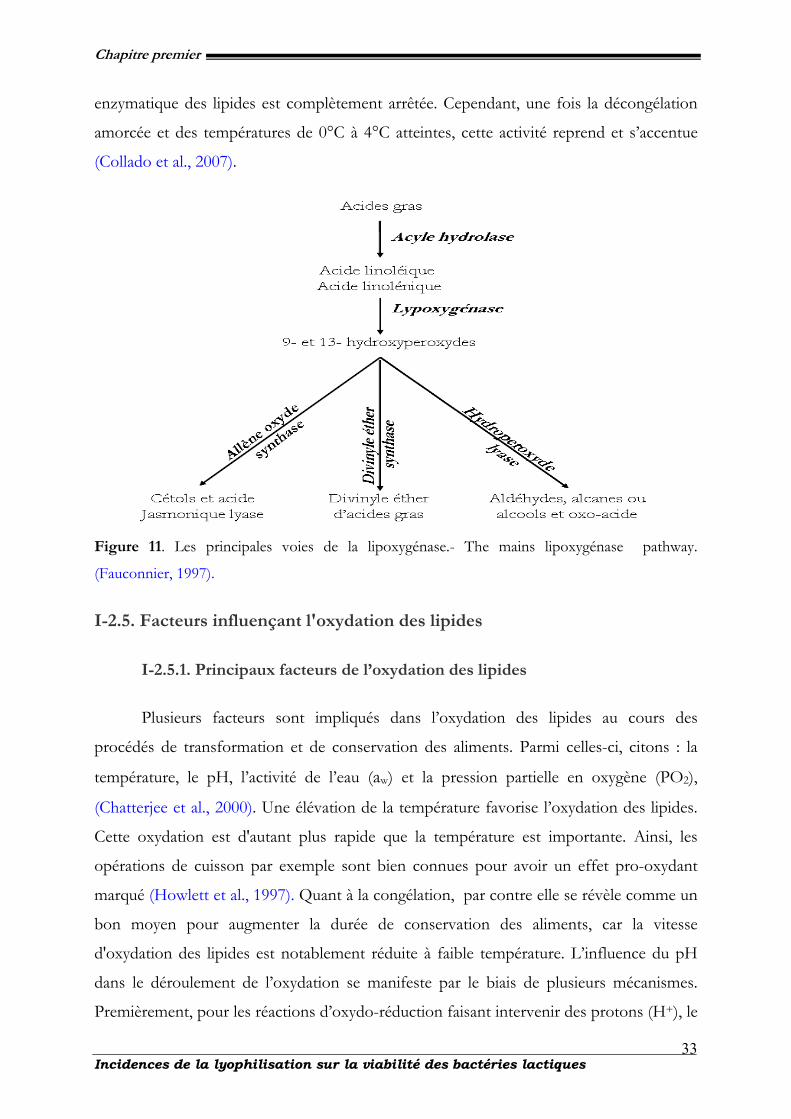
Chapitre premier
Incidences de la lyophilisation sur la viabilité des bactéries lactiques
33
enzymatique des lipides est complètement arrêtée. Cependant, une fois la décongélation
amorcée et des températures de 0°C à 4°C atteintes, cette activité reprend et s’accentue
(Collado et al., 2007).
Figure 11. Les principales voies de la lipoxygénase.- The mains lipoxygénase pathway.
(Fauconnier, 1997).
I-2.5. Facteurs influençant l'oxydation des lipides
I-2.5.1. Principaux facteurs de l’oxydation des lipides
Plusieurs facteurs sont impliqués dans l’oxydation des lipides au cours des
procédés de transformation et de conservation des aliments. Parmi celles-ci, citons : la
température, le pH, l’activité de l’eau (aw) et la pression partielle en oxygène (PO2),
(Chatterjee et al., 2000). Une élévation de la température favorise l’oxydation des lipides.
Cette oxydation est d'autant plus rapide que la température est importante. Ainsi, les
opérations de cuisson par exemple sont bien connues pour avoir un effet pro-oxydant
marqué (Howlett et al., 1997). Quant à la congélation, par contre elle se révèle comme un
bon moyen pour augmenter la durée de conservation des aliments, car la vitesse
d'oxydation des lipides est notablement réduite à faible température. L’influence du pH
dans le déroulement de l’oxydation se manifeste par le biais de plusieurs mécanismes.
Premièrement, pour les réactions d’oxydo-réduction faisant intervenir des protons (H+), le
Chapitre premier
Incidences de la lyophilisation sur la viabilité des bactéries lactiques
34
potentiel redox décroît linéairement avec le pH. Un pH acide favorise donc la réaction
d’oxydation, en particulier quand les espèces pro-oxydantes (ions des métaux de
transition) ou antioxydantes (acide ascorbique, BHT et BHA) solubles en phase aqueuse
sont présentes (Hatch et al., 2003). Le pH intervient également dans la solubilité des
composés impliqués dans l’initiation de la réaction. Ainsi, plus le pH est bas, plus la
solubilité et le potentiel redox de ces ions métalliques, et donc leur réactivité vis à vis des
molécules oxydables sont élevées. Dans le cas des tissus musculaires, un pH bas favorise
la dénaturation des hémoprotéines et la libération du fer qui est un agent pro-oxydant.
L’activité de l’eau d’un système influence les réactions d’oxydation des lipides. En effet,
l'eau permet la mobilisation des substances pro-oxydantes ou antioxydantes (Berger et al.,
2001). En général, une activité d’eau (aw) comprise entre 0,2 et 0,3 correspond aux vitesses
d'oxydation les plus faibles. Ces valeurs correspondent à la formation d'une couche
monomoléculaire d'eau autour des constituants. Une aw comprise entre 0,6 et 0,8
correspond aux vitesses d'oxydation les plus grandes.
Par contre, les réactions d'oxydation enzymatique des lipides sont fortement
ralenties quand l'activité de l'eau est inférieure à 0,6. La concentration d'oxygène (pression
partielle en oxygène) dans l'espace environnant le produit et dans le produit lui-même
influence la vitesse d'oxydation des lipides. Elle intervient également au niveau de la
nature des produits secondaires formés par décomposition des hydroperoxydes. Son
incidence est donc à la fois sur la durée de conservation du produit et sur la nature des
odeurs perçues quand le produit est oxydé. La relation entre vitesse d'oxydation et
pression partielle en oxygène dépend de plusieurs facteurs comme l'activité de l'eau, la
température, la nature des catalyseurs. Quand la concentration en oxygène est
suffisamment élevée, la vitesse d'oxydation est indépendante de cette concentration.
Inversement, quand la concentration d'oxygène est suffisamment faible, la vitesse
d'oxydation est indépendante de la concentration en substrat et directement
proportionnelle à la concentration d'oxygène.
I-2.5.2. Impacts de l’oxydation des lipides sur les starters
Les dommages causés aux LAB pendant le stockage est également attribuée aux
modifications dans le profil des acides gras (taux d’acides gras saturés et des acides gras
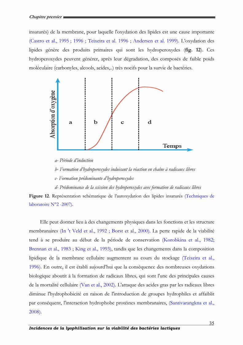
Chapitre premier
Incidences de la lyophilisation sur la viabilité des bactéries lactiques
35
insaturés) de la membrane, pour laquelle l'oxydation des lipides est une cause importante
(Castro et al., 1995 ; 1996 ; Teixeira et al. 1996 ; Andersen et al. 1999). L’oxydation des
lipides génère des produits primaires qui sont les hydroperoxydes (fig. 12). Ces
hydroperoxydes peuvent générer, après leur dégradation, des composés de faible poids
moléculaire (carbonyles, alcools, acides,..) très nocifs pour la survie de bactéries.
a- Période d’induction
b- Formation d’hydroperoxydes induisant la réaction en chaîne à radicaux libres
c- Formation prédominante d’hydroperoxydes
d- Prédominance de la scission des hydroperoxydes avec formation de radicaux libres Figure 12. Représentation schématique de l’autoxydation des lipides insaturés (Techniques de
laboratoire N°2 -2007).
Elle peut donner lieu à des changements physiques dans les fonctions et les structure
membranaires (In 't Veld et al., 1992 ; Borst et al., 2000). La perte rapide de la viabilité
tend à se produire au début de la période de conservation (Korobkina et al., 1982;
Brennan et al., 1983 ; King et al., 1993), tandis que les changements dans la composition
lipidique de la membrane cellulaire augmentent au cours du stockage (Teixeira et al.,
1996). En outre, il est établi aujourd’hui que la conséquence des nombreuses oxydations
biologique aboutit à la formation de radicaux libres, qui sont l'une des principales causes
de la mortalité cellulaire (Van et al., 2002). L'attaque des acides gras par les radicaux libres
diminue l'hydrophobicité en raison de l'introduction de groupes hydrophiles et affaiblit
par conséquent, l'interaction hydrophobe protéines membranaires, (Santivarangkna et al.,
2008).
Chapitre premier
Incidences de la lyophilisation sur la viabilité des bactéries lactiques
36
Par exemple, une diminution de l'activité de l’ ATPase membranaire a été suggérée
comme conséquence du changement de cette interaction (Castro et al., 1996). Les
changements affectent aussi dans la membrane, certains mécanismes de contrôle liés à la
régulation et à la réplication de l'ADN, car l'initiation de la synthèse d'ADN et son
maintien requièrent l’attachement du complexe d’ADN à la membrane cytoplasmique
(Israeli et al., 1975). En outre, les radicaux libres peuvent aussi bien directement
provoquer des dommages de l’ADN (Inouye, 1984 ; Akasaka 1986 ; Hruszkewycz 1988).
Bien qu'il soit plausible d'ajouter un antioxydant pour diminuer l'oxydation des lipides,
l'addition doit être exercé avec soin. Ces méthodes et composés serviront dans la suite à
définir les différentes approches afin d’inhiber l’oxydation cellulaire.
I-2.6. Différentes approches pour inhiber l’oxydation des starters lactiques
lyophilisés au cours du stockage
Le contrôle ou l'inhibition de l'oxydation des lipides est basée sur la maîtrise des
paramètres : température, pH, aw et concentration en oxygène (Coulibaly et al., 2008).
Selon Buettner et al. 1999, les antioxydants susceptibles de protéger les lipides de
l’oxydation peuvent être répartis en deux types : les antioxydants préventifs qui empêchent la
formation d’espèces réactives de l’oxygène ou interceptent les espèces responsables de
l’initiation de la lipoperoxydation et les antioxydants « chain breaking » qui interceptent les
radicaux propagateurs de la peroxydation lipidique et retardent la peroxydation (période
d’induction) (Niki et al., 2000).
I-2.6.1. Mécanismes d’action des antioxydants préventifs
I-2.6.1.1 Les chélateurs des métaux de transition :
Le cycle redox de certains métaux responsable dans la production des radicaux libres
est bloqué par des chélateurs de métaux. Ce sont des protéines telles que la transferrine, la
ferritine, la lactalbumine qui séquestrent le fer, la céruloplasmine et l’albumine qui
séquestrent le cuivre (Mugoli et al., 1993). Notons au passage que les flavonoïdes sont de
bons chélateurs du fer ceci est un des mécanismes de leur activité antioxydante (Van et al.,
1998 ; Morel et al., 1993).
Chapitre premier
Incidences de la lyophilisation sur la viabilité des bactéries lactiques
37
I-2.6.1.2 Les désactivateurs (quencher) de l’oxygène singulet.
Selon Buettner et al. (1999), les « quencher » peuvent agir par désactivation chimique
en se fixant sur une molécule telle qu’un acide gras pour donner un hydroperoxyde : 1O2
+ LOH → LOOH, ou encore par désactivation physique éliminant l’énergie d’excitation
sans changement chimique : 1O2 + β-carotène → O2 + β-carotène. Les caroténoïdes sont
cités particulièrement pour leur efficacité et le lycopène pour sa grande réactivité.
I-2.6.1.3 Elimination des hydroperoxydes
Les hydroperoxydes sont générateurs des radicaux libres, leur élimination prévient
l’oxydation des biomolécules. Les hydroperoxydes (LOOH) peuvent être réduits par des
enzymes. La glutathion peroxydase (GPx) élimine les hydroperoxydes organiques et le
peroxyde d’hydrogène (H2O2).
I-2.6.1.4 Les piégeurs d’oxygène
Ce sont des molécules telles que les sulfites ou l’acide ascorbique.
I-2.6.2 Mécanismes d’action des antioxydants de type « chaîne rompue et
chain breaking »
Cette catégorie d’antioxydants va réagir le plus souvent avec les radicaux peroxyles
(LOO°) ou alcoxyles (LO°) interrompant ainsi la réaction de propagation de la
peroxydation. Il est à noter que ces antioxydants n’inhiberont pas par conséquent
l’autoxydation des lipides par l’oxygène singulet. Ces antioxydants peuvent agir selon deux
mécanismes : - interviennent en donnant de l’hydrogène
- agissent comme des antioxydants sacrifiés.
I-2.6.2.1 Les donneurs d’hydrogène
Ces antioxydants sont principalement des composés phénoliques mono- ou
polyhydroxylés (tocophérols, tocotriènols, BHT, BHA, flavonoïdes). Ils sont susceptibles
de céder un hydrogène au radical alcoxyle et au radical peroxyle Après la réaction
d’oxydation, l’antioxydant est transformé en un radical qui doit être suffisamment stable
pour inhiber la formation d’un autre radical et arrêter ainsi la propagation de la chaîne
Chapitre premier
Incidences de la lyophilisation sur la viabilité des bactéries lactiques
38
radicalaire. Il doit ensuite évoluer vers un produit d’oxydation stable, ce qui conduit à la
consommation de l’antioxydant. Le tocophérol donnera un radical tocophéroxyle qui
évoluera vers un composé d’oxydation non radicalaire tel que la tocophérylquinone ou un
composé de dimérisation ou de polymérisation supérieure.
I-2.6.2.2 Les antioxydants «sacrifiés» :
Le qualificatif de « chain breaking antioxidant sacrificial » employé par Buettner
concerne des molécules, elles mêmes radicalaires, qui réagissent avec les radicaux
peroxyles ou alcoxyles pour donner des produits non radicalaires interrompant ainsi la
propagation de la peroxydation. Deux radicaux sont connus pour se combiner avec les
radicaux peroxyles : le monoxyde d’azote (NO) et l’anion superoxyde (O2-).
LOO + NO→LOONO et
LO + NO→LONO
I-2.6.2.3 Mécanismes d’action mixtes des antioxydants :
Certains antioxydants ont des modes d’action mixtes. Deux exemples peuvent
illustrer ces mécanismes multiples. L’exemple de l’acide ascorbique qui est un
désactivateur de l’oxygène singulet, élimine aussi l’oxygène moléculaire, il est aussi un
donneur d’hydrogène aux radicaux lipidiques et aux radicaux tocophéroxyles pour
régénérer le tocophérol.
Quand aux flavonoïdes tels que les anthocyanines, les catéchines, les flavones, les
flavonols, les isoflavones et les proanthocyanidines ce sont des chélateurs de métaux,
piégeurs d’anions superoxyde et donneurs d’hydrogène. L'utilisation des antioxydants
(tocophérols, polyphénols, flavonoïdes, vitamine E, vitamine C, etc.) est souvent la
méthode la plus courante en industries agroalimentaires pour inhiber l'oxydation des
lipides.
Les antioxydants utilisés sont soit des agents de prévention qui bloquent la phase
d'initiation en réagissant avec les initiateurs de la réaction (O2, lumière, métaux, ...), soit
des agents de terminaison qui bloquent la poursuite de la phase de propagation en
réagissant avec les radicaux libres et les transformant en composés stables (Niki et al.,
2000).
Chapitre premier
Incidences de la lyophilisation sur la viabilité des bactéries lactiques
39
I-2.7. Remerciements
Nous remercions très sincèrement le personnel du CWBI (Centre Wallon de
Biologie Industrielle) pour son assistance et son dévouement ainsi que Maryse, Vincent
Hotte et Dany Trisman de l’unité de Chimie organique de la faculté agronomique de
Gembloux. Je remercie également la république de Côte d’Ivoire pour son soutien
financier.
I-2.8. Conclusions
Il est donc possible de conserver par lyophilisation un grand nombre de bactéries
lactiques, à l’échelle du laboratoire, mais aussi industrielle. Les conditions opératoires qui
permettent d’obtenir une bonne conservation de l’activité métabolique des bactéries sont
relativement bien connues, à l’inverse des mécanismes de leur altération. La résolution des
difficultés, telles que la mauvaise conservation de certaines souches à l’état lyophilisé et la
reprise d’activité souvent trop lente des ferments destinés aux ensemencements directs,
passe certainement par une meilleure compréhension de ces mécanismes. Pour optimiser
la conservation, plusieurs techniques et voies de recherches doivent être menées de
concert. Parmi les plus prometteuses, on peut citer l’étude des modifications subies par
les structures membranaires au cours des traitements de conservation, la recherche de
formulation incorporant des cryoprotecteurs plus efficaces, l’induction des mécanismes
de résistance à ces traitements (par adaptation des bactéries par leur culture dans des
conditions appropriées ou éventuellement par utilisation d’antioxydant afin de bloquer le
phénomène de peroxydation).
I-2.9. Références bibliographiques
Abdel Gawad, A.M., Nassar, A.M. and Hilali, M., 1989. Isolation of Toxoplasma gondii, Isospora felis and Isospora rioolfa from the meat of some farm animals. J. Egypt. Vet. Med. Assoc., 49, 405 - 414.
Aller R. C., 1998. Mobile deltaic and continental shelf muds as suboxic, fluidized bed reactors. Mar. Chem.,
61, 143-155. Bayrock D., and Ingledew W.M., 1997a. Fluidized bed drying of baker's yeast : moisture levels, drying
rates and viability changes during drying. Food. Research. Intern., 30, 407-415. Bayrock D., and Ingledew W.M., 1997b. Mechanism of viability loss during fluidized bed drying of baker's
yeast. Food. Research. Intern., 30, 417-425.
Chapitre premier
Incidences de la lyophilisation sur la viabilité des bactéries lactiques
40
Berger S., Weichert H., Porzel A., Wasternack C., Kühn H., Feussner I., 2001. Enzymatic and non-
enzymatic lipid peroxidation in leaf development. Biochim. Biophys. Acta., 1533, 266-276. Bert P. and Glaasker E. (1998). Regulation of compatible solute accumulation in bacteria. Mol. Microbiol.,
29, 397- 407. Besson P., and Cutayar S., 1987. Stabilité et Conservation des Microorganismes, 12ème colloque, p. 54, Société
Française de Microbiologie, Section Microbiologie Industrielle, Bhandari B.R. and Howes T. 1999. Implication of glass transition for the drying and stability of dried foods. J. Food Eng., 40, 71-79.
Blond G., 1994. Congelation-Lyophilisation. ENS. BANA. 127-148. Bozoglu T.F., and Gurakan G.C., 1989. Freeze-drying injury of Lactobacillus acidophilus. J. Food. Protect., 52,
259-260. Bradley D.G., Min D.B., 1992. Singulet oxydation in foods. Critical Reviews in Foods Sc. and Nutr., 31, 211-
236. Buettner G. 1999. Singlet oxygen toxicity is cell line-dependant: a study of lipid peroxidation in nine
leukemia cell lines. Photochem. Photobiol., 70, 858-67. Caillet A.., Rivoire A.., Galvan J.M., Puel F., Févotte G., 2007. Crystallization of Monohydrate Citric Acid.
Part 1: In situ monitoring through the joint use of raman spectroscopy and image analysis. Cry. Gr. Design., 7, 2080-2087.
Carpenter J.F., and Crowe J.H., 1988. Modes of stabilization of protein by organic solutes during
dessiccation. Cryobiol., 25, 459-470. Carpenter J.F., Crow J.H., and Arakawa T., 1990. Comparison of solute-induced protein stabilisation in
aqueous solution and in the Frozen and Dried states. J. Dairy Sci. 73, 3627-3636. Carpenter J.F., Prestrelski S.J., Arakawa T. (1993). Separation of freezing- and drying-induced denaturation
of lyophilized proteins using stress-specific stabilization. I. Enzyme activity and calorimetric studies. Arch. Biochem. Biophys. 303, 456-464.
Champion D.; Le Meste M.; Simatos D. 2000. Towards an improved understanding of glass transition and
relaxations in foods: molecular mobility in the glass transition range. Trends. Food. Sc. Technol. 11, 41-55.
Chatterjee M. T., Seunath, A. Khalawan, S. A. and Curran B. P. G. 2000. Cellular lipid composition
influences stress activation of the yeast general stress response elements (STRE). Chena Z-H., Yoshidaa Y., Saitoa Y., Noguchib N., E. Niki., 2006. Adaptive response induced by lipid
peroxidation products in cell cultures. FEBS. Letters., 580, 479-483. Chouvenc P., Vessot S., Andrieu J., Vacus P., 2004. Optimization of the freeze-drying cycle: a new model
for pressure rise analysis. Drying. Technol., 22, 1577-1601. Chyau C.C., Mau J-L., 1999. Release of volatile compounds from microwave heating of garlic juice with
2,4-decadienals. Food Chem., 64, 531-535. Coulibaly I., Yao A. A., Lognay G., Fauconnier M. L., Thonart P. 2009. Survival of freeze-dried
Leuconostoc mesenteroides and Lactobacillus plantarum related to their cellular fatty acids composition during storage. Appl. Biochem. Biotechnol., 157, 70-84.
Chapitre premier
Incidences de la lyophilisation sur la viabilité des bactéries lactiques
41
Crowe L..M., Reid D.S., and Crowe J.H., 1996. Is trehalose special for Preserving dry Biomaterials ? Biophys. J., 71, 2087-2093.
Csonka L.N., 1989. Physiological and genetic responses of bacteria to osmotic stress. Revew. Microbiol., 53,
121-147. Csonka L.N., and Hanson A.D., 1991. Prokaryotic osmoregulation: genetics and physiology. Annu. Rev.
Microbiol., 45, 569-606. De Angelis M., Gobbetti M., 2004. Environmental stress responses in Lactobacillus: A review. Proteomics., 4,
106-122. De Beer T.R.M., Baeyens W. R.G., Ouyang J., Vervaet C., Remon J.P., 2006. Raman spectroscopy as a
process analytical technology tool for the understanding and the quantitative in-line monitoring of the homogenization process of a pharmaceutical suspension. The Analyst,. 131, 1137-1144.
Del Moral A.., Severin J., Ramoscorlenzana D., Truper H.G. and Galinski E.A.., 1994. Compatible solutes
in new moderately Halophilic isolates. FEMS. Miccrobiol. Lett., 12, 165-172. Faivelet M., 2003. L’eau et la conservation des aliments Techniques de l’ingénieur-Traité agro-alimentaire
dosier F1011, 11p. URL://techniques-ingenieur.fr Fauconnier M.L., Marlier M., 1997. Revue bibliographique : les lipoxygénases du soja. Biotechnol. A g ron.
Soc. Environ. 1, 125-141. Fernandes P., Cabral J.M.S. 2007. Phytosterols: applications and recovery methods. Biores. Technol., 98,
2335-2350. Flambert C.M.F., 1979. Principes généraux de séchage. Gembloux Fonseca F., Corrieu G., 2001. Cryoprotection and freezing influence on acidification activity of thermophilic lactic acid
bacteria. Relationships between biologic and thermodynamic properties. Thèse de doctorat : Institut national agronomique Paris-Grignon, Paris (France).
Gardiner G. E., O'Sullivan E., Kelly J., Auty M. A. E., Fitzgerald G. F., Collins J. K., Ross R. P. and
Stanton C. 2000. Comparative Survival Rates of Human-Derived Probiotic Lactobacillus paracasei and L. salivarius Strains during Heat Treatment and Spray Drying. Appl. Environ. Microbiol., 66, 2605-2612.
Guesnet P., Alessandri JM., Astorg P., Pifferi F., Lavialle M. 2005. Les rôles physiologiques majeurs
exercés par les acides gras polyinsaturés (AGPI). OCL 12 : 333-343. Hans J.B., Donald B.E.N., and Richard G.J., 1995. Adaptations to environemental stresses. Plant. Cell., 7,
1099-1111. Hausman D.S., Cambron R.T., Sakr A., 2005. Application of on-line Raman spectroscopy for
characterizing relationships between drug hydration state and tablet physical stability. Int. J. Pharm., 299, 19-33.
Hebert C., Roux S., and Egly J.M., 1993. La cryopréservation des échantillons biologiques. Techn. biof.,
218, 3-11. Hey, J. M. MacFarlane, D. R. 1998. «Crystallization of ice in aqueous solutions of glycerol and dimethyl
sulfoxide 2: ice crystal growth kinetics». Cryobiology., 37, 119-130.
Chapitre premier
Incidences de la lyophilisation sur la viabilité des bactéries lactiques
42
Hottot A.., Vessot S., Andrieu J., 2004. A direct characterization method of the ice morphology. Relationship between mean crystal size and primary drying times of freeze-drying process. Drying. Technol., 22, 2009-2021.
Howlett N., and Avery S.V., 1997. Induction of lipid peroxidation during heavy metal stress in
Saccharomyces cerevisiae and influence of plasma membrane fatty acid unsaturation. Appl. Environ. Microbiol., 63, 2971-2976.
Jennings T.A. 1999. lyophilisation, Introduction and Basic Principles. Jennings T. A. (Ed.), Interpharm Press,
Denver, Colorado. Jewell J.B., and Kashket E.R., 1991. Osmotically regulated transport of proline by Lactobacillus acidophilus
IFO 3532. Appl. Environ. Microbiol., 57, 2829-2833. Kets E.P.W., Calinski E.A., Wit M., De Bont J.A.M., and Heipiepper H.J., 1996a. Mannitol: a novel
bacterial compatible solute in Pseudomonas putida S12. J. Bacteriol., 178, 6665-6670. King V.A-E., Lin H-J., and Liu C-F., 1998. Accelerated storage testing of freeze-dried and controlled low-
temperature vacuum dehydrated Lactobacillus acidophilus J. Gen. Appl. Microbiol., 44, 161-165. Labuza, T.P., K. Roe, C. Payne, F. Panda, T.J. Labuza, P.S. Labuza, and L. Krusch. 2004. Storage stability
of dry food systems: Influence of state changes during drying and storage. In: Drying 2004. M. Silva and S. Rocha (eds.). pp. 48-68. Ourograf Grafica Campinas, Brazil (ISBN # 85-904573-1-1; http://www.feq.unicamp.br/~ids2004/).
Lai M.C., Sowers K.R., Robertson D.E., Roberts M.F., and Gunsalus R.P., 1991. Distribution of
compatible solutes in the halophilic methanogenic archaebacteria . J. Bacteriol., 173, 5352-5358. Laine G., Gobel C., Du Jardin P., Feussner I., Fauconnier M-L., 2006. Study of Precursors responsible
for Off-flavor formation during Storage of potato flakes. J. Agric. Food. Chem., 54, 5445-5452. Leslie S.B., Lighthart I.B., Crowe J.H., and Crowe L.M., 1995. Trehalose and sucrose protect both
membranes and proteins in intact bacteria during drying. Appl. Enviro. Microbiol., 61, 3592-3597. Mason H.S., Fowlks W.L., Peterson E., 1955. Oxygen transfer and electron transport by the phenolase
complex. J. Am. Chem. Soc., 77, 2914-2915. Mazur p., 1977. The role of intracellular freezing in the death of cells cooled at supraoptimal rates.
Cryobiol. 14, 251-272. Meryman, H. T., 1974. «Freezing injury and its prevention in living cells». Annu Rev Biophys Bioeng., 3, 341-
363. Mille Y., Beney L., Gervais P., 2005. Compared tolerance to osmotic stress in various microorganisms:
towards a survival prediction test. Biotechnol. Bioeng. 92, 479- 484. Niki E., Kawakami A., Yamamoto Y., Kamiya Y., 2000. Oxidation of lipids. VIII. Synergistic inhibition of
oxidation of phosphatidylcholine liposome in aqueous dispersion by vitamin E and vitamin C. Bull. Chem. Soc. Jpn., 58, 1971-1975.
Niki E., Kawakami A., Yamamoto Y., Kamiya Y., Noguchi N., 2003. Lipid peroxidation: Mechanisms,
inhibition, and biological effects. Biochim. Biophys. Res., 338, 668-676. Niki E., Noguchi N. 2005. Evaluation of antioxidant capacity. What capacity is being measured by which
method? IUBMB Life., 50 , 323-329.
Chapitre premier
Incidences de la lyophilisation sur la viabilité des bactéries lactiques
43
Noguchi N., Yamashita H., Hamahara J., Nakamura A., Kühn H., Niki E., 2002. The specificity of lipoxygenase-catalyzed lipid peroxidation and the effects of radical-scavenging antioxidants. Biol. Chem., 383, 619-626.
O’Byrne C.P. and Booth I.R. 2002. Osmoregulation and its importance to food-borne microorganisms. Int.
J. Food Microbiol., 74, 203-216. Okumura T., Matsuoka M., 2005. Evaluation of the microcrystallinity of a drug substance, Indomethacin,
in a pharmaceutical model tablet by chemometric FT-Raman spectroscopy. Pharm. Res., 22, 1350-1357.
Palasz, A. T. et MapJetoft, R. 1. 1996. «Cryopreservation of mammalian embryos and oocytes: recent
advances». Biotechnol Adv., 14, 127-149. Pegg DE., 2002. The history and principles of cryopreservation. Semin. Reprod. Med., 20, 5-13. Pegg, D. E. et Diaper, M. P. 1988. «On the mechanism of injury to slowly frozen erythrocytes». Biophys.,
54, 471-488. Pereira E.D.J., Panek A..D., Eleutherio E.C.A., 2003. Protection against oxidation during dehydratation of
yeast. Cell. Str. Chap., 8, 120-124. Pikal S.N.., Warnakulasuriya E.V.L, Margaret L.B., and William S. A. K., 1995. Survival of Lactobacillus
acidophilus and Bifidobacterium bifidum in commercial yoghurt during refrigerated storage. Int. Dairy. J., 5, 515-21.
Porter N.A., Caldwell S.E., Mills K.A.., 1995. Mechanisms of free radical oxidation of unsaturated lipids.
Lipids., 30, 277-290. Potts M., 1994. Dessication tolerance of prokaryotes. Microbiol. Rev., 58, 755-805. Rasul M. G., Rudolph V., and Wang F.Y., 2000. Particles separation using fluidization techniques. Int. J.
Min. Proc. 60, 163-179. Rey., 1986. Cellular damage in dried Lactobacillus acidophilus. J. Food. Protect., 49, 47-53. Roméo Y., Bouvier J., Gutierrez C., 2001. La réponse au stress osmotique des bactéries lactiques
Lactococcus lactis et Lactobacillus plantarum. Lait. 81, 49-55. Ross Y.H and Karel M. 1991. Water and molecular weight effects on glass transitions in amorphous
carbohydrates and carbohydrate solutions. J. Food. Sc., 56, 1676-161. Rousch J.M., Scott E.B., and Milton R.S., 2003. Changes in fatty acid profiles of thermo-tolerant marine
diatoms during temperature stress. J. Exp. Mar. Biol. Ecol., 295, 145-156. Russell N.J., 1989. Functions of lipids: structural roles and membranes functions. Microbial lipids, 2 : 279-
365, Ratledge C. and Wilkinson S.C. (Eds.), Academic Press, London. Russell N.J., Evans R. I., TerSeeg P. F., Hellemons J., Verheul A., and Abee T., 1995. Membrane as a
target for stress adaptation. Int. J. Food. Microbiol., 28, 255-261. Santivarangkna C., Kulozik U. and. Foerst P. 2008. Inactivation mechanisms of lactic acid starter cultures
preserved by drying processes. J. Appl. Microbiol., 105, 1-13 Schuck P., Bouhallaba S., Duruptb D., Vareilleb P., Humbertb J.P., Marin M., 2004. Séchage des
lactosérums et dérivés : rôle du lactose et de la dynamique de l’eau. Lait., 84, 243–268.
Chapitre premier
Incidences de la lyophilisation sur la viabilité des bactéries lactiques
44
Shalaev E.Y., Johnson-Elton T.D., Chang L., Pikal MJ., 2002. Thermophysical properties of pharmaceutically compatible buffers at sub-zero temperatures: implications for freezedrying. Pharm. Res., 19, 195-201.
Simatos D., Blond G., Le Meste M., and Morice M., 1994. Conservation des bactéries lactiques par
congélation et lyophilisation, dans «Bactéries lactiques, Aspects fondamentaux et technologiques» Edition Lorica. 1, 555-573.
Smith P.G., 2007. Application of fluidization to food processing. In Fluidization in Food Processing
Handbook from C.H.I.P.S .Willey Blackwell. p 244. Steinberg D., Parthasarathy S., Carew T.E., Witztum J.L., 1989. Beyond cholesterol. Modifications of low-
density lipoprotein that increase its atherogenicity. N. Engl. J. Med., 320, 915-924. Takebe G., Yarimizu J., Saito Y., Hayashi T., Nakamura H., Yodoi J., Nagasawa S. and Takahashi K. 2002.
A comparative study on the hydroperoxide and thiol specificity of the glutathione peroxidase family and selenoprotein P. J. Biol. Chem., 277, 41254-41258
Tang X., Pikal MJ., 2004. Design of freeze-drying processes for pharmaceuticals: practical advice, Pharm.
Res. 21, 191-200. Thonart Ph., 1997. Les applications des bactéries lactiques. In :"Séminaire sur la sélection, la production et le
conditionnement des ferments lactiques" p.8-31. Compte rendu des allocutions et des conférences, Ecole Supérieure des Industries Alimentaires de Tunis et Centre wallon de Biologie Industrielle-Liège.
Thonart Ph., 2004. Communication personnelle. Professeur à la FUSAGx, Unité de Bio-Industries, Centre
Wallon de Biologie Industrielle, Gembloux, Belgique Whitfield F.B., 1992. Volatils from interactions of Maillard recations and lipids. CRC. Critical Reviews in
Foods Sc. and Nutr., 31, 1-58. Yancey P., Clark M., Hand S., Bowlus R. and Somers G., 1982. Living with water stress evolution of
osmolyte systems. Science., 217, 1214-1222. Yin H., Porter N.A., 2005. New insights regarding the autoxidation of polyunsaturated fatty acids.
Antioxid. Redox. Signal., 7, 170-184. Yin H., Brooks J.D., Gao L., Porter N.A., Morrow J. D., 2007. Identification of Novel Autoxidation
Products of the ω-3 Fatty Acid Eicosapentaenoic Acid in Vitro and in Vivo., J. Biol. Chem. 282, 29890-29901
Zhao G., Zhang G., 2005. Effect of protective agents, freezing temperature, rehydration media on
viability of malolactic bacteria subjected to freeze-drying. J. Appl. Microbiol., 99, 333-338.
Chapitre premier
Incidences de la lyophilisation sur la viabilité des bactéries lactiques
45
I. Synthèse bibliographique – Deuxième partie
I-3. Contexte et objectifs
Les bactéries lactiques sont utilisées pour les nombreuses propriétés fonctionnelles
dont elles font preuve dans le domaine de la technologie alimentaire, de la conservation
des aliments et de l’amélioration de la santé humaine et animale (Thonart, 1997). Parmi
ces caractéristiques, citons le métabolisme fermentaire (homo et hetérolactique), la
production d’acide lactique, de composés aromatiques, d’enzymes et leur activité
antimicrobienne. Ce groupe de bactéries est essentiellement constitué des genres
Streptococcus, Leuconostoc, Bifidobacterium, Pediococcus et Lactobacillus. Le genre Lactobacillus est
caractérisé par des cellules en forme de bâtonnet régulier ayant une réponse positive à la
coloration Gram. Les cellules ne sont pas mobiles, elles ne possèdent pas de catalase et ne
sont pas sporulantes. La production d'acide lactique fait partie intégrante de leur
fonctionnalité. Elles sont mésophiles et aéro-anaérobies facultatives. La deuxième partie
de cette synthèse bibliographique met en évidence les fonctionnalités connues de cette
espèce.
Chapitre premier
Incidences de la lyophilisation sur la viabilité des bactéries lactiques
46
I.4. Functional aspect of Lactobacillus spp. : a review
Publication 2 – Afr. J. Biotechnol
African Journal of Biotechnology,
Coulibaly, I(1♣)., Dubois-D, R (1)., Majad, L (1).,, Mejoub, Th(2)., Destain, J(1).,Béra,
F(3)., Wathelet, J.P(4)., Thonart, Ph (1, 2)
(1)Wallon Center for Industrial Biology, Bio-Industry Unit, Gembloux
Agricultural University, Passage des déportés 2, 5030 Gembloux, Belgium.
(2)Wallon Center for Industrial Biology, Microbial Technology Unit, University
of Liège, Sart-Tilman B40, 4000 Liège, Belgium.
(3)Food Technology Unit, Gembloux Agricultural University, Passages des
déportés 2, 5030 Gembloux, Belgium.
(4)Organic Chemistry Unit, Gembloux Agricultural University, Passages des
déportés 2, 5030 Gembloux, Belgium
♣ Corresponding Author : I. Coulibaly, Phone : +3281622305, Fax : +32816614222 E-mail : [email protected]
Chapitre premier
Incidences de la lyophilisation sur la viabilité des bactéries lactiques
47
Résumé
Le genre Lactobacillus est Gram-positif anaérobie facultative ou microcroaérophile.
Chez l'homme, ces bactéries sont en symbiose et sont présentes dans la flore intestinale.
Les différentes espèces de Lactobacillus sont utilisées pour la production de yogourt, de
fromage, de choucroute, de cornichons, de bière, du vin, de cidre, de kimchi, de chocolat
et autres aliments fermentés ainsi que les aliments pour animaux, tels que l'ensilage. Au
cours de ces dernières années beaucoup d'intérêt a été accordé à l'utilisation des
lactobacilles comme probiotiques. Leurs potentiels pour la prévention de maladie chez les
humains et les animaux ont été démontrés. Cette synthèse bibliographique passe en revue
les connaissances sur le genre Lactobacillus en général tout en donnant un aperçu sur les
recherches portant sur l’espèce Lactobacillus plantarum en particulier. Il précise la
problématique de la classification des espèces de la famille. Nous nous sommes attardés
aussi sur les mécanismes d’adhérence à la paroi de l’hôte. Les différents types de
métabolismes ont été passés en revue. Les intérêts que présente l’utilisation de ces
souches, comme probiotiques pour l’homme et pour les animaux ont été détaillés.
Mots-clés : genre, Lactobacillus, Lb. plantarum, habitats, métabolisme, potentialités, effets.
Chapitre premier
Incidences de la lyophilisation sur la viabilité des bactéries lactiques
48
Abstract
Lactobacillus is a genus of Gram-positive facultative anaerobic or microaerophilic
bacteria. In humans they are symbiotic and are found in the gut flora. Lactobacillus species
are used for the production of yogurt, cheese, sauerkraut, pickles, beer, wine, cider,
kimchi, chocolate and other fermented foods, as well as animal feeds such as silage. In
recent years much interest has been shown in the use of lactobacilli as probiotic
organisms and their potential for disease prevention in humans and animals. This
literature focused the knowledge on the Lactobacillus genus in general, while giving an
overview of research on the species Lactobacillus plantarum in particular. It states the
problem of classification of species of the family. We are focused on mechanisms for
adherence to the wall of the host. The different types of metabolism have been reviewed.
The interests of the use of these strains as probiotics for humans and even for animals
have been detailed.
Keywords: genus, Lactobacillus, Lb. plantarum, ecology, metabolism, potential, benefits.
Chapitre premier
Incidences de la lyophilisation sur la viabilité des bactéries lactiques
49
I-4.1. Introduction
The genus Lactobacillus is the largest group among the Lactobacteriaceae, and
contains over 100 species (Dellaglio et al., 2005 ; Satokari et al., 2003). Lactobacillus is a
genus of Gram-positive facultative anaerobic or microaerophilic bacteria (Black et al.,
1996). They are a major part of the lactic acid bacteria group, named as such because
most of its members convert lactose and other sugars to lactic acid. They are common
and usually benign. In humans they are present in the vagina and the gastrointestinal tract,
where they are symbiotic and make up a small portion of the gut flora. Many species are
prominent in decaying plant material. The production of lactic acid makes its
environment acidic, which inhibits the growth of some harmful bacteria. Several members
of the genus have had their genome sequenced (Ljungh et al., 2009). Lactobacilli are
members of the lactic acid bacteria, whose primary fermentation end product is lactic acid
(Tannock, 2004). Lactobacilli are nutritionally fastidious, and are associated with a large
variety of plants and animals, a factor that has presumably contributed to their diversity,
by adaptive radiation. Lactobacilli are used extensively for fermentation of plant material,
dairy products and meat (reviewed by Stiles, 1996), and thus have been intensively
investigated for their industrial applications (Konings et al., 2000). In addition, some
species have potential for production of raw ingredients for industrial processes, such as
propanediol production for the textile industry (Nakamura & Whited, 2003). Lactobacilli
are part of the normal human gastrointestinal (GI) microbiota and they may also be found
in the GI tracts of other mammalian species (Klaenhammer & Russell, 2000 ; Tannock,
2004 ; Vaughan et al., 2005). Some Lactobacillus species have been attributed with
probiotic properties, implying ‘living micro-organisms which upon ingestion in certain
numbers exert health benefits beyond inherent nutrition’ (Guarner & Schaafsma, 1998).
This has added further incentive to detailed microbiological, biochemical and genomic
studies of lactobacilli (Klaenhammer et al., 2005). Taxonomic analysis has already led to a
recognition of the unusual diversity of the genus Lactobacillus (Dellaglio et al., 2005;
Dellaglio & Felis, 2005b), and one objective of this study was to extend the 16S rRNA
phylogeny of the lactobacilli (Dellaglio & Felis, 2005) and investigate its correlation with
genome based comparison.
Chapitre premier
Incidences de la lyophilisation sur la viabilité des bactéries lactiques
50
I-4.2. Lactobacillus genus
I-4.2.1. Ecology
Lactobacillus plantarum is a versatile and widespread microorganism found in
environments ranging from vegetable, dairy and meat fermentations to the human
gastrointestinal (GI) tract (Kleerebezem et al., 2003). L. plantarum is a lactic acid bacteria
(LAB) able to colonize habitat as diverse as fresh and fermented plant materials, meat
products, fish, dairy, sourdoughts, fermented beverages, and the gastrointestinal tract of
animals and humans (Tannock et al., 2004). Strains of species are increasingly used as
starter for the fermentation of feed and foods of plant origin, but also as a silage
inoculant and as human probiotic. Some strains are isolated from chicken faeces of West
Africa (Coulibaly et al., 2008). L. plantarum strains my face antimicrobial selection in
numerous habitats (e.g. in animals, consumers, or the environment).
I-4.2.2. Metabolism
The genus Lactobacillus currently consists of over 125 species and encompasses a
wide variety of organisms. The genus is polyphyletic, with the genus Pediococcus dividing
the L. casei group, and the species L. acidophilus, L. salivarius, and L. reuteri being
representatives of three distinct subclasses. The genus Lactobacillus is divided into three
groups based on the fermentative abilities of the species (Kandler & Weiss, 1986) :
• Obligately homofermentative (Group I) ,
L. acidophilus, L. delbrueckii, L. helveticus, L. salivarius
• Facultatively heterofermentative (Group II),
L. casei, L. curvatus, L. plantarum, L. sakei
• Obligately heterofermentative (Group III).
L. brevis, L. buchneri, L. fermentum, L. reuteri
The genus Paralactobacillus falls within the L. salivarius group. In recent years, other
members of the genus Lactobacillus (formerly known as the Leuconostoc branch of
Lactobacillus) have been reclassified into the genera Atopobium, Carnobacterium, Weissella,
Oenococcus, and Leuconostoc.
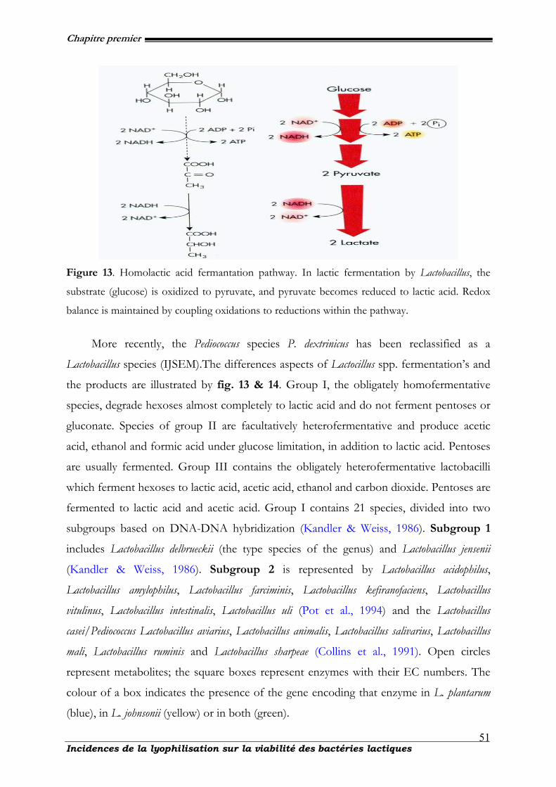
Chapitre premier
Incidences de la lyophilisation sur la viabilité des bactéries lactiques
51
Figure 13. Homolactic acid fermantation pathway. In lactic fermentation by Lactobacillus, the
substrate (glucose) is oxidized to pyruvate, and pyruvate becomes reduced to lactic acid. Redox
balance is maintained by coupling oxidations to reductions within the pathway.
More recently, the Pediococcus species P. dextrinicus has been reclassified as a
Lactobacillus species (IJSEM).The differences aspects of Lactocillus spp. fermentation’s and
the products are illustrated by fig. 13 & 14. Group I, the obligately homofermentative
species, degrade hexoses almost completely to lactic acid and do not ferment pentoses or
gluconate. Species of group II are facultatively heterofermentative and produce acetic
acid, ethanol and formic acid under glucose limitation, in addition to lactic acid. Pentoses
are usually fermented. Group III contains the obligately heterofermentative lactobacilli
which ferment hexoses to lactic acid, acetic acid, ethanol and carbon dioxide. Pentoses are
fermented to lactic acid and acetic acid. Group I contains 21 species, divided into two
subgroups based on DNA-DNA hybridization (Kandler & Weiss, 1986). Subgroup 1
includes Lactobacillus delbrueckii (the type species of the genus) and Lactobacillus jensenii
(Kandler & Weiss, 1986). Subgroup 2 is represented by Lactobacillus acidophilus,
Lactobacillus amylophilus, Lactobacillus farciminis, Lactobacillus kefiranofaciens, Lactobacillus
vitulinus, Lactobacillus intestinalis, Lactobacillus uli (Pot et al., 1994) and the Lactobacillus
casei/Pediococcus Lactobacillus aviarius, Lactobacillus animalis, Lactobacillus salivarius, Lactobacillus
mali, Lactobacillus ruminis and Lactobacillus sharpeae (Collins et al., 1991). Open circles
represent metabolites; the square boxes represent enzymes with their EC numbers. The
colour of a box indicates the presence of the gene encoding that enzyme in L. plantarum
(blue), in L. johnsonii (yellow) or in both (green).
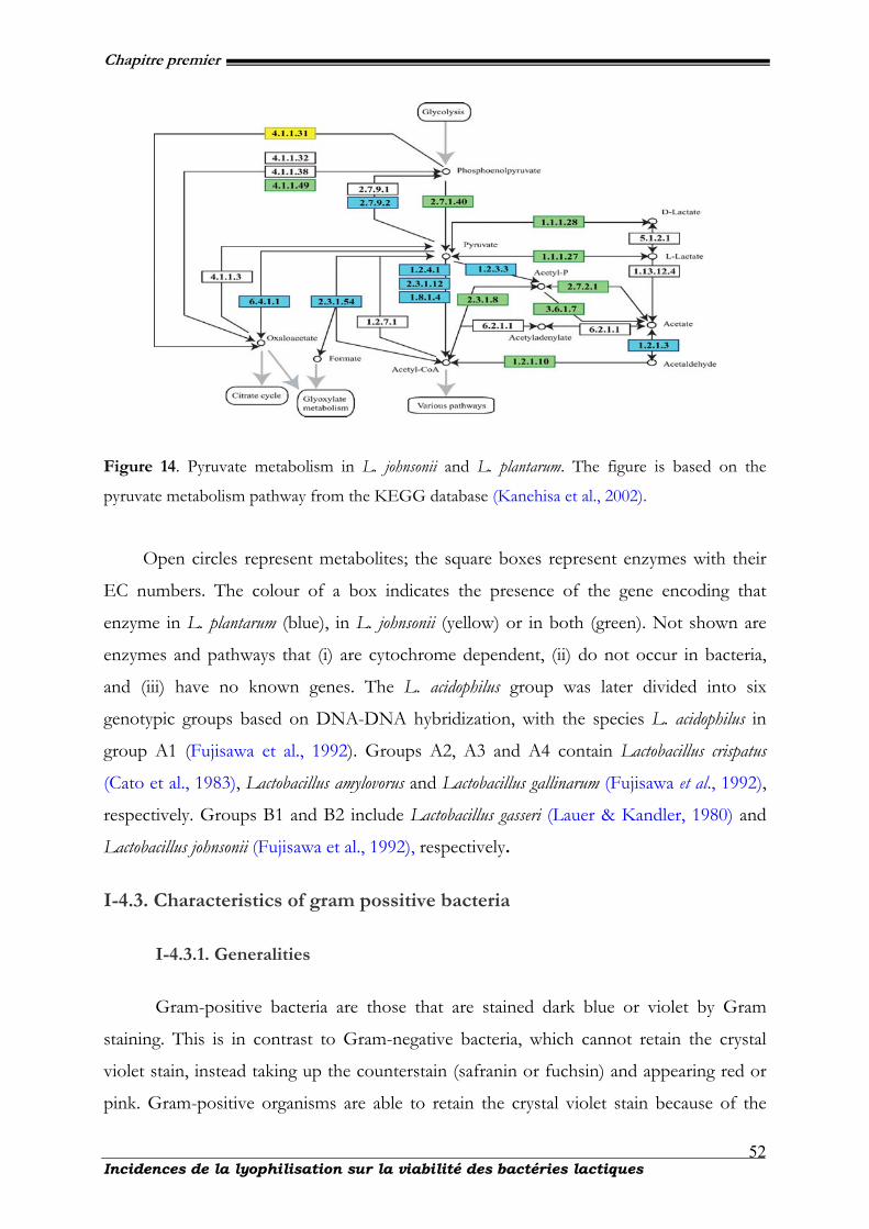
Chapitre premier
Incidences de la lyophilisation sur la viabilité des bactéries lactiques
52
Figure 14. Pyruvate metabolism in L. johnsonii and L. plantarum. The figure is based on the
pyruvate metabolism pathway from the KEGG database (Kanehisa et al., 2002).
Open circles represent metabolites; the square boxes represent enzymes with their
EC numbers. The colour of a box indicates the presence of the gene encoding that
enzyme in L. plantarum (blue), in L. johnsonii (yellow) or in both (green). Not shown are
enzymes and pathways that (i) are cytochrome dependent, (ii) do not occur in bacteria,
and (iii) have no known genes. The L. acidophilus group was later divided into six
genotypic groups based on DNA-DNA hybridization, with the species L. acidophilus in
group A1 (Fujisawa et al., 1992). Groups A2, A3 and A4 contain Lactobacillus crispatus
(Cato et al., 1983), Lactobacillus amylovorus and Lactobacillus gallinarum (Fujisawa et al., 1992),
respectively. Groups B1 and B2 include Lactobacillus gasseri (Lauer & Kandler, 1980) and
Lactobacillus johnsonii (Fujisawa et al., 1992), respectively.
I-4.3. Characteristics of gram possitive bacteria
I-4.3.1. Generalities
Gram-positive bacteria are those that are stained dark blue or violet by Gram
staining. This is in contrast to Gram-negative bacteria, which cannot retain the crystal
violet stain, instead taking up the counterstain (safranin or fuchsin) and appearing red or
pink. Gram-positive organisms are able to retain the crystal violet stain because of the
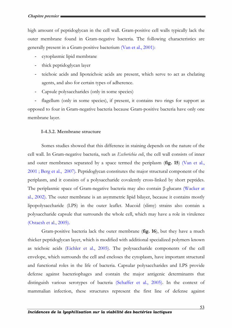
Chapitre premier
Incidences de la lyophilisation sur la viabilité des bactéries lactiques
53
high amount of peptidoglycan in the cell wall. Gram-positive cell walls typically lack the
outer membrane found in Gram-negative bacteria. The following characteristics are
generally present in a Gram-positive bacterium (Van et al., 2001):
- cytoplasmic lipid membrane
- thick peptidoglycan layer
- teichoic acids and lipoteichoic acids are present, which serve to act as chelating
agents, and also for certain types of adherence.
- Capsule polysaccharides (only in some species)
- flagellum (only in some species), if present, it contains two rings for support as
opposed to four in Gram-negative bacteria because Gram-positive bacteria have only one
membrane layer.
I-4.3.2. Membrane structure
Somes studies showed that this difference in staining depends on the nature of the
cell wall. In Gram-negative bacteria, such as Escherichia coli, the cell wall consists of inner
and outer membranes separated by a space termed the periplasm (fig. 15) (Van et al.,
2001 ; Berg et al., 2007). Peptidoglycan constitutes the major structural component of the
periplasm, and it consists of a polysaccharide covalently cross-linked by short peptides.
The periplasmic space of Gram-negative bacteria may also contain β-glucans (Wacker at
al., 2002). The outer membrane is an asymmetric lipid bilayer, because it contains mostly
lipopolysaccharide (LPS) in the outer leaflet. Mucoid (slimy) strains also contain a
polysaccharide capsule that surrounds the whole cell, which may have a role in virulence
(Ostacsh et al., 2005).
Gram-positive bacteria lack the outer membrane (fig. 16), but they have a much
thicker peptidoglycan layer, which is modified with additional specialized polymers known
as teichoic acids (Eichler et al., 2005). The polysaccharide components of the cell
envelope, which surrounds the cell and encloses the cytoplasm, have important structural
and functional roles in the life of bacteria. Capsular polysaccharides and LPS provide
defense against bacteriophages and contain the major antigenic determinants that
distinguish various serotypes of bacteria (Schaffer et al., 2005). In the context of
mammalian infection, these structures represent the first line of defense against
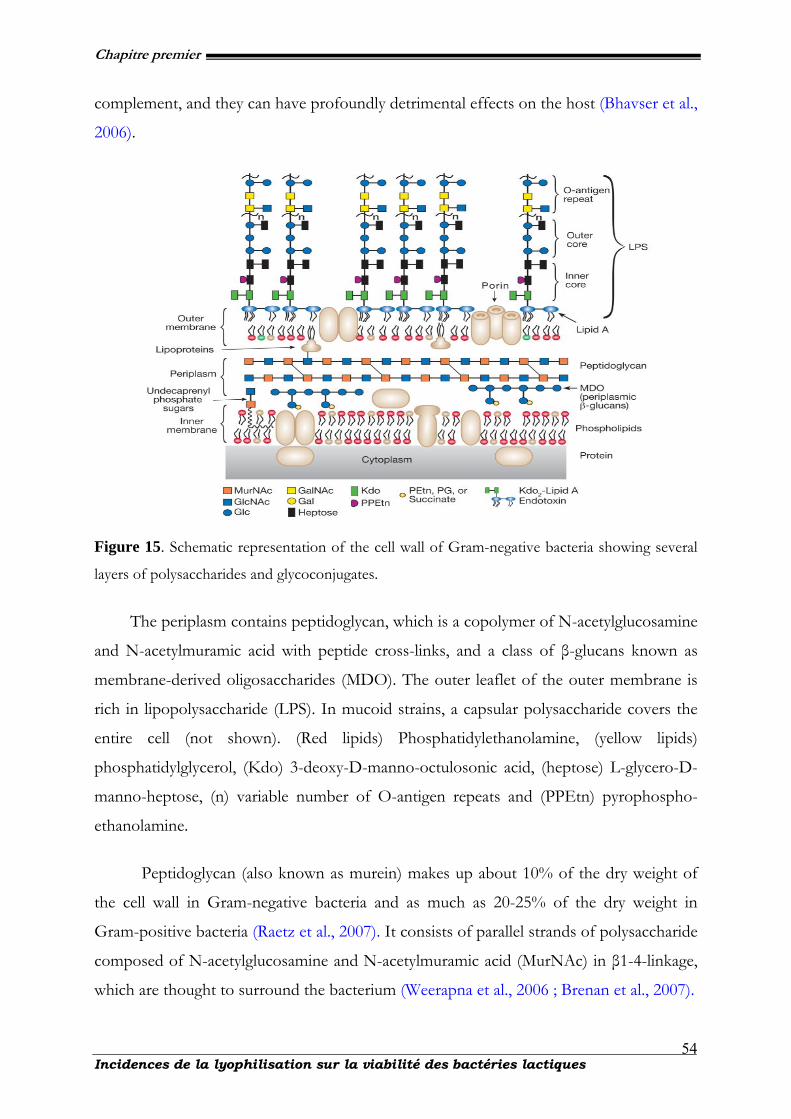
Chapitre premier
Incidences de la lyophilisation sur la viabilité des bactéries lactiques
54
complement, and they can have profoundly detrimental effects on the host (Bhavser et al.,
2006).
Figure 15. Schematic representation of the cell wall of Gram-negative bacteria showing several
layers of polysaccharides and glycoconjugates.
The periplasm contains peptidoglycan, which is a copolymer of N-acetylglucosamine
and N-acetylmuramic acid with peptide cross-links, and a class of β-glucans known as
membrane-derived oligosaccharides (MDO). The outer leaflet of the outer membrane is
rich in lipopolysaccharide (LPS). In mucoid strains, a capsular polysaccharide covers the
entire cell (not shown). (Red lipids) Phosphatidylethanolamine, (yellow lipids)
phosphatidylglycerol, (Kdo) 3-deoxy-D-manno-octulosonic acid, (heptose) L-glycero-D-
manno-heptose, (n) variable number of O-antigen repeats and (PPEtn) pyrophospho-
ethanolamine.
Peptidoglycan (also known as murein) makes up about 10% of the dry weight of
the cell wall in Gram-negative bacteria and as much as 20-25% of the dry weight in
Gram-positive bacteria (Raetz et al., 2007). It consists of parallel strands of polysaccharide
composed of N-acetylglucosamine and N-acetylmuramic acid (MurNAc) in β1-4-linkage,
which are thought to surround the bacterium (Weerapna et al., 2006 ; Brenan et al., 2007).
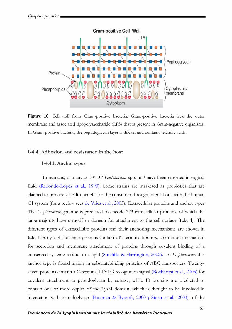
Chapitre premier
Incidences de la lyophilisation sur la viabilité des bactéries lactiques
55
Figure 16. Cell wall from Gram-positive bacteria. Gram-positive bacteria lack the outer
membrane and associated lipopolysaccharide (LPS) that is present in Gram-negative organisms.
In Gram-positive bacteria, the peptidoglycan layer is thicker and contains teichoic acids.
I-4.4. Adhesion and resistance in the host
I-4.4.1. Anchor types
In humans, as many as 107-108 Lactobacillus spp. ml-1 have been reported in vaginal
fluid (Redondo-Lopez et al., 1990). Some strains are marketed as probiotics that are
claimed to provide a health benefit for the consumer through interactions with the human
GI system (for a review sees de Vries et al., 2005). Extracellular proteins and anchor types
The L. plantarum genome is predicted to encode 223 extracellular proteins, of which the
large majority have a motif or domain for attachment to the cell surface (tab. 4). The
different types of extracellular proteins and their anchoring mechanisms are shown in
tab. 4 Forty-eight of these proteins contain a N-terminal lipobox, a common mechanism
for secretion and membrane attachment of proteins through covalent binding of a
conserved cysteine residue to a lipid (Sutcliffe & Harrington, 2002). In L. plantarum this
anchor type is found mainly in substratebinding proteins of ABC transporters. Twenty-
seven proteins contain a C-terminal LPxTG recognition signal (Boekhorst et al., 2005) for
covalent attachment to peptidoglycan by sortase, while 10 proteins are predicted to
contain one or more copies of the LysM domain, which is thought to be involved in
interaction with peptidoglycan (Bateman & Bycroft, 2000 ; Steen et al., 2003), of the
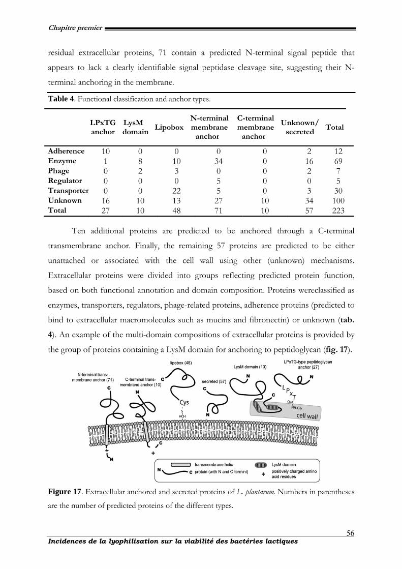
Chapitre premier
Incidences de la lyophilisation sur la viabilité des bactéries lactiques
56
residual extracellular proteins, 71 contain a predicted N-terminal signal peptide that
appears to lack a clearly identifiable signal peptidase cleavage site, suggesting their N-
terminal anchoring in the membrane.
Table 4. Functional classification and anchor types.
LPxTG anchor
LysM domain
LipoboxN-terminal membrane
anchor
C-terminal membrane
anchor
Unknown/ secreted
Total
Adherence 10 0 0 0 0 2 12 Enzyme 1 8 10 34 0 16 69 Phage 0 2 3 0 0 2 7 Regulator 0 0 0 5 0 0 5 Transporter 0 0 22 5 0 3 30 Unknown 16 10 13 27 10 34 100 Total 27 10 48 71 10 57 223
Ten additional proteins are predicted to be anchored through a C-terminal
transmembrane anchor. Finally, the remaining 57 proteins are predicted to be either
unattached or associated with the cell wall using other (unknown) mechanisms.
Extracellular proteins were divided into groups reflecting predicted protein function,
based on both functional annotation and domain composition. Proteins wereclassified as
enzymes, transporters, regulators, phage-related proteins, adherence proteins (predicted to
bind to extracellular macromolecules such as mucins and fibronectin) or unknown (tab.
4). An example of the multi-domain compositions of extracellular proteins is provided by
the group of proteins containing a LysM domain for anchoring to peptidoglycan (fig. 17).
Figure 17. Extracellular anchored and secreted proteins of L. plantarum. Numbers in parentheses
are the number of predicted proteins of the different types.
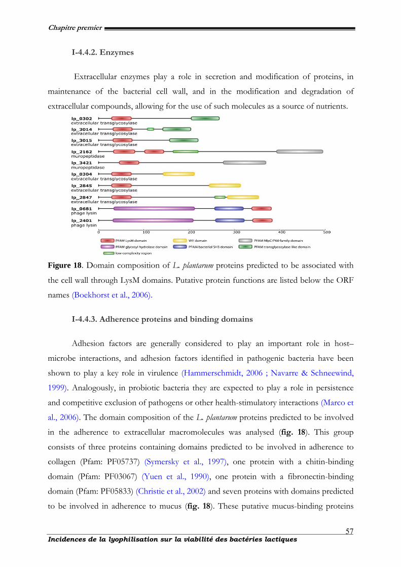
Chapitre premier
Incidences de la lyophilisation sur la viabilité des bactéries lactiques
57
I-4.4.2. Enzymes
Extracellular enzymes play a role in secretion and modification of proteins, in
maintenance of the bacterial cell wall, and in the modification and degradation of
extracellular compounds, allowing for the use of such molecules as a source of nutrients.
Figure 18. Domain composition of L. plantarum proteins predicted to be associated with
the cell wall through LysM domains. Putative protein functions are listed below the ORF
names (Boekhorst et al., 2006).
I-4.4.3. Adherence proteins and binding domains
Adhesion factors are generally considered to play an important role in host–
microbe interactions, and adhesion factors identified in pathogenic bacteria have been
shown to play a key role in virulence (Hammerschmidt, 2006 ; Navarre & Schneewind,
1999). Analogously, in probiotic bacteria they are expected to play a role in persistence
and competitive exclusion of pathogens or other health-stimulatory interactions (Marco et
al., 2006). The domain composition of the L. plantarum proteins predicted to be involved
in the adherence to extracellular macromolecules was analysed (fig. 18). This group
consists of three proteins containing domains predicted to be involved in adherence to
collagen (Pfam: PF05737) (Symersky et al., 1997), one protein with a chitin-binding
domain (Pfam: PF03067) (Yuen et al., 1990), one protein with a fibronectin-binding
domain (Pfam: PF05833) (Christie et al., 2002) and seven proteins with domains predicted
to be involved in adherence to mucus (fig. 18). These putative mucus-binding proteins
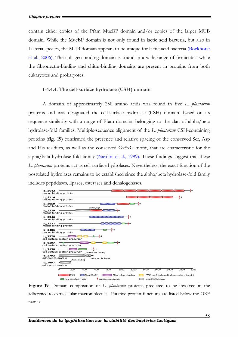
Chapitre premier
Incidences de la lyophilisation sur la viabilité des bactéries lactiques
58
contain either copies of the Pfam MucBP domain and/or copies of the larger MUB
domain. While the MucBP domain is not only found in lactic acid bacteria, but also in
Listeria species, the MUB domain appears to be unique for lactic acid bacteria (Boekhorst
et al., 2006). The collagen-binding domain is found in a wide range of firmicutes, while
the fibronectin-binding and chitin-binding domains are present in proteins from both
eukaryotes and prokaryotes.
I-4.4.4. The cell-surface hydrolase (CSH) domain
A domain of approximately 250 amino acids was found in five L. plantarum
proteins and was designated the cell-surface hydrolase (CSH) domain, based on its
sequence similarity with a range of Pfam domains belonging to the clan of alpha/beta
hydrolase-fold families. Multiple-sequence alignment of the L. plantarum CSH-containing
proteins (fig. 19) confirmed the presence and relative spacing of the conserved Ser, Asp
and His residues, as well as the conserved GxSxG motif, that are characteristic for the
alpha/beta hydrolase-fold family (Nardini et al., 1999). These findings suggest that these
L. plantarum proteins act as cell-surface hydrolases. Nevertheless, the exact function of the
postulated hydrolases remains to be established since the alpha/beta hydrolase-fold family
includes peptidases, lipases, esterases and dehalogenases.
Figure 19. Domain composition of L. plantarum proteins predicted to be involved in the
adherence to extracellular macromolecules. Putative protein functions are listed below the ORF
names.
Chapitre premier
Incidences de la lyophilisation sur la viabilité des bactéries lactiques
59
Another distinct set of eight homologous proteins of L. plantarum also have the
characteristics of alpha/beta hydrolases, but differ in sequence alignment from the CSH
domains, and are classified as members of the Pfam DUF915 family.
I-4.5. Phospholipids and fatty acids of Lactobacillus spp.
Phospholipids are the major components of bacterial cell membranes. These lipid
compounds possess closely related and complex structures classed according to the polar
head group linked to the phosphate moiety. The diversity of the polar head groups is
important, and as far as prokaryotic organisms are concerned, the main classes of
phospholipids are phosphatidylethanolamine (PE), phosphatidylglycerol (PG), and their
respective derivatives: mono-, bi-, or trimethylated forms of PE and a dimeric form of
PG [diphosphatidylglycerol (DPG)], (Exterkate et al., 1971 ; Fischer et al., 1990). The
other phospholipid classes, such as phosphatidylinositol (PI), phosphatidylcholine (PC),
and phosphatidylserine (PS), are considered to be less characteristic of Lactobacillus cell
membranes (Lechevalier, 1977 ; Fang et al., 2000). At neutral pH, the former are
negatively charged and the latter are globally neutral. Each phospholipid class is made up
of several molecular species. The diversity of these molecular species depends on the two
fatty acyl chains linked to the glycerol backbone (mainly saturated or monounsaturated
and from 16 to 18 carbon atom chain length). Moreover, the study of the fatty acid
distribution (sn-1or sn-2 position) is of interest to determine the stereochemistry of the
molecular species.
I-4.5.1. Structure of phospholipids
The structure of the most common class of phospholipids, phosphoglycerides, is
based on glycerol, a three-carbon alcohol with the formula CH2OH–CHOH–CH2OH.
Two fatty acid chains, each typically having an even number of carbon atoms between 14
and 20, attach (via a dual esterification) to the first and second carbons of the glycerol
molecule, denoted as the sn1 and sn2 positions, respectively (Exterkate et al., 1971). The
third hydroxyl group of glycerol, at position sn3, reacts with phosphoric acid to form
phosphatidate. Common phospholipids, widely distributed in nature, are produced by
further reaction of the phosphate group in phosphatidate with an alcohol, such as serine,
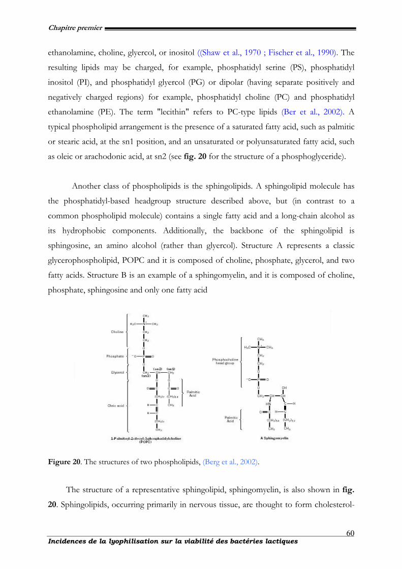
Chapitre premier
Incidences de la lyophilisation sur la viabilité des bactéries lactiques
60
ethanolamine, choline, glyercol, or inositol ((Shaw et al., 1970 ; Fischer et al., 1990). The
resulting lipids may be charged, for example, phosphatidyl serine (PS), phosphatidyl
inositol (PI), and phosphatidyl glyercol (PG) or dipolar (having separate positively and
negatively charged regions) for example, phosphatidyl choline (PC) and phosphatidyl
ethanolamine (PE). The term "lecithin" refers to PC-type lipids (Ber et al., 2002). A
typical phospholipid arrangement is the presence of a saturated fatty acid, such as palmitic
or stearic acid, at the sn1 position, and an unsaturated or polyunsaturated fatty acid, such
as oleic or arachodonic acid, at sn2 (see fig. 20 for the structure of a phosphoglyceride).
Another class of phospholipids is the sphingolipids. A sphingolipid molecule has
the phosphatidyl-based headgroup structure described above, but (in contrast to a
common phospholipid molecule) contains a single fatty acid and a long-chain alcohol as
its hydrophobic components. Additionally, the backbone of the sphingolipid is
sphingosine, an amino alcohol (rather than glyercol). Structure A represents a classic
glycerophospholipid, POPC and it is composed of choline, phosphate, glycerol, and two
fatty acids. Structure B is an example of a sphingomyelin, and it is composed of choline,
phosphate, sphingosine and only one fatty acid
Figure 20. The structures of two phospholipids, (Berg et al., 2002).
The structure of a representative sphingolipid, sphingomyelin, is also shown in fig.
20. Sphingolipids, occurring primarily in nervous tissue, are thought to form cholesterol-
Chapitre premier
Incidences de la lyophilisation sur la viabilité des bactéries lactiques
61
rich domains within lipid bilayer membranes that may be important to the functions of
some membrane proteins (Voet et al., 1999).
I-4.5.2. Fatty acids
Numerous studies have investigated the lipid composition of Lactobacillus spp.
(Exterkate et al., 1971 ; Thorne et al., 1964 ; Uchida et al., 1973). Lactobacilli typically
have n-C16:0, n-C18:1, and cyc-C19 as the major carboxylate constituents, with lesser amounts
of n-C14:0, n-C16:1, and n-C18:0 and traces of odd-carbon-number acids in some species
(Drucker et al., 1981). Such fatty acid profiles are typical of gram-positive groups but
common in gram-positive bacteria (O’Leary et al., 1988). Fatty acids are a carboxylic acid
with a long unbranched aliphatic tail (chain), which is either saturated or unsaturated.
Saturated fatty acids do not contain any double bonds or other functional groups along
the chain. The term "saturated" refers to hydrogen, in that all carbons (apart from the
carboxylic acid [-COOH] group) contain as many hydrogens as possible. Saturated fatty
acids form straight chains and, as a result, can be packed together very tightly, allowing
living organisms to store chemical energy very densely. The fatty tissues of Lactobacillus
spp. contain large amounts of long-chain saturated fatty acids.
I-4.6. Probiotics and biotherapeutics
I-4.6.1. Probiotics and food production
Lactobacillus species, widespread in food and the environment, are important in
technological applications as starter or adjunct nonstarter strains in food productions. The
simultaneous presence of various lactic acid bacteria (LAB), expressing different species-
specific enzyme activities, is essential for the development of flavor and specific quality
traits of cheese (Baruzzi et al., 2005 ; Morea et al. 1998 ; Cappello et al. 2001). There is a
need to characterize regional dairy products and to monitor the natural adjuvant
nonstarter strains responsible for the production process. Many Lactobacillus species are
associated with fermented foods and often may characterize products with traditional
designation of origin. L. plantarum strains are found in fermented products as olives and
pickled vegetables and in sourdoughs (Corsetti et al., 2001). L. fermentum is associated to
Chapitre premier
Incidences de la lyophilisation sur la viabilité des bactéries lactiques
62
fermented meats, sausages, and other food products as sourdoughs (Corsetti et al., 2001).
In various cheese productions, L. paracasei, L. zeae, L. fermentum, and L. buchneri were found
at different times of ripening, contributing to final cheese quality to a different extent,
according to the type of cheese (Baruzzi et al., 2000 ; Morea et al., 1998 ; Cappello et al.,
2001 ; Corsetti et al., 2001 ; Ercolini et al., 2001, 2004). In addition to typical foods with
standard manufacturing protocols, new foods have been produced with the addition of
probiotic strains, belonging to L. rhamnosus, L. paracasei, L. plantarum, L. reuteri, L. johnsoni,
and L. acidophilus species. Their presence is frequent in cheeses, fermented milks, table
olives, and pickles. Thus, there is a need to ascertain their presence for certifying the
benefits claimed in these products. For these reasons, we studied the detection and
identification of six species, L. rhamnosus, L. paracasei, L. zeae, L. fermentum, and L. buchneri,
as these species are frequently present in fermented foods, cheeses, and probiotic-
enriched foods. Some Lactobacillus spp. and other lactic acid bacteria may possess potential
therapeutic properties including anti-inflammatory and anti-cancer activities, as well as
other features of interest. Research studies have demonstrated the protective effects of
some strains of these bacteria for anti-tumor and anti-cancer effects (Gladwin et al.,
2007).
Dietary administration alleviated the risks of certain types of cancers and
suppressed colonic tumor incidence, volume and multiplicity induced by various
carcinogens. For a few strains oral administration effectively reduced DNA adduct
formation, ameliorated DNA damage and prevented putative preneoplastic lesions such
as aberrant crypt foci induced by chemical carcinogens in the gastrointestinal tract.
Reports also indicated that some cultures administered to animals inhibited liver, colon,
bladder, and mammary tumors, highlighting potential systemic effects of probiotics with
anti-neoplastic activities (Madigan et al., 2005). Lactobacilli are also used to restore
particular physiological balance such as in the vaginal eco-system (Gynoflor). Their role is
(1) to physically protect the vaginal epithelium by building a thick layer separating the
epithelium from pathogens, (2) to physiologically keep the balance of the vaginal
ecosystem in maintaining the pH at ~4.5 and (3) generating hydrogen peroxide against
pathogens.
Chapitre premier
Incidences de la lyophilisation sur la viabilité des bactéries lactiques
63
I-4.6.2. Biotherapeutics and potential benefits
Experiments into the benefits of probiotic therapies suggest a range of potentially
beneficial medicinal uses for probiotics. For many of the potential benefits, research is
limited and only preliminary results are available. It should be noted that the effects
described are not general effects of probiotics. Recent research on the molecular biology
and genomics of Lactobacillus has focused on the interaction with the immune system,
anti-cancer potential, and potential as a biotherapeutic agent in cases of antibiotic-
associated diarrhoea, travellers' diarrhoea, pediatric diarrhoea, inflammatory bowel disease
and irritable bowel syndrome (Ljungh et al., 2009).
I-4.6.2.1. Managing lactose intolerance
As lactic acid bacteria actively convert lactose into lactic acid, ingestion of certain
active strains may help lactose intolerant individuals tolerate more lactose than what they
would have otherwise (Sanders et al., 2000). In practice probiotics are not specifically
targeted for this purpose, as most are relatively low in lactase activity as compared to the
normal yogurt bacteria.
I-4.6.2.2. Prevention of colon cancer
In laboratory investigations, some strains of LAB (Lactobacillus bulgaricus) have
demonstrated anti-mutagenic effects thought to be due to their ability to bind with
heterocyclic amines, which are carcinogenic substances formed in cooked meat
(Wollowski et al., 2001). Animal studies have demonstrated that some LAB can protect
against colon cancer in rodents, though human data is limited and conflicting (Brady et
al., 2000). Most human trials have found that the strains tested may exert anti-
carcinogenic effects by decreasing the activity of an enzyme called β-glucuronidase (Brady
et al., 2000) (which can generate carcinogens in the digestive system). Lower rates of
colon cancer among higher consumers of fermented dairy products have been observed
in some population studies (Sanders et al., 2000).
Chapitre premier
Incidences de la lyophilisation sur la viabilité des bactéries lactiques
64
I-4.6.2.3. Lowering cholesterol
Animal studies have demonstrated the efficacy of a range of LAB to be able to
lower serum cholesterol levels, presumably by breaking down bile in the gut, thus
inhibiting its reabsorption (which enters the blood as cholesterol). Some, but not all
human trials have shown that dairy foods fermented with specific LAB can produce
modest reductions in total and LDL cholesterol levels in those with normal levels to
begin with, however trials in hyperlipidemic subjects are needed (Sanders et al., 2000).
I-4.6.2.4. Lowering blood pressure
Several small clinical trials have shown that consumption of milk fermented with
various strains of LAB can result in modest reductions in blood pressure. It is thought
that this is due to the ACE inhibitor-like peptides produced during fermentation (Sanders
et al., 2000).
I-4.6.3. Benefits for health
I-4.6.3.1. Improving immune function and preventing infections
LABs are thought to have several presumably beneficial effects on immune
function. They may protect against pathogens by means of competitive inhibition (i.e., by
competing for growth) and there is evidence to suggest that they may improve immune
function by increasing the number of IgA-producing plasma cells, increasing or
improving phagocytosis as well as increasing the proportion of T lymphocytes and natural
killer cells (Reid et al., 2003 ; Ouwehand et al., 2002). Clinical trials have demonstrated
that probiotics may decrease the incidence of respiratory tract infections (Hatakka et al.,
2001) and dental caries in children (Näse et al., 2005). LAB foods and supplements have
been shown to be effective in the treatment and prevention of acute diarrhea, and in
decreasing the severity and duration of rotavirus infections in children and travelers'
diarrhea in adults (Reid et al., 2003). Recently, clear immune enhancing effect of
probiotics is demonstrated in the gut of healthy subjects. In a randomized, double blind,
placebo controlled, crossover study, healthy volunteers ingested either live probiotic cells
(L. plantarum) inactivated cells of the same probiotic, or a placebo.
Chapitre premier
Incidences de la lyophilisation sur la viabilité des bactéries lactiques
65
These processes activate the immune system enabling it to play its protective role
(the immune response).
I-4.6.3.2. Helicobacter pylori
LABs are also thought to aid in the treatment of Helicobacter pylori infections
(which cause peptic ulcers) in adults when used in combination with standard medical
treatments. However more studies are required into this area (Hamilton-Miller et al.,
2003).
I-4.6.3.3. Antibiotic-associated diarrhea
A meta-analysis suggested probiotics may reduce antibiotic-associated diarrhea
(Cremonini et al., 2002). A subsequent randomized controlled trial also found benefit in
elderly patients (Hickson et al., 2007). In a randomized clinical trial, published in 2007, a
University of Montreal team of pharmacologists demonstrated that lactobacilli-fermented
solution can be effective in AAD prevention in hospitalized patients” (Beausoleil et al.,
2007). In 2009 Encap Drug Delivery announced that they had entered into collaboration
with Probac AB to develop a novel probiotic capsule product aimed at treating antibiotic
associated diarrhoea.
I-4.6.3.4. Reducing inflammation
LAB foods and supplements have been found to modulate inflammatory and
hypersensitivity responses, an observation thought to be at least in part due to the
regulation of cytokine function (Reid et al., 2003). Clinical studies suggest that they can
prevent reoccurrences of inflammatory bowel disease in adults (Reid et al., 2003) , as well
as improve milk allergies (Kirjavainen et al., 2003). They are not effective for treating
eczema, a persistent skin inflammation (Boyle et al., 2008)
I-4.6.3.5. Improving mineral absorption
It is hypothesized that probiotic lactobacilli may help correct malabsorption of
trace minerals, found particularly in those with diets high in phytate content from whole
grains, nuts and legumes (Famularo et al., 2005)
Chapitre premier
Incidences de la lyophilisation sur la viabilité des bactéries lactiques
66
I-4.6.3.6. Prevents harmful bacterial growth under stress
In a study done to see the effects of stress on intestinal flora, rats that were fed
probiotics had little occurrence of harmful bacteria latched onto their intestines compared
to rats that were fed sterile water (Hitt et al., 2006).
I-4.6.3.7. Irritable bowel syndrome and colitis
B. infantis 35624, sold as Align, was found to improve some symptoms of irritable
bowel syndrome in women in a recent study (Whorwell et al., 2006). Another probiotic
bacterium, Lactobacillus plantarum 299v was also found to be effective in reducing IBS
symptoms (Niedzielin et al., 2001). Additionally, a probiotic formulation, VSL#3, was
found to be safe in treating ulcerative colitis, though efficacy in the study was uncertain
(Kerr et al., 2003). Bifidobacterium animalis DN-173 010 may help (Guyonnet et al.,
2007).
I-4.6.3.8. Synbiotics
As probiotics are mainly active in the small intestine and prebiotics are only
effective in the large intestine, the combination of the two may give a synergistic effect
(Gibson et al., 1995). Appropriate combinations of pre- and probiotics are symbiotics.
I-4.7. Conclusions
The genus Lactobacillus could be important to the host, because deconjugated bile
salts are less effective in emulsification of dietary lipids and micelle formation. Thus,
interactions of lactobacilli with their hosts and their impact on host characteristics
continue to fascinate microbiologists. Lactobacilli clearly offer microbiologists exciting
research prospects, both for biomedical applications and for acquiring fundamental
knowledge of how bacterial cells function in the gut ecosystem. As model gut bacteria,
they may provide lessons in the molecular mechanisms that define autochthony as well as
in understanding bacterial physiology in relation to host welfare. For these reasons,
lactobacilli are set to remain the fond favorites of many microbiologists. Lactobacilli are
members of the lactic acid bacteria, a broadly defined group characterized by the
Chapitre premier
Incidences de la lyophilisation sur la viabilité des bactéries lactiques
67
formation of lactic acid as a sole or main end product of carbohydrate metabolism. The
lactobacilli are gram-positive, non-spore-forming rods
I-4.8. References
Baruzzi F., Matarante A., Caputo L., Morea M., 2005. Development of a culture-independent polymerase chain reaction-based assay for the detection of lactobacilli in stretched cheese. J. Rapid. Methods. Autom. Microbiol., 13, 177-192.
Baruzzi F., Morea M., Matarante A., Cocconcelli P.S., 2000. Changes in the Lactobacillus community during
Ricotta forte cheese natural fermentation, J. Appl. Microbiol., 89, 807-814. Bateman A.., & Bycroft M., 2000. The structure of a LysM domain from E. coli membrane-bound lytic
murein transglycosylase D (MltD). J .Mol .Biol. 299, 1113-1119. Beausoleil M., Fortier N., Guénette S., L'ecuyer A., Savoie M., Franco M., Lachaine J., Weiss K., 2007.
Effect of a fermented milk combining Lactobacillus acidophilus CL1285® and Lactobacillus casei in the prevention of antibiotic-associated diarrhea: A randomized, double-blind, placebo-controlled trial. Can. J. Gastro., 21, 732-736.
Berg S., Kaur D., Jackson M., Brennan P.J., 2007. The glycosyltransferases of Mycobacterium tuberculosis -
Roles in the synthesis of arabinogalactan, lipoarabinomannan, and other glycoconjugates. Glycobiol., 17, 35-56.
Berg Jeremy M., Tymoczko John L., and Stryer Lubert., 2002. Biochemistry, 5th edition. New York: W. H.
Freeman. Bhavsar A.P., Brown E.D., 2006. Cell wall assembly in Bacillus subtilis: How spirals and spaces challenge
paradigms. Mol. Microbiol., 60,1077-1090. Black J.G., 1996. Microbiology. Principles and Applications. Third Edition. Prentice Hall. Upper Saddle River,
New Jersey. pp. 144-148. Boekhorst J., de Been M.W., Kleerebezem M., & Siezen R.J., 2005. Genome-wide detection and analysis
of cell wall-bound proteins with LPxTG-like sorting motifs. J. Bacteriol., 187, 4928-4934. Boekhorst J., Helmer Q., Kleerebezem M., & Siezen R.J., 2006. Comparative analysis of proteins with a
mucus-binding domain found exclusively in lactic acid bacteria. Microbiol., 152, 273-280. Boyle R.J., Bath-Hextall F.J., Leonardi-Bee J., Murrell D.F., Tang M.L., 2008. Probiotics for treating
eczema. Coch. Database. Syst. Rev., 4, CD006135. Brady L.J., Gallaher D.D., Busta F.F., 2000. The role of probiotic cultures in the prevention of colon
cancer. J. Nutr., 130, 410-414. Brennan P.J., Crick D.C., 2007. The cell-wall core of Mycobacterium tuberculosis in the context of drug
discovery. Curr. Top. Med. Chem., 7, 475-488. Cappello M.S., Laddomada B., Poltronieri P., Zacheo G., 2001. Mededelingen Faculteit Landbouwkundige
en Toegepaste Biologische Wetenschappen Universiteit Gent 66(3B), 569.
Chapitre premier
Incidences de la lyophilisation sur la viabilité des bactéries lactiques
68
Cato E.P., Moore W.E.C., & Johnson J.L., 1983. Synonymy of strains of `` Lactobacillus acidophilus'' group A2 (Johnson et al. 1980) with the type strain of Lactobacillus crispatus (Brygoo and Aladame 1953) Moore and Holdeman 1970. Int. J Syst. Bacteriol., 33, 426-428.
Cavalier-Smith T., 2006. Rooting the tree of life by transition analyses. Biol. Direct., 1, 19. Christie J., McNab R., & Jenkinson H.F., 2002. Expression of fibronectin-binding protein FbpA
modulates adhesion in Streptococcus gordonii. Microbiol., 148, 1615-1625. Collins M.D., Rodrigues U.M., Ash C., Aguirre M., Farrow J.A.E., Martinez-Murcia A., Phillips B.A.,
Williams A.M., & Wallbanks S., 1991. Phylogenetic analysis of the genus Lactobacillus and related lactic acid bacteria as determined by reverse transcriptase sequencing of 16S rRNA. FEMS. Microbiol. Lett., 77, 5-12.
Corsetti A., Lavermicocca P., Morea M., Baruzzi F., Tosti N., Gobbetti M., 2001. Phenotypic and
molecular identification and clustering of lactic acid bacteria and yeasts from wheat (species Triticum durum and Triticum aestivum) sourdoughs of Southern Italy. Int. J. Food. Microbiol., 64, 95-104.
Cremonini F., Di Caro S., Nista EC., 2002. Meta-analysis: the effect of probiotic administration on
antibiotic-associated diarrhoea. Aliment. Pharmacol. Ther. 16, 1461-1467. Dellaglio F., & Felis G.E., 2005. Taxonomy of lactobacilli and bifidobacteria. Probiotics and Prebiotics:
Scientific Aspects, Edited by G. W. Tannock. Norfolk, UK: Caister Academic Press. pp. 25-49. Dellaglio F., Felis G.E., & Torriani S., 2005. Is the genus Lactobacillus a single genus? In LAB8
Symposium on Lactic Acid Bacteria. Egmond aan Zee, Netherlands: FEMS. Drucker D.B., 1981. Microbiological applications of gas chromatography. Cambridge University Press,
Cambridge. Eichler J., Adams M.W. 2005. Posttranslational protein modification in Archaea. Microbiol. Mol. Biol.
Rev., 69, 393-425. Ercolini D., Mauriello G., Blaiotta G., Moschetti G., Coppola S., 2001. The potential of a polyphasic PCR-
DGGE approach in evaluating microbial diversity of natural whey cultures for water-buffalo mozzarella cheese production: bias of culture dependent and culture independent approaches. Syst. Appl. Microbiol., 24, 610-617.
Ercolini D., Mauriello G., Blaiotta G., Moschetti G., Coppola S., 2004. PCR-DGGE fingerprints of
microbial succession during a manufacture of traditional water buffalo mozzarella cheese. J. Appl. Microbiol., 96, 263-270.
Exterkate F.A., Otten B.J., Wassenberg H.W., and Veerkamp J.H., 1971. Comparison of the phospholipid
composition of Bifidobacterium and Lactobacillusstrains. J. Bacteriol., 106,824-829. Famularo G., De Simone C., Pandey V., Sahu A.R., Minisola G., 2005. Probiotic lactobacilli: an innovative
tool to correct the malabsorption syndrome of vegetarians? Med. Hypotheses., 65, 1132-1135. Fang J., Barcelona M.J., and Semrau J.D., 2000. Characterization of methanotrophic bacteria on the basis
of intact phospholipid profiles. FEMS. Microbiol. Lett. 189, 67-72. Fernandes C.F., Shahani K.M., & Amer M.A., 1987. Therapeutic role of dietary lactobacilli and lactobacilli
fermented dairy products. FEMS. Microbiol. Rev. 46, 343-356.

Chapitre premier
Incidences de la lyophilisation sur la viabilité des bactéries lactiques
69
Fischer, W. 1990. Bacterial phosphoglycolipids and lipoteichoic acids, In M. Kates (ed.), Handbook of lipid research, vol. 6. Plenum Press, New York. p. 123-234.
Fischer W., Laine R.A., and Nakano M., 1978. On the relationship between glycerophosphoglycolipids
and teichoic acids in Gram-positive bacteria. II. Structures of glycerophosphoglycolipids. Biochim. Biophys. Acta., 528, 298-308.
Fujisawa T., Benno Y., Yaeshima T. & Mitsuoka T., 1992. Taxonomic study of the Lactobacillus acidophilus
group, with recognition of Lactobacillus gallinarum sp. nov. and Lactobacillus johnsonii sp. nov. and synonomy of Lactobacillus acidophilus group A3 (Johnson et al. 1980) with the type strain of Lactobacillus amylovorus (Nakamura 1981). Int. J Syst. Bacteriol., 42, 487-491.
Gibson G.R., Roberfroid M.B., 1995. Dietary modulation of the human colonic microbiota: introducing
the concept of prebiotics. J. Nutr. 125, 1401-1412. Gilliland S.E., Walker D.K., 1990. Factors to consider when selecting a culture of Lactobacillus
acidophilus as a dietary adjunct to produce a hypocholesterolemic effect in humans. J Dairy Sci 73, 905-911.
Giorgi A., Torriani S., Dellaglio F., Bo G., Stola G. & Bernuzzi L. 1987. Identi®cation of vaginal
lactobacilli from asymptomatic women. Microbiologica., 10, 377-384. Gladwin M., Trattler Bi., 2007. Clinical Microbiology made ridiculously simple. Miami, FL: MedMaster, Inc.
pp. 4-5. Guarner F., & Schaafsma G.J., 1998. Probiotics. Int J Food Microbiol 39, 237-238. Guyonnet D., Chassany O., Ducrotte P., 2007. Effect of a fermented milk containing Bifidobacterium
animalis DN-173 010 on the health-related quality of life and symptoms in irritable bowel syndrome in adults in primary care: a multicentre, randomized, double-blind, controlled trial. Aliment. Pharmacol. Ther. 26, 475-86.
Hamilton-Miller J.M., 2003. The role of probiotics in the treatment and prevention of Helicobacter pylori
infection. Int. J. Antimicrobiol. Ag., 22, 360-366. Hammerschmidt S., 2006. Adherence molecules of pathogenic pneumococci. Cur.r Opin. Microbiol., 9, 12-
20. Hatakka K., Savilahti E., Pönkä A., 2001. Effect of long term consumption of probiotic milk on infections
in children attending day care centres: double blind, randomised trial. B.M.J., 322, 1327-1330. Hickson M., D'Souza A.L., Muthu N., 2007. Use of probiotic Lactobacillus preparation to prevent
diarrhea associated with antibiotics: randomised double blind placebo controlled trial. B.M.J., 335, 7610-76 80.
Kandler O., & Weiss N., 1986. Genus Lactobacillus Beijerinck 1901, 212AL. In Bergey's Manual of Systematic
Bacteriology, Edited by P. H. A. Sneath, N. S. Mair, M. E. Sharpe & J. G. Holt. Baltimore: Williams & Wilkins. vol. 2, pp. 1209-1234.
Kerr Martha. 2003. Probiotics Significantly Reduce Symptoms of IBS, Ulcerative Colitis.
http://www.medscape.com/viewarticle/455964. Retrieved on 2009-06-15. Kirjavainen P.V., Salminen S.J., Isolauri E., 2003. Probiotic bacteria in the management of atopic disease:
underscoring the importance of viability. J. Pediatr. Gastroenterol. Nutr., 36, 223-227.
Chapitre premier
Incidences de la lyophilisation sur la viabilité des bactéries lactiques
70
Klaenhammer T.R., Barrangou R., Buck B.L., Azcarate-Peril M.A., & Altermann E., 2005. Genomic features of lactic acid bacteria effecting bioprocessing and health. FEMS. Microbiol. Rev., 29, 393-409.
Kleerebezem M., Boekhorst J., van Kranenburg R., & 17 other authors 2003. Complete genome sequence
of Lactobacillus plantarum WCFS1. Proc. Natl. Acad. Sci. U S A 100, 1990-1995. Konings W.N., Kok J., Kuipers O.P., & Poolman B., 2000. Lactic acid bacteria: the bugs of the new
millennium. Curr. Opin. Microbiol. 3, 276-282. Dicks L.M.T., Silvester M., Lawson P.A., and Collins M.D., 2000. Lactobacillus fornicalis sp. nov., isolated
from the posterior fornix of the human vagina. Int. J. Syst. Evol. Microbiol., 50, 1253-1258. Lauer E., & Kandler O., 1980. Lactobacillus gasseri sp. nov., a new species of the subgenus Thermobacterium.
Zentbl. Bakteriol Parasitenkd. Infektkrankh. Hyg. Abt. I. Orig., C1, 75-78. Lechevalier, M.P., 1977. Lipids in bacteria taxonomy-a taxonomist’s view. Crit. Rev. Microbiol., 7, 109-210. Ljungh A., Wadstrom T., 2009. Lactobacillus Molecular Biology: From Genomics to Probiotics. Eds.,. Caister
Academic Press. ISBN 978-1-904455-41-7. Madigan M., Martinko J., 2005. Brock Biology of Microorganisms (11th ed.). Eds., Prentice Hall. Marco M.L., Pavan S., & Kleerebezem M., 2006. Towards understanding molecular modes of probiotic
action. Curr. Opin. Biotechnol., 17, 204-210. Morea M., Baruzzi F., Cappa F., Cocconcelli P.S., 1998 Molecular characterization of the Lactobacillus
community in traditional processing of Mozzarella cheese. Int. J. Food Microbiol., 43,53-60. Nakamura C.E., & Whited G.M., 2003. Metabolic engineering for the microbial production of 1,3-
propanediol. Curr. Opin. Biotechnol., 14, 454-459. Nardini M., & Dijkstra B.W., 1999. Alpha/beta hydrolase fold enzymes: the family keeps growing. Curr.
Opin. Struct. Biol., 9, 732-737. Näse L., Hatakka K., Savilahti E., 2001. Effect of long-term consumption of a probiotic bacterium,
Lactobacillus rhamnosus GG, in milk on dental caries and caries risk in children. Caries Res., 35, 412-420.
Navarre W.W., & Schneewind O., 1999. Surface proteins of gram-positive bacteria and mechanisms of
their targeting to the cell wall envelope. Microbiol. Mol. Biol. Rev., 63, 174-229. Niedzielin K., Kordecki H., Birkenfeld B., 2001. A controlled, double-blind, randomized study on the
efficacy of Lactobacillus plantarum 299V in patients with irritable bowel syndrome. Eur. J. Gastroenterol. Hepatol., 13, 1143-1147.
O’Leary W.M., and Wilkinson S.G., 1988. Gram-positive bacteria, p. 117–201. In C. Ratledge and S. G.
Wilkinson (ed.), Microbial lipids, vol. 1. Academic Press, London. Ostash B., Walker S., 2005. Bacterial transglycosylase inhibitors. Curr. Opin. Chem. Biol., 9, 459-466. Ouwehand A.C., Salminen S., Isolauri E., 2002. Probiotics: an overview of beneficial effects". Ant. Van.
Leeuw., 82, 279-289.
Chapitre premier
Incidences de la lyophilisation sur la viabilité des bactéries lactiques
71
Pot B., Ludwig W., Kersters K., & Schleifer K-H., 1994. Taxonomy of lactic acid bacteria. In Bacteriocins of Lactic Acid Bacteria: Microbiology, Genetics and Applications, pp. 13-90. Edited by L. De Vuyst & E. J. Vandamme. London: Blackie Academic & Professional.
Raetz C.R.H., Reynolds C.M., 2007. Trent MS, Bishop RE. Lipid A modification systems in Gram-
negative bacteria. Annu. Rev .Biochem., 76, 295-329. Redondo-Lopez, V., Cook R.L., & Sobel, J.D., 1990. Emerging role of lactobacilli in the control and
maintenance of the vaginal bacterial microflora. Rev. Infect. Dis., 12, 856-872. Reid G., Jass J., Sebulsky M.T., McCormick J.K., 2003. Potential uses of probiotics in clinical practice".
Clin. Microbiol. Rev., 16, 658-672. Sanders M.E., 2000. Considerations for use of probiotic bacteria to modulate human health. J. Nutr., 130,
384-390. Satokari R.M., Vaughan E.E., Smidt H., Saarela M., Matto J., & de Vos W.M., 2003. Molecular approaches
for the detection and identification of bifidobacteria and lactobacilli in the human gastrointestinal tract. Syst. Appl. Microbiol., 26, 572-584.
Schaffer C., Messner P., 2005. The structure of secondary cell wall polymers: How Gram-positive bacteria
stick their cell walls together. Microbiol., 151, 643-651. Shaw N., 1970. Bacterial glycolipids. Bacteriol. Rev., 34,365-377. Steen A., Buist G., Leenhouts K.J., El Khattabi M., Grijpstra F., Zomer A.L., Venema G., Kuipers, O. P.,
& Kok J., 2003. Cell wall attachment of a widely distributed peptidoglycan binding domain is hindered by cell wall constituents. J. Biol. Chem., 278, 23874-23881.
Stiles M.E., 1996. Biopreservation by lactic acid bacteria. Ant. Van. Leeuw., 70, 331-345. Sutcliffe, I.C., & Harrington, D.J., 2002. Pattern searches for the identification of putative lipoprotein
genes in Gram-positive bacterial genomes. Microbiol., 148, 2065-2077. Symersky J., Patti J.M., Carson M., & 8 other authors 1997. Structure of the collagen-binding domain from
a Staphylococcus aureus adhesin. Nat. Struct .Biol., 4, 833-838. Tannock G.W., 2004. A special fondness for lactobacilli. Appl. Environ. Microbiol., 70, 3189-3194. Thorne K.J.I., 1964. The phospholipids of Lactobacillus casei. Biochim. Biophys. Acta., 84, 350-353. Uchida K., and Mogi K., 1973. Cellular fatty acid spectra of Hiochi bacteria, acid-tolerant lactobacilli, and
their separation. J. Gen. Appl. Microbiol.19, 233-249. Van H.J., 2001. Formation of the glycan chains in the synthesis of bacterial peptidoglycan. Glycobiol., 11, 2-
36. Vaughan E.E., Heilig H.G., Ben-Amor K., & de Vos W.M., 2005. Diversity, vitality and activities of
intestinal lactic acid bacteria and bifidobacteria assessed by molecular approaches. FEMS. .Microbiol. Rev., 29, 477-490.
Voet Donald., Voet Judith G., and Pratt Charlotte., 1999. Fundamentals of Biochemistry. New York: Wiley. Wacker M., Linton D., Hitchen PG., Nita-Lazar M., Haslam S.M., North S.J., Panico M., Morris H.R.,
Dell A., Wren B.W., Aebi M., 2002. N-linked glycosylation in Campylobacter jejuni and its functional transfer into E. coli. Science., 298, 1790-1793.
Chapitre premier
Incidences de la lyophilisation sur la viabilité des bactéries lactiques
72
Weerapana E., Imperiali B., 2006. Asparagine-linked protein glycosylation: From eukaryotic to prokaryotic systems. Glycobiol., 16, 91-101.
Weidenmaier C., Peschel A., 2008.Teichoic acids and related cell-wall glycopolymers in Gram-positive
physiology and host interactions. Nat .Rev. Microbiol., 6, 276-287. Whorwell P.J, Altringer L., Morel J., 2006. Efficacy of an encapsulated probiotic Bifidobacterium infantis
35624 in women with irritable bowel syndrome. Am. J. Gastroentero.l, 101, 1581-1590. Wollowski I., Rechkemmer G., Pool-Zobel B.L., 2001. Protective role of probiotics and prebiotics in
colon cancer. Am. J. Clin. Nutr., 73, 451-455. Yuen L., Dionne J., Arif B., & Richardson C., 1990. Identification and sequencing of the spheroidin gene
of Choristoneura biennis entomopoxvirus. Virol., 175, 427-433.
Chapitre premier
Incidences de la lyophilisation sur la viabilité des bactéries lactiques
73
I-5. Conclusions : Incidences de la lyophilisation sur la viabilité
des bactéries lactiques au cours du stockage
Il est donc possible de conserver par atomisation, fluidisation ou lyophilisation un
grand nombre de bactéries lactiques, non seulement dans le cadre du laboratoire, mais
aussi à l’échelle industrielle. Les conditions opératoires qui permettent d’obtenir une
bonne conservation de l’activité métabolique des bactéries sont relativement bien
connues, mais les mécanismes de leur altération et de leur réservation sont, eux mal
connus. La résolution des difficultés qui subissent, telles que la mauvaise conservation de
certaines souches à l’état lyophilisé et la reprise d’activité souvent trop lente des ferments
pour ensemencements directs, passe certainement par une meilleure compréhension de
ces mécanismes. Pour optimiser la conservation donc, plusieurs techniques et voies de
recherches doivent être menées de concert. Parmi ces voies de recherche les plus
prometteuses, on peut citer l’étude des modifications subies par les structures membra-
naires au cours des traitements de conservation, la recherche de formulation incorporant
des cryoprotecteurs plus efficaces, l’induction des mécanismes de résistance à ces
traitements (par adaptation des bactéries au cours de leur culture dans des conditions
appropriées ou éventuellement par utilisation d’antioxydant afin de bloquer les
phénomènes de peroxydation).
Chapitre II
Sélection et identification de nouvelles souches
résistantes de bactéries lactiques
Chapitre II
Sélection et identification de nouvelles souches resistantes de bactéries lactiques
75
II. Sélection et identification de nouvelles souches
résistantes de bactéries lactiques
II.1. Contexte et objectifs
Le premier objectif fixé dans ce travail est la sélection de souches naturellement
résistantes. Une sélection rigoureuse est le premier des facteurs proposés pour améliorer
la conservation des bactéries lactiques par Klaenhammer et Kleeman (1981). Ainsi, pour
garantir une viabilité commercialement satisfaisante, après un stockage à long terme, il est
opportun de sélectionner des souches qui ont survécu à des conditions drastiques
(températures élevées, sécheresse). Le choix de ces deux critères repose sur l’hypothèse
suivante : une souche qui a survécu dans un environnement hostile : chaud et sec, possède
ou a développé un mécanisme lui permettant une meilleure adaptation à la chaleur et la
déshydratation. Or, en biotechnologie, lors des procédés technologiques ou au cours du
stockage, la perte de viabilité « systématiquement observée » des souches de bactéries
lactiques est imputable à ces deux facteurs.
C’est dans cette optique que nous avons dans un premier temps, sélectionné des
souches de bactéries lactiques sur la base de leur résistance à un traitement thermique.
Chapitre II
Sélection et identification de nouvelles souches resistantes de bactéries lactiques
76
II.2. Characterization of lactic acid bacteria isolated from poultry
farms in Senegal
Publication 3 – Afr. J. Biotechnol
African Journal of Biotechnology
Coulibaly, I(1♣)., Dubois-D, R(1)., Destain, J(1)., Thonart, Ph (1, 2)
(1)Wallon Center for Industrial Biology, Bio-Industry Unit, Gembloux
Agricultural University, Passage des déportés 2, 5030 Gembloux,
Belgium.
(2)Wallon Center for Industrial Biology, Microbial Technology Unit,
University of Liège, Sart-Tilman B40, 4000 Liège, Belgium.
♣ Corresponding Author : I. Coulibaly, Phone : +3281622305, Fax : +32816614222 E-mail : [email protected]
Chapitre II
Sélection et identification de nouvelles souches resistantes de bactéries lactiques
77
Résumé
Le groupe comprenant les bactéries lactiques est l'un des groupes les plus divers de
bactéries connues et ces organismes ont été caractérisés en détail par l'utilisation de
différentes techniques. Dans cette étude, une trentaine de souches bactériennes ont été
isolées à partir des fèces et des plumes de poulets. Un total de dix-neuf isolats ont été
obtenus et par le dépistage séquentiel pour l'activité de la catalase et la coloration de
Gram, huit ont été jugées LAB, dont six ont été établies afin d'être homofermentative par
le test plug gel. Cinq isolats ont été identifiés par l'utilisation du kit API 50CHL : quatre
souches de lactobacilles et une souche lactocoques ont été sélectionnées pour étudier leur
croissance et leur profil de production d'acide lactique dans une expérience au cours du
temps. Les souches de lactobacilles, isolées à partir des fèces et des sols, produisent des
quantités élevées de cellules et de l'acide lactique par rapport à la souche de lactocoque
isolée des plumes. L’isomère L(+)-lactique est le seul isomère optique d’utilisation dans les
industries pharmaceutique et alimentaire parce que c'est seulement cette forme adaptée
qui est assimilée. Les isomères optiques de l'acide lactique ont été examinés par la L(+) et
D(-) lactate déshydrogénase kit. Les souches de lactobacilles produisent une combinaison
des deux isomères optiques de l'acide lactique. Parmi ces produits, L. casei ssp. casei
produit une faible quantité de D(-)-lactique (2%). Le taux optimal de glucose pour la
production d'acide lactique par des souches de Lactobacillus est élevé à 180g/l et 120g/l
pour L. plantarum et L. paraplantarum respectivement.
Mots-clés: caractérisation, les bactéries lactiques, Lactobacillus, L(+)-lactique, D(-)-lactique
acide, les isomères optiques.
Chapitre II
Sélection et identification de nouvelles souches resistantes de bactéries lactiques
78
Abstract
The group that includes the lactic acid bacteria is one of the most diverse groups of
bacteria known and these organisms have been characterized extensively by using
different techniques. In this study, thirty lactic acid bacterial strains isolated from soils
chicken faeces and feathers. A total of nineteen isolates were obtained and by sequential
screening for catalase activity and Gram-staining, eight were determined to be LAB out of
which six were established to be homofermentative by the gel plug test. Five isolates were
identified by use of the API 50 CHL kit and four Lactobacilli strains and one Lactococci
strain were selected to study their growth and lactic acid production profiles in a time
course experiment. The Lactobacilli strains, both isolated from faeces, produced higher
amounts of cells and lactic acid from soils as compared to the lactococci strain isolated
from feathers. L(+)-lactic acid is the only optical isomer for use in pharmaceutical and
food industries because is only one adapted to assimilate this form. The optical isomers of
lactic acid were examined by L(+) and D(-) lactate dehydrogenase kit. Lactobacilli strains
produce combination of both optical isomers of lactic acid. Among them, L. casei subsp.
casei produces the low amount of D(-)-lactic (2%). The optimum rate of glucose for lactic
acid production by Lactobacillus strains were 180 g/l and 120 g/l for L. plantarum and L.
paraplantarum respectively.
Keywords: characterization, lactic acid bacteria, Lactobacillus, L(+)-lactic acid, D(-)-lactic
acid, optical isomers.
Chapitre II
Sélection et identification de nouvelles souches resistantes de bactéries lactiques
79
II-2.1. Introduction
The isolation and screening of microorganisms from natural sources has always been
the most powerful means for obtaining useful and genetically stable strains for industrially
important products. Lactic acid bacteria (LAB) are important in the food and dairy
industries because the lactic acid and other organic acids produced by these bacteria act as
natural preservatives as well as flavour enhancers. LAB find increasing acceptance as
probiotics which aid in stimulating immune responses, preventing infection by
enteropathogenic bacteria, and treating and preventing diarrhoea (Reid, 1999). LABs are a
diverse bacterial group consisting of 11 genuses. These bacteria are Gram-positive, non-
spore-forming cocci or rods which produce lactic acid as their main metabolic product.
Lactobacilli are found in a variety of habitats, also in the gastrointestinal (GI) tract of
human. Lactic acid bacteria have a long history as GRAS organisms (generally regarded as
safe) and especially members of genus Lactobacillus, Lactococcus and Streptococcus are
widely used in fermentation industry. Several species of genus Lactobacillus have been used
in food products as probiotic organisms. Probiotic strains are selected for potential
application on the basis of particular physiological and functional properties. Since
generalizations about the probiotic performance of species cannot be yet made based on
scientific evidence, it is more reliable to assume that probiotic properties are strain-
specific (Sanders, 1999). In non-food applications, lactic acid is the building blocks used
in the chemical polymerisation of poly lactic acid (PLA), a biodegradable and
biocompatible material which is a good alternative to synthetic plastics. Lactic acid could
be produced by chemical synthesis, e.g., as a by-product in the petrochemical industries,
or it could be made by microbial fermentation. The fermentation process is becoming
more relevant because the raw materials used in fermentation are renewable in contrast to
petrochemicals (Kharras et al., 1993). Furthermore, the fermentation process could
produce optically pure isomers of lactic acid. Pure isomers, L(+) or D(-)-lactic acid, are
more valuable than the racemic DL form because each isomer has its own applications in
the cosmetics and pharmaceuticals industries. In PLA, the ratio of L(+) and D(-)-lactic
acid influences the degradability of the polymers (Kharras et al., 1993); therefore it is
easier to manufacture PLA with specific properties, e.g., degradability, if L(+) and D(-)-
lactic acid are supplied separately. LAB that have industrial potential should be
Chapitre II
Sélection et identification de nouvelles souches resistantes de bactéries lactiques
80
homofermentative, i.e., they produce only lactic acid The lactic acid bacteria (LAB) are of
major economic and nutritional importance because of their use in the production and
preservation of a many variety of fermented products. In particular, species closely related
to the Lactobacillus group are increasingly used as adjunct cultures in milk fermentations
(Mayra-Makinen et al., 1998). Much current research is focused on characterizing their
industrially relevant traits and the opportunities for strain improvement by metabolic
engineering (Gasson, 1993 ; de Vos, 1997 ; Mollet, 1999). Lactic acid bacteria are
characterized as Gram-positive, usually non-motile, non- sporulating bacteria that
produce lactic acid as a major or sole product of fermentative metabolism. Kandler and
Weiss have classified Lactobacillus isolates from temperate regions according to their
morphology, physiology and molecular characters (Kandler et al., 1986). Schleifer
classified LAB based on the molecular characteristics (Schleifer et al., 1987). LAB from
food and their current taxonomical status have been described by many (Gonzalez, 2000 ;
Ringoe, 1998 ; Schleifer, 1987 ; Stiles, 1997). Taxonomic studies on LAB from African
animals are rare. Molecular methods are important for bacterial identification (Drancourt
et al., 2000 ; Greetham et al., 2002 ; Sghir et al., 2000) and possibly more accurate for
LAB than are conventional phenotypic methods. The objective of this study was to
isolate lactic acid bacteria from chicken faeces and poultry soils and to screen these
isolates for desirable traits such as homofermentative ability, production of isomeric form
lactic acid, and tolerance to high concentration of lactic acid, low pH and high
temperature.
II-2.2. Materials and Methods
II-2.2.1. Isolation of LAB strains, culture media and growth conditions
Soils and faeces samples were collected from different poultry farms in Dakar
(Senegal, West Africa). Strains isolation was carried out according to Nakayama et al.
(1967). Five grams of sample were mixed with 100 ml of GYP medium contained 1%
glucose (w/v), 1% yeast and 1% peptone. The sample suspension was heated for 10 min
at 80°C and incubated anaerobically at 30°C. After 48 h of incubation, 100 µl of the
mixture were spread onto the surface of GYP agar containing 1% CaCO3. The culture
was incubated anaerobically at 30°C. Acid producing bacteria were recognised by the clear
Chapitre II
Sélection et identification de nouvelles souches resistantes de bactéries lactiques
81
zones around the colonies. Bacteria were purified by several isolations and fresh cultures
of these isolates were conserved at -80°C with glycerol (30%) as cryoprotective agents
(Saw et al., 2004). Each of the isolates was first tested for catalase by placing a drop of 3%
hydrogen peroxide solution on the cells. Immediate formation of bubbles indicated the
presence of catalase in the cells. Only those isolates which were catalase- negative were
Gram-stained, and only those which were Gram-positive were put through the gel plug
test (Gibson et al., 1945) to determine whether the isolate produced carbon dioxide
during fermentation. An isolate was deemed to be a homofermentative lactic acid
producer if no gas was produced. Based on the results, five homofermentative isolates
were selected for further studies. These isolates were identified by the API 50CHL
identification kit (BioMérieux, Marcy-l’Etoile, France).
II-2.2.2. Determination of lactic acid isomers
Bacterial cell concentration was determined at 610 nm and calibrated into colony
forming units (CFU) by colony count method and into dry mass weight (Siebold et al.,
1995). Bacterial cell concentration was determined by dry mass weight (Vaccari et al.,
1993 ; Monteagudo et al., 1997). The dry mass weight of bacterial and fungal cells was
determined by centrifugation of the fermentation broth and freeze dried sediments
(Monteagudo et al., 1997). The concentration of lactic acid was measured based on
colorimetric determination by L(+) and D(-)-lactate dehydrogenase kit (Megazyme
International Ireland Ltd., Co. Wicklow, Ireland). In this method, known amounts of
production medium were taken during fermentation and centrifuged at 3000 × g for 10
min. The supernatant was used directly for determination of lactic acid concentration.
The assays are specific for either D-lactic acid or L-lactic acid. In the assay of lithium D-
lactate (MW = 96.0) results of approx. 96% (w/w) can be expected, while a value of 98%
(w/w) should be obtained with lithium L-lactate. The smallest differentiating absorbance
for the assay is 0.005 absorbance units. This corresponds to 0.107 mg/L of sample
solution at the maximum sample volume of 1.50 mL (or to 1.60 mg/L with a sample
volume of 0.1 mL). The absorbance of the solutions (A1) was read against blank at 340
nm (Beckman Coulter, spectrometer AD 340s) after approx. 3 min and start the reactions
by addition of 0.02 ml of (D-LDH)/ (L-LDH) suspensions and then the second
Chapitre II
Sélection et identification de nouvelles souches resistantes de bactéries lactiques
82
absorbance’s read of the solutions (A2) at the end of the reaction (approx. 20 min). If the
reaction has not stopped after 20 min, continue to read the absorbances at 5 min intervals
until the absorbance either remain the same, or increase constantly over 5 min. The D(-)
and L(+)-lactic acid were also determined by enzyme test kit according to the
manufacture instruction. (Megazyme International Ireland Ltd)
II-2.2.3. Assay of residual glucose
Consumption of glucose during cultivation is measured at the end of fermentation.
The diluted supernatant as above was also used for the assay of residual glucose. After
centrifugation, the supernatant was collected for analysis. The determination of glucose in
the supernatant was measured by using an analyzer of sugar YSI2700 SELECT (Yellow
Springs Instrument Co., Inc.)
II-2.2.4. Behaviour for tolerance to high temperatures, high concentrations of
lactic acid and sodium chloride, and low pH
A basal MRS medium was used in these series of studies (de Man et al., 1960) but
without beef extract, and with 0.15 g/l bromocresol purple added as pH indicator (pH 7).
A lowering of this pH would change the medium from purple to yellow, and was taken to
indicate cell growth because the production of lactic acid is growth-related. No change in
the colour of the medium was taken to indicate no cell growth because no acids were
released. Universal bottles with screw caps were each filled with 20 ml of the basal MRS
medium and autoclaved. An 18 h culture of each isolate was used as the inoculum
whereby the cells were spun down, resuspended in 0.85% saline, and 100 µl of the
suspension was inoculated into each test bottle. The temperatures tested were 15, 30, 37,
44 and 55°C, the concentrations of lactic acid tested were 3%, 5%, 7,5%, 9% and 12%
(w/v), and the concentrations of NaCl tested were 1.5%, 2.5%, 5%, 7.5% and 10% (w/v).
Four pH were tested, i.e., 4, 5.5, 7 and 9. The basal MRS medium was adjusted with 1M
phosphoric acid and 1M NaOH to prepare this initial pH. The bottles were placed in
water baths with reciprocal shaking, set at the specific test temperatures or at 37 °C for
the tests on pH and concentrations of lactic acid and NaCl. At the end of 42 h, the colour
Chapitre II
Sélection et identification de nouvelles souches resistantes de bactéries lactiques
83
change and turbidity of each bottle was noted as a simple indication of growth or no-
growth. Each treatment was tested with triplicate bottles.
II-2.2.5. Determination of the turbidity
The biomass evolution was followed by using the turbidimetrical method (the optical
density: OD) at 540 nm. Acidity of the culture media was measured by titration with 0.5N
NaOH using phenolphthalein as pH indicator.
II-2.3. Results and discussion
II-2.3.1. Screening of lactic acid bacteria
Thirty bacterial isolates were obtained from the poultry farms in Senegal (Dakar) and
glucose was used in the enrichment medium. Of these, 25 tested Gram-positive and
catalase-negative, and all but one was rod-shaped. The one remaining LAB isolate was
coccus-shaped. Fifteen of these isolates did not produce gas in the gel plug test and were,
therefore, deemed to be homofermenters. Based on the origin which the isolate was
obtained, five of these 15 were selected for characterization and the tolerance tests. Tab.
5 lists these five isolates, giving details about the food origin, according to the substrate
used in the enrichment process (sugar), cell morphology, and identification by the API
50CHL kit, probalitybof fit to
the closest species, and the type of lactic acid isomer produced.
Table 5. Some characteristics of the five homofermentative LAB isolates
Isolate name
Origin from Which
isolated
Cell morphology
Identification by API 50CHL kit
Identification Probabilities
(%)
Lactic acidisomer
ProducedSpo03 Fleachers Rod Lactobacillus casei 97,6 L(+)
Spo05 Fresh intestine Cocci Lactococcus lactis 98,5 L(+)
Spo11 soils Rod Lb. plantarum 99,8 D/L
Spo04 soils Rod Lb. plantarum 99,8 D/L
Spo20 Chicken faeces Rod Lb.paraplantarum 99,9 D/L
Chapitre II
Sélection et identification de nouvelles souches resistantes de bactéries lactiques
84
The isolate from fleachers regardless of the sugar used in the enrichment medium,
were identified as L. casei, while the isolate from soils was identified as L. plantarum. The
isolate obtained from fresh intestine was identified as L. lactis. Isolates identified as L. casei
and L. lactis produced only the L-form of the lactic acid while the isolate identified as L.
plantarum produced a mixture of D and L isomers of lactic acid. LAB strains that produce
a single isomeric form of lactic acid are more desirable as industrial strains compared to
those strains that produce a racemic mixture of lactic acid.
This is because the material properties of poly (lactic acid) are governed by the ratio
of L and D isomers in the polymer, so the polymerization process would be better
controlled when the lactic acid used as starting material is in single isomeric forms.
II-2.3.2. Rapid screening for tolerance to high temperatures, high concentrations
of lactic acid and sodium chloride, and low pH
Tab. 6 shows the tolerance of each of the five selected isolates to the environmental
conditions tested. Three strains of the Lactobacillus sp. grew at 15 to 44°C, while the
isolated from fleachers, Spo03, could not grow at 15°C. The Lactococcus, Spo5, isolated
from fresh chiken’s intestine grew at higher temperatures ranging from 30 to 44°C with
no growth indicated at 15°C. Wouters et al. (2000) noted reduced glycolytic activity
leading to reduced production of lactic acid in L. lactis at low temperature. The ability to
grow at high temperature is a desirable trait as it could translate to increased rate of
growth and lactic acid production. At the same time, a high fermentation temperature
reduces contamination by other microorganisms. Spo05, however, was the least tolerant
to high concentrations of lactic acid as growth was indicated only at 2 % and not at 5%
and higher. The two isolates from soils and chicken faeces, Spo4 and Spo20 respectively,
were the most tolerant of high lactic acid concentrations as they were able to grow at
7.5%. The other one isolate from soils Spo11 could tolerate up to 5% lactic acid
concentration. None of the five isolates grew at 9% and 12% lactic acid concentrations. A
higher tolerance to lactic acid is a desirable trait for an industrial strain of LAB as it could
produce more lactic acid in the fermentation broth without prematurely affecting itself
adversely. In contrast to its low tolerance to lactic acid, Spo5 was the most tolerant to
high NaCl concentration compared to the other isolates. Spo5 grew in concentrations up
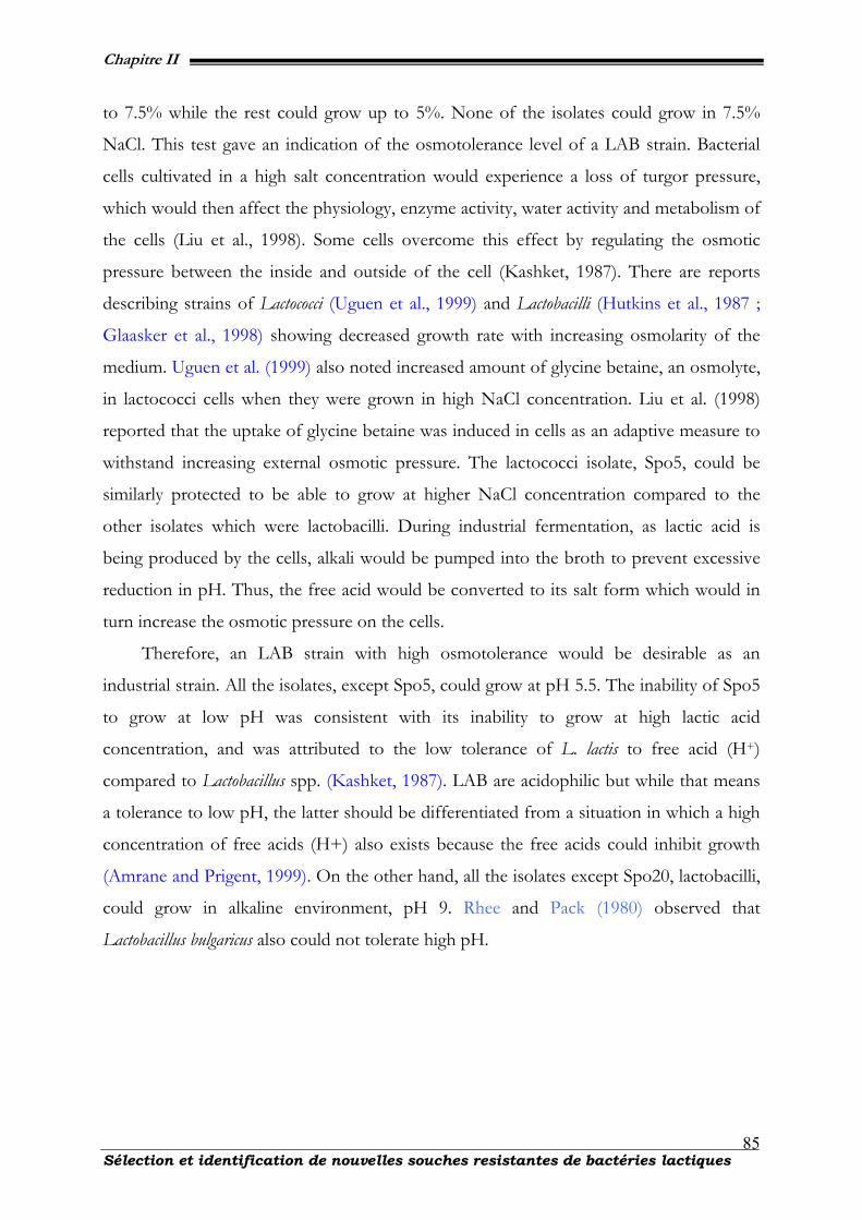
Chapitre II
Sélection et identification de nouvelles souches resistantes de bactéries lactiques
85
to 7.5% while the rest could grow up to 5%. None of the isolates could grow in 7.5%
NaCl. This test gave an indication of the osmotolerance level of a LAB strain. Bacterial
cells cultivated in a high salt concentration would experience a loss of turgor pressure,
which would then affect the physiology, enzyme activity, water activity and metabolism of
the cells (Liu et al., 1998). Some cells overcome this effect by regulating the osmotic
pressure between the inside and outside of the cell (Kashket, 1987). There are reports
describing strains of Lactococci (Uguen et al., 1999) and Lactobacilli (Hutkins et al., 1987 ;
Glaasker et al., 1998) showing decreased growth rate with increasing osmolarity of the
medium. Uguen et al. (1999) also noted increased amount of glycine betaine, an osmolyte,
in lactococci cells when they were grown in high NaCl concentration. Liu et al. (1998)
reported that the uptake of glycine betaine was induced in cells as an adaptive measure to
withstand increasing external osmotic pressure. The lactococci isolate, Spo5, could be
similarly protected to be able to grow at higher NaCl concentration compared to the
other isolates which were lactobacilli. During industrial fermentation, as lactic acid is
being produced by the cells, alkali would be pumped into the broth to prevent excessive
reduction in pH. Thus, the free acid would be converted to its salt form which would in
turn increase the osmotic pressure on the cells.
Therefore, an LAB strain with high osmotolerance would be desirable as an
industrial strain. All the isolates, except Spo5, could grow at pH 5.5. The inability of Spo5
to grow at low pH was consistent with its inability to grow at high lactic acid
concentration, and was attributed to the low tolerance of L. lactis to free acid (H+)
compared to Lactobacillus spp. (Kashket, 1987). LAB are acidophilic but while that means
a tolerance to low pH, the latter should be differentiated from a situation in which a high
concentration of free acids (H+) also exists because the free acids could inhibit growth
(Amrane and Prigent, 1999). On the other hand, all the isolates except Spo20, lactobacilli,
could grow in alkaline environment, pH 9. Rhee and Pack (1980) observed that
Lactobacillus bulgaricus also could not tolerate high pH.
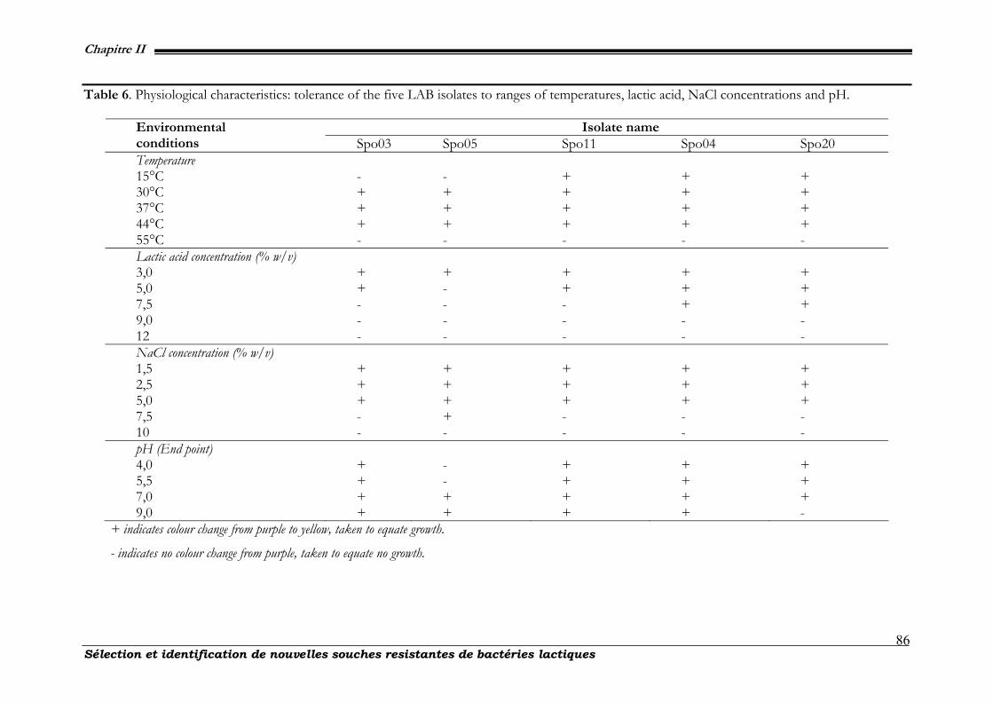
Chapitre II
Sélection et identification de nouvelles souches resistantes de bactéries lactiques
86
Table 6. Physiological characteristics: tolerance of the five LAB isolates to ranges of temperatures, lactic acid, NaCl concentrations and pH.
+ indicates colour change from purple to yellow, taken to equate growth.
- indicates no colour change from purple, taken to equate no growth.
Isolate name Environmental conditions Spo03 Spo05 Spo11 Spo04 Spo20 Temperature 15°C - - + + + 30°C + + + + + 37°C + + + + + 44°C + + + + + 55°C - - - - - Lactic acid concentration (% w/v) 3,0 + + + + + 5,0 + - + + + 7,5 - - - + + 9,0 - - - - - 12 - - - - - NaCl concentration (% w/v) 1,5 + + + + + 2,5 + + + + + 5,0 + + + + + 7,5 - + - - - 10 - - - - - pH (End point) 4,0 + - + + + 5,5 + - + + + 7,0 + + + + + 9,0 + + + + -

Chapitre II
87 Sélection et identification de nouvelles souches résistantes bactéries lactiques
II-2.3.3. Growth and lactic acid production profiles from time-course studies
In the first 4 h, Spo05, the lactococci strain, grew faster than Spo04 and Spo20, the
lactobacilli strains, based on cell dry weight (cdw) measurements. After this, the growth of
Spo5 leveled off at about 1.8 g/l cdw while Spo04 and Spo20 continued to register
biomass increase until 19h before their growth too leveled off at around 3.4 and 2.8 g/l
cdw, respectively (fig. 21). The low biomass produced by Spo5 with respect to Spo04 and
Spo20 correlated well with the lower amount of lactic acid produced by Spo05 (fig. 22)
resulting in a higher pH in the medium (fig. 23) and lower consumption of glucose (fig.
24).
0
0.5
1
1.5
2
2.5
3
3.5
4
0 6 12 18 24 30 36 42Time (hours)
Cel
l Dry
Wei
ght (
g/l).
Figure 21. Biomass produced by three strains of Lactobacilli: (♦) Spo05, (■) Spo04 and (▲)
Spo20 isolates over 42 h.
These results were consistent with those shown in tab. 5 where Spo5 was found to
be unable to tolerate high lactic acid concentrations and low pH compared to Spo04 and
Spo20. While the growth profiles of Spo04 and Spo20 did not appear to be different from
one another (fig. 21), the amount of lactic acid produced by Spo4 was higher than Spo20
for the first day, 24h (fig. 22).
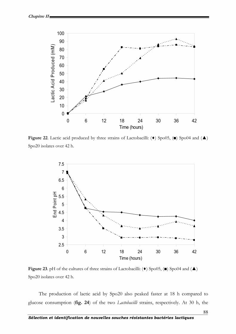
Chapitre II
88 Sélection et identification de nouvelles souches résistantes bactéries lactiques
010
20304050
607080
90100
0 6 12 18 24 30 36 42Time (hours)
Lact
ic A
cid
Pro
duce
d (m
M)
Figure 22. Lactic acid produced by three strains of Lactobacilli: (♦) Spo05, (■) Spo04 and (▲)
Spo20 isolates over 42 h.
2.5
3
3.5
4
4.5
5
5.5
6
6.5
7
7.5
0 6 12 18 24 30 36 42Time (hours)
End
Poi
nt p
H
Figure 23. pH of the cultures of three strains of Lactobacilli: (♦) Spo05, (■) Spo04 and (▲)
Spo20 isolates over 42 h.
The production of lactic acid by Spo20 also peaked faster at 18 h compared to
glucose consumption (fig. 24) of the two Lactobacilli strains, respectively. At 30 h, the
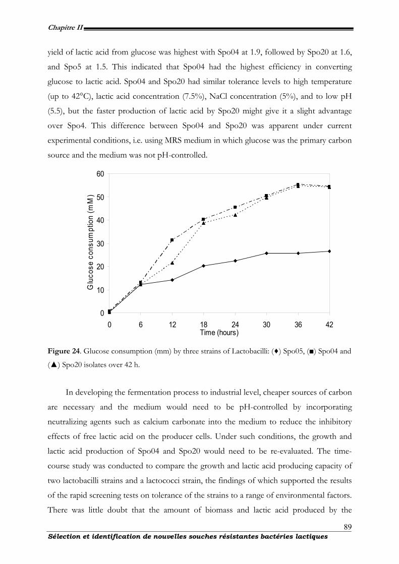
Chapitre II
89 Sélection et identification de nouvelles souches résistantes bactéries lactiques
yield of lactic acid from glucose was highest with Spo04 at 1.9, followed by Spo20 at 1.6,
and Spo5 at 1.5. This indicated that Spo04 had the highest efficiency in converting
glucose to lactic acid. Spo04 and Spo20 had similar tolerance levels to high temperature
(up to 42°C), lactic acid concentration (7.5%), NaCl concentration (5%), and to low pH
(5.5), but the faster production of lactic acid by Spo20 might give it a slight advantage
over Spo4. This difference between Spo04 and Spo20 was apparent under current
experimental conditions, i.e. using MRS medium in which glucose was the primary carbon
source and the medium was not pH-controlled.
0
10
20
30
40
50
60
0 6 12 18 24 30 36 42Time (hours)
Glu
cose
con
sum
ptio
n (m
M)
Figure 24. Glucose consumption (mm) by three strains of Lactobacilli: (♦) Spo05, (■) Spo04 and
(▲) Spo20 isolates over 42 h.
In developing the fermentation process to industrial level, cheaper sources of carbon
are necessary and the medium would need to be pH-controlled by incorporating
neutralizing agents such as calcium carbonate into the medium to reduce the inhibitory
effects of free lactic acid on the producer cells. Under such conditions, the growth and
lactic acid production of Spo04 and Spo20 would need to be re-evaluated. The time-
course study was conducted to compare the growth and lactic acid producing capacity of
two lactobacilli strains and a lactococci strain, the findings of which supported the results
of the rapid screening tests on tolerance of the strains to a range of environmental factors.
There was little doubt that the amount of biomass and lactic acid produced by the
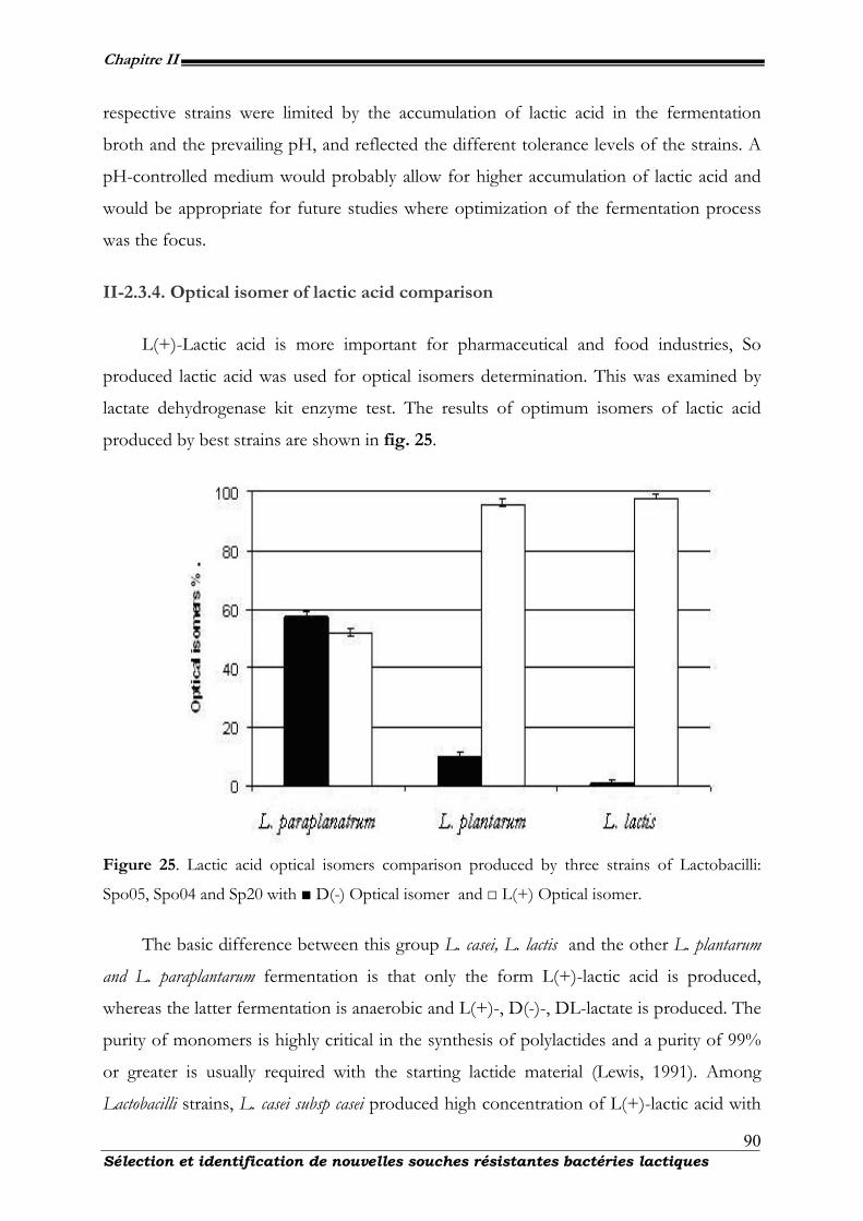
Chapitre II
90 Sélection et identification de nouvelles souches résistantes bactéries lactiques
respective strains were limited by the accumulation of lactic acid in the fermentation
broth and the prevailing pH, and reflected the different tolerance levels of the strains. A
pH-controlled medium would probably allow for higher accumulation of lactic acid and
would be appropriate for future studies where optimization of the fermentation process
was the focus.
II-2.3.4. Optical isomer of lactic acid comparison
L(+)-Lactic acid is more important for pharmaceutical and food industries, So
produced lactic acid was used for optical isomers determination. This was examined by
lactate dehydrogenase kit enzyme test. The results of optimum isomers of lactic acid
produced by best strains are shown in fig. 25.
Figure 25. Lactic acid optical isomers comparison produced by three strains of Lactobacilli:
Spo05, Spo04 and Sp20 with ■ D(-) Optical isomer and □ L(+) Optical isomer.
The basic difference between this group L. casei, L. lactis and the other L. plantarum
and L. paraplantarum fermentation is that only the form L(+)-lactic acid is produced,
whereas the latter fermentation is anaerobic and L(+)-, D(-)-, DL-lactate is produced. The
purity of monomers is highly critical in the synthesis of polylactides and a purity of 99%
or greater is usually required with the starting lactide material (Lewis, 1991). Among
Lactobacilli strains, L. casei subsp casei produced high concentration of L(+)-lactic acid with
Chapitre II
91 Sélection et identification de nouvelles souches résistantes bactéries lactiques
98% purity. Other Lactobacillus strains produce both optical isomers in combination. This
confirmed experimental work of Vaccari et al. 1993. This study described the sequential
steps of isolating bacteria from south African poultry farms (Senegal), screening the
isolates for LAB traits, selection of isolates based on a series of tests for industrially-
desirable traits, and finally compared the growth and lactic acid production profiles
between the lactobacilli and lactococci strains. In this study, the determination of optical
active isomer producers of lactic acid in the fermentation broth and provide a complete
profile of substrate utilization by these microorganisms.
II-2.4. Acknowledgements
The authors would like to acknowledge the republic of Ivory cost for its financial
assistance to this work and Mrs Maryse Hardenne and Mr Vincent Hote for their
contribution. We thank all the technical personal of CWBI (Centre Wallon de Biologie
Industrielle). We also like to express our gratitude to the republic of Ivory Cost and the
communauté française de Belgique for financial assistance.
II-2.5. References
Amrane A, Prigent Y (1999). Effect of the main culture parameters on the growth and production coupling of lactic acid bacteria. Appl. Microbiol. 2:101-108.
De Vos WM, Kleerebezem M, Kuipers OP (1997). Expression systems for industrial Gram-positive
bacteria with low guanine and cytosine content. Curr. Opin. Biotechnol. 8: 547-553. Drancourt M, Bollet C, Carta A, Rousselier P (2001). Phylogenetic analyses of Klebsiella species delineate
Klebsiella and Raoultella gen. nov., with description of Raoultella ornithinolytica comb. nov., Raoultella terrigena comb. nov. and Raoultella planticola comb. nov. Int. J. Syst. Evol. Microbiol. 51: 925-932.
Gasson, M. J. (1993) Progress and potential in the biotechnology of lactic acid bacteria. FEMS. Microbiol.
Rev. 12: 3-20. Gibson T, Abdel Malek T (1945) The formation of carbon dioxide by lactic acid bacteria and Bacillus
licheniformis and a cultural method of detecting the process. J. Dairy. Res. 14: 35-44. Glaasker E, Tjan FSB, Steeg PFT, Konings WN, Poolman B (1998). Physiological response of Lactobacillus
plantarum to salt and nonelectrolyte stress. J. Bacteriol. 180: 4718-4723. Gonzalez CJ, Encinas JP, Gracia-Lopez ML, Otero A (2000). Characterization and identification of lactic
acid bacteria from freshwater fishes. Food. Microbiol. 17: 383-391 Greetham H, Greetham L, Glenn R., Gibson CG, Hans H, Birgit M, Ulrike S, Enevold F, Matthew DC
(2003). Clostridium colicanis sp. nov., from canine faeces. Int. J. Syst. Evol. Microbiol. 53: 259-262.
Chapitre II
92 Sélection et identification de nouvelles souches résistantes bactéries lactiques
Hutkins RW, Ellefson WL, Kashket ER (1987). Betaine transport imparts osmotolerance on a strain of
Lactobacillus acidophilus. Appl. Environ. Microbiol. 43: 2275–2281 Kandler O, Weiss N (1986). In: Bergey'sManual of Systematic Bacteriology. Sneath, P. H. A. Mair, N. S.,
Sharpe, M. E., Holt, J. G. (Eds), Vol. 2, Baltimore: Williams and Wilkins, pp.1209-1234. Kashket ER (1987). Bioenergetics of lactic acid bacteria: Cytoplasmic pH and osmotolerance. FEMS.
Microbiol. Rev. 46: 233-244. Kharras GB, Sanchez-Riera F, Severson DK (1993). Polymers of lactic acid. In: Molby, D.B. (Ed.),
Plastics from microbes: Microbial synthesis of polymers and polymer precursors. Hanser Publ., pp.93-137.
Lewis DH, (1991). Controlled release of bioactive agents lactide/glycolide polymers. In: Polymers as drug
delivery systems. (Chasin, M. and Langer, R. eds.), Dekker Inc, New York, pp.1-41. Mayra-Makinen A, Bigret M (1998). Industrial use and production of lactic acid bacteria. In Lactic Acid
Bacteria. Microbiology and Functional Aspects, 2nd. (eds)S. Salminen & A. V. Wright. New York: Marcel Dekker. pp.73-102
Mollet B (1999). Genetically improved starter strains: opportunities for the dairy industry Int. Dairy. J
9:11-15. Monteagudo JM, Rodriguez L, Rincon J, Fuertes J (1997). Kinetics of lactic acid fermentation by
Lactobacillus delbrueckii grown on beet molasses. J. Chem .Tech. Biotech. 68: 271-276. Nakayama O, Yanoshi M (1967). Spore-bearing lactic acid bacteria isolated from rhizosphere. I.
Taxonomic studies on Bacillus laevolacticus nov. sp. and Bacillus racemilacticus nov. sp. J. Gen. Appl. Microbiol. 13: 139-153.
Reid G (1999). The scientific basis for probiotic strains of Lactobacillus. Appl. Environ. Microbiol. 65:
3763-3766. Ringoe E, Gatesoupe FJ (1998). Lactic acid bacteria in fish: a review Aquaculture. 160: 177-203. Rhee SK, Pack MY (1980). Effect of environmental pH on fermentation balance of Lactobacillus
bulgaricus. J. Bacteriol. 144: 217–221. Sanders W, Gerard V, Jan K (1999). Environmental stress responses in Lactococcus lactis. FEMS.
Microbiol. Rev. 23 483-501. Sghir A, Gramet G, Suau A, Rochet V, Pochart P, Doré J (2000). Quantification of bacterial groups within
human fecal flora by oligonucleotide probe hybridization. Appl. Environ. Microbiol. 66 2263-2266. Siebold M, Frieling PV, Joppien R, Rindfleisch D, Schugerl K, Roper H (1995). Comparision of the
production of lactic acid by three different Lactobacilli and its recovery by extraction and electrodialysis. Process. Biochem. 30: 81-95.
Sow N. M, Dubois, D. R., Roblain, D, Guiro AT, Thonart P. (2005) Polyphasic identification of a new thermotolerant species of lactic acid bacteria isolated from chicken faeces. A. J. Biotechnol. 4: 409-421.
Stiles ME, Holzapfel WH (1997). Lactic acid bacteria of foods and their current taxonomy Int. J. Food.
Microbiol. 36: 1-29.
Chapitre II
93 Sélection et identification de nouvelles souches résistantes bactéries lactiques
Uguen P, Hamelin J, Le Pennec J, Blanco C (1999). Influence of osmolarity and the presence of osmoprotectant on Lactococcus lactis growth and bacteriocin production. Appl Environ Microbiol. 63: 291–293.
Vaccari G, Gonzalez-Vara YRA, Campi AL, Dosi E, Brigidi P, Matteuzzi D (1993). Fermentative
production of L-lactic acid by Lactobacillus Casei DSM 20011 and product recovery using ion exchange resins. Appl. Microbiol. Biotech. 40: 23-27.
Ventura M, Casas I, Morelli L, Callegari ML (2000). Rapid amplified ribosomal DNA restriction analysis
(ARDRA) of Lactobacillus spp. isolated from fecal and vaginal samples. Syst Appl Microbiol 23:504-509.
Wouters JA, Kamphuis HH, Hugenholtz J, Kuipers P, De Vos WM, Abee T (2000). Changes in glycolytic
activity of Lactococcus lactis induced by low temperature. Appl. Environ. Microbiol. 66: 3686-3691.
Chapitre II
94 Sélection et identification de nouvelles souches résistantes bactéries lactiques
II-3. Conclusions : Sélection et identification de nouvelles souches
résistantes de bactéries lactiques
A l’issue de l’identification des isolats, les souches, Spo04 et Spo20 présentent les
caractéristiques de souches thermorésistantes. Pour identifier et caractériser ces souches,
la vérification de la présence de la catalase a été effectuée car elle permet de discriminer
les souches acidifiantes appartenant au genre Bacillus de celles appartenant au genre
Lactobacillus. L’absence de catalase chez les souches milite en faveur de l’hypothèse selon
laquelle ces souches appartiendraient au genre Lactobacillus. La resistance à l’acidité
gastrique a été étudiée afin de garantir les propriètés probiotiques des souches. D’après
l’étude de leur métabolisme fermentaire et la comparaison de l’ARNr16S avec la base de
donnée du NCBI grâce au logiciel Ribosomal Data Project II plus élargi de souches témoins,
sur le plan biochimique, la plupart d’entre elles sont relativement proches du genre
Lactobacillus. Cette conclusion partielle est confirmée par l’étude de la capacité
d’acidification des souches. Mais la comparaison des profils de croissance des souches
dans différentes conditions montre que ces souches présentent certaines différences par
rapport aux genres Lactobacillus et Bacillus. Le chapitre suivant est consacré à l’étude des
effets de l’utilisation des agents protecteurs sur la composition en acides gras de la
membrane et la viabilité au cours du stockage à température ambiante.
Chapitre III
Effets des cryoprotecteurs sur la composition en
acides gras cellulaires de Lactobacillus
plantarum CWBI-B1419
Chapitre III
Effets des cryoprotecteurs sur la composition en acides gras cellulaires 96
III. Effets des cryoprotecteurs sur la composition en acides acides
gras cellulaires de L. plantarum CWBI-B1419
III-1. Contexte et objectifs
L’ajout d’agents protecteurs ou cryo-protecteurs est l’un des facteurs les plus
déterminants pour améliorer la résistance des souches lors de la congélation ou de la
lyophilisation. Cet ajout a une influence sur la composition de la membrane bactérienne,
lorsque les composés sont utilisés soit dans le milieu de culture ou soit dans la pâte avant
lyophilisation. De Roissart et Luquet, (1994) ont montré qu’une variation, dans le rapport
entre la teneur en acides gras insaturés et celle en acides gras saturés, avait une incidence
sur la cryorésistance après que le milieu de culture soit supplémenté avec du Tween 80 ou
des additifs comme de l’acide oléique. Ceci pourrait expliquer l’amélioration de la
cryorésistance notée chez L. plantarum par Goldberg et Eschar, (1977) et la résistance chez
Lactobacillus. Dans ce chapitre, l’effet du sorbitol, du glutamate monosodique et du
glycérol sur la composition membranaire de la cellule a été étudié ; l’objectif étant de
trouver un agent protecteur pouvant garantir une survie à long terme des bactéries
lyophilisées.
Chapitre III
Effets des cryoprotecteurs sur la composition en acides gras cellulaires 97
III-2. The resistance to freeze-drying and to storage was
determined as the cellular ability to recover its survival rate and
acidification activity
Publication 4 – Int. J. Microbiol
International Journal of Microbiology
Coulibaly, I(1♣)., Dubois-D, R(1)., Destain, J(1)., Lognay, G(3)., Fauconnier, M.L
(4).,,Thonart, Ph (1, 2)
(1)Wallon Center for Industrial Biology, Bio-Industry Unit, Gembloux
Agricultural University, Passage des déportés 2, 5030 Gembloux,
Belgium.
(2)Wallon Center for Industrial Biology, Microbial Technology Unit,
University of Liège, Sart-Tilman B40, 4000 Liège, Belgium.
(3)Analytical Chemistry Unit, Gembloux Agricultural University, Passages
des déportés 2, 5030 Gembloux, Belgium.
(4)Plant Biology Unit, Gembloux Agricultural University, Passages des
déportés 2, 5030 Gembloux, Belgium.
♣ Corresponding Author : I. Coulibaly, Phone : +3281622305, Fax : +32816614222 E-mail : [email protected]
Chapitre III
Effets des cryoprotecteurs sur la composition en acides gras cellulaires 98
Résumé
L’effet des agents de protection sur le taux de survie et la résistance de Lactobacillus
plantarum CWBI-B1419 et Lactobacillus paracasei ssp. paracasei LMG9192T au cours de la
lyophilisation et pendant le stockage ultérieur est lié à la composition en acides gras de la
membrane. Les effets de ces protecteurs sur la composition en acides gras membranaires
et sur l’action de l'activité d'acidification des deux souches de Lactobacillus conservées à
20°C ont été étudiés. Par addition du sorbitol, du glutamate monosodique et du glycérol
comme agents cryoprotecteurs au cours du stockage, on note une baisse de l'acidification
pendant le stockage des cellules lyophilisées des deux souches et l’augmentation des
concentrations d'acides gras insaturés. L'addition du sorbitol et du glutamate
monosodique ne modifie pas la composition en acides gras quelle que soit la souche, mais
augmente la résistance à la lyophilisation de L. plantarum CWBI-B1419 et améliore le taux
de survie pendant le stockage. L'ajout de ces agents de protection et la diminution de
l'acidification améliorent le ratio des acides gras insaturés/saturés. Ces résultats indiquent
que la survie au cours du stockage et la résistance à la lyophilisation sont étroitement liées
à la composition en acides gras membranaires. Ce comportement peut être interprété
comme une adaptation de L. plantarum CWBI-B1419 à l'ajout du sorbitol et du glutamate
monosodique comme additifs. Une adaptation plus prononcée est observée chez L.
plantarum CWBI-B1419 que chez L. paracasei ssp. casei LMG9192.
Mots clés: taux de survie, bactéries lactiques, lyophilisation, stockage, viabilité,
composition des acides gras.
Chapitre III
Effets des cryoprotecteurs sur la composition en acides gras cellulaires 99
Abstract
The protective effects of the fatty acid composition and membrane action of the
acidification activity of two strains of Lactobacillus kept at 20°C were studied. The addition
of sorbitol, monosodium glutamate and glycerol during storage is causing the decline of
acidification and increased concentrations of unsaturated fatty acids observed in both
strains. The addition of sorbitol and monosodium glutamate does not alter the fatty acid
composition, whatever the strain, but increases the resistance to freeze-drying of L.
plantarum CWBI-B1419 and improves survival during storage. The addition of these
preservatives and decreased activity of acidification improves the ratio unsaturated. These
results indicate that the survival during storage and freeze-drying resistance are closely
related to the composition of membrane fatty acids. This behaviour can be interpreted as
an adaptation of L. plantarum CWBI-B1419 supplemented by cryoprotectant additives
such as sorbitol or monosodium glutamate sorbitol and monosodium glutamate as an
additive. L. plantarum CWBI-B1419 presents a greater adaptation to culture conditions
than L. paracasei ssp. casei LMG9192T.
Keywords: Survival rate, lactic acid bacteria, freeze-drying, storage, viability, fatty acid
composition.
Chapitre III
Effets des cryoprotecteurs sur la composition en acides gras cellulaires 100
III-2.1. Introduction
The acidification activity of lactic acid bacteria at the different steps of their production
(fermentation, cooling, concentration, cryoprotection, freezing, or freeze-drying) and
during storage differs depending on the strain considered and on the operating conditions
[1,2]. The freezing and freeze-drying step is especially critical as it negatively affects both
viability and physiological state of the bacteria [3,4]. The formation of ice crystals induces
mechanical damage that leads to cellular death during freezing [5]. In addition, the
crystallization of the water leads to a cryoconcentration of the solutes, which induces
some osmotic damage [6]. Some agents such as those used in lyoprotection are of
undoubted importance for the survival of cells because they can act on biological
functions in preserving the integrity of the lipid bilayer by the phenomenon of water
replacement, vitrification (glass formation), and depression of membrane transition
temperatures (Tm), as reviewed in detail by Castro et al. [7,8] and Champagne et al. [9].
The same observations with sucrose for survival of Lb. coryniformis Si3 were noticed by
Schoug et al. [10]. Adding cryoprotective agents such as sorbitol, monosodium glutamate,
and glycerol before freeze-drying process attenuated the damaging effects of freezing,
thus improving the bacterial resistance to drying [11,12]. This protective effect was
ascribed to interactions between sorbitol and the membrane phospholipids duringthe first
step of freeze-drying, freezing [13]. Because the cell membrane is the first target to
modification of the cell environment, its ability to adapt largely determines the
survivability of the cell [14,15]. By considering the important role of fatty acid
organization in membrane permeability, the membrane viscosity [16] and the membrane
thickness [17] were ascribed to the unsaturation index of membrane fatty acids: the cell
membrane adapts by increasing the proportion of unsaturated fatty acids, [18-20].
Unsaturated fatty acids promote exchanges between extracellular and intracellular media
by rigidifying the membrane and enhancing the membrane permeability. The increased
membrane permeability is related to the presence of the double bounds that tend to form
less stable Van-der-waals interactions with adjacent lipids [17]. As a consequence, altering
the fatty acid composition of the membrane may improve membrane permeability at low
temperature and then may allow the cell to adapt itself to freezing and freeze-drying [21].
They considered either the concentration in some unsaturated fatty acids, or the ratio
Chapitre III
Effets des cryoprotecteurs sur la composition en acides gras cellulaires 101
between unsaturated and saturated fatty acids (U/S). The U/S ratio depends on the
medium and environmental conditions in which the cells are cultivated and stored.
Concerning lactic acid bacteria, the addition of ethanol or polyol such as sorbitol in the
culture medium enhances the concentration in dihydrosterculic acid and the U/S ratio
[22,23]. The biosynthesis of unsaturated C18:1 fatty acids by some lactic acid bacteria is
stimulated by the addition of ethanol and leads to an increase of the U/S ratio [24].
Finally, the fatty acid composition evolves during storage. Castro et al. [7] observed two
phases: a first increase of the U/S ratio, which is explained by lipolysis reactions, followed
by a decrease. Linders et al. [25] showed that U/S ratio is stable within 90 d of storage
and then decreases. This decrease is linked to the oxidation of unsaturated fatty acids that
are very sensitive to oxygen [7] and is accentuated by an increase in the residual relative
humidity that activates the oxidation processes [8]. It is clear that acting on the membrane
fatty acid composition can modulate the U/S ratio. This was achieved by using
appropriate operating conditions and led to a better recovery of cellular viability after
freeze-drying and subsequent storage [26]. Nevertheless, as viability measurements are
insufficient to express both viability and physiological states of lactic acid bacteria, these
have to be proved by considering the acidification activity of lactic acid bacteria. This
work aimed to characterize the survival rate, resistance, and subsequent storage of a
freeze-dried Lactobacillus plantarum strain is in relation to its fatty acid composition. The
resistance to freeze-drying and to storage was determined as the cellular ability to recover
its survival rate and acidification activity.
III-2.2. Materials and methods
III-2.2.1. Microorganisms and growth conditions
III-2.2.1.1. Growth conditions
Lactobacillus plantarum CWBIB1419 was obtained fromthe collection of the Wallon
Center for Industrial’s Biology, Belgium and was originally isolated from a poultry farm.
Lactobacillus paracasei ssp. paracasei LMG9192T was obtained from the culture collection of
the Laboratory of Microbiology of Ghent’s University (Belgium). The original reference
cultures were maintained in cryogenic storage on glass beads at -80◦C. Working cultures
were maintained as slopes on de Man-Rogosa-Sharp agar (MRSa). Slopes were stored at
Chapitre III
Effets des cryoprotecteurs sur la composition en acides gras cellulaires 102
4°C and subcultured every month. L. plantarum CWBI-B1419 and L. paracasei ssp. paracsei
LMG9192T were grown in MRS medium (Merck) at 37°C. The inoculum of the tested
bacteria was prepared from strains stored on a substrate with glycerol at -80◦C using
appropriate media. Bacteria were proliferated for 24 hours and further culturing was
carried out after increasing the volume of the medium for the next 48 hours anaerobically
at 37°C. Bacteria prepared in this way provided the inoculum which was used at the
amount of 1% (v/v).
III-2.2.1.2. Scaling up
Strains were grown and performed in 100 L bioreactor at 37°C containing MRS
medium for 18 hours and then concentrated 20 times by centrifugation. The pH was
controlled at 6.5 by adding a 14% KOH. Cells were freeze-dried in a low freeze-drier
(Leybold, Belgium) with a standard programme by increasing the temperature gradually
from -45◦C to 25◦C at 0.9 mbar pressure (30 hours) followed by 15 hours at 0.15 mbar.
All fermentations were done in duplicate and average values reported.
III-2.2.1.3. Cells concentrations and storage
CellsCells were harvested in the stationary phase of growth by centrifugation
(7000×g, 30 minutes, 4°C). Concentrated cells were resuspended in the same weight of
supernatant at 4°C.Washed cells were then resuspended in sterile Ringer’s solution with
(3% w/w) of glycerol containing selected concentrations of compounds to be tested : 12
g/L sorbitol (s) and 12 g/L monosodium glutamate (msg), (Sigma, St. Louis, MO, USA).
Protectant solutions were sterilized at 121°C for 10 minutes before mixing with a volume
of washed cells. Cellular suspensions were maintained for 1 hour at room temperature
before freeze-drying, in order to create an isotonic environment between cells and the
compound added. Pastes were freezedried in trays, using an industrial freeze-dryer
(HETO, Heto-Holten A/S, Allerd, Denmark). The initial pressure was 0.2 mbar, the plate
temperature was -45°C and the end temperature of the product was 25°C. Freeze-drying
time varied according to the amount of material in the drying room and was
approximately 18-30 hours. Dried powders were stored in closed container at -20°C for
one night without light. Freeze-dried powders are divided in two parts: the first one of
about 2 ± 0.5 g was sealed in 15mL Falcon tubes and stored at room temperature (25°C)
Chapitre III
Effets des cryoprotecteurs sur la composition en acides gras cellulaires 103
without light for 8 months. The second part was vacuum-sealed in aluminium foil and
stored at room temperature in darkness.
III-2.2.2. Rehydration/enumeration
Viable counts were made before freeze-drying, after drying, and during storage at
regular intervals. After freeze-drying, samples were immediately brought to their original
volume 9mL with each peptoned water at 25°C. Then, samples were homogenized for 1
minute by Vortex mixer (SA-5, Stuart Scientific, UK) and incubated at room temperature
for 15 minutes. Serial dilutions were spread-plated on the surface of petri plates
containing MRS agar. These plates were incubated at 37°C for 48 hours, and the viability
was then determined by the drop count technique. Survival levels were expressed as the
ratio of colony-forming units per millilitre (cfu/ml) on MRS agar before (N0) and after
(Nf) freeze-drying. Viability (%) = (Nf/N0) × 100.
III-2.2.3. Acidification activity and water content
Acidification was carried out at 30°C in 120 mL MRS [27] broth inoculated with
1% of 107 Cfu·ml−1 of the freeze-dried sample. The total titratable acidity (% lactic acid/g
DW) was determinate after 18 hours, according to the AOAC method (1997). The water
content (g H2O/100 g dry weight) of the freezedried samples was determined after drying
at 105°C until constant weight. Water activities of the dried samples after freeze-drying
and the saturated salt solutions were measured and confirmed with a Novasina (Novasina,
Pf¨affikon, Switzerland) water activity meter (Aqua Lab, CX-2, Decagon Devices,
INC,Washington, U.S.A.) by the dew point method with a standard deviation SD ± 0.003,
respectively. Residual water content was < 3% in all samples.
III-2.2.4. Fatty acids analyses (FAME)
Total lipids extraction was performed according to the adapted method of [28].
Fatty acid methyl esters were prepared by incubating the lipid extracts at 70°C for 90
minutes in 0.5 mL of methanol-BF3, containing 15% (v/v) of KOH and 0.2 mL of
hexane.The fatty acids were extracted to the upper phase of regent mixture containing
1mL of hexane, 0.5mL of saturated NaCl; H2SO4 10% 0.2 mL and analysed by gas
Chapitre III
Effets des cryoprotecteurs sur la composition en acides gras cellulaires 104
chromatography on a HP 6890 (Hewlett Packard, Germany) equipped with a flame
ionization detector and a SPTM-2560, 100m∗ 0.25mm∗ 0.2 µmfused silica capillary
column. The conditions were as follows: injector temperature, 260°C; detector
temperature, 260°C; carrier gas (helium) flow rate, 3 mL/min. The oven temperature was
programmed from 140°C during 5 minutes to 240°C at 4°C/min. For peak identification,
standard solution (Sigma) was used. The results were relative percentages of fatty acids,
determined from peak areas of methyl esters. They were means of three independent
experiments.
III-2.2.5. Statistical Analysis.
A four-factor analysis of variance with two-factor interactions (Stastica 9) was
performed to determine the effects of sorbitol, the addition of monosodium glutamate
and glycerol, and the length of storage. The Neuman-Keuls multiple comparison
procedure was used to discriminate among the means for significant differences at the
5% confidence level. All results presented in this paper are the average of three
independent replicate assays. The ratio between the standard deviations and the means
values was between 2% and 5%.
III-2.3. Results and discussion
III-2.3.1. Fatty acid composition of freeze-dried L. plantarum CWBI-B1419
and L. paracasei ssp. paracasei LMG9192T.
Cellular fatty acids (CFAs) from L. plantarum CWBI-B1419 and L. paracasei ssp.
paracasei LMG9192T (control) were analyzed before and after freeze-drying. A total of six
fatty acids made up of the membrane of those strains cultivated at pH 6.5, in absence or
presence of sorbitol and monosodium glutamate in the washed cells solution (tab.7). This
presence or absence of those additives did not affect the number of fatty acid detected or
identified. As control U/S ratios were 0.57 and 0.58 for L. plantarum CWBI-B1419 and L.
paracasei LMG9192, respectively; saturated and unsaturated fatty acids were well balanced.
When sorbitol and monosodium glutamate were added in the cellular suspensions, the
U/S ratio was higher than 0.66 for L. plantarum CWBI-B1419 and 0.62 for L. paracasei
Chapitre III
Effets des cryoprotecteurs sur la composition en acides gras cellulaires 105
LMG9192, respectively, thus indicating a shift from saturated to unsaturated fatty acids in
the membrane composition. This was due to a decrease in C16:0, and C18:0 and an increase
in C16:1 and C18:1 fatty acids. As confidence intervals overlapped, C18:2 and C18:3 did not
show any significant difference, whether or not additives were present. Except for the
C16:1, most of these fatty acids have been identified in other species of lactic acid bacteria:
L. lactis subsp. lactis and Lactobacillus sp. [23], Lb. bulgaricus [8, 29], Lb. plantarum [30, 31],
Lb. buchneri and Lb. fermentum [32], Lactobacillus sp. [33] and Leuconostoc mesenteroides [24].
III-2.3.2. Impacts of protectives agents and storage time on membrane fatty
acid composition of L. plantarum CWBI-B1419 and L. paracasei sp. paracasei
LMG9192T.
The effect of addition of sorbitol, monosodium glutamate and glycerol as
protectives agents and storage time on membrane fatty acid composition was studied. The
ratio between unsaturated and saturated fatty acids was more affected by the addition of
those additives and the storage time, while the addition of glycerol had no effect. Relative
fatty acid concentrations were diversely influenced by glycerol, monosodium glutamate
and sorbitol. The addition of glycerol as cryoprotective agent did not modify the fatty acid
composition of the membrane because this molecule was added after the concentration
step, immediately before freeze-drying. At this stage of production, cells were still in a
quiet physiological state. Moreover, at this temperature, the membrane permeability is not
high [34] and the passive diffusion of the glycerol from extracellular to intracellular
medium was clarified. Consequently, glycerol probably acted mainly as an extracellular
cryoprotective agent [13]. As expected, the addition of sorbitol and monosodium
glutamate increased the concentration of unsaturated fatty acids C18:1 and C16:1, and
decreased the concentration of saturated fatty acids C16:0 and C18:0 (P < .001), without any
modification of C18:1 and C18:2 proportions, (tab. 7). The U/S ratio increased from 0.57 to
0.66 with sorbitol and from 0.57 to 0.62 when monosodium glutamate was used. These
results agree with those reported by Champagne et al. [9] and Anchordoguy et al. [13]
who showed that the U/S ratio is strongly dependent on the presence of polyol in the
medium. This indicated that sorbitol, when incorporated in the intracellular medium,
increase thus unbalancing of the fatty acid composition..
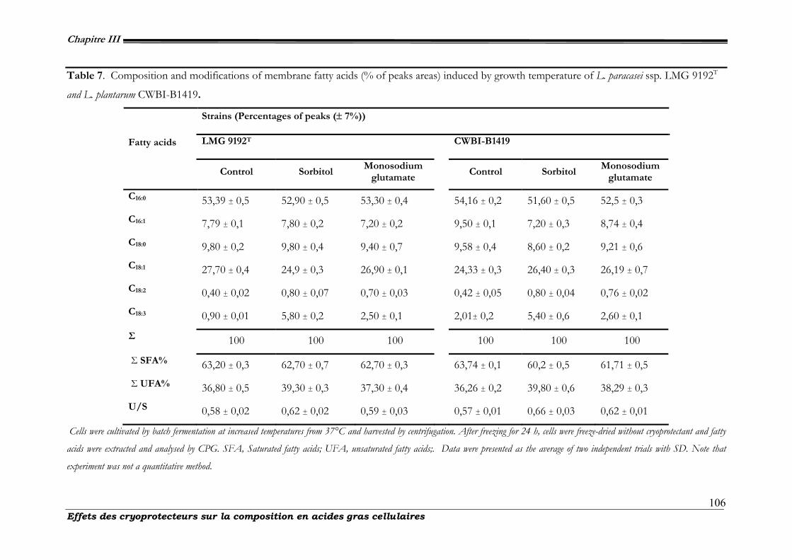
Chapitre III
Effets des cryoprotecteurs sur la composition en acides gras cellulaires 106
Table 7. Composition and modifications of membrane fatty acids (% of peaks areas) induced by growth temperature of L. paracasei ssp. LMG 9192T
and L. plantarum CWBI-B1419.
Strains (Percentages of peaks (± 7%))
LMG 9192T
CWBI-B1419 Fatty acids
Control Sorbitol Monosodium
glutamate Control Sorbitol
Monosodium glutamate
C16:0 53,39 ± 0,5 52,90 ± 0,5 53,30 ± 0,4 54,16 ± 0,2 51,60 ± 0,5 52,5 ± 0,3
C16:1 7,79 ± 0,1 7,80 ± 0,2 7,20 ± 0,2 9,50 ± 0,1 7,20 ± 0,3 8,74 ± 0,4
C18:0 9,80 ± 0,2 9,80 ± 0,4 9,40 ± 0,7 9,58 ± 0,4 8,60 ± 0,2 9,21 ± 0,6
C18:1 27,70 ± 0,4 24,9 ± 0,3 26,90 ± 0,1 24,33 ± 0,3 26,40 ± 0,3 26,19 ± 0,7
C18:2 0,40 ± 0,02 0,80 ± 0,07 0,70 ± 0,03 0,42 ± 0,05 0,80 ± 0,04 0,76 ± 0,02
C18:3 0,90 ± 0,01 5,80 ± 0,2 2,50 ± 0,1 2,01± 0,2 5,40 ± 0,6 2,60 ± 0,1
Σ 100 100 100 100 100 100
Σ SFA% 63,20 ± 0,3 62,70 ± 0,7 62,70 ± 0,3 63,74 ± 0,1 60,2 ± 0,5 61,71 ± 0,5
Σ UFA% 36,80 ± 0,5 39,30 ± 0,3 37,30 ± 0,4 36,26 ± 0,2 39,80 ± 0,6 38,29 ± 0,3
U/S 0,58 ± 0,02 0,62 ± 0,02 0,59 ± 0,03 0,57 ± 0,01 0,66 ± 0,03 0,62 ± 0,01
Cells were cultivated by batch fermentation at increased temperatures from 37°C and harvested by centrifugation. After freezing for 24 h, cells were freeze-dried without cryoprotectant and fatty
acids were extracted and analysed by CPG. SFA, Saturated fatty acids; UFA, unsaturated fatty acids;. Data were presented as the average of two independent trials with SD. Note that
experiment was not a quantitative method.
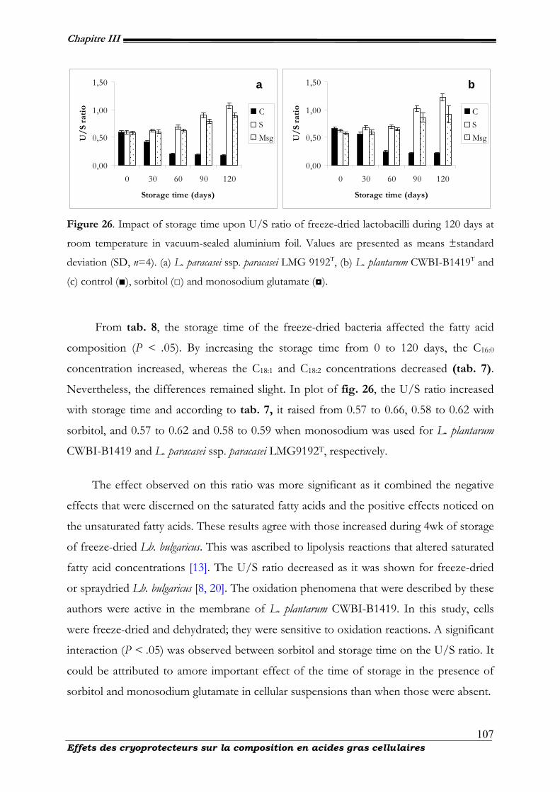
Chapitre III
Effets des cryoprotecteurs sur la composition en acides gras cellulaires 107
0,00
0,50
1,00
1,50
0 30 60 90 120
Storage time (days)
U/
S ra
tio C
SMsg
0,00
0,50
1,00
1,50
0 30 60 90 120
Storage time (days)
U/
S ra
tio C
SMsg
Figure 26. Impact of storage time upon U/S ratio of freeze-dried lactobacilli during 120 days at
room temperature in vacuum-sealed aluminium foil. Values are presented as means ±standard
deviation (SD, n=4). (a) L. paracasei ssp. paracasei LMG 9192T, (b) L. plantarum CWBI-B1419T and
(c) control (■), sorbitol (□) and monosodium glutamate (◘).
From tab. 8, the storage time of the freeze-dried bacteria affected the fatty acid
composition (P < .05). By increasing the storage time from 0 to 120 days, the C16:0
concentration increased, whereas the C18:1 and C18:2 concentrations decreased (tab. 7).
Nevertheless, the differences remained slight. In plot of fig. 26, the U/S ratio increased
with storage time and according to tab. 7, it raised from 0.57 to 0.66, 0.58 to 0.62 with
sorbitol, and 0.57 to 0.62 and 0.58 to 0.59 when monosodium was used for L. plantarum
CWBI-B1419 and L. paracasei ssp. paracasei LMG9192T, respectively.
The effect observed on this ratio was more significant as it combined the negative
effects that were discerned on the saturated fatty acids and the positive effects noticed on
the unsaturated fatty acids. These results agree with those increased during 4wk of storage
of freeze-dried Lb. bulgaricus. This was ascribed to lipolysis reactions that altered saturated
fatty acid concentrations [13]. The U/S ratio decreased as it was shown for freeze-dried
or spraydried Lb. bulgaricus [8, 20]. The oxidation phenomena that were described by these
authors were active in the membrane of L. plantarum CWBI-B1419. In this study, cells
were freeze-dried and dehydrated; they were sensitive to oxidation reactions. A significant
interaction (P < .05) was observed between sorbitol and storage time on the U/S ratio. It
could be attributed to amore important effect of the time of storage in the presence of
sorbitol and monosodium glutamate in cellular suspensions than when those were absent.
a b
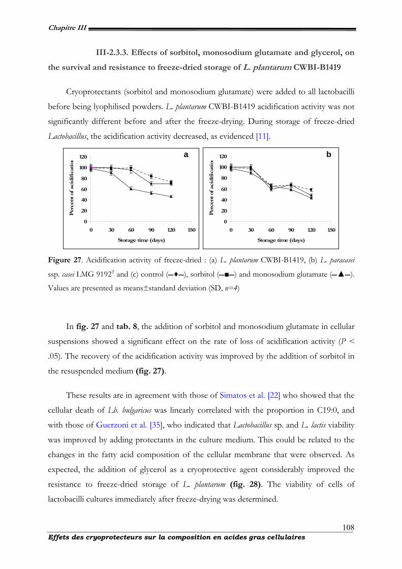
Chapitre III
Effets des cryoprotecteurs sur la composition en acides gras cellulaires 108
III-2.3.3. Effects of sorbitol, monosodium glutamate and glycerol, on
the survival and resistance to freeze-dried storage of L. plantarum CWBI-B1419
Cryoprotectants (sorbitol and monosodium glutamate) were added to all lactobacilli
before being lyophilised powders. L. plantarum CWBI-B1419 acidification activity was not
significantly different before and after the freeze-drying. During storage of freeze-dried
Lactobacillus, the acidification activity decreased, as evidenced [11].
0
20
40
60
80
100
120
0 30 60 90 120 150
Storage time (days)
Per
cent of
aci
dif
icat
ion
0
20
40
60
80
100
120
0 30 60 90 120 150
Storage time (days)
Per
cent of
aci
dif
icat
ion
Figure 27. Acidification activity of freeze-dried : (a) L. plantarum CWBI-B1419, (b) L. paracasei
ssp. casei LMG 9192T and (c) control (♦), sorbitol (■) and monosodium glutamate (▲).
Values are presented as means±standard deviation (SD, n=4)
In fig. 27 and tab. 8, the addition of sorbitol and monosodium glutamate in cellular
suspensions showed a significant effect on the rate of loss of acidification activity (P <
.05). The recovery of the acidification activity was improved by the addition of sorbitol in
the resuspended medium (fig. 27).
These results are in agreement with those of Simatos et al. [22] who showed that the
cellular death of Lb. bulgaricus was linearly correlated with the proportion in C19:0, and
with those of Guerzoni et al. [35], who indicated that Lactobacillus sp. and L. lactis viability
was improved by adding protectants in the culture medium. This could be related to the
changes in the fatty acid composition of the cellular membrane that were observed. As
expected, the addition of glycerol as a cryoprotective agent considerably improved the
resistance to freeze-dried storage of L. plantarum (fig. 28). The viability of cells of
lactobacilli cultures immediately after freeze-drying was determined.
a b
Chapitre III
Effets des cryoprotecteurs sur la composition en acides gras cellulaires 109
The L. paracasei ssp. paracasei LMG9192T strain revealed the lowest percentage of cell
recuperation when monosodium glutamate was used as a protective agent. L. plantarum
CWBI-B1419 was observed to have higher survival than L. paracasei ssp. paracasei LMG
9192T during storage. It was found good viability of lactobacilli species after drying and
subsequent storage when sorbitol and monosodium glutamate were added in cell culture
[9]. The rate of loss in acidification activity was three times lower when glycerol was
added. This result confirmed the findings of Fonseca et al. [12]. Moreover, the glycerol
effect was revealed whatever the other experimental conditions were (presence of sorbitol
and monosodium glutamate in cellular suspensions). This indicated that the glycerol
displayed an additional effect that can be combined with other beneficial effects. It was
found that, survival and cryoprotectants effects of the freeze-dried samples decreased as
the storage time increased.
However, after the 120 days storage period, a significantly lost of viable population
were noted in the freeze-dried samples held at 20◦C in vacuum-sealed aluminium foil at
20◦C, respectively (P < .05) (fig. 28). For example, viable population of freeze-dried
lactobacilli held in vacuum-sealed aluminium foil was reduced from an initial population
of 7.4 × 10¹¹ to 4.77 × 10⁹ or 3.07 × 10⁹ cfu/g with a survival percentage of 64.4% or
41.4% after 120d at 20◦C for L. paracasei ssp. paracasei LMG 9192T with sorbitol,
respectively (tab. 8).
At the same period, survival of Lactobacillus plantarum CWBIB1419 with
monosodium glutamate decrease again, after 120 days at 20◦C in the same conditions, the
viable population was about 8.3 × 10¹¹ or 3.57 × 10⁹ cfu/g with a survival of 43.7%,
respectively. In addition, viability was significantly higher for freeze-dried strain in
vacuum-sealed with sorbitol, aw = 0.11 (P < .05). At the end of freeze-drying, the water
content of freeze-dried sample was 3.6 ± 0.6 g H2O/100 g DW with aw = 0.09 ± 0.01.
After the 120 days storage period, the water content for all the samples stored at 20◦C
was not changed significantly (approximately 0.6-fold) according to the package (P < .05).
As expected, water content for samples stored under vacuum did not change significantly
during storage with sorbitol or monosodium glutamate (P < .05), respectively (tab.9).
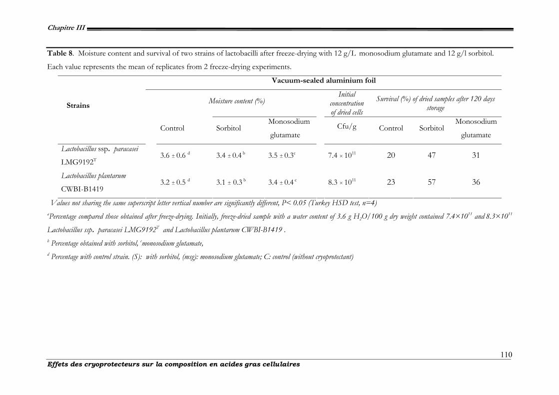
Chapitre III
Effets des cryoprotecteurs sur la composition en acides gras cellulaires 110
Table 8. Moisture content and survival of two strains of lactobacilli after freeze-drying with 12 g/L monosodium glutamate and 12 g/l sorbitol.
Each value represents the mean of replicates from 2 freeze-drying experiments.
Vacuum-sealed aluminium foil
Moisture content (%) Initial
concentration of dried cells
Survival (%) of dried samples after 120 days storage Strains
Control Sorbitol Monosodium
glutamate Cfu/g Control Sorbitol
Monosodium
glutamate
Lactobacillus ssp. paracasei
LMG9192T 3.6 ± 0.6 d 3.4 ± 0.4 b 3.5 ± 0.3c 7.4 × 10¹¹ 20 47 31
Lactobacillus plantarum
CWBI-B1419
3.2 ± 0.5 d 3.1 ± 0.3 b 3.4 ± 0.4 c
8.3 × 10¹¹ 23 57 36
Values not sharing the same superscript letter vertical number are significantly different, P< 0.05 (Turkey HSD test, n=4) aPercentage compared those obtained after freeze-drying. Initially, freeze-dried sample with a water content of 3.6 g H2O/100 g dry weight contained 7.4×10¹¹ and 8.3×10¹¹
Lactobacillus ssp. paracasei LMG9192T and Lactobacillus plantarum CWBI-B1419 . b Percentage obtained with sorbitol, cmonosodium glutamate, d Percentage with control strain. (S): with sorbitol, (msg): monosodium glutamate; C: control (without cryoprotectant)
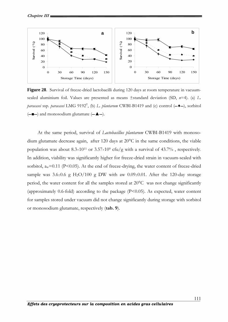
Chapitre III
Effets des cryoprotecteurs sur la composition en acides gras cellulaires 111
0
20
40
60
80
100
120
0 30 60 90 120 150
Storage Time (days)
Surv
ival
( %
)
0
20
40
60
80
100
120
0 30 60 90 120 150
Storage Time (days)
Surv
ival
( %
)
Figure 28. Survival of freeze-dried lactobacilli during 120 days at room temperature in vacuum-
sealed aluminium foil. Values are presented as means ±standard deviation (SD, n=4). (a) L.
paracasei ssp. paracasei LMG 9192T, (b) L. plantarum CWBI-B1419 and (c) control (♦), sorbitol
(■) and monosodium glutamate (▲).
At the same period, survival of Lactobacillus plantarum CWBI-B1419 with monoso-
dium glutamate decrease again, after 120 days at 20°C in the same conditions, the viable
population was about 8.3×10¹¹ or 3.57×10⁹ cfu/g with a survival of 43.7% , respectively.
In addition, viability was significantly higher for freeze-dried strain in vacuum-sealed with
sorbitol, aw=0.11 (P<0.05). At the end of freeze-drying, the water content of freeze-dried
sample was 3.6±0.6 g H2O/100 g DW with aw 0.09±0.01. After the 120-day storage
period, the water content for all the samples stored at 20°C was not change significantly
(approximately 0.6-fold) according to the package (P<0.05). As expected, water content
for samples stored under vacuum did not change significantly during storage with sorbitol
or monosodium glutamate, respectively (tab. 9).
a b

Chapitre III
Effets des cryoprotecteurs sur la composition en acides gras cellulaires 112
Table 9. Dry cell weight of freeze-dried cells, conserved at 20°C and safe from oxygen and moisture. Water activity of freeze-dried cells, conserved at
20°C and safe from oxygen and moisture.
Vacuum-sealed aluminium foil
Dry cell weight (±1%) Water activity aw (± 0.01)
Strains Sorbitol Monosodium
glutamate Control Sorbitol
Monosodium
glutamate Control
LMG 9192T 98.1 ± 0.89a 98.1 ± 0.90 97.98 ± 0.98 0.10 ± 0.01 0.12 ± 0.01 0.18 ± 0.009
CWBI-B1419
98.6 ± 0.98 97.2 ± 0.93 97.56 ± 0.83
0.11 ± 0.009 0.11 ± 0.007 0.11 ± 0.001
a The means correspond to four repetitions. s: with sorbitol; msg: monosodium glutamate; c: control (without cryoprotectant) a Initial value of survival immediately after freeze-drying. b Value of cell survival obtained after 120 days
Chapitre III
Effets des cryoprotecteurs sur la composition en acides gras cellulaires 113
III-2.3.4. Relationship between the resistance, freeze-dried L. plantarum
CWBI-B1419 storage and its membrane fatty acid composition.
The fatty acid composition of L. plantarum CWBI-B1419 and the ability of the cells
to restore their acidification activity were closely related. The importance of the ratio rate
between saturated fatty acids and unsaturated fatty acids results on resistance to freezing
and better long-term storage of freeze-dried bacteria [36]. This was observed in all the
operating conditions used during the fermentation and freeze-dried storage. Addition of
sorbitol or monosodium glutamate to the drying medium indicates a significant change in
the survival rate during storage as the confidence level (P < .05). Furthermore, the effects
were synergistic, except in the case of the existence of interactions [37]. A better
resistance to freeze-dried storage was achieved by adding sorbitol and more monosodium
glutamate in resuspended medium that led to higher U/S ratios. The positive effect of
sorbitol was detected regardless of the fermentation pH and whether or not glycerol was
added. Adding sorbitol increased the permeability for water of the membrane, thus
favoring transport across the membrane [17]. The ability of monosodium glutamate to
protect microorganisms during cryopreservation and freeze-drying has previously been
described [38,39]. The majority of Lactobacillus sp. tested and freeze-dried with
monosodium glutamate has shown an increased survival during storage. The stabilisation
of their protein structure via reactions between the amino group of the protectant and the
carboxyl groups of the microorganism’s proteins and the ability to retain greater amounts
of residual moisture have been pointed out by de Valdez et al. [40] as explanations that
account for protection by monosodium glutamate during freeze-drying and subsequent
storage. The cells were then more adapted to suffer the intracellular ice crystallization
during freezing and the water mobility during storage [41]. Furthermore, the membrane
lipids interact with protein interfaces, either by maintaining the protein structure and
activity, or by inhibiting or activating protein functions, such as carrier proteins, which
mediate solute transport [9]. For example, the activity of the membrane bound enzyme
Na-K-ATPase was shown to be regulated by the lipid portion of the membrane [42].
When sorbitol was added, the effect on the rate of loss in acidification activity could be
explained by an influence on some enzymatic activities that may modify the protein
Chapitre III
Effets des cryoprotecteurs sur la composition en acides gras cellulaires 114
composition and content of the cell. This is corroborated by the results of Rallu et al. [43]
who reported, in the case of L. lactis, an increase in the concentration of cold-shock
proteins when the cells suffered a stress. The appearance of unsaturated fatty acids can
then be linked to disadvantageous growth conditions [10]. The positive effect of glycerol
on the resistance to powders storage is independent of the U/S ratio, which was not
affected by this factor [42]. This indicates that two different mechanisms accounted for
the different rates of loss in acidification activity: first, a better cellular adaptation, related
to the higher water permeability of the membrane, achieved with high U/S ratios; second
a cryoprotective effect of glycerol that took place in addition to the previous effects. This
interpretation corroborated the previous hypothesis that this molecule probably acted as
an extracellular cryoprotective agent. The relationship between the fatty acid composition
of L. plantarum and its ability to recover acidification activity led to important
consequences for performing starter production [25]. From our results, the resistance to
freeze-dried storage was improved by increasing the U/S ratio that was obtained by
applying unfavorable experimental conditions for growth and by adding sorbitol in the
resuspended medium [28]. This concept can be broadened by relating the membrane fatty
acid composition to disadvantageous growth conditions, such as low temperature [22,44,
45] acid stress [43], ethanol stress [45], salt stress osmotic stress [32] or high age of the
culture [3,46]. These results demonstrated that sorbitol has a strong protective effect
upon the survival of L. plantarum CWBI-B1419 and L. paracasei subsp. paracasei LMG
9192T, during storage, even though no significant differences were observed in terms of
viability of cells during freeze-drying in the presence or absence of sorbitol.
III-2.4. Conclusions
The rate of loss in acidification activity during freeze-dried storage varied according
to the conditions in which the cells were cultivated and cryoprotected. The resistance to
drying and freeze-dried storage of lactic acid starters was defined by its ability to recover
acidification activity after thawing. The resistance to freeze-dried powder during storage
was improved by growing at unfavorable conditions and by adding additives (sorbitol and
monosodium glutamate) in the resuspended medium and glycerol as protective agent.
This improvement was related to an increase of the membrane ratio between unsaturated
Chapitre III
Effets des cryoprotecteurs sur la composition en acides gras cellulaires 115
and saturated fatty acids. The relationship between the U/S ratio and the addition of
protectants was obvious. Cryoprotectants such as sorbitol and monosodium glutamate are
almost equally effective in protecting lactic acid bacteria dried by freeze-drying. The
mechanism of the action of sorbitol and monosodium glutamate appears related to water
permeation as sorbitol equally protects cells under hypertonic stress and preserves the
osmotic response of the bacteria.
III-2.5. Acknowledgements
The authors thank all the technical’s personals of Walloon Center for industrial
Biology. Maryse Hardenne and Celine Pierrart are acknowledged for her very important
contribution in the experiments. They also like to express our gratitude to the Republic of
Ivory cost for its financial assistance to this work. The authors gratefully acknowledge the
French community of Belgium.
III-2.6. References
1. C. Beal, F. Mihoub, M. Marin, and G. Corrieu, “Demande de Brevet d’invention FR N◦2829147. Procedé de préparation d’une composition lyophilisée contenant des bactéries lactiques à viabilité et activité bactériennes améliorées lors d’un stockage à température ambiante et composition obtenue,” 2001.
2. E. Selmer-Olsen, S.-E. Birkeland, and T. Sørhaug, “Effect of protective solutes on leakage from and survival of immobilized Lactobacillus subjected to drying, storage and rehydration,” Journal of Applied Microbiology, vol. 87, no. 3, pp. 429-437, 1999.
3. M. M. Brashears and S. E. Gilliland, “Survival during frozen and subsequent refrigerated storage of Lactobacillus acidophilus cells as influenced by the growth phase,” Journal of dairy science, vol. 78, no. 11, pp. 2326-2335, 1995.
4. R. Foschino, E. Fiori, and A. Galli, “Survival and residual activity of Lactobacillus acidophiluss frozen cultures under different conditions,” Journal of Dairy Research, vol. 63, no. 2, pp. 295-303, 1996.
5. E. E. Tymczyszyn, A. Gomez-Zavaglia, and E. A. Disalvo, “Influence of the growth at high osmolality on the lipid composition, water permeability and osmotic response of Lactobacillus bulgaricus,” Archives of Biochemistry and Biophysics, vol. 443, no. 1-2, pp. 66-73, 2005.
6. B. M. Corcoran, R. P. Ross, G. F. Fitzgerald, P. Dockery, and C. Stanton, “Enhanced survival of GroESL-overproducing Lactobacillus paracasei NFBC 338 under stressful conditions induced by drying,” Applied and Environmental Microbiology, vol. 72, no. 7, pp. 5104-5107, 2006.
7. H. P. Castro, P. M. Teixeira, and R. Kirby, “Changes in the cell membrane of Lactobacillus bulgaricus during storage following freeze-drying,” Biotechnology Letters, vol. 18, no. 1, pp. 99-104, 1996.

Chapitre III
Effets des cryoprotecteurs sur la composition en acides gras cellulaires 116
8. H. Castro, P. P. M. Teixeira, and R. Kirby, “Storage of lyophilised cultures of Lactobacillus bulgaricus under different relative humilities and atmospheres,” Applied Microbiology and Biotechnology, vol. 44, pp. 172-176, 1995.
9. C. P. Champagne, F. Mondou, Y. Raymond, and D. Roy, “Effect of polymers and storage temperature on the stability of freeze-dried lactic acid bacteria,” Food Research International, vol. 29, no. 5-6, pp. 555-562, 1996.
10. A. Schoug, J. Fischer, H. J. Heipieper, J. Schn¨urer, and S. H°akansson, “Impact of fermentation pH and temperature on freeze-drying survival and membrane lipid composition of Lactobacillus coryniformis Si3,” Journal of Industrial Microbiology and Biotechnology, vol. 35, no. 3, pp. 175-181, 2008.
11. F. Fonseca, C. Béal, and G. Corrieu, “Method of quantifying the loss of acidification activity of lactic acid starters during freezing and frozen storage,” Journal of Dairy Research, vol. 67, no. 1, pp. 83-90, 2000.
12. F. Fonseca, C. B´eal, F. Mihoub, M. Marin, and Gs. Corrieu, “State diagrams and sorption isotherms of bacterial suspensions and fermented medium,” Thermochimica Acta, vol. 366, no. 2, pp. 167-182, 2001.
13. T. J. Anchordoguy, A. S. Rudolph, J. F. Carpenter, and J.H. Crowe, “Modes of interaction of cryoprotectants with membrane phospholipids during freezing,” Cryobiology, vol. 24, no. 4, pp. 324-331, 1987.
14. J. ˇSajbidor, “Effect of some environmental factors on the content and composition of microbial membrane lipids,” Critical Reviews in Biotechnology, vol. 17, no. 2, pp. 87-103, 1997.
15. R. K. Thunell, W. E. Sandine, and F. W. Bodyfelt, “Frozen starters from internal pH control grown cultures,” Journal of Dairy Science, vol. 67, pp. 24-36, 1984.
16. E. P.W. Kets and J. A. M. de Bont, “Protective effect of betaine on survival of Lactobacillus plantarum subjected to drying,” FEMS Microbiology Letters, vol. 116, no. 3, pp. 251-256, 1994.
17. G. In’t Veld, A. J. M. Driessen, and W. N. Konings, “Effect of the unsaturation of phospholipid acyl chains on leucine transport of Lactococcus lactis and membrane permeability,” Biochimica et Biophysica Acta, vol. 1108, no. 1, pp. 31-39, 1992.
18. M. Brennan, B. Wanismail, M. C. Johnson, and B. Ray, “Cellular damage in dried Lactobacillus acidophilus,” Journal of Food Protection, vol. 49, pp. 47-53, 1986.
19. S. E. Gilliland and C. N. Rich, “Stability during frozen and subsequent refrigerated storage of Lactobacillus acidophilus grown at different pH,” Journal of Dairy Science, vol. 73, pp. 1187-1192, 1991.
20. M. Suutari and S. Laakso, “Temperature adaptation in Lactobacillus fermentum: interconversions of oleic, vaccenic and dihydrosterulic acids,” Journal of General Microbiology, vol. 138, no. 3, pp. 445-450, 1992.
21. E. Costa, J. Usall, N. Teixido, N. Garcia, and I. Vinas, “Effect of protective agents rehydration media and initial cell concentration on viability of Pantoea agglomerans strain CPA-2 subjected to freeze-drying,” Journal of Applied Microbiology, vol. 89, no. 5, pp. 793-800, 2000.
22. D. Simatos, G. Blond, M. LeMeste, and M. Morice, “Conservation des bactéries lactiques par congélation et lyophilisation,” in Bact´eries Lactiques, H. de Roissart and F. M Luquet, Eds., vol. 2, pp. 555–573, 1994.
Chapitre III
Effets des cryoprotecteurs sur la composition en acides gras cellulaires 117
23. I. Goldberg and L. Eschar, “Stability of lactic acid bacteria to freezing as related to their fatty acid composition,” Applied and Environmental Microbiology, vol. 33, no. 3, pp. 489-496, 1977.
24. P. A. Schmitt, G. Mathot, and C. Divies, “Fatty acid composition of the genus Leuconostoc,” Milchwissenschaft, vol. 44, pp. 556-558, 1989.
25. L. J. Linders, W. F. Wolkers, F. A. Hoekstra, and K. Van’t Riet, “Effect of added carbohydrates onmembrane phase behaviour and survival of dried Lactobacillus plantarum,” Cryobiology, vol. 35, no. 1, pp. 31-40, 1997.
26. F. Dellaglio, G. E. Felis, and S. Torriani, “The status of Lactobacillus casei (Orla-Jensen 1916) Hansen and Lessel 1971 and Lactobacillus paracasei Collins et al. 1989. Request for opinion,” International Journal of Systematic and Evolutionary Microbiology, vol. 52, no. 1, pp. 285-287, 2002.
27. J. C. DeMan, A. M. Rogosa, and M. E. Sharpe, “A medium for the cultivation of lactobacilli,” Journal of Applied Bacteriology, vol. 23, pp. 130-135, 1960.
28. E. P.W.Kets, P. J. M. Teunissen, and J. A. M. de Bont, “Effect of compatible solutes on survival of lactic acid bacteria subjected to drying,” Applied and Environmental Microbiology, vol. 62, no. 1, pp. 259-261, 1996.
29. P.M. Teixeira, H. P. Castro, and R. Kirby, “Evidence of membrane lipid oxidation of spray-dried Lactobacillus bulgaricus during storage,” Letters in Applied Microbiology, vol. 22, no. 1, pp. 34-38, 1996.
30. A. A. Yao, I. Coulibaly, L. Georges, M.-L. Fauconnier, and P. Thonart, “Impact of polyunsaturated fatty acid degradation on survival and acidification activity of freeze-dried Weissella paramesenteroides LC11 during storage,” Applied Microbiology and Biotechnology, vol. 79, no. 6, pp. 1045-1052, 2008.
31. N. Rozes, S. Garbay, M. Denayrolles, and A. Lonvaud-Funel, “A rapid method for the determination of bacterial fatty acid composition,” Letters in Applied Microbiology, vol. 17, no. 3, pp. 126-131, 1993.
32. L. J. M. Linders, E. P. W. Kets, J. A. M. de Bont, and K. van’t Riet, “Combined effect of growth and drying conditions on the activity of dried Lactobacillus plantarum,” Biotechnology Progress, vol. 14, pp. 537-539, 1998.
33. F. Dionisi, P.-A. Golay, M. Elli, and L. B. Fay, “Stability of cyclopropane and conjugated linoleic acids during fatty acid quantification in lactic acid bacteria,” Lipids, vol. 34, no. 10, pp. 1107-1115, 1999.
34. Y. Mille, L. Beney, and P. Gervais, “Compared tolerance to osmotic stress in various microorganisms: towards a survival prediction test,” Biotechnology and Bioengineering, vol. 92, no. 4, pp. 479-484, 2005.
35. M. E. Guerzoni, R. Lanciotti, and P. S. Cocconcelli, “Alteration in cellular fatty acid composition as a response to salt, acid, oxidative and thermal stresses in Lactobacillus helveticus,” Microbiology, vol. 147, no. 8, pp. 2255-2264, 2001.
36. N. J. Russell and N. Fukunaga, “A comparison of thermaladaptation of membrane lipids in psychrophilic and thermophilic bacteria,” FEMS Microbiology Reviews, vol. 75, no.2-3, pp. 171-182, 1990.
37. H. W. Wisselink, R. A. Weusthuis, G. Eggink, J. Hugenholtz, and G. J. Grobben, “Mannitol production by lactic acid bacteria: a review,” International Dairy Journal, vol. 12, no. 2-3, pp. 151-161, 2002.
Chapitre III
Effets des cryoprotecteurs sur la composition en acides gras cellulaires 118
38. G. F. de Vald´ez, G. S. De Giori, A. P. De Ruiz Holgado, and G. Oliver, “Protective effect of adonitol on lactic acid bacteria subjected to freeze-drying,” Applied and EnvironmentalMicrobiology, vol. 45, no. 1, pp. 302-304, 1983.
39. R. S. Porubcan and R. L. Sellars, “Stabilized dry cultures of lactic acid-producing bacteria,” US Patent 3 897 307, July 1975.
40. G. F. de Vald´ez, G. S. De Giori, A. P. De RuizHolgado, and G. Oliver, “Effect of drying medium on residual moisture content and viability of freeze-dried lactic acid bacteria,” Applied and Environmental Microbiology, vol. 49, no. 2, pp. 413-415, 1985.
41. Y. Wang, G. Corrieu, and C. B´eal, “Fermentation pH and temperature influence the cryotolerance of Lactobacillus acidophilus RD758,” Journal of Dairy Science, vol. 88, no. 1, pp. 21-29, 2005.
42. N. J. Russell, “Bacterial membranes: the effects of chill storage and food processing. Overview,” International Journal of FoodMicrobiology, vol. 79, no. 1-2, pp. 27–34, 2002.
43. F. Rallu, A. Gruss, S. D. Ehrlich, and E. Maguin, “Acid- and multistress-resistant mutants of Lactococcus lactis: identification of intracellular stress signals,”MolecularMicrobiology, vol. 35, no. 3, pp. 517-528, 2000.
44. N. J. Russell, R. I. Evans, P. F. TerSeeg, J.Hellemons, A. Verheul, and T. Abee, “Membranes as a target for stress adaptation,” International Journal of Food Microbiology, vol. 28, no. 2, pp.255-261, 1995.
45. A. Lonvaud-Funel and C. Desens, “Constitution en acides gras des membranes des bact´eries lactiques du vin. Incidences des conditions de culture,” Sciences des Aliments, vol. 10, pp. 817-829, 1990.
46. Z. Drici-Cachon, J. F. Cavin, and C. Divi`es, “Effect of pH and age of culture on cellular fatty acid composition of Leuconostoc oenos,” Letters in Applied Microbiology, vol. 22, no. 5, pp. 331-334, 1996.
.
Chapitre III
Effets des cryoprotecteurs sur la composition en acides gras cellulaires 119
III-3. Conclusions : Effets des cryoprotecteurs sur la composition
en acides gras cellulaires de Lactobacillus plantarum CWBI-B1419
L’utilisation des agents additifs permet d’obtenir des taux de viabilité très élevé.
Cette étude a montré que la survie des bactéries lactiques, telle que L. plantarum CWBI-
B1419, L. paracasei ssp. casei LMG9192 soumises à une lyophilisation dépendent des agents
de protection utilisés pendant la lyophilisation et au cours du stockage. Une sélection
appropriée de ces facteurs est essentielle pour obtenir la viabilité maximale des cellules
pour leur utilisation comme probiotiques. Ces résultats sont nécessaires pour le
développement industriel de la formulation de la culture du starter. Les cryoprotecteurs
tels que le sorbitol et le monosodium glutamate sont aussi efficaces dans la protection des
bactéries lactiques lyophilisées. La concentration et la conservation des cellules
bactériennes sont donc tributaires de l’utilisation d’additifs. Malgré toutes ces techniques
et formulations, il faut constater une perte de viabilité engendrée par la présence de
l’oxygène qui engendre des composés primaires (hydroperoxydes) et secondaires
(composés aldéhydiques principalement). C’est dans l’optique de comprendre ces
phénomènes et leur importance sur la survie cellulaire que le chapitre suivant est consacré
à leur analyse.
Chapitre IV
Suivi de la dégradation des acides gras
polyinsaturés
Chapitre IV
Suivi de la dégradation des acides gras polyinsaturés
121
IV. Suivi de la dégradation des acides gras polyinsaturés
IV.1. Contexte et objectifs
L’un des soucis majeurs de tout industriel travaillant dans la production de starters
lactiques reste la perte de viabilité des cellules déshydratées. Stabiliser cette viabilité des
starters lactiques au cours du stockage est et demeure un challenge quotidien. Parmi les
techniques de séchage (atomisation, lyophilisation, fluidisation, etc), il est indispensable
d’adopter celle qui garantit une stabilité des cellules ayant gardé leurs potentialités. Dans
cette optique, la lyophilisation la plus couramment utilisée a retenu notre attention.
Comme toute technique de séchage, la lyophilisation n’est pas sans effet sur la cellule elle-
même. Au nombre des dégats engendrés par la lyophilisation sur la cellule déshydratée,
l’oxydation des lipides membranaires revêt une importance primordiale, car elle est
responsable de la perte de viabilité au cours du temps des cellules lyophilisées. L’un de
nos objectifs serait de comprendre les différents mécanismes d’oxydation qui s’établissent
au niveau de la cellule une fois lyophilisée. Dans cette étude, Lactobacillus plantarum CWBI-
B1419 a été utilisée en comparant sa résistance à celle de Leuconostoc mesenteroides. En
relation avec le taux de viabilité au cours du stockage, l’activité d’eau, le taux de matière
séche ont été déterminés. Différentes températures de stockage ont été necessaires afin
d’établir une corrélation entre la mortalité cellulaire et l’oxydation des lipides
membranaires chez Lactobacillus plantarum pendant nonante jours.
Chapitre IV
Suivi de la dégradation des acides gras polyinsaturés
122
IV.2. Survival of freeze-dried Leuconostoc mesenteroides KMrog
and Lactobacillus plantarum CWBI-B1419 related to their cellular
fatty acids composition during storage
Publication 5 – Appl. Biochem. Biotechnol.
Applied Biochemistry and Biotechnology
Coulibaly, I (1♣)., Yao, A.A (2)., Fauconnier, M.L (3)., Lognay, G (4) and Ph. Thonart
(1,2)
(1)Wallon Center for Industrial Biology, Bio-Industry Unit, Gembloux
Agricultural University, Passage des déportés 2, 5030 Gembloux, Belgium.
(2)Wallon Center for Industrial Biology, Microbial Technology Unit, University
of Liège, Sart-Tilman B40, 4000 Liège, Belgium.
(3)Analytical Chemistry Unit, Gembloux Agricultural University, Passages des
déportés 2, 5030 Gembloux, Belgium.
(4)Plant Biology Unit, Gembloux Agricultural University, Passages des déportés
2, 5030 Gembloux, Belgium.
♣ Corresponding Author : I. Coulibaly, Phone : +3281622305, Fax : +32816614222 E-mail : [email protected]
Chapitre IV
Suivi de la dégradation des acides gras polyinsaturés
123
Résumé
Les souches de bactéries de Lactobacillus plantarum CWBI-B534 et de Leuconostoc ssp.
mesenteroides (L. mesenteroides) Kenya MRog2 ont été produites en bioréacteur, concentrées,
avec ou sans cryoprotecteurs. En général, le taux de viabilité n'a pas subi de changements
significatifs après lyophilisation (p>0,05). Dans la plupart des cas, le taux de viabilité des
cellules avec cryoprotecteurs était significativement plus élevé que celles sans additifs
(p<0,05). Les acides gras cellulaires (AGC) des deux souches ont été analysés avant et
après lyophilisation. Six AGC ont été identifiés, à savoir, l’acide palmitique (C16:0), l’acide
palmitoléique (C16:1), l’acide stéarique (C18:0), l’acide oléique (C18:1), l’acide linoléique (C18:2)
et l'acide linolénique (C18:3). Quatre d'entre eux, C16:0, C16:1, C18:0 et C18:1, représentent plus
de 94% ou 93% des acides gras cellulaires chez L. mesenteroides et L. plantarum,
respectivement, avec une faible proportion du C18:3, soit 5 et 6%, respectivement. Le C18:2
a été détecté avec des pourcentages très faibles (≤ 1%) au niveau de chaque souche.
L’acide palmitique (C16:0) est présenté en forte proportion au niveau des deux souches. La
teneur en humidité et l'activité de l'eau (aw) a significativement augmenté au cours du
stockage. On a observé que les ratios C16:1/C16:0, C18:0/C16:0 et C18:1/C16:0 pour les cellules
lyophilisées de L. mesenteroides ou L. plantarum, avec ou sans cryoprotecteurs, n'ont pas
beaucoup changé au cours de la période de stockage. Selon le mode d'emballage et des
températures d'entreposage, les ratios C18:2/C16:0 et C18:3/C16:0 pour L mesenteroides et L.
plantarum, avec ou sans cryoprotecteurs ont diminué au cours de cette même période.
Toutefois, un ratio plus élevé de C18:2 /C16:0 et de C18:3/C16:0 pour L. mesenteroides et L.
plantarum a été noté au niveau des poudres conservées à 4°C ou sous vide à l’abri de
lumière et à 20°C en présence d'oxygène et de lumière.
Mots-clés: viabilité, stress oxydatif, stress thermique, lyophilisation, acides gras cellulaires,
bactéries lactiques.
Chapitre IV
Suivi de la dégradation des acides gras polyinsaturés
124
Abstract
Lactic acid bacteria strains Lactobacillus plantarum CWBI-B534 and Leuconostoc ssp.
mesenteroïdes (L. mesenteroïdes) Kenya MRog2 were produced in bioreactor, concentrated,
with or without cryoprotectants. In general, viable population did not change significantly
after freeze-drying (p>0.05). In most cases, viable population for cells added with
cryoprotectants was significantly higher than those without (p<0.05). Cellular fatty acids
(CFAs) from the two strains in this study were analyzed before and after freeze-drying.
Six CFAs were identified, namely, palmitic (C16:0), palmitoleic (C16:1), stearic (C18:0), oleic
(C18:1), linoleic (C18:2), and linolenic (C18:3) acids were identified. Four of them, C16:0, C16:1,
C18:0 and C18:1, make up more than 94% or 93% of the fatty acids in L. mesenteroides and L.
plantarum, respectively, with another one, namely, C18:3, making a smaller (on average 5-
6%, respectively) contribution. The C18:2 contributed very small percentages (on average≤
1%) to the total in each strain. C16:0 had the highest proportion at most points relative to
other fatty acids. Moisture content and water activity (aw) increased significantly during
the storage period. It was observed that C16:1/C16:0, C18:0/C16:0 and C18:1/C16:0 ratios for
freeze-dried L. mesenteroides or L. plantarum, with or without cryoprotectants, did not
change significantly during the storage period. According to the packaging mode and
storage temperatures, C18:2/C16:0 and C18:3/C16:0 ratios for freeze-dried L. mesenteroides and
L. plantarum with or without cryoprotectants decreased as the storage time increased.
However, a higher C18:2/C16:0 or C18:3/C16:0 ratio for L. mesenteroides and L. plantarum was
noted in the freeze-dried powder held at 4°C or under vacuum and in dark at 20°C or in
the presence of oxygen and light.
Keywords: viability, oxidative stress, heat stress, freeze-drying, cellular fatty acids, lactic
acid bacteria.
Chapitre IV
Suivi de la dégradation des acides gras polyinsaturés
125
IV-2.1. Introduction
The introduction of appropriate starter culture techniques may constitute one major
step towards improved safety, quality and security of traditional small-scale fermentation
in Africa (Holzapfel, 1997). The industrial use of lactic starter cultures for the food
industry depends on the concentration and preservation technologies employed, which
are required to guaranty long-term delivery of stable cultures in term of viability and
functional activity (Carvalho et al., 2003). Freeze-drying has commonly been used for this
purpose; nevertheless freeze-dried cells are likely to lose their viability during storage
(Fonseca et al., 2000 ; Béal et al., 1994 ; Champagne et al., 1991). Lipid oxidation of
membrane fatty acids was deemed responsible for cell death during storage (Teixeira et al.,
1996). This is supported by indirect evidence presented in previous reports. For example,
chemical injury in the form of free radical damage has been suggested by most researchers
as one of the major culprits in desiccation injury (Pereira et al., 2003). Loss of water
increases the ionic concentration (which can lead to the formation of reactive oxygen
species [ROS]), and in the dry state, bio-molecules become more susceptible to the attack
of oxygen. These species can damage proteins, modify bases and sugars in
deoxyribonucleic acid (DNA) and cause lipid peroxidation (Pereira et al., 2003 ; Hansen et
al., 2006). It is reported that the presence of antioxidants increased the survival rate of
dried bacteria during storage (Teixeira et al., 1996, 1995). Dimmick and Heckly (1961)
found a strong similarity between the loss of viability and the increase in free-radical
concentration during storage of freeze-dried Serratia marcescens. Marshall et al. (1974)
supported the hypothesis that reactions between carbonyl compounds and cellular
components are a major cause of mortality during storage of dried micro-organisms.
However, to date no papers have been published identifying which bacterial cellular fatty
acids are in fact oxidized or degraded during storage. In living organisms lipids,
particularly polyunsaturated fatty acids (PUFAs) components of cell membranes, are
described as being extremely subject to environmental stress (Prior, 1971 ; Girotti et al.,
1984). Stresses such as decreases in aw (Halverson et al., 2000) or increases in temperature
(Théberge et al., 1996) are known to affect the fatty acid composition of bacterial. A
number of factors, such as temperature, atmosphere, exposure to light and moisture
influence the viability of freeze-dried cultures (Andersen et al., 1999). It is well-established
Chapitre IV
Suivi de la dégradation des acides gras polyinsaturés
126
that cryoprotectants are almost indispensable when freezing and drying micro-organisms,
but the contribution of these compounds to stability of dried-microorganisms during
storage is discussed (Hamoudi et al., 2007). The disadvantages of dried cultures undermine
their application, but advantages can outweigh disadvantages if the inactivation during
storage can be more clearly understood and consequently reduced. Therefore, the
purpose of this work was to study the influence of oxygen, humidity and temperature on
survival or cellular fatty acids of freeze-dried Leuconostoc mesenteroides and Lactobacillus
plantarum, with or without cryoprotectants, during storage and, if possible, to show the
interrelation between viability and polyunsaturated fatty acids degradation. We used
whole-cell fatty acid methyl ester (FAME) analysis to examine the effect of storage
conditions on fatty acid composition, as this approach reflects the fatty acid composition
of phospholipids (Cronan et al., 1987 ; Suzuki et al., 1993) and lipopolysaccharides (Kieft et
al., 1994).
IV-2.2. Materials and methods
IV-2.2.1. Micro-organisms and cultivation
The lactic acid bacteria L. mesenteroides ssp. mesenteroides Kenya MRog2, isolated and
identified from cassava fermentation for gari production (Kostinek et al., 2005) was
provided by the Federal Research Centre for Nutrition, Institute of Hygiene and
Toxicology (Karlsruhe, Germany). The Lactobacillus plantarum CWBI-B534 was isolated
from poultry farms in vicinity of Dakar (Senegal, West Africa). MRS broths inoculated
with each strain were incubated at 30°C for 18h. The supernatants obtained after
centrifugation (Sorvall RC2-B, Sorvall, USA) at 2500 x g for 20 min, were decanted. The
cell pellets obtained were resuspended in 5 ml (50% v/v) glycerol and frozen at -80°C.
IV-2.2.2. Productions
The strains were grown in 500 L bioreactor containing MRS medium for 18h and
then concentrated 20 times by centrifugation. After centrifugation cryoprotectants (2%
(w/w) glycerol and 5% (w/w) maltodextrine)) were added to the pellets (PC). Cell
suspensions without additifs were used as control (P). Cells were freeze-dried in a Low
Chapitre IV
Suivi de la dégradation des acides gras polyinsaturés
127
freeze-drier (Leybold, Belgium) with a standard programme by increasing the temperature
gradually from -45°C to 25°C at 0.9 mbar pressure (30 h) followed by 15h at 0.15 mbar.
All fermentations were done in duplicate and average values reported.
IV-2.2.3. Storage conditions
Freeze-dried samples were stored during 90 days, in portion-size (2 ± 0.5 g in each
portion) at 20°C (accelerated stability test, 30% RH) and at 4°C (stability at refrigerated
temperature, 70% RH). At each storage temperature, samples were stored, in open 20 ml
white glass tubes and in aluminium foil packets which were vacuum-sealed. Samples were
withdrawn for analysis each 30 days and kept at -20°C (48-72h) before analysis.
IV-2.2.4. Dry cell weight and water content determinations
The dry cell weight of 1g of powder was determined at the end of freeze-drying. The
water content (100 g dry weight)-1 of the freeze-dried samples during storage at 20 and
4°C with aeration was determined each 15 days. The samples were dried in a convection
oven until constant weight and results are mean of four determinations.
IV-2.2.5. Water activity measurements
The water activity (aw) of the freeze-dried samples was measured at 25°C using a
water activity meter Novasina (Novasina, Pfäffikon, Switzerland). Standard salt solutions
(Novasina) of known water activity were used for calibration of the sensor at the
measuring temperature. Readings were taken until four sequential readings gave the same
results. Results obtained were the average of four determinations for each sample.
IV-2.2.6. Survival rate
Percentage survival of the strains after freeze-drying process was expressed as
follows:
Survival (%) = 10011
22 ×××
PCPC
Chapitre IV
Suivi de la dégradation des acides gras polyinsaturés
128
Where C1 and C2 are the CFU/g of the suspension before and after freeze-drying and P1
and P2 are amount of matter obtained before and after freeze-drying. Percentage survival
after 90- day storage was calculated as 100×N/N0, where N is the CFU/g of the freeze-
dried sample at a given time and N0 is the CFU/g of the sample at the end of freeze-
drying.
IV-2.2.7. Analysis of cellular fatty acids
Cells wall fractions were obtained as described previously by Ndoye et al., 2006. The
lipids were extracted overnight from cell wall fractions (2.5 ml) and dried cells (1 g), with
ethanol-ether (3:1, v/v) mixture according to an adaptation of the method of Ito et al.,
(1969). Ethanol ether extracts were pooled, filtrated, and then evaporated and
concentrated under reduced pression at 35°C. Fatty acid methyl esters (FAME) were
prepared from the concentrate with 14% (w/w) solution of boron trifluoride in methanol
as reagent (Sigma, St Louis, Missouri, USA). After heating at 70°C in a water bath for 90
min, 0.5 ml of saturated Nacl, 0.2 ml of sulphuric acid (10%) and 0.5 ml of hexane were
added. The methylated fatty acids were extracted from the upper phase after decanting by
means of a Pasteur-pipette. Gas chromatographic analysis of the methyl esters was carried
out on a HP 6890 (Hewlett Packard) gas chromatograph equipped with a flame ionization
detector at 250°C. A capillary column (30 m x 0.25 mm (i.d), film thickness 0.25 µm) was
used. Helium was used as carrier gas (2.4 ml/min) and the injection volume was 1 µl.
Injection was done at 250°C in splitless mode for 1 min. The oven temperature was held
at 50°C for 1 min, increased by 30°C/min to 120°C, and then from 120°C to 240°C at
4°C/min with a final hold of 10 min at 240°C. Fatty acids methyl esters were identified by
comparing their retention times with standards mixtures FAME MIX 47885U (Supelco,
Bellefonte, USA).
The relative fatty acid content was estimated as a percentage of the total peak area
using a DP 700 integrator (Spectra physics). The relative content (%) of each fatty acid
was normalized by expressing it as a ratio of the relative content (%) of palmitic (C16:0)
acid for two reasons: [1] C16:0 had the highest proportion at most points relative to other
fatty acids, [2] C16:0 did not change significantly during storage (p> 0.05) (data not shown).
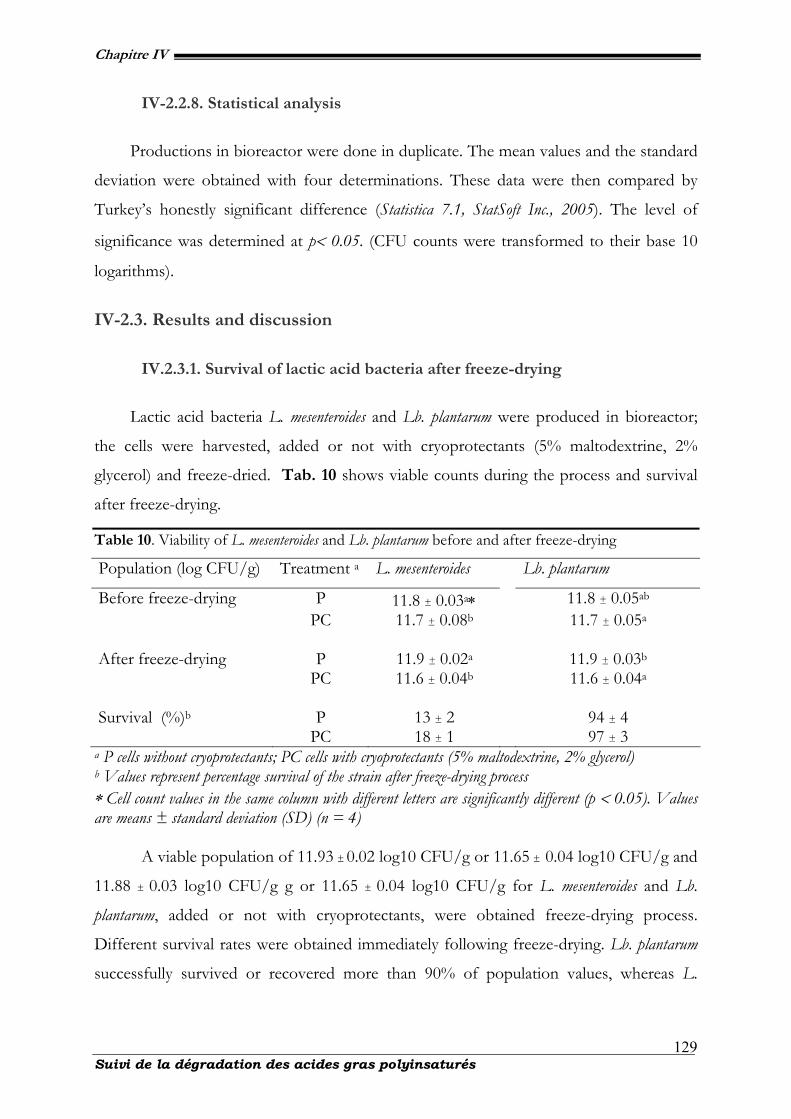
Chapitre IV
Suivi de la dégradation des acides gras polyinsaturés
129
IV-2.2.8. Statistical analysis
Productions in bioreactor were done in duplicate. The mean values and the standard
deviation were obtained with four determinations. These data were then compared by
Turkey’s honestly significant difference (Statistica 7.1, StatSoft Inc., 2005). The level of
significance was determined at p< 0.05. (CFU counts were transformed to their base 10
logarithms).
IV-2.3. Results and discussion
IV.2.3.1. Survival of lactic acid bacteria after freeze-drying
Lactic acid bacteria L. mesenteroides and Lb. plantarum were produced in bioreactor;
the cells were harvested, added or not with cryoprotectants (5% maltodextrine, 2%
glycerol) and freeze-dried. Tab. 10 shows viable counts during the process and survival
after freeze-drying.
Table 10. Viability of L. mesenteroides and Lb. plantarum before and after freeze-drying
Population (log CFU/g) Treatment a L. mesenteroides Lb. plantarum
Before freeze-drying P 11.8 ± 0.03a∗ 11.8 ± 0.05ab PC 11.7 ± 0.08b 11.7 ± 0.05a After freeze-drying P 11.9 ± 0.02a 11.9 ± 0.03b PC 11.6 ± 0.04b 11.6 ± 0.04a Survival (%)b P 13 ± 2 94 ± 4 PC 18 ± 1 97 ± 3
a P cells without cryoprotectants; PC cells with cryoprotectants (5% maltodextrine, 2% glycerol) b Values represent percentage survival of the strain after freeze-drying process ∗ Cell count values in the same column with different letters are significantly different (p < 0.05). Values are means ± standard deviation (SD) (n = 4)
A viable population of 11.93 ± 0.02 log10 CFU/g or 11.65 ± 0.04 log10 CFU/g and
11.88 ± 0.03 log10 CFU/g g or 11.65 ± 0.04 log10 CFU/g for L. mesenteroides and Lb.
plantarum, added or not with cryoprotectants, were obtained freeze-drying process.
Different survival rates were obtained immediately following freeze-drying. Lb. plantarum
successfully survived or recovered more than 90% of population values, whereas L.
Chapitre IV
Suivi de la dégradation des acides gras polyinsaturés
130
mesenteroides behaved with low biomass yield (<11%). The effect of the additives is not
significant.
IV-2.3.2. Influence of water activity on survival rate.
Moisture content and water activity (aw) were determined each 15 days during
storage at 4°C (70% RH) and 20°C (30% RH) in opened white glass tube containing
2±0.2g of dried powder. Relationships between aw and moisture content (100 g dry
weight)-1 for freeze-dried strains during storage at 4 or 20°C was established for storage
in opened white glass tube. The water contents were stable in vacuum-sealed aluminium
foil at the same temperature. Moisture content and aw increased significantly during the
storage period. At the end of freeze-drying, moisture content for freeze-dried L.
mesenteroides or Lb. plantarum P and PC was 3.6 ± 0.7 % and 4.8 ± 0.5% or 3.1 ± 0.3 % and
4.7±0.7% with aw 0.1 ± 0.01 and 0.1 ± 0.01 or aw 0.1 ± 0.01 and 0.1 ± 0.01, respectively.
After 90-day of storage, water content for freeze-dried L. mesenteroides or L. plantarum P
and PC, increased to 20.2 ± 0.4% and 21.6 ± 0.3% or 19.4 ± 0.8% and 21.8 ± 0.2% at 4°C,
to 11.4 ± 0.7% and 16.9 ± 1.1% or 12.1 ± 1.1 % and 15.4 ± 0.4 % at 20°C respectively (tab.
12, 13, 14 & 15). During the same time, aw increases to 0.6±0.01 and 0.6 ± 0.01 or 0.6 ± 0.01
and 0.6 ± 0.01 at 4°C, to 0.4 ± 0.01 and 0.4 ± 0.01 or 0.4 ± 0.01 and 0.4 ± 0.01 at 20°C,
respectively.
The rate of dry matter in a lyophilized powder, which is nearly 94% and a water
activity less than 0.2, would be necessary to ensure a level of storage stability at room
temperature. These conditions are consistent with research undertaken by French Patent
FR No. 2829147, Beal et al. (2001), which stipulates that in a dried product the following
parameters: very low water activity (<0.2), rate of dry matter around 96% and a Tg = Ts -
25°C> 20°C (Ts=surface temperature of the product), contribute to the stability of the
product for an extended period during the storage.
IV-2.3.3. Survival of L. mesenteroides and Lb. plantarum during storage
These authors (Karel, 1975 ; Ishibashi et al. 1985) showed that stability of dried lactic
acid bacteria upon storage was better in the 0.1-0.2 aw zone. In this study, the water
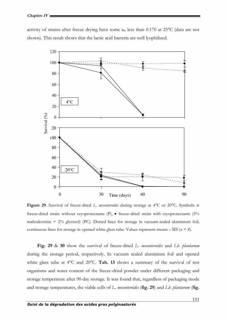
Chapitre IV
Suivi de la dégradation des acides gras polyinsaturés
131
activity of strains after freeze drying have some aw less than 0.170 at 25°C (data are not
shown). This result shows that the lactic acid bacteria are well lyophilized.
Figure 29. Survival of freeze-dried L. mesenteroides during storage at 4°C or 20°C. Symbols: ο
freeze-dried strain without cryoprotectants (P), • freeze-dried strain with cryoprotectants (5%
maltodextrine + 2% glycerol) (PC). Dotted lines for storage in vacuum-sealed aluminium foil,
continuous lines for storage in opened white glass tube. Values represent means ± SD (n = 4).
Fig. 29 & 30 show the survival of freeze-dried L. mesenteroides and Lb. plantarum
during the storage period, respectively. In vacuum sealed aluminium foil and opened
white glass tube at 4°C and 20°C. Tab. 13 shows a summary of the survival of test
organisms and water content of the freeze-dried powder under different packaging and
storage temperature after 90-day storage. It was found that, regardless of packaging mode
and storage temperatures, the viable cells of L. mesenteroides (fig. 29) and Lb. plantarum (fig.
0
20
40
60
80
100
120
0 30 60 90Time (days)
20°C
0
20
40
60
80
100
120
4°C
Surv
ival
(%)
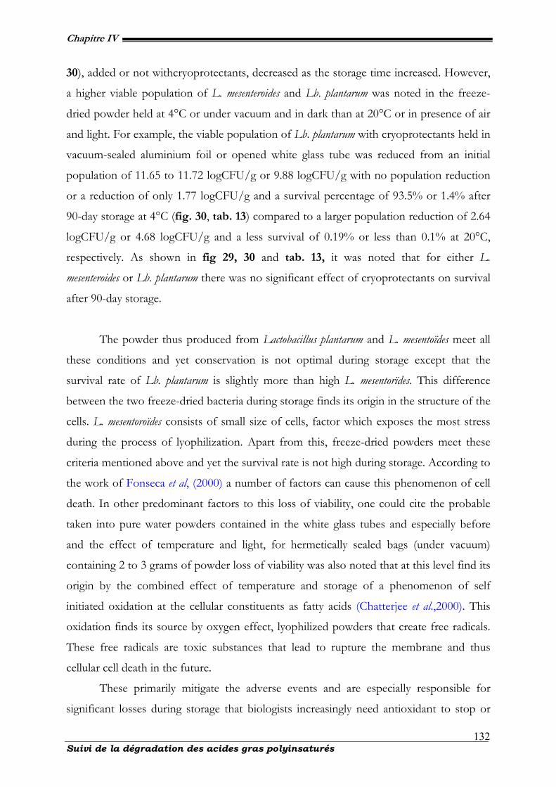
Chapitre IV
Suivi de la dégradation des acides gras polyinsaturés
132
30), added or not withcryoprotectants, decreased as the storage time increased. However,
a higher viable population of L. mesenteroides and Lb. plantarum was noted in the freeze-
dried powder held at 4°C or under vacuum and in dark than at 20°C or in presence of air
and light. For example, the viable population of Lb. plantarum with cryoprotectants held in
vacuum-sealed aluminium foil or opened white glass tube was reduced from an initial
population of 11.65 to 11.72 logCFU/g or 9.88 logCFU/g with no population reduction
or a reduction of only 1.77 logCFU/g and a survival percentage of 93.5% or 1.4% after
90-day storage at 4°C (fig. 30, tab. 13) compared to a larger population reduction of 2.64
logCFU/g or 4.68 logCFU/g and a less survival of 0.19% or less than 0.1% at 20°C,
respectively. As shown in fig 29, 30 and tab. 13, it was noted that for either L.
mesenteroides or Lb. plantarum there was no significant effect of cryoprotectants on survival
after 90-day storage.
The powder thus produced from Lactobacillus plantarum and L. mesentoïdes meet all
these conditions and yet conservation is not optimal during storage except that the
survival rate of Lb. plantarum is slightly more than high L. mesentorïdes. This difference
between the two freeze-dried bacteria during storage finds its origin in the structure of the
cells. L. mesentoroïdes consists of small size of cells, factor which exposes the most stress
during the process of lyophilization. Apart from this, freeze-dried powders meet these
criteria mentioned above and yet the survival rate is not high during storage. According to
the work of Fonseca et al, (2000) a number of factors can cause this phenomenon of cell
death. In other predominant factors to this loss of viability, one could cite the probable
taken into pure water powders contained in the white glass tubes and especially before
and the effect of temperature and light, for hermetically sealed bags (under vacuum)
containing 2 to 3 grams of powder loss of viability was also noted that at this level find its
origin by the combined effect of temperature and storage of a phenomenon of self
initiated oxidation at the cellular constituents as fatty acids (Chatterjee et al.,2000). This
oxidation finds its source by oxygen effect, lyophilized powders that create free radicals.
These free radicals are toxic substances that lead to rupture the membrane and thus
cellular cell death in the future.
These primarily mitigate the adverse events and are especially responsible for
significant losses during storage that biologists increasingly need antioxidant to stop or
Chapitre IV
Suivi de la dégradation des acides gras polyinsaturés
133
reduce the oxidation products at the level of freeze-dried powders. It is safe to say that
based on our experiences and those of others; the loss of viability during storage is
subject to a phenomenon of autooxidation, which should optimize the survival rate
especially during storage at room temperature.
Figure 30. Survival of freeze-dried Lb. plantarum during storage at 4°C or 20°C. Symbols: ο
freeze-dried strain without cryoprotectants (P), • freeze-dried strain with cryoprotectants (5%
maltodextrine + 2% glycerol) (PC). Dotted lines for storage in vacuum-sealed aluminium foil,
continuous lines for storage in opened white glass tube. Values represent means ± SD (n = 4).
The important and intrinsic parameters (dry cell matter, water activity) merit
attention while optimizing for the increase and prolonged time storage of lyophilized
powders of lactic acid bacteria. For this reason and many others, some authors argue that
0
20
40
60
80
100
120
0 30 60 90Time (days)
20°C
0
20
40
60
80
100
120
4°C
Surv
ival
(%)
Chapitre IV
Suivi de la dégradation des acides gras polyinsaturés
134
the cell membrane is the seat of the cell survival in the sense that it contains
phospholipids that are probably modified by the oxidation, Castro et al., 1977.
IV-2.3.4. Cellular fatty acid relative contents after freeze-drying and changes
in fatty acid composition due to storage
Cellular fatty acids (CFAs) from the two strains in this study were analysed before
and after freeze-drying. The mean relative contents of CFAs from L. mesenteroides and Lb.
plantarum are presented in tab. 11 & 12, respectively. Six CFAs, namely palmitic (C16:0),
palmitoleic (C16:1), stearic (C18:0), oleic (C18:1), linoleic (C18:2) and linolenic (C18:3) acids were
identified. Four of the CFAs, namely C16:0, C16:1, C18:0, and C18:1 make up more than 94%
or 93% of the fatty acids in L. mesenteroides and Lb. plantarum respectively, with another
one, namely, C18:3 making a smaller (on average 5-6%, respectively) contribution. The
C18:2 represent very small percentages (on mean ≤ 1%) to the total in each strain. C16:0 had
the highest proportion. There was no significant modification in CFAs of L. mesenteroides
and Lb. plantarum in response to dehydratation (p > 0.05). Moreover, the addition of
glycerol and maltodextrine before freeze-drying did not modify significantly the CFAs of
the two strains. Cellular fatty acids freeze-dried L. mesenteroides and Lb. plantarum packaged
in vacuum-sealed aluminium foil or opened white glass tube and held at 20 or 4°C during
90 days were analysed. The palmitic acid (C16:0) relative content remained high and
changed only little during storage. Therefore, the content of the others fatty acids was
expressed as a ratio between each fatty acid and the C16:0. It was observed that C16:1/C16:0,
C18:0/C16:0 and C18:1/C16:0 ratios for freeze-dried L. mesenteroides or Lb. plantarum, added or
not with cryoprotectants did not change significantly during the storage period.
Fig. 31 & 32 show linoleic/ palmitic (C18:2/C16:0) or linolenic/palmitic (C18:3/C16:0)
ratios for freeze-dried L. mesenteroides and Lb. plantarum during the storage period,
respectively. Tab. 14 & 15 show a summary of linoleic/palmitic (C18:2/C16:0) and
linolenic/palmitic (C18:3/C16:0) ratios for freeze-dried organisms under different packaging
and storage temperature after 90-day storage, respectively. It was found that regardless of
packaging mode and storage temperatures, C18:2/C16:0 and C18:3/C16:0 ratios for freeze-
dried L. mesenteroides (fig. 31) and Lb. plantarum (fig. 32) added or not with cryoprotectants
decreased in function of time. However, a higher C18:2/C16:0 or C18:3/C16:0 ratio for L.
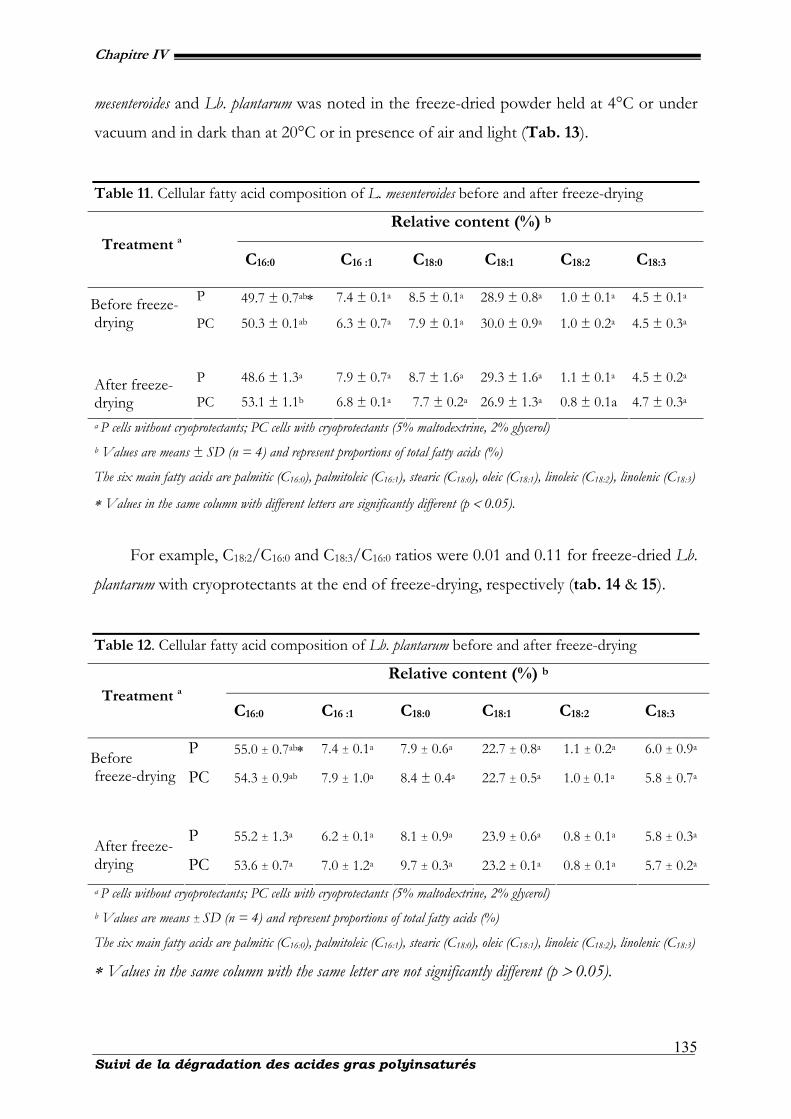
Chapitre IV
Suivi de la dégradation des acides gras polyinsaturés
135
mesenteroides and Lb. plantarum was noted in the freeze-dried powder held at 4°C or under
vacuum and in dark than at 20°C or in presence of air and light (Tab. 13).
Table 11. Cellular fatty acid composition of L. mesenteroides before and after freeze-drying
Relative content (%) b
Treatment a
C16:0 C16 :1 C18:0 C18:1 C18:2 C18:3
P 49.7 ± 0.7ab∗ 7.4 ± 0.1a 8.5 ± 0.1a 28.9 ± 0.8a 1.0 ± 0.1a 4.5 ± 0.1a Before freeze-drying PC 50.3 ± 0.1ab 6.3 ± 0.7a 7.9 ± 0.1a 30.0 ± 0.9a 1.0 ± 0.2a 4.5 ± 0.3a
P 48.6 ± 1.3a 7.9 ± 0.7a 8.7 ± 1.6a 29.3 ± 1.6a 1.1 ± 0.1a 4.5 ± 0.2a After freeze-drying PC 53.1 ± 1.1b 6.8 ± 0.1a 7.7 ± 0.2a 26.9 ± 1.3a 0.8 ± 0.1a 4.7 ± 0.3a a P cells without cryoprotectants; PC cells with cryoprotectants (5% maltodextrine, 2% glycerol) b Values are means ± SD (n = 4) and represent proportions of total fatty acids (%)
The six main fatty acids are palmitic (C16:0), palmitoleic (C16:1), stearic (C18:0), oleic (C18:1), linoleic (C18:2), linolenic (C18:3)
∗ Values in the same column with different letters are significantly different (p < 0.05).
For example, C18:2/C16:0 and C18:3/C16:0 ratios were 0.01 and 0.11 for freeze-dried Lb.
plantarum with cryoprotectants at the end of freeze-drying, respectively (tab. 14 & 15).
Table 12. Cellular fatty acid composition of Lb. plantarum before and after freeze-drying
Relative content (%) b
Treatment a
C16:0 C16 :1 C18:0 C18:1 C18:2 C18:3
P 55.0 ± 0.7ab∗ 7.4 ± 0.1a 7.9 ± 0.6a 22.7 ± 0.8a 1.1 ± 0.2a 6.0 ± 0.9a Before freeze-drying PC 54.3 ± 0.9ab 7.9 ± 1.0a 8.4 ± 0.4a 22.7 ± 0.5a 1.0 ± 0.1a 5.8 ± 0.7a
P 55.2 ± 1.3a 6.2 ± 0.1a 8.1 ± 0.9a 23.9 ± 0.6a 0.8 ± 0.1a 5.8 ± 0.3a After freeze-drying PC 53.6 ± 0.7a 7.0 ± 1.2a 9.7 ± 0.3a 23.2 ± 0.1a 0.8 ± 0.1a 5.7 ± 0.2a
a P cells without cryoprotectants; PC cells with cryoprotectants (5% maltodextrine, 2% glycerol) b Values are means ± SD (n = 4) and represent proportions of total fatty acids (%)
The six main fatty acids are palmitic (C16:0), palmitoleic (C16:1), stearic (C18:0), oleic (C18:1), linoleic (C18:2), linolenic (C18:3)
∗ Values in the same column with the same letter are not significantly different (p > 0.05).
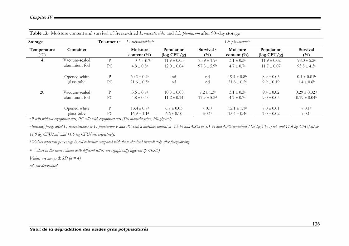
Chapitre IV
Suivi de la dégradation des acides gras polyinsaturés
136
Table 13. Moisture content and survival of freeze-dried L mesenteroides and Lb. plantarum after 90–day storage
Storage Treatment a L. mesenteroides b Lb. plantarum b
Temperature (°C)
Container Moisture content (%)
Population (log CFU/g)
Survival c (%)
Moisture content (%)
Population (log CFU/g)
Survival (%)
P 3.6 ± 0.7a∗ 11.9 ± 0.03 83.9 ± 1.9a 3.1 ± 0.3a 11.9 ± 0.02 98.0 ± 5.2a Vacuum-sealed aluminium foil PC 4.8 ± 0.5a 12.0 ± 0.04 97.8 ± 5.9b 4.7 ± 0.7a 11.7 ± 0.07 93.5 ± 4.3a
P 20.2 ± 0.4b nd nd 19.4 ± 0.8b 8.9 ± 0.03 0.1 ± 0.01b
4
Opened white glass tube PC 21.6 ± 0.3b nd nd 21.8 ± 0.2c 9.9 ± 0.19 1.4 ± 0.6b
P 3.6 ± 0.7a 10.8 ± 0.08 7.2 ± 1.3c 3.1 ± 0.3a 9.4 ± 0.02 0.29 ± 0.02 b Vacuum-sealed
aluminium foil PC 4.8 ± 0.5a 11.2 ± 0.14 17.9 ± 5.2d 4.7 ± 0.7a 9.0 ± 0.05 0.19 ± 0.04b
P 13.4 ± 0.7c 6.7 ± 0.03 < 0.1c 12.1 ± 1.1d 7.0 ± 0.01 < 0.1b
20
Opened white glass tube PC 16.9 ± 1.1d 6.6 ± 0.10 < 0.1c 15.4 ± 0.4e 7.0 ± 0.02 < 0.1b
a P cells without cryoprotectants; PC cells with cryoprotectants (5% maltodextrine, 2% glycerol) b Initially, freeze-dried L. mesenteroides or L. plantarum P and PC with a moisture content of 3.6 % and 4.8% or 3.1 % and 4.7% contained 11.9 log CFU/ml and 11.6 log CFU/ml or
11.9 log CFU/ml and 11.6 log CFU/ml, respectively. d Values represent percentage in cell reduction compared with those obtained immediately after freeze-drying
∗ Values in the same column with different letters are significantly different (p < 0.05)
Values are means ± SD (n = 4)
nd: not determined
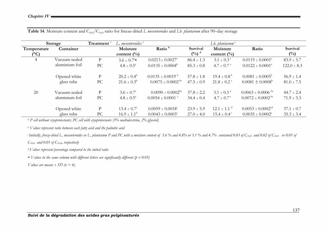
Chapitre IV
Suivi de la dégradation des acides gras polyinsaturés
137
Table 14. Moisture content and C18:2/C16:0 ratio for freeze-dried L mesenteroides and Lb. plantarum after 90–day storage
a P cell without cryoprotectants; PC cell with cryoprotectants (5% maltodextrine, 2% glycerol) b Values represent ratio between each fatty acid and the palmitic acid c Initially, freeze-dried L. mesenteroides or L. plantarum P and PC with a moisture content of 3.6 % and 4.8% or 3.1 % and 4.7% contained 0.03 of C16:0 and 0.02 of C16:0 or 0.01 of
C16:0 and 0.01 of C16:0, respectively d Values represent percentage compared to the initial ratio
∗ Values in the same column with different letters are significantly different (p < 0.05)
Values are means ± SD (n = 4).
Storage Treatment a L. mesenteroides c Lb. plantarum c
Temperature (°C)
Container Moisture content (%)
Ratio b Survival (%) d
Moisture content (%)
Ratio Survival (%)
P 3.6 ± 0.7a∗ 0.0213 ± 0.0027a 86.4 ± 1.3 3.1 ± 0.3 a 0.0119 ± 0.0001a 83.9 ± 5.7 Vacuum-sealed aluminium foil PC 4.8 ± 0.5a 0.0135 ± 0.0004b 85.3 ± 0.8 4.7 ± 0.7 a 0.0122 ± 0.0001a 122.0 ± 8.3
P 20.2 ± 0.4b 0.0135 ± 0.0019 b 57.8 ± 1.8 19.4 ± 0.8 b 0.0081 ± 0.0005b 56.9 ± 1.4
4
Opened white glass tube PC 21.6 ± 0.3b 0.0075 ± 0.0002 bc 47.5 ± 0.9 21.8 ± 0.2 c 0.0081 ± 0.0008b 81.0 ± 7.5
P 3.6 ± 0.7a 0.0090 ± 0.0002bc 37.8 ± 2.2 3.1 ± 0.3 a 0.0063 ± 0.0006 bc 44.7 ± 2.4 Vacuum-sealed
aluminium foil PC 4.8 ± 0.5a 0.0054 ± 0.0001 c 34.4 ± 0.4 4.7 ± 0.7 a 0.0072 ± 0.0002 bc 71.9 ± 3.3
P 13.4 ± 0.7c 0.0059 ± 0.0018c 23.9 ± 5.9 12.1 ± 1.1 d 0.0053 ± 0.0002cd 37.1 ± 0.7
20
Opened white glass tube PC 16.9 ± 1.1d 0.0043 ± 0.0003c 27.0 ± 4.0 15.4 ± 0.4 e 0.0035 ± 0.0002c 35.3 ± 3.4
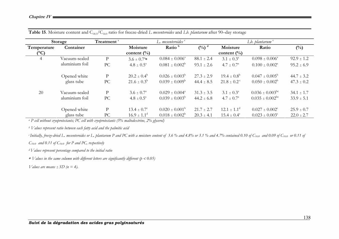
Chapitre IV
Suivi de la dégradation des acides gras polyinsaturés
138
Table 15. Moisture content and C18:3/C16:0 ratio for freeze-dried L mesenteroides and Lb. plantarum after 90–day storage
a P cell without cryoprotectants; PC cell with cryoprotectants (5% maltodextrine, 2% glycerol) b Values represent ratio between each fatty acid and the palmitic acid c Initially, freeze-dried L. mesenteroides or L. plantarum P and PC with a moisture content of 3.6 % and 4.8% or 3.1 % and 4.7% contained 0.10 of C16:0 and 0.09 of C16:0 or 0.11 of
C16:0 and 0.11 of C16:0 for P and PC, respectively d Values represent percentage compared to the initial ratio
∗ Values in the same column with different letters are significantly different (p < 0.05)
Values are means ± SD (n = 4).
Storage Treatment a L. mesenteroides c Lb. plantarum c
Temperature (°C)
Container Moisture content (%)
Ratio b (%) d Moisture content (%)
Ratio (%)
P 3.6 ± 0.7a∗ 0.084 ± 0.006a 88.1 ± 2.4 3.1 ± 0.3a 0.098 ± 0.006a 92.9 ± 1.2 Vacuum-sealed aluminium foil PC 4.8 ± 0.5a 0.081 ± 0.002b 93.1 ± 2.6 4.7 ± 0.7a 0.100 ± 0.002a 95.2 ± 6.9
P 20.2 ± 0.4b 0.026 ± 0.003b 27.3 ± 2.9 19.4 ± 0.8b 0.047 ± 0.005b 44.7 ± 3.2
4
Opened white glass tube PC 21.6 ± 0.3b 0.039 ± 0.009b 44.4 ± 8.5 21.8 ± 0.2 c 0.050 ± 0.002b 47.3 ± 0.2
P 3.6 ± 0.7a 0.029 ± 0.004a 31.3 ± 3.5 3.1 ± 0.3a 0.036 ± 0.003bc 34.1 ± 1.7 Vacuum-sealed
aluminium foil PC 4.8 ± 0.5a 0.039 ± 0.003b 44.2 ± 6.8 4.7 ± 0.7a 0.035 ± 0.002bc 33.9 ± 5.1
P 13.4 ± 0.7c 0.020 ± 0.001b 21.7 ± 2.7 12.1 ± 1.1d 0.027 ± 0.002c 25.9 ± 0.7
20
Opened white glass tube PC 16.9 ± 1.1d 0.018 ± 0.002b 20.3 ± 4.1 15.4 ± 0.4e 0.023 ± 0.003c 22.0 ± 2.7
Chapitre IV
Suivi de la dégradation des acides gras polyinsaturés
139
After a 90-day storage in vacuum-sealed aluminium foil or opened white glass tube,
C18:2/C16:0 ratio decreased from the original value by 0% or 19.0% with a ratio reduction
of only 0 or 0.002 at 4°C (fig. 31, tab. 14) compared to a larger decreased from the
original value by 28.1% or 64.71% with a ratio reduction of 0.003 or 0.007 at 20°C,
respectively. During the same time C18:3/C16:0 ratio decreased from the original value by
4.8% or 52.7% with a ratio reduction of only 0.010 or 0.060 at 4°C (fig. 31, tab. 15)
compared to a larger decreased from the original value by 66.1% or 78.0% with a ratio
reduction of only 0.075 or 0.087 at 20°C, respectively. In most cases, after 90-day storage,
the decreased in C18:2/C16:0 ratio was less than that in C18:3/C16:0 ratio (tab.14 & 15).
C18:2/C16:0 and C18:3/C16:0 ratios for freeze-dried strains added with or without
cryoprotectants are not significantly different after 90-day storage (p > 0.05).
Lipid oxidation and specially lipids of membrane fatty acid was probably deemed
responsible for cell death during storage (Teixeira et al., 1995, 1996). Cellular fatty acids of
L. mesenteroides and Lb. plantarum consisted of : palmitic, palmitoleic, stearic, oleic, linoleic
and linolenic. All these acids have been identified previously in cellular membrane of
acetic acid bacteria and lactic acid bacteria (Sow et al., 2005, Ndoye et al., 2007) ratios.
Contrarily to their saturated and unsaturated analogs, polyunsaturated fatty acids (PUFAs)
are much more susceptible to oxidation (Halliwell et al., 1993). After 90-day, C18:2/C16:0
and C18:3/C16:0 ratios were significantly lower for both freeze-dried strain held in opened
glass tube at 20°C. The effect of temperature during storage on freeze-dried powder is
harmful to the microorganisms’ survival. What concern light, its action is detrimental
and leads to decrease the survival rate. Upon exposure to combined light, temperature
and oxygen, a cooperative, deleterious effect was noted resulting in an even higher loss
in viability. A possible interpretation is that under these conditions linoleic and linolenic
acids can be further rapidly converted in hydroperoxides (Howe et al., 2002; Halliwell et
al., 1993). This is supported by the fact that, during storage in opened white glass tube
at 4°C and in vacuum-sealed aluminium foil at 20°C C18:2/C16:0 and C18:3/C16:0 ratios
decrease more slowly. The occurrence of many of these compounds has been explained
on the basis of the formation and/or combination of free radicals resulting from
homolytic cleavage of C-C linkages near the double bound.
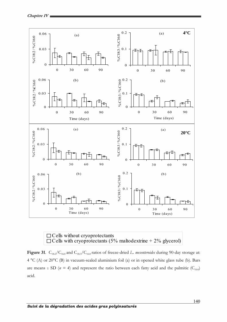
Chapitre IV
Suivi de la dégradation des acides gras polyinsaturés
140
0
0.1
0.2
0 30 60 90
% C
18:3
/ %
C16
:0
(a)
0
0.03
0.06
0 30 60 90
% C
18:2
/ %
C16
:0(a)
0
0.03
0.06
0 30 60 90
Time (days)
% C
18:2
/ %
C16
:0 (b)
0
0.1
0.2
0 30 60 90
Time (days)%
C18
:3 /
% C
16:0 (b)
0
0.03
0.06
0 30 60 90
% C
18:2
/ %
C16
:0
(a)
0
0.03
0.06
0 30 60 90Time (days)
% C
18:2
/ %
C16
:0
(b )
0
0.1
0.2
0 30 60 90Time (days)
% C
18:3
/ %
C16
:0
(b )
0
0 .1
0.2
0 30 60 90
% C
18:3
/ %
C16
:0
(a)
Figure 31. C18:2/C16:0 and C18:3/C16:0 ratios of freeze-dried L. mesenteroides during 90-day storage at:
4 °C (A) or 20°C (B) in vacuum-sealed aluminium foil (a) or in opened white glass tube (b). Bars
are means ± SD (n = 4) and represent the ratio between each fatty acid and the palmitic (C16:0)
acid.
00 . 0 50 . 10 . 1 50 . 2
0 3 0 6 0 9 0
Cells without cryoprotectantsCells with cryoprotectants (5% maltodextrine + 2% glycerol)
4°C
20°C
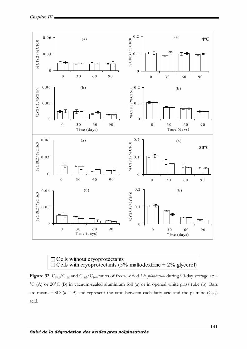
Chapitre IV
Suivi de la dégradation des acides gras polyinsaturés
141
0
0.1
0.2
0 30 60 90
% C
18:3
/ %
C16
:0
(a)
0
0.03
0.06
0 30 60 90
% C
18:2
/ %
C16
:0(a)
0
0.03
0.06
0 30 60 90Time (days)
% C
18:2
/ %
C16
:0
(b )
0
0.1
0.2
0 30 60 90Time (days)
% C
18:3
/ %
C16
:0 (b )
0
0.03
0.06
0 30 60 90
% C
18:2
/ %
C16
:0
(a)
0
0 .03
0.06
0 30 60 90Time (days)
% C
18:2
/ %
C16
:0
(b )
0
0 .1
0 .2
0 30 60 90Time (days)
% C
18:3
/ %
C16
:0
(b )
0
0.1
0.2
0 30 60 90
% C
18:3
/ %
C16
:0
(a)
00 . 0 50 . 10 . 1 50 . 2
0 3 0 6 0 9 0
Cells without cryoprotectantsCells with cryoprotectants (5% maltodextrine + 2% glycerol)
Figure 32. C18:2/C16:0 and C18:3/C16:0 ratios of freeze-dried Lb. plantarum during 90-day storage at: 4
°C (A) or 20°C (B) in vacuum-sealed aluminium foil (a) or in opened white glass tube (b). Bars
are means ± SD (n = 4) and represent the ratio between each fatty acid and the palmitic (C16:0)
acid.
4°C
20°C
Chapitre IV
Suivi de la dégradation des acides gras polyinsaturés
142
Thus linoleic and linolenic acids degradation may not be dependant on the
presence or absence of free molecular oxygen but on the presence of reactive free
radicals in the dried matrix. Linolenic acid was degraded faster than linoleic acid and
even in cell membrane, as previous studies have shown, an unsaturated fatty acid
compound with more double bounds (and/or conjugated double bonds) would be easily
oxidized (Zhang et al., 2007). Our results strongly point towards the possibility that the
loss of water during freeze-drying process generates an oxidative stress. Pereira et al.,
(2003) reported also that freeze-dried cells showed increase in oxidation during storage.
However, the origin of free radicals during dehydratation remains unknown (França et
al., 2007). The influence of temperature during storage of unsaturated fatty acids in the
absence of oxygen, dimeric compounds and substances of lower molecular weight are
more likely to be produced (Nawar, 1996).
IV-2.4. Conclusions
These results presented here provide experimental support to the hypothesis that
storage in presence of air, light, high moisture content and high temperature is
detrimental to freeze-dried powders. Low survival during storage was associated with a
decrease in C18:2/C16:0 and C18:3/C16:0 ratios. Our results confirm the importance
controlling temperature, atmosphere and residual water activity of dried cultures in order
to optimize survival. These data further support the view that PUFA (linoleic and
linolenic acids) could play a key role in determining cellular susceptibility to oxidative
and/or heat stress. The beneficial effect of the use of cryoprotectants is associated clearly
with high viability after freeze-dying rather than increased stability during storage. Further
attempts to stabilize freeze-dried cells during storage must be made, and the addition of
compounds that interact with the cytoplasmic membrane could be explored. Detection of
linoleic or linolenic acids degradation by products will allow having a correct picture of
lipid degradation proceeds.
IV-2.5. Acknowledgements
The authors would like to acknowledge Federal Research Centre for Nutrition,
Institute of Hygiene and Toxicology, Karlsruhe, Germany for providing L. mesenteroides
Chapitre IV
Suivi de la dégradation des acides gras polyinsaturés
143
ssp. mesenteroides Kenya MRog2 strain and Mrs Maryse Hardenne and Mr Philippe Mottet
for their contribution. We thank all the technical’s personals of CWBI (Centre Wallon de
Biologie Industrielle). We also express our gratitude to the republic of Ivory Cost and
communauté française de Belgique for its financial assistance.
IV-2.6. References
[1] Abadias M, Benabarre A, Teixido N, Usall J, Viñas I. Effect of freeze-drying and protectants on viability of the biocontrol yeast Candida sake. Int J Food Microbiol 2001; 65 :173-182.
[2] Andersen AB, Fog-Petersen MS, Larsen H, Skibsted LH. Storage stability of freeze-dried starter
cultures (Strepococcus thermophilus) as Related to Physical State of Freezing Matrix. Lebensm Wiss u Technol 1999; 32: 540-547.
[3] Béal C, Corrieu G. Viability and acidification activity of pure and mixed starters of Streptococcus salivarius
ssp thermophilus 404 and Lactobacillus delbrueckii ssp. bulgaricus 398 at the different steps of their production. Lebensm Wiss U Technol 1994; 27: 86-92.
[4] Béal C, Mihoub F, Marin M. and Corrieu G. Procédé de préparation d’une composition lyophilisée
contenant des bactéries lactiques à viabilité et activité bactériennes améliorées lors d’un stockage à température ambiante et composition obtenue. Demande de Brevet d’invention FR N°2829147 2001.
[5] Bozoglu TF, Ozilgen M, Bakir U. Survival kinetics of lactic acid starter cultures during and after freeze-
drying. Enz Microbiol Technol 1987; 9: 531-537. [6] Carvalho AS, Silva JH, Teixera P, Malcata FX, Gibbs P. Impedimetric method for estimating the
residual activity of freeze-dried Lactobacillus delbrueckii ssp.bulgaricus. International dairy journal 2003; 13(6): 463-468.
[7] Castro HP, Teixeira PM, and Kirby R. Evidence of membrane damage in Lactobacillus bulgaricus
following freeze-drying. Journal of Applied Microbiology 1977; 82: 87-94. [8] Champagne CP, Detournay H, Hardy MJ. Effect of medium on growth and subsequent survival, after
freeze-drying, of Lactobacillus delbrueckii subsp bulgaricus. J Ind Microbiol 1991;7: 147-120. [9] Champagne CP, Mondou F, Raymond Y, Roy D. Effect of polymers and storage temperature on the
stability of freeze-dried lactic acid bacteria. Food Research International 1996; 29(5-6): 555-562. [10] Chatterjee MT, Seunath A, Khalawan SA, Curran BPG. Cellular lipid composition influences stress
activation of the yeast general stress response elements (STRE). Microbiology 2000; 146: 877-884. [11] Cronan Jr, Yan Z, John E. Transcriptional Analysis of Essential Genes of the Escherichia coli Fatty
Acid Biosynthesis Gene Cluster by Functional Replacement with the Analogous Salmonella typhimurium Gene Cluster. Journal of bacteriology; 1998, p. 3295-3303.
[12] Dimmick, R. L.; Heckly, R. J. Free radical formation during storage of freeze dried Serratia marcescens.
Nature 1987; 192: 776. [13] Fonseca F, Béal C, Corrieu, J. Method of quantifying the loss of acidification activity of lactic starters
during freezing and frozen storage. Journal of Dairy Res 2000; 67: 83-90.
Chapitre IV
Suivi de la dégradation des acides gras polyinsaturés
144
[14] França MB, Panek AD, Eleutherio ECA. Oxidative stress and its effects during deshydratation. Comparative Biochemistry and Physiology Part A: Molecular & Integrative Physiology 2007; 146(4): 621-632.
[15] Girotti AW, Thomas JP, Damaging effects of oxygen radicals on resealed erythrocyte ghots. J Biol
Chem 1984; 259: 1744. [16] Gu MB, Choi SH, Kim SW, Some observations in freeze-drying of recombinant bioluminescent E
coli for toxicity monitoring. J Biotechnol 2001; 88: 95-105. [17] Halliwell B, Chirico S. Lipid peroxidation: Its mechanisms, measurement, and significance. Am Clin
Nutr 1993; 57: 715-725. [18] Halverson LJ, Firestone M K. Differential effects of permeating and nonpermeating solutes on the
fatty acid composition of Pseudomonas putida. Appl Environ Microbiol 2000; 66: 2114-2421. [19] Hamoudi L, Goulet J, Ratti C. Effect of protective agents on the viability of Geotricum candidum during
freeze-drying and storage. Food microbiology and safety 2007; 72(2): M45-M49. [20] Hansen JM, Go YM, Jones DP. Nuclear and mitochondrial compartmentation of oxidative stress and
redox signaling. Annu Rev Pharmacol Toxicol 2006; 46: 215-234. [21] Holzapfel W. Use of starter cultures in fermentation on a household scale. Food Control 1997;
8(5/6): 241-258. [22] Howe AG, Schilmiller AL, Oxylipin metabolism in response to stress. Current Opinion in Plant
Biology 2002; 5: 230-236. [23] Ishibashi N, Tatematsu I, Shimamura S, Tomita M, Okonogi S. Effect of water activity on the
viability of freeze-dried bifidobacteria and lactic acid bacteria. Fundamentals and applications of Freeze-drying to biological materials, Drugs and foodstuffs. I. I. o. Refrigeration. Paris 1985; 227-232.
[24] Ito M, Connor WE, Blanchette EJ, Treadwell CR, Vahounny GV. Inhibition of lymphatic absorption
of cholesterol by cholestane-3beta, 5 alpha, 6 beta-triol. Journal of Lipid Research 1969; 10: 694-702.
[25] Karel M. Interaction of food proteins with water and with lipids, and some effects of these
interactions on functional properties, in "Biotechnological Applications of Proteins and Enzymes", Academic Press Inc NY; 1977, p. 317-338.
[26] Kieft TL, Ringleberg DB, White DC. Changes in ester-linked phospholipid fatty acid profiles of
subsurface bacteria during starvation and dessication in a porous medium. Appl Environ Microbiol 1994; 60: 3292-3299.
[27] Kostinek M, Specht I, Edward VA, Schillinger U, Hertel C, Holzapfel WH, Franz CMA. Diversity
and technological properties of predominant lactic acid bacteria from fermented cassava used for the preparation of Gari, a traditional African food. Syst Appl Microbiol 2005; 28: 527-540.
[28] Magasanik B, Schaechter M, Umbarger HEC, Escherichia coli and Salmonella thyphimurium Cellular and
molecular biology American Society for Food Microbiology Washington, DC. Bd 1.S 1987; 474-497.
[29] Marshall BJ, Goote GG, Scott WJA. Study of factors affecting the survival of dried bacteria during
storage. Technical Paper, CSIRO Australian Division of Food Research 1974; 39: 1-29.
Chapitre IV
Suivi de la dégradation des acides gras polyinsaturés
145
[30] Nawar. Lipids. Food chemistry. F. O. R. New york, Marcel Dekker, Inc 1996; 1: 225-319. [31] Nawar, W. W. Lipids. Principles of Food Science. O. R. Fennema 1976. [32] Ndoye B, Weekers F, Diawara B, Guiro AT, Thonart P. Survival and preservation after freeze-drying
process of thermoresistant acetic acid bacteria isolated from tropical products of Subsaharan Africa. J food Eng 2007; 79: 1374-1382.
[33] Pereira EJ, Panek AD, Eleutherio ECA. Protection against oxidation during dehydratation of yeast.
Cell Stress and Chaperones 2003; 8: 120-124. [34] Porubcan RS, Sellars RL, Lactic starter culture concentrates. Microbial technology. H. J. Peppler,
Perlman, D. (Eds). New York, Academic Press; 1979, p. 59-92. [35] Pryor WA. Free radical pathology. Chem Eng News 1971; 49: 34-51. [36] Sow NM, Dubois-Dauphin R, Roblain, D, Guiro AT, Thonart P. Polyphasic identification of a new
thermotolerant species of lactic acid bacteria isolated from chicken faeces. African Journal of Biotechnology 2005; 4(5): 409-421.
[37] Suzuki K, Goodfellow M, O'Donnell AG. Cell envelopes and classification. London, England,
Academic Press; 1993. [38] Teixeira PC, Castro MH, Malcata FX, Kirby RM. Survival of Lactobacillus delbruckii ssp. bulgaricus
following spray drying. Journal of Dairy Science 1995; 78: 1025-1031. [39] Teixeira PC, Castro MH, Kirby RM. Evidence of membrane lipid oxidation of spray-dried Lactobacillus
bulgaricus during storage. Letters in Applied Microbiology 1996; 22: 34-38. [40] ThébergeM. C, Prévost D, Chalifour, FP. The effect of different temperatures on the fatty acids
composition of Rhizobium bv. viciae in the faba bean symbiosis. New Phytologist 1996 ; 134: 657-664. [41] Wang YC, Yu RC, Chou CC. Viability of lactic acid bacteria and bifidobacteria in fermented soymilk
after drying, subsequent rehydratation and storage. Int J Food Microbiol 2004; 93: 209-217. [42] Zhang W, Shi Bi, Shi J. A theorical study on autoxidation of unsaturated fatty Acids and Antioxidant
Activity Of phenolic Compounds. Journal of American Leather Chemists Association 2007; 102: 99-105.
Chapitre IV
Suivi de la dégradation des acides gras polyinsaturés
146
IV.3. Conclusions : Suivi de la dégradation des acides gras
polyinsaturés
A l’issue de ces travaux, les résultats récoltés permettent d’établir un support
expérimental à l'hypothèse que le stockage en présence de l'air, de la lumière, d’une
humidité importante en combinaison avec une température élévée est préjudiciable à la
conservation des cellules lyophilisées. La principale difficulté résulte de leurs fortes
teneurs en acides gras polyinsaturés qui sont très sensibles aux réactions d’oxydation. Ces
réactions induisent des dégradations des propriétés du produit au cours des procédés de
transformation et de conservation. L’objectif de cette étude était d’identifier les étapes
critiques du procédé de production et de lyophilisation des bactéries, afin de proposer des
solutions permettant de limiter le développement des réactions d’oxydation des lipides
responsables de la dégradation des starters au cours de sa fabrication et de sa
conservation. Au cours de la conservation à 4°C sous vide, les poudres issues de la
lyophilisation contiennent de faibles concentrations en produits d’oxydation des
polyinsaturés. Cette faible concentration résulterait de réactions de décomposition de ces
produits et/ou d’interactions intervenant entre ces produits et les protéines. D'autres
tentatives pour stabiliser les cellules lyophilisées pendant le stockage doivent être
effectuées, et l'addition de composés qui, par exemple interagiraient avec la membrane
cytoplasmique, pourrait être explorée. Dans le souci de compréhension des phénomènes
de mortalité cellulaire, l’analyse des composés volatils s’avère importante. C’est pourquoi
le chapitre V portera sur les produits volatils de dégradation des acides gras cellulaires.
Chapitre V
Caractérisation des composés volatils issus de la
dégradation des acides gras de bactéries
lactiques
Chapitre V
Caractérisation des composés volatils issus de la dégradation des acides gras
148
V. Caractérisation des composés volatils issus de la
dégradation des acides gras membranaires
V.1. Contexte et objectifs
L'objectif de ce chapitre était de suivre l'oxydation des lipides par la formation des
composés volatils au cours du stockage à température ambiante 20°C. Les acides gras
totaux de Lactobacillus plantarum CWBI-B1419 se caractérisent par un taux élevé d'acides
gras polyinsaturés (~30%). Les phospholipides, principalement les phosphatidyléthano-
lamines (PE) contiennent une forte proportion d'acides gras polyinsaturés à chaîne longue
dont 15% d'acides gras de la série n-3 et un taux élevé d'aldéhydes. Le niveau initial des
composés volatils (aldéhydes) issus de l’oxydation des lipides des cellules lyophilisées de
bactéries lactiques est faible. Mais lors de la dégradation des polyinsaturés et particulière-
ment des phospholipides tels que les phosphatidyléthanolamines, l'indice des produits
primaires d’oxydation des lipides augmente et les quantités des produits volatils issus de
l'oxydation des lipides sont multipliées par 18 à 20 (Nordvi et al., 2007). Dans le cadre de
ce chapitre, cette étude a pour intérêt fondamental de caractériser les composés volatils
produits par des starters lactiques utilisés comme prébiotiques, probiotiques voire
synbiotiques. Les produits (poudres) issus de la lyophilisation sont stockés à température
ambiante. L’extraction par la technique d’espace de tête dynamique ou head space («purge
and trap») permet de mettre en évidence 109 composés. Parmi les composés identifiés 30
composés sont susceptibles, selon les auteurs (Salmeron et al., 2009 ; Cheng et al., 2005)
d’être produits par le métabolisme bactérien des lipides, des glucides ou des acides aminés.
Une analyse en composantes principales des composés volatils permet de distinguer
différents groupes que sont : les aldéhydes, les alcools, les alcènes, les alcanes, les esters,
les cétones, et les composés dérivés des amino acides. Cette étude permet une approche
différente dans la compréhension des phénomènes d’oxydation au niveau du groupe de
Lactobacilli par la production de composés volatils lors de l’altération d’acides gras
membranaires polyinsaturés.
Chapitre V
Caractérisation des composés volatils issus de la dégradation des acides gras
149
V.2. Characterization of volatiles compounds emitted from freeze-
dried L. plantarum CWBI-B1419 product during storage.
Publication 6 – Biotechnol. Appl. Biochem
Biotechnology and Applied Biochemistry
Coulibaly, I(1♣)., Destain, J(1)., R, Dubois-D(1)., Wathelet, J. P (3)., and P. Thonart (1,2)
(1)Wallon Center for Industrial Biology, Bio-Industry Unit, Gembloux
Agricultural University, Passage des déportés 2, 5030 Gembloux, Belgium.
(2)Wallon Center for Industrial Biology, Microbial Technology Unit, University
of Liège, Sart-Tilman B40, 4000 Liège, Belgium.
(3)Organic Chemistry Unit, Gembloux Agricultural University, Passages des
déportés 2, 5030 Gembloux, Belgium
♣ Corresponding Author : I. Coulibaly, Phone : +3281622305, Fax : +32816614222 E-mail : [email protected]
Chapitre V
Caractérisation des composés volatils issus de la dégradation des acides gras
150
Résumé
La poudre issue de la lyophilisation de Lactobacillus plantarum CWBI-B1419 a été
analysée à trois stades, au cours du stockage. La teneur en eau et le taux de viabilité des
bactéries ont été suivis, les composés volatils ont été extraits des poudres par la technique
de prélèvement de l'espace de tête dynamique et analysés par chromatographie en phase
gazeuse couplée au spectromètre de masse (GC-MS). L’analyse révèle 109 composés
volatils identifiés : des aldéhydes, des alcools, des alcènes, des alcanes, des esters, des
cétones et des composés dérivés d'acides aminés comme les groupes de volatils les plus
répandus. Dix volatils ont été retenus en raison de leur pourcentage élévé des surfaces des
pics. Sept de ces volatils ont été analysés en suivant leur évolution au cours du stockage.
Parmi ces composés on note la diminution des taux d’acide éthanoique et du
cyclotetrasiloxane octamethyl, contrairement à l'acide acétique, l'acide propionique, l'acide
2-méthylpropanoïque, l'acide butanoïque et 3-méthylbutanoïque dont les pourcentages
augmentent de façon significative (p< 0.1) durant les 90 jours de stockage. Le pourcentage
des volatils issu de L. plantarum CWBI-B1419 est fonction de la période de stockage et
aussi de la quantité des acides gras polyinsaturés. En dépit de la forte teneur en acides gras
polyinsaturés, le catabolisme des acides aminés et la synthèse d'esters ont conduit à
d’importants changements dans la composition des produits volatils des bactéries
lactiques issus de réactions d'oxydation des lipides.
Mots-clés: Composés volatils, Acides organiques volatils; cellules lyophilisées, le
stockage, Lactobacillus, (GC-MS); microextraction en phase solide (SPME).
Chapitre V
Caractérisation des composés volatils issus de la dégradation des acides gras
151
Abstract
The powder of freeze-dried Lactobacillus plantarum CWBI-B1419 was analyzed at
three stages during storage. The water content, the rate of viable bacteria were monitored
and the volatile compounds were extracted from the powders by the technique of
sampling of the dynamic headspace and analyzed by gas chromatography coupled with
mass spectrometry (GC -MS). The analysis revealed 109 volatils compounds identified
aldehydes, alcohols, alkenes, alkanes, esters, ketones and compounds derived from amino
acids such as the predominates groups. Ten volatiles were selected according to their high
percentage of peak areas. Seven of these volatils were analyzed by following their
evolution during storage. Among these compounds we noticed a decreased of ethanoic
acid and cyclotetrasiloxane octamethyl unlike acetic acid, propionic acid, 2-methyl-,
butanoic acid and 3-methylbutanoic with increasing percentages of significantly (p <0.1)
during 90 days of storage. The contents of volatiles from L. plantarum CWBI-B1419 is a
function of the storage period and also the quatntité polyunsaturated fatty acids. Despite
the high content of polyunsaturated fatty acids, catabolism of amino acids and synthesis
of esters led to important changes in the composition of volatile products of lactic acid
bacteria from oxidation reactions of lipids.
Keywords: Volatils compound, Volatile organic acid; Freeze-dried cells, storage,
Lactobacillus, (GC–MS); Solid-phase microextraction (SPME).
Chapitre V
Caractérisation des composés volatils issus de la dégradation des acides gras
152
V-2.1. Introduction
The composition of volatile compounds gives characteristic aroma profiles that are
specific for different types of dry-cured meat sausages (Montel et al., 1998 ; Schmidt et al.,
1998 ; Dainty et al., 1999 ; Edwards et al., 1999 ; Toldra, 2002). Unsaturated fatty acids
can be oxidised easily by diverse oxidation mechanisms. Light exposure and presence of
photosensitisers such as riboflavin and chlorophyll can accelerate lipid oxidation through
type I pathway of excited photosensitisers and singlet oxygen oxidation or type II
pathway (Boff et al., 2002). Changes of volatile profiles can be used as indicators for
determining the degree of oxidation in fats and oils (Lemaître et al., 2006). The most
important odor and flavour compounds in dry-cured meat sausages are formed by the
decomposition of fatty acids and amino acids (Ordonez et al., 1999 ; Toldra 2002). Free
unsaturated fatty acids (UFA) will easily oxidize to lipid hydroperoxides, which are
unstable and rapidly decompose to secondary oxidation volatile compounds such as
aldehydes, alcohols, ketones, furans, alkanes, and alkenes (Frankel, 2005). Amino acid
catabolism gives different volatile compounds like sulphides, pyrazines, and branched-
chained amino acids (BCAA) (Ordonez et al., 1999). The aroma of fresh lactic acid
bacteria can be attributed to secondary lipid oxidation compounds formed from
hydroperoxides either by lipoxygenases or autooxidation of UFA and PUFA (Kawai,
1996 ; Dunford and Shahidi, 1998 ; Cadawallader, 2002).
During storage, increasing levels of these compounds can lead to the development
of rancid off-flavors in bacteria fatty acid. The objectives of this study were to determine
the effects of light irradiation and oxygen effect on the formation of volatile compounds
from polyinsaturated fatty acid model systems. In addition, the general changes in volatils
during cold storage as well as the influence of 4 different milk protein ingredients were
studied. The volatile compounds were collected using dynamic headspace sampling and
separated and identified by use of dynamic headspace gas chromatography-mass
spectrometer (GC-MS). Previous studies (Nordvi et al., 2007) have shown that in order to
obtain an adequate lipid to protein ratio, extra protein needs to be added to secure
physical stabilization of the lactic acid bacteria oil throughout processing. However,
intrinsic properties of commercial protein powders can influence on the oxidative
stability.
Chapitre V
Caractérisation des composés volatils issus de la dégradation des acides gras
153
V-2.2. Materials and methods
V-2.2.1. Microorganism and inoculum
A selected L. plantarum CWBI-B1419 bacterial strain (CWBI, Wallon Center for
Industrial Bio-Industry Unit, Gembloux Agricultural University, Belgium), isolated from
poultry farm was used as inoculum in this study. MRS broths inoculated with strain were
incubated at 30°C for 18 h. The bottles were incubated at 30°C to the exponential phase
of growth (18h). The bacterial suspensions were then used to inoculate the fermenting
media at 1%(v/v). In all cases, the initial microbial concentration was approximately
7.5log10 CFU/ml. The cell pellets obtained were resuspended in 5 ml (50% v/v) glycerol
and frozen at -80 °C. The supernatants obtained after centrifugation (Sorvall RC2-B,
Sorvall, USA) at 2,500 × g for 20 min, were decanted.
V-2.2.2. Productions and storage conditions
The strains were grown in 100 L bioreactor containing MRS medium for 18 h and
then concentrated 20 times by centrifugation. The supernatants obtained after
centrifugation (Sorvall RC2-B, Sorvall, USA) at 2,500 × g for 20 min, were decanted with
2%, w/w glycerol and 5%, w/w maltodextrine. Cells were freeze-dried in a low freeze-
drier (Leybold, Belgium) with a standard program by increasing the temperature gradually
from -45°C to 25°C at 0.9 mbar pressure (30h) followed by 15h at 0.15 mbar. All
fermentations were done in duplicate and average values reported. Freeze-dried samples
were stored during 90 days, in portion size (2 ± 0.5 g in each portion) at 20°C in aluminum
foil packets which were vacuum-sealed. Samples were withdrawn for analysis each 30 days
and kept at -20°C (48-72 h) before analysis
V-2.2.3. Dry cell weight, water Content and survival rate
The dry cell weight of 1 g of powder was determined at the end of freeze drying.
The water content (100 g dry weight)-1 of the freeze-dried samples during storage at 20°C
was determined each 30 days. The samples were dried in a convection oven until constant
weight and results are mean of triplicate determinations. The water activity (aw) of the
freeze-dried samples was measured at 20°C using a water activity meter Novasina
Chapitre V
Caractérisation des composés volatils issus de la dégradation des acides gras
154
(Novasina, Pfäffikon, Switzerland). Standard salt solutions (Novasina) of known water
activity were used for calibration of the sensor at the measuring temperature. Readings
were taken until four sequential readings gave the same results. Results obtained were the
average of three determinations. The viable counts were obtained by plate count method
after 48h. Percentage survival after 90-day storage was calculated as 100 × N/N0, where N
is the CFU/g of the freeze-dried sample at a given time and N0 is the CFU/g of the
sample at the end of freeze-drying.
V-2.2.4. Data analysis of SPME
Two grams of freez-dried cells were added to a 20-mL glass sample vial (Perkin
Elmer, Mass., U.S.A.) and fitted with a self-sealing septum at its top, through which the
solid-phase microextraction (SPME) syringe needle (bearing a fiber coated with 8 µm
thick film of Carboxen-polydimethylsiloxane, Supelco, Bellefonte, Pa., U.S.A.) was
introduced and maintain in the headspace at 35°C for 30min. The SPME fiber was
conditioned according to the manufacturer’s recommendations (280°C for 30min in the
GC injector). The volatile analytes absorbed to the SPME fiber were analyzed by GC-MS
(Hewlett-Packard, 5890 series II plus, N.J., U.S.A.). The compounds were separated by a
CPWAX-52CB fused silica column (50 m × 0.25 mm × 0.2 µm, Agilent, Calif., U.S.A.).
V-2.2.5. GC–MS operating parameters
The injection port was heated at 250°C, in splitless mode, and helium flow rate was
maintained at 1 mL/min. The SPME fiber was maintained in the injection port for 10min.
The oven temperature program was held at 40°C for 4min, increased to 250°C at a rate of
8°C/min, and held for 5min. Detection was performed with the mass spectrometer
operating in a scan mode (mass range from 35 to 400 m/z using a scan rate of 4.51
scans/s) and the ionization energy was set at 70 eV. The temperatures of the ion source
and the quadrupole mass analyzer were held at 200 and 100°C, respectively. The eluted
compounds were identified by their retention times and by comparison of their mass
spectra with the Wiley Mass Spectral database (Wiley & Sons Inc., N.Y., U.S.A.).
Triplicate samples were analyzed. Gas chromatography with flame ionization detector
(GC-FID) (HP5890 series II plus) was used to quantify volatils. The compounds
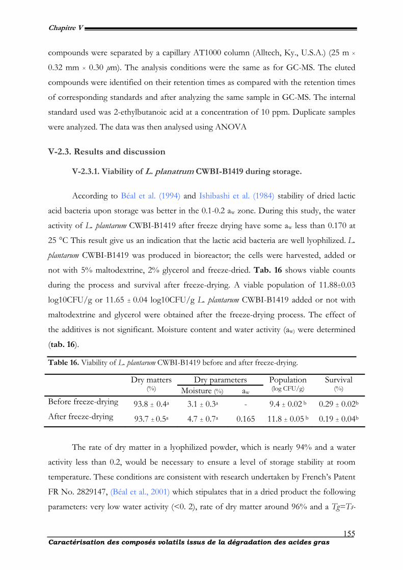
Chapitre V
Caractérisation des composés volatils issus de la dégradation des acides gras
155
compounds were separated by a capillary AT1000 column (Alltech, Ky., U.S.A.) (25 m ×
0.32 mm × 0.30 µm). The analysis conditions were the same as for GC-MS. The eluted
compounds were identified on their retention times as compared with the retention times
of corresponding standards and after analyzing the same sample in GC-MS. The internal
standard used was 2-ethylbutanoic acid at a concentration of 10 ppm. Duplicate samples
were analyzed. The data was then analysed using ANOVA
V-2.3. Results and discussion
V-2.3.1. Viability of L. planatrum CWBI-B1419 during storage.
According to Béal et al. (1994) and Ishibashi et al. (1984) stability of dried lactic
acid bacteria upon storage was better in the 0.1-0.2 aw zone. During this study, the water
activity of L. plantarum CWBI-B1419 after freeze drying have some aw less than 0.170 at
25 °C This result give us an indication that the lactic acid bacteria are well lyophilized. L.
plantarum CWBI-B1419 was produced in bioreactor; the cells were harvested, added or
not with 5% maltodextrine, 2% glycerol and freeze-dried. Tab. 16 shows viable counts
during the process and survival after freeze-drying. A viable population of 11.88±0.03
log10CFU/g or 11.65 ± 0.04 log10CFU/g L. plantarum CWBI-B1419 added or not with
maltodextrine and glycerol were obtained after the freeze-drying process. The effect of
the additives is not significant. Moisture content and water activity (aw) were determined
(tab. 16).
Table 16. Viability of L. plantarum CWBI-B1419 before and after freeze-drying.
Dry parameters Dry matters (%) Moisture (%) aw
Population (log CFU/g)
Survival (%)
Before freeze-drying 93.8 ± 0.4a 3.1 ± 0.3a - 9.4 ± 0.02 b 0.29 ± 0.02b After freeze-drying 93.7 ± 0.5a 4.7 ± 0.7a 0.165 11.8 ± 0.05 b 0.19 ± 0.04b
The rate of dry matter in a lyophilized powder, which is nearly 94% and a water
activity less than 0.2, would be necessary to ensure a level of storage stability at room
temperature. These conditions are consistent with research undertaken by French’s Patent
FR No. 2829147, (Béal et al., 2001) which stipulates that in a dried product the following
parameters: very low water activity (<0. 2), rate of dry matter around 96% and a Tg=Ts-
Chapitre V
Caractérisation des composés volatils issus de la dégradation des acides gras
156
25°C >20°C, contribute to the stability of the product for an extended period during the
storage.
The powder produced from this lactic acid bacterium meets all these conditions
and yet conservation is not optimal during storage. Apart from this, freeze-dried powders
meet the above-mentioned criteria and yet the survival rate is not high during storage.
According to the work of (Carvaho et al., 2003), a number of factors can influence the
rate of survival in function of time. With regard to the other predominant factors to this
loss of viability, one could cite (the water activity), probably taken into contained pure
water powders, the effect of temperature and light for hermetically sealed bags (under
vacuum) containing 2-3 g of powder. Loss of viability was also noted that at this level find
its origin by the combined effect of temperature and oxidation of cellular constituents as
unsaturated fatty acids (Niki et al., 2003). Peroxides (R-OOH) are primary reaction
products formed in the initial stages of oxidation, and therefore give an indication of the
progress of lipid oxidation (Nawar, 1996).
This oxidation finds its source by oxygen effect, lyophilized powders that create
free radicals. These free radicals are toxic substances that lead to rupture the membrane
and thus cellular cell death in the future. These primarily mitigate the adverse events and
are especially responsible for significant losses during storage that biologists increasingly
need antioxidant to stop or reduce the oxidation products at the levelof freeze-dried
powders. It is safe to say that based on our experiences and those of others; the loss of
viability during storage is subject to a phenomenon of autooxidation, which should
optimize the survival rate especially during storage at room temperature. The oxidation
products are varied. Hydroperoxides from the decomposition of polyunsaturated give way
to volatile secondary products that represent the same phenomenon. These compounds
are also important indicators of cellular oxidation
V-2.3.2. Volatile compound analysis.
Volatile compounds from freeze-dried powders were extracted by SPME. Sevral
types of fibers (CAR/PDMS, and CAR/PDMS/DVB) were tested under the same
conditions to evaluate the more adapted one. Best results were obtained with the
Chapitre V
Caractérisation des composés volatils issus de la dégradation des acides gras
157
CAR/PDMS/DVB fiber, which is in good accordance with Sanches et al. (2004) and
Marco et al. (2004). Analysis of the volatile SPME profiles obtained for the different time
points of freeze-dried cells. 109 coumpounds were found as volatile compounds and very
low levels of these compounds as compared to volatile compounds extracted from freeze-
dried Lactobacillus palntarum NCIMB 8826 in cereal-bases substrates, (Ivan et al., 2009).
Details about the peak identities and relative abundances of the volatile compounds
expressed as (compoundpeak area/total compounds peak area) x 100 are shown in tab.
16. Among the identified organic compound there are acids, amides, alcohols, aldehydes,
aromatic hydrocarbons, esters, furans, ketones, peroxides, and pyrans. High concentra-
tions of ten volatils compounds were found in freeze-dried powder (tab. 17).
1,3-butanediol, 2,3 butanediol, acetic acid, hexanal, cyclotertasiloxane octamethyl
and benzaldhyde are mainly identified. Beside these compounds, additional volatile
compounds were also identified in very small amounts (tab. 17). Hexanal is the only
aldehyde formed from both the 9- and the 13-hydroperoxide of linoleic acid at least by
chemical reactions. Hexenal may be the direct product of the chemical fragmentation of
the 13-hydroperoxide of linoleic acid, or it may derive from 2,4 decadienal that may be a
product of chemical oxidation of 9-hydroperoxy linoleic acid (HPOD) (Petersen et al.,
1998 ; Goodridge et al., 2003). Freeze-dried powder of Lactobacillus plantarum had known
to contain mainly linoleic acid as PUFAs and the major LOX activity in tubers forms 9-
derivatives, the precursors of enzymatic C-9 aldehyde formation (Coulibaly et al., 2009).
Our study failed to detect other compound as aldhyde. However, hexenal may be the
preferred product of 9-hydroperoxides from chemical reactions as well. Moreover, the
SPME technique may discriminate as the fibers adsorb more preferentially the most
volatile compounds. Despite this disadvantage, the technique is well-adapted to define
smell perception (Sanches et al., 2005). Heptane and hexene were also identified and
quantified by HPLC; benzaldehydel, pyrazine and nonadecane were identified as minor
products. Except hexenal, none of aldehydes have been identified by SPME-GC-MS. The
rate of hexenal has been increased during storage. Based on these results and the work
carried out by Fauconnier et al. (2007) and Yao et al. (2008), assuming an oxidation by
enzymatic formation of hexenal in the lyophilized powder has been issued. Another
group of compounds originally aroma can be generated during the treatment because of
the enzymatic activity of lactic bacteria.
Chapitre V
Caractérisation des composés volatils issus de la dégradation des acides gras
158
The high concentrations of acetic acid could be explained by the aerobic
metabolism of L. plantarum. Sedewitz et al. (1984) have showed that L. plantarum, during
the stationary phase, under aerobic conditions and low glucose concentration, can
consume the previously secreted lactic acid and an equivalent amount of acetic acid is
released. Similar behaviour of L. plantarum in residual media was also reported by Vazquez
et al. (2008). The high relative concentrations of acetic acid found in freeze-dried powder
suggest that part of the lactic acid produced could have been metabolised to acetic acid.
Aldehydes were found in freeze-dried Lactobacillus in relatively quantities (tab. 17 & 18).
The final stages of the fermentation of bacteria, plants and yeast involve the conversion
of pyruvate to acetaldehyde by the enzyme pyruvate decarboxylase (EC 4.1.1.1), followed
by conversion of acetaldehyde into ethanol. The latter reaction is further catalyzed by
alcohol dehydrogenase, running in the opposite direction (Molimard et al., 1996) or
formed by the metabolism of lactate or by the oxidation of ethanol (McSweeney et al.,
2000 ; Ortigosa et al., 2005 and Bosset et al., 1997). Branched aldehydes originate from
amino acid degradation (Engels et al., 1997), i.e. 2-methyl propanal, 2-methyl butanal and
3-methyl butanal were produced from Val, Ile and Leu, respectively (Urbach, 1995) were
not detected, benzaldehyde has also been found in some trials with important
concentration.
Table 17. Relative abundances of volatile compounds detected in freeze-dried Lactobacillus
plantarum CWBI-B1419 during storage.
Peak n°
Compounds RT Identification (%)
Trial-1 Trial-2 Trial-3 Trial-4 ∑trials 1. Hexane 0,15 0,17 - - - 0,17 2. Heptane 0,96 - 5,19 - - 5,19 3. Ethanol 1,11 0,79 - - - 0,79 4. Acetic acid 1,60 6,89 - 13,18 - 20,07 5. Ethanoic acid 1,65 3,42 - 6,23 - 9,65 6. 1-Butanol 1,86 0,68 - - - 0,68 7. 1,3-Butanediol 2,91 15,26 0,87 12,89 6,84 35,86 8. 2,3-Butanediol 2,95 6,75 6,48 19,89 - 33,12 9. Silane triethyl(ethoxy) 2,96 - 4,31 - - 4,31 10. Butanol trimethyl ester 2,98 - - - 14,67 14,67 11. 1,3-Dihydroxybutane 2,99 18,54 - - - 18,54 12. Butane-2,3-diol 3,07 1,74 - 11,83 - 13,57 13. Ethyl 2-hydroxypropanoate 3,26 0,46 - 0,73 - 1,19 14. Cyclotrisiloxane hexamethyl 3,38 2,58 1,78 2,40 1,53 8,29 15. Pyrazine, 2,5 dimethyl 4,19 - 4,05 - - 4,05
Chapitre V
Caractérisation des composés volatils issus de la dégradation des acides gras
159
16. hexenal 4,79 - - - 18,80 18,8 17. Ethyl metoxy acetate 4,81 0,25 - - 2,44 2,69 18. Ethanol 2- (21-methylethoxy) 4,83 - 0.21 - - 0,21 19. 1-Propene, 1-methylthio-(z) 4,84 - - 0.42 - 0,42 20. Disiloxane ethenylpentamethyl 5,47 0,58 0,81 0,53 2,60 4,52 21. Pentamethylheptane –(2,2,4,6,6) 5,57 0,69 - - - 0,69 22. Cyclotrisiloxane hexamethyl 5,58 0,51 - - - 0,51 23. Ethanol 2,2-diethoxy 5,61 - 6,29 - 2,04 8,33 24. Cyclotetrasiloxane octamethyl 5,69 2,71 0,76 7,37 7,91 18,75 25. Trimethyl ethoxysilane 5,72 - 0,62 - - 0,62 26. Methylanisole 5,76 - - - 6,11 6,11 27. Cyclotetrasiloxane 5,88 5,06 - - - 5,06 28. Dimethylpyrazine 5,89 - 0,91 - - 0,91 29. Pyrazine ,2 5-dimethyl-3-(3-methylbutyl) 5,95 - 4,45 - - 4,45 30. Benzene , 1-methoxy-4-methyl 5,96 - - - 3,25 3,25 31. Trimethylpyrazine 5,98 - - 0,63 - 0,63 32. Pyrazine 3-ethyl-2,5-dimethyl 7,13 - 4,37 - 1,56 5,93 33. 2-allyl-5-methylpyrazine 7,25 - 4,90 - - 4,9 34. Tetrapyrazine 7,27 - - 0,69 - 0,69 35. Ethoxycarbonylbenzophenone 7,60 0,23 - - - 0,23 36. Benzaldehyde 2,4-bis[trimethylsil)oxy] 7,62 0,38 - - - 0,38 37. Benzaldehyde 7,65 - - - 1,76 1,76 38. 3-Heptene 7,69 0,09 - - - 0,09 39. Benzeneethanol 7,70 - - 0,96 - 0,96 40. Pyrimidine, 2-(2-nitro-3-thienyl) 7,98 - 5,55 - - 5,55 41. Benzaldehyde 2,5-bis[(trimethylsil)oxy] 8,22 3,71 - - 9,11 12,82 42. Benzoic acid 8,23 9,86 - - - 9,86 43. Benzeneacetic acid 8,25 - - 5,09 - 5,09 44. 2,3,5-Triethyl-6-ethylpyrazine 8,30 - - 0,29 - 0,29 45. 5-Methyl-1-heptene 8,69 - - - 2,11 2,11 46. Dodecane 8,90 0,45 0,94 - - 1,39 47. Trisiloxane hexamethyl 9,00 - - 0,56 - 0,56 48. Ethanone, 1-(3,5-dimethyl-2-pyrazinyl) 9,27 - - - 0,65 0,65 49. Cyclotetrasiloxane octamethyl 9,51 0,51 0,89 - - 1,4 50. Propenylpyrazine 9,70 - - - 0,76 0,76 51. 1,4-Di-tert-butylbenzene 9,73 - - 0,12 - 0,12 52. 1-Tetradecene 9,75 0,38 - - - 0,38 53. Phenethyl acetate 9,77 - - - 0.11 0,11 54. Cyclotetrasiloxane hexamethyl 9,82 0,38 0,64 - - 1,02 55. 2-Undecanone 10,24 - 1,21 0,32 - 1,53 56. 4-Ethyl-4-methyl-1-hexene 10,42 0,32 0,87 - - 1,19 57. Methylnonadecane 10,44 - - - 0,38 0,38 58. Cyclotetrasiloxane octamethyl 10,47 - - 0,35 - 0,35 59. Pyrazine 2,5-dimethyl-3-(methylbutyl) 10,56 - 2,48 - - 2,48 60. 16-oxosalutaridine 10,66 9,90 9,46 4,61 9,35 33,32 61. Tetradecane 2-methyl 10,95 - 0,32 - - 0,32 62. Tritriacontane 10,96 0,18 - - - 0,18 63. Hydroxycyclohexane 10,99 - 0.21 - - 0,21 64. 1-benzothiopyran-4-one 1-oxide 11,07 0,16 - 0,14 0,84 1,14 65. 4H-1-benzothiopyran-4-one 11,08 - 0,83 - - 0,83 66. Cyclohexane 11,11 0,34 0,50 - - 0,84
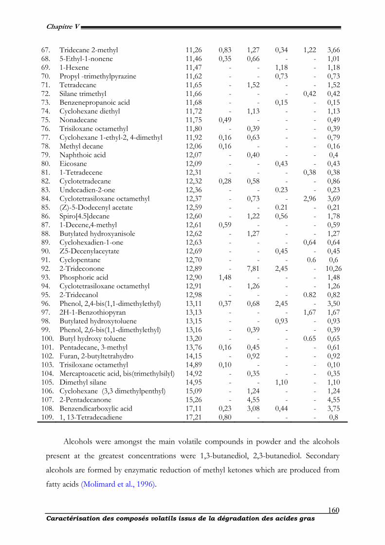
Chapitre V
Caractérisation des composés volatils issus de la dégradation des acides gras
160
67. Tridecane 2-methyl 11,26 0,83 1,27 0,34 1,22 3,66 68. 5-Ethyl-1-nonene 11,46 0,35 0,66 - - 1,01 69. 1-Hexene 11,47 - - 1,18 - 1,18 70. Propyl -trimethylpyrazine 11,62 - - 0,73 - 0,73 71. Tetradecane 11,65 - 1,52 - - 1,52 72. Silane trimethyl 11,66 - - - 0,42 0,42 73. Benzenepropanoic acid 11,68 - - 0,15 - 0,15 74. Cyclohexane diethyl 11,72 - 1,13 - - 1,13 75. Nonadecane 11,75 0,49 - - - 0,49 76. Trisiloxane octamethyl 11,80 - 0,39 - - 0,39 77. Cyclohexane 1-ethyl-2, 4-dimethyl 11,92 0,16 0,63 - - 0,79 78. Methyl decane 12,06 0,16 - - - 0,16 79. Naphthoic acid 12,07 - 0,40 - - 0,4 80. Eicosane 12,09 - - 0,43 - 0,43 81. 1-Tetradecene 12,31 - - - 0,38 0,38 82. Cyclotetradecane 12,32 0,28 0,58 - - 0,86 83. Undecadien-2-one 12,36 - - 0.23 - 0,23 84. Cyclotetrasiloxane octamethyl 12,37 - 0,73 - 2,96 3,69 85. (Z)-5-Dodecenyl acetate 12,59 - - 0.21 - 0,21 86. Spiro[4.5]decane 12,60 - 1,22 0,56 - 1,78 87. 1-Decene,4-methyl 12,61 0,59 - - - 0,59 88. Butylated hydroxyanisole 12,62 - 1,27 - - 1,27 89. Cyclohexadien-1-one 12,63 - - - 0,64 0,64 90. Z5-Decenylaceytate 12,69 - - 0,45 - 0,45 91. Cyclopentane 12,70 - - - 0.6 0,6 92. 2-Trideconone 12,89 - 7,81 2,45 - 10,26 93. Phosphoric acid 12,90 1,48 - - - 1,48 94. Cyclotetrasiloxane octamethyl 12,91 - 1,26 - - 1,26 95. 2-Tridecanol 12,98 - - - 0.82 0,82 96. Phenol, 2,4-bis(1,1-dimethylethyl) 13,11 0,37 0,68 2,45 - 3,50 97. 2H-1-Benzothiopyran 13,13 - - - 1,67 1,67 98. Butylated hydroxytoluene 13,15 - - 0,93 - 0,93 99. Phenol, 2,6-bis(1,1-dimethylethyl) 13,16 - 0,39 - - 0,39 100. Butyl hydroxy toluene 13,20 - - - 0.65 0,65 101. Pentadecane, 3-methyl 13,76 0,16 0,45 - - 0,61 102. Furan, 2-butyltetrahydro 14,15 - 0,92 - - 0,92 103. Trisiloxane octamethyl 14,89 0,10 - - - 0,10 104. Mercaptoacetic acid, bis(trimethylsilyl) 14,92 - 0,35 - - 0,35 105. Dimethyl silane 14,95 - - 1,10 - 1,10 106. Cyclohexane (3,3 dimethylpenthyl) 15,09 - 1,24 - - 1,24 107. 2-Pentadecanone 15,26 - 4,55 - - 4,55 108. Benzendicarboxylic acid 17,11 0,23 3,08 0,44 - 3,75 109. 1, 13-Tetradecadiene 17,21 0,80 - - - 0,8
Alcohols were amongst the main volatile compounds in powder and the alcohols
present at the greatest concentrations were 1,3-butanediol, 2,3-butanediol. Secondary
alcohols are formed by enzymatic reduction of methyl ketones which are produced from
fatty acids (Molimard et al., 1996).
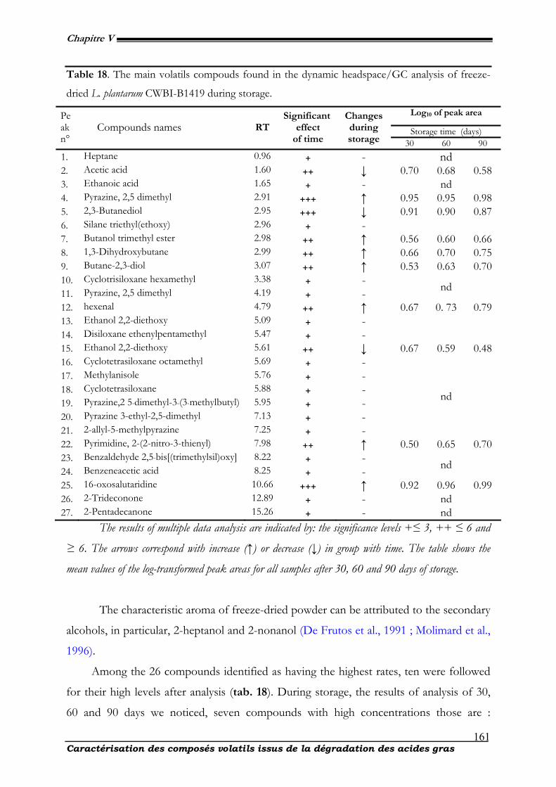
Chapitre V
Caractérisation des composés volatils issus de la dégradation des acides gras
161
Table 18. The main volatils compouds found in the dynamic headspace/GC analysis of freeze-
dried L. plantarum CWBI-B1419 during storage.
Log10 of peak area
Storage time (days)
Peakn°
Compounds names RT Significant
effect of time
Changes during storage 30 60 90
1. Heptane 0.96 + - nd 2. Acetic acid 1.60 ++ ↓ 0.70 0.68 0.58 3. Ethanoic acid 1.65 + - nd 4. Pyrazine, 2,5 dimethyl 2.91 +++ ↑ 0.95 0.95 0.98 5. 2,3-Butanediol 2.95 +++ ↓ 0.91 0.90 0.87 6. Silane triethyl(ethoxy) 2.96 + - 7. Butanol trimethyl ester 2.98 ++ ↑ 0.56 0.60 0.66 8. 1,3-Dihydroxybutane 2.99 ++ ↑ 0.66 0.70 0.75 9. Butane-2,3-diol 3.07 ++ ↑ 0.53 0.63 0.70 10. Cyclotrisiloxane hexamethyl 3.38 + - 11. Pyrazine, 2,5 dimethyl 4.19 + - nd
12. hexenal 4.79 ++ ↑ 0.67 0. 73 0.79 13. Ethanol 2,2-diethoxy 5.09 + - 14. Disiloxane ethenylpentamethyl 5.47 + -
15. Ethanol 2,2-diethoxy 5.61 ++ ↓ 0.67 0.59 0.48 16. Cyclotetrasiloxane octamethyl 5.69 + - 17. Methylanisole 5.76 + - 18. Cyclotetrasiloxane 5.88 + - 19. Pyrazine,2 5-dimethyl-3-(3-methylbutyl) 5.95 + - 20. Pyrazine 3-ethyl-2,5-dimethyl 7.13 + - 21. 2-allyl-5-methylpyrazine 7.25 + -
nd
22. Pyrimidine, 2-(2-nitro-3-thienyl) 7.98 ++ ↑ 0.50 0.65 0.70 23. Benzaldehyde 2,5-bis[(trimethylsil)oxy] 8.22 + - 24. Benzeneacetic acid 8.25 + - nd
25. 16-oxosalutaridine 10.66 +++ ↑ 0.92 0.96 0.99 26. 2-Trideconone 12.89 + - nd 27. 2-Pentadecanone 15.26 + - nd
The results of multiple data analysis are indicated by: the significance levels +≤ 3, ++ ≤ 6 and
≥ 6. The arrows correspond with increase (↑) or decrease (↓) in group with time. The table shows the
mean values of the log-transformed peak areas for all samples after 30, 60 and 90 days of storage.
The characteristic aroma of freeze-dried powder can be attributed to the secondary
alcohols, in particular, 2-heptanol and 2-nonanol (De Frutos et al., 1991 ; Molimard et al.,
1996).
Among the 26 compounds identified as having the highest rates, ten were followed
for their high levels after analysis (tab. 18). During storage, the results of analysis of 30,
60 and 90 days we noticed, seven compounds with high concentrations those are :
Chapitre V
Caractérisation des composés volatils issus de la dégradation des acides gras
162
hexanal, 16-oxosalutaridine, pyrimidine, 2-(2-nitro-3-thienyl), butane-2,3-diol, butanol
trimethyl ester, 1,3-dihydroxybutane and pyrazine, 2,5 dimethyl while acetic acid, 2,3-
butanediol and 2,2-ethanol diethoxy experiencing decline during the same storage. This
difference could be due to the fact that the mortality increased in the cells and the rate of
peroxide with increasing rates in acetate and alcohol decreasing (Barron et al., 2005 ;
McSweeney et al., 2000).
This assertion is consistent with the work carried out by Arora et al. (1995) ; Moio
et al. (1998). These alcohols are used as precursor for the formation of ethyl esters (Fox et
al., 1997 ; Molimard et al., 1996). There is a link between cell death and the oxidation
phenomena observed during storage and production of certain volatile compounds.
Among the most important in Lactobacillus plantarum CBI-B1419, hexenal is a compound
key product of the secondary decomposirion related to membrane oxidation. Our work is
consistent with those made by Molimard et al. (1996). Products are essentially volatile
compound and hexenal has the highest concentration. Methyl ketones are products of
microbial β-oxidation of saturated fatty acids followed by β -keto acid decarboxylation
(Okumura et al., 1985).
V-2.4. Conclusions
The alcohols, alkanes, esters, methyl-branched and phenylalanine derivates, ketones,
and aldehydes were the most abundant groups of volatils in freeze-dried L. planatrum
CWBI-B1419. The amino acid catabolism and ester synthesis seemed to be more
prominent than the lipid oxidation and glycolysis during 90 days storage with no access to
air. Many common secondary lipid oxidation products remained stable or decreased
during storage. Some of the differents additives affected only the esters and pyrazines, not
the lipid oxidation compounds. Even with a high content of long chained PUFA in
freeze-dried cell product, the occurrence of lipid oxidation compounds of these vacuum-
packed products was lower than expected. In general, the volatile production depends
more on the substrate than on the microorganism. The results obtained add to the
knowledge of the volatile composition of cereals and cereal-based products, and provide
Chapitre V
Caractérisation des composés volatils issus de la dégradation des acides gras
163
useful information for further development of suitable dairy probiotic used for drinks and
others products.
V-2.5. Aknowlege
We thank all the technical’s personals of Walloon Center for industrial Biology.
Maryse Hardenne and Celine Pierrart are acknowledged for her very important
contribution in the experiments. We also like to express our gratitude to the republic of
Ivory cost for its financial assistance to this work. Gratefully acknowledged for French
community of Belgium
V-2.6. References
Arora, G., Cormier, F., & Lee, B. 1995. Analysis of odor active volatils in Cheddar cheese headspace by multidimensional GC/MS/Sniffing, J. Agr. Food Chem., 43, 748-752.
Barron, L. J. R., Redondo, Y., Aramburu, M., Perez-Elotondo, F. J., Albisu, M., Najera, A. I., et al. 2005.
Variations in volatile compounds and flavour in Idiazabal cheese manufactured from ewe’s milk in farmhouse and factory, J. Sc. Food. Agr., 85, 1660-1671.
Béal, C., & Corrieu, G. 1994. Lebensmittel-Wissenschaft and Technologie, 27, 86-92.
Béal, C., Mihoub, F., Marin, M., & Corrieu, G. 2001. Demande de Brevet d’invention FR N°2829147.
Boff, J. M., & Min, D. B. 2002. Chemistry and reaction of singlet oxygen in foods. Comprehensive Reviews.
Food. Sc. Safety, 1, 158-172. Bosset, J. O., Butikofer, U., Gauch, R., & Sieber, R. 1997. Ripening of Emmental cheese wrapped in foil
with and without addition of Lactobacillus casei subsp. casei. II. Gas chromatographic investigation of some volatile neutral compounds using dynamic headspace. Lebens. Wis. Techn., 30, 464-470.
Cadawallader KR. 2002. Enzymes and flavor biogenesis in fish. In:Haard NF, Simpson BK, editors.
Seafood enzymes. Utilization and influence on postharvest seafood quality. New York:Marcel Dekker. p 365-83.
Carvalho, A. S., Silva, J. H., Teixera, P., Malcata, F. X., & Gibbs, P. 2003. Int. Dairy. J., 13, 463-468.
Cheng H.Z., Kyoung H.K., Hwan K., and Hyong J.L., 2005. Analysis and characterization of aroma-active
compounds of Schizandra chinensis (omija) leaves, J. Sci. Food. Agric., 85, 161-166. Dainty R., Blom H. 1995. Flavour of fermented sausage. In: Campbell-Platt G, Cook PE, editors.
Fermented meats. London, U.K.: Blackie Academic and Professional. p 176-93. De Frutos, M., Sanz, J., & Martinez-Castro, J. 1991. Characterization of artisanal cheeses by GC/MS
analysis of their medium volatils (SDE) fraction, J. Agr. Food Chem., 39, 524-530.
Chapitre V
Caractérisation des composés volatils issus de la dégradation des acides gras
164
Dunford E., Shahidi F. 1998. Flavours of fish meat. In: Shahidi F, editor. Flavor of meat, meat products, and seafoods. New York: Blackie Academic & Professional. p 131-58.
Edwards R.A., Ordonez J.A., Dainty R.H., Hierro E.M., de la Hoz L. 1999. Characterization of the
headspace volatile compounds of selected Spanish dry fermented sausages, Food. Chem., 64:461-465.
Engels, W. J. M., Dekker, R., De Jong, C., Neeter, R., & Visser, S. 1997. A comparative study of volatile
compounds in the water-soluble fraction of various types of ripened cheese. Int. Dairy. J., 7, 255-263.
Fox, P. F., Law, J., McSweeney, P. L. H., Wallace, J. 1993. Biochemistry of cheese ripening. In 2nd ed. P.
F. Fox (Ed.), Cheese: Chemistry, physics and microbiology, General aspects, 389-438. London: Chapman & Hall
Frankel EN. 2005. Lipid oxidation. 2nd ed. Bridgewater, U.K.: The Oily Press. 470 p. Ishibashi, N., Tatematsu, I., Shimamura, S., Tomita, M., & Okonogi, S. 1985. Drugs and foodstuffs (pp.
227–232). Paris: International Institute of Refrigeration Kawai T. 1996. Fish flavor, Crit. Rev. Food. Sci. Nutr. 36., 257-98. Langsrud Ø. 2002. 50-50 multivariate analysis of variance for colinear responses, J. R. Stat. Soc. Ser D. Stat.,
51, 305-17. Lemaitre, R., King, B., Mozaffarian, D., Sootodehnia, N., & Siscovick, D. 2006. Trans-Fatty acids and
sudden cardiac death. Atheros. Suppl. 7, 13-15. Marco, A.; Navarro, J. L.; Flores, M. 2004. Volatile compounds of dry-fermented sausages as affected by
solid-phase microextraction (SPME), Food Chem., 84, 633-641. McSweeney, P. L. H., & Sousa, M. J. 2000. Biochemical pathways for the production of flavour
compounds in cheese during ripening: A review. Lait, 80, 293-324. Montel MC, Masson F, Talon R. 1998. Bacterial role in flavour developmen, Meat. Sci., 49, S111-23. Nawar W W .1996. Lipids. In Food Chemistry , pp 225-319. Fennema O R, ed. New York, NY: M. Dekker,
Inc. Nordvi B., Egelandsdal B., LangsrudØ, Ofstad R., Slinde E. 2007. Development of a novel fermented and
dried saithe and salmon product, Innov. Food. Sci Emerging. Technol., 8,163-71. Ordonez J.A., Hierro E.M., Bruna J.M., de la Hoz L. 1999. Changes in the components of dry-fermented
sausages during ripening, Critical. Rev. Food. Sci. Nutri., 39, 329-67. Ortigosa, M., Arizcun, C., Torre, P., & Izco, J. M. 2005. Use of wild Lactobacillus strains in an adjunct
culture for a Roncal-type cheese. J. Dairy. Res., 72, 168-178. Sanches-Silva, A.; Lopez-Hernandez, J.; Paseiro-Losada, P. 2005. Profiling flavor compounds of potato
crisps during storage using solid-phase microextraction. J. Chromatogr. A., 1064, 239-245. Sanches-Silva, A.; Rodriguez-Bernaldo de Quiros, A.; Lopez-Hernandez, J.; Paseiro-Losada, P. 2004.
Determination of hexanal as indicator of the lipidic oxidation state in potato crisps using gas chromatography and high-performance liquid chromatography,J. Chromatogr. A., 1046, 75-81.
Chapitre V
Caractérisation des composés volatils issus de la dégradation des acides gras
165
Schmidt S., Berger R.G. 1998. Aroma compounds in fermented sausages of different origins. Lebensm-Wiss Technol., 31, 559-567.
Sedewitz, B., Schleifer, K. H., & Gotz, F. 1984. Physiological role of pyruvate oxidase in the aerobic
metabolism of Lactobacillus plantarum, J. Bacteriol., 160, 462-465. Stolz, P., Vogel, R. F., & Hammes, W. P. 1995. Utilization of electron acceptors by lactobacilli isolated
from sourdough. II. Lactobacillus pontis, Lact. reuteri, Lact. amylovorus and Lact. fermentum. Zeit. Lebens. Unt. Forsch., 201, 402-410.
Toldra F. 2002. Dry-cured meat products. Trumbull: Food & Nutrition Press. 244 p. Vazquez, J. A., Docasal, S. F., Prieto, M. A., Gonzalez, M. A., & Murado, M. A. 2008. Growth and
metabolic features of lactic acid bacteria in media with hydrolysed fish viscera. An approach to bio-silage of fishing by-products, Biores. Tech., 99, 6246-6257.
Viallon C., Berdague J.L,Montel M.C, Talon R,Martin J.F., Kondjoyan N., Denoyer C. 1996. The effect of
stage of ripening and packaging on volatile content and flavour of dry sausages, Food. Res. Int., 29, 667-74.
Chapitre V
Caractérisation des composés volatils issus de la dégradation des acides gras
166
V-3. Conclusions : Caractérisation des composés volatils issus de
la dégradation des acides gras
L’étude des composés volatils issus des cellules lyophilisées de Lactobacillus plantarum
CWB-B1419 montre la présence de divers composés parmi lesquels, l’hexenal qui est
détecté en quantité notable. La présence de composés volatils principalement l’hexenal
montre la corrélation entre la dégradation des acides gras par le phénomène d’oxydation.
Les produits secondaires de cette oxydation sont en général des aldéhydes, des cétones,
des alcanes et des dérivés aminoacides. En ce qui concerne ces composés secondaires
d’oxydation, de nombreux indices chimiques permettent de doser les aldéhydes (indices
de paranisidine, test TBA, indice de carbonyle…) mais avec une sensibilité et un domaine
d’application parfois réduits. Des composés volatils sélectionnés (hexenal, pyrazine, 2,5
dimethyl, butanol trimethyl ester, 1,3-dihydroxybutane, butane-2,3-diol, pyrimidine, 2-(2-
nitro-3-thienyl), 16-oxosalutaridine) ont été analysés par chromatographie en phase
gazeuse après microextraction en phase solide. Parmi les composés identifiés par la
SPME-GC-MS, le taux de l'hexenal est relativement important au cours du stockage. Les
résultats ont montré que les concentrations de la plupart de ces composés augmentaient
au cours du stockage, surtout à température ambiante (25°C). Il se formait plus de
composés d'oxydation des lipides dans les poudres sans cryoprotecteur que dans celles
avec cryoprotecteurs, cependant plus faiblement en l'absence d'oxygène. La présence des
composés volatils (hexenal) est proportionnelle à celle des hydroperoxydes au cours du
stockage.
Chapitre VI
Analyse des phospholipides de Lactobacillus
plantarum CWBI-B1419 au cours du stockage
Chapitre VI
Analyse des phospholipides de Lactobacillus plantarum CWBI-B1419
168
VI. Analyse des phospholipides de L. plantarum CWBI-B1419 au
cours du stockage à température ambiante
VI-1. Contexte et objectifs
L’objectif de cette partie de notre travail, consistera à mettre en évidence la relation
entre le taux de mortalité et la présence des composés primaires (hydroperoxydes) au
cours du stockage dus à la dégradation des acides gras polyinsaturés. Cette étude
permettra de comprendre certains mécanismes à l’origine de la perte de stabilité des
souches lyophilisées. Certes, l’oxydation des lipides membranaires des bactéries fait depuis
un certains temps l’objet d’intenses recherches, mais il existe très peu d’études qui mettent
en relation le taux de mortalité en relation avec la modification de la composition des
phospholipides membranaires des cellules lyophilisées. Dans cette étude, nous mènerons
des expérimentations afin de montrer l’existence de liens entre l’oxydation des lipides
membranaires et la perte de viabilité cellulaire ou du pouvoir d’acidification de Lactobacillus
plantarum CWBI-B1419 lyophilisé au cours du stockage. Selon les résultats précédents
(Chapitres IV & V), nous avons démontré qu’au cours du stockage en présence
d’oxygène et d’humidité, il se produisait une accumulation de produits primaires de
dégradations issues des acides linoléiques et linoléniques de même que les produits
secondaires volatils détectés par SPME. L’intérêt de ce chapitre-ci serait de montrer que la
mortalité cellulaire est un résultat de la modification de la composition phospho-lipidique
de la membrane de bactéries lactiques lyophilisées en général et en particulier de
Lactobacillus plantarum CWBI-B1419, notre souche de référence.
Chapitre VI
Analyse des phospholipides de Lactobacillus plantarum CWBI-B1419
169
VI-2. Effect of storage temperature on phospholipids compositions
and viability upon Lactobacillus plantarum CWB-B1419
Publication 7 – Microbiol. Appl. Biotechnol.
Microbiology and Applied Biotechnology
Coulibaly, I(1♣)., Destain, J(1)., Dubois-D, R(1)., Lognay, G(3)., Fauconnier, M.L(4)
and Ph. Thonart (1, 2)
(1)Wallon Center for Industrial Biology, Bio-Industry Unit, Gembloux
Agricultural University, Passage des déportés 2, 5030 Gembloux,
Belgium.
(2)Wallon Center for Industrial Biology, Microbial Technology Unit,
University of Liège, Sart-Tilman B40, 4000 Liège, Belgium.
(3)Analytical Chemistry Unit, Gembloux Agricultural University, Passages
des déportés 2, 5030 Gembloux, Belgium.
(4)Plant Biology Unit, Gembloux Agricultural University, Passages des
déportés 2, 5030 Gembloux, Belgium.
♣ Corresponding Author : I. Coulibaly, Phone : +3281622305, Fax : +32816614222 E-mail : [email protected]
Chapitre VI
Analyse des phospholipides de Lactobacillus plantarum CWBI-B1419
170
Résumé
Les lipides totaux de Lactobacillus plantarum CWBI-B1419 ont été analysés après
lyophilisation. Sept classes de lipides particuliers ont été identifiées comme étant des
lipides neutres (LN), des acides gras (AG), des phospholipides (PL), des esters de stérol
(ES), des triglycérides (TG), diglycérides (DG) et monoglycérides (MG). Les principaux
acides gras identifiés dans la plupart des classes lipidiques sont les acides : palmitique
(C16:0), palmitoléique (C16:1), oléique (C18:1), linoléique (C18:2) et l'acide linolénique (C18:3).
Les phospholipides (PL) sont les principaux constituants et représentaient 50-60% des
lipides totaux. Ces phospholipides ont été fractionnés. Les phospholipides de L. plantarum
CWBI-B1419 contiennent : l’acide phosphatidique (PA), la phosphatidyléthanolamine
(PE), la phosphatidylinositole (PI), la phosphatidylcholine (PC), la sphingomyéline (SM),
la lysophosphatidylcholine (LPC) et la phosphatidylglycérol (PG). On a observé que la PG
était en proportion plus élevée dans la plupart des pourcentages relatifs aux autres PL et
était le composant prédominant des PL (30% -56%). L’évolution du taux individuel de
chaque PL a été suivi au cours du stockage à 20°C et 40°C avec ou sans Lithothamne
400®, respectivement.
Mots-clés: bactéries lactiques, acides gras, classes de phospholipides, détection par
diffusion de la lumière, survie, température.
Chapitre VI
Analyse des phospholipides de Lactobacillus plantarum CWBI-B1419
171
Abstract
The total lipids of Lactobacillus plantarum CWBI-B1419 were analysed after freeze-
drying. Seven individual lipids classes were identified namely neutral lipids (NL), fatty
acids (FA), phospholipids (PL), sterols ester (ES), triglycerides (TG), diglycerides (DG)
and monoglycerides (MG). The principal fatty acids identified in most lipid classes were
palmitic (C16:0), palmitoleic (C16:1), oleic (C18:1), linoleic (C18:2), and linolenic (C18:3). PL
were the major constituents and accounted for 50-60% of the total lipids. PLs were
fractionated. PL of Lactobacillus plantarum CWBI-B1419 content phosphatidic acid (PA),
phosphatidylethanolamine (PE), phosphatidylinositole (PI), phosphatidylcholine (PC),
sphingomyelin (SM), lysophosphatidylcholine (LPC) and phosphatidylglycerol (PG). It
was observed that PG had the highest proportion at most points relative to other PL and
was the predominant component of PL (30%-56%). Evolution of individual rate was
followed during stored at 20°C and 40°C with or without lithothamne®400, respectively.
Keywords: Lactic acid bacteria, Fatty acids, phospholipids class separation, light
scattering detection, survival, temperature.
Nomenclature
PA : phosphatidic acid TL : total lipid
PE : phosphatidylethanolamine NL : neutral lipids
PI : phosphatidylinositol PL : phospholipids
PC : phosphatidylcholine FD : freeze-drying
SM : sphingomyelin C : cell suspension without protective compound
LPC : lysophosphatidylcholine CL : cell suspension with 8% lithothamne 400
PG : phosphatidyl-DL-glycerol FFA : free fatty acid
SPE : solid phase extraction ELDS :evaporative light scattering detection signal
ES : esters de stérol TG : triglycerides
MG : monoglycerides DG : diglycerides
Chapitre VI
Analyse des phospholipides de Lactobacillus plantarum CWBI-B1419
172
VI-2.1. Introduction
For the last decade, HPLC has been the method of choice for the separation and
quantification of phospholipids (PL) classes. This methodology has the advantage of
avoiding the extensive derivatization needed in GC analysis of PL (Descalzo et al., 2003).
Previous studies have demonstrated that the properties of an ELSD are useful for a
quantitative HPLC detection of lipid classes within different polarity classes (Homanzt et
al.,1998 ). This detection method has been described as highly sensitive for neutral lipids
and phospholipids, with an acceptable reproducibility to enable quantification (Grizard et
al., 2000 ; Silversand et al., 1997). A clear benefit of this technique is the possibility of
detecting lipophilic substances independent of the absorption properties of eluents or
samples and without any probe labeling of detected molecules (Seppänen-Laasko et al.,
2001). The signal intensity of an ELSD seems to be dependent on the development of
droplets of eluents by the nebulizer/evaporator, as well as on the detector temperature.
Nowadays, the use of an ELSD eliminates problems of derivatization methodology and
transparent of mobile phase in UV associated with other HPLC detectors and allows
qualitative/quantitative studies of nonvolatile analytes (Mounts et al., 1990 ; Patton et al.,
1982 and Stolyhwo et al., 1984). For example, unlike refractive index and low-wavelength
UV, ELSD uses multisolvent gradients to improve resolution and perform faster
separation of the eluted compounds (Christie et al., 1985 ; Vaghela et al., 1995 and Mounts
et al., 1992). Therefore, an HPLC-ELSD system could analyze underivatized intact lipids,
thus saving time and avoiding losses during derivatization and hydrolysis. Evaporative
light scattering is based on the detection of nonvolatile molecules carried by a volatile
mobile phase. The column effluents are nebulized to an aerosol, followed by volatile
compound vaporization and formation of small solute droplets. The laser light scattered
by these droplets is detected by a photodiode (Stolyhwo et al., 1992). The separation of all
PL classes from different matrixes by use of a variety of multisolvent gradients had been
reported previously (Van der Meeren et al., 1988 ; Juanéda, et al., 1990 ; Christie et al., 1988
and Picchioni et al., 1996). Lipids ranging from cholesterol to lysophosphatidylcholine
(LPC) can be resolved in a single normal-phase HPLC run using a gradient of increasing
polarity. Even nonpolar molecules like hydrophobic pulmonary surfactant proteins can be
accurately detected by ELSD (Bünger et al., 200). The polarity of different lipid classes
Chapitre VI
Analyse des phospholipides de Lactobacillus plantarum CWBI-B1419
173
constitutes a useful tool allowing PL separation. Solid-phase extraction (SPE) takes
advantage of this property and has been used as an alternative to liquid–liquid extraction.
A selective elution of the desired compounds can be obtained with SPE by simple
fractionation of the analytes through differences in their polarity (Wachob et al., 1991).
For dairy products like freeze-dried LAB, important phospholipids are phosphatidyl-
ethanolamine (PE), phosphatidylinositol (PI), phosphatidylserine (PS) and phosphatidyl-
choline (PC). Important sphingolipids in dairy products are sphingomyelin (SM),
glucosylceramide and lactosylceramide. Lysophosphatidylcholine (LPC) and phosphatidic
acid (PA) are rarely reported in dairy products; when they do appear, it is likely due to
careless sample preparation or to phospholipase activity (Christie et al., 1987). The aim of
this study was to determine the distribution of individual molecular species of phospho-
lipids of Lactobacillus palntarum CWBI-B1419 during storage at 20°C and 40°C with or
without lithothamne 400® respectively by HPLC-ELSD.
VI-2.2. Materials and methods
VI-2.2.1. Strain and culture
The fermentation in a 20L bioreactor was performed in triplicate, using the culture
medium and the operational parameters of 30°C, 120 rpm, and pH 6, controlled. The
fermentation time was 17h. The biomass was centrifuged in a Beckman Avanti J-25 High-
Performance Centrifuge Systems (Analys, Belgium) centrifuge. The cream was collected,
and analysis of dry matter content, viability, and pH of the cream was performed; this
cream was the raw material for the freeze-drying according to Armando et al., 2006. The
viable counts were obtained by plate count method after 48 h. Acidification was carried
out at 30°C in 150 ml MRS broth inoculated with 1% of 107 cfu ml-1 of the freezedried
sample. The total titratable acidity (% of lactic acid/g DW) was determinate after 18 h,
according to the AOAC method, (1997).
VI-2.2.2. Solvents and standards
Reagents. n-Hexane, 2-propanol, methanol, and water were HPLC grade from VWR
International bvba Haasrode Resaerchpark, Geldenaddksebaan 464 B-3001 Leuven. All
Chapitre VI
Analyse des phospholipides de Lactobacillus plantarum CWBI-B1419
174
other chemicals were analytical reagent grade. L-α-phosphatidylethanolamine (PE), L-α-
phosphatidyl-DL-glycerol (PG), phosphatidic acid (PA), L-α-phosphatidylcholine (PC), L-
α-lysophosphatidylcholine (LPC), L-α-phosphatidylinositole (PI) and sphingomyelin (SM)
with purity of approximately 98% were obtained from Avanti Polar Lipids (Alabaster,
USA)). To prepare calibration curves, standards were diluted by the appropriate solvent
considering its solubility. Duplicate injections of each dilution were used.
VI-2.2.3. Lipid extraction
Fatty acids were extracted overnight from dried cells of Lactobacillus plantarum (2±0.2
g), with ether-ethanol (3:1v/v) mixture according to an adaptation of the method of Ito et
al., (1969). The extraction was performed by successively adding 10mL of chloroform
containing butyl-hydroxy-toluene (0.01%), 10 mL of chloroform and 12.8 mL of NaCl
(1.56%). After each new addition, the mixture was homogenized by a 20s blending period
(Ultra-turrax, IKA, Staufen, Germany). The mixture was centrifuged for 10 min at 6000 g.
Total lipids (TL) were obtained by collecting an aliquot (10 mL) of the chloroform phase.
During the extraction procedure, tubes were kept at 0-4°C.
VI-2.2.4. Solid-phase extraction (SPE)
After evaporation of the chloroform, the TL extract was diluted in 1.5mL of
chloroform/methanol (2:1, vol/vol). The organic phase was collected into a conical vial,
evaporated to dryness under a nitrogen stream, and made up to 0.5-mL final volume with
chloroform. The extract was applied to the top of a normal-phase silica cartridge (Bond
Elut NH2, Anion Exchange, varian Inc. N.V, Varian S.A, Belgium) that had been
previously conditioned with 10 mL of chloroform. Sequential elution was performed with
the cartridge column was equilibrated by rinsing twice with 2 ml of hexane using the
Supelco extraction apparatus. Lipids dissolved in 10 mL of chloroform were loaded onto
the column and the chloroform was pulled through. There after, the column was eluted
with 4 ml of a chloroform-2-propanol 2:1 mixture (neutral lipid fraction). Then, 4 ml of
ether containing 2% acetic acid followed by 4 ml of methanol was applied to elute free
fatty acids and neutral phospholipids, respectively (Hee-Yong et al., 1990). Finally, acidic
lipids were eluted using 4 ml of a mixture of hexane-2-propanolethanol-0.1 M ammonium
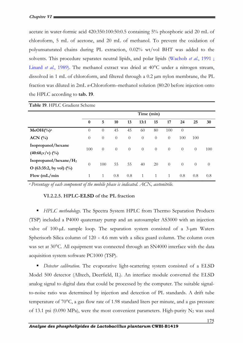
Chapitre VI
Analyse des phospholipides de Lactobacillus plantarum CWBI-B1419
175
acetate in water-formic acid 420:350:100:50:0.5 containing 5% phosphoric acid 20 mL of
chloroform, 5 mL of acetone, and 20 mL of methanol. To prevent the oxidation of
polyunsaturated chains during PL extraction, 0.02% wt/vol BHT was added to the
solvents. This procedure separates neutral lipids, and polar lipids (Wachob et al., 1991 ;
Linard et al., 1989). The methanol extract was dried at 40°C under a nitrogen stream,
dissolved in 1 mL of chloroform, and filtered through a 0.2 µm nylon membrane, the PL
fraction was diluted in 2mL n-Chloroform–methanol solution (80:20 before injection onto
the HPLC according to tab. 19.
Table 19. HPLC Gradient Scheme
Time (min)
0 5 10 13 13:1 15 17 24 25 30
MeOH(%)a 0 0 45 45 60 80 100 0
ACN (%) 0 0 0 0 0 0 0 100 100
Isopropanol/hexane
(40:60,v/v) (%) 100 0 0 0 0 0 0 0 0 100
Isopropanol/hexane/H2
O (63:35:2, by vol) (%) 0 100 55 55 40 20 0 0 0 0
Flow (mL/min 1 1 0.8 0.8 1 1 1 0.8 0.8 0.8
a Percentage of each component of the mobile phase is indicated. ACN, acetonitrile.
VI.2.2.5. HPLC-ELSD of the PL fraction
HPLC methodology. The Spectra System HPLC from Thermo Separation Products
(TSP) included a P4000 quaternary pump and an autosampler AS3000 with an injection
valve of 100-µL sample loop. The separation system consisted of a 3-µm Waters
Spherisorb Silica column of 120 × 4.6 mm with a silica guard column. The column oven
was set at 30°C. All equipment was connected through an SN4000 interface with the data
acquisition system software PC1000 (TSP).
Detector calibration. The evaporative light-scattering system consisted of a ELSD
Model 500 detector (Alltech, Deerfield, IL). An interface module converted the ELSD
analog signal to digital data that could be processed by the computer. The suitable signal-
to-noise ratio was determined by injection and detection of PL standards. A drift tube
temperature of 70°C, a gas flow rate of 1.98 standard liters per minute, and a gas pressure
of 13.1 psi (0.090 MPa), were the most convenient parameters. High-purity N2 was used
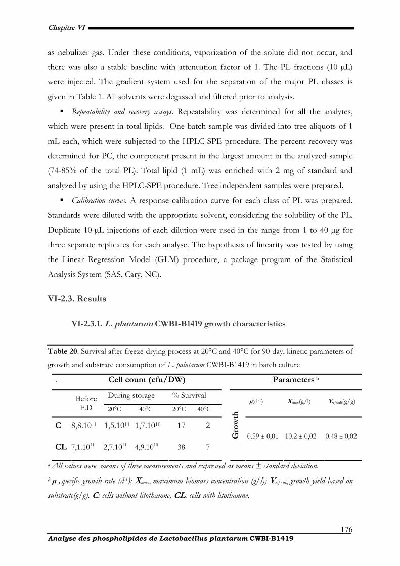
Chapitre VI
Analyse des phospholipides de Lactobacillus plantarum CWBI-B1419
176
as nebulizer gas. Under these conditions, vaporization of the solute did not occur, and
there was also a stable baseline with attenuation factor of 1. The PL fractions (10 µL)
were injected. The gradient system used for the separation of the major PL classes is
given in Table 1. All solvents were degassed and filtered prior to analysis.
Repeatability and recovery assays. Repeatability was determined for all the analytes,
which were present in total lipids. One batch sample was divided into tree aliquots of 1
mL each, which were subjected to the HPLC-SPE procedure. The percent recovery was
determined for PC, the component present in the largest amount in the analyzed sample
(74-85% of the total PL). Total lipid (1 mL) was enriched with 2 mg of standard and
analyzed by using the HPLC-SPE procedure. Tree independent samples were prepared.
Calibration curves. A response calibration curve for each class of PL was prepared.
Standards were diluted with the appropriate solvent, considering the solubility of the PL.
Duplicate 10-µL injections of each dilution were used in the range from 1 to 40 µg for
three separate replicates for each analyse. The hypothesis of linearity was tested by using
the Linear Regression Model (GLM) procedure, a package program of the Statistical
Analysis System (SAS, Cary, NC).
VI-2.3. Results
VI-2.3.1. L. plantarum CWBI-B1419 growth characteristics
Table 20. Survival after freeze-drying process at 20°C and 40°C for 90-day, kinetic parameters of
growth and substrate consumption of L. palntarum CWBI-B1419 in batch culture
. Cell count (cfu/DW) Parameters b
During storage % Survival Before
F.D 20°C 40°C 20°C 40°C µ(d-1) Xmax(g/l) Yx/sub(g/g)
C 8,8.1011 1,5.1011 1,7.1010 17 2
CL 7,1.1011 2,7.1011 4,9.1010 38 7
Gro
wth
0.59 ± 0,01 10.2 ± 0,02 0.48 ± 0,02
a All values were means of three measurements and expressed as means ± standard deviation.
b µ ,specific growth rate (d-1); Xmax, maximum biomass concentration (g/l); Yx/sub, growth yield based on
substrate(g/g). C: cells without litothamne, CL: cells with litothamne.
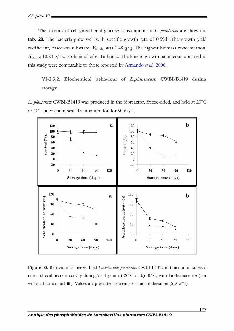
Chapitre VI
Analyse des phospholipides de Lactobacillus plantarum CWBI-B1419
177
The kinetics of cell growth and glucose consumption of L. plantarum are shown in
tab. 20. The bacteria grew well with specific growth rate of 0.59d1.The growth yield
coefficient, based on substrate, Yx/sub, was 0.48 g/g. The highest biomass concentration,
Xmax of 10.20 g/l was obtained after 16 hours. The kinetic growth parameters obtained in
this study were comparable to those reported by Armando et al., 2006.
VI-2.3.2. Biochemical behaviour of L.plantarum CWBI-B1419 during
storage
L. plantarum CWBI-B1419 was produced in the bioreactor, freeze-dried, and held at 20°C
or 40°C in vacuum-sealed aluminium foil for 90 days.
-20
0
20
40
60
80
100
120
0 30 60 90 120
Storage time (days)
Surv
ival
(%
).
-20
0
20
40
60
80
100
120
0 30 60 90 120
Storage time (days)
Surv
ival
(%
).
0
30
60
90
120
0 30 60 90 120
Storage time (days)
Aci
dif
icat
ion
act
ivit
y (%
)
0
30
60
90
120
0 30 60 90 120
Storage time (days)
Aci
dif
icat
ion
act
ivit
y (%
)
Figure 33. Behaviour of freeze dried Lactobacillus plantarum CWBI-B1419 in function of survival
rate and acidification activity during 90 days at a) 20°C or b) 40°C, with litothamene (-●-) or
without litothamne (-■-). Values are presented as means ± standard deviation (SD, n=3).
a b
a b
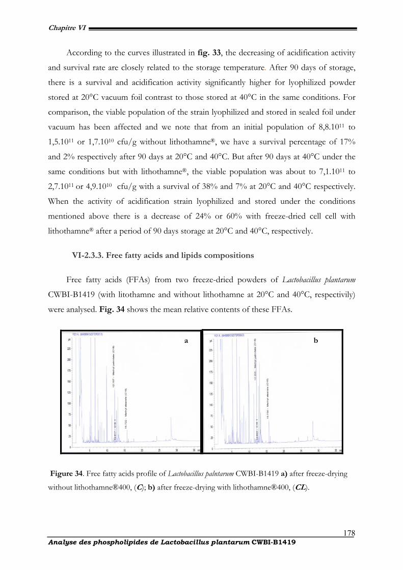
Chapitre VI
Analyse des phospholipides de Lactobacillus plantarum CWBI-B1419
178
According to the curves illustrated in fig. 33, the decreasing of acidification activity
and survival rate are closely related to the storage temperature. After 90 days of storage,
there is a survival and acidification activity significantly higher for lyophilized powder
stored at 20°C vacuum foil contrast to those stored at 40°C in the same conditions. For
comparison, the viable population of the strain lyophilized and stored in sealed foil under
vacuum has been affected and we note that from an initial population of 8,8.1011 to
1,5.1011 or 1,7.1010 cfu/g without lithothamne®, we have a survival percentage of 17%
and 2% respectively after 90 days at 20°C and 40°C. But after 90 days at 40°C under the
same conditions but with lithothamne®, the viable population was about to 7,1.1011 to
2,7.1011 or 4,9.1010 cfu/g with a survival of 38% and 7% at 20°C and 40°C respectively.
When the activity of acidification strain lyophilized and stored under the conditions
mentioned above there is a decrease of 24% or 60% with freeze-dried cell cell with
lithothamne® after a period of 90 days storage at 20°C and 40°C, respectively.
VI-2.3.3. Free fatty acids and lipids compositions
Free fatty acids (FFAs) from two freeze-dried powders of Lactobacillus plantarum
CWBI-B1419 (with litothamne and without lithothamne at 20°C and 40°C, respectivily)
were analysed. Fig. 34 shows the mean relative contents of these FFAs.
Figure 34. Free fatty acids profile of Lactobacillus palntarum CWBI-B1419 a) after freeze-drying
without lithothamne®400, (C); b) after freeze-drying with lithothamne®400, (CL).
a b
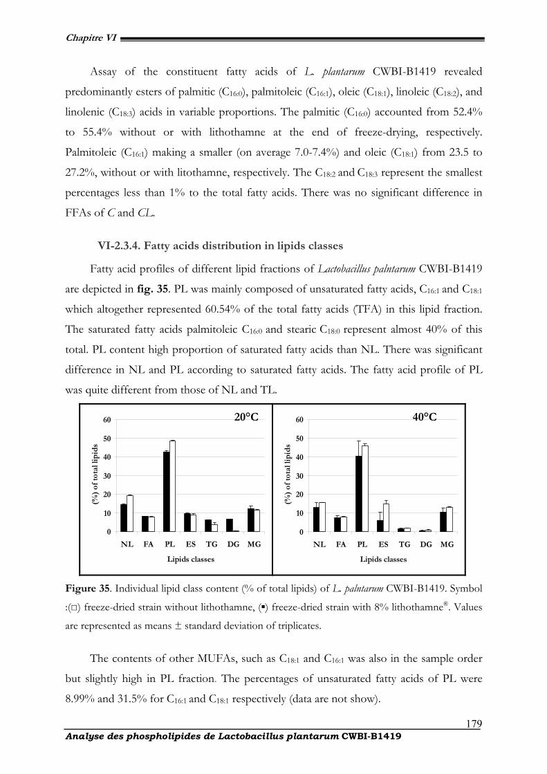
Chapitre VI
Analyse des phospholipides de Lactobacillus plantarum CWBI-B1419
179
Assay of the constituent fatty acids of L. plantarum CWBI-B1419 revealed
predominantly esters of palmitic (C16:0), palmitoleic (C16:1), oleic (C18:1), linoleic (C18:2), and
linolenic (C18:3) acids in variable proportions. The palmitic (C16:0) accounted from 52.4%
to 55.4% without or with lithothamne at the end of freeze-drying, respectively.
Palmitoleic (C16:1) making a smaller (on average 7.07.4%) and oleic (C18:1) from 23.5 to
27.2%, without or with litothamne, respectively. The C18:2 and C18:3 represent the smallest
percentages less than 1% to the total fatty acids. There was no significant difference in
FFAs of C and CL.
VI-2.3.4. Fatty acids distribution in lipids classes
Fatty acid profiles of different lipid fractions of Lactobacillus palntarum CWBI-B1419
are depicted in fig. 35. PL was mainly composed of unsaturated fatty acids, C16:1 and C18:1
which altogether represented 60.54% of the total fatty acids (TFA) in this lipid fraction.
The saturated fatty acids palmitoleic C16:0 and stearic C18:0 represent almost 40% of this
total. PL content high proportion of saturated fatty acids than NL. There was significant
difference in NL and PL according to saturated fatty acids. The fatty acid profile of PL
was quite different from those of NL and TL.
0
10
20
30
40
50
60
NL FA PL ES TG DG MG
Lipids classes
(%
) of
tota
l lip
ids
0
10
20
30
40
50
60
NL FA PL ES TG DG MG
Lipids classes
(%
) of
tota
l lip
ids
Figure 35. Individual lipid class content (% of total lipids) of L. palntarum CWBI-B1419. Symbol
:(□) freeze-dried strain without lithothamne, (▪) freeze-dried strain with 8% lithothamne®. Values
are represented as means ± standard deviation of triplicates.
The contents of other MUFAs, such as C18:1 and C16:1 was also in the sample order
but slightly high in PL fraction. The percentages of unsaturated fatty acids of PL were
8.99% and 31.5% for C16:1 and C18:1 respectively (data are not show).
20°C 40°C
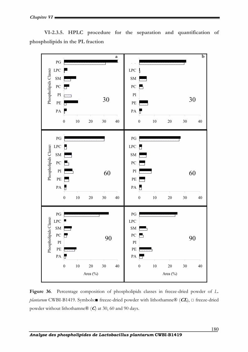
Chapitre VI
Analyse des phospholipides de Lactobacillus plantarum CWBI-B1419
180
VI-2.3.5. HPLC procedure for the separation and quantification of
phospholipids in the PL fraction
0 10 20 30 40
PA
PE
PI
PC
SM
LPC
PG
Phos
phol
ipid
s Cla
sses
0 10 20 30 40
PA
PE
PI
PC
SM
LPC
PG
0 10 20 30 40
PA
PE
PI
PC
SM
LPC
PG
Phos
phol
ipid
s Cla
sses
0 10 20 30 40
PA
PE
PI
PC
SM
LPC
PG
0 10 20 30 40
PA
PEPI
PC
SMLPC
PG
Phos
phol
ipid
s Cla
sses
Area (%)
0 10 20 30 40
PA
PEPI
PC
SMLPC
PG
Area (%)
Figure 36. Percentage composition of phospholipids classes in freeze-dried powder of L.
plantarum CWBI-B1419. Symbols:■ freeze-dried powder with lithothamne® (CL), □ freeze-dried
powder without lithothamne® (C) at 30, 60 and 90 days.
30
90 90
60 60
30
b a
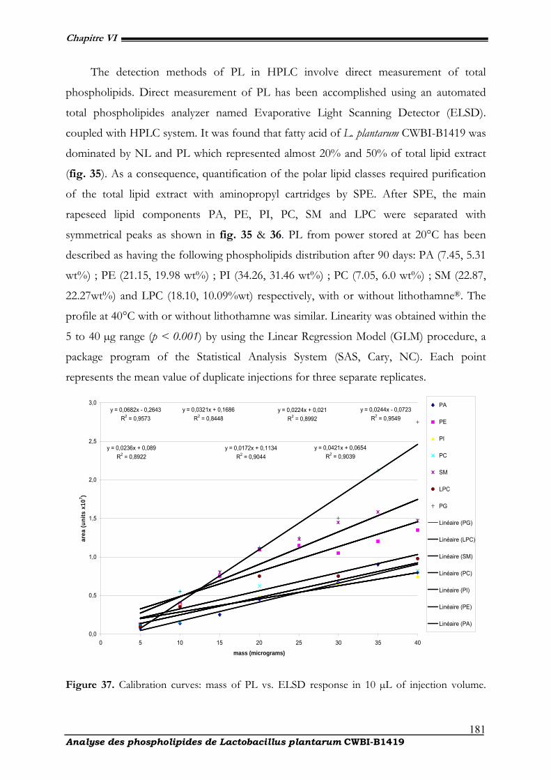
Chapitre VI
Analyse des phospholipides de Lactobacillus plantarum CWBI-B1419
181
The detection methods of PL in HPLC involve direct measurement of total
phospholipids. Direct measurement of PL has been accomplished using an automated
total phospholipides analyzer named Evaporative Light Scanning Detector (ELSD).
coupled with HPLC system. It was found that fatty acid of L. plantarum CWBI-B1419 was
dominated by NL and PL which represented almost 20% and 50% of total lipid extract
(fig. 35). As a consequence, quantification of the polar lipid classes required purification
of the total lipid extract with aminopropyl cartridges by SPE. After SPE, the main
rapeseed lipid components PA, PE, PI, PC, SM and LPC were separated with
symmetrical peaks as shown in fig. 35 & 36. PL from power stored at 20°C has been
described as having the following phospholipids distribution after 90 days: PA (7.45, 5.31
wt%) ; PE (21.15, 19.98 wt%) ; PI (34.26, 31.46 wt%) ; PC (7.05, 6.0 wt%) ; SM (22.87,
22.27wt%) and LPC (18.10, 10.09%wt) respectively, with or without lithothamne®. The
profile at 40°C with or without lithothamne was similar. Linearity was obtained within the
5 to 40 µg range (p < 0.001) by using the Linear Regression Model (GLM) procedure, a
package program of the Statistical Analysis System (SAS, Cary, NC). Each point
represents the mean value of duplicate injections for three separate replicates.
y = 0,0682x - 0,2643R2 = 0,9573
y = 0,0244x - 0,0723R2 = 0,9549
y = 0,0421x + 0,0654R2 = 0,9039
y = 0,0321x + 0,1686R2 = 0,8448
y = 0,0236x + 0,089R2 = 0,8922
y = 0,0224x + 0,021R2 = 0,8992
y = 0,0172x + 0,1134R2 = 0,9044
0,0
0,5
1,0
1,5
2,0
2,5
3,0
0 5 10 15 20 25 30 35 40
mass (micrograms)
area
(uni
ts x
107 )
PA
PE
PI
PC
SM
LPC
PG
Linéaire (PG)
Linéaire (LPC)
Linéaire (SM)
Linéaire (PC)
Linéaire (PI)
Linéaire (PE)
Linéaire (PA)
Figure 37. Calibration curves: mass of PL vs. ELSD response in 10 µL of injection volume.
Chapitre VI
Analyse des phospholipides de Lactobacillus plantarum CWBI-B1419
182
Results are given as linear curves of phospholipids extracted of three separate
extractions from the same sample of L. plantarum CWBI-B1419 freeze-dried. Each curve
includes a linear equation of the regression line as well as the R2. The experiments were performed thrice. SPE, the solid phase extraction has
yielded the following phospholipids: PA, phosphatidic acid ; PE, phosphatidylethano-
lamine ; PC, phosphatidylcholine ; PI, phosphatidylinositol ; SM, sphingomyelin; LPC
lyso- phosphatidycholine ; PG, phophoglycerol as shown by fig. 37.
VI-2.4. Discussion
Detection of phospholipids is favored because (i) lipids are extracted by the
hydrophobic solvent and (ii) SPE selectively desorbs surface-active molecules such as
polar lipids, though not all types equally well. Many studies have focused on the lipid
composition of Lactobacillus spp. (Exterkate et al., 1971 ; Thorne, 1961 ; Uchida, 1973).
Authors such as (O'Leary et al., 1988 ; Ikawa et al., 1963) showed that lactobacilli are
generally composed of n-C16:0, n-C18:1 and C19 CYC-carboxylate as the main constituents
with a small amount of n-C14:0, n-C16:1, and n-C18:0 and traces in some species (Drucker,
1994). These atypical profiles in terms of fatty acids are those of gram-positive, but the
main groups are the common gram-positive bacteria (O'Leary, 1988). The analysis of
polar groups in L. plantarum B1419-CWBI revealed the same trends and showed that the
majority of polar lipids is composed of phosphatidylglycerol (PG). These results are fully
consistent with studies by Fischer et al. (1978). As for other components such as
phosphatidic acids, diphosphatidylglycerol (cardiolipin), phosphatidylglycerol (Exterkate et
al., 1971 ; Fischer et al., 19990), phosphoglycolipids and diglycosyldiacylglycerol (Heller et
al., 1988), there are small quantities. During storage we noticed a decrease in PG for
freeze-dried at 20°C while at 40°C not only PG but also decreased PE and SM begin a
significant decline in 90 days. These results are similar to those we obtained with L.
plantarum. At this temperature the powders without lithothamne are most vulnerable. As
explanation and following this period the decrease in the ratio unsaturated/saturated fatty
acids and cell death occurred essentially at the same rate (data are not showed). As stated
before, the mechanisms of death during storage are still unknown but from these results it
seems evident that lipid oxidation and survival during storage may be related. This is
Chapitre VI
Analyse des phospholipides de Lactobacillus plantarum CWBI-B1419
183
supported by indirect evidence presented in previous reports. For example, it is reported
that the presence of antioxidants increased the survival rate of dried bacteria during
storage (Porubcan et al., 1975 ; Teixeira et al., 1995b). Dimmick et al. (1961) found a strong
similarity between the loss of viability and the increase in free-radical concentration
during storage of freeze-dried Serrntia marcrscrns. Work done by Marshall et al. (1974)
supports the hypothesis that reactions between carbonyl compounds and cellular
components are a major cause of mortality during storage of dried micro-organisms. The
nature of these compounds was not presented but it is possible that they are products
formed during lipid oxidation. Slater, (1984) reported that, in eukaryotic cells, extensive
lipid peroxidation can result in membrane disorganization by peroxidizing mainly the
highly unsaturated fatty acids leading to changes in the ratio of unsaturated to other fatty
acids. The uncontrolled peroxidation of biomembranes can thus lead to profound effects
on membrane structure and function, and may be sufficient to cause cell death.
VI-2.5.Conclusion
The present method thus Lactobacillus phospholipids almost free of triglycerides in
one step and does not require the pre-extraction of total lipids is conventional. These
methods have been successfully used and have given a satisfaction in our analysis for our
isolation of phospholipids from bacteria and plant tissues. Work for monitoring changes
in phospholipids classes will be conducted for other species in order to know the limits of
the method.
VI-2.6. Akwnoledge
The authors would like to acknowledge Ir Céline Pierart, Maryse Hardenne and Mr
Vincent Hote for their contribution. We thank all the technical personal of CWBI (Centre
Wallon de Biologie Industrielle). We also like to express our gratitude to the Republic of
Ivory Coast and the Communauté Française de Belgique for financial assistance.
VI-2.7. References
Aluyi, H. S., V. Boote, D. B. Drucker, and J. M. Wilson. 1992. Fast atom bombardment-mass spectrometry for bacterial chemotaxonomy: influence of culture age, growth temperature, gaseous environment and extraction techniques. J. Appl. Bacteriol. 72:80-86.
Chapitre VI
Analyse des phospholipides de Lactobacillus plantarum CWBI-B1419
184
Armando, H., Fréderic, W., Jesús, M., Carlos, B., and Thonart, P. 2006. Freeze-drying of the biocontrol agent Lactobacillus palntarum CWBI-B1419 Predicted stability of formulated powders, Ind. Biotech., 2, 209-212.
Batley, M., N. H. Packer, and J. W. Redmond. 1982. Molecular analysis of the phospholipids of Escherichia
coli K12. Biochim. Biophys. Acta 710:400-405. Becart, J., C. Chevalier, and J.P. Biesse. 1990. Quantitative analysis of phospholipids by HPLC with a light
scattering evaporative detector - application to raw material for cosmetic use, J. High Resolut. Chromatogr., 13, 126-129.
Bünger, H., and U. Pison. 1995. Quantitative analysis of pulmonary surfactant phospholipids by high-
performance liquid chromatography and light-scattering detection, J. Chromatogr. B. Biomed.Sci. Appl., 672, 25-31.
Christie, W. W. 1986. Separation of lipid classes by high-performance liquid chromatography with the
“mass detector.” J. Chromatogr., 361, 396-399. Christie, W. W., and R. A. Urwin. 1995. Separation of lipid classes from plant tissues by high-performance
liquid chromatography on chemically bonded stationary phases, J. High Resolut. Chromatogr., 18, 97-100.
Christie, W.W. 1985. Rapid Separation and Quantification of Lipid Classes by High Performance Liquid
Chromatography and Mass (light-scattering) Detection, J. Lipid Res., 26, 507–512. Christie, W.W., and Morrison, W.R. 1988. Separation of Complex Lipids of Cereals by High-Performance
Liquid Chromatography with Mass Detection, J. Chromatogr., 436, 510-513. Cole, M. J., and C. G. Enke. 1991. Direct determination of phospholipid structures in microorganisms by
fast atom bombardment triple quadrupole mass spectrometry. Anal. Chem. 63:1032–1038. Drucker, D. B. 1994. Fast atom bombardment mass spectrometry of phospholipids for bacterial
chemotaxonomy, p. 18–35. In C. Fenselau (ed.), Mass spectrometry for the characterization of microorganisms. American Chemical Society, Washington, D.C.
Eckard, P. R., Taylor, L. T. and Slack, G. C. 1998. Method Development for the Separation of
Phospholipids by Subcritical Fluid Chromatography, J. Chrom. A., 826, 241. Exterkate, F. A., B. J. Otten, H. W. Wassenberg, and J. H. Veerkamp. 1971. Comparison of the
phospholipid composition of Bifidobacterium and Lactobacillus strains. J. Bacteriol. 106:824-829. Fang, J., M. J. Barcelona, and J. D. Semrau. 2000. Characterization of methanotrophic bacteria on the
basis of intact phospholipids profiles, FEMS Microbiol. Lett., 189, 67-72. Fenselau, C. 1994. Mass spectrometry for characterization of microorganisms. An overview, p. 1–17. In C.
Fenselau (ed.), Mass spectrometry for the characterization of microorganisms. American Chemical Society, Washington,D.C.
Fischer, W. 1990. Bacterial phosphoglycolipids and lipoteichoic acids, p. 123–234. In M. Kates (ed.),
Handbook of lipid research, vol. 6. Plenum Press, New York. Fischer, W., R. A. Laine, and M. Nakano. 1978. On the relationship between glycerophosphoglycolipids
and teichoic acids in Gram-positive bacteria. II. Structures of glycerophosphoglycolipids, Biochim. Biophys. Acta., 528,298–308.
Chapitre VI
Analyse des phospholipides de Lactobacillus plantarum CWBI-B1419
185
Grizard, G., B. Sion, D. Bauchart, and D. Boucher. 2000.Separation and Quantification of Cholesterol and Major Phospholipid Classes in Human Semen by High-Performance Liquid Chromatography and Light Scattering Detection, Ibid., 740,101-107.
He, J. Vaghela, M.N., and Kilara, A. 1995. Quantitative Analysis of Phospholipids from Whey Protein
Concentrates by High Performance Liquid Chromatography with a Narrow Bore Column and an Evaporative Light-Scattering Detector, J. Am. Oil. Chem. Soc., 72, 729-733.
Cheung, A.P., Wang, E., Fang, K., and Liu, P. 2001. High-Performance Liquid Chromatographic Analysis
for a Non-chromophore-containing Phosphatidyl Inositol Analog, 1-[(1-O-octadecyl-2-O-methyl-sn-glycero)-phospho]-1D-3-deoxy-myo-inositol, Using Indirect UV Detection, J. Chromatogr. A., 913, 355–363.
Heller, D. N., C. M. Murphy, R. J. Cotter, C. Fenselau, and O. M. Uy. 1988. Constant neutral loss
scanning for the characterisation of bacterial phospholipids desorbed by fast atom bombardment, Anal. Chem., 60, 2787-2791.
Homan, R., and M.K. Anderson. 1998. Rapid Separation and Quantification of Combined Neutral and
Polar Lipid Classes by High-Performance Liquid Chromatography and Evaporative Light Scattering Mass Detection, J. Chromatogr. B., 708, 21-26.
Ikawa, M. 1963. Nature of the lipids of some lactic acid bacteria, J. Bacteriol., 85, 772-781. Juanéda, P., Rocquelin, G., and Astorg, P.O. 1990. Separation and Quantification of Heart and Liver
Phospholipid Classes by High-Performance Liquid Chromatography Using a New Light-Scattering Detector, Lipids., 25, 756-759.
Lechevalier, M. P. 1977. Lipids in bacteria taxonomy - A taxonomist’s view, Crit. Rev. Microbiol., 7, 109-
210. Linard, A. 1989. Separation des Classes de Phospholipides par Chromatographie Liquide Haute
Performance, Cah. Techn. INRA., 20, 41-48. Miwa, H., Yamamoto, M., Futata, T., Kan, K. and Asano, T. 1996. Thin-layer Chromatography and High-
performance Liquid Chromatography for the Assay of Fatty Acid Compositions of Individual Phospholipids in Platelets from Non-insulin-dependent Diabetes Mellitus Patients: Effect of Eicosapentaenoic Acid Ethyl Ester Administration, J. Chrom. B., 677, 217.
Montanari, L., Fantozzi, P., Snyder, J.M. and King, F.W. 1999. Selective Extraction of Phospholipids from
Soybeans with Supercritical Carbon Dioxide and Ethanol, J. Supercritical. Fluids., 14, 87. Mounts T.L., Abidi, S.L., and Rennick, K.A. 1992. HPLC Analysis of Phospholipids by Evaporative Laser
Light-Scattering Detection, J. Am. Oil. Chem. Soc., 69, 438-442. Mounts, T.L., and Nash, A.M. 1990. HPLC Analysis of Phospholipids in Crude Oil for Evaluation of
Soybean Deterioration, J. Am.Oil Chem. Soc., 67, 757-760. Munster, C., Lu, J., Schinzel, S., Bechinger, B. and Salditt, T. 2000. Grazing Incidence X-ray Diffraction of
Highly Aligned Phospholipid Membranes Containing the Antimicrobial Peptide Magainin 2, Eur. Biophys. J., 28, 683.
O’Leary, W. M., and S. G. Wilkinson. 1988. Gram-positive bacteria, p. 117-201. In C. Ratledge and S. G.
Wilkinson (ed.), Microbial lipids, vol. 1. Academic Press, London. Olsson, N. U. and Salem, N. Jr. 1997. Molecular Species Analysis of Phospholipids, J. Chrom. B., 692, 245.
Chapitre VI
Analyse des phospholipides de Lactobacillus plantarum CWBI-B1419
186
Patton, G.M., Fasulo, J.M., and Robins, S.J. 1982. Separation of Phospholipids and Individual Molecular Species of Phospholipids by High-Performance Liquid Chromatography, J. Lipid Res., 23, 190-196.
Picchioni, G.A., Watada, A.E., and Whitaker, B.D. 1996. Quantitative High-Performance Liquid
Chromatography Analysis of Plant Phospholipids and Glycolipids Using Light-Scattering Detection, Lipids., 31, 217-221.
Row, K.H. and Lee, J.W., (1999). Gradient Separation of Soybean Phospholipids with Retention Factors
of a New Polynomial Correlation, Korean J. Chem. Eng., 16, 170. Sas, B., E. Peys, and M. Helsen. 1999. Efficient method for (lyso) phospholipid class separation by high-
performance liquid chromatography using an evaporative light-scattering detector, J. Chromatogr.A,. 864, 179-182.
Seppänen-Laasko, T., I. Laasko, H. Vanhanen, K. Kiviranta, T. Lehtimäki, and R. Hiltunen.,2001. Major
Human Plasma Lipid Classes Determined by Quantitative High-Performance Liquid Chromatography, Their Variation and Associations with Phospholipid Fatty Acids, Ibid., 75, 437-445.
Servillo, L., Iorio, E. L., Quagliuolo, L., Camussi, G., Balestrieri, C. and Giovane, A.. 1997. Simultaneous
Determination of Lysophospholipids by High-performance Liquid Chromatography with Fluorescence Detection, J. Chrom. B., 689, 281.
Silversand, C., and C. Haux.1997. Improved High-Performance Liquid Chromatographic Method for the
Separation and Quantification of Lipid Classes: Application to Fish Lipids, Ibid., 703,7-14. Stolyhwo, A., Colin, H., Guiochon, G. 1983. Use of Light Scattering as a Detector Principle in Liquid
Chromatography, J. Chromatogr., 265, 1-18. Stolyhwo, A., Colin, H., Martin, M., and Guiochon, G. 1984. Study of the Qualitative and Quantitative
Properties of the Light-Scattering Detector, J. Chromatogr., 288, 253-275. Thorne, K. J. I. 1964. The phospholipids of Lactobacillus casei. Biochim. Biophys. Acta., 84, 350-353. Uchida, K., and K. Mogi. 1972. Cellular fatty acid spectra of Pediococcus species in relation to their
taxonomy, J. Gen. Appl. Microbiol., 18, 109-129. Uchida, K., and K. Mogi. 1973. Cellular fatty acid spectra of Hiochi bacteria, acid-tolerant lactobacilli, and
their separation, J. Gen. Appl. Microbiol., 19:233-249.
Vaghela, M.N., and Kilara, A. 1995. Quantitative Analysis of Phospholipids from Whey Protein Concentrates by High Performance Liquid Chromatography with a Narrow Bore Column and an Evaporative Light-Scattering Detector, J. Am. Oil. Chem. Soc., 72, 729-733.
Chapitre VI
Analyse des phospholipides de Lactobacillus plantarum CWBI-B1419
187
VI-3. Conclusions : Analyse des phospholipides de L. plantarum
CWBI-B1419 au cours du stockage à température ambiante
L’analyse des phospholipides membranaires a permis de voir la variation engendrée
par l’effet de la température sur les différents composants de ce complexe lipidique. Mais
au delà de cette analyse, il ressort que le taux de mortalité est étroitement lié à la
modification de cette entité. Parmi les différentes techniques, celle de la SPME semble
donner de très bons résultats en un temps record. La maîtrise des différents paramètres
essentiels de cette étude permet donc d’envisager des perspectives prometteuses. A la
lumière des résultats de l’analyse des phospholipides, on est en mesure d’affirmer que la
modification des composés phospholipidiques est à l’origine de la mortalité cellulaire
observée chez Lactobacillus plantarum CWBI-B1419. Les résultats obtenus ont mis en
évidence l’impact des modifications des phospholipides au cours du stockage (acide
palmitique C16:0 et acide oléique C18:1). De ces résultats, nous pouvons donc conclure que
la résistance cellulaire et l’adaptation des bactéries aux conditions drastiques de
production et de stockage sont fonction de la modification des phospholipides
membranaires.
Chapitre VII
Discussions générales
Discussions générales
189
Discussions générales
Au cours de ces décennies, le marché des bactéries lactiques en général et celui des
probiotiques en particulier suscite un intérêt de plus en plus croissant au niveau de la
recherche scientifique. La préoccupation des biotechnologues étant de produire et de
commercialiser des souches performantes à l’image de Lactobacillus plantarum CWBI-B1419
autant pour leurs propriétés fonctionnelles (acidifiantes), que pour leurs comportements
au cours du stockage à température ambiante (peu de perte de viabilité due à une bonne
résistance à la dessiccation), leurs résistances à l’acidité de l’estomac, aux sels biliaires et
aux phénomènes d’échauffement couramment rencontrés dans la plupart des procédés
technologiques. Les travaux de cette thèse avaient pour objectif principal l’étude de la
résistance des bactéries lactiques à la lyophilisation. Pour mener à bien ce travail, cet
objectif général a été scindé en cinq objectifs fondamentaux :
la sélection de souches lactiques performantes capables de résister non seulement
aux procédés technologiques mais surtout aux conditions de stockage.
la réalisation de différentes formulations mises en jeu au cours de la lyophilisation
pour optimiser la conservation à température ambiante.
l’étude des produits primaires de dégradation des acides gras polyinsaturés afin de
comprendre les mécanismes de la mortalité observée au cours du stockage. Ce volet s’est
surtout intéressé aux produits primaires de la dégradation (hydroperoxydes au nombre de
quatre 9- et 13- HOD, 9- et 13- HPOD).
l’étude des produits secondaires issus de la dégradation des acides gras insaturés
en l’occurrence les composés volatils.
l’étude et le suivi des phospholipides membranaires au cours du stockage.
L’utilisation des bactéries à des fins industrielles se trouve confrontée à la problématique
de la production et surtout à celle de la conservation compte tenu de l’agressivité des
procédés de séchage vis à vis des cellules. Pour accroître le taux de survie, les scientifiques
ont recours à la sélection de souches de plus en plus résistantes (chapitre II) mais
également à l’usage d’additifs protecteurs pendant le séchage et au cours du stockage.
L’utilisation d’additifs permet d’obtenir des résultats satisfaisants comme le montrent,

Chapitre VII
Discussions générales
190
les travaux décrits au chapitre III : « Effets des agents protecteurs sur la
composition en acides gras cellulaires de Lactobacillus plantarum CWBI-B1419 ».
Il est bien connu que les bactéries se protègent contre les effets néfastes des phénomènes
osmotiques par l'accumulation de composés organiques à bas poids moléculaires, les
solutés compatibles (Glaasker et al., 1996). Ces additifs n'inhibent pas les fonctions vitales
de la cellule même à des concentrations cytoplasmiques élevées. En effet, ils ne
produisent pas d'effets néfastes sur la structure et la solubilité des protéines, l'interaction
protéines-protéines, l'interaction enzyme-substrat ou l'interaction protéine-acide nucléique
(Low, 1985 ; Yancey et al., 1982). L'accumulation intracellulaire de ces composés provient
d'une synthèse de novo ou d'un transport à partir du milieu de croissance (Ko et al.,
1994). Ces additifs sont de diverses structures : des polyols (glycérol, sorbitol, et
mannitol), des sucres (sucrose, sorbitol, tréhalose), des acides aminés et dérivés comme la
bétaïne, la carnitine, l'acide tetrahydroxypyrimidine carboxylique (hydroxyectoïne), le
glutamate, la glutamine, proline et l’alanine. Ils se présentent aussi sous le vocable de
cryoprotecteurs ou de solutés compatibles et remplissent trois fonctions principales au
niveau de la cellule :
rétablir la pression de turgescence dans les cellules par rapport à l'environnement
extracellulaire (Csonka, 1989 ; Csonka et al., 1991).
améliorer la stabilité des enzymes à basse activité d'eau (Lippert & Galinski, 1992).
stabiliser les membranes durant les périodes de dessiccation (Crowe et al., 1987 ;
Leslie et al., 1995 ; Rudolph et al.,1986). Ces fonctions sont étroitement liées à la structure
moléculaire des solutés compatibles qui présentent des caractéristiques spécifiques
communes (Pocard et al., 1994 ; Gutierrez et al., 1995) : - molécules organiques à bas
poids moléculaire- une grande solubilité dans l'eau -une charge neutre à pH physiologique
- une absence de toxicité sur les enzymes in vitro.
De nouvelles techniques et méthodes, permettent de nos jours de prédire la stabilité
de la viabilité cellulaire au stockage ; parmi celles-ci, les techniques de mesure de la
température de transition vitreuse (Tg) par l’analyse calorimétrique différentielle, les
techniques de marquage par la fluorescence, et la détermination des constituants
membranaires (les acides gras membranaires et les phospholipides) par les techniques de
chromatographie en phase gazeuse.
Chapitre VII
Discussions générales
191
Selon Matthieu et al. (2002), la température de transition vitreuse est l’un des
paramètres importants pour la caractérisation d’un produit. La perte de la viabilité et de
l'activité des bactéries se produisent généralement pendant les différentes étapes de la
fabrication et du stockage. Des changements de leur état physique se produisent puisque
ces matériaux biologiques se composent principalement d'hydrates de carbone et de
protéines, qui peuvent être amorphes ou partiellement amorphes. Afin de mieux
comprendre les changements qui se produisent pendant le traitement et le stockage des
bactéries lactiques, on analyse leurs propriétés physiques. Des diagrammes d'état pour
décrire les différentes régions de l'état physique des matériaux montrent le rapport entre
la composition et la température. Ces diagrammes sont habituellement établis avec des
mesures effectuées par calorimétrie à balayage différentiel ou Differential Scanning
Calorimetry (DSC). Les diagrammes associés aux isothermes de sorption d'eau sont
souvent liés à la qualité du produit sec et sont employés pour expliquer la stabilité de ces
produits pendant le stockage. Nous avons utilisé ces données pour attester de la qualité
de la lyophilisation et par conséquent déterminer la durée de conservation des poudres
issues de la lyophilisation à différentes températures de stockage.
Toujours dans l’optique de prévoir et suivre la survie cellulaire, les biologistes et
microbiologistes ont de plus en plus recours à la technique de la fluorescence appelée
cytométrie en flux. La cytométrie en flux (CMF) est une technique d’analyse des cellules,
des bactéries et des particules en suspension dans un liquide qui permet de les faire passer
une à une à grande vitesse devant un ou plusieurs faisceaux lasers. Un système optique et
informatique sophistiqué analyse les propriétés optiques des cellules (diffraction et
réflexion lumineuses). L’utilisation de molécules fluorescentes permet d’augmenter le
nombre de paramètres étudiés par cellule (jusqu’à 8 ou 10 simultanément). On peut ainsi
analyser le contenu en ADN des cellules, et donc la répartition d’une population cellulaire
dans les différentes phases du cycle cellulaire (Hoerni et al., 2006). Cette technique de
caractérisation individuelle, quantitative et qualitative de particules en suspension dans un
liquide, permet d’analyser les signaux optiques ou physiques émis par une particule
coupant le faisceau lumineux d’un laser ou d’une lampe à arc. Dans le cas qui nous
concerne, cette technique sert à faire la discrimination des bactéries basée sur l’activité
métabolique.
Chapitre VII
Discussions générales
192
Deux types de méthodes existent pour détecter les cellules métaboliquement
actives :
celles basées sur des processus énergie-dépendant ((biosynthèses, potentiel de
membrane, transport de molécules à travers les membranes, etc.),
celles basées sur des processus énergie-indépendant (activité enzymatique : activité
estérase, diacétate de fluorescéine, déshydrogénase (CTC)). Ces signaux séparés par des
filtres optiques sont collectés par des photomultiplicateurs (PMT), amplifiés, numérisés,
traités et stockés par un ordinateur par l'intermédiaire d'une composante informatique et
optique (miroir dichroïque et filtre optique). La fonction tri des cytomètres en flux les
plus évolués permet de trier physiquement une ou deux populations cellulaires définies
par leurs propriétés optiques. L’une des applications de la cytométrie en flux est le tri
cellulaire, cette fonction permet de séparer les cellules selon les informations données par
le phénotype.
Tous ces instruments de prévisions aboutissent à une seule conclusion, les cellules
au cours du stockage subissent divers stress et des modifications au niveau de la
membrane cellulaire. Parmi ces modifications, l’oxydation des acides gras tient une place
de premier rôle car elle est responsable de la production de radicaux libres
(hydroperoxydes) à partir des acides gras polyinsaturés linoléique et linolénique comme
démontré au chapitre IV. Nos résultats sont en accord avec les travaux réalisés par
Zhang et al., 2007 ; França et al., 2007 et Howe et al., 2002. L’analyse des produits
volatils, les produits secondaires de dégradation des poudres issus de la lyophilisation des
bactéries lactiques montre une corrélation entre le taux de mortalité et la production de
certains composés aldéhydiques, cétones et autres selon les résultats du chapitre V :
« Analyses des composés volatils issus de la dégradation des acides gras de
bactéries lactiques ». L’étude des acides gras et des phospholipides permet ensuite de
rendre compte de la détérioration des constituants phospholipidiques au cours du
stockage. Cela traduit l’importance des peroxydes dans la mortalité cellulaire voir chapitre
VI : « Détermination des phospholipides chez L. plantarum CWBI-B1419 au cours
du stockage ». Ce volet montre également l’intérêt de l’utilisation des antioxydants
pendant le stockage.
Chapitre VII
Discussions générales
193
De l’importance de la sélection et de l’identification de souches lactiques
thermorésistantes et performantes
Selon Klaenhammer et al. (1981), une sélection rigoureuse est le premier des
facteurs pour l’amélioration de la conservation des bactéries lactiques. Afin de garantir
une viabilité commercialement satisfaisante, après un stockage à long terme, il est
opportun de sélectionner des souches qui ont survécu à des conditions drastiques
(température élevée, sécheresse).
Le choix de ces deux critères repose sur plusieurs paramètres parmi lesquels,
l’hypothèse qu’une souche ayant survécu dans un environnement drastique : chaud et sec,
possède ou a développé un mécanisme de défense contre la chaleur et la déshydratation.
Or, en biotechnologie, lors de l’étape de production ou au cours du stockage, la perte de
viabilité « systématiquement observée » des souches de bactéries lactiques est imputable à
ces deux facteurs. C’est dans cette optique, que nous avons sélectionné des souches de
bactéries lactiques sur la base de leur résistance à un traitement thermique et à la
déshydratation.
En ce qui concerne la déshydratation, les techniques utilisées se basent dès lors sur
les capacités d’une bonne reprise d’activité des souches après application d’un traitement
osmotique sur les cellules. Celui-ci permet de mobiliser l’eau libre dans l’environnement et
de déclencher un système de protection contre ce stress. Les travaux constituants les
objectifs fixés au chapitre II, nous ont permis de montrer les paramètres et les facteurs
sur lesquels un contrôle et une optimisation étaient susceptibles d’améliorer la résistance
thermique et la viabilité au cours de la conservation des souches. Les différentes
techniques utilisées ont permis de discriminer les cinq souches pour aboutir à une
résistance conforme aux critères cités ci-dessus. Ces résultats sont en phase avec les
travaux effectués par Niamsup et al. (2003) et Prasad et al. (2003). Dans le cadre des
études visant à isoler les microorganismes thermotolérants pour une utilisation dans
l'industrie de la fermentation, nous avons isolé un grand nombre de bactéries lactiques
(Sp03, Sp04, Sp05, Sp011 et Sp020) comme indiqué par le tab. 21 à partir de fientes de
poulets et les sols proches de poulaillers au Sénégal qui atteignent des températures
relativement élevées, soit 40-50°C.
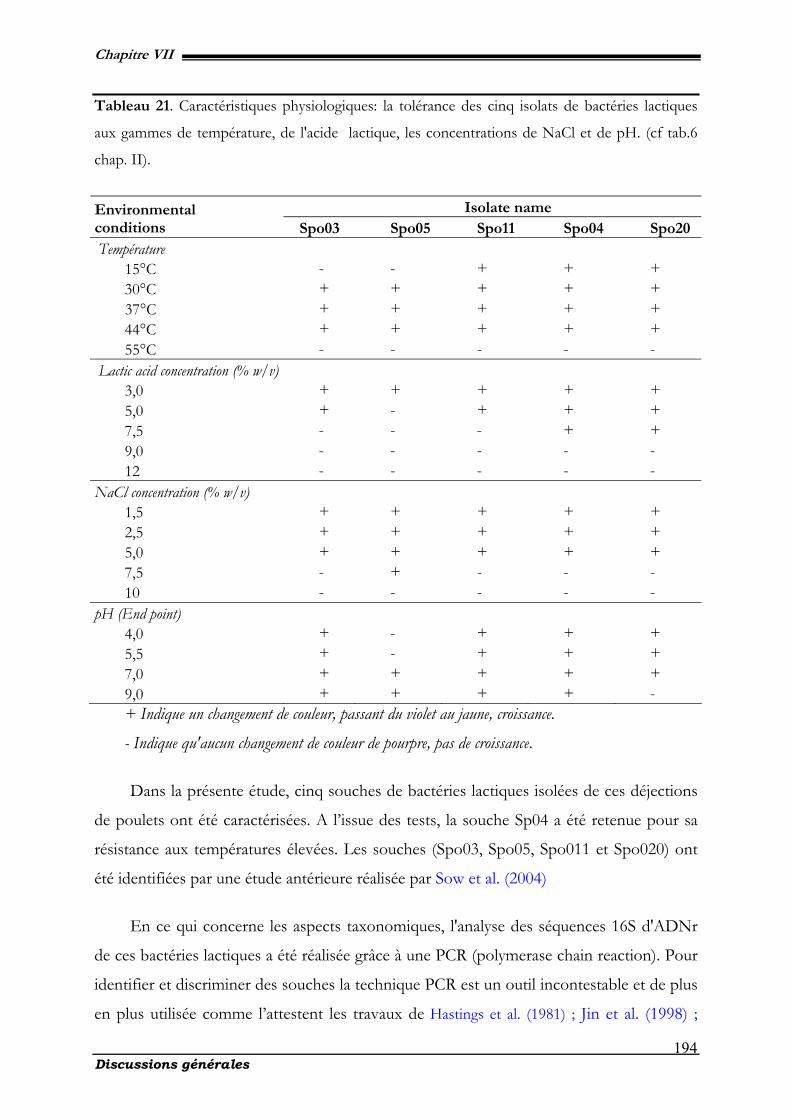
Chapitre VII
Discussions générales
194
Tableau 21. Caractéristiques physiologiques: la tolérance des cinq isolats de bactéries lactiques
aux gammes de température, de l'acide lactique, les concentrations de NaCl et de pH. (cf tab.6
chap. II).
+ Indique un changement de couleur, passant du violet au jaune, croissance.
- Indique qu'aucun changement de couleur de pourpre, pas de croissance.
Dans la présente étude, cinq souches de bactéries lactiques isolées de ces déjections
de poulets ont été caractérisées. A l’issue des tests, la souche Sp04 a été retenue pour sa
résistance aux températures élevées. Les souches (Spo03, Spo05, Spo011 et Spo020) ont
été identifiées par une étude antérieure réalisée par Sow et al. (2004)
En ce qui concerne les aspects taxonomiques, l'analyse des séquences 16S d'ADNr
de ces bactéries lactiques a été réalisée grâce à une PCR (polymerase chain reaction). Pour
identifier et discriminer des souches la technique PCR est un outil incontestable et de plus
en plus utilisée comme l’attestent les travaux de Hastings et al. (1981) ; Jin et al. (1998) ;
Isolate name Environmental conditions Spo03 Spo05 Spo11 Spo04 Spo20 Température
15°C - - + + + 30°C + + + + + 37°C + + + + + 44°C + + + + + 55°C - - - - -
Lactic acid concentration (% w/v) 3,0 + + + + + 5,0 + - + + + 7,5 - - - + + 9,0 - - - - - 12 - - - - -
NaCl concentration (% w/v) 1,5 + + + + + 2,5 + + + + + 5,0 + + + + + 7,5 - + - - - 10 - - - - -
pH (End point) 4,0 + - + + + 5,5 + - + + + 7,0 + + + + + 9,0 + + + + -
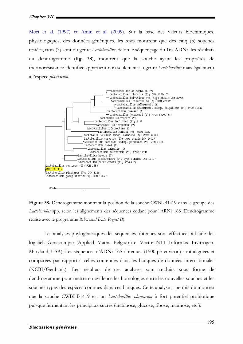
Chapitre VII
Discussions générales
195
Mori et al. (1997) et Amin et al. (2009). Sur la base des valeurs biochimiques,
physiologiques, des données génétiques, les tests montrent que des cinq (5) souches
testées, trois (3) sont du genre Lactobacillus. Selon le séquençage du 16s ADNr, les résultats
du dendrogramme (fig. 38), montrent que la souche ayant les propriétés de
thermorésistance identifiée appartient non seulement au genre Lactobacillus mais également
à l’espèce plantarum.
Figure 38. Dendrogramme montrant la position de la souche CWBI-B1419 dans le groupe des
Lactobacillus spp. selon les alignements des séquences codant pour l'ARNr 16S (Dendrogramme
réalisé avec le programme Ribosomal Data Project II).
Les analyses phylogénétiques des séquences obtenues sont effectuées à l’aide des
logiciels Genecompar (Applied, Maths, Belgium) et Vector NTI (Informax, Invitrogen,
Maryland, USA). Les séquences d’ADNr 16S obtenues (1500 pb environ) sont alignées et
comparées par rapport à celles contenues dans les banques de données internationales
(NCBI/Genbank). Les résultats de ces analyses sont traduits sous forme de
dendrogramme pour mettre en évidence les homologies entre les nouvelles souches et les
souches types des espèces connues dans ces banques. Cette analyse a permis de montrer
que la souche CWBI-B1419 est un Lactobacillus plantarum à fort potentiel probiotique
puisque fermentant les principaux sucres (arabinose, glucose, ribose, mannose, etc.).
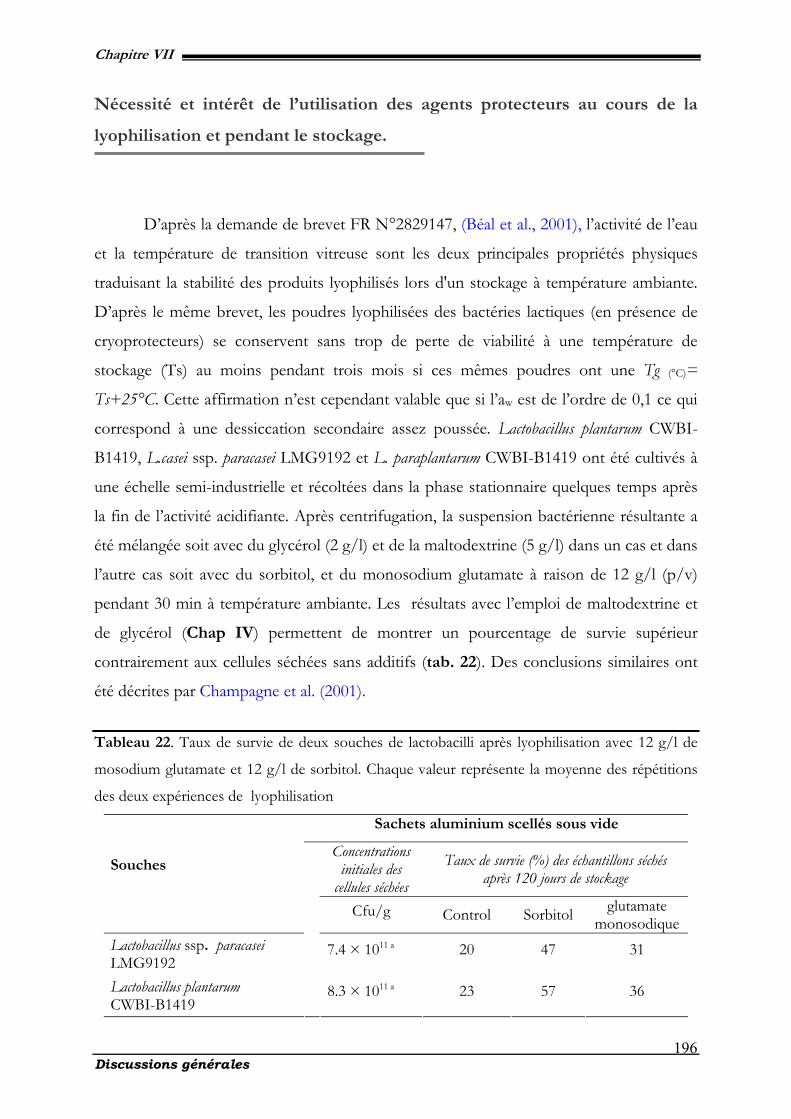
Chapitre VII
Discussions générales
196
Nécessité et intérêt de l’utilisation des agents protecteurs au cours de la
lyophilisation et pendant le stockage.
D’après la demande de brevet FR N°2829147, (Béal et al., 2001), l’activité de l’eau
et la température de transition vitreuse sont les deux principales propriétés physiques
traduisant la stabilité des produits lyophilisés lors d'un stockage à température ambiante.
D’après le même brevet, les poudres lyophilisées des bactéries lactiques (en présence de
cryoprotecteurs) se conservent sans trop de perte de viabilité à une température de
stockage (Ts) au moins pendant trois mois si ces mêmes poudres ont une Tg (°C)=
Ts+25°C. Cette affirmation n’est cependant valable que si l’aw est de l’ordre de 0,1 ce qui
correspond à une dessiccation secondaire assez poussée. Lactobacillus plantarum CWBI-
B1419, L.casei ssp. paracasei LMG9192 et L. paraplantarum CWBI-B1419 ont été cultivés à
une échelle semi-industrielle et récoltées dans la phase stationnaire quelques temps après
la fin de l’activité acidifiante. Après centrifugation, la suspension bactérienne résultante a
été mélangée soit avec du glycérol (2 g/l) et de la maltodextrine (5 g/l) dans un cas et dans
l’autre cas soit avec du sorbitol, et du monosodium glutamate à raison de 12 g/l (p/v)
pendant 30 min à température ambiante. Les résultats avec l’emploi de maltodextrine et
de glycérol (Chap IV) permettent de montrer un pourcentage de survie supérieur
contrairement aux cellules séchées sans additifs (tab. 22). Des conclusions similaires ont
été décrites par Champagne et al. (2001).
Tableau 22. Taux de survie de deux souches de lactobacilli après lyophilisation avec 12 g/l de
mosodium glutamate et 12 g/l de sorbitol. Chaque valeur représente la moyenne des répétitions
des deux expériences de lyophilisation
Sachets aluminium scellés sous vide
Concentrations initiales des
cellules séchées
Taux de survie (%) des échantillons séchés après 120 jours de stockage
Souches
Cfu/g Control Sorbitol glutamate monosodique
Lactobacillus ssp. paracasei LMG9192
7.4 × 10¹¹ a 20 47 31
Lactobacillus plantarum CWBI-B1419
8.3 × 10¹¹ a 23 57 36
Chapitre VII
Discussions générales
197
Dans les deux cas, on assiste à une amélioration du taux de survie ou de stabilité
de l’ordre de 90% pour les échantillons traités avec les additifs protecteurs contrairement
aux cellules sans protecteurs où le taux de survie ne dépasse guère les 20%. On note un
taux de survie de 57% quand le sorbitol est utilisé et de 35% quand c’est le glutamate
monosodique après 120 jours de stockage pour L. plantarum CWBI-B1419. De même, la
souche L. plantarum est plus résistante avec des taux doubles (36 à 57%) par rapport à L.
casei ssp. paracasei LMG9192. Ces résultats sont en parfaite harmonie avec les travaux
effectués par Giulio et al. (2005), montrant que la résistance de L. plantarum est plus
élevée que celle enregistrée par L. paraplantarum. A la lumière de ces résultats, nous
pouvons affirmer que l'influence des additifs de protection est spécifique de
l'espèce et doit donc être déterminée au cas par cas. La sensibilité élevée de la souche
de L. plantarum comparativement à L. paraplantarum peut être due à la plus grande fragilité
de la surface membranaire des cellules avec comme conséquence une augmentation des
dégâts membranaires (Fonseca et al., 2000).
Les dommages causés aux systèmes biologiques séchés par lyophilisation peuvent
être attribués principalement à l'évolution de l'état des lipides membranaires et à des
changements dans la structure des protéines sensibles (Leslie et al., 1995). Cependant,
même si la survie est importante après la congélation et/ou le séchage, la viabilité peut
être réduite par la suite pendant le stockage (Abadias et al., 2001; Fonseca et al., 2000;
Wisselink et al., 2002). Les additifs ont un rôle important dans la conservation et le
maintien de la viabilité cellulaire. Pour remplir ces fonctions, l’additif utilisé comme
protecteur devrait fournir une bonne cryoprotection des cellules pendant la lyophilisation,
il doit être facile à sécher et fournir les atouts d’une matrice permettant la stabilité et la
facilité de réhydratation (Costa et al., 2000). Le sorbitol et le monosodium glutamate
(gms) sont présentés comme des protecteurs de qualité (Abadias et al., 2001 ; Hubalek et
al., 1996 ; Lindres et al., 1997). L’augmentation de l'activité résiduelle et de la viabilité
après le séchage en présence du sorbitol comme matrice de séchage a été précédemment
décrite pour divers organismes (Abadias et al., 2001 ; Lindres et al., 1997). Bien que le
mécanisme de protection cellulaire par des polyols n’est pas entièrement compris, trois
hypothèses ont été proposées (Wisselink et al., 2002): (i) le maintien de la turgescence,
(Kets et al., 1996), (ii) la stabilisation de la structure des lipides membranaires et des
Chapitre VII
Discussions générales
198
protéines à faible activité d'eau (Leslie et al., 1995 ; Yoo et al., 1993), (iii) la prévention des
dommages oxydatifs par inhibition des réactions responsables de la production des
radicaux libres par l'oxygène (Leslie et al., 1995). La stabilisation des structures de
protéines par des réactions entre les groupes aminés (-NH2) du protecteur et les groupes
carboxyle (-COOH) des protéines du micro-organisme, et la capacité de retenir de plus
grandes quantités d'eau résiduelle ont été présentées comme des explications de la
protection par le monosodium glutamate pendant la lyophilisation (Front de Valdez et al.,
1983). Les résultats sont en accord avec les travaux expérimentaux relatifs à la survie de
cellules lyophilisées de L. plantarum et L. rhamnosus pendant le stockage à température
ambiante, en présence de chacun des quatre sucres (myo-inositol, sorbitol, fructose et de
tréhalose), le monosodium glutamate (msg) et l'antioxydant (propyl-gallate) par Leslie et
al., 1995. Tous les composés testés au cours de ces travaux se sont avérés efficaces dans la
protection pour L. plantarum CWBI-B1419 et un peu moins pour les autres souches (L.
paraplantarum CWBI-B545, L. casei ssp. paracasei LMG9192). En accord avec les résultats
publiés par Font de Valdez et al. (1983), aucune corrélation n'a été observée entre
l'efficacité de protection du sorbitol sur la bactérie et leur pouvoir fermentescible. Ce
résultat suggère que l'effet est de nature physico-chimique. Les mécanismes proposés
pour tenter de rationaliser la protection par le sorbitol en cours de stockage comprennent
la prévention d’endommager les membranes par interaction avec la membrane (Linder et
al., 1997b) et la stabilisation de la fonctionnalité des protéines de structure (Yoo et al.,
1993).
Incidences de la dégradation des acides gras polyinsaturés et leurs effets sur
la viabilité cellulaire (les hydroperoxydes).
L’action des additifs sur la membrane cellulaire observée au cours du stockage,
nous amène à analyser les produits résultant de l’oxydation des acides gras. Les résultats
obtenus permettent de confirmer qu’au cours de la croissance bactérienne, les différentes
phases de la cinétique de croissance ont une incidence sur la thermorésistance, de même
que le pH du milieu d’incubation qui joue un rôle prépondérant, (Sakamoto, 2002). Cette
Chapitre VII
Discussions générales
199
constatation entraîne la nécessité de stabiliser les paramètres que sont le pH et la
température. Elles présentent une résistance thermique qui leur permet de croître et de se
reproduire à 45°C contrairement à L. paracasei sp. casei LMG 9192 et L. paraplantarum
CWBI-B545. Selon les travaux effectués par Sow et al. (2004), la thermotolérance de la
souche L. plantarum CWBI-B1419 peut être la résultante de plusieurs facteurs, dont la
synthèse d’un fort pourcentage d’acide oléique (C18:1) et dans une moindre mesure d’acide
linolénique (C18:3) ; comme décrit par les travaux de Russell et al. (2002) et Yoo et al.
(2005) qui sont unanimes sur ce sujet. Pour ces auteurs la diminution de la synthèse des
acides gras monoinsaturés est étroitement liée à l’augmentation de la température
d’incubation. Ceci constitue en fait une réponse à une modification de la fluidité
membranaire. Cette constatation est aussi vérifiée pour la souche CWBI-B1419.
Au cours de la lyophilisation et surtout pendant le stockage, la membrane subit les
phénomènes d’oxydation initiés par la présence d’oxygène ce qui est illustré par les
résultats du chapitre IV. En effet, les acides gras cellulaires (AGC) de L. mesenteroides et
de L. plantarum CWBI-B1419 ont été analysés avant et après lyophilisation. Six principaux
acides gras ont été identifiés, à savoir les acides palmitique (C16:0), palmitoléique (C16:1),
stéarique (C18:0), oléique (C18: 1), linoléique (C18:2) et l'acide linolénique (C18:3). Quatre de
ces acides, à savoir les C16:0, C16:1, C18:0 et C18:1, représentent plus de 94% ou 93% des
acides gras chez L. mesenteroides et L. plantarum CWBI-B1419, respectivement, et un taux
de 5-6%, en moyenne pour le C18:3, respectivement. Le C18:2 représente un pourcentage
très réduit (en moyenne ≤ 1%). Les C16:0 constituent chez L. mesenteroides et L. plantarum
CWBI-B1419, la plus forte proportion. Il n'existe aucune modification significative au
niveau des acides gras cellulaires (CFA) de L. mesenteroides et L. plantarum en réponse à la
déshydratation. En outre, l'ajout de glycérol et de maltodextrine, avant la lyophilisation ne
modifie pas significativement les acides gras polyinsaturés des deux souches. Les acides
gras cellulaires de poudres de L. mesenteroides et L. plantarum emballées et scellées sous vide
dans du papier d'aluminium ou dans des tubes de verre blanc et maintenues à 20 ou 4°C
pendant 90 jours ont été analysés. L'acide palmitique (C16:0) n’a pas subi de modification
au cours du stockage. Par conséquent, les teneurs en acides gras sont exprimées par un
rapport entre chaque acide gras et le C16:0. On a ainsi observé que les ratios C16:1/C16:0,
C18:0/C16:0 et C18:1/C16:0 pour les cellules lyophilisées de L. mesenteroides ou de L. plantarum,
Chapitre VII
Discussions générales
200
avec ou sans cryoprotecteurs n'ont pas beaucoup évolué au cours de la période de
stockage. On a constaté que, indépendamment du mode d'emballage et des températures
d'entreposage, les ratios C18:2/C16:0 et C18:3/C16:0 des cellules lyophilisées de L. mesenteroides
et de L. plantarum, avec ou sans cryoprotecteurs ont subi une diminution au cours du
temps. Toutefois, un ratio plus élevé de C18:2/C16:0 ou C18:3/C16:0 pour L. mesenteroides et L.
plantarum a été noté dans les poudres lyophilisées et conservées à 4°C sous vide et dans
l'obscurité qu’à 20°C et en présence d'air et de la lumière. Pour exemple, les ratios de
C18:2/C16:0 et de C18:3/C16:0 sont de 0,01 et 0,11 pour L. mesenteroides et L. plantarum
lyophilisés avec cryoprotecteurs. A titre comparatif, L. plantarum est plus résistant que L.
mesenteroides, ce qui lui procure une viabilité à long terme au cours du stockage.
Aux regards de ces résultats, l’oxydation des lipides et spécialement des acides gras
des lipides membranaires est probablement lié à la mortalité cellulaire (Teixeira et al., 1995
; 1996). Les acides gras cellulaires de L. mesenteroides et L. plantarum sont composés des
acides: palmitique, palmitoléique, stéarique, oléique, linoléique et linolénique. Tous ces
acides ont été identifiés précédemment dans la membrane cellulaire des bactéries lactiques
et bactéries acétiques (Ndoye et al., 2007 ; Sow et al., 2004). Contrairement à leurs
analogues d’acides gras saturés, les acides gras insaturés et polyinsaturés (AGI, AGPI)
sont beaucoup plus sensibles à l'oxydation (Catterijee et al., 2000). Les résultats de nos
travaux ont attesté qu’après 90 jours, les ratios C18:2/C16:0 et C18:3/C16:0 avaient
significativement baissé pour les deux souches stockées en tubes de verre ouverts à 20°C.
L'effet de l’élevation de la température lors de stockage de la poudre lyophilisée est
nuisible à la survie des microorganismes. Quand à la lumière, son action est néfaste et
conduit à la diminution du taux de survie. La lumière, combinée à de haute température et
la présence de l'oxygène agissent de façon synergique entraînant une perte encore plus
élevée dans la viabilité. Ces pertes seront dues à la transformation des acides linoléiques et
linoléniques en hydroperoxydes (Howe et al., 2002 ; Halliwell et al., 1993). Cette
hypothèse est étayée par le fait que, pendant le stockage en tube de verre ouvert à 4°C et
dans les sachets d’aluminium scellés sous vide et stockés à 20°C, les ratios C18:2/C16:0 et
C18:3/C16:0 diminuent plus lentement.
Chapitre VII
Discussions générales
201
La dégradation des acides linoléiques et linoléniques est en rélation avec la présence
ou non d'oxygène. Nos travaux ont révélé que l’acide linolénique se dégradait plus
rapidement que l'acide linoléique. Les acides gras les plus insaturés avec plusieurs doubles
liaisons (et/ou des doubles liaisons conjuguées) seraient plus facilement oxydés (Zhang et
al., 12007). Ces mêmes résultats suggèrent la possibilité que la perte d'eau pendant les
processus “congélation plus séchage” génèrent un stress oxydatif. Pereira et al. (2003) ont
également signalé que des cellules lyophilisées sont susceptibles d’une augmentation de
l'oxydation pendant le stockage.
Toutefois, l'origine de radicaux libres au cours de la déshydratation reste inconnue
selon les travaux réalisés par França et al. (2007). L'influence de la température lors du
stockage des acides gras insaturés même en l'absence d'oxygène, est susceptible de
produire des composés dimèriques et des substances de faibles poids moléculaires (Nawar
et al., 1996). Ces composés de faibles poids moléculaires, sont pour la plupart des
composés volatils qui se dégagent lors de l’oxydation et sont identifiés comme produits
secondaires de la dégradation. Quelle est donc l’importance de ces produits secondaires
dans la compréhension de la mortalité cellulaire ? Répondre à cette question revient à
montrer les principaux composés volatils et leur évolution au cours du stockage.
Détermination des produits secondaires de la dégradation des acides gras
polyinsaturés (les composés volatils).
Les premiers produits formés par attaque de l’oxygène activé sur les doubles
liaisons des chaînes d’acides gras, sont des composés peroxydés instables, les
hydroperoxydes, dont la structure va dépendre de la nature des acides gras attaqués
(acides mono-, di-, tri- ou polyinsaturés). Les radicaux alcoxyles qui en dérivent par
scission, conduisent à de multiples produits secondaires dont la nature et les proportions
dépendent de différents paramètres Parmi ces produits, apparaissent des molécules
volatiles (principalement des aldéhydes, hydrocarbures, alcools, acides…) qui modifient la
flaveur d’origine des corps gras. Le groupe de produits volatils le plus important en
quantité (quelques centaines de ppb ou µg/kg) est celui des aldéhydes (de C5 à C12) d’où la
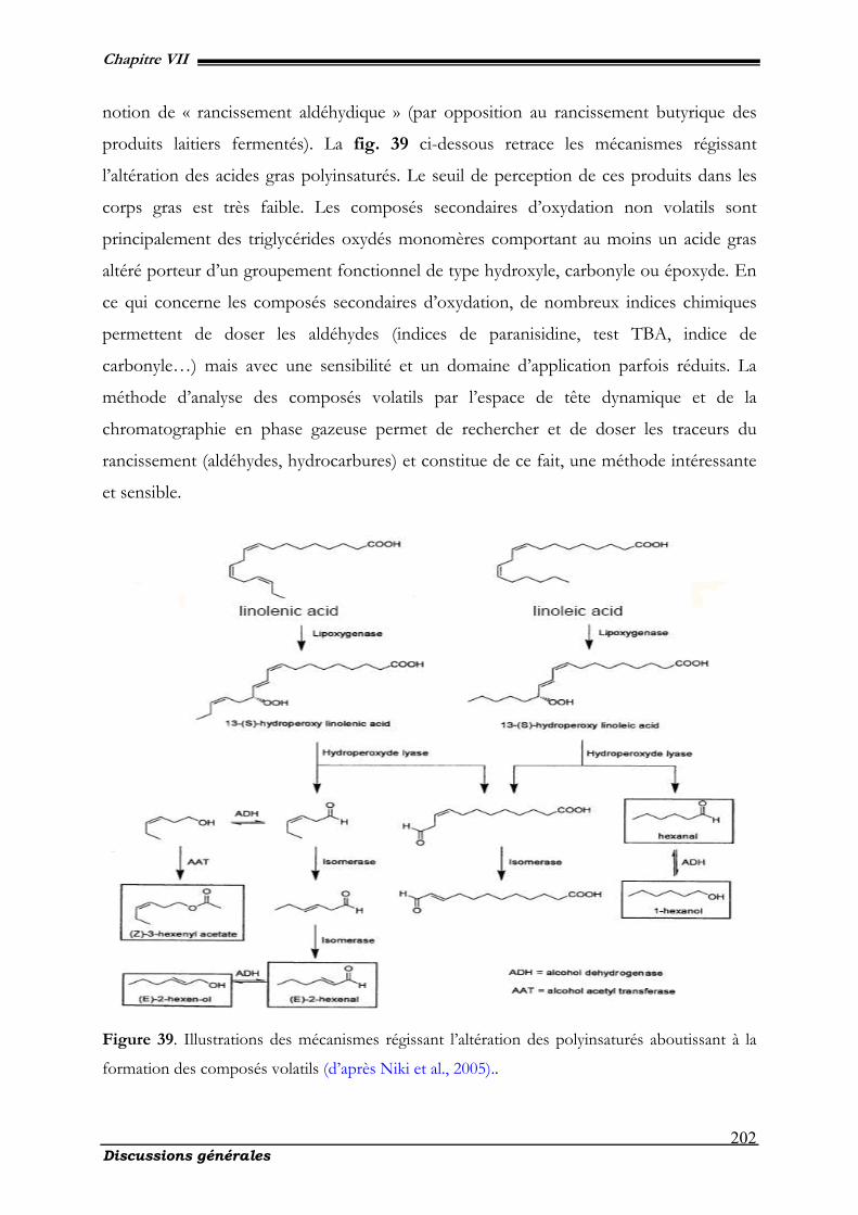
Chapitre VII
Discussions générales
202
notion de « rancissement aldéhydique » (par opposition au rancissement butyrique des
produits laitiers fermentés). La fig. 39 ci-dessous retrace les mécanismes régissant
l’altération des acides gras polyinsaturés. Le seuil de perception de ces produits dans les
corps gras est très faible. Les composés secondaires d’oxydation non volatils sont
principalement des triglycérides oxydés monomères comportant au moins un acide gras
altéré porteur d’un groupement fonctionnel de type hydroxyle, carbonyle ou époxyde. En
ce qui concerne les composés secondaires d’oxydation, de nombreux indices chimiques
permettent de doser les aldéhydes (indices de paranisidine, test TBA, indice de
carbonyle…) mais avec une sensibilité et un domaine d’application parfois réduits. La
méthode d’analyse des composés volatils par l’espace de tête dynamique et de la
chromatographie en phase gazeuse permet de rechercher et de doser les traceurs du
rancissement (aldéhydes, hydrocarbures) et constitue de ce fait, une méthode intéressante
et sensible.
Figure 39. Illustrations des mécanismes régissant l’altération des polyinsaturés aboutissant à la
formation des composés volatils (d’après Niki et al., 2005)..
Chapitre VII
Discussions générales
203
Les résultats et l'examen de nos travaux sur les composés volatils illustrés par les
tab. 17 & 18 du chapitre V permettent de mettre en évidence plusieurs renseignements.
On remarque de prime à bord que les poudres lyophilisées de Lactobacillus contiennent un
certain nombre d'aldéhydes (benzaldéhydes, benzaldéhyde 2,4-bis (trimethylsil)-oxy,
hexenal). Ceci peut être interprété comme un signe d'oxydation de la matière grasse et
montre que le butylhydroxytoluène (B.H.T) que l'on trouve dans le tab. 17 et qui a été
ajouté à dessein comme antioxygène, n'a pas permis d'éviter totalement l'oxydation. Les
composés cétones et les méthylcétones (ethoxycarbonylbenzophenone, 4 H-1-benzo-
thiopyran-4-one, undecadien-2-one, cyclohexadien-1-one, 2-trideconone et 2-pentadeca-
none) se retrouvent en quantités relativement importantes dans les échantillons analysés ;
pourtant il s'agit là de substances volatiles. On peut expliquer cette anomalie apparente
par le fait que les méthylcétones sont susceptibles de se former spontanément par action
de la chaleur en présence d'eau même à l'état de traces [6] en cétoacides qui peuvent
représenter jusqu'à 4 p. 100 des acides gras totaux de la matière grasse butyrique. Les
conditions d'une telle transformation sont évidemment remplies pendant la phase de
séchage. Ces résultats ont également montré la présence d’hydrocarbures dans les poudres
lyophilisées avec Lactobacillus plantarum CWBI-B1419. La présence de ces composés
pourrait trouver son origine par la présence d’un phénomène et/ou un mécanisme
d’oxydation. A ce sujet les travaux effectués par Barbieri et al. (1994) attestent que ces
composés sont probablement les produits secondaires d'auto-oxydation des lipides. Pour
Arora et al. (1995), ces composés issus de cette oxydation sont des précurseurs pour la
formation d’autres composés aromatiques forts appréciés dans les produits fermentés
laitiers. Ces changements dans le profil des volatils sont le résultat des processus
d'oxydation. La dégradation des matières grasses provoque chez la bactérie une présence
accrue des composés volatils, en particulier les aldéhydes, méthyle cétones, alcools,
(Widder et al., 1991). Les résultats de nos travaux sont en adéquation avec ceux publiés
par Prasadet al. (2003) montrant le comportement des bactéries au cours du stockage.
Pour avoir une vue d’ensemble de la mortalité cellulaire au cours du stockage des
cellules lyophilisées, nos travaux se sont penchés sur l’analyse des phospholipides
membranaires afin de comprendre les différentes variations préjudiciables à la survie
cellulaire pendant le processus de stockage. C’est pour cette raison que le chapitre suivant
permettra à travers la discussion de comprendre l’importance des phospholipides.
Chapitre VII
Discussions générales
204
Intérêts de l’étude des phospholipides membranaires chez le genre
Lactobacillus ssp.
L’étude des phospholipides est motivée par la compréhension des phénomènes
d’oxydation et leur impact sur la mortalité. De nombreuses études se sont penchées sur la
composition lipidique de Lactobacillus spp. et principalement des phospholipides
membranaires dans le souci de la compréhension des mécanismes intervenant dans
l’oxydation (Drucker et al., 1995). Les travaux menés par O'Leary et al. (1988) ont montré
par leurs recherches que les phospholipides des lactobacilles sont généralement composés
de n-C16:0, n-C18:1 et C19 et d'une faible proportion des acides n-C14:0, n-C16:1, et n-C18:0
(Drucker, 1994). Des travaux similaires effectués au Centre Wallon de Biologie
Industrielle par Sow et al. (2004), Coulibaly et al. (2008) montrent la présence de six
acides gras notables chez Lactobacillus plantarum CWBI-B1419, L. paraplantarum CWBI-
B545 Leuconostoc. mesenteroides KMrog et L. paracasei ssp casei LMG9192 à savoir : l’acide
palmitique (C16:0), palmitoléique (C16:1), stéarique (C18:0), oléique (C18:1), linoléique (C18:2) et
l'acide linolénique (C18:3). O'Learyet al. (1988) ont montré que ces profils sont typiques
d'acides gras des bactéries gram-positifs. L'analyse des groupes polaires chez L. plantarum
CWBI-B1419 a révélé la même tendance et a montré que la grande majorité des lipides
polaires est composée de phosphatidylglycérol (PG). Ces résultats sont entièrement
compatibles avec les études effectuées par Fischer et al. (1990), pour ce qui concerne les
composants tels que les acides phosphatidique, diphosphatidylglycérol, phosphatidyl-
glycérol phosphoglycolipides et diglycosyldiacylglycérol. Pendant le stockage on a
remarqué une diminution du PG pour les poudres lyophilisées et stockées à 20°C, alors
qu’à 40°C en plus de celle du PG, on observe une baisse significative après 90 jours des
PE et SM. Ces résultats sont similaires à ceux que nous avons obtenus avec L. plantarum
CWBI-B1419. A des températures élevées, les poudres sans lithothamne sont les plus
vulnérables, ce qui entraînerait une baisse de viabilité due aux phénomènes d’oxydation.
Ces altérations provoquent la production de radicaux libres qui seraient toxiques pour les
cellules lyophilisées, la baisse du ratio U/S et la mort cellulaire se sont produites
essentiellement au même rythme.
Chapitre VII
Discussions générales
205
Ce ralentissement serait occasionné par le fait que l’oxydation est au seuil maximal.
Les mécanismes de mort cellulaire au cours du stockage sont encore mal connus, mais à
partir de ces résultats, il paraît évident que la corrélation entre l'oxydation des lipides et la
survie pendant le stockage est liée. Cette hypothèse est étayée par des preuves indirectes
présentées dans différents travaux. Par exemple, il est indiqué que la présence
d'antioxydants a augmenté le taux de survie des bactéries séchées pendant le stockage
(Alexander et al., 2006 ; Teixeira et al., 1995b). Dimmick et al. (1987) ont constaté une
forte similarité entre la perte de la viabilité et l'augmentation de la concentration des
radicaux libres pendant le stockage des cellules lyophilisées de Serratia marcescens. Les
travaux réalisés par Richard et al. (1997) soutiennent l'hypothèse que les réactions entre
les groupements carbonylés et les composants cellulaires sont une cause majeure de
mortalité au cours du stockage des cellules séchées de micro-organismes. La nature de ces
composés n’est pas présentée, mais il est possible que ces produits soient formés pendant
l'oxydation des lipides. Slater, (1984) a signalé que, dans les cellules eucaryotes, la
peroxydation lipidique peut entraîner la désorganisation de la membrane par peroxydation
principalement des acides gras hautement insaturés entraînant ainsi des changements dans
le ratio des acides gras insaturés. La peroxydation incontrôlée des biomembranes peut
donc conduire à des effets profonds sur la structure des membranes. La technologie et la
méthode utilisée nous permettent de quantifier l'évolution des phospholipides durant le
stockage. La grande sensibilité à l’oxydation des acides gras polyinsaturés en occurrence
les acides linoléiques (C18:2) et linoléniques (C18:3) a été montrée par les résultats antérieurs
et permettent de supposer que ces changements au niveau des phospholipides peuvent
affecter la perméabilité membranaire. Cette supposition est en accord avec les travaux
réalisés par Livense et al. (1994). Ce chapitre a permis non seulement d’identifier les
différentes classes de phospholipides mais a surtout permis de faire un lien entre
l’évolution des différents phospholipides au cours du temps et la mortalité cellulaire
observée au cours du stockage à température ambiante.
Rappelons que les résultats issus de nos travaux (chapitre IV), permettent
d’observer une baisse des rapports acide linoléique (C18:2)/acide palmitique (C16:0) et acide
linolénique (C18:3)/acide palmitique (C16:0) d’une part et d’autre part une accumulation des
produits primaires de dégradation du C18:2 ou C18:3 au cours du stockage de la souche
lyophilisée W. paramesenteroides LC11 dans des conditions d’aération, en présence d’une
Chapitre VII
Discussions générales
206
humidité et d’une température élevées. Partant de ces constatations et se basant sur les
résultats du chapitre VI, nous en avons déduit que l’oxydation des acides gras
membranaires, et plus particulièrement celle des acides gras polyinsaturés, contribuerait à
une instabilité (perte de viabilité cellulaire et/ou activité) des souches lyophilisées au cours
de leur stockage. L’instabilité provoquée par cette oxydation se justifierait à partir de
l’instant où la fraction polaire des acides gras issus des lipides membranaires serait oxydée.
Références bibliographiques
Abadias M., Benabarre A., Teixidó N., Usall J., Viñas I., 2001. Effect of freeze-drying and protectants on viability of the biocontrol yeast Candida sake, Int. J. Food. Microbiol., 65, 173-182.
Alexander G.S., Xianli W., Ronald L.P., Boxin O., Dejian H., John O., Amit A., Gitte S.J., Aaron N. H., and Edward S. 2006. Antioxidant Capacity and Other Bioactivities of the Freeze-Dried Amazonian Palm Berry, Euterpe oleraceae Mart. (Acai), J. Agric. Food Chem., 54 (22), 8604-8610.
Amin M., Jorfi M., Khosravi A.D., Samarbafzadeh A.R., Farajzadeh S. A., 2009. Isolation and Identification of Lactobacillus casei and Lactobacillus plantarum from Plants by PCR and Detection of their Antibacterial Activity, J. Biol. Sc., 9, 810-814.
Baumann D. P., 1964. Preservation of lactic cultures. Ph. D. thesis, Iowa state university of science and technology, Ames, Iowa.
Baumann D. P., Reinboid G.W., 1966. Freezing oflactic cultures, J. Dairy. Sei., 49, 259-264.
Bergère J. L., Hermier J. 1967. - La production massive de cellules de streptocoques lactiques. II. Croissance de Streptococcus lactis dans un milieu à pH constant. Le Lait., 48, 13-30.
Costa E., Usall J., Teixidó N., Garcia N., Viñas I. 2000. Effect of protective agents, rehydration media and initial cell concentration on viability of Pantoea agglomerans strain CPA-2 subjected to freeze-drying, J. Appl. Microbiol., 89, 793-800.
Coulibaly I., Anastasie Yao A., G. Lognay., M-L. Fauconnier., and P. Thonart., 2009. Survival of freeze-dried Leuconostoc mesenteroides and Lactobacillus plantarum related to their cellular fatty acids composition during storage. Appl. Biochem. Biotechnol., 157, 70-84.
David A.S., Moreno S.N.J., Docampo R., 1995. Ca2+ storage in Trypanosoma brucei: the influence of cytoplasmic pH and importance of vacuolar acidity, Biochem. J. 310, 789-794
Delcour, J., Ferain, T. & Hols, P. 2000. Advances in the genetics of thermophilic lactic acid bacteria, Curr.Opin.Biotechnol., 11, 497-504.
Dimmick, R. L., Heckly, R. J. 1987. Free radical formation during storage of freeze dried Serratia marcescens. Nature ., 192, 776.
Chapitre VII
Discussions générales
207
Drucker, D. B. 1994. Fast atom bombardment mass spectrometry of phospholipids for bacterial chemotaxonomy, p. 18-35. In C. Fenselau (ed.), Mass spectrometry for the characterization of microorganisms. American Chemical Society, Washington, D.C.
Drucker D.B., Megson G.,. Harty D. W. S., I. Riba, and S. J. Gaskell. 1995. Phospholipids of Lactobacillus spp J. bacteriol., 177, 6304-6308.
Fischer, W. 1990. Bacterial phosphoglycolipids and lipoteichoic acids, p. 123-234. In M. Kates (ed.), Handbook of lipid research, vol. 6. Plenum Press, New York.
Fonseca F., Béal C. and Corrieu G., 2000. Method of quantifying the loss of acidification activity of lactic acid starters during freezing and frozen storage, J. Dairy Res. 67, 83-90.
Fonseca F., Béal C., Corrieu G. 2000. Method for quantifying the loss of acidification activity of lactic acid starters during freezing and frozen storage. J. Dairy Re.s, 67, 83-90.
Font de Valdéz G., de Giori G., de Ruiz Holgado A.P., Oliver G.1983. Protective effect of adonitol on lactic acid bacteria subjected to freeze-drying, Appl. Environ. Microbiol., 45 302-304.
Font de Valdéz G., de Giori G., de Ruiz Holgado A.P., Oliver G.1985. Effect of drying medium on residual moisture content and viability of freeze-dried lactic acid bacteria, Appl. Environ. Microbiol., 49, 413-415.
Gibson O.A., Landerkin G.B., Morse P.M., 1965. Survival of strains of lactic streptococci during frozen storage, J. Dairy .Sci., 32, 151-156.
Gibson O.A., Landerkin G.B., Morse P.M., 1966. Effects of additives on the survival of lactic streptococci in frozen storage, Appl. mierobiol., 14, 665-669.
Giulio B.D., Orlando P., Barba G., Coppola R., Rosa M.D., Sada A., Prisco P.P.D., and Nazzaro F. 2005. Use of alginate and cryo-protective sugars to improve the viability of lactic acid bacteria after freezing and freeze-drying, World J. Microbiol. Biotechnol., 21, 739-746.
Hastings, J. W., and K. H. Nealson. 1981. The symbiotic luminous bacteria, p. 1322-1345. InIn M. Starr, H. Stolp, H. Trüper, A. Balows, and H. Schlegel (ed.), The prokaryotes. A handbook on habitats, isolation, and identification of bacteria. Springer-Verlag, Berlin, Germany.
Hoerni B., 2006. Dictionnaire des cancers ; 3eds. Ed Frison-Roche Paris pp 617.
Hubalék Z., Cryopreservation of microorganisms, Academia, Prague, Czech Republic, 1996.
Jin, L. Z., Ho, Y. W., Abdullah, N. & Jalaludin, S. 1998. Acid and bile tolerance of Lactobacillus isolated from chicken intestine. Lett. Appl. Microbiol., 27, 183-185.
Kets E.P.W., Galinski E.A., De Wit M., De Bont J.A.M., Heipieper H.J. 1996. Mannitol, a novel bacterial compatible solute in Pseudomonas putida S12, J. Bacteriol., 178, 6665-6670.
Konings W.N., Kok J., Kuipers O.P, Poolman B. 2000. Lactic acid bacteria: the bugs of the new millennium, Curr. Opin. Microbiol., 3, 276-282.
Lamprech E.D., Foster E.M., 1963. Tthe survival of starter organisms in concentrated suspensions J. Aappl. Baci., 26, 359-369.
Chapitre VII
Discussions générales
208
Leslie S.B., Israeli E., Lighthart B., Crowe J.H., Crowe L.M. 1995. Trehalose and sucrose protect both membranes and proteins in intact bacteria during drying, Appl. Environ. Microbiol. 61, 3592-3597.
Lievense L.C., Verbeek M.A.M., Noomen A., Van’t Riet K. 1994. Mechanism of dehydration inactivation of Lactobacillus plantarum. Appl. Microbiol. Biotechnol. 41: 90-94.
Linders L.J.M., Wolkers W.F., Hoekstra F.A., van’t Riet K. 1997. Effect of added carbohydrates on membrane phase behaviour and survival of dried Lactobacillus plantarum, Cryobiol., 35, 31-40.
Mazur P., 1966. Physical and chemical basis of injury in singlecelled microorganisms subjected to freezing and thawing, dans« oryobiology» (h. t. meryman, ed.) pp. 213-315. Academie press, London and New-york.
Mori, K., Yamazaki, K., Ishiyama, T., Katsumata, M., Kobayashi, K., Kawai, Y., Inoue, N. & Shinano H., 1997. Comparative sequence analyses of the genes coding for 16S rRNA of Lactobacillus caseirelated taxa, Int. J. Syst. Bacteriol., 47, 54-57.
Moss O.W., Speck M.L., 1963. Injury and death of streptococcus laetis due to freezing and frozen storage, Appl. Mierobiol., 11, 326-329.
Niamsup P., Sujaya N., Tanaka M., Sone T., Hanada S. Kamagata Y., Lumyong S. Assavanig A., Asano K., Tomita F. and Yokota A. 2003. Lactobacillus thermotolerans sp.nov., a novel thermotolerant species isolated from chicken faeces, Int. J. Syst. Evol. Microbiol.,, 53, 263-268.
O’Leary, W. M., and S. G. Wilkinson. 1988. Gram-positive bacteria, p. 117–201. In C. Ratledge and S. G. Wilkinson (ed.), Microbial lipids, vol. 1. Academic Press, London.
Oowman R.A., Speck M.L., 1965. Ultra-low temperature st orage of lactic streptococci, J. Dairy.Ssci., 48, 1531-1532.
Palmfedt J., Hahn-Hägerdal B. 2000. Influence of culture pH on survival of Lactobacillus reuteri subjected to freeze-drying, Int. J. Food Microbiol., 55, 235-238.
Porubcan R.S., Sellars R.L. 1975. Stabilized dry cultures of lactic acid-producing bacteria, US Patent 3, 897-907.
Porubcan R.S., Sellars R.L. 1979. Lactic starter culture concentrates. Microbial technology. H. J. Peppler, Perlman, D. (Eds). New York, Academic Press, 59-92.
Prasad J., Mc. Jarrow P. and Gopal P. 2003. Heat and Osmotic Responses of Probiotic Lactobacillus rhamnosus HN001 (DR20) in Relation to Viability after Drying, Appl. Environ. Microbiol., 69, 917-925.
Richard M.J., Belleville F., Chalas J., Ceballos P. I., Vitoux D., Boyer M.J., Chaudière J., Favier A. 1997. Les glutathion peroxydases : intérêt de leur dosage en biologie clinique, Ann. Biol. Clin., 55, 195-208.
Santivarangkna C., Wenning M., Foerst P., Kulozik U. 2007. Damage of cell enveloppe of Lactobacillus helveticus during vacuum drying, J. Appl. Microbiol., 102, 748-756.
Selmer-Olsen E., Birkeland S.-E., Sorhaug T. 1999. Effect of protective solutes on leakage from and survival of immobilized Lactobacillus subjected to drying, storage and rehydration, J. Appl. Microbiol., 87, 429-437.
Teixeira PC, Castro MH, Malcata FX, Kirby RM. 1995. Survival of Lactobacillus delbruckii ssp. bulgaricus following spray drying. J.Dairy Sc., 78, 1025-1031.
Chapitre VII
Discussions générales
209
Uchida, K., and K. Mogi. 1973. Cellular fatty acid spectra of Hiochi bacteria, acid-tolerant lactobacilli, and their separation, J. Gen. Appl. Microbiol., 19, 233-249.
Vassal L., Mocquot G., 1967. Fermentation lactique accélérée en fromagerie grâce à l'emploi d'un nombre très élevé de bactéries. Tech. lait., 541, 9-13.
Wisselink H.W., Weusthuis R.A., Eggink G., Hugenholtz J., Grobben G.J. 2002. Mannitol production by lactic acid bacteria: a review, Int. Dairy. J., 12, 151-161.
Yao A. Anastasie., Coulibaly I., G. Lognay., M-L. Fauconnier and P. Thonart., 2008. Impact of polyunsaturated fatty acid degradation on survival and acidification activity of freeze-dried Weissella paramesenteroides LC11 during storage. Appl. Microbiol. Biotechnol., 79,1045-1052
Yoo B., Lee C.M. 1993. Thermoprotective effect of sorbitol on proteins during dehydration, J. Agric. Food Chem., 41, 190-192.
Chapitre VIII
Conclusions générales et perspectives
Conclusions générales et perspectives
211
Conclusions générales
Les cellules sont soumises à différents types de stress (thermique, acide, nutritionnel,
oxydatif, mécanique, osmotique), tout au long du procédé de production de ferments
lactiques. La conservation et la stabilisation des cellules par lyophilisation représentent, en
particulier, une situation de stress hydrique important pour les cellules. Elle constitue une
étape critique pour le maintien de la qualité des ferments lactiques et/ou starters. La non
optimisation ou le mauvais contrôle des conditions de lyophilisation est, en effet,
responsable d’une dégradation des propriétés technologiques des cellules. Dans le souci
d’améliorer la résistance des bactéries lactiques, une approche intéressante consiste à la
sélection de nouvelles souches de bactéries lactiques naturellement résistantes, présentent
dans un environnement chaud et sec gage d’une bonne stratégie pour augmenter et
diversifier les ressources génétiques disponibles. Car ce stress modéré permet aux cellules
de développer des mécanismes physiologiques de défense et d’adaptation qui vont, par la
suite, leur permettre de mieux résister aux conditions de stress réel. De même, l’utilisation
des antioxydants et des procédés d’élimination de l’oxygène au cours du stockage serait un
atout dans l’optimisation du taux de survie des cellules lyophilisées. Dans ce contexte,
l’objectif de cette thèse était d’étudier l’impact du séchage (lyophilisation) sur la résistance
des bactéries lactiques et le comportement des cellules lyophilisées de L. plantarum CWBI-
B1419 au cours du stockage. L’amélioration de la résistance des cellules à l’issue des
différentes étapes de la production, de la lyophilisation et du stockage est envisageable et
même possible avec des formulations et d’additifs.
La première partie de ce travail a réalisé un bilan des recherches récentes dans le
domaine de la lyophilisation des bactéries lactiques. Subdivisée en deux parties la synthèse
bibliographique a présenté les principales notions relatives aux bactéries lactiques, leur
conservation par lyophilisation, le rôle et l’intérêt des cryoprotecteurs, le rôle et l’effet des
antioxydants sur la physiologie cellulaire (partie I).
La deuxième partie de cette thèse a démontré que pour des critères technologiques
et économiques, il était impératif de mettre au point des méthodes de sélection
rigoureuses, spécifiques, fiables et reproductibles. Ce fut le premier objectif de notre
travail de recherche. A partir d’échantillons prélevés à proximité de poulaillers au Sénégal
Chapitre VIII
Conclusions générales et perspectives
212
(Afrique de l’ouest), dans un environnement chaud et sec, des souches de bactéries
lactiques ont été sélectionnées sur la base de leur résistance aux stress thermique et
osmotique et aux effets croisés de ces deux facteurs. L’adaptation bactérienne aux
modifications des conditions environnementales comme critère de sélection est une
alternative pour améliorer la viabilité des souches lors du stockage ainsi que leurs
performances technologiques. La grande mortalité cellulaire observée chez la plupart des
bactéries lactiques est justement due à l’absence d’une réponse adéquate face à la
déshydratation, d’où leur grande sensibilité. Ce volet a permis d’enrichir la collection
d’une nouvelle souche Gram positive et catalase négative correspondant au numéro de
collection CWBI-B1419 et identifiée comme Lactobacillus plantarum.
Lors de la troisième partie de la thèse, des substances protectrices ont été ajoutées
avant la lyophilisation et les cellules ont été stockées selon les conditions décrites ci-
dessus, afin d’étudier l’effet de ces additifs sur la résistance des cellules au cours de la
lyophilisation et pendant le stockage. Les performances technologiques et probiotiques de
L. plantarum CWBI-B1419 au cours du stockage se trouvent renforcées suite à l’ajout de
ces additifs. Ce qui permet donc de démontrer qu’elles peuvent être produites à grande
échelle et conservées sous formes lyophilisées.
La quatrième partie des résultats a permis de mettre en lumière, le rôle
prépondérant joué par l’oxydation des acides gras polyinsaturés pour la première fois sur
la mortalité cellulaire. Ce chapitre avait donc pour objectif majeur de comprendre certains
mécanismes à l’origine de la perte de stabilité des souches lyophilisées au cours du
stockage. Parmi ces mécanismes, l’oxydation des lipides membranaires des bactéries a été
étudiée. Nous avons montré que les acides gras polyinsaturés étaient instables à
l’oxydation et produisaient des oxylipines (hydroperoxydes). Ces hydroperoxydes étant
instables à la chaleur, se décomposent en de nombreux produits secondaires volatiles
(Nawar, 1996), ce qui entraîne une forte diminution du ratio de C18:2 /C16:0 ou C18:3/C16:0 et
une accumulation rapide des oxylipines issus des acides linoléique ou linolénique pendant
le stockage à 20°C de poudres lyophilisées de L. plantarum CWBI-B1419 à une aw=0.23. Ce
phénomène pourrait être suffisant pour entraîner une augmentation de la mortalité
cellulaire et/ou la perte de l'activité métabolique. A partir de l’analyse des réponses
physiologiques des cellules, nous avons montré que la meilleure tolérance de la souche L.
Chapitre VIII
Conclusions générales et perspectives
213
plantarum CWBI-B1419 est liée à des rapports différents entre acides gras insaturés et
saturés, correspondant à des cellules avec une fluidité membranaire plus importante.
La cinquième partie a mis en évidence la présence de composés volatils issus de la
dégradation des acides gras polyinsaturés linoléiques et linoléniques qui sont à l’origine de
la production des hydroperoxydes et responsables de la mortalité cellulaire observée
pendant le stockage. Il ressort que les composés aldéhydiques sont les composés volatils
les plus importants, mais la présence d’autres composés (cétones et hydrocarbures) a été
notée. Cette étude permet de faire une relation entre les produits primaires d’oxydations
(oxylipines) et les produits secondaires qui en découlent (aldéhydes).
Enfin la sixième partie de ce travail a permis non seulement de connaître la
composition en phospholipides de L. plantarum CWBI-B1419 mais aussi a permis de
comprendre le rôle joué par les différents constituants phospholipidiques dans le
phénomène de peroxydation au cours du stockage à température ambiante. Les travaux de
ce chapitre ont montré la présence de sept (7) principaux phospholipides (PA, PE, PI, PS,
PG, SM et LPG). Il a également établi la relation entre la variation de la composition de
ces phospholipides et la mortalité cellulaire des cellules lyophilisées au cours du stockage.
La résistance au stockage varie aussi selon les conditions opératoires utilisées. Ces
résultats sont intéressants en vue d’applications industrielles.
L’analyse de l’ensemble de ces résultats permet de conclure que les procédés de
séchage (lyophilisation) ont un effet sur la résistance des cellules, et sur leur stabilisation
au cours du stockage sous forme lyophilisée (poudres). Selon les procédés et les
conditions appliquées, les réponses observées diffèrent, dans le sens de l’amélioration en
fonction de la résistance des cellules. Ces résultats sont intéressants pour une application
industrielle, car ils permettent d’identifier les conditions opératoires qui doivent être
appliquées pour obtenir un ferment lactique actif avec un fort potentiel probiotique. Pour
optimiser, la production de ferments lactiques avec une activité acidifiante élevée, il
conviendra de jouer sur les paramètres de centrifugation, de congélation, de séchage et
aussi le type d’additif à incorporer. De plus, la combinaison entre sélection rigoureuse et
des conditions de séchage optimisées et non drastiques permet également la production
de cellules avec une bonne activité.
Conclusions générales et perspectives
214
Perspectives
Ces résultats permettent d’ouvrir de nombreuses perspectives, qui peuvent être
présentées selon deux niveaux. Il s’agira dans un premier temps de poursuivre
directement et d’approfondir certains aspects de l’étude, et les méthodes de travail
relatives à l’état physiologique de la cellule. Dans un autre volet, il serait souhaitable de
revoir l’environnement de stockage des poudres lyophilisées des starters lactiques.
Ce travail constitue donc une contribution qui pourra être valorisée, à la fois à
l’échelle du laboratoire, mais surtout au stade industriel, pour préserver efficacement la
stabilité des ferments lactiques pendant le stockage. Cette technologie a plusieurs champs
d’applications en Afrique et en particulier en Côte d’ivoire
1. Pour les pays en voies de développement (PVD) et les grandes villes
comme Abidjan, l’acquisition des techniques servant à la conservation des
starters lactiques a plusieurs débouchés. Les bactéries lactiques pourraient
être utilisées pour résoudre d’énormes problèmes sanitaires d’intérêts
publics comme l’épuration des lagunes et étangs d’eaux pollués.
2. L’acquisition des techniques de conservation des bactéries lactiques
pourrait être utilisée pour améliorer et amplifier les techniques de
fermentation des mets traditionnels comme "l’attiéké, le gari, le soumala et
le dêguê". Ce qui permet d’obtenir une grande innocuité et une plus value.
3. La fabrication d’un starter lactique nécessite une technologie très
précise. La sélection, l’identification, l’étude des propriètés fonctinnelles, la
production et les techniques de stockage exigent un savoir-faire. Ces
connaissances sont souvent régies par des brèvets qui souvent rendent
l’accés difficile à cette technologie, d’où l’importance de vulgarisation ces
connaissances en direction du content africain.
ChapitreVIII
Conclusions générales et perspectives
215
4. La mise au point d’un starter mixte original (microorganismes-lipase)
pouvant être utilisé dans un procédé de traitement des effluents graisseux
industriels comme le cas de Yarrowia lipolytica (Alloué et al., 2008 ; Destain
et al., 1997)
Le deuxième enseignement important qui ressort de ces travaux de thèse concerne
l’identification de certaines réponses physiologiques, en relation avec les réponses
technologiques décrites précédemment.
a. Les réponses physiologiques ont donc été analysées, au niveau
membranaire (composition en acides gras membranaires), une perspective à
envisager serait de coupler une analyse des réponses aux niveaux
membranaires et une autre au niveau cytoplasmique (protéome cellulaire)
en fonction de la variation des changements environnementaux ou
opératoires effectués lors de la récolte ou de la concentration des cellules.
b. A travers l’analyse des acides gras membranaires et in fine du protéome
de L. plantarum CWBI-B1419, certaines réponses générales de ce
microorganisme pourront être identifiées et étendre ces acquis à d’autres
souches d’intérêts industriels.
c. L’action des composés utilisés pour la protection des cellules pour
pallier aux stress hydrique et/ou physiologique n’est pas clairement défini et
reste mal illucidé. L’étude des phénomènes de rélargage des ions (potentiel
électrochimique) et l’analyse en cytométrie de flux ou microscopie
électronique de la membrane cellulaire, ainsi que l’analyse en spectrométrie
infrarouge de la fraction polaire des lipides membranaires, avant ou après la
lyophilisation et en présence de différentes concentrations de glycérol,
maltodextrine ou saccharose pourraient être envisagées.
d. Parmi les perspectives donnant suite une directe à nos travaux, il
pourrait être judicieux de suivre le comportement des cellules au cours du
ChapitreVIII
Conclusions générales et perspectives
216
stockage par l’analyse et le suivi des modifications au niveau de chaque
classe de phospholipides membranaires.
Une autre perspective d’étude pourrait approfondir et étendre le phénomène de
relargage à d’autres souches. Une attention particulière doit être accordée au
phénomène de relargage des ions. En effet, l’augmentation d’une part de la
concentration en sels ou du monosodium glutamate, de la température ainsi qu’une
diminution de la concentration en protons dans le milieu de réhydratation
entraîneraient une augmentation de la concentration en solutés dissouts et entraînerait
une réduction du nombre de cellules viables; d’autre part, l’augmentation de la
concentration en glycérol, maltodextrine ou saccharose entraînerait une réduction de la
concentration en solutés dissouts et un pourcentage plus élevé de cellules viables
(Santivarangkna et al., 2007). L’étude des mécanismes des enzymes impliquées dans la
modification des acides gras membranaires (désaturases) serait une voie à explorer dans
le souci de la compréhension des phénomènes responsables de la mortalité cellulaire.
Enfin une étude des composés volatils (produits secondaires d’oxydation)
produits par d’autres souches de Lactobacillus sp. serait intéressante d’envisager.
Annexes
Annexe 1
Annexes
218
Annexe 1 : Article publié dans la revue « African Journal of Biotechnology »
Annexe 2
Annexes
219
Annexe 2 : Article publié dans la revue « Applied Biochemistry and Biotechnology »
Annexe 3
Annexes
220
Annexe 3 : Article publié dans la revue « Applied Microbiology and Biotechnology »
Annexe 4
Annexes
221
Annexe 4 : Article publié dans la revue « International Journal of Microbiology »
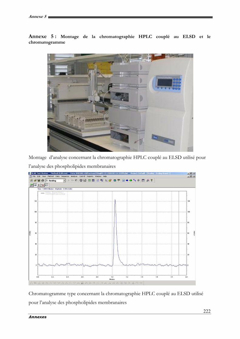
Annexe 5
Annexes
222
Annexe 5 : Montage de la chromatographie HPLC couplé au ELSD et le chromatogramme
Montage d’analyse concernant la chromatographie HPLC couplé au ELSD utilisé pour
l’analyse des phospholipides membranaires
Chromatogramme type concernant la chromatographie HPLC couplé au ELSD utilisé
pour l’analyse des phospholipides membranaires
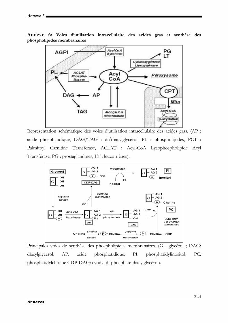
Annexe 7
Annexes
223
Annexe 6: Voies d’utilisation intracellulaire des acides gras et synthèse des phospholipides membranaires
Représentation schématique des voies d’utilisation intracellulaire des acides gras. (AP :
acide phosphatidique, DAG/TAG : di/triacylglycérol, PL : phospholipides, PCT :
Palmitoyl Carnitine Transferase, ACLAT : Acyl-CoA Lysophospholipide Acyl
Transférase, PG : prostaglandines, LT : leucotriènes).
Principales voies de synthèse des phospholipides membranaires. (G : glycérol ; DAG:
diacylglycérol; AP: acide phosphatidique; PI: phosphatidylinositol; PC:
phosphatidylcholine CDP-DAG: cytidyl di-phosphate-diacylglycérol).
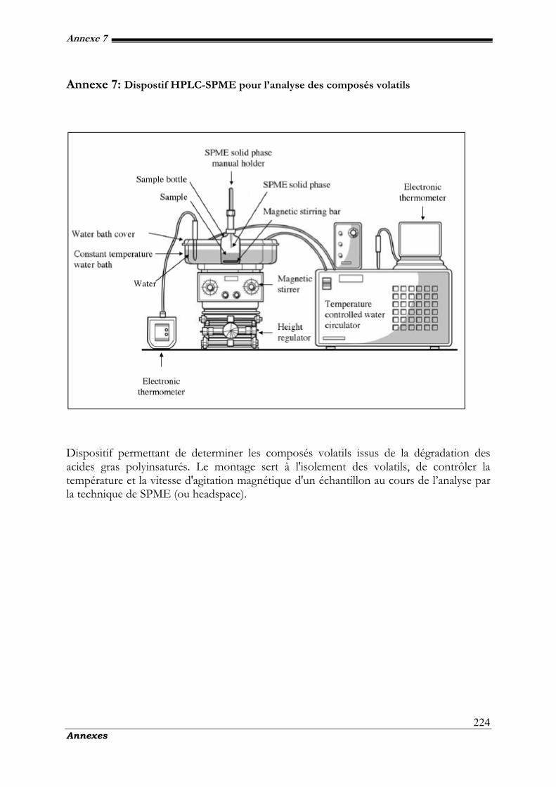
Annexe 7
Annexes
224
Annexe 7: Dispostif HPLC-SPME pour l’analyse des composés volatils
Dispositif permettant de determiner les composés volatils issus de la dégradation des acides gras polyinsaturés. Le montage sert à l'isolement des volatils, de contrôler la température et la vitesse d'agitation magnétique d'un échantillon au cours de l’analyse par la technique de SPME (ou headspace).
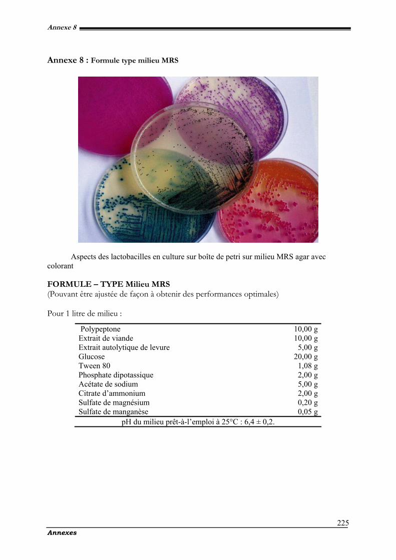
Annexe 8
Annexes
225
Annexe 8 : Formule type milieu MRS
Aspects des lactobacilles en culture sur boîte de petri sur milieu MRS agar avec colorant FORMULE – TYPE Milieu MRS (Pouvant être ajustée de façon à obtenir des performances optimales) Pour 1 litre de milieu :
Polypeptone 10,00 g Extrait de viande 10,00 g Extrait autolytique de levure 5,00 g Glucose 20,00 g Tween 80 1,08 g Phosphate dipotassique 2,00 g Acétate de sodium 5,00 g Citrate d’ammonium 2,00 g Sulfate de magnésium 0,20 g Sulfate de manganèse 0,05 g
pH du milieu prêt-à-l’emploi à 25°C : 6,4 ± 0,2.
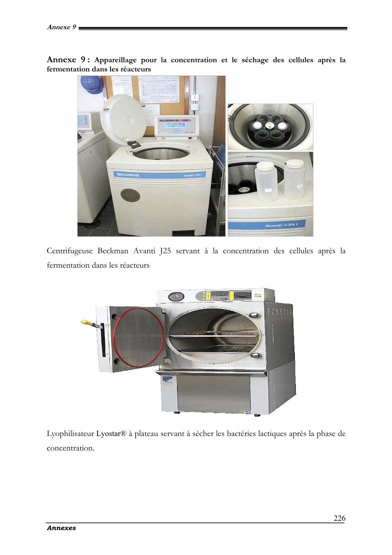
Annexe 9
Annexes
226
Annexe 9 : Appareillage pour la concentration et le séchage des cellules après la fermentation dans les réacteurs
Centrifugeuse Beckman Avanti J25 servant à la concentration des cellules après la
fermentation dans les réacteurs
Lyophilisateur Lyostar® à plateau servant à sécher les bactéries lactiques après la phase de
concentration.
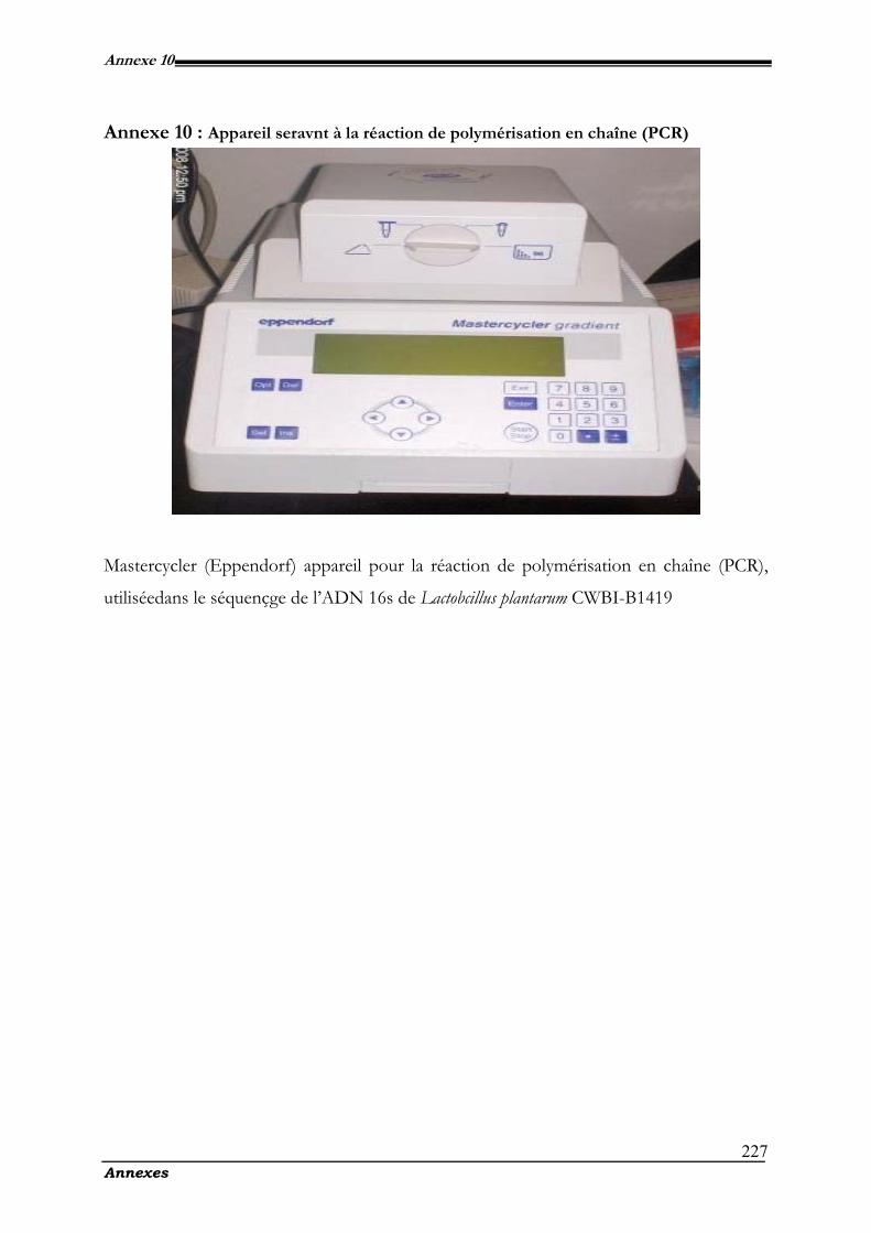
Annexe 10
Annexes
227
Annexe 10 : Appareil seravnt à la réaction de polymérisation en chaîne (PCR)
Mastercycler (Eppendorf) appareil pour la réaction de polymérisation en chaîne (PCR),
utiliséedans le séquençge de l’ADN 16s de Lactobcillus plantarum CWBI-B1419

Tableaux et figures
229 Liste des tableaux et des figures
Liste des tableaux
Tableau 1. Quelques substances utilisés comme cryoprotecteurs cellulaires (d’après Adjizian et al., 1987)..................... 23
Tableau 2. Effet de la température de culture sur la composition en acides gras (%) chez L. plantarum........................ 25
Tableau 3. Peroxidation des lipides (produits des linoléates). Les caractéristiques des réctions chimiques et les produits
spécifiques issus de l'oxydation des linoléates par différents mécanismes. ........................................................................... 31
Table 4. Functional classification and anchor types....................................................................................................... 56
Table 5. Some characteristics of the five homofermentative LAB isolates ....................................................................... 83
Table 6. Physiological characteristics: tolerance of the five LAB isolates to ranges of temperatures, lactic acid, NaCl
concentrations and pH.................................................................................................................................................... 86
Table 7. Composition and modifications of membrane fatty acids (% of peaks areas) induced by growth temperature of L.
paracasei ssp. LMG 9192T and L. plantarum CWBI-B1419.................................................................................... 106
Table 8. Moisture content and survival of two strains of lactobacilli after freeze-drying with 12 g/L monosodium
glutamate and 12 g/l sorbitol. Each value represents the mean of replicates from 2 freeze-drying experiments................ 110
Table 9. Dry cell weight of freeze-dried cells, conserved at 20°C and safe from oxygen and moisture. Water activity of
freeze-dried cells, conserved at 20°C and safe from oxygen and moisture. ....................................................................... 112
Table 10. Viability of L. mesenteroides and Lb. plantarum before and after freeze-drying .......................................... 129
Table 11. Cellular fatty acid composition of L. mesenteroides before and after freeze-drying.......................................... 135
Table 12. Cellular fatty acid composition of Lb. plantarum before and after freeze-drying............................................ 135
Table 13. Moisture content and survival of freeze-dried L mesenteroides and Lb. plantarum after 90–day storage ..... 136
Table 14. Moisture content and C18:2/C16:0 ratio for freeze-dried L mesenteroides and Lb. plantarum after 90–day storage
.................................................................................................................................................................................... 137
Table 15. Moisture content and C18:3/C16:0 ratio for freeze-dried L mesenteroides and Lb. plantarum after 90–day storage
.................................................................................................................................................................................... 138
Liste des tableaux
230 Liste des tableaux et des figures
Table 16. Viability of L. plantarum CWBI-B1419 before and after freeze-drying. .....................................................155
Table 17. Relative abundances of volatile compounds detected in freeze-dried Lactobacillus plantarum CWBI-B1419
during storage. .............................................................................................................................................................. 158
Table 18. The main volatils compouds found in the dynamic headspace/GC analysis of freeze-dried L. plantarum CWBI-
B1419 during storage................................................................................................................................................... 161
Table 19. HPLC Gradient Scheme........................................................................................................................... 175
Table 20. Survival after freeze-drying process at 20°C and 40°C for 90-day, kinetic parameters of growth and substrate
consumption of L. palntarum CWBI-B1419 in batch culture ...................................................................................... 176
Tableau 21. Caractéristiques physiologiques: la tolérance des cinq isolats de bactéries lactiques aux gammes de température,
de l'acide lactique, les concentrations de NaCl et de pH. (cf tab.6 chap. II). ................................................................. 194
Tableau 22. Taux de survie de deux souches de lactobacilli après lyophilisation avec 12 g/l de mosodium glutamate et 12
g/l de sorbitol. Chaque valeur représente la moyenne des répétitions des deux expériences de lyophilisation .................... 196
.
231 Liste des tableaux et des figures
Liste des figures
Figure 1. Représentation schématique d'un produit en cours de lyophilisation. Symbole: S=interface de
sublimation; E=zone externe poreuse; I=zone interne congélée (Simatos et al., 1994). ......................................18
Figure 2. Evolution du flux de chaleur (dQ/dt) en fonction de la température (T), mésurée par analyse
calorimétrique différentielle. La temperature de transition vitreuse (Tg, Tg midpoint) correspond à la médiane aux
deux droites paralélles entre tangeantes aux points (Tgonset et Tgendset) au point d’inflexion de la courbe. .........20
Figure 3. Facteurs influençant la survie des bactéries la lyophilisation. [Encadrés] : Depuis leur culture jusqu’à
leur décongélation ou réhydration (Besson et al., 1987). .....................................................................................22
Figure 4. Réaction auto-catalytique de l’oxydation des acides gras insaturés, (Yin et al., 2007).......................28
Figure 5. Phase d’initiation...........................................................................................................................28
Figure 6. Phase de propagation ....................................................................................................................29
Figure 7. Phase de terminaison......................................................................................................................29
Figure 8. Schema de l’autooxydation (Chyau et al., 1999) ............................................................................29
Figure 9. Les différentes étapes de la réaction d'oxydation des linoléates (d’après Niki et al., 2005)................30
Figure 10. Auto-oxydation et photo-oxydation des ∆5-stérols, (Fernades et al., 2007)....................................32
Figure 11. Les principales voies de la lipoxygénase.- The mains lipoxygénase pathway. (Fauconnier, 1997)....33
Figure 12. Représentation schématique de l’autoxydation des lipides insaturés (Techniques de laboratoire N°2 -
2007). .............................................................................................................................................................35
Figure 13. Homolactic acid fermantation pathway. In lactic fermentation by Lactobacillus, the substrate
(glucose) is oxidized to pyruvate, and pyruvate becomes reduced to lactic acid. Redox balance is maintained by
coupling oxidations to reductions within the pathway. ........................................................................................51
Figure 14. Pyruvate metabolism in L. johnsonii and L. plantarum. The figure is based on the pyruvate metabolism pathway from the KEGG database (Kanehisa et al., 2002).................................................... 52
Liste des figures
232 Liste des tableaux et des figures
Figure 15. Schematic representation of the cell wall of Gram-negative bacteria showing several layers of
polysaccharides and glycoconjugates. ...................................................................................................................54
Figure 16. Cell wall from Gram-positive bacteria. Gram-positive bacteria lack the outer membrane and
associated lipopolysaccharide (LPS) that is present in Gram-negative organisms. In Gram-positive bacteria, the
peptidoglycan layer is thicker and contains teichoic acids.....................................................................................55
Figure 17. Extracellular anchored and secreted proteins of L. plantarum. Numbers in parentheses are the
number of predicted proteins of the different types. ..............................................................................................56
Figure 18. Domain composition of L. plantarum proteins predicted to be associated with the cell wall through
LysM domains. Putative protein functions are listed below the ORF names (Boekhorst et al., 2006). ................57
Figure 19. Domain composition of L. plantarum proteins predicted to be involved in the adherence to
extracellular macromolecules. Putative protein functions are listed below the ORF names....................................58
Figure 20. The structures of two phospholipids, (Berg et al., 2002). ...............................................................60
Figure 21. Biomass produced by three strains of Lactobacilli: (♦) Spo05, (■) Spo04 and (▲) Spo20 isolates
over 42 h. ........................................................................................................................................................87
Figure 22. Lactic acid produced by three strains of Lactobacilli: (♦) Spo05, (■) Spo04 and (▲) Spo20
isolates over 42 h..............................................................................................................................................88
Figure 23. pH of the cultures of three strains of Lactobacilli: (♦) Spo05, (■) Spo04 and (▲) Spo20 isolates
over 42 h. ........................................................................................................................................................88
Figure 24. Glucose consumption (mm) by three strains of Lactobacilli: (♦) Spo05, (■) Spo04 and (▲) Spo20
isolates over 42 h..............................................................................................................................................89
Figure 25. Lactic acid optical isomers comparison produced by three strains of Lactobacilli: Spo05, Spo04 and
Sp20 with ■ D(-) Optical isomer and □ L(+) Optical isomer. .......................................................................90
Figure 26. Impact of storage time upon U/S ratio of freeze-dried lactobacilli during 120 days at room
temperature in vacuum-sealed aluminium foil. Values are presented as means ±standard deviation (SD, n=4).
(a) L. paracasei ssp. paracasei LMG 9192T, (b) L. plantarum CWBI-B1419T and (c) control (■), sorbitol (□)
and monosodium glutamate (◘). .....................................................................................................................107
Liste des figures
233 Liste des tableaux et des figures
Figure 27. Acidification activity of freeze-dried : (a) L. plantarum CWBI-B1419, (b) L. paracasei ssp. casei
LMG 9192T and (c) control (�♦�), sorbitol (�■�) and monosodium glutamate (�▲�). Values are
presented as means±standard deviation (SD, n=4) ........................................................................................108
Figure 28. Survival of freeze-dried lactobacilli during 120 days at room temperature in vacuum-sealed
aluminium foil. Values are presented as means ±standard deviation (SD, n=4). (a) L. paracasei ssp. paracasei
LMG 9192T, (b) L. plantarum CWBI-B1419 and (c) control (♦), sorbitol (■) and monosodium
glutamate (▲). .........................................................................................................................................111
Figure 29. Survival of freeze-dried L. mesenteroides during storage at 4°C or 20°C. Symbols: ο freeze-dried
strain without cryoprotectants (P), • freeze-dried strain with cryoprotectants (5% maltodextrine + 2% glycerol)
(PC). Dotted lines for storage in vacuum-sealed aluminium foil, continuous lines for storage in opened white glass
tube. Values represent means ± SD (n = 4)..................................................................................................131
Figure 30. Survival of freeze-dried Lb. plantarum during storage at 4°C or 20°C. Symbols: ο freeze-dried
strain without cryoprotectants (P), • freeze-dried strain with cryoprotectants (5% maltodextrine + 2% glycerol)
(PC). Dotted lines for storage in vacuum-sealed aluminium foil, continuous lines for storage in opened white glass
tube. Values represent means ± SD (n = 4)..................................................................................................133
Figure 31. C18:2/C16:0 and C18:3/C16:0 ratios of freeze-dried L. mesenteroides during 90-day storage at: 4 °C
(A) or 20°C (B) in vacuum-sealed aluminium foil (a) or in opened white glass tube (b). Bars are means ± SD (n
= 4) and represent the ratio between each fatty acid and the palmitic (C16:0) acid..............................................140
Figure 32. C18:2/C16:0 and C18:3/C16:0 ratios of freeze-dried Lb. plantarum during 90-day storage at: 4 °C (A)
or 20°C (B) in vacuum-sealed aluminium foil (a) or in opened white glass tube (b). Bars are means ± SD (n =
4) and represent the ratio between each fatty acid and the palmitic (C16:0) acid..................................................141
Figure 33. Behaviour of freeze dried Lactobacillus plantarum CWBI-B1419 in function of survival rate and
acidification activity during 90 days at a) 20°C or b) 40°C, with litothamene (-●-) or without litothamne (-■-).
Values are presented as means ± standard deviation (SD, n=3). ...................................................................177
Figure 34. Free fatty acids profile of Lactobacillus palntarum CWBI-B1419 a) after freeze-drying without
lithothamne®400, (C); b) after freeze-drying with lithothamne®400, (CL). ..................................................178
Figure 35. Individual lipid class content (% of total lipids) of L. palntarum CWBI-B1419. Symbol :(□)
freeze-dried strain without lithothamne, (▪) freeze-dried strain with 8% lithothamne®. Values are represented as
means ± standard deviation of triplicates. .......................................................................................................179
Liste des figures
234 Liste des tableaux et des figures
Figure 36. Percentage composition of phospholipids classes in freeze-dried powder of L. plantarum CWBI-
B1419. Symbols:■ freeze-dried powder with lithothamne® (CL), □ freeze-dried powder without lithothamne®
(C) at 30, 60 and 90 days.............................................................................................................................180
Figure 37. Calibration curves: mass of PL vs. ELSD response in 10 µL of injection volume.......................181
Figure 38. Dendrogramme montrant la position de la souche CWBI-B1419 dans le groupe des Lactobacillus
spp. selon les alignements des séquences codant pour l'ARNr 16S (Dendrogramme réalisé avec le programme
Ribosomal Data Project II). ...........................................................................................................................195
Figure 39. Illustrations des mécanismes régissant l’altération des polyinsaturés aboutissant à la formation des
composés volatils (d’après Niki et al., 2005).. ................................................................................................202
Liste des membres du jury
235
Liste des membres du jury
Pr. Marianne Sindic : Présidente du jury - Unité de Technologie des Industries Agro-
Alimentaires (GxABT) ; passage des Déportés 2, 5030 Gembloux Belgique.
Courriel : [email protected].
Pr. Philippe Thonart : Promoteur - CWBI-Unité des Bio-Industries (GxABT); Services
de Technologie Microbienne (ULg) ; passage des Déportés 2, 5030 Gembloux
Belgique. Courriel : [email protected].
Dr. Jacqueline Destain : Co-promoteur - CWBI-Unité des Bio-Industries (GxABT) ;
passage des Déportés 2, 5030 Gembloux Belgique. Courriel : [email protected].
Pr. Daniel Portetelle : Membre - Unité de Biologie Animale et Microbienne (GxABT) ;
passage des Déportés 2, 5030 Gembloux Belgique. Courriel :
Pr. Georges Lognay : Membre - Unité de Chimie Analytique (GxABT) ; passage des
Déportés 2, 5030 Gembloux Belgique. Courriel : [email protected].
Pr. François Béra : Membre - Unité de Technologie des Industries Agro-Alimentaires
(GxABT); passage des Déportés 2, 5030 Gembloux Belgique. Courriel :
Dr. Sabine Danthine : Rapporteur interne - Unité de Technologie des Industries
Agro-Alimentaires (GxABT); passage des Déportés 2, 5030 Gembloux Belgique.
Courriel : [email protected].
Pr. Carmen Socasiu : Rapporteur externe - Department de Chimie et de Biochimie
Agroalimentaire. Str. Mănăştur, nr. 3-5, 400372, Cluj-Napoca, România. Courriel :
Pr. Gnakri Dago : Membre Université Régionale d'Enseignement Supérieur (URES-
Daloa, Côte d’Ivoire). 02 B.P. 801 Abidjan 02, Côte d'Ivoire Courriel :