Embed Size (px)
Citation preview
295
https://doi.org/10.1590/0004-282X20170042
ARTICLE
Cranial venous sinus dominance: what to expect? Analysis of 100 cerebral angiographiesDominância dos seios venosos cranianos: o que esperar? Análise de 100 angiografias cerebraisMatheus Augusto Pinto Kitamura1, Leonardo Ferraz Costa1, Danilo Otávio de Araújo Silva2, Laécio Leitão Batista3, Maurus Marques de Almeida Holanda4, Marcelo Moraes Valença1
1Universidade Federal de Pernambuco, Departamento de Neuropsiquiatria, Recife PB, Brasil; 2Cleveland Clinic, Neurological Institute, Department of Neurosurgery, Cleveland, OH, USA; 3Universidade Federal de Pernambuco, Departamento de Radiologia, Recife PE, Brasil;4Universidade Federal da Paraíba, Departamento de Neurologia, João Pessoa PB, Brasil.
Correspondence: Matheus Augusto Pinto Kitamura; Avenida Consul Joseph Noujaim, 146 / ap.603; 51110-150 Recife PE, Brasil; E-mail: [email protected]
Conflict of interest: There is no conflict of interest to declare.
Received 02 September 2016; Received in final form 30 December 2016; Accepted 24 January 2017.
ABSTRACTWe report an analysis of the cranial venous sinuses circulation, emphasizing morphological and angiographic characteristics. Methods: Data of 100 cerebral angiographies were retrospectively analyzed (p = 0.05). Results: Mean age was 56.3 years, 62% female and 38% male. Measurements and dominance are shown in the Tables. There was no association between age or gender and dominance. Right parasagittal division of the superior sagittal sinus was associated with right dominance of the transverse sinus, sigmoid sinus and internal jugular vein; and left parasagittal division of the superior sagittal sinus was associated with left dominance of the transverse sinus, sigmoid sinus and internal jugular vein. Conclusion: A dominance pattern of cranial venous sinuses was found. Age and gender did not influence this pattern. Angiographic findings, such as division of the superior sagittal sinus, were associated with a pattern of cranial venous dominance. We hope this article can add information and assist in preoperative venous analysis for neurosurgeons and neuroradiologists.
Keywords: cranial sinuses; cerebral angiography; anatomy.
RESUMORelatamos uma análise da circulação dos seios venoso cranianos, enfatizando características morfológicas e angiográficas. Métodos: Dados de 100 angiografias cerebrais foram retrospectivamente analisados (p = 0,05). Resultados: Média de idade 56,3 anos, 62% feminino e 38% masculino. Medições e dominância expostos em tabelas. Sem associação entre idade ou sexo e dominância. Divisão parassagittal direita do Seio Sagital Superior (SSS) foi associada com dominância direita do Seio Transverso (ST), Seio Sigmóide (SS) e Veia Jugular Interna (VJI), e divisão parassagittal esquerda do SSS foi associada com dominância esquerda do ST, SS e VJI. Conclusão: Um padrão de dominância dos seios venosos do crânio foi encontrado. Idade e sexo não influenciaram esse padrão. Achados angiográficos, como divisão do SSS, foram associados com o padrão de dominância venoso cerebral. Esperamos que este artigo acrescente informações e auxilie na análise venosa pré-operatória para neurocirurgiões e neuroradiologistas.
Palavras-chave: seios cranianos; angiografia cerebral; anatomia.
The cranial venous sinuses are channels lined by endothelial layers located between the periosteal (outer) and meningeal (internal) dura mater1. They collect blood from the superficial and deep cerebral veins, meninges and calvarium, and form the main drainage route of the cranial cavity2.
The understanding of the cranial venous sinus anatomy is fundamental in neurosurgery and radiology, especially in surgical planning and treatment of neurological diseases, and to avoid complications3,4,5,6,7,8,9. The cerebral venous drainage dominance analysis should be considered before operations on patients for a large variety of neurosurgical
diseases, especially if possible coagulation of venous struc-tures is needed5. Analysis of cranial venous dominance is of great importance in many neurosurgeries, as well in neck surgeries5. Venous sinus analysis by angiography is usually recommended as the best preoperative evaluation for dis-eases involving the major sinuses10. We found a lack of pub-lications in our region on this matter.
In this article, we report on the angiographic analysis of cranial venous sinus circulation, emphasizing morpho-logical and angiographic characteristics and possible asso-ciations, aiming to contribute to current neurosurgical and radiological knowledge.
296 Arq Neuropsiquiatr 2017;75(5):295-300
METHODS
A total of 104 cerebral angiographies were retrospec-tively obtained from two reference centers. All exami-nations were performed by the same radiology team, with a similar pattern, and analyzed by the chief radi-ologist of the team. The research project was approved by the Ethics Committee of the Federal University of Pernambuco. Consent letters were obtained for the col-lection of the data.
The study was of 2D angiograms, adapting to a real-ity of a developing center in our region. After four angio-grams were excluded due to venous sinus thrombosis, a total of 100 cerebral angiograms were analyzed. Data were analyzed descriptively (average) and by association tests (contingency table and Fisher Exact Test). The level of significance was 5% (p = 0.05). The variables were orga-nized in epidemiological and angiographic groups. The epidemiological variables were age, gender and reason for the examination. The angiographic variables were mea-surement and measurement ratio of the transverse sinus (TS), sigmoid sinus (SS) and internal jugular vein (IJV); jugular bulb height; division point of the superior sagittal sinus (SSS); presence of occipital sinus (OS); visualization of the mastoid emissary vein; and presence of the suboc-cipital venous plexus.
Data were organized in spreadsheets using Microsoft Excel (v. 2011, Microsoft Corp.) and were statistically analyzed using Graphpad Prism (v.6, Graphpad Software, Inc.). Angiographies were analyzed using an Osirix Dicom Viewer (v. 5.8, Pixmeo Sarl Corp.). Measurements were initially acquired in pixels and subsequently converted into millimeters.
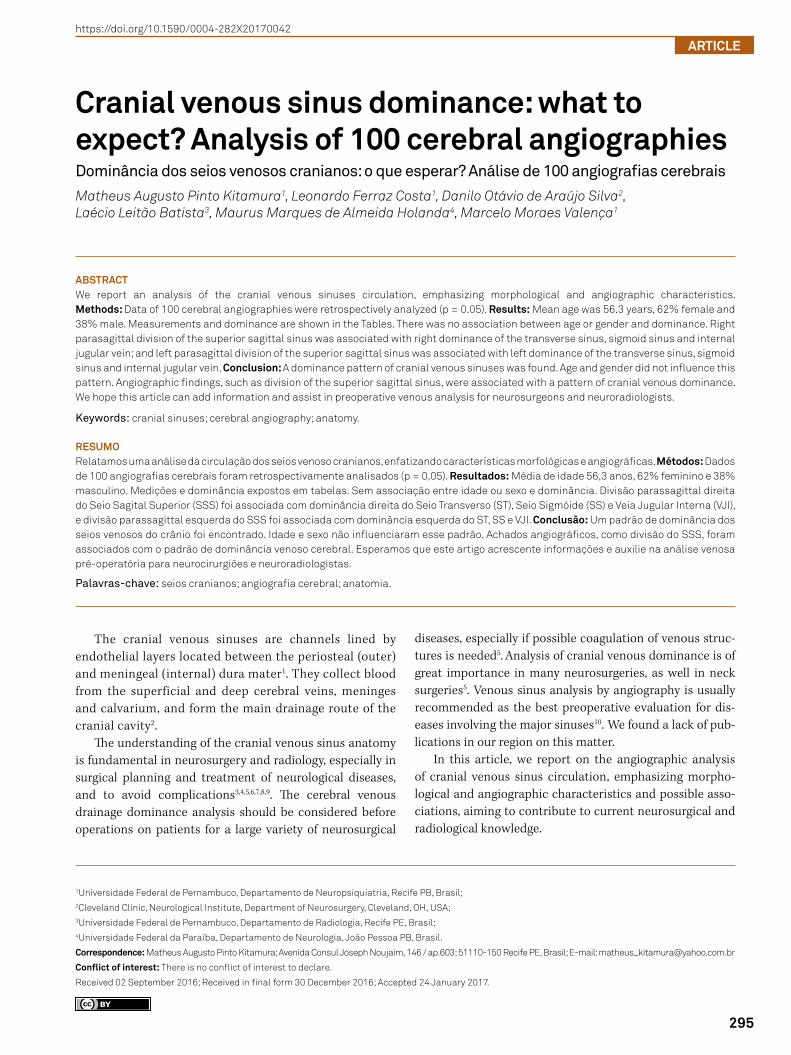
The midline was defined by a line perpendicular to the mean distance between the medial border of the orbital rims, and assisted by calcification of faux when visible. The TS measurement was acquired between the ninth and tenth portion of the distance between the edges of one side to the other. The SS measurement was acquired at the aver-age distance of the length of the sinus. The IJV measure-ment was acquired at the first possible moment of defining the limits of its walls, just below the jugular bulb, at the frontal incidence of angiography. The analysis is exempli-fied in Figure 1.
The measurement ratio right/left of the sinuses (or IJV) were stratified according to dominance. A venous sinus was classified as dominant when the measurement ratio was more than 1.5 (dominant right) or less than 0.67 (dominant left) (sinus measurement was more than 50% than the con-tralateral side). A venous sinus was classified as symmetri-cal when the measurement ratio was equal or between 1.5 and 0.67 (sinuses measuring between the limit of 50%).
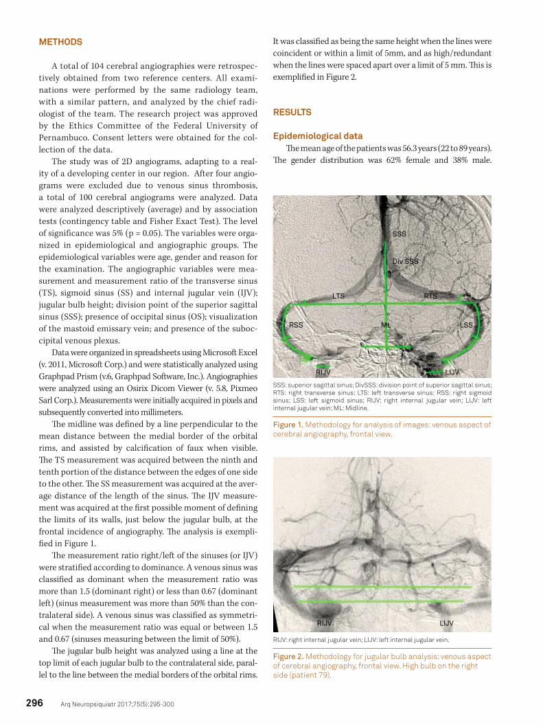
The jugular bulb height was analyzed using a line at the top limit of each jugular bulb to the contralateral side, paral-lel to the line between the medial borders of the orbital rims.
It was classified as being the same height when the lines were coincident or within a limit of 5mm, and as high/redundant when the lines were spaced apart over a limit of 5 mm. This is exemplified in Figure 2.
RESULTS
Epidemiological data The mean age of the patients was 56.3 years (22 to 89 years).
The gender distribution was 62% female and 38% male.
SSS
Div SSS
RTS
ML LSSRSS
RIJV LIJV
LTS
SSS: superior sagittal sinus; DivSSS: division point of superior sagittal sinus; RTS: right transverse sinus; LTS: left transverse sinus; RSS: right sigmoid sinus; LSS: left sigmoid sinus; RIJV: right internal jugular vein; LIJV: left internal jugular vein; ML: Midline.
Figure 1. Methodology for analysis of images: venous aspect of cerebral angiography, frontal view.
RIJV LIJV
RIJV: right internal jugular vein; LIJV: left internal jugular vein.
Figure 2. Methodology for jugular bulb analysis: venous aspect of cerebral angiography, frontal view. High bulb on the right side (patient 79).
297Kitamura MAP et al. Cranial venous sinus dominance
The most common reason for angiographic examination was suspicion of a cerebral aneurysm (38%).
Dominance of cranial venous sinuses The overall average measurements, including gender dis-
tribution, and the dominance pattern of the TS, SS and IJV are reported in Tables 1 and 2. Patterns of dominance are exemplified in Figures 3 and 4.
The division of the SSS was parasagittal right in 34%, para-sagittal left in 12%, and sagittal in 54% of patients.
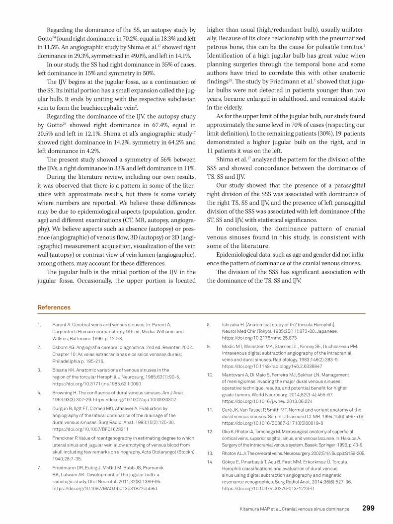
There was no statistically significant association between age or gender and venous sinus dominance. The data of dom-inance according to gender are shown in Table 3.
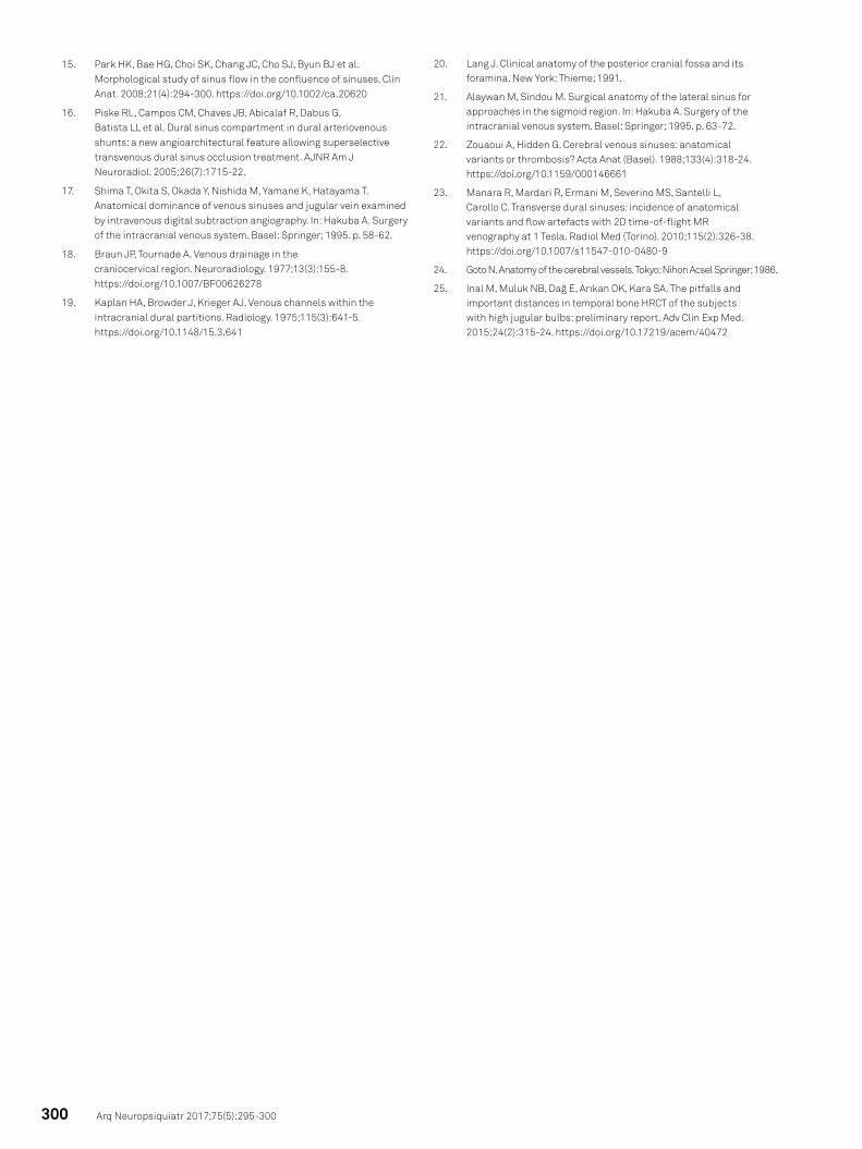
The presence of right parasagittal division of the SSS was associated with right dominance of the TS, SS and IJV, with an odds ratio of 33.8 (p < 0.0001), 9.4 (p < 0.0001) and 15.2 (p < 0.0001), respectively. The presence of left parasagittal division of the SSS was associated with left dominance of the TS, SS and IJV, with an odds ratio of 27.3 (p < 0.0001), 8.7 (p = 0.0023) and 5.8 (p = 0.0255), respectively. The domi-nance data according to the division of SSS are shown in Table 4. An example of the division correlation between the SSS and dominance is shown in Figure 4.
Additional findings For the upper limit of the jugular bulb, our study found
the same level in 70% of patients: 19% high bulb on the right side and 11% high bulb on the left side. A high bulb is exem-plified in Figure 2.
The occipital marginal sinus was present in 18% of patients. The right mastoid vein was present in 29% and left mastoid vein in 22%. The suboccipital venous plexus was present in 58%.
Table 2. Dominance analysis.
Sinus/vein vs. Dominance Right(%)
Left(%)
Symmetry (%)
TS 36 14 50
SS 35 15 50
IJV 33 11 56TS: transverse sinus; SS: sigmoid sinus; IJV: internal jugular vein.
Table 1. Measurements vs distribution according to gender.
Measurement vs. Distribution General (mm)
Male (mm)
Female (mm)
Right TS 6.4 6.8 6.1
Left TS 5.4 5.1 5.6
Right SS 6.5 6.8 6.3
Left SS 5.3 5.2 5.3
Right IJV 9.1 9.0 9.3
Left IJV 7.4 6.8 7.8 TS: transverse sinus; SS: sigmoid sinus; IJV: internal jugular vein.
DISCUSSION
The SSS originates just behind the paranasal frontal sinuses, near the crista galli and progressively increases in size to con-tinue later in a shallow depression of the mid-sagittal inner table of the skull, collecting numerous parasagittal veins11-13. It drains into the transverse sinus in the internal occipital pro-tuberance through a complex plexiform venous confluence, called the torcula Herophili (confluence of sinuses)12.
Variations of the torcula have been described according to its anatomic composition14. The SSS can end directly in the
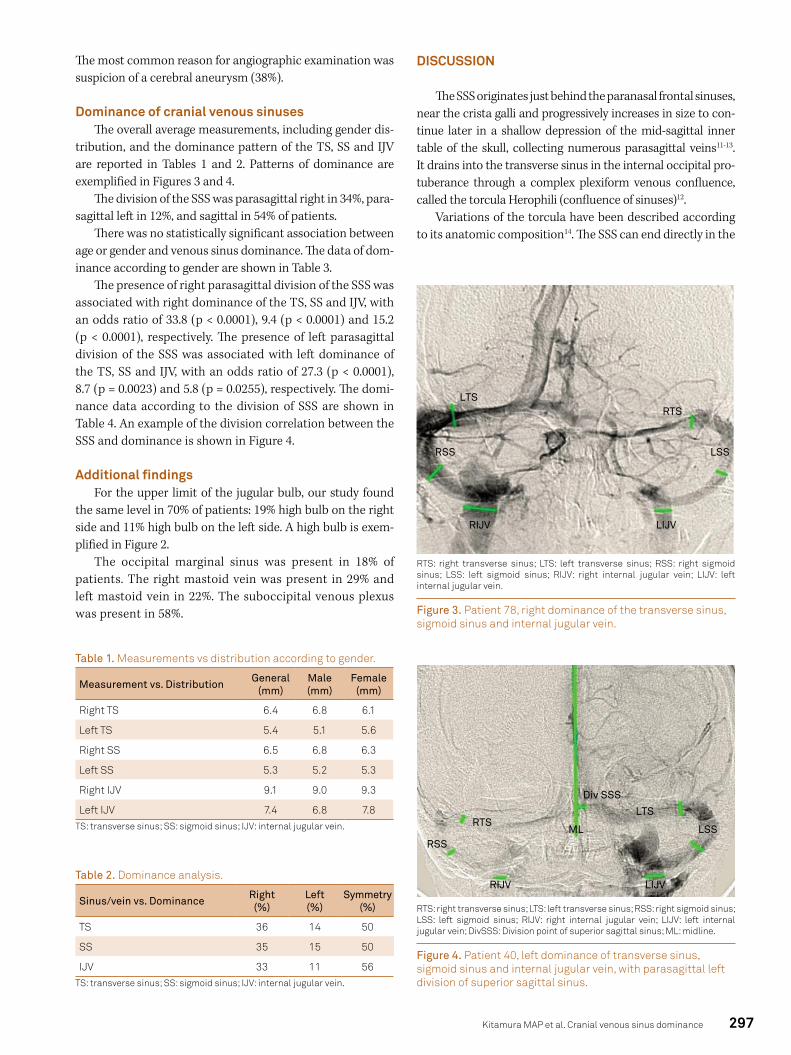
RTS: right transverse sinus; LTS: left transverse sinus; RSS: right sigmoid sinus; LSS: left sigmoid sinus; RIJV: right internal jugular vein; LIJV: left internal jugular vein.
Figure 3. Patient 78, right dominance of the transverse sinus, sigmoid sinus and internal jugular vein.
RIJV LIJV
LSSRSS
LTSRTS
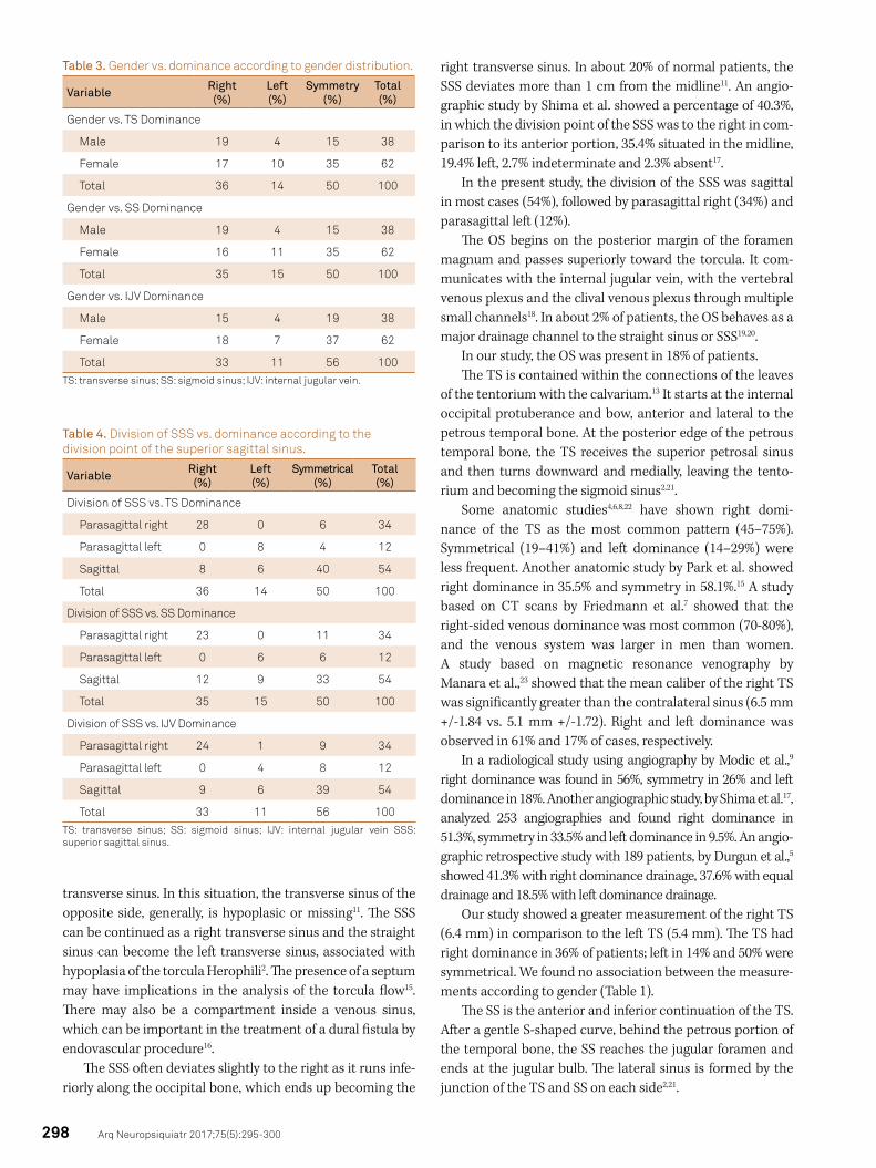
RTS: right transverse sinus; LTS: left transverse sinus; RSS: right sigmoid sinus; LSS: left sigmoid sinus; RIJV: right internal jugular vein; LIJV: left internal jugular vein; DivSSS: Division point of superior sagittal sinus; ML: midline.
Figure 4. Patient 40, left dominance of transverse sinus, sigmoid sinus and internal jugular vein, with parasagittal left division of superior sagittal sinus.
RIJV LIJV
LSSRSS
LTS
ML
Div SSS
RTS
298 Arq Neuropsiquiatr 2017;75(5):295-300
transverse sinus. In this situation, the transverse sinus of the opposite side, generally, is hypoplasic or missing11. The SSS can be continued as a right transverse sinus and the straight sinus can become the left transverse sinus, associated with hypoplasia of the torcula Herophili2. The presence of a septum may have implications in the analysis of the torcula flow15. There may also be a compartment inside a venous sinus, which can be important in the treatment of a dural fistula by endovascular procedure16.
The SSS often deviates slightly to the right as it runs infe-riorly along the occipital bone, which ends up becoming the
right transverse sinus. In about 20% of normal patients, the SSS deviates more than 1 cm from the midline11. An angio-graphic study by Shima et al. showed a percentage of 40.3%, in which the division point of the SSS was to the right in com-parison to its anterior portion, 35.4% situated in the midline, 19.4% left, 2.7% indeterminate and 2.3% absent17.
In the present study, the division of the SSS was sagittal in most cases (54%), followed by parasagittal right (34%) and parasagittal left (12%).
The OS begins on the posterior margin of the foramen magnum and passes superiorly toward the torcula. It com-municates with the internal jugular vein, with the vertebral venous plexus and the clival venous plexus through multiple small channels18. In about 2% of patients, the OS behaves as a major drainage channel to the straight sinus or SSS19,20.
In our study, the OS was present in 18% of patients.The TS is contained within the connections of the leaves
of the tentorium with the calvarium.13 It starts at the internal occipital protuberance and bow, anterior and lateral to the petrous temporal bone. At the posterior edge of the petrous temporal bone, the TS receives the superior petrosal sinus and then turns downward and medially, leaving the tento-rium and becoming the sigmoid sinus2,21.
Some anatomic studies4,6,8,22 have shown right domi-nance of the TS as the most common pattern (45–75%). Symmetrical (19–41%) and left dominance (14–29%) were less frequent. Another anatomic study by Park et al. showed right dominance in 35.5% and symmetry in 58.1%.15 A study based on CT scans by Friedmann et al.7 showed that the right-sided venous dominance was most common (70-80%), and the venous system was larger in men than women. A study based on magnetic resonance venography by Manara et al.,23 showed that the mean caliber of the right TS was significantly greater than the contralateral sinus (6.5 mm +/-1.84 vs. 5.1 mm +/-1.72). Right and left dominance was observed in 61% and 17% of cases, respectively.
In a radiological study using angiography by Modic et al.,9 right dominance was found in 56%, symmetry in 26% and left dominance in 18%. Another angiographic study, by Shima et al.17,
analyzed 253 angiographies and found right dominance in 51.3%, symmetry in 33.5% and left dominance in 9.5%. An angio-graphic retrospective study with 189 patients, by Durgun et al.,5 showed 41.3% with right dominance drainage, 37.6% with equal drainage and 18.5% with left dominance drainage.
Our study showed a greater measurement of the right TS (6.4 mm) in comparison to the left TS (5.4 mm). The TS had right dominance in 36% of patients; left in 14% and 50% were symmetrical. We found no association between the measure-ments according to gender (Table 1).
The SS is the anterior and inferior continuation of the TS. After a gentle S-shaped curve, behind the petrous portion of the temporal bone, the SS reaches the jugular foramen and ends at the jugular bulb. The lateral sinus is formed by the junction of the TS and SS on each side2,21.
Table 4. Division of SSS vs. dominance according to the division point of the superior sagittal sinus.
Variable Right(%)
Left(%)
Symmetrical (%)
Total(%)
Division of SSS vs. TS Dominance
Parasagittal right 28 0 6 34
Parasagittal left 0 8 4 12
Sagittal 8 6 40 54
Total 36 14 50 100
Division of SSS vs. SS Dominance
Parasagittal right 23 0 11 34
Parasagittal left 0 6 6 12
Sagittal 12 9 33 54
Total 35 15 50 100
Division of SSS vs. IJV Dominance
Parasagittal right 24 1 9 34
Parasagittal left 0 4 8 12
Sagittal 9 6 39 54
Total 33 11 56 100TS: transverse sinus; SS: sigmoid sinus; IJV: internal jugular vein SSS: superior sagittal sinus.
Table 3. Gender vs. dominance according to gender distribution.
Variable Right(%)
Left(%)
Symmetry (%)
Total(%)
Gender vs. TS Dominance
Male 19 4 15 38
Female 17 10 35 62
Total 36 14 50 100
Gender vs. SS Dominance
Male 19 4 15 38
Female 16 11 35 62
Total 35 15 50 100
Gender vs. IJV Dominance
Male 15 4 19 38
Female 18 7 37 62
Total 33 11 56 100TS: transverse sinus; SS: sigmoid sinus; IJV: internal jugular vein.
299Kitamura MAP et al. Cranial venous sinus dominance
Regarding the dominance of the SS, an autopsy study by Gotto24 found right dominance in 70.2%, equal in 18.3% and left in 11.5%. An angiographic study by Shima et al.17 showed right dominance in 29.3%, symmetrical in 49.0%, and left in 14.1%.
In our study, the SS had right dominance in 35% of cases, left dominance in 15% and symmetry in 50%.
The IJV begins at the jugular fossa, as a continuation of the SS. Its initial portion has a small expansion called the jug-ular bulb. It ends by uniting with the respective subclavian vein to form the brachiocephalic vein2.
Regarding the dominance of the IJV, the autopsy study by Gotto24 showed right dominance in 67.4%, equal in 20.5% and left in 12.1%. Shima et al.’s angiographic study17 showed right dominance in 14.2%, symmetry in 64.2% and left dominance in 4.2%.
The present study showed a symmetry of 56% between the IJVs, a right dominance in 33% and left dominance in 11%.
During the literature review, including our own results, it was observed that there is a pattern in some of the liter-ature with approximate results, but there is some variety where numbers are reported. We believe these differences may be due to epidemiological aspects (population, gender, age) and different examinations (CT, MR, autopsy, angiogra-phy). We believe aspects such as absence (autopsy) or pres-ence (angiographic) of venous flow, 3D (autopsy) or 2D (angi-ographic) measurement acquisition, visualization of the vein wall (autopsy) or contrast view of vein lumen (angiographic), among others, may account for these differences.
The jugular bulb is the initial portion of the IJV in the jugular fossa. Occasionally, the upper portion is located
higher than usual (high/redundant bulb), usually unilater-ally. Because of its close relationship with the pneumatized petrous bone, this can be the cause for pulsatile tinnitus.2 Identification of a high jugular bulb has great value when planning surgeries through the temporal bone and some authors have tried to correlate this with other anatomic findings25. The study by Friedmann et al.7 showed that jugu-lar bulbs were not detected in patients younger than two years, became enlarged in adulthood, and remained stable in the elderly.
As for the upper limit of the jugular bulb, our study found approximately the same level in 70% of cases (respecting our limit definition). In the remaining patients (30%), 19 patients demonstrated a higher jugular bulb on the right, and in 11 patients it was on the left.
Shima et al.17 analyzed the pattern for the division of the SSS and showed concordance between the dominance of TS, SS and IJV.
Our study showed that the presence of a parasagittal right division of the SSS was associated with dominance of the right TS, SS and IJV, and the presence of left parasagittal division of the SSS was associated with left dominance of the ST, SS and IJV, with statistical significance.
In conclusion, the dominance pattern of cranial venous sinuses found in this study, is consistent with some of the literature.
Epidemiological data, such as age and gender did not influ-ence the pattern of dominance of the cranial venous sinuses.
The division of the SSS has significant association with the dominance of the TS, SS and IJV.
References
1. Parent A. Cerebral veins and venous sinuses. In: Parent A. Carpenter’s Human neuroanatamy, 9th ed. Media: Williams and Wilkins; Baltimore. 1996. p. 120-8.
2. Osborn AG. Angiografia cerebral diagnóstica. 2nd ed. Revinter, 2002. Chapter 10: As veias extracranianas e os seios venosos durais; Philadellphia p. 195-216.
3. Bisaria KK. Anatomic variations of venous sinuses in the region of the torcular Herophili. J Neurosurg. 1985;62(1):90-5. https://doi.org/10.3171/jns.1985.62.1.0090
4. Browning H. The confluence of dural venous sinuses. Am J Anat. 1953;93(3):307-29. https://doi.org/10.1002/aja.1000930302
5. Durgun B, Ilglt ET, Cizmeli MO, Atasever A. Evaluation by angiography of the lateral dominance of the drainage of the dural venous sinuses. Surg Radiol Anat. 1993;15(2):125-30. https://doi.org/10.1007/BF01628311
6. Frenckner P. Value of roentgenography in estimating degree to which lateral sinus and jugular vein allow emptying of venous blood from skull including few remarks on sinography. Acta Otolaryngol (Stockh). 1940;28:7-35.
7. Friedmann DR, Eubig J, McGill M, Babb JS, Pramanik BK, Lalwani AK. Development of the jugular bulb: a radiologic study. Otol Neurotol. 2011;32(8):1389-95. https://doi.org/10.1097/MAO.0b013e31822e5b8d
8. Ishizaka H. [Anatomical study of th2 torcula Herophili]. Neurol Med Chir (Tokyo). 1985;25(11):873-80. Japanese. https://doi.org/10.2176/nmc.25.873
9. Modic MT, Weinstein MA, Starnes DL, Kinney SE, Duchesneau PM. Intravenous digital subtraction angiography of the intracranial veins and dural sinuses. Radiolology. 1983;146(2):383-9. https://doi.org/10.1148/radiology.146.2.6336847
10. Mantovani A, Di Maio S, Ferreira MJ, Sekhar LN. Management of meningiomas invading the major dural venous sinuses: operative technique, results, and potential benefit for higher grade tumors. World Neurosurg. 2014;82(3-4):455-67. https://doi.org/10.1016/j.wneu.2013.06.024
11. Curé JK, Van Tassel P, Smith MT. Normal and variant anatomy of the dural venous sinuses. Semin Ultrasound CT MR. 1994;15(6):499-519. https://doi.org/10.1016/S0887-2171(05)80019-8
12. Oka K, Rhoton A, Tomonaga M. Microsurgical anatomy of superficial cortical veins, superior sagittal sinus, and venous lacunae. In: Hakuba A. Surgery of the intracranial venous system. Basek: Springer; 1995. p. 43-9.
13. Rhoton AL Jr. The cerebral veins. Neurosurgery. 2002;51(4 Suppl):S159-205.
14. Gökçe E, Pınarbaşılı T, Acu B, Fırat MM, Erkorkmaz Ü. Torcula Herophili classifications and evaluation of dural venous sinus using digital subtraction angiography and magnetic resonance venographies. Surg Radiol Anat. 2014;36(6):527-36. https://doi.org/10.1007/s00276-013-1223-0
300 Arq Neuropsiquiatr 2017;75(5):295-300
15. Park HK, Bae HG, Choi SK, Chang JC, Cho SJ, Byun BJ et al. Morphological study of sinus flow in the confluence of sinuses. Clin Anat. 2008;21(4):294-300. https://doi.org/10.1002/ca.20620
16. Piske RL, Campos CM, Chaves JB, Abicalaf R, Dabus G, Batista LL et al. Dural sinus compartment in dural arteriovenous shunts: a new angioarchitectural feature allowing superselective transvenous dural sinus occlusion treatment. AJNR Am J Neuroradiol. 2005;26(7):1715-22.
17. Shima T, Okita S, Okada Y, Nishida M, Yamane K, Hatayama T. Anatomical dominance of venous sinuses and jugular vein examined by intravenous digital subtraction angiography. In: Hakuba A. Surgery of the intracranial venous system. Basel: Springer; 1995. p. 58-62.
18. Braun JP, Tournade A. Venous drainage in the craniocervical region. Neuroradiology. 1977;13(3):155-8. https://doi.org/10.1007/BF00626278
19. Kaplan HA, Browder J, Krieger AJ. Venous channels within the intracranial dural partitions. Radiology. 1975;115(3):641-5. https://doi.org/10.1148/15.3.641
20. Lang J. Clinical anatomy of the posterior cranial fossa and its foramina. New York: Thieme; 1991.
21. Alaywan M, Sindou M. Surgical anatomy of the lateral sinus for approaches in the sigmoid region. In: Hakuba A. Surgery of the intracranial venous system. Basel: Springer; 1995. p. 63-72.
22. Zouaoui A, Hidden G. Cerebral venous sinuses: anatomical variants or thrombosis? Acta Anat (Basel). 1988;133(4):318-24. https://doi.org/10.1159/000146661
23. Manara R, Mardari R, Ermani M, Severino MS, Santelli L, Carollo C. Transverse dural sinuses: incidence of anatomical variants and flow artefacts with 2D time-of-flight MR venography at 1 Tesla. Radiol Med (Torino). 2010;115(2):326-38. https://doi.org/10.1007/s11547-010-0480-9
24. Goto N. Anatomy of the cerebral vessels. Tokyo: Nihon Acsel Springer; 1986.
25. Inal M, Muluk NB, Dağ E, Arıkan OK, Kara SA. The pitfalls and important distances in temporal bone HRCT of the subjects with high jugular bulbs: preliminary report. Adv Clin Exp Med. 2015;24(2):315-24. https://doi.org/10.17219/acem/40472