Embed Size (px)
Citation preview
Cryptic insertions of the immunoglobulin light chain enhancer region near CCND1 in t(11;14)-negative mantlecell lymphoma
by Carla Fuster, David Martín-Garcia, Olga Balagué, Alba Navarro, Ferran Nadeu, Dolors Costa, Miriam Prieto, Itziar Salaverria, Blanca Espinet, Alfredo Rivas-Delgado, Maria José Terol, Eva Giné, Pilar Forcada, Margaret Ashton-Key, Xose S. Puente, Steven H. Swerdlow, Sílvia Beà, and Elias Campo
Haematologica 2019 [Epub ahead of print]
Citation: Carla Fuster, David Martín-Garcia, Olga Balagué, Alba Navarro, Ferran Nadeu, Dolors Costa, Miriam Prieto, Itziar Salaverria, Blanca Espinet, Alfredo Rivas-Delgado, Maria José Terol, Eva Giné, Pilar Forcada, Margaret Ashton-Key, Xose S. Puente, Steven H. Swerdlow,Sílvia Beà, and Elias Campo. Cryptic insertions of the immunoglobulin light chain enhancer region near CCND1 in t(11;14)-negative mantle cell lymphoma. Haematologica. 2019; 104:xxxdoi:10.3324/haematol.2019.237073
Publisher's Disclaimer.E-publishing ahead of print is increasingly important for the rapid dissemination of science.Haematologica is, therefore, E-publishing PDF files of an early version of manuscripts thathave completed a regular peer review and have been accepted for publication. E-publishingof this PDF file has been approved by the authors. After having E-published Ahead of Print,manuscripts will then undergo technical and English editing, typesetting, proof correction andbe presented for the authors' final approval; the final version of the manuscript will thenappear in print on a regular issue of the journal. All legal disclaimers that apply to thejournal also pertain to this production process.
Copyright 2019 Ferrata Storti Foundation.Published Ahead of Print on November 21, 2019, as doi:10.3324/haematol.2019.237073.
Cryptic insertions of the immunoglobulin light chain enhancer region
near CCND1 in t(11;14)-negative mantle cell lymphoma
Carla Fuster1*
, David Martín-Garcia2,3;*
, Olga Balagué1,4
, Alba Navarro2,3
, Ferran
Nadeu2,3
, Dolors Costa1,3
, Miriam Prieto2, Itziar Salaverria
2,3, Blanca Espinet
5, Alfredo
Rivas-Delgado2,6
, Maria José Terol7, Eva Giné
2,3,6, Pilar Forcada
8, Margaret Ashton-
Key9, Xose S Puente
3,10, Steven H Swerdlow
11, Sílvia Beà
2,3,4+, and Elias Campo
1,2,3,4+
1Hematopathology Section, Laboratory of Pathology, Hospital Clínic de Barcelona,
Barcelona, Spain
2Institut d'Investigacions Biomèdiques August Pi i Sunyer (IDIBAPS), Barcelona, Spain
3Centro de Investigación Biomédica en Red de Cáncer (CIBERONC), Madrid, Spain
4University of Barcelona, Barcelona, Spain
5Laboratori de Citogenètica Molecular, Servei de Patologia, Hospital del Mar,
Barcelona, Spain; Grup de Recerca Translacional en Neoplàsies Hematològiques,
Cancer Research Programme, IMIM-Hospital del Mar, Barcelona, Spain
6Department of Hematology Hospital Clínic de Barcelona, Barcelona, Spain
7Department of Hematology, Hospital Clínico, INCLIVA Biomedical Research Institute,
University of Valencia, Valencia, Spain
8Department of Pathology, Hospital Mutua Terrassa, Terrassa, Spain
9Department of Cellular Pathology, Southampton University Hospitals National Health
Service Trust, UK
10Departamento de Bioquímica y Biología Molecular, IUOPA, Universidad de Oviedo,
Oviedo, Spain
11Department of Pathology, University of Pittsburgh School of Medicine, Pittsburgh,
PA, USA
*CF, DMG and +SB, EC contributed equally to this work.
Running Title: Cryptic CCND1 rearrangements in MCL
Corresponding Authors:
Sílvia Beà, IDIBAPS, Rosselló 149, 08036-Barcelona, Spain, [email protected]
Elias Campo, Hospital Clinic, Villarroel 170, 08036-Barcelona, Spain, [email protected]
Words: 1474; Figures: 2; Tables: 1; References: 15; Supplemental Files: 1
2
Funding
This work was supported by research funding from Fondo de Investigaciones
Sanitarias, Instituto de Salud Carlos III PI17/01061 (SB), Ministerio de Economía y
Competitividad, SAF2015-64885-R (EC), SAF2017-87811-R (XSP) from Plan Nacional de
I+D+I, the NIH grant number 1 P01CA229100 (EC), Generalitat de Catalunya Suport
Grups de Recerca 2017-SGR-709 (SB), 2017-SGR-1142 (EC), and the European Regional
Development Fund “Una manera de fer Europa”, CERCA Programme/Generalitat de
Catalunya. EC is an Academia Researcher of the "Institució Catalana de Recerca i
Estudis Avançats" of the Generalitat de Catalunya. Miriam Prieto is supported by
"Acció instrumental d’incorporació de científics i tecnòlegs PERIS 2016"
(SLT002/16/00347) from Generalitat de Catalunya. Alfredo Rivas-Delgado is supported
by “Josep Font” grant from Hospital Clínic de Barcelona.
Acknowledgements
The authors would like to thank the IDIBAPS Genomics Core Facility and the
Hematopathology Collection from Hospital Clinic/IDIBAPS; the Molecular Cytogenetic
Platform of IMIM, Hospital del Mar (Barcelona) for providing one IGK BAC clone.
Miriam Prieto, Silvia Martín, Cándida Gómez, and Amparo Arias for their excellent
technical assistance and Montserrat Puiggròs and Romina Royo from Barcelona
SuperComputing Center. This work was developed at the Centro Esther Koplowitz
(CEK), Barcelona, Spain.
3
Cyclin D1+ mantle cell lymphoma (MCL) is molecularly characterized by the
t(11;14)(q13;q32) or rearrangements of CCND1 gene with the immunoglobulin (IG)
light chains.1,2
Most MCL can be diagnosed based on the characteristic pathologic
features and cyclin D1 expression without the need for demonstrating the genetic
translocation. However, in cases with atypical morphologic or phenotypic features
other B-cell neoplasms that sometimes also have cyclin D1 positivity may be in the
differential diagnosis.1 In these situations the detection of the CCND1 rearrangements
may assist in the diagnosis since most other lymphomas do not carry translocations of
the gene.3-7
A subset of plasma cell myelomas express cyclin D1 associated with the
t(11;14) but are usually not in the differential diagnosis of MCL and are SOX11-
negative.5 Nevertheless, there are occasional lymphoid neoplasms with MCL features
in which cyclin D1 is strongly expressed without an apparent gene rearrangement
using conventional cytogenetics or fluorescence in situ hybridization (FISH) with fusion
or break-apart probes. The mechanisms of cyclin D1 overexpression in these cases are
unclear and the MCL diagnosis may be questioned.
In this study, we have identified 4 MCL and one mature B-cell lymphoma with
marked plasmacytic differentiation with strong cyclin D1 overexpression but in which
CCND1 rearrangements could not be detected by conventional cytogenetics or FISH
using fusion or break-apart probes. To determine the mechanism leading to cyclin D1
overexpression in these cases we analyzed the index case by whole-genome
sequencing (WGS) followed by FISH studies with custom probes for the IG light chain
enhancer regions in all cases and demonstrated the presence of cryptic translocations
of the enhancer region of the IG light chains with CCND1 in the four cyclin D1-positive
MCL.
The study was approved by the Institutional Review Board of the Hospital Clinic
of Barcelona and informed consent was obtained in accordance with the Declaration of
Helsinki. Lymphomas were studied by immunohistochemistry with a panel of
antibodies (Online Methods, Supplementary Table S1) and reviewed by four
pathologists. Tumor genomic DNA was isolated from formalin-fixed paraffin-
embedded tissue biopsies (cases 2, 3, and 5), fresh bone marrow aspirate (case 1) and
peripheral blood cells (case 4). Paired-end WGS was performed on tumor and germline
DNA from case 1 (index case) using standard Illumina protocols and analyzed as
4
previously described.8 CCND1 rearrangement was analyzed by FISH using CCND1 and
IG commercial and custom BAC-labeled probes (Online Supplementary Table S2). Copy
number alterations were investigated using Oncoscan FFPE or SNP6.0 (ThermoFisher
Scientific, Waltham, MA).8,9
The mutational status of 115 genes was examined by
targeted next-generation sequencing (NGS) strategy (Online Supplementary Table
S3).10
Further details can be found in Online Methods.
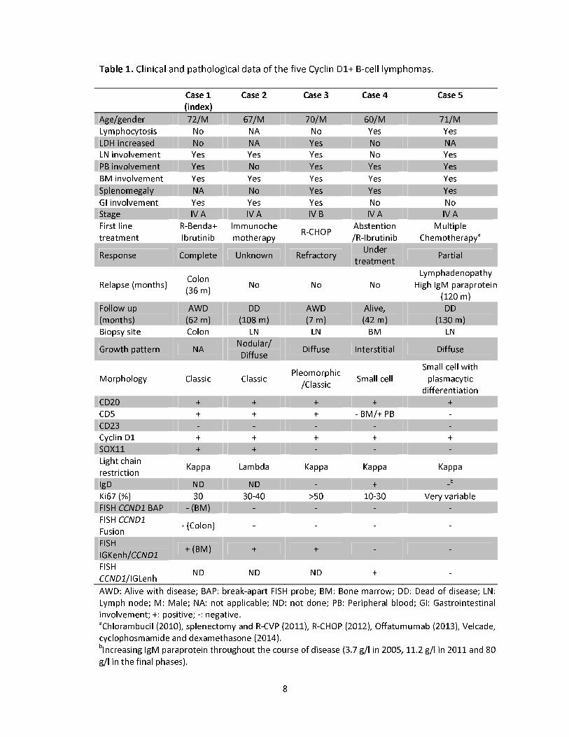
The clinical and pathological features of the patients are summarized in Table 1.
All patients were in stage IV with peripheral blood and bone marrow involvement.
Cases 1-3 presented with generalized lymphadenopathy, whereas case 4 had
asymptomatic lymphocytosis and splenomegaly. Case 5 had an unusual clinical history
and evolution. The patient was exposed to extensive radiation due to atomic bomb
tests in the early 1960s and developed several solid tumors (thyroid papillary
carcinoma, basal cell carcinoma, melanoma and pleomorphic sarcomas) that were
treated without further relapses. In 2003 he was diagnosed with a lymphoproliferative
disorder consistent with atypical CLL which did not require treatment until 2010, when
chlorambucil was started. Two years later the patient developed splenomegaly and
lymphadenopathy, that was biopsied. The bone marrow biopsy from 2005 showed a
nodular and interstitial lymphoid infiltration. The patient had an IgM lambda
paraprotein, which was stable for several years but progressively increased in the final
phases of the disease. The patient required several lines of therapy without response
and died of progressive disease 11 years after the initial diagnosis. The other 4 patients
received different treatments. Case 2 died of disease and cases 1, 3, and 4 were alive
at the last follow-up.
Cases 1 and 2 had classic MCL cytology, whereas case 3 showed a large cell
pleomorphic component with unusual clear cytoplasm intermingled with abundant
atypical small cells. Cyclin D1 was strongly positive in both components. Case 4 had
small cell morphology and case 5 showed small cells with marked plasmacytic
differentiation and occasional Dutcher bodies (Figure 1). CD5 was positive in all cases
except case 5 and the bone marrow cells of case 4. SOX11 was positive in cases 1 and 2
and negative in cases 3 to 5. Four cases had kappa light chain restriction and case 2
expressed lambda. Case 5 had strong cytoplasmic IgM/kappa expression (Table 1).
5
All cases were negative for CCND1 rearrangements by FISH using commercial
fusion and break-apart probes on the diagnostic biopsies (Table 1, Online
Supplementary Figure S1A-B). In cases 1 and 5 the FISH analyses were performed in
multiple samples. The karyotype in case 1 was
46,XY,der(11)t(3;11)(q13;q25)[9]/46,XY[11]. The unbalanced chromosome 11
translocation with chromosome 3 did not involve the CCND1 locus (11q13), and was
further confirmed by whole chromosome painting (Online Supplementary Figure S2).
Case 2 had a complex karyotype, 44-46,XY,-
Y[2],del(6)(q21q23),del(7)(p15p22),add(9)(p11.2),del(11)(q21q23.1),del(13)(q11q34)
[2],add(22)(p11.2)[6][cp7]/46,XY[9], without the t(11;14) translocation.
We performed WGS of case 1 and identified a rearrangement involving IGK in
chromosome 2 and CCND1 in chromosome 11. A 412 Kb region of the IGK, including
the IGK enhancer (IGKenh) and the IGK constant (IGKC) region was inserted 226.3 Kb
upstream of CCND1 gene (Figure 2A-B). We confirmed the rearrangement by PCR,
Sanger sequencing and FISH using custom fusion probes combining CCND1 gene (red)
and IGKenh probes (green) that we had used previously (Figure 2C).8 FISH using the
commercial IGK break-apart probe confirmed the rearrangement detected by WGS
(Supplementary Figure 1C). The finding of the cryptic rearrangement of IGK with
CCND1 in case 1, prompted us to analyze this cryptic rearrangement in the remaining
four cases by FISH. The IGKenh/CCND1 rearrangement was also detected in cases 2
and 3, both in the small and large cells (Figure 2D-E). However, cases 4 and 5 were
negative. We next tested the combination of CCND1 with IGLenh and case 4 was
positive (Figure 2F) whereas case 5 was negative for both IGKenh and IGLenh with
CCND1 probes.
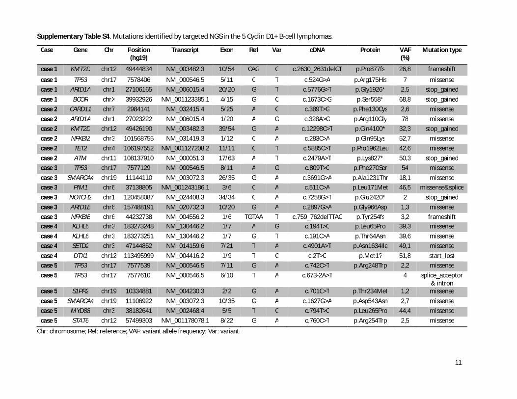
Targeted sequencing identified mutations in 24 different genes (Online
Supplementary Table S4). Cases 1-3 had mutations already reported in MCL,9 including
ATM, TP53, NOTCH2, KMT2D, NFKBIE, ARID1A, SMARCA4, and CARD11. Case 4 had
mutations in KLHL6, DTX1 and SETD2, whereas case 5 had a clonal (allelic frequency of
44.4%) p.L265P MYD88 mutation (Online Supplementary Figure S3). The genomic
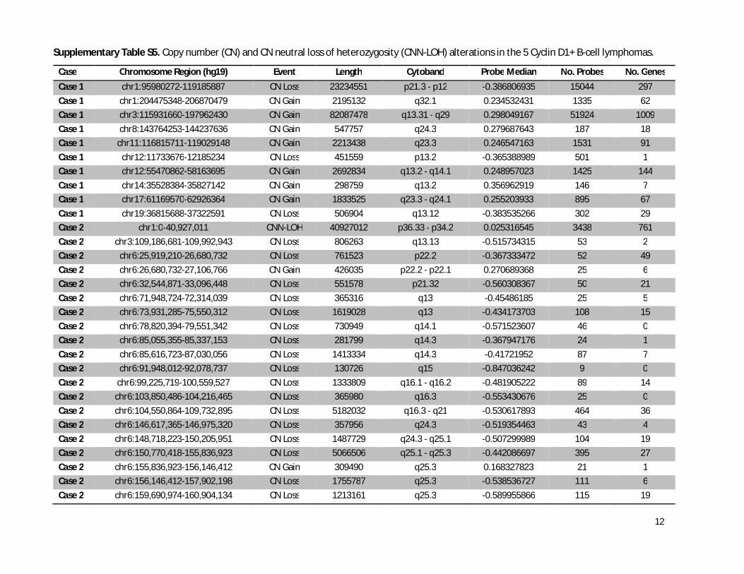
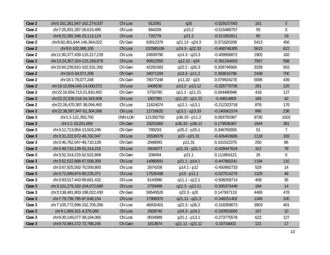
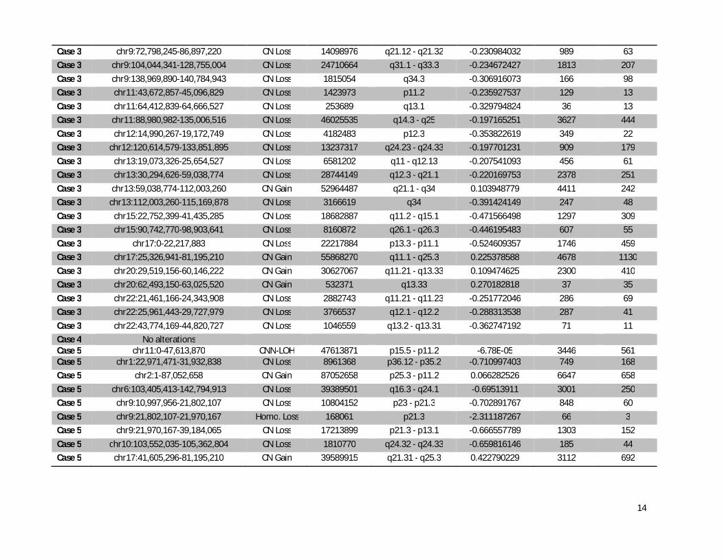
profile was complex in cases 1-3 with copy number alterations commonly found in
MCL (+3q, -9p, -9q, -11q, -13q, and -17p), whereas case 4 did not show any copy
number alterations, in line with its indolent clinical course and SOX11-negativity
6
(Online Supplementary Table S5). Case 5 showed 8 copy number alterations including
+2p, +17q, -6q and -9p.
In summary, cyclin D1 overexpresion in 4 out of 5 cases was associated with
cryptic CCND1 rearrangements that involved the enhancers of IGK and IGL in 3 and 1
cases, respectively. Similar to conventional rearrangements with IGH, the IG light chain
translocated fragments (including the enhancers) could be responsible for the
dysregulation of cyclin D1 in MCL. These findings are similar to our recent observations
in cyclin D1-negative MCL overexpressing cyclin D2 or cyclin D3 which carried cryptic
insertions of the IGK and IGL enhancers near CCND2 or CCND3, respectively.8 The
detection of cryptic rearrangements of MYC and BCL2 with regulatory regions of IG
genes has been recently reported in B-cell neoplasms.11-13
The findings in case 5 were intriguing and its specific taxonomic classification
difficult. The IgM, kappa paraprotein and plasmacytic differentiation was consistent
with a lymphoplasmacytic lymphoma, and concordantly the tumor carried the p.L256P
MYD88 mutation. However, cyclin D1 was diffusely expressed without evidences of
CCND1 rearrangements. The lack of CCND1 rearrangement detection with our probes
does not completely rule out other alternative rearrangements. In this sense, a recent
study of a MCL without apparent CCND1 rearrangements has detected an insertion of
the entire CCND1 coding region into the IGH locus which was not detected by standard
probes and would not have been detected with our IG light chain probes.14
The
characteristics of our case with marked plasmacytic differentiation, strong cyclin D1
expression and MYD88 mutation are similar to a previously reported case but in which
the t(11;14) could be demonstrated by FISH.15
Whether these cases should be
classified as lymphoplasmacytic lymphoma with CCND1 rearrangements or MCL with
MYD88 mutations is debatable. Independent of the possible taxonomy of these
tumors, it is important to recognize their clinical and biological peculiarities.
In conclusion, cryptic translocations of the IG light chain regulatory region with
CCND1 may be an alternative mechanism to deregulate this gene in MCL. FISH testing
for the IG light chain enhancer region could be incorporated into the diagnostic work
up of MCL negative for the t(11;14) or CCND1 rearrangements with standard probes,
especially in cases with atypical pathological or clinical features.
7
References
1. Sander B, Quintanilla-Martinez L, Ott G, et al. Mantle cell lymphoma--a spectrum from
indolent to aggressive disease. Virchows Arch. 2016;468(3):245-257.
2. Wlodarska I, Meeus P, Stul M, et al. Variant t(2;11)(p11;q13) associated with the IgK-
CCND1 rearrangement is a recurrent translocation in leukemic small-cell B-non-Hodgkin
lymphoma. Leukemia. 2004;18(10):1705-1710.
3. Bosch F, Campo E, Jares P, et al. Increased expression of the PRAD-1/CCND1 gene in
hairy cell leukaemia. Br J Haematol. 1995;91(4):1025-1030.
4. Chen BJ, Ruminy P, Roth CG, et al. Cyclin D1-positive Mediastinal Large B-Cell Lymphoma
With Copy Number Gains of CCND1 Gene: A Study of 3 Cases With Nonmediastinal
Disease. Am J Surg Pathol. 2019;43(1):110-120.
5. Chen YH, Gao J, Fan G, Peterson LC. Nuclear expression of sox11 is highly associated with
mantle cell lymphoma but is independent of t(11;14)(q13;q32) in non-mantle cell B-cell
neoplasms. Mod Pathol. 2010;23(1):105-112.
6. Hsiao SC, Cortada IR, Colomo L, et al. SOX11 is useful in differentiating cyclin D1-positive
diffuse large B-cell lymphoma from mantle cell lymphoma. Histopathology.
2012;61(4):685-693.
7. Vela-Chavez T, Adam P, Kremer M, et al. Cyclin D1 positive diffuse large B-cell lymphoma
is a post-germinal center-type lymphoma without alterations in the CCND1 gene locus.
Leuk Lymphoma. 2011;52(3):458-466.
8. Martin-Garcia D, Navarro A, Valdes-Mas R, et al. CCND2 and CCND3 hijack
immunoglobulin light-chain enhancers in cyclin D1(-) mantle cell lymphoma. Blood.
2019;133(9):940-951.
9. Bea S, Valdes-Mas R, Navarro A, et al. Landscape of somatic mutations and clonal
evolution in mantle cell lymphoma. Proc Natl Acad Sci U S A. 2013;110(45):18250-18255.
10. Nadeu F, Delgado J, Royo C, et al. Clinical impact of clonal and subclonal TP53, SF3B1,
BIRC3, NOTCH1, and ATM mutations in chronic lymphocytic leukemia. Blood.
2016;127(17):2122-2130.
11. King RL, McPhail ED, Meyer RG et al. False-negative rates for MYC fluorescence in situ
hybridization probes in B-cell neoplasms. Haematologica. 2019;104(6):e248-e251.
12. Yamamoto K, Okamura A, Inui Y, et al. Cryptic insertion of BCL2 gene into
immunoglobulin heavy locus in follicular lymphoma with t(6;9)(p23;p13). Leuk Res.
2012;36(9):e202-205.
13. Hilton LK, Tang J, Ben-Neriah S, et al. The double hit signature identifies double-hit
diffuse large B-cell lymphoma with genetic events cryptic to FISH. Blood.
2019;134(18):1528-1532.
14. Peterson JF, Baughn LB, Ketterling RP, et al. Characterization of a cryptic IGH/CCND1
rearrangement in a case of mantle cell lymphoma with negative CCND1 FISH studies.
Blood Adv. 2019;3(8):1298-1302.
15. Ribera-Cortada I, Martinez D, Amador V, et al. Plasma cell and terminal B-cell
differentiation in mantle cell lymphoma mainly occur in the SOX11-negative subtype.
Mod Pathol. 2015;28(11):1435-1447.
8
Table 1. Clinical and pathological data of the five Cyclin D1+ B-cell lymphomas.
Case 1
(index)
Case 2 Case 3 Case 4 Case 5
Age/gender 72/M 67/M 70/M 60/M 71/M
Lymphocytosis No NA No Yes Yes
LDH increased No NA Yes No NA
LN involvement Yes Yes Yes No Yes
PB involvement Yes No Yes Yes Yes
BM involvement Yes Yes Yes Yes Yes
Splenomegaly NA No Yes Yes Yes
GI involvement Yes Yes Yes No No
Stage IV A IV A IV B IV A IV A
First line
treatment
R-Benda+
Ibrutinib
Immunoche
motherapy R-CHOP
Abstention
/R-Ibrutinib
Multiple
Chemotherapya
Response Complete Unknown Refractory Under
treatment Partial
Relapse (months) Colon
(36 m) No No No
Lymphadenopathy
High IgM paraprotein
(120 m)
Follow up
(months)
AWD
(62 m)
DD
(108 m)
AWD
(7 m)
Alive,
(42 m)
DD
(130 m)
Biopsy site Colon LN LN BM LN
Growth pattern NA Nodular/
Diffuse Diffuse Interstitial Diffuse
Morphology Classic Classic Pleomorphic
/Classic Small cell
Small cell with
plasmacytic
differentiation
CD20 + + + + +
CD5 + + + - BM/+ PB -
CD23 - - - - -
Cyclin D1 + + + + +
SOX11 + + - - -
Light chain
restriction Kappa Lambda Kappa Kappa Kappa
IgD ND ND - + -b
Ki67 (%) 30 30-40 >50 10-30 Very variable
FISH CCND1 BAP - (BM) - - - -
FISH CCND1
Fusion - (Colon) - - - -
FISH
IGKenh/CCND1 + (BM) + + - -
FISH
CCND1/IGLenh ND ND ND + -
AWD: Alive with disease; BAP: break-apart FISH probe; BM: Bone marrow; DD: Dead of disease; LN:
Lymph node; M: Male; NA: not applicable; ND: not done; PB: Peripheral blood; GI: Gastrointestinal
involvement; +: positive; -: negative. aChlorambucil (2010), splenectomy and R-CVP (2011), R-CHOP (2012), Offatumumab (2013), Velcade,
cyclophosmamide and dexamethasone (2014).
bIncreasing IgM paraprotein throughout the course of disease (3.7 g/l in 2005, 11.2 g/l in 2011 and 80
g/l in the final phases).
9
Figure Legends
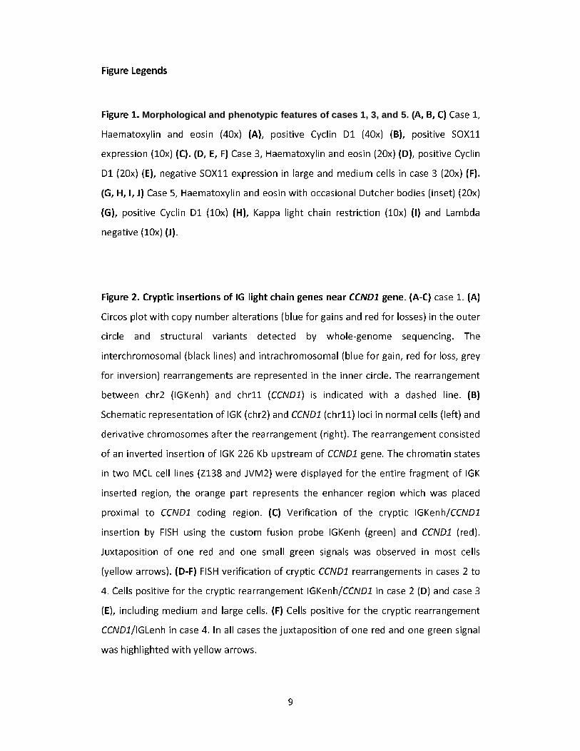
Figure 1. Morphological and phenotypic features of cases 1, 3, and 5. (A, B, C) Case 1,
Haematoxylin and eosin (40x) (A), positive Cyclin D1 (40x) (B), positive SOX11
expression (10x) (C). (D, E, F) Case 3, Haematoxylin and eosin (20x) (D), positive Cyclin
D1 (20x) (E), negative SOX11 expression in large and medium cells in case 3 (20x) (F).
(G, H, I, J) Case 5, Haematoxylin and eosin with occasional Dutcher bodies (inset) (20x)
(G), positive Cyclin D1 (10x) (H), Kappa light chain restriction (10x) (I) and Lambda
negative (10x) (J).
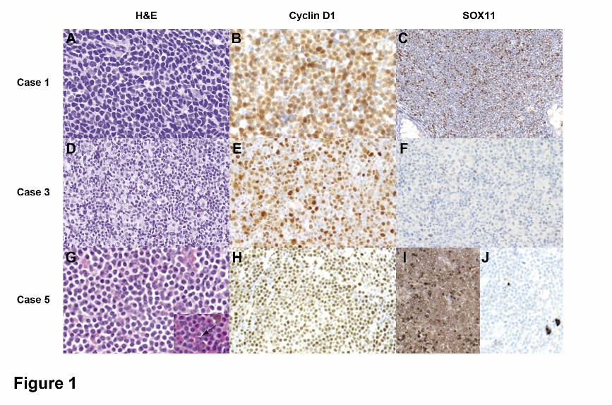
Figure 2. Cryptic insertions of IG light chain genes near CCND1 gene. (A-C) case 1. (A)
Circos plot with copy number alterations (blue for gains and red for losses) in the outer
circle and structural variants detected by whole-genome sequencing. The
interchromosomal (black lines) and intrachromosomal (blue for gain, red for loss, grey
for inversion) rearrangements are represented in the inner circle. The rearrangement
between chr2 (IGKenh) and chr11 (CCND1) is indicated with a dashed line. (B)
Schematic representation of IGK (chr2) and CCND1 (chr11) loci in normal cells (left) and
derivative chromosomes after the rearrangement (right). The rearrangement consisted
of an inverted insertion of IGK 226 Kb upstream of CCND1 gene. The chromatin states
in two MCL cell lines (Z138 and JVM2) were displayed for the entire fragment of IGK
inserted region, the orange part represents the enhancer region which was placed
proximal to CCND1 coding region. (C) Verification of the cryptic IGKenh/CCND1
insertion by FISH using the custom fusion probe IGKenh (green) and CCND1 (red).
Juxtaposition of one red and one small green signals was observed in most cells
(yellow arrows). (D-F) FISH verification of cryptic CCND1 rearrangements in cases 2 to
4. Cells positive for the cryptic rearrangement IGKenh/CCND1 in case 2 (D) and case 3
(E), including medium and large cells. (F) Cells positive for the cryptic rearrangement
CCND1/IGLenh in case 4. In all cases the juxtaposition of one red and one green signal
was highlighted with yellow arrows.
1
Supplementary Material
Cryptic insertions of the immunoglobulin light chain
enhancer region near CCND1 in t(11;14)-negative mantle
cell lymphoma
Carla Fuster & David Martín-Garcia et al.
2
SUPPLEMENTARY METHODS..............................................................................3
SUPPLEMENTARY FIGURES.................................................................................5
SUPPLEMENTARY TABLES...................................................................................8
SUPPLEMENTARY REFERENCES.........................................................................15
3
SUPPLEMENTARY METHODS
Case selection and immunohistochemistry
We studied 4 mantle cell lymphoma and one mature B-cell neoplasm with marked
plasmacytic differentiation, all with strong cyclin D1 expression but the t(11;14)
translocation or CCND1 rearrangements were not found using conventional cytogenetics
or FISH with fusion or break-apart probes. All the original haematoxylin and eosin stained
sections or newly stained sections were reviewed. Immunohistochemical stains were
performed for each case on 2 μm thick sections using a peroxidase-labeled detection
system, standard antigen retrieval protocols, and an automated immunostainer
(AutostainerLink 48, Dako, Glostrup, Denmark or BenchmarkXT, Roche Diagnostics, Basel,
Switzerland) as previously described1 or phenotype information was retrieved from the
original reports (Online Supplementary Table S1).
FISH analysis
CCND1 rearrangement was analyzed by FISH on formalin-fixed paraffin-embedded tissue
sections or peripheral blood fixed cells. The FISH panel including commercial and BAC-
labeled probes is detailed in Online Supplementary Table S2. DNA labeling and
hybridization was performed according to standard procedures.2 Digital images were
obtained using the ISIS FISH Imaging System (MetaSystems, Altlussheim, Germany).
Control FISH experiments were performed in 2 non-neoplastic lymphoid tissues. Whole
chromosome painting of chromosomes 3 and 11, and FISH with a chr12 centromeric probe
and LSI BCL6-BAP probe were performed in case 1 for refinement of the derivative
chromosomes. Conventional cytogenetics was performed on G-banded chromosomes for
cases 1 and 2 and results were described according to International System for Human
Cytogenomic Nomenclature.3
Copy number and next-generation sequencing (NGS)
Copy number alterations were investigated using Oncoscan FFPE or SNP6.0 Array
(ThermoFisher Scientific, Waltham, MA) and analyzed as described previously using Nexus
4
Biodiscovery v9 (Biodiscovery, Hawthorne, CA, USA).4,5 Paired-end whole-genome
sequencing was performed on tumor and germline DNA from case 1 using standard
Illumina protocols and sequenced in an Illumina HiSeq 2000 instrument.4 Smufin,6 Lumpy,7
and custom algorithms were used to detect genome-wide structural variants (SV) and
potential cryptic CCND1 rearrangements. Single nucleotide variant and indel calling was
performed by Smufin and Sidrón,8 and copy number alterations analysis was performed by
Battenberg (https://github.com/cancerit/cgpBattenberg) (manuscript with additional
details in preparation).
The mutational status of 115 genes previously described in aggressive B-cell
lymphoma was examined by targeted NGS strategy (Online Supplementary Table S3).
Libraries were prepared with 150 ng of genomic DNA using SureSelect XT Target
Enrichment System Capture strategy (Agilent Technologies Inc.) and sequenced in a MiSeq
instrument (Illumina, 2x150bp). The bioinformatics analysis was performed using an
updated version of our previously validated pipeline.9 The mean coverage was 564x (range
363x-1020x) with >90% of target region covered >200x. Synonymous, intronic variants,
and polymorphisms reported in dbSNP149 database with a European population
frequency higher than 1% (1000 Genomes Project, ExAC, or gnomAD database) or present
in our custom chronic lymphocytic leukemia (CLL) database10 were removed.

5
SUPPLEMENTARY FIGURES
Supplementary Figure S1. Absence of conventional CCND1 rearrangement in case 1.
(A) Tumor metaphase hybridized with the dual-color dual fusion CCND1/IGH probe
showing two non-rearranged IGH signals (green) in both normal chr14 and two non-
rearranged CCND1 signals, one in the normal chr11 and one in the derivative chr11 (the
larger one). (B) FISH normal pattern of CCND1 break-apart probe in a bone marrow
formalin-fixed paraffin-embedded sample, all cells show two green and two red signals
juxtaposed (non-split). (C) FISH with commercial IGK break-apart probe showing two
normal non-rearranged genes and one extra red signal (red arrow) corresponding to a
portion of the probe containing the inserted fragment.
6
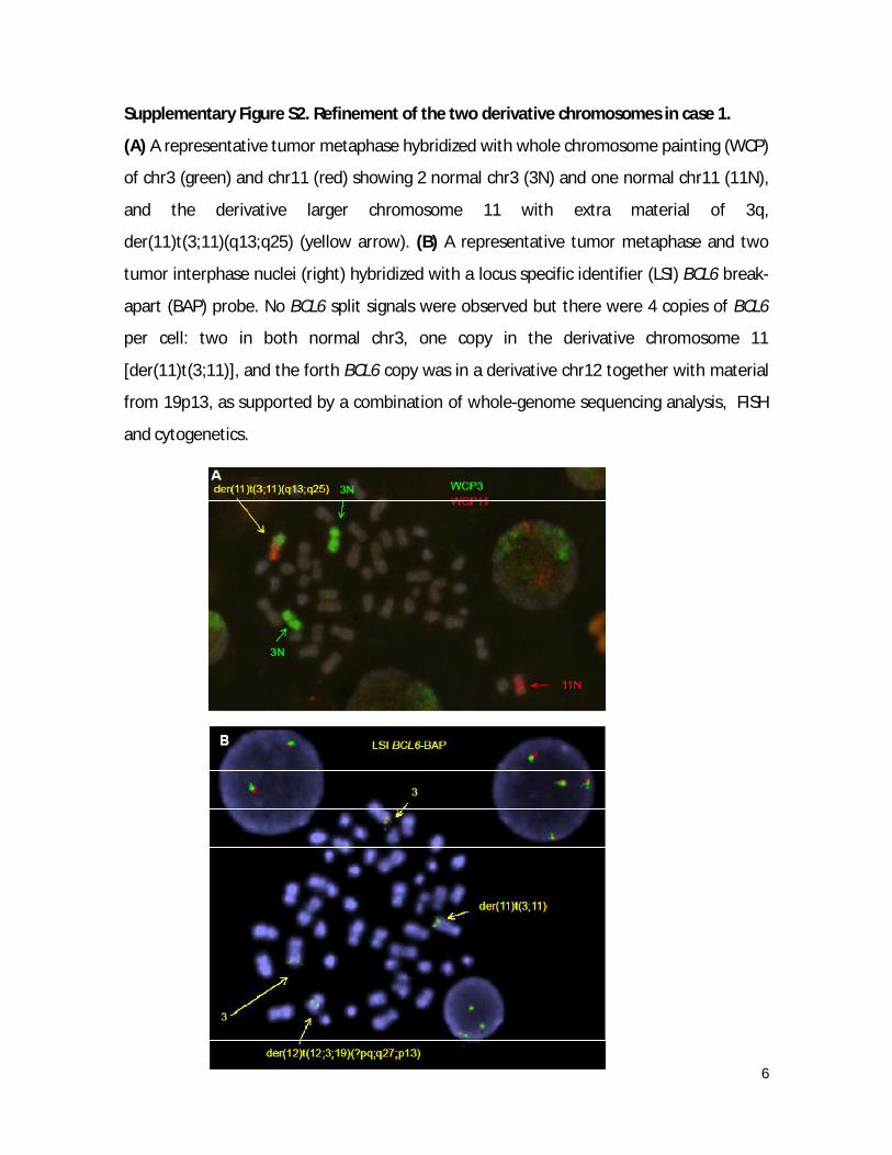
Supplementary Figure S2. Refinement of the two derivative chromosomes in case 1.
(A) A representative tumor metaphase hybridized with whole chromosome painting (WCP)
of chr3 (green) and chr11 (red) showing 2 normal chr3 (3N) and one normal chr11 (11N),
and the derivative larger chromosome 11 with extra material of 3q,
der(11)t(3;11)(q13;q25) (yellow arrow). (B) A representative tumor metaphase and two
tumor interphase nuclei (right) hybridized with a locus specific identifier (LSI) BCL6 break-
apart (BAP) probe. No BCL6 split signals were observed but there were 4 copies of BCL6
per cell: two in both normal chr3, one copy in the derivative chromosome 11
[der(11)t(3;11)], and the forth BCL6 copy was in a derivative chr12 together with material
from 19p13, as supported by a combination of whole-genome sequencing analysis, FISH
and cytogenetics.
7
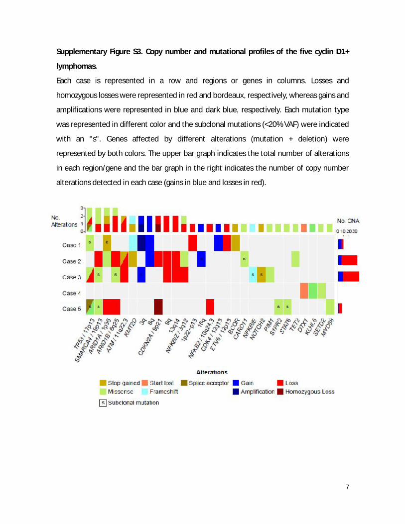
Supplementary Figure S3. Copy number and mutational profiles of the five cyclin D1+
lymphomas.
Each case is represented in a row and regions or genes in columns. Losses and
homozygous losses were represented in red and bordeaux, respectively, whereas gains and
amplifications were represented in blue and dark blue, respectively. Each mutation type
was represented in different color and the subclonal mutations (<20% VAF) were indicated
with an "s". Genes affected by different alterations (mutation + deletion) were
represented by both colors. The upper bar graph indicates the total number of alterations
in each region/gene and the bar graph in the right indicates the number of copy number
alterations detected in each case (gains in blue and losses in red).
8
SUPPLEMENTARY TABLES
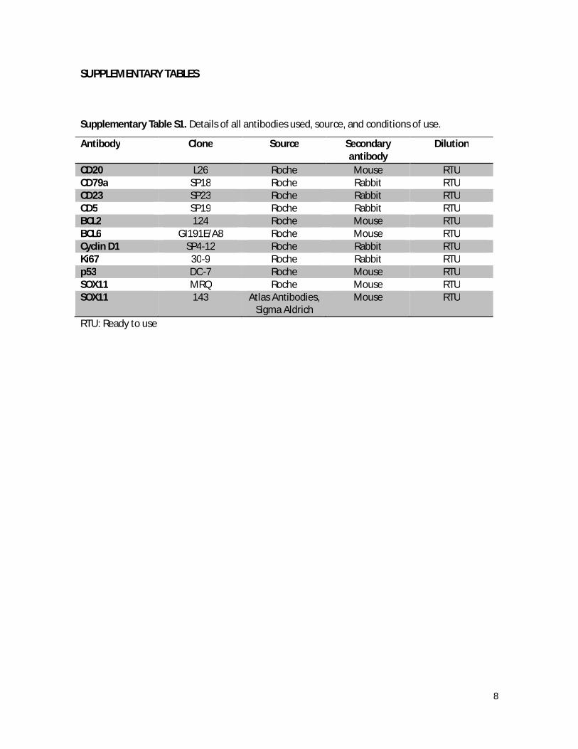
Supplementary Table S1. Details of all antibodies used, source, and conditions of use.
Antibody Clone Source Secondary antibody
Dilution
CD20 L26 Roche Mouse RTU CD79a SP18 Roche Rabbit RTU CD23 SP23 Roche Rabbit RTU CD5 SP19 Roche Rabbit RTU BCL2 124 Roche Mouse RTU BCL6 GI191E/A8 Roche Mouse RTU Cyclin D1 SP4-12 Roche Rabbit RTU Ki67 30-9 Roche Rabbit RTU p53 DO-7 Roche Mouse RTU SOX11 MRQ Roche Mouse RTU SOX11 143 Atlas Antibodies,
Sigma Aldrich Mouse RTU
RTU: Ready to use
9
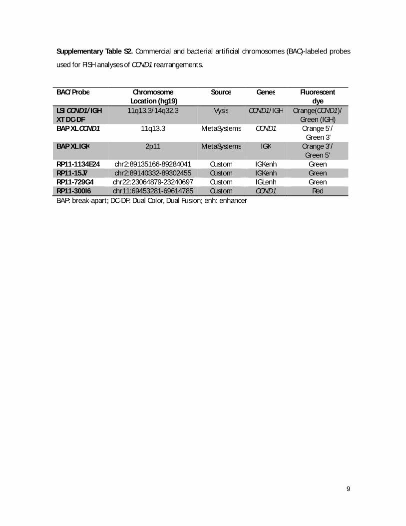
Supplementary Table S2. Commercial and bacterial artificial chromosomes (BAC)-labeled probes
used for FISH analyses of CCND1 rearrangements.
BAC/Probe Chromosome Location (hg19)
Source Genes Fluorescent dye
LSI CCND1/IGH XT DC-DF
11q13.3/14q32.3 Vysis CCND1/IGH Orange(CCND1)/ Green (IGH)
BAP XL CCND1 11q13.3 MetaSystems CCND1 Orange 5'/ Green 3'
BAP XL IGK 2p11 MetaSystems IGK Orange 3'/ Green 5'
RP11-1134E24 chr2:89135166-89284041 Custom IGKenh Green RP11-15J7 chr2:89140332-89302455 Custom IGKenh Green RP11-729G4 chr22:23064879-23240697 Custom IGLenh Green RP11-300I6 chr11:69453281-69614785 Custom CCND1 Red BAP: break-apart; DC-DF: Dual Color, Dual Fusion; enh: enhancer
10
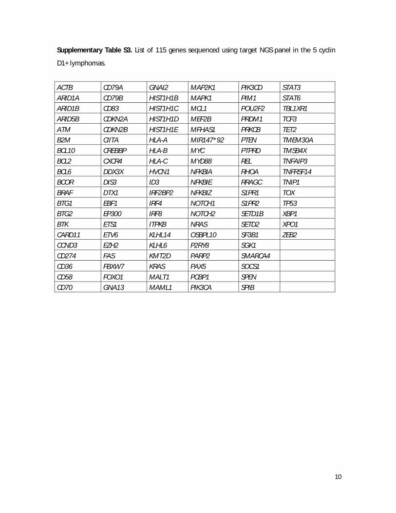
Supplementary Table S3. List of 115 genes sequenced using target NGS panel in the 5 cyclin
D1+ lymphomas.
ACTB CD79A GNAI2 MAP2K1 PIK3CD STAT3 ARID1A CD79B HIST1H1B MAPK1 PIM1 STAT6 ARID1B CD83 HIST1H1C MCL1 POU2F2 TBL1XR1 ARID5B CDKN2A HIST1H1D MEF2B PRDM1 TCF3 ATM CDKN2B HIST1H1E MFHAS1 PRKCB TET2 B2M CIITA HLA-A MIR147*92 PTEN TMEM30A BCL10 CREBBP HLA-B MYC PTPRD TMSB4X BCL2 CXCR4 HLA-C MYD88 REL TNFAIP3 BCL6 DDX3X HVCN1 NFKBIA RHOA TNFRSF14 BCOR DIS3 ID3 NFKBIE RRAGC TNIP1 BRAF DTX1 IRF2BP2 NFKBIZ S1PR1 TOX BTG1 EBF1 IRF4 NOTCH1 S1PR2 TP53 BTG2 EP300 IRF8 NOTCH2 SETD1B XBP1 BTK ETS1 ITPKB NRAS SETD2 XPO1 CARD11 ETV6 KLHL14 OSBPL10 SF3B1 ZEB2 CCND3 EZH2 KLHL6 P2RY8 SGK1 CD274 FAS KMT2D PARP2 SMARCA4 CD36 FBXW7 KRAS PAX5 SOCS1 CD58 FOXO1 MALT1 PCBP1 SPEN CD70 GNA13 MAML1 PIK3CA SPIB
11
Supplementary Table S4. Mutations identified by targeted NGS in the 5 Cyclin D1+ B-cell lymphomas.
Case Gene Chr Position (hg19)
Transcript Exon Ref Var cDNA Protein VAF (%)
Mutation type
case 1 KMT2D chr12 49444834 NM_003482.3 10/54 CAG C c.2630_2631delCT p.Pro877fs 26,8 frameshift
case 1 TP53 chr17 7578406 NM_000546.5 5/11 C T c.524G>A p.Arg175His 7 missense case 1 ARID1A chr1 27106165 NM_006015.4 20/20 G T c.5776G>T p.Gly1926* 2,5 stop_gained case 1 BCOR chrX 39932926 NM_001123385.1 4/15 G C c.1673C>G p.Ser558* 68,8 stop_gained case 2 CARD11 chr7 2984141 NM_032415.4 5/25 A C c.389T>G p.Phe130Cys 2,6 missense case 2 ARID1A chr1 27023222 NM_006015.4 1/20 A G c.328A>G p.Arg110Gly 78 missense case 2 KMT2D chr12 49426190 NM_003482.3 39/54 G A c.12298C>T p.Gln4100* 32,3 stop_gained case 2 NFKBIZ chr3 101568755 NM_031419.3 1/12 C A c.283C>A p.Gln95Lys 52,7 missense case 2 TET2 chr4 106197552 NM_001127208.2 11/11 C T c.5885C>T p.Pro1962Leu 42,6 missense case 2 ATM chr11 108137910 NM_000051.3 17/63 A T c.2479A>T p.Lys827* 50,3 stop_gained case 3 TP53 chr17 7577129 NM_000546.5 8/11 A G c.809T>C p.Phe270Ser 54 missense case 3 SMARCA4 chr19 11144110 NM_003072.3 26/35 G A c.3691G>A p.Ala1231Thr 18,1 missense case 3 PIM1 chr6 37138805 NM_001243186.1 3/6 C A c.511C>A p.Leu171Met 46,5 missense&splice case 3 NOTCH2 chr1 120458087 NM_024408.3 34/34 C A c.7258G>T p.Glu2420* 2 stop_gained case 3 ARID1B chr6 157488191 NM_020732.3 10/20 G A c.2897G>A p.Gly966Asp 1,3 missense case 3 NFKBIE chr6 44232738 NM_004556.2 1/6 TGTAA T c.759_762delTTAC p.Tyr254fs 3,2 frameshift case 4 KLHL6 chr3 183273248 NM_130446.2 1/7 A G c.194T>C p.Leu65Pro 39,3 missense case 4 KLHL6 chr3 183273251 NM_130446.2 1/7 G T c.191C>A p.Thr64Asn 39,6 missense case 4 SETD2 chr3 47144852 NM_014159.6 7/21 T A c.4901A>T p.Asn1634Ile 49,1 missense case 4 DTX1 chr12 113495999 NM_004416.2 1/9 T C c.2T>C p.Met1? 51,8 start_lost case 5 TP53 chr17 7577539 NM_000546.5 7/11 G A c.742C>T p.Arg248Trp 2,2 missense case 5 TP53 chr17 7577610 NM_000546.5 6/10 T A c.673-2A>T 4 splice_acceptor
& intron case 5 S1PR2 chr19 10334881 NM_004230.3 2/2 G A c.701C>T p.Thr234Met 1,2 missense case 5 SMARCA4 chr19 11106922 NM_003072.3 10/35 G A c.1627G>A p.Asp543Asn 2,7 missense case 5 MYD88 chr3 38182641 NM_002468.4 5/5 T C c.794T>C p.Leu265Pro 44,4 missense case 5 STAT6 chr12 57499303 NM_001178078.1 8/22 G A c.760C>T p.Arg254Trp 2,5 missense
Chr: chromosome; Ref: reference; VAF: variant allele frequency; Var: variant.
12
Supplementary Table S5. Copy number (CN) and CN neutral loss of heterozygosity (CNN-LOH) alterations in the 5 Cyclin D1+ B-cell lymphomas.
Case Chromosome Region (hg19) Event Length Cytoband Probe Median No. Probes No. Genes Case 1 chr1:95980272-119185887 CN Loss 23234551 p21.3 - p12 -0.386806935 15044 297 Case 1 chr1:204475348-206870479 CN Gain 2195132 q32.1 0.234532431 1335 62 Case 1 chr3:115931660-197962430 CN Gain 82087478 q13.31 - q29 0.298049167 51924 1009 Case 1 chr8:143764253-144237636 CN Gain 547757 q24.3 0.279687643 187 18 Case 1 chr11:116815711-119029148 CN Gain 2213438 q23.3 0.246547163 1531 91 Case 1 chr12:11733676-12185234 CN Loss 451559 p13.2 -0.365388989 501 1 Case 1 chr12:55470862-58163695 CN Gain 2692834 q13.2 - q14.1 0.248957023 1425 144 Case 1 chr14:35528384-35827142 CN Gain 298759 q13.2 0.356962919 146 7 Case 1 chr17:61169570-62926364 CN Gain 1833525 q23.3 - q24.1 0.255203933 895 67 Case 1 chr19:36815688-37322591 CN Loss 506904 q13.12 -0.383535266 302 29 Case 2 chr1:0-40,927,011 CNN-LOH 40927012 p36.33 - p34.2 0.025316545 3438 761 Case 2 chr3:109,186,681-109,992,943 CN Loss 806263 q13.13 -0.515734315 53 2 Case 2 chr6:25,919,210-26,680,732 CN Loss 761523 p22.2 -0.367333472 52 49 Case 2 chr6:26,680,732-27,106,766 CN Gain 426035 p22.2 - p22.1 0.270689368 25 6 Case 2 chr6:32,544,871-33,096,448 CN Loss 551578 p21.32 -0.560308367 50 21 Case 2 chr6:71,948,724-72,314,039 CN Loss 365316 q13 -0.45486185 25 5 Case 2 chr6:73,931,285-75,550,312 CN Loss 1619028 q13 -0.434173703 108 15 Case 2 chr6:78,820,394-79,551,342 CN Loss 730949 q14.1 -0.571523607 46 0 Case 2 chr6:85,055,355-85,337,153 CN Loss 281799 q14.3 -0.367947176 24 1 Case 2 chr6:85,616,723-87,030,056 CN Loss 1413334 q14.3 -0.41721952 87 7 Case 2 chr6:91,948,012-92,078,737 CN Loss 130726 q15 -0.847036242 9 0 Case 2 chr6:99,225,719-100,559,527 CN Loss 1333809 q16.1 - q16.2 -0.481905222 89 14 Case 2 chr6:103,850,486-104,216,465 CN Loss 365980 q16.3 -0.553430676 25 0 Case 2 chr6:104,550,864-109,732,895 CN Loss 5182032 q16.3 - q21 -0.530617893 464 36 Case 2 chr6:146,617,365-146,975,320 CN Loss 357956 q24.3 -0.519354463 43 4 Case 2 chr6:148,718,223-150,205,951 CN Loss 1487729 q24.3 - q25.1 -0.507299989 104 19 Case 2 chr6:150,770,418-155,836,923 CN Loss 5066506 q25.1 - q25.3 -0.442086697 395 27 Case 2 chr6:155,836,923-156,146,412 CN Gain 309490 q25.3 0.168327823 21 1 Case 2 chr6:156,146,412-157,902,198 CN Loss 1755787 q25.3 -0.538536727 111 6 Case 2 chr6:159,690,974-160,904,134 CN Loss 1213161 q25.3 -0.589955866 115 19
13
Case 2 chr6:161,361,947-162,274,037 CN Loss 912091 q26 -0.529157043 161 5 Case 2 chr7:25,931,287-26,615,495 CN Loss 684209 p15.2 -0.515489757 59 8 Case 2 chr8:22,392,346-23,113,124 CN Loss 720779 p21.3 -0.513352811 65 19 Case 2 chr8:82,451,644-146,364,022 CN Gain 63912379 q21.13 - q24.3 0.371620208 5413 456 Case 2 chr9:0-102,585,105 CN Loss 102585106 p24.3 - q22.33 -0.468746305 5615 622 Case 2 chr11:90,377,439-115,217,228 CN Loss 24839790 q14.3 - q23.3 -0.409956872 1902 182 Case 2 chr13:24,357,324-115,169,878 CN Loss 90812555 q12.12 - q34 -0.351244003 7567 598 Case 2 chr15:60,239,810-102,531,392 CN Gain 42291583 q22.2 - q26.3 0.209746569 3339 503 Case 2 chr16:0-34,071,208 CN Gain 34071209 p13.3 - p11.2 0.393616796 2438 700 Case 2 chr18:1-78,077,248 CN Gain 78077248 p11.32 - q23 0.079924278 5595 439 Case 2 chr19:10,594,043-14,000,572 CN Loss 3406530 p13.2 - p13.12 -0.325779736 291 125 Case 2 chr22:16,054,713-21,810,492 CN Gain 5755780 q11.1 - q11.21 0.264480948 418 127 Case 2 chr22:22,536,518-24,343,908 CN Loss 1807391 q11.22 - q11.23 -0.49614805 183 42 Case 2 chr22:26,470,387-38,094,460 CN Loss 11624074 q12.1 - q13.1 -0.212323718 876 176 Case 2 chr22:38,587,947-51,304,566 CN Loss 12716620 q13.1 - q13.33 -0.240061574 990 236 Case 3 chr1:1-121,350,750 CNN-LOH 121350750 p36.33 - p11.2 0.093750387 9730 1503 Case 3 chr1:0-19,251,659 CN Gain 19251660 p36.33 - p36.13 0.179936387 1644 381 Case 3 chr3:12,713,954-13,503,246 CN Gain 789293 p25.2 - p25.1 0.346782655 51 7 Case 3 chr3:31,222,672-46,762,047 CN Loss 15539376 p23 - p21.31 -0.426403686 1118 159 Case 3 chr3:46,762,047-49,710,139 CN Gain 2948093 p21.31 0.101522375 250 86 Case 3 chr3:49,710,139-52,314,215 CN Loss 2604077 p21.31 - p21.1 -0.428447634 312 78 Case 3 chr3:52,314,215-52,522,668 CN Gain 208454 p21.1 0.111854121 26 9 Case 3 chr3:52,522,668-67,508,358 CN Loss 14985691 p21.1 - p14.1 -0.447860241 1184 110 Case 3 chr3:67,925,592-70,599,800 CN Loss 2674209 p14.1 - p13 -0.450992733 529 14 Case 3 chr3:72,699,874-90,225,371 CN Loss 17525498 p13 - p11.1 -0.527514279 1329 46 Case 3 chr3:93,517,443-99,661,432 CN Loss 6143990 q11.1 - q12.1 -0.508259714 409 35 Case 3 chr3:101,279,182-104,072,680 CN Loss 2793499 q12.3 - q13.11 -0.505373448 184 14 Case 3 chr3:138,481,903-198,022,430 CN Gain 59540528 q22.3 - q29 0.147937119 4465 478 Case 3 chr7:79,739,785-97,648,154 CN Loss 17908370 q21.11 - q21.3 -0.348251402 1349 100 Case 3 chr7:105,772,996-152,705,396 CN Loss 46932401 q22.3 - q36.2 -0.318359673 3903 401 Case 3 chr9:1,869,321-4,379,060 CN Loss 2509740 p24.3 - p24.2 -0.320553005 167 10 Case 3 chr9:30,149,077-39,184,065 CN Loss 9034989 p21.1 - p13.1 -0.273775578 622 127 Case 3 chr9:70,984,372-72,798,245 CN Gain 1813874 q21.11 - q21.12 0.10734003 121 17
14
Case 3 chr9:72,798,245-86,897,220 CN Loss 14098976 q21.12 - q21.32 -0.230984032 989 63 Case 3 chr9:104,044,341-128,755,004 CN Loss 24710664 q31.1 - q33.3 -0.234672427 1813 207 Case 3 chr9:138,969,890-140,784,943 CN Loss 1815054 q34.3 -0.306916073 166 98 Case 3 chr11:43,672,857-45,096,829 CN Loss 1423973 p11.2 -0.235927537 129 13 Case 3 chr11:64,412,839-64,666,527 CN Loss 253689 q13.1 -0.329794824 36 13 Case 3 chr11:88,980,982-135,006,516 CN Loss 46025535 q14.3 - q25 -0.197165251 3627 444 Case 3 chr12:14,990,267-19,172,749 CN Loss 4182483 p12.3 -0.353822619 349 22 Case 3 chr12:120,614,579-133,851,895 CN Loss 13237317 q24.23 - q24.33 -0.197701231 909 179 Case 3 chr13:19,073,326-25,654,527 CN Loss 6581202 q11 - q12.13 -0.207541093 456 61 Case 3 chr13:30,294,626-59,038,774 CN Loss 28744149 q12.3 - q21.1 -0.220169753 2378 251 Case 3 chr13:59,038,774-112,003,260 CN Gain 52964487 q21.1 - q34 0.103948779 4411 242 Case 3 chr13:112,003,260-115,169,878 CN Loss 3166619 q34 -0.391424149 247 48 Case 3 chr15:22,752,399-41,435,285 CN Loss 18682887 q11.2 - q15.1 -0.471566498 1297 309 Case 3 chr15:90,742,770-98,903,641 CN Loss 8160872 q26.1 - q26.3 -0.446195483 607 55 Case 3 chr17:0-22,217,883 CN Loss 22217884 p13.3 - p11.1 -0.524609357 1746 459 Case 3 chr17:25,326,941-81,195,210 CN Gain 55868270 q11.1 - q25.3 0.225378588 4678 1130 Case 3 chr20:29,519,156-60,146,222 CN Gain 30627067 q11.21 - q13.33 0.109474625 2300 410 Case 3 chr20:62,493,150-63,025,520 CN Gain 532371 q13.33 0.270182818 37 35 Case 3 chr22:21,461,166-24,343,908 CN Loss 2882743 q11.21 - q11.23 -0.251772046 286 69 Case 3 chr22:25,961,443-29,727,979 CN Loss 3766537 q12.1 - q12.2 -0.288313538 287 41 Case 3 chr22:43,774,169-44,820,727 CN Loss 1046559 q13.2 - q13.31 -0.362747192 71 11 Case 4 No alterations Case 5 chr11:0-47,613,870 CNN-LOH 47613871 p15.5 - p11.2 -6.78E-05 3446 561 Case 5 chr1:22,971,471-31,932,838 CN Loss 8961368 p36.12 - p35.2 -0.710997403 749 168 Case 5 chr2:1-87,052,658 CN Gain 87052658 p25.3 - p11.2 0.066282526 6647 658 Case 5 chr6:103,405,413-142,794,913 CN Loss 39389501 q16.3 - q24.1 -0.69513911 3001 250 Case 5 chr9:10,997,956-21,802,107 CN Loss 10804152 p23 - p21.3 -0.702891767 848 60 Case 5 chr9:21,802,107-21,970,167 Homo. Loss 168061 p21.3 -2.311187267 66 3 Case 5 chr9:21,970,167-39,184,065 CN Loss 17213899 p21.3 - p13.1 -0.666557789 1303 152 Case 5 chr10:103,552,035-105,362,804 CN Loss 1810770 q24.32 - q24.33 -0.659816146 185 44 Case 5 chr17:41,605,296-81,195,210 CN Gain 39589915 q21.31 - q25.3 0.422790229 3112 692
15
SUPPLEMENTARY REFERENCES
1. Garcia-Herrera A, Song JY, Chuang SS et al. Nonhepatosplenic gammadelta T-cell lymphomas represent a spectrum of aggressive cytotoxic T-cell lymphomas with a mainly extranodal presentation. Am.J.Surg.Pathol. 2011;35(8):1214-1225.
2. Ventura RA, Martin-Subero JI, Jones M et al. FISH analysis for the detection of lymphoma-associated chromosomal abnormalities in routine paraffin-embedded tissue. J.Mol.Diagn. 2006;8(2):141-151.
3. ISCN 2016: McGowan-Jordan, J., Simons A., Schmid, M. ISCN: An International System for Human Cytogenomic Nomenclature (2016). S. Karger, Basel.
4. Martin-Garcia D, Navarro A, Valdes-Mas R et al. CCND2 and CCND3 hijack immunoglobulin light-chain enhancers in cyclin D1(-) mantle cell lymphoma. Blood 2019;133(9):940-951.
5. Bea S, Valdes-Mas R, Navarro A et al. Landscape of somatic mutations and clonal evolution in mantle cell lymphoma. Proc.Natl.Acad.Sci.U.S.A 2013;110(45):18250-18255.
6. Moncunill V, Gonzalez S, Bea S et al. Comprehensive characterization of complex structural variations in cancer by directly comparing genome sequence reads. Nat.Biotechnol. 2014;32(11):1106-1112.
7. Layer RM, Chiang C, Quinlan AR, Hall IM. LUMPY: a probabilistic framework for structural variant discovery. Genome Biol. 2014;15(6):R84.
8. Puente XS, Pinyol M, Quesada V et al. Whole-genome sequencing identifies recurrent mutations in chronic lymphocytic leukaemia. Nature 2011;475 (7354):101-105.
9. Nadeu F, Delgado J, Royo C et al. Clinical impact of clonal and subclonal TP53, SF3B1, BIRC3, NOTCH1, and ATM mutations in chronic lymphocytic leukemia. Blood 2016;127(17):2122-2130.
10. Puente XS, Bea S, Valdes-Mas R et al. Non-coding recurrent mutations in chronic lymphocytic leukaemia. Nature 2015;526(7574):519-524.