Embed Size (px)
Citation preview
University of Groningen
Elapid Snake Venom Analyses Show the Specificity of the Peptide Composition at the Levelof Genera Naja and NotechisMunawar, Aisha; Trusch, Maria; Georgieva, Dessislava; Hildebrand, Diana; Kwiatkowski,Marcel; Behnken, Henning; Harder, Soenke; Arni, Raghuvir; Spencer, Patrick; Schlueter,HartmutPublished in:Toxins
DOI:10.3390/toxins6030850
IMPORTANT NOTE: You are advised to consult the publisher's version (publisher's PDF) if you wish to cite fromit. Please check the document version below.
Document VersionPublisher's PDF, also known as Version of record
Publication date:2014
Link to publication in University of Groningen/UMCG research database
Citation for published version (APA):Munawar, A., Trusch, M., Georgieva, D., Hildebrand, D., Kwiatkowski, M., Behnken, H., Harder, S., Arni,R., Spencer, P., Schlueter, H., & Betzel, C. (2014). Elapid Snake Venom Analyses Show the Specificity ofthe Peptide Composition at the Level of Genera Naja and Notechis. Toxins, 6(3), 850-868.https://doi.org/10.3390/toxins6030850
CopyrightOther than for strictly personal use, it is not permitted to download or to forward/distribute the text or part of it without the consent of theauthor(s) and/or copyright holder(s), unless the work is under an open content license (like Creative Commons).
The publication may also be distributed here under the terms of Article 25fa of the Dutch Copyright Act, indicated by the “Taverne” license.More information can be found on the University of Groningen website: https://www.rug.nl/library/open-access/self-archiving-pure/taverne-amendment.
Take-down policyIf you believe that this document breaches copyright please contact us providing details, and we will remove access to the work immediatelyand investigate your claim.
Toxins 2014, 6, 850-868; doi:10.3390/toxins6030850
toxins ISSN 2072-6651
www.mdpi.com/journal/toxins
Article
Elapid Snake Venom Analyses Show the Specificity of the Peptide Composition at the Level of Genera Naja and Notechis
Aisha Munawar 1,2, Maria Trusch 3, Dessislava Georgieva 1, Diana Hildebrand 4,
Marcel Kwiatkowski 4, Henning Behnken 3, Sönke Harder 4, Raghuvir Arni 5, Patrick Spencer 6,
Hartmut Schlüter 4 and Christian Betzel 1,*
1 Laboratory of Structural Biology of Infection and Inflammation, Institute of Biochemistry and
Molecular Biology, University of Hamburg, c/o DESY, Notkestreet 85, Building 22a,
Hamburg 22603, Germany; E-Mails: [email protected] (A.M.);
[email protected] (D.G.) 2 Department of Chemistry, University of Engineering & Technology, G.T. Road, Lahore 54890,
Pakistan 3 Institute of Organic Chemistry, University of Hamburg, Martin-Luther-King-Platz 6,
Hamburg 20146, Germany; E-Mails: [email protected] (M.T.);
[email protected] (H.B.) 4 Institute of Clinical Chemistry, University Medical Centre Hamburg-Eppendorf (UKE),
Martinistraße 52, Hamburg 20246, Germany; E-Mails: [email protected] (D.H.);
[email protected] (M.K); [email protected] (S.H.); [email protected] (H.S.)
5 Department of Physics, IBILCE/UNESP, Rua Cristóvão Colombo 2265, São José do Rio Preto
CEP 15054-000, SP Brazil; E-Mail: [email protected] 6 Centro de Biotecnologia, Instituto de Pesquisas Energéticas e Nucleares, Avenue Lineu Prestes 2242,
São Paulo 05508-000, Brazil; E-Mail: [email protected]
* Author to whom correspondence should be addressed; E-Mail: [email protected];
Tel.: +49-4089-984744; Fax: +49-4089-984747.
Received: 3 November 2013; in revised form: 24 January 2014 / Accepted: 5 February 2014 /
Published: 28 February 2014
Abstract: Elapid snake venom is a highly valuable, but till now mainly unexplored, source
of pharmacologically important peptides. We analyzed the peptide fractions with molecular
masses up to 10 kDa of two elapid snake venoms—that of the African cobra,
N. m. mossambica (genus Naja), and the Peninsula tiger snake, N. scutatus, from Kangaroo
Island (genus Notechis). A combination of chromatographic methods was used to isolate the
peptides, which were characterized by combining complimentary mass spectrometric
OPEN ACCESS
Toxins 2014, 6 851
techniques. Comparative analysis of the peptide compositions of two venoms showed
specificity at the genus level. Three-finger (3-F) cytotoxins, bradykinin-potentiating peptides
(BPPs) and a bradykinin inhibitor were isolated from the Naja venom. 3-F neurotoxins,
Kunitz/basic pancreatic trypsin inhibitor (BPTI)-type inhibitors and a natriuretic peptide
were identified in the N. venom. The inhibiting activity of the peptides was confirmed
in vitro with a selected array of proteases. Cytotoxin 1 (P01467) from the Naja venom
might be involved in the disturbance of cellular processes by inhibiting the cell
20S-proteasome. A high degree of similarity between BPPs from elapid and viperid snake
venoms was observed, suggesting that these molecules play a key role in snake venoms
and also indicating that these peptides were recruited into the snake venom prior to the
evolutionary divergence of the snakes.
Keywords: Naja mossambica mossambica; Notechis scutatus from Kangaroo Island;
pharmacologically active peptides; snake venom; cytotoxin; neurotoxin; natriuretic
peptides; Kunitz-type inhibitor; bradykinin-potentiating peptides
1. Introduction
Elapid snake venoms contain a large number of pharmacologically active peptides, influencing
important physiological functions, like blood coagulation and the cardiovascular and nervous
systems [1]. Venom peptides are a rich and potent source of the prototypes of novel drugs. Their high
target specificity, structural stability, relative ease of chemical synthesis and the possibility to improve
the drug efficacy by chemical modifications are very suitable for pharmaceutical application and for
the design of novel medicines. In this connection, the partnership of the Australian biopharmaceutical
company, QRxPharma Ltd (North Sydney, Australia), and its subsidiary, Venomics Pty Ltd (VPL)
(North Sydney, Australia), with the University of Queensland for the development of novel drug
prototypes from elapid snake venom is a remarkable example of collaboration between science and the
pharmaceutical industry. As a result, three novel compounds acting on the hemostatic system are in
preclinical development [2,3]. There exists a number of other examples of the successful application of
the knowledge about venom peptide structure and function for pharmaceutical purposes. Probably the
most impressive example is the development of the well-known anti-hypertensive drug, Captopril® [4]
and other derivatives [5]—angiotensin-converting enzyme (ACE) inhibitors designed on the basis of
the B. jararaca venom peptide structure. The necessity of new generation medicines and the
application of the venom peptide structure for drug design are discussed in a review published
recently [6]. Venom peptides are a novel alternative to a number of contemporary existing drugs. For
this reason, further studies on the snake venom peptidome are of pharmaceutical and clinical
significance. At present, there is a demand to develop a new generation of anti-hypertensive drugs
without or with lesser side effects. This can be achieved by selective blocking of one of the two
domains of ACE [7]. Snake venom peptide structures can serve as models for respective drug design
investigations. The crystal structure of BPPb (snake venom bradykinin-potentiating peptide, a selective
inhibitor of the C-domain of ACE) in complex with the C-domain of human ACE illustrates that the
Toxins 2014, 6 852
inhibitor binds at the active site in a Zn-independent manner, revealing new modes of active site
interactions compared to the so far described ACE-inhibitor complex structures [8].
This study describes a comparative venom peptide analysis of two elapid snakes: Naja mossambica
mossambica (representative of the genus, Naja) and N. scutatus from Kangaroo Island (genus Notechis).
Both snakes are widely recognized species of the genera, Naja and Notechis. Naja species belong to the
most widespread group of snakes known as cobras, found in Africa and Asia. Notechis (tiger snakes) is a
large group of snakes distributed in Australia. They are among the most venomous in the world [9].
2. Results
2.1. Purification and Identification of Peptides from N. m. mossambica Venom
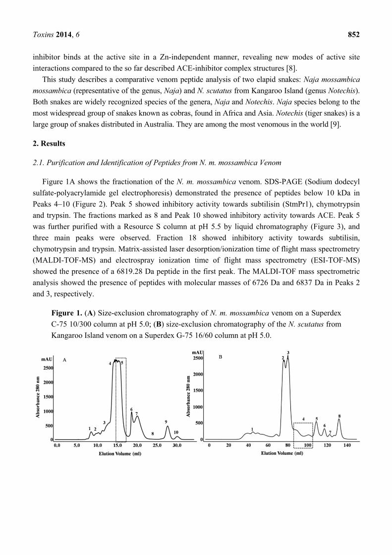
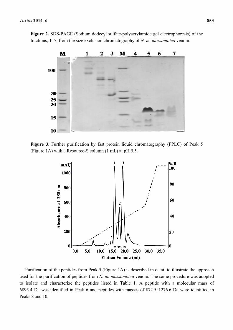
Figure 1A shows the fractionation of the N. m. mossambica venom. SDS-PAGE (Sodium dodecyl
sulfate-polyacrylamide gel electrophoresis) demonstrated the presence of peptides below 10 kDa in
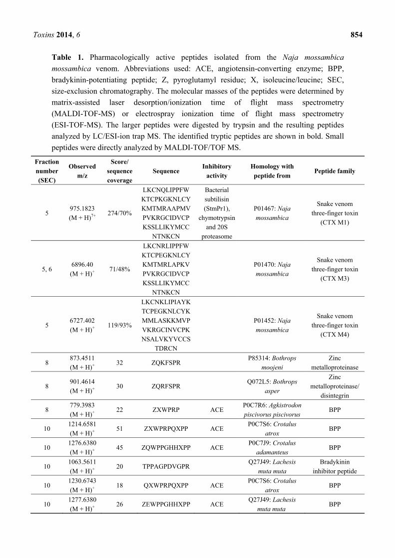
Peaks 4–10 (Figure 2). Peak 5 showed inhibitory activity towards subtilisin (StmPr1), chymotrypsin
and trypsin. The fractions marked as 8 and Peak 10 showed inhibitory activity towards ACE. Peak 5
was further purified with a Resource S column at pH 5.5 by liquid chromatography (Figure 3), and
three main peaks were observed. Fraction 18 showed inhibitory activity towards subtilisin,
chymotrypsin and trypsin. Matrix-assisted laser desorption/ionization time of flight mass spectrometry
(MALDI-TOF-MS) and electrospray ionization time of flight mass spectrometry (ESI-TOF-MS)
showed the presence of a 6819.28 Da peptide in the first peak. The MALDI-TOF mass spectrometric
analysis showed the presence of peptides with molecular masses of 6726 Da and 6837 Da in Peaks 2
and 3, respectively.
Figure 1. (A) Size-exclusion chromatography of N. m. mossambica venom on a Superdex
C-75 10/300 column at pH 5.0; (B) size-exclusion chromatography of the N. scutatus from
Kangaroo Island venom on a Superdex G-75 16/60 column at pH 5.0.
Toxins 2014, 6 853
Figure 2. SDS-PAGE (Sodium dodecyl sulfate-polyacrylamide gel electrophoresis) of the
fractions, 1–7, from the size exclusion chromatography of N. m. mossambica venom.
Figure 3. Further purification by fast protein liquid chromatography (FPLC) of Peak 5
(Figure 1A) with a Resource-S column (1 mL) at pH 5.5.
Purification of the peptides from Peak 5 (Figure 1A) is described in detail to illustrate the approach
used for the purification of peptides from N. m. mossambica venom. The same procedure was adopted
to isolate and characterize the peptides listed in Table 1. A peptide with a molecular mass of
6895.4 Da was identified in Peak 6 and peptides with masses of 872.5–1276.6 Da were identified in
Peaks 8 and 10.
Toxins 2014, 6 854
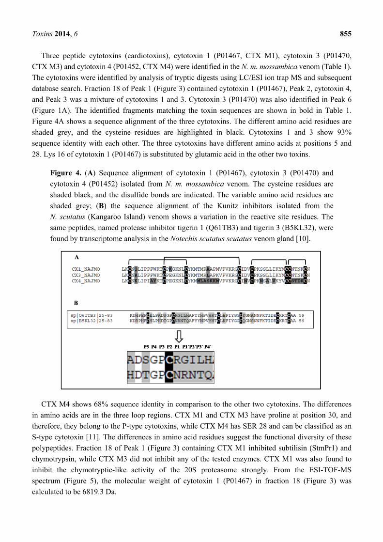
Table 1. Pharmacologically active peptides isolated from the Naja mossambica
mossambica venom. Abbreviations used: ACE, angiotensin-converting enzyme; BPP,
bradykinin-potentiating peptide; Z, pyroglutamyl residue; X, isoleucine/leucine; SEC,
size-exclusion chromatography. The molecular masses of the peptides were determined by
matrix-assisted laser desorption/ionization time of flight mass spectrometry
(MALDI-TOF-MS) or electrospray ionization time of flight mass spectrometry
(ESI-TOF-MS). The larger peptides were digested by trypsin and the resulting peptides
analyzed by LC/ESI-ion trap MS. The identified tryptic peptides are shown in bold. Small
peptides were directly analyzed by MALDI-TOF/TOF MS.
Fraction
number
(SEC)
Observed
m/z
Score/
sequence
coverage
Sequence Inhibitory
activity
Homology with
peptide from Peptide family
5 975.1823
(M + H)7+ 274/70%
LKCNQLIPPFW
KTCPKGKNLCY
KMTMRAAPMV
PVKRGCIDVCP
KSSLLIKYMCC
NTNKCN
Bacterial
subtilisin
(StmPr1),
chymotrypsin
and 20S
proteasome
P01467: Naja
mossambica
Snake venom
three-finger toxin
(CTX M1)
5, 6 6896.40
(M + H)+ 71/48%
LKCNRLIPPFW
KTCPEGKNLCY
KMTMRLAPKV
PVKRGCIDVCP
KSSLLIKYMCC
NTNKCN
P01470: Naja
mossambica
Snake venom
three-finger toxin
(CTX M3)
5 6727.402
(M + H)+ 119/93%
LKCNKLIPIAYK
TCPEGKNLCYK
MMLASKKMVP
VKRGCINVCPK
NSALVKYVCCS
TDRCN
P01452: Naja
mossambica
Snake venom
three-finger toxin
(CTX M4)
8 873.4511
(M + H)+ 32 ZQKFSPR P85314: Bothrops
moojeni
Zinc
metalloproteinase
8 901.4614
(M + H)+ 30 ZQRFSPR Q072L5: Bothrops
asper
Zinc
metalloproteinase/
disintegrin
8 779.3983
(M + H)+ 22 ZXWPRP ACE P0C7R6: Agkistrodon
piscivorus piscivorus BPP
10 1214.6581
(M + H)+ 51 ZXWPRPQXPP ACE
P0C7S6: Crotalus
atrox BPP
10 1276.6380
(M + H)+ 45 ZQWPPGHHXPP ACE
P0C7J9: Crotalus
adamanteus BPP
10 1063.5611
(M + H)+ 20 TPPAGPDVGPR
Q27J49: Lachesis
muta muta
Bradykinin
inhibitor peptide
10 1230.6743
(M + H)+ 18 QXWPRPQXPP ACE
P0C7S6: Crotalus
atrox BPP
10 1277.6380
(M + H)+ 26 ZEWPPGHHXPP ACE
Q27J49: Lachesis
muta muta BPP
Toxins 2014, 6 855
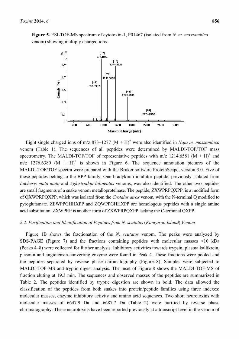
Three peptide cytotoxins (cardiotoxins), cytotoxin 1 (P01467, CTX M1), cytotoxin 3 (P01470,
CTX M3) and cytotoxin 4 (P01452, CTX M4) were identified in the N. m. mossambica venom (Table 1).
The cytotoxins were identified by analysis of tryptic digests using LC/ESI ion trap MS and subsequent
database search. Fraction 18 of Peak 1 (Figure 3) contained cytotoxin 1 (P01467), Peak 2, cytotoxin 4,
and Peak 3 was a mixture of cytotoxins 1 and 3. Cytotoxin 3 (P01470) was also identified in Peak 6
(Figure 1A). The identified fragments matching the toxin sequences are shown in bold in Table 1.
Figure 4A shows a sequence alignment of the three cytotoxins. The different amino acid residues are
shaded grey, and the cysteine residues are highlighted in black. Cytotoxins 1 and 3 show 93%
sequence identity with each other. The three cytotoxins have different amino acids at positions 5 and
28. Lys 16 of cytotoxin 1 (P01467) is substituted by glutamic acid in the other two toxins.
Figure 4. (A) Sequence alignment of cytotoxin 1 (P01467), cytotoxin 3 (P01470) and
cytotoxin 4 (P01452) isolated from N. m. mossambica venom. The cysteine residues are
shaded black, and the disulfide bonds are indicated. The variable amino acid residues are
shaded grey; (B) the sequence alignment of the Kunitz inhibitors isolated from the
N. scutatus (Kangaroo Island) venom shows a variation in the reactive site residues. The
same peptides, named protease inhibitor tigerin 1 (Q61TB3) and tigerin 3 (B5KL32), were
found by transcriptome analysis in the Notechis scutatus scutatus venom gland [10].
CTX M4 shows 68% sequence identity in comparison to the other two cytotoxins. The differences
in amino acids are in the three loop regions. CTX M1 and CTX M3 have proline at position 30, and
therefore, they belong to the P-type cytotoxins, while CTX M4 has SER 28 and can be classified as an
S-type cytotoxin [11]. The differences in amino acid residues suggest the functional diversity of these
polypeptides. Fraction 18 of Peak 1 (Figure 3) containing CTX M1 inhibited subtilisin (StmPr1) and
chymotrypsin, while CTX M3 did not inhibit any of the tested enzymes. CTX M1 was also found to
inhibit the chymotryptic-like activity of the 20S proteasome strongly. From the ESI-TOF-MS
spectrum (Figure 5), the molecular weight of cytotoxin 1 (P01467) in fraction 18 (Figure 3) was
calculated to be 6819.3 Da.
Toxins 2014, 6 856
Figure 5. ESI-TOF-MS spectrum of cytotoxin-1, P01467 (isolated from N. m. mossambica
venom) showing multiply charged ions.
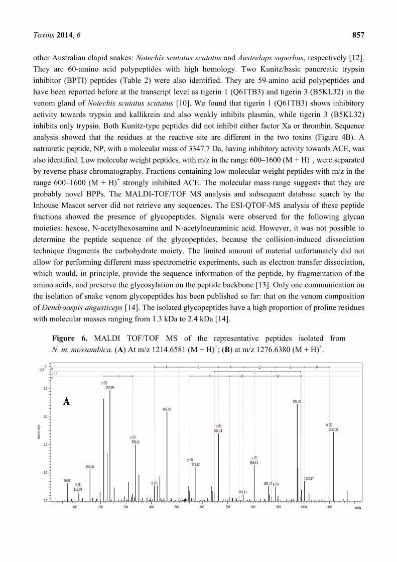
Eight single charged ions of m/z 873–1277 (M + H)+ were also identified in Naja m. mossambica
venom (Table 1). The sequences of all peptides were determined by MALDI-TOF/TOF mass
spectrometry. The MALDI-TOF/TOF of representative peptides with m/z 1214.6581 (M + H)+ and
m/z 1276.6380 (M + H)+ is shown in Figure 6. The sequence annotation pictures of the
MALDI-TOF/TOF spectra were prepared with the Bruker software ProteinScape, version 3.0. Five of
these peptides belong to the BPP family. One bradykinin inhibitor peptide, previously isolated from
Lachesis muta muta and Agkistrodon bilineatus venoms, was also identified. The other two peptides
are small fragments of a snake venom metalloproteinase. The peptide, ZXWPRPQXPP, is a modified form
of QXWPRPQXPP, which was isolated from the Crotalus atrox venom, with the N-terminal Q modified to
pyroglutamate. ZEWPPGHHXPP and ZQWPPGHHXPP are homologous peptides with a single amino
acid substitution. ZXWPRP is another form of ZXWPRPQXPP lacking the C-terminal QXPP.
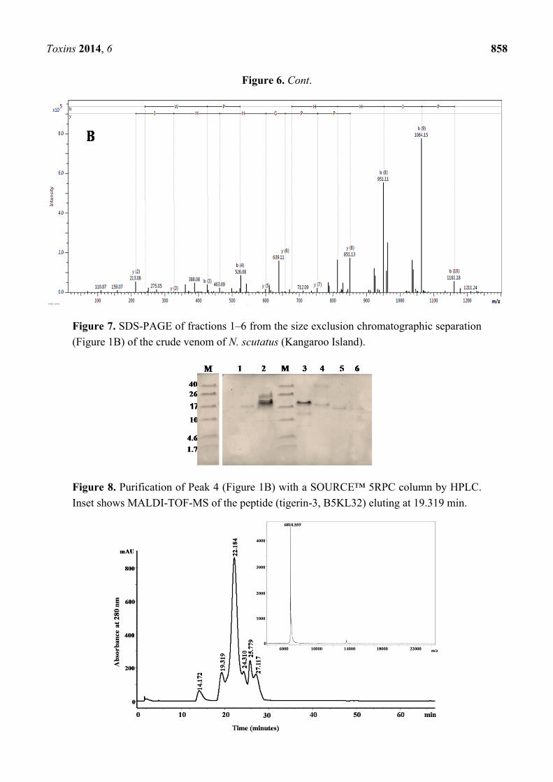
2.2. Purification and Identification of Peptides from N. scutatus (Kangaroo Island) Venom
Figure 1B shows the fractionation of the N. scutatus venom. The peaks were analyzed by
SDS-PAGE (Figure 7) and the fractions containing peptides with molecular masses <10 kDa
(Peaks 4–8) were collected for further analysis. Inhibitory activities towards trypsin, plasma kallikrein,
plasmin and angiotensin-converting enzyme were found in Peak 4. These fractions were pooled and
the peptides separated by reverse phase chromatography (Figure 8). Samples were subjected to
MALDI-TOF-MS and tryptic digest analysis. The inset of Figure 8 shows the MALDI-TOF-MS of
fraction eluting at 19.3 min. The sequences and observed masses of the peptides are summarized in
Table 2. The peptides identified by tryptic digestion are shown in bold. The data allowed the
classification of the peptides from both snakes into protein/peptide families using three indexes:
molecular masses, enzyme inhibitory activity and amino acid sequences. Two short neurotoxins with
molecular masses of 6647.9 Da and 6687.7 Da (Table 2) were purified by reverse phase
chromatography. These neurotoxins have been reported previously at a transcript level in the venom of
Toxins 2014, 6 857
other Australian elapid snakes: Notechis scutatus scutatus and Austrelaps superbus, respectively [12].
They are 60-amino acid polypeptides with high homology. Two Kunitz/basic pancreatic trypsin
inhibitor (BPTI) peptides (Table 2) were also identified. They are 59-amino acid polypeptides and
have been reported before at the transcript level as tigerin 1 (Q61TB3) and tigerin 3 (B5KL32) in the
venom gland of Notechis scutatus scutatus [10]. We found that tigerin 1 (Q61TB3) shows inhibitory
activity towards trypsin and kallikrein and also weakly inhibits plasmin, while tigerin 3 (B5KL32)
inhibits only trypsin. Both Kunitz-type peptides did not inhibit either factor Xa or thrombin. Sequence
analysis showed that the residues at the reactive site are different in the two toxins (Figure 4B). A
natriuretic peptide, NP, with a molecular mass of 3347.7 Da, having inhibitory activity towards ACE, was
also identified. Low molecular weight peptides, with m/z in the range 600–1600 (M + H)+, were separated
by reverse phase chromatography. Fractions containing low molecular weight peptides with m/z in the
range 600–1600 (M + H)+ strongly inhibited ACE. The molecular mass range suggests that they are
probably novel BPPs. The MALDI-TOF/TOF MS analysis and subsequent database search by the
Inhouse Mascot server did not retrieve any sequences. The ESI-QTOF-MS analysis of these peptide
fractions showed the presence of glycopeptides. Signals were observed for the following glycan
moieties: hexose, N-acetylhexosamine and N-acetylneuraminic acid. However, it was not possible to
determine the peptide sequence of the glycopeptides, because the collision-induced dissociation
technique fragments the carbohydrate moiety. The limited amount of material unfortunately did not
allow for performing different mass spectrometric experiments, such as electron transfer dissociation,
which would, in principle, provide the sequence information of the peptide, by fragmentation of the
amino acids, and preserve the glycosylation on the peptide backbone [13]. Only one communication on
the isolation of snake venom glycopeptides has been published so far: that on the venom composition
of Dendroaspis angusticeps [14]. The isolated glycopeptides have a high proportion of proline residues
with molecular masses ranging from 1.3 kDa to 2.4 kDa [14].
Figure 6. MALDI TOF/TOF MS of the representative peptides isolated from
N. m. mossambica. (A) At m/z 1214.6581 (M + H)+; (B) at m/z 1276.6380 (M + H)+.
Toxins 2014, 6 858
Figure 6. Cont.
Figure 7. SDS-PAGE of fractions 1–6 from the size exclusion chromatographic separation
(Figure 1B) of the crude venom of N. scutatus (Kangaroo Island).
Figure 8. Purification of Peak 4 (Figure 1B) with a SOURCE™ 5RPC column by HPLC.
Inset shows MALDI-TOF-MS of the peptide (tigerin-3, B5KL32) eluting at 19.319 min.
Toxins 2014, 6 859
Table 2. Pharmacologically active peptides isolated from N. scutatus venom.
Abbreviations used: ACE, angiotensin-converting enzyme; NP, natriuretic peptide; X,
Leucine/isoleucine. All peptides were purified from Peak 4 of Figure 1B. The molecular
masses of the peptides were determined by MALDI-TOF-MS. Tryptic peptides of larger
peptides were analyzed by LC/ESI-ion trap MS. The peptides identified by tryptic digestion
are shown in bold. Fractions containing glycopeptides were analyzed by ESI-Q-TOF MS.
Observed
m/z
(M + H)+
Score/sequence
coverage Sequence determined
Inhibitory
activity
Homology with
peptide from
Peptide
family
6647.921* 185/44%
MTCCNQQSSQPKTTTT
CAESSCYKKTWRDHRG
TITERGCGCPNVKPGVQ
INCCKTDECNN
A8HDK0: Notechis
scutatus scutatus
Snake venom
three-finger
toxin
6687.729 121/27%
MTCCNQQSSQPKTTTT
CAESSCYKKTWRDHRG
TIIERGCGCPNVKPGIQL
VCCETNECNN
A8S6A4: Austrelaps
superbus
Snake venom
three-finger
toxin
6810.559 1569/65%
KDRPHFCHLPHDTGPC
NRNTQAFYYNPVYHTC
LKFIYGGCQGNSNNFK
TIDECKRTCAA
Trypsin B5KL32: Notechis
scutatus scutatus Kunitz/BPTI
6694.233 231/45%
KDHPEFCELPADSGPCR
GILHAFYYHPVHRTCLE
FIYGGCYGNANNFKTID
ECKRTCAA
Trypsin,
plasma
kallikrein,
plasmin
Q6ITB3: Notechis
scutatus scutatus Kunitz/BPTI
3347.688 54/30% SGSEVAKIGDGCFGLPL
DRIGSASGMGCRSVPKPACE
Q3SAE8: Notechis
scutatus scutatus NP
1154.556 QNPPKPXSGES Q3SAX8: Oxyuranus
scutellatus scutellatus NP
764.201
(M + H)2+ Glycopeptide
681.251 Glycopeptide
Notes: * The mass measured by us by MALDI mass spectrometry is 6647.921 (M + H)+, whereas the calculated one is
6639.4 Da. Clearly, there is a discrepancy between the calculated and the measured mass. The identity of A8HDK0 was
obtained by analyzing tryptic peptides with LC-MS followed by a Mascot search. Since the Mascot score for the
identification of A8HDK0 was reasonable, it can be assumed that this protein was present in the corresponding fraction.
Several reasons are possible, also in combination, which may explain the discrepancy of the measured and calculated
mass: 1. We obtained a sequence coverage of 44%. Thus, we do not have any information about the rest of the protein.
The missing peptides may include posttranslational modifications, which also might explain why we missed these
peptides in the search result of MASCOT. 2. An exchange of amino acids cannot be excluded. 3. Mistakes in the protein
database can also not be excluded. The same explanation can be taken into account for other mass differences.
3. Discussion
Elapid species of the superfamily Colubroidea [15] are among the most toxic snakes of the world ([2,9]
and the references therein). However, their venoms are poorly known from a toxicological and drug
Toxins 2014, 6 860
discovery point of view [2]. The analyses of the N. m. mossambica and N. scutatus venom peptide
compositions revealed specificity at the genus level. The Naja m. mossambica venom peptidome
contains cytotoxic-type three-finger toxins, bradykinin-potentiating peptides and a bradykinin
inhibitor, but lacks Kunitz/BPTI inhibitors. At the same time, the peptide fraction analysis of
N. scutatus venom revealed the presence of Kunitz/BPTI inhibitors, neurotoxic-type three-finger toxins
and natriuretic peptides. Neurotoxins have been isolated from N. m. mossambica venom [16]. We did
not find representatives of this group of toxins in the venom of the snake mentioned above. It is known
that the venom composition depends on the diet of the snake, the geographical region, ontogenetic
variations, etc. ([17] and the references therein). Furthermore, very low quantities of the respective
toxins in the venom can prevent their identification. α-neurotoxins make up only 0.4%–12.6% of the
venom proteins of the Naja species [18].
The Naja species possesses cardiotoxins, 60–62 residue basic peptides, pharmacologically distinct
from the so-called “short neurotoxins” [19,20]. Cytotoxins predominate in Naja venom toxins and
constitute 72.8% of the Naja nigricollis total venom proteins [18]. The three-finger toxins found in
elapid venoms constitute a superfamily of non-enzymatic polypeptides, consisting of 60–74 amino
acids [1,21]. Three-finger cardiotoxins have been reported exclusively in cobra venom. These toxins
form ion pores in the lipid membranes [22]. The structure of CTXs was investigated by
crystallographic [19,23] and NMR [24] methods. The cytotoxin 1 (P01467) that we have isolated from
Naja. m. mossambica venom (Table 1) shows inhibitory activity towards the 20S proteasome, a 700 kDa
multi-catalytic complex constituting the proteolytic core of the 26S proteasome complex. The complex
possesses three active sites: chymotrypsin-like, trypsin-like and peptidyl-glutamyl peptide hydrolyzing
(PGPH)-like ([25] and the references therein). Proteasomes are present in the cell nucleus and
cytoplasm, and their main function is to maintain the concentration of specific proteins and to degrade
damaged, oxidized or misfolded proteins [26]. The cells use ubiquitin-proteasome systems to maintain
cellular homeostasis [27]. It can be supposed that after envenomation, the Naja m. mossambica venom
cytotoxin 1 (P01467) might be involved in the disturbance of cellular processes by inhibiting a vital
catalytic machinery of the cell, the 20S proteasome. Inhibition of this complex could be one of the
mechanisms involved in cytotoxin-mediated cell death. Cytotoxin 1 (P01467) can serve as a promising
candidate for cancer therapy, as the proteasome is an important target for the treatment of
cancer [28,29]. Already, CTX III isolated from Naja naja atra venom was reported to have anticancer
activity against human breast cancer cells [30].
The N. scutatus peptidome contains three-finger α-neurotoxins (Table 2). Neurotoxins are
responsible for neurological effects as a result of envenomation by Australian snakes [12]. They inhibit
nicotinic acetylcholine receptors (nAChRs) [12,17].
The snakes use venom hypotensive peptides to weaken their prey. Bradykinin-potentiating peptides
are natural inhibitors of angiotensin converting enzymes, which play a key role in blood pressure
regulation [5]. The pharmaceutical industry developed a large number of antihypertensive drugs, like
captopril and other derivatives, designed using the BPP structure [7]. However the synthetic ACE
inhibitors, used at present, cause serious side effects, which could be ascribed to their inability to
discriminate between the C- and N- domains of the enzyme. C-domain-specific inhibitors could have
the same effect as the currently available drugs, but with low or no side effects, due to decreased
bradykinin and substance P levels [5]. The structure of novel BPPs, like that identified in
Toxins 2014, 6 861
N m. mossambica venom (Table 1), might help to design more specific C-domain ACE inhibitors,
since this region of the enzyme is the major site of angiotensin-1 processing. It was demonstrated by
Fry and Wüster [31] that snake venom toxins evolved from the recruitment of body proteins into the
venom. Comparison of BPPs from elapid (this work) and viperid [32] snake venoms showed a high
similarity, suggesting the recruitment of these peptides into the snake venom before the split of the two
families. A possible reason for this is the important role of BPPs in prey immobilization. Hypotension
is usually associated with poisoning upon snake envenomation [33–35]. Natriuretic peptides (NPs),
like that described in Table 2, have profound effects on the cardiovascular system [36]. Their
structures are promising for the design of novel medicines with vasodilator, natriuretic and diuretic
properties [37]. The physiological role of the Kunitz/BPTIs (bovine pancreatic trypsin inhibitors) in
snake venom is not completely understood. It was proposed that they participate in the processes of
coagulation, fibrinolysis and inflammation through protease inactivation [38]. The sequence analysis
of the two Kunitz inhibitors, isolated from the venom of N. scutatus, showed that the reactive site
residues are different in the two peptides. The P1 residue is considered to be the most critical site
defining the specificity and inhibitory interaction with serine proteases [39,40]. The amino acid
sequence of the Kunitz/BPTI inhibitors, isolated from N. scutatus venom, was found also by analyzing
the transcriptome of the Notechis scutatus scutatus venom gland [10]. The sequences are compared in
Figure 4B. The amino acid residues at position P1 of the two inhibitors are different: a positively
charged Arg in the first peptide and a neutral Asn in the second (Figure 4B). Other amino acids at the
primary binding loop (P3P2P1P1′P2′P3′P4′) can also interact with proteolytic enzymes [41]. The
sequences of this loop in the two inhibitors are quite different, PCRGILH and TGPCNRNTQ,
respectively. Previous studies have shown that at least one of the residues at P2′ or P4′ should have a
basic histidine residue to form a complex with kallikrein ([42] and the references therein). The first
polypeptide has a histidine at the P4′ position (Figure 4B), and this explains its inhibitory activity
towards kallikrein, as we have observed (Table 2). The second has a glutamine at the same position
(Figure 4B), and we did not observe the inhibition of kallikrein by this peptide. It can be concluded
that the different amino acid residues at the primary binding loop of both Kunitz/BPTI inhibitors are
responsible for their inhibitory specificity towards serine proteases. A search in the UniProt database
indicated that the sequence motif, NRN, at P1P1′P2′ position was found only in the Notechis scutatus
scutatus venom Kunitz inhibitor, a species related to N.scutatus. The observed high affinity of the
isolated polypeptide inhibitor towards kallikrein could be used for the design of specific inhibitors of
plasma kallikrein to treat diseases, such as hereditary angioedema and septicemia, related to the
increased blood levels of this enzyme [43].
4. Experimental Section
4.1. Material
Crude venoms from N. m. mossambica and N.scutatus from Kangaroo Island were a kind gift from
Venom Supplies, Australia. The venoms were filtered to remove potential mucosal contaminants,
lyophilized and stored at −20 °C until required.
Toxins 2014, 6 862
4.2. Fractionation of the Crude Venoms and Purification of Peptides
The crude venoms were fractionated by size-exclusion chromatography (SEC). Twenty five
milligrams of N. m. mossambica and 40 mg of the N.scutatus venom were dissolved in 0.1 M
ammonium acetate, pH 5.0, and applied on a Superdex G-75 column previously equilibrated with the
same buffer. The chromatography was performed using the same mobile phase at a flow rate of
1 mL/min. The UV absorbance of the eluate was monitored at 220 and 280 nm. Fractions were
collected and analyzed by SDS-PAGE in 15% glycine gel or 18% Tris/Tricine gel. The gels were
stained with Coomassie Blue. The collected fractions were further separated and peptides purified by
liquid chromatography as follows: The peptides of Peak 5 (Figure 1A) were separated on a
Resource S-1 mL cation-exchange column using NaCl gradient, 0 to 50% B, at a flow rate of
1 mL/min. Buffer A was 0.05 M sodium acetate, pH 5.5, and Buffer B was 0.05 M sodium acetate
containing 1.0 M NaCl, pH 5.5. The peptides of Peaks 8 and 9 (Figure1A) were separated by RPC
(Reverse Phase Chromatography) using a C18 column (150/4.6). A linear gradient of 0%–75% B was
used for elution at a flow rate of 0.8 mL/min. Zero-point-zero-five percent formic acid was used as
Solvent A and acetonitrile as Solvent B. Peak 4 (Figure 1B) was fractionated on a SOURCE™ 5RPC
column (150/4.6) by a linear gradient of 5%–75% at a flow rate of 1 mL/min for 55 min. Solvent A
was 0.1% formic acid, and Solvent B was acetonitrile.
Peptides inhibiting ACE were isolated by the filtration of the fractions of Peak 4 (Figure 1B) through
a 3-kDa Amicon membrane. Peptides of the filtrate were further fractionated on a Chromolith C18
column (100/4.6) with a linear gradient of 0.3% B to 60% B, at a flow rate of 1 mL/min for
40 min. Zero-point-two percent formic acid was used as Solvent A and acetonitrile as Solvent B.
4.3. Enzyme Inhibition Assays
The inhibitory activity of snake venom peptides was tested towards trypsin, thrombin, recombinant
subtilisin (Stmpr1), chymotrypsin, factor Xa, plasmin, plasma kallikrein and the angiotensin I
converting enzyme. All enzymes were purchased from Sigma, except Stmpr1 (which was supplied in
terms of collaboration). All substrates were purchased from Bachem. The measurements were
performed at room temperature applying a micro-plate reader (infinite 200 PRO®, TECAN). Twenty
microliters of each fraction were incubated with the respective protease for 15 min, and the residual
protease activity was determined using the corresponding substrate. Bz-Phe-Val-Arg-pNA-HCl was the
substrate for trypsin and thrombin, Suc-Ala-Ala-Pro-Phe-pNA for subtilisin and chymotrypsin,
Z-D-Arg-Gly-Arg-pNA-HCl for factor Xa, Bz-Arg-pNA for plasmin and Bz-Pro-Phe-Arg-pNA for
kallikrein. The release of 4-nitroaniline (pNA) was monitored at 405 nm. The inhibition of the 20S
proteasome was measured with Z-Gly-Gly-Leu-7-amido-4-methyl-coumarin (AMC) as a substrate.
The release of the 7-amido-4-methyl-coumarin (AMC) group was followed spectrofluorometrically at
λex = 360 nm and λem = 480 nm. The angiotensin I-converting enzyme activity in the presence of
venom peptides was determined by a fluorescence resonance energy transfer assay using
Abz-Phe-Arg-Lys(Dnp)-Pro-OH as a substrate [44]. The hydrolysis of the peptide bond between the
fluorescent group (o-aminobenzoic acid, Abz) and the quencher (2,4 dinitrophenyl group, Dnp)
generates a fluorescence emission, which was followed at λex = 320 nm and λem = 420 nm.
Toxins 2014, 6 863
4.4. Mass Spectrometric Analyses
Snake venom peptides with molecular masses in the range 3.5–7 kDa were subjected to tryptic
digestion in the presence of 6 M urea. One-point-three microliters of 100 mM dithiothreitol were
added to 50 µL of the urea solution containing the respective peptide, and the mixture was incubated at
60 °C for 10 min. Then, 1.3 μL of 300 mM iodoacetamide were added, and the solution was incubated
for 30 min in the dark. Four hundred twenty five microliters, 100 mM NaHCO3, pH 8.3, and 5 µL
(0.25 µg/µL) of trypsin solution (sequencing grade modified trypsin; Promega, Madison, WI, USA)
were added to the peptide solution, and the mixture was incubated at 37 °C for 16 h. The reaction was
quenched by the addition of formic acid to a final pH of 3.0. Peptide identification was performed on a
ESI-ion trap mass spectrometer (LC/MSD trap XCT Ultra, Agilent Technologies, Palo Alto, CA,
USA), equipped with HPLC chip cube technology (Agilent Technologies). The HPLC-chip (large
capacity chip, Agilent Technologies) integrating two on-chip columns, an enrichment column (internal
volume: 160 nL) and a separation column (150 mm, both 5 µm Zorbax 300 SB-C18 material), as well
as a nanospray emitter. A capillary pump attached to a microwell plate autosampler was used for the
sample injection. Gradient elution was performed with a Nanoflow LC pump (1100 series Nanoflow
LC System for MS, Agilent Technologies). Agilent ChemStation and MSD Trap Control software
were used for system control and data acquisition. Mobile phase gradients consisted of 0.2% formic
acid (Solvent A) and acetonitrile (Solvent B). For the analysis of tryptic bovine serum albumin (BSA)
peptides, 1 µL of a 100 fmol/µL sample in Solvent A was injected. Samples were loaded from the
autosampler onto the enrichment column of the HPLC-chips with a mobile phase of 2% Solvent B at a
flow rate of 3 µL/min. The separation was performed with a gradient of 2%–40% Solvent B in 40 min
at a flow rate of 400 nL/min. Data were acquired in the positive ion mode, applying a voltage of
−1.8 kV at the electrospray inlet capillary, a nitrogen drying gas flow of 4 L/min and a temperature of
325 °C at the transfer capillary for desolvation. The mass spectrometer was operated in a
data-dependent mode in which the three most intense ions in the precursor ion scan were subjected to
subsequent automated MS/MS. Doubly charged ions were preferably selected for fragmentation. The
isolation width was set to 4 m/z and the MS/MS fragmentation amplitude to 1.25 V. Active exclusion
was enabled after three cycles of MS/MS; the precursor ion was released from the exclusion
after 1 min. The generic files for database searching were generated by Data Analysis software for
6300 Series Ion Trap LC/MS version 3.4; for precursor ion selection, a threshold of 100,000 and a
retention time window of 0.5 min were applied, and the absolute number of compounds was restricted
to 1000 per MS/MS experiment. Protein identification was performed with a Mascot online search [45]
MS/MS datasets were used to search the spectra against the subset “other lobe-finned fish and tetrapod
clade” of the Swiss-Prot database [46]. Carbamidomethyl (C) and oxidation (M) were fixed as variable
modifications, and the MS/MS tolerance was set to ± 0.6 Da.
MALDI-TOF and MALDI-TOF-TOF analyses were performed on an ultrafleXtreme instrument
(BrukerDaltonics, Bremen, Germany) equipped with a smartbeam-II laser with a repetition rate of
1 kHz. The spots were measured automatically by an autoXecute method within the FlexControl
software (version 3.3). In MS mode, spectra were acquired in a mass range of m/z 800–3500 by
summing up 1500 laser shots. Mass spectra were externally calibrated using a peptide calibration
mixture II (BrukerDaltonics) within the m/z range between m/z 1046 and m/z 3147. For acquiring
Toxins 2014, 6 864
MS/MS spectra, up to 20 precursor masses were selected meeting the following criteria: peak intensity
above 500 and a signal-to-noise ratio greater than 10. The primary mass choice range was set to
m/z 1000–2500, while the secondary mass choice range was m/z 800–3500. MS/MS spectra were
obtained with laser-induced dissociation (LID) summing up 2000 laser shots. The spectra were
processed using FlexAnalysis software (version 3.3). Samples were spotted on a MALDI target plate
(MTP Anchor Chip 384, BrukerDaltonics) using cyano-4-hydroxycinnamic acid matrix. Samples
mixed with 2,5-dihydroxybenzoic acid matrix were spotted on ground steel target (BrukerDaltonics).
FlexAnalysis (version 3.3, BrukerDaltonics) was used to process the MS spectra. Further data analysis
was performed using BioTools (Version3.2, BrukerDaltonics) and Mascot Inhouse Search.
Mascot [45] version 2.1.03 was used to analyze the spectra against the subset “other lobe-finned fish
and tetrapod clade” of the Swiss-Prot database.
ESI-TOF-MS analysis was performed on an LC/ESI-TOFMS instrument (Agilent 6224 ESI-TOF,
Agilent Technologies). The injection volume was 5 µL for each sample. Data acquisition was carried
out using the Agilent MassHunter software (version B.03.01) in positive ESI mode. The parameters
were: gas temperature, 325 °C; drying gas flow, 10 L/min; nebulizer gas, 15 psig; VCap, 4000 V;
fragmentor voltage, 200 V; Skimmer, 65 V; Oct 1 RF Vpp 750 V; mass range, 110–3200 m/z;
permanent lock mass calibration (m/z 121.0509 and m/z 922.009); acquisition rate 1, 03 spectra/s.
ESI-QTOF analysis was performed on a Q-TOF-2 electrospray mass spectrometer (Waters,
Eschborn, Germany). The experiments were carried out in the positive ion mode (ES (+)). Nanoflow
capillaries were drawn and coated with gold (done in-house). The capillaries were loaded with a 2-μL
sample, and low pressure nitrogen gas was used to initiate the flow through the capillary. The capillary
tip was set to a potential of 0.66 kV, and the cone voltage was set to 34 V. The source temperature was
20 °C. For MS/MS experiments using collision-induced dissociation (CID) experiments, ions were
selected within a precursor mass window of ± 1 Da in the quadrupole analyzer and fragmented in the
collision cell using a collision gas (Ar) and collision energies of 27 to 30 eV. The cycle time was about
1.1 s with a scan duration of 1 s. Raw data were acquired and analyzed using the software, MassLynx 3.5
(Micromass, Manchester, UK). MS/MS spectra were automatically processed with the micromass charge
state de-encryption algorithm (MaxEnt-3). Glycan analysis was performed manually.
The MALDI-TOF, MALDI-TOF-TOF MS and ESI-TOF experiments were performed under the
supervision of Dr. M. Trusch at the Department of Organic Chemistry, University of Hamburg.
ESI-QTOF analysis and analysis of the tryptic digest were performed under the supervision of
Prof. Dr. H. Schlüter at University Medical Centre Hamburg-Eppendorf, Hamburg.
5. Conclusions
The comparative analyses of the peptide composition of two elapid venoms, one from Elapinae, the
other from Acanthophiinae, indicates that both contain pharmacologically interesting peptides. Our
data also indicate that there are sub-family specificities in the peptide composition of these venoms.
The N. scutatus venom contains Kunitz-type inhibitors, which were not detected in the
Naja mossambica mossambica sample. Three-finger toxins were found in both venoms. However, the
N. scutatus venom contains only neurotoxin-type three-finger peptides and lacks cytotoxin-type
three-finger peptides, which constitute the main peptide composition of N. m. mossambica venom.
Toxins 2014, 6 865
Interestingly, hypotensive peptides are present in both venoms, indicating that during evolution, these
peptides were conserved, due to their essential functions in snake venoms. Although the two
Kunitz/BPTI inhibitors isolated from N. scutatus venom are homologues to each other, they display
differences in the reactive bond loop residues, suggesting that nature has engineered these peptides to
perform a variety of functions by incorporating subtle mutations at exposed binding loops. The data
obtained indicate that peptides are important constituents of snake venoms and functions, by impairing
or affecting vital components of the prey’s homeostasis. Among the identified molecules, several can,
in principle, possess potential pharmacological applications and can serve as tools or as prototypes for
drug design studies, with the advantage that unlike other active components of the venom, peptides are
much easier to synthesize and less prone to inducing an immune response.
Acknowledgments
This research was financed by grants from the Deutsche Forschungsgemeinschaft (Project BE
1443-18-1, 26-1) and FAPESP/CNPq/CAPES, PROBRAL. The work of Aisha Munawar was
supported by the Higher Education Commission of Pakistan and German Academic Exchange Service
(DAAD), Germany. Dessislava Georgieva thanks the Alexander von Humboldt Foundation for grant
3.3-BUL/1073481 STP. The authors thank the venom suppliers in Australia for kindly providing the
venom samples.
Abbreviations
ACE: angiotensin-converting enzyme;
CTX: cardiotoxin;
BPP: bradykinin-potentiating peptide;
NP: natriuretic peptide;
3-F: three-finger peptide
Conflicts of Interest
The authors declare no conflict of interest.
References
1. Fry, B.G.; Wuster, W.; Kini, R.M.; Brusic, V.; Khan, A.; Venkataraman, D.; Rooney, A.P. Molecular
evolution and phylogeny of elapid snake venom three-finger toxins. J. Mol. Evol. 2003, 57, 110–129.
2. Earl, S.T.; Masci, P.P.; de Jersey, J.; Lavin, M.F.; Dixon, J. Drug development from Australian
elapid snake venoms and the Venomics pipeline of candidates for haemostasis: Textilinin-1
(Q8008), Haempatch (Q8009) and CoVase (V0801). Toxicon 2012, 59, 456–463.
3. Millers, E.K.; Johnson, L.A.; Birrell, G.W.; Masci, P.P.; Lavin, M.F.; de Jersey, J.; Guddat, L.W.
The structure of human microplasmin in complex withTextilinin-1, an Aprotinin-like inhibitor
from the Australian brown snake. PLoS ONE 2013, 8, e54104.
4. Cushman, D.W.; Ondetti, M.A. Design of angiotensin converting enzyme inhibitors. Nat. Med.
1999, 5, 1110–1113.
Toxins 2014, 6 866
5. Akif, M.; Georgiadis, D.; Mahajan, A.; Dive, V.; Sturrock, E.D.; Isaac, R.E.; Acharya, K.R.
High-resolution crystal structures of Drosophila melanogaster angiotensin-converting enzyme in
complex with novel inhibitors and antihypertensive drugs. J. Mol. Biol. 2010, 400, 502–517.
6. King, G.F. Venoms as a platform for human drugs: Translating toxins into therapeutics. Expert.
Opin. Biol. Th. 2011, 11, 1469–1484.
7. Steckelings, U.M.; Artuc, M.; Wollschlager, T.; Wiehstutz, S.; Henz, B.M. Angiotensin-converting
enzyme inhibitors as inducers of adverse cutaneous reactions. Acta Derm. Venereol. 2001, 81,
321–325.
8. Masuyer, G.; Schwager, S.L.; Sturrock, E.D.; Isaac, R.E.; Acharya, K.R. Molecular recognition
and regulation of human angiotensin-I converting enzyme (ACE) activity by natural inhibitory
peptides. Sci. Rep. 2012, 2, srep00717.
9. Fry, B.G. Structure-function properties of venom components from Australian elapids. Toxicon
1999, 37, 11–32.
10. St Pierre, L.; Earl, S.T.; Filippovich, I.; Sorokina, N.; Masci, P.P.; De Jersey, J.; Lavin, M.F.
Common evolution of waprin and kunitz-like toxin families in Australian venomous snakes. Cell.
Mol. Life Sci. 2008, 65, 4039–4054.
11. Dubovskii, P.V.; Lesovoy, D.M.; Dubinnyi, M.A.; Utkin, Y.N.; Arseniev, A.S. Interaction of the
P-type cardiotoxin with phospholipid membranes. Eur. J. Biochem. 2003, 270, 2038–2046.
12. St Pierre, L.; Fischer, H.; Adams, D.J.; Schenning, M.; Lavidis, N.; de Jersey, J.; Masci, P.P.;
Lavin, M.F. Distinct activities of novel neurotoxins from Australian venomous snakes for
nicotinic acetylcholine receptors. Cell. Mol. Life Sci. 2007, 64, 2829–2840.
13. Syka, J.E.; Coon, J.J.; Schroeder, M.J.; Shabanowitz, J.; Hunt, D.F. Peptide and protein sequence
analysis by electron transfer dissociation mass spectrometry. Proc. Natl. Acad. Sci. USA 2004,
101, 9528–9533.
14. Quinton, L.; Gilles, N.; Smargiasso, N.; Kiehne, A.; de Pauw, E. An unusual family of
glycosylated peptides isolated from Dendroaspis angusticeps venom and characterized by
combination of collision induced and electron transfer dissociation. J. Am. Soc. Mass Spectrom.
2011, 22, 1891–1897.
15. Calvete, J.J.; Ghezellou, P.; Paiva, O.; Matainaho, T.; Ghassempour, A.; Goudarzi, H.; Kraus, F.;
Sanz, L.; Williams, D.J. Snake venomics of two poorly known Hydrophiinae: Comparative
proteomics of the venoms of terrestrial Toxicocalamus longissimus and marine Hydrophis
cyanocinctus. J. Proteomics 2012, 75, 4091–4101.
16. Gregoire, J.; Rochat, H. Amino acid sequences of neurotoxins I and III of the elapid snake Naja
mossambica mossambica. Eur. J. Biochem. 1977, 80, 283–293.
17. Georgieva, D.; Ohler, M.; Seifert, J.; von Bergen, M.; Arni, R.K.; Genov, N.; Betzel, C. Snake
venomic of Crotalus durissus terrificus--correlation with pharmacological activities. J. Proteome
Res. 2010, 9, 2302–2316.
18. Petras, D.; Sanz, L.; Segura, A.; Herrera, M.; Villalta, M.; Solano, D.; Vargas, M.; Leon, G.;
Warrell, D.A.; Theakston, R.D.; et al. Snake venomics of African spitting cobras: Toxin
composition and assessment of congeneric cross-reactivity of the pan-African EchiTAb-Plus-ICP
antivenom by antivenomics and neutralization approaches. J. Proteome Res. 2011, 10, 1266–1280.
Toxins 2014, 6 867
19. Rees, B.; Samama, J.P.; Thierry, J.C.; Gilibert, M.; Fischer, J.; Schweitz, H.; Lazdunski, M.; Moras, D.
Crystal structure of a snake venom cardiotoxin. Proc. Natl. Acad. Sci. USA 1987, 84, 3132–3136.
20. Chien, K.Y.; Chiang, C.M.; Hseu, Y.C.; Vyas, A.A.; Rule, G.S.; Wu, W. Two distinct types of
cardiotoxin as revealed by the structure and activity relationship of their interaction with
zwitterionic phospholipid dispersions. J. Biol. Chem. 1994, 269, 14473–14483.
21. Fry, B.G.; Lumsden, N.G.; Wuster, W.; Wickramaratna, J.C.; Hodgson, W.C.; Kini, R.M.
Isolation of a neurotoxin (alpha-colubritoxin) from a nonvenomous colubrid: Evidence for early
origin of venom in snakes. J. Mol. Evol. 2003, 57, 446–452.
22. Kini, R.M. Molecular moulds with multiple missions: Functional sites in three-finger toxins. Clin.
Exp. Pharmacol. Physiol. 2002, 29, 815–822.
23. Bilwes, A.; Rees, B.; Moras, D.; Ménez, R.; Ménez, A. X-ray structure at 1.55 A of toxin gamma,
a cardiotoxin from Naja nigricollis venom. Crystal packing reveals a model for insertion into
membranes. J. Mol. Biol. 1994, 239, 122–136.
24. O’Connell, J.F.; Bougis, P.E.; Wüthrich, K. Determination of the nuclear-magnetic-resonance
solution structure of cardiotoxin CTX IIb from Naja mossambica mossambica. Eur. J. Biochem.
1993, 213, 891–900.
25. Nam, S.; Smith, D.M.; Dou, Q.P. Ester bond-containing tea polyphenols potently inhibit
proteasome activity in vitro and in vivo. J. Biol. Chem. 2001, 276, 13322–13330.
26. Peters, J.M.; Franke, W.W.; Kleinschmidt, J.A. Distinct 19 S and 20 S subcomplexes of the 26 S
proteasome and their distribution in the nucleus and the cytoplasm. J. Biol. Chem. 1994, 269,
7709–7718.
27. Wang, J.; Maldonado, M.A. The ubiquitin-proteasome system and its role in inflammatory and
autoimmune diseases. Cell. Mol. Immunol. 2006, 3, 255–261.
28. Cron, K.R.; Zhu, K.; Kushwaha, D.S.; Hsieh, G.; Merzon, D.; Rameseder, J.; Chen, C.C.;
D’Andrea, A.D.; Kozono, D. Proteasome inhibitors block DNA repair and radiosensitize
non-small cell lung cancer. PLoS ONE 2013, 8, e73710.
29. Almond, J.B.; Cohen, G.M. The proteasome: a novel target for cancer chemotherapy. Leukemia
2002, 16, 433–443.
30. Lin, K.L.; Su, J.C.; Chien, C.M.; Chuang, P.W.; Chang, L.S.; Lin, S.R. Down-regulation of the
JAK2/PI3K-mediated signaling activation is involved in Taiwan cobra cardiotoxin III-induced
apoptosis of human breast MDA-MB-231 cancer cells. Toxicon 2010, 55, 1263–1273.
31. Fry, B.G.; Wuster, W. Assembling an arsenal: Origin and evolution of the snake venom proteome
inferred from phylogenetic analysis of toxin sequences. Mol. Biol. Evol. 2004, 21, 870–883.
32. Munawar, A.; Trusch, M.; Georgieva, D.; Spencer, P.; Frochaux, V.; Harder, S.; Arni, R.K.;
Duhalov, D.; Genov, N.; Schluter, H.; et al. Venom peptide analysis of Vipera ammodytes
meridionalis (Viperinae) and Bothrops jararacussu (Crotalinae) demonstrates subfamily-specificity
of the peptidome in the family Viperidae. Mol. Biosyst. 2011, 7, 3298–3307.
33. El-Saadani, M.A.; El-Sayed, M.F. A bradykinin potentiating peptide from Egyptian cobra venom
strongly affects rat atrium contractile force and cellular calcium regulation. Compa. Biochem.
Physiol. C Toxicol. Pharmacol. 2003, 136, 387–395.
34. Vyas, V.K.; Brahmbhatt, K.; Bhatt, H.; Parmar, U.; Patidar, R. Therapeutic potential of snake
venom in cancer therapy: Current perspectives. Asian Pac. J. Trop. Biomed. 2013, 3, 156–162.
Toxins 2014, 6 868
35. St Pierre, L.; Flight, S.; Masci, P.P.; Hanchard, K.J.; Lewis, R.J.; Alewood, P.F.; de Jersey, J.;
Lavin, M.F. Cloning and characterisation of natriuretic peptides from the venom glands of
Australian elapids. Biochimie 2006, 88, 1923–1931.
36. Hodgson, W.C.; Isbister, G.K. The application of toxins and venoms to cardiovascular drug
discovery. Curr. Opin. Pharmacol. 2009, 9, 173–176.
37. Johns, D.G.; Ao, Z.; Heidrich, B.J.; Hunsberger, G.E.; Graham, T.; Payne, L.; Elshourbagy, N.;
Lu, Q.; Aiyar, N.; Douglas, S.A. Dendroaspis natriuretic peptide binds to the natriuretic peptide
clearance receptor. Biochem. Biophys. Res. Commun. 2007, 358, 145–149.
38. Zupunski, V.; Kordis, D.; Gubensek, F. Adaptive evolution in the snake venom Kunitz/BPTI
protein family. FEBS Letters 2003, 547, 131–136.
39. Helland, R.; Leiros, I.; Berglund, G.I.; Willassen, N.P.; Smalas, A.O. The crystal structure of
anionic salmon trypsin in complex with bovine pancreatic trypsin inhibitor. Eur. J. Biochem. 1998,
256, 317–324.
40. Scheidig, A.J.; Hynes, T.R.; Pelletier, L.A.; Wells, J.A.; Kossiakoff, A.A. Crystal structures of
bovine chymotrypsin and trypsin complexed to the inhibitor domain of Alzheimer’s amyloid
beta-protein precursor (APPI) and basic pancreatic trypsin inhibitor (BPTI): Engineering of
inhibitors with altered specificities. Protein Sci. 1997, 6, 1806–1824.
41. Millers, E.K.; Trabi, M.; Masci, P.P.; Lavin, M.F.; de Jersey, J.; Guddat, L.W. Crystal structure of
textilinin-1, a Kunitz-type serine protease inhibitor from the venom of the Australian common
brown snake (Pseudonaja textilis). Febs. J. 2009, 276, 3163–3175.
42. Ritonja, A.; Meloun, B.; Gubensek, F. The primary structure of Vipera ammodytes venom trypsin
inhibitor I. Biochim. Biophys. Acta 1983, 748, 429–435.
43. Tang, J.; Yu, C.L.; Williams, S.R.; Springman, E.; Jeffery, D.; Sprengeler, P.A.; Estevez, A.;
Sampang, J.; Shrader, W.; Spencer, J.; et al. Expression, crystallization, and three-dimensional
structure of the catalytic domain of human plasma kallikrein. J. Biol. Chem. 2005, 280, 41077–41089.
44. Carmona, A.K.; Schwager, S.L.; Juliano, M.A.; Juliano, L.; Sturrock, E.D. A continuous
fluorescence resonance energy transfer angiotensin I-converting enzyme assay. Nat. Protoc. 2006,
1, 1971–1976.
45. Perkins, D.N.; Pappin, D.J.; Creasy, D.M.; Cottrell, J.S. Probability-based protein identification
by searching sequence databases using mass spectrometry data. Electrophoresis 1999, 20,
3551–3567.
46. Boeckmann, B.; Bairoch, A.; Apweiler, R.; Blatter, M.C.; Estreicher, A.; Gasteiger, E.; Martin, M.J.;
Michoud, K.; O’Donovan, C.; Phan, I.; et al. The SWISS-PROT protein knowledgebase and its
supplement TrEMBL in 2003. Nucleic Acids Res. 2003, 31, 365–370.
© 2014 by the authors; licensee MDPI, Basel, Switzerland. This article is an open access article
distributed under the terms and conditions of the Creative Commons Attribution license
(http://creativecommons.org/licenses/by/3.0/).