Embed Size (px)
Citation preview
How Detergent Impacts Membrane Proteins: Atomic-Level Views ofMitochondrial Carriers in DodecylphosphocholineVilius Kurauskas,† Audrey Hessel,† Peixiang Ma,† Paola Lunetti,‡ Katharina Weinhaupl,† Lionel Imbert,†
Bernhard Brutscher,† Martin S. King,§ Remy Sounier,∥ Vincenza Dolce,⊥ Edmund R. S. Kunji,§
Loredana Capobianco,*,‡ Christophe Chipot,# Francois Dehez,*,# Beate Bersch,† and Paul Schanda*,†
†Universite Grenoble Alpes, CNRS, CEA, IBS, 38000 Grenoble, France‡Department of Biological and Environmental Sciences and Technologies, University of Salento, 73100 Lecce, Italy§MRC-MBU, University of Cambridge, Cambridge CB2 0XY, United Kingdom∥CNRS, INSERM, Universite de Montpellier, 34094 Montpellier, France⊥Dept of Pharmacy, University of Calabria, 87036 Arcavacata di Rende, Italy#LPCT, UMR 7019 Universite de Lorraine, CNRS and Laboratoire International Associe & University of Illinois atUrbana−Champaign, F-54500 Vandoeuvre-les-Nancy, France
*S Supporting Information
ABSTRACT: Characterizing the structure of membrane proteins (MPs) generallyrequires extraction from their native environment, most commonly with detergents. Yet,the physicochemical properties of detergent micelles and lipid bilayers differ markedlyand could alter the structural organization of MPs, albeit without general rules.Dodecylphosphocholine (DPC) is the most widely used detergent for MP structuredetermination by NMR, but the physiological relevance of several prominent structureshas been questioned, though indirectly, by other biophysical techniques, e.g., functional/thermostability assay (TSA) and molecular dynamics (MD) simulations. Here, weresolve unambiguously this controversy by probing the functional relevance of threedifferent mitochondrial carriers (MCs) in DPC at the atomic level, using an exhaustiveset of solution-NMR experiments, complemented by functional/TSA and MD data. Ourresults provide atomic-level insight into the structure, substrate interaction anddynamics of the detergent−membrane protein complexes and demonstrates cogentlythat, while high-resolution NMR signals can be obtained for MCs in DPC, theysystematically correspond to nonfunctional states.
Mitochondrial carriers (MCs) constitute a large family oftransport proteins of ca. 30−35 kDa molecular weight
that play important roles in the intracellular translocation ofmetabolites, nucleotides, and coenzymes.1 The transportmechanism involves the alternating exposure of a central cavityto the cytosolic side (c-state) or the mitochondrial matrix (m-state).2 Crystallographic structures of the most prominentmember, the adenosine diphosphate (ADP)/adenosine tri-phosphate (ATP) carrier (AAC), extracted from the nativemitochondrial membrane3,4 could only be obtained in thepresence of the strong inhibitor carboxyatractyloside (CATR),which locks the carrier in an aborted c-state. These structuresrevealed a central cavity, in which CATR is bound, surroundedby six transmembrane helices. No other atomic-resolutionstructures of MCs are available, underscoring the difficulty ofobtaining crystals of these dynamic proteins. Solution-NMRmay overcome the difficulties that crystallography faces fordynamic MPs. Substrate, lipid, and inhibitor binding5−10 as wellas dynamics,8,11 detected by NMR have been reported andrelated to functional mechanisms of this class of membraneproteins. Other biophysical techniques, e.g., functional/thermo-
stability assays and molecular-dynamics simulations, havechallenged some of these interpretations.12−14 Solving thisapparent controversy has been hampered by the lack of directexperimental atomic-resolution data and stringent controlexperiments. Here, we resort to an extensive set of NMRexperiments to address the structure, substrate interactions anddynamics of MCs in dodecylphosphocholine (DPC) alongsidewith functional/thermostability assays and molecular-dynamicssimulations and conclude on their physiological significance.To probe the structural integrity of the yeast ADP/ATP
carrier 3 (AAC3) in different detergents, we first investigated itsthermal stability, consistently with our previous investiga-tions.13,14 We produced AAC3 in the yeast mitochondrialmembrane, extracted it with the mild detergent dodecylmalto-side (DDM), diluted it into solutions of either DDM, laurylmaltoside neopentyl glycol (LMNG) or DPC, and performed
Received: January 26, 2018Accepted: February 2, 2018Published: February 3, 2018
Letter
pubs.acs.org/JPCLCite This: J. Phys. Chem. Lett. 2018, 9, 933−938
© 2018 American Chemical Society 933 DOI: 10.1021/acs.jpclett.8b00269J. Phys. Chem. Lett. 2018, 9, 933−938
This is an open access article published under an ACS AuthorChoice License, which permitscopying and redistribution of the article or any adaptations for non-commercial purposes.
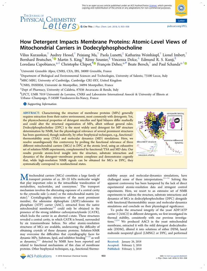
thermostability shift assay (TSA) experiments.14,15 In DDMand LMNG, a typical protein melting curve reveals acooperative thermal denaturation event with an apparentmelting temperature of ca. 49 °C (Figure 1, blue/green
lines). In the CATR-bound state, the melting temperature isdramatically increased to 80−90 °C. In contrast, the sameprotein preparation diluted into DPC already shows a highfluorescence signal at low temperature, and no transition in thecourse of the temperature scan, irrespective of the presence ofCATR. These observations indicate that AAC3 in DPC doesnot form a stable tertiary structure, nor binds CATR, in linewith previous findings.13 To understand this behavior at theatomic level, we turned to solution-NMR of three members ofthe MC family, namely, AAC3, guanine diphosphate (GDP)/guanosine triphosphate (GTP) carrier (GGC1), and ornithinecarrier (ORC1). Given their low sequence identity (<32%,Figure S1), common traits observed in DPC are likely sharedby the entire MC family. Samples of AAC3, GGC1, and ORC1,prepared through refolding from inclusion bodies, just like inprevious NMR studies of MCs,5,7,8,11 are all homogeneous (asshown by size-exclusion chromatograms and analytical ultra-centrifugation; Figure S2). Consistently with previous NMRstudies,5−9,11 all these samples yield high-quality NMR spectra(Figure S3), a criterion that is commonly employed to assessthe functional relevance of MP samples and motivate structuralstudies. To reconcile these data with TSA experiments and tocharacterize the structural organization of MCs that isresponsible for high-quality NMR spectra, we performed an
NMR-observed temperature series up to ca. 70 °C (Figure S3).One expects a discontinuity in the NMR parameters ifcollective unfolding arises. By contrast, we observe linearchemical-shift changes up to high temperatures, mirroring theTSA data, and suggesting that these MCs undergo a gradualchange of the structural ensemble without defined denaturationtransition.We probed the structural and dynamical properties of MCs
in DPC further with three different NMR observables. First, theresidue-specific chemical-shift assignments were used with theTALOS+ software16 to derive local backbone geometry at thelevel of each residue (Figures 1C and S4). While there is anoverall good agreement between the residues that, according to
Figure 1. Stability of secondary and tertiary structures in MCs. (A, B)Thermostability of AAC3, extracted from yeast mitochondria andpurified in DDM, diluted 20-fold into 0.1% DDM (blue line), 0.1%LMNG (green line) or 0.1% DPC (red line) in the absence (A) orpresence (B) of CATR, measured with TSA experiments (seeSupplementary Methods). (C) Residue-wise helix propensity inGGC1, determined from NMR chemical shifts using the programTALOS+,16 plotted onto a structural model of GGC1. Residues inwhite color are not in a helical conformation. (D) Residue-wise solventaccessibility in GGC1, as probed with the paramagnetic agentgadodiamide. Residues shown in white-to-blue colors are accessibleto solvent. In panels C and D, amide sites undergoing μs−msdynamics (discussed further below) are indicated by spheres.
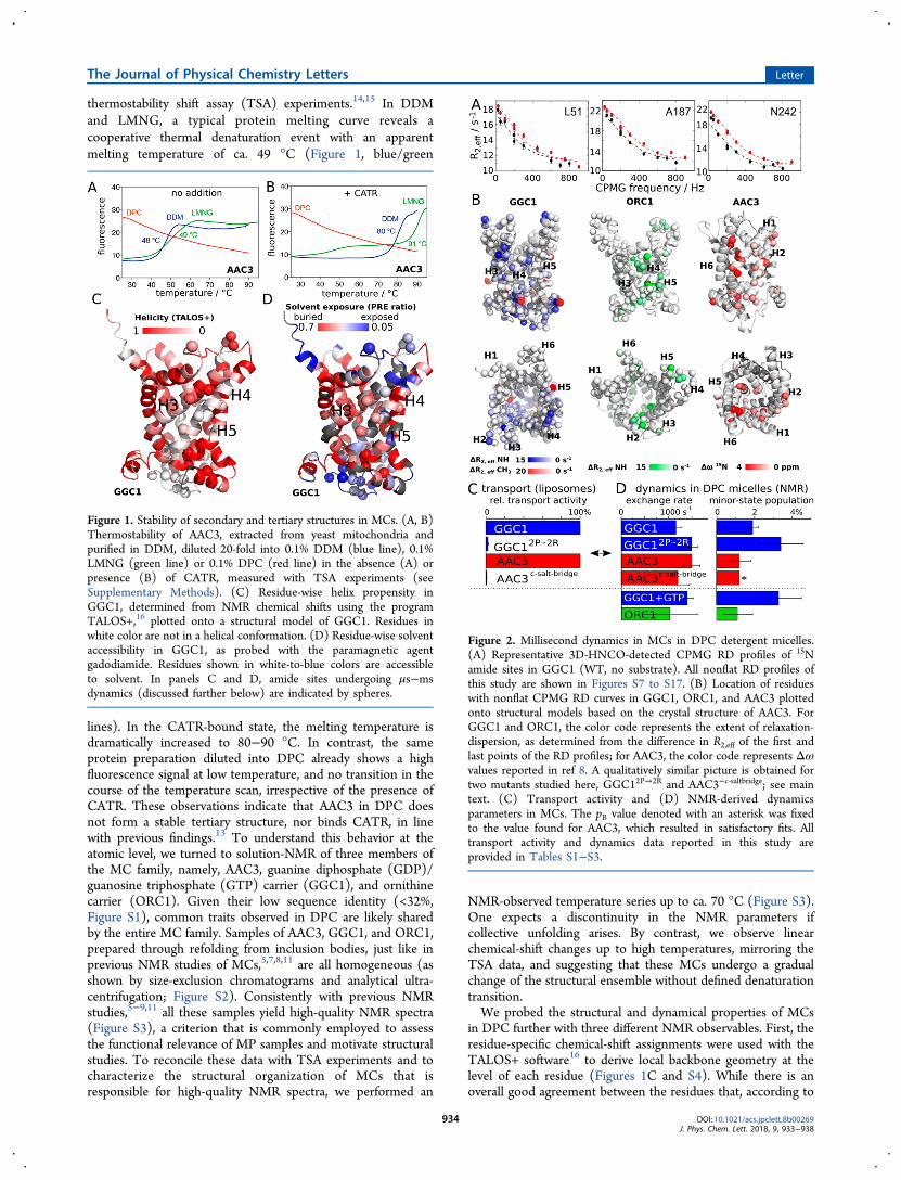
Figure 2. Millisecond dynamics in MCs in DPC detergent micelles.(A) Representative 3D-HNCO-detected CPMG RD profiles of 15Namide sites in GGC1 (WT, no substrate). All nonflat RD profiles ofthis study are shown in Figures S7 to S17. (B) Location of residueswith nonflat CPMG RD curves in GGC1, ORC1, and AAC3 plottedonto structural models based on the crystal structure of AAC3. ForGGC1 and ORC1, the color code represents the extent of relaxation-dispersion, as determined from the difference in R2,eff of the first andlast points of the RD profiles; for AAC3, the color code represents Δωvalues reported in ref 8. A qualitatively similar picture is obtained fortwo mutants studied here, GGC12P→2R and AAC3−c‑saltbridge; see maintext. (C) Transport activity and (D) NMR-derived dynamicsparameters in MCs. The pB value denoted with an asterisk was fixedto the value found for AAC3, which resulted in satisfactory fits. Alltransport activity and dynamics data reported in this study areprovided in Tables S1−S3.
The Journal of Physical Chemistry Letters Letter
DOI: 10.1021/acs.jpclett.8b00269J. Phys. Chem. Lett. 2018, 9, 933−938
934
their chemical shifts, have α-helical conformation and thelocation of helices in the crystal structures of AAC3, severalstretches of residues in the TM helices appear disordered inDPC, particularly in helices H4 (GGC1), and H2/H3 (AAC3).Independent support for the flexible nature of these partscomes from 15N transverse relaxation rate constants (R2),which reveal enhanced flexibility in GGC1’s H4 (Figure S5).Second, we performed solvent-paramagnetic relaxation-en-hancement (sPRE) experiments with the soluble hydrophilicparamagnetic complex Gd-DTPA-BMA (gadodiamide), whichprobe the solvent-accessibility of each amide site (Figure 1D).As expected, the most exposed parts are located in loops ofGGC1 and AAC3. Unexpectedly, however, sizable sPRE effectsare also found in TM helices, in particular those for whichTALOS+ finds a loop conformation. Third, we measured amidehydrogen/deuterium exchange (HDX) by dilution of GGC1from H2O- into D2O-buffer. The observed very fast HDXkinetics (<1 min) indicates that backbone hydrogen bonds aremarginally stable (Figure S6).To gain further insight into the properties and possibly
functional relevance of MCs in DPC, we investigated the detailsof their dynamics. Functional turnover of MCs in themembrane occurs at a rate of ca. 500 s−1.17 Functional MCsare, thus, expected to undergo motions on this time scale, andthese motions are expected to be sensitive to substrates ormutations. Carr−Purcell−Meiboom−Gill (CPMG) relaxation-dispersion (RD) NMR experiments18 are ideally suited toprobe μs-ms motions at the level of individual residues. In thisstudy we performed extensive CPMG RD measurements on a
total of 10 different samples, including wild-type (WT) andmutant proteins, as well as samples with substrates and lipids,and on up to three magnetic field strengths per sample (seeTable S1 and Figures S7−S17); in addition, previous workreported CPMG data for WT AAC3.8 In AAC3, GGC1 andORC1, we find extensive μs−ms motions for about 20% of the15N backbone amide sites, and in all cases the exchangingresidues are clustered on one side of the molecule (Figure 2).We have performed additional 13C CPMG experiments onmethyl groups of Ala, Val, and Leu residues in GGC1 (Table S1and Figure S8), independently confirming the presence of μs−ms dynamics in the region revealed by 15N CPMG experiments(red in Figure 2B, left). In order to obtain more insight into thisprocess, we fitted a two-state exchange model to the CPMGRD data. Statistical analyses, shown in Figure S18, indicate thatthis simple model describes the data satisfyingly. The fittedparameters are similar for all three proteins, with an exchangerate constant, kex = kAB + kBA, on the order of 1000−2000 s−1,and populations of the minor state, pB, of ca. 1−3% (Figure 2Dand Table S1).
Figure 3. GGC1 in DPC lacks the expected binding specificity. (A)Chemical-shift perturbations in GGC1 upon addition of GTP. (B)Plot of amide (H/N/CO) and methyl (H/C) CSPs onto a structuralmodel of GGC1. (C,D) GTP and ATP produces very similar CSPs inGGC1, as exemplified with extracts from HNCO spectra (D) andshown as a correlation plot in (C). The inset shows the electrostaticsurface of a AAC-derived structural model of GGC1. Equivalent datafor AAC3 are shown in Figure S21.
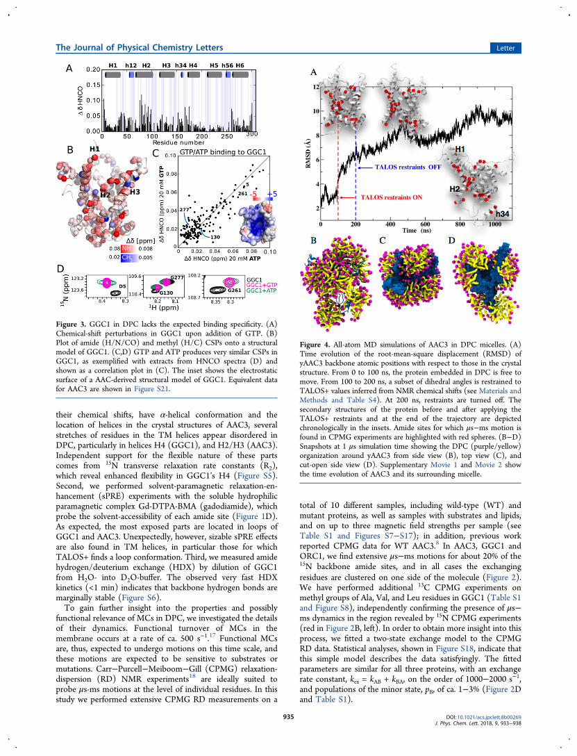
Figure 4. All-atom MD simulations of AAC3 in DPC micelles. (A)Time evolution of the root-mean-square displacement (RMSD) ofyAAC3 backbone atomic positions with respect to those in the crystalstructure. From 0 to 100 ns, the protein embedded in DPC is free tomove. From 100 to 200 ns, a subset of dihedral angles is restrained toTALOS+ values inferred from NMR chemical shifts (see Materials andMethods and Table S4). At 200 ns, restraints are turned off. Thesecondary structures of the protein before and after applying theTALOS+ restraints and at the end of the trajectory are depictedchronologically in the insets. Amide sites for which μs−ms motion isfound in CPMG experiments are highlighted with red spheres. (B−D)Snapshots at 1 μs simulation time showing the DPC (purple/yellow)organization around yAAC3 from side view (B), top view (C), andcut-open side view (D). Supplementary Movie 1 and Movie 2 showthe time evolution of AAC3 and its surrounding micelle.
The Journal of Physical Chemistry Letters Letter
DOI: 10.1021/acs.jpclett.8b00269J. Phys. Chem. Lett. 2018, 9, 933−938
935
A major challenge in the interpretation of relaxationdispersion data is to understand the underlying motionalprocess structurally. An intriguing observation is that the timescale of motion observed in all three MCs (ca. 1 ms) is similarto the kinetics of transmembrane transport,17 suggesting apossible role in functional turnover. To test this hypothesis, weinvestigated whether the introduction of function-abolishingmutations or the presence of substrates and lipids alter thedynamics. We designed two mutants that address differentstructural/functional aspects of the mitochondrial carriers,reasoning that if they inactivate the proteins, they would doso for different reasons. In a first mutant, termed GGC12P→2R,two highly conserved Pro residues of the MC signature motifs(PX[ED]XX[KR]) in H3 and H5 were mutated to Arg. Giventhe importance of these Pro kinks in the structure andmechanism,2 and the unfavorable energetics of placing Argresidues in the core of TM helices,19 we expected significantchanges in the activity and dynamics. A second mutant targetsAsp, Lys, and Gln residues on the cytosolic side of the TMhelices of AAC3, which have been proposed to form a saltbridge network in the elusive m-state.4 In the AAC3−c‑saltbridge
mutant, we inverted charges such that the putative salt-bridgenetwork cannot form any more. Liposome transport assaysshow that the residual transport activity in GGC12P→2R andAAC3−c‑saltbridge is below 3%, i.e., that the mutations render theproteins essentially nonfunctional (Figure 2C and Table S3).Strikingly, however, the characteristics of the mutants in
DPC micelles are hardly different from those of the WT proteinsamples. NMR spectra of these mutants are highly similar tothe respective WT proteins. Chemical-shift differences aresmall, and restricted to residues in the immediate vicinity of themutation sites (Figure S19). If the helices were in close contactto each other, as in the crystal structure, one would expectpronounced chemical-shift changes in the neighboring helicesin these mutants. Another surprising observation is that theμs−ms dynamics in these nonfunctional mutants is very similarto the one of WT proteins, even though the mutants arenonfunctional, likely suggesting that the μs−ms motions arenot directly related to function (Figures 2D, S12−S14 andTable S1). Along the same lines, we find that the presence ofsubstrate (GTP) does not lead to a detectable difference in thedynamics compared to apo-GGC1 (Figure 2D), indicating thatsubstrate cannot influence this dynamic process. Put together,the observed dynamics does not appear to be related tofunction. We reasoned that the functionally important2 lipidcardiolipin (CL) might resurrect the native properties of theprotein. However, CPMG experiments with CL reveal anidentical behavior for WT GGC1 and GGC12P→2R (seeSupplementary Discussion and Figures S9, S11, and S20),showing that these MCs in DPC are nonfunctional, irrespectiveof added lipids.An important criterion to assess functionality of a MP is its
ability to interact with substrates. MCs are highly substrate-specific transporters. In liposome transport assays, we find thatrecombinantly expressed and reconstituted AAC3 and GGC1bind and transport specifically their substrate (ATP/GTP,respectively), while they do not bind the other nucleotide(GTP/ATP; see Table S3). Furthermore, CATR inhibitsAAC3, but not GGC1 (see Supplementary Discussion).Testing the binding specificity is, thus, a convenient way toinvestigate the structural integrity of MCs in DPC. Addition ofthe substrate GTP to GGC1 leads to significant chemical shiftperturbations (CSPs) for many amides and Ala, Leu, and Val
methyls (Figure 3A). The largest CSPs are observed forresidues lining H2, and on the cytoplasmic side of H1 and H6.Weaker yet significant effects are observed in the threeamphipathic matrix helices (Figure 3B); these CSPs are ingood agreement with previously reported data.11 However,addition of ATP, which does not interact with GGC1 inmembranes, unexpectedly produces essentially the same CSPsas GTP (Figure 3C,D). Likewise, addition of GTP or ATP toAAC3 produces CSPs that are nearly identical to each other(Figure S21). Thus, while AAC3 and GGC1 in their lipid-bilayer environment can discriminate between GTP and ATP,this ability is lost in DPC micelles. We repeated GTP-titrationexperiments with the nonfunctional GGC12P→2R mutant. Eventhough this protein is nonfunctional and likely distortedthrough the Arg in the TM part, the GTP-induced CSPs arevery similar to those observed in WT protein (Figure S22).This observation suggests that the observed GTP interaction isnot dependent on structural integrity, and is thus nonspecific.We then titrated WT GGC1 with CATR, a doubly negatively
charged inhibitor of AAC, but not of GGC1.20 Despite theabsence of GGC1/CATR interaction in membranes, theaddition of CATR to GGC1 in DPC produces significantCSPs. The affected set of residues partly coincides with the sitesthat are modulated by the presence of GTP and ATP, and theeffects are similar to those observed in AAC3 upon addition ofCATR (Figure S23). These observations lead us to proposethat the reported interactions of CATR with GGC1 or AAC3 inDPC are nonspecific and not reminiscent of binding inbiological membranes. Indeed, the affinity of CATR to AAC3 inDPC (Kd = 20−150 μM8) is ca. 3 orders of magnitude weakerthan the AAC/CATR interaction in lipid bilayers (Kd = 10−20nM21) or of AAC extracted from mitochondria with LAPAOdetergent (Kd = 310 nM21). A plausible driving force for allthese nonspecific low-affinity interactions is the electrostaticattraction between the negatively charged solutes (nucleotides,CATR) and the positively charged cavity of GGC1 and AAC3(Figures 3C and S21D). In agreement with this view, ORC1,which has a significantly lower electrostatic charge, does notshow significant CSPs upon addition of its substrate, L-ornithine (Figure S24).To obtain a mechanistic understanding of the structural
organization of the protein and detergent, we turned to all-atom molecular dynamics (MD) simulations in explicit DPC.MD simulations have been applied previously to MCs in amembrane environment22−24 and in DPC.12 We specificallysought a description of the structural organization of AAC3 inDPC, which would reflect our experimental data. In initialunrestrained all-atom simulations over 0.7 μs, AAC3 remainsclose to the conformation adopted in crystals,4 which is indisagreement with the TALOS-derived secondary structure inDPC. Presumably, the time scale accessible to simulation is tooshort to allow the transition of the compact conformation incrystals to the one in DPC micelles (see SupplementaryDiscussion and Figures S25, S26). To better reflect theexperimental data, we, thus, used chemical-shift derivedbackbone torsional angles (limited to the ϕ,ψ angles forwhich the TALOS+ result was unambiguous; see Materials andMethods), and introduced a soft harmonic potential to theseresidues over a simulation period of 0.1 μs. This strategy, whichrests on the use of NMR restraints to guide conformationalanalyses of proteins, is similar in spirit to that proposed byothers.25,26 When the torsional angles coincide with thoseinferred from the NMR data, the overall structure of AAC3 is
The Journal of Physical Chemistry Letters Letter
DOI: 10.1021/acs.jpclett.8b00269J. Phys. Chem. Lett. 2018, 9, 933−938
936
markedly distorted, corresponding to a shift from ca. 3 to 6 Å ofthe root-mean-square deviation (RMSD) of atomic positions(Figure 4A). The evolution of the tertiary structure uponintroduction of the TALOS+ restraints is asymmetrical andchiefly involves the interfacial helix h34 (connecting H3 andH4) and the cytoplasmic side of H1, H2, and H3 (Figure 4 andS27). In a last step, the restraints were removed, and thesimulation was extended to 1.1 μs. Over this time scale, AAC3remains distorted. The cytoplasmic side of the TM helices, H1and H2, and the interfacial helix h34 undergo relatively slowdynamics on the simulation time scale (see Movie 1).Interestingly, this region coincides with the segments thatwere found experimentally to undergo μs−ms motions (redspheres in Figure 4). We speculate that the motions detectedexperimentally might correspond to the kind of slow rearrange-ments seen by MD. Most importantly, the simulations providea view of how the detergent may organize around the protein.Figures 4B−D show that DPC molecules do not only form acorona around the hydrophobic core of the protein, as onemight expect, but also form micelles interacting with both sidesof the protein (cf. Movie 2). DPC molecules protrude betweenthe TM helices and penetrate deep within the protein cavity(Figure 4C,D and Movie 2).DPC has been employed for solving about 40% of all known
NMR structures, but less than 1% of all crystal structures(Figure S28). Our data provide possible clues about the originof this intriguing track record. In our hands, DPC is the onlydetergent able to maintain MCs in solution for extended timeperiods (a prerequisite for solution-NMR), and the embeddedproteins exhibit well-resolved spectra and solute interactionsthat may be interpreted as signs of a functionally relevantsample. Our study reveals that these observations do notnecessarily point to a functionally relevant protein sample, andstresses the importance of stringent control experiments.
■ ASSOCIATED CONTENT
*S Supporting InformationThe Supporting Information is available free of charge on theACS Publications website at DOI: 10.1021/acs.jpclett.8b00269.
Movie 1: 1.1 s molecular-dynamics simulation of yeastAAC3 in DPC (MPG)Movie 2: Same as Movie 1 including the explicitrepresentation of DPC organization around yAAC3(AVI)Detailed experimental materials and methods, tablessummarizing the results of transport activity measure-ments, CPMG fits and assigned chemical shifts, andfigures with supporting experimental data (PDF)
■ AUTHOR INFORMATION
Corresponding Authors*E-mail: [email protected].*E-mail: [email protected].*E-mail: [email protected].
ORCIDBernhard Brutscher: 0000-0001-7652-7384Christophe Chipot: 0000-0002-9122-1698Paul Schanda: 0000-0002-9350-7606NotesThe authors declare no competing financial interest.
■ ACKNOWLEDGMENTS
We thank Klaus Zangger for providing gadodiamide, and AstridC. Sivertsen, Guillaume Bouvignies, Eva Pebay-Peyroula,Stephanie Ravaud, Ewen Lescop, Cecile Breyton, SvenBruschweiler, Christine Ebel and Aline LeRoy for insightfuldiscussions, Jason R. Schnell for help in preparing Figure S28and Michael Kohlhaas for inspiring thoughts. This work wassupported by the European Research Council (ERC-Stg-311318) to P. S. and the UK Medical Research Council(U105663139) to E.R.S.K. This work used the platforms of theGrenoble Instruct Center (ISBG; UMS 3518 CNRS-CEA-UJF-EMBL) with support from FRISBI (ANR-10-INSB-05-02) and GRAL (ANR-10-LABX-49-01) within the GrenoblePartnership for Structural Biology (PSB).
■ REFERENCES(1) Palmieri, F.; Monne, M. Discoveries, Metabolic Roles andDiseases of Mitochondrial Carriers: A Review. Biochim. Biophys. Acta,Mol. Cell Res. 2016, 1863, 2362−2378.(2) Kunji, E. R. S.; Aleksandrova, A.; King, M. S.; Majd, H.; Ashton,V. L.; Cerson, E.; Springett, R.; Kibalchenko, M.; Tavoulari, S.;Crichton, P. G.; et al. The Transport Mechanism of the MitochondrialADP/ATP Carrier. Biochim. Biophys. Acta, Mol. Cell Res. 2016, 1863,2379−2393.(3) Pebay-Peyroula, E.; Dahout-Gonzalez, C.; Kahn, R.; Trezeguet,V.; Lauquin, G.; Brandolin, G. Structure of Mitochondrial ADP/ATPCarrier in Complex with Carboxyatractyloside. Nature 2003, 426, 39−44.(4) Ruprecht, J. J.; Hellawell, A. M.; Harding, M.; Crichton, P. G.;McCoy, A. J.; Kunji, E. R. S. Structures of Yeast Mitochondrial ADP/ATP Carriers Support a Domain-Based Alternating-Access TransportMechanism. Proc. Natl. Acad. Sci. U. S. A. 2014, 111, E426−34.(5) Run, C.; Yang, Q.; Liu, Z.; OuYang, B.; Chou, J. J. MolecularBasis of MgATP Selectivity of the Mitochondrial SCaMC Carrier.Structure 2015, 23, 1394−1403.(6) Zhao, L.; Wang, S.; Zhu, Q.; Wu, B.; Liu, Z.; OuYang, B.; Chou,J. J. Specific Interaction of the Human Mitochondrial UncouplingProtein 1 with Free Long-Chain Fatty Acid. Structure 2017, 25, 1371−1379.e3.(7) Zhao, L.; Wang, S.; Run, C.; OuYang, B.; Chou, J. J. SpecificLipid Binding of Membrane Proteins in Detergent MicellesCharacterized by NMR and Molecular Dynamics. Biochemistry 2016,55, 5317−5320.(8) Bruschweiler, S.; Yang, Q.; Run, C.; Chou, J. J. Substrate-Modulated ADP/ATP-Transporter Dynamics Revealed by NMRRelaxation Dispersion. Nat. Struct. Mol. Biol. 2015, 22, 636−641.(9) Berardi, M. J.; Shih, W. M.; Harrison, S. C.; Chou, J. J.Mitochondrial Uncoupling Protein 2 Structure Determined by NMRMolecular Fragment Searching. Nature 2011, 476, 109−113.(10) Berardi, M. J.; Chou, J. J. Fatty Acid Flippase Activity of UCP2Is Essential for Its Proton Transport in Mitochondria. Cell Metab.2014, 20, 541−552.(11) Sounier, R.; Bellot, G.; Chou, J. J. Mapping ConformationalHeterogeneity of Mitochondrial Nucleotide Transporter in Unin-hibited States. Angew. Chem., Int. Ed. 2015, 54, 2436−2441.(12) Zoonens, M.; Comer, J.; Masscheleyn, S.; Pebay-Peyroula, E.;Chipot, C.; Miroux, B.; Dehez, F. Dangerous Liaisons betweenDetergents and Membrane Proteins. The Case of MitochondrialUncoupling Protein 2. J. Am. Chem. Soc. 2013, 135, 15174−15182.(13) Dehez, F.; Schanda, P.; King, M. S.; Kunji, E. R. S.; Chipot, C.Mitochondrial ADP/ATP Carrier in Dodecylphosphocholine BindsCardiolipins with Non-Native Affinity. Biophys. J. 2017, 113, 2311−2315.(14) Crichton, P. G.; Lee, Y.; Ruprecht, J. J.; Cerson, E.;Thangaratnarajah, C.; King, M. S.; Kunji, E. R. S. Trends inThermostability Provide Information on the Nature of Substrate,
The Journal of Physical Chemistry Letters Letter
DOI: 10.1021/acs.jpclett.8b00269J. Phys. Chem. Lett. 2018, 9, 933−938
937
Inhibitor, and Lipid Interactions with Mitochondrial Carriers. J. Biol.Chem. 2015, 290, 8206−8217.(15) Alexandrov, A. I.; Mileni, M.; Chien, E. Y. T.; Hanson, M. A.;Stevens, R. C. Microscale Fluorescent Thermal Stability Assay forMembrane Proteins. Structure 2008, 16, 351−359.(16) Shen, Y.; Delaglio, F.; Cornilescu, G.; Bax, A. TALOS+: AHybrid Method for Predicting Protein Backbone Torsion Angles fromNMR Chemical Shifts. J. Biomol. NMR 2009, 44, 213−223.(17) Gropp, T.; Brustovetsky, N.; Klingenberg, M.; Muller, V.;Fendler, K.; Bamberg, E. Kinetics of Electrogenic Transport by theADP/ATP Carrier. Biophys. J. 1999, 77, 714−726.(18) Mittermaier, A. K.; Kay, L. E. Observing Biological Dynamics atAtomic Resolution Using NMR. Trends Biochem. Sci. 2009, 34, 601−611.(19) Zhou, H.-X.; Cross, T. A. Influences of Membrane MimeticEnvironments on Membrane Protein Structures. Annu. Rev. Biophys.2013, 42, 361−392.(20) Vozza, A.; Blanco, E.; Palmieri, L.; Palmieri, F. Identification ofthe Mitochondrial GTP/GDP Transporter in Saccharomyces cerevisiae.J. Biol. Chem. 2004, 279, 20850−20857.(21) Kramer, R.; Klingenberg, M. Reconstitution of Inhibitor BindingProperties of the Isolated Adenosine 5′-diphosphate, Adenosine 5′-triphosphate Carrier-Linked Binding Protein. Biochemistry 1977, 16,4954−49561.(22) Wang, Y.; Tajkhorshid, E. Electrostatic Funneling of Substrate inMitochondrial Inner Membrane Carriers. Proc. Natl. Acad. Sci. U. S. A.2008, 105, 9598−9603.(23) Dehez, F.; Pebay-Peyroula, E.; Chipot, C. Binding of ADP in theMitochondrial ADP/ATP Carrier Is Driven by an Electrostatic Funnel.J. Am. Chem. Soc. 2008, 130, 12725−12733.(24) Hedger, G.; Rouse, S. L.; Domanski, J.; Chavent, M.; Koldso,H.; Sansom, M. S. P. Lipid-Loving ANTs: Molecular Simulations ofCardiolipin Interactions and the Organization of the AdenineNucleotide Translocase in Model Mitochondrial Membranes.Biochemistry 2016, 55, 6238−6249.(25) Vallurupalli, P.; Hansen, D. F.; Kay, L. E. Structures of Invisible,Excited Protein States by Relaxation Dispersion NMR Spectroscopy.Proc. Natl. Acad. Sci. U. S. A. 2008, 105, 11766−11771.(26) Robustelli, P.; Kohlhoff, K.; Cavalli, A.; Vendruscolo, M. UsingNMR Chemical Shifts as Structural Restraints in Molecular DynamicsSimulations of Proteins. Structure 2010, 18, 923−933.
The Journal of Physical Chemistry Letters Letter
DOI: 10.1021/acs.jpclett.8b00269J. Phys. Chem. Lett. 2018, 9, 933−938
938


![Identification of Low-Abundance Lipid Droplet Proteins · Identification of Low-Abundance Lipid Droplet Proteins in Seeds and Seedlings1[OPEN] Franziska K. Kretzschmar,a,2 Nathan](https://img.pdfslide.fr/doc/110x75/5f1b6eccd9db36017f49896d/identiication-of-low-abundance-lipid-droplet-identiication-of-low-abundance.jpg)