Embed Size (px)
Citation preview
Standards of OASIS diagnosis: Primary (clinical) and
Secondary (ultrasound)
How to protect the perineum and prevent obstetric perineal trauma
Vincent Letouzey, MD, PhD
Obst/Gyne Dept
Nîmes University Hospital
France
Standards of OASIS diagnosis: Primary: Clinical examination
How to protect the perineum and prevent obstetric perineal trauma
Vincent Letouzey, MD, PhD
Obst/Gyne Dept
Nîmes University Hospital
France
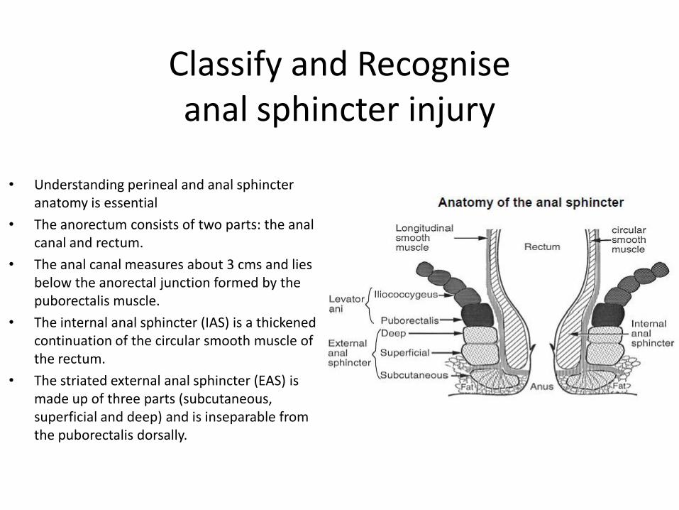
Classify and Recognise anal sphincter injury
• Understanding perineal and anal sphincter
anatomy is essential
• The anorectum consists of two parts: the anal canal and rectum.
• The anal canal measures about 3 cms and lies below the anorectal junction formed by the puborectalis muscle.
• The internal anal sphincter (IAS) is a thickened continuation of the circular smooth muscle of the rectum.
• The striated external anal sphincter (EAS) is made up of three parts (subcutaneous, superficial and deep) and is inseparable from the puborectalis dorsally.
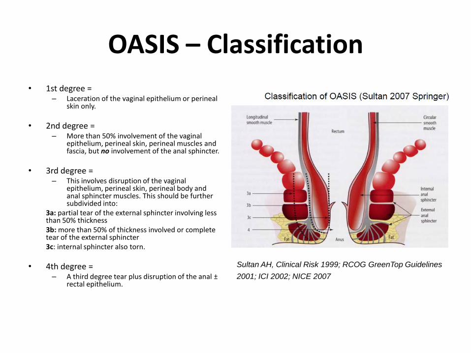
OASIS – Classification
• 1st degree = – Laceration of the vaginal epithelium or perineal
skin only.
• 2nd degree = – More than 50% involvement of the vaginal
epithelium, perineal skin, perineal muscles and fascia, but no involvement of the anal sphincter.
• 3rd degree = – This involves disruption of the vaginal
epithelium, perineal skin, perineal body and anal sphincter muscles. This should be further subdivided into:
3a: partial tear of the external sphincter involving less than 50% thickness
3b: more than 50% of thickness involved or complete tear of the external sphincter
3c: internal sphincter also torn.
• 4th degree = – A third degree tear plus disruption of the anal ±
rectal epithelium.
Sultan AH, Clinical Risk 1999; RCOG GreenTop Guidelines
2001; ICI 2002; NICE 2007
In the labor room:
• History – Fecal or anal incontinence
– Instrumental delivery
• Delivery – 2nd stage > 60 min
– Extraction: Forceps > Vaccum
– Episiotomy ?
– Baby weight ?
Perineal Examination
• Up to 30% of 3rd/4th degree tears go unrecognised at delivery.
• Examine in lithotomy and do a rectal exam (PR) to ascertain the extent.
• Inspection – perineal area – Vaginal area
• All skin tears that extend to the anal margin are 3rd degree tears until proven
otherwise by the charge midwife.
• The full extent of the injury should be evaluated by a careful vaginal and rectal examination in lithotomy and the tear should be classified as above.
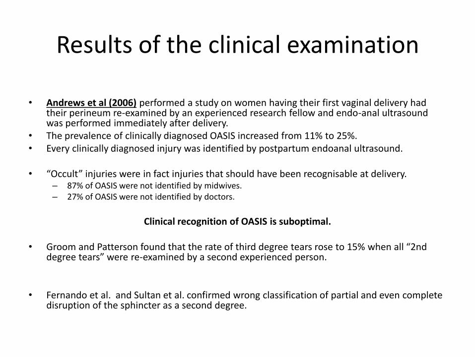
Results of the clinical examination
• Andrews et al (2006) performed a study on women having their first vaginal delivery had
their perineum re-examined by an experienced research fellow and endo-anal ultrasound was performed immediately after delivery.
• The prevalence of clinically diagnosed OASIS increased from 11% to 25%. • Every clinically diagnosed injury was identified by postpartum endoanal ultrasound.
• “Occult” injuries were in fact injuries that should have been recognisable at delivery.
– 87% of OASIS were not identified by midwives. – 27% of OASIS were not identified by doctors.
Clinical recognition of OASIS is suboptimal.
• Groom and Patterson found that the rate of third degree tears rose to 15% when all “2nd
degree tears” were re-examined by a second experienced person.
• Fernando et al. and Sultan et al. confirmed wrong classification of partial and even complete disruption of the sphincter as a second degree.
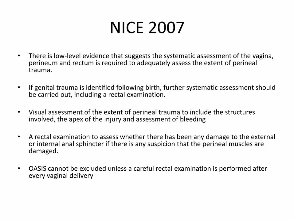
NICE 2007
• There is low-level evidence that suggests the systematic assessment of the vagina, perineum and rectum is required to adequately assess the extent of perineal trauma.
• If genital trauma is identified following birth, further systematic assessment should be carried out, including a rectal examination.
• Visual assessment of the extent of perineal trauma to include the structures involved, the apex of the injury and assessment of bleeding
• A rectal examination to assess whether there has been any damage to the external or internal anal sphincter if there is any suspicion that the perineal muscles are damaged.
• OASIS cannot be excluded unless a careful rectal examination is performed after every vaginal delivery
Standards of OASIS diagnosis: Secondary: ultrasound
How to protect the perineum and prevent obstetric perineal trauma
Intrapartum Transperineal Ultrasound (TPUS)
Rational, Faisability, Preliminary Results
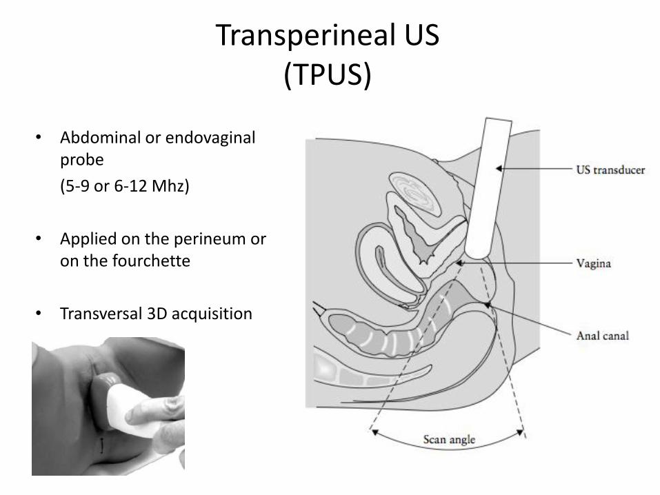
• Abdominal or endovaginal probe
(5-9 or 6-12 Mhz)
• Applied on the perineum or on the fourchette
• Transversal 3D acquisition
Transperineal US (TPUS)
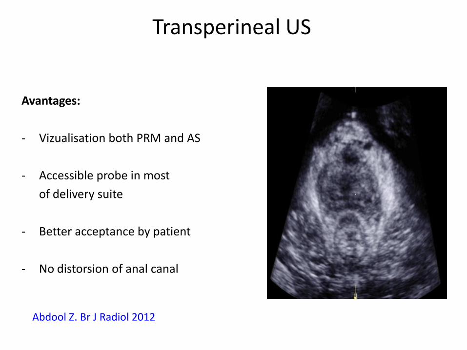
Avantages:
- Vizualisation both PRM and AS
- Accessible probe in most
of delivery suite
- Better acceptance by patient
- No distorsion of anal canal
Abdool Z. Br J Radiol 2012
Transperineal US
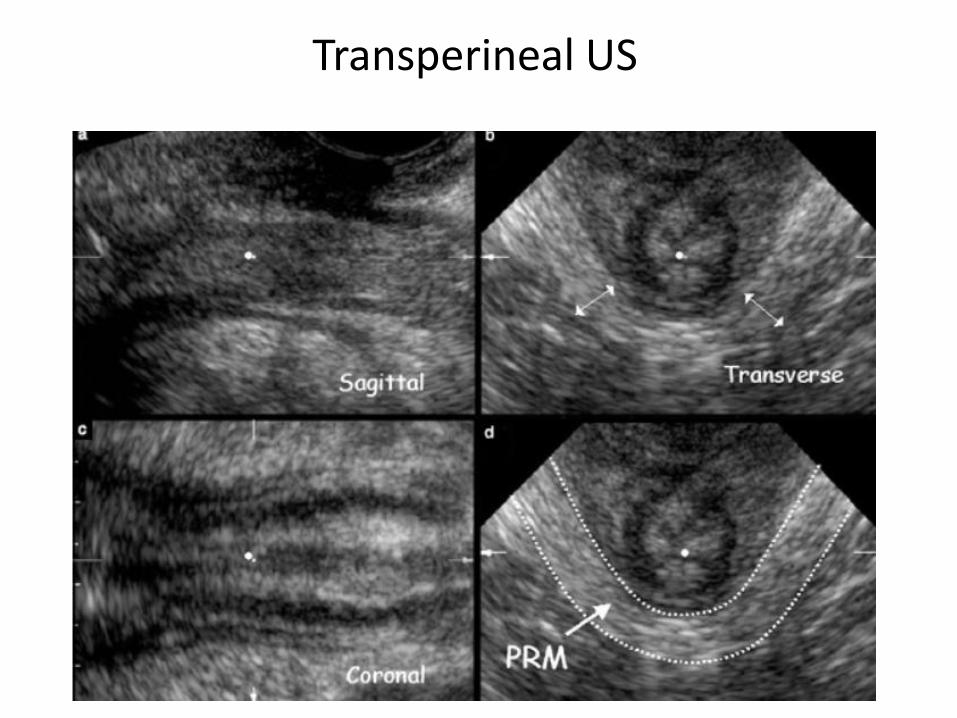
Transversal Sagittal
Coronal
Transperineal US
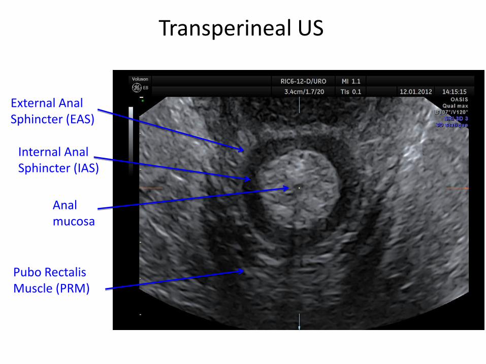
External Anal Sphincter (EAS)
Transperineal US
Internal Anal Sphincter (IAS)
Pubo Rectalis Muscle (PRM)
Anal mucosa
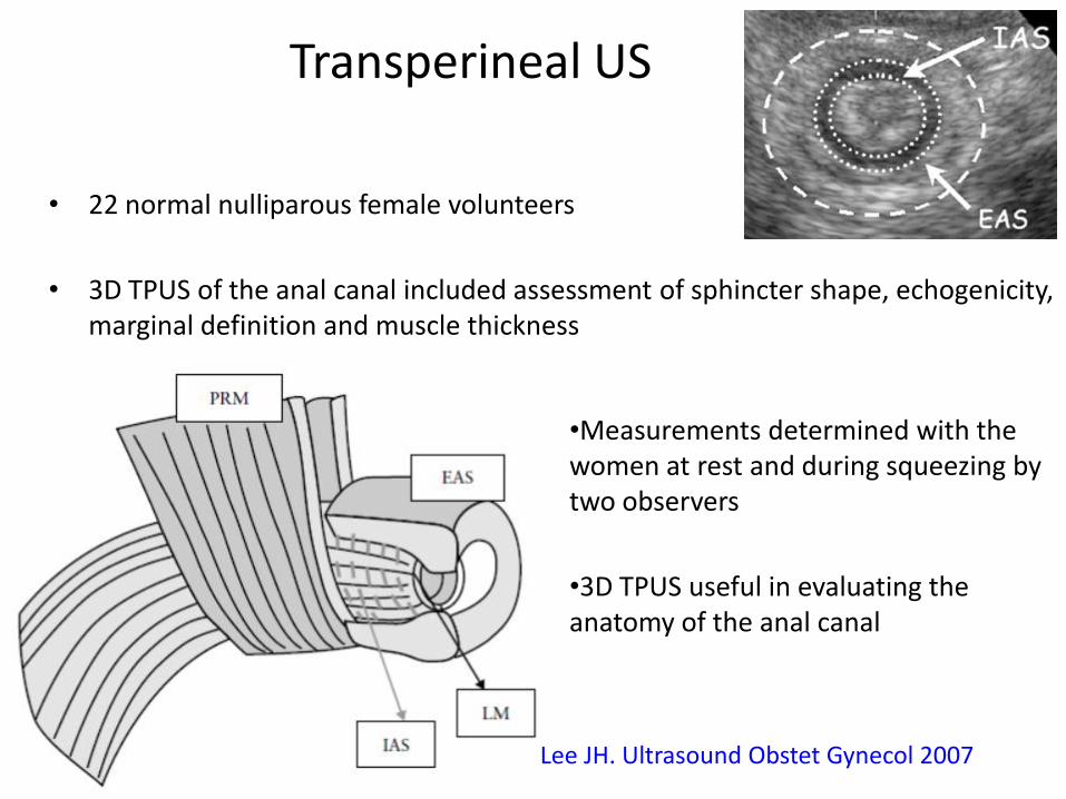
• 22 normal nulliparous female volunteers
• 3D TPUS of the anal canal included assessment of sphincter shape, echogenicity, marginal definition and muscle thickness
Transperineal US
Lee JH. Ultrasound Obstet Gynecol 2007
•Measurements determined with the women at rest and during squeezing by two observers
•3D TPUS useful in evaluating the anatomy of the anal canal
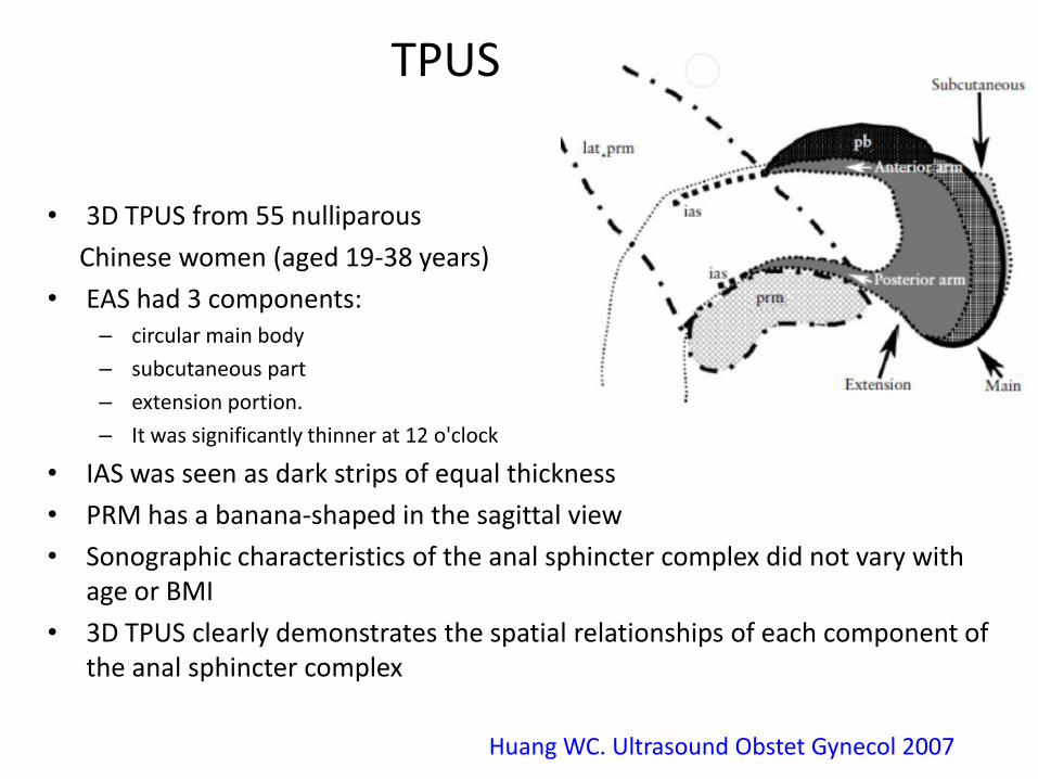
• 3D TPUS from 55 nulliparous
Chinese women (aged 19-38 years)
• EAS had 3 components: – circular main body
– subcutaneous part
– extension portion.
– It was significantly thinner at 12 o'clock
• IAS was seen as dark strips of equal thickness
• PRM has a banana-shaped in the sagittal view
• Sonographic characteristics of the anal sphincter complex did not vary with age or BMI
• 3D TPUS clearly demonstrates the spatial relationships of each component of the anal sphincter complex
TPUS
Huang WC. Ultrasound Obstet Gynecol 2007
• 139 primiparous
• Tears degree ≤ 2
• TP US 24-72h after delivery
91.4% interpretable volume
84.6% entire EAS vizualisation
7.9% OASIS
Valsky DV. Ultrasound Obstet Gynecol 2007
TPUS Faisability in early post-partum
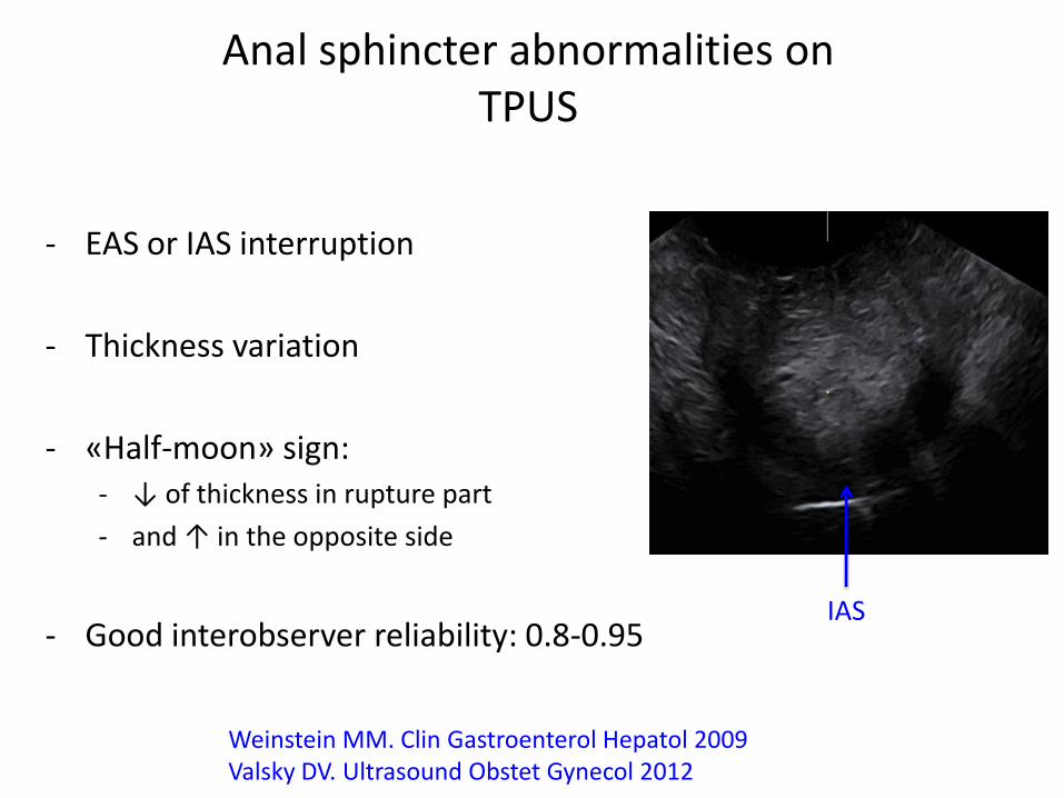
Weinstein MM. Clin Gastroenterol Hepatol 2009 Valsky DV. Ultrasound Obstet Gynecol 2012
- EAS or IAS interruption
- Thickness variation
- «Half-moon» sign: - ↓ of thickness in rupture part
- and ↑ in the opposite side
- Good interobserver reliability: 0.8-0.95
IAS
Anal sphincter abnormalities on TPUS
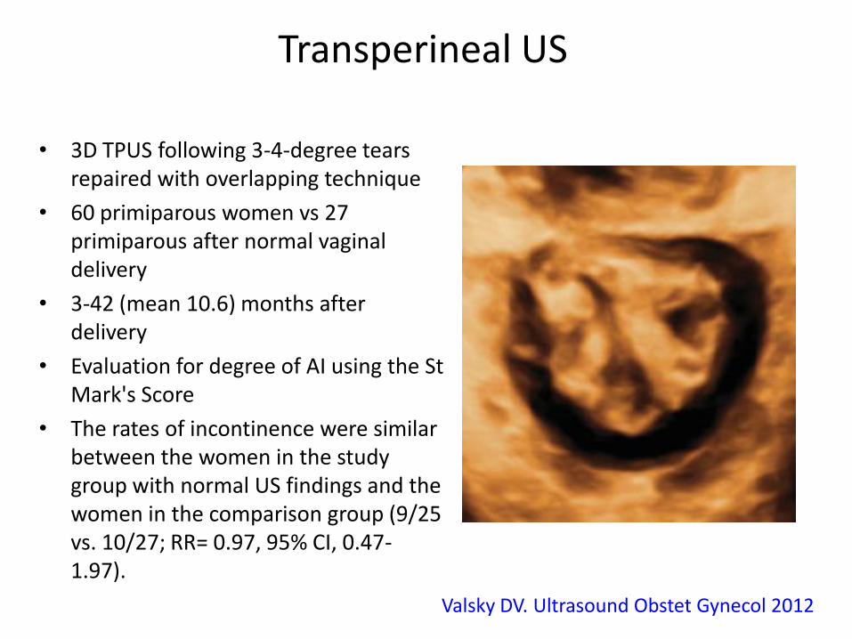
• 3D TPUS following 3-4-degree tears
repaired with overlapping technique
• 60 primiparous women vs 27 primiparous after normal vaginal delivery
• 3-42 (mean 10.6) months after delivery
• Evaluation for degree of AI using the St Mark's Score
• The rates of incontinence were similar between the women in the study group with normal US findings and the women in the comparison group (9/25 vs. 10/27; RR= 0.97, 95% CI, 0.47-1.97).
Transperineal US
Valsky DV. Ultrasound Obstet Gynecol 2012
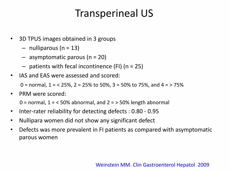
• 3D TPUS images obtained in 3 groups
– nulliparous (n = 13)
– asymptomatic parous (n = 20)
– patients with fecal incontinence (FI) (n = 25)
• IAS and EAS were assessed and scored:
0 = normal, 1 = < 25%, 2 = 25% to 50%, 3 = 50% to 75%, and 4 = > 75%
• PRM were scored:
0 = normal, 1 = < 50% abnormal, and 2 = > 50% length abnormal
• Inter-rater reliability for detecting defects : 0.80 - 0.95
• Nullipara women did not show any significant defect
• Defects was more prevalent in FI patients as compared with asymptomatic parous women
Transperineal US
Weinstein MM. Clin Gastroenterol Hepatol 2009
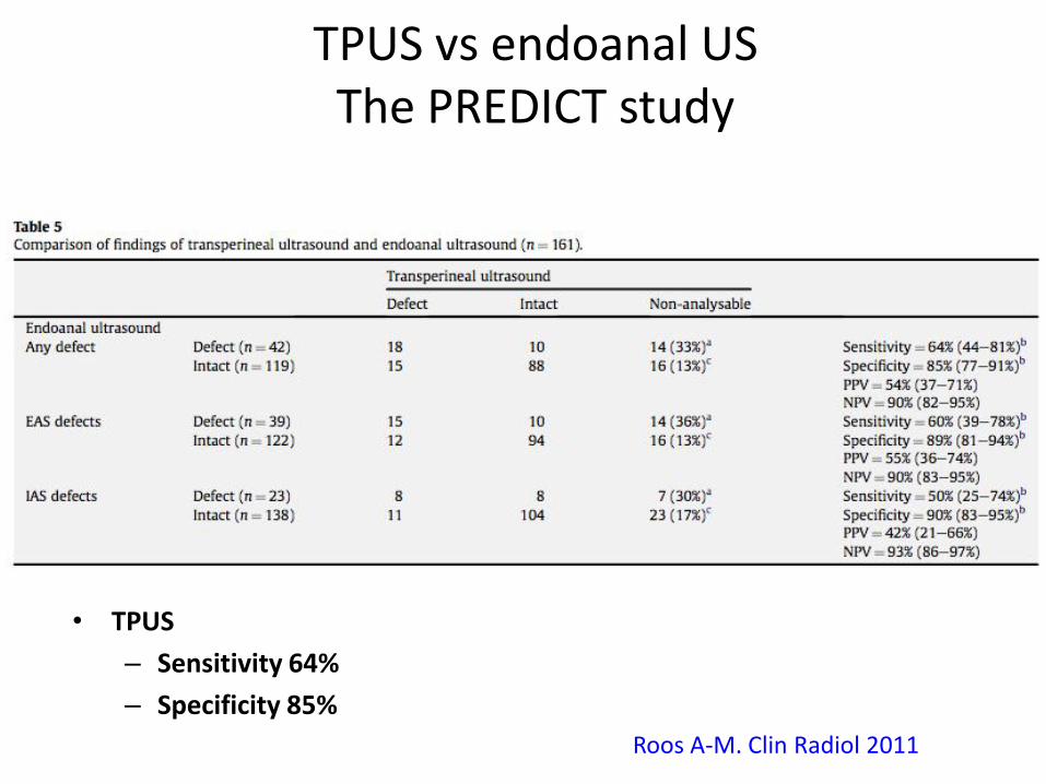
Roos A-M. Clin Radiol 2011
TPUS vs endoanal US The PREDICT study
• TPUS
– Sensitivity 64%
– Specificity 85%
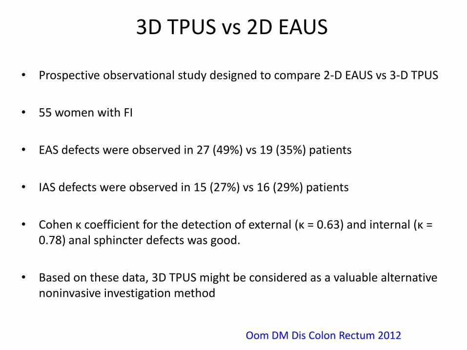
• Prospective observational study designed to compare 2-D EAUS vs 3-D TPUS
• 55 women with FI
• EAS defects were observed in 27 (49%) vs 19 (35%) patients
• IAS defects were observed in 15 (27%) vs 16 (29%) patients
• Cohen κ coefficient for the detection of external (κ = 0.63) and internal (κ = 0.78) anal sphincter defects was good.
• Based on these data, 3D TPUS might be considered as a valuable alternative noninvasive investigation method
3D TPUS vs 2D EAUS
Oom DM Dis Colon Rectum 2012
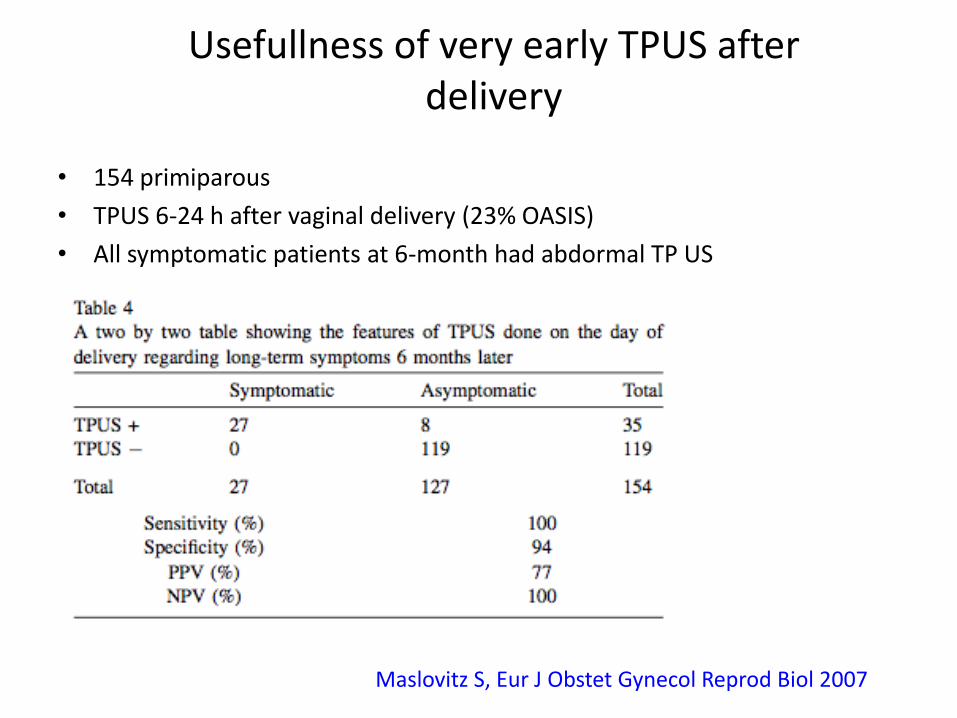
• 154 primiparous
• TPUS 6-24 h after vaginal delivery (23% OASIS)
• All symptomatic patients at 6-month had abdormal TP US
Maslovitz S, Eur J Obstet Gynecol Reprod Biol 2007
Usefullness of very early TPUS after delivery
Use 3D TPUS at the time of delivery
Improve diagnosis of SA defect
Suture AS tears is any (only in non intact perineum?)
Reduce post-partum risk of fecal incontinence
More studies are needed
Our goal for the future