Embed Size (px)
Citation preview
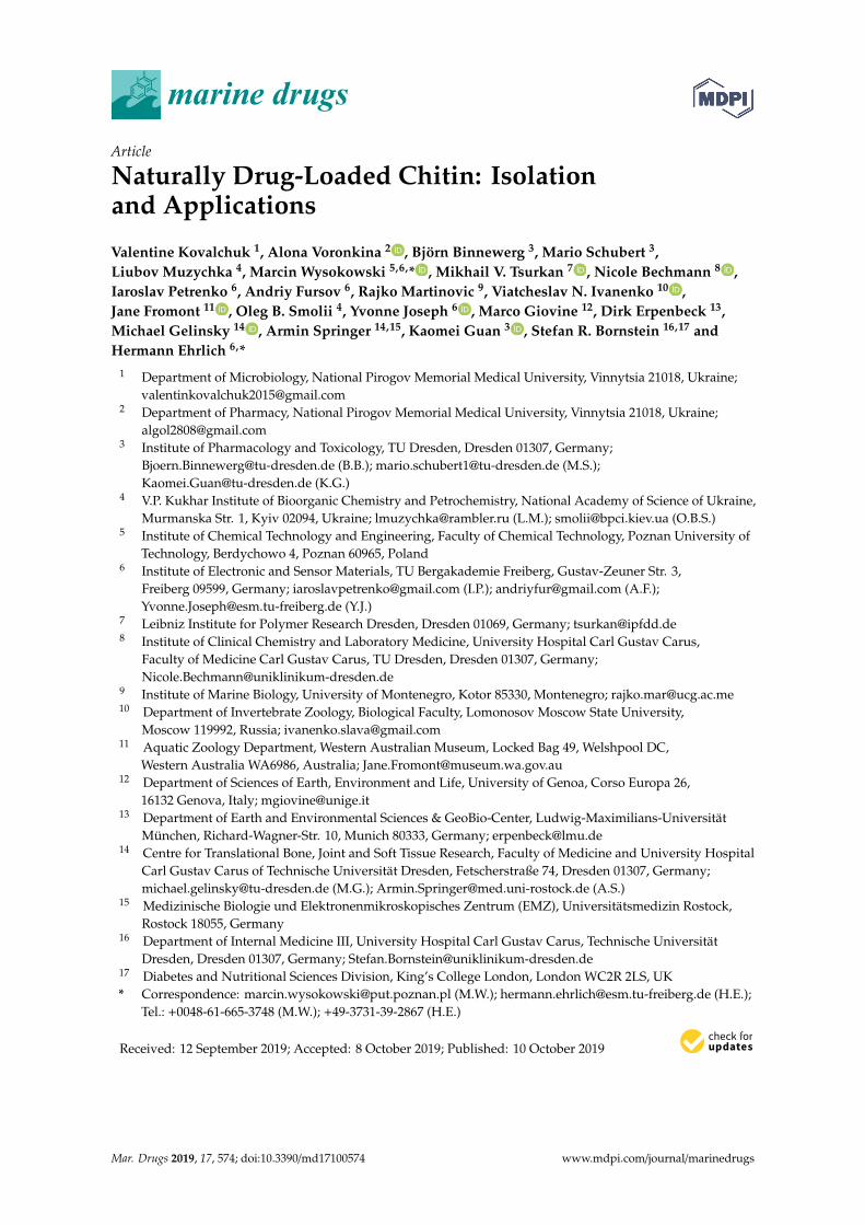
marine drugs
Article
Naturally Drug-Loaded Chitin: Isolationand Applications
Valentine Kovalchuk 1, Alona Voronkina 2 , Björn Binnewerg 3, Mario Schubert 3,Liubov Muzychka 4, Marcin Wysokowski 5,6,* , Mikhail V. Tsurkan 7 , Nicole Bechmann 8 ,Iaroslav Petrenko 6, Andriy Fursov 6, Rajko Martinovic 9, Viatcheslav N. Ivanenko 10 ,Jane Fromont 11 , Oleg B. Smolii 4, Yvonne Joseph 6 , Marco Giovine 12, Dirk Erpenbeck 13,Michael Gelinsky 14 , Armin Springer 14,15, Kaomei Guan 3 , Stefan R. Bornstein 16,17 andHermann Ehrlich 6,*
1 Department of Microbiology, National Pirogov Memorial Medical University, Vinnytsia 21018, Ukraine;[email protected]
2 Department of Pharmacy, National Pirogov Memorial Medical University, Vinnytsia 21018, Ukraine;[email protected]
3 Institute of Pharmacology and Toxicology, TU Dresden, Dresden 01307, Germany;[email protected] (B.B.); [email protected] (M.S.);[email protected] (K.G.)
4 V.P. Kukhar Institute of Bioorganic Chemistry and Petrochemistry, National Academy of Science of Ukraine,Murmanska Str. 1, Kyiv 02094, Ukraine; [email protected] (L.M.); [email protected] (O.B.S.)
5 Institute of Chemical Technology and Engineering, Faculty of Chemical Technology, Poznan University ofTechnology, Berdychowo 4, Poznan 60965, Poland
6 Institute of Electronic and Sensor Materials, TU Bergakademie Freiberg, Gustav-Zeuner Str. 3,Freiberg 09599, Germany; [email protected] (I.P.); [email protected] (A.F.);[email protected] (Y.J.)
7 Leibniz Institute for Polymer Research Dresden, Dresden 01069, Germany; [email protected] Institute of Clinical Chemistry and Laboratory Medicine, University Hospital Carl Gustav Carus,
Faculty of Medicine Carl Gustav Carus, TU Dresden, Dresden 01307, Germany;[email protected]
9 Institute of Marine Biology, University of Montenegro, Kotor 85330, Montenegro; [email protected] Department of Invertebrate Zoology, Biological Faculty, Lomonosov Moscow State University,
Moscow 119992, Russia; [email protected] Aquatic Zoology Department, Western Australian Museum, Locked Bag 49, Welshpool DC,
Western Australia WA6986, Australia; [email protected] Department of Sciences of Earth, Environment and Life, University of Genoa, Corso Europa 26,
16132 Genova, Italy; [email protected] Department of Earth and Environmental Sciences & GeoBio-Center, Ludwig-Maximilians-Universität
München, Richard-Wagner-Str. 10, Munich 80333, Germany; [email protected] Centre for Translational Bone, Joint and Soft Tissue Research, Faculty of Medicine and University Hospital
Carl Gustav Carus of Technische Universität Dresden, Fetscherstraße 74, Dresden 01307, Germany;[email protected] (M.G.); [email protected] (A.S.)
15 Medizinische Biologie und Elektronenmikroskopisches Zentrum (EMZ), Universitätsmedizin Rostock,Rostock 18055, Germany
16 Department of Internal Medicine III, University Hospital Carl Gustav Carus, Technische UniversitätDresden, Dresden 01307, Germany; [email protected]
17 Diabetes and Nutritional Sciences Division, King’s College London, London WC2R 2LS, UK* Correspondence: [email protected] (M.W.); [email protected] (H.E.);
Tel.: +0048-61-665-3748 (M.W.); +49-3731-39-2867 (H.E.)
Received: 12 September 2019; Accepted: 8 October 2019; Published: 10 October 2019�����������������
Mar. Drugs 2019, 17, 574; doi:10.3390/md17100574 www.mdpi.com/journal/marinedrugs
Mar. Drugs 2019, 17, 574 2 of 17
Abstract: Naturally occurring three-dimensional (3D) biopolymer-based matrices that can be usedin different biomedical applications are sustainable alternatives to various artificial 3D materials.For this purpose, chitin-based structures from marine sponges are very promising substitutes. Marinesponges from the order Verongiida (class Demospongiae) are typical examples of demosponges withwell-developed chitinous skeletons. In particular, species belonging to the family Ianthellidae possesschitinous, flat, fan-like fibrous skeletons with a unique, microporous 3D architecture that makes themparticularly interesting for applications. In this work, we focus our attention on the demospongeIanthella flabelliformis (Linnaeus, 1759) for simultaneous extraction of both naturally occurring(“ready-to-use”) chitin scaffolds, and biologically active bromotyrosines which are recognizedas potential antibiotic, antitumor, and marine antifouling substances. We show that selectedbromotyrosines are located within pigmental cells which, however, are localized within chitinousskeletal fibers of I. flabelliformis. A two-step reaction provides two products: treatment with methanolextracts the bromotyrosine compounds bastadin 25 and araplysillin-I N20 sulfamate, and a subsequenttreatment with acetic acid and sodium hydroxide exposes the 3D chitinous scaffold. This scaffold isa mesh-like structure, which retains its capillary network, and its use as a potential drug deliverybiomaterial was examined for the first time. The results demonstrate that sponge-derived chitinscaffolds, impregnated with decamethoxine, effectively inhibit growth of the human pathogenStaphylococcus aureus in an agar diffusion assay.
Keywords: chitin; scaffolds; pigmental cells; demosponges; Ianthella; bromotyrosines; decamethoxine;drug delivery
1. Introduction
Development of three-dimensional (3D) scaffolds based on natural biopolymers is a recent trendin materials and biomaterials science. The scientific community now focuses on development offabrication methods which will allow for precise control of the architecture and pore structures in suchscaffolds. Evolutionary optimized 3D constructs of natural origin can be found in marine demosponges(phylum Porifera, class Demospongiae), which are recognized among the first multicellular organismson our planet. These organisms evolved and survived for more than 600 million years due to theirability to excellently combine the mechanical stability of their voluminous, fiber-based, water-filteringskeletons [1] and their diverse chemical defense strategies [2,3] due to biosynthesis of secondarymetabolites with anti-predatory and antibiotic properties [4]. The demosponges can produce bothproteinaceous (spongin)- or polysaccharide (chitin)-based and up to 2-m-high skeletons (for anoverview, see References [5–10]). Representatives of the family Ianthellidae (Hyatt, 1875) possesschitinous, flat, fan-like skeletons with unique 3D architecture [11,12] (Figures 1 and 2A), some of whichcan reach up to 2 m in diameter [12].
Mar. Drugs 2019, 17, 574 3 of 17Mar. Drugs 2019, 17, x 3 of 17
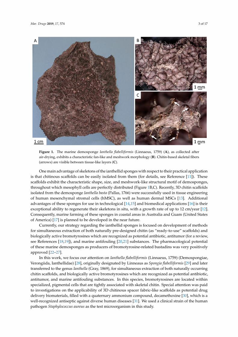
Figure 1. The marine demosponge Ianthella flabelliformis (Linnaeus, 1759) (A), as collected after air-drying, exhibits a characteristic fan-like and meshwork morphology (B). Chitin-based skeletal fibers (arrows) are visible between tissue-like layers (C).
One main advantage of skeletons of the ianthellid sponges with respect to their practical application is that chitinous scaffolds can be easily isolated from them (for details, see Reference [11]). These scaffolds exhibit the characteristic shape, size, and meshwork-like structural motif of demosponges, throughout which mesophyll cells are perfectly distributed (Figures 1B,C). Recently, 3D chitin scaffolds isolated from the demosponge Ianthella basta (Pallas, 1766) were successfully used in tissue engineering of human mesenchymal stromal cells (hMSC), as well as human dermal MSCs [13]. Additional advantages of these sponges for use in technological [14,15] and biomedical applications [16] is their exceptional ability to regenerate their skeletons in situ, with a growth rate of up to 12 cm/year [12]. Consequently, marine farming of these sponges in coastal areas in Australia and Guam (United States of America) [17] is planned to be developed in the near future.
Currently, our strategy regarding the ianthellid sponges is focused on development of methods for simultaneous extraction of both naturally pre-designed chitin (as “ready-to-use” scaffolds) and biologically active bromotyrosines which are recognized as potential antibiotic, antitumor (for a review, see References [18,19]), and marine antifouling [20,21] substances. The pharmacological potential of these marine demosponges as producers of bromotyrosine-related bastadins was very positively approved [22–27].
In this work, we focus our attention on Ianthella flabelliformis (Linnaeus, 1759) (Demospongiae, Verongiida, Ianthellidae) [28], originally designated by Linneaus as Spongia flabelliformis [29] and later transferred to the genus Ianthella (Gray, 1869), for simultaneous extraction of both naturally occurring chitin scaffolds, and biologically active bromotyrosines which are recognized as potential antibiotic, antitumor, and marine antifouling substances. In this species, bromotyrosines are located within specialized, pigmental cells that are tightly associated with skeletal chitin. Special attention was paid to investigations on the applicability of 3D chitinous spacer fabric-like scaffolds as potential drug delivery biomaterials, filled with a quaternary ammonium compound, decamethoxine [30], which is a well-recognized antiseptic against diverse human diseases [31]. We used a clinical strain of the human pathogen Staphylococcus aureus as the test microorganism in this study.
Figure 1. The marine demosponge Ianthella flabelliformis (Linnaeus, 1759) (A), as collected afterair-drying, exhibits a characteristic fan-like and meshwork morphology (B). Chitin-based skeletal fibers(arrows) are visible between tissue-like layers (C).
One main advantage of skeletons of the ianthellid sponges with respect to their practical applicationis that chitinous scaffolds can be easily isolated from them (for details, see Reference [11]). Thesescaffolds exhibit the characteristic shape, size, and meshwork-like structural motif of demosponges,throughout which mesophyll cells are perfectly distributed (Figure 1B,C). Recently, 3D chitin scaffoldsisolated from the demosponge Ianthella basta (Pallas, 1766) were successfully used in tissue engineeringof human mesenchymal stromal cells (hMSC), as well as human dermal MSCs [13]. Additionaladvantages of these sponges for use in technological [14,15] and biomedical applications [16] is theirexceptional ability to regenerate their skeletons in situ, with a growth rate of up to 12 cm/year [12].Consequently, marine farming of these sponges in coastal areas in Australia and Guam (United Statesof America) [17] is planned to be developed in the near future.
Currently, our strategy regarding the ianthellid sponges is focused on development of methodsfor simultaneous extraction of both naturally pre-designed chitin (as “ready-to-use” scaffolds) andbiologically active bromotyrosines which are recognized as potential antibiotic, antitumor (for a review,see References [18,19]), and marine antifouling [20,21] substances. The pharmacological potentialof these marine demosponges as producers of bromotyrosine-related bastadins was very positivelyapproved [22–27].
In this work, we focus our attention on Ianthella flabelliformis (Linnaeus, 1759) (Demospongiae,Verongiida, Ianthellidae) [28], originally designated by Linneaus as Spongia flabelliformis [29] and latertransferred to the genus Ianthella (Gray, 1869), for simultaneous extraction of both naturally occurringchitin scaffolds, and biologically active bromotyrosines which are recognized as potential antibiotic,antitumor, and marine antifouling substances. In this species, bromotyrosines are located withinspecialized, pigmental cells that are tightly associated with skeletal chitin. Special attention was paidto investigations on the applicability of 3D chitinous spacer fabric-like scaffolds as potential drugdelivery biomaterials, filled with a quaternary ammonium compound, decamethoxine [30], which is awell-recognized antiseptic against diverse human diseases [31]. We used a clinical strain of the humanpathogen Staphylococcus aureus as the test microorganism in this study.
Mar. Drugs 2019, 17, 574 4 of 17
2. Results
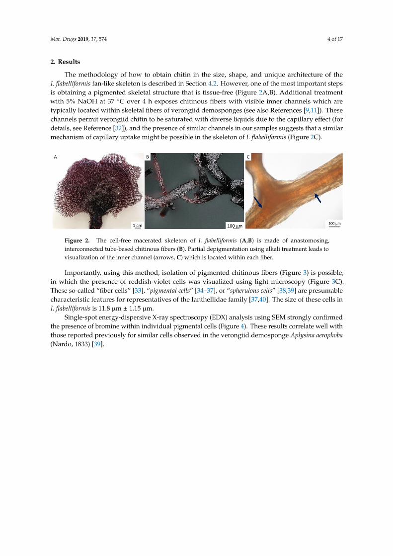
The methodology of how to obtain chitin in the size, shape, and unique architecture of theI. flabelliformis fan-like skeleton is described in Section 4.2. However, one of the most important stepsis obtaining a pigmented skeletal structure that is tissue-free (Figure 2A,B). Additional treatmentwith 5% NaOH at 37 ◦C over 4 h exposes chitinous fibers with visible inner channels which aretypically located within skeletal fibers of verongiid demosponges (see also References [9,11]). Thesechannels permit verongiid chitin to be saturated with diverse liquids due to the capillary effect (fordetails, see Reference [32]), and the presence of similar channels in our samples suggests that a similarmechanism of capillary uptake might be possible in the skeleton of I. flabelliformis (Figure 2C).
Mar. Drugs 2019, 17, x 4 of 17
2. Results
The methodology of how to obtain chitin in the size, shape, and unique architecture of the I. flabelliformis fan-like skeleton is described in Section 4.2. However, one of the most important steps is obtaining a pigmented skeletal structure that is tissue-free (Figures 2 A,B). Additional treatment with 5% NaOH at 37 °C over 4 h exposes chitinous fibers with visible inner channels which are typically located within skeletal fibers of verongiid demosponges (see also References [9,11]). These channels permit verongiid chitin to be saturated with diverse liquids due to the capillary effect (for details, see Reference [32]), and the presence of similar channels in our samples suggests that a similar mechanism of capillary uptake might be possible in the skeleton of I. flabelliformis (Figure 2C).
Figure 2. The cell-free macerated skeleton of I. flabelliformis (A,B) is made of anastomosing, interconnected tube-based chitinous fibers (B). Partial depigmentation using alkali treatment leads to visualization of the inner channel (arrows, C) which is located within each fiber.
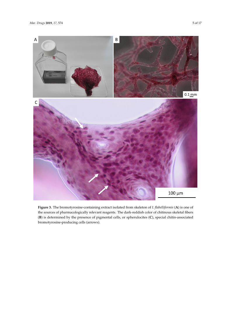
Importantly, using this method, isolation of pigmented chitinous fibers (Figure 3) is possible, in which the presence of reddish-violet cells was visualized using light microscopy (Figure 3C). These so-called “fiber cells” [33], “pigmental cells” [34–37], or “spherulous cells” [38,39] are presumable characteristic features for representatives of the Ianthellidae family [37,40]. The size of these cells in I. flabelliformis is 11.8 µm ± 1.15 µm.
Figure 2. The cell-free macerated skeleton of I. flabelliformis (A,B) is made of anastomosing,interconnected tube-based chitinous fibers (B). Partial depigmentation using alkali treatment leads tovisualization of the inner channel (arrows, C) which is located within each fiber.
Importantly, using this method, isolation of pigmented chitinous fibers (Figure 3) is possible,in which the presence of reddish-violet cells was visualized using light microscopy (Figure 3C).These so-called “fiber cells” [33], “pigmental cells” [34–37], or “spherulous cells” [38,39] are presumablecharacteristic features for representatives of the Ianthellidae family [37,40]. The size of these cells inI. flabelliformis is 11.8 µm ± 1.15 µm.
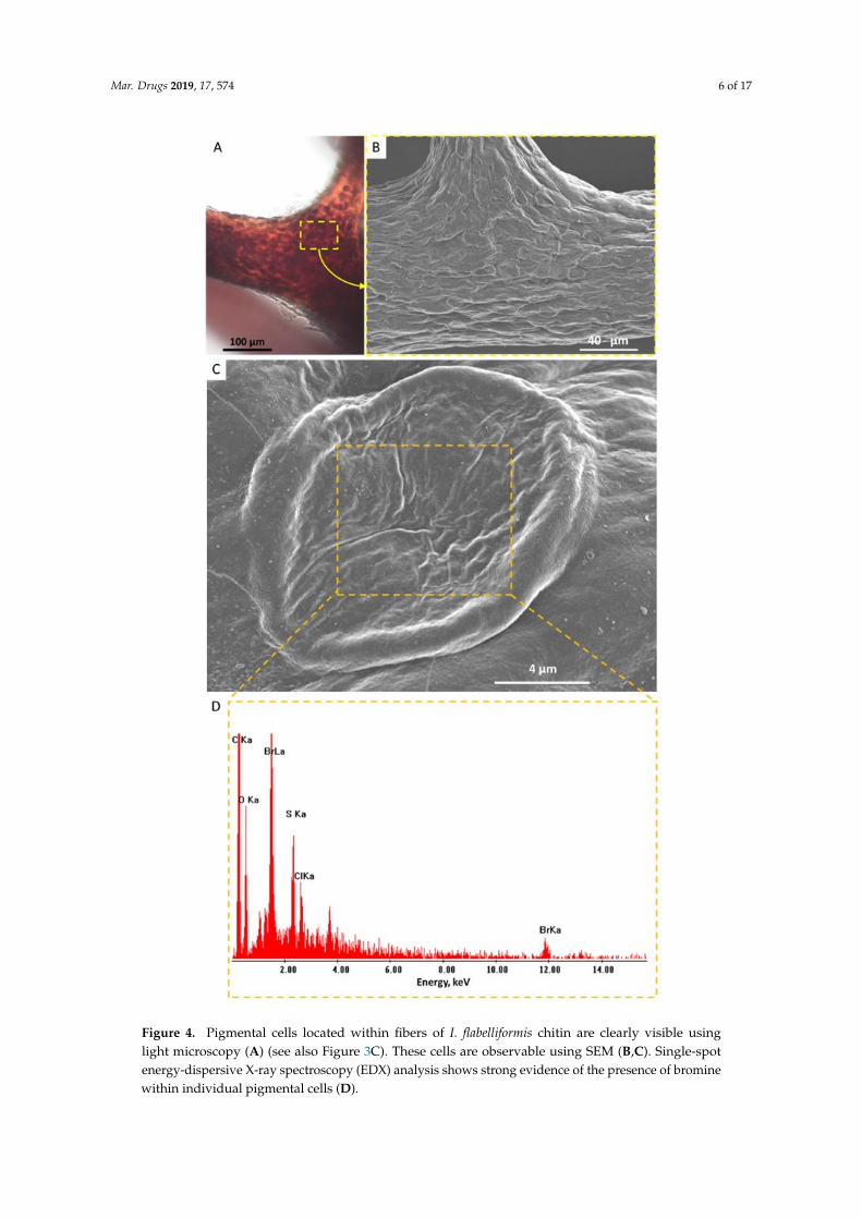
Single-spot energy-dispersive X-ray spectroscopy (EDX) analysis using SEM strongly confirmedthe presence of bromine within individual pigmental cells (Figure 4). These results correlate well withthose reported previously for similar cells observed in the verongiid demosponge Aplysina aerophoba(Nardo, 1833) [39].
Mar. Drugs 2019, 17, 574 5 of 17Mar. Drugs 2019, 17, x 5 of 17
Figure 3. The bromotyrosine-containing extract isolated from skeleton of I. flabelliformis (A) is one of the sources of pharmacologically relevant reagents. The dark-reddish color of chitinous skeletal fibers (B) is determined by the presence of pigmental cells, or spherulocites (C), special chitin-associated bromotyrosine-producing cells (arrows).
Single-spot energy-dispersive X-ray spectroscopy (EDX) analysis using SEM strongly confirmed the presence of bromine within individual pigmental cells (Figure 4). These results correlate well with those reported previously for similar cells observed in the verongiid demosponge Aplysina aerophoba (Nardo, 1833) [39].
Figure 3. The bromotyrosine-containing extract isolated from skeleton of I. flabelliformis (A) is one ofthe sources of pharmacologically relevant reagents. The dark-reddish color of chitinous skeletal fibers(B) is determined by the presence of pigmental cells, or spherulocites (C), special chitin-associatedbromotyrosine-producing cells (arrows).
Mar. Drugs 2019, 17, 574 6 of 17Mar. Drugs 2019, 17, x 6 of 17
Figure 4. Pigmental cells located within fibers of I. flabelliformis chitin are clearly visible using light microscopy (A) (see also Figure 3C). These cells are observable using SEM (B,C). Single-spot energy-dispersive X-ray spectroscopy (EDX) analysis shows strong evidence of the presence of bromine within individual pigmental cells (D).
Figure 4. Pigmental cells located within fibers of I. flabelliformis chitin are clearly visible usinglight microscopy (A) (see also Figure 3C). These cells are observable using SEM (B,C). Single-spotenergy-dispersive X-ray spectroscopy (EDX) analysis shows strong evidence of the presence of brominewithin individual pigmental cells (D).
Mar. Drugs 2019, 17, 574 7 of 17
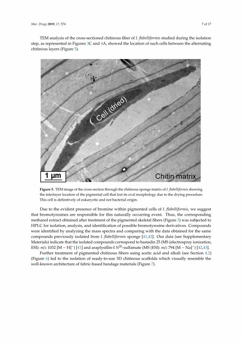
TEM analysis of the cross-sectioned chitinous fiber of I. flabelliformis studied during the isolationstep, as represented in Figures 3C and 4A, showed the location of such cells between the alternatingchitinous layers (Figure 5).
Mar. Drugs 2019, 17, x 7 of 17
TEM analysis of the cross-sectioned chitinous fiber of I. flabelliformis studied during the isolation step, as represented in Figures 3C and 4A, showed the location of such cells between the alternating chitinous layers (Figure 5).
Figure 5. TEM image of the cross-section through the chitinous sponge matrix of I. flabelliformis showing the interlayer location of the pigmental cell that lost its oval morphology due to the drying procedure. This cell is definitively of eukaryotic and not bacterial origin.
Due to the evident presence of bromine within pigmented cells of I. flabelliformis, we suggest that bromotyrosines are responsible for this naturally occurring event. Thus, the corresponding methanol extract obtained after treatment of the pigmented skeletal fibers (Figure 3) was subjected to HPLC for isolation, analysis, and identification of possible bromotyrosine derivatives. Compounds were identified by analyzing the mass spectra and comparing with the data obtained for the same compounds previously isolated from I. flabelliformis sponge [41,42]. Our data (see Supplementary Materials) indicate that the isolated compounds correspond to bastadin 25 (MS (electrospray ionization, ESI): m/z 1032 [M − H]−) [41] and araplysillin-I N20-sulfamate (MS (ESI): m/z 794 [M − Na]−) [42,43].
Further treatment of pigmented chitinous fibers using acetic acid and alkali (see Section 4.2) (Figure 6) led to the isolation of ready-to-use 3D chitinous scaffolds which visually resemble the well-known architecture of fabric-based bandage materials (Figure 7).
Figure 5. TEM image of the cross-section through the chitinous sponge matrix of I. flabelliformis showingthe interlayer location of the pigmental cell that lost its oval morphology due to the drying procedure.This cell is definitively of eukaryotic and not bacterial origin.
Due to the evident presence of bromine within pigmented cells of I. flabelliformis, we suggestthat bromotyrosines are responsible for this naturally occurring event. Thus, the correspondingmethanol extract obtained after treatment of the pigmented skeletal fibers (Figure 3) was subjected toHPLC for isolation, analysis, and identification of possible bromotyrosine derivatives. Compoundswere identified by analyzing the mass spectra and comparing with the data obtained for the samecompounds previously isolated from I. flabelliformis sponge [41,42]. Our data (see SupplementaryMaterials) indicate that the isolated compounds correspond to bastadin 25 (MS (electrospray ionization,ESI): m/z 1032 [M − H]−) [41] and araplysillin-I N20-sulfamate (MS (ESI): m/z 794 [M − Na]−) [42,43].
Further treatment of pigmented chitinous fibers using acetic acid and alkali (see Section 4.2)(Figure 6) led to the isolation of ready-to-use 3D chitinous scaffolds which visually resemble thewell-known architecture of fabric-based bandage materials (Figure 7).

Mar. Drugs 2019, 17, 574 8 of 17
Mar. Drugs 2019, 17, x 8 of 17
Figure 6. The spherulocite-free chitinous skeleton of I. flabelliformis can be isolated after alternating treatment of the construct with acetic acid (A) and NaOH (B) (see also Figure 7).
Figure 7. The chitinous skeleton isolated from I. flabelliformis (A) represents a mechanically elastic, flat, but still three-dimensional (3D)-based construct made of interconnected tubular fibers (B). These fibers show excellent capacity for saturation with diverse liquids including water (C).
As reported by us previously [13], similar tubular, flat, 3D scaffolds isolated from the marine demosponge Ianthella basta closely related to I. flabelliformis [11] were applied in the tissue engineering of hMSCs. However, in this study, we decided to examine the possibility of such constructs as drug delivery matrices for the future development of alternatives to well-recognized antimicrobial textiles (for an overview, see References [44–47]).
Figure 6. The spherulocite-free chitinous skeleton of I. flabelliformis can be isolated after alternatingtreatment of the construct with acetic acid (A) and NaOH (B) (see also Figure 7).
Mar. Drugs 2019, 17, x 8 of 17
Figure 6. The spherulocite-free chitinous skeleton of I. flabelliformis can be isolated after alternating treatment of the construct with acetic acid (A) and NaOH (B) (see also Figure 7).
Figure 7. The chitinous skeleton isolated from I. flabelliformis (A) represents a mechanically elastic, flat, but still three-dimensional (3D)-based construct made of interconnected tubular fibers (B). These fibers show excellent capacity for saturation with diverse liquids including water (C).
As reported by us previously [13], similar tubular, flat, 3D scaffolds isolated from the marine demosponge Ianthella basta closely related to I. flabelliformis [11] were applied in the tissue engineering of hMSCs. However, in this study, we decided to examine the possibility of such constructs as drug delivery matrices for the future development of alternatives to well-recognized antimicrobial textiles (for an overview, see References [44–47]).
Figure 7. The chitinous skeleton isolated from I. flabelliformis (A) represents a mechanically elastic, flat,but still three-dimensional (3D)-based construct made of interconnected tubular fibers (B). These fibersshow excellent capacity for saturation with diverse liquids including water (C).
As reported by us previously [13], similar tubular, flat, 3D scaffolds isolated from the marinedemosponge Ianthella basta closely related to I. flabelliformis [11] were applied in the tissue engineeringof hMSCs. However, in this study, we decided to examine the possibility of such constructs as drug
Mar. Drugs 2019, 17, 574 9 of 17
delivery matrices for the future development of alternatives to well-recognized antimicrobial textiles(for an overview, see References [44–47]).
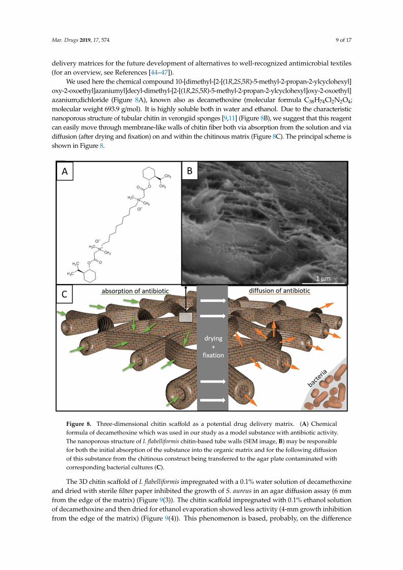
We used here the chemical compound 10-[dimethyl-[2-[(1R,2S,5R)-5-methyl-2-propan-2-ylcyclohexyl]oxy-2-oxoethyl]azaniumyl]decyl-dimethyl-[2-[(1R,2S,5R)-5-methyl-2-propan-2-ylcyclohexyl]oxy-2-oxoethyl]azanium;dichloride (Figure 8A), known also as decamethoxine (molecular formula C38H74Cl2N2O4;molecular weight 693.9 g/mol). It is highly soluble both in water and ethanol. Due to the characteristicnanoporous structure of tubular chitin in verongiid sponges [9,11] (Figure 8B), we suggest that this reagentcan easily move through membrane-like walls of chitin fiber both via absorption from the solution and viadiffusion (after drying and fixation) on and within the chitinous matrix (Figure 8C). The principal scheme isshown in Figure 8.
Mar. Drugs 2019, 17, x 9 of 17
We used here the chemical compound 10-[dimethyl-[2-[(1R,2S,5R)-5-methyl-2-propan-2-ylcyclohexyl]oxy-2-oxoethyl]azaniumyl]decyl-dimethyl-[2-[(1R,2S,5R)-5-methyl-2-propan-2-ylcyclohexyl]oxy-2-oxoethyl]azanium;dichloride (Figure 8A), known also as decamethoxine (molecular formula C38H74Cl2N2O4; molecular weight 693.9 g/mol). It is highly soluble both in water and ethanol. Due to the characteristic nanoporous structure of tubular chitin in verongiid sponges [9,11] (Figure 8B), we suggest that this reagent can easily move through membrane-like walls of chitin fiber both via absorption from the solution and via diffusion (after drying and fixation) on and within the chitinous matrix (Figure 8C). The principal scheme is shown in Figure 8.
Figure 8. Three-dimensional chitin scaffold as a potential drug delivery matrix. (A) Chemical formula of decamethoxine which was used in our study as a model substance with antibiotic activity. The nanoporous structure of I. flabelliformis chitin-based tube walls (SEM image, B) may be responsible for both the initial absorption of the substance into the organic matrix and for the following diffusion of this substance from the chitinous construct being transferred to the agar plate contaminated with corresponding bacterial cultures (C).
The 3D chitin scaffold of I. flabelliformis impregnated with a 0.1% water solution of decamethoxine and dried with sterile filter paper inhibited the growth of S. aureus in an agar diffusion assay (6 mm from the edge of the matrix) (Figure 9(3)). The chitin scaffold impregnated with 0.1% ethanol solution of decamethoxine and then dried for ethanol evaporation showed less activity (4-mm growth inhibition from the edge of the matrix) (Figure 9(4)). This phenomenon is based, probably, on the difference between water and ethanol with respect to their hydrophilicity. Both samples retained properties to release antiseptic on their second and third use, after moving to a fresh
Figure 8. Three-dimensional chitin scaffold as a potential drug delivery matrix. (A) Chemicalformula of decamethoxine which was used in our study as a model substance with antibiotic activity.The nanoporous structure of I. flabelliformis chitin-based tube walls (SEM image, B) may be responsiblefor both the initial absorption of the substance into the organic matrix and for the following diffusionof this substance from the chitinous construct being transferred to the agar plate contaminated withcorresponding bacterial cultures (C).
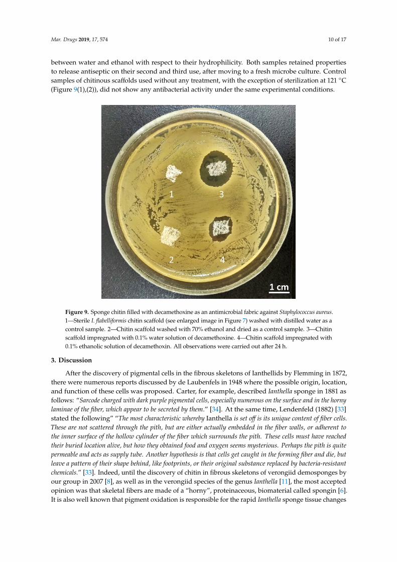
The 3D chitin scaffold of I. flabelliformis impregnated with a 0.1% water solution of decamethoxineand dried with sterile filter paper inhibited the growth of S. aureus in an agar diffusion assay (6 mmfrom the edge of the matrix) (Figure 9(3)). The chitin scaffold impregnated with 0.1% ethanol solutionof decamethoxine and then dried for ethanol evaporation showed less activity (4-mm growth inhibitionfrom the edge of the matrix) (Figure 9(4)). This phenomenon is based, probably, on the difference
Mar. Drugs 2019, 17, 574 10 of 17
between water and ethanol with respect to their hydrophilicity. Both samples retained propertiesto release antiseptic on their second and third use, after moving to a fresh microbe culture. Controlsamples of chitinous scaffolds used without any treatment, with the exception of sterilization at 121 ◦C(Figure 9(1),(2)), did not show any antibacterial activity under the same experimental conditions.
Mar. Drugs 2019, 17, x 10 of 17
microbe culture. Control samples of chitinous scaffolds used without any treatment, with the exception of sterilization at 121 °C (Figures 9(1) and 9(2)), did not show any antibacterial activity under the same experimental conditions.
Figure 9. Sponge chitin filled with decamethoxine as an antimicrobial fabric against Staphylococcus
aureus. 1—Sterile I. flabelliformis chitin scaffold (see enlarged image in Figure 7) washed with distilled water as a control sample. 2—Chitin scaffold washed with 70% ethanol and dried as a control sample. 3—Chitin scaffold impregnated with 0.1% water solution of decamethoxine. 4—Chitin scaffold impregnated with 0.1% ethanolic solution of decamethoxin. All observations were carried out after 24 h.
3. Discussion
After the discovery of pigmental cells in the fibrous skeletons of Ianthellids by Flemming in 1872, there were numerous reports discussed by de Laubenfels in 1948 where the possible origin, location, and function of these cells was proposed. Carter, for example, described Ianthella sponge in 1881 as follows: “Sarcode charged with dark purple pigmental cells, especially numerous on the surface and in the
horny laminae of the fiber, which appear to be secreted by them.” [34]. At the same time, Lendenfeld (1882) [33] stated the following” “The most characteristic whereby Ianthella is set off is its unique content of fiber
cells. These are not scattered through the pith, but are either actually embedded in the fiber walls, or adherent
to the inner surface of the hollow cylinder of the fiber which surrounds the pith. These cells must have reached
their buried location alive, but how they obtained food and oxygen seems mysterious. Perhaps the pith is quite
permeable and acts as supply tube. Another hypothesis is that cells get caught in the forming fiber and die, but
leave a pattern of their shape behind, like footprints, or their original substance replaced by bacteria-resistant
chemicals.” [33]. Indeed, until the discovery of chitin in fibrous skeletons of verongiid demosponges by our group in 2007 [8], as well as in the verongiid species of the genus Ianthella [11], the most accepted opinion was that skeletal fibers are made of a “horny”, proteinaceous, biomaterial called spongin [6]. It is also well known that pigment oxidation is responsible for the rapid Ianthella sponge tissue changes in color (mostly from yellow to purple, or even blackish purple) [33,35,48]. Preserved specimens usually remain in the dark violet condition (Figures 1, 3, and 4A).
Figure 9. Sponge chitin filled with decamethoxine as an antimicrobial fabric against Staphylococcus aureus.1—Sterile I. flabelliformis chitin scaffold (see enlarged image in Figure 7) washed with distilled water as acontrol sample. 2—Chitin scaffold washed with 70% ethanol and dried as a control sample. 3—Chitinscaffold impregnated with 0.1% water solution of decamethoxine. 4—Chitin scaffold impregnated with0.1% ethanolic solution of decamethoxin. All observations were carried out after 24 h.
3. Discussion
After the discovery of pigmental cells in the fibrous skeletons of Ianthellids by Flemming in 1872,there were numerous reports discussed by de Laubenfels in 1948 where the possible origin, location,and function of these cells was proposed. Carter, for example, described Ianthella sponge in 1881 asfollows: “Sarcode charged with dark purple pigmental cells, especially numerous on the surface and in the hornylaminae of the fiber, which appear to be secreted by them.” [34]. At the same time, Lendenfeld (1882) [33]stated the following” “The most characteristic whereby Ianthella is set off is its unique content of fiber cells.These are not scattered through the pith, but are either actually embedded in the fiber walls, or adherent tothe inner surface of the hollow cylinder of the fiber which surrounds the pith. These cells must have reachedtheir buried location alive, but how they obtained food and oxygen seems mysterious. Perhaps the pith is quitepermeable and acts as supply tube. Another hypothesis is that cells get caught in the forming fiber and die, butleave a pattern of their shape behind, like footprints, or their original substance replaced by bacteria-resistantchemicals.” [33]. Indeed, until the discovery of chitin in fibrous skeletons of verongiid demosponges byour group in 2007 [8], as well as in the verongiid species of the genus Ianthella [11], the most acceptedopinion was that skeletal fibers are made of a “horny”, proteinaceous, biomaterial called spongin [6].It is also well known that pigment oxidation is responsible for the rapid Ianthella sponge tissue changes
Mar. Drugs 2019, 17, 574 11 of 17
in color (mostly from yellow to purple, or even blackish purple) [33,35,48]. Preserved specimensusually remain in the dark violet condition (Figures 1, 3 and 4A).
In this study, we showed that the pigmental cells of I. flabelliformis are located within skeletal fibersbetween microlayers of chitin (Figure 5) and contain bromotyrosines. Previously, the biosynthesisof several bromotyrosine-related compounds, i.e., bastadins, by I. flabelliformis demosponge wasreported [41]. Compounds such as bastadin 25, 15-O-sulfonatobastadin 11, and bastadin 26 wereidentified in I. flabelliformis; however, their origin was heretofore elusive, with their production inspecial cells discovered here. Interestingly, there are still no reports of bastadin or araplysillin as typicalbromotyrosines of Ianthella species being microbially derived [48]. The intriguing question about thepossible ancient microbial origin of the pigmental cells remains open.
The biological function of bromotyrosine-producing cells could be based on previously reportedresults [49] concerning the inhibition of microbial chitinases using bromotyrosines. In this case,verongiids developed a unique chemical defense strategy to protect their skeletal fibrous chitin frombacterial and fungal invasion. Taking into account our discovery of exceptionally preserved chitinin 505-million-years-old fossil remains of the vernogiid sponge Vauxia gracilenta [1], we believe thatthe appearance of this strategy was crucial in the evolution of the sponges belonging to the orderVerongiida. Previously, we also showed that partially depigmented chitinous skeletons of selectedverongiids are still resistant to diverse bacterial chitinases under experimental conditions in vitro [11].Only completely purified sponge chitin becomes soluble in chitinase-containing solutions [50,51].
In the near future, we plan to use such a “naturally loaded” bromotyrosine chitin (Figure 3C) tostudy the possible diffusion of corresponding bromotyrosines using model systems with sea water,physiological solutions, and artificial body fluid.
To our best knowledge, there are no reports on the application of pure chitinous scaffolds fordrug delivery. Most papers are dedicated to chitosan or ionically cross-linked chitin microspheres [52].In one case, a chitin–amphipathic anion/quaternary ammonium salt dressing was prepared [53]. In ourstudy, however, we utilized a recognized antibacterial compound—decamethoxine.
Decamethoxine (its structural formula is shown in Figure 8A) is a cationic gemini surfactant [54],which exhibits strong bactericidal and fungicidal effects. It modifies the permeability of the microbialcell membrane, resulting in the destruction and death of diverse microorganisms [55]. For example, ithas a wide spectrum of antimicrobial action on Gram-positive bacteria (Staphylococcus, Streptococcus,Pneumococcus), Gram-negative bacteria (Pseudomonoas, Neisseria gonorrhoeae, Chlamydia trachomatis) [56],protozoa, dermatophyte, yeast-like fungi of Candida genus, and viruses [57]. It was also proven thatdecamethoxine at a concentration of 10 µg/ml drastically reduces the adhesion of coryneform bacteria,Salmonella, Staphylococcus, and Escherichia [56]. Its method of action may be achieved via adhesion orcompetitive binding to bacterial adhesins, or to the surface receptors of host cells. Due to its highbacteriostatic effect, decamethoxine is used for the disinfection of surfaces of diverse surgical tools [58],as well as of contact lenses [59].
Our first results (Figure 9) confirmed that decamethoxine can be successfully absorbedfrom corresponding water and ethanol-containing solutions by chitinous scaffolds isolated fromI. flabelliformis. Furthermore, this compound can subsequently diffuse from the chitinous matrix surface,as well as, probably, from the inner space of microtubular and nanoporous structures. The appearanceof death zones around colonies of S. aureus during 24 h of incubation confirms the antibiotic activity ofdecamethoxine through diffusion from the chitinous scaffold. Now, we need a longer assay includingstudies on a Fickian diffusion, as well as on possible non-Fickian behavior [60,61], of this previouslynon-investigated microtubular chitin matrix. On this first stage, we did not differentiate betweenthe release of substance adsorbed to the outside of the matrix, substance absorbed via nanopores,substance sucked up and released via capillary action, etc. Consequently, it is also not clear with whatkind of diffusion-controlled system (matrix-type system or reservoir-type system) [62] is used here.Understanding the structure–function relationship of the sponge biomaterial system represented in thisstudy as a new antimicrobial drug release scaffold [63] could be the key to the successful application of
Mar. Drugs 2019, 17, 574 12 of 17
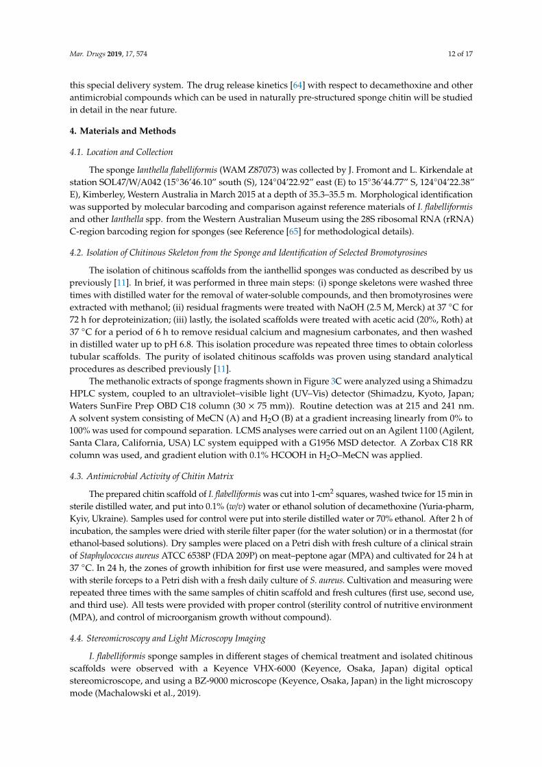
this special delivery system. The drug release kinetics [64] with respect to decamethoxine and otherantimicrobial compounds which can be used in naturally pre-structured sponge chitin will be studiedin detail in the near future.
4. Materials and Methods
4.1. Location and Collection
The sponge Ianthella flabelliformis (WAM Z87073) was collected by J. Fromont and L. Kirkendale atstation SOL47/W/A042 (15◦36’46.10” south (S), 124◦04’22.92” east (E) to 15◦36’44.77” S, 124◦04’22.38”E), Kimberley, Western Australia in March 2015 at a depth of 35.3–35.5 m. Morphological identificationwas supported by molecular barcoding and comparison against reference materials of I. flabelliformisand other Ianthella spp. from the Western Australian Museum using the 28S ribosomal RNA (rRNA)C-region barcoding region for sponges (see Reference [65] for methodological details).
4.2. Isolation of Chitinous Skeleton from the Sponge and Identification of Selected Bromotyrosines
The isolation of chitinous scaffolds from the ianthellid sponges was conducted as described by uspreviously [11]. In brief, it was performed in three main steps: (i) sponge skeletons were washed threetimes with distilled water for the removal of water-soluble compounds, and then bromotyrosines wereextracted with methanol; (ii) residual fragments were treated with NaOH (2.5 M, Merck) at 37 ◦C for72 h for deproteinization; (iii) lastly, the isolated scaffolds were treated with acetic acid (20%, Roth) at37 ◦C for a period of 6 h to remove residual calcium and magnesium carbonates, and then washedin distilled water up to pH 6.8. This isolation procedure was repeated three times to obtain colorlesstubular scaffolds. The purity of isolated chitinous scaffolds was proven using standard analyticalprocedures as described previously [11].
The methanolic extracts of sponge fragments shown in Figure 3C were analyzed using a ShimadzuHPLC system, coupled to an ultraviolet–visible light (UV–Vis) detector (Shimadzu, Kyoto, Japan;Waters SunFire Prep OBD C18 column (30 × 75 mm)). Routine detection was at 215 and 241 nm.A solvent system consisting of MeCN (A) and H2O (B) at a gradient increasing linearly from 0% to100% was used for compound separation. LCMS analyses were carried out on an Agilent 1100 (Agilent,Santa Clara, California, USA) LC system equipped with a G1956 MSD detector. A Zorbax C18 RRcolumn was used, and gradient elution with 0.1% HCOOH in H2O–MeCN was applied.
4.3. Antimicrobial Activity of Chitin Matrix
The prepared chitin scaffold of I. flabelliformis was cut into 1-cm2 squares, washed twice for 15 min insterile distilled water, and put into 0.1% (w/v) water or ethanol solution of decamethoxine (Yuria-pharm,Kyiv, Ukraine). Samples used for control were put into sterile distilled water or 70% ethanol. After 2 h ofincubation, the samples were dried with sterile filter paper (for the water solution) or in a thermostat (forethanol-based solutions). Dry samples were placed on a Petri dish with fresh culture of a clinical strainof Staphylococcus aureus ATCC 6538P (FDA 209P) on meat–peptone agar (MPA) and cultivated for 24 h at37 ◦C. In 24 h, the zones of growth inhibition for first use were measured, and samples were movedwith sterile forceps to a Petri dish with a fresh daily culture of S. aureus. Cultivation and measuring wererepeated three times with the same samples of chitin scaffold and fresh cultures (first use, second use,and third use). All tests were provided with proper control (sterility control of nutritive environment(MPA), and control of microorganism growth without compound).
4.4. Stereomicroscopy and Light Microscopy Imaging
I. flabelliformis sponge samples in different stages of chemical treatment and isolated chitinousscaffolds were observed with a Keyence VHX-6000 (Keyence, Osaka, Japan) digital opticalstereomicroscope, and using a BZ-9000 microscope (Keyence, Osaka, Japan) in the light microscopymode (Machalowski et al., 2019).
Mar. Drugs 2019, 17, 574 13 of 17
4.5. Transmission Electron Microscopy (TEM), Scanning Electron Microscopy (SEM), and EDX
For TEM investigations, samples of I. flabelliformis chitin as represented in Figure 3C were fixedwith 2.5% glutaraldehyde in phosphate-buffered saline (PBS) at room temperature, post-fixed with1.5% osmium tetroxide, dehydrated in a graduated series of acetone (including a staining step with1% uranylacetate), and embedded in Epoxy resin according to Spurr (1969) [66]. Ultra-thin sections(about 70 nm) of samples were prepared on a Leica EM UC6 ultramicrotome (Leica, Wetzlar, Germany)equipped with a Diatome diamond knife, mounted on Pioloform-coated copper grids, post-stainedwith uranylacetate and lead citrate (according to Reynolds, 1963 [67]), and analyzed using a ZeissCTEM 902 (Carl Zeiss, Wetzlar, Germany) at 80 kV (University of Bayreuth).
For SEM analysis, samples were prepared as described for TEM analyses without osmium tetroxideand uranylacetate. For elemental analyses (EDX), the block faces of samples were cut on a Leica EMUC6 ultramicrotome equipped with a Diatome diamond knife, carbon-coated, mounted on an SEMsample holder, and analyzed on a Philips ESEM XL 30 (FEI Company, Peabody, MA, USA) at suitableaccelerating voltages. For EDX spectra, accelerating voltages between 15 kV and 30 kV were used.
5. Conclusions
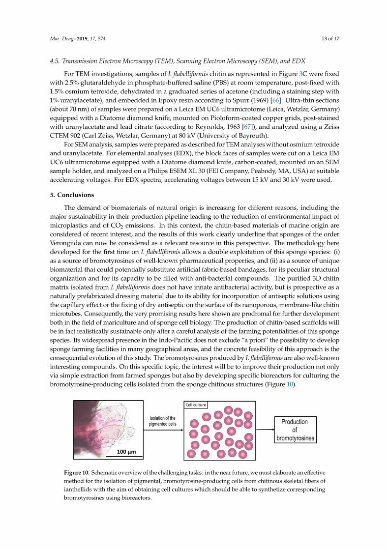
The demand of biomaterials of natural origin is increasing for different reasons, including themajor sustainability in their production pipeline leading to the reduction of environmental impact ofmicroplastics and of CO2 emissions. In this context, the chitin-based materials of marine origin areconsidered of recent interest, and the results of this work clearly underline that sponges of the orderVerongiida can now be considered as a relevant resource in this perspective. The methodology heredeveloped for the first time on I. flabelliformis allows a double exploitation of this sponge species: (i)as a source of bromotyrosines of well-known pharmaceutical properties, and (ii) as a source of uniquebiomaterial that could potentially substitute artificial fabric-based bandages, for its peculiar structuralorganization and for its capacity to be filled with anti-bacterial compounds. The purified 3D chitinmatrix isolated from I. flabelliformis does not have innate antibacterial activity, but is prospective as anaturally prefabricated dressing material due to its ability for incorporation of antiseptic solutions usingthe capillary effect or the fixing of dry antiseptic on the surface of its nanoporous, membrane-like chitinmicrotubes. Consequently, the very promising results here shown are prodromal for further developmentboth in the field of mariculture and of sponge cell biology. The production of chitin-based scaffolds willbe in fact realistically sustainable only after a careful analysis of the farming potentialities of this spongespecies. Its widespread presence in the Indo-Pacific does not exclude “a priori” the possibility to developsponge farming facilities in many geographical areas, and the concrete feasibility of this approach is theconsequential evolution of this study. The bromotyrosines produced by I. flabelliformis are also well-knowninteresting compounds. On this specific topic, the interest will be to improve their production not onlyvia simple extraction from farmed sponges but also by developing specific bioreactors for culturing thebromotyrosine-producing cells isolated from the sponge chitinous structures (Figure 10).
Mar. Drugs 2019, 17, x 13 of 17
4.5. Transmission Electron Microscopy (TEM), Scanning Electron Microscopy (SEM), and EDX
For TEM investigations, samples of I. flabelliformis chitin as represented in Figure 3C were fixed with 2.5% glutaraldehyde in phosphate-buffered saline (PBS) at room temperature, post-fixed with 1.5% osmium tetroxide, dehydrated in a graduated series of acetone (including a staining step with 1% uranylacetate), and embedded in Epoxy resin according to Spurr (1969) [66]. Ultra-thin sections (about 70 nm) of samples were prepared on a Leica EM UC6 ultramicrotome (Leica, Wetzlar, Germany) equipped with a Diatome diamond knife, mounted on Pioloform-coated copper grids, post-stained with uranylacetate and lead citrate (according to Reynolds, 1963 [67]), and analyzed using a Zeiss CTEM 902 (Carl Zeiss, Wetzlar, Germany) at 80 kV (University of Bayreuth).
For SEM analysis, samples were prepared as described for TEM analyses without osmium tetroxide and uranylacetate. For elemental analyses (EDX), the block faces of samples were cut on a Leica EM UC6 ultramicrotome equipped with a Diatome diamond knife, carbon-coated, mounted on an SEM sample holder, and analyzed on a Philips ESEM XL 30 (FEI Company, Peabody, MA, USA ) at suitable accelerating voltages. For EDX spectra, accelerating voltages between 15 kV and 30 kV were used.
5. Conclusions
The demand of biomaterials of natural origin is increasing for different reasons, including the major sustainability in their production pipeline leading to the reduction of environmental impact of microplastics and of CO2 emissions. In this context, the chitin-based materials of marine origin are considered of recent interest, and the results of this work clearly underline that sponges of the order Verongiida can now be considered as a relevant resource in this perspective. The methodology here developed for the first time on I. flabelliformis allows a double exploitation of this sponge species: (i) as a source of bromotyrosines of well-known pharmaceutical properties, and (ii) as a source of unique biomaterial that could potentially substitute artificial fabric-based bandages, for its peculiar structural organization and for its capacity to be filled with anti-bacterial compounds. The purified 3D chitin matrix isolated from I. flabelliformis does not have innate antibacterial activity, but is prospective as a naturally prefabricated dressing material due to its ability for incorporation of antiseptic solutions using the capillary effect or the fixing of dry antiseptic on the surface of its nanoporous, membrane-like chitin microtubes. Consequently, the very promising results here shown are prodromal for further development both in the field of mariculture and of sponge cell biology. The production of chitin-based scaffolds will be in fact realistically sustainable only after a careful analysis of the farming potentialities of this sponge species. Its widespread presence in the Indo-Pacific does not exclude “a priori” the possibility to develop sponge farming facilities in many geographical areas, and the concrete feasibility of this approach is the consequential evolution of this study. The bromotyrosines produced by I. flabelliformis are also well-known interesting compounds. On this specific topic, the interest will be to improve their production not only via simple extraction from farmed sponges but also by developing specific bioreactors for culturing the bromotyrosine-producing cells isolated from the sponge chitinous structures (Figure 10).
Figure 10. Schematic overview of the challenging tasks: in the near future, we must elaborate an effective method for the isolation of pigmental, bromotyrosine-producing cells from chitinous skeletal
Figure 10. Schematic overview of the challenging tasks: in the near future, we must elaborate an effectivemethod for the isolation of pigmental, bromotyrosine-producing cells from chitinous skeletal fibers ofianthellids with the aim of obtaining cell cultures which should be able to synthetize correspondingbromotyrosines using bioreactors.
Mar. Drugs 2019, 17, 574 14 of 17
Supplementary Materials: The following are available online at http://www.mdpi.com/1660-3397/17/10/574/s1:Figure S1: Structures of bromotyrosines isolated from l. flabelliformis, Figure S2: LCMS spectrum of isolatedcompound (Bastadin 25), Figure S3: LCMS spectrum of isolated compound (Araplysillin-I N20-sulfamate).
Author Contributions: Conceptualization, H.E., K.G., V.K., O.B.S., and S.R.B.; methodology, M.S., A.V., L.M.,I.P., D.E., M.V.T., R.M., N.B., and A.F.; validation, H.E., S.R.B., and Y.J.; investigation, B.B., M.S., A.V., L.M.,M.W., I.P., V.K., M.V.T., J.F., V.N.I., D.E., A.S., and O.B.S.; resources, J.F., D.E., K.G., and H.E.; writing—originaldraft preparation, H.E., A.V., M.W., and M.G. (Marco Giovine); writing—review and editing, M.W., J.F., M.G.(Marco Giovine), and V.N.I.; supervision, H.E.; funding acquisition, H.E., K.G., and M.G. (Michael Gelinsky).
Funding: This work was partially supported by DFG Project HE 394/3, SMWK Project no. 02010311 (Germany),and DAAD-Italy Project “Marine Sponges as Sources for Bioinspired Materials Science” (No. 57397326). Workin the Guan group is financially supported by the Free State of Saxony and the European Union EFRE (SABproject “PhänoKard”) and by the DFG (GU595/3-1, IRTG2251). Björn Binnewerg was supported by the Else KrönerFresenius Stiftung (Else-Kröner Promotionskolleg, EKFS Foundation). M.W. is financially supported by the PolishNational Agency for Academic Exchange (PPN/BEK/2018/1/00071).
Acknowledgments: The authors acknowledge Oliver Gomez, Western Australian Museum, for technical support.Collection of the sponge was undertaken as part of the Western Australian Marine Science Institution project(Theme 1.1.1, http://www.wamsi.org.au/Kimberley-science-node). D.E. would like to thank Leonard Namuth andGert Wörheide for various aspects of the barcoding work. The authors are thankful to Sarah Tsurkan for Englishproof reading.
Conflicts of Interest: The authors declare no conflicts of interest.
References
1. Ehrlich, H.; Rigby, J.K.; Botting, J.P.; Tsurkan, M.V.; Werner, C.; Schwille, P.; Petrášek, Z.; Pisera, A.; Simon, P.;Sivkov, V.N.; et al. Discovery of 505-million-year old chitin in the basal demosponge Vauxia gracilenta.Sci. Rep. 2013, 3, 3497. [CrossRef] [PubMed]
2. Mioso, R.; Marante, F.J.T.; Bezerra, R.D.S.; Borges, F.V.P.; Santos, B.V.D.O.; De Laguna, I.H.B. Cytotoxiccompounds derived from marine sponges: A review (2010–2012). Molecules 2017, 22, 208. [CrossRef][PubMed]
3. Helber, S.B.; Hoeijmakers, D.J.J.; Muhando, C.A.; Rohde, S.; Schupp, P.J. Sponge chemical defenses are apossible mechanism for increasing sponge abundance on reefs in Zanzibar. PLoS ONE 2018, 13, e0197617.[CrossRef] [PubMed]
4. Ebel, R.; Brenzinger, M.; Kunze, A.; Gross, H.J.; Proksch, P. Wound activation of protoxins in marine spongeAplysina aerophoba. J. Chem. Ecol. 1997, 23, 1451–1462. [CrossRef]
5. Green, D.W.; Howard, D.; Yang, X.; Kelly, M.; Oreffo, R.O.C. Natural marine sponge fiber skeleton: Abiomimetic scaffold for human osteoprogenitor cell attachment, growth, and differentiation. Tissue Eng.2003, 9, 1159–1166. [CrossRef] [PubMed]
6. Jesionowski, T.; Norman, M.; Zółtowska-Aksamitowska, S.; Petrenko, I.; Joseph, Y.; Ehrlich, H. Marinespongin: Naturally prefabricated 3D scaffold-based biomaterial. Mar. Drugs 2018, 16, 88. [CrossRef][PubMed]
7. Ehrlich, H.; Wysokowski, M.; Zółtowska-Aksamitowska, S.; Petrenko, I.; Jesionowski, T. Collagens ofporiferan origin. Mar. Drugs 2018, 16, 79. [CrossRef] [PubMed]
8. Ehrlich, H.; Maldonado, M.; Spindler, K.-D.; Eckert, S.; Hanke, T.; Born, R.; Goebel, C.; Simon, P.; Heinemann, S.;Worch, H. First evidence of chitin as a component of the skeletal fibres of marine sponges. Part I. Verongidae(Demospongia: Porifera). J. Exp. Zool. B Mol. Dev. Evol. 2007, 308, 347–356. [CrossRef]
9. Ehrlich, H.; Ilan, M.; Maldonado, M.; Muricy, G.; Bavestrello, G.; Kljajic, Z.; Carballo, J.L.; Schiaparelli, S.;Ereskovsky, A.; Schupp, P.; et al. Three-dimensional chitin-based scaffolds from Verongida sponges(Demospongiae: Porifera). Part I. Isolation and identification of chitin. Int. J. Biol. Macromol. 2010, 47,132–140. [CrossRef]
10. Ehrlich, H.; Shaala, L.A.; Youssef, D.T.A.; Zoltowska-Aksamitowska, S.; Tsurkan, M.; Galli, R.; Meissner, H.;Wysokowski, M.; Petrenko, I.; Tabachnick, K.R.; et al. Discovery of chitin in skeletons of non-verongiid RedSea demosponges. PLoS ONE 2018, 13, 1–18. [CrossRef]
11. Brunner, E.; Ehrlich, H.; Schupp, P.; Hedrich, R.; Hunoldt, S.; Kammer, M.; Machill, S.; Paasch, S.;Bazhenov, V.V.; Kurek, D.V.; et al. Chitin-based scaffolds are an integral part of the skeleton of the marinedemosponge Ianthella basta. J. Struct. Biol. 2009, 168, 539–547. [CrossRef] [PubMed]
Mar. Drugs 2019, 17, 574 15 of 17
12. Rohde, S.; Schupp, P.J. Growth and regeneration of the elephant ear sponge Ianthella basta (Porifera).Hydrobiologia 2012, 687, 219–226. [CrossRef]
13. Mutsenko, V.V.; Gryshkov, O.; Lauterboeck, L.; Rogulska, O.; Tarusin, D.N.; Bazhenov, V.V.; Schütz, K.;Brüggemeier, S.; Gossla, E.; Akkineni, A.R.; et al. Novel chitin scaffolds derived from marine sponge Ianthellabasta for tissue engineering approaches based on human mesenchymal stromal cells: Biocompatibility andcryopreservation. Int. J. Biol. Macromol. 2017, 104, 1955–1965. [CrossRef] [PubMed]
14. Stepniak, I.; Galinski, M.; Nowacki, K.; Wysokowski, M.; Jakubowska, P.; Bazhenov, V.V.; Leisegang, T.;Ehrlich, H.; Jesionowski, T. A novel chitosan/sponge chitin origin material as a membrane forsupercapacitors-preparation and characterization. RSC Adv. 2016, 6, 4007–4013. [CrossRef]
15. Wysokowski, M.; Petrenko, I.; Stelling, A.L.; Stawski, D.; Jesionowski, T.; Ehrlich, H. Poriferan chitin as aversatile template for extreme biomimetics. Polymers (Basel) 2015, 7, 235–265. [CrossRef]
16. Ehrlich, H. Chitin of poriferan origin as a unique biological material. In Blue Biotechnology: Production andUse of Marine Molecules; Bates, S.S., La Barre, S., Eds.; Wiley-VCH: Weinheim, Germany, 2019; pp. 821–854.
17. Fromont, J.; Wahab, M.A.A.; Gomez, O.; Ekins, M.; Grol, M.; Hooper, J.N.A. Patterns of sponge biodiversityin the Pilbara, Northwestern Australia. Diversity 2016, 8, 21. [CrossRef]
18. Bechmann, N.; Ehrlich, H.; Eisenhofer, G.; Ehrlich, A.; Meschke, S.; Ziegler, C.G.; Bornstein, S.R.Anti-tumorigenic and anti-metastatic activity of the sponge-derived marine drugs aeroplysinin-1 andisofistularin-3 against pheochromocytoma in vitro. Mar. Drugs 2018, 16, 172. [CrossRef]
19. Kumar, M.S.; Adki, K.M. Marine natural products for multi-targeted cancer treatment: A future insight.Biomed. Pharmacother. 2018, 105, 233–245. [CrossRef]
20. Niemann, H.; Hagenow, J.; Chung, M.-Y.; Hellio, C.; Weber, H.; Proksch, P. SAR of Sponge-inspiredhemibastadin congeners inhibiting blue mussel phenoloxidase. Mar. Drugs 2015, 13, 3061–3071. [CrossRef]
21. Bayer, M.; Hellio, C.; Marechal, J.P.; Frank, W.; Lin, W.; Weber, H.; Proksch, P. Antifouling bastadin congenerstarget mussel phenoloxidase and complex copper(II) Ions. Mar. Biotechnol. 2011, 13, 1148–1158. [CrossRef]
22. Calcul, L.; Inman, W.D.; Morris, A.A.; Tenney, K.; Ratnam, J.; McKerrow, J.H.; Valeriote, F.A.; Crews, P.Additional insights on the bastadins: Isolation of analogues from the sponge Ianthella cf. reticulata andexploration of the oxime configurations. J. Nat. Prod. 2010, 73, 365–372. [CrossRef] [PubMed]
23. Franklin, M.A.; Penn, S.G.; Lebrilla, C.B.; Lam, T.H.; Pessah, I.N.; Molinski, T.F. Bastadin 20 and bastadinO-sulfate esters from Ianthella basta: Novel modulators of the Ry1R FKBP12 receptor complex. J. Nat. Prod.1996, 59, 1121–1127. [CrossRef] [PubMed]
24. Greve, H.; Kehraus, S.; Krick, A.; Kelter, G.; Maier, A.; Fiebig, H.H.; Wright, A.D.; König, G.M. Cytotoxicbastadin 24 from the Australian sponge Ianthella quadrangulata. J. Nat. Prod. 2008, 71, 309–312. [CrossRef][PubMed]
25. Mathieu, V.; Wauthoz, N.; Lefranc, F.; Niemann, H.; Amighi, K.; Kiss, R.; Proksch, P. Cyclic versushemi-bastadins. Pleiotropic anti-cancer effects: From apoptosis to anti-angiogenic and anti-migratory effects.Molecules 2013, 18, 3543–3561. [CrossRef] [PubMed]
26. Le Norcy, T.; Niemann, H.; Proksch, P.; Tait, K.; Linossier, I.; Réhel, K.; Hellio, C.; Faÿ, F. Sponge-inspireddibromohemibastadin prevents and disrupts bacterial biofilms without toxicity. Mar. Drugs 2017, 15, 222.[CrossRef]
27. Gartshore, C.J.; Salib, M.N.; Renshaw, A.A.; Molinski, T.F. Isolation of Bastadin-6-O-Sulfate and expedientpurifications of Bastadins-4, −5 and −6 from extracts of Ianthella basta. Fitoterapia 2018, 126, 16–21. [CrossRef][PubMed]
28. Van Soest, R.W.M.; Boury-Esnault, N.; Hooper, J.N.A.; Rützler, K.; de Voogd, N.J.; Alvarez, B.; Hajdu, E.;Pisera, A.B.; Manconi, R.; Schönberg, C.; et al. World Porifera Database. Ianthella Flabelliformis (Linnaeus,1759). Available online: http://www.marinespecies.org/aphia.php?p=taxdetails&id=169690 (accessed on 8October 2019).
29. Linnaeus, C. Systema Naturæ per Regna Tria Naturæ, Secundum Classes, Ordines, Genera, Species, CumCharacteribus, Differentiis, Synonymis, Locis; Vindobonae: Vienna, Austria, 1759. [CrossRef]
30. Weuffen, W.; Berencsi, G.; Groschel, D.; Kemter, B.; Kramer, A.K.A.P. Handbuch der Antiseptik; Band 2:Antiseptika; Teil 3: Antibakterielle, Antifungielle und antivirale Antiseptik-Ausgewählte Wirkstoffe; VEB Verlag Volkund Gesundheit.: Berlin, Germany, 1987.
Mar. Drugs 2019, 17, 574 16 of 17
31. Paliy, G.K.; Nazarchuk, O.A.; Kulakov, O.I.; Paliy, V.G.; Nazarchuk, S.A.; Paliy, D.V.; Kordon, Y.V.;Gonchar, O.O. Substaniation of antimicrobial dressings use in surgery. Med. Perspekt. 2014, 19, 152–158.[CrossRef]
32. Klinger, C.; Zółtowska-Aksamitowska, S.; Wysokowski, M.; Tsurkan, M.V.; Galli, R.; Petrenko, I.;Machałowski, T.; Ereskovsky, A.; Martinovic, R.; Muzychka, L.; et al. Express method for isolationof ready-to-use 3D chitin scaffolds from Aplysina archeri (Aplysineidae: Verongiida) demosponge. Mar. Drugs2019, 17, 131. [CrossRef]
33. Lendenfeld, R.L.R. Das hornfaserwachstum der aplysinidae. Zool. Anz. 1882, 5, 636.34. Carter, H.J. Contributions to our knowledge of the Spongida. Ann. Mag. Nat. Hist. 1881, 8, 241–259.
[CrossRef]35. Lendenfeld, R.L.R. A Monograph of the Horny Sponges; Triibner and Co.: London, UK, 1889.36. Polejaeff, N.N. Report on the Keratosa collected by H.M.S. Challenger during the years 1873–1876. In Report
on the Scientific Results of the Voyage of H.M.S. Challenger during the Years 1873–1876; for Her Majesty’s StationaryOffice, Zoology: London, UK; Edinburgh, Dublin, Ireland, 1884; pp. 1–88.
37. De Laubenfels, M.W. The order of Keratosa of the phylum Porifera. A monographic study. Occ. Pap. AllanHancock Found. 1948, 3, 1–217.
38. Vacelet, J. Les cellules a inclusions de léponge cornée Verongia cavernicola. J. Microsc. 1967, 6, 237–240.39. Turon, X.; Becerro, M.A.; Uriz, M.J. Distribution of brominated compounds within the sponge Aplysina
aerophoba: Coupling of X-ray microanalysis with cryofixation techniques. Cell Tissue Res. 2000, 301, 311–322.[CrossRef] [PubMed]
40. Bergquist, P.R.; de Cook, S.C. Order Verongida Bergquist, 1978. In Systema Porifera; Hooper, J.N.A.,Van Soest, R.W.M., Willenz, P., Eds.; Springer: Boston, MA, USA, 2002; p. 1081.
41. Carroll, A.R.; Kaiser, S.M.; Davis, R.A.; Moni, R.W.; Hooper, J.N.A.; Quinn, R.J. A bastadin with potent andselective δ-opioid receptor binding affinity from the Australian sponge Ianthella flabelliformis. J. Nat. Prod.2010, 73, 1173–1176. [CrossRef] [PubMed]
42. Motti, C.A.; Freckelton, M.L.; Tapiolas, D.M.; Willis, R.H. FTICR-MS and LC-UV/MS-SPE-NMR applicationsfor the rapid dereplication of a crude extract from the sponge Ianthella flabelliformis. J. Nat. Prod. 2009, 72,290–294. [CrossRef] [PubMed]
43. Rogers, E.W.; Molinski, T.F. Highly polar spiroisoxazolines from the sponge Aplysina fulva. J. Nat. Prod. 2007,70, 1191–1194. [CrossRef]
44. Höfer, D. Antimicrobial Textiles – Evaluation of their effectiveness and safety. In Biofunctional Textiles and theSkin; Hipler, U.-C., Elsner, P., Eds.; Karger: Basel, Switzerland, 2006; pp. 42–50.
45. Kramer, A.; Guggenbichler, P.; Heldt, P.; Jünger, M.; Ladwig, A.; Thierbach, H.; Weber, U.; Daeschlein, G.Hygienic relevance and risk assessment of antimicrobial-impregnated textiles. Curr. Probl. Dermatol. 2006,33, 78–109. [PubMed]
46. Elsner, P. Antimicrobials and the skin: Physiological and pathological flora. Curr. Probl. Dermatol. 2006, 33,35–41.
47. Gokarneshan, N.; Nagarajan, V.B.; Viswanath, S.R. Developments in antimicrobial textiles—Some insightson current research trends. Biomed. J. Sci Tech. Res. 2017, 1, 230–233.
48. Freckelton, M.L.; Luter, H.M.; Andreakis, N.; Webster, N.S.; Motti, C.A. Qualitative variation in colourmorphotypes of Ianthella basta (Porifera: Verongida). In Ancient Animals, New Challenges. Developments inHydrobiology; Springer: Dordrecht, The Netherlands, 2011.
49. Tabudravu, J.N.; Eijsink, V.G.H.; Gooday, G.W.; Jaspars, M.; Komander, D.; Legg, M.; Synstad, B.; VanAalten, D.M.F. Psammaplin A, a chitinase inhibitor isolated from the Fijian marine sponge Aplysinella rhax.Bioorganic Med. Chem. 2002, 10, 1123–1128. [CrossRef]
50. Ehrlich, H.; Bazhenov, V.V.; Debitus, C.; de Voogd, N.; Galli, R.; Tsurkan, M.V.; Wysokowski, M.; Meissner, H.;Bulut, E.; Kaya, M.; et al. Isolation and identification of chitin from heavy mineralized skeleton of Subereaclavata (Verongida: Demospongiae: Porifera) marine demosponge. Int. J. Biol. Macromol. 2017, 104,1706–1712. [CrossRef] [PubMed]
51. Shaala, L.A.; Asfour, H.Z.; Youssef, D.T.A.; ółtowska-Aksamitowska, S.Z.; Wysokowski, M.; Tsurkan, M.;Galli, R.; Meissner, H.; Petrenko, I.; Tabachnick, K.; et al. New source of 3D chitin scaffolds: The red seademosponge Pseudoceratina arabica (pseudoceratinidae, verongiida). Mar. Drugs 2019, 17, 92. [CrossRef][PubMed]
Mar. Drugs 2019, 17, 574 17 of 17
52. Shang, Y.; Ding, F.; Xiao, L.; Deng, H.; Du, Y.; Shi, X. Chitin-based fast responsive pH sensitive microspheresfor controlled drug release. Carbohydr. Polym. 2014, 102, 413–418. [CrossRef]
53. Zhou, D.; Yang, R.; Yang, T.; Xing, M.; Gaoxing, L.G. Preparation of chitin-amphipathic anion/quaternaryammonium salt ecofriendly dressing and its effect on wound healing in mice. Int. J. Nanomed. 2018, 13,4157–4169. [CrossRef] [PubMed]
54. Dement’eva, O.V.; Naumova, K.A.; Senchikhin, I.N.; Roumyantseva, T.B.; Rudoy, V.M. Sol–gel synthesis ofmesostructured SiO2 containers using vesicles of hydrolyzable bioactive gemini surfactant as a template.Colloid J. 2017, 79, 451–458. [CrossRef]
55. Shchetina, V.N.; Belanov, E.F.; Starobinets, Z.G.; Volianskiı, I. The effect of dexamethoxin on the integrity ofcytoplasmic membrane in gram-positive and gram-negative microorganisms. Mikrobiol. Zh 1990, 52, 24–28.[PubMed]
56. Nazarchuk, O.A.; Chereshniuk, I.L.; Nazarchuk, H.H. The research of antimicrobial efficacy of antisepticsdecamethoxin, miramistin and their effect on nuclear DNA fragmentation and epithelial cell cycle. Wiad. Lek.2019, 72, 374–380.
57. Fuss, J.; Palii, V.; Voloboyeva, A. evaluating the effectivenes of antiseptic solution decasan in treatment ofnecrotic soft tisue diseases. Pol. Przegl. Chir. 2016, 88, 233–237. [CrossRef]
58. Paliy, G.K.; Lukhimel, A.D.; Onofreıchuk, I.F. Disinfection of surgical silk with decamethoxin, antibiotics andtheir combinations. Antibiotiki 1978, 88, 629–633.
59. Galatenko, N.A.; Nechaeva, L.; Bufius, N.N. Possibilities of the chemical sterilization of soft contact lensesmade of polyacrylamide. Gig. Sanit. 1991, 7, 67–68.
60. Ritger, P.L.; Peppas, N.A.A. Fickian and non-Fickian release from non-swellable devices in the form of slabs,spheres, cylinders or discs. J. Control. Release 1987, 5, 23–36. [CrossRef]
61. Peppas, A.N. Analysis of Fickian and non-Fickian drug release from polymers. Pharm. Acta Helv. 1985, 60,110–111.
62. He, C.; Nie, W.; Feng, W. Engineering of biomimetic nanofibrous matrices for drug delivery and tissueengineering. J. Mater. Chem. B 2014, 2, 7828–7848. [CrossRef]
63. Prabu, P.; Kim, K.W.; Dharmaraj, N.; Park, J.H.; Khil, M.S.; Kim, H.Y. Antimicrobial drug release scaffolds ofnatural and synthetic biodegradable polymers. Macromol. Res. 2008, 16, 303–307. [CrossRef]
64. Kamaly, N.; Yameen, B.; Wu, J.; Omid, C.; Farokhzad, O.C. Degradable controlled-release polymers andpolymeric nanoparticles: Mechanisms of controlling drug release. Chem. Rev. 2016, 116, 2602–2663.[CrossRef]
65. Erpenbeck, D.; Voigt, O.; Al-Aidaroos, A.M.; Berumen, M.L.; Büttner, G.; Catania, D.; Guirguis, A.N.;Paulay, G.; Schätzle, S.; Wörheide, G. Molecular biodiversity of Red Sea demosponges. Mar. Pollut. Bull.2016, 105, 507–514. [CrossRef]
66. Spurr, A.R. A low-viscosity epoxy resin embedding medium for electron microscopy. J. Ultrastruct. Res.1969, 26, 31–43. [CrossRef]
67. Reynolds, E.S. The use of lead citrate at high pH as an electron-opaque stain in electron microscopy. J. Cell Biol.1963, 208–212. [CrossRef]
© 2019 by the authors. Licensee MDPI, Basel, Switzerland. This article is an open accessarticle distributed under the terms and conditions of the Creative Commons Attribution(CC BY) license (http://creativecommons.org/licenses/by/4.0/).


![Liposomes the potential drug carriers - IOSR-PHR · Liposomes – the potential drug carriers 28 1.3.1.2. Membrane Additives [Sterols] Cholesterol is the most commonly used sterol,](https://img.pdfslide.fr/doc/110x75/5ec63da195aa25320c743ecf/liposomes-the-potential-drug-carriers-iosr-liposomes-a-the-potential-drug-carriers.jpg)