Embed Size (px)
Citation preview
CMLS, Cell. Mol. Life Sci. 55 (1999) 1015–10281420-682X/99/091015-14 $ 1.50+0.20/0© Birkhauser Verlag, Basel, 1999
Nitric oxide biosynthesis, nitric oxide synthase inhibitorsand arginase competition for L-arginine utilizationJ. L. Bouchera,*, C. Moalia and J. P. Tenub
aLaboratoire de Chimie et Biochimie Pharmacologiques et Toxicologiques, URA 400 CNRS, 45 rue des SaintsPeres, F-75270 Paris Cedex 06 (France), Fax +33 1 42 86 83 87, e-mail: [email protected] 571 CNRS, Bat. 430, Universite Paris-Sud XI, F-91405 Orsay Cedex (France)
well as NOS stability are finely regulated by Ca2+/Abstract. Nitric oxide (NO) is a recently discoveredcalmodulin interactions, by the cofactor tetrahydro-mediator produced by mammalian cells. It plays a keybiopterin, and by substrate availability. Strongrole in neurotransmission, control of blood pressure,interactions between the L-arginine-metabolizing en-and cellular defense mechanisms. Nitric oxide synthaseszymes are clearly demonstrated by competition between(NOSs) catalyze the oxidation of L-arginine to NO and
L-citrulline. NOSs are unique enzymes in that they NOSs and arginases for L-arginine utilization, and bypotent inhibition of arginase activity by Nv-hydroxy-L-possess on the same polypeptidic chain a reductase
domain and an oxygenase domain closely related to arginine, an intermediate in the L-arginine to NO path-way.cytochrome P450s. NO and superoxide formation as
Key words. L-Arginine; nitric oxide; nitric oxide synthase; arginase; urea cycle; Nv-hydroxy-L-arginine; superoxideradical; peroxynitrite.
Introduction
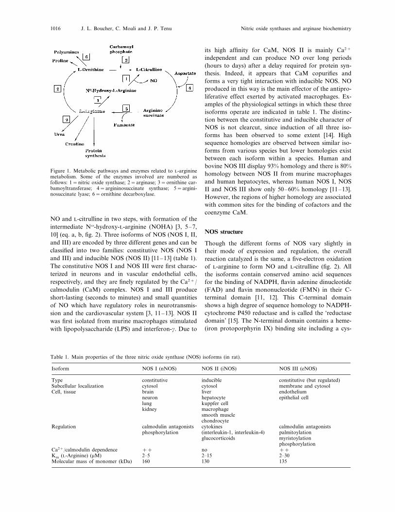
In mammalian cells, the semi-essential amino acid L-arginine is involved in protein synthesis. It is also usedas a substrate by enzymes like nitric oxide (NO) syn-thases (NOSs), arginases, arginine decarboxylase, orglycine transamidinase [1–3]. Among these pathways,the biosynthesis of NO and the urea cycle are presumedto play the most important roles in the metabolism ofL-arginine (fig. 1). L-Arginine is metabolized by NOSsto form NO and L-citrulline, and by arginase to formurea and L-ornithine, a precursor of the polyaminesspermine and spermidine used as growth factors. NO isan important mediator of many (patho)physiologicalevents [3] whereas arginase is a key enzyme of the ureacycle, an essential metabolic pathway for the removal ofhighly toxic ammonium ions resulting from proteindegradation [2, 4].Many books and reviews summarize current knowledgeconcerning the roles of NO in (patho)physiological situ-
ations and its biosynthesis from L-arginine [5–8]. Thepresent article will focus on some aspects of the biologyand enzymology of NOSs and arginases. Readers inter-ested in the transcriptional and translational regulationof these enzymes are directed to reviews that haveappeared in the literature [8, 9]. Regulation of NObiosynthesis has been achieved using L-arginineanalogs, able to compete for access to the active site. Itnow appears that regulation of NO formation can alsobe obtained by modulation of L-arginine availability,through the involvement of arginases. These resultshave led many research teams to investigate the cross-regulations between some of these L-arginine-metaboliz-ing enzymes. There is currently a resurgence of interestin arginases in the light of their newly perceived roles inthe regulation of NO synthesis.
Three isoforms of NOS
In mammalian cells, NOSs are the enzymes responsiblefor NO generation and they catalyze the oxidation ofone Nv-atom of the guanidino group of L-arginine to* Corresponding author.
J. L. Boucher, C. Moali and J. P. Tenu Nitric oxide synthases and arginase biochemistry1016
Figure 1. Metabolic pathways and enzymes related to L-argininemetabolism. Some of the enzymes involved are numbered asfollows: 1=nitric oxide synthase; 2=arginase; 3=ornithine car-bamoyltransferase; 4=argininosuccinate synthase; 5=argini-nosuccinate lyase; 6=ornithine decarboxylase.
its high affinity for CaM, NOS II is mainly Ca2+
independent and can produce NO over long periods(hours to days) after a delay required for protein syn-thesis. Indeed, it appears that CaM copurifies andforms a very tight interaction with inducible NOS. NOproduced in this way is the main effector of the antipro-liferative effect exerted by activated macrophages. Ex-amples of the physiological settings in which these threeisoforms operate are indicated in table 1. The distinc-tion between the constitutive and inducible character ofNOS is not clearcut, since induction of all three iso-forms has been observed to some extent [14]. Highsequence homologies are observed between similar iso-forms from various species but lower homologies existbetween each isoform within a species. Human andbovine NOS III display 93% homology and there is 80%homology between NOS II from murine macrophagesand human hepatocytes, whereas human NOS I, NOSII and NOS III show only 50–60% homology [11–13].However, the regions of higher homology are associatedwith common sites for the binding of cofactors and thecoenzyme CaM.
NOS structure
Though the different forms of NOS vary slightly intheir mode of expression and regulation, the overallreaction catalyzed is the same, a five-electron oxidationof L-arginine to form NO and L-citrulline (fig. 2). Allthe isoforms contain conserved amino acid sequencesfor the binding of NADPH, flavin adenine dinucleotide(FAD) and flavin mononucleotide (FMN) in their C-terminal domain [11, 12]. This C-terminal domainshows a high degree of sequence homology to NADPH-cytochrome P450 reductase and is called the ‘reductasedomain’ [15]. The N-terminal domain contains a heme-(iron protoporphyrin IX) binding site including a cys-
NO and L-citrulline in two steps, with formation of theintermediate Nv-hydroxy-L-arginine (NOHA) [3, 5–7,10] (eq. a, b, fig. 2). Three isoforms of NOS (NOS I, II,and III) are encoded by three different genes and can beclassified into two families: constitutive NOS (NOS Iand III) and inducible NOS (NOS II) [11–13] (table 1).The constitutive NOS I and NOS III were first charac-terized in neurons and in vascular endothelial cells,respectively, and they are finely regulated by the Ca2+/calmodulin (CaM) complex. NOS I and III produceshort-lasting (seconds to minutes) and small quantitiesof NO which have regulatory roles in neurotransmis-sion and the cardiovascular system [3, 11–13]. NOS IIwas first isolated from murine macrophages stimulatedwith lipopolysaccharide (LPS) and interferon-g. Due to
Table 1. Main properties of the three nitric oxide synthase (NOS) isoforms (in rat).
Isoform NOS I (nNOS) NOS II (iNOS) NOS III (eNOS)
constitutive inducible constitutive (but regulated)Typecytosol cytosol membrane and cytosolSubcellular localization
liverCell, tissue brain endotheliumhepatocyte epithelial cellneuronkuppfer celllungmacrophagekidneysmooth musclechondrocytecytokinesRegulation calmodulin antagonistscalmodulin antagonists(interleukin-1, interleukin-4)phosphorylation palmitoylationglucocorticoids myristoylation
phosphorylation++no++Ca2+/calmodulin dependence
2–15Km (L-Arginine) (mM) 2–302–5130Molecular mass of monomer (kDa) 135160
CMLS, Cell. Mol. Life Sci. Vol. 55, 1999 1017Multi-author Review Article
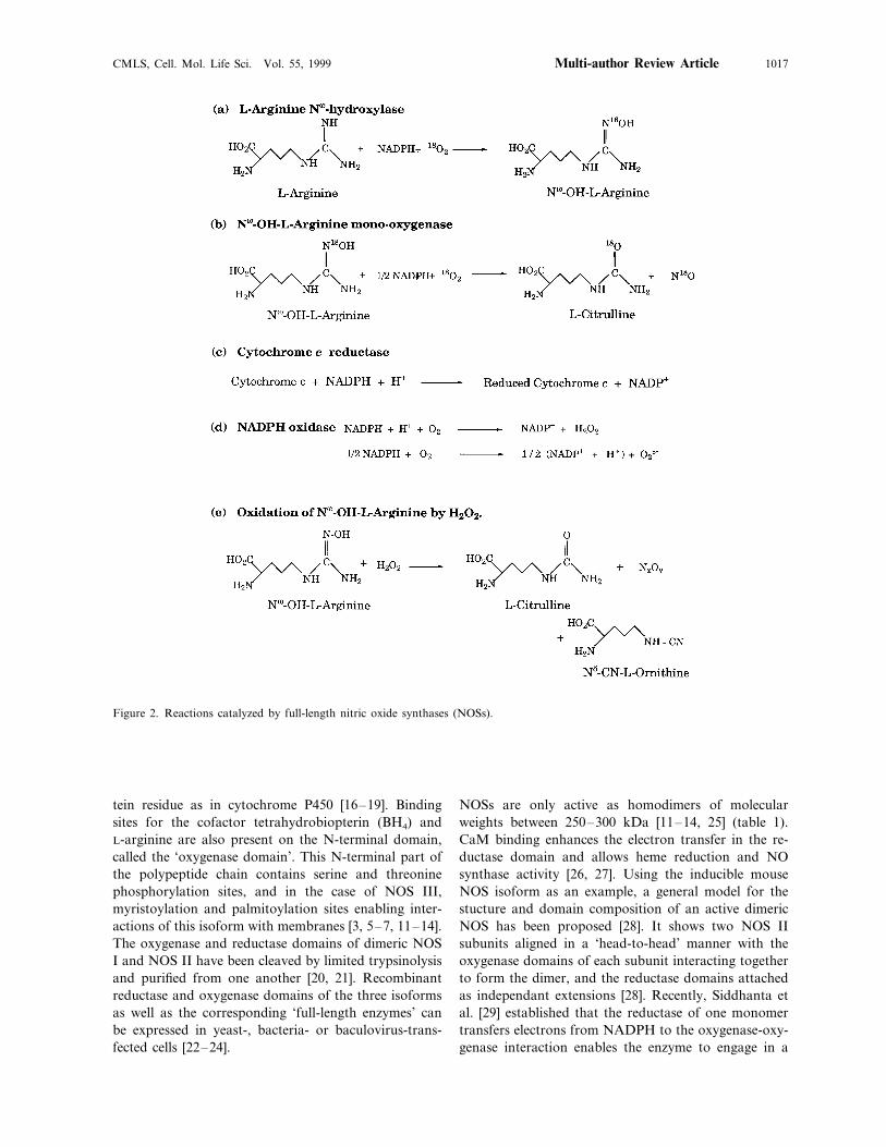
Figure 2. Reactions catalyzed by full-length nitric oxide synthases (NOSs).
tein residue as in cytochrome P450 [16–19]. Bindingsites for the cofactor tetrahydrobiopterin (BH4) andL-arginine are also present on the N-terminal domain,called the ‘oxygenase domain’. This N-terminal part ofthe polypeptide chain contains serine and threoninephosphorylation sites, and in the case of NOS III,myristoylation and palmitoylation sites enabling inter-actions of this isoform with membranes [3, 5–7, 11–14].The oxygenase and reductase domains of dimeric NOSI and NOS II have been cleaved by limited trypsinolysisand purified from one another [20, 21]. Recombinantreductase and oxygenase domains of the three isoformsas well as the corresponding ‘full-length enzymes’ canbe expressed in yeast-, bacteria- or baculovirus-trans-fected cells [22–24].
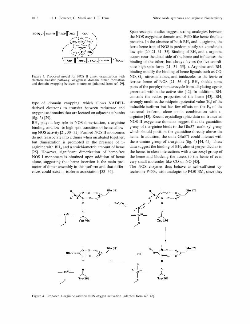
NOSs are only active as homodimers of molecularweights between 250–300 kDa [11–14, 25] (table 1).CaM binding enhances the electron transfer in the re-ductase domain and allows heme reduction and NOsynthase activity [26, 27]. Using the inducible mouseNOS isoform as an example, a general model for thestucture and domain composition of an active dimericNOS has been proposed [28]. It shows two NOS IIsubunits aligned in a ‘head-to-head’ manner with theoxygenase domains of each subunit interacting togetherto form the dimer, and the reductase domains attachedas independant extensions [28]. Recently, Siddhanta etal. [29] established that the reductase of one monomertransfers electrons from NADPH to the oxygenase-oxy-genase interaction enables the enzyme to engage in a
J. L. Boucher, C. Moali and J. P. Tenu Nitric oxide synthases and arginase biochemistry1018
Figure 3. Proposed model for NOS II dimer organization withelectron transfer pathway, oxygenase domain dimer formationand domain swapping between monomers [adapted from ref. 29].
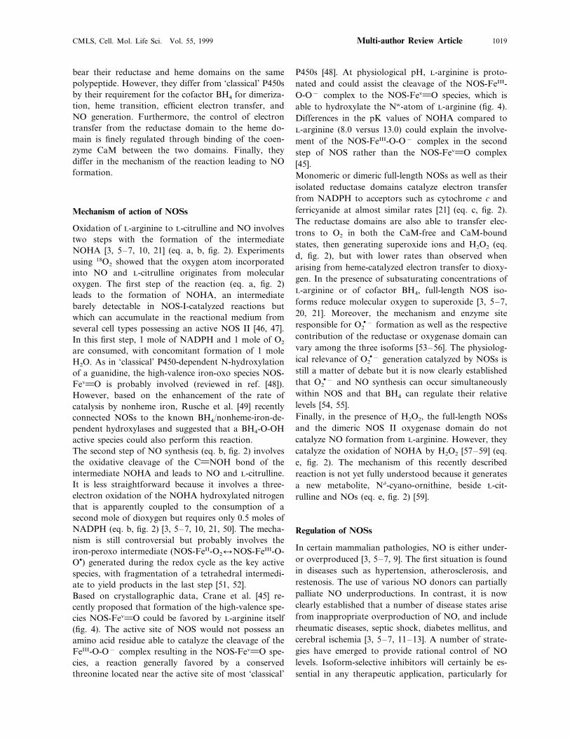
Spectroscopic studies suggest strong analogies betweenthe NOS oxygenase domain and P450-like heme-thiolateproteins. In the absence of both BH4 and L-arginine, theferric heme iron of NOS is predominantly six-coordinatelow spin [20, 21, 31–35]. Binding of BH4 and L-arginineoccurs near the distal side of the heme and influences thebinding of the other, but always favors the five-coordi-nate high-spin form [21, 31–35]. L-Arginine and BH4
binding modify the binding of heme ligands such as CO,NO, O2, nitrosoalkanes, and imidazoles to the ferric orferrous heme of NOS [21, 36–41]. BH4 shields someparts of the porphyrin macrocycle from alkylating agentsgenerated within the active site [42]. In addition, BH4
controls the redox properties of the heme [43]. BH4
strongly modifies the midpoint potential value (E0) of theinducible isoform but has few effects on the E0 of theneuronal isoform, alone or in combination with L-arginine [43]. Recent crystallographic data on truncatedNOS II oxygenase domains suggest that the guanidinogroup of L-arginine binds to the Glu371 carboxyl groupwhich should position the guanidine directly above theheme. In addition, the same Glu371 could interact withthe a-amino group of L-arginine (fig. 4) [44, 45]. Thesedata suggest the binding of BH4 almost perpendicular tothe heme, in close interactions with a carboxyl group ofthe heme and blocking the access to the heme of evenvery small molecules like CO or NO [45].The NOS enzymes thus behave as self-sufficient cy-tochrome P450s, with analogies to P450 BM3 since they
type of ‘domain swapping’ which allows NADPH-derived electrons to transfer between reductase andoxygenase domains that are located on adjacent subunits(fig. 3) [29].BH4 plays a key role in NOS dimerization, L-argininebinding, and low- to high-spin transition of heme, allow-ing NOS activity [21, 30–32]. Purified NOS II monomersdo not reassociate into a dimer when incubated together,but dimerization is promoted in the presence of L-arginine with BH4 and a stoichiometric amount of heme[25]. However, significant dimerization of heme-freeNOS I monomers is obtained upon addition of hemealone, suggesting that heme insertion is the main pro-moter of dimer assembly in this isoform and that differ-ences could exist in isoform association [33–35].
Figure 4. Proposed L-arginine assisted NOS oxygen activation [adapted from ref. 45].
CMLS, Cell. Mol. Life Sci. Vol. 55, 1999 1019Multi-author Review Article
bear their reductase and heme domains on the samepolypeptide. However, they differ from ‘classical’ P450sby their requirement for the cofactor BH4 for dimeriza-tion, heme transition, efficient electron transfer, andNO generation. Furthermore, the control of electrontransfer from the reductase domain to the heme do-main is finely regulated through binding of the coen-zyme CaM between the two domains. Finally, theydiffer in the mechanism of the reaction leading to NOformation.
Mechanism of action of NOSs
Oxidation of L-arginine to L-citrulline and NO involvestwo steps with the formation of the intermediateNOHA [3, 5–7, 10, 21] (eq. a, b, fig. 2). Experimentsusing 18O2 showed that the oxygen atom incorporatedinto NO and L-citrulline originates from molecularoxygen. The first step of the reaction (eq. a, fig. 2)leads to the formation of NOHA, an intermediatebarely detectable in NOS-I-catalyzed reactions butwhich can accumulate in the reactional medium fromseveral cell types possessing an active NOS II [46, 47].In this first step, 1 mole of NADPH and 1 mole of O2
are consumed, with concomitant formation of 1 moleH2O. As in ‘classical’ P450-dependent N-hydroxylationof a guanidine, the high-valence iron-oxo species NOS-Fev.O is probably involved (reviewed in ref. [48]).However, based on the enhancement of the rate ofcatalysis by nonheme iron, Rusche et al. [49] recentlyconnected NOSs to the known BH4/nonheme-iron-de-pendent hydroxylases and suggested that a BH4-O-OHactive species could also perform this reaction.The second step of NO synthesis (eq. b, fig. 2) involvesthe oxidative cleavage of the C.NOH bond of theintermediate NOHA and leads to NO and L-citrulline.It is less straightforward because it involves a three-electron oxidation of the NOHA hydroxylated nitrogenthat is apparently coupled to the consumption of asecond mole of dioxygen but requires only 0.5 moles ofNADPH (eq. b, fig. 2) [3, 5–7, 10, 21, 50]. The mecha-nism is still controversial but probably involves theiron-peroxo intermediate (NOS-FeII-O2lNOS-FeIII-O-O�) generated during the redox cycle as the key activespecies, with fragmentation of a tetrahedral intermedi-ate to yield products in the last step [51, 52].Based on crystallographic data, Crane et al. [45] re-cently proposed that formation of the high-valence spe-cies NOS-Fev.O could be favored by L-arginine itself(fig. 4). The active site of NOS would not possess anamino acid residue able to catalyze the cleavage of theFeIII-O-O− complex resulting in the NOS-Fev.O spe-cies, a reaction generally favored by a conservedthreonine located near the active site of most ‘classical’
P450s [48]. At physiological pH, L-arginine is proto-nated and could assist the cleavage of the NOS-FeIII-O-O− complex to the NOS-Fev.O species, which isable to hydroxylate the Nw-atom of L-arginine (fig. 4).Differences in the pK values of NOHA compared toL-arginine (8.0 versus 13.0) could explain the involve-ment of the NOS-FeIII-O-O− complex in the secondstep of NOS rather than the NOS-Fev.O complex[45].Monomeric or dimeric full-length NOSs as well as theirisolated reductase domains catalyze electron transferfrom NADPH to acceptors such as cytochrome c andferricyanide at almost similar rates [21] (eq. c, fig. 2).The reductase domains are also able to transfer elec-trons to O2 in both the CaM-free and CaM-boundstates, then generating superoxide ions and H2O2 (eq.d, fig. 2), but with lower rates than observed whenarising from heme-catalyzed electron transfer to dioxy-gen. In the presence of subsaturating concentrations ofL-arginine or of cofactor BH4, full-length NOS iso-forms reduce molecular oxygen to superoxide [3, 5–7,20, 21]. Moreover, the mechanism and enzyme siteresponsible for O2
�− formation as well as the respectivecontribution of the reductase or oxygenase domain canvary among the three isoforms [53–56]. The physiolog-ical relevance of O2
�− generation catalyzed by NOSs isstill a matter of debate but it is now clearly establishedthat O2
�− and NO synthesis can occur simultaneouslywithin NOS and that BH4 can regulate their relativelevels [54, 55].Finally, in the presence of H2O2, the full-length NOSsand the dimeric NOS II oxygenase domain do notcatalyze NO formation from L-arginine. However, theycatalyze the oxidation of NOHA by H2O2 [57–59] (eq.e, fig. 2). The mechanism of this recently describedreaction is not yet fully understood because it generatesa new metabolite, Nd-cyano-ornithine, beside L-cit-rulline and NOs (eq. e, fig. 2) [59].
Regulation of NOSs
In certain mammalian pathologies, NO is either under-or overproduced [3, 5–7, 9]. The first situation is foundin diseases such as hypertension, atherosclerosis, andrestenosis. The use of various NO donors can partiallypalliate NO underproductions. In contrast, it is nowclearly established that a number of disease states arisefrom inappropriate overproduction of NO, and includerheumatic diseases, septic shock, diabetes mellitus, andcerebral ischemia [3, 5–7, 11–13]. A number of strate-gies have emerged to provide rational control of NOlevels. Isoform-selective inhibitors will certainly be es-sential in any therapeutic application, particularly for
J. L. Boucher, C. Moali and J. P. Tenu Nitric oxide synthases and arginase biochemistry1020
the treatment of chronic disorders. Most efforts havebeen directed toward selective inhibition of the in-ducible NOS II that could be used to treat endotoxicshock and clinical problems related to abnormally highlevels of NO. As described above, since there are severalimportant differences among the isoforms, isoform-se-lective inhibition should be, at least in principle, possi-ble [11–14, 52].Regulation of NOS activity at the transcriptional leveland by post-transcriptional modifications (phosphoryla-tion, palmitoylation), as well as prevention of enzymeinduction [60, 61] are described in other reviews in thisissue. We will focus on the effects of cofactor (BH4 andL-arginine) depletion or coenzyme (CaM) inhibition.The flavoprotein inhibitor diphenyleneiodonium in-hibits all NOS isoforms [62] but also inhibits any flavin-dependent electron transfer. The formation of products(NO, O2
�−, and peroxynitrite) is another possible wayto regulate NOS activity and the potential of NO toinhibit NOSs is still an open question. The formation ofa nitrosyl complex of NOS during turnover is clearlyobserved and represents about 80% of the NOS content[63]. However, this complex readily turns back to nativeNOS FeIII in the presence of O2. Similarly, peroxynitriteinactivates NOS II through changes in the heme envi-ronment [64]. These potential modes of inhibition seemunlikely to be of pharmacological interest.
Regulation by coenzyme CaM and cofactor BH4
Ca2+/CaM antagonistsCaM regulates electron transfer from the NADPH tothe heme by acting as a switch between the reductaseand oxygenase domains and by stimulating electrontransfer into the reductase domain itself [26, 27]. Dis-tinction between constitutive and inducible NOS lies intheir differences toward CaM binding: NOS I and NOSIII bind CaM in a reversible Ca2+-dependent manner.In contrast, NOS II binds CaM so tightly that itsactivity does not seem susceptible to control by tran-sient variations in Ca2+ concentrations [11–14]. Consti-tutive NOS isoforms that show a typical type of CaMinteraction are inhibited by Ca2+ chelators (EDTA,EGTA) and CaM antagonists (chlorpromazine, W7, ortamoxifen) [3, 5–7, 11–14, 65]. Due to the involvementof CaM in many physiological processes, inhibition ofNOSs through CaM antagonists should be fraught withselectivity problems. However, this view is beginning tochange. The interaction of CaM with NOSs appears tobe unique and the design of agents that could selectivelyinterfere with the CaM/NOS complex formation seemspossible. Significant progress has been made on CaM-NOS interactions using peptides derived from the CaM-binding site [66, 67].
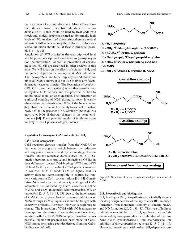
Figure 5. Structure of some L-arginine analogs, inhibitors ofNOSs.
BH4 biosynthesis and binding siteBH4 binding or BH4 biosynthesis are potentially targetsfor drug design because of the key role for BH4 in dimerformation from monomers, stability of dimeric NOSs,and NO formation [20, 21, 31–35]. This type of indirectinhibition uses inhibitors of BH4 synthesis such as 2,4-diamino-6-hydroxypyrimidine, an inhibitor of the en-zyme GTP cyclohydrolase-I, and methotrexate, aninhibitor of dihydropteridine reductase [3, 5–7, 11–14].However, interference with other BH4-dependent en-
CMLS, Cell. Mol. Life Sci. Vol. 55, 1999 1021Multi-author Review Article
zymes such as phenylalanine or tyrosine hydroxylasesillustrates the potential problems of nonspecific effectsof these compounds. Recent crystallographic data showthat dimerization results in strong modifications of themonomeric protein, including interactions between BH4
and the heme carboxylate group, the a-NH2 group ofL-arginine, and residues involved in the dimer interface[45] (fig. 4). Better knowledge of the BH4 binding siteshould allow the design of more selective pterinantagonists.
Active-site inhibitors
Active-site inhibitors should be the most suitable toolsto achieve selective inhibition of NOS and could include(i) interaction at the L-arginine binding site with com-petitive inhibitors, (ii) interaction at the active site withmechanism-based inhibitors, and (iii) ligands directedtoward the heme.
Arginine analogsStudies of NO synthesis have been facilitated by theearly identification of Nv-methyl-L-arginine (L-NMMA) as a competitive inhibitor of NO formationwith a Ki in the 1–10 mM range, but with poor isoformselectivity [68]. Since this initial finding, many reportshave used other simple Nv-derivatives of L-arginine, orstructurally related compounds to inhibit the NOSs [3,5–7, 52] (fig. 5). Nv-Nitro-L-arginine, its methyl ester,and Nv-amino-L-arginine are relatively selective for theconstitutive isoforms [3, 5–7, 52]. Several other alkylanalogs have been synthesized and studied. Nv-Cyclo-propyl-L-arginine displays modest selectivity for theconstitutive isoforms whereas Nv-propyl-L-arginine is apotent and selective inhibitor of the neuronal isoform[69, 70]. (Iminoethyl)-L-ornithine (L-NIO) which differsfrom L-arginine by the presence of an amidino group inplace of a guanidino group (fig. 5), is a potent butnonselective inhibitor of NOSs. Its higher homolog,(iminoethyl)-L-lysine (L-NIL), selectively inhibits the in-ducible isoform, suggesting that limited changes instructure have remarkable effects on isoform selectivity[71, 72].Recently, L-thiocitrulline and S-alkyl-L-thiocitrullinehave been synthesized and evaluated [73, 74] (fig. 5).They are the most potent competitive inhibitors of bothclasses of NOS, with a good selectivity toward NOS I invitro. S-Methyl-L-thiocitrulline is a potent, reversible,slow-binding inhibitor of NOS I, but seems to be lesspotent when tested in a cell-based enzyme assay. Higherhomologs, homo-L-thiocitrulline and S-methyl-homo-L-thiocitrulline, display less efficiency and selectivity [75].For L-thiocitrulline, the sulfur atom of the thiourea
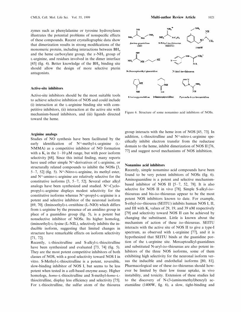
Figure 6. Structure of some nonamino acid inhibitors of NOSs.
group interacts with the heme iron of NOS [45, 73]. Inaddition, L-thiocitrulline and Nv-nitro-L-arginine spe-cifically inhibit electron transfer from the reductasedomain to the heme, inhibit dimerization of NOS II [76,77] and suggest novel mechanisms of NOS inhibition.
Nonamino acid inhibitorsRecently, simple nonamino acid compounds have beenfound to be very potent inhibitors of NOSs (fig. 6).Aminoguanidine is a potent and selective mechanism-based inhibitor of NOS II [5–7, 52, 78]. It is alsoselective for NOS II in vivo [78]. Simple S-alkyl-iso-thioureas and bis-iso-thioureas appear to be the mostpotent NOS inhibitors known to date. For example,S-ethyl-iso-thiourea (SEITU) inhibits human NOS I, II,and III with Ki values of 29, 19, and 39 nM respectively[79] and selectivity toward NOS II can be achieved bychanging the substituent. Little is known about themechanism of action of these iso-thioureas. SEITUinteracts with the active site of NOS II to give a type-Ispectrum, as observed with L-arginine [77], and it ishypothesized that SEITU binds at the guanidine por-tion of the L-arginine site. Mercaptoalkyl-guanidinesand substituted N-aryl-iso-thioureas are also potent in-hibitors of the three NOS isoforms, some of themexhibiting high selectivity for the neuronal isoform ver-sus the inducible and endothelial isoforms [80, 81].Pharmacological use of these iso-thioureas should how-ever be limited by their low tissue uptake, in vivoinstability, and toxicity. Extension of these studies ledto the discovery of N-(3-(aminomethyl)benzyl) ac-etamidine (1400W, fig. 6), a slow, tight-binding and
J. L. Boucher, C. Moali and J. P. Tenu Nitric oxide synthases and arginase biochemistry1022
Figure 7. Some heme ligands used as inhibitors of NOSs.
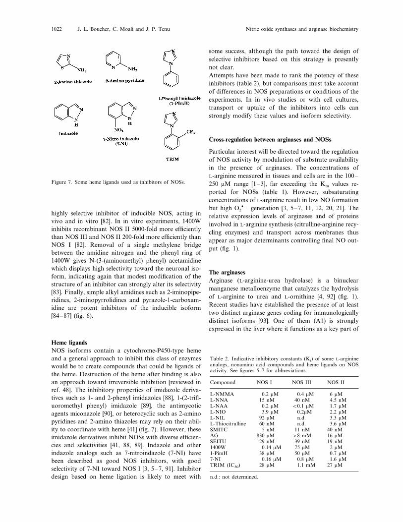
some success, although the path toward the design ofselective inhibitors based on this strategy is presentlynot clear.Attempts have been made to rank the potency of theseinhibitors (table 2), but comparisons must take accountof differences in NOS preparations or conditions of theexperiments. In in vivo studies or with cell cultures,transport or uptake of the inhibitors into cells canstrongly modify these values and isoform selectivity.
Cross-regulation between arginases and NOSs
Particular interest will be directed toward the regulationof NOS activity by modulation of substrate availabilityin the presence of arginases. The concentrations ofL-arginine measured in tissues and cells are in the 100–250 mM range [1–3], far exceeding the Km values re-ported for NOSs (table 1). However, subsaturatingconcentrations of L-arginine result in low NO formationbut high O2
�− generation [3, 5–7, 11, 12, 20, 21]. Therelative expression levels of arginases and of proteinsinvolved in L-arginine synthesis (citrulline-arginine recy-cling enzymes) and transport across menbranes thusappear as major determinants controlling final NO out-put (fig. 1).
The arginasesArginase (L-arginine-urea hydrolase) is a binuclearmanganese metalloenzyme that catalyzes the hydrolysisof L-arginine to urea and L-ornithine [4, 92] (fig. 1).Recent studies have established the presence of at leasttwo distinct arginase genes coding for immunologicallydistinct isoforms [93]. One of them (A1) is stronglyexpressed in the liver where it functions as a key part of
highly selective inhibitor of inducible NOS, acting invivo and in vitro [82]. In in vitro experiments, 1400Winhibits recombinant NOS II 5000-fold more efficientlythan NOS III and NOS II 200-fold more efficiently thanNOS I [82]. Removal of a single methylene bridgebetween the amidine nitrogen and the phenyl ring of1400W gives N-(3-(aminomethyl) phenyl) acetamidinewhich displays high selectivity toward the neuronal iso-form, indicating again that modest modification of thestructure of an inhibitor can strongly alter its selectivity[83]. Finally, simple alkyl amidines such as 2-iminopipe-ridines, 2-iminopyrrolidines and pyrazole-1-carboxam-idine are potent inhibitors of the inducible isoform[84–87] (fig. 6).
Heme ligandsNOS isoforms contain a cytochrome-P450-type hemeand a general approach to inhibit this class of enzymeswould be to create compounds that could be ligands ofthe heme. Destruction of the heme after binding is alsoan approach toward irreversible inhibition [reviewed inref. 48]. The inhibitory properties of imidazole deriva-tives such as 1- and 2-phenyl imidazoles [88], 1-(2-trifl-uoromethyl phenyl) imidazole [89], the antimycoticagents miconazole [90], or heterocyclic such as 2-aminopyridines and 2-amino thiazoles may rely on their abil-ity to coordinate with heme [41] (fig. 7). However, theseimidazole derivatives inhibit NOSs with diverse efficien-cies and selectivities [41, 88, 89]. Indazole and otherindazole analogs such as 7-nitroindazole (7-NI) havebeen described as good NOS inhibitors, with goodselectivity of 7-NI toward NOS I [3, 5–7, 91]. Inhibitordesign based on heme ligation is likely to meet with
Table 2. Indicative inhibitory constants (Ki) of some L-arginineanalogs, nonamino acid compounds and heme ligands on NOSactivity. See figures 5–7 for abbreviations.
NOS III NOS IINOS ICompound
L-NMMA 0.2 mM 0.4 mM 6 mML-NNA 15 nM 40 nM 4.5 nML-NAA 0.2 mM B0.1 mM 1.7 mM
3.9 mM 2.2 mM0.2mML-NIO92 mML-NIL n.d. 3.3 mM
3.6 mML-Thiocitrulline n.d.60 nM11 nMSMITC 40 nM5 nM
AG 830 mM \8 mM 16 mMSEITU 29 nM 39 nM 19 nM
2 mM75 mM0.14 mM1400W1-PimH 38 mM 50 mM 0.7 mM
0.8 mM0.16 mM7-NI 1.6 mMTRIM (IC50) 27 mM1.1 mM28 mM
n.d.: not determined.
CMLS, Cell. Mol. Life Sci. Vol. 55, 1999 1023Multi-author Review Article
the urea cycle. AII is found in mitochondria of extra-hepatic tissues and cells such as the red blood cell, thelactating mammary gland, the kidney, and macrophages[reviewed in refs. 4, 94–96]. The extrahepatic arginase(AII) subserves a number of as yet not well definedroles, including participation in polyamine synthesis viathe cytosolic enzyme ornithine decarboxylase, and for-mation of proline, creatine, glutamate, agmatine, andg-amino-butyric acid [reviewed in ref. 4]. Arginases AIand AII appear to be expressed constitutively in murinemacrophages, although different stimuli may modulatethe level of expression of these isoforms (see below). AIand AII contribute to immune system function sinceL-ornithine is a key precursor for polyamines involvedin cell replication. Ornithine/urea production markedlyincreases during tumor growth whereas arginase activitydecreases during tumor rejection [97]. Recently, it wassuggested that AI and AII could be involved in theregulation of several cytostatic and cytotoxic actions ofactivated macrophages mediated by NO synthesis. Inthese cases, modulation of local L-arginine concentra-tions by arginases could regulate high NO production[3, 5–7, 96–99].Substantial amounts of NOHA are liberated from theactive site of inducible NOS [46, 47, 50] and it has beendemonstrated that NOHA is a potent inhibitor of liverarginase [100, 101]. NOHA may thus act as an endoge-nous arginase inhibitor in NO-producing cells, as ob-served in endothelial cells or alveolar macrophages [47,102, 103]. However, NOHA is a substrate for NOSs andis useless as a pharmacological tool to elucidate interac-tions between the arginases and NOS pathways. Werecently synthesized a new analog of NOHA, Nv-hy-droxy-nor-L-arginine (nor-NOHA) and showed that itis a potent competitive inhibitor of rat liver arginase[104] without being a substrate for NOSs [50]. Inhibi-tion of arginase by nor-NOHA could thus increase theavailability of L-arginine for NO biosynthesis in cellspossessing an active NOS. nor-NOHA could be a selec-tive tool allowing detailed studies of the interactions,between NOSs and arginases [47, 100–103, 105].In some cases, NOS II and arginases I or II seem to beco-induced but in other cases, both types of enzyme aresubmitted to opposite or reciprocal regulation. Inducerscan be tentatively classified into three classes: (i) media-tors which induce both NOS II and arginases; (ii) medi-ators which exert similar or opposite effects onarginases and NOS II, depending on the cellular system;(iii) mediators which exert opposite effects on arginasesand NOS II expression.
Mediators which induce both NOS II and arginasesLipopolysaccharides (LPSs) seem to be the only class ofmolecules which are able to activate arginases and NOSII genes.
LPSs in vivo and in vitro induce expression of NOS IIand AI and/or AII. In vivo, LPSs increase mRNA ofarginases and NOS II in lung, heart, spleen, and liver ofrats as well as other enzymes able to recycle L-citrullineto L-arginine (arginino succinate synthase and argininosuccinate lyase) [106]. In vitro, LPSs induce arginasesand NOS II in various systems including rat aorticendothelial cells [47], rat [107] and mouse peritonealmacrophages [108], the mouse macrophage cell lineRAW 264.7 [109], and mouse bone-marrow-derivedmacrophages [110].Although arginases and NOS II genes are both acti-vated by LPS, their modulation can differ at the kineticlevel, and be influenced by other mediators. For exam-ple, kinetic modulation was observed [106] in vivo inrats where NOS II mRNA in the spleen reached aplateau 5–6 h after intraperitoneal LPS injection, de-creased thereafter and returned to a hardly detectablelevel at 24 h. In parallel, arginino succinate synthasemRNA increased up to 12 h and arginino succinatelyase mRNA increased continuously up to 24 h [106],probably resulting in increased L-arginine availabilityand compensating for the decrease in NOS II efficiency.In vitro, rat peritoneal macrophages were cultured inthe presence of LPS, and NOS II mRNA appeared 2 hafter LPS addition and increased up to 12 h. On theother hand, AI mRNA began to increase only after 4 h(with a lag) and reached a maximum at 12 h [106].Unexpectedly, because AI has been thought to be ex-pressed almost exclusively in the liver, immunoblotsshowed that NOS II and AI proteins were produced inmacrophages [106]. The RAW264.7 NOS II level wasalso observed to reach a plateau after 24 h whereas theAII level increased up to 48 h [109]. Thus, there is notrue co-induction of arginases and NOS II by LPS.Arginase induction appeared delayed compared to NOSII, perhaps to limit the duration of NO production. Onecan also mention that molecules such as pyrrolidonedithiocarbamate which prevents translocation of NF-kB, an essential transcription factor for NOS II induc-tion [3, 5–7], are ineffective in AII induction [109].
Mediators which can exert similar or opposite effectson arginases and NOS II expressionInduction of NOS II and generation of NO in pancre-atic islet b-cells may mediate interleukin (IL)-1-induceddysfunction leading to insulin-dependent diabetes melli-tus [3, 5–7]. Cytosol analysis from the rat insulinoma-derived cell line RINm5F treated with IL-1b has shownthat IL-1b increased NOS II and significantly decreasedarginase expresssion, thereby increasing the availabilityof L-arginine for NO production and resulting in a longlasting cytotoxic effect of NO [111]. In contrast, whenmesangial cells were stimulated with IL-1, AI, AII and

J. L. Boucher, C. Moali and J. P. Tenu Nitric oxide synthases and arginase biochemistry1024
NOS II were up-regulated, IL-1 increasing arginaseactivity by 60% [112].Oxygen tension can exert complex effects. Hyperoxia(100% O2) increases AI and AII in rat lung, withoutaffecting NOS II expression [113]. In contrast, hypoxiaor anoxia increases AI and NOS II expression [114]but decreases AII mRNA expression in rat and mouseperitoneal macrophages, while L-arginine transportermCAT-2 expression is increased more than twofold[108]. Arginases AI and AII as well as NOS II and themCAT-2 L-arginine transporter are thus O2-regulatedgenes.
Mediators which exert opposite effects on arginases andNOS II expressionAs a general rule, cytokines inducing NOS II do notinduce arginases and vice versa. For example, cytoki-nes such as interferon-g produced by the Th1 subpopu-lation of CD4+ T cell, induce NOS II in mousebone-marrow-derived macrophages whereas Th2-derived cytokines such as IL-4, IL-10, and IL-13 arepotent inducers of arginases [115]. Exposure ofmacrophage cultures to inducers of NO synthase ex-hausts their ability to respond subsequently to inducersof arginases. Conversely, exposure of the cells to in-ducers of arginase exhausts their ability to respond toinducers of NOS II. Inhibition of NOS II induction byIL-4 in murine macrophages and endothelial cells hasbeen explained by an increased rate of NOS II mRNAdegradation [116] whereas IL-10 could prevent synthe-sis of tumor necrosis factor-a, a cytokine inducingNOS II [117]. Arginase levels induced by Th2 cells farexceeded those inducible by the individual cytokinesderived from the Th2 subpopulation. Antibody-block-ing experiments revealed strong synergistic effects be-tween IL-4/IL-13 and IL-10 sufficient to account forthe high arginase activity induced by the Th2 cells[118]. During immune reactions, the NOS II andarginase enzymes appear to define two alternatemacrophage functional states induced by Th1- andTh2-derived cytokines, respectively [118].Glucocorticoids are known to inhibit NOS II induc-tion [3, 5–7, 11–14, 60]. In contrast, AI is induced byglucocorticoids in a delayed manner. Induction ofarginase mRNA by dexamethasone is preceded by anincrease in C/EBP binding activity which follows anincrease in C/EBP beta mRNA [107, 119]. In vivo inrats, steroids increase urea cycle enzyme mRNA levelsand this effect is abolished by growth hormone [120].Transforming growth factor (TGF)-b has been shownto down-regulate NOS II expression, probably like IL-4, by increasing the rate of NOS II mRNA degrada-tion. TGF-b up-regulates arginase activity in ratperitoneal macrophages and hence might limit
macrophage-dependent cytostasis [121]. These resultsare consistent with competition of both enzymes forL-arginine and a reciprocal inhibition in their induc-tion.
Concluding remarksMany results are now consistent with the conclusionthat the pathway used by macrophages to metabolizeL-arginine can influence the type of host immune re-sponses against pathogens, parasites, or tumors. Theco-induction of extrahepatic arginase and NOS by thesame stimuli [106–109], the induction of arginase bysuppressors of NOS [110], and the identification ofcompounds that co-regulate the expression of bothNOS and arginases [110–115], support the hypothesisthat arginase may be essential in the regulation ofNOS activity by modulating local L-arginine concen-trations [97–109, 121].Arginase I, previously thought to be restricted to theliver, accounts for high arginase activity at inflamma-tory sites where it may limit high-output NO produc-tion and generate (along with AII) polyamines andproline involved in tissue repair and cell proliferation.Contrasting temporal expression of the NOS II andarginase pathways has been observed in inflammation,NOS II being observed at the early stages and thearginases at a later stage. IL-4, a mediator generatedduring inflammation, may play a major role in theswitch from high NO to low NO formation and in-crease L-ornithine and polyamine synthesis during tis-sue repair. Finally, inhibition of proliferation of ahuman tumor cell line by NOHA and NO seems to berelated to distinct mechanism, at sequential steps ofthe arginine-polyamine pathway. NOHA inhibitsCaco-2 tumor cell proliferation likely by inhibitingarginase activity, whereas NO causes cytostasis bymechanisms that might involve inhibition of ornithinedecarboxylase [122].A recent study demonstrates that extracellularly ap-plied arginase can inhibit neuronal apoptosis inducedby multiple stimuli [123]. The protective effects ofarginase could not be reproduced by an array of NOSinhibitors but rather seemed to depend on depletion ofL-arginine, resulting in protein synthesis inhibition.This identifies amino acid depletion as a novel biologi-cal strategy to prevent pathological neuronal apoptosisand suggests that additional insight into the localiza-tion and regulation of arginases could elucidate novelapproaches to regulate cell death and NO synthesis[123]. L-Arginine enzymes, capable of simultaneouslyinhibiting protein synthesis and NO generation, couldbe therapeutic targets for acute neurological diseases.
CMLS, Cell. Mol. Life Sci. Vol. 55, 1999 1025Multi-author Review Article
Conclusion
Ten years ago, the free radical NO was only consideredas a highly reactive and toxic compound and it washardly conceivable that it might serve as a beneficialmediator in biological processes. This remains true aslong as it is delivered at excessive rates or excessiveconcentrations. Since the identification of endothelium-derived relaxing factor with NO, an increasing numberof studies have clarified its key roles in physiologicaland pathophysiological processes in mammals. Furtherstudies have elucidated its roles in other species such asfishes, insects, bacteria, and, very recently, plants, whereit serves either as a neuromediator or as an element ofthe response against pathogens. This resulted in itsdesignation as ‘molecule of the year’ by Nature in 1992and by the award of the Nobel Prize for Medicine in1998 to Drs. Murad, Furchgott, and Ignarro, three ofthe earliest investigators of the L-arginine/NO pathway.
1 (1992) Metabolic, physiological and nutritional importanceof arginine In: Guanidino Compounds in Biology andMedicine, pp. 61–144, DeDeyn P. P., Marescau B., StalonV. and Qureshi I. A. (eds), Libbey, London
2 Wu G. and Morris S. M. (1998) Arginine metabolism: nitricoxide and beyond. Biochem. J. 336: 1–17
3 Ignarro L. and Murad F. (eds) (1995) Nitric Oxide: Bio-chemistry, Molecular Biology, and Therapeutic Implications,Academic Press, San Diego
4 Jenkinson C. P., Grody W. W. and Cederbaum S. D. (1997)Comparative properties of arginases. Comp. Biochem. Phys-iol. Biochem. Mol. Biol. 114B: 107–132
5 Feldman P. L., Griffith O. W. and Stuehr D. J. (1993) Thesurprising life of nitric oxide. Chem. Eng. News: 26–38
6 Kerwin J. F. and Heller M. (1994) The arginine-nitric oxidepathway: a target for new drugs. Med. Res. Rev. 14: 23–74
7 Kerwin J. F., Lancaster J. R. and Feldman P. L. (1995)Nitric oxide: a new paradigm for second messengers. J. Med.Chem. 38: 4343–4362
8 Nathan C. and Xie Q. W. (1994) Regulation of biosynthesisof nitric oxide. J. Biol. Chem. 269: 13725–13728
9 Moncada S. and Higgs E. A. (1995) Molecular mechanismsand therapeutic strategies related to nitric oxide. FASEB J.9: 13619–13630
10 Stuehr D. J., Kwon N. S., Nathan C. F., Griffith O. W.,Feldman P. L. and Wiseman J. (1991) Nv-Hydroxy-L-arginine is an intermediate in the biosynthesis of nitric oxidefrom L-arginine. J. Biol. Chem. 266: 6259–6263
11 Knowles R. G. and Moncada S. (1994) Nitric oxide syn-thases in mammals. Biochem. J. 298: 249–258
12 Forstermann U., Closs E. I., Pollock J. S., Nakane M.,Schwarz P., Gath I. et al. (1994) Nitric oxide synthaseisozymes. characterization, purification, molecular cloning,and functions. Hypertension 23: 1121–1131
13 Forstermann U. and Kleinert H. (1995) Nitric oxide syn-thase: expression and expressional control of the three iso-forms. Naunyn-Schmiedebergs Arch. Pharmacol. 352:351–364
14 Forstermann U., Boissel J. P. and Kleinert H. (1998) Expres-sional control of the ‘‘constitutive’’ isoforms of nitric oxidesynthase (NOS I and NOS III). FASEB J. 12: 773–790
15 Bredt D. S., Hwang P. M., Glatt C. E., Lowenstein C., ReedR. R. and Snyder S. H. (1991) Cloned and expressed nitricoxide synthase structurally resembles cytochrome P-450 re-ductase. Nature 351: 714–718
16 White K. A. and Marletta M. A. (1992) Nitric oxide syn-thase is a cytochrome P-450 type hemoprotein. Biochemistry31: 6627–6631
17 Stuehr D. J. and Ikeda-Saito M. (1992) Spectral characteri-zation of brain and macrophage nitric oxide synthases: cy-tochrome P-450-like hemeproteins that contain a flavinsemiquinone radical. J. Biol. Chem. 267: 20547–20550
18 McMillan K., Bredt D. S., Hirsch D. J., Snyder S. H., ClarkJ. E. and Masters B. S. S. (1992) Cloned, expressed ratcerebellar nitric oxide synthase contains stoichiometricamounts of heme, which binds carbon monoxide. Proc. Natl.Acad. Sci. USA 89: 11141–11145
19 Klatt P., Schmidt K. and Mayer B. (1992) Brain nitric oxidesynthase is a haemoprotein. Biochem. J. 288: 15–17
20 Masters B. S. S., McMillan K., Sheta E. A., Nishimura J. S.,Roman L. J. and Martasek P. (1996) Neuronal nitric oxidesynthase, a modular enzyme formed by convergent evolu-tion: structure studies of a cysteine thiolate-liganded hemeprotein that hydroxylates L-arginine to produce NO as acellular signal. FASEB J. 10: 552–558
21 Stuehr D. J. (1997) Structure-function aspects in the nitricoxide synthases. Annu. Rev. Pharmacol. Toxicol. 37: 339–359
22 McMillan K. and Masters B. S. S. (1995) Prokaryotic ex-pression of the heme- and flavin-binding domains of ratneuronal nitric oxide synthase as distinct polypeptides: iden-tification of the heme-binding proximal thiolate ligand ascysteine-415. Biochemistry 34: 3686–3693
23 Sari M. A., Booker S., Jaouen M., Vadon S., Boucher J. L.,Pompon D. et al. (1996) Expression in yeast and purificationof functional macrophage nitric oxide synthase: evidence forcysteine-194 as iron proximal ligand. Biochemistry 35: 7204–7213
24 Chen P. F., Tsai A. L. and Wu K. K. (1994) Cysteine 184 ofendothelial nitric oxide synthase is involved in heme coordi-nation and catalytic activity. J. Biol. Chem. 269: 25062–25066
25 Baek K. J., Thiel B. A., Lucas S. and Stuehr D. J. (1993)Macrophage nitric oxide synthase subunits: purification,characterization and role of prosthetic groups and substratein regulating their association into a dimeric enzyme. J. Biol.Chem. 268: 21120–21129
26 Matsuoka A., Stuehr D. J., Olson J. S., Clark P. andIkeda-Saito M. (1994) L-Arginine and calmodulin regulationof the heme iron reactivity in neuronal nitric oxide synthase.J. Biol. Chem. 269: 20335–20339
27 Abu-Soud H. M., Yoho L. L. and Stuehr D. J. (1994)Calmodulin controls neuronal nitric oxide synthase by adual mechanism: activation of intra- and interdomain elec-tron transfer. J. Biol. Chem. 269: 32047–32050
28 Ghosh D. K. and Stuehr D. J. (1995) Macrophage NOsynthase: characterization of isolated oxygenase and reduc-tase domains reveals a head-to-head subunit interaction.Biochemistry 34: 801–807
29 Siddhanta U., Presta A., Fan B., Wolan D., Rousseau D. L.and Stuehr D. J. (1998) Domain swapping in inducible nitricoxide synthase: electron transfer occurs between flavin andheme groups located on adjacent subunit in the dimer. J.Biol. Chem. 273: 18950–18958
30 Abu-Soud H. M., Loftus M. and Stuehr D. J. (1995) Subunitdissociation and unfolding of macrophage NO synthase:relationship between enzyme structure, prosthetic groupbinding and catalytic function. Biochemistry 34: 11167–11175
31 Mayer B., Wu C., Gorren A. C. F., Pfeiffer S., Schmidt K.,Clark P. et al. (1997) Tetrahydrobiopterin binding tomacrophage inducible nitric oxide synthase: heme-spin shiftand dimer stabilization by the potent pterin antagonist 4-amino-tetrahydrobiopterin. Biochemistry 36: 8422–8427
32 Presta A., Siddhanta U., Wu C., Sennequier N., Huang L.,Abu-Soud H. M. et al. (1998) Comparative functioning ofdihydro- and tetrahydropterins in supporting electron trans-fer, catalysis, and subunit dimerization in inducible nitricoxide synthase. Biochemistry 37: 298–310

J. L. Boucher, C. Moali and J. P. Tenu Nitric oxide synthases and arginase biochemistry1026
33 Klatt P., Pfeiffer S., List B. M., Lehner D., Glatter O.,Bachinger H. P. et al. (1996) Characterization of heme-defi-cient neuronal nitric oxide synthase reveals a role for hemein subunit dimerization and binding of the amino acidsubstrate and tetrahydrobiopterin. J. Biol. Chem. 271: 7336–7342
34 Gorren A. C. F., List B. M., Schrammel A., Pitters E.,Hemmens B., Werner E. R. et al. (1996) Tetrahydro-biopterin-free neuronal nitric oxide synthase: evidence fortwo identical highly anticooperative pteridine binding sites.Biochemistry 35: 16735–16745
35 Hemmens B., Gorren A. C. F., Schmidt K., Werner E. R.and Mayer B. (1998) Haem insertion, dimerization andreactivation of haem-free rat neuronal nitric oxide synthase.Biochem. J. 332: 337–342
36 Scheele J. S., Kharitonov V. G., Martasek P., Roman L. J.,Sharma V. S., Masters B. S. S. et al. (1997) Kinetics of COligation with nitric oxide synthase by flash photolysis andstopped-flow spectrophotometry. J. Biol. Chem. 272: 12523–12528
37 Abu-Soud H. M., Wu C., Ghosh D. K. and Stuehr D. J.(1998) Stopped-flow analysis of CO and NO binding toinducible nitric oxide synthase. Biochemistry 37: 3777–3786
38 Wang J., Rousseau D. L., Abu-Soud H. M. and Stuehr D. J.(1994) Heme coordination of NO in NO synthase. Proc.Natl. Acad. Sci. USA 91: 10512–10516
39 Abu-Soud H. M., Gachhui R., Raushel F. M. and Stuehr D.J. (1997) The ferrous-dioxy complex of neuronal nitric oxidesynthase: divergent effects of L-arginine and tetrahydro-biopterin on its stability. J. Biol. Chem. 272: 17349–17353
40 Renodon A., Boucher J. L., Wu C., Gacchui R., Sari M. A.,Mansuy D. et al. (1998) Formation of nitric oxide synthase-iron(II) nitrosoalkane complexes: severe restriction of accessto the iron(II) site in the presence of tetrahydrobiopterin.Biochemistry 37: 6367–6374
41 Chabin R. M., McCauley E., Calaycay J. R., Kelly T. M.,MacNaul K. L., Wolfe G. C. et al. (1996) Active-site struc-ture analysis of recombinant human inducible nitric oxidesynthase using imidazole. Biochemistry 35: 9567–9575
42 Counts-Gerber N., Rodriguez-Crespo I., Nishida C. R. andOrtiz de Montellano P. R. (1997) Active site topologies andcofactor-mediated conformational changes of nitric oxidesynthases. J. Biol. Chem. 272: 6285–6290
43 Presta A., Weber-Main A. M., Stankovich M. T. and StuehrD. J. (1998) Comparative effects of substrates and pterincofactor on the heme midpoint potential in inducible andneuronal nitric oxide synthases. J. Am. Chem. Soc. 120:9460–9465
44 Crane B. R., Arvai A. S., Gachhui R., Wu C., Ghosh D. K.,Getzoff E. D. et al. (1997) The structure of nitric oxidesynthase oxygenase domain and inhibitor complexes. Science278: 425–431
45 Crane B. R., Arvai A. S., Ghosh D. K., Wu C., Getzoff E.D., Stuehr D. J. et al. (1998) Structure of nitric oxidesynthase oxygenase dimer with pterin and substrate. Science279: 2121–2126
46 Chenais B., Yapo A., Lepoivre M. and Tenu J. P. (1991)High-performance liquid chromatographic analysis of theunusual pathway of oxidation of L-arginine to citrulline andnitric oxide in mammalian cells. J. Chromatogr. 539: 433–441
47 Buga G. M., Singh R., Pervin S., Rogers N. E., Schmitz D.A., Jenkinson C. P. et al. (1996) Arginase activity in en-dothelial cells: inhibition by NG-hydroxy-L-arginine duringhigh-output NO production. Am. J. Physiol. 271: H1988–H1998
48 Ortiz de Montellano P. R. (ed.) (1995) Cytochrome P450:Structure, Mechanism, and Biochemistry, 2nd ed., pp. 245–304, Plenum, New York
49 Rusche K. M., Spiering M. M. and Marletta M. A. (1998)Reactions catalyzed by tetrahydrobiopterin-free nitric oxidesynthase. Biochemistry 37: 15503–15512
50 Moali C., Boucher J. L., Sari M. A., Stuehr D. J. andMansuy D. (1998) Substrate specificity of NO synthases:detailed comparison of L-arginine, homo-L-arginine, theirNv-hydroxy derivatives, and Nv-hydroxy-nor-L-arginine.Biochemistry 37: 10453–10460
51 Korth H. G., Sustmann R., Thater C., Butler A. R. andIngold K. U. (1994) On the mechanism of the nitric oxidesynthase-catalyzed conversion of Nv-hydroxy-L-arginine tocitrulline and nitric oxide. J. Biol. Chem. 269: 17776–17779
52 Babu B. R. and Griffith O. W. (1998) Design of isoform-se-lective inhibitors of nitric oxide synthase. Curr. Opin. Chem.Biol. 2: 491–500
53 Miller R. T., Martasek P., Roman L. J., Nishimura J. S. andMasters B. S. S. (1997) Involvement of the reductase domainof neuronal nitric oxide synthase in superoxide anion pro-duction. Biochemistry 36: 15277–15284
54 Vasquez-Vivar J., Kalyanaraman B., Martasek P., Hogg N.,Masters B. S. S., Karoui H. et al. (1998) Superoxide genera-tion by endothelial nitric oxide synthase: the influence ofcofactors. Proc. Natl. Acad. Sci. USA 95: 9220–9225
55 Xia Y., Tsai A. L., Berka V. and Zweier J. L. (1998)Superoxide generation from endothelial nitric oxide syn-thase: a Ca2+/calmodulin-dependent and tetrahydro-biopterin regulatory process. J. Biol. Chem. 273:25804–25808
56 Xia Y., Roman L. J., Masters B. S. S. and Zweier J. L.(1998) Inducible nitric oxide synthase generates superoxidefrom the reductase domain. J. Biol. Chem. 273: 22635–22639
57 Pufahl R. A., Wishnock J. S. and Marletta M. A. (1995)Hydrogen peroxide-supported oxidation of NG-hydroxy-L-arginine by nitric oxide synthase. Biochemistry 34: 1930–1941
58 Ghosh D. K., Abu-Soud H. M. and Stuehr D. J. (1995)Reconstitution of the second step in NO synthesis using theisolated oxygenase and reductase domains of macrophageNO synthase. Biochemistry 34: 11316–11320
59 Clague M. J., Wishnok J. S. and Marletta M. A. (1997)Formation of Nd-cyano-ornithine from NG-hydroxy-L-arginine and hydrogen peroxide by neuronal nitric oxidesynthase: implications for mechanism. Biochemistry 36:14465–14473
60 Geller D. A., Lowenstein C. J., Shapiro R. A., Nussler A.K., DiSilvio M., Wang S. C. et al. (1993) Molecular cloningand expression of inducible nitric oxide synthase from hu-man hepatocytes. Proc. Natl. Acad. Sci. USA 90: 3491–3495
61 Ding M, Zhang M., Wong J. L., Rogers N. E., Ignarro L. J.and Voskuhl R. R. (1998) Antisense knockdown of induciblenitric oxide synthase inhibits induction of experimental au-toimmune encephalomyelitis in SJL/J mice. J. Immunol. 160:2560–2564
62 Stuehr D. J., Fesahun O. A., Kwon N. S., Gross S. S.,Gonzalez J. A., Levi R. et al. (1991) Inhibition ofmacrophage and endothelial cell nitric oxide synthase bydiphenyleneiodonium and its analogs. FASEB J. 5: 98–103
63 Abu-Soud H. M., Wang J., Rousseau D. L., Fukuto J. M.,Ignarro L. J. and Stuehr D. J. (1995) Neuronal nitric oxidesynthase self-inactivates by forming a ferrous-nitrosyl com-plex during aerobic catalysis. J. Biol. Chem. 270: 22997–23006
64 Huhmer A. F. R., Nishida C. R., Ortiz de Montellano P. R.and Schoneich C. (1997) Inactivation of the inducible nitricoxide synthase by peroxynitrite. Chem. Res. Toxicol. 10:618–626
65 Renodon A., Boucher J. L., Sari M. A., Delaforge M.,Ouazzani J. and Mansuy D. (1997) Strong inhibition ofneuronal nitric oxide synthase by the calmodulin antagonistand anti-estrogen drug tamoxifen. Biochem. Pharmacol. 54:1109–1114
66 Zhang M. and Vogel H. J. (1994) Characterization of thecalmodulin-binding domain of rat cerebellar nitric oxidesynthase. J. Biol. Chem. 269: 981–985
CMLS, Cell. Mol. Life Sci. Vol. 55, 1999 1027Multi-author Review Article
67 Salerno J. C., Harris D. E., Irizarry K., Patel B., Morales A.J., Smith S. M. E. et al. (1997) An autoinhibitory controlelement defines calcium-regulated isoforms of nitric oxidesynthase. J. Biol. Chem. 272: 29769–29777
68 Hibbs J. B. J., Vavrin Z. and Taintor R. R. (1987) L-Arginine is required for expression of the activatedmacrophage effector mechanism causing selective metabolicinhibition in target cells. J. Immunol. 138: 550–565
69 Olken N. M. and Marletta M. A. (1992) NG-Allyl- andNG-cyclopropyl-L-arginine: two novel inhibitors ofmacrophage nitric oxide synthase. J. Med. Chem. 35: 1137–1144
70 Zhang H. Q., Fast W., Marletta M. A., Martasek P. andSilverman R. B. (1997) Potent and selective inhibition ofneuronal nitric oxide synthase by Nv-propyl-L-arginine. J.Med. Chem. 40: 3869–3870
71 McCall T. B., Feelisch M., Palmer R. M. J. and Moncada S.(1991) Identification of N-iminoethyl-L-ornithine as an irre-versible inhibitor of nitric oxide synthase in phagocytic cells.Br. J. Pharmacol. 102: 234–238
72 Moore W. M., Webber R. K., Jerome G. M., Tjoeng F. S.,Misko T. P. and Currie M. G. (1994) L-N6-(1-Iminoethyl)lysine: a selective inhibitor of inducible nitric oxide synthase.J. Med. Chem. 37: 3886–3888
73 Frey C., Narayanan K., McMillan K., Spack L., Gross S. S.,Masters B. S. S. et al. (1994) L-Thiocitrulline: a stereospe-cific, heme-binding inhibitor of nitric oxide synthases. J.Biol. Chem. 269: 26083–26091
74 Furfine E. S., Harmon M. F., Paith J. E., Knowles R. G.,Salter M., Kiff R. J. et al. (1994) Potent and selectiveinhibition of human nitric oxide synthases: selective inhibi-tion of neuronal nitric oxide synthase by S-methyl-L-thioc-itrulline and S-ethyl-L-thiocitrulline. J. Biol. Chem. 269:26677–26683
75 Narayanan K., Spack L., McMillan K., Kilbourn R. C.,Hayward M. A., Masters B. S. S. et al. (1995) S-Alkyl-L-thiocitrullines: potent stereoselective inhibitors of nitric ox-ide synthase with strong pressor activity in vivo. J. Biol.Chem. 270: 11103–11110
76 Abu-Soud H. M., Feldman P. L., Clark P. and Stuehr D. J.(1994) Electron transfer in the nitric oxide synthases: charac-terization of L-arginine analogs that block heme iron reduc-tion. J. Biol. Chem. 269: 32318–32326
77 Sennequier N. and Stuehr D. J. (1996) Analysis of substrate-induced electronic, catalytic, and structural changes in in-ducible NO synthase. Biochemistry 35: 5883–5892
78 Bryk R. and Wolff D. J. (1998) Mechanism of induciblenitric oxide synthase inactivation by aminoguanidine andL-N6(1-Iminoethyl) lysine. Biochemistry 37: 4844–4852
79 Garvey E. P., Oplinger J. A., Tanoury G. J., Sherman P. A.,Fowler M., Marshall S. et al. (1994) Potent and selectiveinhibition of human nitric oxide synthases: inhibition bynon-amino acid isothioureas. J. Biol. Chem. 269: 26669–26676
80 Szabo C., Ferrer-Sueta G., Zingarelli B., Southan G. J.,Salzman A. L. and Radi R. (1997) Mercaptoethylguanidineand guanidine inhibitors of nitric oxide synthase react withperoxynitrite and protect against peroxynitrite-induced oxi-dative damage. J. Biol. Chem. 272: 9030–9036
81 Shearer B. G., Lee S., Oplinger J. A., Frick L. W., Garvey E.P. and Furfine E. S. (1997) Substituted N-phenylisoth-ioureas: potent inhibitors of human nitric oxide synthasewith neuronal isoform selectivity. J. Med. Chem. 40: 1901–1905
82 Garvey E. P., Oplinger J. A., Furfine E. S., Kiff R. J., LaszloF., Whittle B. J. R. et al. (1997) 1400W is a slow, tightbinding, and highly selective inhibitor of inducible nitricoxide synthase in vitro and in vivo. J. Biol. Chem. 272:4959–4963
83 Collins J. L., Shearer B. G., Oplinger J. A., Lee S., GarveyE. P., Salter M. et al. (1998) N-phenylamidines as selectiveinhibitors of human neuronal nitric oxide synthase: struc-ture-activity studies and demonstration of in vivo activity. J.Med. Chem. 41: 2858–2871
84 Webber R. K., Metz S., Moore W. M., Connor J. R., CurrieM. G., Fok K. F. et al. (1998) Substituted 2-iminopiperidi-nes as inhibitors of human nitric oxide synthase isoforms. J.Med. Chem. 41: 96–101
85 Hansen D. W., Peterson K. B., Trivedi M., Kramer S. W.,Webber R. K., Tjoeng F. S. et al. (1998) 2-Iminohomopipe-ridinium salts as selective inhibitors of inducible nitric oxidesynthase. J. Med. Chem. 41: 1361–1366
86 Hagen T. J., Bergmanis A. A., Kramer S. W., Fok K. F.,Schmelzer A. E., Pitzele B. S. et al. (1998) 2-Iminopyrrolidi-nes as potent and selective inhibitors of human induciblenitric oxide synthase. J. Med. Chem. 41: 3675–3683
87 Southan G. J., Gauld D., Lubeskie A., Zingarelli B., Cuz-zocrea S., Salzman A. L. et al. (1997) Inhibition of nitricoxide synthase with pyrazole-1-carboxamidine and relatedcompounds. Biochem. Pharmacol. 54: 409–417
88 Berka V., Palmer G., Chen P. F. and Tsai A. L. (1998)Effects of various imidazole ligands on heme conformationin endothelial nitric oxide synthase. Biochemistry 37: 6136–6144
89 Handy R. L. C., Wallace P., Gaffen Z. A., Whitehead K. J.and Moore P. K. (1995) The antinociceptive effect of 1-(2-trifluoromethylphenyl) imidazole (TRIM): a potent inhibitorof neuronal nitric oxide synthase in vitro, in the mouse. Br.J. Pharmacol. 116: 2349–2350
90 Bogle R. G., Whitley G. J., Soo S.C., Johnstone A. P. andVallance P. (1994) Effect of anti-fungal imidazoles onmRNA levels and enzyme activity of inducible nitric oxidesynthase. Br. J. Pharmacol. 111: 1257–1261
91 Wolff D. J. and Gribin B. J. (1994) The inhibition of theconstitutive and inducible nitric oxide synthase isoforms byindazole agents. Arch. Biochem. Biophys. 311: 300–306
92 Kanyo Z. F., Scolnick L. R., Ash D. E. and Christianson D.W. (1996) Structure of a unique binuclear manganese clusterin arginase. Nature 383: 554–557
93 Spector E. B., Jenkinson C. P., Grigor M. R., Kern R. M.and Cederbaum S. D. (1994) Subcellular localization anddifferential antibody specificity of arginase in tissue cultureand whole animal. Int. J. Dev. Neurosci. 12: 337–342
94 Gotoh T., Sonoki T., Nagasaki A., Terada K., Takiguchi M.and Mori M. (1996) Molecular cloning of cDNA for non-hepatic mitochondrial arginase (arginase II) and comparisonof its induction with nitric oxide synthase in a murinemacrophage-like cell line. FEBS Lett. 395: 119–122
95 Morris S. M., Bhamidipati D. and Kepka-Lenhart D. (1997)Human type II arginase: sequence analysis and tissue-specificexpression. Gene 193: 157–161
96 Cook H. T., Jansen A., Lewis S., Largen P., O’Donnell M.,Reaveley D. et al. (1994) Arginine metabolism in experimen-tal glomerulonephritis: interaction between nitric oxide syn-thase and arginase. Am. J. Physiol. 267: F646–F653
97 Mills C. D., Shearer J., Evans R. and Caldwell M. D. (1992)Macrophage arginine metabolism and the inhibition or stim-ulation of cancer. J. Immunol. 149: 2709–2714
98 Hrabak A., Idei M. and Temesi A. (1994) Arginine supplyfor nitric oxide synthesis and arginase is mainly exogenous inelicited murine and rat macrophages. Life Sci. 55: 797–805
99 Chang C. I., Liao J. C. and Kuo L. (1998) Arginase modu-lates nitric oxide production in activated macrophages. Am.J. Physiol. 274: H342–H348
100 Daghigh F., Fukuto J. M. and Ash D. E. (1994) Inhibitionof rat liver arginase by an intermediate in NO biosynthesis,NG-hydroxy-L-arginine: implication for the regulation ofnitric oxide biosynthesis by arginase. Biochem. Biophys. Res.Commun. 202: 174–180
101 Boucher J. L., Custot J., Vadon S., Delaforge M., LepoivreM., Tenu J. P. et al. (1994) Nw-Hydroxy-L-arginine, anintermediate in the L-arginine to nitric oxide pathway, is astrong inhibitor of liver and macrophage arginase. Biochem.Biophys. Res. Commun. 203: 1614–1621
102 Hey C., Wessler I. and Racke K. (1995) Nitric oxide syn-thase activity is inducible in rat, but not rabbit alveolarmacrophages, with a concomitant reduction in arginase ac-
J. L. Boucher, C. Moali and J. P. Tenu Nitric oxide synthases and arginase biochemistry1028
tivity. Naunyn-Schmiedebergs Arch. Pharmacol. 351: 651–659
103 Hecker M., Nematollahi H., Hey C., Busse R. and Racke K.(1995) Inhibition of arginase by NG-hydroxy-L-arginine inalveolar macrophages: implication for the utilisation of L-arginine for nitric oxide synthesis. FEBS Lett. 359: 251–254
104 Custot J., Moali C., Brollo M., Boucher J. L., Delaforge M.,Mansuy D. et al. (1997) The new a-amino acid Nv-hydroxy-nor-L-arginine: a high-affinity inhibitor of arginase welladapted to bind to its manganese cluster. J. Am. Chem. Soc.119: 4086–4087
105 Hey C., Boucher J. L., Vadon-LeGoff S., Ketterer G.,Wessler I. and Racke K. (1997) Inhibition of arginase in ratand rabbit alveolar macrophages by Nv-hydroxy-D,L-in-dospicine: effects on L-arginine utilization by nitric oxidesynthase. Br. J. Pharmacol. 121: 395–400
106 Mori M., Gotoh T., Nagasaki A., Takiguchi M. and SonokiT. (1998) Regulation of the urea cycle enzymes genes innitric oxide synthesis. J. Inherited Metab. Dis. 21: 59–71
107 Sonoki T., Nagasaki A., Gotoh T., Takiguchi M., TakeyaM., Matsuzaki H. et al. (1997) Coinduction of nitric-oxidesynthase and arginase I in cultured rat peritonealmacrophages and rat tissues in vivo by lipopolysaccharide. J.Biol. Chem. 272: 3689–3693
108 Louis C. A., Reichner J. S., Henry W. L., MastrofrancescoB., Gotoh Y., Mori M. et al. (1998) Distinct arginaseisoforms expressed in primary and transformedmacrophages: regulation by oxygen tension. Am. J. Physiol.274: R775–R782
109 Wang W. W., Jenkinson C. P., Griscavage J. M., Kern R.M., Arabolos N. S., Byrns R. E. et al. (1995) Co-inductionof arginase and nitric oxide synthase in murine macrophagesactivated by lipopolysaccharide. Biochem. Biophys. Res.Commun. 210: 1009–1016
110 Corraliza I. M., Soler G., Eichmann K. and Modolell M.(1995) Arginase induction by suppressors of nitric oxidesynthesis (IL-4, IL-10 and PGE2) in murine bone-marrow-derived macrophages. Biochem. Biophys. Res. Commun.206: 667–673
111 Cunningham J. M., Mabley J. G. and Green I. C. (1997)Interleukin 1-beta-mediated inhibition of arginase inRINm5F cells. Cytokine 9: 570–576
112 Waddington S. N., Tam F. W. K., Cook H. T. and CattellV. (1998) Arginase activity is modulated by IL-4 and HOArgin nephritic glomeruli and mesangial cells. Am. J. Physiol.274: F473–F480
113 Que L. G., Kantrow S. P., Jenkinson C. P., Piantadosi C. A.and Huang Y. C. (1998) Induction of arginase isoforms inthe lung during hyperoxia. Am. J. Physiol. 275: L96–L102
114 Albina J. E., Henry W. L., Mastrofrancesco B., Martin B. A.and Reichner J. S. (1995) Macrophage activation by culturein an anoxic environment. J. Immunol. 155: 4391–4396
115 Modolell M., Corraliza I. M., Link F., Soler G. and Eich-mann K. (1995) Reciprocal regulation of the nitric oxidesynthase/arginase balance in mouse bone marrow-derivedmacrophages by Th1 and Th2 cytokines. Eur. J. Immunol.25: 1101–1104
116 Bogdan C., Vodovotz Y., Paik J., Xie Q. W. and Nathan C.(1994) Mechanism of suppression of nitric oxide synthaseexpression by interleukin-4 in primary mouse macrophages.J. Leukoc. Biol. 55: 227–233
117 Oswald I. P., Wynn T. A., Sher A. and James S. L. (1992)Interleukin-10 inhibits macrophage microbicidal activity byblocking the endogenous production of tumor necrosis fac-tor alpha required as a costimulatory factor for interferongamma-induced activation. Proc. Natl. Acad. Sci. USA 89:8676–8680
118 Munder M., Eichmann K. and Modolell M. (1998) Alterna-tive metabolic states in murine macrophages reflected by thenitric oxide synthase/arginase balance: competitive regula-tion by CD4+ T cells correlates with Th1/Th2 phenotype. J.Immunol. 160: 5347–5354
119 Gotoh T., Chowdhury S., Takiguchi M. and Mori M. (1997)The glucocorticoid-responsive gene cascade: activation of therat arginase gene through induction of C/EBPbeta. J. Biol.Chem. 272: 3694–3698
120 Grofte T., Jensen D. S., Gronbaek H., Wolthers T., JensenS. A., Tygstrup N. et al. (1998) Effects of growth hormoneon steroid-induced increase in ability of urea synthesis andurea enzyme mRNA levels. Am. J. Physiol. 275: E79–E86
121 Boutard V., Havouis R., Fouqueray B., Philippe C., Mouli-noux J. P. and Baud L. (1995) Transforming growth factor-beta stimulates arginase activity in macrophages: implicationfor the regulation of macrophage cytotoxicity. J. Immunol.155: 2077–2084
122 Buga G. M., Wei L. H., Bauer P. M., Fukuto J. M. andIgnarro L. J. (1998) NG-Hydroxy-L-arginine and nitric oxideinhibit caco-2 tumor cell proliferation by distinct mecha-nisms. Am. J. Physiol. 275: R1256–R1264
123 Esch F., Lin K. I., Hills A., Zaman K., Baraban J. M.,Chatterjee S. et al. (1998) Purification of a multipotentantideath activity from bovine liver and its identification asarginase: nitric oxide-independent inhibition of neuronalapoptosis. J. Neurosci. 18: 4083–4095
.
![Investigation of oxide crystals by means of synchrotron ... · X-ray diffraction topography [12 - 24] is a method, which can be effectively used for the characterization of oxide](https://img.pdfslide.fr/doc/110x75/5f643048d97a2737ec6c8884/investigation-of-oxide-crystals-by-means-of-synchrotron-x-ray-diffraction-topography.jpg)