Embed Size (px)
Citation preview
Pr Jean Amiel Dr Alexandre Marsaud Mme Sonia Buscemi (IBODE) 14 Septembre 2015
Nouvelles formes d’énergie en Urologie
Déclaration de liens d’intérêts
Journées Euro-Pharmat NICE 13-14-15 Octobre 2015
Professeur Jean AMIEL : Aucun
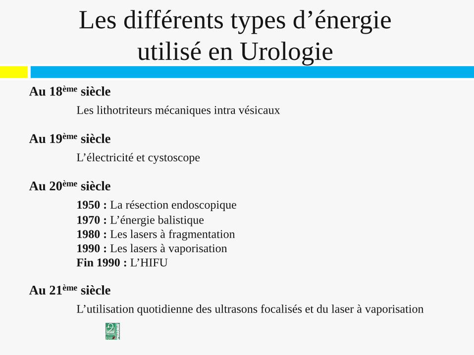
Les différents types d’énergie
utilisé en Urologie Au 18ème siècle
Les lithotriteurs mécaniques intra vésicaux Au 19ème siècle L’électricité et cystoscope Au 20ème siècle 1950 : La résection endoscopique 1970 : L’énergie balistique 1980 : Les lasers à fragmentation 1990 : Les lasers à vaporisation Fin 1990 : L’HIFU Au 21ème siècle L’utilisation quotidienne des ultrasons focalisés et du laser à vaporisation
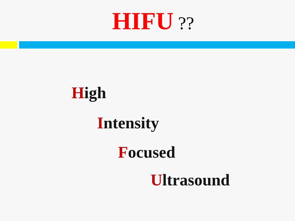
High
HIFU ??
Intensity
Focused
Ultrasound
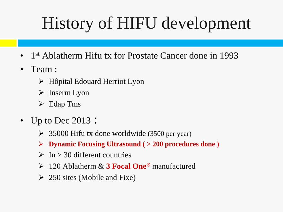
History of HIFU development
• 1st Ablatherm Hifu tx for Prostate Cancer done in 1993 • Team :
Hôpital Edouard Herriot Lyon Inserm Lyon Edap Tms
• Up to Dec 2013 : 35000 Hifu tx done worldwide (3500 per year) Dynamic Focusing Ultrasound ( > 200 procedures done ) In > 30 different countries 120 Ablatherm & 3 Focal One® manufactured 250 sites (Mobile and Fixe)
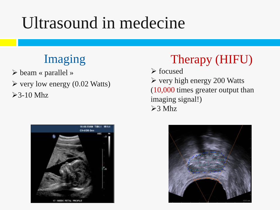
Therapy (HIFU) focused very high energy 200 Watts (10,000 times greater output than imaging signal!) 3 Mhz
Imaging beam « parallel » very low energy (0.02 Watts) 3-10 Mhz
Ultrasound in medecine
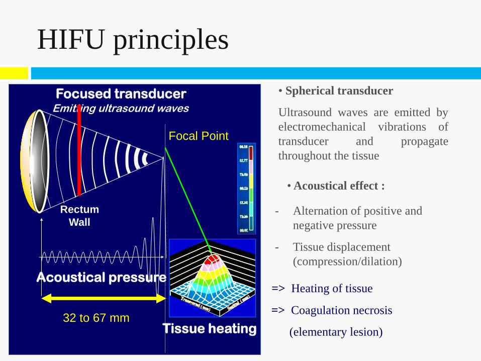
• Spherical transducer
Ultrasound waves are emitted by electromechanical vibrations of transducer and propagate throughout the tissue
- Alternation of positive and negative pressure
- Tissue displacement (compression/dilation)
=> Heating of tissue
=> Coagulation necrosis
(elementary lesion)
• Acoustical effect :
HIFU principles
Rectum Wall
Focused transducer Emitting ultrasound waves
Focal Point
Rectum Wall
32 to 67 mm
Acoustical pressure
Tissue heating
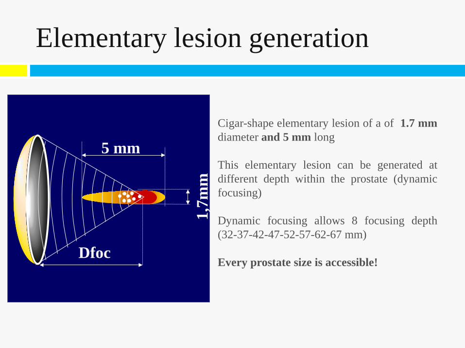
Cigar-shape elementary lesion of a of 1.7 mm diameter and 5 mm long This elementary lesion can be generated at different depth within the prostate (dynamic focusing) Dynamic focusing allows 8 focusing depth (32-37-42-47-52-57-62-67 mm) Every prostate size is accessible!
Dfoc
1,7m
m
5 mm
Elementary lesion generation
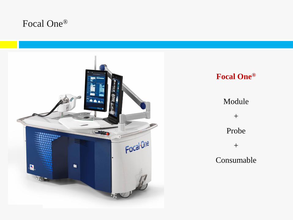
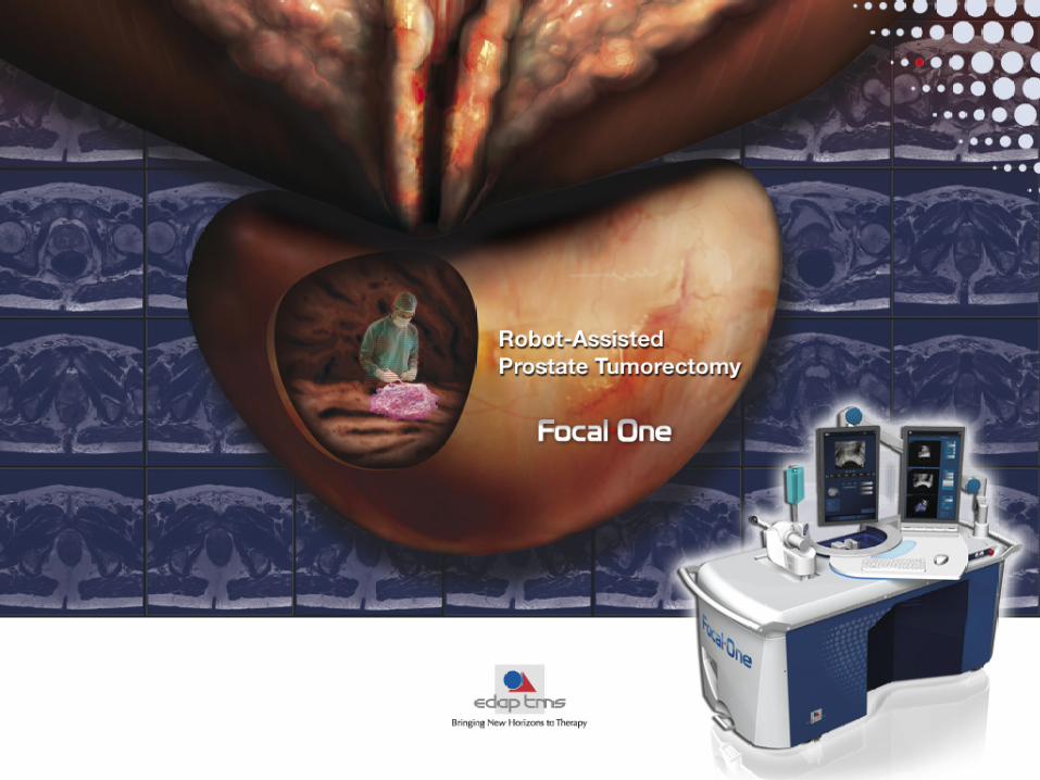
Focal One®
Module
+
Probe
+
Consumable
Focal One®
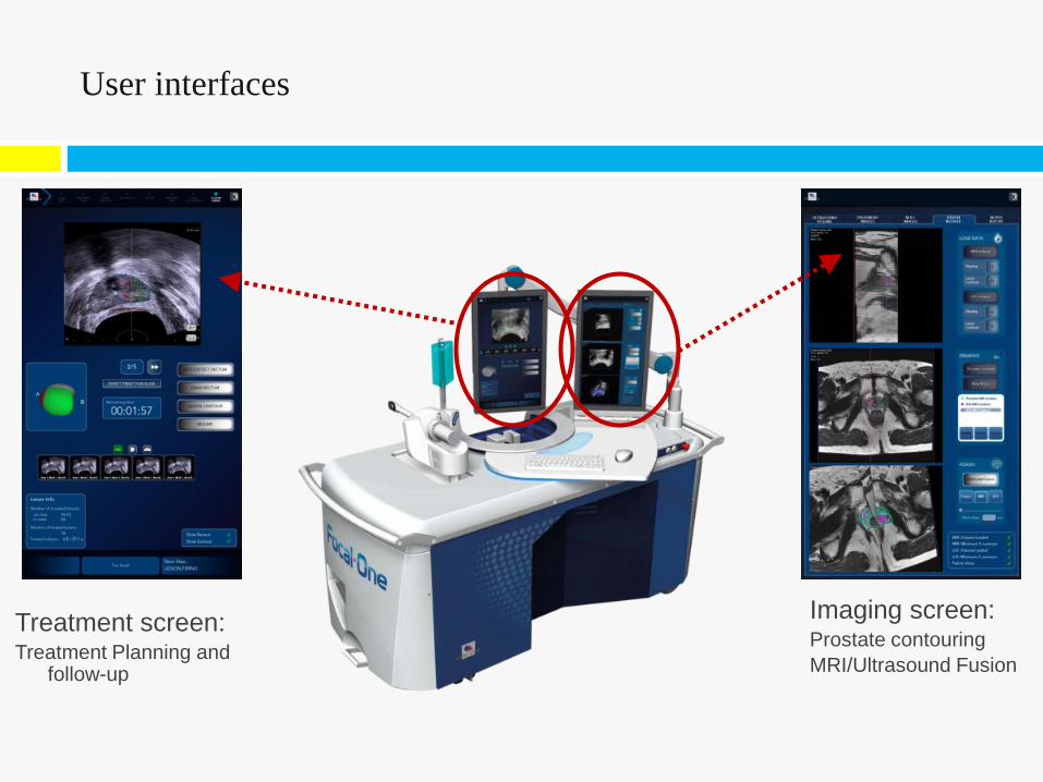
Treatment screen: Treatment Planning and
follow-up
Imaging screen: Prostate contouring MRI/Ultrasound Fusion
User interfaces
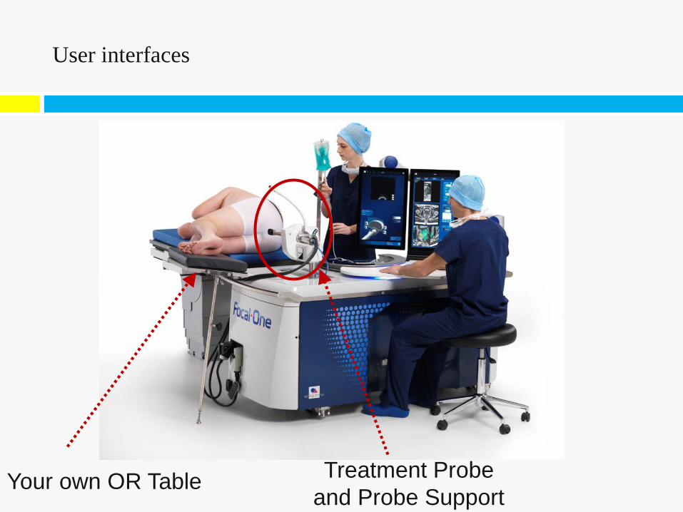
User interfaces
Your own OR Table Treatment Probe and Probe Support
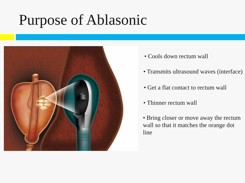
• Cools down rectum wall
Purpose of Ablasonic
• Transmits ultrasound waves (interface)
• Get a flat contact to rectum wall
• Thinner rectum wall
• Bring closer or move away the rectum wall so that it matches the orange dot line
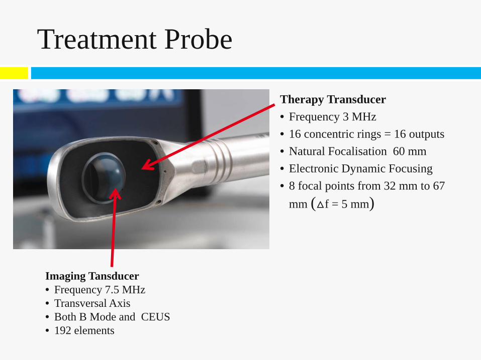
Therapy Transducer • Frequency 3 MHz • 16 concentric rings = 16 outputs • Natural Focalisation 60 mm • Electronic Dynamic Focusing • 8 focal points from 32 mm to 67
mm (△f = 5 mm)
Imaging Tansducer • Frequency 7.5 MHz • Transversal Axis • Both B Mode and CEUS • 192 elements
Treatment Probe
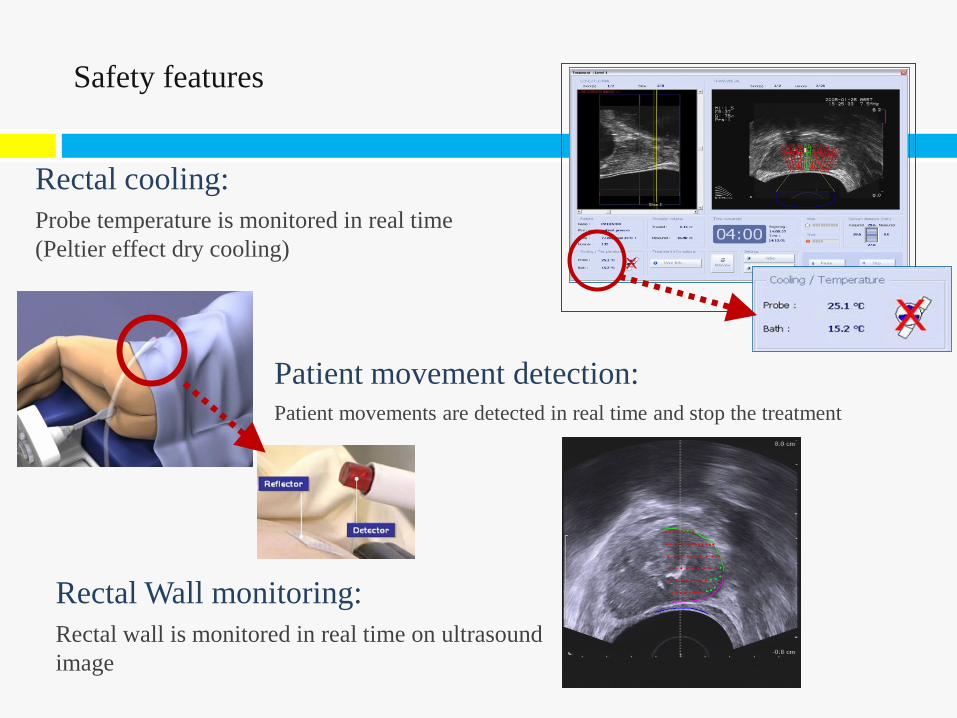
Safety features
Rectal cooling: Probe temperature is monitored in real time (Peltier effect dry cooling)
Patient movement detection: Patient movements are detected in real time and stop the treatment
Rectal Wall monitoring: Rectal wall is monitored in real time on ultrasound image
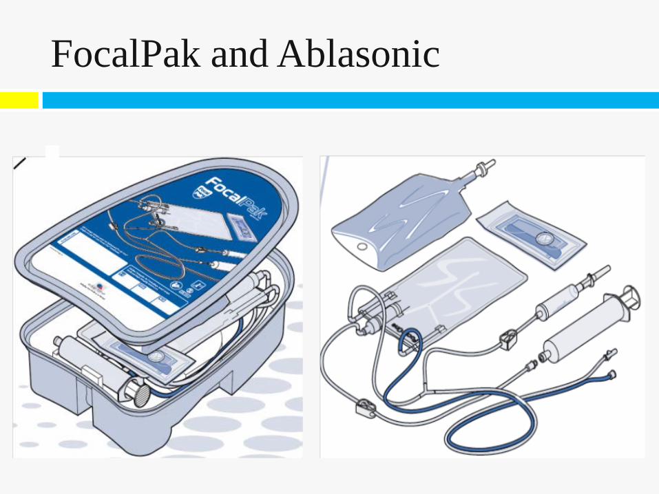
FocalPak and Ablasonic
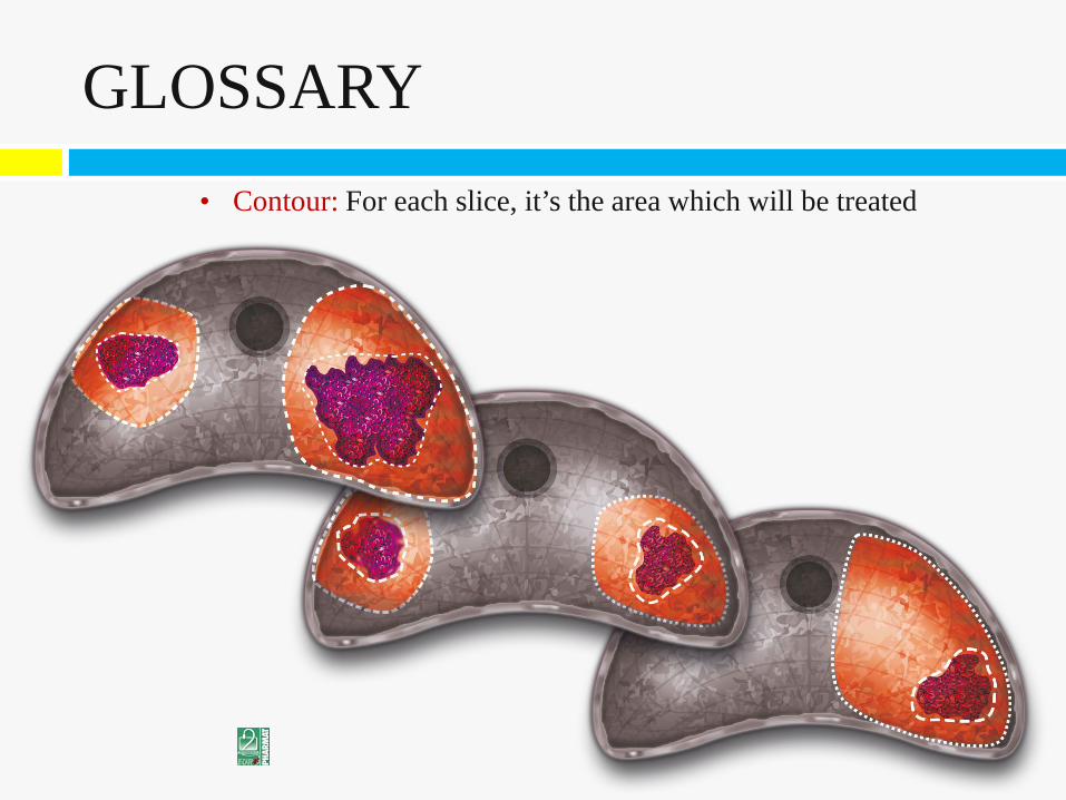
• Contour: For each slice, it’s the area which will be treated
GLOSSARY
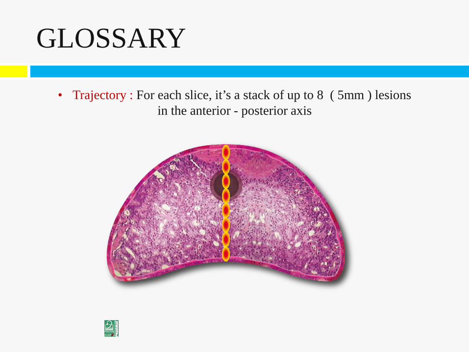
• Trajectory : For each slice, it’s a stack of up to 8 ( 5mm ) lesions in the anterior - posterior axis
GLOSSARY
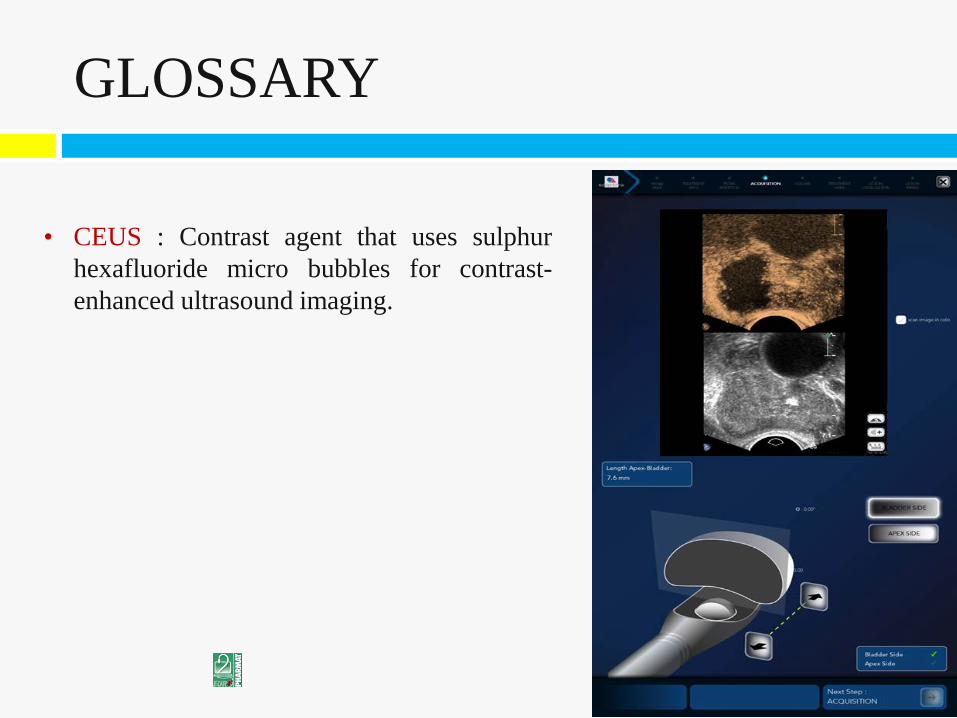
• CEUS : Contrast agent that uses sulphur hexafluoride micro bubbles for contrast-enhanced ultrasound imaging.
GLOSSARY
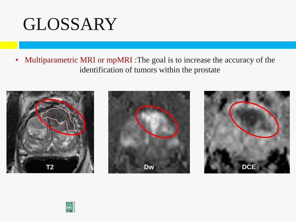
• Multiparametric MRI or mpMRI :The goal is to increase the accuracy of the identification of tumors within the prostate
GLOSSARY
DCE Dw T2
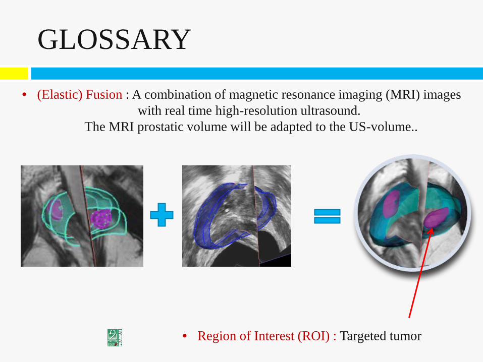
• (Elastic) Fusion : A combination of magnetic resonance imaging (MRI) images with real time high-resolution ultrasound.
The MRI prostatic volume will be adapted to the US-volume..
• Region of Interest (ROI) : Targeted tumor
GLOSSARY
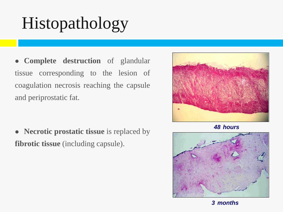
Complete destruction of glandular tissue corresponding to the lesion of coagulation necrosis reaching the capsule and periprostatic fat.
Necrotic prostatic tissue is replaced by fibrotic tissue (including capsule).
48 hours
3 months
Histopathology
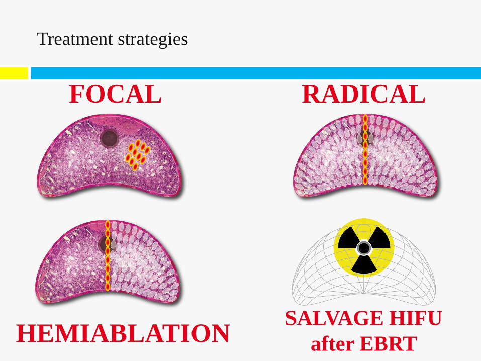
SALVAGE HIFU after EBRT
RADICAL FOCAL
Treatment strategies
HEMIABLATION