Embed Size (px)
Citation preview
Kidney International, Vol. 31(1987), pp. 15—24
Parathyroid response to aluminum in vitro: Ultrastructuralchanges and PTH release
AGNES M. BOURDEAU, JEAN—JACQUES PLACHOT, GIuLIA COURNOT—WITMER,ALAIN POINTILLART, S0NIA BALSAN, and CHARLES SACHS
Laboratoire des Tissus Calcifles, CNRS UA-583 et INSERM U-30, Station de Recherches de Nutrition, INRA, Jouy-en Josas et Départementde Physiologie CHU Necker—Enfants Malades, Paris, France
Parathyroid response to aluminum in vitro: ultrastructural changesand PTH release. The endocrine response of porcine parathyroid glandtissue slices in vitro to aluminum was studied by electron microscopyand radioimmunoassay of PTH. Medium aluminum concentrationswere 20 to 500 ng/ml covering the range corresponding to concentra-tions reported in the plasma of aluminum—intoxicated hemodialyzedpatients. Aluminum inhibited iPTH-release and caused severe cellalterations. This inhibition was incomplete and there was an aluminum—insensitive iPTH-release capacity. This phenomenon seemed to be dueto heterogeneous parathyroid cell population as regards aluminumsensivity, perhaps linked to the spontaneous asynchronous cyclicparathyroid cell changes. Sensitivity to aluminum was modulated by theextra—cellular calcium concentration. Sensitivity to extra—cellular cal-cium concentration variations persisted in aluminum intoxicated tis-sues. The severity of the observed cell lesions induced by highconcentrations of aluminum suggested that the recovery of an iPTH-release capacity when parathyroid tissue was withdrawn from a toxicenvironment and switched to aluminum—free media is more likely to bedue to activation of a "less—sensitive to aluminum" cell pool than to atrue reversibility of the toxic effect.
Considerable evidence has accumulated over the past decadeon the deleterious effects of aluminum in patients being treatedfor chronic renal insufficiency by hemodialysis [1, 2]. Thesource of aluminum could be the dialysate fluid itself [1, 3, 4] orthe oral intake of aluminum containing drugs [5, 6]. Aluminumwas first implicated in the development of dialysis dementia[2—4] and of microcytic anemia [7, 8]. More recently a peculiarbone disease characterized as a vitamin D-resistant osteomala-cia has been reported [9—11]. Several observations have sug-gested that there may be, in addition to the local toxic effect onbone, a direct toxic effect of aluminum on the parathyroidgland: aluminum intoxicated patients have normal or minimallyelevated levels of iPTH [9, 12], poor histological evidence ofosteitis fibrosa [9—11] and their parathyroid gland responsive-ness to acute hypocalcemia is depressed [13, 14]. There isevidence of increased uptake of aluminum by the parathyroidglands [15]. Moreover, in vitro, pharmacological doses ofaluminum inhibit the hormonal release by bovine parathyroidgland cells [16].
The purpose of the present work was to evaluate the toxiceffects of aluminum at the parathyroid ultrastructural level andcorrelate them with hormonal secretion by studying the effect ofaluminum addition to the culture medium of an in vitro prepa-ration of porcine parathyroid tissue slices.
Methods
Preparation of tissue samplePorcine parathyroid glands, (pPTG) obtained within 15 min-
utes of slaughter, were placed in cold tissue culture medium(Krebs—Ringer 5) for transportation to the laboratory and wereused within two hours. After removal of fat and connectivetissues, glands were sliced into 2 mm thick sections. The pPTGslices were pooled in each experimental set and divided intoaliquots (approximately 20 mg) which were enclosed in smallnylon bags (NY 48 HC, 48 t mesh) and placed into 50 mlErlenmeyer flasks (1 bag per flask) containing 5 ml of incubationmedium,
The "basal" incubation medium (BIM) was a modifiedKrebs—Ringer bicarbonate buffer solution [17]. Although fetalcalf serum is usually added to this medium we chose not to doso in order to avoid any unidentified interfering agent [18].
Calcium and magnesium were added as chloride and sulfatesalts at 1.2 and 1 mr't concentrations, respectively. Three levelsof calcium were used: basal (1.2 m total, 0.90 m ionized),low (0.75 mivi total, 0.65 m ionized) and high (2.80 m total,2.10 m ionized).
Aluminum was added to the BIM as the sulfate salt(Merck—Darmstadt, West Germany). Several concentrationswere tested between 25 and 500 ng/ml of aluminum (that is,approximately 1 to 20 tIM) in the range of plasma aluminumconcentrations reported in clinical cases [6, 91. Experimentswere also carried out in an incubation medium containing a lowcalcium concentration to crosscheck the toxicity of aluminumat different levels of PTFI secretion.
Actual medium substance concentrations were systemati-cally measured in each batch solution. The results have beenpublished elsewhere [18].
15
Incubation
Packed tissue slices were incubated at 37°C in flasks gassedwith a 95% oxygen—5% carbon dioxyde mixture, placed in a
Received for publication October 2, 1985and in revised form April 3 and July 2, 1986
© 1987 by the International Society of Nephrology
16 Bourdeau et a!
Dubnoff shaking incubator (agitation rate 60 cycles/mm). Theexperimental sequence was as follows. The pPTG tissue wasfirst incubated for two hours in BIM before being placed forthree hours in the appropriate test medium: basal, high or lowcalcium concentrations for controls, aluminum added media atvarious concentrations of aluminum. All tests were run induplicate. During these five hours the incubation medium wasrenewed hourly. The five media were collected for radioim-munological analysis. At the end of each experiment, repre-sentative samples from the incubates were saved for electronmicroscopic studies. A sample of fresh tissue from the initialpool was also saved as a control. To check the reversibility ofthe aluminum effects on the tissue, in some experiments, tissuesamples were switched for an extra hour to media without anyadded aluminum, with low or basal calcium concentrations. Forpractical reasons all data have been refered to the effectivechronological time. Thus, fifth hour data (which constitutes themain part of the reported data) correspond to three hours ofexposure to the experimental medium (low or high calcium andaluminum supplemented).
Analytical proceduresTotal calcium was measured by atomic absorption spec-
trophotometry (IL-253, Instrument Laboratory, Lexington,Massachusetts, USA); ionized calcium (SS-20, Orion Biomed-ical Research, Cambridge, Massachusetts, USA) and pH andpCO2 (IL-613) by ion selective electrometry; sodium and po-tassium by flame photometry (IL-343); phosphate and total CO2by continuous flow automatic colorimetry (Autoanalyzer, Tech-nicon Instruments, Tarrytown, New York, USA); aluminum byflameless atomic absorption [21].
iPTH radioimmunoassay [22] was performed using synthetichuman 1-34 fragment as standards and for radiolabelling with1251, The antibody used was an anti-1-34 fragment goat antise-rum at a final concentration of 1/110,000. This anti-serum gavestrictly superimposable dilution curves with highly purifiedporcine PTH and synthetic human 1-34 fragment, indicatingcomplete immunological cross—reaction. Non-specific bindingwas measured on the culture medium used in each experiment.The range of the standard curve was 150 to 500 pg/ml. Eachspecimen was assayed in triplicate at three different dilutionsand all samples from one experiment were analyzed in the sameassay. The intra- and inter-assay coefficients of variation wereless than 2% and 4%, respectively. Preliminary studies showedthat aluminum at concentrations ranging from 20 to 1,000 ng/mldid not interfere with the iPTH measurement.
Calculations and statisticsQuantification of iPTH-release is probably more accurate
when expressed per number of cells [16], but since the sampleswere also used for ultrastructural studies, we were obliged towork on tissue slices rather than on dispersed cells. The rate ofiPTH-release, as indicated by the hormone concentration in theincubation medium, depends of the effective number of activeendocrine cells present in the tissue sample. However, thisnumber is not directly correlated with the tissue weight or theprotein content of the tissue slices, even when the slices aretaken from the same gland [23]. iPTH-release (pg/ml) per hour,measured for each individual tissue sample and for each hourhas therefore been expressed as a percentage of a reference
80-
70-
60-
I I I I2 3 4 5
Incubation time, hours
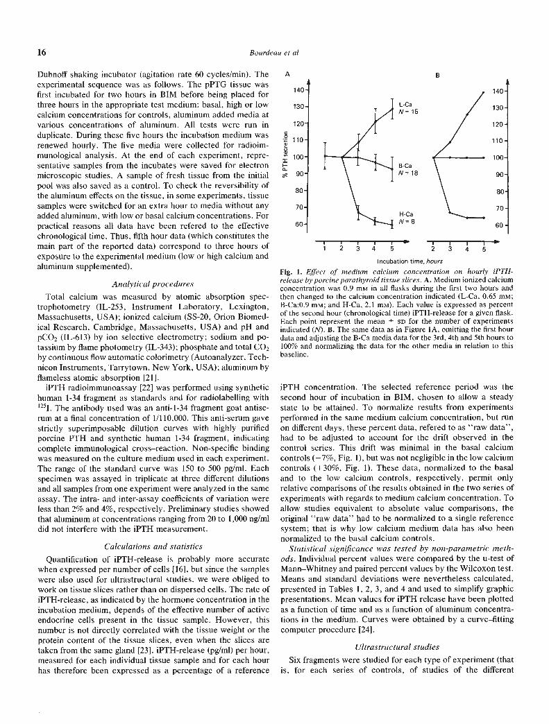
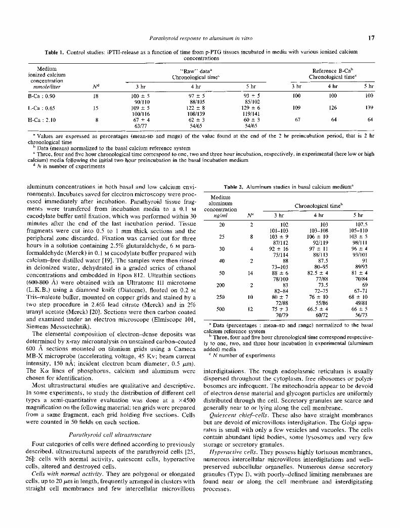
Fig. 1. Effect of medium calcium concentration on hourly iPTH-release by porcine parathyroid tissue slices. A. Medium ionized calciumconcentration was 0.9 mM in all flasks during the first two hours andthen changed to the calcium concentration indicated (L-Ca, 0.65 mM;B-Ca:0.9 mM; and H-Ca, 2.1 mM). Each value is expressed as percentof the second hour (chronological time) iPTH-release for a given flask.Each point represent the mean SD for the number of experimentsindicated (N). B. The same data as in Figure IA, omitting the first hourdata and adjusting the B-Ca media data for the 3rd, 4th and 5th hours to100% and normalizing the data for the other media in relation to thisbaseline.
iPTH concentration. The selected reference period was thesecond hour of incubation in BIM, chosen to allow a steadystate to be attained. To normalize results from experimentsperformed in the same medium calcium concentration, but runon different days, these percent data, refered to as "raw data",had to be adjusted to account for the drift observed in thecontrol series. This drift was minimal in the basal calciumcontrols (—7%, Fig. 1), but was not negligible in the low calciumcontrols (+30%, Fig. 1). These data, normalized to the basaland to the low calcium controls, respectively, permit onlyrelative comparisons of the results obtained in the two series ofexperiments with regards to medium calcium concentration. Toallow studies equivalent to absolute value comparisons, theoriginal "raw data" had to be normalized to a single referencesystem; that is why low calcium medium data has also beennormalized to the basal calcium controls.
Statistical significance was tested by non-parametric meth-ods. Individual percent values were compared by the u-test ofMann—Whitney and paired percent values by the Wilcoxon test.Means and standard deviations were nevertheless calculated,presented in Tables 1, 2, 3, and 4 and used to simplify graphicpresentations. Mean values for iPTH release have been plottedas a function of time and as a function of aluminum concentra-tions in the medium. Curves were obtained by a curve—fittingcomputer procedure [24].
Ultrastructural studiesSix fragments were studied for each type of experiment (that
is, for each series of controls, of studies of the different
A B
140
130 L-CaN= 15
120
- 110
100B-Ca
e 90 N18
80
70H-Ca
60 N=8
140'
130-
120'
110-
100'
90'
I I I
1 2 3 4 5
Para thyroid response to aluminum in vitro 17
Table 1. Control studies: iPTH-release as a function of time from p-PTG tissues incubated in media with various ionized calciumconcentrations
•Medium
ionized calciumconcentrationmmole/liter N'1
"Raw" dataaChronological timec
Reference B-C&'Chronological timec
3 hr 4 hr 5 hr 3 hr 4 hr 5 hr
B-Ca: 0.90 18 100 590/1 10
97 588/105
93 585/102
100 100 100
L-Ca : 0.65 15 109 5100/116
122 8108/139
129 6119/141
109 126 139
H-Ca:2.l0 8 67±463/77
62±354/65
60±354/65
67 64 64
a Values are expressed as percentages (mean-SD and range) of the value found at the end of the 2 hr preincubation period, that is 2 hrchronological time
b Data (means) normalized to the basal calcium reference systemC Three, four and five hour chronological time correspond to one, two and three hour incubation, respectively, in experimental (here low or high
calcium) media following the initial two hour preincubation in the basal incubation mediumd N is number of experiments
aluminum concentrations in both basal and low calcium envi-ronments). Incubates saved for electron microscopy were proc-essed immediately after incubation. Parathyroid tissue frag-ments were transfered from incubation media to a 0.1 Mcacodylate buffer until fixation, which was performed within 30minutes after the end of the last incubation period. Tissuefragments were cut into 0.5 to 1 mm thick sections and theperipheral zone discarded. Fixation was carried out for threehours in a solution containing 2.5% glutaraldehyde, 6 M para-formaldehyde (Merck) in 0.1 M cacodylate buffer prepared withcalcium—free distilled water [19]. The samples were then rinsedin deionized water, dehydrated in a graded series of ethanolconcentrations and embedded in Epon 812. Ultrathin sections(600-800 A) were obtained with an Ultratome III microtome(L.K.B.) using a diamond knife (Diatome), floated on 0.2 MTris—maleate buffer, mounted on copper grids and stained by atwo step procedure in 2.6% lead citrate (Merck) and in 2%uranyl acetate (Merck) [20]. Sections were then carbon coatedand examined under an electron microscope (Elmiscope 101,Siemens Messetechnik).
The elemental composition of electron—dense deposits wasdetermined by x-ray microanalysis on unstained carbon—coated600 A sections mounted on titanium grids using a CamecaMB-X microprobe (accelerating voltage, 45 Ky; beam currentintensity, 150 nA; incident electron beam diameter, 0.5 gm).The Ka lines of phosphorus, calcium and aluminum werechosen for identification.
Most ultrastructural studies are qualitative and descriptive.In some experiments, to study the distribution of different celltypes a semi-quantitative evaluation was done at a x4500magnification on the following material: ten grids were preparedfrom a same fragment, each grid holding five sections. Cellswere counted in 50 fields on each section.
Parathyroid cell ultrastructureFour categories of cells were defined according to previously
described, ultrastructural aspects of the parathyroid cells [25,26]: cells with normal activity, quiescent cells, hyperactivecells, altered and destroyed cells.
Cells with normal activity. They are polygonal or elongatedcells, up to 20 tm in length, frequently arranged in clusters withstraight cell membranes and few intercellular microvillous
interdigitations. The rough endoplasmic reticulum is usuallydispersed throughout the cytoplasm, free ribosomes or polyri-bosomes are infrequent. The mitochondria appear to be devoidof electron dense material and glycogen particles are uniformlydistributed through the cell. Secretory granules are scarce andgenerally near to or lying along the cell membrane.
Quiescent chief—cells. These also have straight membranesbut are devoid of microvillous interdigitation. The Golgi appa-ratus is small with only a few vesicles and vacuoles. The cellscontain abundant lipid bodies, some lysosomes and very fewstorage or secretory granules.
Hyperactive cells. They possess highly tortuous membranes,numerous intercellular microvillous interdigitations and well—preserved subcellular organelles. Numerous dense secretorygranules (Type I), with poorly—defined limiting membranes arefound near or along the cell membrane and interdigitatingprocesses.
Table 2. Aluminum studies in basal calcium mediuma
Mediumaluminum
concentration ___________
ng/ml NC 3 hr
20 2 10210 1—103
25 8 103±987/112
30 4 92±1675/114
40 2 8873—103
50 14 88±678/100
200 2 8382—84
250 10 80±772/88
500 12 75±370/79
Chronological time'
4 hr
103103—108
106 1092/1 19
97 11
88/11387.5
80—9582.5 4
77/88
73.572—75
76 1055/86
66.5 460/72
5 hr
107.5
105—110103 5
98/11196 493/101
9189/93
81 470/84
6967—71
68 1049/81
66 556/73
a Data (percentages : mean—SD and range) normalized to the basalcalcium reference system' Three, four and five hour chronological time correspond respective-ly to one, two, and three hour incubation in experimental (aluminumadded) media
N number of experiments
18 Bourdeau et a!
Table 3. Aluminum stud ies in low calcium medium
Mediumaluminum
concentrationng/ml N"
Reference LCaaChronological timec
Reference BCabChronological timec
3 hr 4 hr 5 hr 3 hr 4 hr 5 hr
25 4 96 2.5
93/9991 1.5
89/9290.5 3.5
86/94105 3
101/108114 1.5
112/115126 5119/130
50 4 88±882/100
85±1576/107
82±280/85
96±989/109
107±18101/135
115±3111/118.5
125 4 78±670/81
76±867/84
69±564/75
85±676/88
96±1084.5/100
96±789/104
250 4 76 13
62/9071 13
58/8967 859/75.5
83 14
69/9889 17
73/11294 11
79/105500 4 72.5±12
58/8764±8
54/7462±5.5
55/6879±13
63/9581±10
68/9387±8
76/95
a Data (percentages : mean-so and range) normalized to the low calcium reference system (Fig. 2B)b Data (percentages : mean-SD and range) normalized to the basal calcium reference system (Fig. 2C)C Three, four and five hour chronological time correspond respectively to one, two and three hour incubation in experimental (aluminum added)
mediadN number of experiments
Table 4. Recovery of the iPTH-release capacity at the sixth hour
.Medium
aluminumng/ml
3rd to 5th hriPTH5th hr
iPTHreleasea
Medium Calcium ConcentrationB-Ca L-Ca
N = 3" N 4b2550
250500
Controls
10280686593
100 5.5 105 8.5
90±2.5 84±1088±11.5 90±888 6 86 11.5
91 —
a Values (percentages : mean-SD) have been normalized to the basalcalcium reference system
bNnumber of experiments for each aluminum concentration
Altered or destroyed cells. These have a reduced volume, andhighly condensed chromatin with partially disintegrated nuclearmembranes. Organelles, with the exception of a few mitochon-dna, are difficult to identify. The cell membranes are disruptedand altered, secretory granules are small and some of them havedense cores which are separated from their limiting membranesby a halo (Type II). These granules are seen in the cytoplasmand lying just outside the cells themselves.
Results
Control studies
Results obtained from the control studies are summarized inFigure 1 and Table 1. They are similar to those of previously
reported in in vitro studies [27, 28]. While mean values ofiPTH-release from slices incubated in B.I.M. (N = 18) did notchange significantly, low calcium medium (N = 15) stimulated1PTH-release up to 130% (P < 0.01) at the fifth hour. On thecontrary, high calcium medium (N = 8) produced a significant
(P < 0.01) decrease in IPTH-release, reaching 60% at the fifthhour.
iPTH-release and time
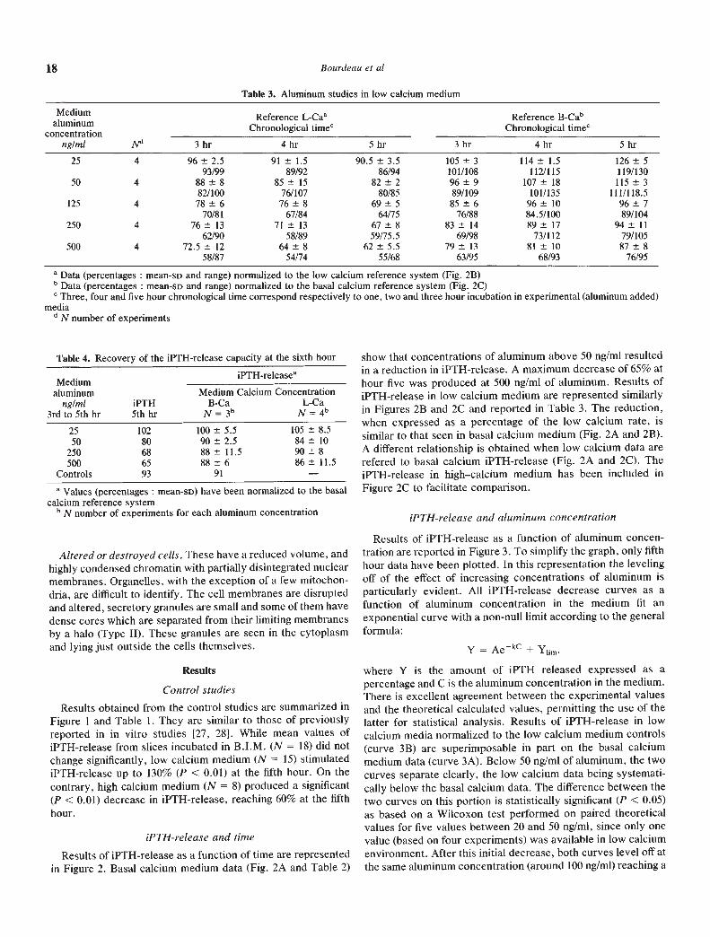
Results of iPTH-release as a function of time are representedin Figure 2. Basal calcium medium data (Fig. 2A and Table 2)
show that concentrations of aluminum above 50 ng/ml resultedin a reduction in 1PTH-release. A maximum decrease of 65% athour five was produced at 500 ng/ml of aluminum. Results ofiPTH-release in low calcium medium are represented similarlyin Figures 2B and 2C and reported in Table 3. The reduction,when expressed as a percentage of the low calcium rate, issimilar to that seen in basal calcium medium (Fig. 2A and 2B).A different relationship is obtained when low calcium data arerefered to basal calcium iPTH-release (Fig. 2A and 2C). TheiPTH-release in high—calcium medium has been included inFigure 2C to facilitate comparison.
iPTH-release and aluminum concentration
Results of iPTH-release as a function of aluminum concen-tration are reported in Figure 3. To simplify the graph, only fifthhour data have been plotted. In this representation the levelingoff of the effect of increasing concentrations of aluminum isparticularly evident. All iPTFI-release decrease curves as afunction of aluminum concentration in the medium fit anexponential curve with a non-null limit according to the generalformula:
Y = Ae_' + Yiim.
where Y is the amount of iPTH released expressed as apercentage and C is the aluminum concentration in the medium.There is excellent agreement between the experimental valuesand the theoretical calculated values, permitting the use of thelatter for statistical analysis. Results of iPTH-release in lowcalcium media normalized to the low calcium medium controls(curve 3B) are superimposable in part on the basal calciummedium data (curve 3A). Below 50 ng/ml of aluminum, the twocurves separate clearly, the low calcium data being systemati-cally below the basal calcium data. The difference between thetwo curves on this portion is statistically significant (P < 0.05)as based on a Wilcoxon test performed on paired theoreticalvalues for five values between 20 and 50 ng/ml, since only onevalue (based on four experiments) was available in low calciumenvironment. After this initial decrease, both curves level off atthe same aluminum concentration (around 100 ng/ml) reaching a
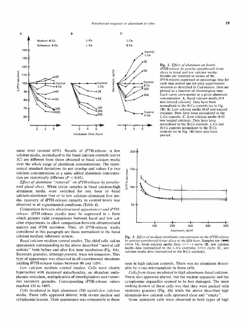
Parathyroid response to aluminum in vitro 19
same level (around 65%). Results of iPTH-release :n lowcalcium media, normalized to the basal calcium controls (curve3C) are different from those obtained in basal calcium mediaover the whole range of aluminum concentrations. The repre-sented standard deviations do not overlap and values f r twocalcium concentrations at a same added aluminum concentra-tion are statistically different (P < 0.01).
Effect of aluminum "removal" on iPTH-release by parathy-roid gland slices. When tissue samples in basal calcium—highaluminum media were switched for one hour to basalcalcium—aluminum free or to low calcium—aluminum free me-dia, recovery of iPTH-release capacity to control levels wasobserved in all experimental conditions (Table 4).
Comparison between ultrastructural appearances and iPTH-release. iPTH-release results must be expressed in . formwhich permits valid comparisons between basal and low cal-cium experiments to allow comparison between ultrastructuralaspects and iPTH secretion. Thus, all iPTH-release esultsconsidered in this paragraph are those normalized to the basalcalcium medium reference system.
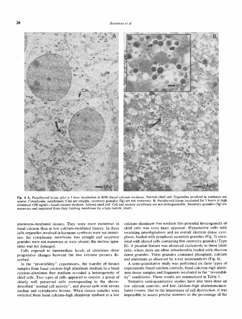
Basal calcium medium control studies. The chief cells md anappearance corresponding to the above described "normal cellactivity" both before and after five hour incubation (Fit;. 4A).Secretory granules, although present, were not numerous. Thistype of appearance was observed in all experimental situationsyielding iPTH-release values between 90 and 110%.
Low calcium medium control studies. Cells were clearlyhyperactive with increased mitochondria, an abundant endo-plasmic reticulum, multiplication of interdigitations and rumer-ous secretory granules. Corresponding iPTH-release valuesreached 130 to 140%.
Cells incubated in high aluminum (500 ng/ml)—low calciummedia. These cells appeared altered, with severe nuclear andcytoplasmic lesions. Their appearance was comparable to those
seen in high calcium controls. There was no aluminum detect-able by x-ray—microanalysis in these cells.
Cells from tissue incubated in high aluminum—basal calcium.These also appeared altered, but the nuclear apparatus and thecytoplasmic organdIes seemed to be less damaged. The moststriking feature of these cells was that they were packed withsecretory granules (Fig. 4B) while the above described highaluminum—low calcium cells appeared clear and "empty".
Some quiescent cells were observed in both types of high
A B C
L-Ca L-Ca
L-Ca 6-Ca
Medium: B-Ca
Reference: B-Ca
i20
C00)
II-0 I
'500
I U I3 4 5 3 4 5 3 4 5
Incubation time, hours
ControlL-Ca
'25Fig. 2. Effect of aluminum on hourly
50 iPTH-release by porcine parathyroid tissueslices in basal and low calcium media.Results are reported as means of theiPTH-release expressed as percentage data for
Control each time period and for each experimental
250a situation as described in Calculation. Data areplotted as a function of chronological time.
500 Each curve corresponds to a given aluminumconcentration. A. Basal calcium media (0.9m ionized calcium). Data have beennormalized to the B-Ca controls (as in Fig.1B). B. Low calcium media (0.65 m ionized
Control calcium). Data have been normalized to theH-Ca L-Ca controls. C. Low calcium media (0.65
mM ionized calcium). Data have been_____ normalized to the B-Ca controls. L-Ca and
H-Ca controls normalized to the B-Cacontrols (as in Fig. 1B) have also beenplotted.
C00C,0=I-a-
200
150
100
50
1
a II
I I P
100 200 300Aluminum, og/mi
400 500
Fig. 3. Effect of medium aluminum concentration on the iPTH-releaseby porcine parathyroid tissue slices at the fifth hour. Samples are: (•)curve 3A, basal calcium media data; (+++) curve 3B, low calciummedia data (normalized to the L-Ca controls); (000) curve 3C, lowcalcium media data (normalized to the B-Ca controls).
—-
I
•; r-' a,-' -•' A
1 -Ft -
-
I4- _,
-- Clu, -
- --'I a
JtS?fI ' ••.IS %.
.c- It'., It
20 Bourdeau et a!
Fig. 4 A. Parathyroid tissue after a 5 hour incubation in BIM (basal calcium medium). Normal chief cell. Organelles involved in synthesis aresparse. Cytoplasmic membranes (Cm) are straight, secretory granules (Sg) are not numerous. B. Parathyroid tissue incubated for 3 hours in highaluminum (500 ng/ml)—basal calcium medium. Altered chief cell. Cell and nuclear membrane are not distinguishable. Secretory granules (Sg) arenumerous and separated from their limiting membrane by a halo (arrow, inset).
aluminum—incubated tissues. They were more numerous inbasal calcium than in low calcium—incubated tissues. In thesecells organelles involved in hormone synthesis were not numer-ous, the cytoplasmic membrane was straight and secretorygranules were not numerous or were absent; the nuclear appa-ratus was not damaged.
Cells exposed to intermediate levels of aluminum showprogressive changes between the two extreme pictures de-scribed.
In the "reversibility" experiments, the transfer of tissuessamples from basal calcium—high aluminum medium to a basalcalcium—aluminum free medium revealed a heterogeneity ofchief cells. Two types of cells appeared to coexist: a group ofclearly well preserved cells corresponding to the above—described "normal cell activity", and altered cells with severenuclear and cytoplasmic lesions. When tissues samples wereswitched from basal calcium—high aluminum medium to a low
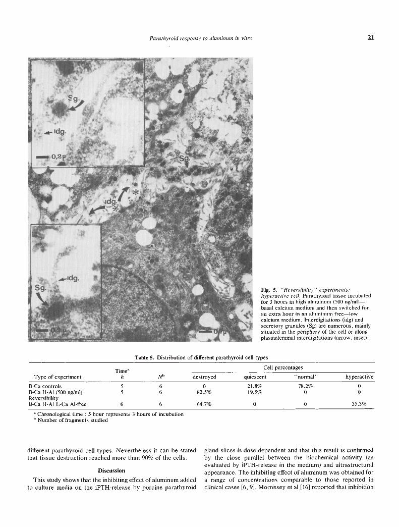
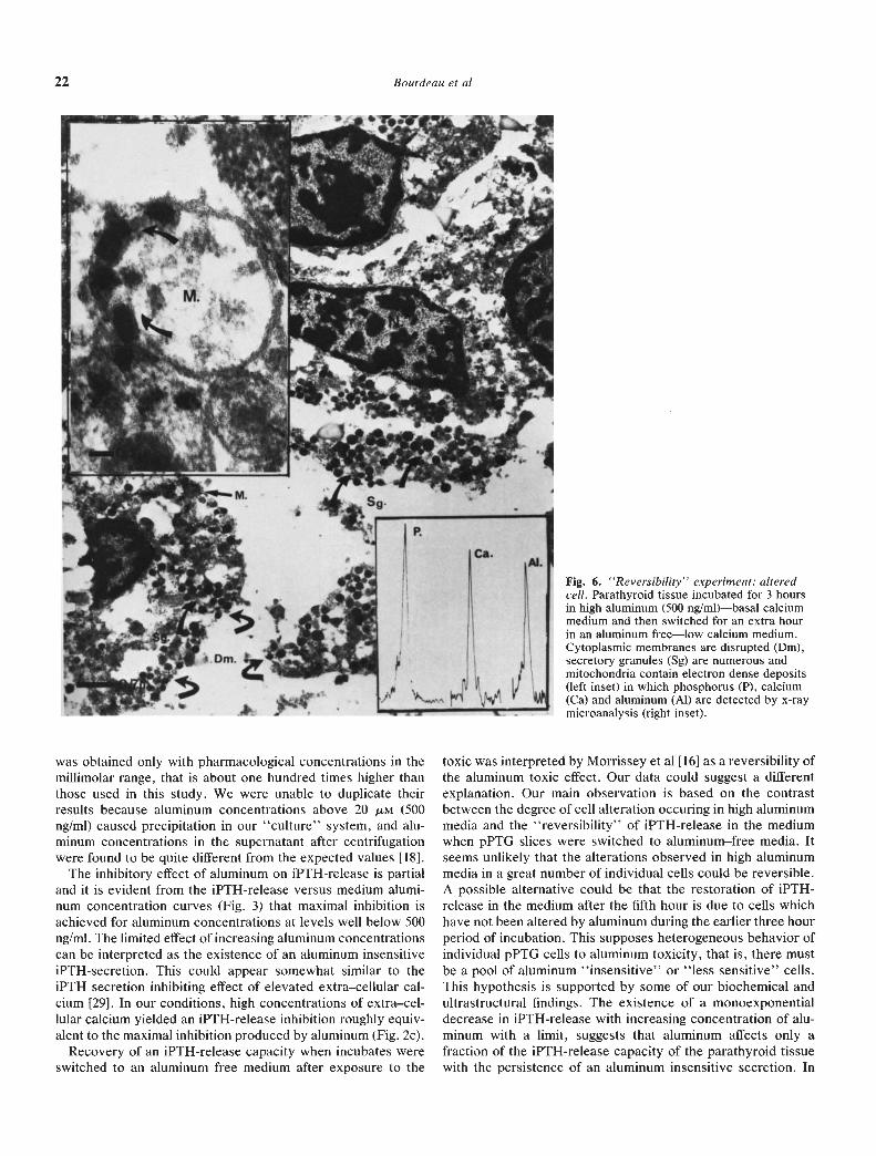
calcium—aluminum free medium this potential heterogeneity ofchief cells was even more apparent. Hyperactive cells withswarming interdigitations and an overall electron dense cyto-plasm, loaded with peripheral secretory granules (Fig. 5) coex-isted with altered cells containing few secretory granules (TypeII). A peculiar feature was observed exclusively in these lattercells, where there are often mitochondria loaded with electrondense granules. These granules contained phosphate, calciumand aluminum as observed by x-ray microanalysis (Fig. 6).
A semi-quantitative study was performed on three types ofexperiments (basal calcium controls, basal calcium—high alumi-num tissue samples and fragments incubated in the "reversibil-ity" conditions). These results are summarized in Table 5.
Tentative semi-quantitative studies have also been done onlow calcium controls, and low calcium—high aluminum—incu-bated tissues. Due to the importance of cell destruction, it wasimpossible to assess precise numbers to the percentage of the
Parathyroid response to aluminum in vitro 21
Fig. 5. Reversibility' experiments:hyperactive cell. Parathyroid tissue incubatedfor 3 hours in high aluminum (500 ng/ml)—basal calcium medium and then switched foran extra hour in an aluminum free—lowcalcium medium. Interdigitations (idg) andsecretory granules (Sg) are numerous, mainlysituated in the periphery of the cell or alongplasmalemmal interdigitations (arrow, inset).
Table 5. Distnbution of different parathyroid cell types
Type of experiment
.Time
h Nb
Cell percentages
destroyed quiescent "normal" hyperactive
B-Ca controls 5 6 0 21.8% 78.2% 0B-Ca H-Al (500 ng/ml) 5 6 80.5% 19.5% 0 0ReversibilityB-Ca H-Al L-Ca Al-free 6 6 64.7% 0 0 35.3%
Chronological time 5 hour represents 3 hours of incubationb Number of fragments studied
different parathyroid cell types. Nevertheless it can be stated gland slices is dose dependent and that this result is confirmedthat tissue destruction reached more than 90% of the cells. by the close parallel between the biochemical activity (as
evaluated by iPTH-release in the medium) and ultrastructuralDiscussion appearance. The inhibiting effect of aluminum was obtained for
This study shows that the inhibiting effect of aluminum added a range of concentrations comparable to those reported into culture media on the iPTH-release by porcine parathyroid clinical cases [6, 9]. Morrissey et al [16] reported that inhibition
— ____
k:
22 Bourdeau Ct a!
was obtained only with pharmacological concentrations in themillimolar range, that is about one hundred times higher thanthose used in this study. We were unable to duplicate theirresults because aluminum concentrations above 20 pM (500ng/ml) caused precipitation in our "culture" system, and alu-minum concentrations in the supernatant after centrifugationwere found to be quite different from the expected values [181.
The inhibitory effect of aluminum on iPTH-release is partialand it is evident from the iPTH-release versus medium alumi-num concentration curves (Fig, 3) that maximal inhibition isachieved for aluminum concentrations at levels well below 500nglml. The limited effect of increasing aluminum concentrationscan be interpreted as the existence of an aluminum insensitiveiPTH-secretion. This could appear somewhat similar to theiPTH secretion inhibiting effect of elevated extra—cellular cal-cium [29]. In our conditions, high concentrations of extra—cel-lular calcium yielded an iPTH-release inhibition roughly equiv-alent to the maximal inhibition produced by aluminum (Fig. 2c).
Recovery of an iPTH-release capacity when incubates wereswitched to an aluminum free medium after exposure to the
Fig. 6. 'Reversibility" experiment: alteredcell, Parathyroid tissue incubated for 3 hoursin high aluminum (500 nglml)—basal calciummedium and then switched for an extra hourin an aluminum free—low calcium medium.Cytoplasmic membranes are disrupted (Dm),secretory granules (Sg) are numerous andmitochondria contain electron dense deposits(left inset) in which phosphorus (P), calcium(Ca) and aluminum (Al) are detected by x-raymicroanalysis (right inset).
toxic was interpreted by Morrissey et al [16] as a reversibility ofthe aluminum toxic effect. Our data could suggest a differentexplanation. Our main observation is based on the contrastbetween the degree of cell alteration occuring in high aluminummedia and the "reversibility" of 1PTH-release in the mediumwhen pPTG slices were switched to aluminum—free media. Itseems unlikely that the alterations observed in high aluminummedia in a great number of individual cells could be reversible.A possible alternative could be that the restoration of 1PTH-release in the medium after the fifth hour is due to cells whichhave not been altered by aluminum during the earlier three hourperiod of incubation. This supposes heterogeneous behavior ofindividual pPTG cells to aluminum toxicity, that is, there mustbe a poo1 of aluminum "insensitive" or "less sensitive" cells.This hypothesis is supported by some of our biochemical andultrastructural findings. The existence of a monoexponentialdecrease in iPTH-release with increasing concentration of alu-minum with a limit, suggests that aluminum affects only afraction of the 1PTH-release capacity of the parathyroid tissuewith the persistence of an aluminum insensitive secretion. In
Parathyroid response to aluminum in vitro 23
pPTG sections incubated in high aluminum medium, irrevers-ibly destroyed cells can be clearly differentiated from cellspresenting no alterations and appearing to be in a quiescentphase. In addition, when the pPTG slices were switched toaluminum—free media, hyperactive cells were found to coexistwith irreversibly destroyed cells which evidently did not re-cover from the aluminum intoxication (Figs, 5 and 6).
These differences in sensitivity to aluminum may be a func-tion of the cell population itself. It is known that parathyroidcells go through a spontaneous cycle from a resting state to asecreting state, with several intermediate phases [25, 26].Resting cells might be insensitive to aluminum or resistant toaluminum entry into the cell.
It has been well documented that changes in extra—cellularcalcium concentrations can stimulate or suppress overall chiefcell activity [30]. This modulation of iPTH-release by extra—cel-lular calcium is still possible in the presence of aluminum asshown by the comparison of curves 3A and 3C. For a givenaluminum concentration, there is more iPTH released (in abso-lute terms) into the medium in low—calcium experiments than inthe basal calcium experiments when data are normalized to asame reference. This agrees with the observed difference in agranule density in cells incubated in low calcium—high alumi-num medium ("empty" cells) as opposed to those maintained innormal calcium—high aluminum environment.
It is also possible that extra—cellular calcium concentrationmight play a role in the aluminum sensitivity process itself byinterfering with the spontaneous cyclic changes in parathyroidcells. However there is little information on the effect ofextra-cellular calcium concentration on parathyroid cell heter-ogeneity. Preliminary results on pPTG cell activities studied bya different approach have also shown the existence of a cellheterogeneity which was more marked during low extracellularcalcium stimulation [311. Some of our biochemical and ultra-structural observations suggest that "sensitivity" to aluminumwould occur predominantly in cells in an activated phase, andstimulation by low extracellular calcium would potentiate thiseffect. In the lower aluminum range (up to 50 ng/ml) there isalways relatively more iPTH released as compared to therespective control in basal calcium medium than in low calciummedium (curves 3A and 3B), while toxic effects level off above50 nglml and are similar in both experimental situations. Celldestruction was more evident in sections from slices treatedwith low calcium—high aluminum media. Altered cells contain-ing mitochondria loaded with phosphate-calcium—aluminumgranules were mainly found in low calcium—high aluminumtreated slices switched to low calcium—aluminum free medium.
These results suggest the following main conclusion: theeffect of aluminum on pPTG cells is dose dependent with aninhibition at levels corresponding to concentrations reported inthe plasma of aluminum intoxicated hemodialized patients. Thisinhibition is partial and there is an aluminum insensitive iPTH-release capacity.
Furthermore this work suggests the following hypotheses:(1.) The phenomenon described could be the result of theheterogeneous behavior of parathyroid cells to aluminum tox-icity, probably linked to spontaneous asynchronous cyclicchanges in the parathyroid cells. (2.) Extra-cellular calciumconcentration could influence the process in two ways: (a)modulation of iPTH-release by extra—cellular calcium varia-
tions could still be possible even in the presence of toxic levelsof aluminum; and (b) cells exposed to low calcium environmentcould be sensitized to aluminum intoxication. (3.) In view of theseverity of the cell lesions observed in high concentrations ofaluminum, the recovery of an iPTH-release capacity when thegland tissues were withdrawn from the toxic environment andswitched to aluminum—free media could more likely be due toan activation of the "less sensitive" to aluminum cell pool (cellswhich were in a resting phase while exposed to aluminum)rather than to a true reversibility of the toxic effect.
AcknowledgmentsPreliminary results of this study have been reported at the VIlith mt.
Conference on Calcium Regulating Hormones, Kobé, Japan, October16—24, 1983 [321. This study was supported by Institut National de IaSante et de la Recherche Médicale grant 85/6981BCR. Anti-l-34 frag-ment goat antiserum used in this study was provided by Dr. M.S.Moukhtar, INSERM U-113, Paris, France. The authors thank MissAnnie De Smet for skilful technical assistance, Pr Bourdon for thealuminum measurements, Pr Lepecq for the computer calculations andMrs. Hernandez for typing the manuscript.
Reprint requests to Agnes M. Bourdeau, Ph.D., Departement dePhysiologie, CHU Necker-Enfants Malades, 156, rue de Vaugirard,75730 Paris Cedex 15, France.
References
1. WARD MK, FEE5T TO, ELLIs HA, PARKINSON IS, KERR DNS:Osteomalacic dialysis osteodystrophy: Evidence for a waterborneetiological agent, probably aluminum. Lancet 1:841—845, 1979
2. ALFREY AC, LEGENDRE OR, KAEHNY WE: Dialysis encephalop-athy syndrome: Possible aluminum intoxication. N EngI J Med294:184—188, 1976
3. PLATTS MM, GOODE GC, HISLOP JS: Composition of the domesticwater supply and the incidence of fractures and encephalopathy inpatients on home dialysis. Br Med J 2:657—660, 1977
4. PARKINSON IS, FEE5T TG, WARD MK, FAWCETT RWP, KERRDNS: Fracturing dialysis osteodystrophy and dialysis encephalop-athy. Lancet 1:406—409, 1979
5. RECKER RR, BLOTCKY AJ, LEFFLER JA, RACK EP: Evidence foraluminum absorption from the gastrointestinal tract and bonedeposition by aluminum carbonate ingestion with normal renalfunction. J Lab C/in Med 90:810—815, 1977
6. BOUKARI M, ROTTENBOURG J, JAUDON MC, CLAVEL JP, LEGRAINM, GALLI A: Influence de la prise prolongee de gels d'alumine surles taux sériques d'aluminium chez les patients atteints d'insuffis-ance rénale chronique. Nouvelle Presse Médicale 7:85—88, 1978
7. SHORT AUK, WINNEY RJ, R0B5ON JS: Reversible microcytichypochromic anemia in dialysis patients due to aluminum intoxica-tion. Proc EDTA 17:226—233, 1980
8. TOUAM M, MARTINEZ F, LACOUR B, BOURDON R, ZINGRAFF J, DiGluLlo S, DRUEKE T: Aluminum—induced reversible microcyticanemia in chronic renal failure: Clinical and experimental studies.C/in Nephrol 19:295—298, 1983
9. COURNOT—WITMER G, ZINGRAFF J, PLACHOT JJ, ESCAIG F,LEFEVRE R, BOUMATI P, BOURDEAU A, GARABEDIAN M, GALLEP, BOURDON R, DRUEKE T, BALSAN S: Aluminum localization inbone from hemodialysed patients: Relationship to matrix mineral-ization. Kidney mt 20:375—385, 1981
10. HODSMAN AB, SHERRARD DJ, ALFREY AC, OTT S, BRICKMAN AS,MILLER NL, MALONEY NA, COBURN JW: Bone aluminum andhistomorphometric features of renal osteodystrophy. J C/in EndocrMetab 54:539—546, 1982
11. OTT SM, MALONEY NA, COBURN JW, ALFREY AC, SHERRARD Di:The prevalence of bone aluminum deposition in renal osteodistro-phy and its relation to the response to calcitriol therapy. N EngI JMed 307, 2:709—713, 1982
12. HODSMAN AB, SHERRARD DJ, WONG EGC, BRICKMAN AS, LEEDB, ALFREY AC, SINGER FR, NORMAN AW, COBURN JW: Vitamin
24 Bourdeau et al
D resistant osteomalacia hemodialysis patients lacking secondaryhyperparathyroidism. Ann Intern Med 94:629—637, 1981
13. KRAUT JA, SHINABERGER JH, SINGER FR, SHERRARD Di, SAXTONJ, MILLER JH, KUROKAWA K, COBURN JW: Parathyroid glandresponsiveness to acute hypocalcemia in dialysis osteomalacia.Kidney mt 23:725—730, 1983
14. ANDRESS D, FELSENFELD AJ, VOIGTS A, LLACH F: Parathyroidhormone response to hypocalcemia in hemodialysis patients withosteomalacia. Kidney mt 24:364—370, 1983
15. CANN L, PRussiN SG, GORDAN GS: Aluminum uptake by theparathyroid glands. J Gun Endocrinol Metab 49:543—545, 1979
16. MORRISSEY JJ, ROTHSTEIN M, MAYOR G, SLATOPOLSKY E: Sup-pression of parathyroid hormone secretion by aluminum. Kidneymt 23:699—704, 1983
17. MORRISSEY JJ, HAMILTON JW, COHN DV: The secretion ofparathormone and glycosylated proteins by parathyroid cells inculture. Biochem Biophys Res Comm 82, 4:1279—1286, 1978
18. BOURDEAU AM, BOURDON R, KINDERMANS C, SACHS CH: Effectof aluminum addition on parathyroid tissue incubation mediumcomposition. Kidney mt 29:924—926, 1986
19. KARLSON U, SCHULTZ R: Fixation of the central nervous systemfor electron microscopy by aldehyde fixation. I. Preservation withspecial reference to membranes and the extracellular space. (ab-stract) J Ultrastruct Res 12:160, 1965
20. REYNOLDS ES: The use of lead citrate at high pH as an electron—opaque stain in electron microscopy. (abstract) J Cell Biol 17:208,1963
21. RANISTEANO—BOURDON S, PROUILLET F, BOURDON R: Dosage del'aluminium et du gallium dans les liquides biologiques. Ann BiolC/in 36:39—44, 1978
22. DE5PLAN C, JULIENNE A, MOUKHTAR MS, MILHAUD G: Sensitiveassay for biologically active fragment of human PTH. (abstract)Lancet ii:198, 1977
23. DIETEL M, DORN 0, MONTZ R, ALTENAHR E: The effect of calciumand dibutyryl-cAMP on the secretion of parathyroid hormone byhuman parathyroid adenomas in organ culture. Acta Endocrinol85:541—547, 1977
24. MARQUARDT DW: An algorithm for least—squares estimation of
nonlinear parameters. J Soc Industr AppI Math 11:431—441, 196325. ROTH SI, RAISZ LU: The course and reversibility of the calcium
effect on the ultrastructure of the rat parathyroid gland in organculture. Lab Invest 15:1187—1211, 1966
26. EBERHARD A: The parathyroid glands, in Electron Microscopy inhuman medicine, edited by JOHANNESSEN JV. New York,McGraw—Hill International Book Company, 10, 3, 111—146, 1981
27. WILLIAMS GA, HARGIS GK, BOWSER EN, HENDERSON Wi,MARTINEZ NJ: Evidence for a role of adenosine 3',5'-monophos-phate in parathyroid hormone release. Endocrinology 92:687—691,1973
28. GOLDEN P, GREENWALT A, MARTIN K, BELLORINT—FONT E,MAZEY R, KLAHR S, SLATOPOLSKY E: Lack of a direct effect of1-25 dihydroxycholecalciferol on parathyroid hormone secretion bynormal bovine parathyroid glands. Endocrinology 107:602—607,1980
29. MAYER GP, HABENER F, POTTS iT JR: Parathyroid hormonesecretion in vivo. Demonstration of a calcium—independent nonsup-pressible composent of secretion. J Clin Invest 57:678—683, 1976
30. TARGOVNIK JH, RODMAN iS, SHERWOOD LM: Regulation ofparathyroid hormone secretion in vitro: Quantitative aspects ofcalcium and magnesium ion control. Endocrinology 88:1477—1482,1971
31. BOURDEAU AM, BACHELET M, ULMANN A, BALSAN 5, SACHs CH:A new approach of the effect of calcium on parathyroid intermedi-ary metabolism: Microdensitometric quantification of cytosolicNADPH production and utilisation, in Proceedings of the VilithConference of Calcium Regulating Hormones, (poster) Kobe,Japan, October 1983. Edited by COHN DV, FUJITA T, POTTS JT JR,TALMAGE R, Amsterdam, Elsevier Science Publishers By., p. 399,1984
32. BOURDEAU AM, PLACHOT ii, COURNOT—WITMER G, POINTILLARTA, SACHS C, BALSAN S: In vitro effects of aluminum on parathyroidglands: Correspondence between hormonal secretion and ultrastruc-tural aspects, in Proceedings of the VIIth Conference of CalciumRegulating Hormones, (oral presentation), Kobe, Japan, October1983. Edited by COHN DV, FUJITA T, POTTS JR iT, TALMAGE R,Amsterdam, Elsevier Science Publishers B.V., pp. 230—231, 1984